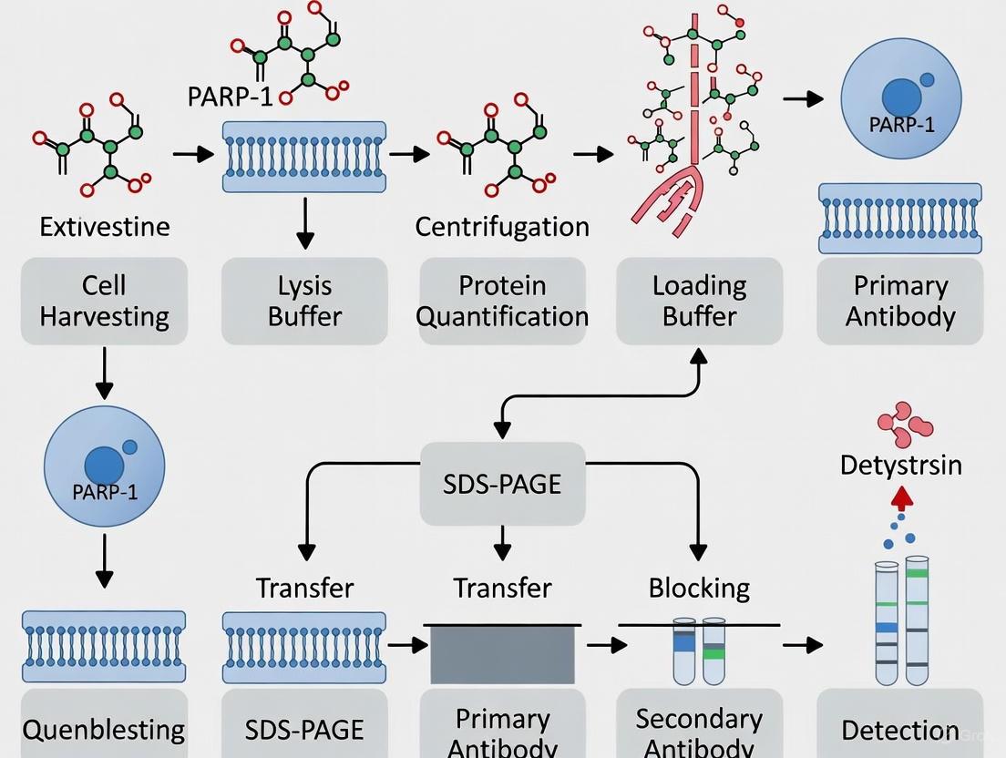
A Complete Guide to Preparing Cell Lysates for PARP-1 Cleavage Western Blot
This guide provides a comprehensive, step-by-step protocol for researchers and drug development professionals to successfully prepare cell lysates for detecting PARP-1 cleavage via western blot, a key marker of apoptosis.
A Complete Guide to Preparing Cell Lysates for PARP-1 Cleavage Western Blot
Abstract
This guide provides a comprehensive, step-by-step protocol for researchers and drug development professionals to successfully prepare cell lysates for detecting PARP-1 cleavage via western blot, a key marker of apoptosis. It covers the foundational biology of PARP-1 and its fragments, a detailed methodological protocol optimized for preserving labile cleavage products, solutions to common troubleshooting challenges, and essential techniques for data validation and interpretation. By integrating the latest research on sample preparation and antibody specificity, this article ensures accurate and reliable detection of PARP-1 cleavage events in diverse experimental contexts.
Understanding PARP-1 Cleavage: Biology, Significance, and Fragment Analysis
The Role of PARP-1 in DNA Repair and as a Central Apoptosis Marker
Poly(ADP-ribose) polymerase-1 (PARP-1) is a ubiquitous nuclear enzyme that plays a dual role in cellular homeostasis, functioning as both a key DNA damage sensor and a central marker in cell death pathways. As the most abundant member of the PARP enzyme family, PARP-1 accounts for approximately 85% of cellular PARP activity and possesses a characteristic multi-domain structure that enables its diverse functions [1]. This application note details the essential methodologies for investigating PARP-1's roles, with particular emphasis on its function as a biomarker for apoptosis and other forms of cell death. The cleavage of PARP-1 by various proteases generates specific signature fragments that serve as recognizable biomarkers for distinct cell death programs, making it an invaluable tool for basic research and drug discovery [1]. Within the context of preparing cell lysates for western blot analysis, understanding these cleavage events is paramount for accurate interpretation of experimental results in DNA repair studies, cancer research, and neurodegeneration.
Biological Background and Significance
Domain Architecture and Molecular Functions
PARP-1 is organized into three primary functional domains that dictate its activity and fate during cellular stress:
- DNA-Binding Domain (DBD): Located at the N-terminus, this domain contains two zinc finger motifs that enable PARP-1 to detect and bind to DNA strand breaks with high affinity. This binding event triggers enzymatic activation [1].
- Automodification Domain (AMD): This central domain contains a BRCT fold (a motif found in many DNA repair proteins) that facilitates protein-protein interactions and serves as the primary target for auto-poly(ADP-ribosyl)ation, which modulates PARP-1's interaction with other proteins and DNA [1].
- Catalytic Domain (CD): Situated at the C-terminus, this domain catalyzes the transfer of ADP-ribose units from β-NAD+ to target proteins, forming linear or branched poly(ADP-ribose) (PAR) chains [1].
In response to DNA damage, PARP-1 binds to DNA breaks and initiates the poly(ADP-ribosyl)ation of itself and various nuclear acceptor proteins. This post-translational modification serves as a critical signal for the recruitment of DNA repair factors and facilitates chromatin relaxation, thereby enabling DNA repair machinery to access damage sites [2]. PARP-1's function extends beyond DNA repair to include regulation of transcription, chromatin remodeling, and modulation of cellular energy metabolism [3] [4].
PARP-1 Cleavage as a Hallmark of Protease Activation
PARP-1 serves as a preferred substrate for multiple proteases activated during different cell death programs. The proteolytic cleavage of PARP-1 generates specific fragments that serve as "signature patterns" for identifying the active proteases and the specific form of cell death occurring in experimental models or pathological conditions [1].
Table 1: PARP-1 Cleavage Fragments Generated by Different Proteases
| Protease | Cleavage Fragments | Primary Cell Death Context | Functional Consequences of Cleavage |
|---|---|---|---|
| Caspase-3/7 | 24 kDa (DBD) + 89 kDa (AMD+CD) | Apoptosis [5] [1] | Inactivation of DNA repair; conservation of cellular energy [1] |
| Caspase-1/7 (Inflammasome) | 24 kDa + 89 kDa | Inflammation-mediated cell death [6] | Enhanced NF-κB target gene expression [6] |
| Calpain | 55-62 kDa fragments | Necrosis, excitotoxicity [1] | Alternative cell death pathway modulation |
| Granzyme A | 50 kDa fragment | Immune-mediated cell killing [1] | PARP-1 degradation without typical apoptotic signature |
| MMP-2/9 | 55-65 kDa fragments | Extracellular matrix remodeling-associated death | Non-apoptotic fragmentation patterns |
The most extensively characterized cleavage event occurs during apoptosis, when caspases-3 and -7 cleave PARP-1 at the DEVD214↓G motif located within the nuclear localization signal of the DBD [5] [6]. This proteolysis produces a 24 kDa fragment containing the DBD and a 89 kDa fragment comprising the AMD and CD [5]. The 24 kDa fragment retains the ability to bind DNA but lacks catalytic activity, while the 89 kDa fragment exhibits reduced DNA binding capacity [1]. This cleavage event is considered a biochemical hallmark of apoptosis and serves to inactivate PARP-1's DNA repair function, thereby preventing futile DNA repair attempts and conserving cellular ATP pools for the execution of the apoptotic program [7] [1].
Beyond its established role in apoptosis, recent evidence indicates that PARP-1 cleavage also participates in regulating inflammatory responses. Studies utilizing noncleavable PARP-1 (PARP-1UNCL) with mutations at the caspase cleavage site (D214N) have demonstrated that PARP-1 cleavage influences NF-κB transcriptional activity and the expression of proinflammatory mediators such as iNOS and COX-2 [5] [7]. Specifically, inflammasome-activated caspase-1 can activate caspase-7, which translocates to the nucleus and cleaves PARP-1 at specific NF-κB target gene promoters, thereby enhancing their expression by removing the repressive influence of full-length PARP-1 [6].
Quantitative Data and Experimental Parameters
Table 2: Key Antibody Reagents for PARP-1 Detection in Western Blotting
| Antibody Target | Catalog Number | Host Species | Applications | Recommended Dilution | Detected Bands | Key Validation Data |
|---|---|---|---|---|---|---|
| Full-length & Cleaved PARP1 | 13371-1-AP (Proteintech) | Rabbit | WB, IHC, IF, IP, FC | 1:1000-1:8000 (WB) | 113-116 kDa (full-length), 89 kDa (cleaved) | KO-validated; detects endogenous full-length and cleaved PARP1 [8] |
| Cleaved PARP1 | ab225715 (Abcam) | Rabbit | WB, IHC-P | 1:100 (WB) | 27 kDa (cleaved fragment) | Recombinant monoclonal; specific for cleaved PARP1; KO-validated [9] |
| PARP1 | #9532 (Cell Signaling) | Rabbit | WB, IP | Manufacturer's recommendation | 113-116 kDa | Used in multiple studies for PARP1 detection [2] |
Detailed Experimental Protocols
Cell Lysate Preparation for PARP-1 Cleavage Detection
Principle: The preparation of high-quality cell lysates is critical for accurate detection of PARP-1 cleavage fragments. This protocol is optimized for preserving both full-length and cleaved PARP-1 while minimizing post-lysis proteolysis.
Materials:
- Lysis Buffer: 50 mM Tris-HCl (pH 7.4), 150 mM NaCl, 1% Triton X-100, 1% NP-40, 0.25% Sodium deoxycholate, 1 mM EDTA [2]
- Protease Inhibitor Cocktail (include caspase inhibitors if studying apoptosis induction)
- Phosphatase Inhibitors (if studying phosphorylation events)
- PMSF (1 mM final concentration) or other serine protease inhibitors
- Cell Scraper (for adherent cells)
- Refrigerated Centrifuge capable of 13,500 × g
Procedure:
- Treatment and Harvest: Treat cells according to experimental design (e.g., apoptosis inducers, DNA damaging agents). For adherent cells, place culture dishes on ice, rapidly aspirate media, and wash twice with ice-cold PBS.
- Lysis: Add appropriate volume of ice-cold lysis buffer containing freshly added protease inhibitors (approximately 100-200 μL per 10⁶ cells). For adherent cells, scrape immediately while submerged in lysis buffer.
- Incubation: Incubate lysates on ice for 30 minutes with occasional vortexing to ensure complete lysis.
- Clarification: Centrifuge lysates at 13,500 × g for 20 minutes at 4°C.
- Collection: Carefully transfer the supernatant (cleared lysate) to a fresh pre-chilled tube.
- Protein Quantification: Determine protein concentration using a compatible assay (e.g., BCA, Bradford).
- Sample Preparation: Mix lysates with 2× SDS-PAGE sample buffer (final 1× concentration) and denature at 95°C for 5-10 minutes.
- Storage: Store aliquots at -80°C if not used immediately. Avoid repeated freeze-thaw cycles.
Technical Notes:
- Inclusion of caspase inhibitors in the lysis buffer is recommended when analyzing basal PARP-1 levels without induced apoptosis to prevent artefactual cleavage during sample preparation.
- For tissues, mechanical disruption (e.g., Dounce homogenization) may be necessary following lysis buffer addition.
- The optimal protein loading amount for PARP-1 detection typically ranges from 20-40 μg per lane for western blotting [9].
Western Blot Analysis of PARP-1 Cleavage
Materials:
- Electrophoresis System: SDS-PAGE gel system (8-12% gels suitable for resolving 24-116 kDa proteins)
- Transfer System: Wet or semi-dry transfer apparatus
- Membrane: Nitrocellulose or PVDF
- Primary Antibodies: Anti-PARP-1 antibodies (see Table 2 for specifications)
- Secondary Antibodies: HRP-conjugated or fluorescently-labeled antibodies appropriate for primary antibody host species
- Blocking Buffer: 5% non-fat dry milk or BSA in TBST
- Detection System: Chemiluminescent, fluorescent, or colorimetric substrate compatible with secondary antibodies
Procedure:
- SDS-PAGE: Load equal protein amounts (20-40 μg) of prepared lysates onto SDS-PAGE gels. Include pre-stained protein molecular weight markers.
- Electrophoresis: Run gels at constant voltage (100-150V) until the dye front reaches the bottom.
- Transfer: Transfer proteins to membrane using appropriate method (wet transfer at 100V for 1 hour or 30V overnight at 4°C).
- Blocking: Incubate membrane in blocking buffer for 1 hour at room temperature with gentle agitation.
- Primary Antibody Incubation: Dilute primary antibody in blocking buffer or antibody dilution buffer according to manufacturer's recommendations (see Table 2). Incubate membrane with primary antibody with gentle agitation overnight at 4°C or for 1-2 hours at room temperature.
- Washing: Wash membrane 3-4 times for 5-10 minutes each with TBST.
- Secondary Antibody Incubation: Incubate membrane with appropriately conjugated secondary antibody (typically 1:10,000-1:20,000 dilution) in blocking buffer for 1 hour at room temperature with gentle agitation.
- Washing: Repeat washing step as above.
- Detection: Develop blots using preferred detection method according to manufacturer's instructions.
Troubleshooting Guide:
- High Background: Increase number or duration of washes; optimize blocking conditions; titrate antibody concentrations.
- Weak or No Signal: Check antibody expiration and storage conditions; verify antigen retrieval; increase protein loading; try enhanced chemiluminescence incubation.
- Non-specific Bands: Include knockout controls; optimize antibody dilution; use more stringent washing conditions.
- Poor Transfer Efficiency: Verify transfer apparatus setup; ensure proper membrane activation; check transfer time and buffer conditions.
Signaling Pathways and Experimental Workflows
Figure 1: PARP-1 Cleavage in Cellular Stress Response Pathways. PARP-1 activation initiates from DNA damage detection, leading to either DNA repair or progression through apoptotic and inflammatory pathways via caspase-mediated cleavage.
The Scientist's Toolkit: Essential Research Reagents
Table 3: Key Research Reagent Solutions for PARP-1 Studies
| Reagent Category | Specific Examples | Research Application | Functional Role |
|---|---|---|---|
| PARP-1 Antibodies | 13371-1-AP (Proteintech), #9532 (CST), ab225715 (Abcam) | WB, IHC, IF, IP | Detection of full-length and cleaved PARP-1; validation via knockout controls essential [8] [9] |
| PARP Inhibitors | Olaparib (FDA-approved), PJ34 | DNA repair studies, synthetic lethality approaches | Inhibition of PARP catalytic activity; induction of synthetic lethality in BRCA-deficient cells [2] |
| Apoptosis Inducers | Staurosporine, Etoposide, Fas antibody | Induction of caspase-dependent PARP-1 cleavage | Activation of caspase-3/7 leading to characteristic PARP-1 cleavage at DEVD214 site [8] [9] |
| Cell Lines | SH-SY5Y, MCF-7, HeLa, Primary cortical neurons | Disease modeling, drug screening | Neuronal ischemia models (SH-SY5Y); breast cancer models (MCF-7) [5] [2] |
| Activity Assays | PAR detection antibodies, NAD+ consumption assays | Measurement of PARP-1 enzymatic activity | Detection of PAR formation; monitoring cellular NAD+ depletion as indicator of PARP-1 activation [7] |
PARP-1 serves as a critical molecular switch governing cell fate decisions in response to genomic insult. The detection of its proteolytic fragments in cell lysates provides invaluable insights into the activation of specific cell death pathways and inflammatory responses. The methodologies detailed in this application note—from optimized cell lysis conditions to validated antibody-based detection—provide researchers with robust tools for investigating PARP-1's dual roles in DNA repair and cell death. These techniques find particular relevance in cancer research (where PARP inhibitors are used clinically), neurodegenerative disease studies (where PARP-1 overactivation contributes to pathology), and inflammatory condition investigations. As research continues to elucidate the complex functions of PARP-1 cleavage fragments beyond their traditional role as apoptosis markers, the standardized protocols presented here will facilitate consistent and reproducible analysis across experimental systems.
{article title}
Caspase-Mediated Cleavage: Generating the Signature 89 kDa and 24 kDa Fragments
Poly(ADP-ribose) polymerase-1 (PARP-1) is a 116 kDa nuclear enzyme that plays a key role in the cellular response to DNA damage. During the early stages of apoptosis, PARP-1 is a primary target for cleavage by executioner caspases. This proteolytic event is a definitive biochemical hallmark of apoptosis and serves to inactivate PARP-1's DNA repair activity, thereby facilitating cellular disassembly. Caspase-mediated cleavage occurs at a specific aspartic acid residue (Asp214), generating signature fragments of 89 kDa and 24 kDa. Detecting this cleavage event via western blotting is a critical technique for researchers confirming apoptosis in experimental models, from cancer drug screening to studies of neurodegenerative diseases. This application note provides a detailed protocol for preparing cell lysates and analyzing PARP-1 cleavage, framing the methodology within the broader context of apoptosis research.
Molecular Mechanism of PARP-1 Cleavage
The cleavage of PARP-1 is a tightly regulated process executed by caspases, a family of cysteine proteases that are central to apoptosis.
- Cleavage Site: Caspases cleave human PARP-1 at the conserved amino acid sequence DEVD²¹⁴↓G, located between its two zinc-finger DNA-binding domains and the catalytic domain [10] [11]. The scission occurs specifically after aspartic acid 214 (Asp214).
- Resulting Fragments: This single cleavage event produces two primary fragments:
- Functional Consequences: The separation of the DNA-binding domain from the catalytic domain effectively inactivates PARP-1 [10] [12]. This prevents the enzyme from responding to DNA damage with massive poly(ADP-ribosyl)ation, which would deplete cellular NAD⁺ and ATP levels. Inactivation of PARP-1 conserves cellular energy and promotes the efficient dismantling of the cell during apoptosis [12].
- Key Caspases Involved: While multiple caspases can cleave PARP-1 in vitro, caspase-3 is considered the primary protease responsible for this event in vivo [13] [11] [14]. However, caspase-7 has also been shown to cleave PARP-1 in non-apoptotic contexts, such as in inflammasome signaling, where it enhances the expression of specific NF-κB target genes [6].
The following diagram illustrates the caspase-mediated cleavage process of full-length PARP-1 and the domains of the resulting fragments.
Quantitative Profile of Cleavage Fragments
The table below summarizes the core quantitative data for the PARP-1 protein and its signature cleavage fragments, which is essential for accurate identification in western blot experiments.
Table 1: Characteristics of PARP-1 and Its Caspase-Generated Fragments
| Protein / Fragment | Molecular Weight (kDa) | Primary Domains | Key Functions |
|---|---|---|---|
| Full-length PARP-1 | 116 | DNA-binding, Automodification, Catalytic | DNA damage repair, NAD⁺ consumption, transcriptional regulation [10] [15] |
| Cleaved PARP-1 (C-terminal) | 89 | Automodification, Catalytic | Inactivated catalytic activity; used as a western blot marker for apoptosis [11] |
| Cleaved PARP-1 (N-terminal) | 24 | DNA-binding | Can bind DNA but lacks catalytic function; may regulate chromatin structure [15] |
The specific caspases responsible for generating these fragments have distinct roles in apoptosis, as detailed in the following table.
Table 2: Caspases Responsible for PARP-1 Cleavage
| Caspase | Role in Apoptosis | Specificity for PARP-1 Cleavage |
|---|---|---|
| Caspase-3 | Key executioner caspase | Primary caspase responsible for cleaving PARP-1 at Asp214 during apoptosis [13] [14]. |
| Caspase-7 | Executioner caspase | Can cleave PARP-1; also activated by caspase-1 in inflammasome signaling to regulate gene expression [6]. |
| Caspase-9 | Initiator caspase (intrinsic pathway) | Activates executioner caspases (3 & 7); does not directly cleave PARP-1 [13]. |
Detailed Experimental Protocol for PARP-1 Cleavage Detection
This section provides a step-by-step methodology for inducing apoptosis, preparing cell lysates, and detecting PARP-1 cleavage via western blotting.
Cell Culture and Apoptosis Induction
- Culture Conditions: Maintain appropriate mammalian cells (e.g., SH-SY5Y neuroblastoma cells, primary cortical neurons, or other relevant models) in their recommended medium and conditions [10] [15].
- Induction of Apoptosis: Treat cells with an apoptotic stimulus. Common methods include:
- Chemical Ischemia: For in vitro modeling of ischemia, subject cortical cultures to glucose-free balanced salt solution containing 0.5 mM 2-deoxyglucose and 5 mM sodium azide for 15 minutes to 2 hours, depending on the desired intensity [10].
- Serum Withdrawal: For intrinsic apoptosis induction, subject cells to serum-free medium for 12-24 hours [13].
- Pharmacological Agents: Treat with agents like staurosporine (1 μM) or other pro-apoptotic compounds for 4-6 hours.
- Caspase Inhibition (Control): To confirm the caspase-dependence of PARP-1 cleavage, pre-treat a group of cells with a pan-caspase inhibitor such as 50 μM Z-VAD-FMK or a specific caspase-3 inhibitor like 20 μM DEVD-CHO for 1 hour prior to the apoptotic stimulus [10].
Cell Lysis and Protein Extraction
The goal is to obtain a high-quality, denatured protein extract while preserving protein modifications and preventing degradation.
- Preparation: Place culture plates on ice and wash cells with cold, sterile phosphate-buffered saline (PBS).
- Lysis Buffer: Use an ice-cold RIPA lysis buffer supplemented with critical additives:
- 1% SDS
- 1 mM Na₃VO₄ (sodium orthovanadate)
- 10 mM Tris-HCl (pH 7.4)
- Protease Inhibitor Cocktail (including 0.1 mM PMSF, 2.5 μg/ml pepstatin, 10 μg/ml aprotinin, 5 μg/ml leupeptin) [10].
- Lysis Procedure: Add a small volume of lysis buffer (e.g., 100-200 μl per 10⁶ cells) directly to the culture dish. Scrape the cells thoroughly and transfer the lysate to a microcentrifuge tube.
- Homogenization and Clarification: Sonicate the lysate briefly (10-15 seconds) on ice to reduce viscosity by shearing genomic DNA. Centrifuge at 12,000-16,000 × g for 15 minutes at 4°C. Carefully transfer the clear supernatant (the protein lysate) to a new tube.
- Protein Quantification: Determine the protein concentration of each lysate using a standard assay like the BCA or Bradford assay.
Western Blot Analysis
- Gel Electrophoresis: Load an equal amount of protein (20-30 μg) per lane on a 8-12% SDS-PAGE gel to achieve optimal separation of the 116 kDa full-length and 89 kDa cleaved PARP-1 fragments.
- Protein Transfer: Transfer the separated proteins from the gel to a nitrocellulose or PVDF membrane.
- Blocking: Block the membrane with 5% non-fat dry milk or BSA in TBST (Tris-Buffered Saline with 0.1% Tween-20) for 1 hour at room temperature to prevent non-specific antibody binding.
- Antibody Incubation:
- Primary Antibody: Incubate the membrane with a anti-PARP-1 primary antibody (e.g., PARP Antibody #9542 from Cell Signaling Technology) that detects both full-length (116 kDa) and the large cleaved fragment (89 kDa) [11]. Dilute the antibody 1:1000 in blocking buffer and incubate overnight at 4°C with gentle agitation.
- Washing: Wash the membrane three times for 5 minutes each with TBST.
- Secondary Antibody: Incubate with an appropriate HRP-conjugated secondary antibody for 1 hour at room temperature.
- Detection: Visualize the protein bands using a enhanced chemiluminescence (ECL) substrate and image the membrane with a digital imager.
- Reprobing (Optional): To confirm equal loading, the membrane can be stripped and reprobed with an antibody for a housekeeping protein, such as β-actin or GAPDH.
The complete workflow for detecting PARP-1 cleavage, from cell treatment to data analysis, is summarized in the diagram below.
The Scientist's Toolkit: Key Research Reagents
The following table lists essential reagents and their specific functions for studying PARP-1 cleavage.
Table 3: Essential Reagents for PARP-1 Cleavage Research
| Reagent / Resource | Function / Specificity | Example Product / Citation |
|---|---|---|
| Anti-PARP-1 Antibody | Detects endogenous levels of full-length (116 kDa) and the large cleaved fragment (89 kDa) of PARP1. | PARP Antibody #9542 (Cell Signaling Technology) [11] |
| Anti-Cleaved Caspase-3 Antibody | Detects the large fragment (17/19 kDa) of activated caspase-3; confirms upstream apoptotic activation. | Cleaved Caspase-3 (Asp175) Antibody #9661 (Cell Signaling Technology) [14] |
| Caspase Inhibitor (Pan) | Broad-spectrum caspase inhibitor; used to confirm caspase-dependence of PARP-1 cleavage. | Z-VAD-FMK or DEVD-CHO [10] [12] |
| Apoptosis Inducer | Triggers the intrinsic or extrinsic apoptotic pathway to induce PARP-1 cleavage. | Staurosporine, Serum Withdrawal, Chemical Ischemia [10] [13] |
| Protease Inhibitor Cocktail | Added to lysis buffer to prevent non-specific proteolysis during sample preparation. | Commercial cocktails containing PMSF, pepstatin, aprotinin, leupeptin [10] |
Data Interpretation and Technical Considerations
- Band Pattern Analysis: A successful apoptosis experiment will show a decrease in the band intensity of the 116 kDa full-length PARP-1 and a corresponding increase in the 89 kDa cleaved fragment. The 24 kDa fragment is less commonly detected in standard western blots, as the antibody provided in the example recognizes the C-terminal portion of the protein [11] [16].
- Quantification: Use densitometry software (e.g., ImageJ) to quantify the band intensities. The ratio of cleaved PARP-1 (89 kDa) to full-length PARP-1 (116 kDa) provides a semi-quantitative measure of the extent of apoptosis [16].
- Controls are Critical: Always include the following controls for valid interpretation:
- Untreated Control: To establish the baseline state.
- Caspase Inhibitor Control: To confirm that cleavage is caspase-dependent. This should significantly reduce or abolish the appearance of the 89 kDa band [10].
- Loading Control: An antibody for a housekeeping protein (e.g., β-actin, GAPDH) to ensure equal protein loading across all lanes.
- Common Challenges: A faint or absent cleavage signal may result from insufficient apoptotic induction, overly rapid processing of cells before caspase activation peaks, or degradation of the protein fragments due to incomplete protease inhibition during lysis [16]. Optimizing the timing of apoptosis induction and ensuring fresh, complete lysis buffer are essential steps for success.
Poly(ADP-ribose) polymerase-1 (PARP-1) is a 116 kDa nuclear enzyme that plays a dual role in cellular stress responses. While its cleavage during apoptosis is a well-established hallmark, its processing during necrosis represents a distinct biochemical pathway with significant implications for cell fate decisions. During apoptosis, caspase-3 and -7 cleave PARP-1 at the DEVD site (Asp214-Gly215 in human PARP-1), generating characteristic fragments of 89 kDa and 24 kDa [12] [17]. This cleavage separates the DNA-binding domain from the catalytic domain, inactivating the enzyme and preventing futile DNA repair cycles during apoptotic execution.
In contrast, necrotic cell death triggers an alternative cleavage pattern through lysosomal proteases, producing different PARP-1 fragments ranging from 40-55 kDa [18] [19]. This necrotic cleavage is not inhibited by broad-spectrum caspase inhibitors like zVAD-fmk, confirming its independence from apoptotic signaling pathways [18]. The discovery of these alternative fragments provides critical insights into the molecular distinctions between apoptotic and necrotic cell death, with important implications for understanding pathological conditions including cerebral ischemia, neurodegenerative diseases, and viral infections [15] [20].
Biochemical Characterization of Necrotic PARP-1 Cleavage
Proteases Involved in Necrotic Cleavage
The necrotic cleavage of PARP-1 is mediated primarily by lysosomal proteases released during cellular disruption. Research using lysosomal-rich fractions from Jurkat T cells demonstrates that cathepsins B and G can cleave affinity-purified PARP-1 into fragments corresponding to those observed in cells treated with necrotic inducers such as 0.1% H₂O₂, 10% EtOH, or 100 μM HgCl₂ [18]. This proteolytic pattern differs significantly from apoptotic cleavage, as summarized in Table 1.
Table 1: Characteristics of PARP-1 Cleavage in Apoptosis vs. Necrosis
| Feature | Apoptotic Cleavage | Necrotic Cleavage |
|---|---|---|
| Primary Triggers | Death receptor activation, DNA damage | Oxidative stress, chemical toxicity, ATP depletion |
| Key Proteases | Caspase-3, -7 | Cathepsins B, D, G |
| Characteristic Fragments | 89 kDa + 24 kDa | 50 kDa (major), 40-55 kDa range |
| Caspase Inhibitor Sensitivity | Sensitive (inhibited by zVAD-fmk) | Insensitive |
| Functional Consequences | Inactivation of DNA repair, energy conservation | Cellular disassembly, inflammatory response |
Structural and Functional Consequences
Necrotic cleavage of PARP-1 generates fragments with potentially distinct biological activities. Research indicates that different PARP-1 fragments can differentially modulate cellular protection through NF-κB-dependent signaling [15]. Expression of a 24 kDa fragment (PARP-124) conferred protection from oxygen/glucose deprivation in neuronal models, while expression of the 89 kDa fragment (PARP-189) was cytotoxic [15]. This suggests that PARP-1 cleavage products may regulate cellular viability and inflammatory responses in opposing ways during ischemic challenges.
The functional impact of PARP-1 cleavage extends to its role as a cofactor for NF-κB. All PARP-1 constructs induce NF-κB translocation into the nucleus during ischemic challenge, but the PARP-189 fragment induces significantly higher NF-κB activity than wild-type PARP-1 [15]. This differential regulation of inflammatory pathways may contribute to the distinct outcomes of apoptotic versus necrotic cell death.
Experimental Protocols for Detection
Cell Lysis and Nuclear Extraction
For comprehensive PARP-1 cleavage analysis, a protocol that preserves both apoptotic and necrotic fragments is essential. The following nuclear extraction method ensures optimal recovery of PARP-1 and its cleavage products:
- Cell Harvesting: Detach cells with trypsin-EDTA and collect by centrifugation at 500 ×g for 5 minutes.
- Hypotonic Lysis: Resuspend cell pellet in 10 mM Hepes (pH 8.0), 10 mM KCl, 1.5 mM MgCl₂, 0.5 mM DTT, and a complete EDTA-free protease inhibitor cocktail. Incubate on ice for 10 minutes.
- Membrane Disruption: Add 0.1% NP-40 and vortex briefly. Centrifuge at 1,500 ×g for 10 minutes at 4°C to separate cytoplasmic (supernatant) and nuclear (pellet) fractions.
- Nuclear Protein Extraction: Resuspend nuclear pellet in RIPA buffer (50 mM Tris-HCl pH 8.0, 150 mM NaCl, 1% NP-40, 0.5% sodium deoxycholate, 0.1% SDS) with protease inhibitors. Incubate on ice for 30 minutes with occasional vortexing.
- Clarification: Centrifuge at 1,500 ×g for 30 minutes at 4°C. Collect supernatant and determine protein concentration using Bradford assay [21].
Western Blotting for PARP-1 Cleavage Fragments
The following protocol optimizes detection of both apoptotic and necrotic PARP-1 fragments:
- Gel Electrophoresis: Separate 30 μg of nuclear protein extracts by 10% SDS-PAGE [21]. For better resolution of smaller fragments (40-55 kDa), 4-20% gradient Tris-Glycine gels can be used [22].
- Membrane Transfer: Transfer proteins to nitrocellulose membrane using standard wet or semi-dry transfer systems. The iBlot transfer apparatus provides efficient transfer for a wide molecular weight range [22].
- Blocking: Incubate membrane in Tris-buffered saline with 0.5% Tween 20 (TBS-T) containing 5% bovine serum albumin (BSA) for 1 hour at room temperature [22].
- Primary Antibody Incubation: Incubate with primary antibody diluted in blocking buffer overnight at 4°C with gentle agitation. See Section 5 for antibody selection guidelines.
- Secondary Antibody Detection: After washing, incubate with HRP-conjugated secondary antibody for 1 hour at room temperature. Detect using enhanced chemiluminescence reagents [22].
Diagram 1: Experimental workflow for PARP-1 cleavage analysis, covering from cell culture to fragment detection.
Biological Significance and Functional Consequences
PARP-1 as a Molecular Switch Between Cell Death Modes
PARP-1 activation and cleavage function as a critical molecular switch determining whether cells undergo apoptosis or necrosis. During apoptosis, caspase-mediated PARP-1 cleavage inactivates the enzyme, preventing NAD+ and ATP depletion, which allows the cell to maintain energy-dependent apoptotic execution [12]. In contrast, during necrosis, PARP-1 overactivation in response to DNA damage consumes large amounts of NAD+, and efforts to resynthesize NAD+ cause massive ATP depletion, shifting cell death toward necrosis [12].
This switch mechanism is particularly evident in death receptor signaling. In L929 cells, CD95 ligation induces apoptosis with characteristic PARP-1 cleavage, while TNF treatment triggers PARP-1 activation leading to ATP depletion and subsequent necrosis [12]. The caspase inhibitor zVAD-fmk prevents CD95-mediated apoptosis but potentiates TNF-induced necrosis by preventing PARP-1 cleavage and thus exacerbating ATP depletion [12].
Pathophysiological Implications
The distinct PARP-1 cleavage patterns have significant implications for various pathological conditions:
- Cerebral Ischemia: PARP-1 cleavage products differentially modulate neuronal survival following oxygen/glucose deprivation. The 24 kDa fragment confers protection, while the 89 kDa fragment promotes cytotoxicity [15].
- Viral Infection: Zika virus infection activates PARP-1, leading to NAD+ and ATP depletion and subsequent cell death [20].
- Inflammatory Responses: Different PARP-1 fragments differentially regulate NF-κB activity and subsequent expression of inflammatory mediators like iNOS and COX-2 [15].
Table 2: Functional Consequences of Different PARP-1 Fragments
| PARP-1 Form | Effect on Cell Viability | Impact on NF-κB Activity | Effect on Inflammatory Mediators |
|---|---|---|---|
| Full-length (116 kDa) | Baseline | Baseline | Baseline |
| Uncleavable Mutant | Increased viability in OGD | Similar to wild-type | Decreased iNOS and COX-2; Increased Bcl-xL |
| 24 kDa Fragment | Protective | Similar to wild-type | Decreased iNOS and COX-2; Increased Bcl-xL |
| 89 kDa Fragment | Cytotoxic | Significantly increased | Increased iNOS and COX-2; Decreased Bcl-xL |
| Necrotic Fragments (40-55 kDa) | Not fully characterized | Not fully characterized | Not fully characterized |
Research Reagent Solutions and Technical Considerations
Antibody Selection for PARP-1 Cleavage Detection
The detection of specific PARP-1 cleavage fragments requires careful antibody selection, as different antibodies recognize distinct epitopes and fragments. Table 3 summarizes key antibodies and their specificities for detecting apoptotic versus necrotic PARP-1 cleavage fragments.
Table 3: Antibody Reagents for PARP-1 Cleavage Detection
| Antibody | Specificity | Recognized Fragments | Applications | Technical Notes |
|---|---|---|---|---|
| Cleaved PARP (Asp214) (D64E10) XP Rabbit mAb #5625 [19] | Caspase-cleaved PARP-1 at Asp214 | 89 kDa apoptotic fragment | Western Blot, ICC | Does not recognize necrotic fragments (40-55 kDa) |
| PARP Antibody #9542 [17] | Caspase cleavage site | Full-length (116 kDa) and 89 kDa apoptotic fragment | Western Blot | Does not recognize necrotic fragments |
| PARP (46D11) Rabbit mAb #9532 [19] | Not fully specified | May recognize 55 kDa necrotic fragment | Western Blot | May cross-react with some necrotic fragments |
| Anti-Cleaved PARP1 antibody [Y34] (ab32561) [23] | p85 cleaved form of PARP1 | 85 kDa apoptotic fragment | WB, IP, ICC/IF, Flow Cyt | Recombinant format for batch consistency |
Critical Validation and Controls
Appropriate experimental controls are essential for accurate interpretation of PARP-1 cleavage data:
- Genetic Controls: Use PARP-1 knockout cells or tissues to confirm antibody specificity [24] [23].
- Induction Controls: Include cells treated with known apoptotic inducers (e.g., 4μM camptothecin for 5 hours) and necrotic inducers (e.g., 0.1% H₂O₂) [18] [23].
- Specificity Controls: For necrotic cleavage, confirm insensitivity to caspase inhibitors like zVAD-fmk [18].
- Loading Controls: Use nuclear protein loading controls such as B23 for nuclear extracts [21].
Diagram 2: PARP-1 cleavage pathways in apoptosis versus necrosis, showing different inducers, proteases, and functional consequences.
The detection and characterization of PARP-1 cleavage fragments beyond the classical apoptotic pattern provides valuable insights into alternative cell death mechanisms. The 40-55 kDa fragments generated during necrosis represent a distinct proteolytic signature with potentially unique functional consequences. Researchers investigating PARP-1 cleavage should employ specific lysate preparation methods, select appropriate antibody reagents, and include rigorous controls to distinguish between these different cleavage events. Understanding the full spectrum of PARP-1 processing enhances our ability to diagnose cell death modes in physiological and pathological contexts, potentially informing therapeutic strategies for conditions where the balance between apoptosis and necrosis determines disease outcomes.
Poly(ADP-ribose) polymerase-1 (PARP-1) is a 116 kDa nuclear enzyme that plays a central role in detecting and repairing DNA damage through its function in the base excision repair pathway [5] [1]. Beyond its DNA repair capabilities, PARP-1 influences diverse cellular processes including transcription, inflammation, and energy metabolism [5]. The cleavage of PARP-1 by various cell death proteases represents a critical control point that determines cellular fate, shifting its function from a survival molecule to a participant in cell death pathways [1]. This application note examines the functional consequences of PARP-1 cleavage, with particular emphasis on practical methodologies for detecting cleavage fragments in western blot experiments, providing researchers with essential tools for investigating cell death mechanisms.
Biological Significance of PARP-1 Cleavage
Caspase-Mediated Cleavage and Apoptosis
PARP-1 is a well-established substrate for caspase proteases during apoptosis. Caspases-3 and -7 cleave PARP-1 at the conserved DEVD214-G motif, separating the 24 kDa DNA-binding domain (DBD) from the 89 kDa automodification and catalytic domain [5] [25]. This cleavage event serves as a definitive biochemical marker of apoptosis, with the 89 kDa fragment being widely detected as an indicator of caspase activation [26] [1].
The biological consequences of this cleavage are significant: the 24 kDa fragment retains the DNA-binding capability but irreversibly binds to DNA strand breaks, acting as a trans-dominant inhibitor of DNA repair [25] [1]. Meanwhile, the 89 kDa fragment, which contains the catalytic domain, is inactivated regarding its DNA repair function but gains new functions in cell death signaling [25]. This cleavage event conserves cellular energy by preventing excessive NAD+ and ATP consumption that would otherwise occur through PARP-1 overactivation [5].
Differential Functions of Cleavage Fragments in Cell Fate Decisions
Research has revealed that PARP-1 cleavage fragments exert opposing effects on cell survival and inflammatory responses:
Table 1: Functional Consequences of PARP-1 and Its Cleavage Fragments
| PARP-1 Form | Effect on Cell Viability | Effect on NF-κB Activity | Downstream Consequences |
|---|---|---|---|
| Full-length PARP-1 | Maintains viability through DNA repair | Serves as NF-κB cofactor | Promotes DNA repair and cell survival |
| Uncleavable PARP-1 (PARP-1UNCL) | Cytoprotective in OGD/ROG models | Similar induction of NF-κB nuclear translocation | Decreases iNOS and COX-2; increases Bcl-xL |
| 24 kDa Fragment (PARP-124) | Cytoprotective in OGD/ROG models | Similar induction of NF-κB nuclear translocation | Decreases iNOS and COX-2; increases Bcl-xL |
| 89 kDa Fragment (PARP-189) | Cytotoxic | Significantly higher NF-κB activity | Increases COX-2 and iNOS; decreases Bcl-xL |
Studies utilizing oxygen/glucose deprivation (OGD) and OGD/restoration of oxygen and glucose (ROG) models demonstrate that expression of uncleavable PARP-1 (PARP-1UNCL) or the 24 kDa fragment (PARP-124) confers protection from ischemic damage, while expression of the 89 kDa fragment (PARP-189) is cytotoxic [5]. These differential effects are not accompanied by changes in cellular PAR or NAD+ levels, but rather correlate with modified NF-κB transcriptional activity and altered expression of inflammatory mediators including iNOS and COX-2, as well as the anti-apoptotic protein Bcl-xL [5].
Novel Roles of the 89 kDa Fragment in Programmed Cell Death
Recent research has revealed that the 89 kDa PARP-1 fragment serves as a carrier for poly(ADP-ribose) (PAR) polymers, facilitating their translocation from the nucleus to the cytoplasm during caspase-dependent apoptosis [25]. This PAR-bound 89 kDa fragment interacts with apoptosis-inducing factor (AIF) in the cytoplasm, promoting AIF release from mitochondria and its subsequent translocation to the nucleus, where it contributes to nuclear shrinkage and large-scale DNA fragmentation [25]. This pathway represents a convergence point between caspase-dependent apoptosis and PARthanatos, a caspase-independent programmed cell death pathway [25].
PARP-1 Cleavage Detection: Reagent Solutions and Methodologies
Research Reagent Solutions
Table 2: Commercially Available Antibodies for Detecting Cleaved PARP-1
| Product Name | Host Species | Reactivity | Applications | Recommended Dilution | Specificity |
|---|---|---|---|---|---|
| Cleaved PARP (Asp214) Antibody #9541 [26] | Rabbit | Human, Mouse | WB, Simple Western | 1:1000 (WB) | Detects 89 kDa fragment only |
| PARP1 (cleaved Asp214) Antibody (14-6668-82) [27] | Mouse | Human | WB | 0.1-0.25 µg/mL | Detects 85 kDa fragment only |
| Cleaved PARP1 Antibody (60555-1-Ig) [28] | Mouse | Human, Mouse, Rat | WB, IHC, IF/ICC, FC, ELISA | 1:5000-1:50000 (WB) | Detects cleaved form only |
Nuclear Extraction Protocol for PARP-1 Cleavage Detection
For optimal detection of PARP-1 cleavage fragments, particularly in western blot applications, preparation of high-quality nuclear extracts is essential. The following protocol has been adapted from established methodologies [21]:
Cell Harvesting: Detach cells using trypsin-EDTA and collect by centrifugation.
Hypotonic Lysis:
- Resuspend cell pellet in 10 mM HEPES (pH 8.0), 10 mM KCl, 1.5 mM MgCl₂, 0.5 mM DTT, and complete EDTA-free protease inhibitor cocktail.
- Incubate on ice for 10 minutes.
- Add 0.1% NP-40 and mix thoroughly to lyse cells.
- Centrifuge at 1,500 × g for 10 minutes at 4°C.
Nuclear Extraction:
- Resuspend nuclear pellet in RIPA buffer (50 mM Tris-HCl pH 8.0, 150 mM NaCl, 1% NP-40, 0.5% sodium deoxycholate, 0.1% SDS) with protease inhibitors.
- Incubate on ice for 30 minutes with occasional vortexing.
- Centrifuge at 1,500 × g for 30 minutes at 4°C.
- Collect supernatant as nuclear extract.
Protein Quantification: Determine protein concentration using Bradford assay [21].
Western Blot Analysis of PARP-1 Cleavage
For detection of PARP-1 cleavage fragments:
Electrophoresis: Separate 30-40 µg of nuclear protein extract by 10% SDS-PAGE [21].
Transfer: Transfer proteins to nitrocellulose or PVDF membrane using standard western blot transfer techniques.
Immunodetection:
- Block membrane with 5% BSA in TBST for 1 hour.
- Incubate with primary antibody diluted in blocking buffer overnight at 4°C (refer to Table 2 for recommended dilutions).
- Wash membrane and incubate with appropriate HRP-conjugated secondary antibody.
- Detect using chemiluminescent substrates.
Loading Control: Use B23 antibody (1:2000 dilution) as a nuclear protein loading control [21].
PARP-1 in Cell Death Pathways: Visualization
Experimental Workflow for PARP-1 Cleavage Studies
Technical Considerations for PARP-1 Cleavage Research
Quantitative Western Blot Best Practices
Attaining reliable quantitative data from western blot experiments requires careful attention to multiple factors throughout the experimental process [29]. Key considerations include:
- Linear Range Detection: Ensure that antibody detection falls within the linear range of detection to allow accurate quantification.
- Appropriate Normalization: Use proper loading controls (e.g., B23 for nuclear proteins) to account for sample loading variations.
- Antibody Validation: Confirm antibody specificity for the target cleavage fragment through appropriate controls.
- Sample Preparation: Utilize optimized extraction buffers to maintain protein integrity and modifications.
Multiple Proteases Generate Distinct PARP-1 Fragments
While caspases are the most well-characterized proteases that cleave PARP-1, several other "suicidal" proteases can process PARP-1 into distinct signature fragments [1]:
- Calpains: Generate 55-62 kDa fragments
- Cathepsins: Produce 50 kDa fragments
- Granzymes: Create 50 kDa and 64 kDa fragments
- Matrix Metalloproteinases: Yield 42-55 kDa fragments
These alternative cleavage events represent different cell death programs and should be considered when interpreting PARP-1 cleavage patterns, particularly in pathological contexts where multiple proteases may be activated simultaneously [1].
PARP-1 cleavage represents a critical control point in cell fate decisions, with the resulting fragments executing distinct and often opposing functions in survival and death signaling. The 89 kDa fragment, once considered merely an inactive byproduct of caspase cleavage, is now recognized as an active participant in cell death pathways through its role as a PAR carrier that facilitates AIF-mediated DNA fragmentation. Detection of this fragment through carefully optimized western blot protocols provides researchers with a valuable tool for investigating apoptotic mechanisms in both basic research and drug development contexts. The methodologies outlined in this application note offer a robust framework for preparing cell lysates and detecting PARP-1 cleavage fragments, enabling researchers to accurately monitor this key event in cell death pathways.
Step-by-Step Protocol for Lysate Preparation and PARP-1 Immunoblotting
The integrity of cell death research fundamentally depends on robust pre-lysis procedures. For the specific detection of PARP-1 cleavage, a well-established hallmark of apoptosis, careful preparation of cell lysates is paramount. This application note details critical considerations for apoptosis induction and subsequent cell handling to ensure the accurate detection of the characteristic 89 kDa cleavage fragment of PARP-1 by western blot, while avoiding potential artifacts that could compromise data interpretation [30] [18].
PARP-1, a 116 kDa nuclear enzyme, is a crucial DNA repair protein. During apoptosis, it is specifically cleaved by caspase-3 and caspase-7 at the Asp214-Gly215 bond, separating its N-terminal DNA-binding domain (24 kDa) from its C-terminal catalytic domain (89 kDa) [30] [5]. This cleavage event inactivates the enzyme and facilitates cellular disassembly. The detection of the 89 kDa fragment serves as a definitive marker for apoptotic activity, making it a key readout in cell death studies, drug development, and cancer research [30].
Apoptosis Signaling Pathways and PARP-1 Cleavage
Understanding the pathways leading to PARP-1 cleavage is essential for selecting an appropriate induction method. The following diagram illustrates the primary apoptotic pathways and their convergence on caspase-3 activation, which directly cleaves PARP-1.
Diagram 1: Apoptotic Signaling Pathways Converging on PARP-1 Cleavage. The extrinsic (death receptor) and intrinsic (mitochondrial) pathways ultimately activate executioner caspases that cleave PARP-1, generating the 89 kDa apoptotic fragment.
Methods for Apoptosis Induction
Selecting an appropriate apoptosis inducer depends on the cell type, biological question, and experimental timeline. Both biological and chemical methods are reliable for inducing PARP-1 cleavage.
Biological Induction via Death Receptors
This method provides a specific, receptor-mediated induction of apoptosis, particularly effective in immune cells like Jurkat cells [31].
Protocol: Anti-Fas Antibody-Induced Apoptosis
- Cell Preparation: Grow Jurkat cells in RPMI-1640 medium supplemented with 10% fetal bovine serum (FBS) in a humidified 5% CO₂ incubator at 37°C.
- Harvesting: Harvest exponentially growing cells (density of 1 x 10⁵ cells/mL) by centrifugation at 300–350 x g for 5 minutes.
- Resuspension: Resuspend the cell pellet in fresh, pre-warmed medium to a final density of 5 x 10⁵ cells/mL.
- Induction: Add an appropriate concentration of anti-Fas (anti-CD95) monoclonal antibody to the cell suspension.
- Incubation: Incubate the cells for 2–4 hours in a 37°C incubator.
- Control: Include a negative control of untreated cells (without anti-Fas antibody) incubated under identical conditions [31].
Chemical Induction using DNA-Damaging Agents
Chemical inducers are broadly applicable across many cell types and work primarily through the intrinsic pathway by causing DNA damage or cellular stress.
Protocol: Chemical Induction of Apoptosis
- Cell Seeding: Inoculate adherent cells into 10 cm² tissue culture dishes or suspension cells into T75 flasks at a density of ~1 x 10⁶ cells/mL.
- Agent Preparation: Prepare stock solutions of chemical inducers and dilute them to the required working concentration in culture medium.
- Treatment: Add the cellular-damaging agent to the culture medium. The table below provides recommended concentrations for common inducers.
- Incubation and Harvest: Harvest cells at different time points (e.g., 8, 12, 16, 24, 48, and 72 hours) after addition of the agent to capture the kinetics of PARP-1 cleavage [31].
Table 1: Common Chemical Apoptosis Inducers and Working Concentrations
| Inducing Agent | Recommended Final Concentration | Stock Solution Preparation | Primary Mechanism of Action |
|---|---|---|---|
| Doxorubicin | 0.2 µg/mL | 25 µg/mL in H₂O | DNA intercalation; Topoisomerase II inhibition |
| Etoposide | 1 µM | 1 mM in DMSO | Topoisomerase II inhibition |
| Camptothecin | 1–10 µM | 1 mM in DMSO | Topoisomerase I inhibition |
| Staurosporine | 2–10 µM | 1 mM in DMSO | Broad-spectrum kinase inhibitor |
| Actinomycin D | 50–100 nM | Prepared in DMSO | Transcription inhibitor |
Source: Adapted from [31]. Optimal concentration and duration should be determined empirically for each cell line.
Critical Cell Handling and Harvesting Post-Induction
Proper handling of cells after apoptosis induction is critical to preserve the native proteolytic cleavage signature and prevent accidental necrosis or other artifacts.
Protocol: Harvesting and Washing Apoptotic Cells
- Harvesting: Gently collect both adherent and suspension cells. For adherent cells, use gentle scraping or trypsin-EDTA followed by neutralization with serum-containing medium [21].
- Centrifugation: Pellet cells by centrifugation at 300–350 x g for 5 minutes at 4°C.
- Washing: Carefully remove the supernatant and gently resuspend the cell pellet in ice-cold Phosphate Buffered Saline (PBS).
- Repeat Centrifugation: Centrifuge again at 300–350 x g for 5 minutes and carefully decant the PBS supernatant [31].
- Storage or Lysis: The cell pellet can be processed immediately for cell lysis or flash-frozen in liquid nitrogen and stored at -80°C for later use.
The Scientist's Toolkit: Key Research Reagents
Table 2: Essential Reagents for Apoptosis Induction and PARP-1 Cleavage Detection
| Reagent / Resource | Function / Specificity | Example & Notes |
|---|---|---|
| Anti-Fas mAb | Agonist antibody that activates the Fas death receptor pathway, inducing extrinsic apoptosis. | Used for biological induction in sensitive cell lines (e.g., Jurkat) [31]. |
| Cleaved PARP (Asp214) Antibody | Primary antibody that specifically detects the 89 kDa large fragment of PARP-1 generated by caspase cleavage. Does not recognize full-length PARP. | e.g., CST #9541; ideal for Western Blot at 1:1000 dilution [30]. |
| Caspase Inhibitor (z-VAD-fmk) | Broad-spectrum, cell-permeable caspase inhibitor. Used as a negative control to confirm caspase-dependent PARP cleavage. | Validates the specificity of the apoptotic signal; should prevent the appearance of the 89 kDa fragment [18]. |
| Protease Inhibitor Cocktail | Prevents non-specific proteolysis of proteins during cell lysis and sample preparation, preserving the integrity of protein fragments. | Essential for all lysis buffers to avoid artifacts [21]. |
| DNA-Damaging Agents | Chemical inducers that trigger the intrinsic apoptotic pathway via DNA damage and p53 activation. | e.g., Doxorubicin, Etoposide; see Table 1 for concentrations [31]. |
Detection Workflow and Expected Results
The final stage involves lysing the harvested cells and detecting PARP-1 cleavage by western blot. The workflow below outlines the key steps from lysis to detection.
Diagram 2: Experimental Workflow for PARP-1 Cleavage Detection. Key steps from cell lysis to western blot analysis ensure specific detection of the PARP-1 cleavage fragment.
Expected Results:
- Viable Cells (Control): A single band at ~116 kDa, corresponding to full-length PARP-1.
- Apoptotic Cells (Induced): A dominant band at ~89 kDa, corresponding to the C-terminal cleavage fragment. The full-length band may be diminished or absent depending on the extent of apoptosis [30].
Troubleshooting and Key Considerations
- Distinguishing Apoptosis from Necrosis: PARP-1 is also cleaved during necrosis, but the fragment pattern is distinct. Necrotic cleavage, mediated by lysosomal proteases like cathepsins, produces a predominant 50 kDa fragment, not the 89 kDa fragment characteristic of apoptosis [18]. Ensure healthy cell cultures and gentle handling to avoid accidental necrosis.
- Lack of Cleavage Signal: If the 89 kDa band is not detected, confirm that the apoptosis induction was successful. Optimize the concentration of the inducing agent and the duration of treatment. Use positive control inducers like Staurosporine. Verify antibody specificity by including a known apoptotic sample.
- Non-Specific Bands: Ensure the antibody used is specific for cleaved PARP-1 (Asp214). Antibodies against full-length PARP-1 may not recognize the cleaved fragment and vice versa [30]. Always include both induced and uninduced controls.
The detection of specific protein modifications, such as PARP-1 cleavage, by western blot is a fundamental technique in apoptosis research and drug development. However, the integrity of these biological signals is highly dependent on the methods used during cell lysis and sample preparation. Certain post-translational modifications, particularly those with labile chemical bonds, can be easily lost or altered under standard lysis conditions. This application note provides detailed methodologies for formulating lysis buffers that effectively balance efficient protein extraction with the preservation of delicate modifications, with a specific focus on PARP-1 cleavage detection within the broader context of apoptosis signaling research.
The Challenge: Labile Modifications in Cell Signaling
Ester-linked post-translational modifications, including specific forms of ADP-ribosylation, have gained recognition as important cellular signals but present a significant detection challenge due to the chemical lability of the ester bond [32]. Standard sample preparation workflows often involve harsh conditions such as high temperatures and extreme pH levels, which can systematically artifactually labile modifications. While robust detection of PARP-1 cleavage has become a hallmark of apoptosis, researchers must be aware that other related signaling events, such as the initial wave of DNA damage-induced mono-ADP-ribosylation on aspartate and glutamate, are far more susceptible to degradation during sample preparation [32]. The key insight is that the chemical stability varies significantly between different modifications; for instance, serine ADP-ribosylation remains stable under acidic conditions, while aspartate/glutamate ADP-ribosylation does not [32]. This understanding directly informs lysis buffer formulation strategies aimed at preservation.
Quantitative Analysis of Lysis Buffer Components
Table 1: Critical Lysis Buffer Components and Their Impact on Modification Preservation
| Component | Standard Concentration | Preservation-Optimized Concentration | Impact on Modification Integrity |
|---|---|---|---|
| SDS | 1-2% | 1-2% | Ensures efficient denaturation and enzyme inactivation; constant in both protocols [32]. |
| pH Condition | Often neutral to basic | Controlled acidic conditions (for specific modifications) | Critical for preserving acid-labile ester-linked modifications like Asp/Glu-ADPr [32]. |
| Temperature | Boiling (95-100°C) or room temperature | Never above room temperature; primarily 4°C [32] | Single most critical factor for preserving labile ester bonds; avoids thermal hydrolysis [32]. |
| Protease Inhibitors | Standard cocktail included | Standard cocktail included, plus PARP/PARG inhibitors if needed | Prevents protein degradation; specific enzyme inhibitors prevent post-lysis signaling alterations [32]. |
| Salt Concentration | Varies (e.g., 150 mM NaCl) | May include higher salt (e.g., 0.42 M NaCl) for certain fractionations | Helps retain chromatin-bound proteins like PARP1 during subcellular fractionation [33]. |
Table 2: Comparison of Standard vs. Preservation-Optimized Lysis Protocols
| Parameter | Standard Protocol | Preservation-Optimized Protocol | Rationale for Change |
|---|---|---|---|
| Cell Lysis Temperature | Often boiling or 37°C [18] | Room temperature or 4°C [32] | Prevents heat-induced hydrolysis of labile ester-linked modifications [32]. |
| Key Outcome for PARP-1 | Reliable detection of apoptotic cleavage (89 kDa fragment) [18] [16] | Enables detection of labile ADP-ribosylation forms alongside cleavage | Reveals a more complete picture of PARP-1's role in early DNA damage response and apoptosis [32]. |
| Primary Application | Routine apoptosis detection via caspase and PARP cleavage [16] | Research on labile signaling events, DNA damage response, and novel PTMs | Expands experimental capabilities to include previously undetectable, chemically sensitive biomarkers [32]. |
| Validation Requirement | Cleaved PARP/caspase bands present | Comparison with heated samples to confirm preservation | Demonstrates that the detected signal is not an artifact of the gentle lysis method itself [32]. |
Detailed Experimental Protocols
Protocol 1: Preservation-Optimized Cell Lysis for Labile Modifications
This protocol is designed specifically for the preservation of ester-linked ADP-ribosylation and other labile modifications during cell lysis for western blot analysis [32].
- Reagent Preparation: Prepare a denaturing lysis buffer containing 1-2% SDS, appropriate salts (e.g., 150 mM NaCl), and a comprehensive protease inhibitor cocktail. Optionally, include specific enzyme inhibitors like PARP inhibitors if investigating DNA damage response pathways.
- Pre-Lysis Handling: Harvest cells and pellet them by centrifugation. Keep samples on ice throughout the process unless specified.
- Lysis Procedure: Resuspend the cell pellet in the pre-cooled lysis buffer. Crucially, perform this step and all subsequent steps at room temperature or 4°C. Do not heat the samples [32].
- Incubation: Incubate the lysate for 10-15 minutes with gentle vortexing to ensure complete cell lysis and protein denaturation. The high concentration of SDS ensures effective denaturation even without heating, thereby inactivating enzymes like PARP1 and PARG [32].
- Clarification: Centrifuge the lysate at >12,000 × g for 10 minutes at 4°C to remove insoluble debris.
- Protein Quantification and Storage: Determine protein concentration using a compatible assay (e.g., BCA assay). Aliquot and store the supernatant at -80°C until western blot analysis.
Protocol 2: Standard Lysis for Apoptosis Marker Detection (Control Protocol)
This standard protocol is effective for detecting robust apoptosis markers like PARP-1 and caspase cleavage and serves as a control [16].
- Reagent Preparation: Prepare RIPA buffer or a similar lysis buffer containing protease inhibitors.
- Lysis: Lyse harvested cells in the buffer for 15-30 minutes on ice.
- Clarification: Centrifuge at >12,000 × g for 15 minutes at 4°C.
- Sample Denaturation: Mix the protein supernatant with Laemmli sample buffer. A key difference from the preservation protocol: heat the samples at 95°C for 5 minutes to fully denature proteins [16].
- Storage: Store denatured samples at -20°C or proceed directly to SDS-PAGE.
Protocol 3: In Situ Fractionation for Localized PARP-1 Detection
This specialized protocol allows for the visualization of PARP-1 recruited to specific subnuclear sites, such as UV-induced DNA lesions, by removing the background of "free" nuclear PARP-1 [33].
- Cell Culture and Treatment: Culture and treat cells as required (e.g., local UV irradiation).
- Pre-extraction: Wash cells with CSK buffer (Cytoskeletal buffer).
- Sequential Extraction:
- Extract cells with CSK buffer containing 0.5% Triton X-100 (C+T) for 5-10 minutes on ice. This removes soluble proteins.
- For more complete removal of unbound PARP-1, extract with CSK buffer containing 0.5% Triton X-100 and 0.42 M NaCl (C+T+S) for 5-10 minutes on ice. This salt concentration helps retain chromatin-bound PARP-1 while extracting the free nuclear pool [33].
- Fixation: Fix the remaining cellular structures with formaldehyde (e.g., 4% in PBS) for 15 minutes at room temperature.
- Immunostaining: Proceed with standard immunofluorescence protocols using antibodies against PARP-1 and DNA damage markers (e.g., DDB2 or cyclobutane pyrimidine dimers) to visualize the retained, damage-associated PARP-1 [33].
Visualizing Signaling Pathways and Workflows
The Scientist's Toolkit: Essential Research Reagents
Table 3: Key Research Reagents for PARP-1 and Apoptosis Studies
| Reagent / Tool | Function / Specificity | Application Notes |
|---|---|---|
| Anti-PARP-1 Antibody | Detects both full-length (116 kDa) and apoptotic cleaved fragment (89 kDa) of PARP-1 [16]. | The cornerstone of apoptosis detection via western blot; used in conjunction with caspase antibodies for confirmation [16]. |
| Anti-Cleaved Caspase-3 Antibody | Detects the activated, cleaved form of executioner caspase-3 [16]. | A key marker for mid-stage apoptosis; often shows a correlation with PARP-1 cleavage [16]. |
| SDS (Sodium Dodecyl Sulfate) | Ionic detergent that denatures proteins and solubilizes membranes [32]. | Critical for effective cell lysis and enzyme inactivation in both standard and preservation protocols [32]. |
| PARP Inhibitors (e.g., Olaparib, 3-AB) | Small molecule inhibitors of PARP enzymatic activity [34] [35]. | Used as tools to study PARP1 function in models of disease, such as viral infection (JEV) [35]. |
| Protease Inhibitor Cocktail | Broad-spectrum inhibition of proteases (e.g., cathepsins) [18]. | Essential to prevent non-apoptotic proteolytic degradation of PARP-1 and other target proteins during lysis [18]. |
| Modular SpyTag Antibodies (e.g., AbD43647) | Broad-specificity antibodies capable of detecting various mono-ADP-ribosylation forms, including labile Asp/Glu-ADPr [32]. | When combined with preservation protocols, these tools enable the detection of previously elusive ester-linked ADP-ribosylation [32]. |
The formulation of lysis buffers is not a one-size-fits-all process. The choice between a standard and a preservation-optimized protocol should be a deliberate decision based on the specific research question and the stability of the target protein modifications. For the comprehensive study of PARP-1 biology—encompassing its role in early DNA damage response through labile ADP-ribosylation and its ultimate cleavage during apoptosis—adopting a preservation-focused lysis strategy is essential. The protocols and data presented herein provide a clear framework for researchers to enhance the reliability and scope of their protein analysis in western blot studies.
The integrity of post-translational modifications (PTMs) in cell signaling research is profoundly influenced by sample preparation methodologies. Within DNA damage response and apoptosis signaling, ester-linked modifications, particularly serine ADP-ribosylation (Ser-ADPr), have emerged as crucial regulatory mechanisms [36]. These chemically delicate modifications are increasingly recognized as key components of PARP-1 signaling pathways, yet they are highly susceptible to degradation under standard protein denaturation conditions involving high temperatures. This application note establishes a specialized lysis protocol designed to preserve these labile modifications, thereby ensuring accurate detection and analysis of PARP-1 cleavage fragments that serve as established biomarkers of apoptotic processes [37] [5] [27].
The cleavage of PARP-1 by executioner caspases during apoptosis generates a characteristic 89 kDa fragment, which is widely utilized as a definitive indicator of programmed cell death [37] [38]. Recent research has revealed that PARP-1 itself undergoes serine mono-ADP-ribosylation in concert with its cleavage, creating a complex signaling nexus that regulates downstream DNA damage response pathways [36]. Maintaining this composite modification status through gentle lysis conditions enables researchers to capture a more comprehensive picture of PARP-1 functionality in both DNA repair and cell death pathways. The protocols outlined herein are specifically optimized for the preservation of these ester-linked modifications while maintaining compatibility with standard western blotting workflows for cleaved PARP-1 detection.
Background: PARP-1 Cleavage as an Apoptotic Biomarker and Signaling Event
Biochemical Significance of PARP-1 Cleavage
PARP-1, a 116 kDa nuclear enzyme, functions as a primary sensor of DNA damage through its involvement in the base excision repair pathway [37] [39]. During apoptosis, caspase-3 and caspase-7 cleave PARP-1 at the DEVD214|G215 site, separating the N-terminal DNA-binding domain (24 kDa) from the C-terminal catalytic domain (89 kDa) [37] [5]. This proteolytic event serves dual biological purposes: it inactivates the DNA repair function to prevent futile repair attempts during cell death, and generates cleavage fragments that may participate in signaling amplification [5]. The appearance of the 89 kDa fragment is considered a hallmark of apoptosis and is routinely detected using cleavage-specific antibodies that recognize the neo-epitope created at Asp214 [37] [27].
Ester-Linked Modifications in PARP-1 Signaling
Recent advances in the understanding of ADP-ribosylation have revealed that PARP-1 catalyzes mono-ADPr on serine residues through a transient complex with HPF1 (histone PARylation factor 1) [36]. This serine ADPr occurs predominantly on ester linkages, which are chemically distinct from the aspartate and glutamate linkages historically associated with PARP-1 activity. The identification of ADP-ribosyl-linked serine ubiquitylation on PARP-1 and histones underscores the functional significance of these ester-linked modifications in DNA damage response pathways [36]. These labile modifications create a molecular platform that recruits specific reader proteins, such as the ubiquitin E3 ligase RNF114, to DNA lesion sites, thereby integrating ADP-ribosylation with ubiquitylation signaling networks [36].
Table 1: PARP-1 Cleavage Fragments and Their Characteristics
| Fragment | Size | Domain Composition | Function | Detection Method |
|---|---|---|---|---|
| Full-length PARP-1 | 116 kDa | N-terminal DNA-binding, Automodification, C-terminal catalytic | DNA damage recognition and repair | Standard PARP-1 antibodies |
| Cleaved PARP-1 (C-terminal) | 89 kDa | C-terminal catalytic domain | Apoptosis biomarker; potential signaling function | Cleavage-specific antibodies (e.g., #9541, 14-6668-82) [37] [27] |
| Cleaved PARP-1 (N-terminal) | 24 kDa | N-terminal DNA-binding domain | Unknown function | Specific N-terminal antibodies |
The Core Lysis Protocol
Principles of Gentle Lysis for Modification Preservation
Traditional protein extraction methods often employ boiling in Laemmli buffer containing SDS to achieve complete denaturation. However, these high-temperature conditions can hydrolyze ester-linked modifications, including the biologically relevant serine ADPr on PARP-1 and histones [36]. The core innovation of this protocol lies in the complete elimination of boiling steps while maintaining efficient protein extraction through optimized detergent-based lysis. This approach preserves the integrity of ester-linked PTMs while remaining compatible with subsequent electrophoretic and immunoblotting procedures.
Reagents and Solutions
Table 2: Essential Reagents for the Core Lysis Protocol
| Reagent | Function | Considerations | Alternative Options |
|---|---|---|---|
| RIPA Buffer | Protein extraction | Provides balanced detergent action; avoid commercial formulations with strong esterase activity | Hypotonic lysis buffer with 0.1% NP-40 [21] |
| Complete EDTA-free Protease Inhibitor Cocktail | Prevents proteolytic degradation | EDTA-free formulation preserves magnesium-dependent processes | Individual inhibitors: PMSF (1 mM), Aprotinin (2 μg/mL), Leupeptin (10 μg/mL) |
| Phosphatase Inhibitor Cocktail | Preserves phosphorylation status | Essential for maintaining phosphorylation signaling upstream of caspases | Sodium fluoride (50 mM), Sodium orthovanadate (1 mM) |
| PARP Inhibition Solution (Optional) | Halts ongoing ADP-ribosylation | Prevents artifactual ADP-ribosylation during extraction | PARP inhibitors (e.g., Olaparib, 3-AB) at 10-50 μM in DMSO |
| N-Ethylmaleimide (NEM) | Deubiquitinase inhibition | Preserves ubiquitylation states; use at 10-20 mM | Iodoacetamide (15 mM) as alternative |
| DTT or β-mercaptoethanol | Reducing agent | Add immediately before use; avoids protein oxidation | TCEP (5 mM) as more stable alternative |
Step-by-Step Protocol
Cell Collection and Lysis
- Pre-cool Equipment: Ensure all centrifuges, rotors, and buffers are pre-cooled to 4°C before beginning the procedure.
- Cell Harvesting: For adherent cells, gently rinse with ice-cold PBS (without calcium and magnesium) and scrape into cold PBS using minimal mechanical force. For suspension cells, pellet by centrifugation at 500 ×g for 5 minutes at 4°C.
- Cell Washing: Resuspend cell pellets in 10 volumes of ice-cold PBS and re-pellet by centrifugation. Repeat once to remove residual culture media completely.
- Lysis Buffer Application: Resuspend cell pellets in 3-5 volumes of modified RIPA lysis buffer (50 mM Tris-HCl, pH 8.0, 150 mM NaCl, 1% NP-40, 0.5% sodium deoxycholate, 0.1% SDS) supplemented with fresh protease and phosphatase inhibitors [21]. For nuclear-enriched extracts, begin with a hypotonic buffer followed by NP-40 addition as described in section 3.4.
- Gentle Extraction: Incubate samples on ice for 30 minutes with occasional gentle vortexing (every 10 minutes) to ensure complete lysis without frothing or bubble formation.
- Cellular Debris Removal: Centrifuge lysates at 16,000 ×g for 20 minutes at 4°C to pellet insoluble material.
- Supernatant Collection: Carefully transfer the clarified supernatant to fresh pre-chilled microcentrifuge tubes, taking care not to disturb the pellet.
Protein Quantification and Preparation
- Protein Quantification: Determine protein concentration using the Bradford method or BCA assay according to manufacturer's protocols [21].
- Sample Buffer Preparation: Prepare 2X non-boiling sample buffer (125 mM Tris-HCl, pH 6.8, 4% SDS, 20% glycerol, 0.02% bromophenol blue) supplemented with 100 mM DTT or 200 mM β-mercaptoethanol.
- Sample Denaturation: Mix equal volumes of protein lysate and 2X non-boiling sample buffer. Incubate at 37°C for 15 minutes with occasional gentle mixing. CRITICAL STEP: Do not exceed 37°C to preserve ester-linked modifications.
- Brief Centrifugation: Spin samples briefly (30 seconds at 16,000 ×g) to collect condensation before loading gels.
Alternative Protocol for Nuclear-Enriched Extracts
For studies focusing specifically on nuclear PARP-1, the following nuclear enrichment protocol is recommended:
- Hypotonic Buffer Incubation: After PBS washing, incubate cell pellets in 5 volumes of hypotonic buffer (10 mM HEPES, pH 8.0, 10 mM KCl, 1.5 mM MgCl2, 0.5 mM DTT, plus protease inhibitors) on ice for 10 minutes [21].
- Membrane Disruption: Add NP-40 to a final concentration of 0.1% and mix by vigorous vortexing for 10 seconds.
- Nuclei Collection: Pellet nuclei by centrifugation at 1,500 ×g for 10 minutes at 4°C [21].
- Nuclear Extraction: Resuspend the nuclear pellet in RIPA buffer and incubate on ice for 30 minutes with occasional mixing.
- Clarification: Centrifuge at 1,500 ×g for 30 minutes at 4°C [21].
- Supernatant Collection: Recover the supernatant containing nuclear proteins and proceed with quantification and sample preparation as described above.
Detection and Validation Methods
Western Blotting for Cleaved PARP-1
The successful preservation of PARP-1 cleavage fragments and their associated modifications must be coupled with optimized detection methodologies:
- Electrophoresis: Separate 20-40 μg of protein per lane on 4-12% Bis-Tris gradient gels using MOPS or MES running buffer to optimally resolve the 89 kDa cleaved PARP-1 fragment.
- Transfer: Employ standard wet or semi-dry transfer methods to PVDF membranes. For the 89 kDa fragment, transfer at 100V for 60 minutes or 25V overnight at 4°C ensures efficient migration.
- Antibody Detection: Utilize validated cleavage-specific PARP-1 antibodies such as Cell Signaling Technology #9541 (1:1000 dilution) [37] or Thermo Fisher Scientific 14-6668-82 (0.1-0.25 μg/mL) [27] that specifically recognize the 89 kDa fragment without cross-reacting with full-length PARP-1.
Normalization Strategies for Quantitative Analysis
Accurate quantification of cleaved PARP-1 requires appropriate normalization strategies. While traditional housekeeping proteins (GAPDH, β-actin, β-tubulin) have been widely used, they demonstrate significant expression variability under different experimental conditions [40]. Total protein normalization (TPN) has emerged as the gold standard for quantitative western blotting, as it accounts for variations in protein loading without relying on the stable expression of single proteins [40]. TPN can be achieved through total protein stains (e.g., No-Stain Protein Labeling Reagent) or fluorescent labeling methods performed directly on the membrane prior to immunodetection.
Verification of Ester-Linked Modification Preservation
To confirm the successful preservation of ester-linked ADPr, several verification approaches can be employed:
- Immunodetection with Modification-Specific Reagents: Utilize tools such as the engineered ZUD module from RNF114, which has been adapted for detection of ADP-ribosyl-ubiquitylation via western blotting [36].
- Mass Spectrometry Analysis: Process parallel samples using specialized proteomics approaches tailored to ester-linked modifications, including short, acidic ArgC digestion methods [36].
- Chemical Elution Profiles: Employ zinc ion chelation with EDTA to specifically elute proteins bound through mono-ADPr recognition domains, confirming the presence of preserved ADPr modifications [36].
Troubleshooting Guide
Table 3: Troubleshooting Common Issues in PARP-1 Cleavage Analysis
| Problem | Potential Cause | Solution |
|---|---|---|
| Weak or absent 89 kDa signal | Incomplete lysis or extraction | Increase NP-40 concentration to 1.5% or extend lysis time to 45 minutes; verify apoptosis induction with positive controls (e.g., 1 μM staurosporine for 3-16 hours) [27] [38] |
| High background or non-specific bands | Antibody concentration too high or insufficient blocking | Titrate primary antibody carefully; optimize blocking conditions with 5% BSA in TBST; include secondary-only controls [37] |
| Inconsistent results between replicates | Variable lysis efficiency | Ensure consistent cell numbers per lysate volume; pre-clear lysates by centrifugation; aliquot lysates to avoid freeze-thaw cycles |
| Degradation of ester-linked modifications | Accidental heating or slow processing | Maintain samples at 4°C throughout processing; use pre-chilled buffers; add fresh inhibitors with each experiment |
| Poor resolution of 89 kDa fragment | Improper gel composition or transfer conditions | Use appropriate percentage gels; validate transfer efficiency with pre-stained markers; optimize transfer time and current |
Research Reagent Solutions
The following table summarizes key reagents validated for cleaved PARP-1 research:
Table 4: Essential Research Reagents for PARP-1 Cleavage Studies
| Reagent Category | Specific Product/Example | Application Notes |
|---|---|---|
| Cleaved PARP-1 Antibodies | Cell Signaling #9541 [37] | Rabbit polyclonal; detects endogenous 89 kDa fragment; 1:1000 dilution for WB |
| Cleaved PARP-1 Antibodies | Thermo Fisher 14-6668-82 (HLNC4) [27] | Mouse monoclonal; specific for 85 kDa apoptotic fragment; 0.1-0.25 μg/mL for WB |
| Cleaved PARP-1 Antibodies | Proteintech 60555-1-Ig [38] | Mouse monoclonal; recognizes cleaved but not full-length PARP1; 1:5000-1:50000 for WB |
| PARP Activity Inhibitors | Olaparib, ABT-888 [39] | Positive controls for PARP inhibition studies; use at manufacturer-recommended concentrations |
| Apoptosis Inducers | Staurosporine, Etoposide [27] [38] | Positive controls for PARP-1 cleavage; treat cells with 1 μM for 3-16 hours before lysis |
| Total Protein Normalization | No-Stain Protein Labeling Reagent [40] | For accurate quantification without housekeeping proteins; follows manufacturer's protocol |
The implementation of this core lysis protocol, which systematically eliminates boiling while maintaining efficient protein extraction, represents a significant methodological advancement for research investigating PARP-1 cleavage and its associated ester-linked modifications. By preserving the integrity of serine ADP-ribosylation and related modifications, researchers can now explore the full complexity of PARP-1 signaling in apoptosis and DNA damage response with enhanced accuracy and reliability. This approach not only improves the detection of the established 89 kDa apoptotic fragment but also enables the investigation of the increasingly important crosstalk between ADP-ribosylation and ubiquitylation signaling networks that regulate cellular fate decisions in response to genotoxic stress.
PARP-1 Cleavage and Lysis Method Impact
Non-boiling Lysis Protocol Workflow
Poly (ADP-ribose) polymerase-1 (PARP-1) is a 116 kDa nuclear enzyme that plays a central role in DNA repair and maintenance of genomic integrity. Upon detection of DNA damage, PARP-1 becomes activated and catalyzes the synthesis of poly(ADP-ribose) (PAR) chains on target proteins using NAD+ as a substrate [41] [5]. Beyond its DNA repair function, PARP-1 participates in diverse cellular processes including transcription regulation, inflammation, and cell death signaling [1]. PARP-1 cleavage is a critical event in cell death pathways and serves as a well-established biochemical marker for apoptosis. This cleavage occurs primarily at the DEVD214/G215 site within the nuclear localization signal, mediated by executioner caspases-3 and -7 during apoptosis [41] [5]. This proteolytic cleavage separates the 24 kDa DNA-binding domain (DBD) from the 89 kDa catalytic domain, effectively inactivating the enzyme's DNA repair function and facilitating cellular disassembly [41] [1].
The cleavage of PARP-1 generates two major fragments with distinct biological activities. The 24 kDa fragment contains the DNA-binding domain with two zinc finger motifs and remains tightly bound to DNA strand breaks, where it functions as a trans-dominant inhibitor of DNA repair by blocking access of other repair enzymes to damaged DNA [1]. The 89 kDa fragment encompasses the auto-modification domain and the catalytic domain and has been recently found to serve as a cytoplasmic PAR carrier that can induce AIF-mediated apoptosis (parthanatos) [42]. Understanding the specific roles of these fragments requires antibodies that can distinguish between full-length PARP-1 and its cleavage products, making antibody selection a critical consideration in PARP-1 research.
Antibody Specificity and Selection Criteria
Key Distinctions in Antibody Specificity
Antibodies targeting PARP-1 fall into two primary categories: those recognizing full-length PARP-1 regardless of cleavage status, and those specifically designed to detect cleavage-generated fragments. This distinction is crucial for accurate experimental interpretation and appropriate assay selection. Antibodies that detect full-length PARP-1 (approximately 116 kDa) are valuable for assessing total PARP-1 expression levels but cannot distinguish between intact and cleaved protein. In contrast, cleavage-specific antibodies are engineered to recognize the novel epitopes created by caspase cleavage, particularly around the Asp214/Gly215 site, and typically detect only the 89 kDa fragment while showing no reactivity with full-length PARP-1 [41] [43].
The specificity of cleavage-targeting antibodies is achieved through careful immunogen design and purification strategies. For example, some antibodies are generated using synthetic peptides corresponding to residues surrounding the cleavage site (Asp214 in human PARP-1) and are subsequently purified by negative adsorption against the full-length protein to remove antibodies that recognize epitopes outside the cleavage region [41] [43]. This results in antibodies that specifically recognize the neoepitope exposed after caspase-mediated cleavage, making them exquisitely specific markers for apoptotic cells. When selecting antibodies for PARP-1 detection, researchers must consider whether their experimental question requires monitoring total PARP-1 expression or specifically detecting apoptosis-associated cleavage events.
Comparative Analysis of PARP-1 Antibodies
Table 1: Characteristics of Anti-PARP-1 Antibodies for Full-Length Protein Detection
| Product Name | Clonality | Applications | Reactivity | Specificity |
|---|---|---|---|---|
| PARP1 Antibody [A6.4.12] [44] | Mouse monoclonal | WB, IHC-P, IP, IHC-Fr, ELISA, IF | Human, Hamster, Mouse, Drosophila, Xenopus, Rat | Full-length PARP-1 (116 kDa) |
| PARP1 Antibody [ARC0075] [44] | Rabbit monoclonal | WB, IHC | Human, Mouse, Rat | Full-length PARP-1 (116 kDa) |
| PARP1 Antibody (PA5-34803) [45] | Rabbit polyclonal | WB, IHC(P), ICC/IF, IP, ChIP | Human, Mouse | Full-length PARP-1 (116 kDa) |
| PARP1 Antibody (MA5-15031) [45] | Rabbit monoclonal | WB, ICC/IF, IP, ChIP | Human, Mouse, Non-human primate, Rat | Full-length PARP-1 (116 kDa) |
Table 2: Characteristics of Anti-Cleaved PARP-1 Antibodies
| Product Name | Clonality | Applications | Reactivity | Specificity |
|---|---|---|---|---|
| Cleaved PARP (Asp214) Antibody #9541 [41] | Rabbit polyclonal | Western Blotting, Simple Western | Human, Mouse | 89 kDa fragment only |
| Anti-Cleaved PARP1 (ab4830) [43] | Rabbit polyclonal | WB | Human | 85 kDa fragment (cleaved) |
| Anti-PARP (cleaved Asp214) (A94925) [44] | Rabbit polyclonal | WB, ELISA | Human, Mouse | 89 kDa fragment only |
| Anti-PARP (cleaved Gly215) (A95956) [44] | Rabbit polyclonal | WB, ELISA | Human | 89 kDa fragment only |
Biological Significance of PARP-1 Cleavage Fragments
Distinct Cellular Functions of Cleavage Products
The cleavage of PARP-1 during apoptosis generates fragments with distinct and biologically significant functions that extend beyond simply inactivating the DNA repair capability of the enzyme. The 24 kDa DNA-binding fragment remains bound to DNA strand breaks with high affinity, where it functions as a trans-dominant inhibitor of DNA repair by sterically hindering the access of other DNA repair proteins to damage sites [1]. This irreversible binding to DNA breaks not only prevents DNA repair but also contributes to energy conservation by preventing PARP-1 activation and subsequent NAD+ depletion [1]. Recent research has demonstrated that expression of the PARP-124 construct (corresponding to the 24 kDa fragment) confers significant protection from oxygen/glucose deprivation damage in neuronal models, suggesting a potential protective role for this fragment in ischemic stress [5].
In contrast, the 89 kDa catalytic fragment has been shown to exert cytotoxic effects when expressed in cells [5]. This fragment contains the auto-modification and catalytic domains but lacks the nuclear localization signal, enabling its translocation to the cytoplasm under certain conditions. Once in the cytoplasm, the 89 kDa fragment can serve as a carrier for poly(ADP-ribose) (PAR) polymers and facilitate the release of apoptosis-inducing factor (AIF) from mitochondria, thereby promoting a caspase-independent form of cell death known as parthanatos [42]. This recently discovered role establishes the 89 kDa fragment as an active participant in cell death execution rather than merely an inert byproduct of PARP-1 cleavage. The opposing biological activities of the PARP-1 cleavage fragments—with the 24 kDa fragment potentially protective and the 89 kDa fragment clearly cytotoxic—highlight the importance of specifically detecting these individual fragments in cell death research.
PARP-1 Cleavage in Cell Death Pathways
PARP-1 cleavage serves as a critical nexus point in cell death decision-making and represents a biochemical integration point between different cell death pathways. During caspase-dependent apoptosis, PARP-1 is cleaved by executioner caspases-3 and -7, generating the characteristic 24 kDa and 89 kDa fragments that serve as definitive markers of apoptotic commitment [1] [42]. This cleavage event inactivates PARP-1's DNA repair function, preventing futile DNA repair attempts during the execution phase of apoptosis and conserving cellular ATP pools that would otherwise be depleted by PARP-1 activation [1]. The detection of these cleavage fragments by specific antibodies therefore provides a valuable biochemical indicator of apoptotic progression in experimental systems and potentially in therapeutic contexts.
Beyond its established role in apoptosis, PARP-1 cleavage fragments also participate in other cell death modalities. Recent research has revealed connections between caspase-mediated PARP-1 cleavage and the parthanatos pathway, where the 89 kDa fragment translocates to the cytoplasm with attached PAR polymers and promotes AIF release from mitochondria [42]. This mechanism represents a potentially important amplification loop connecting caspase activation to AIF-mediated cell death. Additionally, PARP-1 is known to be cleaved by other proteases including calpains, cathepsins, granzymes, and matrix metalloproteinases under specific pathological conditions, generating different signature fragments that may serve as biomarkers for particular cell death programs [1]. The ability to distinguish these various cleavage events through carefully selected antibodies provides researchers with powerful tools for dissecting complex cell death mechanisms in physiological and pathological contexts.
Experimental Protocols for PARP-1 Cleavage Detection
Cell Lysis and Nuclear Protein Extraction
Proper preparation of cell lysates is critical for accurate detection of PARP-1 cleavage fragments, particularly because PARP-1 is a nuclear protein and its cleavage fragments may localize to different cellular compartments. The following protocol is adapted from established methods for PARP-1 detection [21] and optimized for preservation of both full-length and cleaved PARP-1 species:
Cell Harvesting: Gently detach adherent cells using trypsin-EDTA or non-enzymatic dissociation methods. Collect floating cells by centrifugation at 500 × g for 5 minutes at 4°C.
Cell Washing: Wash cell pellets twice with ice-cold phosphate-buffered saline (PBS) to remove residual media and proteases.
Hypotonic Lysis: Resuspend cell pellets in ice-cold hypotonic buffer (10 mM HEPES, pH 8.0, 10 mM KCl, 1.5 mM MgCl₂, 0.5 mM DTT) supplemented with a complete EDTA-free protease inhibitor cocktail. Incubate on ice for 10 minutes to allow cell swelling.
Membrane Disruption: Add NP-40 to a final concentration of 0.1% and mix vigorously for 10 seconds to disrupt plasma membranes while keeping nuclear membranes intact.
Nuclear Separation: Centrifuge lysates at 1,500 × g for 10 minutes at 4°C. The supernatant contains the cytoplasmic fraction. The pellet contains intact nuclei.
Nuclear Extraction: Resuspend the nuclear pellet in RIPA buffer (50 mM Tris-HCl, pH 8.0, 150 mM NaCl, 1% NP-40, 0.5% sodium deoxycholate, 0.1% SDS) containing protease inhibitor cocktail. Incubate on ice for 30 minutes with occasional vortexing.
Clarification: Centrifuge nuclear extracts at 1,500 × g for 30 minutes at 4°C. Collect the supernatant containing nuclear proteins.
Protein Quantification: Determine protein concentration using the Bradford method or BCA assay. Adjust samples to equal concentrations with RIPA buffer.
This protocol ensures efficient extraction of both full-length PARP-1 and its cleavage fragments while maintaining the integrity of protease-sensitive epitopes. For experiments specifically examining the 89 kDa fragment's potential cytoplasmic localization, the initial cytoplasmic fraction should be retained and analyzed separately alongside the nuclear fraction.
Western Blotting Protocol for PARP-1 Detection
Western blotting remains the gold standard technique for detecting PARP-1 cleavage due to its ability to resolve the different molecular weight species (116 kDa full-length, 89 kDa cleavage fragment). The following protocol is optimized for clear resolution of PARP-1 fragments:
Gel Electrophoresis:
- Load 30-50 μg of protein extract per lane on a 10% SDS-polyacrylamide gel [21].
- Include pre-stained molecular weight markers spanning 20-120 kDa to verify fragment sizes.
- Run gels at constant voltage (100-120V) until the dye front reaches the bottom.
Protein Transfer:
- Transfer proteins to PVDF or nitrocellulose membranes using standard wet or semi-dry transfer systems.
- Verify transfer efficiency with Ponceau S staining if desired.
Blocking and Antibody Incubation:
- Block membranes with 5% BSA in TBST (Tris-buffered saline with 0.1% Tween-20) for 1 hour at room temperature.
- Incubate with primary antibody diluted in blocking buffer according to manufacturer recommendations:
- Incubate overnight at 4°C with gentle agitation.
Detection:
- Wash membranes 3×10 minutes with TBST.
- Incubate with appropriate HRP-conjugated secondary antibody (e.g., goat anti-rabbit IgG) for 1 hour at room temperature.
- Develop using enhanced chemiluminescence (ECL) substrate.
- Expose to X-ray film or capture images using a digital imaging system.
Membrane Reprobing:
- Strip membranes with mild stripping buffer (15g glycine, 1g SDS, 10ml Tween-20 in 1L, pH 2.2).
- Re-block and reprobe with loading control antibodies (e.g., B23/nucleophosmin for nuclear proteins [21] or GAPDH/β-actin for total cell lysates).
For optimal results, include both positive controls (cells treated with apoptosis inducers such as 1 μM etoposide for 16 hours or 3 μM staurosporine) and negative controls (untreated cells) on each blot to verify antibody specificity and cleavage detection [43]. When analyzing results, the appearance of the 89 kDa band in conjunction with decreased full-length PARP-1 signal indicates apoptotic cleavage, while the 24 kDa fragment is often more challenging to detect due to its tight association with nuclear structures.
The Scientist's Toolkit: Essential Research Reagents
Table 3: Essential Reagents for PARP-1 Cleavage Research
| Reagent Category | Specific Examples | Application Purpose | Key Considerations |
|---|---|---|---|
| Cleavage-Specific Antibodies | Cleaved PARP (Asp214) Antibody #9541 [41]; Anti-Cleaved PARP1 (ab4830) [43] | Specific detection of 89 kDa apoptotic fragment | Validate with apoptosis-positive controls; check species reactivity |
| Total PARP-1 Antibodies | PARP1 Antibody [A6.4.12] [44]; PARP1 Antibody (PA5-34803) [45] | Detection of both full-length and cleaved PARP-1 | Useful for assessing cleavage ratio; may require optimization for fragment detection |
| Apoptosis Inducers | Etoposide (1 μM, 16h) [43]; Staurosporine (3 μM, 16h) [43] | Positive controls for PARP-1 cleavage | Use concentration and time gradients to capture different cleavage stages |
| Protease Inhibitors | EDTA-free protease inhibitor cocktails [21] | Prevent post-lysis protein degradation | Essential for preserving cleavage fragment patterns |
| Nuclear Extraction Reagents | HEPES, NP-40, DTT, protease inhibitors [21] | Isolation of nuclear fractions | PARP-1 is predominantly nuclear; efficient extraction is critical |
| Western Blotting Components | RIPA buffer, 10% SDS-PAGE gels, PVDF membranes [21] | Protein separation and detection | 10% gels optimal for resolving 116 kDa vs 89 kDa fragments |
| Loading Controls | B23/nucleophosmin antibodies [21]; Lamin A/C antibodies | Normalization for nuclear protein content | Essential for quantitative comparisons between samples |
Troubleshooting and Technical Considerations
Common Challenges in PARP-1 Cleavage Detection
Several technical challenges may arise when working with PARP-1 antibodies and detecting cleavage fragments. One frequent issue is incomplete detection of the 24 kDa fragment, which can be attributed to its tight association with nuclear structures and potential loss during extraction procedures. To improve detection of this fragment, researchers may consider increasing the stringency of extraction buffers or including brief sonication steps after initial nuclear extraction. Another common challenge is non-specific bands in western blotting, which can often be addressed by optimizing antibody dilution, increasing blocking time, or including peptide competition experiments to verify specificity.
The apparent molecular weight of detected fragments may sometimes vary from expected sizes. For example, some antibodies detect the cleaved PARP-1 fragment at approximately 85 kDa rather than 89 kDa [43], which may reflect differences in gel systems, molecular weight standards, or post-translational modifications. Researchers should consult manufacturer specifications and published validation data for expected fragment sizes in their specific experimental systems. Additionally, sample processing variables can significantly impact results, as prolonged processing times or inadequate protease inhibition can lead to artifactual cleavage. Always process samples quickly on ice with fresh protease inhibitors and include appropriate positive and negative controls in each experiment.
Validation Strategies for Antibody Specificity
Rigorous validation of antibody specificity is essential for reliable PARP-1 cleavage detection. The following approaches are recommended for verifying antibody performance:
Induced Apoptosis Controls: Treat cells with established apoptosis inducers (etoposide, staurosporine, etc.) and demonstrate the appearance of cleavage fragments in treated but not untreated samples [43].
Knockdown/Knockout Validation: Where possible, use PARP-1 knockdown (siRNA) or knockout cells to confirm the absence of signal, verifying antibody specificity [45].
Peptide Blocking: Pre-incubate antibodies with immunizing peptides to demonstrate competition and specificity of the observed bands.
Multiple Antibody Comparison: Compare results using different antibodies targeting distinct epitopes to confirm consistent detection patterns.
Cellular Fractionation: For localization studies, verify expected subcellular distribution—nuclear for full-length PARP-1, with potential cytoplasmic presence of the 89 kDa fragment under specific conditions [42].
These validation approaches ensure that detected fragments genuinely represent PARP-1 cleavage products rather than non-specific signals or artifacts, providing confidence in experimental conclusions regarding apoptotic activity and cell death mechanisms.
Within the context of preparing cell lysates for research on apoptosis, the detection of Poly (ADP-ribose) polymerase-1 (PARP-1) cleavage serves as a critical biochemical hallmark. PARP-1, a 116 kDa nuclear enzyme involved in DNA repair, is a primary cleavage target of executioner caspases, such as caspase-3, during apoptosis [46] [5]. This cleavage occurs at the Asp214-Gly215 site, separating the protein into a 24 kDa DNA-binding fragment and an 89 kDa catalytic fragment [46] [16]. The appearance of the 89 kDa cleaved fragment is a widely accepted marker for programmed cell death, making its reliable detection via Western blotting an essential technique in cell biology, cancer research, and drug development [16] [18]. This application note provides a detailed, optimized protocol for the detection of PARP-1 cleavage, from cell lysis through to visualization, ensuring robust and reproducible results.
Background and Significance
PARP-1 Cleavage as an Apoptosis Marker
Apoptosis, or programmed cell death, is a controlled process crucial for development, immune regulation, and the elimination of damaged cells [16]. The cleavage of PARP-1 facilitates cellular disassembly and is a definitive indicator that the apoptotic cascade has been initiated [46]. It is important to note that while caspase-mediated cleavage into 89 kDa and 24 kDa fragments is characteristic of apoptosis, PARP-1 can also be processed by other proteases, such as cathepsins during necrosis, yielding different fragment sizes (e.g., a 50 kDa fragment) [18] [47]. Furthermore, the cleavage fragments themselves are not merely inert byproducts; research indicates they may play active and opposing roles in regulating cell viability and inflammatory responses, for instance, by modulating NF-kB activity [5]. Therefore, specific detection of the caspase-cleaved 89 kDa fragment provides critical insight into the mode and mechanism of cell death.
Pathway and Experimental Workflow
The following diagram illustrates the key biological pathway of PARP-1 cleavage during apoptosis and the subsequent experimental workflow for its detection, connecting the cellular process to the laboratory methods.
The Scientist's Toolkit: Key Research Reagents and Materials
Successful detection of PARP-1 cleavage is dependent on the selection of appropriate and validated reagents. The table below details essential materials and their specific functions within the protocol.
Table 1: Essential Reagents for PARP-1 Cleavage Detection by Western Blot
| Item | Function/Description | Specific Examples / Notes |
|---|---|---|
| Cleaved PARP (Asp214) Antibody [46] | Primary antibody specific to the 89 kDa fragment generated by caspase cleavage at Asp214. Does not recognize full-length PARP-1. | Product #9541 (Cell Signaling Technology). Rabbit polyclonal. Recommended WB dilution: 1:1000 [46]. |
| Cleaved PARP1 Monoclonal Antibody [47] | Alternative high-affinity primary antibody. | Cat No. 60555-1-Ig (Proteintech). Mouse monoclonal. Recommended WB dilution: 1:5000 - 1:50000 [47]. |
| PARP-1 mAb (C2-10) [21] | Antibody that detects total PARP-1 (full-length and fragments). Useful for assessing the ratio of cleaved to full-length protein. | Used at 1:2000 dilution in blocking buffer [21]. |
| Secondary Antibodies | Conjugated antibodies for detection of primary antibody. | HRP-conjugated goat anti-mouse/rabbit IgG. Fluorophore-conjugated for fluorescent detection [21]. |
| Cell Lysis Buffer | For efficient extraction of nuclear proteins. Must contain protease inhibitors. | RIPA Buffer (50 mM Tris-HCl, pH 8.0, 150 mM NaCl, 1% NP-40, 0.5% sodium deoxycholate, 0.1% SDS) [21]. |
| Loading Control Antibodies | To normalize for protein loading variations. | Antibodies against nuclear proteins (e.g., B23 / Nucleophosmin) or total protein stains [21]. |
| Apoptosis Inducer | Positive control for inducing PARP-1 cleavage. | Staurosporine (e.g., 1 μM for 3 hours) [47]. |
Detailed Experimental Protocol
Cell Lysis and Nuclear Protein Extraction
The following diagram outlines the optimized steps for preparing protein samples, with particular emphasis on the critical lysis stage to ensure complete extraction of nuclear proteins like PARP-1.
Detailed Procedure:
- Harvest and Wash: Culture and treat cells (e.g., with 1 μM Staurosporine for 3 hours as a positive control for apoptosis [47]). Detach cells using trypsin-EDTA, collect by centrifugation, and wash with cold PBS.
- Cytoplasmic Lysis: Resuspend the cell pellet in a hypotonic buffer (e.g., 10 mM HEPES, pH 8.0, 10 mM KCl, 1.5 mM MgCl2, 0.5 mM DTT) supplemented with a complete EDTA-free protease inhibitor cocktail. Add 0.1% NP-40 to lyse the plasma membrane and incubate on ice for 10 minutes [21].
- Nuclear Isolation: Centrifuge the lysate at 1,500 × g for 10 minutes at 4°C. The supernatant contains the cytoplasmic fraction. The pellet contains the intact nuclei.
- Nuclear Protein Extraction: Solubilize the nuclear pellet in RIPA buffer (50 mM Tris-HCl, pH 8.0, 150 mM NaCl, 1% NP-40, 0.5% sodium deoxycholate, 0.1% SDS) containing protease inhibitors. Incubate on ice for 30 minutes with occasional vortexing [21].
- Clarification and Quantification: Centrifuge the lysate at 1,500 × g for 30 minutes at 4°C. Collect the supernatant, which contains the nuclear proteins. Determine the protein concentration using the Bradford method or a compatible assay [21]. Aliquot and store samples at -80°C.
Gel Electrophoresis and Western Blotting
SDS-PAGE:
- Prepare a 10% SDS-polyacrylamide gel to optimally resolve the 89 kDa cleaved PARP-1 fragment from the full-length 116 kDa protein [21].
- Load 30-50 µg of total protein per lane. Include a pre-stained protein molecular weight marker.
- Run the gel at a constant voltage until the dye front reaches the bottom of the gel.
Protein Transfer:
- Transfer proteins from the gel to a nitrocellulose or PVDF membrane using standard wet or semi-dry transfer systems.
Blocking:
- Incubate the membrane in a blocking solution for 1 hour at room temperature to prevent non-specific antibody binding. A solution of 5% BSA in TBST (Tris-Buffered Saline with 0.1% Tween 20) is recommended for phospho-specific antibodies and generally gives low background [21].
Antibody Incubation and Detection
Table 2: Optimized Antibody Incubation Conditions
| Step | Reagent | Dilution / Amount | Incubation Conditions |
|---|---|---|---|
| Primary Antibody | Cleaved PARP (Asp214) #9541 [46] | 1:1000 in 5% BSA/TBST | Overnight at 4°C with gentle agitation |
| Cleaved PARP1 60555-1-Ig [47] | 1:5000 - 1:50000 in blocking buffer | Overnight at 4°C with gentle agitation | |
| Wash | TBST Buffer | --- | 3 x 5 minutes each at room temperature |
| Secondary Antibody | HRP-conjugated Goat Anti-Rabbit or Anti-Mouse IgG | As per manufacturer's recommendation in blocking buffer | 1 hour at room temperature with gentle agitation |
| Wash | TBST Buffer | --- | 3 x 5 minutes each at room temperature |
| Detection | Chemiluminescent or Fluorescent Substrate | As per manufacturer's protocol | Image with a digital imager (e.g., Azure Sapphire) [48] |
Data Interpretation and Troubleshooting
Expected Results and Quantification
A successful Western blot for PARP-1 cleavage should show:
- Non-apoptotic Control: A single band at 116 kDa (full-length PARP-1).
- Apoptotic Sample: A strong band at 89 kDa (cleaved catalytic fragment) and a corresponding decrease in the intensity of the 116 kDa band. The 24 kDa fragment is often not detected due to poor transfer or antibody epitope location.
Quantification:
- Capture images using a digital imager, ensuring the data is not saturated.
- Use densitometry software (e.g., ImageJ) to measure the band intensities of both cleaved (89 kDa) and full-length (116 kDa) PARP-1 [16].
- Normalize the intensity of each band to a housekeeping protein (e.g., B23 for nuclear fractions [21] or GAPDH/β-actin for total lysates).
- Calculate the ratio of cleaved to full-length PARP-1 to assess the extent of apoptosis. An increasing ratio indicates higher apoptotic activity [16].
Optimization and Troubleshooting Guide
- No Signal for Cleaved PARP-1:
- Cause: Insufficient apoptosis induction; inefficient protein transfer; inappropriate antibody dilution.
- Solution: Include a staurosporine-treated positive control [47]. Verify transfer efficiency with Ponceau S staining. Titrate the primary antibody to find the optimal concentration.
- High Background:
- Cause: Inadequate blocking; insufficient washing; non-optimal antibody concentration.
- Solution: Ensure fresh blocking buffer is used (5% BSA is preferred). Increase the number and duration of washes. Further dilute the primary and secondary antibodies.
- Non-specific Bands:
- Cause: Antibody cross-reactivity; protein degradation.
- Solution: Ensure the antibody is specific for the cleaved fragment (e.g., #9541 does not recognize full-length PARP-1 [46]). Always keep samples on ice and use fresh protease inhibitors.
- Weak Target Band:
- Cause: Low expression of the target; signal saturation issues during exposure.
- Solution: Concentrate your protein sample during preparation. For low-abundance targets, consider more sensitive detection methods like fluorescent Western blotting and optimize every step from blocking to detection [49]. Avoid over- or under-exposure when imaging.
Best Practices for Publication-Quality Data
To meet the stringent requirements of scientific journals, adhere to the following best practices for imaging and data presentation [48]:
- Image Acquisition: Capture high-resolution images (at least 300 dpi at the final print size, typically 190 mm wide) using systems like the Azure Sapphire Imager [48]. Save images in TIFF format.
- Data Integrity: Always save a raw, unmanipulated image file. Any adjustments (e.g., brightness/contrast) must be applied evenly across the entire image and clearly documented in the figure legend. It is never acceptable to digitally alter the data (e.g., remove background bands) [48].
- Figure Presentation: Minimize cropping of blots to provide context. Molecular weight markers and loading controls must be visible on the final figure. Many journals, including those from the Nature portfolio, require unprocessed original images to be submitted as supplementary information [48].
- Experimental Design: Whenever possible, compare samples run on the same gel/blot. Include internal controls (e.g., housekeeping proteins) on the same membrane as the experimental samples to allow for accurate normalization [48].
Solving Common Problems: Weak Signal, Degradation, and Background Issues
Poly (ADP-ribose) polymerase-1 (PARP-1) is a 116 kDa nuclear enzyme that plays a critical role in DNA repair and the maintenance of cellular viability in response to environmental stress [50]. During apoptosis, PARP-1 is cleaved by executioner caspases (primarily caspase-3 and -7) at the conserved aspartic acid residue 214 (DEVD214G), separating the protein into a 24 kDa DNA-binding fragment and an 89 kDa catalytic fragment [50] [5]. This cleavage event serves as a crucial biomarker for apoptosis, as it inactivates PARP-1's DNA repair function and facilitates cellular disassembly [50] [16]. The detection of the 89 kDa cleaved PARP-1 fragment via Western blot is therefore a standard method for confirming and quantifying apoptotic activity in diverse research contexts, from cancer therapeutics to neurodegenerative disease [16].
However, researchers frequently encounter significant challenges in reliably detecting this key biomarker. The issues of no signal or weak band intensity often stem from suboptimal cell lysis conditions that fail to preserve the cleaved fragment or from inadequate antibody validation and application. This Application Note provides detailed protocols and optimization strategies to address these specific challenges within the broader context of preparing cell lysates for PARP-1 cleavage research, enabling robust and reproducible detection of apoptotic signaling.
PARP-1 Biology and Cleavage Significance
PARP-1's primary function involves detecting and repairing DNA single-strand breaks through poly(ADP-ribosyl)ation (PARylation), a process that consumes NAD+ as a substrate [5]. Following caspase-mediated cleavage during apoptosis, the 89 kDa fragment retains the catalytic domain but loses its ability to translocate to DNA damage sites due to the separation from the DNA-binding domain, thereby contributing to the apoptotic process [50] [5].
Research indicates that the cleavage fragments may have distinct biological roles beyond simply inactivating DNA repair. The 89 kDa fragment has been associated with enhanced pro-inflammatory responses under ischemic conditions, while the 24 kDa fragment and uncleavable PARP-1 variants appear to be cytoprotective [5]. This complexity underscores the importance of accurate detection, as the cleavage event represents not merely a marker of cell death but potentially an active modulator of cellular fate in response to stress.
Table 1: Key Characteristics of PARP-1 and its Cleavage Fragment
| Parameter | Full-Length PARP-1 | Cleaved PARP-1 (89 kDa Fragment) |
|---|---|---|
| Molecular Weight | 116 kDa [50] | 89 kDa [50] |
| Primary Domains | N-terminal DNA-binding, Automodification, C-terminal Catalytic [5] | C-terminal Catalytic Domain [50] |
| Function | DNA repair, transcriptional regulation [5] | Apoptosis marker, potential role in inflammatory signaling [5] |
| Detection Antibody Target | Epitopes lost or conformationally altered after cleavage | Neo-epitopes around Asp214 [50] [51] |
The following diagram illustrates the PARP-1 cleavage process and its role in the apoptotic pathway:
Optimized Lysis Protocols for Preserving PARP-1 Cleavage Fragments
The integrity of proteolytic fragments during cell lysis is paramount for successful detection. The following protocol is optimized for the preservation of the 89 kDa PARP-1 cleavage fragment.
Reagents and Equipment
- RIPA Lysis Buffer: Proven effective for preparing HeLa cell lysates for Western blot [52].
- Protease Inhibitor Cocktail: Must include caspase inhibitors to prevent post-lysis cleavage artifacts.
- Phosphatase Inhibitors: Recommended if studying phosphorylation states.
- BCA Protein Assay Kit: For accurate protein quantification [52].
- Pre-cooled Centrifuge (capable of 12,000 × g) and Sonicator or needle/syringe for mechanical disruption.
Step-by-Step Lysis Procedure for Adherent Cells
- Pre-chill all equipment and buffers on ice prior to beginning the procedure.
- Wash Cells: Aspirate culture medium and gently wash the cell monolayer with ice-cold Phosphate-Buffered Saline (PBS).
- Lyse Cells: Add an appropriate volume of ice-cold RIPA buffer supplemented with fresh protease inhibitors directly to the culture dish (e.g., 150-200 µL per 10 cm² dish).
- Harvest Lysate: Use a cell scraper to dislodge and collect the lysate. Transfer the lysate to a pre-chilled microcentrifuge tube.
- Incubate: Place the tube on a rocking platform or rotate for 30 minutes at 4°C to ensure complete lysis.
- Clarify: Centrifuge the lysate at 12,000 × g for 15 minutes at 4°C to pellet insoluble debris, including nuclei and cytoskeletal components.
- Collect Supernatant: Carefully transfer the clarified supernatant (the whole cell lysate) to a new pre-chilled tube.
- Quantify Protein: Determine the protein concentration of the lysate using a BCA assay according to the manufacturer's instructions [52].
- Prepare Samples for SDS-PAGE: Dilute the lysate with Laemmli sample buffer to a final 1x concentration. A standard loading amount is 10-30 µg of total protein per well [52].
- Denature: Heat the samples at 95-100°C for 5 minutes, then immediately place on ice or proceed directly to gel loading.
Critical Lysis Considerations
- Lysis Buffer Selection: While RIPA is effective, it is crucial to note that different buffer compositions (e.g., NP-40-based, SDS-based) can impact protein solubility and antibody affinity. Empirical testing is recommended for new cell types.
- Inhibitor Cocktails: The inclusion of broad-spectrum protease inhibitors is non-negotiable. However, if specifically studying in vivo cleavage, caspase inhibitors should be omitted to avoid preventing the capture of physiological cleavage events.
- Minimize Handling Time: Work quickly to maintain samples at low temperatures throughout the process to prevent protein degradation and dephosphorylation.
Antibody Selection and Validation for Specific Detection
The cornerstone of detecting cleaved PARP-1 is using an antibody that specifically recognizes the caspase-generated neo-epitope and does not cross-react with the full-length protein.
Recommended Validated Antibodies
Several commercial antibodies have been extensively validated for detecting cleaved PARP-1 (Asp214). The table below summarizes key options:
Table 2: Validated Antibodies for Cleaved PARP-1 (Asp214) Detection
| Product Name / Host | Catalog # & Vendor | Reactivity | Recommended Dilution (WB) | Key Specificity Feature |
|---|---|---|---|---|
| Cleaved PARP (Asp214) Rabbit Ab | #9541, Cell Signaling [50] | Human, Mouse | 1:1000 [50] | Detects endogenous 89 kDa fragment only [50] |
| Cleaved PARP (Asp214) Rabbit mAb | #95696, Cell Signaling [51] | Human, Mouse, Monkey | Refer to datasheet | Recombinant; superior lot-to-lot consistency [51] |
| Cleaved PARP1 Mouse mAb (4G4C8) | #60555-1-Ig, Proteintech [53] | Human, Mouse, Rat | 1:5000 - 1:50000 [53] | Specific for cleaved form; not full-length [53] |
Antibody Validation Strategies
Robust validation is essential to ensure antibody specificity and avoid misinterpretation of results. The recommended strategies include [24]:
- Genetic Knockout (KO) Controls: The gold standard. Using lysates from PARP-1 KO cells (or cells where PARP-1 has been knocked down via siRNA) should show complete absence of the 89 kDa band, confirming specificity [24].
- Induction of Apoptosis: Treating cells with a known apoptosis inducer (e.g., Staurosporine) should robustly increase the 89 kDa signal, as demonstrated in validation data for antibody #60555-1-Ig [53].
- Comparison with Full-Length PARP-1 Antibody: Probing parallel blots with an antibody specific for full-length PARP-1 should show a decrease in the 116 kDa band corresponding to the increase in the 89 kDa band during apoptosis.
Advanced Protocol: Sheet Protector Method for Antibody Conservation
A recent innovation, the Sheet Protector (SP) Strategy, offers a powerful solution for optimizing antibody usage, particularly when working with rare or expensive antibodies [52]. This method can significantly reduce the volume of primary antibody required without compromising sensitivity.
Workflow Comparison: Conventional vs. Sheet Protector Method
The diagram below contrasts the traditional method with the sheet protector approach:
Step-by-Step SP Protocol
- Block and Wash: After blocking the membrane in 5% skim milk and a brief TBST wash, blot it thoroughly with a paper towel to absorb residual moisture. The membrane should be semi-dry [52].
- Apply Antibody: Place the membrane on a leaflet of a cropped sheet protector. Pipette a small volume of primary antibody working solution directly onto the membrane. The required volume (V) in µL can be estimated as:
V = 8.5 × N + 20, whereNis the number of lanes on a mini-gel [52]. - Distribute Antibody: Gently place the upper leaflet of the sheet protector over the membrane. The weight and surface tension will allow the antibody solution to disperse as a thin layer over the entire membrane surface [52].
- Incubate: The SP unit (membrane enclosed in the protector) can be incubated at room temperature. For incubations longer than 2 hours, place the SP unit on a wet paper towel and seal it inside a zipper bag to prevent evaporation [52].
- Complete Assay: After incubation, open the SP, retrieve the membrane, and proceed with standard washing and secondary antibody incubation steps.
Advantages of the SP Strategy
- Dramatic Antibody Reduction: Uses 20-150 µL per membrane instead of 10 mL, representing a 50- to 500-fold reduction in consumption [52].
- Faster Incubation Times: Efficient binding allows for incubation on the order of minutes at room temperature rather than overnight at 4°C [52].
- No Agitation Required: The setup simplifies the equipment needed for the incubation step.
The Scientist's Toolkit: Essential Research Reagents
Table 3: Key Research Reagent Solutions for PARP-1 Cleavage Studies
| Reagent / Material | Function / Purpose | Example / Specification |
|---|---|---|
| Validated Primary Antibodies | Specific detection of cleaved PARP-1 (89 kDa) | Cleaved PARP (Asp214) Antibody #9541 [50] |
| Apoptosis Inducers | Positive control for inducing PARP-1 cleavage | Staurosporine (1-3 µM, 3-6 hours) [53] |
| RIPA Lysis Buffer | Efficient extraction of soluble proteins, including cleaved PARP-1 | Thermo Fisher Scientific, Cat# 89900 [52] |
| Protease Inhibitor Cocktail | Prevents protein degradation during and after lysis | Added to lysis buffer [52] |
| Chemiluminescent Substrate | Sensitive detection of HRP-conjugated secondary antibodies | WesternBright Quantum [52] |
| Sheet Protector | Enables minimal-volume antibody incubation | Common stationery item [52] |
Troubleshooting Guide: From Weak Bands to Clear Results
- No Signal for 89 kDa Band:
- Weak or Faint Band Intensity:
- Cause: Insufficient protein loading, low transfer efficiency, or suboptimal antibody concentration.
- Solution: Load at least 20-30 µg of total protein. Confirm transfer with Ponceau S staining. Titrate the primary antibody; for SP strategy, a higher concentration (e.g., 0.2 µg/mL) may be needed versus conventional method (0.1 µg/mL) [52].
- Multiple Non-Specific Bands:
- Cause: Antibody cross-reactivity or protein degradation.
- Solution: Validate antibody specificity using a PARP-1 KO lysate control [24]. Ensure fresh protease inhibitors are used in the lysis buffer.
- High Background:
- Cause: Inadequate blocking or non-optimized antibody dilution.
- Solution: Extend blocking time to 1 hour. Increase the number and duration of washes post-antibody incubation. Titrate both primary and secondary antibodies.
Preventing Protein Degradation and Unintended Cleavage with Protease Inhibitors
The integrity of cellular proteins is the foundation of reliable and interpretable data in molecular biology research, particularly in the study of programmed cell death. When investigating specific proteolytic events, such as the cleavage of Poly(ADP-ribose) polymerase-1 (PARP-1) during apoptosis, distinguishing biologically significant cleavage from artifactual degradation is paramount. PARP-1 is a well-characterized substrate for executioner caspases, and its cleavage into specific fragments (a 24 kDa and an 89 kDa product) is a definitive hallmark of apoptosis [15] [54]. However, during the cell lysis required for western blotting, the carefully regulated cellular environment is disrupted, releasing endogenous proteases that can indiscriminately degrade proteins, including PARP-1 [55] [56]. This unintended proteolysis can obscure the true apoptotic signature, leading to misinterpretation of experimental results. Therefore, the use of protease inhibitors is not merely a technical step but a critical measure to preserve the native cellular protein composition, ensuring that the data reflects the biological reality of the apoptotic process under investigation [56].
The Role of Protease Inhibitors in Lysate Preparation
The Threat of Endogenous Proteases
In a living cell, proteases are essential for various functions, including cellular repair, digestion of extracellular material, and the execution of programmed cell death [56]. Their activity is tightly regulated through compartmentalization and the presence of natural inhibitors [55] [56]. The process of cell lysis, however, dismantles these controls, allowing proteases to come into contact with proteins from which they are normally separated. The consequence is an uncontrolled enzymatic cascade that hydrolyzes peptide bonds, potentially reducing protein yield, altering protein activity, and generating meaningless degradation fragments [55]. For researchers studying apoptosis via PARP-1 cleavage, this presents a significant challenge. The authentic caspase-mediated cleavage of PARP-1 into a 89 kDa fragment and a 24 kDa fragment can be easily masked or confused by random degradation products caused by serine, cysteine, aspartic, or metalloproteases released during lysis [15] [54].
Mechanism of Protease Inhibition
Protease inhibitors are biological or chemical compounds that prevent this degradation by binding to the active sites of proteases. They function through two primary mechanisms: reversible or irreversible binding [55] [56]. No single compound is effective against all protease types, which belong to distinct evolutionary families based on the functional groups involved in peptide bond cleavage [55]. Therefore, a strategic approach involving a mixture of inhibitors—a "cocktail"—is necessary to provide broad-spectrum protection for cellular proteins during and after lysis. By adding these cocktails to the lysis buffer immediately before cell disruption, researchers can inactivate the vast majority of endogenous proteases, thereby "freezing" the protein state at the moment of lysis and preserving the authentic profile of PARP-1 and other key apoptotic markers [56].
Designing an Effective Protease Inhibition Strategy
Classes of Proteases and Their Inhibitors
A targeted inhibition strategy requires an understanding of the major protease classes and their corresponding inhibitors. The following table summarizes the essential inhibitors used to protect protein lysates.
Table 1: Commonly Used Protease Inhibitors and Their Specifications
| Inhibitor | Molecular Weight (kDa) | Target Protease Class | Mechanism of Action | Typical Working Concentration |
|---|---|---|---|---|
| AEBSF | 239.5 | Serine proteases | Irreversible | 0.2 - 1.0 mM |
| Aprotinin | 6511.5 | Serine proteases | Reversible | 100 - 200 nM |
| E-64 | 357.4 | Cysteine proteases | Irreversible | 1 - 20 µM |
| Leupeptin | 475.6 | Serine & Cysteine proteases | Reversible | 10 - 100 µM |
| Pepstatin A | 685.9 | Aspartic acid proteases | Reversible | 1 - 20 µM |
| EDTA | 372.2 | Metalloproteases | Reversible (Chelates cations) | 2 - 10 mM |
Protease Inhibitor Cocktails
Given the diversity of proteases, most protein research benefits from using a pre-formulated, broad-spectrum protease inhibitor cocktail. These cocktails are homogeneous mixtures of several inhibitors, such as AEBSF, E-64, bestatin, leupeptin, and aprotinin, designed to work synergistically [56]. They are typically supplied as 100x concentrated stock solutions for ease of use. Pre-made cocktails offer significant advantages over self-prepared mixtures, including guaranteed consistency, optimized inhibitor ratios, and reduced waste and cost [56]. For most applications, a 1:100 dilution of the cocktail into the lysis buffer is effective. However, samples with exceptionally high protease activity may require optimization of the concentration [56].
Table 2: Commercial Broad-Spectrum Protease Inhibitor Cocktail Example
| Component | Target Proteases | Function in Lysate Preparation |
|---|---|---|
| AEBSF | Serine proteases | Irreversibly inhibits trypsin-like serine proteases. |
| Aprotinin | Serine proteases | Reversibly inhibits plasmin and kallikrein. |
| Bestatin | Aminopeptidases | Inhibits membrane-bound aminopeptidases. |
| E-64 | Cysteine proteases | Irreversibly inhibits papain-like cysteine proteases. |
| Leupeptin | Serine & Cysteine | Broad-range reversible inhibitor of trypsin and cathepsins. |
| Pepstatin A | Aspartic proteases | Inhibits pepsin and cathepsin D. |
| EDTA | Metalloproteases | Chelates zinc and calcium ions, inactivating metal-dependent enzymes. |
Experimental Protocol: Preparing Cell Lysates for PARP-1 Cleavage Detection
Reagents and Equipment
- Lysis Buffer: A suitable buffer such as RIPA, supplemented freshly with protease inhibitors.
- Protease Inhibitor Cocktail: A broad-spectrum cocktail, e.g., containing AEBSF, E-64, Leupeptin, etc., with or without EDTA based on requirements [56].
- Phosphate-Buffered Saline (PBS), ice-cold
- Cell Scraper (for adherent cells)
- Refrigerated Microcentrifuge
- Sonicator or needle and syringe for mechanical disruption
Step-by-Step Procedure
- Preparation of Lysis Buffer: On the day of the experiment, add protease inhibitor cocktail to the lysis buffer at a 1:100 ratio (e.g., 10 µL of cocktail per 1 mL of buffer). Vortex thoroughly to ensure a homogeneous mixture [56]. Keep the buffer on ice.
- Cell Harvesting:
- For adherent cells, gently wash the culture dish twice with ice-cold PBS. Remove all wash solution.
- For suspension cells, pellet the cells by centrifugation (e.g., 500 x g for 5 min at 4°C), discard the supernatant, and wash the pellet with ice-cold PBS.
- Cell Lysis:
- Add the prepared, ice-cold lysis buffer directly to the cells (e.g., 100-200 µL per 1x10⁶ cells).
- For adherent cells, rock the dish gently to coat the entire surface, then use a cell scraper to dislodge the cells. Transfer the lysate to a microcentrifuge tube.
- Incubate the lysate on ice for 15-30 minutes to ensure complete lysis.
- Clarification of Lysate:
- To shear DNA and ensure complete disruption, subject the lysate to brief sonication (a few pulses) or pass it through a fine-gauge needle (e.g., 23-25 gauge) 5-10 times.
- Centrifuge the lysate at high speed (e.g., 12,000 - 16,000 x g) for 15 minutes at 4°C to pellet insoluble debris, including nuclei and unbroken cells.
- Protein Quantification and Storage:
- Carefully transfer the clear supernatant (the soluble protein lysate) to a new, pre-chilled tube.
- Determine the protein concentration immediately using an assay compatible with your lysis buffer (e.g., BCA assay).
- The lysate can now be used directly for western blotting or stored at -80°C for future use. Avoid multiple freeze-thaw cycles.
The Scientist's Toolkit: Essential Research Reagents
Table 3: Key Reagent Solutions for Apoptosis and PARP-1 Research
| Research Reagent | Function in Experiment | Example Application in PARP-1 Research |
|---|---|---|
| Broad-Spectrum Protease Inhibitor Cocktail | Prevents artifactual protein degradation during and after cell lysis. | Essential for preserving the authentic, caspase-cleaved fragments of PARP-1 (24 kDa and 89 kDa) in apoptosis models [15] [56]. |
| Apoptosis-Inducing Agents (e.g., Etoposide, Cytochrome c) | Chemically induces programmed cell death to activate caspases. | Used as a positive control to trigger the caspase cascade that leads to PARP-1 cleavage, validating the experimental system [57]. |
| Control Cell Extracts (e.g., Apoptotic Cell Lysates) | Provide known positive and negative controls for western blotting. | Lysates from etoposide-treated Jurkat cells contain cleaved PARP-1 and caspases, serving as a critical benchmark for antibody validation and signal interpretation [57]. |
| Antibodies against Apoptosis Markers | Detect specific proteins and their cleavage products via western blot. | Antibodies for full-length PARP-1, cleaved PARP-1 (Asp214), Caspase-3, and Cleaved Caspase-3 are used to confirm apoptosis activation [16] [57]. |
| Chemiluminescent Substrate | Enables visualization of protein bands by reacting with HRP-conjugated antibodies. | Used for the final detection step in western blotting to reveal the presence and relative abundance of PARP-1 and its cleavage fragments [58]. |
Visualization of PARP-1 in Apoptosis and Research Workflow
The following diagram illustrates the central role of PARP-1 cleavage in the apoptotic cascade and how it is studied, highlighting the point where protease inhibitors are critical.
Diagram 1: PARP-1 Cleavage Pathway & Research Workflow. This diagram illustrates the role of PARP-1 cleavage in apoptosis and the critical need for protease inhibitors during cell lysis to preserve authentic cleavage signals for accurate western blot detection.
The reliability of data in cell death research, especially when studying specific proteolytic events like PARP-1 cleavage, is intrinsically linked to the quality of the protein lysates. The deliberate and controlled cleavage of PARP-1 by caspases is a key apoptotic event, but it can be easily confounded by the indiscriminate action of endogenous proteases released during cell lysis [15] [54]. The consistent use of broad-spectrum protease inhibitor cocktails is therefore not an optional precaution but a fundamental requirement. By inactivating these proteases, researchers preserve the true biological state of proteins, ensuring that the fragments observed on a western blot are the result of regulated apoptosis and not artifacts of sample preparation [55] [56]. This rigorous approach to lysate preparation, combined with appropriate positive and negative controls [57], forms the foundation for accurate, reproducible, and meaningful interpretation of apoptotic signaling in research and drug development.
Identifying and Eliminating Non-Specific Bands and High Background
Poly(ADP-ribose) polymerase-1 (PARP-1) is a 113-116 kDa nuclear enzyme that plays a critical role in DNA repair, cellular stress response, and the regulation of transcription and inflammatory responses [5]. During apoptosis, PARP-1 is cleaved by caspase-3 and caspase-7 at the DEVD214 site, generating characteristic fragments of 89 kDa and 24 kDa, which has become a well-established biochemical hallmark of programmed cell death [5] [18]. Interestingly, during necrotic cell death, PARP-1 is processed differently, yielding a major fragment of 50 kDa through lysosomal protease activity [18]. Detection of these cleavage products via Western blotting provides crucial insights into cell death mechanisms and PARP-1's non-canonical functions, but is frequently complicated by non-specific bands and high background.
This application note provides a structured framework for preparing cell lysates and optimizing Western blot protocols to specifically detect PARP-1 cleavage products while minimizing artifacts, framed within the context of a broader thesis on PARP-1 biology.
PARP-1 Signaling and Cleavage Context
Understanding the signaling context of PARP-1 cleavage is essential for interpreting Western blot results. The following diagram illustrates the pathways and cell death paradigms that lead to PARP-1 cleavage, providing context for experimental design.
This signaling context is crucial because PARP-1 cleavage products exhibit distinct biological activities. The 89 kDa truncated PARP-1 (tPARP1) translocates to the cytoplasm during apoptosis where it can mono-ADP-ribosylate RNA Polymerase III, facilitating IFN-β production and amplifying the apoptotic signal [59]. Furthermore, compared to wild-type PARP-1, the expression of an uncleavable PARP-1 (PARP-1UNCL) or the 24 kDa fragment (PARP-124) conferred protection from oxygen/glucose deprivation damage in neuronal models, whereas the 89 kDa fragment (PARP-189) was cytotoxic and promoted inflammatory responses through enhanced NF-κB activity [5]. These findings underscore the importance of accurately detecting specific cleavage fragments.
PARP-1 Fragments and Antibody Recognition
The following table summarizes the major PARP-1 fragments researchers may encounter and their biological significance, which is essential for correct interpretation of Western blot results.
Table 1: Characteristic PARP-1 Fragments in Western Blot Analysis
| Fragment Size | Cleavage Process | Proteases Involved | Biological Context | Antibody Recognition |
|---|---|---|---|---|
| 116 kDa | None (Full-length) | N/A | Homeostasis; DNA repair | N-terminal, C-terminal, or catalytic domain antibodies |
| 89 kDa | Apoptotic | Caspase-3/7 [5] | Apoptosis; contains BRCT, WGR, and catalytic domains [59] | C-terminal or catalytic domain antibodies |
| 24 kDa | Apoptotic | Caspase-3/7 [5] | Apoptosis; contains zinc fingers & NLS [5] | N-terminal antibodies |
| 50 kDa | Necrotic | Cathepsins B & G [18] | Necrosis; lysosomal protease activity | Dependent on epitope location |
Experimental Workflow for PARP-1 Cleavage Detection
The following workflow outlines a systematic approach for preparing cell lysates and performing Western blot analysis to specifically detect PARP-1 cleavage.
Detailed Methodologies
Cell Lysis and Protein Extraction
Protocol: Preparation of Cell Lysates for PARP-1 Cleavage Analysis
Materials:
- RIPA Lysis Buffer (Thermo Fisher Scientific, Cat# 89900) [52]
- Protease Inhibitor Cocktail
- Phosphatase Inhibitors (for phospho-specific applications)
- BCA Protein Assay Kit (Thermo Fisher Scientific, Cat# 23225) [52]
Procedure:
- Culture and Treat Cells: Maintain human neuroblastoma SH-SY5Y cells in Dulbecco's Modified Eagle's Medium (DMEM) supplemented with 10% fetal bovine serum, 100 U/ml penicillin, and 100 mg/ml streptomycin at 37°C in 5% CO2 [5]. For primary cortical neurons, use Neurobasal Medium-A supplemented with B27 [5]. Induce apoptosis using appropriate stimuli (e.g., staurosporine at 1-3 μM for 3-24 hours) [9].
Harvest Cells: Wash cells with ice-cold PBS and scrape into centrifugation tubes.
Lyse Cells: Add RIPA lysis buffer supplemented with protease inhibitors. Incubate on ice for 30 minutes with occasional vortexing [60].
Clarify Lysate: Centrifuge at 15,000 × g for 10 minutes at 4°C. Transfer supernatant to a new tube [60].
Quantify Protein: Determine protein concentration using BCA assay according to manufacturer's instructions [52]. Adjust samples to equal concentration with lysis buffer.
Prepare Loading Samples: Mix protein lysate with PAGE SDS sample buffer (e.g., PAGEST, GeneAll) [52]. Heat denature at 95°C for 5 minutes.
Western Blot Optimization for PARP-1
Protocol: Immunodetection with Minimal Background
Materials:
- Nitrocellulose Membrane (0.2 μm, Cytiva) [52]
- Sheet Protector (for reduced antibody volume method) [52]
- HRP-conjugated Secondary Antibodies (GenDEPOT) [52]
- Chemiluminescent Substrate (e.g., WesternBright Quantum) [52]
Procedure:
- Electrophoresis and Transfer: Separate 10-20 μg of protein per well on 8-12% SDS-PAGE gels [52]. Transfer to nitrocellulose membrane using standard protocols.
Blocking: Incubate membrane in 5% skim milk in TBST for 1 hour at room temperature with gentle agitation [52].
Primary Antibody Incubation:
- Conventional Method: Dilute anti-PARP-1 antibody in 5% skim milk/TBST. Incubate membrane with 10 mL antibody solution overnight at 4°C with agitation [52].
- Antibody-Conserving Method (Sheet Protector Strategy): After blocking, briefly wash membrane in TBST and blot residual moisture. Place membrane on a sheet protector leaflet. Apply 20-150 μL of primary antibody solution directly to membrane. Carefully overlay with second sheet protector leaflet, creating a thin, even liquid layer. Incubate at room temperature for 1 hour to overnight (sealed in a zipper bag with moist paper towel to prevent evaporation for extended incubations) [52].
Washing: Wash membrane three times with TBST for 5 minutes each at 200 RPM [52].
Secondary Antibody Incubation: Incubate with HRP-conjugated secondary antibody in TBST for 1 hour at room temperature with agitation [52].
Detection: Wash membrane three times with TBST. Develop with chemiluminescent substrate according to manufacturer's instructions [52].
Troubleshooting Non-Specific Bands and Background
Understanding Multiple Bands in PARP-1 Blots
The appearance of multiple bands in PARP-1 Western blots does not necessarily indicate antibody non-specificity. The following table outlines common causes and validation approaches for multiple band patterns.
Table 2: Troubleshooting PARP-1 Western Blot Banding Patterns
| Observed Pattern | Potential Causes | Solutions | Validation Approach |
|---|---|---|---|
| Bands at 116, 89, 24 kDa | Expected apoptotic cleavage pattern [5] | Confirm with positive controls (staurosporine-treated cells) | Use caspase inhibitors to block cleavage |
| Additional lower molecular weight bands | Protein degradation, alternative proteolytic processing [61] | Fresh protease inhibitors; optimize lysis conditions | Knockdown/knockout validation [61] |
| Band smearing | Protein aggregation, overloading, transfer issues | Reduce protein load; optimize transfer conditions | Vary protein loading concentrations |
| High background throughout | Insufficient blocking, antibody concentration too high | Optimize blocking; titrate antibody; increase wash stringency | Include no-primary antibody control |
| Non-specific bands at unexpected sizes | Cross-reactivity with related proteins or unknown antigens [24] | Antibody validation in knockout cells [24] | Genetic validation (siRNA, knockout) [61] |
Antibody Validation Strategies
Proper antibody validation is essential for accurate interpretation of PARP-1 cleavage data. Well-characterized antibody reagents play a key role in the reproducibility of research findings, and inconsistent antibody performance leads to variability in Western blotting [24]. Implement these critical validation strategies:
Genetic Controls: Use PARP-1 knockout cells as a negative control. The absence of signal in knockout cells confirms antibody specificity [24] [9]. For example, HeLa cells with PARP-1 knockdown show complete loss of signal in Western blot compared to wild-type controls [61].
Orthogonal Validation: Confirm results with an independent method, such as immunocytochemistry. PARP-1 knockdown should show loss of signal in both Western blot and immunocytochemical staining [61].
Multiple Cell Line Testing: Test antibodies across various cell lines to build a protein expression profile and identify potential cross-reactive epitopes that vary by cellular context [24].
Specific Fragment Detection: Use antibodies specifically designed to detect cleaved fragments. For example, Anti-Cleaved PARP1 antibody [SP276] (ab225715) specifically recognizes the 27 kDa cleaved fragment in staurosporine-treated cells but not in PARP-1 knockout controls [9].
The Scientist's Toolkit
Table 3: Essential Research Reagents for PARP-1 Cleavage Studies
| Reagent/Category | Specific Examples | Function/Application |
|---|---|---|
| Validated PARP-1 Antibodies | Anti-Cleaved PARP1 [SP276] (ab225715) [9] | Specifically detects apoptotic 27 kDa cleaved fragment |
| PARP Inhibitors (Research Tools) | Talazoparib, Olaparib, Veliparib [62] | Investigate PARP-1 function; induce PARP trapping |
| Apoptosis Inducers | Staurosporine (0.1-3 μM) [9] | Positive control for PARP-1 cleavage |
| Cell Lines | SH-SY5Y, HeLa, A375, PARP1-deficient 293T [5] [59] | Model systems for PARP-1 function and validation |
| Validation Tools | PARP1 siRNA (Target Sequence: 5'-ACGGTGATCGGTAGCAACAAA-3') [5] | Knockdown controls for antibody validation |
| Specialized Buffers | Subcellular Protein Fractionation Kit (Thermo Scientific #78840) [60] | Monitor PARP-1 translocation during apoptosis |
Accurate detection of PARP-1 cleavage fragments requires careful attention to cell lysis conditions, antibody selection, and appropriate validation controls. The protocols and troubleshooting guidelines presented here provide a framework for generating reliable, reproducible data on PARP-1 processing in cell death pathways. Proper implementation of these methods enables researchers to distinguish specific cleavage events from artifacts and contributes to our understanding of PARP-1's diverse roles in cellular homeostasis and disease pathogenesis.
Within PARP-1 cleavage research, accurate interpretation of western blot data is paramount. The cleavage of PARP-1 by caspases during apoptosis generates characteristic 24 kDa and 89 kDa fragments, serving as a key biochemical marker for programmed cell death [5]. However, researchers often encounter atypical band sizes that can complicate analysis. This guide provides a systematic approach to troubleshooting these discrepancies, ensuring reliable data in the context of cell lysate preparation for PARP-1 studies.
Key Causes of Atypical Molecular Weights
Unexpected protein band sizes typically arise from specific biological processes or technical artifacts. The table below summarizes the primary causes and their general characteristics.
Table 1: Common Causes of Unexpected Band Sizes in Western Blotting
| Category | Specific Cause | Observed MW vs. Theoretical | Biological Context |
|---|---|---|---|
| Post-Translational Modifications (PTMs) | Glycosylation [63] | Higher | Common for secreted and membrane proteins; can significantly increase apparent MW. |
| Phosphorylation [63] | Slightly Higher | Adds ~1 kDa per modification; may require Phos-tag gels for clear separation. | |
| Ubiquitination [63] | Higher (mono- or poly-) | Marks proteins for degradation; can create a ladder of bands at higher MWs. | |
| Protein Processing | Signal Peptide Cleavage [63] | Lower | Removal of the N-terminal signal peptide during protein maturation. |
| Caspase Cleavage [5] | Specific Fragments | e.g., PARP-1 cleavage into 24 kDa and 89 kDa fragments during apoptosis. | |
| Pro-protein Cleavage [63] | Lower | Activation of precursors (e.g., caspases, matrix metalloproteinases). | |
| Structural Complexes | Homo-/Hetero-dimerization [63] | Higher | Stable non-covalent complexes that resist denaturing conditions. |
| Gene Expression | Alternative Splicing Isoforms [64] [63] | Higher or Lower | Different protein products from the same gene. |
Experimental Protocols for Identification
Investigating Post-Translational Modifications
a. Glycosylation
- Principle: Enzymatic removal of glycan chains to reduce molecular weight.
- Protocol:
- Sample Preparation: Divide your cell lysate into two aliquots.
- Denaturation: Denature 10-20 µg of protein in a glycoprotein denaturation buffer at 100°C for 10 minutes.
- Digestion: Incubate one aliquot with PNGase F (for N-linked glycans) according to the manufacturer's protocol. The other aliquot serves as an untreated control [63].
- Western Blot: Analyze both samples by western blotting.
- Expected Outcome: A downward shift in the band for the digested sample confirms N-linked glycosylation.
b. Phosphorylation
- Principle: Use of phosphatases to remove phosphate groups.
- Protocol:
- Lysate Treatment: Incubate a portion of your cell lysate with lambda protein phosphatase [63].
- Control: Set up a parallel control sample with phosphatase inhibitors added.
- Analysis: Run both treated and control samples on the same gel. A slight increase in electrophoretic mobility (lower apparent MW) in the treated sample indicates phosphorylation.
Confirming Protein Cleavage and Degradation
a. Distinguishing Specific Cleavage from Degradation
- Principle: Specific cleavage produces stable, predictable fragments, while degradation creates a smear or multiple random fragments.
- Protocol:
- Prevent Degradation: Always use fresh, complete protease inhibitor cocktails during cell lysis and keep samples on ice [64].
- Use Specific Antibodies: Employ antibodies targeting different protein domains (e.g., N-terminal vs. C-terminal) [63]. The presence of specific fragments, like the 24 kDa N-terminal fragment of PARP-1, indicates regulated cleavage rather than random degradation [5].
- Expected Outcome: Specific antibodies should detect defined fragments, confirming regulated cleavage.
Verifying Isoforms and Protein Complexes
a. Identifying Isoforms
- Principle: Different isoforms have distinct molecular weights.
- Protocol:
- Expected Outcome: Detection of multiple bands corresponding to the predicted weights of different isoforms.
b. Disrupting Protein Complexes
- Principle: Ensure complete protein denaturation to break non-covalent interactions.
- Protocol:
- Optimize Sample Buffer: Use a sample buffer containing 2% SDS and 100 mM DTT or 5% β-mercaptoethanol [63].
- Boiling: Boil samples for 5-10 minutes before loading to fully denature proteins.
- Expected Outcome: Dissociation of complexes should cause high-MW bands to disappear, leaving only monomeric bands.
The Scientist's Toolkit: Essential Research Reagents
The following reagents are critical for troubleshooting band size discrepancies in cell lysate experiments.
Table 2: Key Research Reagents for Troubleshooting
| Reagent / Material | Function / Explanation |
|---|---|
| PNGase F | Enzyme that cleaves N-linked glycans from glycoproteins, used to confirm glycosylation [63]. |
| Lambda Protein Phosphatase | Enzyme that removes phosphate groups from serines, threonines, and tyrosines, used to test for phosphorylation [63]. |
| Protease Inhibitor Cocktail | A mix of inhibitors added to lysis buffers to prevent protein degradation by endogenous proteases during sample preparation [64]. |
| Phosphatase Inhibitor Cocktail | A mix of inhibitors used to preserve the phosphorylation state of proteins during lysis. |
| Strong Reducing Agents (DTT, β-Mercaptoethanol) | Break disulfide bonds and help denature proteins, disrupting stable protein complexes [63]. |
| Positive Control Lysate | A lysate from a source known to express the target protein (e.g., PARP-1) and its cleavage products, confirming the staining protocol works [64]. |
| Negative Control Lysate | A lysate from a knockout cell line or tissue known not to express the target protein, checking for non-specific antibody binding [64]. |
| Isoform-Specific Antibodies | Antibodies that selectively recognize a single protein isoform, used to identify which isoform is being detected [64] [63]. |
Visualizing Troubleshooting Workflows and PARP-1 Biology
The following diagrams, created with the specified color palette and contrast rules, outline the core concepts and experimental workflows.
Diagram 1: PARP-1 Cleavage Pathway during Apoptosis.
Diagram 2: Logical Flow for Troubleshooting Atypical Bands.
Data Normalization and Analytical Best Practices
Obtaining consistent quantitative data from western blots, especially across multiple gels, is challenging. Analytical variations can be significant, but specific normalization strategies can effectively reduce this variability [65].
Table 3: Normalization Methods to Reduce Western Blot Analytical Variation
| Normalization Method | Description | Effectiveness in Reducing Variance |
|---|---|---|
| Sum of Target Protein | Normalizing the target protein value to the sum of all replicates of that target on the same gel. | Most effective; can reduce coefficients of variation (CV) to 5-10% [65]. |
| Target:Loading Control Ratio | Common method using a housekeeping protein (e.g., ERK, Actin). | Moderate; common methods did not have the lowest CVs in a systematic study [65]. |
| Percentage of Control (%) | Expressing values as a percentage of a designated control lane. | Moderate; less effective than the sum of target method [65]. |
| Total Lane Protein | Normalizing to the total protein stained in the lane (e.g., with Ponceau S). | Variable; depends on the uniformity of total protein loading and staining. |
| Analytical Replication | Running replicate test samples on the same gel and across multiple gels. | Highly effective, especially when combined with the "sum of target" method [65]. |
Within the framework of investigating cellular apoptosis, particularly through the analysis of PARP-1 cleavage, the Western blot assay is an indispensable technique. A significant challenge in this routine biochemical assay is the high consumption of costly antibodies, a concern particularly acute when working with rare or expensive antibody stocks [52] [66]. The conventional (CV) method requires large volumes of antibody solution—often 10 mL or more—to ensure complete membrane coverage during the incubation step, leading to substantial waste [52].
This Application Note details the implementation of the Sheet Protector (SP) Strategy, a novel method that drastically reduces antibody consumption and incubation time without compromising the sensitivity and specificity of detection [52] [66]. Presented within the context of preparing and analyzing cell lysates for PARP-1 cleavage research, this protocol offers a universally accessible approach to enhance the efficiency and sustainability of Western blotting in any laboratory.
The Principle of the Sheet Protector Strategy
The core hypothesis of the SP strategy is that a conventional large pool of antibody is not essential for effective detection on a nitrocellulose (NC) membrane. Since the antigen is immobilized on the membrane surface, the antibody-antigen binding reaction occurs primarily at this interface [52].
The SP method uses a common stationery sheet protector to create a minimal-volume antibody layer. When the semi-dried membrane is placed on a leaflet of the sheet protector and a small volume of antibody is applied, overlaying it with a second leaflet allows the solution to disperse evenly across the membrane as a thin layer, maintained by surface tension [52]. This setup forms an "SP unit," enabling effective immunodetection with volumes as low as 20–150 µL for a mini-sized membrane, a reduction of over 98% compared to conventional methods [52] [66].
Table 1: Key Advantages of the Sheet Protector Strategy
| Feature | Conventional (CV) Method | Sheet Protector (SP) Strategy |
|---|---|---|
| Antibody Volume | ~10,000 µL (10 mL) [52] | 20–150 µL [52] [66] |
| Incubation Agitation | Required (rocking/shaker) [52] | Not required [52] |
| Typical Incubation Temperature | 4°C (overnight) [52] | Room Temperature [52] |
| Typical Incubation Duration | Overnight (18 hours) [52] | 15 minutes to 2 hours [52] [66] |
| Specialized Equipment | Often requires orbital shakers/rockers | None; uses common stationery |
The following workflow diagram illustrates the direct comparison between the conventional method and the Sheet Protector Strategy:
Application in PARP-1 Cleavage Research
Biological Context of PARP-1 Cleavage
In the study of apoptosis, PARP-1 (poly(ADP-ribose) polymerase 1) cleavage is a well-established hallmark [67]. During the execution phase of apoptosis, caspases-3 and -7 cleave the 113 kDa full-length PARP-1 at the DEVD214 site, generating two characteristic fragments: a 24 kDa N-terminal fragment and an 89 kDa C-terminal fragment [5] [68] [67]. The appearance of these fragments is a critical biomarker for confirming the induction of apoptosis in experimental models, such as cells treated with apoptotic inducers or subjected to oxygen/glucose deprivation (OGD) [5].
Advantages for Detecting PARP-1 Cleavage Fragments
The SP strategy is particularly advantageous for detecting PARP-1 cleavage products. The method's reported faster detection on the order of minutes allows for rapid assessment of apoptotic progression [52] [66]. Furthermore, the ability to perform incubations at room temperature without agitation simplifies the experimental setup without sacrificing result quality. When probing for the 89 kDa fragment, researchers can use the SP strategy with antibodies specific for the cleaved form, which are available and do not recognize the full-length protein [67].
Table 2: Quantitative Performance Comparison for Housekeeping Proteins
| Target Protein | CV Method Signal (0.1 µg/mL) | SP Strategy Signal (0.1 µg/mL) | SP Strategy Signal (0.2 µg/mL) |
|---|---|---|---|
| GAPDH | 100% (Reference) | ~80% | ~100% |
| α-Tubulin | 100% (Reference) | ~70% | ~95% |
| β-Actin | 100% (Reference) | ~75% | ~98% |
Note: Signal intensities are approximate, based on densitometric analysis presented in the source study [52].
Materials and Reagent Solutions
The Scientist's Toolkit
Table 3: Essential Research Reagents and Materials
| Item | Function/Application | Example/Note |
|---|---|---|
| Sheet Protector | Creates a sealed chamber for even antibody distribution | Common stationery item [52] |
| Nitrocellulose (NC) Membrane | Matrix for protein transfer and immunoblotting | 0.2 µm pore size used in source study [52] |
| Primary Antibodies | Detection of specific target proteins | e.g., Cleaved PARP-1 (89 kDa) antibody [67] |
| HRP-Conjugated Secondary Antibodies | Detection of bound primary antibodies | Species-specific [52] |
| Chemiluminescent Substrate | Visualization of HRP signal | e.g., WesternBright Quantum [52] |
| TBST Buffer | Washing and antibody dilution | Tris-Buffered Saline with 0.1% Tween-20 [52] |
| Skim Milk or BSA | Blocking agent to reduce non-specific binding | 5% skim milk in TBST used for blocking [52] |
Detailed Protocol for Sheet Protector-Based Antibody Incubation
Pre-requisite Steps
- Cell Lysis and Protein Extraction: Harvest and lyse cells (e.g., HeLa, SH-SY5Y) using an appropriate lysis buffer such as RIPA buffer. Determine protein concentration using a BCA assay kit [52] [5].
- Gel Electrophoresis and Transfer: Separate proteins (e.g., 10-30 µg total protein per lane) via SDS-PAGE (using 8-12% acrylamide gels). Subsequently, transfer proteins onto a nitrocellulose membrane (0.2 µm pore size). Confirm transfer efficiency with Ponceau S staining [52].
- Blocking: Incubate the membrane in 5% skim milk solution in TBST for 1 hour at room temperature with gentle rocking [52].
SP Strategy Antibody Incubation
- Prepare the Membrane: After blocking, briefly immerse the membrane in TBST to remove excess milk. Thoroughly blot the membrane using a paper towel to absorb residual moisture, achieving a semi-dried state. This step is critical for optimal antibody distribution.
- Calculate Antibody Volume: Empirically determine the required volume. The source study suggests a volume (in µL) for a 4.5 cm-long membrane can be estimated as
10 × N + 20, whereNis the number of lanes being probed. Adjust based on the total membrane size [52]. - Apply Antibody: Place the semi-dried membrane on a leaflet of a cropped sheet protector. Apply the calculated small volume of primary antibody working solution directly onto the membrane surface.
- Create the SP Unit: Gently lower the upper leaflet of the sheet protector onto the membrane. The antibody solution will disperse by surface tension to form a thin, even layer over the membrane, creating a sealed SP unit. Avoid trapping large air bubbles.
- Incubate:
- For short incubations (under 2 hours), the SP unit can be left on the bench at room temperature.
- For longer incubations (e.g., over 2 hours), place the SP unit on a wet paper towel, seal it inside a zipper bag to prevent evaporation, and incubate at the desired temperature [52].
- Incubation times can range from 15 minutes to several hours, often yielding strong signals in a fraction of the time required for the CV method [52] [66].
- Post-Incubation Wash: After incubation, carefully open the SP unit and transfer the membrane to a container. Wash the membrane three times with TBST for 5 minutes per wash with agitation (e.g., 200 RPM on an orbital shaker) [52].
- Secondary Antibody Incubation: Incubate the membrane with HRP-conjugated secondary antibody diluted in 5% skim milk/TBST (using a conventional volume, e.g., 10 mL, or applying the SP strategy again) for 1 hour at room temperature with agitation [52].
- Detection: Proceed with standard chemiluminescent detection and imaging protocols [52].
The following diagram summarizes the core steps of the SP incubation protocol:
The Sheet Protector Strategy represents a significant practical advancement in Western blot methodology. By integrating this technique into the preparation and analysis of cell lysates for PARP-1 cleavage research, laboratories can achieve substantial reductions in antibody consumption (often exceeding 98%), shorter experimental timelines, and lower costs without investing in specialized equipment [52] [66]. Its simplicity, efficiency, and robust results make it an invaluable tool for modern biochemical research, including critical apoptosis studies.
Ensuring Specificity: Controls, Validation, and Data Interpretation
In western blot analysis of PARP-1 cleavage, the implementation of proper experimental controls is not merely a technical formality but a fundamental requirement for generating biologically meaningful data. PARP-1, or poly(ADP-ribose) polymerase 1, is a 113-116 kDa nuclear protein that plays a dual role in cellular homeostasis: it functions as a key DNA repair enzyme under mild stress conditions, while undergoing specific proteolytic cleavage during apoptosis to generate characteristic 89 kDa and 24 kDa fragments [16] [23]. This cleavage event serves as a well-established biochemical marker for programmed cell death, making it a frequent endpoint in cancer research, neurobiology, and therapeutic development [5] [16].
The critical challenge in interpreting PARP-1 cleavage data lies in distinguishing specific biological signals from experimental artifacts. Without appropriate controls, researchers cannot confirm whether the absence of a cleavage product represents a true biological negative or a technical failure, nor whether a detected band represents specific antibody binding or non-specific background. This application note establishes a comprehensive framework for employing two essential control types—knockout/knockdown lysates and apoptosis-inducing treatments—within the specific context of PARP-1 cleavage detection, providing detailed protocols and analytical frameworks to ensure experimental rigor and reproducibility.
Essential Experimental Controls for PARP-1 Cleavage Studies
Knockout/Knockdown Lysates as Specificity Controls
Genetic control lysates, specifically those derived from cells with targeted PARP-1 gene disruption, serve as indispensable tools for verifying antibody specificity and identifying non-specific binding. These controls validate that the observed bands truly represent PARP-1 or its cleavage products rather than immunologically cross-reacting proteins.
Table 1: Knockout/Knockdown Controls for PARP-1 Western Blotting
| Control Type | Description | Purpose in PARP-1 Research | Expected Outcome | Implementation Notes |
|---|---|---|---|---|
| PARP-1 Knockout Lysate | Lysate from cells with complete PARP-1 gene disruption [23] | Confirm antibody specificity; distinguish specific from non-specific bands [69] [23] | Absence of all PARP-1 bands (full-length and cleaved) [23] | Use commercially available PARP-1 knockout HAP1 cells; run alongside wild-type controls [23] |
| PARP-1 Knockdown Lysate | Lysate from cells with reduced PARP-1 expression via RNAi [5] | Verify target protein reduction; confirm antibody specificity | Significantly diminished PARP-1 signal intensity | Transferd with siRNA targeting PARP-1 (e.g., 25 nM concentration) [5] |
| Negative Expression Control | Lysate from cell lines/tissues with no PARP-1 expression [69] | Identify non-specific antibody binding | Absence of PARP-1 bands | Less common than knockout controls for PARP-1 |
| Isotype Control | Non-specific IgG from same host species [23] | Detect background from secondary antibody | No specific bands | Use rabbit monoclonal IgG for rabbit-derived primary antibodies [23] |
The critical importance of knockout controls is demonstrated in validation experiments for cleaved PARP-1 antibodies, where lysates from PARP-1 knockout HAP1 cells show complete absence of the expected 89 kDa cleaved fragment band, confirming antibody specificity [23]. Similarly, in functional studies, PARP-1 knockdown via siRNA (typically at 25 nM concentration) successfully reduces endogenous PARP-1 expression, enabling researchers to distinguish background signals from true positive results [5].
Apoptosis Inducers as Biological Activity Controls
Apoptosis-inducing treatments serve as essential positive controls for confirming that the experimental system can properly detect PARP-1 cleavage when it occurs. These treatments activate cellular caspases (particularly caspases-3 and -7) which cleave PARP-1 at the DEVD214 site, generating the characteristic 89 kDa and 24 kDa fragments [5] [16].
Table 2: Apoptosis Inducers for PARP-1 Cleavage Detection
| Inducer | Mechanism of Action | Typical Treatment Conditions | Cleavage Detection Timeline | Applications in PARP-1 Research |
|---|---|---|---|---|
| Staurosporine | Broad-spectrum protein kinase inhibitor [23] | 1 μM for 4 hours [23] | 2-4 hours | General apoptosis inducer; positive control for cleavage experiments [23] |
| Camptothecin | Topoisomerase I inhibitor [23] | 4 μM for 5 hours [23] | 4-6 hours | DNA damage-induced apoptosis; flow cytometry and western blot [23] |
| Oxygen/Glucose Deprivation (OGD) | Ischemic stress mimic [5] | 6 hours OGD ± 15h restoration [5] | 6-24 hours | Modeling ischemic injury in neuronal systems [5] |
The effectiveness of these apoptosis inducers is well-documented. In jurkat cells treated with 4μM camptothecin for 5 hours, intracellular flow cytometry analysis demonstrated that 43% of cells were positive for cleaved PARP-1 compared to only 9% in untreated controls [23]. Similarly, staurosporine treatment (1μM, 4 hours) reliably induces PARP-1 cleavage in HAP1 and HeLa cell lines, making it an excellent positive control for cleavage detection experiments [23].
Comprehensive Experimental Workflow
The following diagram illustrates the integrated experimental workflow for preparing and utilizing essential controls in PARP-1 cleavage studies:
Detailed Experimental Protocols
Sample Preparation Methodology
Cell Culture and Treatment:
- Culture appropriate cell lines (e.g., HAP1, HeLa, Jurkat, SH-SY5Y) under standard conditions [23] [5].
- For knockout controls, use validated PARP-1 knockout HAP1 cells [23].
- For apoptosis induction, treat cells with established apoptosis inducers: 1μM staurosporine for 4 hours or 4μM camptothecin for 5 hours [23].
- Include untreated controls for both wild-type and knockout cell lines.
Cell Lysis and Protein Extraction:
- Prepare lysis buffer containing 50 mM Tris-HCl (pH 7.4), 150 mM NaCl, 1% Triton X-100, 1% NP-40, 0.25% sodium deoxycholate, 1 mM EDTA, supplemented with protease inhibitor cocktail [2].
- For adherent cells: wash with PBS, add lysis buffer directly to plates (approximately 1 mL per 10⁷ cells), incubate 10-30 minutes on ice with rocking [70] [2].
- For suspension cells: pellet by centrifugation (100-500 × g, 5 minutes, 4°C), wash with PBS, resuspend in lysis buffer [70].
- Clarify lysates by centrifugation at 14,000-17,000 × g for 10-20 minutes at 4°C [70] [2].
- Transfer supernatant to fresh tubes and determine protein concentration using Bradford or BCA assay [70].
Sample Preparation for Electrophoresis:
- Dilute lysates in Laemmli reducing sample buffer containing DTT [71] [70].
- Adjust to final protein concentration of 1-2 mg/mL [70].
- Load 10-40 μg of total protein per lane for lysates; 10-500 ng for purified proteins [70].
- Include molecular weight ladder for size determination.
- Denature samples by boiling at 100°C for 10 minutes before loading [70].
Western Blotting Procedure
Gel Electrophoresis and Transfer:
- Use 4-12% Bis-Tris gradient gels with MOPS running buffer for optimal separation of PARP-1 (113 kDa) and its 89 kDa cleavage fragment [70].
- Run electrophoresis according to manufacturer's instructions, typically at 120-150V for 1-2 hours.
- Transfer proteins to nitrocellulose or PVDF membrane using semi-dry or wet transfer systems [72].
- Confirm transfer efficiency with Ponceau S staining if necessary.
Immunodetection:
- Block membrane with 5% non-fat dry milk or BSA in TBST for 1 hour at room temperature [23].
- Incubate with primary antibody diluted in blocking buffer overnight at 4°C with gentle agitation:
- Anti-cleaved PARP-1 (e.g., ab32561): 1:1000 dilution [23]
- Anti-total PARP-1: according to manufacturer's recommendation
- Wash membrane 3× with TBST for 10 minutes each.
- Incubate with species-appropriate HRP-conjugated secondary antibody (e.g., 1:10,000 dilution) for 1 hour at room temperature [23].
- Wash again 3× with TBST for 10 minutes each.
- Develop using enhanced chemiluminescence (ECL) substrate according to manufacturer's instructions.
Essential Validation Controls
Specificity Controls:
- Always include PARP-1 knockout lysate (e.g., from HAP1 PARP-1 KO cells) to confirm antibody specificity [23].
- Run no-primary-antibody control to detect secondary antibody non-specificity [69].
- For cleavage-specific antibodies, verify absence of signal in knockout lysates even after apoptosis induction [23].
Loading and Normalization Controls:
- Probe for housekeeping proteins as loading controls: β-actin (42 kDa), GAPDH (37 kDa), or α-tubulin (50 kDa) [69] [23].
- Ensure loading control molecular weight does not overlap with PARP-1 fragments (89 kDa and 24 kDa).
- Validate that experimental treatments do not affect loading control expression (e.g., lamin B1 is cleaved during apoptosis and is unsuitable) [69].
The Scientist's Toolkit: Essential Research Reagents
Table 3: Key Research Reagents for PARP-1 Cleavage Studies
| Reagent Category | Specific Examples | Function/Application | Implementation Notes |
|---|---|---|---|
| PARP-1 Antibodies | Anti-cleaved PARP-1 [Y34] (ab32561) [23]; Anti-PARP-1 (CST #9532) [2] | Detect full-length and cleaved PARP-1 fragments | Validate with knockout lysates; cleaved-specific antibodies recognize 89 kDa fragment [23] |
| Apoptosis Inducers | Staurosporine (1 μM, 4 hr) [23]; Camptothecin (4 μM, 5 hr) [23] | Positive control for PARP-1 cleavage | Activate caspases-3/7 which cleave PARP-1 at DEVD214 site [5] |
| Cell Lines | HAP1 (wild-type and PARP-1 KO) [23]; SH-SY5Y [5]; Jurkat [23] | Provide biological context for PARP-1 studies | KO cell lines essential for antibody validation; different lines show varying cleavage kinetics |
| Lysis Buffers | RIPA buffer [70]; IP lysis buffer (50 mM Tris-HCl pH 7.4, 150 mM NaCl, 1% Triton X-100, 1% NP-40, 0.25% Na-deoxycholate, 1 mM EDTA) [2] | Extract proteins while maintaining integrity | Include protease inhibitors to prevent degradation; PMSF recommended [71] |
| Loading Controls | β-actin [69] [23]; GAPDH [23]; α-tubulin [2] | Normalize for protein loading | Avoid controls affected by apoptosis (e.g., lamin B1) [69] |
Data Interpretation and Troubleshooting
Expected Results and Analysis
Successful PARP-1 Cleavage Detection:
- In untreated control cells: predominant band at ~113 kDa (full-length PARP-1)
- In apoptosis-induced cells: additional band at ~89 kDa (cleavage fragment), with possible reduction in full-length signal
- In PARP-1 knockout controls: absence of both full-length and cleaved bands, confirming antibody specificity [23]
Quantitative Analysis:
- Calculate cleavage ratio: (intensity of 89 kDa band) / (intensity of 113 kDa + 89 kDa bands)
- Normalize signals to loading controls (e.g., β-actin or GAPDH) to account for loading variations [69]
- Use densitometry software (ImageJ, Li-COR Odyssey) for band quantification [16]
Troubleshooting Common Issues
Absence of Expected Bands:
- Confirm antibody specificity using knockout lysates [23]
- Verify apoptosis induction efficiency with positive control inducers
- Check antibody concentrations and optimize dilution factors [72]
Non-Specific or High Background:
- Include no-primary-antibody control to identify secondary antibody issues [69]
- Optimize blocking conditions (5% milk or BSA) and increase wash stringency [72]
- Ensure antibodies are centrifuged to remove aggregates [72]
Unexpected Band Patterns:
- Protease degradation: use fresh samples kept on ice with protease inhibitors [72]
- Multiple cleavage fragments: may indicate alternative cleavage sites or non-specific proteolysis
- Verify molecular weights with precision plus protein ladder
The rigorous implementation of knockout/knockdown lysates and apoptosis inducers as experimental controls is fundamental to generating reliable, interpretable data in PARP-1 cleavage studies. These controls enable researchers to distinguish specific biological signals from technical artifacts, validate antibody specificity, and confirm that experimental systems are capable of detecting the apoptotic processes under investigation. By adhering to the detailed protocols and analytical frameworks presented in this application note, researchers can significantly enhance the reproducibility and biological relevance of their PARP-1 western blot experiments, ultimately contributing to more robust conclusions in cell death research and therapeutic development.
Validating Antibody Specificity for Cleaved vs. Full-Length PARP-1
Poly(ADP-ribose) polymerase-1 (PARP-1) is a 113-116 kDa nuclear enzyme that plays critical roles in DNA repair and the maintenance of genomic integrity [5] [3]. During the early stages of apoptosis, PARP-1 serves as a primary cleavage target for executioner caspases (primarily caspase-3 and -7), which hydrolyze the enzyme at the conserved aspartic acid residue 214 (DEVD214G motif) [73] [5]. This proteolytic event separates the PARP-1 protein into two distinct fragments: a 24 kDa N-terminal DNA-binding fragment and an 89 kDa C-terminal catalytic fragment [73] [5]. The cleavage disables the DNA repair function of PARP-1 and is considered a definitive biochemical marker of cells committed to apoptotic cell death [73] [18]. Accurate detection of this cleavage event through Western blotting requires antibodies with unequivocal specificity for the cleaved form of PARP-1 while lacking cross-reactivity with the full-length protein. This application note details standardized protocols and validation strategies to ensure antibody specificity when preparing cell lysates for PARP-1 cleavage analysis.
PARP-1 Biology and Cleavage Fragments
Structural Domains and Cleavage Consequences
The PARP-1 protein comprises several functional domains: an N-terminal DNA-binding domain (DBD), an automodification domain, and a C-terminal catalytic domain [5]. The caspase cleavage site (DEVD214) resides within the DBD, specifically disrupting the nuclear localization signal (NLS) [5]. Cleavage at this site yields a 24 kDa fragment (containing the DBD) and an 89 kDa fragment (containing the catalytic domain) [73] [5]. This processing event has two major biological consequences: it inactivates the DNA repair function of PARP-1, conserving cellular energy (NAD+ and ATP) during apoptosis, and generates cleavage products that may actively regulate cellular processes, including inflammatory responses via NF-κB signaling [5].
Cleavage Pathways in Different Cell Death Modalities
PARP-1 cleavage serves as a diagnostic marker that can differentiate between apoptosis and other forms of cell death. During apoptosis, caspases generate the characteristic 89 kDa and 24 kDa fragments [73] [18]. In contrast, during necrosis, PARP-1 undergoes a distinct cleavage pattern mediated by lysosomal proteases (such as cathepsins B and G), producing a predominant 50 kDa fragment [18]. This differential cleavage pattern provides researchers with a valuable tool for distinguishing between these two cell death pathways in experimental models.
Figure 1: PARP-1 Cleavage Pathways in Apoptosis vs. Necrosis. During apoptosis, caspase-3/7 cleaves PARP-1 at Asp214, generating 89 kDa and 24 kDa fragments. During necrosis, lysosomal proteases (e.g., cathepsins) produce a characteristic 50 kDa fragment.
Validated Antibodies for Cleaved PARP-1 Detection
Table 1: Commercial Antibodies Specific for Cleaved PARP-1
| Product Name | Supplier | Host Species | Clonality | Reactivity | Applications | Specificity |
|---|---|---|---|---|---|---|
| Cleaved PARP (Asp214) Antibody #9541 | Cell Signaling Technology | Rabbit | Polyclonal | Human, Mouse | WB, Simple Western | Detects only 89 kDa fragment; not full-length PARP-1 [73] |
| Anti-Cleaved PARP1 Antibody (ab4830) | Abcam | Rabbit | Polyclonal | Human | WB | Recognizes 85 kDa fragment; specific for apoptotic cells [43] |
| PARP1 (cleaved Asp214, Asp215) Antibody (44-698G) | Thermo Fisher Scientific | Rabbit | Polyclonal | Human, Mouse, Rat | WB, IHC, ICC | Detects 85 kDa fragment; apoptosis marker [74] |
| Cleaved PARP1 Antibody (60555-1-PBS) | Proteintech | Mouse | Monoclonal | Human, Mouse, Rat | WB, IHC, IF/ICC, FC | Recognizes only cleaved form, not full-length PARP1 [75] |
Experimental Protocol: Cell Lysate Preparation and Western Blot Analysis
Cell Culture and Apoptosis Induction
Materials:
- Human Jurkat T-cells or SH-SY5Y neuroblastoma cells [43] [5]
- Appropriate complete growth medium
- Apoptosis inducers: Staurosporine (3 μM) or Etoposide (25 μM) [43] [74]
Procedure:
- Culture cells under standard conditions (37°C, 5% CO₂) to 70-80% confluence.
- Induce apoptosis by treating cells with staurosporine (3 μM for 16 hours) or etoposide (25 μM for 3-16 hours) [43] [74].
- Include untreated control cells for comparison.
- Harvest cells by gentle scraping or trypsinization followed by centrifugation at 1,500 ×g for 5 minutes.
Nuclear Protein Extraction
Reagents:
- Hypotonic buffer: 10 mM HEPES (pH 8.0), 10 mM KCl, 1.5 mM MgCl₂, 0.5 mM DTT, complete EDTA-free protease inhibitor cocktail [21]
- Detergent solution: 0.1% NP-40 in hypotonic buffer
- RIPA lysis buffer: 50 mM Tris-HCl (pH 8.0), 150 mM NaCl, 1% NP-40, 0.5% sodium deoxycholate, 0.1% SDS, plus protease inhibitors [21]
Protocol:
- Resuspend cell pellet in 1 mL of hypotonic buffer and incubate on ice for 10 minutes.
- Add 0.1% NP-40, vortex briefly, and incubate on ice for an additional 5 minutes.
- Centrifuge lysates at 1,500 ×g for 10 minutes at 4°C to separate cytoplasmic (supernatant) and nuclear (pellet) fractions.
- Resuspend nuclear pellet in RIPA buffer and incubate on ice for 30 minutes with occasional vortexing.
- Clarify nuclear extracts by centrifugation at 1,500 ×g for 30 minutes at 4°C.
- Determine protein concentration using Bradford assay [21].
Western Blot Analysis
Table 2: Western Blot Conditions for Cleaved PARP-1 Detection
| Parameter | Specification | Purpose |
|---|---|---|
| Gel Type | 10% SDS-PAGE | Optimal separation of 89 kDa fragment from full-length PARP-1 (116 kDa) |
| Protein Load | 30-40 μg nuclear extract per lane | Ensure clear detection without overloading [43] [21] |
| Transfer | Standard PVDF or nitrocellulose membrane | Efficient transfer of 89 kDa fragment |
| Blocking | 5% BSA in TBST | Reduce non-specific binding |
| Primary Antibody | Cleaved PARP-1 antibody (1:1000 dilution) | Specific detection of cleaved fragment [73] [43] |
| Incubation | Overnight at 4°C | Optimal antibody binding |
| Secondary Antibody | HRP-conjugated anti-rabbit or anti-mouse IgG (1:2000-1:14000) | Signal detection [43] |
| Detection | Chemiluminescent substrate | Visualize protein bands |
Procedure:
- Separate 30-40 μg of nuclear protein extracts on a 10% SDS-PAGE gel [21].
- Transfer proteins to PVDF membrane using standard wet or semi-dry transfer systems.
- Block membrane with 5% BSA in TBST for 1 hour at room temperature.
- Incubate with primary antibody (diluted 1:1000 in blocking buffer) overnight at 4°C [73].
- Wash membrane 3× with TBST for 10 minutes each.
- Incubate with appropriate HRP-conjugated secondary antibody (1:2000-1:14000 dilution) for 1 hour at room temperature [43].
- Detect signal using enhanced chemiluminescence substrate.
- Include loading controls such as B23 for nuclear proteins [21].
Figure 2: Experimental Workflow for PARP-1 Cleavage Detection. The diagram outlines the complete process from cell culture and apoptosis induction through to Western blot analysis and band interpretation for cleaved PARP-1 detection.
The Scientist's Toolkit: Essential Reagents and Materials
Table 3: Key Research Reagent Solutions for PARP-1 Cleavage Studies
| Reagent/Material | Specification | Function/Application |
|---|---|---|
| Cleaved PARP-1 Antibodies | Specific for 89 kDa fragment (Asp214) | Primary detection of caspase-cleaved PARP-1; apoptosis marker [73] [43] [74] |
| Apoptosis Inducers | Staurosporine (3 μM), Etoposide (25 μM) | Induce caspase-mediated PARP-1 cleavage in positive control samples [43] [74] |
| Protease Inhibitors | Complete EDTA-free protease inhibitor cocktail | Preserve protein integrity and prevent degradation during lysate preparation [21] |
| Nuclear Extraction Buffers | Hypotonic buffer + 0.1% NP-40, RIPA buffer | Isolate nuclear proteins including PARP-1 and its cleavage fragments [21] |
| Caspase Inhibitors | zVAD-fmk (broad-spectrum) | Negative control to confirm caspase-dependent cleavage mechanism [18] |
| HRP-conjugated Secondary Antibodies | Anti-rabbit or anti-mouse IgG | Signal amplification and detection in Western blotting [43] |
| Chemiluminescent Substrate | Enhanced ECL reagent | Visualize protein bands on Western blot membranes [21] |
Validation Strategies and Troubleshooting
Specificity Controls and Validation
To confirm antibody specificity for cleaved PARP-1, implement the following controls:
- Induced Apoptosis Positive Control: Treat cells with known apoptosis inducers (staurosporine or etoposide) to generate the 89 kDa fragment [43] [74].
- Untreated Cell Negative Control: Use untreated cells to verify absence of cleaved PARP-1 signal in non-apoptotic conditions.
- Caspase Inhibition: Pre-treat cells with zVAD-fmk (pan-caspase inhibitor) before apoptosis induction; this should prevent PARP-1 cleavage [18].
- Lysate Spiking: Combine lysates from apoptotic and non-apoptotic cells to confirm cleaved PARP-1 detection in mixed samples.
Troubleshooting Common Issues
- No Cleaved PARP-1 Detection: Verify apoptosis induction efficiency using alternative apoptosis markers. Confirm antibody recognizes species-specific epitopes (see Table 1 for cross-reactivity).
- Weak Signal: Increase protein loading (up to 50 μg), extend ECL exposure time, or try higher antibody concentrations (e.g., 1:500 dilution).
- Non-specific Bands: Ensure adequate blocking (5% BSA) and thorough washing. Verify antibody specificity using peptide competition assays if available.
- Detection of Full-length PARP-1: Re-optimize antibody dilution or select an antibody specifically validated to not recognize full-length PARP-1 [73] [75].
Validating antibody specificity for cleaved versus full-length PARP-1 is essential for accurate interpretation of apoptosis experiments. The protocols outlined herein provide a standardized approach for preparing cell lysates and conducting Western blot analysis to specifically detect the 89 kDa cleavage fragment of PARP-1. By implementing appropriate controls and following optimized procedures, researchers can confidently utilize PARP-1 cleavage as a reliable biomarker for apoptotic events in diverse experimental systems, contributing to more robust and reproducible research outcomes in cell death studies and drug development pipelines.
Within the field of cell death research, the cleavage of Poly (ADP-ribose) polymerase-1 (PARP-1) serves as a definitive biochemical hallmark of apoptosis [1]. During this process, activated caspases cleave the full-length 113-116 kDa PARP-1 protein into signature fragments of 89 kDa and 24 kDa [76] [1]. The detection and quantification of this cleavage event, specifically through the calculation of the cleaved-to-full-length PARP-1 ratio via western blot densitometry, provides researchers with a powerful and quantitative metric for assessing apoptotic activity. This application note details the methodology for preparing cell lysates and performing this analysis, framing it within the essential context of ensuring accurate and interpretable results in apoptosis research and drug development.
Background and Significance
PARP-1 Cleavage as an Apoptosis Marker
PARP-1 is a nuclear enzyme with a well-established role in DNA repair [1]. During the early stages of apoptosis, executioner caspases-3 and -7 cleave PARP-1 at the conserved aspartate residue 214 (within the DEVD214 sequence), separating its DNA-binding domain (DBD) from its catalytic domain [76] [5]. This cleavage event serves two critical functions: it inactivates the DNA repair activity of PARP-1 to prevent futile repair cycles in a doomed cell, and the resulting fragments acquire new functions that can facilitate the apoptotic process [42] [1]. The 24 kDa fragment, which contains the DBD, remains bound to DNA and can act as a trans-dominant inhibitor of DNA repair, while the 89 kDa fragment, containing the catalytic domain, can be poly(ADP-ribosyl)ated and translocate to the cytoplasm, where it may participate in alternative cell death pathways such as parthanatos [42] [1].
The Cleaved-to-Full-Length Ratio
The cleaved-to-full-length PARP-1 ratio is a more sensitive and quantitative measure of apoptosis than the mere presence or absence of the cleaved fragment. A low ratio indicates minimal caspase activation and early-stage apoptosis, while a high ratio signifies extensive PARP-1 cleavage and commitment to cell death. This ratiometric approach helps control for variations in total protein loading across samples, thereby providing a more reliable and normalized assessment of apoptotic activity.
Table 1: Key PARP-1 Fragments in Cell Death Processes
| Fragment Size | Domains Contained | Cellular Localization Post-Cleavage | Protease Responsible | Associated Cell Death Pathway |
|---|---|---|---|---|
| 113-116 kDa (Full-length) | DNA-Binding (DBD), Automodification (AMD), Catalytic (CD) | Nucleus | N/A | N/A (DNA repair function) |
| 89 kDa | Automodification (AMD) and Catalytic (CD) | Cytoplasm (can translocate) | Caspase-3/7 [42] [1] | Apoptosis, Parthanatos [42] |
| 50 kDa | Not Well Defined | Nucleus | Lysosomal Proteases (e.g., Cathepsins) [18] | Necrosis [18] |
| 24 kDa | DNA-Binding (DBD) | Nucleus (remains DNA-bound) | Caspase-3/7 [1] | Apoptosis [1] |
The following diagram illustrates the relationship between different cell death stimuli, the proteases they activate, the resulting PARP-1 fragments, and the final cell death outcomes.
Diagram 1: Protease-specific cleavage of PARP-1 leads to distinct cell death pathways. This map integrates data from multiple studies showing how different stimuli activate specific proteases that generate unique PARP-1 fragments, resulting in different forms of cell death [18] [42] [1].
Methodology
Cell Lysis and Nuclear Protein Extraction
The quality of the cell lysate is the most critical factor for successfully detecting PARP-1 cleavage. Given that PARP-1 is a nuclear protein, a lysis protocol that efficiently enriches for nuclear proteins is recommended.
Protocol: Sequential Cytoplasmic and Nuclear Extraction
This protocol is adapted from established methods for preparing nuclear extracts for PARP-1 detection [21].
- Cell Harvesting: Harvest cells by trypsinization and collect by centrifugation. Wash the cell pellet once with cold phosphate-buffered saline (PBS).
- Hypotonic Lysis: Resuspend the cell pellet in a hypotonic lysis buffer (e.g., 10 mM HEPES, pH 8.0, 10 mM KCl, 1.5 mM MgCl2, 0.5 mM DTT) supplemented with a complete, EDTA-free protease inhibitor cocktail. Incubate on ice for 10 minutes to swell the cells.
- Plasma Membrane Disruption: Add a non-ionic detergent (e.g., NP-40 to a final concentration of 0.1%). Vortex vigorously for 10-15 seconds to lyse the plasma membrane while leaving nuclei intact.
- Cytoplasmic Fraction Separation: Centrifuge the lysate at 1,500 × g for 10 minutes at 4°C. Carefully transfer the supernatant (cytoplasmic fraction) to a fresh, pre-chilled tube.
- Nuclear Lysis: Resuspend the pellet (nuclear fraction) in a RIPA buffer (e.g., 50 mM Tris-HCl, pH 8.0, 150 mM NaCl, 1% NP-40, 0.5% sodium deoxycholate, 0.1% SDS) containing a protease inhibitor cocktail. Incubate on ice for 30 minutes with occasional vortexing.
- Clarification: Centrifuge the nuclear lysate at 1,500 × g for 30 minutes at 4°C.
- Protein Quantification: Transfer the supernatant (nuclear protein extract) to a new tube. Determine the protein concentration using a compatible assay such as the Bradford method [21]. Aliquot and store the extracts at -80°C.
Western Blotting for PARP-1 Cleavage
Table 2: Key Reagents for PARP-1 Cleavage Detection
| Reagent / Resource | Specification / Function | Example (Source) |
|---|---|---|
| Primary Antibody (Cleaved PARP-1) | Detects the 89 kDa fragment without cross-reacting with full-length PARP-1. Critical for specific apoptosis detection. | Cleaved PARP (Asp214) Antibody #9541 (Cell Signaling Technology) [76] |
| Primary Antibody (Total PARP-1) | Detects both full-length and cleaved PARP-1. Used to confirm total protein levels. | PARP-1 mAb (C2-10) (Santa Cruz Biotechnology) [21] |
| Cell Lysis Buffer | RIPA buffer or specialized nuclear extraction buffers are used to solubilize nuclear proteins effectively. | RIPA Buffer (50 mM Tris-HCl, 1% NP-40, 0.5% Na-deoxycholate, 0.1% SDS) [21] |
| Protease Inhibitors | Prevent non-specific proteolysis during lysate preparation, preserving the native cleavage pattern. | EDTA-free Protease Inhibitor Cocktail [21] |
| Loading Control (Nuclear) | Serves as a loading control for normalization in nuclear fractions. | Nucleophosmin (B23) [21] |
- Gel Electrophoresis: Load 20-30 μg of nuclear protein extract per well on a 10% SDS-PAGE gel [21].
- Protein Transfer: Transfer proteins from the gel to a nitrocellulose or PVDF membrane.
- Antibody Incubation:
- Blocking: Incubate the membrane in a blocking buffer (e.g., 5% BSA in TBST) for 1 hour at room temperature.
- Primary Antibody: Incubate with primary antibodies diluted in blocking buffer. A common combination is a cleaved PARP-1 (Asp214) specific antibody (e.g., 1:1000 dilution [76]) alongside a total PARP-1 antibody (e.g., 1:2000 dilution [21]) to detect both forms. Incubate overnight at 4°C with gentle agitation.
- Secondary Antibody: Incubate with an appropriate HRP-conjugated secondary antibody (e.g., 1:2000-1:5000 dilution) for 1 hour at room temperature.
- Detection: Develop the blot using a chemiluminescent substrate and image with a digital imaging system capable of capturing data within a linear, non-saturated range.
Densitometry and Ratio Calculation
- Image Acquisition: Capture the chemiluminescent signal using a CCD camera-based imager. Ensure that neither the full-length nor the cleaved band signals are saturated.
- Band Density Analysis: Use image analysis software (e.g., ImageJ, ImageLab, ImageStudio) to draw regions of interest (ROIs) around each band corresponding to the full-length (116 kDa) and cleaved (89 kDa) PARP-1.
- Background Subtraction: Subtract the background signal from an adjacent area of the lane for each band.
- Calculate the Ratio: For each sample, calculate the cleaved-to-full-length PARP-1 ratio using the background-subtracted density values. > Ratio = Integrated Density (89 kDa band) / Integrated Density (116 kDa band)
- Normalization: Normalize the calculated ratios to the loading control (e.g., B23) to account for any minor differences in total nuclear protein loading.
Discussion
The cleaved-to-full-length PARP-1 ratio provides a robust, quantitative measure of caspase-dependent apoptosis. However, several critical considerations must be addressed for accurate interpretation.
First, the specificity of the antibody is paramount. Antibodies that specifically recognize the cleaved fragment of PARP-1 (e.g., the one generated at Asp214) are essential to avoid cross-reactivity and ensure the signal is truly indicative of apoptosis [76] [77]. Furthermore, researchers must be aware that PARP-1 can be cleaved by other proteases besides caspases. For instance, during necrosis, lysosomal proteases such as cathepsins can cleave PARP-1 into a dominant 50 kDa fragment, a pattern distinct from the 89/24 kDa signature of apoptosis [18] [1]. Therefore, observing the correct molecular weight fragments is crucial for assigning the correct mode of cell death.
The biological functions of the cleavage fragments add another layer of complexity. Recent research indicates that the 89 kDa fragment is not merely an inactive byproduct. It can be poly(ADP-ribosyl)ated and translocate to the cytoplasm, where it acts as a carrier for PAR polymers, potentially inducing AIF-mediated cell death (parthanatos) [42]. This crosstalk between apoptotic and other cell death pathways underscores the importance of this cleavage event beyond a simple binary marker.
The densitometric analysis of the cleaved-to-full-length PARP-1 ratio is a powerful and widely accessible technique for quantifying apoptosis. The reliability of this assay is fundamentally rooted in the quality of the starting material, making the careful preparation of nuclear-enriched cell lysates an indispensable first step. By following the detailed protocols for lysis, western blotting, and quantification outlined herein, researchers can generate robust, quantitative data on apoptotic activation. This methodology is applicable across diverse fields, from basic research into cell death mechanisms to applied drug discovery, where it is routinely used to assess the efficacy of novel chemotherapeutic and other pro-apoptotic agents.
Cross-Validation with Other Apoptosis Markers (e.g., Caspase-3 Activation)
The detection of PARP-1 cleavage serves as a well-established hallmark of apoptosis, providing researchers with a key biomarker indicating programmed cell death has been triggered. However, relying on a single apoptotic marker presents significant limitations in experimental reliability and pathway specificity. Cross-validation with complementary apoptosis markers, particularly caspase-3 activation, provides a robust methodological approach that confirms apoptotic events and offers insights into the specific signaling pathways engaged. This multi-parameter verification strategy is particularly crucial in drug development and disease mechanism studies, where accurate characterization of cell death mechanisms directly impacts experimental conclusions and therapeutic development [16].
The biological relationship between PARP-1 cleavage and caspase-3 activation forms the foundation for their combined use in apoptosis detection. Caspase-3, as a critical executioner caspase, is responsible for the proteolytic cleavage of PARP-1 at a conserved aspartic acid residue (Asp214 in human PARP-1), separating the N-terminal DNA-binding domain from the C-terminal catalytic domain and producing characteristic 89 kDa and 24 kDa fragments [78] [79]. This cleavage event inactivates PARP-1's DNA repair function and serves as an amplification step in the apoptotic cascade. The simultaneous detection of both caspase-3 activation and PARP-1 cleavage provides complementary evidence of apoptosis, with caspase-3 activation representing an earlier event in the execution phase and PARP-1 cleavage serving as a downstream verification point [16] [80].
Key Apoptosis Markers for Cross-Validation
A strategic approach to apoptosis detection involves monitoring multiple markers across different stages of the cell death process. The table below summarizes the primary markers used for cross-validation with PARP-1 cleavage, their roles in apoptosis, and detection characteristics.
Table 1: Key Apoptosis Markers for Cross-Validation with PARP-1 Cleavage
| Marker | Role in Apoptosis | Detection Method | Molecular Weight | Significance |
|---|---|---|---|---|
| Caspase-3 | Executioner caspase; cleaves multiple substrates including PARP-1 | WB: pro-form (35 kDa) and cleaved fragments (17/19 kDa) [81] | 35 kDa (inactive), 17/19 kDa (active) | Central apoptosis executor; early activation marker [16] |
| Cleaved PARP-1 | DNA repair enzyme inactivated by caspase cleavage | WB: full-length (116 kDa) and cleaved fragment (89 kDa) [78] [79] | 116 kDa (full-length), 89 kDa (cleaved) | Hallmark apoptosis marker; downstream of caspase-3 [16] |
| Caspase-7 | Executioner caspase with overlapping substrates | WB: pro-form and cleaved fragments | 35 kDa (inactive), 20 kDa (active) | Redundant functions with caspase-3 [16] |
| Caspase-9 | Initiator caspase for intrinsic pathway | WB: pro-form and cleaved fragments | 46 kDa (inactive), 37/35 kDa (active) | Indicates mitochondrial pathway involvement [16] |
| Caspase-8 | Initiator caspase for extrinsic pathway | WB: pro-form and cleaved fragments | 55 kDa (inactive), 41/43 kDa (active) | Indicates death receptor pathway involvement [16] |
| Bcl-2 Family | Regulators of mitochondrial membrane permeability | WB: pro-apoptotic (Bax, Bid) and anti-apoptotic (Bcl-2, Bcl-xL) | Varies by protein | Indicates apoptotic predisposition and mitochondrial regulation [16] |
Experimental Protocol: Simultaneous Detection of PARP-1 Cleavage and Caspase-3 Activation
Cell Lysate Preparation for Apoptosis Marker Preservation
Proper cell lysate preparation is critical for maintaining protein integrity and detecting cleaved apoptosis markers, which can be sensitive to degradation.
Recommended Lysis Buffers:
- RIPA Buffer: Ideal for preserving protein-protein interactions and phosphorylation states. Use for most apoptosis markers when studying potential interactions [82].
- SDS Hot Lysis Buffer: Provides stronger denaturation and better solubilization of nuclear and membrane-bound proteins. Particularly effective for PARP-1 detection [82].
Step-by-Step Protocol:
- Pre-cool Equipment: Place PBS, scrapers, and centrifuge tubes on ice before harvesting cells [82].
- Harvest Cells:
- For adherent cells: Wash with ice-cold PBS, then scrape in pre-cold PBS.
- For suspension cells: Pellet by centrifugation at 300 × g for 5 minutes [82].
- Wash Cells: Resuspend cell pellet in pre-cold PBS and centrifuge at 300 × g for 5-10 minutes. Repeat twice [82].
- Lyse Cells:
- For RIPA buffer: Resuspend cell pellet in RIPA buffer with protease inhibitors (including caspase inhibitors to prevent post-lysis processing). Place on ice for 15 minutes [82].
- For SDS hot lysis: Resuspend cells in pre-heated 1% SDS lysis buffer (90-95°C) and boil for 10-20 minutes with periodic mixing [82].
- Sonication: Use an ultrasonic cell disruptor with the following parameters: 3-second pulses, 10-second intervals, 5-15 repetitions at 40 kW power. Cool on ice between cycles to prevent overheating [82].
- Clarification: Centrifuge at 15,000-17,000 × g for 5-10 minutes. Collect supernatant containing soluble proteins [82].
- Aliquot and Store: Immediately aliquot lysates to minimize freeze-thaw cycles. Store at -80°C for long-term preservation. Avoid storage at -20°C for more than 3 months [83].
Diagram: Lysate Preparation Workflow for Apoptosis Marker Detection
Western Blot Protocol for Simultaneous Detection
Electrophoresis and Transfer:
- Use 10-15% SDS-PAGE gels to resolve both full-length and cleaved forms of PARP-1 and caspases.
- Load 15-30 μg total protein per lane, with equal loading confirmed by housekeeping proteins [84].
- Transfer to PVDF membranes for better protein retention, especially for low-abundance cleaved fragments.
Antibody Incubation and Detection:
Table 2: Recommended Antibodies and Conditions for Apoptosis Marker Detection
| Target | Antibody Type | Recommended Dilution | Incubation | Key Specificity |
|---|---|---|---|---|
| Cleaved PARP-1 | Polyclonal, anti-cleaved Asp214 [79] | 1:500-1:2,000 [79] | Overnight, 4°C | Detects 89 kDa fragment only [80] |
| Total PARP-1 | Monoclonal [78] | Manufacturer's recommendation | Overnight, 4°C | Detects both full-length and cleaved |
| Cleaved Caspase-3 | Polyclonal, anti-cleaved Asp175 [81] | Per kit instructions [81] | Overnight, 4°C | Detects 17/19 kDa fragments only [81] |
| Total Caspase-3 | Polyclonal [81] | Per kit instructions [81] | Overnight, 4°C | Detects both full-length and cleaved |
Blocking and Buffer Conditions:
- Prepare blotting buffer: 25 mM Tris (pH 7.4), 0.15 M NaCl, 0.1% Tween 20 [84].
- Use 2-5% non-fat dry milk in blotting buffer for blocking [84].
- Dilute primary and secondary antibodies in 1-5% non-fat dry milk in blotting buffer [84].
- For high background, increase NaCl concentration to 0.5M in all buffers [84].
Detection:
- Use HRP-conjugated secondary antibodies with chemiluminescent substrates [84].
- Optimize exposure times to detect both strong (PARP-1 cleavage) and weaker (caspase activation) signals.
The Scientist's Toolkit: Essential Research Reagents
Table 3: Essential Research Reagents for Apoptosis Marker Detection
| Reagent Category | Specific Examples | Function & Importance |
|---|---|---|
| Primary Antibodies | Cleaved PARP-1 (Asp214) Antibody [79], Cleaved Caspase-3 (Asp175) Antibody [81] | Specifically detect activated apoptotic fragments; crucial for pathway-specific interpretation |
| Lysis Buffers | RIPA Buffer, 1% SDS Hot Lysis Buffer [82] | Extract proteins while maintaining integrity of cleaved fragments; choice affects downstream detection |
| Protease Inhibitors | PMSF, protease inhibitor cocktails | Prevent post-lysis protein degradation; essential for preserving cleaved fragments |
| Protein Assay Kits | BCA Assay, Bradford Assay | Quantify total protein for equal loading; critical for quantitative comparisons |
| Positive Controls | Apoptosis-induced cell lysates (e.g., staurosporine-treated) [24] | Verify antibody performance and experimental workflow; essential for validation |
| Housekeeping Antibodies | β-actin, GAPDH, Tubulin [16] | Normalize for loading variations; mandatory for quantitative analysis |
| Detection Systems | HRP-conjugated secondary antibodies, chemiluminescent substrates [84] | Visualize protein bands; sensitivity determines detection of low-abundance cleaved forms |
Interpretation and Validation Strategies
Analyzing Band Patterns for Cross-Validation
Proper interpretation of western blot results requires understanding the expected band patterns for each apoptosis marker and their relationship:
Expected Band Patterns:
- PARP-1: Full-length band at ~116 kDa; cleaved fragment at ~89 kDa [78] [79]
- Caspase-3: Full-length band at ~35 kDa; cleaved fragments at ~17 kDa and ~19 kDa [81]
- Ideal Apoptotic Signature: Reduction in full-length PARP-1 and caspase-3 with corresponding appearance of cleaved fragments
Quantitative Analysis:
- Use densitometry software (e.g., ImageJ) to quantify band intensities [16]
- Calculate cleaved to total protein ratios (e.g., cleaved PARP-1:total PARP-1) to assess activation extent
- Normalize all signals to housekeeping proteins (β-actin, GAPDH) to account for loading variations [16]
- Present results as relative intensity levels or ratios to demonstrate activation patterns
Diagram: Apoptosis Signaling Pathway and Detection Markers
Antibody Validation and Specificity Controls
Rigorous antibody validation is essential for reliable apoptosis detection, as improperly validated antibodies represent a significant source of irreproducible results [24].
Key Validation Strategies:
- Genetic Controls: Use knockout cell lines or siRNA knockdown to confirm antibody specificity [24]
- Peptide Competition: Pre-incubate antibody with immunizing peptide to demonstrate binding specificity
- Multiple Cell Lines: Test antibodies across various cell lines with different expression levels of target proteins [24]
- Orthogonal Methods: Confirm key findings with complementary techniques (e.g., flow cytometry for annexin V) [24]
Essential Experimental Controls:
- Positive Control: Lysates from cells treated with known apoptosis inducers (e.g., staurosporine, camptothecin)
- Negative Control: Lysates from healthy, untreated cells
- Loading Control: Housekeeping proteins (β-actin, GAPDH, tubulin) on the same membrane or stripped blots
- Specificity Control: Knockout cell lines or peptide competition where available
Troubleshooting Common Challenges
Problem: Degraded Lysates
- Cause: Inadequate protease inhibition or excessive freeze-thaw cycles
- Solution: Always use fresh protease inhibitors, aliquot lysates, and store at -80°C [83]
Problem: Multiple Non-specific Bands
- Cause: Antibody cross-reactivity or insufficient blocking
- Solution: Optimize antibody concentration, increase blocking time, use higher salt concentrations (0.5M NaCl) in buffers [84]
Problem: Weak or No Signal for Cleaved Fragments
- Cause: Low apoptosis induction or insufficient protein loading
- Solution: Include positive control lysates, increase protein load, optimize antibody dilution, try enhanced detection substrates [85]
Problem: High Background
- Cause: Non-specific antibody binding
- Solution: Increase milk concentration to 5%, increase wash stringency (more changes, longer duration), include 0.5M NaCl in buffers [84]
Applications in Research and Drug Development
The simultaneous detection of PARP-1 cleavage and caspase-3 activation provides valuable insights across multiple research domains:
Cancer Research: Evaluating efficacy of chemotherapeutic agents by measuring apoptosis induction in tumor cells [16] Neurodegenerative Disease Studies: Assessing neuronal cell death in models of Alzheimer's and Parkinson's diseases [16] Drug Screening: Prioritizing compounds based on their ability to induce apoptosis in target cells [16] Toxicology Studies: Differentiating apoptotic from necrotic cell death in response to toxic insults
The cross-validation approach detailed in this protocol significantly enhances data reliability compared to single-marker detection, providing the methodological rigor required for publication-quality research and robust therapeutic development.
Poly(ADP-ribose) polymerase-1 (PARP-1) is a 116 kDa nuclear enzyme that functions as a critical DNA damage sensor and first responder in the cellular repair machinery [86] [1]. Upon activation by DNA strand breaks, PARP-1 catalyzes the transfer of ADP-ribose units from NAD+ to target proteins, initiating DNA repair pathways. However, during apoptosis, PARP-1 becomes a primary substrate for executioner caspases (caspase-3 and -7), which cleave the protein at the conserved DEVD214 site between its DNA-binding domain (DBD) and catalytic domain [86] [5]. This proteolytic cleavage event produces two definitive fragments: a 24 kDa DNA-binding fragment and an 89 kDa catalytic fragment [86] [1]. The detection of these specific cleavage fragments via western blotting serves as a well-established biochemical hallmark of apoptosis, making it a crucial readout in cancer research, neurobiology, and drug development.
The biological consequences of PARP-1 cleavage are functionally bimodal. The 24 kDa fragment retains the zinc finger motifs necessary for DNA binding but becomes a trans-dominant inhibitor of full-length PARP-1, effectively shutting down DNA repair capacity and conserving cellular ATP during apoptotic execution [5] [1]. Meanwhile, the 89 kDa catalytic fragment exhibits altered subcellular localization, potentially liberating specific functional domains that may participate in distinct signaling pathways [5] [1]. Advanced research now indicates that PARP-1 cleavage fragments may regulate cellular viability and inflammatory responses in opposing ways, with the 89 kDa fragment demonstrating cytotoxic properties while the 24 kDa fragment appears cytoprotective in certain models of ischemic challenge [5].
Experimental Principles: Rationale for Subcellular Fractionation
The standard approach of preparing whole-cell lysates for PARP-1 cleavage analysis, while informative, fails to capture critical spatial information about the subcellular redistribution of cleavage fragments. Different PARP-1 fragments exhibit distinct sublocalization patterns—the 24 kDa fragment remains tightly nuclear-bound due to its DNA-binding capacity, while the 89 kDa fragment can translocate to cytoplasmic compartments [5] [1]. Subcellular fractionation provides this essential spatial resolution, enabling researchers to:
- Correlate fragment localization with functional outcomes
- Detect early apoptotic events before full biochemical commitment
- Distinguish compartment-specific signaling pathways activated by different fragments
- Enhance detection sensitivity for low-abundance fragments by reducing sample complexity
The fractionation approach is particularly valuable when investigating the non-apoptotic functions of PARP-1 cleavage fragments in processes such as transcriptional regulation, inflammation, and NF-κB activation [5]. For instance, research demonstrates that the 89 kDa fragment can significantly increase NF-κB activity and expression of inflammatory mediators like iNOS and COX-2, suggesting distinct nuclear and cytoplasmic roles for the different cleavage products [5].
Methodologies: Integrated Protocols for PARP-1 Analysis
Sequential Biochemical Fractionation Protocol
The following optimized protocol enables the sequential separation of cytoplasmic, nuclear, and chromatin-bound protein fractions for comprehensive PARP-1 cleavage analysis:
Step 1: Cell Harvesting and Permeabilization
- Grow cells to 70-80% confluence in appropriate culture vessels.
- For adherent cells: wash twice with ice-cold PBS, then scrape into fresh PBS.
- Pellet cells by centrifugation at 300 × g for 5 minutes at 4°C.
- Resuspend cell pellet in Cytosolic Extraction Buffer (CEB): 10 mM HEPES (pH 7.9), 10 mM KCl, 1.5 mM MgCl2, 0.5 mM DTT, 0.1% NP-40, plus protease inhibitor cocktail and 1 mM PMSF.
- Incubate on ice for 10 minutes with gentle mixing.
- Centrifuge at 1,500 × g for 10 minutes at 4°C.
- Collect supernatant as Cytosolic Fraction. Transfer to a fresh tube and store on ice.
Step 2: Nuclear Extraction
- Wash the insoluble pellet from Step 1 with CEB without NP-40.
- Resuspend pellet in Nuclear Extraction Buffer (NEB): 20 mM HEPES (pH 7.9), 420 mM NaCl, 1.5 mM MgCl2, 0.5 mM DTT, 0.2 mM EDTA, 25% glycerol, plus protease inhibitors.
- Incubate on ice for 30 minutes with vigorous vortexing every 5 minutes.
- Centrifuge at 15,000 × g for 30 minutes at 4°C.
- Collect supernatant as Soluble Nuclear Fraction.
Step 3: Chromatin-Bound Protein Extraction
- The insoluble pellet from Step 2 contains chromatin-bound proteins.
- Wash pellet with PBS, then resuspend in RIPA buffer (50 mM Tris-HCl, pH 8.0, 150 mM NaCl, 1% NP-40, 0.5% sodium deoxycholate, 0.1% SDS) plus protease inhibitors.
- Incubate on ice for 30 minutes with occasional vortexing.
- Sonicate briefly (3-5 pulses of 3 seconds each at 40 kW power with 10-second intervals).
- Centrifuge at 15,000 × g for 10 minutes at 4°C.
- Collect supernatant as Chromatin-Bound Fraction.
Step 4: Protein Quantification and Storage
- Determine protein concentration of all fractions using Bradford or BCA assay.
- Adjust samples to equal concentration with appropriate buffers.
- Add 4× SDS sample buffer and denature at 95°C for 5-10 minutes.
- Store samples at -80°C or proceed directly to western blot analysis.
In Situ Fractionation for Immunofluorescence Microscopy
For spatial visualization of PARP-1 recruitment to DNA damage sites, an in situ fractionation technique effectively removes unbound "free" PARP-1 while retaining chromatin-associated protein [87]:
Step 1: Cell Culture and Treatment
- Culture cells on sterilized coverslips until 60-70% confluent.
- Apply experimental treatments and DNA damage inducers as required.
Step 2: In Situ Extraction
- Wash cells briefly with CSK buffer (10 mM HEPES pH 7.4, 300 mM sucrose, 100 mM NaCl, 3 mM MgCl2).
- Extract with CSK buffer containing 0.5% Triton X-100 (C+T buffer) for 5 minutes on ice.
- Further extract with C+T buffer containing 0.42 M NaCl (C+T+S buffer) for 10 minutes on ice.
- Fix cells with 3.7% formaldehyde for 15 minutes at room temperature.
Step 3: Immunostaining and Visualization
- Proceed with standard immunostaining protocols using validated PARP-1 antibodies.
- This technique significantly enhances signal-to-noise ratio for imaging PARP-1 at DNA lesion sites.
Western Blot Analysis of PARP-1 Cleavage
- Electrophoresis: Separate 20-50 μg of protein per fraction on 10% SDS-PAGE gels [21].
- Transfer: Use standard PVDF or nitrocellulose transfer protocols.
- Blocking: Incubate membrane in 5% BSA in TBST for 1 hour.
- Antibody Incubation:
- Primary Antibodies: Incubate with cleaved PARP-1 specific antibody (e.g., #9541 from Cell Signaling Technology at 1:1000 dilution [86] or 60555-1-Ig from Proteintech at 1:5000-1:50000 dilution [88]) in blocking buffer overnight at 4°C.
- Secondary Antibodies: Use appropriate HRP-conjugated secondary antibodies for 1 hour.
- Detection: Develop with enhanced chemiluminescence substrate and image.
Data Interpretation: Quantitative Analysis of Fragment Distribution
The table below summarizes the expected distribution patterns of PARP-1 fragments across subcellular compartments under different physiological conditions:
Table 1: PARP-1 Fragment Distribution Across Subcellular Compartments
| PARP-1 Species | Molecular Weight | Cytosolic Fraction | Nuclear Fraction | Chromatin-Bound Fraction | Biological Significance |
|---|---|---|---|---|---|
| Full-length PARP-1 | 116 kDa | Absent/Low | High | High | DNA repair activation |
| Cleaved 89 kDa fragment | 89 kDa | Moderate | Moderate | Low | Apoptosis execution; potential cytoplasmic signaling [5] [1] |
| Cleaved 24 kDa fragment | 24 kDa | Absent | Low | High | Apoptosis execution; dominant-negative inhibitor of DNA repair [5] [1] |
| PAR Polymers | Smear >100 kDa | Variable | High | High | PARP-1 catalytic activity |
The following diagram illustrates the experimental workflow for subcellular fractionation and PARP-1 cleavage analysis:
Diagram 1: Experimental workflow for PARP-1 cleavage analysis.
The Scientist's Toolkit: Essential Research Reagents
Table 2: Essential Research Reagents for PARP-1 Cleavage Studies
| Reagent Category | Specific Examples | Application Notes | Commercial Sources |
|---|---|---|---|
| Cleaved PARP-1 Antibodies | Cleaved PARP (Asp214) Antibody #9541 [86] | Rabbit mAb; detects 89 kDa fragment; 1:1000 WB dilution | Cell Signaling Technology |
| Cleaved PARP-1 Antibodies | Cleaved PARP1 Antibody 60555-1-Ig [88] | Mouse mAb; detects 89 kDa fragment; WB: 1:5000-1:50000 | Proteintech |
| PARP-1 Full-length Antibodies | PARP-1 mAb (C2-10) [21] | Mouse mAb; detects full-length PARP-1; 1:2000 dilution | Santa Cruz Biotechnology |
| Caspase Inhibitors | Z-VAD-FMK (pan-caspase inhibitor) | Validates caspase-dependent cleavage; use 20-50 μM | Multiple suppliers |
| PARP Inhibitors | Olaparib, ABT-888 [89] [2] | Controls for PARP-1 activity; concentration-dependent effects | Selleckchem, MedChemExpress |
| Lysis Buffers | RIPA Buffer [82] [21] | Effective for nuclear and chromatin-bound fractions | Abcam, Thermo Fisher |
| Protease Inhibitors | Complete EDTA-free Protease Inhibitor Cocktail [21] | Essential for preventing protein degradation during fractionation | Roche, Sigma-Aldrich |
| Positive Controls | Staurosporine-treated cell lysates [88] | 1 μM for 3 hours induces robust PARP-1 cleavage | Multiple suppliers |
Advanced Applications: Integrating PARP-1 Cleavage with Functional Pathways
The biological significance of PARP-1 cleavage extends beyond a simple apoptosis marker. Advanced research reveals complex functional relationships between cleavage fragments and key cellular pathways:
Diagram 2: PARP-1 cleavage fragments in cellular signaling pathways.
As illustrated in Diagram 2, the 89 kDa fragment promotes pro-inflammatory signaling through enhanced NF-κB activity and increased expression of inflammatory mediators like iNOS and COX-2 [5]. Concurrently, the 24 kDa fragment acts as a trans-dominant inhibitor of DNA repair by occupying DNA break sites, preventing full-length PARP-1 from initiating repair processes [5] [1]. This coordinated mechanism ensures efficient apoptotic execution while potentially influencing the inflammatory microenvironment.
Troubleshooting and Technical Considerations
- Incomplete Fractionation: Verify fraction purity using compartment-specific markers (e.g., α-tubulin for cytosol, Lamin B1 for nucleus).
- Poor Cleavage Detection: Optimize apoptosis induction time courses; use positive controls (staurosporine); validate antibody specificity.
- Proteolytic Degradation: Maintain samples on ice; include fresh protease inhibitors; minimize processing time.
- High Background in Imaging: Optimize in situ extraction conditions; include proper controls; titrate antibody concentrations.
The integration of subcellular fractionation with PARP-1 cleavage analysis provides researchers with a powerful methodological approach to investigate the spatial dynamics of apoptotic signaling. This advanced protocol enables precise correlation between fragment localization and functional outcomes, offering insights beyond conventional whole-cell lysate analysis. As research continues to reveal the non-apoptotic functions of PARP-1 fragments in inflammation, transcription, and cellular stress response, these techniques will remain essential for drug development targeting PARP-1 in cancer and neurodegenerative diseases.
Conclusion
Mastering cell lysate preparation is fundamental to accurately detecting PARP-1 cleavage, a critical event in apoptosis research. This guide synthesizes key takeaways: understanding the biology of PARP-1 fragments, implementing a gentle lysis protocol that preserves labile modifications, systematically troubleshooting common pitfalls, and rigorously validating data with appropriate controls. The integration of these practices ensures reliable and reproducible results. As research advances, particularly in understanding the diverse roles of PARP-1 fragments in cell signaling and the development of PARP inhibitors in cancer therapy, robust detection methods will remain crucial for future discoveries in molecular biology and clinical drug development.
References
- 1. PARP-1 cleavage fragments: signatures of cell-death ... [https://biosignaling.biomedcentral.com/articles/10.1186/1478-811X-8-31]
- 2. The deubiquitination-PARylation positive feedback loop of ... [https://www.nature.com/articles/s41388-025-03428-7]
- 3. Regulation of poly(ADP-ribose) polymerase-1 (PARP-1) gene ... [https://pmc.ncbi.nlm.nih.gov/articles/PMC2175517/]
- 4. Article Basal Activity of a PARP1-NuA4 Complex Varies ... [https://www.sciencedirect.com/science/article/pii/S221112471400672X]
- 5. Poly(ADP-ribose) polymerase-1 and its cleavage products ... [https://pmc.ncbi.nlm.nih.gov/articles/PMC4013233/]
- 6. Inflammasome-Activated Caspase 7 Cleaves PARP1 to ... [https://www.sciencedirect.com/science/article/pii/S109727651200175X]
- 7. Noncleavable poly(ADP-ribose) polymerase-1 regulates ... [https://pmc.ncbi.nlm.nih.gov/articles/PMC522248/]
- 8. PARP1 antibody (13371-1-AP) [https://www.ptglab.com/products/PARP1-Antibody-13371-1-AP.htm?srsltid=AfmBOooG4EOAueeT_QN-tgnXxuGj59z8iU_ON5KTY_2yTWO4eBrwqA_p]
- 9. Anti-Cleaved PARP1 antibody [SP276] (ab225715) [https://www.abcam.com/en-us/products/primary-antibodies/cleaved-parp1-antibody-sp276-ab225715]
- 10. Ischemic Preconditioning by Caspase Cleavage of Poly ... [https://pmc.ncbi.nlm.nih.gov/articles/PMC6740500/]
- 11. PARP Antibody #9542 [https://www.cellsignal.com/products/primary-antibodies/parp-antibody/9542?srsltid=AfmBOoqJNRVd2W4TjlH_IGMJ5IDBvt74HlxLHQwjcMSx6_jb_oxp6Y8j]
- 12. Activation and Caspase-mediated Inhibition of PARP - PMC [https://pmc.ncbi.nlm.nih.gov/articles/PMC99613/]
- 13. Caspase-9, caspase-3 and caspase-7 have distinct roles ... [https://pmc.ncbi.nlm.nih.gov/articles/PMC3710246/]
- 14. Cleaved Caspase-3 (Asp175) Antibody #9661 [https://www.cellsignal.com/products/primary-antibodies/cleaved-caspase-3-asp175-antibody/9661?srsltid=AfmBOopiFyEofHLM8Flm7imC2dLkdnplUPRzgBLdaBS3_lj0Xfti20WW]
- 15. Poly(ADP-ribose) polymerase-1 and its cleavage products ... [https://www.sciencedirect.com/science/article/pii/S0167488913004242]
- 16. Apoptosis western blot guide [https://www.abcam.com/en-us/knowledge-center/western-blot/western-blot-analysis-of-apoptosis]
- 17. PARP Antibody #9542 [https://www.cellsignal.com/products/primary-antibodies/parp-antibody/9542?srsltid=AfmBOorL0uYL1r-CYRDxomPfbSZ2K1quxDuq5wr7wDZFVJ4VBI6SLT1l]
- 18. Characterization of the necrotic cleavage of poly(ADP- ... [https://www.nature.com/articles/4400851]
- 19. Will your Cleaved PARP (Asp214) (D64E10) XP® Rabbit ... [https://www.cellsignal.com/learn-and-support/technical-support/will-your-cleaved-parp-asp214-d64e10-xp-rabbit-mab-5625-also-recognize-the-smaller-parp-1-fragments-40-55-kda-that-are-generated-during-necrosis/000001276?srsltid=AfmBOoqJUnd-7QglMusOZ3j5pWaztHSJH-NCrBHkSfEE1Ufk2SpP9QKE]
- 20. PARP-1 mediated cell death is directly activated by ZIKV ... [https://www.sciencedirect.com/science/article/pii/S0042682219302375]
- 21. 2.10. PARP-1 Cleavage [https://bio-protocol.org/exchange/minidetail?id=622538&type=30]
- 22. 7. Western blot analysis [https://bio-protocol.org/exchange/minidetail?id=18897592&type=30]
- 23. Anti-Cleaved PARP1 antibody [Y34] (ab32561) [https://www.abcam.com/en-us/products/primary-antibodies/cleaved-parp1-antibody-y34-ab32561]
- 24. Antibody validation for Western blot: By the user, for the user [https://pmc.ncbi.nlm.nih.gov/articles/PMC6983856/]
- 25. The 89-kDa PARP1 cleavage fragment serves as a ... [https://pmc.ncbi.nlm.nih.gov/articles/PMC7948984/]
- 26. Cleaved PARP (Asp214) Antibody #9541 [https://www.cellsignal.com/products/primary-antibodies/cleaved-parp-asp214-antibody/9541?srsltid=AfmBOoqBlKbJ81ytFQbiNh_OnJKrZ0volBqMZZSRombBRvL3iBx2Ud6X]
- 27. PARP1 (cleaved Asp214) Monoclonal Antibody (HLNC4) ... [https://www.thermofisher.com/antibody/product/PARP1-cleaved-Asp214-Antibody-clone-HLNC4-Monoclonal/14-6668-82]
- 28. Cleaved PARP1 antibody (60555-1-Ig) [https://www.ptglab.com/products/Cleaved-PARP1-Antibody-60555-1-Ig.htm?srsltid=AfmBOopibCNsRbZf9fshUYDOtcFyH12nA-o-8FjQPgWfETP4SfYlfDfF]
- 29. A systematic approach to quantitative Western blot analysis [https://www.sciencedirect.com/science/article/pii/S000326971930750X]
- 30. Cleaved PARP (Asp214) Antibody #9541 [https://www.cellsignal.com/products/primary-antibodies/cleaved-parp-asp214-antibody/9541?srsltid=AfmBOopU5s2qFhQ9924r8-8ksH-CClPGRtm5W0GgCkYJjJfnv6KO7KPi]
- 31. Induction of apoptosis in cells [https://www.abcam.com/en-us/technical-resources/protocols/induce-apopotosis]
- 32. Preserving ester-linked modifications reveals glutamate ... [https://pmc.ncbi.nlm.nih.gov/articles/PMC11102441/]
- 33. Characterization of the interactions of PARP-1 with UV- ... [https://pmc.ncbi.nlm.nih.gov/articles/PMC4709520/]
- 34. Mitochondrial PARP1 regulates NAD+-dependent poly ... [https://pmc.ncbi.nlm.nih.gov/articles/PMC9794712/]
- 35. PARP1 inhibition protects mice against Japanese ... [https://www.sciencedirect.com/science/article/pii/S2211124723011142]
- 36. Serine ADPr on histones and PARP1 is a cellular target of ... [https://pmc.ncbi.nlm.nih.gov/articles/PMC12568645/]
- 37. Cleaved PARP (Asp214) Antibody #9541 [https://www.cellsignal.com/products/primary-antibodies/cleaved-parp-asp214-antibody/9541?srsltid=AfmBOopV9SxyxuhLuCMKPYG4D2u_1r1m65i8B5Ngg_nvU7z-zTgI4k6r]
- 38. Cleaved PARP1 antibody (60555-1-Ig) [https://www.ptglab.com/products/Cleaved-PARP1-Antibody-60555-1-Ig.htm?srsltid=AfmBOoqte2OgkKtxKLwCzslKF-lGQI7DshEBgBxaJ43Jnl7RUinM299J]
- 39. PARP1 regulates DNA damage-induced nucleolar ... [https://pmc.ncbi.nlm.nih.gov/articles/PMC6624289/]
- 40. 2024 Guide to Quantitative Western Blot Publication [https://www.thermofisher.com/blog/life-in-the-lab/2023-guide-to-quantitative-western-blot-publication/]
- 41. Cleaved PARP (Asp214) Antibody #9541 [https://www.cellsignal.com/products/primary-antibodies/cleaved-parp-asp214-antibody/9541?srsltid=AfmBOop5pU_H46AOKnwebUKzsKsMjko2ci6VVT8eWN6t9wPRes_-uvuo]
- 42. The 89-kDa PARP1 cleavage fragment serves as a ... [https://www.sciencedirect.com/science/article/pii/S0021925820000320]
- 43. Anti-Cleaved PARP1 antibody (ab4830) [https://www.abcam.com/en-us/products/primary-antibodies/cleaved-parp1-antibody-ab4830]
- 44. PARP1 Antibodies [https://www.antibodies.com/products/primary-antibodies/parp1-antibodies]
- 45. Anti-PARP1 Antibodies | Invitrogen [https://www.thermofisher.com/antibody/primary/target/parp1]
- 46. Cleaved PARP (Asp214) Antibody #9541 [https://www.cellsignal.com/products/primary-antibodies/cleaved-parp-asp214-antibody/9541?srsltid=AfmBOop-Qg93Viubv36OB_vxraGWYEgTMnBYqmzNJwH6sUNVrfOl1I8U]
- 47. Cleaved PARP1 antibody (60555-1-Ig) [https://www.ptglab.com/products/Cleaved-PARP1-Antibody-60555-1-Ig.htm?srsltid=AfmBOor9zmxExxKhSCbXDB6MOa5kJcCktqHcG6q6F13DysLUTl55nNSX]
- 48. 2024 Scientific Publication Requirements for Western Blots ... [https://azurebiosystems.com/blog/publication-requirements-for-western-blots-and-gels/]
- 49. Optimization of a Western blot protocol for the detection of ... [https://pubmed.ncbi.nlm.nih.gov/40994889/]
- 50. Cleaved PARP (Asp214) Antibody #9541 [https://www.cellsignal.com/products/primary-antibodies/cleaved-parp-asp214-antibody/9541?srsltid=AfmBOopLNeTNsjIVyHSg7gAibqsiorC3nKGguIJtH31YJB7I6uMlRTOh]
- 51. Cleaved PARP (Asp214) (D64E10) Rabbit Monoclonal ... [https://www.cellsignal.com/products/primary-antibodies/cleaved-parp-asp214-d64e10-xp-rabbit-mab-bsa-and-azide-free/95696?srsltid=AfmBOoqoex-63_TS6dIGc9y9LC4Ala2YW2em_NQt0yf800VMlFbJXQNY]
- 52. Sheet Protector Strategy for Western Blot to Reduce ... [https://pmc.ncbi.nlm.nih.gov/articles/PMC12462392/]
- 53. Cleaved PARP1 antibody (60555-1-Ig) [https://www.ptglab.com/products/Cleaved-PARP1-Antibody-60555-1-Ig.htm?srsltid=AfmBOookJ11Qm9b7xJGbvn-wP80PGOBxG5NHD5SFpVIdcFM5P70mQnIr]
- 54. RSL3 promotes PARP1 apoptotic functions by distinct ... [https://pmc.ncbi.nlm.nih.gov/articles/PMC12492750/]
- 55. Overview of Protease and Phosphatase Inhibition for ... [https://www.thermofisher.com/us/en/home/life-science/protein-biology/protein-biology-learning-center/protein-biology-resource-library/pierce-protein-methods/protease-phosphatase-inhibitors.html]
- 56. Protease Inhibitor Cocktails: How They Prevent Protein ... [https://www.bosterbio.com/blog/post/what-are-protease-inhibitor-cocktails?srsltid=AfmBOoo-y_6Gxky4xR8fyAm1lnCAy8QksNTAbJzG7GrSouMyzMu2xfDy]
- 57. How to Control Your Apoptosis, Autophagy & Mitophagy ... [https://blog.cellsignal.com/control-extracts-for-western-blot]
- 58. Far-Western Blot Analysis [https://www.thermofisher.com/us/en/home/life-science/protein-biology/protein-biology-learning-center/protein-biology-resource-library/pierce-protein-methods/far-western-blot-analysis.html]
- 59. Truncated PARP1 mediates ADP-ribosylation of RNA ... [https://www.nature.com/articles/s41421-021-00355-1]
- 60. Differential trapping of PARP1 and PARP2 by clinical PARP ... [https://pmc.ncbi.nlm.nih.gov/articles/PMC3528345/]
- 61. Does a Western blot with multiple bands indicate antibody ... [https://www.genuinbiotech.com/post/antibody-non-specificity]
- 62. The PARP inhibitor talazoparib synergizes with reovirus to ... [https://www.nature.com/articles/s41467-025-61297-w]
- 63. Western Blot Troubleshooting: Why Does The Observed ... [https://www.ptglab.com/support/western-blot-protocol/western-blot-troubleshooting-why-does-the-observed-protein-molecular-weight-mw-differ-from-the-calculated-one/?srsltid=AfmBOoqZWlegpkF1ceFTT_u9ZVddDs1uc52q4ZwcJMRM61i4dZ03t94M]
- 64. Common Troubleshooting Tips for Western Blot Results [https://www.bosterbio.com/blog/post/common-troubleshooting-tips-for-western-blot-results-part-1?srsltid=AfmBOorSqGH3qvtwCqvN3IsZrfztyVefYzMj0NBZW5MSO9UBERHbPqW8]
- 65. Optimizing reduction of western blotting analytical variations [https://www.sciencedirect.com/science/article/pii/S000326972300163X]
- 66. Sheet Protector Strategy for Western Blot to Reduce ... [https://pubmed.ncbi.nlm.nih.gov/40993516/]
- 67. Cleaved PARP1 antibody (60555-1-PBS) [https://www.ptglab.com/products/Cleaved-PARP1-Antibody-60555-1-PBS.htm?srsltid=AfmBOopqdFo4c-7TbWijqTQHnSm1wUZpu_ASGLdSXcLlGG68Vc4IWKmX]
- 68. Modulation of DNA Fragmentation Factor 40 Nuclease ... [https://www.sciencedirect.com/science/article/pii/S0021925820660410]
- 69. Western Blot Controls [https://www.antibodies.com/applications/western-blotting/western-blot-controls]
- 70. Western blot protocol [https://www.abcam.com/en-us/technical-resources/protocols/western-blot]
- 71. Lysate preparation and western blotting [https://bio-protocol.org/exchange/minidetail?id=5811996&type=30]
- 72. Knockout/Knockdown Target Confirmation by Western Blot ... [https://www.creativebiolabs.net/knockout-knockdown-target-confirmation-by-western-blot.htm]
- 73. Cleaved PARP (Asp214) Antibody #9541 [https://www.cellsignal.com/products/primary-antibodies/cleaved-parp-asp214-antibody/9541?srsltid=AfmBOooApIMFXU5ClNnSMXh2wXv4tgrNTVJ0YvnKLDFk6SDZpXd-oikt]
- 74. PARP1 (cleaved Asp214, Asp215) Polyclonal Antibody [https://www.thermofisher.com/antibody/product/PARP1-cleaved-Asp214-Asp215-Antibody-Polyclonal/44-698G]
- 75. Cleaved PARP1 antibody (60555-1-PBS) [https://www.ptglab.com/products/Cleaved-PARP1-Antibody-60555-1-PBS.htm?srsltid=AfmBOoqcQWf7mpuXMSeIQAk2jTXPBP7_p5Tj4SV89QKf0W54_EpR1o4Z]
- 76. Cleaved PARP (Asp214) Antibody #9541 [https://www.cellsignal.com/products/primary-antibodies/cleaved-parp-asp214-antibody/9541?srsltid=AfmBOoo7oAqSeYQ5AzRO7K2r4AgVzfpbzZA17j5_tQGLMTnQw-GB_1fr]
- 77. Cleaved PARP1 antibody (60555-1-PBS) [https://www.ptglab.com/products/Cleaved-PARP1-Antibody-60555-1-PBS.htm?srsltid=AfmBOorkMQowppYAFeDxb5GalsXFwHnKJQdGBKDzc2jI340JdhQB3dR8]
- 78. Antibody (194C1439) | SCBT - Santa Cruz Biotechnology PARP 1 [https://www.scbt.com/p/cleaved-parp-1-antibody-194c1439]
- 79. ( PARP Asp214) Polyclonal Antibody (PA5-114686) 1 Cleaved [https://www.thermofisher.com/antibody/product/PARP1-Cleaved-Asp214-Antibody-Polyclonal/PA5-114686]
- 80. (Asp214) Antibody | Cell Signaling Technology Cleaved PARP [https://www.cellsignal.com/products/primary-antibodies/cleaved-parp-asp214-antibody-rat-specific/9545?_requestid=5535928]
- 81. Cleaved Caspase-3 (Asp175) Western Detection Kit #9660 [https://www.cellsignal.com/products/primary-antibodies/apoptosis-marker-cleaved-caspase-3-asp175-western-detection-kit/9660?srsltid=AfmBOopApIiOZGQ4X-720IGWd5Rx_o_uXMOetYNHtQ43hQt1-_E9Xgqk]
- 82. Lysate preparation protocol for western blotting [https://www.abcam.com/en-us/technical-resources/protocols/lysate-preparation-for-western-blot]
- 83. Western Blotting: Best Practices for Cell Lysate Preparation [https://blog.cellsignal.com/recommended-best-practices-for-lysate-handling]
- 84. Western Blot Protocol for Cell Lysates [https://www.rndsystems.com/resources/protocols/western-blot-cell-lysate-protocol]
- 85. Immunodetection of caspase-3 by Western blot using ... [https://pubmed.ncbi.nlm.nih.gov/21596015/]
- 86. Cleaved PARP (Asp214) Antibody #9541 [https://www.cellsignal.com/products/primary-antibodies/cleaved-parp-asp214-antibody/9541?srsltid=AfmBOoqaFB5MYt7iSZm27jlfHhp2bXCcDHfrL9n_-rcmNp5-aSIdk6pP]
- 87. Characterization of the interactions of PARP-1 with UV- ... [https://www.nature.com/articles/srep19020]
- 88. Cleaved PARP1 antibody (60555-1-Ig) [https://www.ptglab.com/products/Cleaved-PARP1-Antibody-60555-1-Ig.htm?srsltid=AfmBOorrmN-AVcl93LhgjxAdyGNFUgbqI0vmLe54zEUTdwCNkmMJ3ohu]
- 89. Basal activity of a PARP1-NuA4 complex varies ... [https://pmc.ncbi.nlm.nih.gov/articles/PMC4177365/]