A Step-by-Step Western Blot Protocol to Detect PARP-1 Cleavage as a Key Apoptosis Marker
This article provides a comprehensive guide for researchers and drug development professionals on using western blotting to detect PARP-1 cleavage, a established hallmark of apoptosis.
A Step-by-Step Western Blot Protocol to Detect PARP-1 Cleavage as a Key Apoptosis Marker
Abstract
This article provides a comprehensive guide for researchers and drug development professionals on using western blotting to detect PARP-1 cleavage, a established hallmark of apoptosis. It covers the foundational biology of PARP-1 in DNA repair and cell death, delivers a detailed methodological protocol from sample preparation to detection, addresses common troubleshooting challenges, and explores the validation and application of this technique in cancer research and therapeutic development, including the study of novel PARP inhibitors and drug resistance mechanisms.
Understanding PARP-1 Biology and Its Role as an Apoptosis Sentinel
Poly(ADP-ribose) polymerase-1 (PARP-1), also known as ARTD1, is a 113-116 kDa nuclear enzyme that serves as a critical molecular sensor for DNA damage and plays a definitive role in determining cellular fate [1] [2]. As the most abundant and well-studied member of the PARP superfamily (comprising 17 members), PARP-1 accounts for approximately 85% of total cellular PARP activity and is present in approximately 1-2 million copies per cell [2]. This multifunctional enzyme contains several structurally and functionally distinct domains: a 46-kDa DNA-binding domain (DBD) containing two zinc finger motifs at the NH2 terminus, a 22-kDa automodification domain (AMD) in its central region, and a 54-kDa catalytic domain (CD) at the carboxyl terminus [2]. The DBD facilitates tight binding to various DNA structures including double-strand breaks, cruciforms, and nucleosomes, while the catalytic domain polymerizes linear or branched poly(ADP-ribose) (PAR) chains from NAD+ donor molecules onto target proteins [2].
PARP-1's primary role involves detecting DNA single-strand breaks (SSBs) and initiating the base excision repair (BER) pathway [3] [4]. Upon binding to DNA damage sites, PARP-1 undergoes automodification, creating a chromatin scaffold that recruits additional DNA repair proteins such as XRCC1, DNA polymerase β, and DNA ligase IIIα [1] [3]. This repair process is essential for maintaining genomic integrity. However, PARP-1 also participates in diverse physiological and pathological functions beyond DNA repair, including gene transcription, immune responses, inflammation, learning, memory, synaptic functions, and aging [2]. In the central nervous system, PARP inhibition attenuates injury in pathologies like cerebral ischemia, trauma, and excitotoxicity, demonstrating its central role in these conditions [2].
PARP-1 Cleavage: A Hallmark of Apoptotic Cell Death
Caspase-Mediated Cleavage of PARP-1
The proteolytic cleavage of PARP-1 is widely recognized as a biochemical hallmark of apoptosis [5] [2]. During apoptosis, PARP-1 serves as a preferred substrate for caspase proteases, particularly caspase-3 and caspase-7 [6] [2]. These executioner caspases cleave PARP-1 at the highly conserved aspartic acid residue 214 within the DEVD214 motif, which is located in the nuclear localization signal within the DNA-binding domain [7] [6]. This specific cleavage event produces two characteristic fragments: an 89-kDa fragment containing the automodification and catalytic domains, and a 24-kDa fragment containing the DNA-binding domain [6] [2].
The biological consequences of this cleavage are significant. The 24-kDa fragment, which retains the two zinc-finger motifs, remains tightly bound to DNA strand breaks where it acts as a trans-dominant inhibitor of DNA repair by blocking access for other DNA repair enzymes [1] [2]. Meanwhile, the 89-kDa fragment, which has a greatly reduced DNA binding capacity, is liberated from the nucleus into the cytosol [2]. This cleavage event serves two crucial purposes: it inactivates PARP-1's DNA repair function, preventing futile repair attempts during apoptotic dismantling of the cell, and it conserves cellular ATP pools that would otherwise be depleted by PARP-1's intense catalytic activity [2].
Table 1: PARP-1 Fragments Generated by Different Proteases
| Protease | Cleavage Fragments | Cellular Process | Functional Consequences |
|---|---|---|---|
| Caspase-3/7 | 24 kDa + 89 kDa | Apoptosis | Inactivation of DNA repair; conservation of ATP; hallmark of apoptosis |
| Lysosomal Proteases | 50 kDa (major fragment) | Necrosis | Implication of cathepsins B and G; distinct from apoptotic cleavage |
| Calpains | 55 kDa + 62 kDa | Alternative cell death | Association with excitotoxicity and neuronal death |
| Granzyme A | 50 kDa + 62 kDa | Immune-mediated cell death | Cleavage at different site than caspases |
| MMP-2/9 | 55 kDa + 62 kDa | Extracellular remodeling | Potential role in tissue injury responses |
PARP-1 Cleavage in Non-Apoptotic Contexts
Beyond caspase-mediated cleavage during apoptosis, PARP-1 is also processed by other proteases in alternative cell death pathways. During necrosis, PARP-1 undergoes cleavage to generate a major fragment of 50 kDa, which is not inhibited by broad-spectrum caspase inhibitors like zVAD-fmk [5]. This necrotic cleavage is mediated by lysosomal proteases, particularly cathepsins B and G, which are released into the cytosol when lysosomes rupture during necrotic cell death [5]. Additional proteases including calpains, granzymes, and matrix metalloproteinases (MMPs) can also cleave PARP-1, generating signature fragments of varying molecular weights (55 kDa, 62 kDa) that serve as biomarkers for specific patterns of protease activity in unique cell death programs [2].
The following diagram illustrates the PARP-1 cleavage events during apoptosis:
Functional Consequences of PARP-1 Cleavage Fragments
Distinct Biological Activities of Cleavage Products
Research has revealed that the PARP-1 cleavage fragments possess distinct biological activities that extend beyond the simple inactivation of DNA repair. Expression studies of PARP-1 fragments in neuronal models have demonstrated that the 24-kDa and 89-kDa fragments exert opposing effects on cell viability during ischemic stress [7]. Compared to wild-type PARP-1 (PARP-1WT), expression of the 24-kDa fragment (PARP-124) or an uncleavable PARP-1 mutant (PARP-1UNCL) conferred significant protection from oxygen/glucose deprivation (OGD) or OGD/restoration of oxygen and glucose (ROG) damage in vitro [7]. In contrast, expression of the 89-kDa fragment (PARP-189) was cytotoxic in both SH-SY5Y human neuroblastoma cells and rat primary cortical neurons [7].
The mechanisms underlying these differential effects appear to involve regulation of inflammatory responses rather than direct effects on DNA repair or energy metabolism. The higher viability observed with PARP-1UNCL or PARP-124 expression was not accompanied by decreased formation of poly(ADP-ribose) polymers or higher NAD+ levels [7]. Instead, these protective constructs decreased expression of inflammatory mediators including iNOS and COX-2, while increasing expression of the anti-apoptotic protein Bcl-xL [7]. Conversely, the cytotoxic PARP-189 fragment significantly increased NF-κB activity and NF-κB-dependent iNOS promoter binding activity, leading to higher protein expression of COX-2 and iNOS and lower expression of Bcl-xL [7]. These findings establish that PARP-1 cleavage fragments regulate cellular viability and inflammatory responses in opposing ways during ischemic stress.
PARP-1 in Cell Death Pathway Crosstalk
Emerging evidence reveals complex crosstalk between PARP-1 and multiple cell death pathways, including both apoptosis and ferroptosis. The classical ferroptosis activator RSL3 primarily targets glutathione peroxidase 4 (GPX4) to trigger ferroptosis, but recent studies identify RSL3 as a potential pro-apoptotic agent that orchestrates ferroptosis-apoptosis crosstalk via PARP-1 [1]. RSL3 triggers two parallel apoptotic pathways via increased reactive oxygen species (ROS) production during ferroptosis: (1) caspase-dependent PARP-1 cleavage and (2) DNA damage-dependent apoptosis resulting from reduced full-length PARP-1 levels [1]. The latter occurs through inhibition of METTL3-mediated m6A modification and subsequent suppression of PARP-1 translation [1]. Strikingly, RSL3 maintains pro-apoptotic functions in PARP inhibitor (PARPi)-resistant cells and effectively inhibits PARPi-resistant xenograft tumor growth in vivo, demonstrating therapeutic potential against resistant malignancies [1].
Table 2: PARP-1 in Different Cell Death Pathways
| Cell Death Pathway | PARP-1 Role | Key Proteases Involved | Therapeutic Implications |
|---|---|---|---|
| Apoptosis | Caspase substrate; cleavage produces 24 kDa + 89 kDa fragments | Caspase-3, Caspase-7 | Detection of cleavage serves as apoptosis biomarker |
| Ferroptosis | Mediates crosstalk with apoptosis; regulated by METTL3-mediated m6A modification | Caspase-3 | RSL3 induces PARP-1 cleavage in PARPi-resistant cells |
| Necrosis | Cleaved by lysosomal proteases | Cathepsins B, G | Generates 50 kDa fragment; distinct from apoptosis |
| PARthanatos | Excessive activation leads to energy depletion | Calpains | PARP inhibition protective in stroke models |
| Caspase-Independent Apoptosis | PARP-1 independent AIF release possible | None (PARP-1 independent) | α-Eleostearic acid induces AIF release without PARP-1 activation |
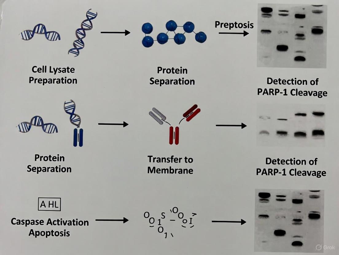
Western Blot Protocols for Detecting PARP-1 Cleavage
Experimental Workflow for Apoptosis Detection
The following diagram outlines the complete experimental workflow for detecting PARP-1 cleavage via Western blotting in apoptosis research:
Detailed Protocol for PARP-1 Cleavage Detection
Materials and Reagents:
- Cell Lines: Jurkat cells (for suspension culture) or HeLa cells (for adherent culture)
- Apoptosis Inducers: Etoposide (25 µM for 5 hours) or Cytochrome c (for caspase-3 activation)
- Lysis Buffer: RIPA buffer supplemented with protease and phosphatase inhibitors
- Antibodies:
- Primary Antibody: Cleaved PARP (Asp214) Antibody (#9541, Cell Signaling Technology) [6]
- Secondary Antibody: HRP-conjugated anti-rabbit IgG
- Control Cell Extracts: Jurkat Apoptosis Cell Extracts (etoposide) #2043 or Caspase-3 Control Cell Extracts #9663 (Cell Signaling Technology) [8]
Procedure:
- Cell Culture and Treatment: Culture Jurkat cells in RPMI-1640 medium supplemented with 10% FBS at 37°C in 5% CO₂. Induce apoptosis by treating cells with 25 µM etoposide for 5 hours [8]. Include untreated cells as a negative control.
Protein Extraction: Harvest cells by centrifugation (500 × g for 5 minutes) and wash with cold PBS. Lyse cell pellets in RIPA buffer (supplemented with protease and phosphatase inhibitors) on ice for 30 minutes. Centrifuge at 14,000 × g for 15 minutes at 4°C and collect the supernatant.
Protein Quantification: Determine protein concentration using the BCA Protein Assay Kit according to manufacturer's instructions.
Gel Electrophoresis: Load 20-30 μg of total protein per well on 4-12% Bis-Tris polyacrylamide gels. Include pre-stained protein molecular weight markers. Run gels at 120-150 V for 60-90 minutes in MOPS or MES running buffer.
Membrane Transfer: Transfer proteins to nitrocellulose or PVDF membranes using wet or semi-dry transfer systems at 100 V for 60-90 minutes on ice.
Blocking: Block membranes with 5% non-fat dry milk in TBST (Tris-buffered saline with 0.1% Tween-20) for 1 hour at room temperature with gentle agitation.
Antibody Incubation:
- Incubate membrane with Cleaved PARP (Asp214) Antibody (#9541) at 1:1000 dilution in 5% BSA/TBST overnight at 4°C with gentle agitation [6].
- Wash membrane 3 times for 5 minutes each with TBST.
- Incubate with HRP-conjugated secondary antibody at 1:2000-1:5000 dilution in 5% non-fat dry milk/TBST for 1 hour at room temperature.
- Wash membrane 3 times for 5 minutes each with TBST.
Detection: Develop blots using enhanced chemiluminescence (ECL) substrate according to manufacturer's instructions. Image using a digital imaging system with appropriate exposure times (typically 1 second to 10 minutes).
Expected Results:
- The Cleaved PARP (Asp214) Antibody specifically detects the 89 kDa large fragment of PARP-1 produced by caspase cleavage at Asp214 [6].
- This antibody does not recognize full-length PARP-1 or other PARP isoforms.
- In etoposide-treated Jurkat cells (positive control), a strong 89 kDa band should be visible.
- In untreated cells (negative control), the 89 kDa band should be absent or very faint.
Troubleshooting and Quality Control
Common Issues and Solutions:
- No Signal in Treated Samples: Ensure apoptosis induction is efficient by using positive control cell extracts (e.g., Jurkat Apoptosis Cell Extracts #2043) [8]. Verify antibody specificity and concentration.
- High Background: Optimize blocking conditions (try 5% BSA instead of milk) and increase wash stringency (more washes or higher Tween-20 concentration).
- Multiple Bands: Check antibody specificity and ensure proper protein separation during electrophoresis. The Cleaved PARP (Asp214) Antibody should primarily detect the 89 kDa fragment [6].
- Weak Signal: Increase protein loading amount or enhance detection sensitivity with more sensitive ECL substrates.
Quality Control Measures:
- Always include positive and negative controls in each experiment. Pre-made control cell extracts are particularly valuable for ensuring consistent results [8].
- Validate antibody performance using known positive and negative samples.
- Ensure proper sample preparation to preserve post-translational modifications and cleavage events.
Research Reagent Solutions for PARP-1 Studies
Table 3: Essential Research Reagents for PARP-1 Apoptosis Studies
| Reagent Category | Specific Products | Application & Purpose | Key Features |
|---|---|---|---|
| Primary Antibodies | Cleaved PARP (Asp214) Antibody #9541 (CST) [6] | Detects 89 kDa PARP-1 fragment in Western blot | Specific for caspase-cleaved PARP-1; does not recognize full-length |
| Control Cell Extracts | Jurkat Apoptosis Cell Extracts (etoposide) #2043 (CST) [8] | Positive control for apoptosis markers in Western blot | Contains PARP cleavage products from etoposide-treated Jurkat cells |
| Control Cell Extracts | Caspase-3 Control Cell Extracts #9663 (CST) [8] | Positive control for caspase-3 activation | Cytoplasmic fraction from cytochrome c-treated Jurkat cells |
| PARP Inhibitors | Olaparib, Rucaparib, Niraparib [9] | Inhibit PARP enzymatic activity; research and clinical use | Induce synthetic lethality in BRCA-deficient cells; used in cancer research |
| Apoptosis Inducers | Etoposide, Cytochrome c [8] | Induce apoptosis in experimental systems | Activate intrinsic apoptosis pathway; positive control for PARP cleavage |
| PROTAC Degraders | 180055 (Rucaparib-based PROTAC) [9] | Selective degradation of PARP1 without DNA trapping | Avoids side effects associated with conventional PARP inhibitors |
PARP-1 represents a critical molecular switch that determines cellular fate in response to DNA damage and other stressors. Its cleavage during apoptosis serves as both a definitive marker of programmed cell death and an active regulatory event that coordinates the shutdown of DNA repair processes. The detection of PARP-1 cleavage fragments, particularly the 89 kDa caspase-generated fragment, through Western blotting provides researchers with a robust method for identifying and quantifying apoptotic events in experimental systems. Recent advances in understanding PARP-1's role in multiple cell death pathways, including its crosstalk with ferroptosis and its functions beyond DNA repair, continue to reveal new therapeutic opportunities, particularly in the context of PARP inhibitor-resistant cancers. The development of novel approaches such as PARP-1-specific PROTAC degraders that avoid DNA trapping effects represents promising directions for future research and therapeutic development [9].
Poly (ADP-ribose) polymerase 1 (PARP1) is a 113-116 kDa nuclear enzyme that plays a critical role in the cellular response to DNA damage, primarily by catalyzing the transfer of ADP-ribose units to target proteins to facilitate DNA repair processes [10] [2]. During the execution phase of apoptosis, PARP1 becomes a primary substrate for executioner caspases-3 and -7 [11] [12]. This proteolytic cleavage occurs at a specific aspartic acid residue (Asp214 in human PARP1), separating the N-terminal DNA-binding domain (DBD) from the C-terminal catalytic domain [13] [2]. The result of this cleavage event is the generation of two signature fragments: a 24-kDa DNA-binding fragment and an 89-kDa catalytic fragment [14] [2]. The detection of this 89-kDa fragment via western blotting has become a established biochemical marker for confirming apoptosis in experimental systems, providing researchers with a reliable method to distinguish between various forms of programmed cell death [12] [2].
Molecular Mechanisms of PARP1 Cleavage
Caspase-Mediated Cleavage Signature
The cleavage of PARP1 by caspases represents a pivotal commitment to apoptotic cell death. Executioner caspases-3 and -7 recognize and hydrolyze the DEVD216↓G motif in human PARP1, located between the DNA-binding domain and the automodification domain [13] [14]. This specific proteolytic event produces two principal fragments with distinct cellular fates:
24-kDa Fragment: Contains the DNA-binding domain with two zinc finger motifs, which remains tightly bound to DNA strand breaks. This fragment acts as a trans-dominant inhibitor of DNA repair by blocking access of intact PARP1 and other repair enzymes to damaged DNA [14] [2].
89-kDa Fragment: Comprises the automodification domain and the catalytic domain. This fragment is translocated from the nucleus to the cytoplasm during apoptosis [14]. Recent research has revealed that this fragment can serve as a carrier for poly(ADP-ribose) (PAR) polymers, facilitating apoptosis-inducing factor (AIF) release from mitochondria and contributing to nuclear shrinkage – thus bridging caspase-dependent apoptosis and AIF-mediated cell death [14].
The following diagram illustrates the domain architecture of PARP1 and the caspase cleavage event:
Biological Consequences of PARP1 Cleavage
The cleavage of PARP1 serves multiple critical functions in the apoptotic cascade. By inactivating PARP1's DNA repair capability, the cell prevents futile energy consumption on DNA repair while committing to the death pathway [2]. The 24-kDa fragment's persistent binding to DNA breaks further ensures that DNA repair processes remain suppressed [14] [2]. Meanwhile, the 89-kDa fragment's translocation to the cytoplasm and its role in AIF-mediated processes may represent a secondary amplification mechanism for cell death execution, creating a feed-forward loop that ensures complete cellular dismantling [14]. This intricate mechanism explains why PARP1 cleavage has become such a reliable indicator of apoptotic commitment in experimental systems.
Western Blot Protocol for Detecting PARP1 Cleavage
Sample Preparation and Optimization
Proper sample preparation is critical for the accurate detection of PARP1 cleavage fragments. The following protocol has been optimized for apoptosis induction and protein extraction:
Apoptosis Induction: Treat cells with 1-3 μM staurosporine for 3-24 hours to induce caspase-dependent apoptosis [15]. Alternatively, use 1-10 μM actinomycin D or other DNA-damaging agents confirmed to activate caspase-3 [14]. Always include untreated controls from the same cell population.
Cell Lysis: Prepare RIPA lysis buffer supplemented with protease inhibitor cocktail (including caspase inhibitors) and 1 mM PMSF. Place culture dishes on ice and wash cells twice with cold PBS. Add appropriate volume of lysis buffer (e.g., 100-200 μL for a 6-well plate) and incubate on ice for 15-30 minutes with occasional agitation [12] [10].
Protein Quantification: Centrifuge lysates at 14,000 × g for 15 minutes at 4°C. Transfer supernatant to fresh tubes and determine protein concentration using BCA assay. Adjust samples to equal concentrations with lysis buffer and Laemmli sample buffer to ensure consistent loading [12].
Electrophoresis and Transfer
Gel Preparation: Prepare 8-12% Tris-Glycine SDS-PAGE gels to optimally resolve the 89-kDa fragment from full-length PARP1. Include pre-stained protein molecular weight markers in at least one lane [12] [15].
Sample Loading: Load 20-40 μg of total protein per lane. Include positive controls (e.g., staurosporine-treated HeLa or A549 cell lysates) to verify antibody performance and cleavage detection [10] [15].
Electrophoresis: Run gels at 100-150 V for 1-2 hours until the dye front reaches the bottom of the gel.
Protein Transfer: Transfer proteins to nitrocellulose or PVDF membranes using wet or semi-dry transfer systems. For the 89-kDa fragment, transfer at 100 V for 1 hour or 30 V overnight at 4°C [12].
Immunoblotting and Detection
Blocking: Block membranes with 5% non-fat dry milk or BSA in TBST for 1 hour at room temperature with gentle agitation [12] [15].
Primary Antibody Incubation: Incubate with anti-PARP1 primary antibodies (see Table 2 for specifications) diluted in blocking buffer. Typical dilutions range from 1:500 to 1:8000 for total PARP1 antibodies and 1:100 for cleaved-specific antibodies. Incubate overnight at 4°C with gentle shaking [10] [15].
Washing: Wash membranes 3-4 times for 5-10 minutes each with TBST.
Secondary Antibody Incubation: Incubate with appropriate HRP-conjugated or fluorescently-labeled secondary antibodies diluted in blocking buffer (typically 1:2000-1:20000) for 1 hour at room temperature [12] [15].
Detection: Develop blots using enhanced chemiluminescence (ECL) or fluorescence detection systems according to manufacturer's instructions. Image using a digital imaging system capable of capturing the dynamic range of protein signals [12].
The complete experimental workflow is visualized below:
Research Reagent Solutions
Selecting appropriate antibodies and reagents is crucial for successful detection of PARP1 cleavage. The following table summarizes key reagents validated for this application:
Table 1: Essential Research Reagents for PARP1 Cleavage Detection
| Reagent Type | Specific Product/Example | Key Features & Applications | Optimal Dilution |
|---|---|---|---|
| Total PARP1 Antibody | PARP1 Polyclonal Antibody #13371-1-AP [10] | Recognizes both full-length (116 kDa) and cleaved (89 kDa) PARP1; suitable for WB, IHC, IF | WB: 1:1000-1:8000 |
| Cleavage-Specific Antibody | Cleaved PARP (Asp214) Antibody #9541 [13] | Specifically detects 89 kDa fragment; does not recognize full-length PARP1 | WB: 1:1000 |
| Cleavage-Specific Antibody | Anti-Cleaved PARP1 [SP276] (ab225715) [15] | Recombinant monoclonal; specific for cleaved PARP1; validated in knockout cells | WB: 1:100 |
| Apoptosis Inducer | Staurosporine [14] [15] | Broad-spectrum kinase inhibitor; induces caspase-3 activation and PARP1 cleavage | 1-3 μM, 3-24 hours |
| Positive Control Lysate | Staurosporine-treated HeLa or A549 cells [15] | Provides reliable positive control for 89 kDa fragment detection | 20 μg per lane |
| Loading Control Antibodies | Anti-GAPDH or Anti-β-actin [12] [15] | Verify equal protein loading and transfer efficiency | WB: 1:20000 |
Data Interpretation and Analysis
Band Pattern Recognition
Proper interpretation of western blot results requires understanding the expected band patterns:
- Healthy Cells: A single dominant band at approximately 113-116 kDa representing full-length PARP1.
- Early Apoptosis: Both the 116 kDa full-length band and the 89 kDa cleavage fragment are visible.
- Late Apoptosis: Significant reduction of the 116 kDa band with a strong 89 kDa fragment signal.
- Complete Cleavage: Only the 89 kDa fragment is detectable, with complete disappearance of the full-length protein.
Table 2: PARP1 Fragment Molecular Weights and Significance
| Fragment | Molecular Weight | Domain Composition | Cellular Localization | Functional Significance |
|---|---|---|---|---|
| Full-length PARP1 | 113-116 kDa | DNA-binding + Automodification + Catalytic domains | Nuclear | DNA repair enzyme activity |
| 89 kDa Fragment | 85-89 kDa | Automodification + Catalytic domains | Cytosolic translocation | PAR carrier; promotes AIF release |
| 24 kDa Fragment | 24-27 kDa | DNA-binding domain only | Nuclear retention | Dominant-negative inhibitor of DNA repair |
Quantification and Normalization
For quantitative analysis of PARP1 cleavage:
- Perform densitometry analysis using software such as ImageJ to measure band intensities.
- Calculate the cleavage ratio: Intensity(89 kDa) / [Intensity(116 kDa) + Intensity(89 kDa)].
- Normalize all signals to housekeeping proteins (GAPDH, β-actin) to account for loading variations.
- Express results as mean ± SEM from at least three independent experiments.
- Include statistical analysis (t-test, ANOVA) to determine significance between experimental groups.
Troubleshooting Common Challenges
Several technical challenges may arise when detecting PARP1 cleavage:
- Weak or No Signal: Optimize antibody concentration and incubation time. Verify apoptosis induction with positive controls. Check protein transfer efficiency using Ponceau S staining.
- Non-specific Bands: Increase blocking time, optimize antibody dilution, or try different blocking buffers (BSA vs. non-fat milk).
- High Background: Increase wash stringency (more frequent washes, higher detergent concentration) and optimize antibody concentrations.
- Incomplete Transfer: For the 89 kDa fragment, ensure adequate transfer time and confirm using reversible membrane staining.
- Multiple Cleavage Fragments: Note that besides caspases, other proteases (calpains, cathepsins, granzymes, MMPs) can cleave PARP1, generating fragments of 50-65 kDa, 42-48 kDa, 35-40 kDa, and 28-35 kDa [2]. Use caspase-specific inhibitors to confirm caspase-mediated cleavage.
Applications in Biomedical Research
The detection of PARP1 cleavage has broad applications across multiple research domains:
- Cancer Research: Evaluating efficacy of chemotherapeutic agents and targeted therapies that induce apoptosis in cancer cells [1] [12].
- Neurodegenerative Disease Studies: Investigating apoptotic pathways in Alzheimer's disease, Parkinson's disease, and cerebral ischemia [14] [2].
- Drug Development: Screening pro-apoptotic compounds and validating their mechanisms of action [1] [12].
- PARP Inhibitor Research: Investigating mechanisms of PARP inhibitor resistance and developing combination therapies [1].
- Toxicology Studies: Assessing compound-induced cytotoxicity and cellular stress responses.
The reliability of PARP1 cleavage as an apoptotic marker continues to make it an invaluable tool for basic research, drug discovery, and mechanistic studies of cell death pathways across diverse biological contexts.
Poly (ADP-ribose) polymerase-1 (PARP-1) is a 116 kDa nuclear enzyme that plays a critical role in detecting and repairing DNA single-strand breaks [2] [7]. Upon activation by DNA damage, PARP-1 catalyzes the transfer of ADP-ribose units from NAD+ to target proteins, initiating the DNA repair process. However, during apoptosis, PARP-1 becomes one of the primary cleavage targets of executioner caspases, particularly caspase-3 and caspase-7 [2]. This cleavage occurs at a specific aspartic acid residue (Asp214) within the nuclear localization signal, generating two characteristic fragments: a 24 kDa DNA-binding fragment and an 89 kDa catalytic fragment [16] [2]. The detection of these cleavage fragments via western blotting has become a established biomarker for identifying apoptotic cells in research and drug development.
Biological Consequences of PARP-1 Cleavage
Inactivation of DNA Repair and Conservation of Cellular Energy
The proteolytic cleavage of PARP-1 serves two primary biological functions in apoptosis. First, it inactivates the DNA repair capability of the cell, ensuring the irreversible commitment to cell death. The 24 kDa fragment, which contains the DNA-binding domain, remains tightly bound to DNA strand breaks but lacks catalytic activity, effectively acting as a trans-dominant inhibitor of any remaining full-length PARP-1 [2]. This prevents the recruitment of DNA repair machinery to damaged DNA. Simultaneously, cleavage halts the massive consumption of NAD+ and ATP that occurs during PARP-1 hyperactivation, thereby conserving cellular energy pools necessary for the orderly execution of the apoptotic program [2] [7].
Active Roles of Cleavage Fragments in Cell Death Signaling
Beyond simply inactivating DNA repair, emerging research indicates that PARP-1 cleavage fragments actively participate in promoting cell death through multiple mechanisms:
Table 1: Biological Functions of PARP-1 Cleavage Fragments
| Fragment | Size | Domains Contained | Primary Functions |
|---|---|---|---|
| 24 kDa Fragment | 24 kDa | Two zinc-finger DNA-binding domains | - Irreversibly binds to damaged DNA- Acts as trans-dominant inhibitor of PARP-1- Blocks DNA repair processes- Retained in nucleus |
| 89 kDa Fragment | 89 kDa | BRCT domain, WGR domain, Catalytic domain | - Translocates to cytoplasm- Serves as PAR carrier to induce AIF-mediated parthanatos- Activates RNA Polymerase III for innate immune response- Can catalyze ADP-ribosylation |
Induction of AIF-Mediated Parthanatos
The 89 kDa fragment, when modified with poly(ADP-ribose) (PAR) polymers, translocates from the nucleus to the cytoplasm where it functions as a PAR carrier [17] [18]. In the cytoplasm, this fragment facilitates the release of Apoptosis-Inducing Factor (AIF) from mitochondria, triggering AIF-mediated chromatin condensation and DNA fragmentation – a caspase-independent cell death pathway known as parthanatos [17]. This mechanism creates an amplification loop connecting caspase-dependent apoptosis with caspase-independent parthanatos.
Enhancement of Innate Immune Response
Recent studies have revealed that the 89 kDa fragment (truncated PARP1 or tPARP1) interacts with the RNA Polymerase III (Pol III) complex in the cytoplasm during apoptosis induced by cytoplasmic DNA [19]. tPARP1 mono-ADP-ribosylates Pol III, enhancing its ability to transcribe foreign DNA and thereby stimulating interferon-beta (IFN-β) production and amplifying the apoptotic response to pathogenic infection [19]. This function is mediated through the BRCT domain of tPARP1, which specifically recognizes and interacts with Pol III subunits.
Regulatory Role in Inflammation and Transcription
PARP-1 cleavage fragments also modulate inflammatory responses and transcription. The 89 kDa fragment can influence NF-κB transcriptional activity, potentially enhancing the expression of pro-inflammatory genes during cell death [7]. This role in regulating the cellular response to inflammatory stimuli connects PARP-1 cleavage to broader pathological contexts beyond straightforward apoptosis.
Western Blot Protocol for Detecting PARP-1 Cleavage
Sample Preparation and Nuclear Extraction
Protocol: Nuclear Extraction for PARP-1 Detection
- Cell Harvesting: Detach cells using trypsin-EDTA and collect by centrifugation.
- Hypotonic Lysis: Resuspend cell pellet in 10 mM HEPES (pH 8.0), 10 mM KCl, 1.5 mM MgCl₂, 0.5 mM DTT, and complete EDTA-free protease inhibitor cocktail. Incubate on ice for 10 minutes.
- Membrane Disruption: Add 0.1% NP-40 and vortex vigorously for 10 seconds.
- Nuclear Pellet Isolation: Centrifuge at 1,500 × g for 10 minutes at 4°C. Collect the nuclear pellet.
- Nuclear Protein Extraction: Resuspend nuclear pellet in RIPA buffer (50 mM Tris-HCl pH 8.0, 150 mM NaCl, 1% NP-40, 0.5% sodium deoxycholate, 0.1% SDS) with protease inhibitors. Incubate on ice for 30 minutes with occasional vortexing.
- Clarification: Centrifuge at 1,500 × g for 30 minutes at 4°C. Collect supernatant containing nuclear proteins.
- Protein Quantification: Determine protein concentration using Bradford assay [20].
Western Blot Procedure
Table 2: Key Reagents for PARP-1 Cleavage Detection
| Reagent | Specifications | Function | Example Product |
|---|---|---|---|
| Primary Antibody | Cleaved PARP (Asp214) Antibody, 1:1000 dilution in WB | Detects 89 kDa fragment specifically | Cell Signaling Technology #9541 [16] |
| Primary Antibody | PARP-1 mAb (C2-10), 1:2000 dilution | Detects both full-length and cleaved PARP-1 | Santa Cruz Biotechnology C2-10 [20] |
| Loading Control | B23 mAb, 1:2000 dilution | Nuclear protein loading control | Sigma-Aldrich [20] |
| Secondary Antibody | HRP-conjugated goat anti-mouse/rabbit IgG | Detection | Pierce [20] |
| Blocking Buffer | 5% BSA in TBS with 0.1% Tween-20 | Reduces non-specific binding | - |
Electrophoresis and Immunoblotting:
- Gel Electrophoresis: Separate 30 μg nuclear protein extracts by 10% SDS-PAGE [20].
- Protein Transfer: Transfer to PVDF or nitrocellulose membrane using standard wet or semi-dry transfer systems.
- Blocking: Incubate membrane in blocking buffer (5% BSA in TBST) for 1 hour at room temperature.
- Primary Antibody Incubation: Incubate with primary antibody diluted in blocking buffer overnight at 4°C.
- Washing: Wash membrane 3×10 minutes with TBST.
- Secondary Antibody Incubation: Incubate with appropriate HRP-conjugated secondary antibody (1:2000-1:5000) for 1 hour at room temperature.
- Detection: Develop using enhanced chemiluminescence (ECL) substrate and image with digital imaging system.
Expected Results and Interpretation
A successful western blot will show:
- Full-length PARP-1 at approximately 116 kDa
- Cleaved PARP-1 fragment at 89 kDa
- The 24 kDa fragment may not be detected with all antibodies
The appearance of the 89 kDa band with corresponding decrease in full-length PARP-1 signal indicates caspase-mediated apoptosis. Densitometric analysis of the band intensities can provide semi-quantitative assessment of apoptosis extent.
Visualizing PARP-1 Cleavage Pathways and Detection Workflow
PARP-1 Cleavage in Cell Death Pathways
Experimental Workflow for PARP-1 Cleavage Detection
Research Reagent Solutions for PARP-1 Cleavage Studies
Table 3: Essential Research Reagents for PARP-1 Apoptosis Studies
| Category | Specific Product | Key Features | Application |
|---|---|---|---|
| Primary Antibodies | Cleaved PARP (Asp214) Antibody #9541 | Rabbit monoclonal, detects endogenous 89 kDa fragment, does not recognize full-length PARP-1 [16] | Western Blot (1:1000) |
| Primary Antibodies | PARP-1 mAb (C2-10) | Mouse monoclonal, detects both full-length and cleaved PARP-1 [20] | Western Blot (1:2000) |
| Control Antibodies | B23/mAb | Nuclear loading control [20] | Western Blot (1:2000) |
| Assay Kits | Caspase-3 Activity Assay | Measures executioner caspase activation | Apoptosis verification |
| Protein Markers | Prestained Protein Ladder | Molecular weight determination | Western Blot |
| Detection Systems | ECL Substrate | High-sensitivity chemiluminescent detection | Western Blot |
| Cell Lines | SH-SY5Y Human Neuroblastoma | Well-characterized apoptosis model [7] | Cellular studies |
| Apoptosis Inducers | Staurosporine, Actinomycin D | Established PARP-1 cleavage inducers [17] [19] | Positive controls |
The cleavage of PARP-1 represents a critical commitment point in the apoptotic pathway, serving both to inactivate DNA repair mechanisms and to actively promote cell death through multiple signaling cascades. The detection of the characteristic 89 kDa cleavage fragment via western blotting provides researchers with a reliable biomarker for apoptosis. The detailed protocol and reagent information provided herein enables consistent detection and interpretation of PARP-1 cleavage, supporting research in neurodegeneration, cancer biology, and drug development where apoptotic pathways are of central importance. The emerging roles of PARP-1 fragments in parthanatos and innate immune activation highlight the expanding significance of this proteolytic event in cellular physiology and pathology.
Connecting PARP-1 Cleavage to Key Apoptotic Pathways (Intrinsic and Extrinsic)
Poly(ADP-ribose) polymerase-1 (PARP-1) is a nuclear enzyme central to DNA repair and maintenance of genomic integrity. During apoptosis, a form of regulated cell death, PARP-1 is cleaved by caspases, a family of cysteinyl-aspartate proteases. This cleavage event is considered a hallmark of apoptosis and serves as a critical mechanism to shut down energy-consuming DNA repair processes, facilitating cellular dismantling [19] [21]. The cleavage of PARP-1 occurs at a specific aspartic acid residue (Asp214), generating two fragments: a 24 kDa DNA-binding fragment and an 89 kDa catalytic fragment [7] [22]. This article details the role of PARP-1 cleavage within the intrinsic and extrinsic apoptotic pathways and provides a detailed application note for its detection via western blotting in apoptosis research.
Biological Significance: A Nexus in Apoptotic Pathways
PARP-1 cleavage acts as a molecular switch, influencing cell fate through its involvement in both major apoptotic pathways.
The Intrinsic (Mitochondrial) Pathway
The intrinsic pathway is triggered by internal cellular stresses, such as DNA damage, leading to mitochondrial outer membrane permeabilization (MOMP) and the release of cytochrome c into the cytosol. This activates caspase-9, which in turn activates executioner caspases like caspase-3 and -7.
- PARP-1 Activation and Cleavage: Severe DNA damage initially activates PARP-1, which can lead to NAD+ and ATP depletion, potentially causing necrosis [23]. However, if the apoptotic program is engaged, caspase-3 cleaves PARP-1. This cleavage prevents further energy depletion, conserving ATP required for the apoptotic process [23].
- Amplification of Apoptosis: Research using the benzene metabolite TGHQ in HL-60 cells showed that PARP-1 inhibition attenuated caspase-3, -7, and -9 activation and cytochrome c release, indicating that PARP-1 activity can participate in amplifying the intrinsic pathway [24]. Furthermore, one of the cleavage products, the 89 kDa fragment (tPARP1), translocates to the cytoplasm [19]. Recent studies have shown that tPARP1 can bind to the RNA Polymerase III (Pol III) complex, mono-ADP-ribosylating it and facilitating innate immune responses and apoptosis during cellular stress, such as pathogen infection [19].
The Extrinsic (Death Receptor) Pathway
The extrinsic pathway is initiated by the ligation of death receptors (e.g., Fas, TNF-R1) at the cell surface, leading to the activation of caspase-8.
- Differential Regulation: The role of PARP-1 can vary depending on the stimulus. In L929 cells, TNF-induced necrosis involves PARP-1 activation and ATP depletion, whereas CD95-mediated apoptosis features caspase-mediated PARP-1 cleavage, which maintains ATP levels and supports apoptotic execution [23].
- Opposing Role in Signaling: Interestingly, in the TGHQ model, PARP-1 inhibition potentiated caspase-8 activation, suggesting that PARP-1 may play a suppressive role in the extrinsic pathway under certain conditions, highlighting its dual and opposing functions [24].
The following diagram illustrates the position of PARP-1 cleavage within these two key apoptotic pathways:
Functional Consequences of Cleavage Fragments
The cleavage of PARP-1 is not merely an inactivation mechanism. The resulting fragments can have distinct biological activities:
- The 24 kDa Fragment: Retains the DNA-binding domain and may act as a dominant-negative inhibitor by occupying DNA strand breaks, thus preventing recruitment of intact PARP-1 and other repair factors [19].
- The 89 kDa Fragment (tPARP1): Loses its nuclear localization signal and translocates to the cytoplasm, where it has been shown to engage in non-canonical functions, such as activating RNA Pol III to potentiate immune signaling and apoptosis [19].
- Impact on Transcription: Studies in neuronal models suggest that the cleavage fragments differentially regulate NF-κB activity and the expression of inflammatory genes like iNOS and COX-2, with the 89 kDa fragment exhibiting pro-inflammatory and cytotoxic properties [7].
Table 1: PARP-1 Cleavage Fragments and Their Functions
| Fragment Size | Domains Contained | Localization Post-Cleavage | Key Proposed Functions |
|---|---|---|---|
| 24 kDa | Zinc finger DNA-binding domain (N-terminal) | Nucleus [19] | Dominant-negative inhibitor of DNA repair; may occupy DNA breaks [19]. |
| 89 kDa | BRCT, WGR, and Catalytic domain (C-terminal) | Cytoplasm [19] | Binds RNA Pol III; catalyzes ADP-ribosylation to promote innate immune response and apoptosis [19]. Pro-inflammatory effects via NF-κB [7]. |
Application Note: Western Blot Protocol for Detecting PARP-1 Cleavage
This protocol is optimized for detecting the full-length and cleaved forms of PARP-1 in human and mouse cell lines, facilitating the assessment of apoptosis in experimental models.
Sample Preparation
- Cell Lysis: Harvest treated and control cells. Lyse cells using a suitable RIPA buffer supplemented with protease and phosphatase inhibitors. Incubate on ice for 15-30 minutes with periodic vortexing.
- Cytosolic and Mitochondrial Fractionation (Optional): For studies investigating cytochrome c release or tPARP1 localization, fractionate cells using a digitonin-based method [24].
- Resuspend cell pellet in ice-cold buffer containing 150 mM NaCl, 50 mM HEPES (pH 7.4), and 25 µg/ml digitonin.
- Gently mix at 4°C for 10 minutes. Centrifuge at 2000 RCF for 10 minutes. The supernatant is the cytosolic fraction.
- Resuspend the pellet in a buffer containing 1% NP-40, incubate on ice for 30 minutes, and centrifuge at 7000 RCF. The supernatant contains mitochondrial proteins.
- Protein Quantification: Determine protein concentration of whole-cell or fractionated lysates using a BCA or Bradford assay. Normalize all samples to the same concentration.
Gel Electrophoresis and Western Blotting
- Gel Loading: Load 20-30 µg of total protein per well onto a 4-12% Bis-Tris polyacrylamide gel. Include a pre-stained protein molecular weight marker.
- Electrophoresis: Run the gel in MOPS or MES SDS running buffer at constant voltage (150-200V) until the dye front reaches the bottom.
- Protein Transfer: Transfer proteins from the gel to a nitrocellulose or PVDF membrane using a wet or semi-dry transfer system.
- Blocking: Block the membrane with 5% non-fat milk or BSA in TBST (Tris-buffered saline with 0.1% Tween 20) for 1 hour at room temperature.
Antibody Incubation and Detection
- Primary Antibody Incubation: Incubate membrane with primary antibodies diluted in blocking buffer or TBST overnight at 4°C with gentle agitation.
- For Cleaved PARP-1 (89 kDa fragment): Use anti-cleaved PARP1 (Asp214) antibody at 1:1000 dilution [22]. This antibody is specific to the cleaved fragment and does not recognize full-length PARP1.
- For Total PARP-1: Use an antibody that recognizes both full-length and cleaved PARP1 (e.g., Abcam ab225715 at 1:100 dilution) [15].
- Loading Control: Incubate with an antibody for GAPDH, β-Actin, or α-Tubulin (e.g., 1:20,000 dilution) [15].
- Washing: Wash membrane 3-4 times for 5 minutes each with TBST.
- Secondary Antibody Incubation: Incubate membrane with an appropriate horseradish peroxidase (HRP)-conjugated secondary antibody (e.g., goat anti-rabbit) at 1:2000-1:10,000 dilution in blocking buffer for 1 hour at room temperature [24] [15].
- Detection: Wash membrane again. Develop the blot using a enhanced chemiluminescence (ECL) substrate according to the manufacturer's instructions. Image the blot using a chemiluminescence imager.
Expected Results and Data Interpretation
- Non-Apoptotic Cells: A single band at ~116 kDa, corresponding to full-length PARP-1.
- Apoptotic Cells: A band at ~89 kDa (cleaved PARP-1 fragment). The full-length band may appear fainter. With antibodies detecting total PARP1, both the 116 kDa and 89 kDa bands are visible [15] [22].
- The appearance of the 89 kDa band is a definitive indicator of caspase-mediated apoptosis. Densitometric analysis of the full-length and cleaved bands can provide a semi-quantitative measure of apoptosis.
The workflow for this protocol and the expected results are summarized below:
Table 2: Key Research Reagent Solutions for PARP-1 Cleavage Analysis
| Reagent / Assay | Specific Example / Catalog Number | Function in Protocol |
|---|---|---|
| Anti-Cleaved PARP1 Antibody | Cleaved PARP (Asp214) Antibody #9541 (Cell Signaling Technology) [22] | Specifically detects the 89 kDa apoptotic fragment without cross-reacting with full-length PARP1. |
| Anti-PARP1 Antibody | Anti-Cleaved PARP1 antibody [SP276] (ab225715) (Abcam) [15] | Detects both full-length (~116 kDa) and cleaved (~89 kDa & 24 kDa) PARP1. |
| Apoptosis Inducer (Positive Control) | Staurosporine (e.g., 1-3 µM for 3-24 hours) [15] | A broad-spectrum kinase inducer used as a positive control to trigger apoptosis and PARP-1 cleavage. |
| Caspase Inhibitor | z-VAD-FMK (pan-caspase inhibitor) [24] [23] | Used to confirm caspase-dependence of PARP-1 cleavage; prevents cleavage when co-treated with apoptosis inducer. |
| Loading Control Antibody | Anti-GAPDH, Anti-β-Actin, or Anti-α-Tubulin [24] [15] | Verifies equal protein loading across lanes. |
| PARP Inhibitor | PJ-34 or 3-Aminobenzamide (3AB) [24] [23] | Pharmacological inhibitor used to study the functional consequences of PARP-1 enzymatic activity on apoptosis. |
Discussion and Concluding Remarks
The detection of PARP-1 cleavage via western blot remains a gold-standard biochemical method for confirming apoptosis in cellular research. Its specificity as a caspase-3 substrate makes it a reliable marker. Understanding its dual role in the intrinsic and extrinsic pathways, and the emerging functions of its cleavage products, adds layers of complexity to its biological significance. The provided detailed protocol and reagent table offer a robust framework for researchers to investigate PARP-1 cleavage in various experimental models, from basic research to drug development, where assessing the efficacy of pro-apoptotic cancer therapeutics is paramount. Furthermore, the exploration of PARP-1's role in other cell death modalities, such as its recently discovered crosstalk with ferroptosis, represents an exciting frontier for future research [1].
A Detailed Western Blot Protocol for Reliable Cleaved PARP-1 Detection
Poly (ADP-ribose) polymerase 1 (PARP1) is a 116 kDa nuclear enzyme that plays a critical role in the cellular response to DNA damage, primarily by initiating the base excision repair pathway [25] [26]. During the early stages of apoptosis, activated executioner caspases, predominantly caspase-3, cleave PARP1 at a specific aspartic acid residue (Asp214) [25] [27]. This proteolytic cleavage separates the 24 kDa DNA-binding domain from the 89 kDa catalytic domain, inactivating the DNA repair function of PARP1 and facilitating the dismantling of the cell [25] [12]. The appearance of the 89 kDa fragment has thus become a well-established biochemical marker for detecting programmed cell death, distinguishing it from other forms of cell death like necrosis [5].
The critical reliance on this biomarker in research and drug development necessitates the use of highly specific antibody reagents. Antibodies that specifically recognize the 89 kDa cleaved fragment of PARP1 are essential tools for accurately identifying and quantifying apoptotic events in various experimental models, from cell culture to patient-derived samples [28] [12]. The selectivity of these antibodies ensures that researchers can confidently interpret Western blot results, directly linking the observed 89 kDa band to caspase-mediated apoptosis.
Key Reagents and Validation Strategies
Antibody Specificity is Paramount
The core challenge in detecting PARP1 cleavage lies in an antibody's ability to discriminate between the full-length (116 kDa) protein and the caspase-generated 89 kDa fragment. Non-specific antibodies may cross-react with other proteins or PARP isoforms, leading to false positives or misinterpreted data [29]. Therefore, selecting an antibody validated for specificity to the 89 kDa fragment is the most critical step in reagent selection.
Recommended validation strategies include [29]:
- Genetic Knockout (KO) Controls: Using cell lines where the PARP1 gene has been knocked out provides the gold standard for confirming antibody specificity. The absence of any signal in the KO sample confirms the antibody's on-target binding.
- Induced Apoptosis Models: Treating cells with known apoptogenic agents (e.g., Etoposide, Staurosporine) and demonstrating the appearance of the 89 kDa band upon treatment provides functional validation [26].
- Peptide Competition: Antibodies whose signal is blocked by pre-incubation with the immunogen peptide demonstrate binding specificity.
Commercial Antibodies for Cleaved PARP1 Detection
The table below summarizes key characteristics of several commercially available antibodies validated for detecting the cleaved 89 kDa PARP1 fragment.
Table 1: Commercial Antibodies for Detecting Cleaved PARP1 (89 kDa)
| Antibody Name / Catalog # | Host & Clonality | Specificity | Reactivity | Recommended Dilution (WB) | Key Validation Data |
|---|---|---|---|---|---|
| Cleaved PARP (Asp214) #9546 [27] | Mouse Monoclonal | 89 kDa fragment resulting from cleavage at Asp214 | Human, Monkey | 1:2000 | Specific detection of the 89 kDa fragment; may detect full-length PARP at high levels. |
| Anti-Cleaved PARP1 (ab4830) [26] | Rabbit Polyclonal | 85 kDa fragment (cleaved PARP1) | Human | 1:1000 | Antibody purified to remove reactivity to full-length PARP1; shows band at ~85 kDa in apoptotic cells. |
| PARP Antibody #9542 [25] | Rabbit Polyclonal | Full-length (116 kDa) and cleaved (89 kDa) PARP1 | Human, Mouse, Rat, Monkey | 1:1000 | Detects both forms; useful for assessing the cleavage ratio. |
| PARP1 Antibody (13371-1-AP) [30] | Rabbit Polyclonal | Full-length and cleaved PARP1 | Human, Mouse, Rat | 1:1000-1:8000 | User reviews confirm detection of full-length and 89 kDa fragment in various cell lines. |
The Scientist's Toolkit: Essential Reagents for PARP1 Cleavage Analysis
Table 2: Essential Reagents and Materials for Western Blot Analysis of PARP1 Cleavage
| Reagent / Material | Function / Role in the Experiment |
|---|---|
| Specific Antibody to 89 kDa fragment [26] [27] | Primary antibody for specific detection of the apoptotic cleavage product. |
| Apoptosis Inducers (e.g., Etoposide, Staurosporine) [26] | Positive control treatments to trigger caspase activation and PARP1 cleavage in experimental cells. |
| Control Cell Lysates [29] [12] | Lysates from untreated (negative control) and apoptotically-induced (positive control) cells essential for assay validation. |
| Caspase Inhibitor (e.g., zVAD-fmk) [5] | To confirm caspase-dependent cleavage; inhibits the appearance of the 89 kDa band. |
| Housekeeping Protein Antibodies (e.g., β-Actin, GAPDH) [12] | Loading controls to normalize for protein content and transfer efficiency across lanes. |
Detailed Western Blot Protocol for Detecting PARP1 Cleavage
Sample Preparation from Cultured Cells
- Induce Apoptosis: Treat cells with an appropriate apoptogenic agent (e.g., 1 µM Etoposide for 16 hours [26]). Include a vehicle-treated control.
- Harvest and Lyse Cells: Wash cells with cold PBS and lyse using a suitable RIPA buffer supplemented with protease and phosphatase inhibitors. Gently scrape and collect the lysate.
- Quantify Protein: Determine protein concentration of the lysates using a standardized assay (e.g., BCA assay). Normalize all samples to the same concentration using lysis buffer.
Gel Electrophoresis and Transfer
- Prepare Samples: Mix normalized lysates (20-40 µg total protein is typical [26]) with 2X or 4X Laemmli sample buffer. Boil samples for 5 minutes to denature proteins.
- SDS-PAGE: Load samples and a pre-stained protein ladder onto a 4-12% or 10% Bis-Tris polyacrylamide gel. Run the gel in MOPS or MES buffer at constant voltage (e.g., 120-150V) until the dye front reaches the bottom.
- Protein Transfer: Transfer proteins from the gel to a PVDF or nitrocellulose membrane using a wet or semi-dry transfer system.
Immunoblotting
- Blocking: Incubate the membrane in 5% non-fat dry milk or BSA in TBST for 1 hour at room temperature to block non-specific binding sites.
- Primary Antibody Incubation: Dilute the primary antibody against cleaved PARP1 (refer to Table 1 for specific dilutions) in blocking solution. Incubate the membrane with the antibody solution with gentle agitation overnight at 4°C.
- Washing: Wash the membrane 3 times for 5-10 minutes each with TBST.
- Secondary Antibody Incubation: Incubate the membrane with an HRP-conjugated secondary antibody (e.g., Goat Anti-Rabbit IgG) diluted in blocking solution for 1 hour at room temperature.
- Washing: Repeat the washing step as above.
Detection and Analysis
- Chemiluminescent Detection: Incubate the membrane with a chemiluminescent substrate according to the manufacturer's instructions. Capture the signal using a digital imager or X-ray film.
- Strip and Re-probe (Optional): The membrane can be stripped and re-probed with an antibody for a housekeeping protein (e.g., β-Actin) to confirm equal loading.
- Data Interpretation: Analyze the band intensities using densitometry software (e.g., ImageJ). The presence of the 89 kDa band in treated samples, and its increase relative to the full-length PARP1 band, confirms apoptosis [12].
Diagram 1: Western Blot Workflow for PARP1 Cleavage Detection.
Interpreting Results and Troubleshooting
Expected Results and Quantification
A successful Western blot for apoptosis detection will show:
- Untreated Control Cells: A dominant band at 116 kDa (full-length PARP1), with little to no signal at 89 kDa.
- Treated Apoptotic Cells: A clear band at 89 kDa (cleaved PARP1 fragment). The intensity of this band should increase with the severity or duration of the apoptotic stimulus, while the 116 kDa band may diminish [12] [26].
For quantification, use densitometry software to measure the band intensities. Calculate the ratio of cleaved PARP1 (89 kDa) to total PARP1 (full-length + cleaved) or to a housekeeping protein like β-actin to obtain a normalized measure of apoptotic activity [12].
Common Pitfalls and Troubleshooting
- Multiple Bands or Smearing: This could indicate protein degradation during sample preparation. Ensure samples are kept on ice and inhibitors are fresh [29].
- No Band in Positive Control: Confirm the efficacy of the apoptosis induction method using an alternative assay. Check antibody dilutions and expiration dates. Verify the protocol for the primary antibody incubation [29].
- High Background: Increase the number or duration of washes after antibody incubations. Optimize the concentration of the primary and secondary antibodies [12].
- Different Cleavage Fragment (~50 kDa): The appearance of a ~50 kDa fragment may indicate necrotic cell death mediated by lysosomal proteases like cathepsins, rather than caspase-mediated apoptosis [5]. This underscores the need for caspase-specific 89 kDa fragment antibodies.
Diagram 2: PARP1 Cleavage in Apoptosis Pathway.
Step-by-Step Sample Preparation from Apoptosis-Induced Cells
Within the framework of a thesis investigating Western blot protocols for apoptosis research, the detection of Poly(ADP-ribose) polymerase (PARP-1) cleavage stands as a critical biochemical hallmark. During the execution phase of apoptosis, caspases-3 and -7 cleave the 116 kDa PARP-1 protein into a characteristic 24 kDa DNA-binding fragment and an 89 kDa catalytic fragment [31] [7]. This cleavage event serves as a definitive indicator of commitment to the apoptotic pathway. This application note provides a detailed, step-by-step protocol for preparing samples from apoptosis-induced cells, specifically optimized for the subsequent detection of the 89 kDa cleaved PARP-1 fragment via Western blotting, enabling researchers and drug development professionals to accurately assess cell death in their experimental models.
Background and Significance
PARP-1 Cleavage as an Apoptotic Marker
PARP-1 is a 116 kDa nuclear enzyme involved in DNA repair and genomic integrity maintenance. Upon induction of apoptosis, activated effector caspases (primarily caspase-3) cleave PARP-1 at the DEVD214 amino acid sequence, separating its N-terminal DNA-binding domain (24 kDa) from its C-terminal catalytic domain (89 kDa) [31] [7]. This cleavage inactivates PARP-1's DNA repair function and prevents futile energy depletion, facilitating cellular disassembly. The appearance of the 89 kDa fragment is thus widely accepted as a reliable biochemical marker of apoptosis [5].
It is crucial to distinguish this caspase-mediated cleavage from PARP-1 processing that occurs during necrosis. Necrotic cell death, often triggered by extreme physicochemical stress, results in a dominant 50 kDa PARP-1 fragment through the action of lysosomal proteases such as cathepsins B and G, a process not inhibited by broad-spectrum caspase inhibitors like zVAD-fmk [5].
The Broader Apoptosis Detection Context
While PARP-1 cleavage detection is a cornerstone of apoptosis confirmation, researchers often employ complementary techniques to provide a more comprehensive view of cell death. The table below summarizes key apoptosis detection methods.
Table 1: Key Methods for Apoptosis Detection
| Method | Target/Principle | Stage Detected | Key Feature |
|---|---|---|---|
| PARP-1 Cleavage (WB) | Caspase-mediated 89 kDa fragment | Mid/Late Apoptosis | Gold standard biochemical confirmation [31] |
| Annexin V Staining | Externalized phosphatidylserine | Early Apoptosis | Allows distinction from necrotic cells (flow cytometry) [32] [33] |
| TUNEL Assay | DNA strand breaks (3'-OH ends) | Late Apoptosis | Labels fragmented nuclear DNA (microscopy/flow) [34] |
| Caspase-3 Activity | Cleaved caspase-3 substrate | Early/Mid Apoptosis | Measures key protease activation |
The following diagram illustrates the key apoptotic event of PARP-1 cleavage and its relation to the Western blot readout.
Materials and Reagents
Research Reagent Solutions
Successful detection of PARP-1 cleavage requires specific, validated reagents. The following table details essential materials.
Table 2: Essential Reagents for PARP-1 Cleavage Detection
| Reagent/Material | Function/Description | Example/Specification |
|---|---|---|
| Anti-Cleaved PARP (Asp214) | Primary antibody specific to 89 kDa fragment [31] | Rabbit mAb, #9541 (CST); 1:1000 dilution for WB |
| Cell Lysis Buffer | Extracts soluble proteins while preserving cleaved fragments | RIPA Buffer (50 mM Tris-HCl, pH 8.0, 150 mM NaCl, 1% NP-40, 0.5% sodium deoxycholate, 0.1% SDS) [20] |
| Protease Inhibitor Cocktail | Prevents post-lysis protein degradation | EDTA-free cocktail to avoid interference with caspases [20] |
| Phosphatase Inhibitors | Preserves protein phosphorylation status | Add if studying phospho-signaling pathways |
| Protein Assay Kit | Quantifies protein concentration for equal loading | Bradford, BCA, or other compatible methods [35] [20] |
| Loading Control Antibody | Normalizes for loading variations (housekeeping protein) | β-actin, GAPDH, α-tubulin, or Total Protein Normalization [35] |
Experimental Protocols
Protocol 1: Sample Preparation from Apoptosis-Induced Cells
1A Apoptosis Induction and Cell Harvesting
- Induction: Treat cells with your chosen apoptotic stimulus (e.g., chemotherapeutic agent, UV irradiation, staurosporine). Include a negative control (untreated, healthy cells) and a positive control (e.g., cells treated with 1-2 µM staurosporine for 2-4 hours).
- Harvesting:
- Adherent Cells: Collect culture supernatant (may contain detached apoptotic cells) and combine with cells detached gently using trypsin-EDTA or a cell scraper [20].
- Suspension Cells: Centrifuge at 500 × g for 5 minutes to pellet cells.
- Washing: Wash the cell pellet once with ice-cold 1X Phosphate-Buffered Saline (PBS).
1B Nuclear Protein Extraction (Recommended for PARP-1)
PARP-1 is a nuclear protein; enrichment via nuclear extraction can enhance detection sensitivity.
- Resuspend Pellet: Resuspend the washed cell pellet in a hypotonic buffer (e.g., 10 mM HEPES, pH 8.0, 10 mM KCl, 1.5 mM MgCl₂, 0.5 mM DTT, 0.1% NP-40, plus protease inhibitors) [20].
- Incubate and Lyse: Incubate on ice for 10-15 minutes to allow swelling. Lyse cells by vortexing briefly or by passing through a pipette tip.
- Separate Fractions: Centrifuge at 1,500 × g for 10 minutes at 4°C. The supernatant constitutes the cytoplasmic fraction. The pellet contains the nuclei.
- Extract Nuclear Proteins: Solubilize the nuclear pellet in RIPA buffer supplemented with protease inhibitors [20]. Incubate on ice for 30 minutes with occasional vortexing.
- Clarify Lysate: Centrifuge at 12,000 × g for 15 minutes at 4°C. Transfer the supernatant (nuclear protein extract) to a new pre-chilled tube.
1C Total Protein Extraction (Alternative Method)
For a simpler total lysate preparation, directly solubilize the washed cell pellet in RIPA buffer with inhibitors, followed by incubation on ice and clarification via centrifugation as in Step 4.1B.5.
1D Protein Quantification and Preparation
- Quantify: Determine the protein concentration of each sample using a compatible protein assay (e.g., Bradford method) [35] [20].
- Prepare for SDS-PAGE: Dilute samples with 4X Laemmli buffer to achieve a final 1X concentration. Boil the samples at 95-100°C for 5 minutes to denature proteins.
Protocol 2: Quantitative Western Blot for Cleaved PARP-1
The workflow for the quantitative Western blot is outlined below.
Gel Electrophoresis: Load an optimized amount of protein (typically 10-30 µg for total lysate, less for nuclear extract) onto a 10% SDS-polyacrylamide gel [20]. Include a pre-stained protein molecular weight marker. Electrophorese at constant voltage until the dye front reaches the bottom.
Protein Transfer: Transfer proteins from the gel to a PVDF or nitrocellulose membrane using a wet or semi-dry transfer system.
Blocking: Incubate the membrane in a blocking buffer (e.g., 5% Bovine Serum Albumin (BSA) or non-fat dry milk in TBST) for 1 hour at room temperature to prevent non-specific antibody binding [20].
Primary Antibody Incubation: Incubate the membrane with a primary antibody specific for cleaved PARP-1 (Asp214), diluted 1:1000 in blocking buffer, overnight at 4°C with gentle agitation [31]. Concurrently, incubate with a primary antibody against a housekeeping protein (e.g., β-actin, B23) for loading control.
Washing and Secondary Antibody: Wash the membrane 3 times for 5 minutes each with TBST. Incubate with an appropriate HRP-conjugated secondary antibody (e.g., goat anti-rabbit IgG) diluted in blocking buffer (e.g., 1:2000 to 1:5000) for 1 hour at room temperature [35] [20].
Detection and Imaging:
- Use a high-quality, extended-duration chemiluminescent substrate (e.g., SuperSignal West Dura) that provides a wide linear dynamic range for quantitation [35].
- Image the blot using a digital imaging system capable of capturing signals without saturation. Ensure the band intensities are within the linear range of the detector.
Critical Controls and Optimization
Essential Controls:
- Positive Control: Lysate from cells treated with a known apoptosis inducer (e.g., staurosporine). Confirms the antibody detects the 89 kDa fragment.
- Negative Control: Lysate from healthy, untreated cells. Should show only the full-length 116 kDa PARP-1 band.
- Specificity Control: (If possible) Lysate from caspase-inhibited, apoptosis-induced cells (should show reduced cleavage).
Optimization for Quantitation:
- Linear Range: Perform a dilution series of a positive control sample to determine the protein load where the chemiluminescent signal for both the target (89 kDa) and loading control is linear and non-saturated [35] [36].
- Antibody Titration: Titrate both primary and secondary antibody concentrations to find the optimal dilution that provides the strongest specific signal with the lowest background [35].
- Normalization: Accurately quantify samples and use a validated loading control. Total protein normalization (TPN) is often superior to single housekeeping proteins, which can saturate or vary under experimental conditions [35].
Troubleshooting and Data Interpretation
Common Issues and Solutions
- Weak or No Cleaved PARP Signal: Ensure apoptosis has been robustly induced (check positive control). Increase protein load or use nuclear-enriched extracts. Verify antibody specificity and expiration date.
- High Background: Increase the number and duration of washes after antibody incubations. Titrate antibody concentrations to find the optimal dilution. Ensure the blocking step was sufficient.
- Saturated Signals: Reduce the protein load or exposure time during imaging. Use a less sensitive chemiluminescent substrate [35].
- No Bands in Any Lane: Check the protein transfer efficiency using Ponceau S staining. Verify the functionality of the chemiluminescent substrate and the imaging system.
Data Interpretation
Successful detection of PARP-1 cleavage is indicated by the presence of the 89 kDa band in apoptosis-induced samples, alongside the diminution of the full-length 116 kDa PARP-1 band. The intensity of the 89 kDa band correlates with the extent of apoptosis. Quantitative analysis involves normalizing the density of the 89 kDa cleaved PARP band to the loading control, allowing for statistical comparison across experimental conditions [35] [36].
Optimized SDS-PAGE Conditions for Resolving Full-Length and Cleaved PARP-1
This application note provides a detailed methodology for the detection of PARP-1 cleavage, a established hallmark of apoptosis, via Western blotting. Within the broader context of apoptosis research, reliable detection of the characteristic PARP-1 fragments is crucial for confirming the activation of programmed cell death pathways in response to various stimuli. We present optimized SDS-PAGE conditions, sample preparation protocols, and troubleshooting guidelines to ensure clear resolution of full-length PARP-1 (113 kDa) from its signature apoptotic cleavage fragments—the 89 kDa and 24 kDa products generated by caspase-3 and -7 activity. This protocol is designed to deliver consistent, high-quality results for researchers and drug development professionals investigating cell death mechanisms.
Poly (ADP-ribose) polymerase-1 (PARP-1) is a 113 kDa nuclear enzyme involved in DNA repair and other nuclear processes. During the execution phase of apoptosis, executioner caspases (primarily caspase-3 and -7) cleave PARP-1 at the DEVD214 amino acid sequence, generating a characteristic 24 kDa fragment containing the DNA-binding domain and an 89 kDa fragment containing the automodification and catalytic domains [7] [37]. This cleavage event serves as a critical biomarker for apoptosis, as it inactivates PARP-1's DNA repair function, facilitating the dismantling of the cell [12] [37]. Consequently, the ability to reliably distinguish full-length PARP-1 from its cleaved fragments via Western blot is a fundamental technique in cell death research, cancer biology, and drug discovery.
However, standard Western blot conditions can sometimes lead to poor resolution or artifactual cleavage of PARP-1. This document provides a meticulously optimized protocol to address these challenges, ensuring specific and reproducible detection of PARP-1 cleavage.
Key Principles & Biological Context
PARP-1 Cleavage as an Apoptosis Marker
The cleavage of PARP-1 is a near-universal event in caspase-dependent apoptosis. The detection of the 89 kDa fragment, and the corresponding decrease in the 113 kDa full-length protein, provides a clear molecular indicator of apoptotic progression [12]. It is important to note that PARP-1 can also be cleaved during necrosis, but this generates a different fragment pattern, notably a major 50 kDa fragment produced by lysosomal proteases such as cathepsins [5]. The protocol described herein is optimized for the specific detection of caspase-mediated apoptotic cleavage.
Significance of Cleavage Fragments
The functional consequences of PARP-1 cleavage extend beyond simply inactivating the protein. Recent research indicates that the 89 kDa fragment, when poly(ADP-ribosyl)ated, can translocate to the cytoplasm and act as a carrier for poly(ADP-ribose) (PAR), potentially amplifying cell death signals by promoting the release of Apoptosis-Inducing Factor (AIF) from mitochondria in a process known as parthanatos [18]. This underscores the importance of accurately detecting these fragments, as their presence can signify the engagement of multiple cell death pathways.
Optimized SDS-PAGE and Western Blot Protocol
Materials and Reagents
- Cell Lysis Buffer: Modified RIPA buffer (50 mM Tris-HCl pH 8.0, 150 mM NaCl, 1% NP-40, 0.5% Sodium Deoxycholate, 0.1% SDS) supplemented with EDTA-free protease inhibitor cocktail.
- SDS-PAGE Gels: Precast 4-12% Tris-Glycine gradient gels or handcast 10% Tris-Glycine gels.
- Running Buffer: 1X Tris-Glycine-SDS running buffer (25 mM Tris, 192 mM Glycine, 0.1% SDS, pH ~8.3).
- Transfer Buffer: 1X Tris-Glycine transfer buffer (25 mM Tris, 192 mM Glycine, 20% Methanol).
- Primary Antibodies:
- Anti-PARP-1 antibody (recommended: rabbit monoclonal for cleaved PARP-1 or mouse monoclonal for total PARP-1).
- Anti-β-actin or anti-GAPDH for loading control.
- Secondary Antibodies: HRP-conjugated anti-rabbit or anti-mouse IgG.
- Sample Buffer: 2X or 4X Laemmli SDS sample buffer.
Sample Preparation
Proper sample preparation is critical for preserving PARP-1 integrity and preventing non-specific degradation.
- Harvest and Lyse Cells: Wash cells with cold PBS and lyse directly in an appropriate volume of modified RIPA buffer. Scrape adherent cells and transfer the lysate to a microcentrifuge tube.
- Shear DNA: Pass the lysate through a 21-25 gauge needle several times to reduce viscosity caused by genomic DNA.
- Clear Lysate: Centrifuge at >12,000 x g for 15 minutes at 4°C. Transfer the supernatant to a new tube.
- Protein Quantification: Determine protein concentration using a BCA or Bradford assay.
- Prepare for SDS-PAGE: Dilute protein lysates with 2X or 4X Laemmli sample buffer to a final 1X concentration. A final concentration of 1X Laemmli buffer with 2% SDS and 100 mM DTT is recommended. Do not use β-mercaptoethanol (BME) if you are subsequently probing for other proteins sensitive to reducing agents (e.g., some collagen subtypes), though DTT is generally preferred for PARP-1 [38].
- Denaturation: Boil samples for 5 minutes at 95-100°C to ensure complete denaturation. This is a key step for PARP-1, though it should be noted that some monoclonal antibodies for other targets may be sensitive to thermal denaturation [38].
Gel Electrophoresis and Transfer
- Load Sample: Load an equal amount of total protein (20-30 μg is a good starting point) per well alongside a pre-strained protein ladder.
- Electrophoresis: Run the gel at constant voltage (120-150V) until the dye front reaches the bottom of the gel, using 1X Tris-Glycine-SDS as the running buffer. The presence of SDS in both the gel and running buffer is crucial for proper separation based on molecular weight [38].
- Protein Transfer: Transfer proteins to a nitrocellulose or PVDF membrane using wet or semi-dry transfer systems at 100V for 60-90 minutes in cold transfer buffer containing 20% methanol.
Immunoblotting
- Blocking: Block the membrane with 5% non-fat dry milk or BSA in TBST (Tris-Buffered Saline with 0.1% Tween-20) for 1 hour at room temperature.
- Primary Antibody Incubation: Incubate with primary antibody diluted in blocking solution or TBST overnight at 4°C with gentle agitation.
- Anti-PARP-1 (for total PARP-1): Typically 1:1000 dilution.
- Anti-cleaved PARP-1 (Asp214): Typically 1:1000 dilution.
- Anti-β-actin: Typically 1:10,000 dilution.
- Washing: Wash membrane 3 times for 5-10 minutes each with TBST.
- Secondary Antibody Incubation: Incubate with HRP-conjugated secondary antibody (1:2000-1:5000) in blocking solution for 1 hour at room temperature.
- Washing: Repeat washing step as above.
- Detection: Develop the blot using a enhanced chemiluminescence (ECL) substrate and image with a digital imager or X-ray film.
Table 1: Expected PARP-1 Fragments
| Protein Species | Molecular Weight | Origin / Significance | Domain Composition |
|---|---|---|---|
| Full-Length PARP-1 | 113 kDa | Intact, functional protein. Decreases during apoptosis. | DNA-binding, Automodification, Catalytic |
| Cleaved PARP-1 (89 kDa fragment) | 89 kDa | Apoptosis Marker. Caspase-3/7 cleavage product. | Automodification + Catalytic domains |
| Cleaved PARP-1 (24 kDa fragment) | 24 kDa | Apoptosis Marker. Caspase-3/7 cleavage product. Often not detected on standard gels. | DNA-binding domain |
| Necrotic PARP-1 Fragment | ~50 kDa | Necrosis Marker. Generated by lysosomal proteases (e.g., cathepsins) [5]. | Varies |
Data Analysis and Interpretation
- Band Quantification: Use densitometry software (e.g., ImageJ) to quantify band intensities.
- Apoptosis Assessment: Calculate the ratio of cleaved PARP-1 (89 kDa) to total PARP-1 (full-length + cleaved) or to a loading control (e.g., β-actin). An increase in this ratio indicates apoptosis progression.
- Multiple Blots: If stripping and re-probing is necessary, ensure complete stripping to avoid residual signal. Probing for total PARP-1 and a loading control on the same blot is ideal but may require careful antibody selection.
The Scientist's Toolkit: Essential Research Reagents
| Reagent / Resource | Function / Role in the Experiment | Example / Note |
|---|---|---|
| Anti-PARP-1 Antibody (Total) | Detects both full-length (113 kDa) and the 89 kDa cleavage fragment. | Mouse monoclonal is common; confirm species reactivity. |
| Anti-Cleaved PARP-1 Antibody | Specifically detects the caspase-generated 89 kDa fragment. | Rabbit monoclonal anti-cleaved PARP-1 (Asp214) is highly specific. |
| Caspase-3 Inhibitor (e.g., DEVD-CHO) | Negative control; inhibits PARP-1 cleavage in apoptotic cells [37]. | Validates the specificity of the cleavage signal. |
| Apoptosis Inducer (e.g., Staurosporine) | Positive control; induces robust apoptosis and PARP-1 cleavage [18]. | Essential for protocol validation. |
| PARP Inhibitor (e.g., 3-aminobenzamide) | Tool compound; inhibits PARP activity, can shift cell death from necrosis to apoptosis [37]. | Useful for mechanistic studies. |
| Precast Tris-Glycine Gels | Provides consistent separation matrix for resolving 113 kDa and 89 kDa proteins. | 4-12% gradient gels offer excellent resolution. |
Visualizing PARP-1 Cleavage in Apoptosis
The following diagram illustrates the key steps of PARP-1 cleavage and its detection within the context of apoptosis signaling.
Diagram Title: PARP-1 Cleavage Workflow in Apoptosis Detection.
Troubleshooting Guide
| Problem | Potential Cause | Solution |
|---|---|---|
| Poor resolution of 113 kDa and 89 kDa bands | Gel percentage too high or too low; overloading. | Use 4-12% gradient gels or 10% gels; optimize protein load. |
| High background | Insufficient blocking or antibody concentration too high. | Optimize blocking agent (try BSA); titrate antibodies. |
| No signal | Inefficient transfer; expired antibodies; insufficient protein. | Check transfer efficiency with Ponceau S; validate antibodies. |
| Non-specific bands | Antibody cross-reactivity; non-specific binding. | Include positive control; pre-clear lysate; use higher stringency washes. |
| Smearing | Sample degradation; incomplete denaturation. | Ensure fresh protease inhibitors; boil samples completely. |
The reliable detection of PARP-1 cleavage is a cornerstone of apoptosis research. This detailed protocol, leveraging optimized SDS-PAGE conditions and rigorous sample preparation, ensures specific and reproducible resolution of full-length and cleaved PARP-1. By integrating these methods into your research workflow, you can generate robust, interpretable data to advance your investigations into cell death mechanisms and the evaluation of novel therapeutic agents.
In apoptosis research, the detection of specific protein cleavage events, such as that of Poly (ADP-ribose) polymerase 1 (PARP-1), serves as a critical biomarker for programmed cell death. The cleavage of full-length PARP-1 (116 kDa) into signature fragments (89 kDa and 24 kDa) by executioner caspases is a definitive hallmark of apoptosis [2]. Western blotting is the primary technique for detecting this cleavage, but its success hinges on the crucial steps of protein transfer and blocking, which directly determine the assay's signal-to-noise ratio and reliability. This application note details optimized protocols for these stages, specifically tailored for the clear resolution of PARP-1 cleavage fragments, to ensure robust and interpretable results for researchers and drug development professionals.
Background: PARP-1 Cleavage as an Apoptosis Marker
PARP-1 is a nuclear enzyme involved in DNA repair. During apoptosis, caspases-3 and -7 cleave PARP-1 at the Asp214-Gly215 bond, separating its DNA-binding domain (24 kDa) from its catalytic domain (89 kDa) [39] [2]. This cleavage event inactivates PARP-1's DNA repair function and facilitates cellular disassembly. The appearance of the 89 kDa fragment is a widely accepted biochemical indicator of apoptosis [2] [17]. It is important to note that different proteases can generate other PARP-1 fragments; for instance, during necrosis, lysosomal proteases like cathepsins can produce a 50 kDa fragment, underscoring the need for specific antibodies and clean results to accurately interpret the cell death pathway [5].
Key Research Reagent Solutions
The following reagents are essential for the specific and sensitive detection of cleaved PARP-1.
Table 1: Essential Reagents for Detecting PARP-1 Cleavage via Western Blot
| Reagent | Specification / Function | Example Product |
|---|---|---|
| Primary Antibody | Detects endogenous 89 kDa large fragment of PARP1 produced by caspase cleavage at Asp214; should not recognize full-length PARP1 [39]. | Cleaved PARP (Asp214) Antibody #9541 (Cell Signaling Technology) [39] |
| Secondary Antibody | HRP-conjugated antibody for chemiluminescent detection. | — |
| Blocking Agent | Non-fat dry milk or BSA to prevent non-specific antibody binding. | — |
| Positive Control | Lysate from apoptotic cells (e.g., treated with Staurosporine or Actinomycin D) [17]. | — |
| Molecular Weight Marker | Precision Plus Protein Kaleidoscope or similar to verify 89 kDa fragment size. | — |
Experimental Protocol for High-Quality Western Blotting
Protein Transfer: Optimizing for High Molecular Weight Proteins
Efficient transfer of proteins, particularly the 89 kDa PARP-1 fragment, from the gel to the membrane is critical.
Materials:
- Nitrocellulose or PVDF membrane
- Transfer buffer (e.g., 25 mM Tris, 192 mM glycine, 20% methanol)
- Transfer apparatus
Method:
- Membrane Preparation: Pre-wet the nitrocellulose membrane in transfer buffer. For PVDF, activate it in 100% methanol for 1 minute before equilibrating in transfer buffer.
- Sandwich Assembly: Assemble the transfer stack in the following order (from cathode to anode):
- Cathode plate
- Fiber pad
- Filter paper
- SDS-PAGE gel
- Membrane
- Filter paper
- Fiber pad
- Anode plate Ensure no air bubbles are trapped between the gel and membrane.
- Transfer Conditions: For a standard wet-tank system, perform transfer at a constant 100 V for 60-90 minutes at 4°C with continuous cooling. Alternatively, for thicker gels or higher molecular weight proteins, use a lower voltage (e.g., 30 V) overnight. These conditions help maintain protein stability and ensure complete transfer of the 89 kDa fragment.
Blocking: The Foundation of a Low-Noise Blot
Blocking saturates unused binding sites on the membrane to minimize non-specific antibody attachment, which is paramount for a clean background.
Materials:
- Blocking buffer: 5% (w/v) non-fat dry milk in TBST (Tris-Buffered Saline with 0.1% Tween-20). For phospho-specific antibodies or when background persists, 3-5% BSA in TBST is a suitable alternative.
- Orbital shaker
Method:
- Immediately after transfer, place the membrane in a container with sufficient blocking buffer to fully cover it.
- Incubate for 1 hour at room temperature with gentle agitation.
- After blocking, proceed directly to antibody incubation. Do not wash the membrane after this step.
The workflow below visualizes the key experimental stages from gel preparation to detection:
Data Presentation and Publication Standards
Accurate documentation and presentation of Western blot data are essential for publication and scientific integrity. The table below summarizes the key quantitative data for PARP-1 fragments, and the subsequent section outlines critical imaging practices.
Table 2: PARP-1 Cleavage Fragments: Key Characteristics
| Fragment | Molecular Weight | Generating Protease | Primary Function/Location | Significance |
|---|---|---|---|---|
| Full-length PARP-1 | 116 kDa [39] | — | DNA repair; cell viability [2] | Marker of viable cells |
| Cleaved PARP-1 (Large Fragment) | 89 kDa [39] | Caspase-3/7 [2] [17] | Cytoplasmic PAR carrier; induces parthanatos; may regulate inflammation and RNA Pol III activity [7] [17] [19] | Hallmark of apoptosis |
| DNA-binding Domain Fragment | 24 kDa [2] | Caspase-3/7 [2] | Binds DNA irreversibly, inhibits DNA repair [2] | Hallmark of apoptosis |
| Necrotic Fragment | 50 kDa [5] | Lysosomal proteases (e.g., Cathepsins B, G) [5] | — | Indicator of necrosis |
Best Practices for Image Acquisition and Presentation:
- Capture Resolution: Acquire blot images at a minimum of 300 DPI and a width of at least 190 mm to meet journal requirements [40].
- Raw Data: Always save a raw, unmanipulated image version. Journals may require these as supplementary information [40].
- Image Adjustments: If adjustments (e.g., brightness/contrast) are applied, they must be made uniformly across the entire image and clearly documented. Never use nonlinear adjustments to hide background or faint bands [40].
- Figure Assembly: Minimize cropping. Include all relevant lanes and molecular weight markers. Internal controls (e.g., housekeeping proteins) must be present on the same blot [40].
The Biological Significance of PARP-1 Cleavage Fragments
The cleavage of PARP-1 during apoptosis is not merely an inactivation mechanism. The resulting fragments have distinct and active biological roles, as illustrated in the following pathway diagram:
The 89 kDa fragment can translocate to the cytoplasm, where it functions as a carrier of poly(ADP-ribose) (PAR) polymers. This can facilitate the release of Apoptosis-Inducing Factor (AIF) from mitochondria, leading to a caspase-independent cell death pathway known as parthanatos [17]. Recent research also indicates that this truncated PARP-1 (tPARP1) can recognize and mono-ADP-ribosylate the RNA Polymerase III (Pol III) complex in the cytosol, which may potentiate innate immune responses and apoptosis during pathogenic challenge [19]. Conversely, the 24 kDa fragment remains nuclear, where its irreversible binding to DNA breaks acts as a trans-dominant inhibitor of BER repair, ensuring the apoptotic process proceeds efficiently [2]. Furthermore, studies using uncleavable PARP-1 mutants suggest that the cleavage event and the resultant fragments can differentially regulate the NF-κB inflammatory response, adding another layer of complexity to their biological roles [7].
Within apoptosis research, the cleavage of Poly (ADP-ribose) Polymerase 1 (PARP-1) serves as a definitive early biochemical marker for programmed cell death. During apoptosis, executioner caspases-3 and -7 cleave the 116 kDa full-length PARP-1 at the conserved DEVD214|G215 motif, generating signature fragments of 89 kDa and 24 kDa [41] [2]. The detection of the 89 kDa fragment, which contains the catalytic domain, specifically indicates caspase-mediated apoptosis. This application note provides detailed protocols and optimized conditions for the antibody-based detection of cleaved PARP-1 in western blot assays, ensuring reliable and reproducible results for researchers and drug development professionals.
Product Specifications and Key Reagents
The following table summarizes commercially available antibodies specifically validated for detecting cleaved PARP-1, along with their recommended dilutions for western blotting.
Table 1: Cleaved PARP-1 Antibody Specifications and Dilutions
| Product Name | Supplier | Clone / Catalog # | Host & Clonality | Recommended WB Dilution | Specificity | Reactivity |
|---|---|---|---|---|---|---|
| Cleaved PARP (Asp214) Antibody | Cell Signaling Technology | #9541 | Rabbit Polyclonal | 1:1000 [41] | 89 kDa fragment only [41] | Human, Mouse |
| Anti-Cleaved PARP1 antibody | Abcam | ab4830 | Rabbit Polyclonal | 1:1000 - 1:2000 [26] | 85 kDa fragment only [26] | Human |
| PARP1 Monoclonal Antibody (HC2R8) | Thermo Fisher Scientific | 14-6667-82 | Mouse Monoclonal | 0.1 µg/mL [42] | Full-length & Cleaved forms [42] | Human |
| Cleaved PARP1 Monoclonal antibody | Proteintech | 60555-1-PBS | Mouse Monoclonal | User Optimized [43] | Cleaved form only [43] | Human, Mouse, Rat |
The Scientist's Toolkit: Essential Research Reagents
Table 2: Key Reagent Solutions for Cleaved PARP-1 Western Blotting
| Essential Material | Function / Role in the Protocol | Specific Product Notes |
|---|---|---|
| Apoptosis Inducer (e.g., Staurosporine, Etoposide) | Positive control; induces caspase-3 activation and subsequent PARP-1 cleavage to validate antibody performance [26] [42]. | Etoposide (1 µM, 16 hrs) and Staurosporine (3 µM, 16 hrs) are well-documented [26]. |
| Caspase Inhibitor (e.g., zVAD-fmk) | Negative control; inhibits caspase activity, preventing PARP-1 cleavage and confirming the specificity of the apoptotic signal [5]. | A broad-spectrum caspase inhibitor used to distinguish apoptosis from other cleavage events [5]. |
| Fluorescent-Compatible Sample Buffer | Sample Preparation; eliminates fluorescent compounds like bromophenol blue that cause high background in fluorescent detection [44]. | Critical for multiplex fluorescent western blots. |
| Low-Fluorescence PVDF or Nitrocellulose Membrane | Matrix for Protein Transfer; minimizes membrane autofluorescence, a major source of background noise in fluorescent detection [44]. | Specialty membranes are essential for achieving a high signal-to-noise ratio. |
| Fluorescent Blocking Buffer (e.g., Blocker FL) | Blocking Agent; formulated to minimize fluorescent artifacts from particles and detergents commonly found in standard buffers [44]. | Ensures clean, low-background blots. |
| Highly Cross-Adsorbed Secondary Antibodies | Detection; reduces cross-reactivity with non-target host species, which is critical for multiplex experiments with multiple primary antibodies [44]. | Improves specificity and signal-to-noise ratio. |
Detailed Experimental Protocol for Western Blotting
Sample Preparation and Apoptosis Induction
- Cell Line Selection: Jurkat, HeLa, and SH-SY5Y cells are commonly used and well-characterized for apoptosis studies [26] [7].
- Induction of Apoptosis: Treat cells with a validated apoptosis inducer to generate a positive control.
- Lysate Preparation: Harvest cells and lyse using a suitable RIPA buffer. The inclusion of protease inhibitors is critical to prevent non-specific protein degradation. For fluorescent western blotting, use sample buffers that do not contain bromophenol blue [44].
Gel Electrophoresis and Transfer
- Load 30-40 µg of total protein per lane [26].
- Use a pre-stained protein ladder to monitor electrophoretic separation. For fluorescent detection, limit the load of the ladder to 2-4 µL to avoid background signal [44].
- Perform a standard wet or semi-dry transfer to a nitrocellulose or low-fluorescence PVDF membrane [44].
Membrane Blocking and Antibody Incubation
- Blocking: Incubate the membrane for 1 hour at room temperature with a suitable blocking buffer. For fluorescent detection, use Blocker FL Fluorescent Blocking Buffer or similar to minimize background [44].
- Primary Antibody Incubation: Dilute the primary antibody in the recommended buffer (often the same blocking buffer or TBST). Refer to Table 1 for specific dilution ranges. Incubate for 1-2 hours at room temperature or overnight at 4°C with gentle agitation.
- Washing: Wash the membrane 3-4 times for 5 minutes each with TBST.
- Secondary Antibody Incubation: For fluorescent detection, use fluorophore-conjugated secondary antibodies (e.g., Alexa Fluor Plus series). These typically require higher concentrations than HRP-conjugated secondaries, often between 1:5,000 to 1:20,000 (0.4 - 0.1 µg/mL) [44]. Incubate for 1 hour at room temperature, protected from light.
- Final Wash: Perform a final series of washes with TBST, again protecting the membrane from light.
Detection and Image Acquisition
- For fluorescent detection, image the membrane using a digital imaging system (e.g., Invitrogen iBright FL1500 Imaging System) with the appropriate excitation and emission filters for your fluorophores [44].
- Ensure you image the membrane before it dries completely.
Data Interpretation and Trouble-Shooting
Expected Results and Band Patterns
A successful experiment will show:
- A band at ~116 kDa corresponding to full-length PARP-1.
- A band at ~89 kDa corresponding to the large caspase-cleaved fragment in apoptotic samples [41] [26] [43].
- The 89 kDa band should be absent or very faint in untreated control samples and samples pre-treated with a caspase inhibitor like zVAD-fmk.
PARP-1 Cleavage in Apoptosis and Necrosis
The diagram below illustrates the distinct proteolytic cleavage patterns of PARP-1 during different modes of cell death, which is critical for accurate data interpretation.
Key Considerations for Optimal Fluorescence Detection
- Multiplexing: When detecting multiple targets, select primary antibodies raised in distantly related host species (e.g., mouse and rabbit) and use highly cross-adsorbed secondary antibodies to minimize cross-reactivity [44].
- Image Processing: Adhere to community standards and journal policies for image integrity. Minimally process images, applying adjustments equally across the entire image. Never use tools that deliberately obscure manipulations [45].
The reliable detection of cleaved PARP-1 is a cornerstone of apoptosis research. Success hinges on selecting antibodies with validated specificity for the 89 kDa fragment, such as Cell Signaling Technology #9541 or Abcam ab4830, and meticulously following optimized protocols for antibody dilution and incubation. Incorporating the appropriate positive and negative controls is non-negotiable for validating your results. Adherence to the detailed protocols and quality control measures outlined in this document will empower researchers to generate robust, reproducible data on PARP-1 cleavage, thereby providing critical insights into the mechanisms of cell death in both basic research and drug development contexts.
Within apoptosis research, detecting the cleavage of Poly(ADP-ribose) Polymerase-1 (PARP-1) serves as a critical biochemical marker for distinguishing programmed cell death. The full-length 116 kDa PARP-1 protein is cleaved by executioner caspases into characteristic 24 kDa and 89 kDa fragments [46] [18]. Accurate quantification of this cleavage is therefore paramount, requiring rigorous western blotting practices to ensure data integrity and reproducibility. This application note details a optimized protocol and best practices for the quantification of PARP-1 cleavage, framed within the broader context of a robust western blot thesis.
PARP-1 Cleavage in Apoptotic Signaling
PARP-1 is a nuclear enzyme involved in DNA repair. During caspase-dependent apoptosis, it is cleaved at Asp214, separating its DNA-binding domain (24 kDa) from its catalytic domain (89 kDa) [46]. This cleavage inactivates DNA repair machinery and facilitates cellular disassembly. The resulting 89 kDa fragment can also function as a cytoplasmic carrier of poly(ADP-ribose) (PAR) polymers, promoting the release of the Apoptosis-Inducing Factor (AIF) from mitochondria and contributing to a form of caspase-independent cell death known as parthanatos [18]. It is crucial to differentiate this apoptotic cleavage from the 50 kDa fragment generated by lysosomal proteases during necrosis [5]. The following diagram illustrates the role of PARP-1 cleavage within the context of these cell death pathways.
Experimental Workflow for Detecting PARP-1 Cleavage
A methodical approach from sample preparation to image analysis is essential for reliable quantification of PARP-1 cleavage. The workflow below outlines the key stages of the protocol, which are subsequently described in detail.
Detailed Methodologies
Sample Preparation and Experimental Setup
- Cell Treatment and Lysis: Induce apoptosis in cultured cells (e.g., PC12, Jurkat) using 1-2 µM Staurosporine or other inducers for 4-6 hours. Include untreated and necrotic controls (e.g., 0.1% H₂O₂) [47] [5]. Lyse cells using RIPA buffer supplemented with protease and phosphatase inhibitors.
- Protein Quantification and Loading: Determine protein concentration using a BCA or similar assay. For a mini-gel system, load 20-30 µg of total protein per lane. Always include a pre-stained protein molecular weight marker to verify the expected sizes of full-length (116 kDa) and cleaved PARP-1 (89 kDa) [46] [48].
Electrophoresis and Transfer
- Perform SDS-PAGE using a 4-20% gradient gel to optimally resolve the high and mid-molecular weight fragments.
- Transfer proteins to a PVDF membrane using a consistent wet or semi-dry transfer method.
Immunoblotting
- Blocking: Block the membrane with 5% non-fat dry milk in TBST for 1 hour at room temperature [47].
- Antibody Incubation:
- Primary Antibody: Incubate with a validated Cleaved PARP (Asp214) Rabbit Antibody at a 1:1000 dilution in blocking buffer overnight at 4°C [46]. This antibody is specific to the 89 kDa fragment and does not recognize full-length PARP-1.
- Secondary Antibody: Incubate with an appropriate HRP-conjugated anti-rabbit IgG antibody for 1 hour at room temperature.
Visualization and Data Capture
- Develop blots using a high-sensitivity chemiluminescent substrate.
- Capture images using a digital imaging system like the iBright Imaging System. Acquire multiple exposures to ensure the signal is within the linear, non-saturated range of the detector [48].
Best Practices for Quantification and Normalization
Accurate quantification requires normalization to correct for technical variability. The field is increasingly moving away from Housekeeping Proteins (HKPs) due to their variable expression and towards Total Protein Normalization (TPN) as the gold standard [48].
Quantitative Data Comparison
Table 1: Comparison of Western Blot Normalization Methods
| Method | Principle | Advantages | Disadvantages | Recommended for PARP-1 Cleavage? |
|---|---|---|---|---|
| Total Protein Normalization (TPN) | Normalizes target band intensity to the total protein loaded in each lane [48]. | - Not affected by experimental conditions- Larger dynamic range- Provides quality control for electrophoresis and transfer | - Requires fluorescent labeling or total protein stain | Yes, highly recommended |
| Housekeeping Protein (HKP) Normalization | Normalizes target band intensity to a constitutively expressed protein (e.g., GAPDH, Actin) [48]. | - Widely used and familiar protocol | - HKP expression can vary with cell type and treatment [48]- Risk of signal saturation- Narrow linear dynamic range | Use with caution and thorough validation |
Data Analysis Protocol
- Measure Band Intensities: Use image analysis software (e.g., ImageJ) to measure the background-subtracted integrated density of the full-length (116 kDa) and cleaved (89 kDa) PARP-1 bands.
- Apply Normalization: Divide the intensity of each PARP-1 band by the total protein signal for that lane (preferred) or by the HKP signal.
- Calculate Cleavage Percentage: > % PARP-1 Cleavage = [Normalized Intensity of 89 kDa Band / (Normalized Intensity of 116 kDa Band + Normalized Intensity of 89 kDa Band)] × 100
The Scientist's Toolkit: Essential Reagents and Materials
Table 2: Key Research Reagent Solutions for PARP-1 Cleavage Detection
| Item | Function / Role | Example Product / Citation |
|---|---|---|
| Cleaved PARP (Asp214) Antibody | Specifically detects the caspase-generated 89 kDa fragment of human and mouse PARP-1; essential for apoptotic marker specificity [46]. | Cell Signaling Technology (CST) #9541 [46] |
| Apoptosis Inducers | Positive control agents used to trigger caspase activation and subsequent PARP-1 cleavage in experimental systems. | Staurosporine, Actinomycin D, RSL3 [18] [1] |
| Caspase Inhibitor | Negative control agent used to confirm caspase-dependent cleavage; inhibits PARP-1 fragmentation. | Z-VAD-FMK (pan-caspase inhibitor) [5] [1] |
| Total Protein Normalization Reagent | Fluorescent dye used to label and quantify total protein on a blot membrane, enabling superior normalization for quantification [48]. | Invitrogen No-Stain Protein Labeling Reagent [48] |
| Digital Imaging System | Instrument for capturing high-resolution, linear-range images of western blots, critical for reliable densitometry. | iBright Imaging System [48] |
The reliable quantification of PARP-1 cleavage is a cornerstone of apoptosis research. Success hinges on a combination of a highly specific antibody, a controlled experimental workflow, and the implementation of Total Protein Normalization for robust data analysis. By adhering to the detailed protocols and best practices outlined in this application note, researchers can generate publication-quality data that accurately captures this fundamental biological event, thereby strengthening the conclusions of their thesis and drug development work.
Solving Common Problems and Optimizing Your Cleaved PARP-1 Western Blot
Troubleshooting Weak or No Signal for the 89 kDa Band
In apoptosis research, the detection of cleaved PARP-1, specifically the 89 kDa fragment, serves as a critical biomarker for programmed cell death. This fragment is generated when executioner caspases (primarily caspase-3) cleave the full-length 116 kDa PARP-1 protein, a key event in the disassembly of the cell. However, obtaining a clear, strong signal for this specific band can be challenging. This application note provides a detailed, systematic troubleshooting guide and optimized protocols to help researchers reliably detect the 89 kDa cleaved PARP-1 band, ensuring accurate interpretation of apoptotic events in both basic research and drug development.
The Biological Context: PARP-1 Cleavage in Apoptosis
Poly (ADP-ribose) polymerase 1 (PARP1) is a 116 kDa nuclear enzyme with a fundamental role in the DNA damage response (DDR) and the maintenance of genomic stability [49]. During the early stages of apoptosis, caspase-3 is activated and cleaves PARP-1 into two predominant fragments: a 24 kDa DNA-binding fragment and the key apoptotic marker, an 89 kDa catalytic fragment [1] [50]. The generation of the 89 kDa fragment is a committed step in apoptosis, as it inactivates PARP-1's DNA repair function, preventing futile energy consumption and facilitating the systematic dismantling of the cell [1]. Therefore, its detection via western blot is a gold-standard method for confirming apoptosis induction in experimental models, including those assessing the efficacy of novel chemotherapeutic agents.
The following diagram illustrates the central role of PARP-1 cleavage within the apoptotic signaling pathway.
Systematic Troubleshooting Guide
A weak or absent 89 kDa signal can stem from issues at any stage of the western blotting process. The table below summarizes the common causes and their respective solutions.
Table 1: Troubleshooting Guide for Weak or No 89 kDa PARP-1 Signal
| Problem Category | Possible Cause | Recommended Solution |
|---|---|---|
| Sample Preparation | Insufficient apoptosis induction | Include a positive control (e.g., cells treated with 20 µM Camptothecin or 1 µM Staurosporine for 4 hours) [50]. |
| Low abundance of the 89 kDa fragment | Load more total protein (e.g., 20-30 µg per lane) [51] [52]. Use protein enrichment methods if needed [51]. | |
| Protein degradation due to proteases | Use fresh, complete protease inhibitor cocktails in lysis buffer [51] [52]. | |
| Gel Electrophoresis | Over-running the gel | Ensure the 89 kDa fragment is not run off the gel; use appropriate run time and voltage [52]. |
| Improper gel composition | For low MW targets, use Tris-tricine gels for better resolution [51]. | |
| Protein Transfer | Inefficient transfer of the 89 kDa fragment | Use Ponceau S staining post-transfer to verify efficiency and protein presence [51] [53] [52]. |
| Protein passes through membrane | For low MW proteins like the 89 kDa fragment, use a smaller pore size membrane (0.22 µm) [51]. Consider reducing transfer time [51]. | |
| Antibody & Detection | Primary antibody issue | Titrate the antibody for optimal concentration [51]. Perform a dot blot to check antibody activity [51] [53]. Use an antibody validated for cleaved PARP-1 [50]. |
| Secondary antibody incompatibility | Ensure the secondary antibody is specific to the host species of the primary antibody [52]. | |
| Substrate inactivity or low sensitivity | Check substrate expiration date. Use a high-sensitivity chemiluminescent substrate for low-abundance targets [53]. Increase film exposure time [51]. | |
| Buffer & Blocking | Sodium azide contamination | Do not use sodium azide in buffers with HRP-conjugated antibodies, as it inhibits HRP activity [51] [52]. |
| Epitope masking by blocking buffer | Reduce the concentration of the blocking reagent or switch to an alternative (e.g., BSA instead of milk) [51] [53]. |
Optimized Experimental Protocol for Detecting Cleaved PARP-1
Sample Preparation and Induction of Apoptosis
- Cell Treatment: Treat cells with a validated apoptotic inducer. For example, treat HeLa or Jurkat cells with 20 µM Camptothecin or 1 µM Staurosporine for 4 hours [50].
- Cell Lysis: Lyse cells in a suitable RIPA buffer supplemented with a fresh protease inhibitor cocktail to prevent protein degradation.
- Protein Quantification: Determine protein concentration using a compatible assay (e.g., BCA assay). Load 20-30 µg of total protein per lane to ensure sufficient antigen for detection [50] [52].
Gel Electrophoresis and Transfer
- SDS-PAGE: Resolve proteins using a standard 4-20% gradient or an appropriate percentage Tris-Glycine gel. Include a pre-stained protein ladder.
- Electrophoresis: Run the gel at a constant voltage to prevent "smiling" bands, which can be caused by the gel running too hot or fast [51].
- Membrane Activation: For PVDF membranes, activate in 100% methanol for 1 minute before use [52].
- Protein Transfer: Perform wet transfer at 100V for 1 hour or 30V overnight at 4°C. To confirm efficient transfer of the 89 kDa fragment, stain the membrane with Ponceau S after transfer [53].
Immunoblotting
- Blocking: Block the membrane in 5% BSA in TBST for 1 hour at room temperature with agitation. BSA is preferred over milk for phosphoprotein detection and can reduce background [53].
- Primary Antibody Incubation: Incubate with the primary antibody against cleaved PARP-1 (e.g., mouse monoclonal [4B5BD2] at 1 µg/mL [50]) diluted in TBST with 5% BSA. Incubate overnight at 4°C with agitation.
- Washing: Wash the membrane 3 times for 5 minutes each with ample TBST to reduce background [53].
- Secondary Antibody Incubation: Incubate with an HRP-conjugated anti-mouse secondary antibody (e.g., at 1:3000 dilution [50]) diluted in TBST with 5% BSA for 1 hour at room temperature.
- Washing: Repeat the washing step as above.
Detection
- Substrate Preparation: Prepare a high-sensitivity chemiluminescent substrate according to the manufacturer's instructions.
- Membrane Incubation: Incubate the membrane with the substrate for 5 minutes.
- Image Acquisition: Capture the signal using a CCD imager or X-ray film. Check several exposure times (from seconds to several minutes) to achieve optimum detection [51].
Accelerated Protocol Using Cyclic Draining and Replenishing (CDR)
For a significant reduction in total protocol time, the CDR method can be implemented to overcome mass transport limitation during antibody incubation [54].
Table 2: Reagent Solutions for CDR-Enhanced Western Blotting
| Item | Function in the Protocol | Example Product/Catalog Number |
|---|---|---|
| Immunoreaction Enhancer (IRE) | Increases antigen-antibody affinity, improving signal-to-noise ratio during short incubations. | Can Get Signal Immunoreaction Enhancer Solution (TOYOBO, NKB-101) [54]. |
| PVDF Membrane | Matrix for protein immobilization. | Immobilon-P PVDF Membrane, 0.45 µm (Millipore Sigma, IPVH304F0) [54]. |
| Hybridization Oven | Provides consistent rotation for even antibody coverage and CDR cycling. | HYBAID Micro-4 or equivalent [54]. |
| Salad Spinner | Enables vigorous, efficient washing of membranes without damage. | OXO Salad Spinner (Model 32480V2B) [54]. |
The workflow for this accelerated protocol is as follows:
Protocol Details:
- Primary Antibody Incubation: Dilute the cleaved PARP-1 primary antibody in a solution containing 10% Immunoreaction Enhancer Solution 1 (IRE-1). Place the membrane and antibody solution in a 14 mL or 50 mL tube. Rotate in a hybridization oven (~6 rpm) for a 5-minute incubation [54].
- Washing: Briefly rinse the membrane in PBS-T, then use a salad spinner for 20-30 seconds with 250 mL of PBS-T for efficient washing [54].
- Secondary Antibody Incubation: Dilute the HRP-conjugated secondary antibody in a solution containing 10% Immunoreaction Enhancer Solution 2 (IRE-2). Repeat the 5-minute CDR incubation as with the primary antibody [54].
- Final Wash and Detection: Perform a final wash in the salad spinner before proceeding with chemiluminescent detection. The entire immunoblotting process can be completed in approximately 20 minutes without sacrificing sensitivity [54].
Validation and Controls
Including the correct controls is non-negotiable for validating your results.
- Positive Control: A lysate from cells treated with a known apoptosis inducer (e.g., Camptothecin-treated Jurkat or HeLa cells) is essential to confirm that your assay can detect the 89 kDa band [50].
- Loading Control: Probe the same membrane for a housekeeping protein like GAPDH (37 kDa) to ensure equal protein loading and transfer across all lanes [50].
- Specificity Control: If possible, include a lysate from caspase-deficient cells or cells treated with a caspase inhibitor (e.g., Z-VAD-FMK) to demonstrate the caspase-dependence of the 89 kDa band [1].
The reliable detection of the 89 kDa cleaved PARP-1 fragment is paramount for accurate apoptosis research. By understanding the biology behind the cleavage event, systematically addressing common pitfalls, and implementing optimized or accelerated protocols, researchers can overcome the challenge of weak or absent signals. The methodologies detailed in this application note provide a robust framework for obtaining high-quality, reproducible data on PARP-1 cleavage, thereby strengthening research findings in cell death mechanisms and the evaluation of novel therapeutics.
Addressing High Background and Non-Specific Bands
In apoptosis research, the detection of cleaved Poly (ADP-ribose) polymerase-1 (PARP-1) serves as a crucial biochemical marker for programmed cell death. The characteristic caspase-mediated cleavage of the 116 kDa full-length PARP-1 into 24 kDa and 89 kDa fragments provides a definitive signature of apoptosis activation [55] [7]. However, the technical challenges of Western blotting—particularly high background and non-specific bands—can compromise data interpretation and obscure critical experimental outcomes. These artifacts introduce uncertainty in determining the presence and abundance of the 89 kDa cleavage fragment, potentially leading to inaccurate conclusions about cellular death mechanisms in response to therapeutic agents or experimental conditions.
The integrity of PARP-1 cleavage data is especially vital in drug development contexts, where decisions about compound efficacy and mechanism of action often rely on precise measurement of apoptotic markers. This application note provides targeted methodologies to overcome the persistent challenges of high background and non-specific signaling, ensuring reliable detection of PARP-1 cleavage events with the sensitivity and specificity required for rigorous scientific research.
Technical Challenges in PARP-1 Cleavage Detection
Understanding PARP-1 Cleavage Fragments
PARP-1 cleavage during apoptosis occurs at the highly conserved DEVD214/G site through the action of executioner caspases-3 and -7, separating the N-terminal DNA-binding domain (24 kDa) from the C-terminal catalytic domain (89 kDa) [55] [7]. The 89 kDa fragment is the primary target detected by most cleaved PARP-1 antibodies, including the widely used #9541 antibody from Cell Signaling Technology [55]. This cleavage event disrupts PARP-1's enzymatic activity and is considered an irreversible commitment to apoptotic cell death.
Researchers must be able to confidently distinguish the 89 kDa cleaved fragment from non-specific bands that may appear at similar molecular weights. The presence of additional unexpected bands or high background interference can lead to false positives or inaccurate quantification of apoptotic levels, particularly in experiments evaluating chemotherapeutic efficacy or other death-inducing stimuli.
High background in Western blotting typically manifests in two distinct patterns: a uniform haze across the entire membrane, or discrete non-specific bands at unexpected molecular weights [56]. For PARP-1 cleavage detection, common technical issues include:
- Antibody cross-reactivity with unrelated proteins of similar molecular weight to the 89 kDa fragment
- Insufficient blocking of the membrane, allowing antibodies to bind non-specifically
- Excessive antibody concentrations that increase both specific and non-specific binding
- Incomplete washing that fails to remove unbound antibodies
- Membrane handling errors such as drying during the transfer or detection process [56] [57]
The higher protein binding capacity of PVDF membranes, while offering superior sensitivity for low-abundance targets, can exacerbate background issues compared to nitrocellulose membranes [56] [57]. Additionally, the use of inappropriate blocking agents—particularly when detecting phospho-proteins or using phospho-specific antibodies—can introduce significant background noise.
Systematic Troubleshooting Strategies
Comprehensive Troubleshooting Guide
Table 1: Systematic Approach to Resolving Western Blot Background Issues
| Problem Area | Specific Issue | Recommended Solution | PARP-1 Specific Considerations |
|---|---|---|---|
| Antibody Optimization | High primary antibody concentration | Titrate antibody; test serial dilutions (e.g., 1:500-1:2000) | For cleaved PARP-1 (Asp214) Antibody #9541, start with 1:1000 dilution [55] |
| High secondary antibody concentration | Reduce secondary antibody concentration; ensure species specificity | Use anti-rabbit HRP-conjugate for PARP-1 #9541 (rabbit source) | |
| Non-specific antibody binding | Switch to BSA blocking for phospho-specific detection; adjust incubation conditions | Incubate at 4°C overnight instead of RT for reduced background [57] | |
| Blocking & Washing | Insufficient blocking | Increase blocking agent concentration (3-5%); extend blocking time (2hr RT or 4°C overnight) | Use BSA instead of milk for potentially cleaner background [56] |
| Inadequate washing | Increase wash frequency/duration (4-5 washes of 10-15 min); include detergent | Use TBST (0.1% Tween-20) for effective removal of unbound antibodies [56] [57] | |
| Membrane Selection | High background with PVDF | Switch to nitrocellulose for abundant targets; ensure PVDF activation with methanol | Nitrocellulose may provide cleaner background for high-abundance PARP-1 fragments |
| Membrane drying | Keep membrane wet throughout process; avoid excessive handling | Drying causes irreversible non-specific binding [57] | |
| Detection | Excessive signal generation | Optimize ECL incubation time (30sec-2min); remove excess reagent before imaging | Shorten exposure time for strong PARP-1 cleavage signals |
Experimental Protocol for Clean PARP-1 Cleavage Detection
Protocol: Optimized Western Blot for Detection of Cleaved PARP-1
Sample Preparation
- Prepare nuclear extracts by incubating cells on ice for 10 min in 10 mM Hepes (pH 8.0), 10 mM KCl, 1.5 mM MgCl₂, 0.5 mM DTT, and complete EDTA-free protease inhibitor cocktail [20]
- Lysate cells by adding 0.1% NP-40 and centrifuge at 1,500 ×g for 10 min at 4°C
- Resuspend pellet in RIPA buffer (50 mM Tris-HCl pH 8.0, 150 mM NaCl, 1% NP-40, 0.5% sodium deoxycholate, 0.1% SDS) with protease inhibitors
- Incubate on ice for 30 min, then centrifuge at 1,500 ×g for 30 min at 4°C
- Measure nuclear protein concentration using Bradford assay [20]
- Load 30 μg of protein extracts per well for 10% SDS-PAGE separation [20]
Membrane Transfer and Blocking
- Transfer proteins to nitrocellulose membrane using standard wet or semi-dry transfer systems
- Block membrane with 5% BSA in TBST (0.1% Tween-20) for 2 hours at room temperature with agitation [20]
- For persistent background, extend blocking to overnight at 4°C with fresh blocking solution
Antibody Incubation and Detection
- Incubate with primary cleaved PARP-1 (Asp214) antibody diluted 1:1000 in 5% BSA/TBST [55]
- Incubate overnight at 4°C with gentle agitation
- Wash membrane 4 times for 15 minutes each with TBST (0.1% Tween-20)
- Incubate with HRP-conjugated secondary antibody (e.g., goat anti-rabbit) diluted in 5% BSA/TBST for 1 hour at room temperature
- Perform 5 washes of 10 minutes each with TBST
- Develop with enhanced chemiluminescence (ECL) reagent, ensuring even application
- Image with appropriate exposure times (start with 30 seconds, adjust as needed)
Visualizing the Workflow and PARP-1 Biology
Experimental Workflow for PARP-1 Cleavage Detection
PARP-1 Cleavage in Apoptosis Signaling
Essential Research Reagent Solutions
Table 2: Key Reagents for PARP-1 Cleavage Detection
| Reagent/Resource | Specific Recommendation | Function & Application Notes |
|---|---|---|
| Primary Antibody | Cleaved PARP-1 (Asp214) Antibody #9541 (Cell Signaling Technology) | Specifically detects 89 kDa fragment; does not recognize full-length PARP-1; rabbit polyclonal [55] |
| Blocking Agent | Bovine Serum Albumin (BSA), 5% in TBST | Preferred over milk for reduced background; especially important for phosphorylation studies [56] [57] |
| Membrane Type | Nitrocellulose (0.2μm or 0.45μm pore size) | Lower background than PVDF for abundant targets like PARP-1; no methanol activation required [56] |
| Wash Buffer | Tris-Buffered Saline with 0.1% Tween-20 (TBST) | Effectively removes non-specifically bound antibodies; critical for clean backgrounds [56] [57] |
| Protease Inhibitors | Complete EDTA-free protease inhibitor cocktail | Prevents PARP-1 degradation during nuclear extraction; maintains protein integrity [20] |
| Detection System | HRP-conjugated secondary antibodies with ECL | Optimal sensitivity for detecting PARP-1 cleavage fragments; wide dynamic range |
Implementing these systematic approaches to addressing high background and non-specific bands will significantly enhance the reliability and interpretability of PARP-1 cleavage data in apoptosis research. The combination of optimized reagent selection, meticulous attention to protocol details, and appropriate troubleshooting strategies ensures that the critical 89 kDa cleavage fragment can be detected with high specificity and minimal background interference. These methodologies provide researchers with the technical foundation necessary for generating publication-quality data that accurately reflects apoptotic processes in response to experimental manipulations, ultimately supporting robust scientific conclusions in both basic research and drug development contexts.
Optimizing Loading Controls and Normalization for Accurate Quantification
In apoptosis research, detecting the cleavage of Poly(ADP-ribose) polymerase-1 (PARP-1) serves as a crucial biochemical hallmark of programmed cell death. During apoptosis, caspases-3 and -7 cleave the 116 kDa full-length PARP-1 into characteristic fragments of 89 kDa and 24 kDa [58] [7]. This cleavage event separates the DNA-binding domain from the catalytic domain, inactivating the enzyme and facilitating cellular disassembly. However, accurate interpretation of PARP-1 cleavage data in western blotting depends entirely on appropriate normalization strategies and loading controls. Without proper normalization, researchers cannot distinguish true biological changes from technical artifacts arising from uneven sample loading, transfer inconsistencies, or detection limitations. This application note provides detailed methodologies for optimizing loading controls and normalization procedures specifically within the context of PARP-1 cleavage detection, enabling researchers to generate quantitative data of publication quality with high confidence.
The Principle of Normalization in Western Blotting
Core Concept and Purpose
Normalization refers to the process of using an internal loading control (ILC) to mathematically correct for small, unavoidable technical variations in western blotting [59]. The core principle states that target and internal loading control signals must vary to the same degree with sample loading [59]. When this principle is maintained, normalization can accurately account for several sources of variability: (1) unequal protein sample concentration despite measurement and adjustment; (2) inconsistent sample loading across the gel due to pipetting variability or sample viscosity differences; and (3) transfer variation caused by temperature fluctuations, membrane binding capacity differences, or edge effects in the transfer apparatus [59].
Limitations of Normalization
Despite its utility, normalization cannot correct for all potential issues in western blotting. Signal saturation, which occurs when signal intensity surpasses the detection limit of the imaging system, cannot be resolved through normalization [59]. Similarly, membrane saturation—when protein exceeds the membrane's binding capacity in a specific area—compromises accurate quantification regardless of normalization strategy [59]. Perhaps most importantly, normalization cannot address fundamental experimental design flaws or inappropriate control selection.
Table 1: Sources of Variance in Western Blotting and Normalization Efficacy
| Source of Variance | Can Normalization Correct? | Additional Required Action |
|---|---|---|
| Unequal protein loading | Yes | Protein concentration assay (e.g., BCA) |
| Inconsistent transfer | Yes | Optimize transfer conditions |
| Signal saturation | No | Adjust exposure time/dilution |
| Membrane saturation | No | Reduce total protein load |
| Biological variation in ILC | No | Validate ILC stability |
Loading Control Selection for PARP-1 Cleavage Studies
Molecular Weight Considerations
PARP-1 cleavage produces specific fragments with distinct molecular weights: full-length PARP-1 at 116 kDa (often referred to as 113-116 kDa in various sources), the large catalytic fragment at 89 kDa, and the small DNA-binding fragment at 24 kDa [58] [5]. When selecting loading controls, it is essential to choose proteins with molecular weights distinct from these PARP-1 fragments to avoid overlapping signals. For detecting the 89 kDa cleavage fragment, ideal loading controls would include proteins such as GAPDH (37 kDa), β-actin (42 kDa), or α-tubulin (55 kDa) [60]. When studying the 24 kDa fragment, appropriate loading controls might include COX IV (16 kDa) or Histone H3 (15 kDa), though special attention must be paid to potential comigration with other cellular proteins [60].
Subcellular Localization
PARP-1 is predominantly nuclear, and its cleavage occurs in this compartment during apoptosis [20]. When working with nuclear extracts or subcellular fractionations, it is critical to use nuclear-specific loading controls rather than cytoplasmic or ubiquitous markers. Appropriate nuclear loading controls include proteins such as Lamin B1 (66 kDa), TBP (38 kDa), HDAC1 (55 kDa), or Histone H3 (15 kDa) [61] [60]. The use of cytoplasmic markers like GAPDH or actin with nuclear extracts invalidates normalization as these proteins should be absent or minimally present in properly prepared nuclear fractions.
Impact of Experimental Conditions
A critical assumption in loading control use is that the control protein's expression remains constant across experimental conditions. However, numerous studies have demonstrated that common housekeeping proteins can be regulated under various physiological and experimental conditions [62] [60] [63]. For apoptosis studies involving PARP-1 cleavage, this is particularly relevant as many traditional loading controls may themselves be proteolyzed or regulated during cell death. Before finalizing a loading control for PARP-1 cleavage studies, researchers should conduct a literature search to verify that their chosen control remains stable under their specific apoptotic induction method.
Table 2: Loading Control Selection Guide for PARP-1 Studies
| Loading Control | Molecular Weight | Primary Localization | Compatibility with PARP-1 Fragments | Potential Concerns |
|---|---|---|---|---|
| GAPDH | 37 kDa | Cytoplasmic | Compatible with 89 kDa and 24 kDa | Expression changes during hypoxia, diabetes |
| β-actin | 42 kDa | Cytoplasmic | Compatible with 89 kDa and 24 kDa | Not suitable for nuclear fractions; changes in muscle samples |
| α-tubulin | 55 kDa | Cytoskeletal | Compatible with 89 kDa and 24 kDa | Expression affected by anti-mitotic drugs |
| COX IV | 16 kDa | Mitochondrial | Compatible with 89 kDa | May comigrate with 24 kDa fragment |
| Histone H3 | 15 kDa | Nuclear | Compatible with 89 kDa | Specific for nuclear fractions |
| Lamin B1 | 66 kDa | Nuclear | Compatible with 89 kDa and 24 kDa | Not suitable when nuclear envelope removed |
| TBP | 38 kDa | Nuclear | Compatible with 89 kDa and 24 kDa | Nuclear specific |
Normalization Strategies: Comparative Methodologies
Housekeeping Protein Normalization
The traditional approach to normalization employs a single housekeeping protein (HKP) as an internal loading control. This method requires extensive validation to ensure accuracy [59]. The HKP normalization strategy essentially reformulates the experimental hypothesis from measuring absolute changes in the target protein to measuring changes in the target relative to the HKP [59] [63]. This approach demands at least two separate validation experiments: (1) a test blot to demonstrate HKP expression stability across experimental conditions, and (2) a test blot to verify that both the target protein and HKP can be detected within the same linear range [59].
For PARP-1 cleavage studies, particular attention should be paid to the potential for HKP degradation during apoptosis. Even proteins traditionally considered stable may undergo partial proteolysis during cell death, complicating interpretation. When using HKP normalization for PARP-1 studies, it is advisable to include an early time point where cleavage is minimal to verify HKP integrity.
Total Protein Normalization
Total protein normalization (TPN) has emerged as a robust alternative to single housekeeping protein approaches [62] [59] [63]. This method uses the total protein signal in each lane as the loading control, effectively eliminating concerns about biological regulation of any single protein. TPN can be performed using stains like SYPRO Ruby (pre-antibody staining) or Amido Black (post-antibody staining) [63], or with stain-free imaging systems that utilize trihalo compounds to label tryptophan residues in proteins upon UV activation.
Research has demonstrated that total protein staining provides superior linearity compared to high-abundance single protein controls like β-actin [62] [63]. At protein concentrations typically used to detect lower-abundance targets (such as cleaved PARP-1 fragments), high-abundance housekeeping proteins often fall outside the linear detection range, while total protein measurements remain within linear range [63]. For PARP-1 cleavage studies, this is particularly advantageous as the cleaved fragments may be less abundant than full-length PARP-1.
Signaling Protein Strategy
In some experimental contexts, researchers may employ a signaling protein strategy, where a protein from the same pathway that is not expected to change under experimental conditions serves as the normalization control [59]. This approach requires minimal validation compared to HKP normalization, needing only a single test blot to verify linearity [59]. For PARP-1 cleavage studies during apoptosis, this approach might be challenging as many signaling proteins are affected during cell death, but in certain controlled apoptosis models, specific pathway components may remain stable.
Experimental Protocol: Validation of Loading Controls for PARP-1 Cleavage Studies
Determining Linear Detection Range
Purpose: To establish the range of protein loading where signal intensity for both PARP-1 fragments and the loading control increases linearly with protein amount.
Materials:
- Cell lysates (e.g., untreated and apoptosis-induced)
- BCA or Bradford protein assay kit
- PARP-1 antibody (detecting full-length and cleaved fragments)
- Loading control antibody
- ECL or fluorescent detection system
Procedure:
- Prepare a serial dilution of protein lysate (e.g., 5, 10, 20, 30, 40, 50 μg) in Laemmli buffer.
- Denature samples at 90-95°C for 5-10 minutes.
- Load equal volumes of each dilution onto an SDS-PAGE gel.
- Perform western blotting following standard protocols.
- Detect your target proteins (full-length and cleaved PARP-1) and loading control.
- Quantify band intensities using appropriate software.
- Plot protein amount versus band intensity for both PARP-1 fragments and loading control.
- Identify the range where the relationship is linear (R² > 0.95).
Interpretation: The optimal loading amount for experimental samples falls within the linear range for all detected proteins. For PARP-1 cleavage studies, this is particularly important as the abundance ratio between full-length and cleaved PARP-1 changes during apoptosis.
Validation of Loading Control Stability
Purpose: To verify that the chosen loading control expression remains constant under experimental conditions that induce PARP-1 cleavage.
Materials:
- Control and treatment cell lysates (multiple time points if studying apoptosis kinetics)
- Loading control antibody
- Additional antibody for protein known to be stable in your system (optional)
Procedure:
- Treat cells with apoptosis-inducing agents for varying durations.
- Prepare lysates and measure protein concentrations.
- Load equal protein amounts (within linear range determined in protocol 5.1) across gel.
- Perform western blotting and probe for loading control.
- Quantify band intensities and normalize to total protein stain if available.
- Statistically compare loading control expression across conditions.
Interpretation: The loading control is suitable if no significant differences (p > 0.05) are observed across experimental conditions. If changes are detected, select an alternative loading control.
Simult Detection of PARP-1 and Loading Control
Purpose: To efficiently detect both PARP-1 fragments and loading controls while ensuring accurate quantification.
Materials:
- Primary antibodies: PARP-1 and loading control from different host species
- Species-specific secondary antibodies with non-overlapping fluorescence labels (e.g., Alexa Fluor 680 and 790)
- Fluorescent-compatible western blotting system
Procedure:
- Separate proteins by SDS-PAGE and transfer to membrane.
- Block membrane with appropriate blocking buffer.
- Incubate with mixed primary antibodies (anti-PARP-1 and anti-loading control) in blocking buffer overnight at 4°C.
- Wash membrane thoroughly.
- Incubate with mixed fluorescent secondary antibodies.
- Wash and image using appropriate channels.
Considerations: When using chemiluminescence instead of fluorescence, sequential probing with stripping between antibodies may be necessary. Always verify complete stripping before reprobing.
The Scientist's Toolkit: Essential Reagents for PARP-1 Cleavage Studies
Table 3: Research Reagent Solutions for PARP-1 Cleavage Analysis
| Reagent Category | Specific Examples | Function in PARP-1 Research |
|---|---|---|
| PARP-1 Antibodies | Cleaved PARP-1 (Asp214) Antibody #9541 [58] | Specifically detects 89 kDa cleavage fragment; does not recognize full-length PARP-1 |
| Nuclear Loading Controls | Anti-TBP #44059 [60], Lamin B1, Histone H3 | Provide appropriate normalization for nuclear proteins |
| Total Protein Stains | SYPRO Ruby, Amido Black [63] | Enable total protein normalization; superior linearity |
| Apoptosis Inducers | Staurosporine [64], Etoposide, Hydrogen Peroxide [5] | Positive controls for inducing PARP-1 cleavage |
| Caspase Inhibitors | Z-VAD-fmk [64] | Negative controls to inhibit PARP-1 cleavage |
| Fluorescent Secondaries | Anti-rabbit 680, Anti-mouse 790 | Enable multiplex detection without stripping |
Workflow and Decision Pathways
Diagram 1: Loading Control Selection Workflow for PARP-1 Studies
Diagram 2: Normalization Strategy Decision Pathway
Accurate quantification of PARP-1 cleavage in apoptosis research demands careful attention to loading controls and normalization strategies. The molecular weight specificity of PARP-1 fragments (89 kDa and 24 kDa) necessitates selection of loading controls with distinct migration patterns, while the nuclear localization of PARP-1 requires compartment-appropriate normalization markers. Traditional housekeeping proteins, while convenient, often fail to provide accurate normalization due to saturation effects and potential regulation during apoptotic processes. Total protein normalization emerges as a robust alternative, particularly when studying low-abundance cleaved fragments. By implementing the validation protocols and decision pathways outlined in this application note, researchers can generate reliable, quantitative data on PARP-1 cleavage that withstands rigorous scientific scrutiny and contributes meaningfully to our understanding of apoptotic mechanisms.
Poly(ADP-ribose) polymerase-1 (PARP-1) is a nuclear enzyme with critical functions in DNA repair and the maintenance of genomic integrity [65] [2]. During the early stages of apoptosis, or programmed cell death, executioner caspases-3 and -7 cleave PARP-1 into specific signature fragments, a process widely recognized as a biochemical hallmark of apoptosis [2] [12] [14]. This cleavage event serves a vital functional purpose: it inactivates PARP-1's DNA repair activity, thereby preventing futile DNA repair efforts and facilitating the orderly dismantling of the cell [2] [14]. For researchers studying apoptosis, particularly in fields like cancer research and neurodegenerative disease, the specific and reliable detection of these cleaved fragments—and not the full-length protein—is crucial for accurate data interpretation. However, this detection is technically challenging, requiring rigorous antibody validation and carefully controlled experimental conditions to ensure specificity [29]. This application note details the principles and protocols for validating antibody specificity to confidently detect PARP-1 cleavage fragments in western blot analysis.
Biology of PARP-1 Cleavage
Caspase-Mediated Cleavage of PARP-1
The canonical cleavage of PARP-1 during apoptosis is executed by caspase-3 and caspase-7, which hydrolyze a specific peptide bond between aspartic acid 214 and glycine 215 in the human PARP-1 sequence [66] [2]. This proteolytic event produces two major fragments: a 24 kDa DNA-binding domain (DBD) fragment and an 89 kDa catalytic fragment that contains the automodification and catalytic domains [2] [14]. The 24 kDa fragment retains the nuclear localization signal and binds irreversibly to DNA strand breaks, acting as a trans-dominant inhibitor of intact PARP-1 and thus contributing to the shutdown of DNA repair processes [2] [14]. The 89 kDa fragment, which loses its nuclear localization signal, is translocated to the cytoplasm [14]. A recent study has revealed that this 89 kDa fragment can act as a carrier for poly(ADP-ribose) (PAR) polymers, facilitating the release of apoptosis-inducing factor (AIF) from mitochondria and contributing to a form of programmed cell death known as parthanatos, thus highlighting a novel crosstalk between apoptotic and parthanatos pathways [14].
Cleavage by Other Proteases in Alternative Cell Death Pathways
It is critical for researchers to recognize that PARP-1 is a substrate for other "suicidal" proteases beyond caspases, and these cleavages are associated with distinct forms of cell death. During necrosis, lysosomal proteases such as cathepsins B and G are released and cleave PARP-1, generating a characteristic 50 kDa fragment [5]. This necrotic cleavage is not inhibited by broad-spectrum caspase inhibitors like zVAD-fmk, providing a key experimental differentiator from apoptotic cleavage [5]. Furthermore, other proteases including calpains, granzymes, and matrix metalloproteinases (MMPs) can process PARP-1 into unique signature fragments, which can serve as biomarkers for specific patterns of protease activity in unique cell death programs [2]. The table below summarizes the key PARP-1 fragments generated by different proteases.
Table 1: Characteristic PARP-1 Cleavage Fragments in Cell Death
| Protease | Cell Death Context | Cleavage Fragments | Functional Implications |
|---|---|---|---|
| Caspase-3/7 | Apoptosis | 24 kDa + 89 kDa | Inactivates DNA repair; 89 kDa fragment can carry PAR to cytoplasm [2] [14] |
| Cathepsins B/G | Necrosis | ~50 kDa fragment | Lysosomal protease involvement; not inhibited by zVAD-fmk [5] |
| Calpains, Granzymes, MMPs | Alternative Death Pathways | Various specific fragments | Biomarkers for specific protease activity and cell death programs [2] |
The following diagram illustrates the caspase-mediated apoptotic pathway and the proteolytic cleavage of PARP-1.
Critical Validation Strategies
Genetic and Pharmacological Controls
A cornerstone of validating antibody specificity for cleaved PARP-1 is the use of robust controls that genetically or pharmacologically induce or inhibit apoptosis.
- Positive Induction Controls: Treat cells with known apoptosis inducers such as staurosporine (e.g., 0.5-1 µM for 4-6 hours) or camptothecin (e.g., 10 µM for 24 hours) [66] [14]. These treatments should robustly generate the characteristic 89 kDa and 24 kDa fragments, providing a clear positive signal for the antibody.
- Caspase Inhibition Control: Pre-treat cells with a broad-spectrum caspase inhibitor like zVAD-fmk (e.g., 20-50 µM) before applying the apoptotic stimulus. This inhibitor should prevent the caspase-mediated cleavage of PARP-1, thereby eliminating the detection of the 89 kDa and 24 kDa fragments and confirming that their generation is caspase-dependent [5] [14].
- PARP-1 Knockdown Control: Using cell lines with stable expression of PARP-1 shRNA can serve as a powerful negative control. In these cells, neither the full-length nor the cleaved fragments of PARP-1 should be detected, which validates the antibody's specificity for PARP-1 itself [14].
Antibody Selection and Validation
The performance of a primary antibody is highly influenced by the assay context, and an antibody validated for one application (e.g., immunofluorescence) may not perform well in western blotting [29].
- Antibody Characterization: Source antibodies from suppliers who provide comprehensive datasheets including the immunogen sequence, the specific epitope recognized, and validation data using knockout (KO) lysates as a gold standard [29]. For cleaved PARP-1 detection, antibodies raised against a neo-epitope of the 89 kDa fragment or the C-terminal catalytic domain are often used.
- User Verification: It is essential to confirm antibody performance in your own experimental system, even if it is vendor-validated. Small differences in sample preparation, lysis buffer composition, blocking agents, and detection systems can dramatically affect antibody specificity and selectivity [29].
- Batch Variation: Be aware that variation between antibody batches is a significant source of irreproducibility. Whenever possible, use recombinant antibodies, which offer greater consistency, or plan to validate new batches upon receipt [29].
Optimized Western Blot Protocol
A standardized and optimized western blot protocol is fundamental for reliable detection.
- Sample Preparation: Lyse cells in a appropriate RIPA buffer supplemented with protease and phosphatase inhibitors. For adherent cells treated with apoptosis inducers, it is recommended to harvest both floating and adherent cells to ensure all apoptotic cells are collected. Perform protein quantification using a colorimetric assay (e.g., BCA assay) to ensure equal loading of 20-30 µg of total protein per lane [12] [67].
- Gel Electrophoresis and Transfer: Use 8-12% SDS-PAGE gels to achieve clear separation between full-length PARP-1 (~116 kDa) and the major 89 kDa cleavage fragment. After transfer, stain the membrane with Ponceau S to assess the quality of transfer and equal loading before proceeding to immunoblotting [67].
- Immunoblotting:
- Blocking: Block the membrane in 5% non-fat dry milk or BSA in TBST for 1 hour at room temperature.
- Primary Antibody Incubation: Incubate with the primary antibody (e.g., anti-PARP-1, anti-cleaved PARP-1) diluted in blocking buffer for 1 hour at room temperature or overnight at 4°C with gentle agitation [67]. Optimal dilution must be determined empirically.
- Washing and Secondary Antibody: Wash the membrane three times for 10 minutes each in TBST. Incubate with an HRP-conjugated secondary antibody for 1 hour at room temperature, followed by another series of washes [67].
- Detection: Use enhanced chemiluminescence (ECL) or a similar detection method according to the manufacturer's instructions. Ensure that exposure times are within the linear range of the detection system to allow for accurate quantification.
The following workflow diagram summarizes the key experimental and validation steps.
The Scientist's Toolkit
Table 2: Essential Research Reagents for PARP-1 Cleavage Detection
| Reagent / Material | Specific Example | Function & Importance |
|---|---|---|
| Apoptosis Inducer | Staurosporine, Camptothecin [66] [14] | Positive control to trigger caspase-3 activation and PARP-1 cleavage. |
| Caspase Inhibitor | zVAD-fmk [5] [14] | Negative control to confirm caspase-dependency of cleavage. |
| Validated Anti-PARP-1 Antibody | Antibodies targeting the 89 kDa fragment or C-terminal domain | Specific detection of cleaved fragment; validation in KO lysates is critical [29]. |
| PARP-1 Knockout/Knockdown Lysates | Lysates from PARP-1-/- cells or shRNA-treated cells [65] [14] | Gold-standard control to confirm antibody specificity and identify non-specific bands [29]. |
| Loading Control Antibodies | Anti-β-actin, Anti-GAPDH [12] | Normalization of protein loading and transfer efficiency across samples. |
| HRP-conjugated Secondary Antibody | Anti-rabbit or anti-mouse IgG-HRP | Detection of primary antibody binding via chemiluminescence. |
Data Interpretation and Troubleshooting
Expected Band Patterns and Quantification
A successful western blot for apoptotic PARP-1 cleavage should show a clear band at ~89 kDa in apoptosis-induced samples, corresponding to the large C-terminal fragment. With high-quality antibodies and optimized conditions, the ~24 kDa fragment may also be visible. Crucially, in samples where apoptosis has been efficiently induced, a concomitant decrease in the full-length PARP-1 band at ~116 kDa should be observed [2] [12]. For quantification, it is essential to normalize the signal intensity of the cleaved fragments to a housekeeping protein like β-actin or GAPDH to account for variations in sample loading [12]. Furthermore, calculating the ratio of cleaved PARP-1 to full-length PARP-1 provides a sensitive measure of the extent of apoptotic activity within the cell population [12].
Common Pitfalls and Troubleshooting Guide
- Non-specific bands: Multiple bands can arise from antibody cross-reactivity, protein degradation, or detection of PARP-1 cleavage by non-caspase proteases [29] [2]. Always include KO/KO-mimic lysates to identify specific bands.
- Weak or no signal for cleaved fragment: This could indicate inefficient apoptosis induction, low antibody titer, or insufficient protein loading. Verify apoptosis with an additional marker (e.g., cleaved caspase-3) and titrate the primary antibody.
- High background: Optimize blocking conditions (e.g.,延长 blocking time, try different blocking agents like BSA) and increase the number or duration of washes after antibody incubations [29].
- Lack of correlation: If the cleaved fragment is detected but the full-length band does not decrease, consider the possibility that the cleavage is occurring in only a subpopulation of cells, and the blot is not sensitive enough to detect the change. Ensure you are analyzing a time-course of apoptosis induction.
Table 3: Troubleshooting Common Issues in PARP-1 Cleavage Detection
| Problem | Potential Causes | Suggested Solutions |
|---|---|---|
| Multiple non-specific bands | Antibody cross-reactivity; Protein degradation; Non-apoptotic cleavage [29] [2] | Include PARP-1 KO lysate control; Use fresh protease inhibitors; Check literature for alternative fragments. |
| No cleaved PARP-1 signal | Apoptosis not induced; Antibody concentration too low; Poor transfer | Confirm apoptosis with other markers (e.g., caspase-3); Titrate antibody; Check transfer with Ponceau S. |
| High background noise | Inadequate blocking; Non-optimal antibody dilution | Extend blocking time; Test different blocking agents (milk vs. BSA); Titrate both primary and secondary antibodies. |
| No change in full-length PARP-1 | Low level of apoptosis; Signal saturation | Analyze a time-course; Ensure ECL exposure is in linear range; Use densitometry for quantification. |
Concluding Remarks
The specific detection of cleaved PARP-1 fragments is a critical tool for confirming apoptotic activity in diverse research contexts. Achieving this specificity rests on a foundation of rigorous validation, including the strategic use of genetic, pharmacological, and antibody-based controls. By adhering to the detailed protocols and validation strategies outlined herein—employing positive and negative controls, verifying antibody performance in your specific experimental system, and carefully interpreting banding patterns—researchers can generate robust, reproducible, and interpretable data. This rigorous approach ensures that the detection of the 89 kDa PARP-1 fragment faithfully reports on apoptotic signaling, thereby strengthening conclusions drawn in fundamental research and drug development.
Using Antibody Cocktails for Efficient Multi-Marker Apoptosis Analysis
Apoptosis, or programmed cell death, is a fundamentally regulated process essential for development and tissue homeostasis. A key biochemical hallmark of apoptosis is the catalytic activation of caspase-3, a principal "executioner" caspase that cleaves specific cellular substrates, including the DNA repair enzyme Poly (ADP-ribose) polymerase 1 (PARP-1) [68] [2]. PARP-1 is a 116 kDa nuclear protein that functions as a critical DNA damage sensor. During apoptosis, caspase-3 cleaves PARP-1 at a specific aspartic acid residue (Asp214) within its nuclear localization signal, separating the 24 kDa DNA-binding domain from the 89 kDa catalytic domain [69] [14] [2]. The appearance of the 89 kDa cleaved PARP-1 fragment is widely recognized as a definitive molecular marker of apoptosis, as it signifies the irreversible commitment of a cell to die and the shutdown of DNA repair mechanisms [69] [70] [71]. Western blot analysis for detecting this cleavage event is a cornerstone technique in cell death research, cancer biology, and drug development.
The Rationale for Antibody Cocktails in Apoptosis Detection
Traditional Western blotting, which analyzes one target per membrane, can be limited by sample variability, lengthy procedures, and high reagent consumption. Antibody cocktails offer a sophisticated solution by enabling the simultaneous detection of multiple proteins on a single blot. In the context of apoptosis, a well-designed cocktail can integrate key biomarkers—such as pro/cleaved caspase-3 and cleaved PARP-1—alongside a loading control (e.g., muscle actin or GAPDH) in a single incubation step [68] [50]. This multi-plexing approach provides several key advantages:
- Internal Validation: The co-detection of both caspase-3 activation (a initiator/executioner) and PARP-1 cleavage (a key downstream substrate) provides a more robust and internally validated assessment of apoptotic induction than a single marker alone [68].
- Experimental Efficiency: It significantly reduces the total hands-on time, number of blots required, and consumption of precious samples, thereby increasing throughput without sacrificing data quality.
- Data Normalization: The inclusion of a loading control antibody in the cocktail ensures that any variations in protein loading or transfer efficiency can be accurately accounted for, leading to more reliable quantitative data [68] [50].
Table 1: Key Biomarkers in Apoptosis Western Blot Cocktails
| Target Protein | Molecular Weight (Full-Length/Cleaved) | Role in Apoptosis | Detection Significance |
|---|---|---|---|
| PARP-1 | 113-116 kDa (Full-length) | DNA repair enzyme | Target of executioner caspases |
| Cleaved PARP-1 | 89 kDa (Fragment) | Inactivated enzyme, apoptosis hallmark | Definitive marker of caspase-mediated apoptosis [69] [70] |
| Caspase-3 | 32 kDa (Pro-form) | Inactive executioner caspase precursor | Indicates potential for apoptosis initiation |
| Cleaved Caspase-3 | 17 kDa (Active subunit) | Active executioner caspase | Direct evidence of caspase activation [68] |
| Muscle Actin / GAPDH | 42 kDa / 36 kDa | Housekeeping proteins | Loading control for sample normalization [68] [50] |
Featured Reagent Solutions and Experimental Design
Commercially available apoptosis Western blot cocktails are robust tools that have been extensively validated and cited in the literature. For instance, the Apoptosis Western Blot Cocktail (ab136812) contains a blend of primary antibodies against pro/p17-caspase-3, cleaved PARP1, and muscle actin, allowing for the detection of all three targets from a single sample loading [68]. The design of experiments using these cocktails requires careful planning of treatments and controls to yield interpretable results.
A well-designed experiment should include:
- Induced and Uninduced Controls: Untreated cells (vehicle control) versus cells treated with a known apoptosis inducer, such as 1 µM staurosporine for 4 hours [68] [70].
- Time-Course or Dose-Response: To capture the dynamics of apoptosis, analyses at multiple time points (e.g., 0, 2, 4, 6 hours post-treatment) or across a range of drug concentrations are recommended [68].
- Specificity Controls: The use of caspase inhibitors (e.g., zVAD-fmk) can confirm the caspase-dependence of the observed cleavage events [14].
Table 2: Example Commercial Antibody Cocktails for Apoptosis Analysis
| Product Name / Supplier | Specific Targets | Reported Dilution | Key Features & Applications |
|---|---|---|---|
| Apoptosis WB Cocktail (ab136812) [68] | Pro/cleaved Caspase-3, Cleaved PARP1, Muscle Actin | 1:250 (Primary Cocktail) | Detects caspase-3 activation (loss of pro-form, gain of p17) and PARP cleavage; includes HRP-conjugated secondary antibody cocktail. |
| Cleaved PARP (Asp214) #9541 [69] | 89 kDa Cleaved PARP1 fragment | 1:1000 | Rabbit monoclonal; highly specific for the caspase-cleaved fragment, does not recognize full-length PARP1. |
| Apoptosis & DNA Damage WB Cocktail (ab131385) [50] | pS139-H2A.X, Cleaved PARP1, GAPDH | 1:250 | Ideal for simultaneous analysis of DNA damage (γ-H2A.X) and apoptosis (Cleaved PARP1). |
| Cleaved PARP1 Antibody (60555-1-Ig) [70] | 89 kDa Cleaved PARP1 fragment | 1:5000-1:50000 (WB) | Mouse monoclonal; validated for WB, IHC, IF/ICC; recognizes cleaved form but not full-length PARP1. |
| PARP1 Antibody (13371-1-AP) [71] | Full-length (116 kDa) and Cleaved (89 kDa) PARP1 | 1:1000-1:8000 (WB) | Rabbit polyclonal; useful for detecting both intact and cleaved PARP to visualize the cleavage shift. |
Detailed Protocol for Apoptosis Analysis via Western Blot
Sample Preparation
- Cell Treatment and Lysis: Culture and treat cells (e.g., HeLa or Jurkat) according to experimental design. Induce apoptosis using 1 µM staurosporine for 4 hours [68]. Wash cells with cold PBS and lyse using a suitable RIPA buffer supplemented with protease and phosphatase inhibitors.
- Protein Quantification: Determine protein concentration of each lysate using a standard assay (e.g., BCA assay). Normalize all samples to the same concentration with lysis buffer.
Gel Electrophoresis and Transfer
- SDS-PAGE: Load 20 µg of total protein per lane [68] onto a pre-cast 4-12% Bis-Tris polyacrylamide gel. Include a pre-stained protein molecular weight marker. Run the gel at constant voltage (e.g., 120-150V) until the dye front reaches the bottom.
- Protein Transfer: Transfer proteins from the gel to a nitrocellulose or PVDF membrane using a wet or semi-dry transfer system according to the manufacturer's instructions.
Immunoblotting with Antibody Cocktail
- Blocking: Incubate the membrane in 5% non-fat dry milk in PBS-Tween 20 (0.05%) for 1 hour at room temperature with gentle agitation [68].
- Primary Antibody Incubation: Dilute the commercial apoptosis antibody cocktail (e.g., ab136812) 1:250 in the same blocking buffer [68]. Incubate the membrane with the cocktail overnight at 4°C with gentle shaking.
- Washing: Wash the membrane three times for 10 minutes each with PBS-Tween 20 (0.05%).
- Secondary Antibody Incubation: If required, incubate with the provided HRP-conjugated secondary antibody cocktail at a 1:100 dilution in blocking buffer for 1 hour at room temperature [68]. Wash the membrane three times for 10 minutes each with PBS-Tween 20.
Detection and Analysis
- Chemiluminescent Detection: Develop the blot using a sensitive enhanced chemiluminescence (ECL) substrate according to the manufacturer's protocol.
- Image Acquisition and Quantification: Capture the chemiluminescent signal using a digital imager. Analyze the band intensities using image analysis software. Key observations should include:
- A decrease in the intensity of the pro-caspase-3 band (32 kDa).
- An appearance or increase in the cleaved caspase-3 band (p17 subunit).
- An appearance or increase in the cleaved PARP-1 band (89 kDa), with a corresponding decrease in the full-length PARP-1 band (116 kDa) [68] [71].
- Consistent intensity of the loading control (e.g., muscle actin at 42 kDa) across all lanes.
The PARP-1 Cleavage Pathway in Apoptosis
The cleavage of PARP-1 is not merely a consequence of cell death but an active step in the apoptotic pathway. The 89 kDa fragment generated by caspase-3 cleavage can be translocated to the cytoplasm, where it may act as a carrier for poly(ADP-ribose) (PAR) polymers [14]. This translocation can facilitate the release of Apoptosis-Inducing Factor (AIF) from mitochondria, leading to caspase-independent DNA fragmentation—a process that underscores the complex interplay between different cell death pathways [14]. The following diagram illustrates the key signaling pathway involving PARP-1 cleavage during apoptosis.
Troubleshooting and Best Practices
Common Challenges and Solutions
- High Background: Ensure the blocking solution is fresh and the membrane is thoroughly washed. Titrate the antibody cocktail if necessary, even if a recommended dilution is provided.
- Weak or No Signal: Confirm that the apoptosis induction was successful using a positive control (e.g., staurosporine-treated cells). Check the expiration and storage conditions of the ECL substrate. Increase the amount of total protein loaded (e.g., to 25-30 µg).
- Non-Specific Bands: The antibody cocktail (ab136812) is designed for specificity, but non-specific bands can occur. Using the recommended buffer (5% milk/PBS+0.05% Tween 20) is crucial [68]. For antibodies detecting phospho-targets (e.g., in ab131385), note that special buffers may be required [50].
Validation is Key
As emphasized in the scientific literature, antibody performance is context-dependent [29]. It is imperative to:
- "Trust, but Verify": Even for pre-validated cocktails, researchers should confirm performance in their specific experimental system [29].
- Use Appropriate Controls: Always include known positive and negative controls to validate the assay results.
- Employ Multiple Strategies: Where possible, use complementary methods (e.g., cell viability assays, caspase activity assays) to corroborate Western blot findings.
The Scientist's Toolkit: Essential Research Reagents
Table 3: Essential Reagents for Apoptosis Analysis by Western Blot
| Reagent / Resource | Function / Purpose | Specific Example / Note |
|---|---|---|
| Apoptosis-Inducing Agent | Positive control for inducing caspase-dependent apoptosis. | Staurosporine (1 µM, 4-hour treatment) [68] [70] |
| Caspase Inhibitor | To confirm caspase-dependence of cleavage events. | zVAD-fmk (pan-caspase inhibitor) [14] |
| Validated Antibody Cocktail | Simultaneous, multiplexed detection of key apoptotic markers. | ab136812 (targets Caspase-3, cleaved PARP1, actin) [68] |
| Cell Line Lysate (Positive Control) | Control lysate to verify antibody performance. | Staurosporine-treated HeLa or Jurkat cell lysate [68] [50] |
| HRP-Conjugated Secondary Cocktail | For multiplex detection of primary antibodies from different hosts. | Included in some kits (e.g., ab136812) for mouse and rabbit primaries [68] |
| Enhanced Chemiluminescence (ECL) Substrate | Sensitive detection of HRP-conjugated antibodies. | Use a high-sensitivity, low-background substrate for best results. |
| Online Expression Databases | To check expected protein expression and molecular weight. | Human Protein Atlas, GeneCards [29] |
The integration of antibody cocktails into Western blot protocols for apoptosis research represents a significant advancement in methodology. This approach streamlines the workflow, conserves valuable samples, and, most importantly, provides a more robust and internally controlled analysis by co-detecting critical, interconnected biomarkers like caspase-3 and PARP-1 cleavage. The ensuing fragmentation of PARP-1 serves as a definitive molecular signature of apoptosis, and its reliable detection is paramount for researchers in cell biology, neuroscience, and oncology. By adhering to detailed protocols, utilizing validated reagent cocktails, and implementing rigorous controls, scientists can efficiently generate high-quality, reproducible data to elucidate the mechanisms of cell death in health and disease.
Interpreting Data and Applying PARP-1 Cleavage Analysis in Research
Poly (ADP-ribose) polymerase-1 (PARP-1) is a 116 kDa nuclear enzyme that plays a critical role in DNA repair and maintenance of genomic integrity [72]. During the early stages of apoptosis, PARP-1 becomes one of the primary cleavage targets of executioner caspases, most notably caspase-3 [72]. This cleavage occurs at a specific aspartic acid residue (Asp214), effectively separating the PARP-1 amino-terminal DNA-binding domain (24 kDa) from the carboxy-terminal catalytic domain (89 kDa) [72]. The detection of this 89 kDa fragment has become a well-established biochemical marker for identifying cells undergoing apoptotic cell death, as it facilitates cellular disassembly and serves as a reliable indicator of caspase activation [72].
The cleavage of PARP-1 represents a definitive molecular event in the apoptotic cascade, making it an invaluable diagnostic tool for researchers studying programmed cell death in various contexts, including cancer research, neurodegenerative diseases, and drug development [12]. The accurate quantification of the ratio between cleaved PARP-1 and full-length PARP-1 provides crucial information about the extent and progression of apoptosis in experimental systems, offering insights into cellular responses to therapeutic agents and other stimuli [12].
PARP-1 in Cell Death Pathways
Apoptotic Cleavage of PARP-1
In apoptotic cell death, PARP-1 cleavage is primarily mediated by caspase enzymes, particularly caspase-3, which recognize and cleave at the Asp214-Gly215 site [72]. This specific cleavage event disrupts PARP-1's functionality in DNA repair, thereby contributing to the systematic dismantling of the cell. The 89 kDa fragment resulting from this cleavage retains the catalytic domain but loses its DNA-binding capability, effectively preventing the enzyme from responding to DNA damage [72]. This process serves as a commitment step toward cellular death, ensuring that damaged cells are efficiently removed without provoking inflammatory responses.
Alternative PARP-1 Cleavage in Necrosis
Interestingly, PARP-1 undergoes different processing during necrotic cell death compared to apoptotic death. During necrosis, PARP-1 is cleaved into a major fragment of 50 kDa, a process not inhibited by broad-spectrum caspase inhibitors like zVAD-fmk [5]. Research indicates that lysosomal proteases, particularly cathepsins B and G, are responsible for this alternative cleavage pattern [5]. This distinction is crucial for accurate interpretation of cell death mechanisms, as it highlights the importance of considering both the molecular weight of cleavage fragments and the experimental context when analyzing PARP-1 processing.
PARP-1-Independent Cell Death Pathways
While PARP-1 cleavage is a established marker for caspase-dependent apoptosis, recent evidence has revealed the existence of PARP-1-independent cell death pathways. Studies have shown that certain stimuli, such as α-eleostearic acid (α-ESA), can induce caspase-independent apoptotic death of neuronal cell lines without activating PARP-1 [64]. In these pathways, apoptosis-inducing factor (AIF) release and translocation to the nucleus occurs independently of PARP-1 activation, demonstrating the complexity of cell death mechanisms and the importance of using multiple markers for accurate interpretation [64].
Figure 1: PARP-1 Cleavage in Apoptotic Pathway. This diagram illustrates the central role of caspase-mediated PARP-1 cleavage in the execution of apoptosis.
Essential Reagents and Controls for Accurate Detection
Key Antibodies for PARP-1 Detection
Table 1: Essential Antibodies for PARP-1 Cleavage Detection
| Antibody Specificity | Target Information | Recommended Dilution | Application |
|---|---|---|---|
| Cleaved PARP (Asp214) Antibody | Detects 89 kDa fragment of PARP-1 produced by caspase cleavage; does not recognize full-length PARP-1 | 1:1000 (Western Blot) | Specific detection of apoptotic PARP-1 cleavage [72] |
| PARP-1 Antibody | Recognizes both full-length (116 kDa) and cleaved (89 kDa) forms | Manufacturer's recommendation | Total PARP-1 detection and ratio calculation |
| Caspase-3 Antibody | Detects both pro-caspase-3 (35 kDa) and cleaved fragments (17/19 kDa) | Manufacturer's recommendation | Apoptosis confirmation [8] |
Critical Control Reagents
The inclusion of appropriate controls is essential for validating Western blot results and ensuring accurate interpretation of PARP-1 cleavage patterns [61]. The following controls are particularly important:
Positive Control Lysates: Jurkat Apoptosis Cell Extracts (etoposide-treated) or Caspase-3 Control Cell Extracts (cytochrome c-treated) provide reliable positive controls for apoptosis detection [8]. These lysates contain induced levels of cleaved PARP and activated caspases, verifying antibody functionality and protocol effectiveness.
Negative Control Lysates: Lysates from untreated cells or validated knockout cell lines demonstrate the absence of non-specific binding and provide baseline expression levels [61].
Loading Controls: Housekeeping proteins such as β-actin, GAPDH, or tubulin ensure equal protein loading across lanes [61]. The selected loading control should have a molecular weight distinct from both full-length (116 kDa) and cleaved (89 kDa) PARP-1 to prevent signal overlap.
No Primary Antibody Control: This control identifies non-specific binding of secondary antibodies, which is particularly important when optimizing detection conditions [61].
Detailed Western Blot Protocol for PARP-1 Cleavage Analysis
Sample Preparation and Experimental Setup
Proper sample preparation is critical for obtaining reliable and reproducible results in PARP-1 cleavage analysis. Begin by treating cells with appropriate apoptotic inducers, such as etoposide (25 µM for 5 hours) or other relevant stimuli for your experimental system [8]. Include both untreated and induced samples for comparison. Prepare cell lysates using RIPA buffer or other suitable lysis buffers supplemented with protease and phosphatase inhibitors to prevent protein degradation and maintain post-translational modifications.
For quantitative analysis, determine protein concentration using a standardized method such as BCA or Bradford assay. Normalize samples to equal protein concentrations (typically 20-30 μg per lane) to ensure comparable loading [61]. Prepare samples in Laemmli buffer containing β-mercaptoethanol or DTT as reducing agents, and heat denature at 95-100°C for 5-10 minutes to linearize proteins.
Electrophoresis and Transfer
Separate proteins using SDS-PAGE with appropriate acrylamide concentration (typically 8-12% gels) to optimally resolve the molecular weight range encompassing full-length PARP-1 (116 kDa) and the cleaved fragment (89 kDa) [12]. Include prestained protein molecular weight markers in at least one lane to facilitate accurate molecular weight determination. The Precision Plus Protein All Blue Prestained Standard or equivalent markers are recommended for this purpose [73].
Following electrophoresis, transfer proteins to nitrocellulose or PVDF membranes using wet or semi-dry transfer systems. To verify efficient and uniform transfer, stain the membrane with Ponceau S or use reversible protein stains before proceeding with immunodetection [61].
Immunodetection and Visualization
Block membranes with 5% non-fat dry milk or BSA in TBST for 1 hour at room temperature to prevent non-specific antibody binding. Incubate with primary antibodies diluted in blocking buffer or antibody dilution buffer overnight at 4°C with gentle agitation [12]. The Cleaved PARP (Asp214) Antibody (#9541) is typically used at 1:1000 dilution for Western blot detection [72].
After primary antibody incubation, wash membranes thoroughly with TBST (3 × 10 minutes) to remove unbound antibodies. Incubate with appropriate HRP-conjugated or fluorescently-labeled secondary antibodies for 1 hour at room temperature, followed by additional washing steps. Detect signals using enhanced chemiluminescence (ECL) substrates for HRP-based detection or appropriate imaging systems for fluorescent detection [12].
Figure 2: Western Blot Workflow for PARP-1 Cleavage Detection. This diagram outlines the key steps in detecting and quantifying PARP-1 cleavage via Western blot.
Quantitative Analysis and Interpretation
Densitometric Analysis and Ratio Calculation
Accurate quantification of PARP-1 cleavage requires careful densitometric analysis of Western blot bands. Capture digital images of your blots using CCD-based imaging systems or scan developed films with high-resolution scanners. Ensure that images are not saturated and that bands fall within the linear range of detection [36]. Use densitometry software such as ImageJ, Image Studio Lite, or other specialized Western blot analysis tools to measure band intensities.
Calculate the cleaved to full-length PARP-1 ratio using the following formula:
Cleaved/Full-length Ratio = Intensity of 89 kDa band / Intensity of 116 kDa band
This ratio provides a quantitative measure of apoptosis progression, with higher values indicating more advanced apoptotic activity. For more comprehensive analysis, normalize these values to loading controls to account for any variations in protein loading and transfer efficiency [61].
Interpretation of Band Patterns
Correct interpretation of PARP-1 band patterns is essential for accurate assessment of apoptotic status:
Healthy Cells: Predominantly show the 116 kDa full-length PARP-1 band with minimal or undetectable 89 kDa cleaved fragment.
Early Apoptosis: Both 116 kDa and 89 kDa bands are visible, with the cleaved/full-length ratio typically between 0.2-1.0.
Advanced Apoptosis: The 89 kDa cleaved fragment becomes the dominant band, with ratios often exceeding 1.0, while the full-length band diminishes significantly.
Complete Apoptosis: Only the 89 kDa fragment may be detectable, with the full-length band nearly or completely absent.
It is crucial to distinguish the specific apoptotic cleavage fragment (89 kDa) from non-specific degradation products or necrotic cleavage patterns, which typically generate a 50 kDa fragment [5]. Always compare banding patterns with positive and negative controls to verify specificity.
Troubleshooting Common Issues
Several technical challenges can affect the accuracy of PARP-1 cleavage quantification:
High Background: Optimize blocking conditions and antibody concentrations. Increase wash stringency and duration.
Non-specific Bands: Verify antibody specificity using knockout controls or peptide competition assays.
Uneven Transfer: Use appropriate transfer conditions and include reversible membrane staining to verify transfer efficiency.
Signal Saturation: Ensure images are captured within the linear detection range by testing multiple exposure times.
Inconsistent Replicate Results: Standardize sample preparation, processing times, and detection conditions across all samples.
Research Reagent Solutions
Table 2: Essential Research Reagents for PARP-1 Cleavage Studies
| Reagent Type | Specific Examples | Function/Application | Key Features |
|---|---|---|---|
| PARP-1 Antibodies | Cleaved PARP (Asp214) Antibody #9541 [72] | Specific detection of apoptotic PARP-1 cleavage | Rabbit polyclonal; detects 89 kDa fragment; does not recognize full-length PARP-1 |
| Control Cell Extracts | Jurkat Apoptosis Cell Extracts (etoposide) #2043 [8] | Positive control for apoptosis markers | Contains etoposide-induced cleaved PARP, caspases; validates experimental conditions |
| Control Cell Extracts | Caspase-3 Control Cell Extracts #9663 [8] | Positive control for caspase-dependent apoptosis | Cytochrome c-induced caspase activation; confirms apoptosis pathway integrity |
| Molecular Weight Markers | Precision Plus Protein Standards [73] | Molecular weight reference for Western blot | Prestained proteins; accurate size determination; quality control for separation |
| Loading Controls | β-actin, GAPDH, Tubulin antibodies [61] | Normalization of protein loading | Constitutively expressed proteins; ensure quantitative accuracy |
| Apoptosis Inducers | Etoposide, Cytochrome c [8] | Induction of apoptotic cell death | Established apoptosis stimuli; positive control generation |
Advanced Applications and Considerations
Multiplexed Detection Strategies
Modern Western blotting increasingly utilizes multiplexed detection approaches to simultaneously analyze multiple apoptosis markers. Fluorescent Western blotting enables detection of several proteins on the same membrane using secondary antibodies conjugated to different fluorophores [73]. This approach allows for simultaneous detection of cleaved PARP, full-length PARP, caspases, and loading controls, providing a more comprehensive view of apoptotic signaling while conserving precious samples.
When designing multiplexed experiments, ensure that the fluorescence channels or detection methods do not overlap significantly. Near-infrared fluorescent detection (e.g., Alexa Fluor 680 and 790 conjugates) often provides superior sensitivity with minimal background autofluorescence compared to visible light detection [61].
Correlation with Other Apoptosis Markers
For robust apoptosis assessment, PARP-1 cleavage analysis should be correlated with other apoptotic markers to confirm the cell death mechanism. Key complementary analyses include:
Caspase Activation: Detect cleavage of caspase-3, caspase-7, and caspase-9 using activation-specific antibodies [8].
Mitochondrial Markers: Analyze cytochrome c release, Bax/Bcl-2 ratio changes, and other mitochondrial apoptosis indicators.
Morphological Assessment: Complement Western blot data with microscopic analysis of characteristic apoptotic features such as chromatin condensation and membrane blebbing.
Alternative Cell Death Pathways: Investigate autophagy markers (LC3-I/II conversion) and necrotic indicators when PARP-1 cleavage is absent despite cell death evidence.
Experimental Design Considerations
Careful experimental design is essential for obtaining meaningful quantitative data on PARP-1 cleavage:
Time Course Studies: Include multiple time points after apoptotic stimulation to capture the dynamics of PARP-1 cleavage.
Dose-Response Relationships: Test various concentrations of apoptotic inducers to establish threshold effects and maximal responses.
Inhibitor Controls: Utilize caspase inhibitors (e.g., Z-VAD-FMK) to confirm the caspase-dependence of observed PARP-1 cleavage.
Cell Type-Specific Considerations: Account for potential variations in PARP-1 expression and cleavage kinetics across different cell types and tissues.
Technical Replicates: Perform sufficient biological and technical replicates to ensure statistical robustness of ratio calculations.
The accurate quantification of cleaved to full-length PARP-1 ratio remains a powerful approach for assessing apoptotic activity in diverse research contexts, from basic mechanistic studies to drug discovery and development applications.
Correlating PARP-1 Cleavage with Other Apoptosis Markers (e.g., Caspase-3)
Apoptosis, or programmed cell death, is a highly regulated process crucial for maintaining cellular homeostasis, and its detection is essential in cancer research and drug development [12]. Western blotting serves as a powerful tool for detecting specific protein markers associated with apoptosis, providing high specificity and the ability to quantify protein levels [12]. Among the key apoptotic markers, the cleavage of Poly (ADP-ribose) polymerase 1 (PARP-1) is a well-established hallmark [2] [23]. During apoptosis, executioner caspases, primarily caspase-3, cleave the full-length 116 kDa PARP-1 protein into signature fragments of 89 kDa and 24 kDa [74] [2]. This cleavage inactivates PARP-1's DNA repair function and facilitates cellular disassembly, serving as a critical indicator of apoptotic commitment [74] [23]. This application note details protocols for the simultaneous detection of PARP-1 cleavage and caspase-3 activation, providing a reliable method for confirming apoptosis in research models.
The Apoptotic Signaling Pathway and Key Markers
The following diagram illustrates the core apoptotic pathway, highlighting the central relationship between caspase-3 activation and PARP-1 cleavage.
Key Apoptosis Markers for Western Blot Analysis
The table below summarizes the primary molecular targets used to confirm apoptosis via western blot.
Table 1: Key Apoptosis Markers for Western Blot Analysis
| Marker | Full-Length Form | Cleaved/Active Form(s) | Biological Significance in Apoptosis |
|---|---|---|---|
| PARP-1 | 116 kDa [74] | 89 kDa (catalytic fragment) and 24 kDa (DNA-binding domain) [74] [2] | Cleavage by caspases inactivates DNA repair, facilitating cellular disassembly and serving as a biomarker for apoptosis [74] [23]. |
| Caspase-3 | 32 kDa (pro-caspase-3) [68] | p17 subunit (and p12) [68] | An executioner caspase that carries out the apoptotic program by cleaving key cellular substrates, including PARP-1 [12] [68]. |
| Caspase-7 | ~35 kDa (pro-caspase-7) | p20 subunit | An executioner caspase that, like caspase-3, cleaves PARP-1 at the DEVD site [2] [23]. |
| Bcl-2 Family | Varies (e.g., Bcl-2, Bax) | Phosphorylated or cleaved forms | Proteins in this family regulate the mitochondrial apoptotic pathway; the balance between pro- and anti-apoptotic members determines cellular commitment to death [12]. |
Materials and Reagents
The Scientist's Toolkit
Table 2: Essential Research Reagents and Materials
| Item | Specification / Function |
|---|---|
| Anti-Cleaved PARP Antibody | Monoclonal antibody specific to the 89 kDa fragment generated by caspase cleavage (e.g., #9541, Cell Signaling Technology) [74]. Does not recognize full-length PARP-1 [75]. |
| Anti-Caspase-3 Antibody | Antibody capable of detecting both full-length (32 kDa) pro-caspase-3 and the cleaved p17 active subunit [68]. |
| Apoptosis Inducer | Staurosporine (e.g., 1 µM for 3-4 hours) [75] [68] or anti-FAS antibody (for Jurkat cells) [68] to trigger the apoptotic pathway. |
| Cell Lines | Apoptosis-sensitive lines such as HeLa (cervical cancer), Jurkat (T-cell leukemia), or SH-SY5Y (neuroblastoma) [7] [68]. |
| Loading Control Antibody | Antibody against a housekeeping protein (e.g., β-actin, GAPDH, or B23 for nuclear extracts) to ensure equal protein loading [12] [20]. |
| HRP-Conjugated Secondary Antibodies | Species-specific antibodies conjugated to Horseradish Peroxidase for chemiluminescent detection. Cocktails are available for detecting multiple primary antibodies simultaneously [68]. |
Experimental Protocol
Sample Preparation and Apoptosis Induction
- Cell Culture and Treatment: Culture adherent cells (e.g., HeLa) to 70-80% confluence. Induce apoptosis by treating with 1 µM Staurosporine for 3-4 hours [75] [68]. Include a vehicle-treated (e.g., DMSO) control group.
- Protein Extraction (Nuclear Extract):
- Harvest cells and wash with cold PBS.
- Resuspend cell pellet in hypotonic buffer (e.g., 10 mM HEPES pH 8.0, 10 mM KCl, 1.5 mM MgCl₂, 0.5 mM DTT, protease inhibitors) and incubate on ice for 10 minutes [20].
- Lyse cells by adding 0.1% NP-40 and vortexing. Centrifuge at 1,500 × g for 10 minutes at 4°C to pellet nuclei.
- Resuspend the nuclear pellet in RIPA buffer (50 mM Tris-HCl, pH 8.0, 150 mM NaCl, 1% NP-40, 0.5% sodium deoxycholate, 0.1% SDS) with protease inhibitors [20].
- Incubate on ice for 30 minutes with occasional vortexing. Centrifuge at 1,500 × g for 30 minutes at 4°C.
- Collect the supernatant (nuclear protein extract) and determine protein concentration using the Bradford assay [20].
- Gel Electrophoresis and Western Blotting:
- Load 20-30 µg of total protein per lane onto a 10% SDS-PAGE gel for separation [20] [68].
- Transfer proteins to a nitrocellulose or PVDF membrane.
- Block the membrane with 5% non-fat milk or BSA in TBST for 1 hour at room temperature.
- Incubate with primary antibodies diluted in blocking buffer overnight at 4°C.
- Wash the membrane and incubate with appropriate HRP-conjugated secondary antibodies for 1 hour at room temperature.
- Detect signals using a enhanced chemiluminescence (ECL) substrate and image the blot.
Data Interpretation and Analysis
Expected Results and Quantification
A successful experiment will show a clear signature of apoptosis in the treated samples compared to the control.
Table 3: Expected Western Blot Results and Interpretation
| Target Protein | Control Sample | Apoptotic Sample | Interpretation |
|---|---|---|---|
| Full-length PARP-1 (116 kDa) | Strong band | Reduced band intensity | PARP-1 is being cleaved during apoptosis. |
| Cleaved PARP-1 (89 kDa) | No band | Strong band | Confirms caspase-mediated cleavage of PARP-1, a hallmark of apoptosis. |
| Pro-Caspase-3 (32 kDa) | Strong band | Reduced band intensity | Pro-caspase-3 is being processed and activated. |
| Cleaved Caspase-3 (p17) | No band | Strong band | Confirms activation of the executioner caspase-3. |
| Loading Control (e.g., β-actin) | Equal band intensity | Equal band intensity | Verifies equal protein loading across all lanes. |
- Densitometric Analysis: Use software like ImageJ to quantify band intensities [12].
- Calculate Ratios: To objectively assess apoptosis, calculate the ratio of cleaved PARP (89 kDa) to full-length PARP (116 kDa), or the ratio of cleaved caspase-3 (p17) to pro-caspase-3 (32 kDa) [12]. Normalize these ratios to the loading control to account for any minor variations in loading.
Correlation and Significance
The power of this dual-marker approach lies in their functional correlation. Activated caspase-3 is the direct executor that cleaves PARP-1. Therefore, the simultaneous appearance of the p17 caspase-3 fragment and the 89 kDa PARP-1 fragment provides compelling, mechanistically-linked evidence that the cell is undergoing caspase-dependent apoptosis [2] [23]. This correlation is particularly important for distinguishing apoptosis from other forms of cell death, such as necrosis, which can produce different PARP-1 cleavage fragments (e.g., a 50 kDa fragment) via lysosomal proteases like cathepsins [5].
Troubleshooting and Technical Notes
- No Cleavage Detected: Optimize the concentration and duration of the apoptosis inducer. Include a positive control (e.g., Staurosporine-treated HeLa or Jurkat cells) to validate your assay conditions and antibodies.
- High Background: Ensure the blocking step is sufficient and increase the number and duration of washes after antibody incubations.
- Unexpected Bands: Confirm the specificity of your antibodies. The cleaved PARP antibody should recognize only the 89 kDa fragment and not full-length PARP-1 [74] [75].
- Use of Antibody Cocktails: Pre-mixed apoptosis western blot cocktails can increase efficiency and reproducibility by allowing simultaneous detection of multiple markers from a single sample load, saving both time and precious sample material [12] [68].
The correlated detection of PARP-1 cleavage and caspase-3 activation via western blot provides a robust and reliable method for confirming apoptotic cell death. The protocols and guidelines outlined in this application note offer researchers a clear framework for implementing this key assay, thereby strengthening investigations into cell death mechanisms and the efficacy of novel chemotherapeutic agents.
Poly (ADP-ribose) polymerase-1 (PARP-1) is a nuclear enzyme that plays a critical role in DNA repair and maintenance of genomic integrity. During apoptosis, PARP-1 is cleaved by caspases, producing characteristic fragments that serve as well-established biochemical markers of programmed cell death. In cancer research, detecting PARP-1 cleavage provides valuable insights into the efficacy of chemotherapeutic agents and targeted therapies, indicating successful initiation of apoptotic pathways in response to treatment. This application note details protocols and methodologies for detecting PARP-1 cleavage via Western blotting in the context of cancer therapy development.
PARP-1 Cleavage Fragments: Signatures of Apoptosis
PARP-1 is a primary substrate for executioner caspases during apoptosis. Caspase-3 and caspase-7 cleave the 116 kDa full-length PARP-1 at the Asp214-Gly215 bond within the DEVD motif, generating two specific fragments: an 89 kDa catalytic fragment and a 24 kDa DNA-binding domain (DBD) fragment [76] [2]. The 24 kDa fragment, which contains two zinc-finger motifs, remains tightly bound to DNA and acts as a trans-dominant inhibitor of DNA repair by blocking further PARP-1 activation [2]. The 89 kDa fragment, comprising the automodification and catalytic domains, has reduced DNA binding capacity and can be liberated from the nucleus into the cytoplasm [2].
Quantitative Analysis of PARP-1 Fragments
Table 1: PARP-1 Fragments and Their Characteristics
| Fragment Size | Domains Contained | Cellular Localization After Cleavage | Functional Consequences | Detection Antibody |
|---|---|---|---|---|
| 116 kDa (Full-length) | DNA-Binding Domain (DBD), Automodification Domain (AMD), Catalytic Domain (CD) | Nuclear | Active in DNA repair | PARP-1 Antibody |
| 89 kDa (Cleaved) | Automodification Domain (AMD), Catalytic Domain (CD) | Cytoplasmic [17] | Disrupted DNA repair; Potential promoter of cell death signals [7] [17] | Cleaved PARP (Asp214) Antibody #9541 [76] |
| 24 kDa (Cleaved) | DNA-Binding Domain (DBD) with two zinc-finger motifs | Nuclear (irreversibly bound to DNA) | Trans-dominant inhibitor of DNA repair [2] | PARP-1 Antibody (specific to DBD) |
Beyond its role as a caspase substrate, recent research reveals that the 89 kDa PARP-1 fragment can serve as a carrier for poly(ADP-ribose) (PAR) polymers into the cytoplasm. This translocation can induce apoptosis-inducing factor (AIF)-mediated cell death, a pathway known as parthanatos, linking caspase-dependent apoptosis to other forms of programmed cell death [17].
Detailed Western Blot Protocol for Detecting PARP-1 Cleavage
Reagent Solutions and Materials
Table 2: Essential Research Reagents for PARP-1 Cleavage Detection
| Reagent/Material | Specification/Example | Function in Protocol |
|---|---|---|
| Primary Antibody (Cleaved PARP) | Cleaved PARP (Asp214) Antibody #9541 (CST) [76] | Specific detection of the 89 kDa cleaved fragment |
| Primary Antibody (Total PARP-1) | PARP-1 mAb (C2-10, Santa Cruz) [20] | Detection of both full-length and cleaved PARP-1 |
| Cell Lysis Buffer | RIPA Buffer (50 mM Tris-HCl, pH 8.0, 150 mM NaCl, 1% NP-40, 0.5% sodium deoxycholate, 0.1% SDS) [20] | Extraction of total cellular or nuclear proteins |
| Protease Inhibitor Cocktail | Complete EDTA-free protease inhibitor cocktail (Roche) [20] | Prevention of non-specific protein degradation |
| Nuclear Extraction Buffer | 10 mM Hepes, pH 8.0, 10 mM KCl, 1.5 mM MgCl2, 0.5 mM DTT, 0.1% NP-40 [20] | Isolation of nuclear proteins |
| Loading Control Antibody | B23 mAb (Sigma-Aldrich) [20] | Normalization for nuclear protein loading |
| HRP-conjugated Secondary Antibody | HRP-conjugated goat anti-mouse IgG [20] | Chemiluminescent detection of primary antibody |
Step-by-Step Experimental Methodology
Sample Preparation
- Cell Treatment: Treat cancer cells (e.g., SH-SY5Y neuroblastoma cells or other relevant cancer cell lines) with the chemotherapeutic agent or targeted therapy of interest. Use appropriate positive controls (e.g., Staurosporine, Actinomycin D) known to induce caspase-dependent apoptosis [17].
- Nuclear Protein Extraction:
- Detach cells with trypsin-EDTA.
- Resuspend cell pellet in ice-cold nuclear extraction buffer (10 mM Hepes, pH 8.0, 10 mM KCl, 1.5 mM MgCl2, 0.5 mM DTT, plus protease inhibitors) and incubate on ice for 10 minutes.
- Add 0.1% NP-40 to lyse cells, then centrifuge at 1,500 ×g for 10 minutes at 4°C to pellet nuclei.
- Resuspend the nuclear pellet in RIPA buffer and incubate on ice for 30 minutes.
- Centrifuge at 1,500 ×g for 30 minutes at 4°C. Collect the supernatant containing nuclear proteins [20].
- Protein Quantification: Determine nuclear protein concentration using the Bradford method [20].
Western Blotting
- Gel Electrophoresis: Load 30 μg of nuclear protein extract per lane on a 10% SDS-PAGE gel for separation [20].
- Protein Transfer: Transfer proteins from gel to nitrocellulose or PVDF membrane.
- Antibody Incubation:
- Blocking: Incubate membrane in blocking buffer (5% BSA in TBS with 0.1% Tween 20) for 1 hour.
- Primary Antibody: Incubate with Cleaved PARP (Asp214) Antibody (#9541) at a 1:1000 dilution in blocking buffer overnight at 4°C [76]. For total PARP-1, use PARP-1 mAb (C2-10) at 1:2000 dilution [20].
- Secondary Antibody: Incubate with HRP-conjugated goat anti-mouse IgG for 1 hour at room temperature [20].
- Detection: Develop blots using a chemiluminescent substrate and image with a digital imaging system.
Data Interpretation
Successful apoptosis induction is indicated by the appearance of the 89 kDa cleaved PARP-1 band. The ratio of cleaved to full-length PARP-1 provides a semi-quantitative measure of apoptotic activity. The 24 kDa fragment is more challenging to detect by standard Western blotting due to its small size and potential masking by other proteins.
PARP-1 Cleavage in Cell Death Pathways
PARP-1 cleavage fragments play distinct and sometimes opposing roles in cell fate. Research demonstrates that expressing the 24 kDa fragment (PARP-124) or an uncleavable PARP-1 mutant (PARP-1UNCL) confers protection from ischemic damage in neuronal models, while the 89 kDa fragment (PARP-189) exhibits cytotoxic properties [7]. The 89 kDa fragment can influence NF-κB transcriptional activity, potentially modulating inflammatory responses during cell death [7].
Diagram 1: PARP-1 Cleavage in Therapy-Induced Cell Death Pathways (76 characters)
Application in Therapy Development
The detection of PARP-1 cleavage serves as a valuable biomarker for evaluating therapy efficacy:
- Chemotherapy Screening: PARP-1 cleavage confirms activation of apoptotic pathways in response to DNA-damaging chemotherapeutic agents.
- Targeted Therapy Validation: For drugs targeting specific apoptotic regulators, PARP-1 cleavage verifies successful engagement of the intended cell death pathway.
- Combination Therapy Development: PARP-1 cleavage analysis helps identify synergistic effects between different therapeutic agents.
- Biomarker Identification: Quantifying PARP-1 cleavage fragments can serve as a pharmacodynamic biomarker in preclinical studies.
Diagram 2: PARP-1 Cleavage Detection Workflow (53 characters)
Detection of PARP-1 cleavage remains a cornerstone method for assessing apoptotic responses in cancer research and drug development. The detailed protocol and application notes provided here offer researchers a robust framework for evaluating therapy efficacy through Western blot analysis. The emerging understanding of the distinct biological activities of PARP-1 cleavage fragments adds depth to the interpretation of experimental results, potentially revealing novel aspects of cell death mechanisms engaged by cancer therapeutics.
Within drug discovery, particularly for novel poly (ADP-ribose) polymerase (PARP) inhibitors, detecting PARP-1 cleavage via Western blot has become a cornerstone assay for monitoring apoptosis, a primary mechanism of action for many anti-cancer therapeutics. During apoptosis, executioner caspases, primarily caspase-3, cleave the 116 kDa full-length PARP-1 into signature fragments of 89 kDa and 24 kDa [77]. The 89 kDa truncated PARP-1 (tPARP1) loses its DNA-binding capacity but retains catalytic function, and its detection serves as a definitive biochemical marker of programmed cell death [19]. This application note details protocols for utilizing PARP-1 cleavage analysis in the context of profiling novel PARP inhibitors (PARPis) and investigating the molecular mechanisms that underlie resistance to these targeted therapies.
Core Signaling Pathways and Workflows
PARP-1 Cleavage in the Apoptotic Signaling Pathway
The following diagram illustrates the key signaling pathway where PARP-1 cleavage serves as a definitive biomarker for apoptosis, connecting the initial cytotoxic stimulus to the final cell death execution.
Experimental Workflow for Profiling PARP Inhibitors and Resistance
This workflow outlines a comprehensive strategy for evaluating novel PARP inhibitors, from initial activity screening to investigating resistance mechanisms.
Key Research Reagent Solutions
The following table details essential reagents and their specific applications in PARP inhibitor research and apoptosis detection.
Table 1: Key Research Reagents for PARP Inhibitor and Apoptosis Studies
| Reagent / Assay | Specific Function & Application | Key Features |
|---|---|---|
| Apoptosis Western Blot Cocktail (ab136812) [68] | Simultaneous detection of cleaved caspase-3 and cleaved PARP1 in a single blot. | Contains antibodies for pro/p17-caspase-3, the 89 kDa cleaved PARP1 fragment, and muscle actin loading control. |
| PARP Activity Screening & Inhibitor Testing Assay (PASTA) [78] | Semi-high-throughput in vitro screening of PARP inhibitor selectivity across PARP family members. | Measures auto-ADP-ribosylation or ADP-ribosylation of a target protein (e.g., SRPK2). |
| Caspase-3 (CASP3) Antibody [68] | Detects both 32 kDa pro-caspase-3 and the p17 subunit of active caspase-3. | Serves as an upstream activation marker for apoptosis. |
| PARP Inhibitors (Clinical Grade) [79] | Induce synthetic lethality in HRD cells (e.g., Olaparib, Rucaparib, Niraparib, Talazoparib). | Used as positive controls and for resistance studies in relevant cell line models. |
Profiling Novel PARP Inhibitors: Application Notes & Protocols
In Vitro Profiling of PARP Inhibitor Selectivity
Understanding the selectivity profile of a novel PARP inhibitor is critical, as off-target engagement can influence both efficacy and toxicity [80]. The PARP Activity Screening and Inhibitor Testing Assay (PASTA) provides a robust method for determining inhibitor selectivity across multiple PARP family members [78].
Protocol 1: PASTA for PARP Inhibitor Selectivity Screening
- Principle: This 96-well plate assay quantifies PARP activity by measuring the conversion of NAD+ to ADP-ribose polymers, which are detected using a fluorescent reagent.
- Key Reagents:
- Recombinant PARP Enzymes: Full-length or catalytic domains of various PARPs (e.g., PARP1, PARP2, PARP3), purified with a His-SUMO tag [78].
- Substrate: SRPK2 protein or histones, which serve as generalizable ADP-ribose acceptors.
- Detection Mix: NAD+ as substrate and QuantaRed reagents for fluorescent signal generation.
- Procedure:
- Immobilization: Coat wells with the target protein (SRPK2) or, for auto-ADP-ribosylation, leave empty.
- Inhibition Reaction: Incubate PARP enzymes with a dilution series of the novel inhibitor (from 800x DMSO stocks) for 15 minutes.
- Activity Reaction: Initiate the reaction by adding NAD+ and any required activating cofactors (e.g., damaged DNA for PARP1). Incubate for 1 hour.
- Detection: Develop the assay using QuantaRed reagents according to the manufacturer's instructions and measure fluorescence.
- Data Analysis: Calculate IC₅₀ values for the novel inhibitor against each PARP enzyme to generate a selectivity profile. This helps identify potential off-target effects early in the discovery process [78].
Cellular Apoptosis Assay via PARP-1 Cleavage Western Blot
Confirming the induction of apoptosis in cells treated with novel PARPis is a critical step in validating their mechanistic efficacy. The following protocol details the detection of PARP-1 cleavage.
Protocol 2: Western Blot for Detecting PARP-1 Cleavage in Apoptosis
- Principle: Caspase-3 mediated cleavage of PARP-1 during apoptosis generates an 89 kDa fragment, which can be specifically detected by Western blot and serves as a definitive apoptotic biomarker [77] [68].
- Key Reagents:
- Cell Lines: BRCA1/2-mutant cancer cells (e.g., SUM149, UWB1.289) for PARPi sensitivity; resistant isogenic lines as controls [81].
- Lysis Buffer: RIPA buffer supplemented with protease and phosphatase inhibitors.
- Primary Antibodies: Anti-cleaved PARP1 (specific to the 89 kDa fragment) and anti-caspase-3 (detects pro-form and cleaved p17 subunit) [68].
- Loading Control: Antibody against a housekeeping protein (e.g., muscle actin, GAPDH).
- Procedure:
- Cell Treatment & Lysis: Treat cells with a novel PARPi (e.g., 1 µM Talazoparib) or a known apoptosis inducer (e.g., 1 µM Staurosporine) for 4-6 hours. Harvest and lyse cells.
- Protein Quantification & Electrophoresis: Determine protein concentration, load 20-30 µg per lane, and separate by SDS-PAGE.
- Membrane Transfer & Blocking: Transfer proteins to a PVDF membrane and block with 5% non-fat milk.
- Antibody Incubation:
- Incubate with primary antibody cocktail (e.g., ab136812 at 1/250 dilution) overnight at 4°C [68].
- Wash and incubate with appropriate HRP-conjugated secondary antibodies.
- Signal Detection: Develop using enhanced chemiluminescence (ECL) substrate.
- Expected Results & Interpretation:
- Apoptotic Cells: Show a strong band at 89 kDa (cleaved PARP1) and a band at 17 kDa (cleaved caspase-3), with a corresponding decrease in the 32 kDa pro-caspase-3 band.
- Viable Cells: Show a dominant band at 116 kDa (full-length PARP1) and the 32 kDa pro-caspase-3 band [68].
Investigating and Overcoming PARP Inhibitor Resistance
A significant challenge in the clinic is the emergence of de novo and acquired resistance to PARPis. Research has identified several mechanisms that cancer cells employ to survive PARPi treatment, many of which can be investigated using the protocols above.
Key Mechanisms of PARPi Resistance
Table 2: Documented Mechanisms of Resistance to PARP Inhibitors
| Resistance Mechanism | Molecular Basis | Detectable Experimental Readout |
|---|---|---|
| HR Restoration via BRCA1-∆11q Splice Variant [81] | Loss of HUWE1 ubiquitin ligase stabilizes the BRCA1-∆11q hypomorphic protein, restoring HR. | • Increased BRCA1-∆11q protein (Western blot)• PARPi resistance (viability assay)• Restoration of RAD51 foci (immunofluorescence) |
| HR Restoration via ZNF251 Haploinsufficiency [82] | Deficiency of ZNF251 gene leads to stimulation of RAD51-mediated HR repair. | • PARPi resistance (viability assay)• Increased RAD51 foci formation |
| Reversion Mutations [79] | Secondary mutations in BRCA1/2 that restore the open reading frame and functional protein. | • Restoration of full-length BRCA1/2 protein (Western blot/sequencing) |
| PARP1 Trapping [79] | Some PARPis stabilize PARP-DNA complexes, creating cytotoxic lesions. Resistance can involve reduced PARP1 trapping. | Altered PARP1 chromatin retention (subcellular fractionation) |
Protocol for Validating a Novel Resistance Mechanism
The following workflow, based on recent publications, outlines steps to confirm the role of a candidate gene (e.g., HUWE1, ZNF251) in PARPi resistance.
Protocol 3: Functional Validation of a Candidate Resistance Gene
- Principle: Silencing a candidate resistance gene (e.g., HUWE1) in a BRCA1-mutant cell line that can express specific splice variants (e.g., exon 11 mutants) should confer resistance, measurable by apoptosis and viability assays [81].
- Procedure:
- Gene Silencing: Use siRNA or CRISPRi to knock down the candidate gene (e.g., HUWE1) in isogenic cell line pairs (e.g., BRCA1 exon 11 mutant vs. exon 22 mutant cells) [81].
- Confirm Knockdown & Protein Stabilization: Perform Western blot to verify reduced HUWE1 and increased levels of its substrate (e.g., BRCA1-∆11q or MCL1).
- Functional Phenotyping:
- Apoptosis Assay: Perform Protocol 2. HUWE1 knockdown should result in reduced cleaved PARP1 upon PARPi treatment.
- Viability Assay: Treat cells with a PARPi dose-response curve (e.g., Talazoparib, 0 nM - 1000 nM). Calculate IC₅₀; a significant increase indicates resistance.
- HR Proficiency Assay: Monitor the formation of RAD51 nuclear foci after ionizing radiation via immunofluorescence. Restoration of foci indicates regained HR function [82].
- Data Interpretation: Resistance conferred by HUWE1 loss is specific to cell lines capable of expressing the rescue protein (e.g., BRCA1-∆11q), highlighting the context-dependency of resistance mechanisms [81].
Strategies to Overcome Resistance
- RAD51 Inhibition: In models where resistance is mediated by HR restoration (e.g., via ZNF251 haploinsufficiency), the administration of a RAD51 inhibitor has been shown to re-sensitize cells to PARPis [82].
- Combination Therapies: Pairing PARPis with agents that target parallel pathways, such as androgen receptor signaling in prostate cancer or immunotherapy, is an active area of clinical research to delay or overcome resistance [83] [84].
The integration of a robust Western blot protocol for detecting PARP-1 cleavage with advanced functional assays is indispensable in the discovery and development of novel PARP inhibitors. This approach not only confirms the on-target induction of apoptosis but also provides a foundational toolkit for deconstructing the complex landscape of clinical PARPi resistance. As new mechanisms, such as those involving HUWE1 and ZNF251, continue to be elucidated, these protocols will empower researchers to design smarter combination strategies and develop next-generation therapeutics to overcome treatment failure.
The cleavage of Poly(ADP-ribose) polymerase-1 (PARP-1) by executioner caspases during apoptosis to generate 24 kDa and 89 kDa fragments has long been recognized as a definitive biochemical hallmark of programmed cell death [85] [7]. However, emerging research reveals that these cleavage fragments are not merely inert byproducts of cellular demise but actively regulate diverse biological processes including transcriptional regulation, inflammatory signaling, and DNA damage response through both caspase-dependent and independent mechanisms [7] [86]. This Application Note details advanced western blot protocols for detecting PARP-1 cleavage and explores the expanding repertoire of non-apoptotic functions exhibited by these fragments, providing researchers with methodologies to investigate these novel roles within apoptotic and non-apoptotic contexts.
Quantitative Western Blot Protocol for PARP-1 Cleavage Detection
Sample Preparation from Nuclear Extracts
Protocol: Nuclear protein extraction is critical for optimal PARP-1 detection due to its primary nuclear localization [20].
- Cell Lysis: Detach cells with trypsin-EDTA, then incubate on ice for 10 minutes in 10 mM HEPES (pH 8.0) containing 10 mM KCl, 1.5 mM MgCl₂, 0.5 mM DTT, and complete EDTA-free protease inhibitor cocktail.
- Membrane Disruption: Add 0.1% NP-40 and centrifuge lysates at 1,500 × g for 10 minutes at 4°C.
- Nuclear Extraction: Resuspend pellet in RIPA buffer (50 mM Tris-HCl pH 8.0, 150 mM NaCl, 1% NP-40, 0.5% sodium deoxycholate, 0.1% SDS) with protease inhibitors and incubate on ice for 30 minutes.
- Clarification: Centrifuge at 1,500 × g for 30 minutes at 4°C, then collect supernatant [20].
- Quantification: Determine nuclear protein concentration using Bradford assay.
Electrophoresis and Immunoblotting
Protocol:
- Load 30 μg of nuclear protein extracts per lane on 10% SDS-PAGE gel [20].
- Transfer to PVDF or nitrocellulose membrane using standard wet or semi-dry transfer systems.
- Block with 5% BSA in TBST (Tris-buffered saline with 0.1% Tween-20) for 1 hour at room temperature.
- Incubate with Primary Antibodies:
- Anti-cleaved PARP (Asp214) antibody (#9541, Cell Signaling Technology) at 1:1000 dilution in blocking buffer [85]
- Anti-PARP-1 monoclonal antibody (C2-10, Santa Cruz) at 1:2000 dilution for total PARP-1 detection [20]
- Anti-B23 antibody (Sigma-Aldrich) at 1:2000 dilution as nuclear loading control [20]
- Incubate with Secondary Antibody: HRP-conjugated goat anti-mouse/rabbit IgG at manufacturer-recommended dilution.
- Detect using chemiluminescent substrate and image with digital imaging system.
Quantitative Normalization and Analysis
Critical Considerations:
- Total Protein Normalization (TPN) is now preferred over housekeeping proteins (HKPs) for quantitative western blotting as it accounts for total protein load per lane and is increasingly required by top journals [48].
- Validation: Ensure detection occurs within the linear range of both target protein and normalization method to avoid signal saturation [48] [87].
- Imaging: Avoid over-exposure and high-contrast adjustments that may mask additional bands; maintain original image files for editorial review [48].
Table 1: Antibody Specifications for PARP-1 Cleavage Detection
| Antibody Target | Product Code/Clone | Dilution | Specificity | Band Size |
|---|---|---|---|---|
| Cleaved PARP-1 (Asp214) | #9541 | 1:1000 | 89 kDa fragment only | 89 kDa |
| Total PARP-1 | C2-10 | 1:2000 | Full-length and fragments | 116, 89, 24 kDa |
| Nuclear Loading Control | B23 | 1:2000 | Nucleophosmin | 37 kDa |
Non-Canonical Functions of PARP-1 Cleavage Fragments
Regulation of Inflammatory Signaling and NF-κB Activation
PARP-1 cleavage fragments differentially modulate inflammatory responses through regulation of NF-κB transcriptional activity, independent of their roles in apoptosis [7]. In models of oxygen/glucose deprivation (OGD) mimicking ischemic stress, the expression of different PARP-1 constructs revealed distinct inflammatory profiles:
Table 2: Functional Profiles of PARP-1 Cleavage Fragments in Inflammatory Signaling
| PARP-1 Form | Effect on Cell Viability | NF-κB Activity | Inflammatory Mediators | * Proposed Mechanism* |
|---|---|---|---|---|
| Full-length (PARP-1WT) | Baseline cytotoxicity | Baseline activation | Moderate iNOS, COX-2 | Canonical co-activator function |
| Uncleavable (PARP-1UNCL) | Cytoprotective | Reduced activation | Decreased iNOS, COX-2; Increased Bcl-xL | Prevents fragment generation |
| 24 kDa Fragment (PARP-124) | Cytoprotective | Reduced activation | Decreased iNOS, COX-2; Increased Bcl-xL | Competes with full-length PARP-1 |
| 89 kDa Fragment (PARP-189) | Cytotoxic | Enhanced activation | Increased iNOS, COX-2; Decreased Bcl-xL | Direct enhancement of NF-κB transactivation |
The 89 kDa fragment particularly demonstrates potent pro-inflammatory activity, significantly increasing NF-κB and iNOS promoter binding activity compared to full-length PARP-1, suggesting this cleavage product may amplify inflammatory responses in pathological conditions [7].
Diagram Title: PARP-1 Cleavage Fragments in Inflammatory Signaling
Novel Mechanisms in Ferroptosis-Apoptosis Crosstalk
Recent research has uncovered non-canonical PARP-1 regulation during ferroptosis-apoptosis crosstalk induced by RSL3, a classical ferroptosis activator [86]. RSL3 triggers two parallel apoptotic pathways via increased ROS production:
- Caspase-dependent PARP-1 cleavage into pro-apoptotic fragments
- Reduction of full-length PARP-1 through inhibition of METTL3-mediated m⁶A modification, suppressing PARP-1 translation
This dual mechanism demonstrates how PARP-1 cleavage occurs in non-apoptotic cell death contexts and contributes to cell fate decisions independent of canonical DNA damage-induced apoptosis [86].
Emerging Roles in DNA Damage Response and Chromatin Remodeling
PARP-1 cleavage fragments participate in DNA damage response through distinct mechanisms. The 24 kDa fragment, containing the DNA-binding domain, can irreversibly bind to DNA breaks, potentially interfering with DNA repair processes and promoting genomic instability [86]. Meanwhile, the 89 kDa fragment can translocate from nucleus to cytoplasm under certain conditions, where it directly induces caspase-mediated DNA fragmentation [86].
The Scientist's Toolkit: Essential Research Reagents
Table 3: Key Reagents for Investigating PARP-1 Cleavage and Function
| Reagent/Category | Specific Examples | Function/Application |
|---|---|---|
| PARP-1 Cleavage Antibodies | Cleaved PARP (Asp214) Antibody #9541 | Specifically detects 89 kDa fragment; does not recognize full-length PARP1 [85] |
| Total PARP-1 Antibodies | PARP-1 mAb (C2-10) | Detects full-length and both cleavage fragments [20] |
| Activity Modulators | RSL3, Olaparib, H₂O₂ | Induce PARP-1 cleavage via ferroptosis, inhibition, or oxidative stress [86] |
| Cell Death Inhibitors | Z-VAD-FMK (apoptosis), Ferrostatin-1 (ferroptosis) | Determine cell death mechanism involvement [86] |
| Nuclear Markers | B23/Nucleophosmin antibody | Nuclear loading control for subcellular fractionation studies [20] |
| Detection Systems | No-Stain Protein Labeling Reagent, HRP-conjugated secondaries | Total protein normalization and sensitive detection [48] |
Advanced Experimental Workflow
The following workflow integrates protocols for detecting PARP-1 cleavage and investigating its non-canonical functions:
Diagram Title: PARP-1 Cleavage Analysis Workflow
The PARP-1 cleavage fragments generated during apoptotic and non-apoptotic processes function as active signaling molecules with distinct roles in inflammation, transcription regulation, and cell fate determination. The protocols detailed herein provide a foundation for investigating these non-canonical functions, emphasizing quantitative approaches that meet current journal standards. As research continues to elucidate the complex biology of PARP-1 fragments, their significance extends far beyond apoptosis biomarkers to encompass important regulatory roles in diverse pathological conditions including neurodegeneration, cancer, and inflammatory diseases.
Conclusion
The detection of PARP-1 cleavage via western blot remains a cornerstone technique for confirming apoptosis in biomedical research. A robust protocol, grounded in an understanding of PARP-1 biology and coupled with effective troubleshooting, provides invaluable data for studying cell death mechanisms. The application of this method is crucial for advancing cancer therapeutics, particularly in the development and evaluation of PARP inhibitors and in understanding drug resistance. Future directions will involve correlating this classic marker with newer cell death paradigms and adapting these principles for more complex disease models and therapeutic screening platforms.
References
- 1. RSL3 promotes PARP1 apoptotic functions by distinct ... [https://pmc.ncbi.nlm.nih.gov/articles/PMC12492750/]
- 2. PARP-1 cleavage fragments: signatures of cell-death ... [https://biosignaling.biomedcentral.com/articles/10.1186/1478-811X-8-31]
- 3. PARP inhibition versus PARP-1 silencing [https://pmc.ncbi.nlm.nih.gov/articles/PMC2490739/]
- 4. PARP Antibody Assays aid both Apoptosis and Cancer ... [https://www.novusbio.com/antibody-news/antibodies/how-parp-antibody-assays-aid-apoptosis-research?srsltid=AfmBOoqRo-I-uNUCa6qfirECY-AH50684MjNIEUQdJBF4av94Yt5oH8q]
- 5. Characterization of the necrotic cleavage of poly(ADP- ... [https://www.nature.com/articles/4400851]
- 6. Cleaved PARP (Asp214) Antibody #9541 [https://www.cellsignal.com/products/primary-antibodies/cleaved-parp-asp214-antibody/9541?srsltid=AfmBOoqDFRSDghcCULO57qZ2R7sJLBX6fREJZDG4uPWIsVNlWJ1hELqA]
- 7. Poly(ADP-ribose) polymerase-1 and its cleavage products ... [https://pmc.ncbi.nlm.nih.gov/articles/PMC4013233/]
- 8. How to Control Your Apoptosis, Autophagy & Mitophagy ... [https://blog.cellsignal.com/control-extracts-for-western-blot]
- 9. Minimizing DNA trapping while maintaining activity ... [https://www.nature.com/articles/s41419-024-07277-2]
- 10. PARP1 antibody (13371-1-AP) [https://www.ptglab.com/products/PARP1-Antibody-13371-1-AP.htm?srsltid=AfmBOopS3ihBuPOC5YzBSq5omGMh_BZ7q4_oofX4DrrC_3WB17zzBoEF]
- 11. Caspases as master regulators of programmed cell death [https://www.nature.com/articles/s12276-025-01470-9]
- 12. Apoptosis western blot guide [https://www.abcam.com/en-us/knowledge-center/western-blot/western-blot-analysis-of-apoptosis]
- 13. Cleaved PARP (Asp214) Antibody #9541 [https://www.cellsignal.com/products/primary-antibodies/cleaved-parp-asp214-antibody/9541?srsltid=AfmBOooi-skG2PZOozowvu34B4u9JF8tD66jq0OhTnpb_zGEmxcK00jr]
- 14. The 89-kDa PARP1 cleavage fragment serves as a ... [https://pmc.ncbi.nlm.nih.gov/articles/PMC7948984/]
- 15. Anti-Cleaved PARP1 antibody [SP276] (ab225715) [https://www.abcam.com/en-us/products/primary-antibodies/cleaved-parp1-antibody-sp276-ab225715]
- 16. Cleaved PARP (Asp214) Antibody #9541 [https://www.cellsignal.com/products/primary-antibodies/cleaved-parp-asp214-antibody/9541?srsltid=AfmBOopFg2cMh8m-RdY4OUAA8q4wIBarTV_GchnDvZBeaFlXDmVkylt6]
- 17. The 89-kDa PARP1 cleavage fragment serves as a ... - PubMed [https://pubmed.ncbi.nlm.nih.gov/33168626/]
- 18. The 89-kDa PARP1 cleavage fragment serves as a ... [https://www.sciencedirect.com/science/article/pii/S0021925820000320]
- 19. Truncated PARP1 mediates ADP-ribosylation of RNA ... [https://www.nature.com/articles/s41421-021-00355-1]
- 20. 2.10. PARP-1 Cleavage [https://bio-protocol.org/exchange/minidetail?id=622538&type=30]
- 21. Apoptotic cell death in disease—Current understanding of ... [https://www.nature.com/articles/s41418-023-01153-w]
- 22. Cleaved PARP (Asp214) Antibody #9541 [https://www.cellsignal.com/products/primary-antibodies/cleaved-parp-asp214-antibody/9541?srsltid=AfmBOop0owlX5l7ooO59k1FBNYF5X2rp7FK1PGflfPjV509-VevmlMc6]
- 23. Activation and Caspase-mediated Inhibition of PARP - PMC [https://pmc.ncbi.nlm.nih.gov/articles/PMC99613/]
- 24. A Dual Role for Poly(ADP-Ribose) Polymerase-1 During ... [https://pmc.ncbi.nlm.nih.gov/articles/PMC3491962/]
- 25. PARP Antibody #9542 [https://www.cellsignal.com/products/primary-antibodies/parp-antibody/9542?srsltid=AfmBOoqxW77j9pXPQJOfi8v1TGhj_e_j1pURHoglTHnN3H4Z9Wuj9VTX]
- 26. Anti-Cleaved PARP1 antibody (ab4830) [https://www.abcam.com/en-us/products/primary-antibodies/cleaved-parp1-antibody-ab4830]
- 27. Cleaved PARP (Asp214) (19F4) Mouse Monoclonal Antibody [https://www.cellsignal.com/products/primary-antibodies/cleaved-parp-asp214-19f4-mouse-monoclonal-antibody/9546?srsltid=AfmBOooWHvoho938YXIfD-vdZcF5dVHZxvJqbh0l2jKme68IPKnEGDo6]
- 28. PARP1-targeted fluorescence molecular endoscopy as novel ... [https://jeccr.biomedcentral.com/articles/10.1186/s13046-024-02963-7]
- 29. Antibody validation for Western blot: By the user, for the user [https://pmc.ncbi.nlm.nih.gov/articles/PMC6983856/]
- 30. PARP1 antibody (13371-1-AP) [https://www.ptglab.com/products/PARP1-Antibody-13371-1-AP.htm?srsltid=AfmBOopDhgiljNyrCiIwY-d7WTWRk86hm4CcKI1hK7kM_05_tjHUNake]
- 31. Cleaved PARP (Asp214) Antibody #9541 [https://www.cellsignal.com/products/primary-antibodies/cleaved-parp-asp214-antibody/9541?srsltid=AfmBOorUSax_gCfZsmV116qo9vlg9LPGzSL_g5ABw5du9F8F6hvwaItr]
- 32. Apoptosis Protocols [https://health.usf.edu/medicine/corefacilities/flowcytometry/methods-apoptosis]
- 33. Apoptosis Assay Protocol | Technical Note 244 [https://www.denovix.com/tn-244-denovix-apoptosis-assay-protocol/]
- 34. Mastering Apoptosis Detection: A Step-by-Step TUNEL Assay ... [https://www.clyte.tech/post/mastering-apoptosis-detection-a-step-by-step-tunel-assay-protocol?srsltid=AfmBOoqCwfOtqem28Wl_zP4ZsfSWnIkx1jsGROaEWpcseEwmaPoVzOo7]
- 35. Quantitative Western Blot Analysis [https://www.thermofisher.com/us/en/home/life-science/protein-biology/protein-biology-learning-center/protein-biology-resource-library/pierce-protein-methods/quantitative-western-blot-analysis.html]
- 36. A systematic approach to quantitative Western blot analysis [https://www.sciencedirect.com/science/article/pii/S000326971930750X]
- 37. Failure of Poly(ADP-Ribose) Polymerase Cleavage by ... [https://pmc.ncbi.nlm.nih.gov/articles/PMC84355/]
- 38. Optimal conditions for western blot characterization of ... [https://pmc.ncbi.nlm.nih.gov/articles/PMC6761371/]
- 39. Cleaved PARP (Asp214) Antibody #9541 [https://www.cellsignal.com/products/primary-antibodies/cleaved-parp-asp214-antibody/9541?srsltid=AfmBOoqAtqEsYRWA7WHQgC1iMHyX4xMM02zpeUROlTplA7Zzh5NxwDqa]
- 40. 2024 Scientific Publication Requirements for Western Blots ... [https://azurebiosystems.com/blog/publication-requirements-for-western-blots-and-gels/]
- 41. Cleaved PARP (Asp214) Antibody #9541 [https://www.cellsignal.com/products/primary-antibodies/cleaved-parp-asp214-antibody/9541?srsltid=AfmBOoqZyG5vmkFrlQpr57o7mcLmq6t3n4TlaCsTsWV3n4pRBE5cueIC]
- 42. PARP1 Monoclonal Antibody (HC2R8) (14-6667-82) [https://www.thermofisher.com/antibody/product/PARP1-Antibody-clone-HC2R8-Monoclonal/14-6667-82]
- 43. Cleaved PARP1 antibody (60555-1-PBS) [https://www.ptglab.com/products/Cleaved-PARP1-Antibody-60555-1-PBS.htm?srsltid=AfmBOoo31NZsQT9LoLX35nyVNRICI7zCUtCPAm9puFMAJ6kKN1p-EEO9]
- 44. Fluorescent Western Blotting | Thermo Fisher Scientific - US [https://www.thermofisher.com/us/en/home/life-science/protein-biology/protein-biology-learning-center/protein-biology-resource-library/pierce-protein-methods/fluorescent-western-blotting.html]
- 45. Image integrity and standards | Nature Communications [https://www.nature.com/ncomms/editorial-policies/image-integrity]
- 46. Cleaved PARP (Asp214) Antibody #9541 [https://www.cellsignal.com/products/primary-antibodies/cleaved-parp-asp214-antibody/9541?srsltid=AfmBOopBLrrOlvKks2kkjZmwu8C79usi1r4VLPFdh7fJwAsfbn3WQDMT]
- 47. 2.20. Poly (ADP-Ribose) Polymerase-1 (PARP- ... [https://bio-protocol.org/exchange/minidetail?id=8382907&type=30]
- 48. 2024 Guide to Quantitative Western Blot Publication [https://www.thermofisher.com/blog/life-in-the-lab/2023-guide-to-quantitative-western-blot-publication/]
- 49. Role of PARP-1 structural and functional features in ... [https://www.sciencedirect.com/science/article/abs/pii/S0045206825000689]
- 50. Western blot - Apoptosis and DNA Damage (H2A.X(S139) ... [https://www.abcam.com/en-us/products/panels/apoptosis-and-dna-damage-h2axs139-cleaved-parp1-gapdh-western-blot-cocktail-ab131385]
- 51. Western Blot Troubleshooting: Weak/No Signal & Other [https://www.ptglab.com/support/western-blot-protocol/western-blot-troubleshooting-weak-no-signal-other/?srsltid=AfmBOooSKu3u5PLaUJYcKZBe_Qi6BEQul806z6FMNK_Hr8koYpnFKQlh]
- 52. Western blot troubleshooting: no signal - Blog [https://www.arp1.com/blog/post/western-blot-troubleshooting-no-signal.html]
- 53. Western Blot Troubleshooting [https://www.thermofisher.com/us/en/home/life-science/protein-biology/protein-biology-learning-center/protein-gel-electrophoresis-information/western-blot-troubleshooting.html]
- 54. Ultra-high-speed western blot with the aid of ... [https://pmc.ncbi.nlm.nih.gov/articles/PMC8504990/]
- 55. Cleaved PARP (Asp214) Antibody #9541 [https://www.cellsignal.com/products/primary-antibodies/cleaved-parp-asp214-antibody/9541?srsltid=AfmBOoqOf4XksXyU7Nwn8lLbFiePnrBg-GkbPxj4MWFfVdcNtEPtvs8S]
- 56. A Scientist's Guide to Conquering High Background in ... - clyte [https://www.clyte.tech/post/guide-to-conquering-high-background-in-western-blotting?srsltid=AfmBOooFuLosh07xq8bXlFzb3EUowNJeKHg-JfJ1UWxjuT8tw-8Yrrm-]
- 57. 5 Tips for Reducing Non-specific Signal on Western Blots [https://nordicbiosite.com/news/5-tips-for-reducing-non-specific-signal-on-western-blots]
- 58. Cleaved PARP (Asp214) Antibody #9541 [https://www.cellsignal.com/products/primary-antibodies/cleaved-parp-asp214-antibody/9541?srsltid=AfmBOoo4LyIC8MEeQzbQQn3-65kEXDVVIfYvMjDXhUj0dCEIsFccnkfQ]
- 59. Normalization Handbook [https://www.licorbio.com/support/contents/applications/western-blots/normalization-handbook.html]
- 60. How to Choose a Loading Control for Western Blot - CST Blog [https://blog.cellsignal.com/choosing-a-western-blot-loading-control-cst-blog]
- 61. Recommended controls for western blot [https://www.abcam.com/en-us/technical-resources/guides/western-blot-guide/recommended-controls-for-western-blot]
- 62. Normalization and Loading Controls for Western Blots [https://blog.addgene.org/antibodies-101-normalization-and-loading-controls-for-western-blots]
- 63. The use of total protein stains as loading controls [https://pmc.ncbi.nlm.nih.gov/articles/PMC2567873/]
- 64. 1-independent Apoptosis-inducing Factor (AIF) Release ... [https://pmc.ncbi.nlm.nih.gov/articles/PMC2857103/]
- 65. Regulation of poly(ADP-ribose) polymerase-1 (PARP-1) gene ... [https://pmc.ncbi.nlm.nih.gov/articles/PMC2175517/]
- 66. 5.2.6. PARP-1 Cleavage [https://bio-protocol.org/exchange/minidetail?id=17483286&type=30]
- 67. LPAR1/LPA1/EDG-2 Antibody: Western Blot Protocol [https://www.novusbio.com/support/protocols/western-blot-protocol-for-lpar1-lpa1-edg-2-antibody-nbp1-03363.html?srsltid=AfmBOoroya9b9Grp_Pc261pZ24eUR-9IVDvDAFDMYFd8vllxYdfdgxwk]
- 68. Apoptosis Western Blot Cocktail (pro/cleaved Caspase-3 ... [https://www.abcam.com/en-us/products/panels/apoptosis-western-blot-cocktail-pro-p17-caspase-3-cleaved-parp1-muscle-actin-ab136812]
- 69. Cleaved PARP (Asp214) Antibody #9541 [https://www.cellsignal.com/products/primary-antibodies/cleaved-parp-asp214-antibody/9541?srsltid=AfmBOorHDUzgUBZTVNkvN6fSCIbR11VWtMSRryyBEnzSAwi7ZmOelfxL]
- 70. Cleaved PARP1 antibody (60555-1-Ig) [https://www.ptglab.com/products/Cleaved-PARP1-Antibody-60555-1-Ig.htm?srsltid=AfmBOoqpKO4bl74xco9qy5hKk5xnCA63-UqvEPAdYuqI1NDxCJ7bJI2j]
- 71. PARP1 antibody (13371-1-AP) [https://www.ptglab.com/products/PARP1-Antibody-13371-1-AP.htm?srsltid=AfmBOooRx2KwgHsuBGMZ4_bkVNYXnJXJEapeSyB2yYE_50EMswkk-Ct5]
- 72. Cleaved PARP (Asp214) Antibody #9541 [https://www.cellsignal.com/products/primary-antibodies/cleaved-parp-asp214-antibody/9541?srsltid=AfmBOoo5L5iTNeObLKfSVXUtoFOKZoEwQ8SQfMVy12qnlrTvY5JAYqn6]
- 73. Anti-RAINBOW dye-specific antibodies as universal tools ... [https://www.nature.com/articles/srep31363]
- 74. Cleaved PARP (Asp214) Antibody #9541 [https://www.cellsignal.com/products/primary-antibodies/cleaved-parp-asp214-antibody/9541?srsltid=AfmBOoofumghLM36K-5KQlpSU0QYGdSUtKdFeAXSYSqpxozL6XbP5kyo]
- 75. Cleaved PARP1 antibody (60555-1-Ig) [https://www.ptglab.com/products/Cleaved-PARP1-Antibody-60555-1-Ig.htm?srsltid=AfmBOoot8XEbMiol7-CAVTDw1_i8fAFq_c5_L24HUhjCRRkXLFFBHeoO]
- 76. Cleaved PARP (Asp214) Antibody #9541 [https://www.cellsignal.com/products/primary-antibodies/cleaved-parp-asp214-antibody/9541?srsltid=AfmBOooiF-KIuQ2osiJxSsZasl52ccx1OgfoOo1QKJh5uA7hl1E6eZql]
- 77. PARP-1 cleavage fragments: signatures of cell-death ... [https://pmc.ncbi.nlm.nih.gov/articles/PMC3022541/]
- 78. PASTA: PARP activity screening and inhibitor testing assay - PMC [https://www.ncbi.nlm.nih.gov/pmc/articles/PMC7905464/]
- 79. Molecular mechanism of PARP inhibitor resistance - PMC [https://pmc.ncbi.nlm.nih.gov/articles/PMC11420906/]
- 80. Methods for profiling the target and off-target landscape of PARP inhibitors - ScienceDirect [https://www.sciencedirect.com/science/article/pii/S266624692200009X]
- 81. A HUWE1 defect causes PARP inhibitor resistance by ... [https://www.nature.com/articles/s41388-023-02782-8]
- 82. ZNF251 haploinsufficiency confers PARP inhibitors ... [https://www.sciencedirect.com/science/article/pii/S0304383525000692]
- 83. Global research trends in PARP inhibitors for ovarian cancer [https://pmc.ncbi.nlm.nih.gov/articles/PMC12379664/]
- 84. The emerging role of PARP inhibitors in prostate cancer [https://www.sciencedirect.com/science/article/pii/S0305737225001227]
- 85. Cleaved PARP (Asp214) Antibody #9541 [https://www.cellsignal.com/products/primary-antibodies/cleaved-parp-asp214-antibody/9541?srsltid=AfmBOop0Uq5Q26la591H1yshczJ7qW6-mafB51UJpNxpkO77L41jsRSK]
- 86. RSL3 promotes PARP1 apoptotic functions by distinct ... [https://cmbl.biomedcentral.com/articles/10.1186/s11658-025-00785-9]
- 87. How to Achieve Quantitative Western Blot Data: a Step-by- ... [https://www.licorbio.com/blog/step-by-step-guide-to-achieving-quantitative-western-blot-data]