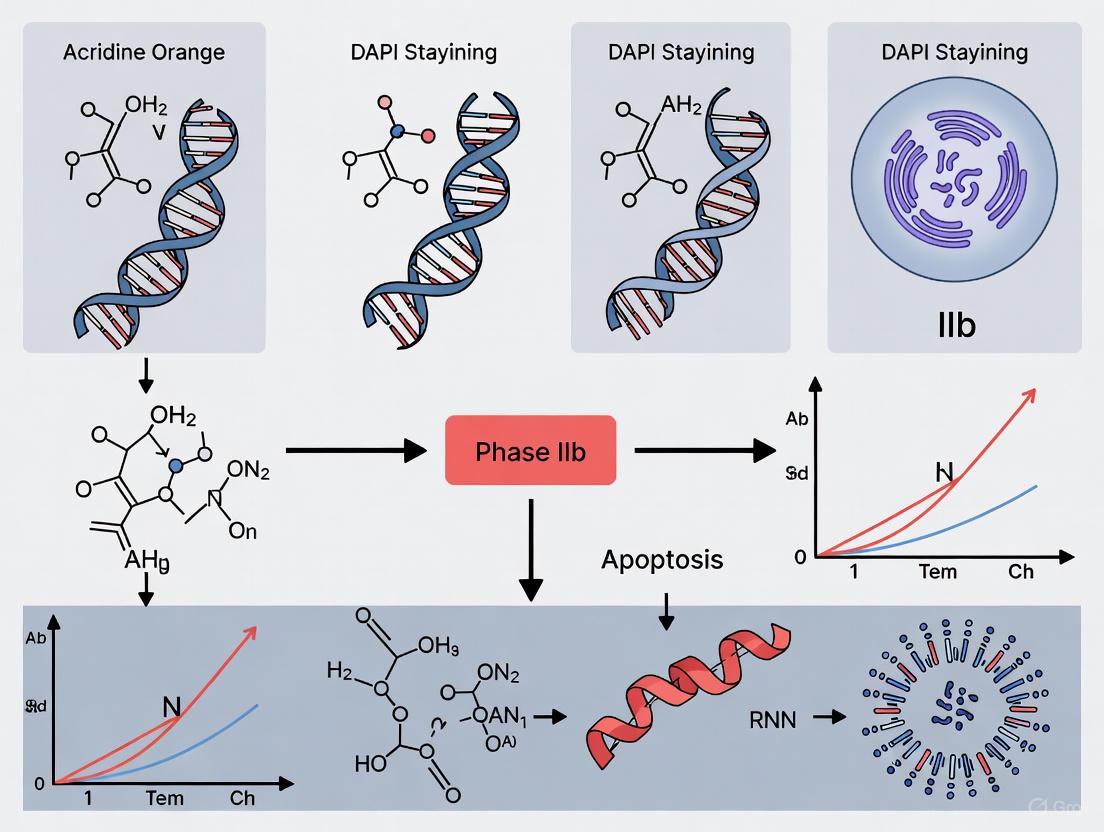
Acridine Orange and DAPI Staining: A Robust Apoptosis Assay for Preclinical and Phase IIb Drug Development
This article provides a comprehensive resource for researchers and drug development professionals on the application of Acridine Orange (AO) and DAPI staining for apoptosis detection in preclinical and Phase IIb...
Acridine Orange and DAPI Staining: A Robust Apoptosis Assay for Preclinical and Phase IIb Drug Development
Abstract
This article provides a comprehensive resource for researchers and drug development professionals on the application of Acridine Orange (AO) and DAPI staining for apoptosis detection in preclinical and Phase IIb studies. We cover the foundational mechanisms of these fluorescent dyes, detailing how they distinguish live, apoptotic, and necrotic cells based on membrane integrity and nuclear morphology. The content delivers optimized protocols for high-throughput 96-well formats, addresses common troubleshooting scenarios, and presents a critical comparative analysis against other viability assays like Annexin V/PI, TUNEL, and caspase activity. By validating AO/DAPI staining against functional long-term proliferation assays and providing strategies for cross-site and cross-instrument comparability, this guide empowers scientists to implement a reliable, specific, and efficient method for quantifying apoptosis in the critical context of cellular therapy and anticancer drug development.
The Science Behind the Stains: How AO and DAPI Reveal Apoptotic Cells
Within the context of advanced apoptosis research (Phase IIb), the selection of appropriate fluorescent dyes is paramount for accurately discerning cell viability and mode of death. Acridine Orange (AO) and 4′,6-Diamidino-2-Phenylindole (DAPI) represent two pivotal tools in the researcher's arsenal. Their utility is fundamentally governed by their distinct mechanisms of cellular entry and subsequent binding to nucleic acids. This application note delineates the core mechanisms of membrane permeability and nuclear binding for AO and DAPI, providing detailed protocols and structured data to facilitate their effective application in drug development research. A critical distinction lies in their interaction with the cell membrane; AO is membrane-permeant, entering all cells, whereas DAPI is membrane-impermeant and typically only enters cells with compromised membrane integrity [1].
Fundamental Dye Mechanisms and Properties
Core Mechanisms of Action
The differential permeability of AO and DAPI forms the basis for their application in sophisticated viability and apoptosis assays. The following diagram illustrates the fundamental journey of each dye from application to nucleic acid binding.
Quantitative Spectral and Binding Characteristics
The distinct spectral signatures and binding preferences of AO and DAPI allow for their simultaneous use in multiplexed assays. The table below summarizes their key characteristics for easy comparison.
Table 1: Spectral and Binding Properties of AO and DAPI
| Property | Acridine Orange (AO) | DAPI |
|---|---|---|
| Membrane Permeability | Permeant (enters all cells) [1] | Impermeant (enters dead cells) [1] |
| Primary Nucleic Acid Target | DNA and RNA [2] | DNA [1] |
| Binding Preference | DNA: Intercalation; RNA: Metachromatic complexes [2] | Minor groove of A-T rich regions [3] |
| Fluorescence Emission (Bound) | DNA: Green (~530 nm) [1]RNA: Red (~635 nm) [2] | Blue (~470 nm) [1] |
| Key Application in Viability | Stains all cells; RNA loss is an early marker of injury [2] | Identifies non-viable cells with compromised membranes [1] |
Experimental Protocols for Apoptosis Detection
The following protocols are adapted from established methodologies used in anticancer research [4] and are tailored for use in a Phase IIb apoptosis research setting.
Protocol 1: AO/DAPI Double Staining for Cell Viability and Count
This protocol allows for the simultaneous quantification of total and viable cell populations [5].
Research Reagent Solutions Table 2: Essential Reagents for AO/DAPI Double Staining
| Reagent | Function |
|---|---|
| Acridine Orange (AO) Stock Solution | Stains total cell population via RNA/DNA binding. |
| DAPI Stock Solution | Stains non-viable cells by binding to DNA in membrane-compromised cells. |
| Phosphate Buffered Saline (PBS) | Provides an isotonic and pH-stable washing and dilution buffer. |
| Via1-Cassette (or equivalent) | Integrated chamber pre-coated with AO and DAPI for standardized staining. |
| Fluorescence Microscope or Cell Counter | Instrument for detecting and quantifying fluorescent signals. |
Methodology
- Cell Preparation: Harvest and wash the cell suspension (e.g., treated with the investigational compound in Phase IIb research) with PBS. Adjust the cell concentration to a range suitable for your detection instrument (e.g.,
5x10^4 to 2x10^7 cells/mLis a typical range for fluorescent counters [1]). - Staining: Load a fixed aliquot (e.g., 20 µL) of the cell suspension into a Via1-Cassette pre-coated with AO and DAPI [5]. Alternatively, mix the cell suspension with prepared AO and DAPI dye solutions to final concentrations optimized for your system.
- Incubation: Incubate the cassette or mixture for 5-10 minutes in the dark at room temperature to allow for complete dye penetration and binding.
- Analysis: Place the cassette into a compatible fluorescence cytometer (e.g., NucleoCounter NC-3000) or analyze under a fluorescence microscope [5].
- Data Acquisition: The instrument's software (e.g., NucleoView) will display total cell concentration based on AO fluorescence and calculate viability as a percentage of viable cells (AO-positive, DAPI-negative) against the total population [5]. Analysis time is typically under 25 seconds [1].
Protocol 2: AO/Propidium Iodide (PI) Staining for Apoptosis Morphology
While DAPI stains dead cells, Propidium Iodide (PI) is a more common red-fluorescent viability counterstain used with AO for live/dead assessment and apoptosis morphology [4]. The workflow for this common assay is outlined below.
Methodology
- Cell Preparation: Culture and treat cells (e.g., MCF-7 breast adenocarcinoma cells) with the test agent. Harvest cells by gentle trypsinization, if adherent, and wash with PBS [4].
- Staining: Re-suspend the cell pellet (~1 x 10^6 cells) in a small volume (e.g., 50 µL) of PBS. Add a mixture of Acridine Orange (e.g., 10 µg/mL final concentration) and Propidium Iodide (e.g., 10 µg/mL final concentration) [4]. Incubate for 5-10 minutes in the dark.
- Microscopy: Place a 10-20 µL aliquot of the stained suspension on a microscope slide, cover with a coverslip, and immediately observe under a fluorescence microscope with appropriate filter sets.
- Scoring and Interpretation: Score a minimum of 200 cells per sample and categorize them based on fluorescence and morphology [4]:
- Viable Cells: Green fluorescence with intact, round-shaped nuclei.
- Early Apoptotic Cells: Green fluorescence with nuclear fragmentation, chromatin condensation, and cell shrinkage.
- Late Apoptotic Cells: Reddish-orange fluorescence (due to AO binding to denatured DNA) with membrane blebbing and apoptotic bodies.
- Necrotic Cells: Red fluorescence (PI-positive) with a swollen appearance.
Quantitative Cytomorphometric Analysis
For a more objective assessment, cytomorphometric analysis can be performed. Following staining with AO and DAPI (or other stains), the nuclear area (NA), cytoplasmic area (CA), and their ratio (N:C ratio) are measured using imaging software. This quantitative approach can reveal subtle, statistically significant changes in cell structure indicative of malignancy, such as an increased N:C ratio in malignant cells compared to normal controls [3].
The strategic application of AO and DAPI is grounded in a firm understanding of their fundamental mechanisms. AO's permeant nature and dual-color emission profile make it a powerful tool for monitoring total cell populations and early RNA loss, a sensitive marker of cellular injury [2]. In contrast, DAPI's impermeant nature provides a clear binary indicator of membrane integrity, a hallmark of late-stage apoptosis and necrosis.
In the context of Phase IIb apoptosis research, combining these dyes in a double-staining protocol, or using AO in conjunction with PI, allows for a nuanced dissection of cell death mechanisms. This enables drug development professionals to not only quantify viability but also to distinguish between apoptotic and necrotic pathways, evaluate the stage of apoptosis, and correlate these findings with molecular assays. The provided protocols and quantitative frameworks offer a reliable foundation for generating robust, reproducible data critical for evaluating the efficacy and mechanism of action of novel therapeutic compounds.
Apoptosis, or programmed cell death, is a genetically controlled process essential for embryogenesis, tissue homeostasis, and disease pathogenesis. The morphological changes in apoptosis are highly conserved and distinct from other forms of cell death such as necrosis. Key hallmarks include cell shrinkage, membrane blebbing, and profound nuclear changes comprising chromatin condensation and nuclear fragmentation. These nuclear events represent the most characteristic and easily identifiable features of apoptotic cells, serving as definitive markers for researchers investigating cell death mechanisms [6].
The significance of detecting these morphological hallmarks extends across biomedical research, particularly in cancer biology and drug discovery. Many therapeutic agents, including novel compounds like gossypin in colorectal cancer and BKS-112 in triple-negative breast cancer, exert their effects by inducing apoptotic pathways in malignant cells [7] [8]. Accurate identification and quantification of chromatin condensation and nuclear fragmentation are therefore crucial for evaluating therapeutic efficacy and understanding fundamental disease processes.
Core Morphological Hallmarks
Chromatin Condensation
Chromatin condensation represents the initial nuclear event in apoptosis, characterized by the compaction and margination of nuclear chromatin against the nuclear envelope. This process results in a characteristic crescent-shaped or ring-like appearance when visualized with DNA-binding fluorescent dyes [6] [9]. At the molecular level, this condensation involves the cleavage of nuclear structural components including lamins and activation of endonucleases that degrade DNA [6].
Recent super-resolution microscopy studies in developing cortical neurons have revealed that chromatin compaction actually precedes caspase activation and obvious nuclear shrinkage, suggesting it may be an early commitment point in the apoptotic pathway rather than merely a late-stage morphological consequence. This compaction occurs progressively and can be classified into distinct stages based on chromatin organization [9].
Nuclear Fragmentation
Following chromatin condensation, the nucleus undergoes systematic fragmentation into discrete membrane-bound apoptotic bodies containing tightly packed, degraded chromatin. This process involves the disassembly of nuclear envelope components and culminates in the formation of multiple pyknotic nuclear fragments [6]. These apoptotic bodies are rapidly recognized and engulfed by neighboring phagocytic cells, preventing the inflammatory responses typically associated with necrotic cell death [6].
The table below summarizes the key characteristics and significance of these morphological hallmarks:
Table 1: Core Morphological Hallmarks of Apoptosis
| Hallmark | Morphological Features | Molecular Mechanisms | Functional Significance |
|---|---|---|---|
| Chromatin Condensation | Chromatin compaction, nuclear marginatio n, crescent formation | Caspase-activated DNase (CAD) activation, lamin cleavage, histone modification | Early apoptotic commitment event, precedes caspase-3 activation [9] |
| Nuclear Fragmentation | Nuclear envelope disruption, formation of pyknotic bodies, apoptotic bodies | Actin cytoskeleton rearrangement, ROCK-I activation, membrane blebbing | Prevents inflammatory response, facilitates phagocytic clearance [6] |
Detection Methodologies and Protocols
Fluorescent Staining with Acridine Orange and DAPI
Fluorescence microscopy using DNA-binding dyes represents one of the most accessible and informative approaches for visualizing apoptotic nuclear morphology. The differential staining patterns and spectral properties of these dyes enable clear discrimination between viable, apoptotic, and necrotic cells.
Acridine Orange (AO) Staining
Acridine orange is a vital dye that permeates all cells and exhibits metachromatic fluorescence properties. When bound to DNA, it emits green fluorescence (∼530 nm), while RNA complexes induce a redshifted emission at ∼635 nm (red fluorescence) [2]. This property is particularly useful for simultaneously assessing RNA and DNA content during cell death processes. In apoptotic cells, characteristic features include condensed or fragmented green or orange chromatin [10].
Table 2: Fluorescent Dyes for Apoptosis Detection
| Dye | Mechanism | Viable Cells | Early Apoptosis | Late Apoptosis | Necrosis |
|---|---|---|---|---|---|
| Acridine Orange (AO) | Intercalates into nucleic acids; green (DNA), red (RNA) | Normal green nucleus | Bright green condensed/fragmented chromatin | Condensed/fragmented orange chromatin | Structurally normal orange nucleus [10] |
| DAPI | AT-selective DNA minor groove binding | Normal blue nucleus | Intense blue condensed chromatin | Fragmented blue nuclei | Normal blue nucleus (membrane permeable) [6] |
| Ethidium Bromide (EB) | DNA intercalator; only enters permeabilized cells | Excluded (no staining) | Excluded in early phase | Orange-red nuclei | Orange-red nuclei [10] |
DAPI Staining
DAPI (4',6-diamidino-2-phenylindole) is a blue fluorescent DNA stain that exhibits enhanced fluorescence when bound to AT-rich regions of DNA. Its high nuclear selectivity and membrane permeability make it ideal for visualizing chromatin organization. Apoptotic nuclei display intensely stained, condensed chromatin that may be fragmented into smaller spherical bodies [6]. The protocol below describes a standardized approach for DAPI staining:
Protocol: DAPI Staining for Apoptotic Nuclei
- Cell Preparation: Culture cells on glass coverslips under experimental conditions.
- Fixation: Incubate cells in 4% paraformaldehyde in PBS for 15 minutes at room temperature.
- Permeabilization: Treat with 0.1% Triton X-100 in PBS for 5 minutes.
- Staining: Apply DAPI solution (1 µg/mL in PBS) for 5 minutes in the dark.
- Washing: Rinse three times with PBS to remove unbound dye.
- Mounting: Apply antifade mounting medium and seal with coverslips.
- Visualization: Examine using fluorescence microscopy with UV excitation (∼350 nm) and blue emission (∼461 nm) [6].
Modified EB/AO Staining in 96-Well Plates
For higher throughput applications, a modified ethidium bromide/acridine orange (EB/AO) staining method performed entirely in 96-well plates offers significant advantages. This approach eliminates cell detachment and washing steps, minimizing mechanical damage and cell loss while enabling rapid quantification of live, apoptotic, and necrotic populations [10].
Protocol: 96-Well EB/AO Staining Assay
- Cell Seeding: Plate adherent or suspension cells in 96-well plates and apply experimental treatments.
- Staining Solution: Prepare a mixture of acridine orange (100 µg/mL) and ethidium bromide (100 µg/mL) in phosphate-buffered saline [10].
- Staining: Add staining solution directly to wells without removing culture medium.
- Centrifugation: Centrifuge plates at low speed (∼200 × g) for 5 minutes to sediment cells.
- Immediate Analysis: Visualize using an inverted fluorescence microscope with blue filter sets.
- Quantification: Count at least 200 cells per well and classify based on nuclear morphology and fluorescence:
- Viable cells: Normal green nuclei
- Early apoptotic: Bright green condensed or fragmented chromatin
- Late apoptotic: Condensed/fragmented orange chromatin
- Necrotic: Normal orange nuclei [10]
Signaling Pathways in Apoptosis
The morphological changes characteristic of apoptosis are orchestrated by complex signaling pathways that converge on effector mechanisms responsible for chromatin condensation and nuclear fragmentation. Two major pathways—the intrinsic (mitochondrial) and extrinsic (death receptor) pathways—ultimately activate executioner caspases that mediate the systematic dismantling of cellular structures [7] [8].
The following diagram illustrates the key signaling pathways connecting apoptotic stimuli to the morphological hallmarks of chromatin condensation and nuclear fragmentation:
Diagram Title: Signaling Pathways Linking Apoptotic Stimuli to Nuclear Hallmarks
The diagram illustrates how diverse apoptotic stimuli, including therapeutic agents like gossypin and BKS-112, activate signaling pathways that converge on caspase activation. Executioner caspases then directly mediate the nuclear events of apoptosis through cleavage of structural proteins and activation of endonucleases, culminating in the characteristic morphological hallmarks of chromatin condensation and nuclear fragmentation [7] [8] [6].
The Scientist's Toolkit: Essential Research Reagents
Successful investigation of apoptotic morphology requires specific reagents and tools. The following table provides a comprehensive overview of essential materials for studying chromatin condensation and nuclear fragmentation:
Table 3: Essential Research Reagents for Apoptosis Morphology Studies
| Reagent/Category | Specific Examples | Application/Function | Experimental Notes |
|---|---|---|---|
| Fluorescent Dyes | Acridine Orange (AO) | Vital dye for DNA/RNA discrimination; identifies apoptotic chromatin patterns [2] [10] | Use at 100 µg/mL; green (DNA) vs red (RNA) emission |
| DAPI | Nuclear counterstain for fixed cells; highlights chromatin condensation [6] | Use at 1 µg/mL; AT-selective DNA binding | |
| Ethidium Bromide (EB) | Membrane integrity assessment; distinguishes late apoptosis/necrosis [10] | Combine with AO for live/dead discrimination | |
| Hoechst 33342 | Live-cell permeable DNA stain; tracks real-time nuclear changes [6] | Use at 1-5 µg/mL; suitable for time-lapse imaging | |
| Antibodies | Cleaved Caspase-3 | Confirms apoptotic pathway activation [7] [8] | IHC, IF, Western blot; marker of execution phase |
| Phospho-JNK | Detects MAPK pathway activation in apoptosis [7] | Western blot, IHC; upstream signaling indicator | |
| Acetylated α-tubulin | HDAC6 inhibition readout [8] | Western blot; mechanistic studies | |
| Pharmacological Inhibitors/Inducers | Staurosporine | Broad-spectrum kinase inhibitor; apoptosis inducer [9] | Use at 0.1-1 µM; positive control for apoptosis |
| SP600125 | JNK pathway inhibitor; mechanistic studies [7] | Use at 10 µM; pathway specificity testing | |
| 3-Methyladenine (3-MA) | Early-stage autophagy inhibitor [7] | Use at 2 mM; apoptosis/autophagy discrimination | |
| Cell Lines | HT-29 | Human colorectal cancer; apoptosis research [7] | Gossypin studies; p-JNK mediated apoptosis |
| MDA-MB-231 | Triple-negative breast cancer; therapy response [8] | HDAC6 inhibitor studies | |
| A375 | Human melanoma; general apoptosis research [10] | Adherent cell model for protocol development | |
| Specialized Reagents | FITC Annexin V | Phosphatidylserine exposure detection [7] [8] | Early apoptosis marker; flow cytometry |
| MTT/XTT Reagents | Cell viability/metabolic activity assessment [7] [8] | Colorimetric assays; correlate with morphology | |
| Caspase Substrates (DEVD) | Caspase activity quantification [11] | Fluorogenic or luminescent detection |
Advanced Techniques and Research Applications
Real-Time Apoptosis Monitoring
Advanced imaging technologies now enable real-time monitoring of apoptosis in live cells. Genetically encoded FRET-based caspase sensors (e.g., ECFP-DEVD-EYFP) allow quantitative assessment of caspase activation kinetics alongside morphological changes. When combined with organelle-specific fluorescent markers (e.g., Mito-DsRed), these tools can discriminate between apoptosis and primary necrosis with high temporal resolution [11]. This approach reveals that after caspase activation, cells typically transition to secondary necrosis within 45 minutes to 3 hours, informing appropriate imaging intervals for accurate classification [11].
Super-Resolution Microscopy
Single-molecule localization microscopy (SMLM) techniques provide unprecedented resolution for characterizing nuclear changes during apoptosis. Recent studies utilizing SMLM have identified five distinct stages of chromatin compaction during neuronal apoptosis, with early compaction preceding both caspase-3 activation and nuclear shrinkage [9]. Quantitative analysis using Sobel edge detection algorithms generates a chromatin compaction parameter (CCP) that objectively measures progression through these stages, providing a more nuanced understanding of nuclear dynamics than traditional morphology assessment alone [9].
The morphological hallmarks of apoptosis—chromatin condensation and nuclear fragmentation—remain cornerstone features for identifying and quantifying programmed cell death in research settings. While traditional staining methods with acridine orange and DAPI continue to provide valuable information, advanced techniques including real-time FRET imaging and super-resolution microscopy are revealing unprecedented details about the spatial and temporal progression of nuclear events. These methodologies, combined with a growing understanding of the underlying molecular pathways, enhance our ability to investigate apoptosis in both basic research and drug discovery contexts.
This application note provides a detailed protocol for using Acridine Orange (AO) and DAPI dual fluorescence staining to differentiate between live, early apoptotic, late apoptotic, and necrotic cells. The method leverages the distinct membrane permeability properties of these dyes to assess cell viability and stage-specific cell death morphology, providing a reliable digital approach for quantifying cell death in planta systems and mammalian cells. This technique is particularly valuable for Phase IIb apoptosis research, enabling high-content screening in drug development pipelines.
Programmed cell death (PCD) is an active process controlling proper development of unicellular and multicellular organisms by eliminating physiologically redundant, damaged, or abnormal cells [12]. In biomedical research and drug development, accurately discriminating between the stages of apoptosis and necrosis is crucial for evaluating therapeutic efficacy and mechanism of action. While apoptosis exhibits distinct morphological changes including cell shrinkage, membrane blebbing, and nuclear fragmentation, necrosis is characterized by cellular swelling and early rupture of the plasma membrane [12] [13]. Fluorescence staining with DNA-binding dyes such as Acridine Orange (AO) and DAPI provides a powerful tool for detecting these morphological changes, allowing researchers to differentiate between live, early apoptotic, late apoptotic, and necrotic cells based on nuclear morphology and membrane integrity.
Principle of AO/DAPI Staining
The AO/DAPI staining method utilizes the differential membrane permeability of these fluorochromes to assess cell viability and stage of cell death:
- Acridine Orange (AO) is a membrane-permeant dye that stains all cells regardless of viability, binding to DNA and RNA. When bound to DNA, it emits green fluorescence, while RNA binding produces red fluorescence [1].
- DAPI is a membrane-impermeant dye that only penetrates cells with compromised membrane integrity, typically late apoptotic and necrotic cells. It stains DNA and emits blue fluorescence [14] [1].
The differential staining pattern allows classification of cell states based on dye accessibility and nuclear morphology, which changes characteristically during cell death progression.
Spectral Characteristics and Binding Properties
The table below summarizes the spectral properties and binding characteristics of AO and DAPI:
Table 1: Fluorescent Dyes for Cell Death Detection
| Dye | Excitation/Emission Max | Membrane Permeability | Nucleic Acid Binding | Fluorescence Color |
|---|---|---|---|---|
| Acridine Orange (AO) | 502/526 nm (DNA) 460/650 nm (RNA) | Permeant to all cells | DNA: Green fluorescence RNA: Red fluorescence | Green/Red |
| DAPI | 358/461 nm | Impermeant (dead cells only) | DNA: Blue fluorescence | Blue |
| Propidium Iodide (PI) | 535/617 nm | Impermeant (dead cells only) | DNA: Red fluorescence | Red |
Materials and Reagents
Research Reagent Solutions
Table 2: Essential Materials and Reagents
| Item | Function/Application | Specifications |
|---|---|---|
| Acridine Orange (AO) | Stains all nucleated cells (live and dead) | 100 µg/mL in phosphate buffer [12] |
| DAPI (4',6-diamidino-2-phenylindole) | Stains non-viable cells with compromised membranes | 2 µg/mL in staining solution [12] |
| Phosphate Buffered Saline (PBS) | Washing and dilution buffer | 0.01 M, pH 7.4 [12] |
| Glutaraldehyde | Fixation agent | 1.0% in phosphate buffer [12] |
| Carnoy's Fixative | Alternative fixation for nuclear morphology | 96% ethanol and glacial acetic acid (3:1) [12] |
| Fluorescence Microscope | Imaging and analysis | Equipped with blue filter and UV light source [12] |
| Via1-Cassette (NanoEntek) | Automated cell counting | Pre-coated with AO and DAPI [5] |
| NucleoCounter NC-3000 | Automated cell viability analysis | Compatible with Via1-Cassette [5] |
Experimental Protocols
Sample Preparation and Staining Protocol
Workflow Overview:
Detailed Procedure:
- Cell Preparation: Harvest cells and wash twice with 0.01 M phosphate buffer (pH 7.4) to remove debris [12].
- Staining Solution Preparation: Prepare fresh staining mixture containing 100 μg/mL Acridine Orange and 100 μg/mL DAPI in phosphate buffer [12] [14]. For automated counters, use pre-coated Via1-Cassettes [5].
- Staining Incubation: Incubate living cell samples with 1 mL of staining mixture for 4 minutes at room temperature, protected from light [12].
- Washing: Gently wash stained cells twice with phosphate buffer to remove excess dye.
- Fixation: Fix samples with 1.0% glutaraldehyde in phosphate buffer for 15 minutes to preserve morphology [12]. Alternatively, cold Carnoy's fixative (96% ethanol:glacial acetic acid, 3:1) can be used for 1 hour [12].
- Slide Preparation: Prepare thin sections or cell suspensions on glass slides with a drop of phosphate buffer.
- Microscopy: Analyze samples using fluorescence microscopy with appropriate filter sets:
Data Acquisition and Analysis
Image Acquisition:
- Capture fluorescence images using a CCD camera coupled to the microscope [12].
- For AO: Use blue excitation (502 nm) and collect green (526 nm, DNA) and red (650 nm, RNA) emissions [1].
- For DAPI: Use UV excitation (358 nm) and collect blue emission (461 nm) [14].
Cell Classification Criteria: Table 3: Interpretation of Fluorescence Staining Patterns
| Cell Status | AO Staining | DAPI Staining | Nuclear Morphology | Membrane Integrity |
|---|---|---|---|---|
| Live Cells | Green chromatin | Negative | Intact, normal structure | Preserved |
| Early Apoptotic | Green-yellow to yellow-orange chromatin | Negative | Chromatin condensation, nuclear shrinkage | Initially intact |
| Late Apoptotic | Bright orange to red chromatin | Positive | Nuclear fragmentation, condensed chromatin | Lost |
| Necrotic Cells | Red chromatin | Positive | Nuclear swelling, diffuse staining | Lost |
Quantitative Analysis:
- Count at least 200 cells per sample across multiple random fields.
- Calculate the percentage of cells in each category:
- % Viability = (AO+ DAPI- cells / Total cells) × 100
- % Early Apoptosis = (AO+ with condensed nuclei, DAPI- / Total cells) × 100
- % Late Apoptosis = (AO+ DAPI+ with fragmented nuclei / Total cells) × 100
- % Necrosis = (AO+ DAPI+ with swollen nuclei / Total cells) × 100
Results and Data Interpretation
Characteristic Fluorescence Patterns
The classification of cell death stages is based on the following fluorescence patterns:
- Live cells exhibit bright green nuclear staining with AO and are negative for DAPI, indicating intact membranes and normal chromatin organization [14] [1].
- Early apoptotic cells show green-yellow to yellow-orange nuclear staining with AO due to chromatin condensation, but remain DAPI-negative because membrane integrity is largely maintained [12] [13]. The chromatin appears condensed but not fragmented.
- Late apoptotic cells display bright orange to red nuclei with AO and are positive for DAPI, indicating complete loss of membrane integrity. The nuclei typically show fragmentation and condensed chromatin [12].
- Necrotic cells exhibit bright red nuclear staining with AO and are DAPI-positive, with swollen nuclei and diffuse chromatin distribution without condensation [13].
Quantitative Assessment
Table 4: Typical Results from ACC-Induced Cell Death in Vicia faba Root Cortex [12]
| Parameter | Control Cells | ACC-Treated Cells | Detection Method |
|---|---|---|---|
| Viable Cells | >95% | ~80% | AO+/DAPI- |
| Total Cell Death | <5% | ~20% | Morphological changes |
| Ion Leakage | Baseline | Increased | Conductivity measurement |
| Nuclear Fragmentation | Absent | Present | DAPI staining |
| Aerenchyma Formation | Absent | Present (few spaces) | Morphological analysis |
Troubleshooting and Technical Notes
Common Issues and Solutions
- Weak Fluorescence Signal: Increase dye concentration or incubation time; verify filter sets match dye specifications.
- High Background: Reduce dye concentration; increase washing steps; check fixative purity.
- Non-specific DAPI Staining: Confirm membrane integrity in control samples; optimize fixation method.
- Inconsistent Results: Use fresh staining solutions; standardize incubation conditions; include appropriate controls.
Important Considerations
- Dye Concentration: Optimal dye concentrations may vary by cell type and should be determined empirically.
- Fixation Time: Prolonged fixation can affect fluorescence intensity and membrane permeability.
- Kinetic Studies: For time-course experiments, minimize light exposure to prevent photobleaching.
- Apoptosis Inducers: Include positive controls (e.g., staurosporine for apoptosis [13]) to validate the assay.
Applications in Drug Development
The AO/DAPI staining method provides a robust platform for screening compounds in drug development:
- High-Content Screening: Automated systems like the NucleoCounter NC-3000 enable rapid assessment of cell viability and death mechanisms [5].
- Mechanistic Studies: Distinguishing between apoptosis and necrosis helps elucidate compound mechanisms of action.
- Toxicity Assessment: Evaluating cell death profiles in primary cells predicts compound toxicity earlier in development pipelines.
- Therapeutic Efficacy: Monitoring apoptosis induction is valuable in oncology drug development where activating cell death pathways is a primary therapeutic goal.
The AO/DAPI dual fluorescence staining method provides a reliable, reproducible approach for discriminating between live, early apoptotic, late apoptotic, and necrotic cells. This protocol enables quantitative assessment of cell death progression based on characteristic nuclear morphology changes and membrane integrity, making it particularly valuable for apoptosis research in drug development. The method's adaptability to both manual microscopy and automated high-content screening platforms makes it suitable for various research and development applications, from basic mechanism studies to preclinical compound screening.
The Critical Role of Apoptosis Quantification in Preclinical Drug Screening and Phase IIb Efficacy Assessment
Apoptosis, or programmed cell death, is a tightly regulated process essential for maintaining tissue homeostasis and eliminating damaged or unnecessary cells. In the context of drug development, particularly for oncology, the ability of therapeutic compounds to induce apoptosis in target cells serves as a critical indicator of efficacy. Apoptosis occurs through two primary pathways: the extrinsic pathway, initiated by external death signals through cell surface receptors, and the intrinsic pathway, triggered by internal cellular stress signals, often involving mitochondrial components. Both pathways converge on the activation of a cascade of cysteine-aspartic proteases known as caspases, which execute the dismantling of cellular structures in a controlled manner [15] [16]. The quantification of apoptosis provides invaluable data for understanding disease mechanisms, screening potential drug candidates, and evaluating treatment efficacy during early clinical trials, including Phase IIb studies [17].
The morphological and biochemical hallmarks of apoptosis include cell shrinkage, chromatin condensation, DNA fragmentation, and membrane blebbing. Unlike necrotic cell death, which triggers inflammatory responses, apoptosis is a clean, non-inflammatory process [15] [16]. Accurate detection and quantification of these changes are therefore paramount in preclinical and clinical research to distinguish between different modes of cell death and to accurately assess a drug's mechanism of action. This application note details established and emerging methodologies for apoptosis quantification, with a specific focus on protocols relevant to preclinical drug screening and Phase IIb efficacy assessment.
Apoptosis Quantification Technologies and Their Applications
A variety of techniques are available for detecting and quantifying apoptosis, each with unique advantages, limitations, and applications in the drug development pipeline. The choice of method depends on the research context, required sensitivity, throughput, and whether the analysis is performed in vitro, ex vivo, or in a clinical setting.
The table below summarizes the key apoptosis detection methods and their typical applications in drug development:
Table 1: Comparison of Apoptosis Detection Methods in Drug Development
| Method | Principle | Key Readout | Application Stage | Advantages | Limitations |
|---|---|---|---|---|---|
| Acridine Orange/DAPI Staining | Differential staining of viable (AO) vs. non-viable (DAPI) cells [1] | Cell viability percentage, total cell concentration [5] | Preclinical screening, cell viability assessment | Rapid (<25 sec), high-throughput, quantitative [1] | Limited to viability, not specific apoptosis mechanisms |
| Annexin V/PI Staining by Flow Cytometry | Detection of phosphatidylserine (PS) externalization (Annexin V) and membrane integrity (PI) [18] | Distinction of viable, early apoptotic, late apoptotic, and necrotic cells | Preclinical mechanism studies, ex vivo analysis | Discerns early vs. late apoptosis, high-throughput | Requires cell suspension, potential for artifact |
| Western Blotting | Detection of protein markers and cleavage products (e.g., caspases, PARP, Bcl-2 family) [16] | Presence/absence and ratio of cleaved to full-length proteins | Preclinical mechanism of action studies | High specificity, information on specific pathways | Semi-quantitative, requires large cell numbers, no single-cell data |
| Drug-Induced Apoptosis (MiCK) Assay | Measures kinetic units (KU) of apoptosis via optical density changes in response to drugs [17] | Apoptotic kinetic curve, KU value for drug response | Phase IIb efficacy assessment, therapy selection | Functional ex vivo test, correlates with clinical outcomes [17] | Requires viable tumor tissue, specialized equipment |
| Stimulated Raman Scattering (SRS) Microscopy | Label-free measurement of biochemical composition (e.g., protein/lipid ratios) [19] | Chemotypic signatures of apoptosis (e.g., increased protein concentration) | Preclinical research, potential for future clinical use | Label-free, non-destructive, live-cell imaging | Emerging technology, specialized equipment required |
| Nuclear Morphometry (ImageJ) | Quantification of nuclear morphological changes (area, circularity, NAF) [20] | Nuclear Area Factor (NAF), circularity | Preclinical research, histopathology analysis | Low-cost, quantitative from standard images, detects early changes | Requires well-separated nuclei for accuracy |
Detailed Experimental Protocols
Protocol: Acridine Orange (AO) and DAPI Staining for Cell Viability and Apoptosis Assessment
This protocol describes a rapid, fluorescence-based method for determining total cell count and viability, which serves as an initial screening tool in apoptosis assessment [1] [5].
Research Reagent Solutions
Table 2: Key Reagents for AO/DAPI Staining Protocol
| Reagent | Function | Mechanism | Specifications |
|---|---|---|---|
| Acridine Orange (AO) | Membrane-permeant nucleic acid stain [1] | Stains all cells (live and dead); emits green when bound to DNA, red when bound to RNA [1] | Working solution prepared per manufacturer's instructions. |
| DAPI (4',6-Diamidino-2-Phenylindole) | Membrane-impermeant nucleic acid stain [1] [21] | Enters only dead cells with compromised membranes; stains DNA and emits blue fluorescence [1] | Stock: 1 mg/mL in water (store at -20°C, wrapped in foil); Working: 10 μg/mL [21]. Carcinogen - handle with care. |
| Cell Culture | Sample material | Provides cells for analysis. | Use early to mid-log phase cultures; gently sonicate if aggregates are visible [21]. |
| Fixative (e.g., Glutaraldehyde or Formaldehyde) | Cell fixation | Cross-links biomolecules to preserve cell state for later analysis [21]. | Toxic. Glutaraldehyde: final conc. 2.5%; Formaldehyde: final conc. 1.0% (avoid for chlorophyll-containing cells) [21]. |
| NucleoCounter Via1-Cassette & NC-3000 | Analysis system | Cassette is pre-coated with AO and DAPI; instrument automates counting and viability calculation [5]. | Compatible with various cell types (cell lines, PBMCs, adipose stem cells) [1]. |
Step-by-Step Procedure
Cell Preparation and Staining:
- Harvest cells and prepare a single-cell suspension. Gently sonicate if necessary to break up aggregates [21].
- If analysis cannot be performed immediately, fix an aliquot of cells. Add glutaraldehyde to a final concentration of 2.5% (v/v) or formaldehyde to 1.0% (v/v). Incubate on ice for 10-30 minutes in a dark vial [21].
- For live-cell analysis, load an aliquot of the cell suspension directly into a Via1-Cassette, which is pre-coated with AO and DAPI [5]. Alternatively, for manual staining, incubate cells with DAPI at a final concentration of 1 μg/mL on ice for 7-10 minutes in the dark [21].
Measurement and Data Acquisition:
- Place the loaded Via1-Cassette into the NucleoCounter NC-3000 cytometer.
- The instrument will automatically perform the count. Total cell concentration and viability results will be displayed using the associated NucleoView software [5].
- Viability of treated cells is calculated as a percentage of the control cells [5].
Protocol: Annexin V/Propidium Iodide (PI) Assay for Flow Cytometry
This protocol allows for the discrimination of cell populations based on apoptotic stages [18].
- Cell Preparation: Harvest and wash cells in cold phosphate-buffered saline (PBS). Resuspend approximately 1x10^5 to 1x10^6 cells in a binding buffer.
- Staining: Add Annexin V conjugated to a fluorochrome (e.g., FITC or PE) and Propidium Iodide (PI) to the cell suspension. Incubate for 15-20 minutes at room temperature in the dark.
- Analysis: Analyze the cells by flow cytometry within 1 hour. Use FITC/Annexin V and PI channels for detection:
- Annexin V-/PI-: Viable cells.
- Annexin V+/PI-: Early apoptotic cells.
- Annexin V+/PI+: Late apoptotic or necrotic cells.
- Annexin V-/PI+: Necrotic cells or cellular debris [18].
Protocol: Western Blot Analysis for Apoptosis Pathway Markers
This protocol is used to confirm the activation of specific apoptotic pathways by detecting key protein markers and their cleavage products [16].
- Cell Lysis and Protein Quantification: Prepare cell lysates from treated and control samples. Quantify protein concentration to ensure equal loading across gels.
- SDS-PAGE and Transfer: Separate proteins by SDS-Polyacrylamide Gel Electrophoresis (SDS-PAGE) based on molecular weight. Transfer the separated proteins from the gel to a western blot membrane.
- Blocking and Antibody Incubation:
- Block the membrane to prevent non-specific antibody binding.
- Incubate the membrane with primary antibodies targeting apoptotic markers of interest (e.g., cleaved caspase-3, cleaved PARP, Bax, Bcl-2).
- Wash the membrane and incubate with enzyme- or fluorophore-conjugated secondary antibodies.
- Detection and Analysis: Visualize protein bands using chemiluminescent or fluorescent detection. Use densitometry software (e.g., ImageJ) to quantify band intensity. Normalize the signal of the target protein to a housekeeping protein (e.g., β-actin, GAPDH). The ratio of cleaved to full-length protein (e.g., cleaved PARP to full-length PARP) indicates the extent of apoptosis [16].
Protocol: Nuclear Morphometric Analysis Using ImageJ for Apoptosis Assessment
This protocol uses open-source software to quantify early apoptotic changes based on nuclear condensation and fragmentation [20].
- Cell Staining and Imaging:
- Culture and treat cells on slides. Fix and stain nuclei using an appropriate stain (e.g., Giemsa, hematoxylin, DAPI) [20].
- Capture digital images of stained cells using a microscope with a consistent magnification.
- Image Analysis with ImageJ:
- Open the image in ImageJ and convert it to 8-bit (Image > Type > 8-bit).
- Set the scale (Analyze > Set Scale).
- Adjust the threshold to highlight nuclei (Image > Adjust > Threshold).
- Analyze particles (Analyze > Analyze Particles). Set appropriate size and circularity limits to exclude debris and aggregates. Ensure "Display results" and "Add to Manager" are selected.
- The results table will provide data for each nucleus, including Area, Perimeter, and Circularity [20].
- Calculation of Nuclear Area Factor (NAF):
Data Analysis and Interpretation for Phase IIb Studies
In Phase IIb clinical trials, which focus on determining the efficacy and optimal dosing of a new drug, robust and quantitative apoptosis data can serve as a valuable pharmacodynamic biomarker.
Correlating Apoptosis with Clinical Outcomes
The MiCK drug-induced apoptosis assay exemplifies how apoptosis quantification can be integrated into clinical trials. In a study of patients with recurrent or metastatic breast cancer, tumor specimens were subjected to the MiCK assay to identify chemotherapy drugs that induced the highest levels of apoptosis in vitro. Physicians who used the assay results to guide treatment decisions achieved a significantly higher response rate (38.1% vs. 0%) and longer time to relapse (7.4 months vs. 2.2 months) compared to those who did not use the assay [17]. This demonstrates the potential of functional apoptosis assays to inform treatment strategies and improve clinical outcomes in later-phase trials.
Analysis of Nuclear Morphometry Data
When using ImageJ for nuclear analysis, statistical evaluation is crucial. A one-way ANOVA followed by a post-hoc test like Tukey's can be used to test for significant differences in NAF between control and treated groups. A strong positive correlation (e.g., R = 0.958) has been demonstrated between decreased NAF and reduced cell viability, validating NAF as a reliable parameter for apoptosis assessment [20].
Diagram: Integrated Workflow for Apoptosis Quantification in Drug Development. This diagram outlines a logical progression of key apoptosis detection methods from preclinical screening to clinical efficacy assessment.
The accurate quantification of apoptosis is indispensable throughout the drug development pipeline. From initial preclinical screening using accessible methods like AO/DAPI staining and Western blotting to more sophisticated ex vivo analyses like the MiCK assay in Phase IIb trials, these techniques provide critical insights into a drug's biological activity. As research advances, the integration of label-free technologies like SRS microscopy and the standardization of quantitative image analysis promise to further enhance the precision and predictive power of apoptosis assessment, ultimately contributing to the development of more effective and targeted therapies.
Optimized Protocols for High-Throughput Apoptosis Analysis in 96-Well Plates
This application note details a modified ethidium bromide and acridine orange (EB/AO) staining assay performed entirely in a 96-well plate format. This method combines the advantages of conventional EB/AO staining—which allows simultaneous quantification of live, apoptotic, and necrotic cells based on nuclear morphology and membrane integrity—with the efficiency and minimal cell manipulation required for modern high-throughput workflows [10]. The protocol is specifically optimized for use in apoptosis phase IIb research, providing a reliable, cost-effective method for screening chemotherapeutic potential of novel compounds.
The study of apoptosis is fundamental in oncology and drug development. A critical need exists for assays that can accurately distinguish between the different stages of apoptosis and necrosis. The EB/AO double staining technique meets this need by providing a morphological assessment of cell death [22]. AO is a cell-permeable dye that intercalates with DNA, emitting green fluorescence, while EB is only taken up by cells that have lost membrane integrity, emitting red fluorescence and dominating over AO [23].
Traditional EB/AO methods require multiple cell-handling steps—detaching, washing, and transferring—which can damage cells, alter population distribution, and increase processing time [10]. The modified protocol presented here eliminates these steps by utilizing a gentle centrifugation in a 96-well plate, minimizing artifacts and making it ideal for both suspension and adherent cell lines [10]. This method is particularly valuable for assessing the efficacy of novel compounds in phase IIb apoptosis research, as demonstrated in studies involving andrographolide derivatives and other cytotoxic agents [24] [25].
Materials and Reagents
Research Reagent Solutions
The following table lists the essential materials required for the successful execution of this protocol.
Table 1: Key Research Reagent Solutions and Materials
| Item Name | Specification / Function | Storage / Notes |
|---|---|---|
| Acridine Orange (AO) Stock | 100 µg/mL in PBS; stains all cells (DNA=green, RNA=red) [22] | 2-8°C, protected from light [23] |
| Ethidium Bromide (EB) Stock | 100 µg/mL in PBS; stains only dead/damaged cells (red) [22] | 2-8°C, protected from light; toxic, handle with care [23] |
| Dilution Buffer | PBS or other suitable buffer | 2-8°C; used to prepare working solution [23] |
| AO/EB Working Solution | Prepared fresh by mixing AO:EB:Buffer in a 1:1:8 ratio [23] | Prepare immediately before use; keep in dark |
| Cell Culture Medium | Appropriate for the cell line (e.g., RPMI1640 with 10% FBS [22]) | - |
| 96-Well Plate | Flat-bottomed, suitable for fluorescence microscopy | Glass-bottom recommended for superior optical clarity [10] |
| Phosphate Buffered Saline (PBS) | For washing cells (if required) | Sterile |
Equipment
- Fluorescence microscope with FITC/TRITC filters (e.g., Leica DM 3000 [26] or OLYMPUS models [22])
- Centrifuge with a 96-well plate rotor
- Laminar flow hood
- Incubator (37°C, 5% CO₂)
- Micropipettes and tips
Method
Experimental Workflow
The following diagram illustrates the simplified workflow of the modified EB/AO staining method, highlighting its one-step advantage over conventional techniques.
Step-by-Step Protocol
Cell Preparation and Treatment
- Suspension Cells: Seed cells directly into the 96-well plate at a density of 1-2 x 10⁵ cells/well [26] [22].
- Adherent Cells: Seed cells and allow them to attach overnight. The key advantage of this method is that adherent cells do not need to be trypsinized prior to staining [10].
- Treat cells with the test compounds (e.g., potential apoptosis-inducing agents) for the desired duration. Include negative (vehicle) and positive (e.g., a known chemotherapeutic) controls.
Centrifugation
- Following treatment, centrifuge the entire 96-well plate at 1000 rpm for 5 minutes [23]. This critical step pellets all cells, including any detached apoptotic or necrotic cells (floaters), at the bottom of the well without requiring washing or transfer. This ensures the entire cell population is retained for analysis.
Staining Solution Preparation
- While the plate is centrifuging, prepare the AO/EB working solution fresh. Combine Component A (AO stain), Component B (EB stain), and Component C (dilution buffer) in a 1:1:8 ratio [23]. For example, mix 10 µL of AO, 10 µL of EB, and 80 µL of buffer. Protect the solution from light.
Staining and Incubation
- After centrifugation, carefully remove the supernatant from each well if desired, but this is not always necessary.
- Add 2 µL of the freshly prepared AO/EB working solution directly to each 25 µL cell suspension [23]. If cells are in a smaller volume, adjust proportionally to ensure adequate staining.
- Gently mix the solution by pipetting up and down.
- Incubate the plate at room temperature for 5-10 minutes, protected from light.
Microscopy and Quantification
- After incubation, immediately observe the cells using a fluorescence microscope with a luciferin filter and a 60x magnification objective [23].
- Using the live preview, allow the cells to settle before capturing images or counting.
- Count at least 100 total cells per well, repeating the measurement at least three times for statistical reliability [23]. Use the criteria in Table 2 to classify cells.
Data Interpretation and Cell Classification
The differential staining properties of AO and EB allow for clear distinction between cell states based on nuclear color and morphology.
Table 2: Quantitative Classification of Cells by AO/EB Staining [10] [22]
| Cell Status | Nuclear Morphology | Fluorescence Color | Biological Basis |
|---|---|---|---|
| Viable/Live | Normal, organized structure | Green | AO permeates intact membrane; DNA is double-stranded. |
| Early Apoptotic | Chromatin condensation, crescent-shaped or granular | Bright Green or Yellow-Green | Membrane intact but chromatin condensing; AO access maintained. |
| Late Apoptotic | Condensed, fragmented chromatin (pyknotic) | Orange or Orange-Red | Loss of membrane integrity allows EB entry; condensed chromatin remains. |
| Necrotic | Structurally normal, may be enlarged | Orange-Red, uniform | Complete membrane failure; EB binds to DNA uniformly. ``` |
The following logic diagram aids in the systematic classification of cells during microscopy based on these criteria.
Application Data and Validation
Representative Results
This method has been successfully applied in multiple research contexts. For instance, in a study on novel chalcone derivatives (AC-10, AC-13, AC-14), AO/EB staining quantitatively revealed a significant increase in apoptotic nuclei in HCT-116 colon cancer cells compared to controls, with AC-13 showing the highest efficacy (31% apoptosis) [25]. Similarly, the method effectively showed kappa-selenocarrageenan-induced apoptosis in human osteosarcoma cells in a dose-dependent manner [22].
Comparison with Flow Cytometry
The validity of the quantitative data from this AO/EB method is high. The table below shows a direct comparison with flow cytometry, the gold standard for apoptosis quantification.
Table 3: Validation of AO/EB Staining Against Flow Cytometry [22]
| Treatment Group | Apoptotic Cells (Flow Cytometry, PI) | Apoptotic Cells (AO/EB Staining) | P Value |
|---|---|---|---|
| 30 μg/ml | 6.25% ± 0.9% | 6.68% ± 1.2% | 0.69 |
| 60 μg/ml | 9.97% ± 1.5% | 10.33% ± 1.7% | 0.75 |
| 120 μg/ml | 20.14% ± 1.8% | 20.46% ± 2.0% | 0.84 |
Statistical analysis (Student's t-test) confirmed no significant difference (P>0.05) between the two methods, establishing the modified AO/EB staining as a reliable and economical alternative to flow cytometry for apoptosis quantification [22].
Troubleshooting
- Low Fluorescence Signal: Ensure the AO/EB working solution was prepared fresh and used immediately. Check that dyes have not expired and were stored correctly, protected from light.
- Excessive Background Stain: Optimize the concentration of the staining solution and the incubation time. Avoid over-incubation.
- Cell Loss in Adherent Cultures: This method specifically minimizes this issue. Ensure gentle centrifugation and that the supernatant is removed carefully without disturbing the cell pellet.
- Poor Nuclear Morphology Definition: If using plastic-bottom plates, consider switching to glass-bottom plates for superior optical clarity [10]. Ensure the microscope is properly focused.
In the context of apoptosis research, particularly during Phase IIb where cells undergo nuclear fragmentation and apoptotic body formation, traditional cell culture methods that involve enzymatic detachment and washing can introduce significant artifacts [27]. These steps can cause unintended mechanical and chemical stress, leading to premature cell death, altered biomarker presentation, and ultimately, compromised data reliability for techniques such as acridine orange and DAPI staining [27] [28]. This application note details optimized protocols that eliminate or minimize these disruptive steps, thereby preserving the integrity of the apoptotic process and enhancing the accuracy of its detection.
The Impact of Traditional Steps on Apoptosis Research
Artifacts from Conventional Detachment and Washing
Standard protocols for subculturing adherent cells require a washing step, typically with a balanced salt solution, followed by enzymatic detachment using reagents like trypsin [29] [30]. While effective for cell passaging, these procedures are problematic for endpoint apoptosis analysis.
- Morphological Disruption: The mechanical forces and enzymatic activity during detachment can damage delicate morphological features characteristic of Phase IIb apoptosis, such as cell shrinkage, chromatin condensation, and apoptotic bodies [27].
- Biomarker Leakage: Washing steps can cause leakage of intracellular metabolites and biomarkers [28]. One study quantified that approximately 90% of small metabolites can be lost within ≤1 second if the cell membrane is damaged during washing or quenching [28].
- Induction of Secondary Necrosis: Enzymatic treatment can compromise plasma membrane integrity, leading to a false-positive necrotic phenotype and confounding the accurate discrimination between apoptosis and necrosis [11].
The Case for Direct Analysis and Novel Detachment
Eliminating these steps helps preserve the native state of the cell, which is crucial for capturing the true dynamics of cell death. This is especially vital for high-throughput drug screening where the accurate classification of death mechanisms directly impacts the assessment of a compound's efficacy and safety profile [11] [31].
Optimized Protocols for Minimal Intervention
Direct In-Situ Staining and Analysis
This protocol bypasses detachment and washing entirely, ideal for endpoint analysis of apoptosis using acridine orange (AO) and DAPI.
Materials:
- Adherent cells of interest (e.g., MDA-MB-231, A549) [28] [31]
- Growth medium (e.g., RPMI-1640) [32] [31]
- Staining Solution: Acridine Orange (AO) and DAPI in a balanced salt solution [27]
- Microscope with fluorescence capabilities [27] [32]
Procedure:
- Culture and Treat Cells: Plate cells on appropriate culture vessels (e.g., 35 mm dishes, multi-well plates) and apply the experimental treatment.
- Prepare Staining Solution: Dilute AO and DAPI in a physiological buffer (e.g., PBS or culture medium without phenol red) to their working concentrations.
- Direct Staining:
- Carefully aspirate the spent culture medium from the adherent cell layer.
- Gently add the pre-warmed AO/DAPI staining solution directly to the cells, ensuring complete coverage without dislodging them.
- Incubate for 10-15 minutes at 37°C in the dark.
- Immediate Imaging: Following incubation, visualize the cells directly on the culture vessel using a fluorescence microscope. Do not wash. Use appropriate filter sets for AO (green fluorescence for viable cells, red for late apoptotic/dead cells) and DAPI (blue fluorescence for condensed chromatin) [27].
Advantages:
- Minimized Manipulation: Completely avoids mechanical and enzymatic stress.
- Preserved Morphology: Maintains authentic cell and nuclear structure for accurate staging of apoptosis [27].
- Rapid Workflow: Streamlines the process, reducing the time from staining to imaging.
Ultrafast, Enzyme-Free Acoustic Detachment
For experiments where single-cell suspension is absolutely necessary, an innovative acoustic technique offers a superior alternative to trypsinization.
Materials:
- Adherent cells in a microfluidic channel or compatible vessel [33]
- Acoustofluidic device [33]
- Appropriate collection medium
Procedure:
- Setup: Place the microchannel containing adherent cells into the acoustofluidic setup.
- Media Exchange: Gently perfuse the channel with fresh medium or buffer to remove debris, if needed.
- Acoustic Excitation: Apply substrate-mediated acoustic excitation. The optimal power and duration are device-specific, but the process is typically complete within seconds [33].
- Collection: Collect the detached cell suspension from the outlet for downstream analysis.
Advantages:
- Speed and Efficiency: Achieves ~99% detachment efficiency in seconds, drastically reducing processing time [33].
- Enhanced Viability: Avoids proteolytic damage, maintaining cell viability and surface receptor integrity comparable to conventional trypsinization [33].
- Chemical-Free: Eliminates potential interference from enzymatic residues in subsequent molecular analyses.
Table 1: Quantitative Comparison of Cell Detachment Methods
| Parameter | Traditional Trypsinization | Acoustic Detachment |
|---|---|---|
| Typical Duration | 5-10 minutes [33] | Seconds [33] |
| Detachment Efficiency | Variable, often >90% [29] | ~99% [33] |
| Impact on Viability | Can damage membranes, reduce viability [33] | Viability similar to trypsin [33] |
| Chemical Exposure | Yes (enzymatic) [29] | No (enzyme-free) [33] |
| Risk of Artifacts | Moderate to High | Low |
Quantitative Assessment of Protocol Efficacy
The success of these minimized-intervention protocols can be evaluated using several quantitative and qualitative metrics, as summarized in the table below.
Table 2: Metrics for Evaluating Minimal-Intervention Protocols
| Assessment Method | What It Measures | Expected Outcome with Optimized Protocol |
|---|---|---|
| ATP Leakage Assay [28] | Integrity of cell membrane during washing/quenching. | Minimal ATP leakage indicating preserved membrane integrity. |
| Cell Viability (Trypan Blue) [29] | Percentage of viable cells post-procedure. | Viability >90%, comparable or superior to traditional methods. |
| Phase IIb Apoptosis Index [27] | Frequency of cells with classic Phase IIb morphology (chromatin condensation, apoptotic bodies) via AO/DAPI. | Higher, more accurate index due to absence of disruptive steps. |
| Metabolite Recovery (GC-MS/LC-MS) [28] | Comprehensive profile of intracellular metabolites. | Improved recovery of labile metabolites; reduced false negatives. |
| SEM Analysis [28] | Preservation of surface morphology and membrane integrity. | Intact cell structure without signs of mechanical or chemical damage. |
The Scientist's Toolkit: Essential Reagents and Materials
Table 3: Research Reagent Solutions for Minimal-Intervention Apoptosis Studies
| Item | Function/Description | Application Note |
|---|---|---|
| Acridine Orange (AO) | Metachromatic dye that stains DNA green and RNA/organelles red; distinguishes viable and dead cells. | Critical for identifying late apoptotic (Phase IIb) cells; use in-situ without washing [27]. |
| DAPI (4',6-Diamidino-2-Phenylindole) | Blue fluorescent DNA stain that binds strongly to A-T regions. | Highlights nuclear morphology and chromatin condensation in apoptosis; use after AO for multiplexing [27]. |
| TrypLE / Trypsin | Enzymatic cell dissociation reagents. | Use only when essential; TrypLE is a less harsh alternative to trypsin [29]. |
| Phosphate Buffered Saline (PBS) | Balanced salt solution for washing and dilution. | If washing is unavoidable, use a single wash with PBS to minimize metabolite leakage [28]. |
| HEPES-Buffered Methanol | Quenching solution (60% methanol, -50°C, with 70 mM HEPES). | For metabolomics; the HEPES additive helps maintain osmotic balance and minimizes metabolite leakage [28]. |
| FRET-based Caspase Sensor | Genetically encoded probe (e.g., CFP-DEVD-YFP) for real-time caspase activity. | Enables live-cell, real-time discrimination of apoptosis from necrosis without fixation [11]. |
| Mito-DsRed | Fluorescent protein targeted to mitochondria. | Serves as a stable marker to distinguish necrotic cells (which lose cytosolic probes but retain Mito-DsRed) from apoptotic cells [11]. |
Workflow and Pathway Visualization
The following diagram illustrates the logical decision-making process for selecting the appropriate minimal-intervention protocol based on experimental goals.
Figure 1: A decision workflow for selecting minimal-intervention methods. This chart guides researchers in choosing the optimal protocol based on whether their analysis requires single-cell suspension, real-time data, or high-fidelity endpoint morphology.
The core signaling pathway of apoptosis, which these protocols aim to accurately capture, culminates in the morphological changes definitive of Phase IIb. The following diagram outlines this pathway and highlights where traditional methods cause interference.
Figure 2: The intrinsic apoptosis pathway and points of methodological interference. The pathway leads to Phase IIb, which is characterized by key morphological readouts. The red dashed line indicates how traditional detachment and washing steps introduce artifacts that can obscure these critical readouts.
Eliminating or optimizing the detaching and washing steps in adherent cell culture is not merely a technical refinement but a critical necessity for rigorous apoptosis research. The protocols detailed herein—direct in-situ staining and enzyme-free acoustic detachment—provide robust, reliable methods to minimize cellular stress and artifact generation. By adopting these approaches, researchers can achieve a more accurate and physiologically relevant understanding of cell death mechanisms, thereby enhancing the validity of their data in foundational research and drug development pipelines.
In the realm of cellular research, particularly in apoptosis studies utilizing acridine orange (AO) and DAPI staining, the integrity of cell population data is paramount. The challenge of recovering all cells, including fragile apoptotic bodies and detached floaters, during sample preparation is a significant methodological hurdle. Traditional techniques that involve detaching and washing adherent cells often lead to the loss of these critical cell populations, potentially skewing experimental results and compromising data validity. Within the specific context of Phase IIb research on acridine orange DAPI staining for apoptosis, the precise quantification of all cell death stages is essential for accurate assessment of therapeutic interventions. This application note details how proper centrifugation of 96-well plates serves as a foundational technique to address this challenge, ensuring comprehensive cell capture and thereby enhancing the reliability of apoptosis assays in drug development workflows.
The Critical Role of Centrifugation in Apoptosis Assays
The Problem of Cell Loss in Conventional Methods
Standard apoptosis detection methods, including many traditional acridine orange (AO) and ethidium bromide (EB) staining protocols, often involve multiple steps of detaching, washing, and transferring cells [34] [10]. These procedures present a substantial risk of damaging cell membranes and altering the natural distribution of live, apoptotic, and necrotic cell populations [10]. Furthermore, these multi-step processes increase the likelihood of losing floating cells—a population that often contains a high proportion of late apoptotic and necrotic cells [10]. This loss is not merely an inconvenience; it introduces a systematic bias in apoptosis quantification, as the very cells that have undergone the process under investigation are selectively excluded from analysis.
Centrifugation as a Solution
Integrating a centrifugation step directly in the 96-well plate format effectively counters the problem of cell loss. The gentle yet firm centrifugal force serves to collect all cells, including floaters, at the bottom of each well, ensuring the entire cell population is retained for staining and analysis [34] [10]. This approach is particularly advantageous for adherent cells, as it eliminates the need for potentially damaging detachment procedures [10]. By preserving the integrity of the cell population, centrifugation enables researchers to obtain a more accurate and representative quantification of apoptotic status, which is a critical requirement for robust Phase IIb research.
Essential Materials and Equipment
Research Reagent Solutions
The following table details key reagents and materials essential for performing centrifugation-enhanced apoptosis assays in 96-well plates.
| Item | Function/Application in the Assay |
|---|---|
| Acridine Orange (AO) | A cell-permeant nucleic acid dye that stains all nuclei green and exhibits unique spectral signatures for RNA, useful for detecting early injury during cell death [2]. |
| Ethidium Bromide (EB) | A cell-impermeant dye that only enters cells when membrane integrity is lost, staining nuclei red and identifying late apoptotic and necrotic cells [10]. |
| 96-Well Cell Culture Plate | Standardized platform for cell culture and staining; optimal for high-throughput screening. Tissue culture-treated plates are recommended for adherent cell lines [35]. |
| Adhesive Plate Seal or Lid | Prevents contamination and spillage of samples or reagents during the centrifugation process [36]. |
| Balancing Plate | A second plate of identical type and weight, used to balance the centrifuge rotor when processing a single plate [36]. |
Laboratory Equipment
- Plate Centrifuge: A centrifuge specifically designed to accommodate 96-well plates is required. Standard microtube centrifuges are not suitable without a specialized rotor adapter [36].
- Inverted Fluorescence Microscope: Equipped with appropriate filters for visualizing AO (green) and EB (red) fluorescence. A 96-well plate holder is necessary for efficient imaging [10].
Standardized Protocol for Cell Collection and Staining
The workflow below outlines the key steps for preparing cells for apoptosis analysis, highlighting the critical centrifugation steps.
Step-by-Step Procedure
Cell Preparation: Culture and treat cells according to the experimental design in a 96-well plate. At the endpoint, add the acridine orange (AO) and ethidium bromide (EB) staining solution directly to the wells containing the culture medium [10]. Note: This protocol eliminates washing or detachment steps.
Plate Sealing: Securely seal the plate with an adhesive film or its lid to prevent any leakage during centrifugation [36].
Balancing the Centrifuge: Place the sealed plate into the centrifuge rotor. For balanced centrifugation, if only one plate is processed, a second plate of the same type and weight (a "balancing plate") must be positioned opposite to it [36].
Centrifugation: Set the centrifuge to a speed between 1,000 to 3,000 RPM and run for 1 to 5 minutes [36]. This gentle spin is sufficient to pellet all cells, including floaters, at the bottom of the wells without causing significant cell damage.
Post-Centrifugation Check: After the spin, visually inspect the wells to confirm that the liquid is collected at the bottom and that air bubbles, which can interfere with microscopy, have been removed [36].
Imaging and Analysis: Immediately image the plate using an inverted fluorescence microscope. The cells are now ready for quantification of live (green, organized chromatin), apoptotic (condensed/fragmented green or orange chromatin), and necrotic (orange, structurally normal nucleus) populations [10].
Centrifugation Parameters
The table below summarizes typical centrifugation parameters for different applications involving 96-well plates, based on the gathered literature.
| Application Context | Recommended Speed (RPM) | Recommended Time | Primary Purpose |
|---|---|---|---|
| General Cell Collection | 1,000 - 3,000 | 1 - 5 minutes | To collect liquids and pellet all cells at the bottom of the well [36]. |
| Apoptosis Staining (EB/AO) | Not specified in results; follow general guidelines. | Not specified in results; follow general guidelines. | To bring down all cells, including floaters, for staining and analysis [10]. |
| PCR Mixtures | ~700 (Manual spinner) | 3 seconds | Quick settling of small-volume solutions before electrophoresis [37]. |
Advantages Over Conventional Methods
The centrifugation-based method in 96-well plates offers distinct benefits, especially when compared to conventional techniques that require cell detachment and transfer.
- Minimized Cell Damage and Loss: By forgoing enzymatic or mechanical detachment and washing steps, the protocol minimizes damage to adherent cells and virtually eliminates the loss of fragile apoptotic cells and floaters [10]. This leads to a more accurate representation of the cell population.
- Time Efficiency and Throughput: Combining staining and centrifugation into a single step within the same plate drastically reduces hands-on time and streamlines the workflow. The 96-well format is inherently compatible with high-throughput screening, making this method ideal for drug discovery applications [34] [10].
- Enhanced Morphological Assessment: For adherent cells, this method allows observation of both attached and detached cells in situ. This provides additional morphological context, such as the ability to see live cells that remain attached versus rounded-up apoptotic cells, which is lost when all cells are detached and resuspended in conventional methods [10].
In the precise field of apoptosis research, particularly for Phase IIb studies reliant on accurate acridine orange DAPI staining, the methodology for cell handling can be as crucial as the detection assay itself. Centrifugation of 96-well plates is not merely a technical step but a critical strategy that ensures the comprehensive capture of the entire cellular narrative—including the often-missed final chapters of cell death represented by floaters. By integrating this simple, robust, and efficient technique into standardized protocols, researchers in drug development can significantly enhance the reliability and validity of their data, thereby making more informed decisions in the therapeutic discovery pipeline.
Within the framework of acridine orange DAPI staining apoptosis phase IIb research, the reliable quantification of drug-induced apoptosis is a critical component of preclinical oncology drug development. The transition from initial compound discovery to Phase IIb clinical trials requires robust, reproducible in vitro data that convincingly demonstrates a candidate drug's ability to trigger programmed cell death in specific cancer cell lines. This application note presents detailed case studies and standardized protocols for quantifying apoptosis, focusing on the use of accessible and informative morphological staining techniques to support the mechanistic evaluation of novel therapeutics.
Quantitative Data from Apoptosis Induction Studies
The following table summarizes quantitative data on apoptosis induction by various natural compounds in human cancer cell lines, as determined by multiple analytical methods.
Table 1: Quantification of Drug-Induced Apoptosis in Cancer Cell Lines
| Compound | Cell Line | Assay(s) Used | Key Quantitative Findings | Citation |
|---|---|---|---|---|
| Helichrysetin | A549 (Lung adenocarcinoma) | MTT, DAPI, Annexin V, Cell Cycle | IC50: 50.72 ± 1.26 µM; Induced nuclear condensation & S-phase cell cycle blockade [38] | |
| Helichrysetin | MCF-7 (Breast adenocarcinoma) | MTT | IC50: 97.35 ± 1.71 µM [38] | |
| Helichrysetin | HT-29 (Colon adenocarcinoma) | MTT | IC50: 102.94 ± 2.20 µM [38] | |
| Raphasatin | MCF-7 (Breast adenocarcinoma) | MTT, AO/PI, DAPI, TUNEL | Induced ~70% apoptosis after 72h; Showed chromatin condensation & nuclear fragmentation [4] |
Detailed Experimental Protocols for Apoptosis Quantification
Morphological Assessment Using DAPI Staining
DAPI staining allows for clear visualization of nuclear morphological changes, a hallmark of apoptosis [38] [4] [39].
Protocol for Adherent Cells (e.g., MCF-7, A549, HT-29):
- Cell Seeding and Treatment: Seed cells onto sterile culture plates or coverslips and allow them to adhere overnight. Treat cells with the test compound for desired durations (e.g., 24, 48, 72 hours) [38] [4].
- Fixation:
- Wash cells with phosphate-buffered saline (PBS), pH 7.2.
- Fix cells with 3.7% paraformaldehyde (PFA) in PBS for 15 minutes at room temperature.
- Wash once with PBS for 5 minutes.
- Permeabilize cells by covering with absolute methanol for 5 minutes at room temperature.
- Wash cells again with PBS [39].
- Staining:
- Prepare a DAPI staining solution (0.2 µg/mL in PBS).
- Cover the fixed cells with the staining solution and incubate for 5 minutes at room temperature in the dark [38].
- Remove excess liquid and mount for microscopy.
- Microscopy and Analysis: Observe under a fluorescence microscope with appropriate UV filters. Apoptotic cells are identified by bright blue, condensed chromatin, and nuclear fragmentation. Count at least 100 cells per sample to determine the apoptotic index [38] [4].
Differentiating Live, Apoptotic, and Necrotic Cells Using Acridine Orange/Ethidium Bromide (AO/EB) Staining
The AO/EB double staining method is ideal for simultaneously quantifying viable, early apoptotic, late apoptotic, and necrotic cell populations based on membrane integrity and nuclear morphology [10].
Protocol for Suspension and Adherent Cells:
- Cell Preparation: For adherent cells, a gentle centrifugation step in a 96-well plate can be used to include detached (floater) cells in the analysis, which is crucial for accurate quantification [10].
- Staining:
- Prepare a dye solution containing 100 µg/mL each of Acridine Orange and Ethidium Bromide in PBS [40].
- Suspend the cell pellet in 25 µL of the dye solution [40].
- Alternatively, for a 96-well plate method, add the dye solution directly to the well, centrifuge to pellet cells, and observe directly under the microscope [10].
- Microscopy and Quantification: Spot 10 µL of stained cells on a slide and observe immediately using an epifluorescence microscope with appropriate filter combinations [40].
- Viable Cells: Normal green nucleus with organized structure.
- Early Apoptotic Cells: Bright green nucleus with condensed or fragmented chromatin.
- Late Apoptotic Cells: Condensed and fragmented orange chromatin.
- Necrotic Cells: Structurally normal orange nucleus [10].
Workflow for Apoptosis Analysis in Drug Screening
The following diagram illustrates the key steps for screening and validating drug-induced apoptosis, integrating the described protocols within a pre-clinical research workflow.
The Scientist's Toolkit: Key Research Reagent Solutions
Table 2: Essential Reagents for Apoptosis Quantification via Staining Methods
| Reagent / Kit | Primary Function in Apoptosis Research |
|---|---|
| DAPI (4′,6-Diamidino-2-Phenylindole) | DNA-specific fluorescent dye that stains the nucleus. Allows visualization of nuclear condensation and fragmentation, key hallmarks of apoptosis [38] [4]. |
| Acridine Orange (AO) | Cell-permeant nucleic acid dye that stains both DNA and RNA. Used in conjunction with EB to distinguish between live, apoptotic, and necrotic cells based on membrane integrity and chromatin organization [10]. |
| Ethidium Bromide (EB) | Cell-impermeant DNA dye that only enters cells upon loss of membrane integrity. Stains nuclei red and dominates over AO, identifying late apoptotic and necrotic cells [10]. |
| NucBlue Fixed Cell Stain | Ready-to-use, commercial DAPI-based kit for simplified and consistent nuclear staining, facilitating the identification of apoptotic nuclear alterations [40]. |
| Annexin V-FITC/PI Apoptosis Detection Kit | Flow cytometry-based standard for detecting phosphatidylserine externalization (early apoptosis) and membrane integrity (necrosis/late apoptosis) [38] [4]. |
| Propidium Iodide (PI) | Alternative to EB for staining DNA in cells with compromised membranes. Also used in cell cycle analysis by flow cytometry [39]. |
Signaling Pathways in Drug-Induced Apoptosis
The efficacy of a candidate drug is often measured by its ability to successfully trigger key pathways leading to programmed cell death. The following diagram summarizes the core molecular events of drug-induced apoptosis and the points where staining methods provide critical morphological evidence.
Solving Common Challenges and Enhancing Assay Precision in AO/DAPI Staining
Accurately determining cell viability is a cornerstone of biomedical research, particularly in drug development and the assessment of cell-based therapies. However, conventional viability assays often suffer from a significant overestimation of true cell health, especially when analyzing cells subjected to stressful processes like cryopreservation or exposure to cytotoxic compounds. This overestimation can lead to flawed data interpretation and poor decision-making in critical research and development phases. Within the context of acridine orange DAPI staining apoptosis phase IIb research, this application note details the specific limitations of common viability assessment methods. We provide validated protocols to identify and mitigate these inaccuracies, ensuring a more reliable quantification of apoptosis and cell death, which is essential for the rigorous evaluation of therapeutic compounds.
The Pitfalls of Conventional Viability Assessment
Routine viability measurements, often based on a single parameter like membrane integrity, can be misleading for stressed cell models. The following table summarizes key limitations and the resulting overestimation risks.
Table 1: Limitations Leading to Viability Overestimation in Stressed Cell Models
| Scenario | Common Assay | Limitation & Mechanism | Risk of Overestimation |
|---|---|---|---|
| Freeze-Thaw Cycles | Trypan Blue Exclusion | Membrane damage is temporarily repaired post-thaw, failing to reveal subsequent apoptosis; does not account for functional deficits. [41] | High; viable cells post-thaw may be functionally compromised and destined for apoptosis. |
| Early Apoptosis | Standard Viability Kits (e.g., MTT) | Measures metabolic activity; early apoptotic cells can remain metabolically active despite committed death pathway. [42] | High; fails to distinguish between healthy and early apoptotic cells. |
| Necroptosis/Necrosis | TUNEL Assay | Can produce false positive signals in necrotic cells due to non-specific DNA strand break labeling. [10] | Moderate to High; incorrectly classifies necrotic cells as apoptotic. |
| Cryopreservation | Post-thaw Membrane Integrity | Controlled-rate freezing default profiles may not be optimized for sensitive cell types (e.g., iPSCs, CAR-T), leading to hidden damage. [41] | High; critical quality attributes may be lost despite apparent membrane integrity. |
A major industry survey highlights that a significant portion of the cell and gene therapy sector faces challenges in cryopreservation, with scaling and post-thaw analytics being major hurdles. Notably, many practitioners do not use freeze curves as part of the release process, relying solely on post-thaw analytics which can mask process-related failures [41].
Advanced Multiparametric Assessment Using Acridine Orange and DAPI
To overcome the limitations of single-parameter assays, a ratiometric method using Acridine Orange (AO) and DAPI provides a more nuanced and accurate picture of cell health by simultaneously evaluating multiple death pathways.
Principle of the AO/DAPI Assay
This assay leverages the distinct spectral properties and cellular uptake of two nucleic acid stains:
- Acridine Orange (AO): A cell-permeant dye that differentially binds to RNA and DNA. When bound to RNA, it emits red fluorescence (~635 nm), and when intercalated into double-stranded DNA, it emits green fluorescence (~530 nm). A key early marker of cellular injury is the loss of cytoplasmic RNA, which manifests as a decrease in the red:green fluorescence ratio [2].
- DAPI: A cell-impermeant dye that stains double-stranded DNA, producing a blue fluorescence. It primarily labels the nuclei of cells with compromised plasma membranes, indicative of late-stage apoptosis or necrosis.
The simultaneous application of these dyes allows for the discrimination of:
- Viable Cells: Normal green nucleus (AO-DNA), red cytoplasm (AO-RNA), DAPI-negative.
- Early Apoptotic Cells: Bright green nucleus with condensed/fragmented chromatin (AO-DNA), diminished red cytoplasmic signal (RNA loss), DAPI-negative.
- Late Apoptotic/Necrotic Cells: Condensed/fragmented orange chromatin (AO-DNA dominated by EB if used, but detectable via AO spectral shift), DAPI-positive.
The following diagram illustrates the experimental workflow and the spectral signatures that distinguish different cell states.
Diagram 1: AO/DAPI Assay Workflow and Cell State Identification.
Detailed Protocol: 96-Well AO/DAPI Apoptosis Quantification
This protocol is optimized for high-throughput screening in a 96-well plate format, minimizing cell loss and damage, which is critical for adherent cells and fragile, post-thaw populations [10].
Materials & Reagents
- Cell suspension (control and stressed cells, e.g., post-freeze-thaw or compound-treated)
- Acridine Orange stock solution (1 mg/mL in PBS)
- DAPI stock solution (1 mg/mL in PBS)
- Phosphate Buffered Saline (PBS)
- 96-well plate (clear bottom, black walls recommended)
- Tabletop centrifuge with 96-well plate carriers
- Fluorescence microscope or high-content imager with DAPI, FITC, and Texas Red filters.
Table 2: Research Reagent Solutions for AO/DAPI Apoptosis Assay
| Item | Function/Description | Example Specification |
|---|---|---|
| Acridine Orange | Cell-permeant nucleic acid stain. Binds to RNA (red emission) and DNA (green emission). Early marker of RNA loss. | 1 mg/mL in PBS; store at 4°C, protected from light. [2] |
| DAPI | Cell-impermeant nuclear counterstain. Identifies cells with compromised membranes (late apoptosis/necrosis). | 1 mg/mL in PBS; store at -20°C, protected from light. |
| Dimethyl Sulfoxide (DMSO) | Cryoprotectant agent (CPA). Standard component of freezing media. Can induce cytotoxicity if not handled properly. | Use at 7.5-10% in freezing medium; minimize exposure at room temperature. [43] [41] |
| Cryopreservation Media | Protects cells during freezing. Serum-free, animal protein-free media are recommended to avoid variability and ethical concerns. | e.g., CryoStor CS10, NutriFreez D10. [43] |
| Controlled-Rate Freezer | Provides controlled cooling rates to minimize intracellular ice formation and osmotic shock, improving post-thaw viability. | Default profiles may require optimization for sensitive cell types. [41] |
Procedure
- Cell Preparation: Seed cells in the 96-well plate. For freeze-thaw studies, cryopreserve cells using an optimized protocol (see Section 4.1) and thaw rapidly. Transfer the entire cell suspension (including floaters) to the 96-well plate.
- Staining Solution: Prepare a working solution of AO (5-10 µg/mL) and DAPI (1-2 µg/mL) in PBS or culture medium.
- Staining: Add the AO/DAPI working solution directly to each well containing the cell suspension. Gently mix by pipetting.
- Incubation: Protect the plate from light and incubate at room temperature for 15-20 minutes.
- Centrifugation: Centrifuge the plate at 200-300 x g for 5 minutes to pellet all cells, including any detached apoptotic or necrotic cells, to the bottom of the well. This step is critical for accurate quantification and is a key advantage over methods requiring washing.
- Imaging and Analysis: Image the plate immediately using a fluorescence microscope. For each well, acquire images in the DAPI (blue), FITC (green, for AO-DNA), and Texas Red (red, for AO-RNA) channels. Count a minimum of 200 cells per condition and classify them based on the criteria in Table 3.
Table 3: Quantitative Spectral Signature Classification for Cell Death States
| Cell State | AO-DNA (Green) | AO-RNA (Red) | DAPI (Blue) | Morphology |
|---|---|---|---|---|
| Viable | Bright Green | Bright Red Cytoplasm | Negative | Normal nucleus |
| Early Apoptotic | Bright Green, Condensed | Diminished/Lost | Negative | Chromatin condensation |
| Late Apoptotic | Orange/Green, Condensed | Diminished/Lost | Positive | Nuclear fragmentation |
| Necrotic | Orange, Normal | Diminished/Lost | Positive | Structurally normal nucleus |
Mitigating Overestimation in Specific Models
Optimized Cryopreservation Protocol for Functional Recovery
Traditional freezing media using FBS and 10% DMSO can introduce variability and cytotoxicity. Serum-free, animal protein-free alternatives have been validated for long-term (2-year) cryopreservation of PBMCs, maintaining high viability and functionality [43].
Protocol: Cryopreservation of PBMCs using Serum-Free Media
- Isolation: Isolate PBMCs from whole blood using a density gradient medium (e.g., Lymphoprep).
- Washing: Wash cells in HBSS buffer.
- Resuspension: Resuspend the cell pellet in a pre-cooled, serum-free freezing medium (e.g., CryoStor CS10 or NutriFreez D10) at a concentration of 10-20 x 10^6 cells/mL. Media with DMSO concentrations below 7.5% showed significant viability loss in long-term storage and are not recommended [43].
- Aliquoting: Dispense 1 mL aliquots into pre-cooled cryovials.
- Controlled-Rate Freezing: Place vials in a Controlled-Rate Freezer (CRF). Use an optimized freezing profile. Do not rely solely on default profiles without qualification, especially for sensitive cell types [41].
- Cool at -1°C/min to -7°C.
- Initiate seeding (induce ice crystallization).
- Cool at -0.3°C/min to -40°C.
- Cool rapidly at -10°C/min to -140°C.
- Transfer: Transfer vials to vapor-phase liquid nitrogen for long-term storage.
Inducing and Quantifying Oxidative Stress
Hydrogen peroxide (H₂O₂) is a well-established agent for inducing oxidative stress in vitro. The following protocol uses H₂O₂ and includes the MTT assay for comparative purposes, highlighting its limitations.
Protocol: H₂O₂-Induced Oxidative Stress and Viability Assessment
- Cell Culture: Culture cells (e.g., HEK-293 or COS-7) in DMEM supplemented with 10% FBS. Seed cells in a 96-well plate and incubate for 24 hours to allow adhesion [42].
- Oxidative Stress Induction: Prepare a fresh dilution series of H₂O₂ in PBS or serum-free medium (e.g., from 100 nM to 10 mM). Remove culture medium from cells and add the H₂O₂ solutions. Incubate for 30-60 minutes.
- Assay Suite:
- DCF-DA Assay for ROS: After H₂O₂ treatment, incubate cells with 10 µM DCF-DA for 1 hour in the dark. Measure fluorescence (Ex/Em: 485/530 nm) [42].
- MTT Assay for Metabolic Activity: Following ROS measurement, add MTT reagent (0.5 mg/mL) and incubate for 2-4 hours. Solubilize formed formazan crystals with isopropanol and measure absorbance at 570 nm [42].
- AO/DAPI Assay for Apoptosis: In parallel wells, perform the AO/DAPI staining protocol as described in Section 3.2.
The data will typically reveal that the MTT assay overestimates viability compared to the AO/DAPI assay at intermediate H₂O₂ concentrations, as cells may be metabolically active but already committed to apoptosis.
Accurate viability assessment in preclinical models is non-negotiable for robust drug development. The overestimation of cell health by conventional assays in freeze-thaw and stressed cell models presents a significant, yet addressable, challenge. The integration of the multiparametric AO/DAPI assay detailed in this application note provides researchers with a powerful tool to detect early apoptotic events and discriminate between different modes of cell death with high sensitivity. When combined with optimized cell handling protocols, such as the use of qualified serum-free cryopreservation media and controlled-rate freezing, this approach enables a more realistic and reliable evaluation of cell viability and functionality. Adopting these refined methodologies within acridine orange DAPI staining apoptosis phase IIb research will lead to higher quality data, reducing the risk of false positives and ultimately contributing to more successful translation of therapeutic candidates.
Optimizing Stain Concentrations and Incubation Times for Different Cell Types
This application note provides detailed protocols for optimizing acridine orange (AO) and DAPI staining parameters for accurate cell viability assessment and apoptosis detection in Phase IIb clinical research. The dual-staining technique leverages the differential membrane permeability and nucleic acid binding properties of these fluorophores to distinguish viable, apoptotic, and necrotic cell populations in drug screening assays. We present standardized methodologies with cell-type-specific modifications to address the unique requirements of oncology drug development professionals evaluating compound efficacy and mechanism of action.
Fluorescent staining with acridine orange (AO) and DAPI provides a powerful approach for quantifying cell concentration, viability, and apoptotic progression in preclinical drug development. Within Phase IIb clinical research, particularly for oncology applications, understanding compound-induced cytotoxicity and apoptosis is essential for evaluating therapeutic efficacy [18]. The cytokinesis-block micronucleus assay, enhanced with dual fluorescent staining, offers increased sensitivity for detecting genotoxic effects and chromosomal damage relevant to drug safety assessment [44].
Mechanistic Basis of AO/DAPI Staining: These dyes function through distinct mechanisms enabled by their differential membrane permeability. AO is membrane-permeant and stains all nucleated cells by intercalating with DNA (emitting green fluorescence) or RNA (emitting red fluorescence) [1]. In contrast, DAPI is membrane-impermeant and selectively stains nonviable cells with compromised membrane integrity, emitting blue fluorescent signal upon binding to DNA [1] [45]. This differential permeability enables researchers to distinguish between viable (AO+ only), apoptotic (typically AO+ with distinctive morphological changes), and necrotic (AO+/DAPI+) cell populations.
Material and Methods
Research Reagent Solutions
The following essential materials are required for implementing AO/DAPI staining protocols:
Table 1: Essential Research Reagents for AO/DAPI Staining
| Reagent/Material | Function/Application | Specifications |
|---|---|---|
| Solution 13 - AO•DAPI [45] | Ready-to-use staining reagent for quantifying cell concentration and viability | Contains both AO and DAPI; for use with NC-slide A2/A8 |
| Acridine Orange (AO) [1] | Membrane-permeant dye staining all nucleated cells; binds DNA (green) and RNA (red) | Distinguishes total cell population |
| DAPI [1] [21] | Membrane-impermeant dye staining only nonviable cells; binds DNA (blue) | Identifies dead/dying cells; working solution: 1-10 μg/mL |
| Propidium Iodide (PI) [18] | Membrane-impermeant dye alternative to DAPI for dead cell staining | Used in apoptosis detection with Annexin V |
| Glutaraldehyde/Formaldehyde [21] | Cell fixation for delayed analysis | Glutaraldehyde: 2.5% final concentration; Formaldehyde: 1.0% |
| Filtration System [21] | Sample preparation for microscopic analysis | 25mm diameter, 0.2μm porosity filters |
| NucleoCounter NC-3000 [5] | Automated fluorescence-based cell counting | Used with Via1-Cassette pre-coated with AO/DAPI |
Staining Optimization Protocols
Materials Preparation:
- DAPI stock solution: 1 mg/mL in water, stored at -20°C protected from light
- DAPI working solution: 10 μg/mL in water or buffer
- Fixative: Glutaraldehyde (2.5% final concentration) or Formaldehyde (1.0% final concentration)
Staining Procedure:
- Cell Preparation: Use early to mid-log culture. Gently sonicate if cell aggregates are visible.
- Fixation (optional): For delayed counting, fix cells with glutaraldehyde (2.5% final concentration) or formaldehyde (1.0% final concentration). Incubate on ice for 10-30 minutes.
- Staining: In a dark vial, use DAPI at a final concentration of 1 μg/mL. Incubate on ice for 7-10 minutes.
- Filtration: Filter cells using appropriate filtration system with 0.2μm porosity filters.
- Microscopy: Mount filters on slides with immersion oil. Image using fluorescent microscope with DAPI-compatible filters.
Materials Preparation:
- Via1-Cassette pre-coated with AO and DAPI fluorophores
- Cell suspension of appropriate concentration
Staining Procedure:
- Cell Preparation: Aliquot cell suspensions after experimental treatments.
- Loading: Load cell suspension into Via1-Cassette. The interior is pre-coated with AO (stains entire cell population) and DAPI (stains non-viable cells).
- Analysis: Place Via1-Cassette in NucleoCounter NC-3000 cytometer.
- Quantification: Determine cell concentration and viability using NucleoView software. Viability is calculated as percentage of viable cells relative to control.
Rationale: This protocol increases sensitivity by combining AO (cytoplasm staining) with DAPI (nuclei and micronuclei staining) to accurately distinguish binucleated from mononucleated cells, preventing underestimation of micronuclei frequencies.
Procedure:
- Cell Culture: Culture cells according to standard protocols with cytochalasin-B to block cytokinesis.
- Staining: Apply both acridine orange (to stain cytoplasm) and DAPI (to stain nuclei and micronuclei).
- Analysis: Score only binucleated cells for micronuclei presence. The cytoplasmic staining facilitates accurate identification of true binucleated cells versus adjacent mononucleated cells.
Apoptosis Detection in Phase IIb Research Context
Annexin V/PI Assay for Apoptosis Detection [18]:
- Principle: During early apoptosis, phosphatidylserine (PS) translocates to the outer membrane leaflet, enabling binding by Annexin V conjugated to fluorochromes (FITC, PE). Membrane-impermeant dyes like PI or 7-AAD penetrate only membrane-compromised cells, indicating late apoptosis/necrosis.
- Procedure:
- Harvest cells after experimental treatments.
- Stain with Annexin V-FITC and PI simultaneously.
- Analyze by flow cytometry to distinguish viable (Annexin V-/PI-), early apoptotic (Annexin V+/PI-), and late apoptotic/necrotic (Annexin V+/PI+) populations.
Mitochondrial Assay for Early Apoptosis Detection [18]:
- Principle: Uses cationic dyes like DilC1(5) to measure mitochondrial membrane potential (ΔΨm). During apoptosis, ΔΨm collapse results in decreased fluorescence intensity.
- Application: Enables detection of early apoptotic changes before plasma membrane alterations occur.
Results and Data Analysis
Optimal Stain Concentrations and Incubation Times
Table 2: Optimized Staining Parameters for Different Cell Types
| Cell Type/Application | DAPI Concentration | AO Concentration | Incubation Time | Temperature |
|---|---|---|---|---|
| General Cell Culture [21] | 1 μg/mL | N/A | 7-10 minutes | On ice |
| Automated Cell Counting [5] | Pre-coated | Pre-coated | Immediate | Room temperature |
| Cytokinesis-Block Micronucleus Assay [44] | Standard concentration | Standard concentration | Protocol-specific | Room temperature |
| Adherent Cell Lines | 0.5-1 μg/mL | 1-5 μg/mL | 10-15 minutes | 4°C |
| Suspension Cells | 1-2 μg/mL | 2-5 μg/mL | 5-10 minutes | Room temperature |
| Primary Cells (PBMCs) | 0.5-1 μg/mL | 1-2 μg/mL | 10 minutes | 4°C |
Phase IIb Clinical Trial Context
Phase IIb trials focus on establishing therapeutic efficacy and optimal dosing in specific patient populations. For oncology applications, these trials typically enroll several hundred participants and last several months to years [46] [47]. Incorporating robust apoptosis detection assays like AO/DAPI staining provides critical mechanistic data on drug-induced cytotoxicity, supporting go/no-go decisions for Phase III trials.
Recent Regulatory Context: The clinical trial landscape in 2025 shows increased activity, particularly in oncology [48]. For example, ImmunityBio anticipates BLA submissions in 2025 for non-small cell lung cancer (NSCLC) treatments based on Phase IIb data (QUILT-3.055 trial) demonstrating prolonged overall survival [49]. Such developments underscore the importance of standardized, sensitive apoptosis detection methods in translational research.
Visual Protocols and Workflows
AO/DAPI Staining Experimental Workflow
Apoptosis Signaling Pathway
Discussion
Optimization of AO and DAPI staining concentrations and incubation times is critical for generating reliable, reproducible data in Phase IIb clinical research. The dual-staining approach provides distinct advantages over single-fluorophore methods by enabling simultaneous quantification of total cell population (AO) and nonviable cells (DAPI). This is particularly valuable in drug screening applications where accurate viability assessment directly impacts efficacy conclusions.
Technical Considerations: The membrane permeability characteristics of these dyes must be considered when interpreting results. While DAPI is generally membrane-impermeant, some cell types may exhibit variable permeability under certain conditions. Additionally, AO's dual DNA/RNA binding capacity requires careful filter selection to distinguish green versus red emission signals. For apoptosis-specific detection, combining AO/DAPI with Annexin V staining provides superior resolution of apoptotic progression stages [18].
Phase IIb Research Implications: Standardized staining protocols enable cross-trial comparisons and facilitate regulatory evaluation of therapeutic mechanisms. As clinical trial activity increases in 2025, particularly in oncology [48], harmonized methodologies become increasingly important for evaluating compounds like ImmunityBio's ANKTIVA in NSCLC [49]. The enhanced cytokinesis-block micronucleus assay with dual AO/DAPI staining represents a significant advancement for detecting genotoxic effects relevant to drug safety assessment [44].
This application note provides comprehensive protocols for optimizing AO and DAPI staining parameters across diverse cell types commonly used in Phase IIb clinical research. The standardized methodologies, optimized stain concentrations, and incubation parameters enable robust assessment of cell viability and apoptotic progression. Implementation of these protocols supports reliable drug efficacy evaluation and enhances data comparability across preclinical studies, ultimately facilitating informed decisions in therapeutic development pipelines.
Within the context of Phase IIb research on acridine orange/DAPI staining for apoptosis detection, the selection between plastic and glass-bottom 96-well plates is a critical methodological consideration. This choice directly influences assay sensitivity, data accuracy, and experimental outcomes in high-throughput drug development screens. Apoptosis assays relying on fluorescent dyes like acridine orange (AO) and 4',6-diamidino-2-phenylindole (DAPI) demand optimal optical clarity, particularly in the UV spectrum, to accurately distinguish between live, apoptotic, and necrotic cells based on nuclear morphology and staining patterns [12] [10].
This application note provides a structured comparison of plastic and glass-bottom microplates, detailing quantitative performance characteristics and practical protocols tailored to apoptosis research. We focus on enabling researchers to make informed material selections and implement strategies that maximize optical clarity for precise fluorescence-based cell death quantification.
Technical Comparison: Plastic vs. Glass-Bottom Plates
The material composition of 96-well plates significantly affects their optical, chemical, and physical performance, directly impacting fluorescence-based apoptosis assays.
Quantitative Material Properties Comparison
Table 1: Key performance characteristics of 96-well plate materials for apoptosis assays
| Property | Standard Plastic | Glass-Bottom | Quartz |
|---|---|---|---|
| UV Transmission at 220 nm | <5% | 10-30% | 80-92% [50] |
| UV Transmission at 260 nm (DAPI) | ~50-70% (varies) | ~80% (varies) | >90% [50] |
| Autofluorescence | High | Low | Very Low |
| Chemical Resistance | Variable | Good | Excellent [50] |
| Thermal Maximum | 120-150°C | 500°C | 1200°C [50] |
| Reusability | Low (disposable) | Moderate | High [50] |
| Relative Cost | Low | Moderate | High |
Impact on Apoptosis Assay Performance
For AO/DAPI apoptosis assays, plastic plates exhibit significant limitations in UV transmission, particularly at the excitation maxima for DAPI (~358 nm) [50]. This reduced transmission necessitates higher laser powers or longer exposure times, potentially increasing background noise and photobleaching. Furthermore, plastic materials often display higher autofluorescence, which can obscure weak signals from early apoptotic cells with minimal dye incorporation [10].
Glass-bottom plates provide superior UV transmission and lower autofluorescence, yielding improved signal-to-noise ratios critical for detecting subtle morphological changes in nuclear chromatin during apoptosis [50] [10]. Quartz plates offer the highest performance but at a premium cost, typically reserved for specialized applications requiring extreme UV clarity [50].
The Scientist's Toolkit: Essential Reagents for AO/DAPI Apoptosis Assays
Table 2: Key research reagents for apoptosis detection via AO/DAPI staining
| Reagent/Material | Function in Assay |
|---|---|
| Acridine Orange (AO) | Cell-permeant nucleic acid stain that intercalates into DNA (green fluorescence) and binds RNA (red fluorescence). Labels all cells in population [5] [10]. |
| DAPI | Cell-impermeant DNA stain that selectively labels nuclei of cells with compromised membrane integrity (non-viable/apoptotic). Binds preferentially to AT-rich regions [12] [51]. |
| Ethidium Bromide (EB) | Alternative to DAPI; cell-impermeant DNA stain entering only cells with lost membrane integrity, staining nuclei red [12] [10]. |
| 96-Well Plates | Microplate platform for high-throughput apoptosis screening. Material selection (plastic/glass) dictates optical performance [50] [10]. |
| Phosphate Buffered Saline (PBS) | Washing and staining buffer to maintain physiological pH and osmolarity during staining procedures [12]. |
| Fluorescence Microscope | Imaging system with appropriate UV/blue excitation filters for DAPI/AO visualization and quantification [12] [10]. |
Methodologies & Experimental Protocols
Protocol: 96-Well Apoptosis Assay Using AO/DAPI Staining
This optimized protocol adapts traditional AO/DAPI staining for high-throughput applications in 96-well formats, minimizing cell disturbance while maximizing optical clarity [12] [10].
Reagent Preparation
- AO Stock Solution: 100 μg/mL in PBS (store at 4°C in the dark)
- DAPI Stock Solution: 100 μg/mL in PBS (store at -20°C in the dark)
- Staining Solution: Combine AO and DAPI at 1:1 ratio (final concentration 10-20 μg/mL each) in PBS. Prepare fresh before use.
- Fixative Solution: 1% glutaraldehyde in PBS (optional, for endpoint assays)
Staining Procedure
Cell Seeding & Treatment: Plate adherent or suspension cells in 96-well plates at optimal density (e.g., 1×10^4 - 5×10^4 cells/well). Apply experimental treatments for desired duration.
Staining: Add 100 μL of AO/DAPI staining solution directly to each well containing 100 μL of culture medium (final volume 200 μL/well).
Incubation: Protect from light and incubate for 10-15 minutes at 37°C.
Centrifugation: For suspension cells or detached adherent cells, centrifuge plates at 300 × g for 5 minutes to pellet cells at the well bottom [10].
Imaging: Visualize immediately using fluorescence microscopy with appropriate filters:
- AO: Excitation 450-490 nm, Emission >515 nm (Green/Red)
- DAPI: Excitation 340-380 nm, Emission >425 nm (Blue)
Data Analysis & Interpretation
- Live Cells: Green nuclear staining (AO only), normal nuclear morphology
- Early Apoptotic Cells: Bright green nuclear staining (AO) with condensed or fragmented chromatin
- Late Apoptotic Cells: Orange/red nuclear staining (DAPI/EB + AO) with condensed/fragmented chromatin
- Necrotic Cells: Orange/red nuclear staining (DAPI/EB + AO) with normal nuclear morphology
Figure 1: Experimental workflow for 96-well plate AO/DAPI apoptosis assay
Strategy Implementation for Enhanced Optical Clarity
Material Selection Guide
For apoptosis phase IIb research requiring high precision:
- Primary Screening: Use glass-bottom plates for superior UV transmission and minimal autofluorescence
- Validation Studies: Employ quartz plates for critical confirmatory experiments requiring maximum sensitivity
- Pilot/Troubleshooting: Standard plastic plates acceptable for protocol optimization despite limitations
Plate Preparation & Handling
- Pre-use Inspection: Verify plate bottom cleanliness and integrity using phase-contrast microscopy
- Surface Treatment: For adherent cells, ensure uniform cell attachment using appropriate coating protocols (e.g., poly-L-lysine)
- Optical Alignment: Confirm optimal working distance between plate bottom and objective lens
Experimental Design & Data Interpretation
Apoptosis Signaling Pathways in Phase IIb Research
Understanding the molecular pathways underlying apoptosis provides essential context for interpreting AO/DAPI staining results in drug development studies.
Figure 2: Key apoptosis signaling pathways detectable via AO/DAPI staining
Data Validation & Quality Control
For Phase IIb research applications, implement these quality control measures:
- Include Appropriate Controls: Untreated cells (negative control), staurosporine-treated cells (positive apoptosis control), freeze-thaw killed cells (necrosis control)
- Standardize Imaging Parameters: Consistent exposure times, magnification, and light source intensity across experiments
- Blinded Analysis: Implement blinded cell counting procedures to minimize observer bias
- Statistical Power: Ensure adequate sample size (n ≥ 3) with multiple fields per well
Optical clarity in 96-well plates significantly impacts data quality in acridine orange/DAPI apoptosis assays for Phase IIb drug development research. Glass-bottom plates provide the optimal balance of UV transmission, low autofluorescence, and practical handling for most applications, while quartz offers premium performance for critical validation studies. The modified 96-well AO/DAPI protocol presented here enables high-throughput, reproducible apoptosis quantification while maintaining cell integrity and minimizing handling artifacts. Proper implementation of these material selection strategies and experimental protocols will enhance data reliability in apoptosis research, supporting robust decision-making in therapeutic development pipelines.
Within the context of acridine orange (AO) and 4',6-diamidino-2-phenylindole (DAPI) staining for apoptosis detection in Phase IIb research, a primary challenge is the accurate and sensitive quantification of cell death. Misidentification of cellular states can lead to significant data misinterpretation. A key problem arises in the cytokinesis-block micronucleus test, where distinguishing a single binucleated cell from two adjacent mononucleated cells using a single stain like DAPI is notoriously difficult, potentially causing an underestimation of genotoxic effects [44]. This application note details a refined double fluorescent staining protocol that synergistically employs AO and DAPI to overcome this limitation, thereby increasing assay sensitivity. Furthermore, we provide standardized methodologies and control materials for flow cytometry gate standardization to ensure robust, reproducible data essential for high-quality drug development research.
Theoretical Background and Key Principles
Apoptosis, a programmed cell death mechanism, is a critical endpoint in cancer drug discovery. It is characterized by distinct morphological changes, including chromatin condensation, cell shrinkage, membrane blebbing, and the formation of apoptotic bodies [52] [4]. Flourescent staining techniques are indispensable for visualizing these changes. DAPI is a blue-fluorescent DNA stain that binds preferentially to adenine-thymine regions in the minor groove of double-stranded DNA. It is particularly useful for identifying nuclear fragmentation and chromatin condensation, which are hallmarks of apoptosis [53] [4]. Acridine Orange (AO) is a versatile, cell-permeable fluorescent dye that differentially stains nucleic acids: it emits green fluorescence when intercalated into double-stranded DNA and red fluorescence when associated with single-stranded RNA or in acidic compartments. This property allows it to stain both the nucleus and the cytoplasm, providing crucial contextual information about the entire cell structure [44] [54].
The principle of increasing sensitivity through double staining leverages the complementary information provided by each dye. While DAPI offers high-resolution nuclear detail, the addition of AO's cytoplasmic stain prevents the misclassification of neighboring mononucleated cells as a single binucleated cell. This is vital for assays like the cytokinesis-block micronucleus test, where accurate counting of binucleated cells is paramount for correct genotoxicity assessment [44]. Beyond this, the dual approach allows for more confident discrimination between different stages of apoptosis and necrosis by providing concurrent data on nuclear morphology and cytoplasmic integrity.
Signaling Pathways in Apoptosis
Research on natural compounds in cancer models reveals that apoptosis often occurs through intrinsic pathways. For instance, compounds like gossypin and those derived from Acacia hydaspica can induce apoptosis by modulating key signaling proteins. The following diagram illustrates a common pathway identified in such studies, where cellular stress triggers a signaling cascade leading to cell death.
Critical Reagents and Research Solutions
A successful double-staining assay relies on a well-characterized toolkit of reagents. The table below outlines essential materials, their functions, and critical application notes for researchers.
Table 1: Key Research Reagent Solutions for Double-Staining Apoptosis Assays
| Reagent/Material | Function in the Assay | Key Considerations |
|---|---|---|
| Acridine Orange (AO) | Metachromatic stain for cytoplasm and nucleic acids; distinguishes dsDNA (green) from ssRNA/acidic vesicles (red) [44]. | Batch-to-batch variability should be checked; working solutions are light-sensitive. |
| DAPI (4',6-Diamidino-2-phenylindole) | Blue-fluorescent, AT-selective DNA stain for nuclear visualization, chromatin condensation, and micronuclei [53]. | Cell-impermeant without fixation; use in fixed or permeabilized cells. |
| Propidium Iodide (PI) | Red-fluorescent, cell-impermeant DNA stain for identifying dead cells with compromised membranes [55]. | Cannot be used with AO in flow cytometry without spectral overlap compensation. |
| Ethidium Bromide (EtBr) | Red-fluorescent DNA intercalator; can be used as an alternative to PI in comet and apoptosis assays [53]. | Considered a mutagen; requires safe handling and disposal. |
| Annexin V-FITC | Binds to phosphatidylserine (PS) externalized on the outer leaflet of the plasma membrane in early apoptosis [56]. | Requires calcium-containing buffer; not specific for apoptosis alone. |
| Cytokinesis-Block Agent (e.g., Cytochalasin B) | Inhibits actin polymerization to prevent cytokinesis, leading to the accumulation of binucleated cells [44]. | Critical for the cytokinesis-block micronucleus assay; concentration and duration must be optimized. |
| Apoptosis Inducers (Positive Controls) | Compounds like Cisplatin [56] or Gossypin [54] provide a known apoptotic response for assay validation. | Essential for establishing gating strategies and assay performance. |
Quantitative Data Comparison of Staining Techniques
The choice of staining method and detection technology significantly impacts the sensitivity and interpretation of viability and apoptosis data. The following table synthesizes quantitative findings from published studies to highlight these differences.
Table 2: Comparative Analysis of Cell Viability and Apoptosis Assessment Techniques
| Assay/Method | Cell Line / Model | Key Quantitative Findings | Advantages & Limitations |
|---|---|---|---|
| MTT/MTS Assay | SW620, SKOV3 [56] | Measures metabolism; may not distinguish cytostasis (GI50=17µM) from cytotoxicity (IC50=7µM) in SKOV3 cells treated with Cisplatin. | Advantage: High-throughput. Limitation: Does not directly measure cell death [56]. |
| AO/DAPI Double Stain (FM) | General Cytogenetics [44] | Increases sensitivity of cytokinesis-block micronucleus test by accurately identifying binucleated cells, preventing underestimation. | Advantage: Provides morphological context. Limitation: Lower throughput, potential for observer bias [44] [55]. |
| Flow Cytometry (Annexin V/PI) | SW620 [56] | Can distinguish early apoptotic (Annexin V+/PI-) and late apoptotic/necrotic (Annexin V+/PI+) populations quantitatively. | Advantage: High-throughput, multi-parameter. Limitation: Requires single-cell suspension, specialized equipment [55]. |
| FM (FDA/PI) vs FCM | SAOS-2 [55] | Under high cytotoxicity (<38µm BG, 100mg/mL), FCM measured 0.2% viability vs. 9% by FM, demonstrating higher sensitivity of FCM. | Advantage (FCM): Superior precision and statistical power under high cytotoxic stress [55]. |
Detailed Experimental Protocols
Protocol: Double Staining with Acridine Orange and DAPI for Apoptosis and Micronucleus Detection
This protocol is designed for the analysis of adherent cancer cells (e.g., HT-29, MCF-7) in a 24-well plate format.
Materials:
- Acridine Orange stock solution (1 mg/mL in dH₂O)
- DAPI stock solution (1 mg/mL in dH₂O or DMSO)
- Phosphate Buffered Saline (PBS), pH 7.4
- Fixative (e.g., 4% Paraformaldehyde in PBS)
- Microscope slides and coverslips
- Fluorescence microscope with appropriate filter sets (DAPI: Ex ~359 nm, Em ~461 nm; FITC/AO: Ex ~492 nm, Em ~530 nm)
Procedure:
- Cell Culture and Treatment: Seed cells onto sterile coverslips placed in a 24-well plate. Allow cells to adhere overnight. Treat cells with your compound of interest and appropriate vehicle and positive controls (e.g., Cisplatin, Gossypin) for the desired duration.
- Fixation: Aspirate the culture medium. Gently wash the cells twice with pre-warmed PBS. Carefully add 4% PFA to cover the cells and fix for 15 minutes at room temperature. Aspirate the PFA and wash the cells three times with PBS.
- Double Staining: a. Prepare a working staining solution containing 1 µg/mL Acridine Orange and 1 µg/mL DAPI in PBS. Protect from light. b. Aspirate PBS from the wells and add 300-500 µL of the staining solution to cover the cells. c. Incubate for 15-20 minutes at room temperature in the dark.
- Washing and Mounting: Aspirate the staining solution and wash the cells gently three times with PBS to remove excess, unbound dye. Using fine forceps, carefully retrieve the coverslips, and mount them onto glass microscope slides using a minimal amount of anti-fade mounting medium. Seal the edges with clear nail polish.
- Visualization and Analysis: Visualize the cells under a fluorescence microscope using DAPI and FITC filter sets.
- Viable Cells: Intact nuclei with homogeneous blue (DAPI) staining and green cytoplasm (AO).
- Early Apoptotic Cells: Chromatin condensation and nuclear fragmentation visible as bright, condensed blue spots (DAPI); cytoplasm may still be visible (AO).
- Late Apoptotic/Necrotic Cells: Highly condensed or fragmented nuclei; loss of cytoplasmic AO staining or a shift to orange/red in necrotic cells.
- Binucleated Cells: Two distinct DAPI-stained nuclei within a single, continuous cytoplasm stained by AO [44].
Protocol: Standardizing Flow Cytometry Gates Using Controlled Apoptosis Induction
This protocol provides a framework for standardizing flow cytometric analysis of apoptosis using Annexin V/PI staining, ensuring consistency across experiments.
Materials:
- Annexin V-FITC Apoptosis Detection Kit
- Propidium Iodide (PI) stock solution
- Binding Buffer (10 mM HEPES/NaOH, pH 7.4, 140 mM NaCl, 2.5 mM CaCl₂)
- Flow cytometry tubes
- Flow cytometer with 488 nm excitation laser
Procedure:
- Preparation of Control Cells:
- Viable Cell Control: Untreated cells.
- Early Apoptosis Control: Treat cells with a low dose of a known inducer (e.g., 5 µM Cisplatin for 4-6 hours [56]).
- Late Apoptosis/Necrosis Control: Treat cells with a high dose of inducer (e.g., 50 µM Cisplatin for 12-18 hours) or subject cells to heat shock (50°C for 30-60 minutes [53]).
- Cell Staining: a. Harvest cells (both treated and controls) by gentle trypsinization. Collect all supernatants as they may contain dead/detached cells. b. Wash cells twice with cold PBS and resuspend in 1X Binding Buffer at a concentration of 1 x 10⁶ cells/mL. c. Transfer 100 µL of cell suspension (1 x 10⁵ cells) to a flow cytometry tube. d. Add 5 µL of Annexin V-FITC and 5 µL of Propidium Iodide (or as per kit instructions). e. Gently vortex the tubes and incubate for 15 minutes at room temperature in the dark. f. Add 400 µL of 1X Binding Buffer to each tube before analysis.
- Flow Cytometry and Gating Strategy: a. Run the viable cell control first. On an FSC vs. SSC dot plot, gate the intact cell population (P1) to exclude debris. b. Create a dot plot of FITC (Annexin V) vs. PI for the P1 population. c. Using the controls, set quadrants: - Q1 (PI+ / Annexin V-): Necrotic cells. Adjust based on the late apoptosis/necrosis control. - Q2 (PI+ / Annexin V+): Late apoptotic or necrotic cells. Adjust based on the late apoptosis/necrosis control. - Q3 (PI- / Annexin V-): Viable cells. This population should be dominant in the viable cell control. - Q4 (PI- / Annexin V+): Early apoptotic cells. Adjust based on the early apoptosis control. d. Once gates and compensation are set, acquire data for all experimental samples without changing the instrument settings.
Data Interpretation and Troubleshooting
Guidance for Analysis
- Microscopy (AO/DAPI): Count a minimum of 1000 cells per condition, scoring for specific morphological endpoints (e.g., percentage of binucleated cells with micronuclei, percentage of cells with apoptotic nuclei). This ensures statistical relevance [44].
- Flow Cytometry: Report the percentage of cells in each quadrant (Q1-Q4). The early apoptotic population (Q4) is often the most critical for assessing initial drug response. Consistency in gating between experiments is paramount; always use the same control samples to define quadrant boundaries.
Common Issues and Solutions
- High Background Fluorescence: This can result from incomplete washing of unbound dye. Ensure adequate washing steps post-staining.
- Weak Staining Signal: Check the dye concentration and incubation time. Prepare fresh dye stocks if degradation is suspected.
- Poor Discrimination in Flow Cytometry: Verify the compensation settings using single-stained controls. Ensure the cells are not over-fixed or over-permeabilized, which can lead to non-specific staining.
- Underestimation of Micronuclei: The primary issue addressed by the double stain. If results seem inconsistent, confirm that the AO cytoplasmic stain is clearly defining individual cell boundaries before scoring DAPI-stained nuclei [44].
The integration of a double fluorescent staining protocol, utilizing AO and DAPI, presents a significant advancement in the sensitivity and accuracy of apoptosis and genotoxicity assays for Phase IIb research. This method directly addresses the critical pitfall of cell misidentification inherent in single-stain approaches. When combined with a rigorous flow cytometry gating strategy standardized with appropriate biological controls, researchers can achieve a highly robust, quantitative, and reproducible framework for assessing compound toxicity and mechanism of action. The adoption of these detailed protocols and standardized materials will enhance data reliability and facilitate cross-comparison of results in the critical field of anticancer drug development.
Benchmarking AO/DAPI Against Gold Standards and Advanced Detection Methods
Within Phase IIb clinical research, the accurate quantification of apoptosis is critical for evaluating the efficacy of novel therapeutic compounds. This application note provides a comparative analysis of five established apoptosis detection methods—Acridine Orange/4',6-Diamidino-2-Phenylindole (AO/DAPI) staining, Acridine Orange/Propidium Iodide (AO/PI) staining, Annexin V/Propidium Iodide assay, Terminal deoxynucleotidyl transferase dUTP nick end labeling (TUNEL) assay, and Caspase-3 immunohistochemistry—to guide researchers in selecting the most appropriate techniques for their specific experimental contexts. The focus is on their application in confirming and characterizing drug-induced apoptotic pathways in human cell lines and xenograft models, with particular emphasis on their integration within a broader thesis on acridine orange DAPI staining for apoptosis detection.
Apoptosis Signaling Pathways and Method Detection Points
The core apoptotic pathways converge on key cellular events that are detected by the assays discussed in this note. The diagram below illustrates the major pathways and the specific stages where each assay functions.
Figure 1. Apoptosis signaling pathways and assay detection points. The core extrinsic (death receptor) and intrinsic (mitochondrial) pathways converge on caspase activation [15]. Activated executioner caspases, such as caspase-3, trigger key apoptotic events: phosphatidylserine (PS) externalization, chromatin condensation (via ACINUS cleavage) [57], oligonucleosomal DNA fragmentation (via CAD activation) [15], and eventual loss of membrane integrity. Each detection assay targets a specific event in this cascade, allowing for the identification of cells at different apoptotic stages.
Comparative Analysis of Apoptosis Detection Methods
Table 1. Comparative analysis of apoptosis detection methods
| Detection Method | Primary Target / Principle | Apoptotic Stages Detected | Key Advantages | Key Limitations | Suitability for Phase IIb Research |
|---|---|---|---|---|---|
| AO/DAPI Staining | DNA morphology via fluorescent dyes; AO (acridine orange) intercalates into DNA, DAPI binds AT-rich regions [4]. | Mid-Late (Chromatin condensation, nuclear fragmentation) [4]. | Simple, cost-effective; provides direct morphological confirmation of nuclear changes [4]. | Does not detect early apoptosis; subjective quantification. | Moderate. Best as a secondary, confirmatory method. |
| AO/PI Staining | Membrane integrity; AO enters all cells, PI only enters membrane-compromised cells [4]. | Late Apoptosis/Necrosis (Loss of membrane integrity) [4]. | Distinguishes viable, apoptotic, and necrotic cells based on membrane integrity [4]. | Cannot detect early apoptosis (before membrane permeabilization). | Moderate. Useful for quantifying late-stage death and necrosis. |
| Annexin V/PI Assay | Externalized PS (Annexin V) and membrane integrity (PI) [58] [59]. | Early Apoptosis (PS exposure), Late Apoptosis/Necrosis (PI uptake) [58] [59]. | Gold standard for flow cytometry; quantifies early apoptotic populations (Annexin V+/PI-) [58] [59]. | Requires fresh, unfixed cells; potential for false positives from mechanical damage [60]. | High. Excellent for kinetic studies and quantifying early response. |
| TUNEL Assay | DNA strand breaks by labeling 3'-OH ends [61] [4] [57]. | Late (DNA fragmentation) [58] [57]. | Can be used on tissue sections; highly specific for DNA fragmentation [4]. | Can label necrotic cells; may miss early apoptotic cells [58] [61] [57]. | High. Ideal for fixed tissue samples from xenograft models. |
| Caspase-3 IHC/IHC | Activated caspase-3 via specific antibodies [61] [7] [57]. | Mid (Executioner phase, pre-morphological changes) [61] [57]. | High specificity for apoptosis; detects commitment phase; suitable for automated quantification in tissues [61] [57]. | Does not confirm final cell death; dependent on antibody specificity. | Very High. Provides mechanistic insight and is highly quantifiable. |
Quantitative Performance Data from Research Studies
Table 2. Correlation and performance metrics of apoptosis assays in preclinical models
| Study Context | Comparison | Key Quantitative Finding | Implication for Assay Selection |
|---|---|---|---|
| Human Lymphocytes (Radiation-Induced Apoptosis) [58] | Annexin V/PI vs. Neutral Comet Assay | Annexin V detected higher apoptosis levels; Comet assay only detected late stages [58]. | Annexin V is more comprehensive for quantifying total apoptotic burden. |
| PC-3 Xenografts (Prostate Cancer) [61] | Caspase-3 IHC vs. TUNEL | Excellent correlation (R=0.89) between Caspase-3 and cleaved CK18; Good correlation (R=0.75) with TUNEL [61]. | Caspase-3 IHC is a sensitive and reliable method for tissue sections, detecting apoptosis earlier than TUNEL. |
| Prostate Cancer Biopsies [57] | ACINUS vs. Caspase-3 vs. TUNEL | For predicting cancer aggressiveness: Caspase-3 (AUC=0.694), ACINUS (AUC=0.677), TUNEL (AUC=0.669) [57]. | Caspase-3 and ACINUS are better predictors of clinical outcomes than TUNEL in automated analysis of tissues. |
| HT-29 Xenografts (Colorectal Cancer) [7] | TUNEL & Caspase-3 IHC (in vivo) | Both TUNEL and activated caspase-3 immunohistochemistry confirmed gossypin-induced apoptosis in mouse tumors [7]. | A multi-method approach (TUNEL & Caspase-3) strengthens conclusions in in vivo models. |
Detailed Experimental Protocols
Modified Annexin V/Propidium Iodide Assay for Flow Cytometry
This protocol includes a critical RNase A step to eliminate false-positive PI staining caused by cytoplasmic RNA, a common issue in conventional protocols that can lead to inaccuracies [60].
Workflow: Modified Annexin V/PI Assay
Figure 2. Workflow of the modified Annexin V/PI assay. The key modification (RNase A treatment after fixation) is highlighted in red, which significantly reduces false-positive events by degrading cytoplasmic RNA that can bind PI [60].
Materials and Reagents
Table 3. Key research reagent solutions for Annexin V/PI assay
| Reagent / Kit | Function / Principle | Example Supplier / Catalog |
|---|---|---|
| Annexin V-FITC | Fluorescently labels externalized phosphatidylserine (PS) on the outer leaflet of the plasma membrane. | Bio-Techne (NBP2-29373) [59] |
| Propidium Iodide (PI) | Membrane-impermeant DNA dye indicating loss of membrane integrity; excluded from viable and early apoptotic cells. | Sigma-Aldrich (P-4864) [60] |
| 1X Annexin Binding Buffer | Provides optimal Ca²⁺ concentration for Annexin V binding and maintains cell viability during staining. | 10 mM HEPES, 140 mM NaCl, 2.5 mM CaCl₂, pH 7.4 [59] |
| RNase A | Critical for modified protocol; degrades cytoplasmic RNA to prevent false-positive PI staining [60]. | Sigma-Aldrich (R4642) [60] |
| Formaldehyde | Fixative used to stabilize the stain and permit subsequent RNase treatment. |
- Cell Preparation: Harvest approximately 4x10⁵ cells and wash twice with cold 1X PBS. Centrifuge at 335 x g for 10 minutes per wash.
- Staining Suspension: Resuspend the cell pellet in 100 µL of 1X Annexin V Binding Buffer.
- Annexin V Incubation: Add 5 µL of Annexin V-FITC staining solution. Swirl gently and incubate for 15 minutes at room temperature in the dark.
- PI Incubation: Add 4 µL of a 1:10 diluted PI working solution (final concentration ~2 µg/mL). Incubate for another 15 minutes at room temperature in the dark.
- Fixation: Add 500 µL of 1% formaldehyde solution (prepared in binding buffer) to fix the cells. Mix by gentle flicking and fix on ice for 10 minutes.
- RNAse Treatment (Critical Step): Wash cells once with PBS, then resuspend the pellet by flicking. Add 16 µL of a diluted RNase A solution to achieve a final concentration of 50 µg/mL. Incubate for 15 minutes at 37°C.
- Analysis: Add 1 mL of PBS, centrifuge, and resuspend in an appropriate volume of binding buffer. Analyze by flow cytometry immediately (within 1 hour).
- Viable Cells: Annexin V-FITC negative / PI negative.
- Early Apoptotic Cells: Annexin V-FITC positive / PI negative.
- Late Apoptotic/Necrotic Cells: Annexin V-FITC positive / PI positive.
Caspase-3 Immunohistochemistry (IHC) for Tissue Sections
This protocol is adapted for formalin-fixed, paraffin-embedded (FFPE) tissue sections, such as those from patient-derived xenograft (PDX) models used in Phase IIb research [61] [57].
Materials
- Primary Antibody: Anti-active-Caspase-3 (e.g., R&D Systems, 1:500 dilution) [57]
- Antigen Retrieval Buffer: Citra buffer or similar citrate-based solution
- Detection System: Dextran polymer-based detection system (e.g., EnVision Doublestain System, Dako) with DAB chromogen
- Counterstain: Hematoxylin
- Section Preparation: Deparaffinize and rehydrate 5 µm thick FFPE sections through a graded alcohol series.
- Antigen Retrieval: Perform heat-induced epitope retrieval using Citra buffer (10 min at 120°C and 21 PSI). Allow slides to cool.
- Blocking: Block endogenous peroxidase with 3% H₂O₂ for 5-10 minutes at 37°C. Follow with a serum block.
- Primary Antibody Incubation: Apply anti-caspase-3 primary antibody at the predetermined dilution (e.g., 1:500) and incubate for 2 hours at 37°C.
- Detection: Use a dextran polymer conjugated with peroxidase (to amplify signal) and visualize with DAB, which produces a brown precipitate.
- Counterstaining and Mounting: Counterstain with hematoxylin to visualize nuclei, then dehydrate and mount with a permanent mounting medium.
Quantification
The Apoptotic Index (AI) is calculated as the percentage of caspase-3-positive tumor cells among the total number of tumor cells counted. A minimum of 1000 tumor cells should be counted per sample for statistical reliability [57]. Automated image analysis software is highly recommended for objectivity and reproducibility [61] [57].
Integrated Strategy for Phase IIb Apoptosis Research
For a comprehensive analysis in a Phase IIb research setting, a multi-faceted approach is recommended:
- Mechanistic Confirmation: Use Caspase-3 IHC on fixed xenograft tumor sections to confirm the activation of the core apoptotic machinery [61] [57].
- Early-Stage Quantification: Employ the modified Annexin V/PI assay on treated cell lines to quantify the kinetics of early apoptosis and distinguish it from late-stage death [58] [60] [59].
- Late-Stage & Morphological Validation: Utilize TUNEL staining on adjacent tissue sections to confirm the late, irreversible stage of apoptosis [61] [7]. Supplement with AO/DAPI or AO/PI staining for qualitative, morphological assessment of nuclear condensation and fragmentation, which can provide compelling visual evidence [4].
This integrated strategy leverages the strengths of each method to build a robust and conclusive body of evidence for drug-induced apoptosis.
Within the context of Phase IIb apoptosis research, confirming that initial staining results correspond to long-term functional consequences is paramount. A critical disconnect can occur when early-phase biomarkers of cell death, such as those revealed by acridine orange (AO) and DAPI staining, do not translate to a sustained anti-proliferative outcome. This protocol details a methodology for correlating early apoptosis detection, specifically via AO/DAPI staining, with the definitive functional endpoint of long-term loss of proliferative capacity. This approach is essential for validating the physiological significance of observed cell death induction in early-stage drug discovery, ensuring that promising in vitro data robustly predicts therapeutic efficacy [7] [62] [63].
Key Experimental Findings and Quantitative Data
The following table summarizes exemplary quantitative data from studies that integrate apoptosis assessment with functional cytotoxicity and proliferation assays, demonstrating the correlation this protocol aims to achieve.
Table 1: Correlation of Apoptosis Staining with Functional Cytotoxicity Outcomes
| Compound / Treatment | Cell Line | Apoptosis Staining Findings | Functional Cytotoxicity (IC₅₀) | Long-term Proliferation Assay | Primary Citation |
|---|---|---|---|---|---|
| Gossypin | HT-29 (Colorectal Cancer) | Increased apoptotic bodies (DAPI); Enhanced autophagy (AO) [7] [54] | ~90 µM (24h MTT assay) [7] | Reduced colony formation; In vivo tumor volume reduction [7] [54] | [7] [54] |
| Naringenin | MCF-7 (Breast Cancer) | Increased early/late apoptosis (AO/EB); Elevated LC3-II (autophagy) [62] | Data provided as % viability vs. control [62] | Inhibition of colony formation [62] | [62] |
| Echium arabicum Phenolic Fraction | HepG2 (Hepatocellular Carcinoma) | Membrane blebbing, chromatin condensation (DAPI & AO/EB) [63] | 38.3 ± 0.3 µg/mL (24h MTT assay) [63] | S-phase cell cycle arrest [63] | [63] |
| BKS-112 (HDAC6 Inhibitor) | MDA-MB-231 (TNBC) | Nuclear condensation (DAPI); Caspase-3 activation [8] | Dose-dependent reduction (MTT) [8] | Decreased colony formation; G1 cell cycle arrest [8] | [8] |
The Scientist's Toolkit: Key Research Reagent Solutions
Table 2: Essential Reagents and Their Functions in Apoptosis/Proliferation Assays
| Reagent / Assay Kit | Function in Experimental Workflow |
|---|---|
| Acridine Orange (AO) | Metachromatic dye that stains viable cells green and accumulates in acidic compartments (e.g., autolysosomes) as red fluorescence, used to detect autophagy and viability [7] [63]. |
| DAPI (4',6-Diamidino-2-Phenylindole) | Blue-fluorescent DNA stain used to visualize nuclear morphology, including chromatin condensation and nuclear fragmentation, which are hallmarks of apoptosis [7] [63]. |
| AO/EB Dual Staining Kit | Allows simultaneous discrimination of live (AO+, green), early apoptotic (EB-, AO+, condensed chromatin), late apoptotic (EB+, AO+, orange/red), and necrotic (EB+, AO-, red) cells [62] [63]. |
| MTT Assay Kit | Measures cell metabolic activity as a surrogate for cell viability and proliferation in a dose- and time-dependent manner [7] [62] [63]. |
| Annexin V-FITC/PI Apoptosis Kit | Provides a standardized flow cytometry method to quantify phosphatidylserine externalization (early apoptosis) and loss of membrane integrity (late apoptosis/necrosis) [7] [8]. |
| 3-Methyladenine (3-MA) | An early-stage autophagy inhibitor used to probe the role of autophagy in the observed cell death pathway (e.g., pro-survival vs. pro-death) [7] [62]. |
Detailed Experimental Protocols
Protocol 1: Concomitant AO/DAPI Staining for Apoptosis Detection
This protocol is adapted from methodologies used in recent studies on gossypin and Echium arabicum [7] [63].
Materials:
- Cell culture plate (e.g., 24-well plate with or without coverslips)
- Phosphate Buffered Saline (PBS), pH 7.4
- Acridine Orange stock solution (1 mg/mL in dH₂O)
- DAPI stock solution (1 mg/mL in dH₂O)
- Fixative (e.g., 4% Paraformaldehyde in PBS, or glutaridaldehyde for specific applications [12])
- Fluorescence microscope with appropriate filter sets
Procedure:
- Seed and Treat Cells: Seed cells onto coverslips in a 24-well plate and allow for attachment. Treat cells with the test compound(s) and appropriate vehicle controls for the desired duration (e.g., 24-48 hours).
- Wash and Fix: Gently wash the cells twice with pre-warmed PBS. Fix the cells with 4% PFA for 15 minutes at room temperature.
- Stain: Prepare a staining mixture in PBS containing Acridine Orange (e.g., 100 µg/mL) and DAPI (e.g., 1-5 µg/mL). Apply the staining solution to the fixed cells and incubate for 10-15 minutes in the dark [63] [12].
- Wash and Mount: Gently wash the cells twice with PBS to remove excess stain. Mount the coverslips onto glass slides using an anti-fade mounting medium.
- Image and Analyze: Visualize the cells using a fluorescence microscope immediately.
- DAPI Channel: Examine for nuclear morphology changes indicative of apoptosis, such as chromatin condensation, nuclear shrinkage, and nuclear fragmentation.
- FITC/GFP Channel: Examine acridine orange staining. Viable cells display diffuse green cytoplasmic and nuclear staining. The appearance of bright red/orange vesicles indicates the induction of autophagy.
Protocol 2: Long-Term Clonogenic Survival Assay
The clonogenic assay is the gold standard for measuring the long-term proliferative capacity of cells following treatment, validating that early apoptosis leads to a sustained functional outcome [7] [62] [8].
Materials:
- 6-well or 60-mm cell culture dishes
- Crystal violet stain (0.5% w/v in 25% methanol) or MTT
Procedure:
- Seed at Low Density: Trypsinize, count, and seed a low number of cells (e.g., 200-1000 cells per well, optimized for the cell line) into 6-well plates. The density should allow for the formation of distinct colonies after 1-2 weeks without confluence-induced growth arrest.
- Administer Treatment: After 24 hours to allow for cell attachment, treat the cells with a range of concentrations of the test compound. Include vehicle controls. The treatment duration can be short (e.g., 24-48 hours) to assess irreversible damage.
- Remove Treatment and Incubate: After the treatment period, carefully remove the drug-containing medium, wash the cells gently with PBS, and add fresh culture medium. Return the plates to the incubator for 7-14 days to allow for colony formation.
- Fix and Stain: Once visible colonies (typically >50 cells) have formed in the control wells, terminate the assay. Aspirate the medium, wash with PBS, and fix the cells with 4% PFA or methanol for 15 minutes. Stain the colonies with 0.5% crystal violet for 30 minutes.
- Wash, Air-Dry, and Count: Gently rinse the plates with tap water to remove excess stain and air-dry. Count the number of stained colonies manually or using image analysis software.
- Analyze Data: Calculate the Plating Efficiency (PE) and Surviving Fraction (SF) for each treatment group.
- Plating Efficiency (PE) = (Number of colonies formed / Number of cells seeded) * 100%
- Surviving Fraction (SF) = (Number of colonies formed after treatment / (Number of cells seeded * PE of control))
Signaling Pathways and Experimental Workflow
The following diagram illustrates the core signaling pathway often involved in compound-induced cell death and the experimental workflow for correlating early apoptosis with long-term functional outcomes.
The integration of early-phase apoptosis detection methods, such as AO/DAPI staining, with long-term functional proliferation assays like the clonogenic survival assay, provides a robust framework for validating anticancer drug candidates. This multi-modal approach moves beyond simple snapshot viability readings and confirms that the initial molecular triggers of cell death successfully translate into a sustained loss of proliferative potential. Following this correlated protocol ensures that research in Phase IIb apoptosis generates physiologically relevant and translationally significant data, de-risking the pipeline of novel therapeutics.
Within the context of Phase IIb clinical research for novel therapeutic agents, the accurate differentiation of apoptotic from necrotic cell death is paramount. A critical challenge in employing acridine orange (AO) and DAPI staining for apoptosis detection is the potential for false positive signals originating from necrotic cells [1] [64]. Both necrotic and late apoptotic cells exhibit loss of membrane integrity, allowing dyes like DAPI to enter and bind to nuclear DNA, thus complicating data interpretation and potentially skewing the assessment of a drug's efficacy and safety profile [65] [64]. This application note details targeted protocols and analytical strategies designed to enhance the specificity of apoptosis detection in the presence of necrosis, thereby supporting robust biomarker analysis in drug development.
Technical Background & Core Challenge
Staining Mechanisms and Specificity
The fundamental mechanism of AO and DAPI staining hinges on cell membrane integrity and dye specificity. Acridine orange (AO) is a membrane-permeant dye that stains all nucleated cells, binding to DNA (emitting green fluorescence) and RNA (emitting red fluorescence) [1] [45]. In contrast, DAPI is typically membrane-impermeant and selectively stains cells with compromised plasma membranes. It enters dead cells, binding to adenine-thymine-rich regions of DNA and emitting blue fluorescence [1] [21] [64]. However, a significant limitation is that at high concentrations, DAPI can also permeate live cells, leading to false positives [64].
The Necrotic Cell Interference
Necrosis, an accidental and inflammatory form of cell death, results in the rapid swelling and rupture of the plasma membrane [65]. This breach allows DAPI unrestricted access to nuclear DNA, causing necrotic cells to stain intensely. The core challenge is that late-stage apoptotic cells also undergo secondary necrosis, with similar membrane disintegration [65]. During AO/DAPI staining, this leads to both late apoptotic and necrotic cells being positive for both AO and DAPI, making them indistinguishable based on fluorescence alone and creating a false elevation in the apoptotic count if not properly accounted for [65] [64].
Enhanced Protocols for Specific Apoptosis Detection
To address this challenge, the following optimized protocols integrate specific inhibitors and complementary assay techniques to validate apoptotic induction and minimize necrotic interference.
Protocol 1: Specific Inhibition of MAPK/JNK-Mediated Apoptosis
This protocol is designed to confirm that observed cell death is specifically due to apoptosis via the MAPK/JNK pathway, as investigated with compounds like gossypin in colorectal cancer models [7].
Detailed Methodology:
- Cell Culture and Pretreatment: Culture target cells (e.g., HT-29 human colorectal cancer cells) in RPMI-1640 medium supplemented with 5% FBS. Pre-treat cells with the JNK-specific inhibitor SP600125 at a concentration of 10 µM for 2 hours at 37°C and 5% CO₂ [7].
- Compound Treatment: Following pretreatment, expose cells to the investigational apoptotic inducer (e.g., 30-150 µM gossypin) for 24 hours [7].
- Dual AO/DAPI Staining:
- Prepare working solutions of AO and DAPI according to manufacturer specifications. For instance, a final concentration of 1 µg/mL for DAPI is commonly used [21].
- After treatment, harvest and wash cells with phosphate-buffered saline (PBS).
- Resuspend the cell pellet in a solution containing both AO and DAPI dyes.
- Incubate on ice for 7-10 minutes in the dark [21].
- Wash cells with PBS or an appropriate buffer to remove excess dye and reduce background fluorescence [64].
- Analyze immediately via fluorescence microscopy or flow cytometry.
Expected Outcomes & Specificity Confirmation: In cells treated only with the apoptotic inducer, you would expect to see a significant population of cells that are AO+/DAPI+ (late apoptotic) and AO+/DAPI- (viable or early apoptotic). The critical confirmation of specificity comes from the inhibitor control: co-treatment with SP600125 should show a statistically significant reduction in the AO+/DAPI+ population and a concomitant increase in the AO+/DAPI- viable population, confirming that the cell death is JNK-pathway-dependent apoptosis [7].
Protocol 2: Caspase-3 Activation Assay for Apoptosis Validation
This protocol leverages a novel fluorescent reporter to detect the activation of caspase-3, a key executioner enzyme of apoptosis, providing an orthogonal method to confirm apoptosis independently of membrane integrity [66].
Detailed Methodology:
- Cell Transfection: Transiently or stably transduce cells with a caspase-3 fluorescence reporter. This reporter is engineered by inserting the caspase-3 cleavage motif (DEVDG) into the structure of Green Fluorescent Protein (GFP), causing a loss of fluorescence upon cleavage by active caspase-3 [66].
- Compound Treatment: Treat the reporter cells with the investigational drug.
- Real-Time Monitoring and Endpoint Staining: Monitor fluorescence loss in real-time using a plate reader or live-cell imaging system to track the kinetics of caspase-3 activation. At the experimental endpoint, perform AO/DAPI staining as described in Protocol 3.1.
Expected Outcomes & Specificity Confirmation: Cells undergoing authentic apoptosis will exhibit a loss of GFP fluorescence due to caspase-3-mediated cleavage. When correlated with AO/DAPI staining, this allows for precise gating: true apoptotic cells will be GFP-negative (caspase-3 active) and AO+/DAPI+. Cells that are GFP-positive (caspase-3 inactive) but AO+/DAPI+ are likely necrotic or undergoing caspase-independent death, enabling their clear exclusion from apoptosis counts [66].
Protocol 3: Integrated Workflow for Differentiating Cell Death States
This consolidated protocol combines the above elements into a single, high-specificity workflow for Phase IIb research applications.
Diagram 1: Integrated workflow for differentiating apoptotic and necrotic cells using caspase-3 activity and AO/DAPI staining.
Data Analysis & Interpretation
Accurate interpretation of fluorescence data is critical. The table below outlines the staining profiles and their corresponding cellular states.
Table 1: Interpretation of fluorescence profiles in combined caspase-3/AO/DAPI assays.
| Caspase-3 Reporter (GFP) | AO Staining | DAPI Staining | Interpreted Cell State | Notes |
|---|---|---|---|---|
| Negative (Cleaved) | + | - | Early Apoptotic | Caspase-3 active; membrane intact. |
| Negative (Cleaved) | + | + | Late Apoptotic | Caspase-3 active; membrane compromised. |
| Positive (Intact) | + | + | Necrotic | Caspase-3 inactive; membrane compromised (false positive excluded). |
| Positive (Intact) | + | - | Viable | Healthy, non-apoptotic cells. |
Quantitative data from assays like the MTT viability assay can be incorporated to provide a complementary measure of overall cell health. For example, gossypin treatment on HT-29 cells showed a concentration-dependent decrease in viability, with IC50 values that can be correlated with the percentage of apoptotic cells determined via AO/DAPI staining [7]. Western blot analysis for apoptosis markers such as cleaved PARP, increased Bax/Bcl-2 ratio, and autophagy markers like LC3-II and Beclin-1 can further validate the mechanism [7].
Table 2: Key reagents and their roles in the described protocols.
| Reagent / Kit | Primary Function | Specific Role in Minimizing False Positives |
|---|---|---|
| DAPI Stain | DNA staining | Identifies cells with compromised membranes. Specificity for dead cells is concentration-dependent [1] [64]. |
| Acridine Orange | DNA/RNA staining | Stains all nucleated cells, providing a total cell count and context for DAPI staining [1] [45]. |
| Caspase-3 Fluorescent Reporter | Apoptosis detection | Provides direct, functional evidence of apoptotic pathway activation, independently confirming apoptosis beyond membrane integrity [66]. |
| JNK Inhibitor (SP600125) | Pathway inhibition | Serves as a critical control to confirm that cell death is mediated specifically through the JNK apoptotic pathway [7]. |
| Annexin V-FITC/PI Kit | Apoptosis detection (alternative) | Allows for quantification of early (Annexin V+/PI-) and late (Annexin V+/PI+) apoptotic stages, though PI shares similar limitations with DAPI regarding necrosis [67]. |
The Scientist's Toolkit: Essential Research Reagents
Table 3: Key reagents and their roles in the described protocols.
| Reagent / Kit | Primary Function | Specific Role in Minimizing False Positives |
|---|---|---|
| DAPI Stain | DNA staining | Identifies cells with compromised membranes. Specificity for dead cells is concentration-dependent [1] [64]. |
| Acridine Orange | DNA/RNA staining | Stains all nucleated cells, providing a total cell count and context for DAPI staining [1] [45]. |
| Caspase-3 Fluorescent Reporter | Apoptosis detection | Provides direct, functional evidence of apoptotic pathway activation, independently confirming apoptosis beyond membrane integrity [66]. |
| JNK Inhibitor (SP600125) | Pathway inhibition | Serves as a critical control to confirm that cell death is mediated specifically through the JNK apoptotic pathway [7]. |
| Annexin V-FITC/PI Kit | Apoptosis detection (alternative) | Allows for quantification of early (Annexin V+/PI-) and late (Annexin V+/PI+) apoptotic stages, though PI shares similar limitations with DAPI regarding necrosis [67]. |
For Phase IIb apoptosis research, moving beyond simple membrane integrity stains is crucial for data integrity. The integrated use of caspase-specific functional assays like the caspase-3 reporter, combined with pathway-specific inhibitors and careful analytical gating, provides a robust framework to distinguish apoptosis from necrosis confidently. These protocols ensure that the efficacy signals of novel therapeutic compounds are accurately measured, thereby de-risking the drug development process and providing clear, interpretable data for regulatory submissions.
Standardization of flow cytometric assays is a critical, yet challenging, requirement for multi-center clinical trials and pre-clinical research, including Phase IIb apoptosis studies utilizing acridine orange (AO) and 4′,6-diamidino-2-phenylindole (DAPI) staining [68] [69]. The inherent variability in instrument setup, reagents, sample handling, and data analysis across different sites can obscure genuine biological findings, such as the subtle shifts in apoptosis rates that signify therapeutic efficacy [69]. This protocol details a standardized methodology employing fixed cell controls and fluorescent bead standards to ensure cross-site comparability, thereby enabling reliable and reproducible quantification of apoptosis.
The Scientist's Toolkit: Research Reagent Solutions
The following table catalogues the essential materials required for the implementation of this standardized protocol.
Table 1: Key Research Reagents and Materials
| Item Name | Function/Brief Explanation |
|---|---|
| AccuCheck ERF Reference Particles [69] | NIST-traceable beads for standardizing the intensity unit of fluorescence across different flow cytometers, enabling quantitative data comparison. |
| Flow Cytometry Compensation Beads [69] | Used with antibody cocktails to set accurate fluorescence compensation and voltage parameters, crucial for multicolor panels. |
| Cell Counting Beads [69] | An internal microsphere standard for obtaining absolute cell counts, avoiding inter-laboratory variability (single-platform testing). |
| Acridine Orange (AO) [4] | A cell-permeable nucleic acid stain that intercalates into double-stranded DNA (emitting green) and binds to single-stranded RNA (emitting red), used for viability assessment and cell cycle analysis. |
| DAPI [4] | A blue-fluorescent DNA stain that binds preferentially to AT-rich regions in the minor groove. Used to assess nuclear morphology (condensation, fragmentation) in apoptosis. |
| Annexin V Conjugates [70] | Binds to phosphatidylserine (PS) exposed on the outer leaflet of the cell membrane during early apoptosis. Requires combination with a viability probe. |
| Propidium Iodide (PI) [70] [4] | A red-fluorescent DNA dye that is impermeant to live and early apoptotic cells. Used as a viability probe to identify late apoptotic and necrotic cells. |
| 7-Aminoactinomycin D (7-AAD) [70] | A DNA-binding dye excluded by viable cells; used as a viability probe in multicolor assays, often as an alternative to PI. |
| Fluorogenic Caspase Substrates (e.g., PhiPhiLux, FLICA) [70] | Cell-permeable, non-fluorescent peptides that are cleaved by active caspases, producing a fluorescent signal to detect early apoptosis. |
| Cryopreservation Medium (e.g., CryoStor) [68] | A standardized, commercial freezing medium to harmonize cell preservation and improve post-thaw viability and function across sites. |
Standardization Challenges and Strategic Solutions
The heterogeneous nature of diseases like Type 1 Diabetes (T1D) is a major barrier to biomarker adoption, underscoring the need for harmonized immunophenotyping assays where data from different clinical trial sites are comparable [68]. Variability is introduced at multiple points:
- Instrumentation: Differences in optical and fluidic systems affect sensitivity and measurement accuracy [69].
- Sample Handling: The choice of anticoagulant (e.g., EDTA for immunophenotyping vs. sodium heparin for functional assays), storage temperature, and transportation conditions can profoundly influence cell state, function, and marker stability [68].
- Reagents and Analysis: Performance of fluorochrome-conjugated antibodies in large panels and inconsistent gating strategies introduce analytical noise [69].
The Bead-Based Standardization Framework
A suite of bead standards is fundamental for managing these variables and providing high-quality, comparable data [69].
Table 2: Fluorescent Bead Standards for Cross-Site Standardization
| Bead Type | Primary Function | Key Application | Benefit |
|---|---|---|---|
| Intensity Reference Beads (e.g., AccuCheck ERF) [69] | Calibrate fluorescence intensity to a NIST-traceable unit. | Standardizing biomarker expression levels across instruments and over time. | Enables quantitative data comparison between different flow cytometers and manufacturers. |
| Compensation Beads [69] | Set voltages and accurate compensation for spectral overlap. | Setting up multicolor panels, especially with limited patient sample. | Ensures pure fluorescence signals are measured, critical for complex immunophenotyping. |
| Counting Beads [69] | Act as an internal standard for absolute cell count. | Determining absolute cell concentrations directly from flow cytometry data. | Prevents inter-laboratory variability and underestimations associated with separate analyzers (single-platform testing). |
Experimental Protocols
Protocol: Multicenter Sample Collection and Handling for Apoptosis Analysis
Objective: To standardize the pre-analytical phase for AO/DAPI apoptosis assays across multiple clinical or research sites.
- Blood Collection: Use consistent anticoagulant tubes throughout the trial. For apoptosis assays involving cell culture, sodium heparin tubes are recommended [68].
- PBMC Isolation and Cryopreservation: Use standardized reagents like leucoSep or SepMate tubes and commercial freezing medium (e.g., CryoStor) at each center to harmonize PBMC yield and viability [68].
- Storage and Transportation: Transport and store whole blood and PBMC samples at a controlled ambient temperature (18–22°C). Temperatures below this range compromise PBMC function and yield and increase granulocyte contamination [68].
- *Centralized Processing*: For the highest level of standardization, ship cryopreserved samples to a single coordinating laboratory for processing, staining, and analysis [68].
Protocol: Instrument Standardization and Setup
Objective: To ensure all flow cytometers across sites generate comparable data.
- Instrument Calibration: Regularly use Intensity Reference Beads (e.g., AccuCheck ERF) to calibrate the fluorescence intensity scale, ensuring consistent sensitivity and accurate quantification of fluorescence between instruments and over time [69].
- Laser Alignment and Calibration: Use bead standards to standardize optical and fluidic systems, ensuring cells pass the lasers at the specific interrogation point without loss of sensitivity [69].
- Assay Setup: Before running experimental samples, use Compensation Beads stained with the specific fluorochrome-conjugated antibodies in your panel (e.g., Annexin V-FITC) to set voltages and calculate compensation matrices accurately [69].
Protocol: Preparation of Fixed Cell Controls for Apoptosis Staining
Objective: To create stable, internal controls for standardizing the AO/DAPI staining procedure and instrument performance over time.
- Generate Control Cells:
- Viable Control: Use untreated, healthy cells from the same cell line under investigation (e.g., MCF-7).
- Apoptotic Control: Induce apoptosis in a portion of the cells. A reliable method is treatment with 1-100 µM raphasatin (an isothiocyanate from radish) or a known chemotherapeutic agent for 24-72 hours, which induces characteristic apoptosis morphology like chromatin condensation and nuclear fragmentation [4].
- Necrotic Control (Optional): Induce primary necrosis by heating a cell aliquot at 65°C for 10-15 minutes.
- Fixation: Harvest all control cells and fix them in a cross-linking fixative like 1-4% paraformaldehyde (PFA) for 15-30 minutes at room temperature. Note: Fixation is compatible with AO/DAPI staining but is not recommended for use with caspase substrates like PhiPhiLux [70].
- Washing and Storage: Wash fixed cells thoroughly in PBS and resuspend in a stabilizing buffer (e.g., PBS with 1% BSA and 0.1% sodium azide). Aliquot and store at 4°C. Fixed controls are stable for several weeks.
- Usage: In every experiment, run these fixed controls alongside experimental samples. They serve as a biological reference for gating viable, apoptotic, and necrotic populations and for monitoring day-to-day instrument and staining performance.
Protocol: Multiparametric Analysis of Apoptosis using AO/DAPI with Annexin V
Objective: To quantitatively distinguish viable, early apoptotic, and late apoptotic/necrotic cell populations in a standardized manner.
- Cell Staining:
- Resuspend approximately (1 \times 10^6) cells in Annexin V Binding Buffer.
- Add a fluorochrome-conjugated Annexin V (e.g., Annexin V-FITC) and incubate for 15 minutes in the dark at room temperature [70].
- Add AO (at a recommended working concentration) and DAPI (e.g., 1 µg/mL) to the tube just prior to analysis [4]. Propidium Iodide (PI) can be substituted for DAPI as a viability probe [70].
- Data Acquisition: Acquire data on the flow cytometer within 15-60 minutes of staining. Use the pre-established instrument settings and compensation.
- Data Analysis:
- Use the fixed cell controls to set gates and define the populations.
- Plot Annexin V-FITC vs. DAPI (or PI).
- Identify populations as follows:
- Annexin V⁻ / DAPI⁻ (or PI⁻): Viable cells.
- Annexin V⁺ / DAPI⁻ (or PI⁻): Early apoptotic cells.
- Annexin V⁺ / DAPI⁺ (or PI⁺): Late apoptotic cells.
- Annexin V⁻ / DAPI⁺ (or PI⁺): Necrotic cells.
Data Presentation and Analysis
Quantitative Data from Standardized Apoptosis Assay
The following table summarizes expected outcomes from a standardized apoptosis assay, using raphasatin-treated MCF-7 cells as a model [4].
Table 3: Representative Apoptosis Data from Raphasatin-Treated MCF-7 Cells
| Treatment Condition | Viable Cells (%) (Annexin V⁻/DAPI⁻) | Early Apoptotic Cells (%) (Annexin V⁺/DAPI⁻) | Late Apoptotic Cells (%) (Annexin V⁺/DAPI⁺) | Necrotic Cells (%) (Annexin V⁻/DAPI⁺) |
|---|---|---|---|---|
| Untreated Control | > 90% | < 5% | < 3% | < 2% |
| Raphasatin (24h) | ~50-70% | ~20-30% | ~5-15% | < 5% |
| Raphasatin (48h) | ~20-40% | ~30-40% | ~20-30% | ~5-10% |
| Raphasatin (72h) | < 20% | ~30-40% | ~30-50% | ~5-10% |
Workflow Visualization
The following diagram illustrates the integrated workflow for ensuring cross-site comparability in apoptosis analysis.
Conclusion
Acridine Orange and DAPI staining establishes itself as a uniquely powerful, specific, and adaptable method for apoptosis quantification, particularly well-suited for the demands of high-throughput screening in Phase IIb drug development. Its key advantages—simultaneous quantification of live, apoptotic, and necrotic cell populations, high specificity for classical apoptotic morphology, and compatibility with 96-well formats—make it an indispensable tool for evaluating compound efficacy. Future directions should focus on the integration of this morphological assay with functional metabolic readouts to provide a more holistic view of cellular health, the continued development of standardized controls to ensure data integrity across global research sites, and its expanded application in complex 3D cell models and organoids that more accurately recapitulate the in vivo tumor microenvironment. By mastering this technique, researchers can generate robust, reliable data critical for making go/no-go decisions in the therapeutic pipeline.
References
- 1. Ch.4-2 Fluorescence dye solution (PI / AO / DAPI) [https://www.blog-nanoentek.com/post/types-of-cell-dye-staining-solution-part-ii]
- 2. Unique spectral signatures of the nucleic acid dye acridine ... [https://pmc.ncbi.nlm.nih.gov/articles/PMC5379938/]
- 3. A comparative study for qualitative and quantitative ... [https://pmc.ncbi.nlm.nih.gov/articles/PMC8247698/]
- 4. Induction of Apoptosis and Cytotoxicity by Raphasatin in ... [https://www.mdpi.com/1420-3049/23/12/3092]
- 5. 2.6. Cell Count and Vitality: Acridine Orange/DAPI Double ... [https://bio-protocol.org/exchange/minidetail?id=9461322&type=30]
- 6. Morphological assessment of apoptosis [https://www.sciencedirect.com/science/article/abs/pii/S1046202307002150]
- 7. Gossypin induces apoptosis and autophagy via the MAPK ... [https://pmc.ncbi.nlm.nih.gov/articles/PMC12101101/]
- 8. BKS-112, a Selective Histone Deacetylase 6 Inhibitor ... [https://www.mdpi.com/2076-3921/14/11/1291]
- 9. Chromatin compaction precedes apoptosis in developing ... [https://www.nature.com/articles/s42003-022-03704-2]
- 10. A simple technique for quantifying apoptosis in 96-well plates [https://bmcbiotechnol.biomedcentral.com/articles/10.1186/1472-6750-5-12]
- 11. A quantitative real-time approach for discriminating ... [https://pmc.ncbi.nlm.nih.gov/articles/PMC5253725/]
- 12. Determination of ACC-induced cell-programmed death in ... [https://pmc.ncbi.nlm.nih.gov/articles/PMC3557382/]
- 13. Cell death stages in single apoptotic and necrotic ... [https://www.nature.com/articles/srep04698]
- 14. AO/DAPI Staining Solution [https://nanoentek.com/theme/nanont2_en/mobile/shop/02/product01_view.php?it_id=1732066240]
- 15. Programmed cell death detection methods - PubMed Central [https://pmc.ncbi.nlm.nih.gov/articles/PMC9308588/]
- 16. Apoptosis western blot guide [https://www.abcam.com/en-us/knowledge-center/western-blot/western-blot-analysis-of-apoptosis]
- 17. Application of a Drug-Induced Apoptosis Assay to Identify ... [https://journals.plos.org/plosone/article?id=10.1371/journal.pone.0122609]
- 18. Apoptosis Detection with Immunostep: Advanced Tools for ... [https://immunostep.com/2025/02/28/apoptosis-detection-with-immunostep-advanced-tools-for-cellular-research/]
- 19. Label-Free Quantification of Apoptosis and Necrosis Using ... [https://pubmed.ncbi.nlm.nih.gov/40513011/]
- 20. Efficacy of ImageJ in the assessment of apoptosis [https://diagnosticpathology.biomedcentral.com/articles/10.1186/1746-1596-7-15]
- 21. Direct Cell Counting using DAPI staining [https://cafgroup.lbl.gov/protocols/optical-imaging/direct-cell-counting-using-dapi-staining]
- 22. Dual AO/EB Staining to Detect Apoptosis in Osteosarcoma ... [https://pmc.ncbi.nlm.nih.gov/articles/PMC4332266/]
- 23. AO/EB Double Staining Kit - ANT Bio [https://www.antbioinc.com/products/ao-be-double-staining-kit]
- 24. Cytotoxic Activity and Apoptosis-Inducing Potential of Di ... [https://pmc.ncbi.nlm.nih.gov/articles/PMC3589478/]
- 25. Selected novel 5'-amino-2'-hydroxy-1, 3-diaryl-2-propen- ... [https://pmc.ncbi.nlm.nih.gov/articles/PMC4849104/]
- 26. Acridine orange/ethidium bromide (AO/EB) staining [https://bio-protocol.org/exchange/minidetail?id=4749082&type=30]
- 27. Apoptosis detection: a purpose-dependent approach ... [https://pmc.ncbi.nlm.nih.gov/articles/PMC8208110/]
- 28. Influence of washing and quenching in profiling the metabolome of... [https://pubs.rsc.org/en/content/articlehtml/2017/an/c7an00207f]
- 29. Adherent Cell Culture Protocol [https://www.thermofisher.com/us/en/home/references/gibco-cell-culture-basics/cell-culture-protocols/subculturing-adherent-cells.html]
- 30. 贴壁细胞培养方案 | Thermo Fisher Scientific - CN [https://www.thermofisher.cn/cn/zh/home/references/gibco-cell-culture-basics/cell-culture-protocols/subculturing-adherent-cells.html]
- 31. Investigation of anticancer activity in lung cancer cells and ... [https://www.sciencedirect.com/science/article/abs/pii/S1359511323004221]
- 32. The Quantitative-Phase Dynamics of Apoptosis and Lytic ... [https://www.nature.com/articles/s41598-020-58474-w]
- 33. An ultrafast enzyme-free acoustic technique for detaching ... adhered [https://pubmed.ncbi.nlm.nih.gov/35493567/]
- 34. A simple technique for quantifying apoptosis in 96-well plates [https://pmc.ncbi.nlm.nih.gov/articles/PMC1142306/]
- 35. Microplate Selection and Recommended Practices in High ... [https://www.ncbi.nlm.nih.gov/books/NBK558077/]
- 36. How to Centrifuge 96 Well Plate? [https://zmshealthbio.com/how-to-centrifuge-96-well-plate/]
- 37. A Simple and Fast Manual Centrifuge to Spin Solutions in ... [https://www.mdpi.com/2409-9279/3/2/41]
- 38. Induction of Apoptosis and Cell Cycle Blockade by ... [https://pmc.ncbi.nlm.nih.gov/articles/PMC3603683/]
- 39. FLUORESCENCE MICROSCOPY ANALYSIS OF ... [http://www.cyto.purdue.edu/sites/default/files/__subsites__/archive/flowcyt/research/cytotech/apopto/data/chap3.htm]
- 40. Nuclear staining with DAPI and acridine orange [https://bio-protocol.org/exchange/minidetail?id=3592820&type=30]
- 41. Cryopreservation - Key Survey Findings from the ISCT ... [https://www.isctglobal.org/telegrafthub/blogs/ken-ip1/2025/05/28/cryopreservation-key-survey-findings-from-the-isct]
- 42. Resistance of HEK-293 and COS-7 cell lines to oxidative ... [https://www.frontierspartnerships.org/journals/acta-biochimica-polonica/articles/10.3389/abp.2025.14164/full]
- 43. Evaluation of the viability and functionality of human ... [https://pmc.ncbi.nlm.nih.gov/articles/PMC12355051/]
- 44. A Double Fluorescent Staining Method Increases the ... [https://pubmed.ncbi.nlm.nih.gov/39846689/]
- 45. Solution 13 [https://chemometec.com/reagents/solution-13/]
- 46. How long do clinical trial phases take? [https://www.antidote.me/blog/how-long-do-clinical-trial-phases-take]
- 47. Phases of Clinical Trials [https://www.cancer.org/cancer/managing-cancer/making-treatment-decisions/clinical-trials/phases-of-clinical-trials.html]
- 48. Clinical trial initiations surge in 2025 [https://www.clinicaltrialsarena.com/analyst-comment/clinical-trial-initiations-surge-2025/]
- 49. ImmunityBio Provides Regulatory Update on Anticipated ... [https://immunitybio.com/immunitybio-provides-regulatory-update-on-anticipated-fda-submissions-in-2025-following-meeting-with-the-agency/]
- 50. 2025 Guide: Quartz, Glass, or Plastic? How to Choose a 96-Well ... [https://toquartz.com/choosing-96-well-plate-quartz-glass-plastic-comparison/]
- 51. Angelica polymorpha Maxim Induces Apoptosis of Human ... [https://pmc.ncbi.nlm.nih.gov/articles/PMC4757799/]
- 52. Growth inhibition and apoptosis in cancer cells induced by ... [https://pmc.ncbi.nlm.nih.gov/articles/PMC4791679/]
- 53. DNA staining with the fluorochromes EtBr, DAPI and YOYO ... [https://www.sciencedirect.com/science/article/abs/pii/S1383571806000453]
- 54. Gossypin induces apoptosis and autophagy via the MAPK ... [https://www.spandidos-publications.com/10.3892/ijmm.2025.5548]
- 55. Evaluating cell viability assessment techniques: a comparative ... [https://biomedical-engineering-online.biomedcentral.com/articles/10.1186/s12938-025-01452-y]
- 56. Drug toxicity assessment: cell proliferation versus cell death [https://www.nature.com/articles/s41420-022-01207-x]
- 57. Comparison of ACINUS, Caspase-3, and TUNEL as ... [https://pmc.ncbi.nlm.nih.gov/articles/PMC4348696/]
- 58. flow cytometry using Annexin V and propidium iodide ... [https://pubmed.ncbi.nlm.nih.gov/12116376/]
- 59. Annexin V and PI Staining for Apoptosis by Flow Cytometry [https://www.bio-techne.com/resources/protocols-troubleshooting/apoptosis-flow-annexin-v-protocol]
- 60. Modified Annexin V/Propidium Iodide Apoptosis Assay For ... [https://pmc.ncbi.nlm.nih.gov/articles/PMC3169266/]
- 61. Comparison of immunohistochemistry for activated ... [https://pubmed.ncbi.nlm.nih.gov/12533835/]
- 62. Naringenin Inhibits Cellular Proliferation, Arrests Cell Cycle ... [https://pmc.ncbi.nlm.nih.gov/articles/PMC12318615/]
- 63. S-phase cell cycle arrest, and apoptotic potential of Echium ... [https://jksus.org/s-phase-cell-cycle-arrest-and-apoptotic-potential-of-echium-arabicum-phenolic-fraction-in-hepatocellular-carcinoma-hepg2-cells/]
- 64. Flexible Viability Dye for Flow Cytometry [https://www.akadeum.com/technology/flow-cytometry/flexible-viability-dye-for-flow-cytometry-how-does-live-dead-stain-work/?srsltid=AfmBOorU3wHFIFPALl7rcbuekwZKB8thMi7_GD-dD053sjOBaFMJZbqR]
- 65. Identification and Comprehensive Co-Detection of Necrotic ... [https://pmc.ncbi.nlm.nih.gov/articles/PMC8534250/]
- 66. "Reading cell death with light: Real-time visualization of ... [https://www.eurekalert.org/news-releases/1096590]
- 67. Apoptosis Assay Market Size Report, 2025 – 2034 [https://www.gminsights.com/industry-analysis/apoptosis-assay-market]
- 68. Guidelines for standardizing T-cell cytometry assays to link ... [https://pmc.ncbi.nlm.nih.gov/articles/PMC9006584/]
- 69. Standardization Challenges: How to Compare Your Flow ... [https://www.thermofisher.com/blog/behindthebench/standardization-challenges-how-to-compare-flow-cytometry-data-across-experiments/]
- 70. Multiparametric Analysis of Apoptosis by Flow Cytometry [https://pmc.ncbi.nlm.nih.gov/articles/PMC8063493/]