Annexin V Binding and Morphological Changes in Apoptosis: A Temporal Analysis for Research and Drug Discovery
This article provides a comprehensive analysis of the temporal correlation between Annexin V binding, a marker for phosphatidylserine externalization, and the subsequent morphological changes characteristic of apoptosis.
Annexin V Binding and Morphological Changes in Apoptosis: A Temporal Analysis for Research and Drug Discovery
Abstract
This article provides a comprehensive analysis of the temporal correlation between Annexin V binding, a marker for phosphatidylserine externalization, and the subsequent morphological changes characteristic of apoptosis. Tailored for researchers and drug development professionals, it explores the foundational biology of this sequence, details advanced methodological applications for real-time monitoring, addresses critical troubleshooting aspects to avoid misinterpretation, and validates the findings against other established techniques. By synthesizing current research and technological advances, this review serves as a critical resource for optimizing apoptosis detection assays, enhancing the accuracy of mechanistic studies, and informing the development of novel therapeutics.
The Sequence of Cell Death: Phosphatidylserine Externalization and Cellular Morphology
Phosphatidylserine (PS) externalization is a fundamental event in the programmed cell death process known as apoptosis. In healthy cells, PS is predominantly maintained on the inner leaflet of the plasma membrane through the action of ATP-dependent enzymes called flippases [1]. This strategic localization changes dramatically during apoptosis, when PS is rapidly translocated to the outer membrane leaflet, where it serves as a critical "eat-me" signal for phagocytic cells [2]. The exposed phosphatidylserine creates a molecular platform that facilitates the recognition and clearance of apoptotic cells before they can release their contents and trigger inflammatory responses [1]. While this PS exposure has been widely established as a hallmark of apoptosis, ongoing research reveals complex timing relationships between PS externalization and other apoptotic markers that vary across cell types and stimuli, creating important implications for both basic research and therapeutic development [3] [4].
The detection of this externalized PS is primarily accomplished through annexin V binding, as this 35-36 kDa protein exhibits calcium-dependent, high-affinity binding to PS residues exposed on the cell surface [2]. This review will objectively compare the performance of annexin V-based detection methods against alternative approaches for monitoring apoptosis, with particular focus on the temporal relationship between PS externalization and other morphological and biochemical events in the apoptotic cascade. Understanding these temporal dynamics is essential for researchers and drug development professionals who rely on accurate apoptosis assessment in both basic research and clinical applications.
Mechanistic Basis of Phosphatidylserine Externalization
Molecular Regulators of Membrane Asymmetry
The externalization of PS during apoptosis is not a passive process but rather the result of precisely coordinated molecular events. Central to this process are two key enzyme families: P4-ATPase flippases and calcium-dependent scramblases [1]. In healthy cells, flippases (particularly ATP11A and ATP11C) actively maintain PS asymmetry by transporting phosphatidylserine from the outer to the inner membrane leaflet in an ATP-dependent manner [1]. During apoptosis, this protective mechanism is disrupted through caspase-mediated cleavage of these flippases, effectively halting the continuous inward transport of PS [1].
Concurrently, calcium-activated scramblases are activated, with TMEM16F playing a particularly important role in facilitating the bidirectional movement of phospholipids across the membrane bilayer [1]. This dual mechanism—flippase inactivation coupled with scramblase activation—creates the perfect conditions for rapid PS externalization. Research has demonstrated that specific caspases recognize and cleave evolutionarily conserved sites within ATP11A and ATP11C, with cells expressing caspase-resistant flippase variants failing to expose PS during apoptosis and consequently evading phagocytic clearance [1].
Figure 1: Molecular Pathway of Phosphatidylserine Externalization During Apoptosis
Comparative Analysis of Apoptosis Detection Methods
Methodological Approaches and Their Temporal Resolution
Researchers have developed diverse methodological approaches to detect and quantify apoptosis, each targeting different aspects of the apoptotic cascade. These methods vary significantly in their temporal resolution, sensitivity, and applicability to different experimental systems. Understanding these differences is crucial for proper experimental design and interpretation of results, particularly when investigating the sequence of apoptotic events.
Annexin V Binding Assays: These assays detect the externalization of phosphatidylserine on the cell surface using fluorescently labeled annexin V proteins [2]. This method provides relatively early detection of apoptosis and can be combined with viability dyes like propidium iodide to distinguish early apoptotic cells (annexin V+/PI-) from late apoptotic/necrotic cells (annexin V+/PI+) [5]. The technique can detect PS externalization within 5-35 minutes after apoptotic induction in some cell types, though timing varies significantly by cell type and stimulus [6] [4].
Morphological Assessments: These include time-lapse video microscopy (TLVM) and analysis of Giemsa-stained cells to identify characteristic morphological changes such as cell shrinkage, membrane blebbing, and nuclear condensation [7]. The microculture kinetic (MiCK) assay represents a specialized approach that monitors changes in optical density correlated with membrane blebbing, providing real-time kinetic data on apoptosis progression [7].
DNA Fragmentation Assays: These methods detect the internucleosomal cleavage of DNA that occurs in later stages of apoptosis, typically through techniques such as TUNEL staining or gel electrophoresis to visualize DNA laddering [7]. This approach identifies apoptosis at relatively late stages compared to PS externalization and morphological changes.
Flow Cytometry Light Scattering: This technique analyzes changes in the forward and side scatter properties of cells that undergo apoptosis, reflecting cell shrinkage and increased granularity/internal complexity [7]. These changes can be monitored in real-time and correlate with other early apoptotic markers.
Caspase Activation Assays: These methods detect the activation of executioner caspases (caspase-3, -7) through fluorogenic substrates or antibody-based detection [8]. Caspase activation typically occurs after initial apoptotic signaling but before many morphological changes and DNA fragmentation.
Quantitative Comparison of Detection Methods
Table 1: Comparative Performance of Apoptosis Detection Methods in Experimental Models
| Method | Detection Principle | Time to Initial Detection | Maximum Apoptosis Detection | Key Advantages | Key Limitations |
|---|---|---|---|---|---|
| Annexin V Binding | PS externalization | 5-35 minutes [6] [4] | 24-30% (neuronal cells) [3] | Early detection, live cell application | Cell-type variability, false positives from membrane damage [2] |
| Morphological (Giemsa) | Visual morphological changes | 2-4 hours [7] | 57-72% (HL-60 cells) [7] | Direct visualization, no special equipment | Subjectivity, endpoint measurement only |
| DNA Fragmentation | Internucleosomal DNA cleavage | 8+ hours [7] | Up to 72% (HL-60 cells) [7] | Specific late-stage confirmation | Late detection, cannot identify early apoptosis |
| MiCK Assay | Optical density changes from membrane blebbing | 1-2 hours [7] | Correlates with morphology [7] | Real-time kinetic monitoring, high temporal resolution | Specialized equipment required |
| Caspase-3 Activation | Protease cleavage activity | 4+ hours (peak) [8] | Varies by cell type | Specific molecular mechanism detection | May miss early and late phases |
Temporal Resolution Across Cell Types
The timing relationship between PS externalization and other apoptotic events displays significant cell-type variability, reflecting differences in molecular machinery across tissues and species. This variability has important implications for method selection in apoptosis research.
Table 2: Cell-Type Variability in PS Externalization Timing Relative to Other Apoptotic Markers
| Cell Type | Apoptotic Stimulus | PS Externalization Timing | Morphological Changes | DNA Fragmentation | Experimental Evidence |
|---|---|---|---|---|---|
| HL-60 (Human leukemia) | Etoposide (10 μmol/L) | Detected by annexin V before DNA fragmentation [7] | Correlated with MiCK assay OD increases [7] | Detected 8h after annexin V positivity [7] | Flow cytometry, time-lapse microscopy [7] |
| HN2-5 (Neuronal) | Anoxia | Late event, after loss of adhesion [3] | Precedes PS externalization | Co-occurs with PS externalization | Annexin V/PI, DNA laddering, caspase-3 activity [3] |
| Ventricular Myocytes (Rat) | Staurosporine (10 μmol/L) | 35 minutes [4] | Observed after 3-7 days | Detected 24h after annexin V positivity [4] | Annexin V binding, TUNEL, nuclear morphology [4] |
| Jurkat (Human T-cell leukemia) | Camptothecin (4-12 μmol/L) | 4 hours [5] | Not specifically reported | Not specifically reported | Flow cytometry with FITC-annexin V/PI [5] |
| Retinal Ganglion Cells (Mouse) | Optic nerve crush | Biphasic: early (apoptotic) and sustained (immune cells) [8] | Peak degeneration at 4 days | Caspase-3 peaks at 4 days [8] | in vivo imaging, immunohistochemistry [8] |
Experimental Protocols for Apoptosis Detection
Annexin V Staining Protocol for Flow Cytometry
The annexin V binding assay represents one of the most widely used methods for detecting early apoptosis. The following protocol, adapted from leading commercial providers and research publications, outlines the standard procedure for flow cytometric analysis of apoptosis using annexin V conjugates:
Cell Preparation: Harvest cells and wash twice with cold phosphate-buffered saline (PBS). Resuspend cells in 1X Binding Buffer at a concentration of 1 × 10^6 cells/mL [5].
Staining Solution Preparation: For each sample, transfer 100 μL of cell suspension (containing 1 × 10^5 cells) to a flow cytometry tube. Add 5 μL of fluorescently labeled annexin V (e.g., FITC annexin V) and 5 μL of viability dye (e.g., propidium iodide or 7-AAD) [5].
Incubation: Gently vortex the cells and incubate for 15 minutes at room temperature (25°C) in the dark to prevent photobleaching of fluorescent dyes [5].
Analysis: Add 400 μL of 1X Binding Buffer to each tube and analyze by flow cytometry within 1 hour [5]. The calcium-dependent binding of annexin V requires maintenance of appropriate calcium concentrations throughout the procedure.
Controls: Include appropriate controls such as unstained cells, single-stained controls for compensation, and a sample pre-incubated with unlabeled annexin V to confirm binding specificity through competitive inhibition [5].
Data Interpretation: Viable cells appear annexin V negative and PI negative; early apoptotic cells are annexin V positive and PI negative; late apoptotic or necrotic cells are positive for both markers [2] [5].
Microculture Kinetic (MiCK) Assay Protocol
The MiCK assay provides a unique approach to monitor apoptosis kinetics through changes in optical density associated with membrane blebbing:
Cell Plating: Suspend cells in complete medium at 2 × 10^5 cells/mL and plate in 240-μL aliquots in a 96-well microtiter plate [7].
Equilibration: Incubate cells for 60 minutes in a fully humidified atmosphere of 5% CO₂ at 37°C to allow stabilization [7].
Treatment: Add apoptotic inducers (e.g., etoposide, cisplatin) in 10-μL aliquots to achieve desired final concentrations [7].
Preparation for Reading: After 30 minutes of incubation, layer 50 μL of sterile mineral oil on top of each microculture to prevent evaporation [7].
Kinetic Reading: Place the microtiter plate in a spectrophotometer pre-equilibrated to 37°C and monitor optical density at 600 nm every 5 minutes for 24 hours [7].
Data Analysis: Determine the time to maximum response (Tm), initiation time (Ti), and development time (Td) from the OD-versus-time curve to quantify apoptosis kinetics [7].
Integrated Workflow for Comprehensive Apoptosis Assessment
Figure 2: Integrated Experimental Workflow for Comprehensive Apoptosis Assessment
Technical Considerations and Limitations
Cell-Type Specific Variations in PS Externalization
A critical consideration in apoptosis detection is the significant cell-type variability in the timing of PS externalization relative to other apoptotic markers. While PS externalization is widely regarded as an early event in many experimental systems, this pattern does not hold universally across all cell types:
Neuronal Cells: In HN2-5 hippocampal neuronal cells undergoing anoxia-induced apoptosis, PS externalization occurs only during the final stages of apoptosis, after cells have completely lost adhesion properties and when DNA laddering is already pronounced [3]. This contrasts with the early PS externalization observed in many other cell types.
Cardiac Myocytes: Adult rat ventricular myocytes treated with staurosporine exhibit early PS translocation detectable within 35 minutes, significantly preceding DNA fragmentation which becomes apparent only days later [4]. This pattern aligns with the conventional view of PS externalization as an early apoptotic event.
Immune Cells: Research using CX3CR1GFP/+ mice reveals that annexin V labels subsets of myeloid cells in the retina, indicating that PS exposure occurs not only on apoptotic cells but also on specific immune cell populations, potentially confounding interpretation of results [8].
Potential Artifacts and Confounding Factors
Several technical artifacts can compromise the accuracy of apoptosis detection methods, particularly annexin V-based approaches:
False Positives in Annexin V Staining: Compromised plasma membranes in dead cells allow annexin V to access PS on the inner membrane leaflet, potentially leading to false positive identification of apoptosis [2]. This underscores the importance of combining annexin V with viability dyes like propidium iodide to distinguish early apoptotic cells (annexin V+/PI-) from late apoptotic/necrotic cells (annexin V+/PI+).
Cell Processing Artifacts: Mechanical stress during cell harvesting, particularly for adherent cell types, can induce membrane changes that mimic early apoptotic events, potentially leading to overestimation of apoptosis [5]. Gentle dissociation methods and minimal processing are recommended to minimize these artifacts.
Time-Dependent Changes: The dynamic nature of apoptosis means that measurements represent a snapshot of a continuously evolving process. Cells progress through apoptotic stages at different rates, creating heterogeneous populations that complicate quantification [7].
Essential Research Reagent Solutions
Table 3: Key Research Reagents for Apoptosis Detection
| Reagent/Category | Specific Examples | Primary Function | Application Notes |
|---|---|---|---|
| Annexin V Conjugates | Alexa Fluor 488, PE, APC conjugates [2] | PS externalization detection | Fluorochrome selection depends on instrument configuration; Alexa Fluor dyes offer brighter signals |
| Viability Dyes | Propidium iodide, 7-AAD, SYTOX Green [2] [5] | Membrane integrity assessment | Distinguish early apoptosis (dye-impermeant) from late apoptosis/necrosis (dye-permeant) |
| Binding Buffers | 10X Annexin V Binding Buffer [5] | Maintain calcium-dependent binding | Critical for optimal annexin V binding; typically contains Hepes, NaCl, and CaCl₂ |
| Positive Controls | Camptothecin, staurosporine, etoposide [7] [5] | Apoptosis induction | Concentrations and exposure times require optimization for different cell types |
| Flow Cytometry Controls | Unlabeled annexin V, single-stained cells [5] | Assay validation and compensation | Unlabeled annexin V confirms binding specificity through competitive inhibition |
The comparative analysis of apoptosis detection methods reveals that phosphatidylserine translocation generally serves as an early marker in the apoptotic cascade, though with notable cell-type exceptions. The temporal relationship between PS externalization and other apoptotic events varies significantly across different experimental systems, with annexin V detection typically preceding DNA fragmentation by several hours in most models [7] [4]. However, the observation that neuronal cells externalize PS only during late apoptosis highlights the importance of cell-type-specific method validation [3].
For researchers and drug development professionals, optimal apoptosis assessment requires careful method selection based on specific experimental questions, cell models, and required temporal resolution. Integrated approaches combining multiple detection methods provide the most comprehensive understanding of apoptotic progression, leveraging the early detection sensitivity of annexin V binding with the confirmatory power of morphological and biochemical assays. As the apoptosis assay market continues to evolve—projected to reach USD 14.6 billion by 2034—advancements in high-content screening, real-time kinetic assays, and automated analysis platforms will further enhance our ability to precisely monitor the complex sequence of events in the apoptotic cascade [9].
The accurate detection of programmed cell death is fundamental to biomedical research, particularly in oncology and drug development. Apoptosis manifests through a tightly regulated sequence of biochemical and morphological events, with phosphatidylserine (PS) externalization and membrane blebbing serving as key early and mid-phase markers respectively [10] [7]. However, a critical challenge persists in correlating the timing of these events due to methodological limitations and cellular heterogeneity. This guide objectively compares the performance of Annexin V binding (detecting PS exposure) against morphological hallmarks across multiple temporal and methodological dimensions, providing researchers with a framework for selecting appropriate detection strategies based on their specific experimental needs.
The central thesis of contemporary apoptosis research emphasizes that these hallmarks are not simultaneous but represent a temporal cascade. Understanding this sequence is crucial for accurate interpretation of cell death assays, especially when evaluating therapeutic efficacy where timing of intervention matters. Recent findings further complicate this interpretation, revealing that Annexin V binds subpopulations of myeloid cells, potentially confounding results in certain experimental models [11]. This guide synthesizes current methodological approaches to address these challenges.
Comparative Analysis of Apoptosis Detection Methods
Temporal Resolution of Key Apoptotic Events
Table 1: Chronological sequence of major apoptotic hallmarks
| Event Sequence | Hallmark | Detection Method | Time Post-Induction | Key Characteristics |
|---|---|---|---|---|
| Early | Phosphatidylserine Exposure | Annexin V binding [10] | 30-120 minutes | Calcium-dependent PS binding, reversible phase |
| Early-Mid | Membrane Blebbing | Time-lapse video microscopy [7] | 2-4 hours | Plasma membrane protrusions, cell shrinkage |
| Mid | Caspase Activation | FRET-based caspase sensors [12] | 3-6 hours | Cleavage of executioner caspases |
| Mid-Late | Nuclear Condensation | DNA-binding dyes (Hoechst/PI) [13] | 4-8 hours | Chromatin compaction, nuclear fragmentation |
| Late | Loss of Membrane Integrity | Propidium iodide uptake [14] | 6-12 hours | Irreversible membrane disruption |
Methodological Comparison of Detection Platforms
Table 2: Performance characteristics of apoptosis detection methods
| Method | Target | Throughput | Temporal Resolution | Key Advantages | Key Limitations |
|---|---|---|---|---|---|
| Annexin V-FITC/PI Flow Cytometry [10] [15] | PS exposure & membrane integrity | High | Single time-point | Quantitative, multi-parameter | No real-time kinetics, requires cell processing |
| Real-Time Annexin V NanoBiT [16] | PS exposure | Medium-high | Continuous (48+ hours) | Non-destructive, kinetic data | Specialized reagents required |
| Time-Lapse Video Microscopy [7] | Membrane blebbing | Low-medium | Continuous (2.5-min intervals) | Direct morphological assessment | Labor-intensive analysis |
| FRET Caspase Sensors + Mito-DsRed [12] | Caspase activation & membrane integrity | Medium-high | Continuous (15-min intervals) | Distinguishes apoptosis/necrosis | Requires engineered cell lines |
| Differential Nuclear Staining [13] | Nuclear morphology & membrane integrity | High | Single time-point | Distinguishes live/apoptotic/necrotic | Limited to nuclear changes |
Experimental Protocols for Correlative Analysis
Simultaneous Annexin V Binding and Morphological Assessment
Principle: This integrated approach detects PS externalization via Annexin V binding while simultaneously monitoring membrane blebbing through time-lapse imaging, enabling direct temporal correlation [7].
Materials:
- Annexin V-FITC conjugate (e.g., Abcam ab14085) [10]
- Propidium iodide staining solution
- 1X Annexin V binding buffer (10 mM HEPES, 140 mM NaCl, 2.5 mM CaCl₂, pH 7.4)
- Time-lapse capable inverted microscope with environmental control
- Appropriate cell culture vessels for imaging
Procedure:
- Seed cells in imaging-compatible plates and apply apoptotic stimulus
- Prepare staining solution: 500 µL 1X binding buffer + 5 µL Annexin V-FITC + 5 µL PI
- At desired timepoints post-induction, add staining solution to cells
- Immediately initiate time-lapse imaging using FITC and phase contrast channels
- Capture images at 2.5-5 minute intervals for up to 24 hours [7]
- Analyze Annexin V fluorescence intensity and membrane blebbing incidence temporally
Critical Considerations:
- Maintain calcium concentration (2.5 mM) in all buffers as Annexin V binding is Ca²⁺-dependent [15]
- Avoid EDTA-containing buffers as they chelate calcium and inhibit binding [15]
- Include controls: unstained, Annexin V only, PI only for flow cytometry compensation [14]
- For flow cytometry applications, analyze immediately without washing to maintain signal integrity [14]
Multiparameter Kinetic Apoptosis Assay Using Genetically Encoded Sensors
Principle: This advanced approach utilizes cells stably expressing FRET-based caspase sensors and organelle-targeted fluorescent proteins to simultaneously monitor caspase activation, mitochondrial integrity, and cell death progression in real-time [12].
Materials:
- Caspase sensor plasmid (ECFP-DEVD-EYFP)
- Mito-DsRed expression plasmid
- Appropriate cell line for transfection/selection
- Fluorescence microscope with environmental control or HTS imager
- Apoptotic inducers (e.g., doxorubicin, etoposide, cisplatin)
Procedure:
- Establish stable cell line expressing both caspase sensor and Mito-DsRed
- Seed cells in imaging-compatible plates and allow adherence
- Apply apoptotic stimulus and initiate time-lapse imaging
- Acquire images at 15-30 minute intervals for 24-48 hours using:
- ECFP/EYFP channels for FRET ratio calculation
- DsRed channel for mitochondrial integrity
- Phase contrast for morphological assessment
- Quantify three distinct populations:
- Apoptotic: FRET ratio change + retained Mito-DsRed
- Necrotic: No FRET signal + retained Mito-DsRed
- Live: No FRET ratio change + intact probes [12]
Data Interpretation:
- Caspase activation indicated by increased ECFP/EYFP ratio
- Primary necrosis: simultaneous loss of FRET probe + retained Mito-DsRed
- Secondary necrosis: FRET ratio change followed by subsequent probe loss
- Membrane blebbing typically observed 45 minutes to 3 hours after caspase activation [12]
Signaling Pathways and Experimental Workflows
Apoptotic Signaling Cascade and Detection Windows
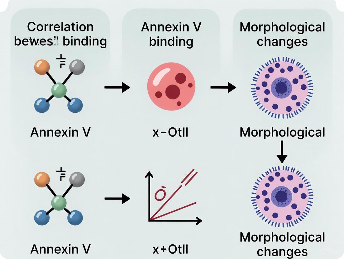
Diagram 1: Temporal sequence of apoptotic events and detection windows. The cascade illustrates the progressive nature of apoptosis, with key detection methods aligned to specific phases. Note the early occurrence of Annexin V binding relative to morphological changes like membrane blebbing and nuclear condensation.
Integrated Experimental Workflow for Temporal Correlation
Diagram 2: Integrated workflow for correlating Annexin V binding with morphological hallmarks. The parallel detection pathways enable direct temporal comparison between biochemical and morphological events, addressing the core challenge of asynchronous hallmark manifestation.
The Scientist's Toolkit: Essential Research Reagents
Table 3: Key research reagents for apoptosis detection
| Reagent/Category | Specific Examples | Function & Application | Key Considerations |
|---|---|---|---|
| Annexin V Conjugates | FITC, PE, APC, eFluor formats [15] | PS exposure detection via flow cytometry | Calcium-dependent binding; avoid EDTA |
| Viability Indicators | Propidium iodide, 7-AAD, Fixable Viability Dyes [15] [13] | Membrane integrity assessment | PI/7-AAD incompatible with fixation |
| Nuclear Stains | Hoechst 33342, DAPI [13] | Nuclear morphology assessment | Hoechst penetrates live cells |
| Caspase Detection | FRET-based sensors (ECFP-DEVD-EYFP) [12] | Real-time caspase activity monitoring | Requires genetically encoded sensors |
| Specialized Kits | Annexin V Apoptosis Detection Kits [10] [15] | Optimized reagent combinations | Follow manufacturer protocols precisely |
| Morphological Tools | Time-lapse imaging systems [7] | Direct visualization of membrane blebbing | Requires environmental control |
Discussion: Implications for Research and Drug Development
The temporal relationship between Annexin V binding and subsequent morphological changes has profound implications for experimental design and data interpretation in apoptosis research. The distinct detection windows for these hallmarks mean that methodological choice significantly influences apoptotic cell quantification and timing assessment.
Recent findings demonstrating Annexin V binding to specific subpopulations of myeloid cells, including retinal microglia and hyalocytes, highlight a critical consideration for researchers [11]. This binding occurs independently of apoptosis and may confound interpretation in models involving immune activation or inflammation. In such contexts, morphological assessment or caspase activation assays may provide more specific apoptosis detection.
The development of real-time kinetic approaches represents a significant advancement, overcoming limitations of traditional endpoint assays. The NanoBiT Annexin V system enables continuous monitoring for 48+ hours without destruction of samples [16], while FRET-based caspase sensors with organelle-targeted fluorescent proteins permit definitive discrimination between apoptosis and necrosis at single-cell resolution [12]. These technologies particularly benefit time-course studies and screening applications where the dynamics of cell death influence therapeutic assessment.
For drug discovery professionals, understanding these temporal relationships aids in differentiating primary therapeutic effects from secondary necrosis. The data suggests that compounds inducing clean apoptotic signatures (with defined PS exposure followed by morphological changes) may be preferable to those causing primary necrosis, as the latter triggers inflammatory responses that could compromise therapeutic outcomes [12].
The correlation between Annexin V binding and morphological hallmarks follows a predictable temporal sequence but demonstrates significant methodological dependence. Researchers must select detection strategies based on their specific needs:
- For high-throughput screening: Flow cytometry with Annexin V/PI or Differential Nuclear Staining provides quantitative, multi-parameter data at defined endpoints [10] [13].
- For kinetic analysis: Real-time Annexin V assays or FRET-based sensors enable continuous monitoring of apoptotic progression [16] [12].
- For mechanistic studies: Integrated approaches combining multiple detection methods offer the most comprehensive assessment of apoptotic timing and sequence.
The evolving understanding of Annexin V's interactions beyond apoptotic cells reinforces the importance of methodological validation in specific experimental systems. As apoptosis research advances, the integration of multiple detection modalities will continue to enhance our understanding of cell death dynamics and improve therapeutic assessment in biomedical research.
{ARTICLE CONTENT}
Live-Cell Imaging Insights into the Kinetic Relationship Between Annexin V Binding and Shape Change
Live-Cell Imaging Insights into the Kinetic Relationship Between Annexin V Binding and Shape Change
In the field of cell death research, a critical question persists: what is the precise temporal relationship between the biochemical "eat-me" signal of phosphatidylserine (PS) exposure and the physical dismantling of the cell? For researchers and drug development professionals, accurately pinpointing the onset and progression of apoptosis is essential for understanding compound mechanisms and therapeutic efficacy. This guide leverages advanced live-cell imaging to objectively compare the performance of Annexin V binding, a gold-standard marker for early apoptosis, with the subsequent morphological changes that define cell death. The data presented herein supports the broader thesis that PS externalization is a consistent and early prelude to the irreversible morphological collapse of the cell, providing a kinetic framework for more sensitive and accurate apoptotic analysis.
Apoptosis, or programmed cell death, is a tightly regulated process crucial for development, tissue homeostasis, and disease pathogenesis [17]. The classic biochemical hallmark of early apoptosis is the loss of plasma membrane asymmetry and the externalization of phosphatidylserine (PS), which is robustly detected by Annexin V protein binding [18] [17]. Traditionally, this has been measured using flow cytometry. However, this method provides only a single snapshot in time, requires extensive sample handling that can itself induce stress, and obscures the kinetic relationship between PS exposure and other cellular events [18] [19].
The advent of high-content live-cell imaging technologies has revolutionized this paradigm. These systems allow for the non-invasive, real-time observation of apoptotic events within the same population of cells over the entire course of an experiment [18] [17]. This capability is indispensable for correlating the timing of biochemical markers like Annexin V binding with the orchestrated morphological changes of apoptosis, such as cell shrinkage, membrane blebbing, and nuclear condensation. Understanding this kinetic relationship is not merely academic; it enables more sensitive detection in high-throughput drug screens, reduces false positives from sample processing, and provides a deeper understanding of a compound's mechanism of action [18] [20].
Comparative Kinetic Data: Quantifying the Sequence of Events
Direct, real-time comparison of fluorescent Annexin V signals with high-definition phase-contrast images has definitively established that PS exposure is an early event, consistently preceding the loss of plasma membrane integrity and major shape changes.
Table 1: Kinetic Comparison of Apoptotic Markers in Live-Cell Imaging
| Cell Line/System | Apoptotic Inducer | Time to Annexin V Detection (hours) | Time to Viability Dye (YOYO3) Detection (hours) | Key Morphological Changes Correlated with Annexin V Positivity | Source |
|---|---|---|---|---|---|
| SV40-MEFs | Cycloheximide (CHX) | Detected early, precise timing varies | ~8 hours post-induction | Cell shrinkage, membrane blebbing | [18] |
| SV40-MEFs | Staurosporine (STS) | Detected early, precise timing varies | Signally delayed versus Annexin V | Cell shrinkage, membrane blebbing | [18] |
| HT-1080 Fibrosarcoma | Cisplatin (12.5 µM) | Progressive increase from 0-72 hours | Not specified (assayed with Annexin V Red Dye) | Cell shrinkage, membrane blebbing observed concurrently with fluorescent signal | [17] |
| A549 Cancer Cells | Camptothecin (CMP) | Kinetic increase, concentration-dependent | Not specified (assayed with Annexin V NIR Dye) | Morphological changes aligned with fluorescent object count | [17] |
| Stable Reporter Cells | Carfilzomib | N/A (Caspase-3/7 activation detected) | N/A | Morphological changes tracked alongside caspase activation and loss of confluence | [21] |
The data consistently demonstrates that Annexin V binding provides an early warning of apoptosis. A pivotal study showed that while Annexin V positivity markedly preceded the uptake of viability dyes like YOYO3 and DRAQ7, the onset of late apoptotic events was characterized by equivalent labeling from both, underscoring the importance of kinetic data [18]. Furthermore, when compared directly to caspase-activatable fluorogenic probes (DEVD), Annexin V staining occurred more rapidly and in a greater number of cells, confirming its status as a highly sensitive early marker [18].
Detailed Experimental Protocols
To ensure the reliability and reproducibility of the kinetic data presented, the following detailed methodologies from key studies are provided.
This protocol established a highly sensitive, zero-handling approach for quantifying apoptosis.
- Cell Preparation: Seed adherent cells (e.g., MEFs, HT-1080, A549) in a multi-well plate suitable for live-cell imaging.
- Labeling: Add recombinant Annexin V conjugated to a fluorophore (e.g., FITC or AlexaFluor 594) directly to the culture medium at a final concentration of 0.25 µg/mL. This concentration is approximately 10-fold lower than traditional flow cytometry protocols and is non-toxic for prolonged incubation.
- Calcium Considerations: Standard cell culture media like DMEM, which contains ~1.8 mM Ca²⁺, provides sufficient divalent cations for Annexin V binding. Supplemental calcium can increase labeling intensity but may also cause punctate artifacts.
- Viability Staining (Optional Multiplexing): For dual-reporter kinetic analysis, co-incubate cells with a non-toxic viability dye such as YOYO3, which outperformed DRAQ7 in sensitivity and speed of labeling late-stage apoptotic cells.
- Image Acquisition and Analysis: Place the plate in a high-content live-cell imager (e.g., Incucyte). Acquire both fluorescent and high-definition phase-contrast images every 2-4 hours for the duration of the experiment (e.g., 24-72 hours). Use integrated software to automatically segment and quantify fluorescent objects (Annexin V-positive and viability dye-positive) and to correlate these signals with morphological metrics like cell confluence.
This protocol uses a genetically encoded reporter to correlate caspase activation with cell death outcomes.
- Reporter Cell Generation: Stably transduce cells with a lentiviral construct expressing a caspase-3/7 biosensor (e.g., a ZipGFP-based DEVD reporter) and a constitutive fluorescent marker (e.g., mCherry) for cell presence normalization.
- Treatment and Imaging: Seed the stable reporter cells in 2D or 3D culture. After treatment with apoptotic inducers, place the culture in a live-cell imager.
- Kinetic Tracking: Acquire time-lapse images over several days. The irreversible activation of GFP fluorescence upon caspase-3/7 cleavage provides a permanent timestamp of apoptotic initiation at single-cell resolution.
- Multiplexed Analysis: Use the constitutive mCherry signal and phase-contrast images to simultaneously track viable cell count (via AI modules) and morphological changes, creating a multi-parametric kinetic profile of cell death.
The Apoptotic Pathway: From Signal to Shape Change
The following diagram illustrates the sequential relationship between Annexin V binding and the subsequent morphological changes in apoptosis, as revealed by live-cell imaging.
Diagram Title: Kinetic Sequence of Apoptotic Markers
This pathway highlights the central thesis confirmed by live-cell imaging: the biochemical event of PS exposure and Annexin V binding is a critical early node, occurring after caspase activation but before the execution phase that leads to the irreversible morphological changes and eventual loss of membrane integrity.
The Scientist's Toolkit: Essential Reagents for Kinetic Apoptosis Assays
Table 2: Key Research Reagent Solutions for Live-Cell Apoptosis Imaging
| Reagent / Solution | Function / Principle | Key Characteristics |
|---|---|---|
| Fluorophore-conjugated Annexin V (e.g., Annexin V-488, -594) | Binds to exposed PS on the outer membrane leaflet, providing an early, specific signal for apoptosis. | Recombinant protein; non-toxic for long-term incubation; works in standard culture media (e.g., DMEM). [18] [17] |
| Caspase-3/7 Activity Reporter (e.g., DEVD-ZipGFP) | Genetically encoded biosensor activated upon cleavage by executioner caspases. | Irreversible signal; enables generation of stable cell lines; provides single-cell resolution of initiation. [21] |
| Cell Impermeant Viability Dyes (e.g., YOYO3, DRAQ7) | Labels DNA upon loss of plasma membrane integrity, indicating late-stage apoptosis/necrosis. | Essential for differentiating early vs. late apoptosis; YOYO3 shows faster kinetics than DRAQ7. [18] |
| RealTime-Glo Annexin V Assay | Uses Annexin V-NanoBiT luciferase subunits that generate luminescence upon binding PS. | "Add-and-read" no-wash protocol; allows multiplexing in high-throughput screening. [20] |
| Incucyte Nuclight Reagents | Lentiviral constructs for constitutive nuclear labeling (e.g., GFP, RFP). | Enables multiplexed measurement of proliferation and apoptosis in parallel; tracks cell count. [17] |
The collective evidence from live-cell imaging provides a clear and consistent narrative: Annexin V binding is a kinetically early event that reliably precedes the dramatic morphological changes associated with apoptotic cell death. The superiority of this real-time, kinetic approach over endpoint assays like flow cytometry is multifaceted. It eliminates the mechanical and chemical stresses of sample handling that can cause artifactual Annexin V staining or membrane damage [18] [19]. Furthermore, it captures the inherent heterogeneity and asynchrony of apoptotic responses within a cell population, data that is lost in bulk, endpoint measurements [21].
However, researchers must also be aware of the limitations and contextual factors. It has been demonstrated that Annexin V can also bind to specific subpopulations of immune cells, such as retinal microglia, which may confound the interpretation of apoptosis in certain in vivo or complex co-culture models [8]. Therefore, while the kinetic relationship holds true in most reductionist models, the cellular context is critical.
In conclusion, for researchers and drug developers aiming to accurately profile compound toxicity or investigate cell death mechanisms, live-cell imaging of Annexin V binding in conjunction with morphological analysis is an indispensable tool. It provides an unbiased, high-fidelity temporal map from the initial commitment to death, marked by PS exposure, to the final physical dismantling of the cell. This kinetic insight is fundamental for advancing both basic biological understanding and pre-clinical drug discovery.
The selection between two-dimensional (2D) and three-dimensional (3D) cell culture models represents a critical decision point in biomedical research, with profound implications for data interpretation, particularly regarding temporal biological processes. This guide objectively compares the performance of these culture systems, drawing on experimental data to illustrate how they impact the observation of dynamics such as apoptotic timing, morphological changes, and drug response kinetics. Framed within broader research on correlating annexin V binding with morphological changes, the analysis demonstrates that 3D models often provide superior physiological relevance but introduce specific challenges for real-time monitoring that 2D systems circumvent. Key experimental protocols and reagent solutions are detailed to equip researchers in making informed methodological choices for temporal resolution studies.
In the investigation of dynamic cellular processes like apoptosis, the spatial context of the cell culture system directly influences the observed kinetics and resolution of the event. The classic paradigm for detecting early apoptosis relies on measuring the translocation of phosphatidylserine (PS) to the outer leaflet of the cell membrane, typically using Annexin V-based probes [22] [23]. The timing of this event and its correlation with subsequent morphological changes are central to understanding cell death mechanisms. However, the ability to capture these events in real-time is not uniform across experimental models. While 2D cultures offer simplicity and ease of observation, their artificial stiffness and forced polarity can accelerate and distort the natural progression of apoptosis. Conversely, 3D culture models, such as spheroids and organoids, better mimic the structural complexity and pathophysiological gradients of in vivo tissues but can obscure real-time analysis and alter the apparent timing of PS exposure and membrane disintegration [24] [23]. This guide provides a comparative framework, grounded in experimental data, to navigate these challenges.
Comparative Performance Data: 2D vs. 3D Models
Direct comparisons between 2D and 3D systems reveal significant differences in cellular behavior and experimental outcomes. The quantitative data below summarize key performance metrics.
Table 1: Comparative Analysis of Cellular Phenomena in 2D vs. 3D Cultures
| Parameter | 2D Culture Findings | 3D Culture Findings | Experimental Context |
|---|---|---|---|
| Proliferation Rate | Significant (p < 0.01) increase in proliferation over time [24]. | Significant (p < 0.01) differences in proliferation pattern compared to 2D; slower growth [24]. | Colorectal cancer (CRC) cell lines [24]. |
| Cell Death Profile | Distinct apoptotic phase profile [24]. | Different apoptotic phase profile compared to 2D; increased resistance [24]. | CRC cell lines; Annexin V/PI staining [24]. |
| Drug Responsiveness | Responsive to 5-fluorouracil, cisplatin, and doxorubicin [24]. | Altered responsiveness and increased resistance to 5-fluorouracil, cisplatin, and doxorubicin [24]. | CRC cell lines treated with chemotherapeutics [24]. |
| Morphological Quantification | 2D analysis of mitochondrial morphology suggested metabolic stress induced fragmentation and loss of biomass [25]. | 3D analysis revealed the mitochondrial network was dissolved without affecting organelle size or biomass [25]. | Human endothelial cells (HUVECs) under metabolic stress [25]. |
| Transcriptomic Profile | Significant (p-adj < 0.05) dissimilarity vs. 3D; thousands of up/down-regulated genes [24]. | Significant (p-adj < 0.05) dissimilarity vs. 2D; shares closer expression pattern with patient FFPE samples [24]. | RNA sequencing of CRC cell lines and patient samples [24]. |
| Methylation & miRNA | Elevated methylation rate and altered microRNA expression [24]. | Shared similar methylation pattern and microRNA expression with patient FFPE samples [24]. | Epigenetic analysis of CRC cell lines and patient samples [24]. |
Table 2: Impact on Real-Time Apoptosis Assay Feasibility
| Assay Aspect | 2D Culture | 3D Culture |
|---|---|---|
| Washing Step | Possible but risks detaching apoptotic cells [23]. | Impractical; unbound probes trapped inside spheroid [23]. |
| Probe Access | Uniform access to all cells [23]. | Limited diffusion to inner cells; creates gradient [23]. |
| Temporal Resolution | Suitable for kinetic studies with appropriate probes [23]. | Challenging but enabled by "OFF-ON" probes like Q-Annexin V [23]. |
| Spatial Information | Limited to a single focal plane, potentially misleading [25]. | 3D context preserved; essential for accurate morphology assessment [25]. |
Detailed Experimental Protocols
To ensure the reproducibility of comparative studies, detailed methodologies from key investigations are outlined below.
Protocol: High-Throughput Purification of Annexin V for 2D Crystallization
This protocol, adapted from PMC9778525, is critical for producing high-quality, tag-free protein essential for structural studies [22].
- Objective: To purify recombinant annexin V without any residual affinity tag at the N-terminus in approximately 3 hours.
- Expression System: Recombinant protein expressed in E. coli.
- Purification System: Profinity eXact fusion-tag system.
- Method:
- The fusion protein is bound to an immobilized subtilisin protease column via the N-terminal tag.
- On-column cleavage is triggered by a buffer containing fluoride anions (F⁻), which releases the native, tag-free annexin V.
- The eluate contains pure annexin V (≈36 kDa), verified by SDS-PAGE.
- An optional DEAE column purification step can be added before the affinity column to prolong the resin's life.
- Key Findings: The N-terminal tag was found to destabilize 2D protein crystals. Tag-free annexin V formed stable, high-resolution crystals, whereas N-terminus His-tagged annexin V formed crystals with frequent, unstable deletions of "non-p6 trimers" [22].
Protocol: Real-Time Apoptosis Imaging in 2D and 3D Cultures
This protocol, based on the use of a quenched annexin V-fluorophore conjugate (Q-annexin V), enables kinetic studies without washing steps [23].
- Probe Principle: Q-annexin V is a conjugate where fluorescence is quenched (OFF) via photoinduced electron transfer. Upon binding to PS in the presence of Ca²⁺, a conformational change occurs, dequenching the fluorescence (ON).
- Cell Culture:
- 2D: Cells are seeded in standard culture plates.
- 3D: Spheroids are formed in super-low attachment U-bottom 96-well microplates.
- Imaging Procedure:
- Cells or spheroids are treated with the apoptotic stimulus.
- Q-annexin V is added directly to the culture medium.
- Crucially, no washing step is performed.
- Real-time fluorescence imaging is conducted using a confocal microscope. For 3D spheroids, z-stack acquisition is recommended.
- Key Advantage: This method allows for the monitoring of intercellular differences in the timing and severity of apoptotic responses, which is crucial for identifying drug-resistant cells in a population [23].
Protocol: 3D Mitochondrial Morphology Analysis
This protocol highlights the necessity of 3D imaging for accurate morphological quantification in non-flat cells [25].
- Cell Preparation: Human endothelial cells (HUVECs) stably expressing a mitochondria-targeted green fluorescent protein (mitoGFP).
- Image Acquisition: Cells are imaged using 3D confocal microscopy to generate z-stacks, capturing the entire volume of the cell.
- 3D Analysis Workflow:
- The 3D image stack is processed to create a 3D representation of the mitochondrial network.
- An automated algorithm segments individual mitochondrial objects in three dimensions.
- The software quantifies 3D shape and network properties, such as volume, surface area, and interconnectivity, without projecting the data onto a 2D plane.
- Key Finding: When human endothelial cells were treated with rotenone (ROT) to induce metabolic stress, 2D analysis incorrectly suggested mitochondrial fragmentation and biomass loss. In contrast, 3D analysis revealed that the mitochondrial network was reorganized into separate, spherical organelles without a change in total biomass or size, providing a fundamentally different and more accurate biological interpretation [25].
Signaling Pathways and Experimental Workflows
The following diagrams illustrate the core concepts and experimental workflows discussed in this guide.
Q-Annexin V Apoptosis Sensing Mechanism
2D vs 3D Analysis Workflow for Morphology
The Scientist's Toolkit: Key Research Reagents
Essential materials and their functions for conducting temporal resolution studies in apoptosis and morphology.
Table 3: Essential Reagents for Apoptosis and Morphology Research
| Reagent / Tool | Function & Application | Key Characteristic |
|---|---|---|
| Q-Annexin V (OFF-ON Probe) [23] | Real-time, wash-free imaging of apoptosis in both 2D and 3D cultures. | Near-infrared (NIR) fluorophore enabled by photoinduced electron transfer; fluorescence turns ON upon binding PS. |
| Profinity eXact System [22] | Single-step purification of recombinant annexin V without residual tags. | Immobilized subtilisin protease enables on-column cleavage and elution of native protein. |
| Nunclon Sphera U-Plates [24] | High-throughput generation of uniform 3D spheroids. | Super-low attachment surface promotes spontaneous spheroid formation in U-bottom wells. |
| MitoGFP [25] | Fluorescent labeling of mitochondria for live-cell morphology tracking. | GFP targeted to the mitochondrial matrix allows visualization of network dynamics. |
| 3D Confocal Microscopy & Analysis Software [25] | Accurate 3D quantification of intracellular structures like mitochondria. | Z-stack acquisition and 3D segmentation prevent morphological misinterpretations from 2D projection. |
Advanced Techniques for Real-Time Kinetic Analysis of Apoptosis
No-Wash, Real-Time Annexin V Assays for Continuous Monitoring
The precise timing of phosphatidylserine (PS) externalization relative to other apoptotic events is a critical area of research in cell biology. Traditional, endpoint Annexin V assays have provided a foundational understanding of apoptosis but are limited to single snapshots in time, requiring extensive washing steps that risk disturbing cellular integrity. The advent of no-wash, real-time Annexin V assays represents a transformative advancement, enabling continuous, kinetic monitoring of cell death within the same culture well. This guide objectively compares the performance of leading real-time Annexin V technologies—bioluminescent and fluorescent—against traditional methods and each other. By providing supporting experimental data and detailed protocols, we equip researchers with the information necessary to select the optimal assay for correlating PS exposure dynamics with other morphological and biochemical changes in their experimental systems.
Apoptosis, or programmed cell death, is a fundamental process critical for development, immune regulation, and tissue homeostasis, and its dysregulation is implicated in pathologies from cancer to neurodegenerative diseases [26] [27] [28]. A key early event in apoptosis is the loss of phospholipid asymmetry in the plasma membrane, leading to the externalization of phosphatidylserine (PS), which is normally confined to the inner leaflet [10] [29]. Annexin V, a 35-36 kDa protein with high, calcium-dependent affinity for PS, has become the gold-standard probe for detecting this event [10] [30].
Conventional Annexin V assays, while widely used, are endpoint measurements. They require binding buffers, multiple washing steps to remove unbound probe, and are typically paired with a viability dye like propidium iodide (PI) to distinguish early apoptotic (Annexin V+/PI-) from late apoptotic/necrotic cells (Annexin V+/PI+) [10] [28]. These workflows are labor-intensive, low-throughput, and susceptible to artifacts, as the washing steps can dislodge dying cells or even induce mechanical stress [26] [31]. Most importantly, they fail to capture the kinetic progression of cell death, providing only a static glimpse of a dynamic process.
The Thesis for Real-Time Correlation: The central thesis motivating the shift to real-time assays is that a precise, temporal understanding of when PS externalization occurs—relative to caspase activation, changes in mitochondrial membrane potential, and the ultimate loss of membrane integrity—is crucial for deciphering the dominant cell death pathway (e.g., classical apoptosis, necroptosis, pyroptosis) in response to a specific stimulus [32] [27]. No-wash, real-time Annexin V assays are uniquely positioned to provide this kinetic data, enabling researchers to move beyond mere confirmation of apoptosis to a detailed analysis of its initiation and progression.
Comparative Performance Analysis of Real-Time Annexin V Assays
The following section provides a data-driven comparison of the primary real-time Annexin V platforms available to researchers. The table below summarizes the core performance characteristics of the two leading technologies.
Table 1: Performance Comparison of Real-Time Annexin V Assay Platforms
| Feature | Traditional Endpoint Annexin V | Bioluminescent Real-Time Assay (e.g., RealTime-Glo) | Fluorescent Real-Time Assay (e.g., Incucyte Annexin V Dyes) |
|---|---|---|---|
| Readout Modality | Fluorescence (Flow Cytometry/Microscopy) | Bioluminescence & Fluorescence | Fluorescence (Live-Cell Imaging) |
| Assay Workflow | Multiple wash steps, cell harvesting (for flow) | Homogeneous "add-mix-measure" | Homogeneous "add-mix-measure" |
| Temporal Resolution | Single endpoint | Kinetic, data points every 0.5-3 hours | Kinetic, continuous imaging every 1-6 hours |
| Throughput | Low to medium | High (HTS-compatible) | High (96/384-well formats) |
| Key Mechanism | Fluorescently-labeled Annexin V | Annexin V-NanoBiT fusions + time-released substrate | Bright, photostable CF dye-Annexin V conjugates |
| Necrosis Detection | Yes (with PI or 7-AAD) | Yes (with proprietary necrosis dye) | Yes (via multiplexing with Cytotox Dyes) |
| Cell Visualizability | No (flow) or limited (microscopy) | No | Yes (provides morphological context) |
| Instrumentation | Flow cytometer or microscope | Luminescence/Fluorescence plate reader | Incucyte Live-Cell Analysis System |
Supporting Experimental Data
Bioluminescent Assay Performance: In a study characterizing the bioluminescent assay, diverse cell lines (Raji, K562, DLD-1, SK-ES-1) were treated with digitonin to induce immediate PS exposure. The assay, comprising Annexin V-LgBiT and Annexin V-SmBiT fusion proteins and a protected, time-released luciferase substrate (Endurazine), generated a robust luminescent signal that was sustained for over 16 hours, demonstrating its suitability for long-term kinetic monitoring [26]. Furthermore, treatment of K562 and Raji cells with bortezomib (500 nM) over 48 hours yielded kinetic, concentration-dependent data on apoptosis induction, with the signal proportional to cell density [26].
Fluorescent Assay Performance: The fluorescent alternative utilizes Annexin V conjugated to exceptionally bright and photostable CF dyes, which are added directly to the culture medium. A key application demonstrated the monitoring of Jurkat T lymphocyte cells treated with camptothecin (1 µM). The assay enabled automated, real-time quantification of apoptotic cells over time, capturing the onset and progression of PS exposure without compromising cell health through washing [31] [30].
Differentiating Cell Death Mechanisms: The bioluminescent assay has been effectively used to discriminate between apoptosis and necroptosis. Treatment of U937 cells with TNF-α induced a rapid luminescent signal (PS exposure) characteristic of apoptosis. However, when co-treated with a caspase inhibitor (Z-VAD-FMK), the PS signal was replaced by a rapid fluorescent signal from the necrosis dye, indicating a shift to necroptosis. This necroptotic signal was abolished by the addition of necrostatin-1, confirming the pathway's specificity [29].
Detailed Experimental Protocols
Below are generalized protocols for performing key experiments with these real-time assays.
Protocol for Real-Time Kinetics Using a Bioluminescent Assay
This protocol is adapted from the RealTime-Glo Annexin V Apoptosis and Necrosis Assay and related research [26] [29].
Research Reagent Solutions & Materials:
- Cells: Adherent or suspension cells (e.g., U937, SKBR3).
- RealTime-Glo Annexin V Assay Reagent: Contains the two Annexin V-NanoBiT fusion proteins, Necrosis Detection Reagent, and CaCl₂.
- NanoBiT Substrate (Endurazine): Time-released luciferase substrate.
- Test Compounds: e.g., TNF-α, caspase inhibitors, necroptosis inducers.
- White-walled 96- or 384-well microplates.
- Multimode plate reader capable of measuring luminescence and fluorescence, preferably with atmospheric control (37°C, 5% CO₂).
Methodology:
- Cell Plating: Plate cells in growth medium at an optimal density (e.g., 10,000-20,000 cells/well for a 96-well plate) in a white-walled microplate. Allow adherent cells to attach overnight.
- Reagent Preparation: Prepare the 2x assay reagent by combining the Annexin V Assay Reagent with the Endurazine substrate according to the manufacturer's instructions.
- Assay Initiation: Add an equal volume of the 2x assay reagent directly to each well containing cells and culture medium. Simultaneously, add your test compounds.
- Real-Time Data Acquisition:
- Place the microplate in the pre-equilibrated plate reader.
- Program the reader to cycle between 37°C incubation and reading at regular intervals (e.g., every 0.5 to 3 hours) for the duration of the experiment (up to 72 hours).
- At each time point, measure luminescence (PS exposure/apoptosis) and fluorescence (necrosis; Ex ~488-500 nm, Em ~520-540 nm, or as specified for the necrosis dye).
- Data Analysis: Plot luminescence and fluorescence over time for each treatment condition. The kinetic traces reveal the onset, rate, and magnitude of apoptosis and secondary necrosis.
Protocol for Live-Cell Imaging and Analysis Using a Fluorescent Assay
This protocol is based on the Incucyte Annexin V Dye system [31].
Research Reagent Solutions & Materials:
- Cells: Adherent or suspension cells (e.g., Jurkat, HT-1080, primary neurons).
- Incucyte Annexin V Dye: e.g., Annexin V CF 488A or Annexin V Red Dye.
- Optional Multiplexing Dyes: Incucyte Cytotox Dye for necrosis, Incucyte Nuclight Reagent for nuclear labeling.
- Test Compounds: e.g., camptothecin, staurosporine.
- Clear-bottom 96- or 384-well microplates.
- Incucyte Live-Cell Analysis System or similar live-cell imaging instrument.
Methodology:
- Cell Plating: Plate cells in growth medium in a clear-bottom microplate. For suspension cells, consider using plates coated with poly-L-ornithine to retain cells in the field of view.
- Dye and Compound Addition: Add the Incucyte Annexin V Dye (at a recommended final concentration, e.g., 1:1000 dilution) and test compounds directly to the wells.
- Real-Time Imaging and Analysis:
- Place the microplate inside the Incucyte instrument, maintained at 37°C and 5% CO₂.
- Program the instrument to automatically capture high-definition phase-contrast and fluorescence images from multiple non-overlapping fields per well at user-defined intervals (e.g., every 1-6 hours).
- Image and Data Processing:
- Use the integrated software to define analysis criteria. A typical mask for Annexin V-positive cells would be created based on fluorescence intensity and morphology.
- The software automatically quantifies the number of Annexin V-positive objects (apoptotic cells) per well per time point.
- Morphological changes such as cell shrinkage and membrane blebbing can be observed and quantified from the phase-contrast images.
Technological Mechanisms and Workflows
The following diagrams illustrate the core principles and experimental workflows of the two main real-time assay types.
Mechanism of Bioluminescent Annexin V Assay
Experimental Workflow for Real-Time Apoptosis Monitoring
The Scientist's Toolkit: Essential Reagents and Materials
Table 2: Key Research Reagent Solutions for Real-Time Annexin V Assays
| Item | Function/Description | Example Use Case |
|---|---|---|
| Annexin V-NanoBiT Fusions | Recombinant proteins that bind PS and complement to form active luciferase. Core of bioluminescent assays. | RealTime-Glo Assay for HTS of compound libraries [26] [29]. |
| Time-Released Luciferase Substrate (Endurazine) | Protected substrate enabling sustained luminescence generation over long time courses. | Long-term (48-72h) kinetic apoptosis studies without reagent depletion [26]. |
| Bright, Photostable CF Dye-Annexin V Conjugates | Fluorophore-labeled Annexin V with superior brightness and stability for live-cell imaging. | No-wash, real-time imaging of PS exposure in sensitive primary cells [31] [30]. |
| Necrosis Detection Reagent | Cell-impermeant, DNA-binding dye with low background and high photostability. | Distinguishing apoptotic from necrotic/necroptotic cells in multiplexed assays [26] [29]. |
| Caspase-3/7 Apoptosis Assay Dyes | Cell-permeant, fluorogenic substrates that become fluorescent upon cleavage by active caspases. | Multiplexing to confirm apoptosis mechanism and correlate PS exposure with caspase activation [31]. |
No-wash, real-time Annexin V assays have fundamentally changed our ability to study cell death, providing a dynamic window into a process that was previously only observable through static snapshots. The choice between bioluminescent and fluorescent platforms depends on the research question's specific needs: the former offers exceptional sensitivity and ease of use in high-throughput screening, while the latter provides invaluable visual confirmation and morphological context.
The integration of these kinetic PS exposure data with other real-time readouts—such as caspase activation, mitochondrial health, and cell proliferation—is paving the way for a systems-level understanding of cell death decision-making networks. As research continues to uncover the nuances of different programmed cell death pathways (e.g., necroptosis, pyroptosis), the ability to correlate the precise timing of PS exposure with other hallmark events will be paramount. These advanced assays are thus not merely incremental improvements but essential tools for dissecting the complex chronology of cell death, with significant implications for drug discovery and the understanding of human disease.
Leveraging Live-Cell Imaging Platforms (e.g., Incucyte) for Longitudinal Studies
In the study of programmed cell death, a pivotal question persists: what is the precise correlation between the timing of phosphatidylserine (PS) externalization, marked by annexin V binding, and the subsequent morphological changes of apoptosis? Traditional endpoint assays provide only snapshots of this dynamic process, potentially missing critical temporal relationships and causal linkages. Live-cell imaging platforms, such as the Incucyte Live-Cell Analysis System, are revolutionizing this field by enabling continuous, non-perturbing observation of living cells over time. This guide objectively compares the performance of real-time imaging against traditional endpoint methods within the specific context of annexin V and morphological change correlation studies, providing researchers with the data and protocols necessary to advance their investigative work.
Platform Comparison: Incucyte Live-Cell Analysis vs. Endpoint Assays
The core advantage of live-cell analysis systems like the Incucyte is their ability to automatically acquire and analyze images of cells continuously over hours, days, or weeks while the cells remain undisturbed inside a standard incubator [33] [34]. This provides kinetic data from the same population of cells, a critical capability for establishing the sequence of apoptotic events.
In contrast, traditional endpoint methods, such as flow cytometry-based annexin V assays, require sampling at predetermined times, necessitating the disruption of the cell culture environment and providing only single-timepoint data [35] [36]. Table 1 summarizes the fundamental differences between these approaches.
Table 1: Core Technology Comparison: Live-Cell Imaging vs. Endpoint Assays
| Feature | Incucyte Live-Cell Analysis | Traditional Endpoint Assays |
|---|---|---|
| Data Type | Continuous, kinetic data from the same cell population | Single-timepoint snapshots from different samples |
| Cell Environment | Non-perturbing; cells remain in incubator | Disruptive; requires cell handling and removal |
| Annexin V Context | Enables direct correlation of PS exposure with subsequent morphology changes in real-time | Requires inference of temporal sequence between separate experiments |
| Throughput | High; can run up to six microplates in parallel [37] | Variable; often limited by cell supply and handling time [35] |
| Key Advantage | Reveals temporal relationships and unexpected dynamics | Established protocols, often lower initial hardware cost |
Performance and Application in Apoptosis Research
Quantitative comparisons reveal significant performance differences. A study directly comparing real-time systems (Incucyte and xCELLigence) with endpoint assays (CellTiter-Glo, resazurin, and nuclei count) found that the real-time systems were "particularly effective at tracking the effects of drug treatment on cell proliferation at sub-confluent growth" [36]. Furthermore, endpoint assays like resazurin reduction and CellTiter-Glo were shown to report higher cell viabilities than direct nuclei counts, suggesting potential overestimation of cell health [36].
In the specific context of immunogenicity risk assessment, a high-throughput DC internalization assay developed on the IncuCyte platform demonstrated a "high correlation between internalization and clinical immunogenicity risk," outperforming previous flow cytometry-based results [35]. This demonstrates the platform's capacity for predictive biology.
Table 2 highlights specific performance characteristics relevant to apoptosis and cell health studies.
Table 2: Experimental Performance Data in Cell Health and Apoptosis Studies
| Experimental Metric | Incucyte Live-Cell Analysis Performance | Endpoint Assay Performance |
|---|---|---|
| Cell Viability Measurement | Accurate tracking of drug effects at sub-confluency [36] | Potential overestimation of viability (Resazurin, CellTiter-Glo) [36] |
| Predictive Power | High correlation with clinical immunogenicity results [35] | Lower correlation in DC internalization assays [35] |
| Cell Requirement | Requires only 5-10% of DCs vs. flow cytometry [35] | Substantial cell number requirement, limiting throughput [35] |
| Data Richness | Provides kinetic data, cell morphology, and confluence | Provides a single data point (e.g., luminescence, fluorescence) |
Experimental Protocols for Longitudinal Apoptosis Studies
Protocol 1: Real-Time DC Internalization Assay for Immunogenicity
This protocol, adapted from a study that successfully predicted clinical immunogenicity, details a kinetic approach to monitor dendritic cell (DC) internalization of biotherapeutics, a key event in initiating an immune response [35].
Key Reagents:
- Therapeutic antibodies or biotherapeutics for testing
- BioTracker Orange-NHS Live Cell pH Dye (Millipore)
- Primary human monocyte-derived Dendritic Cells (DCs)
- Cell culture medium (e.g., RPMI-1640 with 10% FBS)
Methodology:
- Antibody Labeling: Buffer-exchange antibodies into PBS (pH 7.4). Label antibodies using a 5-fold molar excess of BioTracker Orange dye for 1 hour at room temperature. Remove free dye using desalting columns [35].
- Quality Control: Determine the concentration and Degree of Labeling (DOL) using spectrophotometry (A280 and A551 nm). Characterize the conjugate distribution using intact LC-MS to ensure labeling consistency [35].
- Cell Seeding and Assay Setup: Seed DCs into a multi-well plate. Add the directly labeled antibody conjugates to the cells.
- Real-Time Imaging and Analysis: Place the plate in the Incucyte system inside a cell culture incubator. Acquire fluorescence and phase-contrast images at regular intervals (e.g., every 2-4 hours) over 1-3 days. Use integrated software to quantify the fluorescence intensity per well or within individual cells, which serves as a metric for internalization.
Protocol 2: Multiplexed Kinetic Analysis of Apoptosis via Morphology and PS Exposure
This protocol leverages the multiplexing capability of live-cell imaging to concurrently track early (PS exposure) and late (morphological changes) apoptotic events in the same well.
Key Reagents:
Methodology:
- Cell Preparation: Seed cells into a multi-well plate at an optimized density for growth over several days.
- Reagent Addition: Add the fluorogenic apoptosis probe (e.g., Apo-15) to the medium. Apo-15 is preferred for wash-free, calcium-independent staining, which minimizes perturbation [38]. Alternatively, use a compatible annexin V conjugate.
- Baseline Imaging: Initiate live-cell imaging to establish a baseline for both fluorescence (PS exposure) and phase-contrast (morphology) signals.
- Treatment and Kinetic Imaging: Add the apoptosis-inducing treatment. Continue automated imaging at frequent intervals. The Incucyte software can simultaneously analyze multiple parameters:
- Fluorescence Object Count: Quantifies the number of annexin V-positive cells.
- Cell Confluence/Morphology: Tracks changes in cell size, shape, and membrane integrity (blebbing) via phase-contrast.
- Cellularity: Monitors overall cell count decrease due to fragmentation and death.
The experimental workflow for this multiplexed approach is outlined in the diagram below.
The Scientist's Toolkit: Essential Reagent Solutions
Successful longitudinal imaging requires carefully selected reagents that maintain cell health and minimize perturbation. The following table details key solutions for apoptosis and cell health studies.
Table 3: Key Research Reagent Solutions for Live-Cell Apoptosis Imaging
| Reagent / Solution | Function & Rationale | Key Characteristics |
|---|---|---|
| Fluorogenic Peptide Apo-15 [38] | Calcium-independent detection of phosphatidylserine (PS) exposure on apoptotic cells. | Enables wash-free staining, ideal for longitudinal imaging; avoids Ca2+ limitations in diseased tissues. |
| Annexin V Conjugates [2] | Traditional, Ca2+-dependent detection of externalized PS. | Wide commercial availability (e.g., Alexa Fluor conjugates); requires binding buffer with Ca2+. |
| Incucyte Cytolight/Nuclight Lentiviruses [37] | Labels nucleus or cytoplasm with GFP/RFP for automated cell counting and tracking. | Non-perturbing, stable expression; essential for quantifying cell proliferation and loss in real-time. |
| Caspase-3/7 Apoptosis Assay Reagents | Detects activation of executioner caspases, a key biochemical step in apoptosis. | Provides a third, complementary dimension to PS exposure and morphology. |
| Low-Riboflavin Media [33] | Cell culture medium formulation for reduced background fluorescence. | Critical for optimizing signal-to-noise ratio in fluorescence imaging, especially with green probes. |
| Matrigel / ECM Matrices [37] | Substrate for 3D cell culture and spheroid formation. | Enables more physiologically relevant studies of apoptosis in a tissue-like context. |
Integrating Findings into a Coherent Biological Workflow
The data generated from the protocols above must be interpreted within the established biological context of apoptosis. The following diagram maps the key apoptotic events and the corresponding detection methods onto a simplified signaling pathway, illustrating how live-cell imaging captures the dynamic progression of cell death.
The power of live-cell imaging is its ability to kinetically track the sequence from Event A (PS exposure) through Event C (caspase activation) to Event E (morphology changes) in the same cell population, thereby directly testing the correlation central to the user's thesis. This integrated workflow allows researchers to move beyond correlation to potentially establish causality in the apoptotic signaling cascade.
In the study of programmed cell death, a pivotal question revolves around the correlation between the timing of early biochemical events and subsequent morphological changes. The externalization of phosphatidylserine (PS), detected by Annexin V binding, is a recognized early event in apoptosis, while an increase in cellular granularity and changes in size are classic morphological hallmarks observed later in the process [39]. Multiparametric flow cytometry provides a unique platform to investigate this correlation quantitatively and in real-time, by simultaneously analyzing Annexin V binding, propidium iodide (PI) uptake, and light scatter properties indicative of cell morphology. This guide compares the performance of this integrated approach against alternative methods, highlighting its critical role in delineating the precise sequence of cellular events during death induction, a capability essential for accurate mechanistic studies in drug development.
Key Methodologies for Integrated Apoptosis and Morphology Analysis
Annexin V/Propidium Iodide Dual Staining Protocol with Scatter Analysis
The following protocol, adapted from current methodologies, allows for the simultaneous assessment of PS externalization, membrane integrity, and morphological changes [40] [41].
Cell Preparation and Staining:
- Harvesting: Harvest cells (e.g., MDA-MB-231, SAOS-2) using mild trypsinization or non-enzymatic dissociation to preserve membrane integrity. Include the culture supernatant to collect any floating dead cells [40].
- Washing: Wash the cell pellet with cold, calcium-containing phosphate-buffered saline (PBS) to provide the necessary cofactor for Annexin V binding [40].
- Staining: Resuspend the cell pellet (approximately 1x10^6 cells) in 100 µL of Annexin V binding buffer. Add Annexin V conjugated to fluorescein isothiocyanate (FITC) as per manufacturer's recommendation and incubate for 15 minutes in the dark at room temperature [40] [41].
- Propidium Iodide Addition: Add PI to a final concentration of 1-2 µg/mL directly to the staining suspension and incubate for an additional 15 minutes in the dark [40] [41]. Note: A modified protocol suggests a fixation step followed by RNase A treatment (50 µg/mL) post-staining to eliminate false-positive PI signals from cytoplasmic RNA, significantly improving accuracy [41].
- Analysis: Without washing, dilute the cells in an appropriate volume of binding buffer (e.g., 300-500 µL) and analyze immediately by flow cytometry [40].
Flow Cytometry Data Acquisition and Gating:
- Trigger and Scatter Gating: Use forward scatter (FSC) to trigger event detection. Create a dot plot of FSC-Area (FSC-A) vs. Side Scatter-Area (SSC-A) to gate on the main population of cells, excluding debris.
- Singlets Gating: To avoid misinterpretation from cell doublets, plot FSC-Width (FSC-W) vs. FSC-A and gate on the single-cell population [42].
- Viability and Apoptosis Gating: On the gated single-cell population, create a dot plot of Annexin V-FITC vs. PI. Analyze a minimum of 10,000 events per sample [40].
Comparative Experimental Techniques for Cell Death Assessment
While integrated flow cytometry is powerful, other techniques offer complementary insights.
- Fluorescence Microscopy (FM): FM uses stains like fluorescein diacetate (FDA) and PI to visually distinguish live and dead cells on a microscope slide [43]. It allows for direct imaging but has lower throughput, is labor-intensive for quantification, and is susceptible to sampling bias as only a few fields of view are analyzed [43]. It also struggles with consistently differentiating apoptosis from necrosis [43].
- Imaging Cytometry: This technology bridges flow cytometry and microscopy, capturing high-resolution images of each cell as it flows through the system. It preserves spatial and complex morphological information that traditional flow cytometry loses, making it ideal for analyzing rare events or subcellular localization [44]. However, its throughput is significantly lower than that of conventional flow cytometry [44].
- Spectral Flow Cytometry: A recent advancement, spectral cytometry collects the full emission spectrum of every fluorophore using a detector array [45]. This allows the use of more fluorophores in a single panel and provides superior resolution for complex multicolor experiments, such as deep immunophenotyping of apoptotic cell subpopulations [45].
The logical workflow for an integrated analysis of cell death, from experimental setup to data interpretation, is summarized in the diagram below.
Performance Data: Quantitative Comparison of Cytometry Methods
Tabular Comparison of Key Cytometry Techniques
The choice of technique depends heavily on the research question, balancing throughput, quantitative power, and the need for morphological detail.
Table 1: Comparative Analysis of Cell Viability and Apoptosis Assessment Techniques
| Feature | Multiparametric Flow Cytometry (Annexin V/PI/Scatter) | Fluorescence Microscopy (FM) | Imaging Cytometry |
|---|---|---|---|
| Throughput | High (10,000+ events/sec) [44] | Low (manual, few fields of view) [43] | Low to Medium (1-100 events/sec) [44] |
| Quantification | Excellent; robust, statistical | Poor; semi-quantitative, prone to bias [43] | Good; quantitative with spatial context |
| Morphological Info | Basic (cell size & granularity via FSC/SSC) | Good (direct visual assessment) | Excellent (high-resolution images) [44] |
| Subpopulation Distinction | Excellent (clearly distinguishes viable, early/late apoptotic, necrotic) [43] [40] | Poor (difficult to distinguish apoptosis from necrosis) [43] | Excellent (can correlate death status with subcellular morphology) [44] |
| Key Advantage | High-throughput, multiparametric quantitative data on population distributions [43] | Direct visualization of cells | Combines quantification with high-content morphological imaging [44] |
| Primary Limitation | Loss of spatial context and subcellular detail | Low throughput and subjective quantification [43] | Lower throughput and higher data complexity [44] |
Experimental Correlation Between Annexin V Binding and Morphology
Direct comparative studies validate the performance of flow cytometry. A 2025 study comparing flow cytometry (FCM) and fluorescence microscopy (FM) for assessing cytotoxicity of particulate biomaterials found a strong correlation between the two methods (r = 0.94, R² = 0.8879, p < 0.0001) [43]. However, FCM demonstrated superior precision, especially under high cytotoxic stress, and was able to provide distinct quantification of early and late apoptotic populations, which FM could not reliably achieve [43]. Furthermore, the same study highlighted that morphological changes captured by light scattering are a consequential event following the initial biochemical insult, a timeline that integrated FCM is uniquely suited to capture.
Table 2: Exemplary Experimental Data: Cell Viability (%) Under Cytotoxic Stress [43]
| Condition | Treatment Time | Viability by Fluorescence Microscopy (FDA/PI) | Viability by Flow Cytometry (Multiparametric) |
|---|---|---|---|
| Control (Untreated) | 72 h | > 97% | > 97% |
| Bioglass <38 µm, 100 mg/mL | 3 h | 9% | 0.2% |
| Bioglass <38 µm, 100 mg/mL | 72 h | 10% | 0.7% |
Note: The data demonstrates a consistent trend identified by both techniques, with flow cytometry typically showing higher sensitivity, particularly in detecting late-stage apoptotic and necrotic cells [43].
Table 3: Key Research Reagent Solutions for Annexin V/PI Apoptosis Assay
| Item | Function/Description | Example Usage/Note |
|---|---|---|
| Annexin V-FITC Conjugate | Binds to phosphatidylserine (PS) on the outer leaflet of the cell membrane, a marker of early apoptosis [39]. | Must be used with calcium-containing binding buffer. |
| Propidium Iodide (PI) | Membrane-impermeant DNA dye. Stains cells with compromised membrane integrity (late apoptotic/necrotic) [41] [42]. | Requires RNase treatment in a modified protocol to reduce false positives from RNA staining [41] [42]. |
| Annexin V Binding Buffer | Provides optimal ionic and Ca²⁺ conditions for specific Annexin V binding to PS. | Critical for signal-to-noise ratio. |
| RNase A | Ribonuclease that degrades cytoplasmic RNA. | Used in a modified protocol post-staining to eliminate false-positive PI signal, crucial for accuracy in primary cells and large cells [41]. |
| Cell Strainers (40 µm) | Filters cell suspensions to remove clumps before flow cytometry analysis. | Prevents instrument clogging and ensures single-cell data quality [40]. |
| Compensation Controls | Single-stained samples for each fluorophore (e.g., Annexin V-FITC only, PI only). | Essential for correcting spectral overlap in multicolor flow cytometry [40]. |
Multiparametric flow cytometry, integrating Annexin V, PI, and light scatter measurements, stands as the superior method for high-throughput, quantitative correlation of the timing between biochemical and morphological events in cell death. While fluorescence microscopy offers visual confirmation and imaging cytometry provides unparalleled morphological detail, the statistical power, precision, and ability to clearly resolve discrete cell death subpopulations make flow cytometry an indispensable tool for basic research and drug discovery. The ongoing development of spectral flow cytometry further promises to enhance this capability, allowing for even deeper investigation into the complex signaling networks that govern cellular fate in response to therapeutic agents.
The transition from in vitro screening to in vivo validation represents a critical pathway in modern drug development, particularly for therapies designed to modulate programmed cell death. Annexin V, with its specific affinity for phosphatidylserine (PS) exposed on the outer leaflet of apoptotic cell membranes, has emerged as a cornerstone biomarker for detecting apoptosis across this continuum. This review provides a comprehensive comparison of annexin V-based methodologies, from traditional flow cytometry to advanced live-cell imaging and positron emission tomography (PET). We examine the correlation between annexin V binding and morphological changes in the context of timing, highlighting both the capabilities and limitations of these approaches in quantifying pharmacodynamic responses to anticancer therapies. Supporting experimental data and standardized protocols are presented to facilitate informed methodological selection for preclinical drug development.
Apoptosis, or programmed cell death, is a fundamental process in tissue homeostasis and a key therapeutic endpoint for cancer treatments. A hallmark early event in apoptosis is the loss of plasma membrane phospholipid asymmetry, resulting in the externalization of phosphatidylserine (PS)—a membrane phospholipid normally confined to the inner leaflet. Annexin V, a 35-36 kDa calcium-dependent phospholipid-binding protein, exploits this phenomenon by binding with high affinity (Kd ≈ 10⁻⁹ M) to PS residues displayed on the cell surface [46]. This specific interaction, which occurs prior to membrane blebbing and DNA fragmentation, provides a sensitive marker for detecting early apoptotic commitment [46] [27].
In preclinical drug development, the ability to accurately quantify apoptosis is essential for evaluating therapeutic efficacy and understanding compound mechanisms of action. Annexin V-based assays bridge this need across experimental scales, offering applications ranging from high-throughput in vitro screening to non-invasive in vivo imaging in animal models and potentially humans. However, researchers must navigate critical methodological considerations, including the timing of PS externalization relative to other apoptotic markers, potential confounding factors such as binding to immune cells, and the technical limitations of different detection platforms [8]. This guide systematically compares these approaches to support robust experimental design.
Comparative Performance of Annexin V Assay Platforms
Annexin V-based apoptosis detection has been adapted for numerous platforms, each with distinct advantages, limitations, and optimal use cases. The table below provides a structured comparison of key methodologies used in preclinical development.
Table 1: Performance Comparison of Annexin V Assay Platforms
| Methodology | Key Readout | Throughput | Key Advantages | Key Limitations | Best Applications |
|---|---|---|---|---|---|
| Flow Cytometry | Percentage of Annexin V+/PI- (early apoptotic) and Annexin V+/PI+ (late apoptotic/necrotic) cells [47] [48] | Medium | Multiplexable with other markers (e.g., cell cycle); quantitative; well-established | Endpoint measurement only; requires cell harvesting which can induce artifactual apoptosis [48] | In vitro screening of compound libraries; validation studies requiring cell population statistics |
| Live-Cell Imaging (e.g., Incucyte) | Kinetic measurements of Annexin V fluorescent object count or confluence [17] | High | Real-time, kinetic data from undisturbed cells; no-wash protocol reduces artifacts | Fluorescence background can increase over long cultures; requires specialized instrumentation | Kinetic studies of apoptosis onset and progression; long-term time-course experiments |
| Radiolabeled Annexin V for PET/SPECT | In vivo biodistribution and uptake quantified by image-derived standard uptake value (SUV) [46] [49] | Low | Non-invasive imaging in live subjects; enables longitudinal studies within same subject | Lower resolution than microscopy; potential deiodination (124I); high kidney accumulation [46] [49] | Pharmacodynamic studies in animal models; translating apoptosis detection to clinical trials |
Supporting Experimental Data
The utility of these platforms is demonstrated by robust experimental data. In vitro, flow cytometry with annexin V-FITC and propidium iodide (PI) can distinguish viable (Annexin V-/PI-), early apoptotic (Annexin V+/PI-), late apoptotic (Annexin V+/PI+), and necrotic (Annexin V-/PI+) cell populations [48]. This method validated a 2.3-fold increase in annexin V binding to RIF-1 tumor cells 48 hours after 5-fluorouracil treatment in vivo [46].
For kinetic analysis, live-cell imaging with reagents like the Incucyte Annexin V Red Dye showed a time- and concentration-dependent increase in fluorescence in HT-1080 fibrosarcoma cells treated with cisplatin, allowing for continuous monitoring of apoptosis over 72 hours without manual intervention [17].
In vivo, PET imaging with 124I-labeled annexin V successfully detected apoptosis in a murine model of Fas-induced liver apoptosis, with image-derived uptake correlating strongly (r=0.86) with histologically derived apoptotic density [49]. A modified version, [124I]SIB-annexin V, demonstrated superior in vivo stability with reduced deiodination and consequent thyroid accumulation, making it an attractive candidate for clinical PET imaging of apoptosis [46].
Detailed Experimental Protocols
Standardized protocols are vital for generating reproducible and reliable data. Below are detailed methodologies for key annexin V assays.
Flow Cytometry with Annexin V and Propidium Iodide
This protocol enables the quantification of early and late apoptotic cell populations in a single sample [47] [48].
- Sample Preparation: Use trypsin without EDTA to digest adherent cells to prevent artifactual PS externalization. Wash cells gently but thoroughly with phosphate-buffered saline (PBS) to remove serum proteins. After treatment, pellet approximately 0.5-1 x 10⁶ cells by centrifugation (300 x g for 5 minutes).
- Staining Procedure:
- Resuspend the cell pellet in 100 µL of 1X Annexin V Binding Buffer.
- Add fluorescently conjugated Annexin V (e.g., Annexin V-APC, FITC) according to the manufacturer's recommendation.
- Add a viability dye such as Propidium Iodide (PI) or 7-AAD.
- Incubate the mixture for 15 minutes at room temperature in the dark.
- After incubation, add 400 µL of additional Binding Buffer to each tube and analyze by flow cytometry within 1 hour.
- Controls:
- Unstained Cells: For background fluorescence and compensation.
- Annexin V Single Stain: For flow cytometry compensation.
- PI Single Stain: For flow cytometry compensation.
- Induced Apoptosis (e.g., camptothecin-treated): For a positive control.
Radiolabeling of Annexin V for In Vivo PET Imaging
This protocol outlines the process for creating a positron-emitting annexin V probe for non-invasive imaging [46] [49].
- Protein Preparation: Recombinant annexin V, often polyhistidine-tagged for purification, is expressed in systems like E. coli and purified using nickel-nitrilotriacetic acid (Ni-NTA) resin. Activity must be confirmed via a competition assay with FITC-annexin V.
- Direct Iodination vs. SIB Conjugation:
- Direct Iodination: Annexin V is incubated with radio-iodine (125I for biodistribution, 124I for PET) and an oxidizing agent. This method is simpler but can lead to in vivo deiodination [46].
- SIB Conjugation: A more stable method where N-succinimidyl-3-iodobenzoate ([124I]SIB) is first synthesized and purified by HPLC. This compound is then conjugated to annexin V, creating a more stable amide bond. This approach shows reduced thyroid uptake in vivo, indicating less dehalogenation [46].
- Purification and Validation: The radiolabeled protein is purified using size-exclusion chromatography (e.g., PD-10 column). Radiochemical purity is assessed by instant thin-layer chromatography (ITLC) and SDS-PAGE. Specific binding must be validated in an apoptosis model (e.g., 5-FU treated RIF-1 cells) and should be inhibitable by excess unlabeled annexin V [46].
Diagram 1: Radiolabeled Annexin V Probe Workflow. The process for creating and validating radiolabeled annexin V for imaging, highlighting the two primary labeling strategies.
Correlation of Annexin V Binding with Morphological Changes in Apoptosis
A critical consideration in preclinical research is the temporal relationship between annexin V binding (a biochemical event) and the classic morphological changes of apoptosis. Understanding this correlation is essential for accurate data interpretation.
The Temporal Sequence of Apoptotic Events
Apoptosis unfolds in a coordinated sequence. PS externalization, detected by annexin V, is an early event, occurring before the loss of plasma membrane integrity. This is followed by cell shrinkage, chromatin condensation, and membrane blebbing. The final stages involve nuclear fragmentation and the formation of apoptotic bodies [32] [27]. The window during which cells are Annexin V-positive but PI-negative (indicating an intact membrane) defines the early apoptotic population.
Comparative Kinetic Studies
Research directly comparing assays reveals nuanced timing differences. One study treating HL-60 cells with etoposide found that the maximum apoptotic response detected by annexin V binding occurred 4-5 hours earlier than the peak observed in Giemsa-stained morphological assessments, and up to 8 hours before the peak in DNA fragmentation assays [7]. This confirms annexin V as an early marker. Furthermore, time-lapse video microscopy (TLVM) showed that steep increases in annexin V positivity correlated closely with the appearance of cells with plasma membrane blebbing, linking the biochemical and morphological hallmarks [7].
However, this correlation can be complicated in vivo. A recent study in a mouse model of optic nerve injury revealed a two-phase pattern of annexin V labeling: an initial rapid phase that correlated with cleaved caspase-3 immunostaining, followed by a sustained plateau phase driven by annexin V binding to a subpopulation of myeloid cells (e.g., microglia) [8]. This indicates that in complex tissue environments, a persistent annexin V signal may reflect ongoing immune responses rather than a continuous wave of apoptosis, highlighting the necessity of multi-parameter assessment.
Diagram 2: Apoptosis Event Timeline. The sequential relationship between annexin V binding and other key apoptotic events.
The Scientist's Toolkit: Essential Reagents and Materials
Successful execution of annexin V-based assays requires a suite of well-characterized reagents. The following table details essential solutions for the featured experiments.
Table 2: Key Research Reagent Solutions for Annexin V Assays
| Reagent/Material | Function/Description | Key Application Context |
|---|---|---|
| Recombinant Annexin V | The core protein, available conjugated to fluorophores (FITC, APC) for in vitro use or ready for radiolabeling for in vivo studies. | Universal reagent for all annexin V-based detection methods [46] [47]. |
| Viability Dyes (PI, 7-AAD) | Membrane-impermeant DNA dyes used to distinguish early apoptotic (dye-negative) from late apoptotic/necrotic (dye-positive) cells. | Critical for flow cytometry to define different cell death stages [47] [48]. |
| Calcium-Containing Binding Buffer | Provides the necessary Ca²⁺ for the specific interaction between annexin V and phosphatidylserine. | Essential for all annexin V binding reactions; its composition is critical for assay specificity [47]. |
| Radioisotopes (124I, 125I, 99mTc) | Positron or gamma emitters used to label annexin V for non-invasive tracking in live animals via PET or SPECT. | Enables translational research from in vitro models to in vivo pharmacodynamic imaging [46] [49] [50]. |
| Caspase-3/7 Green Reagent | A cell-permeable, non-fluorescent substrate that is cleaved by activated caspases to release a green DNA-binding dye. | Used in multiplex live-cell analysis (e.g., Incucyte) to correlate PS exposure with caspase activation [17]. |
Annexin V remains an indispensable tool in the preclinical drug development arsenal, providing a sensitive and adaptable means to detect apoptosis from the well plate to the whole organism. Flow cytometry offers robust, quantitative population data; live-cell imaging captures kinetic profiles in undisturbed cultures; and radiolabeled annexin V enables non-invasive, translational assessment of treatment response in vivo.
The critical insight for researchers is that while annexin V binding is an early and reliable marker of apoptosis, its interpretation requires careful consideration of the temporal context, especially in relation to downstream morphological changes. Furthermore, recent findings about annexin V binding to immune cells in vivo underscore the importance of not relying on a single parameter. The future of apoptosis assessment in drug development lies in multiplexed, kinetic approaches that combine annexin V with other markers of cell death (e.g., caspase activation) and cellular identity to deconvolute complex biological responses and accurately guide therapeutic candidates toward clinical application.
Resolving Ambiguity: Pitfalls in Interpreting Annexin V Binding and Morphology
For decades, Annexin V (AnxV) has been established as a gold-standard probe for detecting apoptosis by binding to phosphatidylserine (PS), a phospholipid that translocates from the inner to the outer leaflet of the plasma membrane during programmed cell death [10]. This calcium-dependent binding forms the basis for ubiquitous flow cytometry and fluorescence microscopy assays that distinguish early apoptotic cells (AnxV+/PI−) from late apoptotic or necrotic cells (AnxV+/PI+) [10]. However, a growing body of evidence challenges the specificity of AnxV as an exclusive apoptotic marker. Recent research reveals that AnxV also binds to viable, activated immune cells, including microglia and T cells, due to focal PS externalization—a phenomenon distinct from the wholesale loss of membrane asymmetry characteristic of apoptosis [8] [51]. This paradigm shift complicates the interpretation of AnxV-based assays and necessitates a critical comparison of AnxV binding characteristics across different cellular states. This guide synthesizes current evidence to objectively compare the performance of AnxV in detecting apoptosis versus its binding in non-apoptotic contexts, providing researchers with the experimental data and protocols necessary to accurately delineate these phenomena in their work.
Comparative Analysis of Annexin V Binding in Apoptosis vs. Immune Activation
The following table summarizes the key differential features of AnxV binding in classical apoptosis versus its binding in non-apoptotic immune cell activation, synthesizing findings from recent investigations.
| Feature | Classical Apoptosis | Non-Apoptotic Immune Cell Activation |
|---|---|---|
| PS Externalization Pattern | Wholesale, diffuse across entire cell membrane [51] | Localized, focal patches or "scrambled foci" on the membrane [51] |
| Cell Viability & Function | Cell death process; loss of cellular function [10] | Cells remain viable and functionally active; part of signaling [51] |
| Temporal Dynamics | Persistent, progresses until cell disintegration [10] | Transient and reversible; resolves in 1-2 hours [51] |
| Primary Signaling Triggers | Caspase activation, DNA fragmentation [7] | Immune receptor engagement (e.g., TCR, FcεRI), calcium influx [51] |
| Key Regulatory Molecules | Caspase-3, Bax/Bcl-2 ratio, mitochondrial depolarization [52] [7] | TMEM16F scramblase, intracellular calcium [51] |
| Morphological Correlates | Cell shrinkage, membrane blebbing, nuclear fragmentation [7] [53] | Minimal cell shape change; absence of apoptotic bodies [53] [51] |
| Co-staining with Viability Probes | Positive for PI in late stages (loss of membrane integrity) [10] | Typically PI-negative (membrane integrity maintained) [51] |
| Confounding in Assays | Considered true positive for cell death | Can lead to false positive interpretation of cell death [8] |
A pivotal study on microglia in the retina vividly illustrates the confounding potential of non-apoptotic AnxV binding. Following optic nerve injury, the time course of AnxV labeling did not align with the peak of caspase-3 activation, an established apoptotic marker. Instead, AnxV signals remained elevated in a sustained plateau long after caspase-3 activity had declined. This persistent signal was attributed to AnxV binding to a subpopulation of retinal microglia, which were identified using CX3CR1GFP/+ reporter mice [8]. Pharmacological depletion of microglia abolished this bright, elongated AnxV labeling, revealing fainter labeling of round, likely apoptotic, retinal ganglion cells. This demonstrates that non-apoptotic AnxV binding can dominate the signal in complex tissue environments [8].
Detailed Experimental Protocols for Differentiating Binding Contexts
To ensure the accurate interpretation of AnxV binding data, researchers must employ specific experimental protocols designed to distinguish apoptosis from immune activation. Below are detailed methodologies for key assays.
Multicolor Flow Cytometry with Immune Cell Markers
This protocol is essential for identifying which specific cell populations in a heterogeneous sample are binding AnxV.
- Sample Preparation: Prepare a single-cell suspension from tissue or culture. Include a viability dye (e.g., propidium iodide or a near-IR fixable viability dye) in all steps to exclude dead cells from the analysis.
- Cell Staining:
- Fc Receptor Block: Incubate cells with an anti-CD16/32 antibody or species-specific serum to prevent non-specific antibody binding.
- Surface Marker Staining: Stain cells with a cocktail of fluorescently conjugated antibodies against immune cell surface markers (e.g., for microglia: CD11b, CX3CR1; for T cells: CD3, CD4/CD8). Use antibodies conjugated to fluorophores compatible with your flow cytometer and distinct from AnxV and PI channels.
- AnxV Staining: Wash cells and resuspend in 1X AnxV binding buffer. Add AnxV-FITC (or another fluorophore) and incubate for 15-20 minutes at room temperature in the dark, according to the manufacturer's datasheet [10].
- Propidium Iodide (PI) Addition: Just before acquisition, add PI to a final concentration of 1-2 µg/mL.
- Flow Cytometry Acquisition: Acquire data on a flow cytometer equipped with appropriate lasers and filters. Collect a sufficient number of events for robust statistical analysis of subpopulations.
- Data Analysis:
- Gate on single, live cells (viability dye-negative).
- Within the live cell population, gate on specific immune subsets using the surface antibody markers.
- Analyze AnxV and PI staining within each immune subset. AnxV+/PI- cells in a microglial (CD11b+/CX3CR1+) gate indicate non-apoptotic PS exposure, whereas AnxV+/PI- cells in a population lacking immune markers may be undergoing early apoptosis.
Time-Lapse Confocal Microscopy for Kinetic Analysis
This protocol allows for the real-time observation of PS externalization dynamics, crucial for distinguishing transient focal scrambling from progressive apoptosis.
- Cell Preparation:
- Plate cells on glass-bottom imaging dishes. For microglial studies, primary microglia or immortalized cell lines like BV-2 can be used.
- If studying immune activation, pre-treat cells with appropriate stimuli (e.g., LPS for pro-inflammatory activation).
- Staining for Live-Cell Imaging:
- Prepare imaging medium containing AnxV conjugated to a fluorophore (e.g., AnxV-AF647) and a cell-permeable nuclear stain (e.g., Hoechst 33342). A vital dye for cytoplasm or membrane (e.g., CellMask) can also be added to visualize cell morphology.
- Replace the culture medium with the staining solution.
- Image Acquisition:
- Place the dish on a confocal microscope with an environmental chamber maintained at 37°C and 5% CO2.
- Program the microscope to capture images of multiple fields of view at regular intervals (e.g., every 5-10 minutes) over a period of several hours.
- Data Analysis:
- Analyze the time-lapse sequence for the appearance of AnxV signals.
- Focal Scrambling: Look for the rapid appearance of small, bright AnxV puncta on the cell membrane that remain stable or resolve over time, without changes in cell morphology.
- Apoptosis: Look for a gradual, diffuse increase in AnxV signal across the entire cell membrane, accompanied by morphological changes like cell shrinkage and membrane blebbing [7].
Co-staining with Apoptosis and Microglial Markers in Tissue
This protocol is critical for validating the identity of AnxV+ cells in ex vivo or fixed tissue sections, such as the retina or brain.
- Tissue Collection and Fixation: Perfuse the animal with ice-cold PBS followed by 4% paraformaldehyde (PFA). Dissect the tissue of interest (e.g., retina) and post-fix in 4% PFA for 1-2 hours at 4°C.
- Sectioning: Cryoprotect the tissue in 30% sucrose, embed in OCT compound, and section on a cryostat (10-20 µm thickness). Mount sections on glass slides.
- Immunofluorescence Staining:
- Permeabilize and block sections with a buffer containing a detergent (e.g., 0.3% Triton X-100) and 5% normal serum.
- Incubate sections overnight at 4°C with primary antibodies against:
- Active Caspase-3 (a definitive marker of apoptosis)
- Microglial Markers (e.g., Iba1, TMEM119, or P2RY12)
- The following day, wash sections and incubate with species-appropriate secondary antibodies conjugated to distinct fluorophores.
- AnxV Staining (on fixed tissue): Critical Note: For pre-fixed tissue, AnxV staining must be performed after the immunofluorescence staining is complete and without additional permeabilization, as membrane disruption exposes inner-leaflet PS, causing non-specific binding [8]. Incubate sections with AnxV conjugated to a third, compatible fluorophore in AnxV binding buffer for 30 minutes.
- Imaging and Analysis:
- Image sections using a confocal microscope.
- Quantify the co-localization of AnxV signal with caspase-3 (indicative of apoptosis) versus its co-localization with microglial markers (indicative of non-apoptotic binding).
Signaling Pathways and Experimental Workflows
The diagrams below illustrate the key signaling pathways and experimental workflows for differentiating apoptotic and non-apoptotic Annexin V binding.
Signaling Pathways in Apoptotic and Non-Apoptotic PS Exposure
Diagram 1: Signaling Pathways Leading to PS Externalization. Apoptosis triggers caspase-dependent scramblase XKR8, causing wholesale PS exposure. Immune activation triggers calcium-dependent TMEM16F, causing transient, focal PS/PE exposure [51].
Experimental Workflow for Differentiating AnxV Binding
Diagram 2: Experimental Workflow for Differentiating AnxV Binding. Parallel pathways for flow cytometry, live-cell microscopy, and tissue analysis provide complementary data to distinguish non-apoptotic from apoptotic AnxV signals [10] [8] [51].
The Scientist's Toolkit: Essential Research Reagents
The following table details key reagents and their applications for studying the nuanced roles of Annexin V.
| Reagent | Function & Application | Key Considerations |
|---|---|---|
| Recombinant Annexin V (conjugated to FITC, AF647, etc.) | Core probe for detecting exposed PS in flow cytometry, microscopy, and in vivo imaging (e.g., DARC) [10] [8]. | Calcium-dependent binding. Conjugation quality and storage conditions are critical for performance. |
| Propidium Iodide (PI) / Viability Dyes | DNA intercalator used to distinguish late apoptotic/necrotic cells (AnxV+/PI+) from early apoptotic or non-apoptotic cells (AnxV+/PI-) [10]. | Impermeant to live cells. Must be used with care in time-course assays as it is toxic. |
| Lactadherin (LactC2) | Calcium-independent, monomeric PS probe. Can bind PS with higher affinity and less steric hindrance than AnxV, useful for confirming PS exposure [51]. | Does not require calcium, making it suitable for low-calcium environments. |
| Antibodies for Immune Cell Markers (e.g., CD11b, Iba1, CX3CR1, TMEM119) | Identification of specific immune cell populations (e.g., microglia) for co-localization with AnxV signal in complex samples [8]. | Requires multicolor panel optimization and appropriate controls for flow cytometry and IF. |
| Antibodies for Apoptosis Markers (e.g., Cleaved Caspase-3) | Definitive marker for apoptosis. Co-staining is essential to confirm that AnxV+ cells are truly apoptotic [8]. | Requires cell permeabilization for immunostaining, which must be done after AnxV staining on live/fixed cells. |
| Calcium Ionophores (e.g., A23187, Ionomycin) | Positive control for inducing global, non-apoptotic PS scrambling via massive calcium influx [51]. | Induces widespread scrambling that is mechanistically distinct from focal, signaling-induced scrambling. |
| TMEM16F Modulators (e.g., Ani9 (activator)) | Tool to specifically manipulate the calcium-activated scramblase implicated in non-apoptotic PS exposure in immune cells [51]. | Emerging pharmacological tools; specificity and off-target effects should be considered. |
Differentiating Primary Necrosis from Late-Stage Apoptosis (Secondary Necrosis)
Within the context of annexin V binding and morphological changes timing correlation research, distinguishing primary necrosis from late-stage apoptosis (secondary necrosis) represents a significant methodological challenge in experimental pathology and drug development. The externalization of phosphatidylserine (PS), detected by annexin V binding, serves as a key early marker of apoptosis, occurring before morphological changes such as membrane blebbing, nuclear condensation, and eventual loss of membrane integrity [54] [55] [2]. However, the conventional approach of using annexin V combined with membrane integrity dyes like propidium iodide (PI) presents interpretation complexities, as both late apoptotic and primary necrotic cells eventually display positivity for both markers [56] [2]. This guide objectively compares current methodologies for differentiating these distinct cell death pathways, providing researchers with experimental data and protocols to enhance assay accuracy.
Fundamental Principles of Cell Death Markers
Key Events in Apoptotic and Necrotic Pathways
Table 1: Chronological Events in Apoptosis and Primary Necrosis
| Event | Apoptosis | Primary Necrosis | Detection Window | Primary Detection Methods |
|---|---|---|---|---|
| Phosphatidylserine externalization | Early event, precedes membrane breakdown [54] [55] | Can occur transiently before membrane rupture [56] | 1-4 hours (apoptosis) [54] | Annexin V binding [55] [2] |
| Caspase activation | Caspase-3, -8, -9 activation [56] | Typically caspase-independent [56] | 2-6 hours (varies by stimulus) [57] | Caspase activity assays, Red-LEHD-FMK [57] |
| Membrane blebbing | Characteristic morphological change [7] | Not typically observed | 4-8 hours [7] | Time-lapse video microscopy [7] |
| Nuclear condensation | Progressive chromatin condensation | Variable | 4-12 hours | Giemsa staining, DNA-binding dyes [7] |
| DNA fragmentation | Internucleosomal cleavage [7] | Random digestion | 8-16 hours [7] | TUNEL assay, DNA laddering [7] |
| Loss of membrane integrity | Final stage (secondary necrosis) | Early and progressive event [56] | 12-24 hours (apoptosis) | Propidium iodide, 7-AAD uptake [2] [57] |
Traditional vs. Advanced Interpretation Models
The conventional model for differentiating cell death mechanisms using annexin V and propidium iodide posits that:
- Annexin V⁻/PI⁻: Viable, healthy cells
- Annexin V⁺/PI⁻: Early apoptotic cells
- Annexin V⁺/PI⁺: Late apoptotic or necrotic cells [2] [10]
However, recent research challenges this oversimplification. Studies demonstrate that primary necrotic cells can unexpectedly show Annexin V⁺/PI⁻ staining before becoming PI-positive, creating potential false classification of primary necrosis as apoptosis [56]. Furthermore, annexin V binding is not exclusive to apoptotic cells, as specific subpopulations of immune cells, particularly myeloid cells, can bind annexin V independently of apoptosis, potentially confounding interpretation in inflammatory models [8].
Figure 1: Convergence of Apoptotic and Necrotic Pathways in Traditional Assays. Both pathways ultimately result in Annexin V and PI double-positive cells, creating challenges for accurate differentiation using conventional methods.
Comparative Methodologies and Experimental Data
Kinetic Analysis of Cell Death Markers
Table 2: Temporal Resolution of Apoptosis Detection Methods
| Detection Method | Basis of Detection | Earliest Detection | Peak Response | Key Advantage | Key Limitation |
|---|---|---|---|---|---|
| Annexin V binding | PS externalization [54] [55] | 1-2 hours [54] | 4-8 hours | Early detection [54] | Cannot distinguish apoptosis from primary necrosis [56] |
| Caspase activation | Caspase-3, -9 activity [56] [57] | 2-4 hours [57] | 6-12 hours | Specific to apoptosis [57] | Transient signal, missed in late stages [26] |
| Morphological changes (TLVM) | Membrane blebbing, cell shrinkage [7] | 4-6 hours [7] | 12-24 hours | Visual confirmation [7] | Labor-intensive, low throughput [7] |
| DNA fragmentation | Internucleosomal cleavage [7] | 8-12 hours [7] | 16-24 hours | Late-stage confirmation [7] | Misses early apoptosis [7] |
| Real-Time-Glo Annexin V | Luciferase complementation on PS binding [20] [26] | 2-4 hours [26] | 8-16 hours | Kinetic monitoring, no wash [20] | Requires specialized reagents [20] |
Pharmacological Differentiation Approach
A critical advancement in distinguishing these pathways comes from pharmacological inhibition. Research demonstrates that necrostatin-1, an inhibitor of RIP1 kinase activity, specifically inhibits primary necrosis without affecting apoptotic pathways [56]. In experimental models where TNF-α/cycloheximide/zVAD-induced cell death was primarily necrotic, necrostatin-1 reduced cell death from 49.7% to 19.0%, while the caspase inhibitor zVAD-fmk showed no protective effect [56]. This pharmacological profiling provides a definitive tool for pathway differentiation.
Table 3: Response Profiles to Death Pathway Inhibitors
| Cell Death Mode | Necrostatin-1 Response | Caspase Inhibitor Response | Annexin V/PI Pattern | Additional Confirmation |
|---|---|---|---|---|
| Primary Necrosis | Significant protection (≈60% reduction) [56] | No protection [56] | Initially Annexin V⁺/PI⁻, progressing to Annexin V⁺/PI⁺ [56] | RIP1/RIP3 activation [56] |
| Apoptosis | No protection [56] | Significant protection | Sequential: Annexin V⁺/PI⁻ → Annexin V⁺/PI⁺ [2] [10] | Caspase activation [57] |
| Secondary Necrosis | No protection | Protection in early stages only | Annexin V⁺/PI⁺ [2] | Preceding caspase activation [57] |
Experimental Protocols for Differentiation
Comprehensive Annexin V/PI with Pharmacological Inhibition Protocol
This optimized protocol integrates traditional annexin V staining with pharmacological probes to differentiate primary necrosis from secondary necrosis.
Materials Required:
- Annexin V-FITC conjugate (e.g., Alexa Fluor 488, Pacific Blue) [2]
- Propidium iodide (PI) or 7-AAD [2] [57]
- Necrostatin-1 (RIP1 inhibitor) [56]
- zVAD-fmk (pan-caspase inhibitor) [56]
- Annexin V binding buffer (with Ca²⁺) [2] [10]
- Flow cytometer or fluorescence microscope [2]
Procedure:
- Cell Treatment and Inhibition:
Annexin V/PI Staining:
- Harvest cells gently, avoiding mechanical damage
- For adherent cells with fragile extensions (e.g., neurons), use minimal trypsinization and neutralize with serum [55] [10]
- Wash cells in cold PBS and resuspend in 1X Annexin V binding buffer at 1-5×10⁵ cells/mL [10]
- Add 5 µL annexin V-FITC and 5 µL PI (or 7-AAD) per 500 µL cell suspension [10]
- Incubate 15-20 minutes at room temperature in the dark [10]
Analysis and Interpretation:
- Analyze by flow cytometry within 1 hour using FITC (annexin V) and PE/PI channels
- For imaging, place cells on glass slides with coverslips and image immediately [10]
- Key interpretation:
Real-Time Kinetic Apoptosis/Necrosis Assay
Advanced real-time methods enable continuous monitoring of cell death progression without multiple endpoint measurements.
Materials Required:
- RealTime-Glo Annexin V Apoptosis and Necrosis Assay [20] [26]
- Annexin V-LgBiT and Annexin V-SmBiT fusion proteins [20] [26]
- Necrosis Detection Reagent (asymmetric cyanine DNA dye) [20]
- Multimode plate reader with luminescence and fluorescence detection [20] [26]
Procedure:
- Assay Setup:
Real-Time Monitoring:
- Place plate in controlled-atmosphere reader (37°C, 5% CO₂)
- Collect luminescence (apoptosis) and fluorescence (necrosis) readings every 1-2 hours for 24-48 hours [26]
- Maintain physiological conditions throughout monitoring
Data Interpretation:
Figure 2: Integrated Experimental Workflow for Cell Death Pathway Differentiation. Combining endpoint pharmacological profiling with real-time kinetic monitoring provides orthogonal verification of cell death mechanisms.
The Scientist's Toolkit: Essential Research Reagents
Table 4: Key Reagents for Differentiating Cell Death Pathways
| Reagent Category | Specific Examples | Function/Mechanism | Application Notes |
|---|---|---|---|
| PS Binding Probes | Annexin V-FITC, Annexin V-APC [2] | Binds externalized PS on apoptotic cells [55] | Requires calcium; use with viability dye [2] |
| Viability Dyes | Propidium iodide, 7-AAD, SYTOX Green [2] | DNA intercalation upon membrane damage [2] | Distinguishes early vs. late apoptosis [57] |
| Necroptosis Inhibitors | Necrostatin-1 [56] | RIP1 kinase inhibition [56] | Specific for primary necrosis; 10-100 µM [56] |
| Caspase Inhibitors | zVAD-fmk [56] | Pan-caspase inhibition [56] | Confirms apoptotic component; 20-50 µM [56] |
| Caspase Activity Probes | Red-LEHD-FMK (caspase-9) [57] | Binds active caspase-9 [57] | Early apoptosis detection [57] |
| Real-Time Annexin V | Annexin V-NanoBiT fusions [20] [26] | Luciferase complementation on PS binding [26] | No-wash, kinetic monitoring [20] |
| Advanced Viability Dyes | SYTOX AADvanced, 7-AAD [2] [57] | Cell-impermeant DNA dyes [2] | Better dead cell discrimination [2] |
Accurate differentiation between primary necrosis and late-stage apoptosis requires moving beyond simple annexin V/PI staining toward integrated methodological approaches. The combination of pharmacological profiling using pathway-specific inhibitors (necrostatin-1 for necrosis, zVAD-fmk for apoptosis) with kinetic analysis of death markers provides significantly enhanced discrimination capability [56]. Furthermore, recognition that primary necrotic cells can display transient Annexin V⁺/PI⁻ staining challenges traditional interpretation models and necessitates more sophisticated analytical frameworks [56].
For research applications where precise death pathway identification is critical—such as mechanistic toxicology studies, drug development, and pathophysiological investigation—implementing the complementary protocols outlined in this guide will substantially improve experimental accuracy. The ongoing development of real-time, non-disruptive monitoring technologies represents a promising direction for future advancements in cell death research, allowing continuous observation of death progression without artificial endpoint disruptions [20] [26].
In the field of cell biology and drug development, accurately distinguishing between live, apoptotic, and necrotic cells remains a fundamental challenge. The persistence of false-positive results in viability staining can significantly compromise data integrity, leading to incorrect conclusions about compound efficacy and toxicity profiles. A primary source of these inaccuracies stems from the temporal relationship between phosphatidylserine (PS) externalization, detected by Annexin V binding, and the subsequent loss of membrane integrity, a definitive marker of cell death [10] [50]. During early apoptosis, PS translocates to the outer leaflet of the plasma membrane while the membrane itself remains intact, creating a transient state that requires precise dye combinations for accurate identification. Furthermore, the process of secondary necrosis, where apoptotic cells eventually lose membrane integrity, can blur the distinction between apoptosis and primary necrosis if not properly controlled [58]. This article critically examines the sources of false positives in common viability assays and provides a systematic, evidence-based guide on how combining viability dyes with appropriate controls can mitigate these errors, framed within ongoing research into the correlation kinetics of Annexin V binding and morphological changes.
Apoptosis and Necrosis: Distinct Pathways with Overlapping Assay Signatures
Understanding the biochemical and morphological hallmarks of different cell death pathways is essential for designing robust viability assays. Apoptosis, or programmed cell death, is a highly regulated process characterized by caspase activation, chromatin condensation, and membrane blebbing. A critical early event is the loss of phospholipid asymmetry in the plasma membrane, resulting in the externalization of phosphatidylserine (PS) [10] [50]. This event occurs while the cell membrane remains largely intact. In contrast, necrosis involves uncontrolled cell swelling and rupture of the plasma membrane, leading to the release of inflammatory cellular contents [58]. The challenge for researchers is that late apoptotic cells undergo secondary necrosis, acquiring characteristics of primary necrosis, including membrane permeabilization [58]. This progression creates a continuum of cell states that single-parameter assays cannot adequately resolve.
Multiple technical and biological factors contribute to false-positive staining in viability assays:
- Antibody Penetration in Permeabilized Cells: Fluorophore-labeled antibodies cannot cross the intact membrane of a live cell. However, when cells die, their membrane becomes permeable, allowing antibodies to bind specifically and non-specifically to internal targets. This can cause dead cells to be mistaken as positive for intracellular markers during flow cytometry analysis without proper viability gating [59].
- Dye Aggregation and Incubation Time: With dyes like trypan blue, prolonged incubation can result in the dissociation of dye aggregates, enabling them to stain otherwise viable cells. Conversely, short incubation periods might lead to an underestimation of dead cells [60].
- Fixation and Permeabilization Artifacts: DNA-binding dyes like Propidium Iodide (PI) or SYTOX will cross the membranes of live cells if added after fixation and permeabilization, creating massive false-positive signals [59]. One study noted that conventional PI staining methods can generate up to 40% false positive events [59].
- Cellular Stress Responses: Changes in cell metabolism, osmolarity, or spontaneous membrane invagination can allow viability dyes to penetrate cells that are still viable, leading to false positives [60].
- Annexin V Binding Specificity: While Annexin V binding to PS is a hallmark of early apoptosis, it is crucial to note that any disruption of the plasma membrane will allow Annexin V to bind to PS on the inner leaflet of the membrane. This means that late apoptotic and necrotic cells will also be Annexin V-positive, and the assay cannot by itself distinguish between these states without a membrane-integrity dye [10] [15].
Table 1: Common Viability Dyes and Their Associated Risks for False Positives
| Dye Type | Specific Examples | Mechanism of Action | Primary Source of False Positives |
|---|---|---|---|
| DNA Binding Dyes | Propidium Iodide (PI), 7-AAD, SYTOX | Enters cells with compromised membranes and intercalates into DNA/RNA [60] [59]. | Added after fixation/permeabilization; prolonged incubation; cellular stress [60] [59]. |
| Amine-Reactive Dyes | LIVE/DEAD Aqua, Zombie Green | Binds to free amines in the cytoplasm of permeabilized cells; dye is washed away before fixation [59]. | Improper washing leaving unbound dye; excessive dye concentration. |
| Esterase-Based Dyes | Calcein-AM, Carboxyfluorescein Diacetate | Live-cell enzymes convert non-fluorescent substrate to fluorescent product retained in live cells [60]. | Leakage of fluorescent product from viable cells; esterase activity in dying cells [60]. |
| Phosphatidylserine Binders | Annexin V (FITC, APC, etc.) | Calcium-dependent binding to PS exposed on the outer membrane leaflet [10]. | Binding to inner leaflet PS in permeabilized cells (necrosis); other PS-exposing death pathways (e.g., necroptosis) [10]. |
Systematic Comparison of Viability Dye Combinations
The Gold Standard: Annexin V with a Membrane-Impermeant Dye
The most widely adopted strategy to discriminate early apoptosis from necrosis is the combination of Annexin V with a membrane-impermeant DNA dye like PI or 7-AAD.
- Experimental Protocol: Cells are harvested, washed in PBS, and resuspended in a calcium-containing binding buffer. They are then incubated with fluorochrome-conjugated Annexin V for 10-15 minutes at room temperature, protected from light. Without washing, a DNA dye like PI is added directly to the buffer, and the sample is analyzed by flow cytometry within 4 hours [10] [15]. It is critical to avoid buffers containing EDTA or other calcium chelators, as they will inhibit Annexin V binding [15].
- Data Interpretation: This combination distinguishes four populations:
- Viable Cells: Annexin V⁻ / PI⁻
- Early Apoptotic Cells: Annexin V⁺ / PI⁻
- Late Apoptotic/Necrotic Cells: Annexin V⁺ / PI⁺
- Cells Damaged During Preparation: Annexin V⁻ / PI⁺ (though this is rare).
Advanced Multiparametric Staining for Flow Cytometry
For more complex assays involving intracellular staining, fixable viability dyes are superior. Unlike traditional DNA dyes, these amine-reactive dyes covalently bind to intracellular amines in cells with compromised membranes. After binding, the reaction is quenched, and excess dye is washed away. This allows the cells to be fixed and permeabilized for intracellular staining without the risk of the viability dye penetrating and staining live cells, a significant advantage over PI [59].
- Experimental Protocol: After staining cell surface markers, cells are washed in azide-free and serum/protein-free PBS. They are then resuspended and incubated with a fixable viability dye (FVD) for 30 minutes at 2-8°C. Cells are washed thoroughly to remove unbound dye before proceeding to Annexin V staining in binding buffer and subsequent intracellular staining protocols [15].
- Advantages: This method effectively eliminates false-positive viability staining in intracellular marker analysis and allows for complex immunophenotyping of dying cells.
Real-Time Live-Cell Imaging with FRET-Based Sensors
To overcome the limitations of endpoint assays, innovative real-time methods have been developed. One highly sensitive approach uses cells stably expressing a FRET-based caspase sensor (e.g., ECFP-DEVD-EYFP) and a mitochondrially-targeted fluorescent protein (e.g., Mito-DsRed) [58].
- Experimental Protocol: Cells expressing both probes are treated with the agent of interest and imaged in real-time using automated fluorescence microscopy.
- Data Interpretation:
- Apoptosis: Visualized by a loss of FRET (increase in ECFP/EYFP ratio) due to caspase-mediated cleavage of the linker, while mitochondrial fluorescence (Mito-DsRed) is retained.
- Primary Necrosis: Characterized by a sudden loss of the cytosolic FRET probe without a preceding ratio change, while mitochondrial fluorescence is retained.
- Secondary Necrosis: Cells first show FRET loss (apoptosis) and later lose the FRET probe while retaining Mito-DsRed.
- Advantages: This method provides single-cell resolution and kinetic data on the mode and dynamics of cell death, effectively discriminating the sequence of apoptotic and necrotic events [58].
Table 2: Quantitative Comparison of Cell Viability Assessment Techniques
| Technique | Primary Readout | Ability to Distinguish Apoptosis/Necrosis | Throughput | Key Advantage | Key Limitation |
|---|---|---|---|---|---|
| Annexin V/PI Flow Cytometry | PS exposure & membrane integrity [10] | Excellent (with correct gating) [10] | High | Widely accessible, quantitative | Snapshot in time, can miss dynamic transitions [58] |
| Fixable Dye + Intracellular Staining | Membrane integrity & marker expression [59] [15] | Good (when combined with Annexin V) | High | Compatible with complex immunophenotyping | More complex protocol |
| FRET/Mito-DsRed Live Imaging | Caspase activity & organelle integrity [58] | Excellent, at single-cell level | Low to Medium | Real-time kinetic data, visual confirmation | Requires genetically engineered cells |
| Membrane Potential Assay (MPCVA) | Resting membrane potential [61] | Moderate (indicates irreversible death) | Medium | Direct measure of a definitive death event | May not distinguish death pathways early |
| LDH Release Assay | Release of cytoplasmic enzyme [60] | Poor (only indicates necrosis) | High | Easy, no specialized equipment | High background in some systems, indirect [60] |
Experimental Protocols for Validated Assays
Detailed Protocol: Annexin V and PI Staining for Flow Cytometry
This protocol is adapted from standard procedures provided by Abcam and Thermo Fisher Scientific [10] [15].
Materials:
- Fluorochrome-conjugated Annexin V (e.g., FITC, APC)
- Propidium Iodide (PI) Staining Solution or 7-AAD
- 10X Annexin V Binding Buffer
- Phosphate-Buffered Saline (PBS)
- Flow cytometer with appropriate lasers and filters
Procedure:
- Prepare 1X Binding Buffer: Dilute 10X binding buffer with distilled water.
- Harvest and Wash Cells: Collect cells by centrifugation (300 x g for 5 minutes). For adherent cells, use gentle trypsinization and quench with serum-containing media before washing. Wash cells once with PBS and once with 1X Binding Buffer.
- Stain with Annexin V: Resuspend cell pellet at a density of 1-5 x 10⁶ cells/mL in 1X Binding Buffer. Add 5 µL of fluorochrome-conjugated Annexin V to 100 µL of cell suspension. Vortex gently and incubate for 10-15 minutes at room temperature in the dark.
- Add PI and Analyze: Without washing, add 5 µL of PI staining solution to the tube. Gently vortex and analyze by flow cytometry within 4 hours. Do not wash after adding PI.
- Flow Cytometry Setup: Use an excitation wavelength of 488 nm. Detect Annexin V fluorescence with the FITC channel (FL1) and PI with the phycoerythrin channel (FL2). Use unstained, Annexin V-only, and PI-only controls to set compensation and gating.
Detailed Protocol: Fixable Viability Dyes for Complex Immunophenotyping
This protocol is designed for assays that require subsequent intracellular staining [15].
Materials:
- Fixable Viability Dye (FVD) e.g., FVD eFluor 506, eFluor 660, or eFluor 780
- Flow Cytometry Staining Buffer
- Intracellular Staining Buffer Set (e.g., Foxp3/Transcription Factor Staining Buffer Set)
Procedure:
- Stain Surface Antigens: Perform surface marker staining as usual. Wash cells twice with azide-free, serum/protein-free PBS.
- Stain for Viability: Resuspend cells at 1-10 x 10⁶ cells/mL in PBS. Add 1 µL of FVD per 1 mL of cells and vortex immediately. Incubate for 30 minutes at 2-8°C in the dark.
- Wash Excess Dye: Wash cells twice with Flow Cytometry Staining Buffer to ensure all unbound dye is removed.
- Proceed with Annexin V and Intracellular Staining: Wash cells once with 1X Annexin V Binding Buffer. Perform Annexin V staining as in Section 4.1, but include a wash step after Annexin V incubation. Finally, fix and permeabilize cells using the intracellular staining buffer set according to the manufacturer's instructions for intracellular staining.
Signaling Pathways and Experimental Workflows
The following diagrams illustrate the core signaling pathways in apoptosis and the experimental workflow for a robust viability assay, highlighting critical control points.
Figure 1: Signaling Pathway of Membrane Changes During Cell Death. This diagram illustrates the progression of key apoptotic and necrotic events, highlighting the specific binding sites for Annexin V and membrane-impermeant viability dyes at different stages.
Figure 2: Experimental Workflow for Annexin V/PI Staining with Critical Controls. This workflow outlines the key steps for a dual-staining assay and highlights essential control points to prevent common sources of false positives.
The Scientist's Toolkit: Essential Reagents and Materials
Table 3: Research Reagent Solutions for Combined Viability Staining
| Reagent/Material | Function/Purpose | Key Considerations |
|---|---|---|
| Fluorochrome-conjugated Annexin V | Detects phosphatidylserine (PS) externalization on the outer leaflet of the plasma membrane, an early marker of apoptosis [10]. | Calcium-dependent binding. Must avoid EDTA and other calcium chelators in buffers [15]. |
| Propidium Iodide (PI) | Membrane-impermeant DNA dye. Stains cells with compromised plasma membranes (necrotic/late apoptotic) [10]. | Can intercalate into RNA; may require RNase treatment for specific DNA signal. Can generate false positives if used after fixation [59]. |
| 7-Aminoactinomycin D (7-AAD) | Alternative membrane-impermeant DNA dye. Binds preferentially to GC-rich regions of DNA [59]. | More stable than PI and may offer better compensation in multicolor panels. |
| Fixable Viability Dyes (e.g., Zombie Dyes, LIVE/DEAD Fixable Stains) | Amine-reactive dyes that covalently bind to intracellular proteins in cells with permeable membranes. Washed away before fixation [59]. | Essential for intracellular staining protocols. Prevent false-positive staining of live cells during permeabilization. |
| Annexin V Binding Buffer (10X) | Provides the optimal calcium concentration and ionic strength for specific Annexin V binding to PS [10] [15]. | Must be diluted properly and be free of chelators. |
| Flow Cytometry Staining Buffer | Protein-based buffer used for washing and resuspending cells to minimize non-specific antibody binding. | Used after viability staining with fixable dyes to remove unbound dye completely. |
Combining viability dyes is not merely a technical recommendation but a fundamental requirement for generating reliable data in cell death research. The integration of Annexin V with membrane-impermeant dyes like PI provides a robust framework for distinguishing apoptosis from necrosis, while the use of fixable viability dyes is critical for complex immunophenotyping workflows. The emerging application of real-time, label-free imaging techniques and membrane potential assays offers promising avenues for further reducing artifactual results [58] [62] [61]. As research continues to elucidate the precise temporal correlation between Annexin V binding and subsequent morphological changes, the protocols and controls outlined in this guide will empower researchers to critically evaluate their viability data, minimize false positives, and advance our understanding of cell death mechanisms in health and disease.
Optimizing Assay Conditions for Specific Cell Types and Model Systems
In apoptosis research, a significant challenge involves the precise correlation between the biochemical event of phosphatidylserine (PS) externalization, detected by annexin V binding, and the morphological changes characteristic of programmed cell death. Annexin V binding serves as an early marker, identifying cells that have initiated apoptosis by exposing PS on their outer membrane leaflet [39]. In contrast, morphological changes—including cell shrinkage, membrane blebbing, chromatin condensation, and formation of apoptotic bodies—represent later, often irreversible, stages of the cell death process [63]. The temporal relationship between these events varies significantly across different cell types and experimental conditions, making assay optimization crucial for accurate data interpretation. This guide provides a comparative analysis of apoptosis detection methods, supported by experimental data, to assist researchers in selecting and optimizing appropriate assays for their specific model systems.
Table 1: Key Apoptosis Detection Methods and Their Characteristics
| Method | Detection Principle | Primary Readout | Optimal Timing Post-Induction | Key Considerations |
|---|---|---|---|---|
| Annexin V Staining | Binds to externalized PS [40] | Early apoptosis (pre-membrane rupture) | Varies by cell type & inducer; often 2-48 hours [40] | Calcium-dependent; requires intact membrane for early stage specificity [15] |
| Caspase-3/7 Activation | Measures executioner caspase enzyme activity [17] | Mid-stage apoptotic commitment | Can precede PS externalization in some pathways | Irreversible commitment to apoptosis; does not detect caspase-independent death |
| Morphological Analysis (Label-Free) | Detection of membrane blebbing, cell shrinkage, and apoptotic bodies [63] | Mid to late-stage apoptosis | Can be continuous (kinetic) | Requires high-resolution imaging; complex computational analysis [63] |
| Propidium Iodide (PI) / Viability Dyes | DNA intercalation in cells with compromised membranes [40] | Late apoptosis / necrosis | Follows annexin V positivity in time-course experiments | Distinguishes late apoptotic (Annexin V+/PI+) from necrotic (Annexin V-/PI+) cells [40] |
Comparative Performance Data Across Assay Platforms
Different assay platforms offer varying sensitivities and capabilities for correlating annexin V binding with morphological endpoints. The following table summarizes quantitative data from recent studies comparing these approaches.
Table 2: Performance Comparison of Apoptosis Detection Assays
| Assay Platform / Method | Cell Line / Model Used | Apoptosis Inducer | Detection Sensitivity vs. Gold Standard | Key Experimental Finding |
|---|---|---|---|---|
| Label-Free ApoBD Detection (Deep Learning) | Mel526 melanoma cells & TILs [63] | Tumor-infiltrating lymphocytes (TILs) | Detected 70% more apoptosis events than Annexin-V staining alone [63] | Annexin-V provided inconsistent and late indication of apoptotic onset |
| Incucyte Live-Cell Analysis (Annexin V Dye) | HT-1080 fibrosarcoma [17] | Cisplatin (12.5 µM) | Kinetic, real-time quantification | Fluorescent signal aligned with morphological changes (cell shrinkage, membrane blebbing) |
| Incucyte Live-Cell Analysis (Caspase-3/7 Dye) | HT-1080 fibrosarcoma [17] | Camptothecin (dilution series) | Kinetic, real-time quantification | Reduction in nuclear count with corresponding increase in caspase-3/7 signal |
| Flow Cytometry (Annexin V/PI) | MDA-MB-231 breast cancer [40] | Doxorubicin (1 µM, 48h) | Quantitative population analysis | Enabled differentiation of viable, early apoptotic, and late apoptotic/necrotic populations |
| CeDaD Assay (Combined Flow Cytometry) | HCT116 colorectal carcinoma [64] | Volasertib (PLK1 inhibitor, 10⁻⁶ M) | Simultaneous division & death tracking | Showed reduced cell divisions and increased cell death vs. control |
Key Limitations and Cell-Type Specific Considerations
- Annexin V Binding to Non-Apoptotic Cells: A 2024 study revealed that in vivo use of annexin V in retinal injury models labels a specific subpopulation of myeloid cells, including microglia, which can confound interpretation of apoptosis specifically in the context of the immune-privileged retina [8]. This binding persisted long after the peak of caspase-3-mediated apoptosis, highlighting a critical disconnect between the biomarker and the actual cell death process in this model system [8].
- Timing Discrepancies: The same study showed that while cleaved caspase-3 immunostaining peaked at 4 days post-optic nerve crush injury, annexin V labeling peaked at 8 days and remained elevated at 21 days, a timeline that does not align with the expected course of neuronal apoptosis [8].
Detailed Experimental Protocols for Key Assays
This protocol is widely used for quantifying early and late apoptosis in cell populations.
Materials:
- 1X Binding Buffer (0.1 M HEPES, pH 7.4; 1.4 M NaCl; 25 mM CaCl₂) [65]
- Fluorochrome-conjugated Annexin V (e.g., FITC, PE, APC) [15]
- Propidium Iodide (PI) Staining Solution or 7-AAD [65]
- Flow tubes and a flow cytometer equipped with appropriate lasers [40]
Procedure:
- Harvesting and Washing: Harvest cells, wash once with 1X PBS, and then once with 1X Binding Buffer. Resuspend the cell pellet in 1X Binding Buffer at a concentration of 1-5 x 10⁶ cells/mL [15].
- Staining: Transfer 100 µL of cell suspension to a flow tube. Add 5 µL of fluorochrome-conjugated Annexin V. Incubate for 10-15 minutes at room temperature, protected from light [15].
- Viability Staining: Add 2-5 µL of PI (the optimal volume is cell type-dependent) without washing [65]. Incubate for 5-15 minutes on ice or at room temperature.
Critical Note: Do not wash cells after the addition of PI or 7-AAD, as these vital dyes must remain in the buffer during acquisition [15].
This protocol enables real-time, kinetic analysis of apoptosis without wash steps, allowing for continuous observation of the same population of cells.
Materials:
- Incucyte Annexin V Dye (Red, Green, Orange, or NIR) or Incucyte Caspase-3/7 Dye [17]
- Incucyte Live-Cell Analysis System
- 96-well or 384-well tissue culture plates
Procedure:
- Seeding and Treatment: Seed adherent cells (e.g., 2,000 cells/well for A549 or HT-1080 cells) and allow to adhere overnight [17].
- Dye Addition and Treatment: Add the desired Incucyte apoptosis dye directly to the medium according to the manufacturer's instructions. Simultaneously, add apoptosis-inducing compounds (e.g., Camptothecin, Cisplatin).
- Data Acquisition and Analysis: Place the plate in the Incucyte Live-Cell Analysis System. Acquire both phase-contrast and fluorescent images at regular intervals (e.g., every 2 hours) for the duration of the experiment. Use integrated software to automatically quantify fluorescent objects (apoptotic cells) and correlate them with morphological changes [17].
Research Reagent Solutions for Apoptosis Detection
Table 3: Essential Reagents and Kits for Apoptosis Research
| Reagent / Kit Name | Primary Function | Key Features | Compatible Assay Platforms |
|---|---|---|---|
| Annexin V Apoptosis Detection Kits [15] | Detect PS externalization | Multiple fluorochrome conjugates (FITC, PE, APC, etc.); includes binding buffer | Flow cytometry, Fluorescence microscopy |
| Incucyte Annexin V Dyes [17] | Kinetic PS exposure detection in live cells | No-wash, mix-and-read; photostable Cy dyes; red, green, orange, NIR options | Live-cell imaging (Incucyte system) |
| Incucyte Caspase-3/7 Dyes [17] | Kinetic caspase activation detection in live cells | Cell-permeable, non-fluorescent until cleaved; releases DNA-binding dye | Live-cell imaging (Incucyte system) |
| CellTrace Violet (CFSE) [64] | Track cell division history | Dye dilution with each cell division | Flow cytometry (e.g., in CeDaD assay) |
| Propidium Iodide (PI) / 7-AAD [65] [40] | Viability staining; detect loss of membrane integrity | DNA intercalating agents; excluded from viable cells | Flow cytometry |
| Recombinant Annexin V (unconjugated) [66] | Specificity control (blocking) | Produced via bacterial expression and purification [66] | All annexin V-based assays |
Signaling Pathways and Experimental Workflows
Apoptotic Signaling Pathway and Detection Windows
The following diagram illustrates the key stages of the apoptotic pathway and the corresponding detection windows for annexin V binding and morphological changes.
Integrated Experimental Workflow for Apoptosis Assay Comparison
This workflow outlines a comprehensive strategy for comparing annexin V binding with morphological changes across different detection platforms.
Optimizing assay conditions for specific cell types and model systems is paramount for accurate apoptosis research. The data and protocols presented here demonstrate that while annexin V binding is a valuable early marker for apoptosis, its timing and specificity must be rigorously validated against morphological endpoints, especially in complex in vivo environments or specific immune contexts. The choice between flow cytometry, live-cell kinetic imaging, and label-free morphological analysis should be guided by the research question, required throughput, and need for kinetic data. As the field advances, integrated approaches that combine multiple detection modalities will provide the most comprehensive understanding of cell death dynamics, ultimately enhancing drug discovery and therapeutic development.
Corroborating Evidence: Annexin V Against Gold Standard Apoptosis Assays
Cross-Validation with Caspase-3 Activation and TUNEL Staining
The accurate detection of programmed cell death is a cornerstone of biomedical research, playing a critical role in understanding disease mechanisms, screening potential therapeutics, and evaluating treatment efficacy in fields from cancer neuroscience to neurobiology. Apoptosis, the most well-studied form of programmed cell death, proceeds through an orderly sequence of biochemical events, providing multiple potential detection points. Among these, caspase-3 activation and DNA fragmentation, detected via the TUNEL assay (Terminal deoxynucleotidyl transferase dUTP Nick End Labeling), represent two of the most fundamental hallmarks. However, these markers occur at different stages of the apoptotic cascade and possess distinct technical specificities. This guide provides a systematic comparison of these two key methodologies, framing them within the broader context of apoptosis research, particularly in relation to the timing of phosphatidylserine (PS) exposure detected by Annexin V binding. For researchers and drug development professionals, understanding the complementary strengths and limitations of these techniques is essential for designing robust experimental protocols and interpreting complex cellular responses.
Understanding the Apoptotic Pathway and Key Biomarkers
Apoptosis is executed via a cascade of biochemical events primarily mediated by a family of cysteine-aspartic proteases known as caspases. These proteases are synthesized as inactive zymogens (procaspases) and become activated through proteolytic cleavage at specific aspartic acid residues. The signaling pathways converge on caspase-3, a key "executioner" caspase that, upon activation, cleaves a wide range of cellular substrates, leading to the characteristic morphological changes of apoptosis [67] [68].
The diagram below illustrates the key stages of the apoptotic pathway and the points at which major biomarkers, including activated caspase-3 and TUNEL, become detectable.
Key Biomarker Detection Points:
- Activated Caspase-3: This biomarker detects the activated executioner caspase itself, representing a commitment to the apoptotic program. Its detection is a direct measure of a key enzymatic event in the mid-phase of apoptosis [67] [68].
- TUNEL Staining: This method labels the 3'-hydroxyl termini of double-stranded DNA breaks that result from the activation of endonucleases downstream of caspase-3. It is therefore a marker of a later, more terminal event in the apoptotic process [69] [68].
- Annexin V Binding: This assay detects the externalization of phosphatidylserine (PS) on the outer leaflet of the plasma membrane, an early event that can precede caspase activation in some forms of stress and occurs concurrently with it in classic apoptosis [68].
Direct Comparison: Caspase-3 Activation vs. TUNEL Staining
A critical review of the literature reveals that while caspase-3 activation and TUNEL staining are correlated, they are not equivalent. They report on different stages of the cell death process and can exhibit different specificities.
Temporal and Spatial Correlation in Experimental Models
A seminal study in a neonatal rat model of cerebral hypoxia-ischemia provided crucial insights into the temporal sequence of these markers. The research demonstrated that caspase-3 activation was detectable as early as 3 hours post-injury. At this early time point, it showed a close colocalization with an oligonucleotide hairpin probe for DNA damage. In contrast, significant TUNEL staining did not appear until 24 hours post-injury, indicating that DNA fragmentation detectable by TUNEL is a later event [70]. After 24 hours of reperfusion, when cellular injury was extensive, all markers (activated caspase-3, hairpin probe, and TUNEL) showed a high degree of colocalization, coinciding with the loss of the neuronal marker MAP2 [70].
Further validating caspase-3 as a specific marker, a comparative study in PC-3 prostate cancer xenografts quantified apoptosis using various methods. The study found an excellent correlation (R = 0.89) between apoptotic indices obtained via activated caspase-3 immunohistochemistry and another caspase-dependent biomarker, cleaved cytokeratin 18. A good correlation (R = 0.75) was also observed between activated caspase-3 and TUNEL. The authors concluded that activated caspase-3 immunohistochemistry was an easy, sensitive, and reliable method for detecting and quantifying apoptosis in tissue sections [71].
Comparative Analysis of Methodological Characteristics
The table below summarizes the core characteristics of these two detection methods based on experimental data.
Table 1: Methodological Comparison of Caspase-3 Activation and TUNEL Staining
| Feature | Activated Caspase-3 Detection | TUNEL Staining |
|---|---|---|
| Target | Activated caspase-3 enzyme (cleaved form) [72] [71] | 3'-hydroxyl termini in double-stranded DNA breaks [69] |
| Stage of Apoptosis | Mid-phase (execution phase); marks commitment to death [70] [68] | Late phase (after executioner caspase activity) [70] [69] |
| Temporal Appearance | Earlier; detectable within hours of insult (e.g., 3h post-HI in rat model) [70] | Later; appears after caspase-3 (e.g., 24h post-HI in rat model) [70] |
| Specificity for Apoptosis | High; directly measures a key enzymatic event in apoptotic cascade [71] | Can be lower; may also label DNA breaks in necrotic cells [72] |
| Key Advantage | High specificity for apoptotic cells; marks an earlier, more specific event [71] | Widely recognized; confirms terminal stage of cell death [70] |
| Primary Limitation | Does not label cells in final stages of degradation where caspase activity may wane | Later detection can miss early phases; potential for false positives from necrosis [72] |
Detailed Experimental Protocols
To ensure reliable and reproducible results, adherence to standardized protocols is essential. Below are detailed methodologies for key experiments that allow for the direct comparison or combined detection of these biomarkers.
Double-Labeling Protocol for TUNEL and Active Caspase-3 in Tissue Sections
This protocol enables the simultaneous detection of both markers on the same tissue section, allowing for direct cellular-level correlation and the identification of double-positive cells, which are definitively apoptotic [72].
Workflow Overview:
Materials & Reagents:
- TACS TdT DAB Apoptosis Detection Kit (or equivalent): Contains TdT enzyme, reaction buffer, and Br-dUTP for the TUNEL reaction [72].
- Anti-Active Caspase-3 Antibody: A purified polyclonal antibody specific for the cleaved/activated form of caspase-3 (e.g., Catalog #AF835) [72].
- Cell and Tissue Staining Kit: Contains blocking buffers, secondary antibodies, and streptavidin-HRP [72].
- Chromogens: DAB (3,3'-Diaminobenzidine) yields a brown precipitate for TUNEL-positive nuclei. AEC (3-Amino-9-ethylcarbazole) yields a red precipitate for caspase-3-positive cytoplasm [72].
Procedure:
- Tissue Preparation: Process paraffin-embedded or frozen tissue sections. Deparaffinize and rehydrate paraffin sections, then wash in PBS [72].
- Permeabilization and Blocking: Treat sections with Proteinase K (e.g., 20-30 minutes at room temperature) to permeabilize tissues and unmask antigens. Block endogenous peroxidase activity with 3% H₂O₂ [72].
- TUNEL Staining:
- Incubate sections with TdT Labeling Buffer for 5 minutes.
- Apply the TdT Labeling Mixture (containing TdT enzyme and Br-dUTP) and incubate in a humidified chamber for 1 hour at 37°C.
- Stop the reaction with Stop Buffer.
- Incubate with Streptavidin-HRP followed by DAB chromogen. Monitor development under a microscope until TUNEL-positive nuclei are stained brown.
- Wash slides thoroughly in PBS [72].
- Active Caspase-3 Staining:
- Block again with H₂O₂ and then with avidin-biotin blocking reagents.
- Apply the primary Anti-Active Caspase-3 Antibody (e.g., 5-15 µg/mL) and incubate overnight at 2-8°C.
- Wash and apply a biotinylated anti-rabbit secondary antibody.
- Incubate with Streptavidin-HRP.
- Develop with AEC chromogen until caspase-3-positive cytoplasm is stained red.
- Rinse, mount with an aqueous mounting medium, and image with a bright-field microscope [72].
Interpretation:
- TUNEL-Positive: Brown nuclear staining.
- Caspase-3-Positive: Red cytoplasmic staining.
- Double-Labeled: Cells with both brown nuclei and red cytoplasm, confirming apoptosis.
Annexin V/Propidium Iodide Staining Protocol for Flow Cytometry
This protocol distinguishes early apoptotic cells (Annexin V positive) from late apoptotic/necrotic cells (Annexin V and PI positive), providing a functional correlate to caspase-3 activation.
Materials & Reagents:
- Annexin V Apoptosis Detection Kit: Contains fluorochrome-conjugated Annexin V (e.g., FITC, PE, APC) and a 10X Binding Buffer. It is critical that the binding buffer contains Ca²⁺, as Annexin V binding is calcium-dependent [15] [65].
- Propidium Iodide (PI) or 7-AAD: Membrane-impermeant viability dyes that stain the DNA of cells with compromised plasma membranes [65] [14].
- 1X PBS (calcium-free).
Procedure:
- Cell Preparation: Harvest cells, collecting both floating and adherent populations. Wash cells once with PBS and once with 1X Binding Buffer. Resuspend the cell pellet (~1 x 10⁶ cells) in 100 µL of 1X Binding Buffer [65] [14].
- Staining: Add 5 µL of fluorochrome-conjugated Annexin V and 2-5 µL of PI (or 7-AAD) to the cell suspension. Gently vortex and incubate for 15 minutes at room temperature in the dark [65].
- Analysis: Add 400 µL of 1X Binding Buffer to the tubes and analyze by flow cytometry within 1 hour. Do not wash after adding PI [65] [14].
Flow Cytometry Setup and Controls:
- Compensation Controls: Required to correct for spectral overlap.
- Unstained cells.
- Cells stained with Annexin V only.
- Cells stained with PI (or 7-AAD) only [65].
- Gating and Interpretation:
- Viable Cells: Annexin V negative / PI negative.
- Early Apoptotic Cells: Annexin V positive / PI negative.
- Late Apoptotic or Necrotic Cells: Annexin V positive / PI positive [14].
The Scientist's Toolkit: Essential Reagents for Apoptosis Detection
Table 2: Key Research Reagent Solutions
| Reagent | Function & Principle | Example Applications |
|---|---|---|
| Anti-Active Caspase-3 Antibody | Binds specifically to the cleaved/activated form of caspase-3. Provides high specificity for the mid-phase of apoptosis. | Immunohistochemistry, Immunofluorescence, Western Blot on tissue sections or cell lysates [72] [71]. |
| TUNEL Assay Kit | Terminal deoxynucleotidyl transferase (TdT) enzyme catalyzes the addition of labeled dUTP to 3'-OH ends of fragmented DNA. | Labeling late apoptotic cells in situ (tissue sections) or in suspension for flow cytometry [70] [69]. |
| Annexin V (Conjugates) | Binds with high affinity to phosphatidylserine (PS) exposed on the outer leaflet of the plasma membrane, an early event in apoptosis. | Flow cytometry (with a viability dye), microscopy, and in vivo imaging with radiolabels [15] [65] [68]. |
| Caspase-Glo 3/7 Assay | A homogeneous, luminescent assay that measures caspase-3/7 activity. The substrate contains a proluminescent caspase cleavage site (DEVD). | High-throughput screening (HTS) in multi-well plates to quantify apoptosis in cell populations [69]. |
| Propidium Iodide (PI) / 7-AAD | Cell-impermeant nucleic acid dyes that stain cells with compromised plasma membranes, distinguishing late apoptosis/necrosis. | Used as a counterstain in Annexin V assays to exclude dead cells [65] [14]. |
Cross-validation using multiple biomarkers is not merely a best practice but a necessity in modern apoptosis research. The experimental data clearly demonstrates that activated caspase-3 is an earlier and more specific marker for the commitment to apoptosis, while TUNEL staining confirms the subsequent, terminal DNA fragmentation. Relying on a single method, particularly one that detects a late-stage event like TUNEL, risks misclassifying cells and missing the initiation phases of cell death.
The integration of these methods with other techniques, such as Annexin V staining for early membrane changes and caspase activity assays for high-throughput screening, provides a powerful, multi-faceted approach. For researchers in drug development, this comprehensive strategy is crucial for accurately phenotyping cell death in response to therapeutic agents, distinguishing between primary pro-apoptotic effects and secondary toxicity, and ultimately guiding the development of more effective and safer treatments. The continued refinement of these detection methods, including the development of more sensitive and multiplexable assays, will further deepen our understanding of programmed cell death in health and disease.
The accurate detection and quantification of cell death, particularly apoptosis, is a cornerstone of research in cell biology, oncology, and drug development. Similarly, the assessment of sperm DNA integrity is crucial in male fertility evaluation. This guide provides a comparative analysis of three pivotal technological domains—flow cytometry, advanced imaging, and DNA fragmentation analysis—for detecting these critical biological events. The context is framed within ongoing research investigating the correlation between the timing of annexin V binding, an early apoptotic marker, and subsequent morphological changes. Each technique offers distinct advantages and limitations in sensitivity, throughput, quantitative capability, and temporal resolution, making them suited for different experimental and clinical applications. We objectively compare their performance, supported by experimental data, to guide researchers and drug development professionals in selecting the most appropriate methodology for their specific needs.
Comparative Analysis of Core Techniques
The following table summarizes the key characteristics, applications, and performance data of the techniques discussed in this guide.
Table 1: Comprehensive Technique Comparison for Apoptosis and DNA Fragmentation Analysis
| Technique | Primary Application | Key Measurable Parameters | Sensitivity & Performance Data | Throughput | Temporal Resolution |
|---|---|---|---|---|---|
| Annexin V Flow Cytometry | Early apoptosis detection [73] | Phosphatidylserine externalization, membrane integrity (with PI) | Detects apoptosis before DNA condensation; requires 1-5 mM Ca²⁺ [73] | High (population-level) | Single time-point (snapshot) |
| Advanced Live-Cell Imaging (e.g., FRET probes) | Real-time discrimination of apoptosis vs. necrosis [12] | Caspase activation (FRET loss), mitochondrial fluorescence (Mito-DsRed) | Identifies primary necrosis (no FRET loss) and secondary necrosis (FRET loss followed by probe release); interval of 45 min-3h between caspase activation and secondary necrosis [12] | Low to Medium (single-cell) | High (real-time, minutes interval) |
| Quenched Annexin V (Q-Annexin V) Imaging | Real-time apoptosis imaging in 2D/3D cultures and in vivo [23] | Phosphatidylserine exposure via OFF-ON fluorescence | 18.8-fold fluorescence increase upon binding to PS; no washing step required [23] | Medium | High (real-time) |
| Sperm Chromatin Structure Assay (SCSA) | Sperm DNA fragmentation index (DFI) [74] [75] | Acid-induced DNA denaturation, acridine orange metachromatic shift | DFI >26% correlated with male infertility (60% sensitivity, 70% specificity); negative correlation with sperm motility (r = -0.6377) [76] [75] | High (flow cytometry) | Not Applicable |
| TUNEL Assay | Sperm DNA fragmentation (SDF) [76] | Direct labeling of DNA strand breaks | Infertile patients: 32.77% SDF vs. Donors: 22.19% SDF; predicts embryo quality in ART (AUC >0.7) [76] | Medium (microscopy/flow cytometry) | Not Applicable |
Detailed Experimental Protocols
Annexin V Flow Cytometry for Apoptosis Detection
This protocol details a standard method for detecting early apoptosis via phosphatidylserine externalization.
Reagents:
- FITC-coupled annexin V
- Annexin V-binding buffer: 10 mM HEPES (pH 7.4), 150 mM NaCl, 5 mM KCl, 1 mM MgCl₂, 1.8 mM CaCl₂
- Propidium iodide (PI) stock solution: 100 µg/mL
- PBS with 3% (w/v) Bovine Serum Albumin (BSA)
Procedure [73]:
- Cell Preparation: Collect approximately 1 × 10⁵ cells in a 100 µL aliquot. Pellet cells by centrifugation at 200g for 5 minutes at room temperature.
- Staining: Aspirate the supernatant and resuspend the cell pellet in 200 µL of annexin V-binding buffer containing annexin V-FITC (1 µg/mL) and PI (as per experimental design).
- Incubation: Incubate the mixture for 10-15 minutes at room temperature in the dark.
- Analysis: Analyze the cells promptly using a flow cytometer. A minimum of 10,000 events per sample should be collected for robust statistics. Viable cells are annexin V-/PI-; early apoptotic cells are annexin V+/PI-; and late apoptotic or necrotic cells are annexin V+/PI+.
Sperm Chromatin Structure Assay (SCSA) via Flow Cytometry
This protocol describes the SCSA for determining the DNA Fragmentation Index (DFI) in sperm, a key metric in male fertility assessment.
Reagents and Equipment [74]:
- Acid solution (pH 1.2)
- TNE buffer
- Acridine orange (AO) staining solution (0.0015%)
- Flow cytometer with a 488 nm blue laser
Procedure [74]:
- Sample Preparation: Flash-freeze raw semen aliquots at -80°C until analysis. On the day of analysis, thaw samples on ice and dilute to a concentration of 6 × 10⁶ mL⁻¹ in a total volume of 200 µL using TNE buffer.
- Acid Denaturation: Add 400 µL of acid solution (pH 1.2) to the samples to denature DNA in fragmented sperm. Incubate for exactly 30 seconds.
- Staining: Add 1.2 mL of acridine orange staining solution.
- Equilibration: Leave samples on ice to equilibrate in the dark for 3 minutes.
- Flow Cytometry: Analyze samples using the flow cytometer, collecting data from at least 5,000 cells (or 10,000 if concentration is low). Sperm with intact DNA will fluoresce green, while those with fragmented DNA will fluoresce red.
- DFI Calculation: The DFI is calculated as the ratio of red fluorescence to total (red + green) fluorescence.
Real-Time Apoptosis/Necrosis Discrimination with FRET Probe
This protocol utilizes cells stably expressing a FRET-based caspase sensor and a mitochondrial marker (Mito-DsRed) for real-time, single-cell discrimination of apoptosis and necrosis.
Cell Line Generation [12]:
- Develop a stable cell line (e.g., neuroblastoma U251) expressing a FRET-based caspase sensor (ECFP-DEVD-EYFP).
- Stably introduce DsRed targeted to the mitochondria (Mito-DsRed) into the same cell line.
- Select single-cell clones with homogenous expression of both probes for consistent imaging.
Imaging and Analysis [12]:
- Treatment and Imaging: Expose cells to apoptotic or necrotic stimuli. Perform real-time imaging using a wide-field fluorescence or confocal microscope capable of exciting ECFP and detecting both ECFP and EYFP emissions, simultaneously with the Mito-DsRed signal.
- Data Interpretation:
- Live Cells: Exhibit intact FRET probe (no ECFP/EYFP ratio change) and retained mitochondrial red fluorescence.
- Apoptotic Cells: Show caspase activation, visualized as a loss of FRET (increased ECFP/EYFP ratio), while retaining mitochondrial red fluorescence.
- Necrotic Cells: Lose the soluble cytosolic FRET probe (no ECFP/EYFP fluorescence) due to membrane permeabilization, but retain the mitochondrial red fluorescence. The absence of FRET loss before probe release indicates primary necrosis.
Research Reagent Solutions and Essential Materials
Table 2: Key Reagents and Materials for Cell Death and DNA Integrity Analysis
| Item Name | Function / Application | Key Characteristics |
|---|---|---|
| Recombinant Annexin V | Binds exposed phosphatidylserine on apoptotic cell membranes [66]. | Calcium-dependent binding; can be conjugated to fluorophores like FITC, Alexa Fluor dyes, or ATTO655 [23] [66]. |
| Q-Annexin V | OFF-ON sensor for real-time apoptosis imaging without wash steps [23]. | Fluorescence quenched by PET; dequenches upon PS binding (18.8-fold increase); ideal for 3D cultures and in vivo [23]. |
| FRET Caspase Sensor (ECFP-DEVD-EYFP) | Genetically encoded probe for detecting caspase activation in live cells [12]. | Cleavage of DEVD linker by caspases causes loss of FRET, visualized as a change in ECFP/EYFP emission ratio [12]. |
| Mito-DsRed | Fluorescent marker for mitochondrial integrity and cell viability [12]. | Non-soluble, organelle-targeted fluorophore; retention indicates intact plasma membrane, distinguishing necrosis [12]. |
| Acridine Orange (AO) | Metachromatic dye for SCSA to assess sperm DNA fragmentation [74] [75]. | Emits green fluorescence when intercalated with dsDNA and red fluorescence when associated with ssDNA [74]. |
| Propidium Iodide (PI) | Cell-impermeant DNA dye for assessing plasma membrane integrity [73]. | Distinguishes late apoptotic/necrotic cells (annexin V+/PI+) from early apoptotic cells (annexin V+/PI-) in flow cytometry [73]. |
Technical Principles and Workflows
SCSA Workflow for Sperm DNA Fragmentation
The SCSA is a robust, flow cytometry-based method for assessing male fertility by quantifying the susceptibility of sperm DNA to acid-induced denaturation.
Principle of Quenched Annexin V (Q-Annexin V)
Q-Annexin V represents a significant advancement in apoptosis detection technology, enabling real-time imaging without the need for washing steps.
Apoptosis, or programmed cell death, is a fundamental biological process crucial for development, tissue homeostasis, and the elimination of damaged cells. A cornerstone of early apoptosis detection is the translocation of phosphatidylserine (PS) from the inner to the outer leaflet of the plasma membrane. Annexin V, a protein with high affinity for PS, is widely used to mark this event. However, interpreting Annexin V binding requires careful consideration, as it can sometimes coincide with or be influenced by other cellular processes, such as immune cell activation [11]. This case study investigates the correlation and consistency between modern real-time Annexin V assays and traditional endpoint flow cytometry methods, framing the comparison within ongoing research into the correlation between Annexin V binding and the timing of subsequent morphological changes.
The critical biological question underpinning methodological comparisons is the sequence of molecular events. Exposure of PS is an early event, often preceding other hallmarks of apoptosis like membrane blebbing, chromatin condensation, and DNA fragmentation [39]. Consequently, assays detecting PS externalization can identify apoptosis earlier than methods relying on later morphological changes. This temporal sensitivity makes PS detection a valuable tool for kinetic studies of cell death, which is essential for understanding drug mechanisms in drug development [77].
Methodological Principles and Protocols
Traditional Flow Cytometry with Annexin V
Traditional flow cytometry is an endpoint assay that provides a snapshot of apoptosis at a single time point. It typically uses fluorochrome-conjugated Annexin V (e.g., FITC, PE, or APC) combined with a viability dye like propidium iodide (PI) or 7-AAD to distinguish between intact (viable), early apoptotic, and late apoptotic/necrotic cells [15] [14].
- Key Principle: Viable cells with an intact membrane are Annexin V-negative and PI-negative. Early apoptotic cells expose PS and are Annexin V-positive but remain PI-negative due to an intact membrane. Late apoptotic or necrotic cells have compromised membranes and are positive for both Annexin V and PI [14].
- Standardized Protocol: The general workflow involves harvesting both adherent and floating cells, washing them in a calcium-containing buffer, and incubating with Annexin V conjugate and PI. The cells are then analyzed by flow cytometry without a wash step to prevent the loss of potentially fragile apoptotic cells [15] [14]. A critical technical consideration is the avoidance of calcium-chelating buffers (like EDTA) during cell harvesting, as Annexin V binding is calcium-dependent [15].
Real-Time Annexin V Assays
Real-time Annexin V assays are homogenous, live-cell platforms that enable continuous monitoring of PS exposure in the same well over time, capturing the kinetic profile of apoptosis.
- Key Principle: Advanced real-time assays, such as the bioluminescent method, utilize recombinant Annexin V fusion proteins containing complementary subunits of a binary luciferase (e.g., NanoBiT). Upon binding to PS on apoptotic cells, the subunits complement to form a functional luciferase, generating a luminescent signal proportional to PS exposure. This is combined with a necrosis detection dye to simultaneously monitor loss of membrane integrity [77].
- Protocol Advantage: A significant innovation is the use of a time-released luciferase substrate, which allows for sustained signal generation over many hours. The "no-wash" homogenous reagent addition eliminates cumbersome washing steps, reduces hands-on time, and minimizes the risk of disturbing cells, making it highly suitable for high-throughput screening [77].
Experimental Data and Comparative Analysis
Quantitative Comparison of Assay Performance
The table below summarizes the core characteristics of both methods based on experimental data from the cited literature.
Table 1: Direct comparison between traditional Annexin V flow cytometry and real-time Annexin V assays.
| Feature | Traditional Flow Cytometry | Real-Time Annexin V Assay |
|---|---|---|
| Temporal Resolution | Endpoint (single time-point snapshot) [14] | Real-time kinetic data, enabling continuous monitoring (e.g., every hour for 48 hours) [77] |
| Throughput & Workflow | Lower throughput; requires cell harvesting and tube-based analysis [15] | Higher throughput; homogenous, "no-wash" protocol in microplates; amenable to automation [77] |
| Data Output | Quantitative population percentages (viable, early/late apoptotic, necrotic) at a fixed time [14] | Kinetic curves of PS exposure and membrane integrity loss; defines magnitude and timing of apoptotic response [77] |
| Key Advantage | Gold standard for population analysis; can be combined with other cell surface markers | Captures dynamic, asynchronous nature of apoptosis; identifies optimal timepoints for analysis |
| Primary Limitation | Misses kinetic information; potential loss of fragile cells during processing [21] | Lower cellular resolution (population-wide signal vs. single-cell) |
Case Study: Kinetic Profiling Reveals Differential Drug Responses
A direct application of the real-time assay involved treating different cell lines (K562 and Raji) with 500 nM bortezomib. The assay successfully generated real-time kinetic traces of net luminescence over 48 hours, demonstrating a time- and cell density-dependent increase in PS exposure [77]. This experiment highlights the system's ability to define the precise kinetics of an apoptotic response, which is obscured in endpoint flow cytometry.
Furthermore, a study treating PC3, A549, and DLD-1 cells with mechanistically distinct cytotoxins (staurosporine, paclitaxel, and panobinostat) showed that the real-time assay could generate distinct luminescent and fluorescent kinetic signatures for each drug-cell pair [77]. This suggests the method can provide insights into the mechanism of cell death induction, based on the timing and pattern of PS exposure and membrane rupture.
Consistency and Correlation Between Methods
The correlation between these methods is strong in identifying the onset of apoptosis, but each provides a different dimension of information. The real-time assay validates the PS exposure events detected by flow cytometry while placing them in a dynamic context. For instance, a real-time assay can identify the precise hour when PS exposure begins, guiding the optimal timing for a subsequent flow cytometric analysis to characterize specific subpopulations. This synergistic use leverages the strengths of both techniques.
A critical consideration for consistency is the potential for non-apoptotic Annexin V binding. Research using CX3CR1GFP/+ mice has shown that Annexin V can bind to a subpopulation of myeloid cells, such as retinal microglia, following injury [11]. This binding, which can be related to phagocytosis of dying cells rather than intrinsic apoptosis, may persist long after the peak of primary degeneration and could potentially confound apoptosis estimates if not properly accounted for. This underscores the importance of using viability dyes and complementary caspase activation assays to confirm apoptotic death [21] [11].
The Apoptosis Signaling Pathway and Experimental Workflow
Simplified Apoptosis Signaling Pathway
The following diagram illustrates the key pathways leading to phosphatidylserine (PS) externalization, the central event detected by Annexin V-based assays.
caption: Simplified apoptosis pathway leading to PS exposure.
Comparative Experimental Workflow
The diagram below contrasts the procedural steps involved in the two primary methods discussed in this study.
caption: Workflow comparison of traditional vs. real-time Annexin V assays.
The Scientist's Toolkit: Essential Reagents and Materials
Table 2: Key research reagents and materials for Annexin V-based apoptosis detection.
| Reagent / Material | Function & Role in Apoptosis Detection |
|---|---|
| Fluorochrome-conjugated Annexin V (e.g., FITC, PE, APC) | Binds to externally exposed phosphatidylserine (PS) on apoptotic cells; the conjugate allows for detection by flow cytometry or microscopy [15] [39]. |
| Viability Stain (e.g., Propidium Iodide (PI), 7-AAD) | A cell-impermeant dye that stains DNA in cells with compromised plasma membranes, distinguishing late apoptotic/necrotic cells from early apoptotic cells [15] [14]. |
| Annexin V Binding Buffer | Provides the calcium ions (Ca²⁺) essential for Annexin V binding to PS, while maintaining appropriate pH and osmolarity. Must be free of EDTA/EGTA [15]. |
| Real-Time Annexin V Reagent | A homogenous mix containing Annexin V-NanoBiT fusion proteins, a time-released luciferase substrate, and a necrosis dye for continuous, "no-wash" kinetic assays [77]. |
| Caspase-3/7 Reporter | A genetically encoded fluorescent biosensor (e.g., ZipGFP with DEVD motif) that activates upon cleavage by executioner caspases, providing orthogonal confirmation of apoptosis [21]. |
This case study demonstrates that real-time Annexin V assays and traditional flow cytometry are highly complementary techniques. The choice between them depends on the specific research question. For high-throughput kinetic screening and defining the dynamics of cell death, real-time assays offer an unparalleled advantage. For detailed immunophenotyping of apoptotic subpopulations within a heterogeneous sample, traditional flow cytometry remains the gold standard.
The consistency between the methods is rooted in their shared detection of the fundamental biochemical event of PS exposure. The emerging evidence of Annexin V binding to certain immune cells [11] highlights the necessity of a multi-parametric approach. Combining PS exposure detection with caspase activity assays [21] and morphological analysis provides a more robust and definitive assessment of apoptosis, ensuring accurate interpretation in complex biological contexts. For researchers and drug development professionals, integrating these tools provides a powerful strategy to unravel the timing and mechanisms of cell death.
Accurately detecting and validating apoptosis is fundamental to cancer research, drug discovery, and understanding fundamental cellular processes. For decades, the gold standard for apoptosis detection has relied on techniques like annexin V staining, which binds to phosphatidylserine (PS) exposed on the outer leaflet of the cell membrane, and microscopy for identifying characteristic morphological changes such as cell shrinkage and nuclear fragmentation. However, emerging research underscores a critical limitation: the timing of annexin V binding does not always correlate perfectly with the execution of apoptotic cell death, often due to confounding biological interactions. This discrepancy poses a significant challenge for therapeutic development, where precise quantification of cell death is essential for evaluating drug efficacy.
The integration of proteomics and metabolomics offers a powerful solution to this challenge. These omics technologies enable a multi-parametric and systems-level approach to apoptosis validation. By moving beyond single-marker analysis, they provide a more comprehensive and reliable assessment of cell death, capturing the complex protein and metabolic networks involved. This guide objectively compares traditional methods with these emerging omics-based approaches, detailing their performance, underlying experimental protocols, and the advanced reagent tools that are reshaping the landscape of apoptosis research.
The Critical Limitation of a Classic Biomarker: Annexin V
A cornerstone of apoptosis detection, the annexin V assay, is currently facing a paradigm shift in its interpretation. Recent in vivo evidence reveals that annexin V binding is not exclusive to apoptotic cells and its temporal pattern does not always align with other apoptotic markers, challenging its reliability as a standalone validation tool.
Temporal Discrepancy and Cellular Confounders
In a seminal study investigating apoptosis in retinal ganglion cells following optic nerve injury, researchers observed a clear disconnect between annexin V signals and the expected timeline of caspase-mediated cell death. While the activity of cleaved caspase-3 (a key executioner caspase) peaked at 4 days post-injury and subsequently declined, the annexin V signal peaked later, at 8 days, and remained elevated for up to 21 days [8]. This sustained signal was not due to ongoing apoptosis but was attributed to annexin V binding a specific subpopulation of myeloid cells, including retinal microglia [8]. These immune cells, which can become annexin V-positive after phagocytosing dying cells, can therefore confound the accurate quantification of apoptosis, particularly in later stages or in contexts involving inflammation [8].
The diagram below illustrates this critical temporal discrepancy and the source of confounding signals.
Experimental Protocol: In Vivo Annexin V Imaging (DARC)
The protocol that enabled this discovery, known as Detection of Apoptosing Retinal Cells (DARC), involves intravitreal injection of fluorescently labelled annexin V (e.g., 0.2 µg/µL in a volume of 1.25 µL) into the eye of anesthetized mice [8]. Apoptosis is then tracked longitudinally using confocal scanning laser ophthalmoscopy (cSLO) to perform real-time in vivo imaging. For co-localization studies, this technique is used in transgenic reporter mouse lines, such as CX3CR1GFP/+, where microglia express green fluorescent protein (GFP) [8]. This allows for simultaneous visualization of annexin V-positive signals and specific immune cell populations, enabling researchers to deconvolute the sources of the annexin V signal.
Omics Technologies for Advanced Apoptosis Validation
To overcome the limitations of single-parameter assays, researchers are increasingly turning to proteomics and metabolomics. These technologies provide a global, unbiased view of the complex molecular changes that define apoptotic cell death, offering higher specificity and novel biomarker discovery capabilities.
Proteomics: Mapping the Apoptotic Signaling Cascade
Proteomics involves the large-scale study of proteins, including their expression levels, post-translational modifications, and interactions. In apoptosis, proteomics can quantify the activation of key caspases and the phosphorylation events that regulate the cell death pathway.
Key Experimental Protocol: LC-MS/MS-Based Proteomics A standard workflow for apoptosis biomarker discovery involves liquid chromatography coupled with tandem mass spectrometry (LC-MS/MS) [78]. Cell lines or tissue samples (e.g., hepatocellular carcinoma models) are lysed, and proteins are extracted. After digestion into peptides with an enzyme like trypsin, the complex mixture is separated by liquid chromatography. The eluting peptides are then ionized and analyzed by a high-resolution mass spectrometer operating in Data-Dependent Acquisition (DDA) or Data-Independent Acquisition (DIA) mode [78]. DIA, in particular, provides comprehensive and reproducible quantification of thousands of proteins. Identified proteins are validated using targeted methods like Multiple Reaction Monitoring (MRM) or Parallel Reaction Monitoring (PRM) for precise quantification of specific apoptotic markers, such as cleaved caspases, in clinical samples [78].
Performance Data: Proteomic Biomarkers in Triple-Negative Breast Cancer (TNBC) A 2025 study immunohistochemically validated the prognostic power of key apoptosis-related proteins in a clinical cohort of 103 TNBC patients [79]. The table below summarizes the significant correlations with overall survival (OS).
Table 1: Prognostic Value of Apoptosis-Related Proteins in Triple-Negative Breast Cancer
| Protein Biomarker | Role in Apoptosis | Expression Correlation with Overall Survival (OS) | Key Findings |
|---|---|---|---|
| AIF1 | Caspase-independent pathway | Elevated expression grants significant OS advantage [79]. | HR = 0.40; p = 0.0033. Cytoplasmic, granular (mitochondrial) localization [79]. |
| Cleaved Caspase-3 | Executioner caspase | Elevated expression grants significant OS advantage [79]. | High expression linked to improved survival, especially in chemotherapy-treated patients [79]. |
| BCL2 | Anti-apoptotic regulator | Elevated expression grants significant OS advantage [79]. | Contrary to its anti-apoptotic role, high BCL2 was a favorable prognostic indicator in TNBC [79]. |
Metabolomics: Profiling the Metabolic Consequences of Cell Death
Metabolomics focuses on the comprehensive analysis of small-molecule metabolites, providing a direct readout of cellular activity and physiological status. Apoptosis triggers profound metabolic reprogramming, altering levels of amino acids, lipids, and nucleotides, which can serve as sensitive biomarkers.
Key Experimental Protocol: Untargeted Metabolomics with UPLC-MS To identify metabolic biomarkers of cancer recurrence, a 2025 study on cholangiocarcinoma (CCA) used ultra-high-performance liquid chromatography coupled with quadrupole time-of-flight mass spectrometry (UPLC-QTOF-MS) [80]. Serum samples from patients are deproteinized, and the supernatant is injected into the UPLC-MS system for analysis in both positive and negative ionization modes. This untargeted approach can detect thousands of metabolites. Data processing and multivariate statistical analysis, such as Orthogonal Projections to Latent Structures-Discriminant Analysis (OPLS-DA), are then used to identify metabolites that differentiate between groups (e.g., early vs. late cancer recurrence) [80]. Machine learning models, like Support Vector Machines (SVM), can be built on these metabolite panels to create predictive diagnostic tools [80].
Performance Data: Metabolomic Biomarkers in Cholangiocarcinoma The same CCA study identified significant alterations in metabolomics and lipidomics between recurrence subtypes, leading to a predictive model with accuracy comparable to clinical standards [80].
Table 2: Key Metabolite Biomarkers Associated with Cholangiocarcinoma Recurrence
| Metabolite Biomarker | Class | Change in Early Recurrence | Proposed Biological Role in Cancer |
|---|---|---|---|
| Kynurenine | Amino Acid Metabolite | Upregulated [80] | Immunosuppression, energy production [80]. |
| L-cysteine | Amino Acid | Upregulated [80] | Antioxidant defense, nucleotide synthesis [80]. |
| LysoPC(18:3/0:0) | Glycerophospholipid | Downregulated [80] | Membrane lipid remodeling, signal transduction [80]. |
| LysoPE(16:0/0:0) | Glycerophospholipid | Downregulated [80] | Membrane lipid remodeling, signal transduction [80]. |
Comparative Analysis: Traditional vs. Omics-Based Methods
The following table provides a direct, data-driven comparison of the apoptosis validation methods discussed in this guide.
Table 3: Objective Comparison of Apoptosis Validation Methods and Biomarkers
| Method / Biomarker | Primary Readout | Key Strength | Key Limitation | Sensitivity / Performance Data |
|---|---|---|---|---|
| Annexin V Staining | Externalized PS | Real-time, live-cell capability | Binds immune cells; temporal mismatch with caspase activation [8]. | N/A (Qualitative/Low Specificity) |
| Cleaved Caspase-3 IHC | Activated executioner caspase | High specificity for apoptosis [79]. | Requires fixation; end-point measurement. | Predictive of OS in TNBC (HR=NA, p<0.05) [79]. |
| TP53 Mutation (ddPCR) | Tumor DNA in plasma | High sensitivity for liquid biopsy [81]. | Specific to p53-mediated apoptosis. | 87% tissue concordance; LOD <0.1% VAF [81]. |
| Metabolite Panel (SVM Model) | Multi-metabolite signature | Functional readout; high predictive power [80]. | Complex data analysis required. | Predictive accuracy comparable to clinical standards for CCA recurrence [80]. |
| AIF1 Protein Expression | Mitochondrial protein release | Caspase-independent pathway insight [79]. | Role as favorable prognostic marker is complex. | Predictive of OS in TNBC (HR=0.40, p=0.0033) [79]. |
Integrated Workflow for Robust Apoptosis Validation
Given the limitations of individual methods, a synergistic approach that combines techniques is recommended for robust validation. The following diagram outlines a multi-omics workflow that integrates seamlessly with traditional methods to provide a definitive assessment of apoptosis.
The Scientist's Toolkit: Essential Research Reagent Solutions
The experiments cited rely on a suite of specialized reagents and tools. The following table details these essential items for researchers designing apoptosis validation studies.
Table 4: Key Research Reagent Solutions for Apoptosis Validation
| Reagent / Tool | Function in Apoptosis Research | Example Application |
|---|---|---|
| Recombinant Annexin V (FITC conjugate) | Fluorescently labels phosphatidylserine on apoptotic cell surfaces. | Flow cytometry and in vivo DARC imaging for early apoptosis detection [82] [8]. |
| Propidium Iodide (PI) | DNA intercalating dye that stains cells with compromised membranes (late apoptosis/necrosis). | Used with annexin V in flow cytometry to distinguish early vs. late apoptotic cells [82]. |
| Anti-Cleaved Caspase-3 Antibody | Specifically detects the activated form of caspase-3 via immunohistochemistry (IHC) or flow cytometry. | Validates the execution phase of apoptosis in tissue samples (e.g., TNBC cohorts) [79]. |
| Anti-AIF1 Antibody | Detects apoptosis-inducing factor 1 for IHC, highlighting caspase-independent apoptosis. | Prognostic stratification in TNBC and study of alternative cell death pathways [79]. |
| LC-MS/MS Grade Solvents | High-purity solvents for liquid chromatography to minimize background noise and ion suppression. | Critical for reproducible sample preparation in proteomic and metabolomic workflows [78] [80]. |
| Stable Isotope-Labeled Standards | Internal standards for mass spectrometry that enable absolute quantification of metabolites/proteins. | Used in targeted metabolomics (e.g., MRM) to accurately measure biomarker concentrations [78] [80]. |
Conclusion
The precise temporal relationship between Annexin V binding and morphological changes is a cornerstone of accurate apoptosis assessment, yet it is complicated by cellular heterogeneity and technical artifacts. Foundational knowledge confirms phosphatidylserine exposure as an early event, but methodological advances in live-cell imaging reveal kinetic profiles that are highly dependent on the death stimulus and cellular context. Crucially, recent findings necessitate a more nuanced interpretation, as Annexin V can also label subpopulations of immune cells, potentially confounding results if not properly controlled for. Validation against multiple apoptotic markers remains essential. Future directions should focus on developing more specific probes, standardizing kinetic assays across complex 3D models, and integrating these temporal insights into clinical imaging strategies to better predict and monitor therapeutic efficacy in diseases like cancer and neurodegeneration.
References
- 1. Exposure of phosphatidylserine on the cell surface [https://www.nature.com/articles/cdd20167]
- 2. Annexin V Staining | Thermo Fisher Scientific - US [https://www.thermofisher.com/us/en/home/life-science/cell-analysis/cell-viability-and-regulation/apoptosis/annexin-v-staining.html]
- 3. Externalization of phosphatidylserine may not be an early ... [https://pubmed.ncbi.nlm.nih.gov/9798909/]
- 4. redistribution of plasma membrane Early during... phosphatidylserine [https://pubmed.ncbi.nlm.nih.gov/10424235/]
- 5. FITC Annexin V Apoptosis Detection Kit II [https://www.bdbiosciences.com/en-us/products/reagents/flow-cytometry-reagents/research-reagents/panels-multicolor-cocktails-ruo/fitc-annexin-v-apoptosis-detection-kit-ii.556570]
- 6. Dynamics of membrane translocation ... [https://pubmed.ncbi.nlm.nih.gov/15711937/]
- 7. Comparative Analysis of Different Methodological ... [https://pmc.ncbi.nlm.nih.gov/articles/PMC1867030/]
- 8. Annexin-V binds subpopulation of immune cells altering its ... [https://www.ijbs.com/v20p6073.htm]
- 9. Apoptosis Assay Market Size Report, 2025 – 2034 [https://www.gminsights.com/industry-analysis/apoptosis-assay-market]
- 10. Annexin V staining assay protocol for apoptosis [https://www.abcam.com/en-us/technical-resources/protocols/annexin-v-for-apoptosis]
- 11. Annexin-V binds subpopulation of immune cells altering its interpretation as an in vivo biomarker for apoptosis in the retina - PMC [https://pmc.ncbi.nlm.nih.gov/articles/PMC11628321/]
- 12. A quantitative real-time approach for discriminating ... [https://www.nature.com/articles/cddiscovery2016101]
- 13. Differential nuclear staining assay for high-throughput ... [https://pmc.ncbi.nlm.nih.gov/articles/PMC4816492/]
- 14. Protocol for Apoptosis Assay by Flow Cytometry Using ... [https://pmc.ncbi.nlm.nih.gov/articles/PMC4943750/]
- 15. Annexin V Staining Protocol for Flow Cytometry [https://www.thermofisher.com/us/en/home/references/protocols/cell-and-tissue-analysis/protocols/annexin-v-staining-flow-cytometry.html]
- 16. A Nondestructive, Real-Time Annexin V Apoptosis Assay [https://pubmed.ncbi.nlm.nih.gov/36087254/]
- 17. Kinetic quantification of apoptosis using Incucyte® live-cell ... [https://www.news-medical.net/whitepaper/20251027/Kinetic-quantification-of-apoptosis-using-Incucytec2ae-live-cell-imaging-assays.aspx]
- 18. Robust high-throughput kinetic analysis of apoptosis with ... [https://pmc.ncbi.nlm.nih.gov/articles/PMC5261025/]
- 19. Cell Dissociation Enzymes Affect Annexin V/Flow- ... [https://ar.iiarjournals.org/content/42/6/2819]
- 20. RealTime-Glo™ Annexin V Apoptosis Assay [https://www.promega.com/products/cell-health-assays/apoptosis-assays/realtime-glo-annexin-v-apoptosis-assay/]
- 21. Integrated real-time imaging of executioner caspase dynamics ... [https://pmc.ncbi.nlm.nih.gov/articles/PMC12328661/]
- 22. Importance of annexin V N-terminus for 2D crystal ... [https://pmc.ncbi.nlm.nih.gov/articles/PMC9778525/]
- 23. A Quenched Annexin V‐Fluorophore for the Real‐Time ... [https://pmc.ncbi.nlm.nih.gov/articles/PMC7740095/]
- 24. Comparative analysis between 2D and 3D colorectal ... [https://www.nature.com/articles/s41598-023-45144-w]
- 25. Automated Quantification and Integrative Analysis of 2D and ... [https://journals.plos.org/plosone/article?id=10.1371/journal.pone.0101365]
- 26. A real-time, bioluminescent annexin V assay for the ... [https://pmc.ncbi.nlm.nih.gov/articles/PMC6373262/]
- 27. Programmed cell death detection methods: a systematic ... [https://link.springer.com/article/10.1007/s10495-022-01735-y]
- 28. Everything about Annexin V-based apoptosis assays [https://immunostep.com/2022/06/30/everything-about-annexin-v-based-apoptosis-assays/]
- 29. Detecting Apoptosis in Real Time [https://www.promega.com/resources/pubhub/features/introduction-to-luminescent-annexin-v-apoptosis-detection/]
- 30. Annexin V CF® Dye Conjugates, Azide-Free, Lyophilized [https://biotium.com/product/cf-dye-annexin-v-conjugates-azide-free/]
- 31. Incucyte® Apoptosis Assays for Live-Cell Analysis [https://www.sartorius.com/en/applications/life-science-research/live-cell-assays/apoptosis]
- 32. Cytometry in Cell Necrobiology Revisited. Recent Advances ... [https://pmc.ncbi.nlm.nih.gov/articles/PMC2975392/]
- 33. Live Cell Imaging & Analysis [https://www.sartorius.com/en/products/live-cell-imaging-analysis]
- 34. Incucyte® Live-Cell Analysis Systems [https://www.selectscience.net/product/incucyte-r-live-cell-analysis-systems]
- 35. A real-time imaging approach to quantify dendritic cell ... [https://pmc.ncbi.nlm.nih.gov/articles/PMC12463596/]
- 36. A Comparison of Real-Time and Endpoint Cell Viability ... [https://www.sciencedirect.com/science/article/pii/S247255522207294X]
- 37. Introducing real-time live-cell analysis with the Incucyte® [https://www.news-medical.net/whitepaper/20231127/Introducing-real-time-live-cell-analysis-with-the-Incucytec2ae-Live-Cell-Analysis-System.aspx]
- 38. A fluorogenic cyclic peptide for imaging and quantification ... [https://www.nature.com/articles/s41467-020-17772-7]
- 39. Annexin V for Flow Cytometric Detection of ... [https://www.sciencedirect.com/science/article/pii/S0006497120776081]
- 40. Flow cytometry-based quantitative analysis of cellular ... [https://pmc.ncbi.nlm.nih.gov/articles/PMC11260931/]
- 41. Modified Annexin V/Propidium Iodide Apoptosis Assay For ... [https://pmc.ncbi.nlm.nih.gov/articles/PMC3169266/]
- 42. Cell cycle analysis with flow cytometry and propidium iodide [https://www.abcam.com/en-us/technical-resources/protocols/flow-cytometry-with-propidium-iodide-staining]
- 43. Evaluating cell viability assessment techniques [https://pmc.ncbi.nlm.nih.gov/articles/PMC12492793/]
- 44. Comparing Flow Cytometry with Imaging Cytometry [https://www.labx.com/resources/comparing-flow-cytometry-with-imaging-cytometry/5523]
- 45. Spectral Flow Cytometry: The Current State and Future of ... [https://pmc.ncbi.nlm.nih.gov/articles/PMC12193525/]
- 46. In vitro selectivity, in vivo biodistribution and tumour uptake ... [https://pmc.ncbi.nlm.nih.gov/articles/PMC2394302/]
- 47. A Comprehensive Guide to Annexin V Apoptosis Assay [https://www.linkedin.com/pulse/from-basics-mastery-comprehensive-guide-annexin-v-apoptosis]
- 48. Comprehensive analysis of cellular metrics [https://www.nature.com/articles/s41420-025-02391-2]
- 49. Imaging apoptosis in vivo using 124I-annexin V and PET [https://www.sciencedirect.com/science/article/abs/pii/S0969805105000491]
- 50. Radiolabeled annexin V for imaging apoptosis in radiated ... [https://www.sciencedirect.com/science/article/abs/pii/S0969805108002011]
- 51. Immune cell activation produces locally scrambled foci of ... [https://pubs.rsc.org/en/content/articlehtml/2025/fd/d4fd00205a]
- 52. Investigating apoptosis in peripheral blood mononuclear cells ... [https://pmc.ncbi.nlm.nih.gov/articles/PMC12577001/]
- 53. Thapsigargin triggers a non-apoptotic, caspase ... [https://www.nature.com/articles/s41420-025-02602-w]
- 54. Early detection of apoptosis using a fluorescent conjugate ... [https://pubmed.ncbi.nlm.nih.gov/9298227/]
- 55. Annexin V binding assay as a tool to measure apoptosis in ... [https://www.sciencedirect.com/science/article/abs/pii/S0165027098001472]
- 56. Discrimination between primary necrosis and apoptosis by ... [https://www.sciencedirect.com/science/article/abs/pii/S0006291X11011995]
- 57. Early Apoptotic Cells Detected by 7-Aminoactinomycin and ... [https://www.sciencedirect.com/science/article/pii/S0006497119520110]
- 58. A quantitative real-time approach for discriminating ... [https://pmc.ncbi.nlm.nih.gov/articles/PMC5253725/]
- 59. Live Dead Measurement and Cell Viability - Blog [https://blog.td2inc.com/live-dead-measurement-and-cell-viability?hsLang=en]
- 60. An Overview of the Current State of Cell Viability ... [https://pmc.ncbi.nlm.nih.gov/articles/PMC11719996/]
- 61. A Promising Method for the Determination of Cell Viability [https://pmc.ncbi.nlm.nih.gov/articles/PMC9367465/]
- 62. A quantitative method for measurement of HL-60 cell ... [https://opg.optica.org/boe/abstract.cfm?uri=boe-5-7-2172]
- 63. Automated detection of apoptotic bodies and cells in label-free ... [https://pmc.ncbi.nlm.nih.gov/articles/PMC10563152/]
- 64. CeDaD—a novel assay for simultaneous tracking of cell ... [https://www.nature.com/articles/s41420-025-02370-7]
- 65. Annexin V Staining Protocol [https://www.bdbiosciences.com/en-us/resources/protocols/annexin-v-staining-protocol]
- 66. Expression, purification and use of recombinant annexin V ... [https://www.nature.com/articles/nprot.2009.143]
- 67. Programmed cell death detection methods - PubMed Central [https://pmc.ncbi.nlm.nih.gov/articles/PMC9308588/]
- 68. Imaging the Molecular Signatures of Apoptosis and Injury ... [https://pmc.ncbi.nlm.nih.gov/articles/PMC2731806/]
- 69. Apoptosis Marker Assays for HTS - NCBI [https://www.ncbi.nlm.nih.gov/books/NBK572437/]
- 70. Correlation between caspase-3 activation and three ... [https://pubmed.ncbi.nlm.nih.gov/10899960/]
- 71. Comparison of immunohistochemistry for activated ... [https://pubmed.ncbi.nlm.nih.gov/12533835/]
- 72. Detection of Apoptosis with Double Labeling: TUNEL and ... [https://www.rndsystems.com/resources/protocols/technical-notes-detection-apoptosis-double-labeling-tunel-and-active-caspase-3]
- 73. Detection of Apoptosis by Annexin V Labeling [https://www.sciencedirect.com/science/article/abs/pii/S0076687900220041]
- 74. Implementation of an in-house flow cytometric analysis of DNA ... [https://pmc.ncbi.nlm.nih.gov/articles/PMC7275795/]
- 75. Assessment of the sperm DNA Fragmentation using SCSA ... [https://www.jomh.org/articles/10.22514/jomh.2024.167]
- 76. The Utility of Sperm DNA Fragmentation as a Diagnostic ... [https://www.mdpi.com/1422-0067/26/13/6314]
- 77. A real-time, bioluminescent annexin V assay for the ... [https://link.springer.com/article/10.1007/s10495-018-1502-7]
- 78. Integrating molecular biomarkers and emerging technologies [https://pmc.ncbi.nlm.nih.gov/articles/PMC11871436/]
- 79. Prognostic Potential of Apoptosis-Related Biomarker ... [https://www.mdpi.com/1422-0067/26/15/7227]
- 80. Identification of serum metabolite biomarkers and ... [https://www.nature.com/articles/s41598-025-97641-9]
- 81. TP53 apoptosis biomarkers in breast cancer [https://www.sciencedirect.com/science/article/abs/pii/S0009898125005571]
- 82. Flow cytometry-based quantitative analysis of cellular ... [https://www.sciencedirect.com/science/article/pii/S2405844024096968]