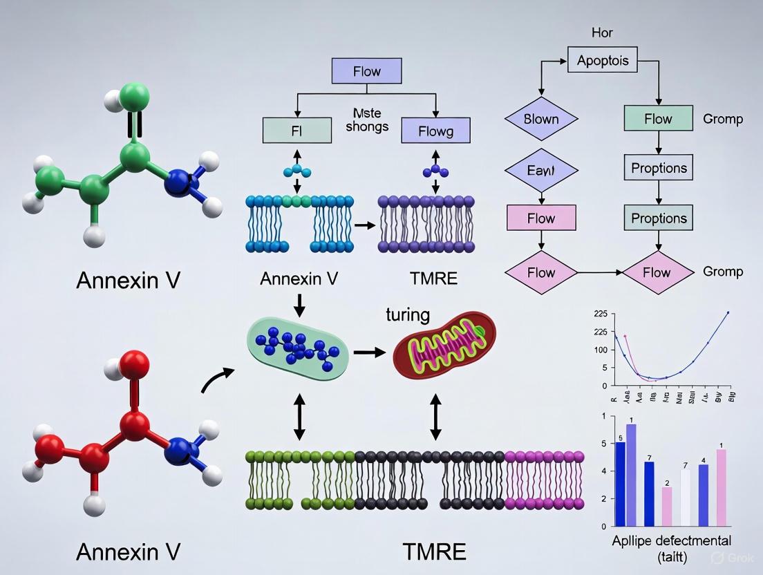
Annexin V vs. TMRE: A Comprehensive Guide for Early Apoptosis Detection Assay Selection
This article provides researchers, scientists, and drug development professionals with a definitive comparison of two fundamental early apoptosis detection methods: Annexin V (detecting phosphatidylserine externalization) and TMRE (assessing mitochondrial membrane...
Annexin V vs. TMRE: A Comprehensive Guide for Early Apoptosis Detection Assay Selection
Abstract
This article provides researchers, scientists, and drug development professionals with a definitive comparison of two fundamental early apoptosis detection methods: Annexin V (detecting phosphatidylserine externalization) and TMRE (assessing mitochondrial membrane potential). We explore the foundational biology behind each marker, detail standardized methodological protocols for flow cytometry and imaging, and offer troubleshooting guidance for common pitfalls. A direct, evidence-based comparison validates the sensitivity, temporal sequence, and specific applications of each assay, empowering you to select the optimal tool or synergistic combination for your specific research context in cancer biology, neurobiology, and therapeutic development.
Understanding the Core Biology: Phosphatidylserine Externalization vs. Mitochondrial Membrane Potential
The precise detection of apoptotic cell death is a cornerstone of biomedical research, playing a critical role in understanding fundamental biology, disease mechanisms, and the mode of action of potential therapeutic compounds. Apoptosis, or programmed cell death, is a highly regulated process essential for tissue homeostasis, embryogenesis, and immune response [1]. The ability to accurately identify and quantify apoptotic cells is particularly valuable in cancer research and drug development, where inducing tumor cell apoptosis is a primary therapeutic goal and a key indicator of treatment efficacy [2]. Discerning the early stages of this process allows researchers to identify potentially therapeutic compounds sooner and understand their specific mechanisms of action.
Flow cytometry has emerged as a powerful tool for apoptosis detection due to its multiparametric capabilities, high-throughput capacity, and quantitative nature. Unlike microscopy, flow cytometry minimizes observer bias by automatically analyzing thousands of cells per second and provides objective quantification of fluorescent signals [3] [4]. It enables the simultaneous assessment of multiple cellular parameters from a single sample, offering a comprehensive view of cellular status and fate. This guide provides an objective comparison of two key flow cytometry-based techniques for detecting early apoptotic events: Annexin V and TMRE staining. By understanding the strengths, applications, and limitations of each method, researchers and drug development professionals can make informed decisions to advance their projects.
The Apoptotic Cascade: A Primer for Detection
The apoptotic cascade is characterized by a sequence of biochemical and morphological events, which can be broadly categorized into early, intermediate, and late stages. Detection methods are tailored to specific events within this timeline. The intrinsic (mitochondrial) pathway is triggered by internal stressors like DNA damage or oxidative stress, leading to mitochondrial outer membrane permeabilization (MOMP) and a loss of mitochondrial membrane potential (ΔΨm) [3] [4]. This is one of the earliest committed steps in apoptosis. Subsequently, key events include the release of cytochrome c into the cytosol, activation of caspase enzymes, and finally, the externalization of phosphatidylserine (PS) from the inner to the outer leaflet of the plasma membrane [1] [5]. The latter event marks a stage where the cell is still intact but irrevocably committed to death.
The following diagram illustrates the sequence of these key events and the points at which different detection probes, including Annexin V and TMRE, interact with the process.
Annexin V vs. TMRE: A Comparative Analysis
Mechanism and Target
Annexin V is a 35-36 kDa protein that binds with high affinity to phosphatidylserine (PS) in a calcium-dependent manner [5]. In viable cells, PS is restricted to the inner leaflet of the plasma membrane. During the early stages of apoptosis, PS is translocated to the outer leaflet, creating a specific binding site for fluorescently conjugated Annexin V on the cell surface [3] [5]. This makes it a direct marker for a well-defined membrane alteration in apoptosis.
TMRE (Tetramethylrhodamine ethyl ester) is a cationic, lipophilic dye that accumulates in the mitochondrial matrix in a manner dependent on the mitochondrial membrane potential (ΔΨm) [6]. Healthy, polarized mitochondria with a strong ΔΨm take up and retain TMRE, resulting in bright fluorescence. During the early intrinsic apoptosis pathway, the collapse of ΔΨm prevents TMRE retention, leading to a measurable loss of fluorescence [6]. Thus, TMRE serves as a functional probe for the metabolic status of the mitochondria, an organelle central to the intrinsic apoptotic pathway.
Temporal Positioning in the Apoptotic Cascade
The different targets of these probes mean they detect sequential events in the apoptotic cascade. The loss of ΔΨm, detected by TMRE, is a very early event, particularly in the intrinsic pathway, and often precedes the externalization of PS [6]. It is considered a point-of-no-return in the cell death decision. PS externalization, detected by Annexin V, typically occurs after the loss of ΔΨm and is a hallmark of the early-to-mid stages of apoptosis, before the loss of plasma membrane integrity [5]. Therefore, in a temporal sequence, TMRE signal loss generally occurs before Annexin V binding becomes detectable.
Key Experimental Data and Performance Comparison
The table below summarizes objective, performance-related data for Annexin V and TMRE based on experimental findings from the literature.
Table 1: Comparative Experimental Data for Annexin V and TMRE Staining
| Feature | Annexin V / PI Assay | TMRE Staining |
|---|---|---|
| Primary Target | Phosphatidylserine (PS) on the outer plasma membrane leaflet [5] | Mitochondrial membrane potential (ΔΨm) [6] |
| Typical Signal Change in Apoptosis | Increase in Annexin V fluorescence [5] | Decrease in TMRE fluorescence (depolarization) [6] |
| Temporal Stage | Early-to-mid apoptosis (after PS externalization) [5] | Very early apoptosis (often first detectable event in intrinsic pathway) [6] |
| Viability Assessment | Requires co-staining with PI or 7-AAD to rule out late apoptosis/necrosis [5] [7] | Does not directly assess plasma membrane integrity |
| Critical Notes | Susceptible to false positives from compromised membranes; requires calcium buffer [5] [7]. Staining can be unstable due to high dissociation constant [6]. | Reversible staining; does not affect cell proliferation/viability [6]. More stable staining suitable for cell sorting [6]. |
| Proliferation Post-Sorting | Not typically used for sorting viable apoptotic cells | TMRE+ sorted cells show higher proliferative potential and negligible apoptosis [6] |
A critical performance difference lies in the application for cell sorting. One study directly demonstrated that sorting cells based on TMRE positivity (intact ΔΨm) yielded a population with a negligible percentage of apoptotic cells and a higher proliferative potential compared to sorting based on DNA viability dyes [6]. In contrast, the use of Annexin V for sorting viable early apoptotic cells is limited because the Annexin V/PS complex has a relatively high dissociation constant, resulting in less stable staining during the sorting process [6].
Detailed Experimental Protocols
Annexin V / Propidium Iodide (PI) Staining Protocol
This protocol is adapted from established methods for detecting apoptosis via flow cytometry [7].
Materials:
- Fluorochrome-conjugated Annexin V (e.g., Annexin V-FITC, Annexin V-APC)
- Propidium Iodide (PI) Staining Solution or 7-AAD
- 10X Annexin Binding Buffer
- 1X Phosphate Buffered Saline (PBS), azide- and serum/protein-free
- Flow cytometer
Procedure:
- Prepare Cells: Harvest and wash cells once with cold 1X PBS.
- Resuspend in Buffer: Resuspend the cell pellet (~1-5 x 10^6 cells) in 200 μL of 1X Annexin Binding Buffer.
- Stain with Annexin V: Add 5 μL of fluorochrome-conjugated Annexin V to the cell suspension. Incubate for 10-15 minutes at room temperature, protected from light.
- Add Viability Dye: Just before analysis, add 5-10 μL of PI or 7-AAD staining solution. Do not wash the cells after this step, as the viability dye must remain in the buffer during acquisition [7].
- Analyze by Flow Cytometry: Analyze the samples within 4 hours. Use a dot plot to discriminate populations: Annexin V-/PI- (viable), Annexin V+/PI- (early apoptotic), Annexin V+/PI+ (late apoptotic), and Annexin V-/PI+ (necrotic) [5].
Critical Considerations:
- Calcium Dependence: The binding of Annexin V to PS is Ca²⁺-dependent. Avoid buffers containing EDTA or other calcium chelators during the staining procedure [7].
- False Positives: Cells with a compromised plasma membrane (necrotic or late apoptotic) allow Annexin V to access PS on the inner leaflet, causing false-positive staining. This makes the co-staining with a membrane-impermeant dye like PI essential for accurate interpretation [5].
- Live Cells Only: This assay is designed for live cells. Fixation is not recommended as it permeabilizes the membrane.
TMRE Staining Protocol for Mitochondrial Membrane Potential
This protocol is designed to assess ΔΨm and identify cells undergoing early apoptosis [6].
Materials:
- TMRE (Tetramethylrhodamine ethyl ester perchlorate)
- Appropriate cell culture medium (without serum)
- Flow cytometer equipped with a 561 nm laser (or 488 nm for Rhodamine 123)
Procedure:
- Prepare TMRE Working Solution: Prepare a TMRE stock solution in DMSO and dilute in culture medium to a final working concentration of 5-100 nM.
- Stain Cells: Incubate cells with the TMRE working solution for 20-30 minutes at 37°C, protected from light.
- Wash Cells (Optional): For some protocols, cells are washed with PBS to remove excess dye. However, because TMRE staining is reversible, analysis should be performed promptly.
- Analyze by Flow Cytometry: Analyze the cells immediately. A shift toward lower TMRE fluorescence intensity indicates a loss of mitochondrial membrane potential and the presence of cells in early apoptosis.
Critical Considerations:
- Reversibility: TMRE staining is reversible and does not affect cell proliferation or viability, making it excellent for subsequent functional assays [6].
- Stability for Sorting: The stable nature of TMRE retention in polarized mitochondria makes it a superior dye for the fluorescence-activated cell sorting (FACS) of viable, non-apoptotic cells [6].
- Controls: Include a control treated with a mitochondrial uncoupler (e.g., FCCP) to fully depolarize mitochondria and establish the baseline for low TMRE fluorescence.
The Scientist's Toolkit: Essential Reagents
Table 2: Key Research Reagent Solutions for Apoptosis Detection
| Reagent | Function | Key Characteristics |
|---|---|---|
| Annexin V Conjugates | Binds to externalized Phosphatidylserine (PS) to detect early apoptotic cells [5]. | Available conjugated to various fluorophores (e.g., FITC, PE, APC); requires calcium buffer. |
| TMRE | Cationic dye that accumulates in active mitochondria; loss of fluorescence indicates depolarization [6]. | Reversible staining; ideal for functional assays and cell sorting; low cytotoxicity. |
| Propidium Iodide (PI) | Membrane-impermeant DNA dye identifies late apoptotic/necrotic cells with compromised membranes [3] [5]. | Incompatible with fixation; must be present in sample during acquisition. |
| 7-AAD | Membrane-impermeant DNA dye used as an alternative to PI for viability staining [5] [7]. | Can be used with fixed samples; compatible with PerCP and PE tandem dyes. |
| JC-1 | Ratiometric mitochondrial dye that shifts from red (J-aggregates) to green (monomers) upon depolarization [3] [4]. | Provides a dual-color readout; can be more sensitive but more complex to use than TMRE. |
| Annexin Binding Buffer | Provides the optimal calcium-containing environment for specific Annexin V binding to PS [5] [7]. | Critical for assay performance; must be free of EDTA and other chelators. |
Integrated Workflow and Data Interpretation
For a comprehensive analysis of cellular health and death mechanisms, researchers can integrate both Annexin V and TMRE into a multiparametric workflow. A sequential staining protocol or the use of a unified protocol that incorporates multiple stains like Annexin V, PI, and JC-1 (a ΔΨm-sensitive dye similar to TMRE) can provide a powerful, multi-faceted dataset from a single sample [3] [4]. This approach can simultaneously reveal changes in proliferation, cell cycle, mitochondrial function, and apoptosis.
The following diagram outlines a potential integrated workflow for a comprehensive analysis of cell death and proliferation using flow cytometry.
When interpreting data, it is crucial to correlate the results from both assays. A population showing TMRE-low and Annexin V-negative/PI-negative status is likely initiating the intrinsic apoptotic pathway but has not yet progressed to PS externalization. Cells that are TMRE-low and Annexin V-positive/PI-negative are firmly in the early apoptotic phase. This multi-parameter confirmation provides robust evidence for the mechanism of cell death induced by an experimental treatment, which is invaluable for drug discovery and basic research.
In cellular biology and drug development, the accurate and timely detection of programmed cell death, or apoptosis, is paramount for understanding compound efficacy, toxicity, and mechanism of action. Apoptosis is a highly regulated process crucial for normal tissue homeostasis, embryonic development, and the immune response [1]. Unlike necrotic cell death, which involves uncontrolled rupture and inflammatory responses, apoptosis is characterized by a series of distinct biochemical and morphological changes [1]. Among the various methods available for detecting this process, two powerful techniques stand out for identifying early apoptotic events: Annexin V binding, which detects changes in the plasma membrane, and TMRE staining, which measures the loss of mitochondrial transmembrane potential (ΔΨm) [8] [9]. This guide provides a objective, data-driven comparison of these two methodologies, equipping researchers with the information needed to select the optimal assay for their specific experimental context.
Fundamental Mechanisms: Where and How They Detect Apoptosis
The two methods operate on fundamentally different cellular principles, detecting sequential events in the apoptotic cascade. The following diagram illustrates their distinct mechanisms of action and the stage of apoptosis at which they act.
The Annexin V Mechanism
Annexin V is a 35 kDa cytoplasmic protein that binds with high affinity to phosphatidylserine (PS) in a calcium-dependent manner [10]. In viable, healthy cells, PS is predominantly located on the inner, cytoplasmic leaflet of the plasma membrane. During the early stages of apoptosis, the loss of membrane asymmetry leads to the rapid translocation and exposure of PS on the outer leaflet, making it accessible to extracellular Annexin V [9] [11]. This "eat-me" signal marks the cell for phagocytosis. The binding mechanism is precise; Annexin V self-assembles into highly ordered two-dimensional lattices on PS-containing membranes in the presence of calcium, which can even induce a phase transition in the underlying lipid bilayer, potentially stabilizing membrane defects [10]. In experimental protocols, Annexin V is conjugated to a fluorochrome (e.g., FITC) to enable detection via flow cytometry or fluorescence microscopy. It is typically used in conjunction with a membrane-impermeant viability dye like propidium iodide (PI) to distinguish early apoptotic cells (Annexin V+/PI-) from late apoptotic or necrotic cells (Annexin V+/PI+) [4].
The TMRE Mechanism
Tetramethylrhodamine ethyl ester (TMRE) is a cell-permeant, cationic fluorescent dye that accumulates in active mitochondria based on the Nernst equation [8] [12]. The internal negative charge of the mitochondrial matrix, typically around -180 mV in a healthy mitochondrion, drives the uptake and retention of TMRE. The intensity of TMRE fluorescence is directly proportional to the mitochondrial membrane potential (ΔΨm) [8]. During apoptosis, particularly via the intrinsic pathway, mitochondrial outer membrane permeabilization (MOMP) occurs, leading to the release of cytochrome c and other pro-apoptotic factors. A key consequence is the dissipation of ΔΨm, which is often considered a "point-of-no-return" in the apoptotic cascade [12]. This depolarization prevents TMRE accumulation within the mitochondria, leading to a measurable loss of fluorescence signal that can be quantified by flow cytometry or fluorescence microscopy [12]. Thus, TMRE serves as a sensitive indicator of mitochondrial health and the commitment to cell death.
Direct Comparison: Annexin V vs. TMRE
The table below provides a consolidated, data-driven comparison of Annexin V and TMRE staining across critical experimental parameters.
Table 1: Comprehensive Comparison of Annexin V and TMRE Assays
| Parameter | Annexin V Staining | TMRE Staining |
|---|---|---|
| Primary Target | Phosphatidylserine (PS) on the outer plasma membrane leaflet [9] | Mitochondrial transmembrane potential (ΔΨm) [8] |
| Cellular Process Detected | Early apoptosis (loss of membrane asymmetry) [9] | Early apoptosis (mitochondrial depolarization); often a "point-of-no-return" [12] |
| Detection Window | Early in apoptosis, before membrane integrity loss [4] | Coincides with or follows PS exposure; can be simultaneous or slightly later [4] [12] |
| Mechanism Principle | Calcium-dependent protein-phospholipid binding [10] | Potential-driven accumulation (Nernstian distribution) [8] |
| Key Experimental Requirement | Calcium-containing buffer [9] | No uncouplers in medium; validation with FCCP required [12] |
| Compatibility with Fixation | Generally incompatible with aldehyde fixation (disrupts membrane and requires Ca²⁺) | Incompatible with standard aldehyde fixation (causes loss of signal) [12] |
| Multiplexing Potential | High (commonly paired with PI, 7-AAD, and cell cycle dyes) [4] | High (can be combined with Annexin V, other fluorochromes in panels) [4] |
| Primary Advantage | Direct, well-established marker of early apoptosis; easily combined with viability dyes. | Indicates commitment to apoptosis via intrinsic pathway; strong correlation with cytochrome c release [8]. |
| Primary Limitation | Cannot distinguish between apoptotic and necrotic cells without a counterstain like PI [13]. | Signal can be influenced by plasma membrane potential and cell type [12]. |
Experimental Protocols & Data Interpretation
Annexin V/Propidium Iodide Staining Protocol
This protocol is adapted for flow cytometry and is typically completed within 1-2 hours [4].
- Cell Harvesting and Washing: Gently harvest cells (e.g., 0.5 x 10⁶ cells per sample) by trypsinization (for adherent cells) or pipetting (for suspension cells). Wash cells once with cold phosphate-buffered saline (PBS).
- Resuspension in Binding Buffer: Resuspend the cell pellet in a commercially available Annexin V binding buffer, which is specifically formulated to provide the necessary calcium ions for efficient binding.
- Staining: Add fluorescently-labeled Annexin V (e.g., Annexin V-FITC) and propidium iodide (PI) to the cell suspension. Incubate for 10-15 minutes at room temperature in the dark.
- Analysis: Analyze the samples by flow cytometry within 1 hour. The use of a no-stain control, Annexin V-only control, and PI-only control is essential for setting compensation and gating boundaries.
Data Interpretation for Flow Cytometry:
- Viable Cells: Annexin V⁻ / PI⁻
- Early Apoptotic Cells: Annexin V⁺ / PI⁻
- Late Apoptotic or Dead Cells: Annexin V⁺ / PI⁺
- Necrotic Cells or Debris: Annexin V⁻ / PI⁺ [4]
TMRE Staining Protocol for Flow Cytometry
This protocol is used for measuring ΔΨm in live, unfixed cells [12].
- Loading: Harvest and wash cells. Resuspend cells in pre-warmed culture medium or a suitable buffer containing a low concentration of TMRE (typically 50-200 nM).
- Incubation: Incubate cells with TMRE for 15-30 minutes at 37°C in the dark to allow for mitochondrial accumulation.
- Washing and Resuspension (Optional): Some protocols recommend a gentle wash to remove excess dye, while others analyze cells directly. Resuspend in fresh buffer if washed.
- Analysis: Analyze cells immediately by flow cytometry, monitoring the fluorescence in the FL2 or PE channel (~585 nm).
- Control: A critical control involves treating a duplicate sample with a mitochondrial uncoupler like FCCP (e.g., 1-10 µM) for 15-30 minutes prior to or during TMRE staining. FCCP collapses the ΔΨm, resulting in a loss of TMRE fluorescence, which serves as a baseline for depolarization [12].
Data Interpretation: A shift or peak toward lower TMRE fluorescence intensity compared to untreated control cells indicates a loss of ΔΨm and mitochondrial depolarization, a hallmark of apoptotic cells.
Advanced Workflow: Multiparametric Analysis
Advanced research often integrates both methods into a single, powerful multiparametric workflow to gain a comprehensive view of cellular health. The following diagram outlines a protocol for analyzing multiple parameters, including apoptosis and mitochondrial potential, from a single sample.
This unified protocol, which can be adapted to include TMRE instead of JC-1, allows for the rapid acquisition of up to eight different parameters from a single sample, providing an unparalleled, interconnected view of the cellular state and the dynamics between proliferation, cell cycle, apoptosis, and mitochondrial health [4].
The Scientist's Toolkit: Essential Reagents and Materials
Table 2: Key Research Reagent Solutions for Apoptosis Detection
| Reagent / Assay Kit | Primary Function | Key Feature / Application Note |
|---|---|---|
| Recombinant Annexin V (FITC, PE conjugates) | Detection of phosphatidylserine exposure during early apoptosis. | Standard for flow cytometry; requires calcium-containing buffer. Often sold as a kit with PI [4]. |
| TMRE (Tetramethylrhodamine Ethyl Ester) | Measurement of mitochondrial membrane potential (ΔΨm). | Cell-permeant; used for live-cell imaging and flow cytometry. Requires FCCP control for validation [8] [12]. |
| Propidium Iodide (PI) | Viability stain; labels dead cells with compromised membranes. | Impermeant to live cells; used to distinguish late apoptosis/necrosis in Annexin V assays [4] [13]. |
| RealTime-Glo Annexin V Apoptosis Assay | Luminescence-based real-time monitoring of PS exposure. | Non-lytic, plate-based assay allowing kinetic monitoring of apoptosis in live cells without harvesting [14]. |
| JC-1 Dye | Rationetric dye for measuring mitochondrial membrane potential. | Emits at different wavelengths (green/red) depending on ΔΨm; can be more sensitive but prone to artifacts [4] [12]. |
| BrdU (Bromodeoxyuridine) | Thymidine analog for monitoring cell cycle progression and proliferation. | Incorporated during S-phase; often used in multiplex assays to link apoptosis to cell cycle status [4]. |
| CellTrace Violet (CFSE-like dye) | Fluorescent cell dye for tracking cell division and proliferation. | Used to measure proliferation rates and trace generations in parallel with death assays [4]. |
| FCCP (Carbonyl cyanide-p-trifluoromethoxyphenylhydrazone) | Mitochondrial uncoupler. | Essential negative control for TMRE/TMRM assays to confirm ΔΨm-dependent staining [12]. |
Both Annexin V and TMRE staining are powerful, yet distinct, tools for detecting early apoptotic events. The choice between them is not a matter of superiority but of strategic application. Annexin V is the definitive choice for directly detecting the externalization of phosphatidylserine, a well-characterized "eat-me" signal of early apoptosis. In contrast, TMRE staining provides a crucial readout of mitochondrial integrity, often signifying a deeper commitment to the cell death pathway via the intrinsic apoptotic cascade.
For researchers, the most insightful approach often involves multiplexing these assays, either together or with other parameters like cell cycle analysis. The integrated workflow presented here demonstrates that a comprehensive understanding of a pharmacological or genetic treatment's effect comes from analyzing the interconnected dynamics of proliferation, cell cycle, mitochondrial function, and cell death [4]. As technologies advance, particularly with the development of real-time, non-lytic assays like the RealTime-Glo Annexin V assay, the ability to kinetically monitor these processes in live cells will continue to refine our understanding of cellular life and death decisions, ultimately accelerating drug discovery and safety assessment.
Tetramethylrhodamine ethyl ester (TMRE) is a cell-permeant, cationic, fluorescent dye that readily accumulates in active mitochondria due to their relative negative charge, serving as a sensitive indicator of mitochondrial membrane potential (ΔΨm) [15]. The reliance of all cell types on mitochondrial function for survival makes accurate assessment of mitochondrial membrane potential crucial across various research fields, from fundamental cell biology to drug development [16]. TMRE staining provides researchers with a reliable method for quantifying changes in ΔΨm, which is critical for cellular energy homeostasis, calcium signaling, and the intrinsic apoptosis pathway [8] [17].
This membrane potential, typically maintained at approximately -180 mV in healthy mitochondria, results from the active transfer of positively charged protons across the mitochondrial inner membrane during oxidative phosphorylation [8]. TMRE's positive charge and lipophilic properties enable it to electrophoretically distribute into the mitochondrial matrix in response to this negative charge, with fluorescence intensity directly correlating with the ΔΨm magnitude [16]. During apoptosis, the loss of ΔΨm is closely associated with cytochrome c release from the mitochondrial intermembrane space into the cytosol, making TMRE staining a valuable surrogate marker for detecting early apoptotic events [8].
Fundamental Mechanism of TMRE Accumulation
Electrochemical Principles of TMRE Staining
The accumulation of TMRE in mitochondria follows fundamental electrochemical principles governed by the Nernst equation. As a cationic dye, TMRE is attracted to and concentrated within the mitochondrial matrix based on the electrical potential difference across the inner mitochondrial membrane [16]. In functional mitochondria with intact membrane potential (ranging between -120 to -200 mV), TMRE accumulates electrophoretically, resulting in intense red-orange fluorescence when excited by appropriate light sources [15] [16]. This accumulation is reversible and concentration-dependent, allowing for quantitative assessment of ΔΨm changes in live cells without fixation [15].
TMRE Response to Membrane Potential Alterations
When mitochondrial membrane potential dissipates, as occurs during early apoptosis or in response to uncouplers like FCCP (carbonyl cyanide 4-(trifluoromethoxy) phenylhydrazone), TMRE fails to sequester within mitochondria and instead distributes homogenously throughout the cell at lower concentrations, resulting in significantly diminished fluorescence [15]. This characteristic enables researchers to distinguish between populations of cells with polarized (functional) and depolarized (dysfunctional) mitochondria using techniques such as flow cytometry, fluorescence microscopy, and microplate spectrophotometry [15] [6]. The specificity of TMRE for ΔΨm has been demonstrated in controlled experiments where treatment with FCCP, an ionophore uncoupler of oxidative phosphorylation, completely eliminates TMRE staining by collapsing the proton gradient [15].
Table 1: Key Characteristics of TMRE Staining
| Property | Description | Experimental Significance |
|---|---|---|
| Charge | Positively charged | Electrophoretically accumulates in negatively charged mitochondrial matrix |
| Permeability | Cell permeant | Easily enters live cells without permeabilization |
| Specificity | Potential-dependent | Fluorescence intensity directly correlates with ΔΨm |
| Reversibility | Reversible staining | Does not affect cell proliferation or viability after removal [6] |
| Compatibility | Live cells only | Not compatible with fixation protocols |
| Optimal Ex/Em | 549/575 nm | Compatible with standard TRITC filter sets |
Comparative Analysis: TMRE Versus Alternative Apoptosis Detection Methods
TMRE vs. Annexin V for Early Apoptosis Detection
TMRE and Annexin V target fundamentally different cellular processes in apoptosis detection, with TMRE identifying mitochondrial membrane depolarization that occurs early in the intrinsic apoptosis pathway, while Annexin V detects phosphatidylserine externalization that occurs later in the apoptotic process [6] [3]. During apoptosis, the decrease in mitochondrial potential precedes gross morphological changes and exposure of phosphatidylserine on the external leaflet of the plasma membrane [6]. This temporal relationship makes TMRE staining an earlier indicator of commitment to apoptosis compared to Annexin V staining.
Research has demonstrated that TMRE positivity is associated with an absence of apoptotic processes, and sorted TMRE+ cells contain a negligible percentage of apoptotic and damaged cells while maintaining higher proliferative potential compared to cells sorted based on DNA viability dye staining [6]. Furthermore, cell sorting based on Annexin V staining is limited by the relatively high dissociation constant of the Annexin V/phosphatidylserine complex, which results in unstable staining, whereas TMRE staining remains stable throughout sorting procedures [6].
TMRE vs. JC-1 and Other Mitochondrial Dyes
While TMRE exhibits a monotonic relationship between fluorescence intensity and membrane potential, JC-1, another popular mitochondrial dye, undergoes a potential-dependent shift in fluorescence emission from green (~529 nm) for monomeric dye to red (~590 nm) for J-aggregates formed at higher membrane potentials [3] [4]. This dual-emission property can be advantageous for ratio-metric measurements but may present challenges in calibration and interpretation. TMRE is generally preferred for quantitative measurements of ΔΨm using flow cytometry or fluorescence microscopy, while JC-1 is often selected for experiments where ratio-metric measurements of potential are desired.
Table 2: Comparison of TMRE with Alternative Apoptosis/Mitochondrial Assessment Methods
| Method | Detection Principle | Stage of Apoptosis Detected | Advantages | Limitations |
|---|---|---|---|---|
| TMRE | Mitochondrial membrane potential dissipation | Early intrinsic pathway | Early detection; reversible; minimal toxicity; compatible with live cell imaging | Requires live cells; not compatible with fixation |
| Annexin V | Phosphatidylserine externalization | Mid-stage (after mitochondrial depolarization) | Well-established; can differentiate early/late apoptosis with PI counterstain | Unstable staining due to high dissociation constant; detects later events [6] |
| JC-1 | Mitochondrial membrane potential-dependent J-aggregate formation | Early intrinsic pathway | Ratiometric measurement; visual color shift | Complex calibration; potential-sensitive aggregates may be slow to form/dissociate [3] |
| Caspase Activation | Cleavage of fluorogenic caspase substrates | Execution phase (downstream of mitochondrial events) | High specificity for apoptosis; multiple caspase targets available | Late-stage detection; may miss early commitment phases [3] |
Experimental Applications and Protocols
Standard TMRE Staining Protocol for Flow Cytometry
The following protocol summarizes the standard methodology for TMRE staining adapted from commercial kits and published research [15] [18]:
Cell Preparation: Harvest and wash cells in appropriate buffer. For adherent cells, gently detach using non-enzymatic methods when possible to preserve mitochondrial function. Adjust cell concentration to 1×10^6 cells/mL in culture medium.
TMRE Solution Preparation: Dilute TMRE stock solution in pre-warmed culture medium to achieve working concentrations typically ranging from 20-500 nM, optimized for specific cell types. Protect from light during preparation and use.
Staining Incubation: Add TMRE working solution to cell suspension and incubate for 15-30 minutes at 37°C in a CO₂ incubator. Include a control sample treated with 10-50 µM FCCP for 10 minutes prior to TMRE addition to validate specificity of potential-dependent staining.
Washing and Analysis: Pellet cells and wash once with PBS containing 0.2% BSA to remove excess dye. Resuspend in appropriate buffer and analyze immediately using flow cytometry with 488 nm laser for excitation and 575 nm emission detection, or fluorescent microscopy with TRITC filters.
Validation Using Targeted Irradiation Experiments
Sophisticated validation of TMRE's response to mitochondrial membrane potential changes comes from targeted irradiation experiments. Research using highly focused carbon ions and protons with beam spots <1 µm demonstrated that targeted irradiation induces near instant loss of TMRE fluorescence specifically in irradiated mitochondrial areas, representing radiation-induced changes in mitochondrial membrane potential [16]. This response was immediate (within the temporal resolution of the imaging system, <300 ms) and highly localized, with no perceptible effect on non-targeted mitochondria in the same cell [16]. Control experiments with FCCP showed similar loss of mitochondrial TMRE signal, confirming that the fluorescence changes reflected genuine membrane potential alterations rather than direct destruction of TMRE molecules by radiation [16].
Quantitative Data from Comparative Studies
Table 3: Experimental Performance Data for TMRE in Research Applications
| Application Context | Cell Type | Key Parameters | Performance Results |
|---|---|---|---|
| Elimination of apoptotic cells [6] | THP-1, Jurkat, HeLa, RAW 264.7 | Purity of sorted population; proliferative potential | TMRE+ cells contained negligible apoptotic cells; higher proliferative potential vs. DNA viability dye-based sorting |
| Targeted mitochondrial irradiation [16] | A549, MCF7 | Fluorescence change post-irradiation; temporal resolution | -87.5% mean fluorescence change in irradiated areas vs. +2.2% in controls; response in <300 ms |
| Mitochondrial hyperpolarization study [19] | HEK293 IF1-KO | Detection of hyperpolarization; correlation with functional assays | IF1-KO cells showed higher resting ΔΨm confirmed by faster cytosolic Ca²⁺ clearance |
| Early apoptosis detection [6] | Various cell lines | Correlation with caspase activation; Annexin V staining | TMRE negativity preceded caspase activation and phosphatidylserine externalization |
Research Reagent Solutions Toolkit
Table 4: Essential Reagents and Tools for TMRE-based Mitochondrial Assays
| Reagent/Equipment | Function/Purpose | Specific Examples/Specifications |
|---|---|---|
| TMRE Assay Kit | Complete solution for ΔΨm measurement | Includes TMRE and FCCP control (e.g., Abcam ab113852, RayBio MT-TMRE) [15] [18] |
| Flow Cytometer | Quantitative analysis of TMRE fluorescence | Instruments with 488 nm laser and 575 nm emission detection (e.g., BD FACSAria II, BD FACSLyric) [6] [3] |
| Fluorescent Microscope | Visual assessment and imaging of mitochondrial staining | Epifluorescence microscopes with TRITC filter sets (Ex/Em: 549/575 nm) [15] [16] |
| Microplate Reader | High-throughput quantification in multi-well formats | Fluorescent plate readers capable of Ex/Em: 549/575 nm measurements [15] |
| FCCP | Positive control for mitochondrial depolarization | Ionophore uncoupler (typically used at 10-50 µM) to validate potential-dependent staining [15] |
| Carbonyl Cyanide m-chlorophenyl Hydrazone (CCCP) | Alternative mitochondrial uncoupler | Can be used similarly to FCCP to collapse ΔΨm [17] |
| MitoTracker Green | Mitochondrial mass control stain | ΔΨm-independent mitochondrial dye for normalization (Ex/Em: 490/516 nm) [16] [19] |
Signaling Pathways and Experimental Workflows
Diagram 1: TMRE Detection in the Intrinsic Apoptosis Pathway. TMRE fluorescence loss directly detects mitochondrial membrane potential (ΔΨm) dissipation, an early event in intrinsic apoptosis that precedes cytochrome c release and caspase activation.
Diagram 2: Experimental Workflow for TMRE Staining. Standard procedure for TMRE-based assessment of mitochondrial membrane potential, including essential controls and detection methods.
TMRE represents a robust, sensitive, and reliable tool for detecting changes in mitochondrial membrane potential, particularly for identifying early events in the intrinsic apoptosis pathway. Its mechanism of potential-dependent accumulation in active mitochondria provides researchers with a direct means of assessing mitochondrial function in live cells. When compared to alternative methods such as Annexin V staining, TMRE offers the advantage of detecting earlier commitment phases to apoptosis, while its simplicity and reversibility make it preferable to more complex ratiometric dyes like JC-1 for many applications. The comprehensive experimental data and protocols presented in this guide provide researchers and drug development professionals with the necessary foundation to implement TMRE-based assays in their experimental workflows, enabling accurate assessment of mitochondrial health and early apoptosis detection in various research contexts.
Programmed cell death, or apoptosis, is a fundamental biological process crucial for maintaining tissue homeostasis, embryogenesis, and immune function [1]. The detection of apoptosis relies on identifying key cellular changes that occur in a sequential manner. Two of the most critical events in this cascade are the loss of mitochondrial membrane potential (ΔΨm) and the externalization of phosphatidylserine (PS) on the cell surface. Understanding the temporal relationship between these events is essential for researchers and drug development professionals selecting appropriate detection methods for their experimental needs. This guide provides a comprehensive comparison between Annexin V-based assays (detecting PS exposure) and TMRE staining (assessing ΔΨm) to determine which event occurs earlier in apoptosis and which method offers the most reliable early detection capabilities.
The Sequence of Apoptotic Events: A Timeline
Molecular Timeline of Key Apoptotic Events
Apoptosis progresses through a defined sequence of molecular events. The intrinsic apoptotic pathway, triggered by cellular stress or damage, initially involves mitochondrial changes before manifesting on the plasma membrane.
This temporal sequence reveals why ΔΨm loss serves as an earlier apoptosis marker than PS externalization. During apoptosis, the decrease in mitochondrial potential precedes the gross morphological changes that occur during the apoptotic process and before exposure of PS on the external leaflet of the plasma membrane [6]. The intrinsic apoptotic pathway begins with mitochondrial depolarization, followed by cytochrome c release, caspase activation, and ultimately PS externalization.
Direct Comparison: Annexin V vs. TMRE for Apoptosis Detection
Comparative Analysis of Detection Methods
The following table summarizes the key characteristics of Annexin V and TMRE as apoptosis detection markers:
| Parameter | Annexin V (PS Exposure) | TMRE (ΔΨm Loss) |
|---|---|---|
| Detection Target | Externalized phosphatidylserine on plasma membrane | Mitochondrial membrane potential |
| Temporal Position in Apoptosis | Intermediate stage | Early stage |
| Detection Window | Mid-stage apoptosis after caspase activation | Early apoptosis before PS exposure |
| Calcium Dependence | Requires calcium for PS binding [7] | Calcium-independent |
| Plasma Membrane Integrity Requirement | Critical - damaged membranes cause false positives [7] | Less critical - detects events before membrane damage |
| Primary Applications | Differentiating apoptosis stages, especially with viability dyes | Early apoptosis detection, functional mitochondrial assessment |
| Key Advantage | Can distinguish early apoptotic (Annexin V+/PI-) from late apoptotic/necrotic (Annexin V+/PI+) cells [4] [20] | Identifies cells committed to apoptosis before morphological changes [6] |
| Main Limitation | Cannot detect very early apoptosis; compromised membranes obscure interpretation | Does not directly confirm execution-phase apoptosis events |
Experimental Evidence for Temporal Sequence
Multiple studies have demonstrated that TMRE-detected ΔΨm loss occurs before Annexin V-detected PS externalization:
- Flow cytometry sorting experiments show that TMRE+ cells contain a negligible percentage of apoptotic and damaged cells and have a higher proliferative potential compared to their counterparts [6].
- Multiparametric analysis reveals that during apoptosis, the decrease in mitochondrial potential precedes exposure of PS on the external leaflet of the plasma membrane [6].
- Comprehensive flow cytometry methodologies that integrate both markers consistently show ΔΨm changes occurring before PS externalization in the apoptotic cascade [4].
Detailed Experimental Protocols
Annexin V/Propidium Iodide Staining Protocol
The Annexin V/PI assay is widely used to distinguish between healthy, early apoptotic, late apoptotic, and necrotic cells based on PS exposure and membrane integrity [7] [20].
Materials Required:
- Annexin V conjugate (FITC, PE, APC, or other fluorochromes)
- Propidium iodide (PI) or 7-AAD staining solution
- 10X binding buffer
- Flow cytometry staining buffer
- 12 × 75 mm round-bottom tubes
- PBS (calcium-free)
Procedure:
- Prepare 1X binding buffer by diluting 10X binding buffer 1:9 with distilled water.
- Harvest and wash cells once with PBS, then once with 1X binding buffer.
- Resuspend cells in 1X binding buffer at 1-5 × 10⁶ cells/mL.
- Add 5 μL of fluorochrome-conjugated Annexin V to 100 μL of cell suspension.
- Incubate for 10-15 minutes at room temperature, protected from light.
- Add 2 mL of 1X binding buffer and centrifuge at 400-600 × g for 5 minutes.
- Discard supernatant and resuspend cells in 200 μL of 1X binding buffer.
- Add 5 μL of PI staining solution and analyze immediately by flow cytometry.
- Critical note: Do not wash cells after PI addition, as PI must remain in buffer during acquisition [7].
Data Interpretation:
- Annexin V-/PI-: Viable, healthy cells
- Annexin V+/PI-: Early apoptotic cells
- Annexin V+/PI+: Late apoptotic cells
- Annexin V-/PI+: Necrotic cells or late-stage apoptosis with complete membrane disruption [4] [20]
TMRE Staining Protocol for ΔΨm Assessment
TMRE (tetramethylrhodamine ethyl ester) is a cationic, lipophilic dye that accumulates in active mitochondria based on their transmembrane potential [6] [21] [22].
Materials Required:
- TMRE stock solution (prepared in DMSO)
- Dimethyl sulfoxide (DMSO)
- PBS or appropriate cell culture buffer
- Flow cytometry tubes
- Optional: Carbonyl cyanide m-chlorophenyl hydrazone (CCCP) as positive control for depolarization
Procedure:
- Prepare TMRE working solution in pre-warmed buffer or culture medium. Typical working concentrations range from 5-100 nM [6].
- Harvest cells and wash with PBS.
- Resuspend cells in TMRE working solution at 1-5 × 10⁶ cells/mL.
- Incubate for 20 minutes at 37°C, protected from light.
- Centrifuge at 400-600 × g for 5 minutes and discard supernatant.
- Resuspend cells in fresh pre-warmed buffer or culture medium.
- Analyze immediately by flow cytometry using 488 nm or 561 nm excitation with detection around 574-580 nm [21].
- Optional: Include a positive control pre-treated with 50 μM CCCP for 10 minutes to confirm specificity of TMRE staining.
Data Interpretation:
- High TMRE fluorescence: Cells with intact mitochondrial membrane potential (healthy)
- Low TMRE fluorescence: Cells with depolarized mitochondria (apoptotic)
- The TMRE signal directly correlates with ΔΨm, with decreased fluorescence indicating mitochondrial depolarization [6] [22]
Integrated Workflow for Comprehensive Apoptosis Assessment
Multiparametric Apoptosis Analysis Workflow
For comprehensive understanding of apoptotic progression, researchers can combine both methods with additional markers in a unified protocol:
This integrated approach enables simultaneous assessment of multiple apoptosis parameters from a single sample, providing a comprehensive view of cellular status and death mechanisms [4].
Research Reagent Solutions
Essential Materials for Apoptosis Detection
| Reagent/Tool | Primary Function | Application Notes |
|---|---|---|
| Annexin V Conjugates | Binds externalized phosphatidylserine | Available as FITC, PE, APC, eFluor; calcium-dependent binding [7] |
| TMRE | Mitochondrial potential-sensitive dye | 549/574 nm Ex/Em; use 5-100 ng/mL; reversible staining [6] [21] |
| Propidium Iodide | DNA intercalator for dead cell identification | Membrane-impermeant; indicates loss of membrane integrity [7] [20] |
| 7-AAD | Alternative viability dye | Can be used instead of PI; different spectral properties [7] |
| Binding Buffer | Provides optimal calcium concentration | Critical for Annexin V-PS interaction; avoid EDTA contamination [7] |
| JC-1 | Alternative mitochondrial potential dye | Forms aggregates (red) at high ΔΨm; monomers (green) at low ΔΨm [4] |
| Fixable Viability Dyes | Distinguish live/dead cells | Compatible with intracellular staining; use before permeabilization [7] |
The temporal relationship between ΔΨm loss and PS externalization has significant implications for apoptosis research and drug development. TMRE detection of mitochondrial depolarization provides an earlier window into apoptotic commitment, while Annexin V detection of PS externalization marks a definitive, intermediate stage of apoptosis.
For researchers investigating early apoptosis triggers or screening compounds for initial apoptotic effects, TMRE offers superior sensitivity for detecting the earliest mitochondrial changes. Conversely, for studies quantifying apoptosis levels or distinguishing between apoptotic stages, Annexin V with viability staining provides clearer stage-specific information.
The choice between these methods should be guided by specific research questions, with the understanding that an integrated approach combining both markers with complementary assays (such as caspase activation or cell cycle analysis) delivers the most comprehensive understanding of apoptotic dynamics in experimental systems [4]. This multifaceted analysis is particularly valuable in drug discovery, where understanding the timing and mechanism of compound-induced cell death can inform development decisions and mechanism-of-action studies.
In the field of cell biology, accurately detecting programmed cell death is fundamental to understanding disease mechanisms and developing therapeutic interventions. Apoptosis, a highly regulated form of cell death, occurs through multiple interconnected pathways that manifest different molecular signatures at various stages. While Annexin V and tetramethylrhodamine ethyl ester (TMRE) represent two prominent tools for early apoptosis detection, each targets distinct cellular events with inherent limitations. This review objectively compares the performance, experimental applications, and technical constraints of these methodologies, demonstrating that a multiparametric approach is essential for comprehensive apoptosis assessment. The complex nature of apoptotic signaling, with its morphological hallmarks and biochemical cascades, necessitates complementary detection strategies to overcome the limitations of any single marker [1].
The Biological Landscape of Apoptosis
Understanding the Pathways
Apoptosis proceeds primarily through two interconnected pathways that converge on a common execution phase. The extrinsic pathway initiates when external death ligands bind to cell surface receptors, recruiting adaptor proteins that activate initiator caspases [1]. Conversely, the intrinsic pathway triggers in response to internal cellular damage or stress, leading to mitochondrial outer membrane permeabilization and the release of cytochrome c into the cytoplasm [1]. This release activates the apoptosome complex and executioner caspases. Both pathways ultimately result in the systematic dismantling of cellular components, though they originate from different stimuli and involve distinct molecular initiators [1].
These pathways are not isolated; significant cross-talk occurs between them, and components involved in apoptosis also participate in other forms of programmed cell death like necroptosis [1]. This interplay further complicates the detection and interpretation of cell death events using single markers.
Visualization of key apoptotic pathways and corresponding detection events for Annexin V and TMRE.
Direct Comparison: Annexin V vs. TMRE
Annexin V - Detecting Phosphatidylserine Externalization
Principle and Target: Annexin V is a 35-36 kDa calcium-dependent phospholipid-binding protein with high affinity for phosphatidylserine (PS), a membrane phospholipid normally restricted to the inner leaflet of the plasma membrane in viable cells [5]. During early apoptosis, PS translocates to the external membrane leaflet, creating a specific binding site for fluorescently conjugated Annexin V [23] [24]. This externalization occurs within 5-10 minutes after an apoptotic stimulus, making it one of the earliest detectable events [23].
Advantages: The Annexin V assay provides non-perturbing detection of apoptotic cells without requiring cell permeabilization [23]. The difference in fluorescence intensity between apoptotic and non-apoptotic cells is typically about 100-fold, providing excellent signal resolution [5]. When combined with viability dyes like propidium iodide (PI), the assay can distinguish between early apoptotic (Annexin V+/PI-), late apoptotic (Annexin V+/PI+), and necrotic cells (Annexin V-/PI+) [20] [5].
Limitations: A significant limitation is that PS externalization is not absolutely specific for apoptosis. It also occurs during other processes including platelet activation, cellular stress responses, and in the tumor vasculature [23]. Furthermore, Annexin V cannot differentiate between apoptosis and necrosis in cells with compromised membrane integrity, as the protein can access internal PS in leaky cells, creating false positives [5]. The binding is also calcium-dependent, requiring optimized buffer conditions [25].
TMRE - Monitoring Mitochondrial Membrane Potential
Principle and Target: TMRE (tetramethylrhodamine ethyl ester perchlorate) is a cationic, lipophilic dye that accumulates in active mitochondria based on the inner mitochondrial membrane potential (ΔΨm) [6]. During apoptosis, particularly via the intrinsic pathway, mitochondrial membrane depolarization occurs, leading to reduced TMRE retention and fluorescence [6] [26]. This depolarization represents one of the earliest events in the intrinsic apoptotic pathway, preceding phosphatidylserine externalization [6].
Advantages: TMRE staining is reversible and does not significantly affect cell proliferation or viability, making it suitable for functional assays following analysis [6]. The dye provides a functional assessment of mitochondrial health beyond just apoptosis detection. TMRE-positive cells show minimal apoptotic contamination and maintain higher proliferative potential compared to cells selected by DNA viability dyes [6].
Limitations: Mitochondrial depolarization can occur in response to various cellular stresses not necessarily leading to apoptosis, including metabolic perturbations and energy insufficiency [26]. The staining is also affected by factors influencing mitochondrial function beyond apoptosis, such as alterations in electron transport chain activity [26]. Unlike Annexin V, TMRE requires cell permeabilization for accurate assessment of ΔΨm, potentially affecting cell viability in subsequent experiments.
Table 1: Comparative Analysis of Key Apoptosis Detection Markers
| Parameter | Annexin V | TMRE |
|---|---|---|
| Primary Target | Externalized phosphatidylserine on plasma membrane | Mitochondrial membrane potential (ΔΨm) |
| Detection Window | Early to mid-apoptosis (post-caspase activation) | Early apoptosis (pre-caspase activation in intrinsic pathway) |
| Cellular Process Monitored | Loss of membrane phospholipid asymmetry | Mitochondrial membrane depolarization |
| Specificity Challenges | Not specific to apoptosis; also positive in necrosis, platelet activation | Not specific to apoptosis; also sensitive to metabolic stress, energy depletion |
| Viability Dye Required | Essential (e.g., PI, 7-AAD) to exclude necrotic cells | Recommended for comprehensive interpretation |
| Calcium Dependency | Required for PS binding | Not required |
| Temporal Relationship | Later event in apoptotic cascade | Earlier event in intrinsic pathway |
Table 2: Experimental Performance Metrics from Comparative Studies
| Performance Metric | Annexin V-based Sorting | TMRE-based Sorting |
|---|---|---|
| Purity of Sorted Population | Moderate (unstable staining due to high dissociation constant) | High (negligible apoptotic cells in TMRE+ population) |
| Post-Sort Cell Viability | Variable | High (dye does not affect proliferation) |
| Proliferative Capacity Post-Sort | Reduced | Significantly higher |
| Apoptotic Cell Contamination | Present in "viable" population | Minimal in TMRE+ population |
| Staining Stability | Limited (high dissociation constant) | Excellent |
| Compatibility with Downstream assays | Moderate | High |
Experimental Protocols and Methodologies
Annexin V Staining Protocol for Flow Cytometry
Sample Preparation: Harvest approximately 1×10⁶ cells, combining both adherent (after trypsinization) and floating cell populations to capture all apoptotic stages [20]. Wash cells twice with PBS and centrifuge at 670 × g for 5 minutes at room temperature.
Staining Procedure: Resuspend cell pellet in 400 μL of PBS. Add 100 μL of incubation buffer containing 2 μL of Annexin V conjugate (1 mg/mL) and 2 μL of propidium iodide (1 mg/mL) [20]. For controls, prepare unstained cells (cells + buffer only), Annexin V-only stained cells, and PI-only stained cells.
Analysis: Analyze samples by flow cytometry without additional washing to prevent loss of weakly bound Annexin V [20]. Identify populations as follows: viable cells (Annexin V-/PI-), early apoptotic (Annexin V+/PI-), late apoptotic/necrotic (Annexin V+/PI+)
Critical Considerations: The assay must be performed on live, unfixed cells as fixation disrupts membrane integrity and PS accessibility [5]. Calcium concentration must be optimized in the binding buffer (typically 2.5 mM) for efficient Annexin V-PS interaction [5] [24]. Always include viability dye controls to distinguish true apoptosis from necrosis.
TMRE Staining Protocol for Mitochondrial Potential Assessment
Sample Preparation: Culture cells under standard conditions. For suspension cells, concentrate to approximately 1×10⁶ cells/mL. Adherent cells should be trypsinized gently to preserve mitochondrial function.
Staining Procedure: Incubate cells with 5-100 ng/mL TMRE for 20 minutes at 37°C [6]. For flow cytometry, use TMRE concentrations in the lower range (5-20 ng/mL) to avoid artifacts from dye overload. For microscopy, higher concentrations (50-100 ng/mL) may provide better signal.
Analysis: Analyze by flow cytometry using 561 nm laser excitation with emission capture at 582/15 nm [6]. For imaging, use appropriate tetramethylrhodamine filter sets. Cells with intact mitochondrial potential show bright punctate mitochondrial staining, while apoptotic cells exhibit diffuse, dim fluorescence.
Critical Considerations: TMRE staining is reversible and concentration-dependent - titration is essential for accurate results [6]. Include a positive control (e.g., cells treated with carbonyl cyanide m-chlorophenyl hydrazone/CCCP) to fully depolarize mitochondria and establish background fluorescence. Avoid prolonged staining as TMRE can potentially exert mild mitochondrial toxicity at high concentrations.
Integrated Workflows and Complementary Approaches
Multiparametric Assessment
Given the limitations of individual markers, researchers are increasingly adopting multiparametric approaches that combine Annexin V, TMRE, and additional probes for comprehensive cell death assessment [4]. One recently published workflow simultaneously analyzes eight different parameters from a single sample, including cell count, proliferation, cell cycle dynamics, apoptosis, membrane permeability, and mitochondrial depolarization [4].
This integrated methodology typically combines Annexin V/PI staining with JC-1 (a mitochondrial potential dye similar to TMRE), BrdU for cell cycle analysis, and CellTrace Violet for proliferation tracking [4]. Such approaches reveal interconnected cellular responses, such as how mitochondrial depolarization may precede both apoptosis induction and cell cycle arrest following specific treatments [4].
Advanced Detection Technologies
Beyond traditional flow cytometry, several innovative approaches are emerging for apoptosis detection:
Caspase-Activatable Probes: These probes contain caspase recognition sequences (typically DEVD) flanked by fluorophore-quencher pairs [25]. Upon caspase cleavage during apoptosis, fluorescence is activated, providing direct readout of executioner caspase activity. However, potential cross-reaction with other proteases like cathepsins remains a concern [25].
ApoSense Molecules: These small non-peptidic compounds show selective accumulation in apoptotic cells through mechanisms involving selective membrane binding and transport [25]. Some variants can be labeled with PET isotopes for in vivo imaging applications.
Reporter Gene Imaging: Innovative constructs link luciferase or fluorescent protein expression to caspase activation through cleavable linkers, enabling real-time monitoring of apoptosis dynamics in live cells and animals [25].
The Scientist's Toolkit: Essential Research Reagents
Table 3: Key Reagents for Apoptosis Detection Assays
| Reagent | Function | Application Notes |
|---|---|---|
| Annexin V Conjugates | Binds externalized phosphatidylserine | Available conjugated to Alexa Fluor, FITC, PE, APC; requires calcium buffer |
| TMRE | Mitochondrial potential-sensitive dye | Reversible staining; concentration-critical; 561 nm excitation |
| Propidium Iodide (PI) | DNA intercalator; membrane integrity indicator | Cell-impermeant in viable cells; 535/617 nm ex/em |
| 7-AAD | Alternative viability dye | Preferred for multicolor panels with FITC-conjugated Annexin V |
| JC-1 | Rationetric mitochondrial potential dye | Forms J-aggregates (red) at high potential; monomers (green) at low potential |
| SYTOX Green | High-affinity nucleic acid stain | Impermeant to live cells; bright green fluorescence upon membrane compromise |
| Caspase 3/7 Substrates | Fluorogenic caspase activity probes | Cell-permeant; cleaved to fluorescent product by active caspases |
| BrdU/Anti-BrdU | S-phase proliferation marker | Requires DNA denaturation for antibody access |
| CellTrace Violet | Cell proliferation dye | CFSE-like dye; dilutes with each cell division |
The comparative analysis of Annexin V and TMRE underscores a fundamental principle in cell death research: no single marker provides a complete picture of the apoptotic process. Each methodology captures different facets of this complex cellular program, with Annexin V detecting plasma membrane alterations and TMRE monitoring early mitochondrial events. Their inherent limitations—including pathway specificity, temporal resolution, and susceptibility to non-apoptotic cellular changes—highlight the necessity of multiparametric assessment strategies. As research advances, integrating these complementary detection methods with emerging technologies will provide increasingly comprehensive insights into cell death mechanisms, ultimately enhancing both basic research and drug development efforts.
Protocols and Best Practices: From Kit Selection to Multicolor Panel Design
Standardized Annexin V Staining Protocol for Suspension and Adherent Cells
The accurate detection of apoptosis is fundamental to cancer research, drug development, and understanding cellular responses to treatment. Among the various methods available, flow cytometry-based approaches have become the gold standard for quantifying cell death. This guide focuses on two prominent techniques: Annexin V staining, which detects phosphatidylserine externalization on the cell membrane, and TMRE (tetramethylrhodamine ethyl ester) staining, which measures changes in mitochondrial membrane potential [6] [23]. While both methods identify early apoptotic events, they target distinct biochemical processes in the cell death cascade. Annexin V binds to phosphatidylserine that has translocated from the inner to outer leaflet of the plasma membrane, one of the earliest features of apoptosis [27] [23]. In contrast, TMRE accumulates in active mitochondria with intact membrane potential, which is lost during early apoptosis, making TMRE negativity a marker of mitochondrial dysfunction preceding phosphatidylserine exposure in some cellular contexts [6] [12]. This article provides standardized protocols for both suspension and adherent cell cultures, compares the performance characteristics of these techniques, and presents experimental data to guide researchers in selecting the most appropriate method for their specific applications.
Theoretical Foundations: Detection Principles and Signaling Pathways
Phosphatidylserine Externalization and Annexin V Binding
In viable cells, phosphatidylserine (PS) is predominantly restricted to the inner leaflet of the plasma membrane through the activity of ATP-dependent translocases [23]. During early apoptosis, this membrane asymmetry collapses due to the activation of scramblases and inhibition of translocases, resulting in PS exposure on the outer leaflet [23]. Annexin V is a 35-36 kDa calcium-dependent phospholipid-binding protein with high affinity for PS [27]. When conjugated to fluorochromes, it enables detection of PS-exposing cells by flow cytometry. This exposure creates an "eat-me" signal recognized by phagocytes, representing a key physiological step in apoptotic clearance [23]. The Annexin V binding assay is particularly valuable because it detects apoptosis before complete loss of membrane integrity, allowing distinction between early apoptotic cells (Annexin V-positive, viability dye-negative) and late apoptotic/necrotic cells (Annexin V-positive, viability dye-positive) [7] [27].
Mitochondrial Membrane Potential and TMRE Staining
TMRE is a cationic, lipophilic fluorescent dye that accumulates in the mitochondrial matrix driven by the mitochondrial inner membrane potential (ΔΨm) [6] [12]. In healthy cells with maintained ΔΨm, TMRE emits strong fluorescence. During apoptosis, particularly through the intrinsic pathway, mitochondrial permeability transition occurs, resulting in dissipation of ΔΨm and subsequent release of TMRE fluorescence [6] [12]. This loss of mitochondrial membrane potential represents a "point-of-no-return" in the apoptotic cascade, often preceding phosphatidylserine externalization and DNA fragmentation [12]. TMRE staining is reversible and does not affect cell proliferation or viability, making it suitable for live cell sorting and functional assays after sorting [6].
Figure 1: Apoptosis Signaling Pathways and Detection Points. The intrinsic pathway leads to mitochondrial dysfunction detected by TMRE release, while the extrinsic pathway leads to phosphatidylserine (PS) externalization detected by Annexin V. Cross-talk occurs between pathways (dashed lines).
Comparative Performance Analysis: Annexin V vs. TMRE
Quantitative Comparison of Detection Capabilities
Table 1: Performance Characteristics of Annexin V and TMRE Staining
| Parameter | Annexin V | TMRE |
|---|---|---|
| Detection Principle | Binds externalized phosphatidylserine [27] | Accumulates in polarized mitochondria [6] |
| Primary Application | Early apoptosis detection, phagocytosis studies [23] | Functional mitochondrial assessment, cell sorting [6] |
| Temporal Sequence | Early-mid apoptosis (after caspase activation) [23] | Early apoptosis (often preceding PS exposure) [6] |
| Viability Assessment | Requires combination with PI, 7-AAD, or FVD [7] | Can be used alone or with viability dyes [6] |
| Fixation Compatibility | Compatible with fixation after staining [7] | Not compatible with aldehyde fixation [12] |
| Cell Sorting Compatibility | Possible but may affect cell function [6] | Excellent for live cell sorting; maintains function [6] |
| Signal Stability | Moderate (calcium-dependent) [6] | High (potential-dependent) [6] |
| Specificity for Apoptosis | Moderate (also occurs in other conditions) [23] | High (strong correlation with apoptotic commitment) [12] |
Experimental Performance Data
Table 2: Experimental Comparison of Sorted Cell Populations
| Parameter | TMRE+ Sorted Cells | DNA Viability Dye Sorted Cells |
|---|---|---|
| Apoptotic Cells | Negligible percentage [6] | Significant percentage present [6] |
| Necrotic/Damaged Cells | Minimal content [6] | Higher proportion [6] |
| Proliferative Potential | Significantly higher [6] | Reduced compared to TMRE+ [6] |
| Functional Activity | Maintained after sorting [6] | Often compromised [6] |
| Caspase 3/7 Activation | Low levels [6] | Higher levels detected [6] |
| Cell Sorting Purity | High purity yield [6] | Moderate purity [6] |
Research indicates that TMRE staining provides superior selection of functionally active cells. One study demonstrated that sorted TMRE+ cells contained a negligible percentage of apoptotic and damaged cells and had significantly higher proliferative potential compared to cells sorted based on DNA viability dye staining [6]. This makes TMRE particularly valuable for applications requiring sorted cells with high functional activity, such as transplantation experiments or clonal expansion studies.
Standardized Staining Protocols
Annexin V Staining Protocol for Suspension and Adherent Cells
Materials Required:
- Fluorochrome-conjugated Annexin V (e.g., FITC, PE, APC) [7]
- 1X Binding Buffer (10 mM HEPES/NaOH, pH 7.4, 140 mM NaCl, 2.5 mM CaCl₂) [7] [27]
- Propidium Iodide (PI), 7-AAD, or Fixable Viability Dye (FVD) [7]
- Phosphate-buffered saline (PBS), cold
- Flow cytometry tubes (12 × 75 mm)
Protocol Steps:
Cell Harvesting (Critical Step):
- Suspension cells: Collect all media and cells in a 15 ml tube. Add 3 ml cold PBS to rinse and collect in the same tube [27].
- Adherent cells: First collect media containing floating (potentially dead) cells. Then gently detach adherent cells using minimal enzyme-free methods (preferably gentle scraping). Collect all fractions in the same tube [27]. Note: Rough harvesting creates holes in healthy cells, allowing Annexin V to access internal PS and cause false positives [27].
Washing and Resuspension:
Staining:
Viability Staining:
- Add 2 mL 1X Binding Buffer and centrifuge at 400-600×g for 5 minutes.
- Resuspend in 200 μL 1X Binding Buffer.
- Add 5 μL PI or 7-AAD staining solution and incubate 5-15 minutes on ice or at room temperature [7].
- Critical: Do not wash after adding PI or 7-AAD; these must remain in buffer during acquisition [7].
Analysis:
Figure 2: Annexin V Staining Workflow. The protocol begins with careful cell harvesting, differs slightly for suspension vs. adherent cells, and culminates in flow cytometry analysis within 4 hours.
TMRE Staining Protocol for Mitochondrial Membrane Potential Assessment
Materials Required:
- TMRE (tetramethylrhodamine ethyl ester perchlorate) stock solution [6]
- Cell culture medium without serum
- Carbonyl cyanide p-(trifluoromethoxy) phenylhydrazone (FCCP) - for control
- Flow cytometry tubes
Protocol Steps:
Cell Preparation:
- Harvest cells as gently as possible to maintain mitochondrial integrity.
- Wash cells once with serum-free culture medium.
Staining:
Controls:
- For a positive control (depolarized mitochondria), pre-treat cells with 10-50 μM FCCP (uncoupler) for 15-30 minutes before TMRE staining [12].
- Include unstained cells for autofluorescence assessment.
Analysis:
Research Reagent Solutions Toolkit
Table 3: Essential Reagents for Apoptosis Detection Assays
| Reagent | Function | Application Notes |
|---|---|---|
| Annexin V Conjugates | Binds externalized phosphatidylserine [7] | Available as FITC, PE, APC, etc.; calcium-dependent binding [7] |
| TMRE | Mitochondrial potential-sensitive dye [6] | 5-100 ng/mL working concentration; reversible staining [6] |
| Propidium Iodide (PI) | DNA intercalator, membrane integrity indicator [7] | Cannot penetrate intact membranes; use at 5 μL/test [7] |
| 7-AAD | DNA intercalator, viability marker [7] | Alternative to PI; different fluorescence spectrum [7] |
| Fixable Viability Dyes | Covalently labels compromised cells [7] | Allows subsequent fixation/permeabilization; avoid FVD eFluor 450 with Annexin V [7] |
| Binding Buffer | Provides calcium for Annexin V binding [7] | Must be calcium-containing; avoid EDTA-containing buffers [7] |
| FCCP | Mitochondrial uncoupler (positive control) [12] | Used at 10-50 μM to depolarize mitochondria for TMRE controls [12] |
| Staurosporine | Apoptosis inducer (positive control) [27] | Use at 1μM to induce apoptosis for control samples [27] |
Method Selection Guide and Applications
The choice between Annexin V and TMRE staining depends on specific research requirements, cell type, and downstream applications.
Select Annexin V when:
- Studying phagocytosis or "eat-me" signals [23]
- Working with fixed samples or requiring intracellular staining after apoptosis assessment [7]
- Researching cell types with well-characterized phosphatidylserine externalization patterns
- Performing high-throughput screening where established protocols exist
Select TMRE when:
- Cell sorting for functional assays after sorting is required [6]
- Studying intrinsic apoptosis pathway and mitochondrial involvement [12]
- Working with cell types where phosphatidylserine exposure may be atypical
- Assessing mitochondrial function beyond just apoptosis detection
- Minimal perturbation of cellular physiology is critical
Combined approaches using both Annexin V and TMRE can provide comprehensive insights into apoptotic progression, revealing subpopulations with different stages of apoptotic commitment. For critical experiments, verification with additional apoptosis markers such as caspase activation is recommended [6] [4].
Both Annexin V and TMRE staining provide valuable, complementary approaches for apoptosis detection, each with distinct advantages and limitations. Annexin V staining offers a standardized, widely accepted method for detecting phosphatidylserine externalization with compatibility with fixation procedures. TMRE staining enables functional assessment of mitochondrial membrane potential and superior selection of viable cells for downstream functional applications. The choice between these methods should be guided by specific research goals, cell type characteristics, and technical requirements. By implementing the standardized protocols provided in this guide and understanding the comparative performance data, researchers can optimize their apoptosis detection strategies for more reliable and reproducible results in diverse experimental contexts.
A guide to harnessing mitochondrial potential for superior early apoptosis detection.
In the field of apoptosis detection, the choice of detection method can profoundly influence experimental outcomes, particularly when assessing cellular viability for downstream applications like cell sorting and transplantation. While Annexin V has been a traditional staple for identifying phosphatidylserine exposure on the cell surface, Tetramethylrhodamine ethyl ester (TMRE) staining offers a functionally distinct approach by targeting the mitochondrial membrane potential (ΔΨm). This guide provides a detailed, data-driven comparison of these methodologies, with particular emphasis on optimizing TMRE staining protocols—including critical parameters such as concentration, incubation time, and the often-overlooked requirement for polypropylene labware—to ensure researchers can reliably obtain populations of highly viable, functionally active cells.
Fundamental Principles: TMRE vs. Annexin V
The core difference between these techniques lies in their mechanism and temporal placement within the apoptosis cascade. TMRE functions as a cationic, lipophilic dye that accumulates in the mitochondrial matrix driven by an intact inner membrane potential. Its retention is exclusively dependent on ΔΨm, making it a sensitive indicator of mitochondrial health [6]. During the early phases of apoptosis, a loss of ΔΨm is one of the first irreversible commitment steps, preceding key events like phosphatidylserine (PS) externalization [6].
In contrast, Annexin V is a calcium-binding protein that detects the externalization of PS, a later event in the apoptotic process. While useful, this method has limitations, including a relatively high dissociation constant of the Annexin V/PS complex, which can result in unstable staining during cell sorting procedures [6].
The following diagram illustrates the sequential relationship of these events in the apoptosis pathway and the respective points of detection for each method:
Optimizing TMRE Staining: A Practical Guide
Achieving robust and reliable results with TMRE requires careful attention to experimental parameters. The following protocol synthesizes recommendations from foundational research.
TMRE Staining Protocol
- Dye Preparation: Prepare a stock solution of TMRE in DMSO. From this stock, dilute TMRE in your complete cell culture medium to the desired working concentration immediately before use [6] [28].
- Cell Staining: Remove the culture media from your live cells (adherent or in suspension) and replace it with the TMRE staining solution.
- Incubation: Incubate cells for 15–30 minutes at 37°C in the dark [6] [28].
- Washing: Gently wash the cells 2-3 times with a clear, pre-warmed buffer like PBS to remove excess, non-accumulated dye.
- Analysis: Resuspend cells in an appropriate buffer and proceed immediately with flow cytometry or fluorescence microscopy analysis. For flow cytometry, TMRE is typically excited by a 561 nm laser and its fluorescence captured using a 582/15 nm bandpass filter [6].
Critical Parameter Optimization
The table below summarizes key experimental parameters for TMRE staining, directly informed by published research:
Table 1: Optimized TMRE Staining Parameters for Apoptosis Detection
| Parameter | Recommended Range | Key Considerations & Experimental Data |
|---|---|---|
| Working Concentration | 5 - 100 ng/mL [6] | Lower range (e.g., 20-50 nM) is typical for non-quenching mode. Higher concentrations may be used but require validation. |
| Incubation Time | 15 - 30 minutes [6] [28] | 20-minute incubation is sufficient for robust staining in human and mouse cell lines (THP-1, Jurkat, RAW 264.7) [6]. |
| Incubation Temperature | 37°C [28] | Critical for active dye uptake dependent on mitochondrial function. |
| Cell Viability Post-Sort | >99% [6] | TMRE staining is reversible and does not affect cell proliferation or viability, making it ideal for functional assays post-sort. |
The Critical Role of Polypropylene
The requirement for polypropylene labware during TMRE staining is not arbitrary; it is a direct consequence of the dye's chemical properties. TMRE is a lipophilic compound. Polystyrene, the material used for standard cell culture flasks and plates, is also hydrophobic. If TMRE is used directly in polystyreneware, the dye will non-specifically adsorb to the plastic surface, depleting the effective concentration available to the cells and leading to weak, inconsistent staining.
Polypropylene, however, exhibits lower binding affinity for lipophilic dyes like TMRE. Using polypropylene tubes for staining preparation and incubation ensures that the dye remains in solution, available for cellular uptake, thereby guaranteeing consistent and reproducible staining intensities. This is a critical, non-negotiable step for quantitative experiments.
Direct Comparative Data: TMRE vs. Alternatives
To objectively evaluate performance, it is essential to examine direct experimental comparisons between TMRE and other viability assessment methods.
Table 2: Performance Comparison of Cell Viability and Apoptosis Detection Methods
| Method | Mechanism of Action | Advantages | Disadvantages/Limitations |
|---|---|---|---|
| TMRE | ΔΨm-dependent accumulation in active mitochondria [6] | - Detects very early apoptosis [6]- Reversible staining, minimal toxicity [6]- High purity yield of functional cells post-sort [6]- Simple, single-dye protocol | - Sensitive to any perturbation affecting ΔΨm- Requires polypropylene labware- May not distinguish between apoptosis and other causes of ΔΨm loss |
| Annexin V | Binds externalized Phosphatidylserine (PS) [4] | - Well-established, widely used- Can distinguish early (Annexin V+/PI-) from late apoptosis (Annexin V+/PI+) [4] | - Unstable staining due to high dissociation constant, problematic for sorting [6]- Later event than ΔΨm loss [6]- Requires calcium-containing buffer |
| DNA Viability Dyes(e.g., Propidium Iodide, 7-AAD) | Enters cells with compromised membranes and intercalates into DNA [6] [4] | - Simple and inexpensive- Clearly identifies dead/necrotic cells | - Inherent toxicity can perturb cell cycle and induce DNA damage [6]- Can overestimate viability in samples with compromised membranes [6] |
| Light Scattering(FSC/SSC) | Measures cell size and granularity [6] | - Non-invasive, no reagents required | - Insufficient for accurately discriminating apoptotic cells [6] |
The Scientist's Toolkit: Essential Reagent Solutions
Successful implementation of these protocols relies on a set of key reagents, each with a specific function.
Table 3: Essential Research Reagents for Mitochondrial and Apoptosis Assays
| Reagent / Assay | Primary Function | Key Application in Context |
|---|---|---|
| TMRE (Tetramethylrhodamine ethyl ester) | Fluorescent indicator of mitochondrial membrane potential (ΔΨm) [6] | Primary dye for identifying early apoptotic cells and sorting highly viable cell populations. |
| TMRM | Analog of TMRE with similar function as a ΔΨm sensor [28] | Can often be used interchangeably with TMRE in optimization protocols. |
| Annexin V (conjugates) | Marker for mid-stage apoptosis via externalized phosphatidylserine binding [6] [4] | Comparator in apoptosis assay development; used in multi-parametric staining. |
| Propidium Iodide (PI) / 7-AAD | DNA viability dyes to mark cells with permeable plasma membranes [6] [4] | Used to exclude necrotic and late-stage apoptotic cells in combination with TMRE or Annexin V. |
| JC-1 | Rationetric fluorescent dye for monitoring ΔΨm [4] | An alternative to TMRE that exhibits a potential-dependent shift in fluorescence emission. |
| Caspase 3/7 Assays(e.g., CellEvent) | Fluorogenic substrates for detecting effector caspase activity [6] [4] | Provides a complementary, specific endpoint for the apoptosis pathway. |
| Click-IT EdU Assay | Detection of DNA synthesis and cell proliferation [6] | Used post-sort to validate the proliferative capacity of TMRE+ sorted cells. |
The selection between TMRE and Annexin V is not merely a matter of preference but should be guided by the specific biological question and technical requirements of the experiment. For research demanding the isolation of the most viable, functionally unbiased cell populations—especially for downstream applications like transplantation, cloning, or metabolic studies—TMRE staining offers a distinct advantage. Its ability to identify cells at the earliest stages of commitment to apoptosis, coupled with its non-toxic and reversible nature, makes it a superior tool for high-fidelity cell sorting. By adhering to optimized protocols, including the critical use of polypropylene labware, researchers can consistently harness the power of mitochondrial potential staining to drive more reliable and impactful scientific discoveries.
Within the broader framework of apoptosis research, particularly when comparing early detection methods like Annexin V binding to phosphatidylserine (PS) exposure and TMRE for mitochondrial membrane potential loss, the analysis of late apoptotic stages remains crucial for comprehensive cell death assessment. While Annexin V detects the initial flipping of PS to the outer leaflet of the plasma membrane—an early apoptosis hallmark—and TMRE identifies the dissipation of mitochondrial membrane potential (ΔΨm), an event often preceding caspase activation, these markers do not capture the terminal phases of cell death. The integration of viability dyes, specifically Propidium Iodide (PI) and 7-Aminoactinomycin D (7-AAD), is essential for identifying late apoptotic and necrotic cells based on the loss of plasma membrane integrity, a defining characteristic of these end-stage events [29] [30] [31].
These DNA-binding dyes function on a straightforward principle: they are normally excluded from viable cells with intact membranes. During the early and intermediate stages of apoptosis, the cell membrane remains selectively permeable, preventing these dyes from entering. However, upon progression to late apoptosis, the plasma membrane becomes compromised, allowing dyes like PI and 7-AAD to enter the cell, intercalate with DNA, and generate a bright fluorescent signal [29] [32]. This physical characteristic provides a critical functional demarcation, enabling researchers to distinguish early apoptotic cells (Annexin V positive, PI/7-AAD negative) from late apoptotic cells (Annexin V positive, PI/7-AAD positive) in a straightforward and reliable assay [31]. This guide provides a detailed, objective comparison of PI and 7-AAD to inform their optimal application in multiparametric flow cytometry for apoptosis research and drug development.
Dye Comparison: Spectral Properties and Functional Characteristics
The choice between Propidium Iodide (PI) and 7-Aminoactinomycin D (7-AAD) is primarily governed by their spectral characteristics and the specific configuration of the flow cytometer being used. The following table summarizes their key properties for direct comparison.
Table 1: Comparative Analysis of Propidium Iodide (PI) and 7-AAD
| Parameter | Propidium Iodide (PI) | 7-Aminoactinomycin D (7-AAD) |
|---|---|---|
| Primary Mechanism | Intercalates into double-stranded DNA/dsRNA [29] [33]. | Intercalates into double-stranded DNA, preferentially in GC-rich regions [29] [33]. |
| Excitation (Laser) | 488 nm [29] | 488 nm [29] |
| Emission Peak | ~617 nm [29] | ~647 nm [29] |
| Common Detection Channel | PE or PI channel (e.g., 585/42 nm) [32] | Peridinin-chlorophyll-protein complex (PerCP) or APC-Cy7 channel (e.g., 670 nm LP) [32] |
| Membrane Permeability | Impermeant to intact membranes [33] [34] | Impermeant to intact membranes [33] [32] |
| Compatibility with Fixation | Not compatible; leaches out after fixation [32] | Not compatible; leaches out after fixation [32] |
| Key Advantage | Very bright fluorescence; simple and inexpensive [32]. | Better spectral separation from FITC and PE; easier for multicolor panels [32]. |
| Key Disadvantage | Broad emission spectrum can cause spillover into other channels [32]. | Less bright than PI [32]. |
| Typical Staining Time | 5-15 minutes [33] | 5-20 minutes [33] [30] |
Experimental Protocols for Apoptosis Detection
Standard Annexin V/PI Staining Protocol
The Annexin V/PI staining protocol is a cornerstone for distinguishing between viable, early apoptotic, and late apoptotic/necrotic cells [31]. The following workflow outlines the key steps, from cell preparation to data acquisition.
Diagram 1: Annexin V/PI Staining Workflow
Materials Needed:
- Cells: Cultured cells or single-cell suspension from tissues.
- Annexin V Conjugate: Fluorescently labeled (e.g., FITC, PE).
- Propidium Iodide (PI) Solution: Typically 50 µg/mL stock [31].
- Binding Buffer: 10 mM HEPES, 140 mM NaCl, 2.5 mM CaCl₂, pH 7.4 [31].
- Flow Cytometer.
Step-by-Step Procedure [31]:
- Cell Preparation: Harvest cells (for adherent cells, use non-enzymatic detachment to preserve membrane integrity) and wash twice with cold phosphate-buffered saline (PBS). Centrifuge at 300–500 × g for 5 minutes.
- Resuspension: Resuspend the cell pellet in binding buffer at a concentration of 1 × 10⁶ cells/mL.
- Staining: Aliquot 100 µL of cell suspension into a flow cytometry tube. Add 5 µL of Annexin V conjugate and 5 µL of PI solution. Gently mix the components.
- Incubation: Incubate the cells at room temperature for 15 minutes in the dark.
- Analysis: After incubation, add 400 µL of binding buffer to each tube. Keep the samples on ice and analyze by flow cytometry within one hour.
Critical Controls:
- Unstained Cells: For setting fluorescence baselines.
- Annexin V Single-Stained Control: For compensating spectral overlap into the PI channel.
- PI Single-Stained Control: For compensating spectral overlap into the Annexin V channel.
- Positive Control: Cells treated with an apoptosis inducer (e.g., 1 µM staurosporine for 4–6 hours).
Protocol for 7-AAD Staining in Multiparametric Panels
When 7-AAD is used as the viability marker in a multicolor panel, the protocol can be integrated with surface marker staining.
Procedure [33]:
- After staining cells for surface antigens with fluorochrome-conjugated antibodies, wash the cells 1–2 times with flow cytometry staining buffer.
- Resuspend the cell pellet in an appropriate volume of buffer.
- Add 5 µL of 7-AAD staining solution per 100 µL of cell suspension.
- Incubate for 5–20 minutes on ice or at room temperature. Do not wash the cells after adding 7-AAD.
- Analyze samples by flow cytometry, ideally within 4 hours.
Data Interpretation and Gating Strategies
The power of integrating Annexin V with a viability dye like PI or 7-AAD lies in the ability to resolve four distinct cell populations on a two-dimensional dot plot. The interpretation of these quadrants is standardized, as illustrated below.
Diagram 2: Flow Cytometry Quadrant Analysis
- Viable Cells (Annexin V⁻ / PI⁻): These cells have intact membranes and no exposed PS. They are negative for both fluorochromes and appear in the lower-left quadrant [31].
- Early Apoptotic Cells (Annexin V⁺ / PI⁻): This population has exposed PS on the outer membrane leaflet but maintains an intact membrane that excludes PI. They are positive for Annexin V only and appear in the upper-right quadrant [30] [31].
- Late Apoptotic Cells (Annexin V⁺ / PI⁺): These cells are in the final stages of apoptosis, exhibiting both PS exposure and a compromised plasma membrane. They are positive for both Annexin V and PI and appear in the upper-right quadrant [30] [31]. This is the key population distinguished by the integration of the viability dye.
- Necrotic Cells (Annexin V⁻ / PI⁺): Cells that have undergone primary necrosis (traumatic death) will have permeable membranes but have not had time to expose PS in a regulated manner. They are positive for PI only and appear in the lower-right quadrant [31].
The Scientist's Toolkit: Essential Reagents for Apoptosis Detection
Table 2: Key Research Reagent Solutions
| Reagent / Kit | Primary Function | Application Note |
|---|---|---|
| Annexin V (FITC/PE Conjugate) | Binds to phosphatidylserine (PS) exposed on the outer membrane leaflet during early apoptosis [31]. | Calcium-dependent binding; requires calcium-containing binding buffer. |
| Propidium Iodide (PI) | DNA intercalating dye used as a viability probe to stain late apoptotic and necrotic cells [29] [34]. | Must be present in the buffer during acquisition; not compatible with fixation. |
| 7-Aminoactinomycin D (7-AAD) | DNA intercalating dye with spectral properties that facilitate multicolor panel design [29] [32]. | Preferentially binds GC-rich regions; less bright than PI but better for FITC/PE-heavy panels. |
| Fixable Viability Dyes (FVDs) | Amine-reactive dyes that covalently label dead cells, allowing for fixation and permeabilization [29] [33]. | Essential for intracellular staining protocols post-viability assessment. |
| Fluorogenic Caspase Substrates (e.g., FLICA) | Cell-permeant substrates that become fluorescent upon cleavage by active caspases [30]. | Detects an early biochemical event in apoptosis, prior to PS exposure. |
| TMRE | Cell-permeant dye that accumulates in active mitochondria based on membrane potential (ΔΨm) [30]. | Loss of signal indicates loss of ΔΨm, an early event in the intrinsic apoptotic pathway. |
Propidium Iodide and 7-AAD are both robust and reliable DNA-binding dyes for the critical task of identifying late apoptotic cells by flow cytometry. The decision to use one over the other is not a matter of superior performance, but of optimal application. PI offers simplicity and high fluorescence intensity, making it ideal for basic viability assessment and experiments where brightness is paramount. In contrast, 7-AAD provides superior spectral characteristics for complex multicolor panels, particularly those employing common fluorochromes like FITC and PE. By understanding their distinct properties and integrating them effectively within protocols like Annexin V staining, researchers can acquire precise and comprehensive data on cell death dynamics, thereby strengthening conclusions in apoptosis research, cytotoxicity screening, and drug development.
The study of apoptosis, or programmed cell death, is an integral component of exploring cell biology, responses to cellular stress, and performing high-throughput drug screens. Multiparametric flow cytometry has emerged as a powerful technique for investigating the complex and sequential events of apoptosis, allowing researchers to capture multiple parameters from a single sample. This guide focuses on the comparative analysis of two key reagents—Annexin V and Tetramethylrhodamine Ethyl Ester (TMRE)—for detecting early apoptotic events, and details their successful integration with antibody staining in complex panels. The fundamental thesis underpinning this comparison is that Annexin V and TMRE report on distinct yet complementary early apoptotic events—phosphatidylserine externalization and mitochondrial membrane depolarization, respectively. When combined with specific antibody markers, they enable a comprehensive understanding of cell death mechanisms within heterogeneous populations, which is crucial for fields ranging from basic immunology to preclinical drug development.
Principles of Detection: Annexin V vs. TMRE
Annexin V: Detecting Phosphatidylserine Externalization
Annexin V is a 35–36 kDa, calcium-dependent phospholipid-binding protein that exhibits high affinity for phosphatidylserine (PS). In viable cells, PS is predominantly confined to the inner leaflet of the plasma membrane. During the early stages of apoptosis, the plasma membrane undergoes structural changes that include the translocation of PS from the inner to the outer leaflet, making it accessible for Annexin V binding. Fluorescently conjugated Annexin V is therefore used to identify cells in the early phases of apoptosis. It is critical to note that Annexin V staining requires calcium-containing buffers and should be performed on live cells; compromised plasma membranes of dead cells allow Annexin V to access PS on the inner leaflet, potentially causing false positives. Consequently, Annexin V is typically used in combination with a viability dye to distinguish early apoptotic (Annexin V+/viability dye-) from late apoptotic and necrotic cells (Annexin V+/viability dye+) [35].
TMRE: Assessing Mitochondrial Membrane Potential (ΔΨm)
TMRE is a cell-permeant, cationic, red-orange dye that accumulates in active mitochondria driven by the relative negative charge of the mitochondrial matrix. This accumulation is directly proportional to the mitochondrial membrane potential (ΔΨm). In healthy cells with a high ΔΨm, TMRE readily enters and labels mitochondria, producing a strong fluorescent signal. During the intrinsic apoptotic pathway, mitochondrial outer membrane permeabilization (MOMP) occurs, leading to a loss of ΔΨm and the release of intermembrane space proteins. This depolarization results in a failure of mitochondria to sequester TMRE, manifesting as a measurable decrease in fluorescence intensity. A key control for TMRE staining involves treating cells with an uncoupler like FCCP (carbonyl cyanide-p-trifluoromethoxyphenylhydrazone), which abolishes ΔΨm and serves as a baseline for depolarized staining [15] [36].
Table 1: Core Characteristics of Annexin V and TMRE
| Feature | Annexin V | TMRE |
|---|---|---|
| Detection Target | Externalized Phosphatidylserine (PS) | Mitochondrial Membrane Potential (ΔΨm) |
| Apoptosis Pathway | Extrinsic & Intrinsic | Primarily Intrinsic |
| Cellular Process | Plasma Membrane Alteration | Mitochondrial Outer Membrane Permeabilization (MOMP) |
| Staining Prerequisites | Calcium, Live Cell | Live Cell |
| Signal Change in Apoptosis | Increase in Fluorescence | Decrease in Fluorescence |
| Key Control | Viability Dye (e.g., PI, 7-AAD) | Uncoupler (e.g., FCCP) |
Comparative Performance and Experimental Data
Sensitivity and Kinetics of Apoptosis Detection
A direct comparison of Annexin V with other common apoptosis detection methods reveals significant differences in sensitivity and kinetic profiles. In a robust kinetic real-time high-content imaging study, Annexin V staining occurred more rapidly and on more cells than a DEVD reporter (a caspase-cleavable peptide). Furthermore, when compared to viability dyes like DRAQ7 or YOYO3, Annexin V-positive staining markedly preceded the loss of plasma membrane integrity, confirming its utility for detecting earlier apoptotic events [37]. Another comparative study of flow cytometry methods found that both the TUNEL assay and Annexin V were sensitive and specific, producing similar and reliable data, whereas immunocytochemical detection of lamin B was less reliable [38].
Contextual Advantages and Limitations
The choice between Annexin V and TMRE often depends on the biological context and research question. Annexin V is considered a gold standard for detecting commitment to apoptosis and is relevant to both extrinsic and intrinsic pathways. However, it can be susceptible to false positives from mechanically damaged cells during processing [37]. TMRE, on the other hand, provides a direct window into the intrinsic pathway's core event—mitochondrial depolarization. This makes it particularly valuable for studying BCL-2 family protein interactions and the effects of metabolic perturbations. Its signal loss can be more challenging to quantify than the signal gain of Annexin V, requiring careful setting of voltage and compensation controls.
Table 2: Performance Comparison in Apoptosis Detection
| Parameter | Annexin V | TMRE | Viability Dyes (e.g., PI) | Caspase Reporters (e.g., DEVD) |
|---|---|---|---|---|
| Detection Stage | Early Apoptosis (pre-membrane rupture) | Early-Mid Apoptosis (MOMP) | Late Apoptosis/Necrosis (post-membrane rupture) | Execution Phase (caspase activation) |
| Reported Sensitivity | High (more sensitive than DEVD) [37] | High for intrinsic pathway | Lower (late event) | Can be less sensitive than Annexin V [37] |
| Kinetic Profile | Rapid, precedes viability dye uptake [37] | Follows apoptotic initiation, precedes caspase activation in some contexts | Late event | Varies; can follow PS exposure |
| Key Advantage | Broad pathway detection, established gold standard | Direct insight into mitochondrial health/intrinsic pathway | Simple, distinguishes late-stage death | Specificity for caspase-dependent apoptosis |
Integrated Panel Design and Experimental Workflow
Strategic Panel Design and Fluorochrome Selection
Designing a panel with Annexin V, TMRE, and antibodies requires careful planning to minimize spectral overlap and ensure signal clarity. The general principle is to assign the brightest fluorochromes to markers of low abundance and to consider the inherent brightness of your primary probes. TMRE produces a very bright signal, and Annexin V conjugates (e.g., Alexa Fluor 488) are also typically bright. Therefore, these should be paired with antibodies conjugated to fluorochromes of matching intensity on channels where spillover can be best managed.
- Laser and Filter Configuration: TMRE is excited by the blue (488 nm) or green (532 nm) laser and emits at ~575 nm, typically detected in the PE channel (e.g., 585/42 nm). Annexin V can be conjugated to a wide array of fluorochromes (e.g., FITC, Alexa Fluor 488, PE, APC). To avoid significant spillover, it is advisable not to place both TMRE and a bright antibody in the PE channel.
- Recommended Configuration:
- TMRE: Detect in PE channel (Ex 488 nm / Em 575 nm).
- Annexin V: Conjugate to a fluorochrome like APC or Alexa Fluor 647 (Ex 633-640 nm / Em 660 nm) to move it spectrally away from TMRE.
- Antibodies: Assign other fluorochromes (e.g., PerCP-Cy5.5, BV421, BV510) to your target antibodies, ensuring proper compensation with single-stained controls.
- Viability Staining: A fixable viability dye (e.g., FVD eFluor 780) is essential to exclude dead cells from the analysis, which is critical for accurate interpretation of both Annexin V and TMRE staining [7] [35].
Step-by-Step Combined Staining Protocol
The following integrated protocol allows for the concurrent detection of surface antigens, apoptosis, and mitochondrial membrane potential from a single sample [7] [4] [15].
- Cell Preparation: Harvest and wash cells in cold, azide-free PBS. Count and aliquot approximately 0.5-1 x 10^6 cells per tube.
- Surface Marker Staining: Resuspend cells in a suitable staining buffer (e.g., PBS with 1% FBS). Add titrated antibodies for surface markers of interest. Vortex and incubate for 20-30 minutes on ice or at 4°C, protected from light.
- Viability Staining: Wash cells twice with cold PBS to remove unbound antibody. Resuspend the pellet in PBS and add the appropriate volume of fixable viability dye (e.g., FVD eFluor 780). Incubate for 30 minutes at 2-8°C, protected from light.
- Annexin V Staining: Wash cells once with 1X Annexin Binding Buffer. Resuspend the cell pellet in 100 µL of 1X Annexin Binding Buffer. Add 5 µL of fluorochrome-conjugated Annexin V (e.g., Annexin V-APC). Incubate for 10-15 minutes at room temperature, protected from light.
- TMRE Staining: Without washing, add the pre-titrated working concentration of TMRE (typically 100-400 nM) directly to the cell suspension from the previous step. Incubate for 15-30 minutes at 37°C, protected from light.
- Acquisition: Add 2 mL of 1X Annexin Binding Buffer to the tubes, centrifuge, and resuspend the pellet in a small volume (e.g., 200-300 µL) of 1X Annexin Binding Buffer. Keep samples on ice and protected from light. Acquire data on a flow cytometer within 4 hours due to the transient nature of the TMRE signal and potential adverse effects on cell viability.
Critical Controls and Validation
- Single-Stain Controls: Essential for compensation. Use cells or compensation beads stained individually with each fluorochrome-conjugated antibody, Annexin V, FVD, and TMRE.
- TMRE Controls: Include an unstained control and a control treated with 10-50 µM FCCP for 10 minutes prior to TMRE staining. The FCCP-treated sample defines the population with fully depolarized mitochondria.
- Annexin V Controls: Include an unstained control and a sample without Annexin V to assess autofluorescence and viability dye spillover. An apoptosis-induced positive control (e.g., with staurosporine or camptothecin) is highly recommended.
- FMO Controls: Fluorescence Minus One controls are critical for accurate gating, especially for dim populations and to account for spillover spreading error in multicolor panels [39].
The Scientist's Toolkit: Essential Reagent Solutions
Table 3: Key Research Reagents for Multiparametric Apoptosis Analysis
| Reagent / Kit | Primary Function | Key Feature / Application Note |
|---|---|---|
| Annexin V, Alexa Fluor 488 Conjugate | Detects phosphatidylserine externalization in apoptosis. | Bright, green fluorescent signal. Compatible with 488 nm laser. Requires calcium buffer [35]. |
| TMRE-Mitochondrial Membrane Potential Assay Kit (ab113852) | Quantifies changes in mitochondrial membrane potential in live cells. | Includes TMRE and FCCP control. Suitable for flow cytometry and microscopy [15]. |
| Fixable Viability Dye eFluor 780 | Distinguishes live from dead cells; fixable post-staining. | Near-IR emission frees up other channels. Critical for excluding false-positive Annexin V cells [7]. |
| FOXP3 / Transcription Factor Staining Buffer Set | Permeabilizes cells for intracellular antibody staining. | Allows for concurrent detection of intracellular BCL-2 family proteins (e.g., BCL-2, BIM, MCL-1) [36]. |
| Pacific Blue Annexin V / SYTOX AADvanced Apoptosis Kit | Detects apoptosis and dead cells in a violet-excitable channel. | Ideal for complex panels where FITC/PE channels are occupied [35]. |
The strategic combination of Annexin V and TMRE within a multiparametric flow cytometry panel provides a powerful, complementary approach for dissecting the complex process of apoptosis. While Annexin V serves as a robust and sensitive indicator for the early membrane alterations common to many apoptotic pathways, TMRE offers a specific window into the pivotal mitochondrial events of the intrinsic pathway. The experimental data confirms that this combined approach yields richer, more mechanistically insightful data than either method alone. By adhering to the detailed panel design, workflow, and critical controls outlined in this guide, researchers can confidently implement this strategy to advance their research in cell death, drug discovery, and immunology.
Programmed cell death, or apoptosis, is a fundamental biological process critical for maintaining tissue homeostasis, embryogenesis, and proper immune function [1]. The accurate detection of early apoptosis is paramount in both basic research and drug discovery, particularly in oncology and neurodegenerative disease research [40]. Among the various biomarkers available, phosphatidylserine (PS) externalization detected by Annexin V and mitochondrial membrane potential (ΔΨm) loss measured by TMRE (Tetramethylrhodamine ethyl ester) represent two cornerstone approaches for identifying cells in the early stages of apoptosis [4]. These biomarkers, however, exhibit distinct strengths and limitations that become particularly evident when deployed across different analytical platforms.
The selection of an appropriate detection platform—flow cytometry, fluorescence microscopy, or high-content imaging (HCI)—significantly influences the type, quality, and quantity of data that can be acquired. Flow cytometry excels in rapid, quantitative analysis of large cell populations, while fluorescence microscopy provides detailed spatial and morphological context. High-content imaging bridges these domains, offering multiparametric data from individual cells within a population context [41] [42]. This guide provides a detailed, data-driven comparison of these three platforms for Annexin V and TMRE-based apoptosis detection, empowering researchers to make informed decisions aligned with their specific experimental requirements.
Comparative Analysis of Detection Platforms
The choice between flow cytometry, fluorescence microscopy, and high-content imaging involves careful consideration of throughput, multiparametric capability, spatial context, and data complexity. The table below summarizes the core performance characteristics of each platform for apoptosis detection.
Table 1: Platform Comparison for Apoptosis Detection Assays
| Feature | Flow Cytometry | Fluorescence Microscopy | High-Content Imaging (HCI) |
|---|---|---|---|
| Primary Strength | High-speed, quantitative population analysis | Spatial and morphological detail | Multiparametric analysis with spatial context |
| Throughput | Very High (up to 35,000 cells/sec) [43] | Low to Medium | High (automated) [42] |
| Multiplexing Capacity | High-parameter (30+ colors with spectral) [44] | Limited by filters & channels | Medium to High (4-8 colors typical) [42] |
| Spatial Context | None | High (subcellular) | High (subcellular to population) |
| Data Output | Population statistics | Qualitative/ Semi-quantitative images | Quantitative, image-derived metrics |
| Cell Surface Markers (e.g., Annexin V) | Excellent | Good | Excellent |
| Organelle Function (e.g., TMRE) | Good (population mean) | Excellent (single-cell & spatial) | Excellent (single-cell & spatial) |
| Best for | Quantifying percentages of apoptotic cells in a heterogeneous population, rare cell detection | Visualizing morphological hallmarks of apoptosis (e.g., blebbing) | Complex mechanistic studies in disease models (e.g., 3D cultures) [42] |
Platform-Specific Performance with Annexin V and TMRE
Each platform interacts uniquely with Annexin V and TMRE assays, shaping the experimental outcomes.
Flow Cytometry is the gold standard for quantifying the percentage of Annexin V-positive cells within a population. Its statistical power is unparalleled, enabling precise measurement of shifts in apoptosis in response to therapeutic agents [40]. For TMRE, flow cytometry measures the fluorescence intensity of the dye, which correlates with the average ΔΨm across thousands of individual cells. However, it lacks the spatial resolution to discern subtle, heterogeneous changes in mitochondrial potential within a single cell. The advent of spectral flow cytometry has further enhanced its utility by improving the resolution of overlapping fluorochromes and managing cellular autofluorescence, which is particularly beneficial for complex multicolor panels involving Annexin V and other probes [43] [44].
Fluorescence Microscopy excels in visualizing the spatial distribution of Annexin V binding on the cell surface and the heterogeneous loss of TMRE staining within the mitochondrial network of individual cells. Researchers can directly observe classic apoptotic morphology, such as membrane blebbing and chromatin condensation, alongside the probe signals [1]. The primary limitation is its lower throughput and the semi-quantitative nature of traditional analysis, although advanced image analysis software has improved quantification capabilities.
High-Content Imaging (HCI) combines the statistical rigor of flow cytometry with the spatial detail of microscopy. It allows for the simultaneous quantification of Annexin V positivity, TMRE fluorescence intensity, and other parameters (e.g., nuclear morphology, other marker co-localization) on a cell-by-cell basis [41] [42]. This is invaluable for understanding cell-to-cell heterogeneity in response to treatment. HCI is particularly powerful for complex models like 3D spheroids and organoids, where spatial relationships are critical [42]. The integration of AI-powered image analysis is a key trend, enhancing the accuracy and efficiency of quantifying these multiparametric datasets [42].
Experimental Data and Protocol Integration
To ensure reliable and reproducible results, adherence to standardized protocols for Annexin V and TMRE staining is critical. The following section outlines core methodologies and integrates quantitative data on platform performance.
Detailed Experimental Protocols
Annexin V Staining Protocol for Flow Cytometry
The following protocol is adapted from standard procedures provided by leading reagent manufacturers [45] [7].
Table 2: Key Reagents for Annexin V Staining
| Reagent/Material | Function |
|---|---|
| Fluorochrome-conjugated Annexin V | Binds externalized phosphatidylserine on apoptotic cells. |
| Propidium Iodide (PI) or 7-AAD | Membrane-impermeant viability dye; stains nucleic acids in dead/necrotic cells. |
| 10X Binding Buffer | Provides calcium necessary for Annexin V-PS binding and optimal ionic strength. |
| Cell Staining Buffer (PBS-based) | For washing and resuspending cells without chelating calcium. |
Procedure:
- Harvest and Wash: Harvest cells (adherent cells may require gentle trypsinization) and wash once with cold 1X PBS.
- Resuspend in Buffer: Resuspend cell pellet in 1X Binding Buffer at a density of 1-5 x 10^6 cells/mL.
- Stain with Annexin V: Add 5 μL of fluorochrome-conjugated Annexin V to 100 μL of cell suspension (approx. 1-5 x 10^5 cells). Mix gently and incubate for 10-15 minutes at room temperature in the dark.
- Add Viability Dye: Without washing, add 2-5 μL of PI or 7-AAD staining solution. Incubate for 5-15 minutes on ice or at room temperature in the dark.
- Critical Note: Do not wash cells after adding PI/7-AAD, as this can lead to loss of the viability signal.
- Analyze: Add 400 μL of 1X Binding Buffer to the tube and analyze by flow cytometry within 1 hour.
Controls are essential:
- Unstained cells.
- Cells stained with Annexin V only (no PI).
- Cells stained with PI only (no Annexin V).
- An induced apoptotic sample (e.g., with staurosporine) for a positive control [45].
TMRE Staining Protocol for Mitochondrial Membrane Potential
TMRE is a cell-permeant, cationic dye that accumulates in active mitochondria based on ΔΨm. A loss of fluorescence indicates mitochondrial depolarization, an early event in apoptosis [4].
Table 3: Key Reagents for TMRE Staining
| Reagent/Material | Function |
|---|---|
| TMRE Stock Solution | Accumulates in active mitochondria; fluorescence loss indicates depolarization. |
| Carbonyl cyanide 4-(trifluoromethoxy)phenylhydrazone (FCCP) | Mitochondrial uncoupler; used as a control to collapse ΔΨm and validate staining. |
| Assay Buffer (e.g., PBS or culture media) | Environment for staining live cells. |
| Flow Cytometry, Microscopy, or HCI System | Platform for detection and quantification. |
Procedure:
- Prepare Cells: Culture or harvest cells and keep them in a suitable buffer or culture medium.
- Stain with TMRE: Load cells with a pre-optimized concentration of TMRE (typically 50-200 nM) for 15-30 minutes at 37°C in the dark. The optimal concentration and time should be determined empirically for each cell type.
- Wash (Optional): For flow cytometry, cells can be washed and resuspended in fresh buffer to remove excess dye. For live-cell imaging, dye can often be left in the medium.
- Analyze: Immediately analyze samples on the chosen platform.
- Validation Control: Treat a control sample with FCCP (e.g., 10-20 μM) for 10-15 minutes prior to and during TMRE staining. This should result in a strong loss of TMRE signal, confirming the assay's specificity for ΔΨm.
Quantitative Data Comparison
The following table synthesizes experimental data from the literature to illustrate typical outcomes when comparing these assays and platforms.
Table 4: Experimental Data Comparison for Apoptosis Detection
| Experiment Context | Platform Used | Key Metric | Annexin V Result | TMRE Result | Interpretation |
|---|---|---|---|---|---|
| Staurosporine-treated Cancer Cells [4] | Flow Cytometry | % Positive Cells at 4h | ~35% (Annexin V+/PI-) | ~50% (Low TMRE) | TMRE detects depolarization earlier than PS externalization in a larger fraction of cells. |
| Analysis of Mouse Spleen [43] | Spectral Flow Cytometry (Attune Xenith) | Population Resolution at High Flow Rate | Preserved resolution at 1,000 µL/min | N/A | Demonstrates platform robustness for high-throughput Annexin V screening in complex samples. |
| Complex I vs. III Inhibition in CRC Cells [4] | Multiparametric Flow Cytometry | Correlation with Cell Cycle Arrest | Moderate increase | Strong increase only with Complex III inhibitor | TMRE loss specifically linked to metabolic-induced S-phase arrest, highlighting mechanistic insight. |
Instrumentation and Reagent Solutions
The effective implementation of these assays relies on a ecosystem of instruments, reagents, and software.
Table 5: Research Reagent and Instrument Solutions
| Category / Product | Key Features | Best Suited For |
|---|---|---|
| BD FACSDiscover S8 Cell Sorter [43] | Spectral cell sorter with real-time imaging. | Sorting Annexin V+ populations based on both spectral signature and visual morphology. |
| Cytek Aurora Evo [43] | Full Spectrum Profiling flow cytometer with high-throughput automation. | High-parameter, high-throughput apoptosis panels in core facilities. |
| Invitrogen Attune Xenith [43] | Acoustic focusing; high speed, clog-resistant. | High-speed analysis of rare apoptotic events in complex samples (e.g., whole blood, tissue digests). |
| Annexin V Apoptosis Detection Kits [7] | Multiple fluorochrome conjugates (FITC, PE, APC); include viability dye. | Flexible integration into multicolor flow panels or microscopy. |
| TMRE Assay Kits | Optimized dye formulations with validation protocols. | Standardized measurement of mitochondrial health across platforms. |
| AI-Powered HCS Software [41] [42] | Automated cell segmentation, classification, and feature extraction. | Unbiased, high-content analysis of Annexin V/TMRE co-staining in complex assays. |
Signaling Pathways and Experimental Workflows
Understanding the biological context of Annexin V and TMRE biomarkers is essential for experimental design. The following diagrams illustrate the key apoptosis signaling pathways and a generalized workflow for a multiparametric apoptosis assay.
Apoptosis Signaling Pathways
Diagram 1: Apoptosis Signaling Pathways. This diagram illustrates the key steps in the extrinsic (death receptor) and intrinsic (mitochondrial) apoptosis pathways. The intrinsic pathway leads to mitochondrial depolarization, detected by TMRE signal loss. Both pathways converge on caspase-3 activation, leading to phosphatidylserine (PS) externalization, which is detected by Annexin V binding.
Multiparametric Apoptosis Assay Workflow
Diagram 2: Multiparametric Apoptosis Assay Workflow. This diagram outlines a generalized workflow for a complex apoptosis assay, from sample preparation through data analysis. Cells are treated, stained with a multiplexed panel including Annexin V and TMRE, acquired on a chosen platform (Flow Cytometry or HCI), and analyzed to yield quantitative and correlative insights.
The choice between Annexin V and TMRE, and the selection of a detection platform, are interdependent decisions that must align with the specific research question. Annexin V is the established choice for definitive quantification of mid-stage apoptosis, especially when paired with a viability dye. TMRE provides earlier insight into the intrinsic apoptotic pathway and metabolic status. For pure, high-throughput quantification of apoptotic cell percentages, flow cytometry remains unmatched. When spatial context, morphological detail, and single-cell heterogeneity in complex models are paramount, high-content imaging is the superior tool. Fluorescence microscopy retains its value for focused, qualitative studies and when resources are limited.
The future of apoptosis detection lies in multiparametric integration. The trends in spectral flow cytometry [43] [44] and AI-driven HCI analysis [41] [42] empower researchers to move beyond single-marker assays. Combining Annexin V, TMRE, and other probes (e.g., for caspases, cell cycle) within a single experiment on a compatible platform provides a systems-level view of cell death, offering the most powerful approach for advanced research and drug discovery.
Apoptosis detection represents a cornerstone of cellular biology research, particularly in cancer biology and therapeutic development. The accurate identification of early apoptotic events is essential for understanding drug mechanisms, cellular stress responses, and pathological processes. Among the various techniques available, Annexin V and tetramethylrhodamine ethyl ester (TMRE) staining have emerged as prominent methods for detecting early apoptosis, yet each operates on fundamentally different biological principles. Annexin V detects the externalization of phosphatidylserine (PS) on the outer leaflet of the plasma membrane [46] [4], while TMRE measures the loss of mitochondrial membrane potential (ΔΨm), an early event in the intrinsic apoptotic pathway [6] [46].
The selection between these methods is not merely a technical preference but a critical decision influenced by cell type-specific characteristics. Different cell lines exhibit variations in mitochondrial density, metabolic activity, PS flipping mechanisms, and overall apoptotic pathway engagement. This guide provides a comprehensive, data-driven comparison of Annexin V and TMRE performance across four widely used experimental cell lines: Jurkat (human T-cell leukemia), HeLa (human cervical adenocarcinoma), PC12 (rat pheochromocytoma), and NIH3T3 (mouse embryonic fibroblasts), empowering researchers to make informed decisions based on their specific experimental models.
Key Apoptosis Signaling Pathways and Detection Principles
Fundamental Pathways Leading to Apoptosis
Apoptosis proceeds primarily via two interconnected pathways: the extrinsic (death receptor) pathway and the intrinsic (mitochondrial) pathway. The extrinsic pathway is triggered by external death ligands binding to cell surface receptors, leading to caspase-8 activation. The intrinsic pathway is initiated by internal cellular stresses—such as DNA damage, oxidative stress, or growth factor withdrawal—which cause mitochondrial outer membrane permeabilization (MOMP) and the release of cytochrome c into the cytosol [47] [46]. Both pathways converge on the activation of executioner caspases (e.g., caspase-3) that mediate the proteolytic cleavage of cellular substrates, resulting in the characteristic morphological changes of apoptosis [47].
Detection Method Principles and Their Place in the Apoptotic Cascade
The figures below illustrate the precise points at which Annexin V and TMRE detect events within these pathways, highlighting their utility as markers for different apoptotic stages and pathways.
Figure 1: Apoptosis Signaling Pathways and Detection Method Targets. This diagram illustrates the intrinsic and extrinsic apoptosis pathways, highlighting the specific stages detected by TMRE (loss of mitochondrial membrane potential) and Annexin V (phosphatidylserine externalization). TMRE detection occurs earlier in the intrinsic pathway following mitochondrial outer membrane permeabilization, while Annexin V detects a later event downstream of caspase activation.
Comparative Performance Across Cell Types
Quantitative Comparison of Annexin V and TMRE Performance
The performance characteristics of Annexin V and TMRE staining vary significantly across different cell types due to differences in mitochondrial content, metabolic profiles, and regulation of phosphatidylserine translocation. The table below summarizes key quantitative and qualitative findings from the literature.
Table 1: Cell Type-Specific Performance of Annexin V and TMRE Staining
| Cell Line | Origin/Cell Type | Annexin V Performance | TMRE Performance | Key Considerations & Experimental Evidence |
|---|---|---|---|---|
| Jurkat | Human T-cell leukemia (suspension) | Robust staining protocol established [45] [20] | High efficacy; used for sorting TMRE+ viable populations [6] | TMRE sorting yields highly pure, viable cells with low caspase 3/7 activity [6] |
| HeLa | Human cervical adenocarcinoma (adherent) | Standard protocol applicable [7] [45] | Effective staining demonstrated [6] | Used alongside Jurkat in TMRE optimization studies [6] |
| PC12 | Rat pheochromocytoma (neural-crest derived) | Expected performance based on neuronal lineage | High expected sensitivity due to neuronal origin | Absence of REST transcriptional repressor permits high expression of pro-survival genes; highly sensitive to mitochondrial perturbations [48] |
| NIH 3T3 | Mouse embryonic fibroblasts (adherent) | Detects shrinkage-induced apoptosis [49] | Suitable for detecting early apoptosis | Osmotic shrinkage (687 mosmol l⁻¹) induces Rac/p38-mediated apoptosis with caspase-3 activation after 1.5-3 h [49] |
Critical Experimental Insights
Jurkat and HeLa Cells: These canonical cell lines demonstrate reliable performance with both Annexin V and TMRE staining. For Jurkat cells, a critical advantage of TMRE staining is its utility in fluorescence-activated cell sorting (FACS), where TMRE+ sorted populations show superior purity, viability, and functional capacity compared to sorting based on DNA viability dyes [6]. This makes TMRE particularly valuable for experiments requiring subsequent functional analysis of sorted populations.
PC12 Cells: As neuron-like cells, PC12 cells lack expression of the REST transcriptional repressor, which allows high expression of neuronal-specific genes and potentially increased sensitivity to mitochondrial dysfunction [48]. This characteristic suggests that TMRE staining, which detects early mitochondrial membrane depolarization, may be exceptionally well-suited for identifying early apoptotic events in this cell line, particularly in neurotoxicology studies or neuronal differentiation research.
NIH 3T3 Fibroblasts: These cells demonstrate a well-characterized apoptotic response to osmotic shrinkage, which activates a Rac/p38 MAPK signaling pathway leading to p53 phosphorylation, nuclear translocation, and eventual caspase-3 activation [49]. In this model, Annexin V staining effectively detects the apoptotic population following the initial signaling events. The defined temporal progression of apoptosis in NIH 3T3 cells (caspase-3 activation beginning at 1.5 hours post-stimulation) makes them particularly useful for kinetic studies comparing multiple detection methods.
Detailed Experimental Protocols
Annexin V Staining Protocol for Flow Cytometry
The Annexin V binding protocol capitalizes on the early externalization of phosphatidylserine during apoptosis. The following procedure is adapted from standardized protocols [7] [45] [20]:
- Cell Preparation: Harvest cells (approximately (1 \times 10^6)) and wash twice with cold phosphate-buffered saline (PBS). Centrifuge at 400-600 × g for 5 minutes at room temperature between washes.
- Resuspension: Resuspend the cell pellet in 100 µL of 1X Binding Buffer at a concentration of (1-5 \times 10^6) cells/mL.
- Staining: Add 5 µL of fluorochrome-conjugated Annexin V to the cell suspension. Incubate for 10-15 minutes at room temperature, protected from light.
- Viability Staining: Add 2-5 µL of a viability dye such as propidium iodide (PI) or 7-AAD. Incubate for an additional 5-15 minutes on ice or at room temperature. Note: Do not wash cells after adding PI or 7-AAD, as these dyes must remain in the buffer during acquisition [45].
- Analysis: Add 400 µL of 1X Binding Buffer to each tube and analyze by flow cytometry within 1 hour.
Critical Considerations: The binding of Annexin V is calcium-dependent, so buffers must not contain EDTA or other calcium chelators [7]. Appropriate controls are essential, including unstained cells, cells stained with Annexin V only, and cells stained with viability dye only [45].
TMRE Staining Protocol for Mitochondrial Membrane Potential Assessment
TMRE staining measures the collapse of mitochondrial membrane potential (ΔΨm), an early event in the intrinsic apoptotic pathway [6] [46]:
- Dye Preparation: Prepare a working solution of TMRE in culture medium or PBS at concentrations typically ranging from 5-100 nM. The optimal concentration should be determined empirically for each cell type.
- Staining Incubation: Incubate cells with the TMRE working solution for 20-30 minutes at 37°C, protected from light.
- Washing (Optional): For some applications, cells may be washed with PBS to remove excess dye. However, because TMRE staining is reversible, analysis should be performed promptly.
- Analysis: Analyze cells by flow cytometry or fluorescence microscopy. Healthy cells with intact ΔΨm display bright TMRE fluorescence, while apoptotic cells with depolarized mitochondria show diminished fluorescence.
Critical Considerations: TMRE staining is reversible and does not typically affect cell proliferation or viability, making it excellent for subsequent functional assays [6]. The decrease in fluorescence intensity directly correlates with the loss of mitochondrial membrane potential.
Integrated Workflow for Comprehensive Apoptosis Assessment
To maximize the reliability of apoptosis detection, researchers are encouraged to implement a multi-parametric approach. The integrated workflow below combines Annexin V, TMRE, and other complementary assays to provide a comprehensive assessment of cellular status.
Figure 2: Integrated Experimental Workflow for Apoptosis Assessment. This workflow diagram outlines a comprehensive approach to apoptosis detection, combining TMRE and Annexin V staining with flow cytometric analysis to capture different stages of the apoptotic process from a single sample.
Research Reagent Solutions Toolkit
Table 2: Essential Reagents for Apoptosis Detection Assays
| Reagent/Category | Specific Examples | Function & Application | Key Considerations |
|---|---|---|---|
| Annexin V Kits | Annexin V-FITC, Annexin V-PE, Annexin V-APC [7] [45] | Detection of PS externalization during early-mid apoptosis | Must use calcium-containing binding buffer; often sold as kits with viability dyes |
| Mitochondrial Dyes | TMRE, JC-1, Rhodamine 123 [6] [4] | Assessment of mitochondrial membrane potential (ΔΨm) | TMRE is reversible and suitable for cell sorting; JC-1 exhibits potential-dependent emission shift |
| Viability Dyes | Propidium Iodide (PI), 7-AAD, Fixable Viability Dyes [7] [45] [4] | Discrimination of viable vs. non-viable cells; crucial for Annexin V interpretation | PI and 7-AAD are membrane impermeant; fixable dyes allow subsequent cell processing |
| Caspase Assays | Caspase-3/7 Green Reagent, Fluorogenic Substrates [6] [46] | Detection of caspase enzyme activity; mid-apoptosis marker | More specific for apoptosis than Annexin V alone; can be combined with other markers |
| Binding Buffers | 10X Annexin V Binding Buffer [45] | Provides optimal calcium concentration and ionic strength for Annexin V-PS interaction | Must be diluted to 1X and free of EDTA/chelators for proper function |
| DNA Staining Dyes | Hoechst 33342, DAPI [47] | Assessment of nuclear/chromatin morphology; cell cycle analysis | Hoechst is cell-permeable for live-cell staining; DAPI is generally used for fixed cells |
The selection between Annexin V and TMRE for apoptosis detection should be guided by the specific research question, cell type under investigation, and required downstream applications. Based on the comparative data and protocols presented in this guide, the following strategic recommendations emerge:
For Early Apoptosis Detection in the Intrinsic Pathway: TMRE staining provides superior sensitivity for detecting the initial mitochondrial membrane depolarization that occurs in response to DNA damage, oxidative stress, and other intrinsic apoptosis inducers.
For Confirmation of Apoptotic Commitment: Annexin V staining offers reliable detection of the point of no return in apoptosis, particularly when combined with viability dyes like PI to distinguish early apoptotic (Annexin V+/PI-) from late apoptotic (Annexin V+/PI+) populations.
For Cell Sorting and Functional Assays: TMRE staining is preferable when subsequent functional analysis of sorted populations is required, as it demonstrates minimal toxicity and does not compromise cellular function [6].
For Comprehensive Mechanistic Studies: Employ both methods in parallel to capture different stages of the apoptotic process, providing a more complete understanding of apoptotic dynamics and mechanisms in your specific experimental system.
The Nomenclature Committee on Cell Death (NCCD) strongly recommends using multiple assays to confirm apoptosis, as no single method is completely specific under all conditions [50]. By understanding the strengths and limitations of each detection method across different cellular contexts, researchers can design more robust experimental approaches and generate more reliable, interpretable data in apoptosis research.
Solving Common Problems and Enhancing Assay Precision
Troubleshooting Weak Signal and High Background in Annexin V Binding
Accurately detecting early apoptosis is critical in cancer research and drug development, yet researchers frequently encounter the dual challenges of weak signal and high background fluorescence in Annexin V binding assays. This problem stems from the fundamental nature of apoptosis—a highly heterogeneous and asynchronous process where cells in various death stages coexist for many hours within the same population [51]. When the phosphatidylserine (PS) externalization signal is weak or the control samples show elevated background, distinguishing true apoptosis becomes problematic. This article objectively compares Annexin V with TMRE, a mitochondrial potential dye, examining their performance characteristics within the broader thesis of early apoptosis detection research. We present experimental data and methodologies to help researchers select the optimal detection strategy for their specific applications, whether for basic research or high-throughput drug screening.
Fundamental Mechanisms: How Annexin V and TMRE Detect Apoptosis
Annexin V Mechanism: Phosphatidylserine Externalization
Annexin V is a 35-36 kDa Ca²⁺-dependent phospholipid-binding protein with high affinity for phosphatidylserine (PS) [5]. In viable cells, PS is restricted to the inner leaflet of the plasma membrane, but during early apoptosis, it translocates to the outer leaflet, creating a specific binding site for Annexin V [52] [5]. This exposure marks the cell for recognition and represents one of the earliest measurable events in the apoptotic cascade. The binding is reversible and requires precise calcium concentrations in the binding buffer for optimal results [52]. A key limitation is that compromised plasma membranes in late-stage apoptotic or necrotic cells allow Annexin V to access internal PS, potentially causing false positives without proper viability dye controls [5].
TMRE Mechanism: Mitochondrial Membrane Potential Loss
TMRE (tetramethylrhodamine ethyl ester) is a cationic, lipophilic dye that accumulates in active mitochondria based on the highly negative inner membrane potential (ΔΨm) [6]. During apoptosis, mitochondrial outer membrane permeabilization (MOMP) and the consequent loss of ΔΨm occur early in the process [51]. This depolarization prevents TMRE retention, resulting in decreased fluorescence intensity [53]. Notably, mitochondrial depolarization typically precedes PS externalization in the apoptotic sequence, potentially offering earlier detection capability [6] [51]. TMRE staining is reversible and, at proper concentrations, does not affect cell proliferation or viability, making it suitable for retrieving cells for further functional assays [6].
Temporal Sequence of Apoptotic Events
The relationship between these detection events follows a defined temporal sequence. Research using single-cell analysis has demonstrated that in response to various anti-cancer drugs, MOMP (detectable by TMRE loss) occurs rapidly and is tightly coordinated with apoptotic volume decrease and Na+ influx [51]. Phosphatidylserine externalization (detected by Annexin V) typically begins after MOMP and precedes caspase 3/7 activation [51]. This established sequence—MOMP → PS externalization → caspase activation—explains why TMRE can detect apoptosis at an earlier stage than Annexin V in many experimental contexts.
Figure 1: Apoptosis Progression and Detection Timeline. The diagram illustrates the sequence of key biochemical events during cell death and where TMRE and Annexin V binding occur in this continuum. TMRE detects mitochondrial membrane potential (ΔΨm) loss, which typically precedes phosphatidylserine (PS) externalization detected by Annexin V.
Comparative Performance Analysis
Quantitative Performance Metrics
The table below summarizes key performance characteristics of Annexin V and TMRE based on experimental data from published studies:
Table 1: Direct Performance Comparison of Annexin V and TMRE Apoptosis Detection
| Performance Metric | Annexin V | TMRE |
|---|---|---|
| Detection Target | Externalized phosphatidylserine [5] | Mitochondrial membrane potential (ΔΨm) [6] [53] |
| Detection Stage | Early to mid-apoptosis [52] | Early apoptosis (preceding PS exposure) [6] [51] |
| Binding Affinity/Response | KD ~13-20 μM [54] | Concentration-dependent fluorescence response to ΔΨm [53] |
| Signal-to-Noise Ratio | ~100-fold difference between apoptotic/non-apoptotic cells [5] | High when optimized; reversible staining reduces background [6] |
| Viability Impact | No effect on viability [52] | Reversible, no effect on proliferation or viability at working concentrations [6] |
| Compatible Fixation | Limited (specific aldehyde-based, alcohol-free methods only) [5] | Not typically fixed; live-cell imaging preferred [53] |
| Multiplexing Compatibility | High (works with viability dyes, cell cycle probes) [4] | High (compatible with Annexin V, caspase substrates, Hoechst) [51] |
Troubleshooting Weak Signal and Background Issues
Both technologies face distinct challenges that can compromise data quality:
Annexin V Specific Issues:
- Weak Signal Causes: Suboptimal calcium concentrations, excessive cell washing, insufficient incubation time, improper pH (should be neutral), loss of PS externalization in certain apoptosis types [52] [5].
- High Background Causes: Compromised membrane integrity allowing internal PS binding, incomplete washing, autofluorescence interference, fluorophore aggregation, non-specific antibody binding [5].
- Solutions: Optimize calcium concentration in binding buffer, use fresh reagents, include viability dye (PI, 7-AAD) to exclude late apoptotic/necrotic cells, titrate antibody concentrations, include proper controls (unstained, single stains) [52] [5].
TMRE Specific Issues:
- Weak Signal Causes: Low dye concentration, insufficient incubation time, dye quenching at high concentrations, photobleaching from excessive illumination [53].
- High Background Causes: Non-specific binding, residual dye in media, autofluorescence, inappropriate loading temperature [6] [53].
- Solutions: Optimize loading concentration (typically 5-100 ng/ml), incubate at 37°C for 20 minutes, wash thoroughly after incubation, use low laser power during imaging, include FCCP control for depolarization [6] [53].
Experimental Protocols for Optimal Detection
Standardized Annexin V Staining Protocol
The following protocol is adapted from manufacturer specifications and validated research methodologies for flow cytometry applications [52] [5]:
- Induction Preparation: Treat approximately 1×10⁶ Jurkat cells with 4-6 μM camptothecin for 4-6 hours at 37°C as a positive control [52].
- Cell Preparation: Wash cells twice with cold PBS and resuspend in 1× Binding Buffer at a concentration of 1×10⁶ cells/ml [52].
- Staining Solution: Transfer 100 μl of cell suspension (1×10⁵ cells) to a 5 ml culture tube. Add 5 μl of V500 Annexin V and 5 μl of 7-AAD (for viability assessment) [52].
- Incubation: Gently vortex cells and incubate for 15 minutes at room temperature (25°C) in the dark [52].
- Analysis: Add 400 μl of 1× Binding Buffer to each tube and analyze by flow cytometry within 1 hour [52].
Critical Notes: Always include unstained cells, Annexin V-only stained cells, and 7-AAD-only stained cells as controls for compensation and quadrant settings [52]. For adherent cells, special care must be taken during detachment as enzymatic or mechanical methods can cause false-positive staining [52].
TMRE Staining Protocol for Early Apoptosis Detection
This protocol is optimized for detecting early apoptosis through mitochondrial membrane potential changes in live cells [6] [53]:
- Stock Solution: Prepare 10 mM TMRE stock solution in anhydrous DMSO. Aliquot and store at -20°C protected from light [53].
- Cell Preparation: Wash cultured cells 3 times with Tyrode's buffer (145 mM NaCl, 5 mM KCl, 10 mM glucose, 1.5 mM CaCl2, 1 mM MgCl2, and 10 mM HEPES; pH 7.4) [53].
- Staining Solution: Prepare 20 nM TMRE working solution by diluting stock in buffer. Add 2 μl of diluted TMRE per 1 ml of buffer [53].
- Loading: Incubate cells with TMRE for 20-45 minutes in the dark at 37°C [6] [53].
- Analysis: Analyze by flow cytometry or microscopy. For flow cytometry, excite at 561 nm and detect emission at 582/15 nm [6] [51].
Validation: Include controls with the mitochondrial uncoupler FCCP (1 μM) to confirm specificity of depolarization response [53]. TMRE staining is reversible and does not affect subsequent functional assays, making it ideal for cell sorting applications [6].
Integrated Multiparameter Apoptosis Assessment
Recent methodologies enable comprehensive apoptosis assessment by combining multiple stains in a unified protocol that can evaluate cell count, proliferation, cell cycle dynamics, apoptosis, membrane permeability, and mitochondrial depolarization from a single sample [4]. This integrated approach helps contextualize Annexin V or TMRE data within broader cellular responses:
Figure 2: Integrated Multiparameter Apoptosis Assessment Workflow. This experimental approach allows researchers to contextualize apoptosis data within broader cellular responses by combining multiple fluorescent probes in a unified protocol [4].
Research Reagent Solutions Toolkit
Table 2: Essential Reagents for Apoptosis Detection Assays
| Reagent/Category | Specific Examples | Function & Application Notes |
|---|---|---|
| Annexin V Conjugates | Alexa Fluor 488, Pacific Blue, PE, APC conjugates [5] | PS binding for apoptosis detection; choice depends on laser/flow cytometer configuration. |
| Viability Dyes | Propidium iodide (PI), 7-AAD, SYTOX Green, SYTOX AADvanced [52] [5] | Membrane integrity assessment; critical for distinguishing early vs. late apoptosis. |
| Mitochondrial Dyes | TMRE, TMRM, JC-1, Rhodamine 123 [6] [53] | ΔΨm measurement for early apoptosis detection; TMRE offers reversibility. |
| Binding Buffers | Annexin Binding Buffer (5X or 10X) [52] [5] | Provides optimal Ca²⁺ and salt concentrations for Annexin V-PS binding. |
| Caspase Substrates | CellEvent Caspase 3/7 Green [51] | Detection of executive caspase activation; occurs after PS externalization. |
| Apoptosis Inducers | Camptothecin, staurosporine, etoposide, TRAIL [52] [51] | Positive controls for assay validation; work through intrinsic/extrinsic pathways. |
The choice between Annexin V and TMRE for apoptosis detection depends critically on research objectives, experimental timeline, and required data comprehensiveness. Annexin V remains the gold standard for detecting PS externalization with well-established protocols and extensive validation across cell types [52] [5]. However, TMRE offers distinct advantages for detecting earlier apoptotic events through mitochondrial membrane potential loss, with reversible staining that enables subsequent functional analysis of sorted cells [6] [51].
For researchers troubleshooting weak signal and high background, we recommend:
- Validate with multiple assays when possible, as integrated approaches provide the most comprehensive view of cellular status [4].
- Include relevant controls in every experiment—FCCP for TMRE specificity [53], and viability dye controls for Annexin V to exclude false positives from compromised membranes [5].
- Optimize timing based on the apoptotic stimulus, recognizing that heterogeneity means cells at different death stages will coexist for many hours [51].
- Consider multiplex approaches that combine both TMRE and Annexin V with other parameters like cell cycle status or caspase activation to obtain a complete picture of cellular response to experimental treatments [4].
This comparative analysis demonstrates that while both technologies have distinct strengths, understanding their mechanisms and optimal application conditions enables researchers to overcome common detection challenges and generate reliable, reproducible apoptosis data.
In the pursuit of accurate cell death analysis, researchers often face a critical choice between detecting early apoptotic events or later-stage membrane alterations. TMRE (Tetramethylrhodamine ethyl ester) represents a powerful approach for monitoring the earliest stages of apoptosis through assessment of mitochondrial membrane potential (ΔΨm) [6] [12]. Unlike Annexin V-based methods that detect phosphatidylserine externalization—a later event in apoptosis—TMRE staining reveals mitochondrial depolarization that precedes caspase activation and DNA fragmentation [6] [12]. This fundamental difference positions TMRE as a superior tool for identifying cells at the point-of-no-return in the apoptotic cascade, making it invaluable for studies seeking to intercept cell death pathways at their initial stages [12].
However, the accuracy of TMRE-based assays depends critically on proper implementation. Two significant technical challenges can compromise data integrity: non-specific adherence of the dye to polystyrene materials and inappropriate use of uncoupler controls with FCCP (carbonyl cyanide p-trifluoromethoxy phenylhydrazone) [12]. This guide examines these artifacts and provides validated protocols to ensure reliable TMRE measurements in comparative apoptosis research.
TMRE vs. Annexin V: Fundamental Detection Principles
Mechanism of Action Comparison
Table 1: Comparative Analysis of TMRE and Annexin V Apoptosis Detection Methods
| Feature | TMRE | Annexin V |
|---|---|---|
| Detection Target | Mitochondrial membrane potential (ΔΨm) | Phosphatidylserine (PS) externalization |
| Detection Stage | Early apoptosis (point-of-no-return) | Early to mid-apoptosis |
| Binding Principle | Potential-dependent accumulation in energized mitochondria | Calcium-dependent binding to exposed PS |
| Reversibility | Reversible staining | High dissociation constant (unstable staining) |
| Fixation Compatibility | Not compatible with aldehyde fixation [12] | Compatible with fixation |
| Functional Insight | Reports on mitochondrial function | Reports on membrane asymmetry loss |
| Key Advantage | Detects commitment to apoptosis before caspase activation | Distinguishes apoptotic from necrotic cells with DNA dye |
Temporal Relationship in Apoptosis Detection
The following diagram illustrates the sequential detection of apoptosis by TMRE and Annexin V:
As visualized, TMRE detects mitochondrial depolarization during early apoptosis, while Annexin V binding occurs later with phosphatidylserine externalization [6] [12]. This temporal sequence makes TMRE particularly valuable for identifying the initial commitment to apoptosis before cells exhibit surface changes detectable by Annexin V.
Experimental Protocols for Reliable TMRE Assessment
Optimized TMRE Staining Protocol
For accurate ΔΨm measurement, follow this standardized protocol derived from methodological comparisons [6] [12]:
Cell Preparation: Harvest approximately 0.5-1×10⁶ cells per condition. Use gentle detachment methods without EDTA, as calcium chelation interferes with subsequent apoptosis assays [55].
TMRE Working Solution: Prepare 5-100 ng/ml TMRE in pre-warmed culture medium or buffer. Protect from light throughout the procedure [6].
Staining Incubation: Incubate cells with TMRE for 20 minutes at 37°C in the dark. The optimal concentration may require titration for different cell types [6].
Washing Considerations: Centrifuge at 300-400 × g for 5 minutes and carefully resuspend in fresh buffer. Note that some protocols recommend analysis without washing to prevent dye loss [6].
Immediate Analysis: Analyze by flow cytometry within 1 hour using a 561 nm laser for excitation and 582/15 nm bandpass filter for detection [6].
Proper FCCP Control Implementation
The mitochondrial uncoupler FCCP is essential for validating TMRE specificity, but requires careful application:
Control Preparation: Prepare 10-50 µM FCCP in DMSO, with DMSO alone as vehicle control [56] [12].
Pre-incubation: Treat control cells with FCCP for 15-60 minutes at 37°C before TMRE staining to completely dissipate ΔΨm [56] [12].
Concurrent Staining: Include FCCP during TMRE staining as an additional control to confirm ΔΨm-dependent staining [12].
Expected Result: FCCP-treated cells should show ≥80% reduction in TMRE fluorescence, confirming the ΔΨm-dependence of the signal [12].
Preventing Polystyrene Adherence Artifacts
TMRE's lipophilic nature causes non-specific binding to labware, particularly polystyrene. Implement these preventive measures:
- Surface Selection: Use polypropylene or glass tubes instead of polystyrene for sample preparation
- Pre-blocking: Pre-incubate tubes with 1-5% BSA in buffer for 30 minutes before adding TMRE-containing solutions
- Consistent Material: Use the same tube type throughout experiments to minimize variability
- Background Measurement: Include TMRE-only controls (without cells) to quantify adherence
Comparative Performance Data: TMRE vs. Alternative Approaches
Quantitative Comparison of Mitochondrial Dyes
Table 2: Performance Characteristics of Mitochondrial Membrane Potential Dyes
| Dye | ΔΨm Specificity | Fixation Compatibility | Photostability | Apoptosis Detection Utility |
|---|---|---|---|---|
| TMRE | High (validated by FCCP response) [12] | Not compatible with aldehyde fixation [12] | High | Excellent for early apoptosis [6] |
| Rhodamine 123 | Moderate (influenced by factors beyond ΔΨm in apoptotic cells) [12] | Moderate | Low (photounstable) [12] | Good, but with limitations |
| JC-1 | Moderate (influenced by medium potassium content) [12] | Limited | Moderate | Good for ratio-metric measurements |
| H₂-CMX-Ros | Moderate to High | Partial (20-30% fixation resistant) [12] | High | Cell type-dependent [12] |
| MitoTracker Red 580 | Low (uptake not primarily ΔΨm-dependent) [12] | High | High | Limited for ΔΨm measurement [12] |
TMRE Performance in Functional Assays
Experimental data demonstrates that TMRE-based cell sorting yields populations with superior functional characteristics:
Reduced Apoptotic Contamination: TMRE+ sorted cells contain negligible percentages of apoptotic and damaged cells compared to DNA viability dye-based sorting [6]
Enhanced Proliferative Capacity: TMRE+ cells exhibit higher proliferative potential post-sorting, as measured by EdU incorporation assays [6]
Minimal Assay Interference: Unlike DNA viability dyes that can induce cell cycle arrest and DNA damage, TMRE staining is reversible and does not affect cell proliferation or viability [6]
The Scientist's Toolkit: Essential Reagents and Materials
Table 3: Research Reagent Solutions for TMRE-Based Apoptosis Detection
| Reagent/Material | Function | Specification |
|---|---|---|
| TMRE | ΔΨm-dependent mitochondrial staining | 5-100 ng/ml working concentration [6] |
| FCCP | Mitochondrial uncoupler for control validation | 10-50 µM in DMSO [56] [12] |
| Polypropylene Tubes | Sample processing to minimize dye adherence | 5 ml round-bottom recommended |
| BSA | Blocking agent to reduce surface adherence | 1-5% in buffer for pre-treatment |
| EDTA-free Dissociation Reagent | Gentle cell detachment | Accutase or similar enzymes [55] |
| Calcium-Containing Buffer | Maintenance of Annexin V binding capacity when used in parallel | 2.5 mM Ca²⁺ for Annexin V assays [55] |
Integrated Workflow for Comprehensive Apoptosis Assessment
The following diagram outlines a complete experimental workflow combining TMRE with other cellular assessments:
This integrated approach enables researchers to correlate ΔΨm changes with other critical parameters such as proliferation (via BrdU or CellTrace Violet), cell cycle status, and secondary apoptosis confirmation through Annexin V/PI staining [4].
TMRE provides a sensitive method for detecting early apoptotic commitment through monitoring mitochondrial membrane potential dissipation. Its superiority over Annexin V for early detection and over other mitochondrial dyes for ΔΨm specificity makes it particularly valuable for intervention studies. However, reliable implementation requires rigorous attention to methodological details—particularly the use of proper FCCP controls and prevention of polystyrene adherence artifacts. When properly validated, TMRE-based sorting and analysis yields functionally active cell populations with minimal apoptotic contamination, enabling more accurate assessment of cellular responses in pharmacological and functional studies [6].
Calcium Dependency and Buffer Optimization for Reproducible Annexin V Results
Annexin V and TMRE represent two distinct pillars of early apoptosis detection in flow cytometry, each with unique mechanisms and technical requirements. This guide provides a direct, data-driven comparison of these methods, focusing on their operational principles, susceptibility to experimental variables, and overall performance in detecting early apoptotic cells. We place particular emphasis on the critical, yet often overlooked, calcium dependency of Annexin V and outline optimized protocols to ensure robust and reproducible results for researchers and drug development professionals.
The accurate detection of early apoptosis is a critical endpoint in cell biology and drug development, enabling the assessment of cellular health and treatment efficacy. Two principal methods dominate this landscape: Annexin V, which detects the externalization of phosphatidylserine (PS) on the cell membrane, and tetramethylrhodamine ethyl ester (TMRE), which measures the loss of mitochondrial membrane potential (ΔΨm). These markers identify apoptosis at distinct but closely linked stages; a decrease in ΔΨm is an early event in the intrinsic apoptotic pathway, often preceding the externalization of PS [6]. The choice between these assays, or the decision to use them in concert, depends on the research question, the cell model, and the required experimental workflow. This guide objectively compares the performance of Annexin V and TMRE-based assays, providing experimental data and optimized protocols to inform method selection and implementation.
Methodological Principles and Signaling Pathways
Annexin V: Phosphatidylserine Externalization
The Annexin V assay is founded on the calcium-dependent binding of the Annexin V protein to phosphatidylserine (PS). In viable, healthy cells, PS is predominantly restricted to the inner leaflet of the plasma membrane. During the early stages of apoptosis, PS is translocated to the outer leaflet, providing a specific "eat-me" signal for phagocytes. Annexin V conjugated to a fluorochrome (e.g., FITC, PE, APC) binds to these exposed PS residues with high affinity, allowing for the detection of early apoptotic cells by flow cytometry. A critical and defining feature of this interaction is its absolute dependence on calcium ions (Ca²⁺), which act as a essential cofactor for the binding [55]. This dependency makes the assay sensitive to the buffer composition, as chelating agents like EDTA or EGTA will sequester Ca²⁺ and abrogate binding, leading to false-negative results.
TMRE: Mitochondrial Membrane Potential
The TMRE assay functions on a fundamentally different principle. TMRE is a cell-permeant, cationic, fluorescent dye that accumulates actively within the mitochondria, driven by the large electrochemical gradient (ΔΨm) across the inner mitochondrial membrane. In healthy, non-apoptotic cells with a high ΔΨm, TMRE accumulates, resulting in intense fluorescence. During the early phases of apoptosis, particularly via the intrinsic pathway, mitochondrial permeability increases and ΔΨm collapses. This depolarization prevents TMRE retention, leading to a marked decrease in fluorescence signal * [6] [57]. This loss of TMRE signal is thus a functional indicator of mitochondrial integrity and an early hallmark of apoptotic commitment. Notably, TMRE staining is reversible and does not adversely affect cell proliferation or viability post-sorting, making it suitable for functional assays following cell enrichment * [6].
Integrated Apoptotic Pathway
The following diagram illustrates the sequential relationship between mitochondrial depolarization and PS externalization within the intrinsic apoptotic pathway, and highlights the points of detection for TMRE and Annexin V.
Diagram 1: Key detection points for TMRE and Annexin V in the apoptosis pathway. TMRE signal loss occurs upon mitochondrial membrane depolarization, an event that typically precedes phosphatidylserine externalization and Annexin V binding.
Direct Performance Comparison: Annexin V vs. TMRE
The choice between Annexin V and TMRE is guided by their distinct performance characteristics, as summarized in the table below. This data is synthesized from direct comparisons and established protocols [6] [57] [55].
Table 1: Direct performance comparison of Annexin V and TMRE for apoptosis detection.
| Feature | Annexin V | TMRE |
|---|---|---|
| Detection Principle | Binding to externalized PS | Retention by mitochondrial ΔΨm |
| Primary Readout | Positive fluorescence signal | Loss of fluorescence signal |
| Temporal Stage | Early-to-mid apoptosis | Very early apoptosis (pre-PS exposure) |
| Calcium Dependent | Yes (absolute requirement) | No |
| Key Buffer Component | Ca²⁺ (2.5 mM typical) | None specific; standard media or buffer |
| Compatible Cell Dissociation | EDTA-free enzymes (e.g., Accutase) | Standard trypsin-EDTA typically acceptable |
| Signal Stability Post-Staining | Lower (analyze within 1 hour) [55] | Higher |
| Compatibility with Fixation | Not recommended post-staining | Not compatible |
| Suitability for Cell Sorting | Lower (due to Ca²⁺ dependency and signal stability) | High (negligible effect on viability/function) [6] |
Calcium Dependency and Buffer Optimization for Annexin V
The requirement for calcium is the most critical factor influencing the reproducibility of Annexin V assays. Failure to optimize buffer conditions is a primary source of experimental failure and variability.
The Critical Role of Calcium
Annexin V binding to phosphatidylserine is absolutely dependent on the presence of calcium ions * [55]. The binding site of Annexin V for PS is a calcium-rich domain, and the removal of Ca²⁺ instantly disrupts this interaction. Consequently, the use of calcium-chelating agents anywhere in the sample preparation or staining protocol is detrimental. A common mistake is using trypsin supplemented with EDTA for cell detachment, as the residual EDTA will chelate calcium from the binding buffer, leading to profoundly weakened or false-negative signals * [55].
Optimized Annexin V Binding Buffer Protocol
To ensure reproducible and robust results, the following protocol details the preparation and use of an optimized Annexin V binding buffer.
- Buffer Recipe: 10 mM HEPES/NaOH (pH 7.4), 140 mM NaCl, 2.5 mM CaCl₂.
- Preparation: The buffer should be prepared fresh or from frozen aliquots to maintain pH and prevent contamination. Commercially available, pre-formulated Annexin V binding buffers (e.g., from STEMCELL Technologies [58]) ensure consistency and save preparation time.
- Cell Preparation: Use gentle, EDTA-free cell dissociation enzymes such as Accutase to harvest adherent cells [55]. After detachment, wash cells thoroughly with PBS to remove any residual enzymes or cellular debris that could interfere.
- Staining Procedure:
- Resuspend the cell pellet (1 x 10⁶ cells) in 100 µL of Annexin V Binding Buffer.
- Add the recommended amount of fluorochrome-conjugated Annexin V (e.g., Annexin V-FITC, -PE, or -APC).
- Incubate for 15-20 minutes at room temperature in the dark.
- Add a viability dye such as Propidium Iodide (PI) or 7-AAD immediately before analysis. Do not wash the cells after staining, as this can lead to the loss of weakly bound Annexin V and the loss of early apoptotic cells from the analysis.
- Analyze samples by flow cytometry within 1 hour.
The workflow below visualizes the key steps and critical control points for a successful Annexin V assay.
Diagram 2: Optimized workflow for Annexin V staining, highlighting critical steps and controls to ensure assay reproducibility.
Experimental Protocols for Direct Comparison
To facilitate a head-to-head evaluation, the following section provides detailed, step-by-step protocols for both Annexin V and TMRE assays, suitable for use in parallel experiments.
Detailed Protocol: Annexin V / PI Staining
This protocol is adapted from established best practices and troubleshooting guides [55] [4].
- Cell Preparation: Harvest cells using a gentle, EDTA-free dissociation enzyme like Accutase. Centrifuge and wash the cell pellet once with cold PBS.
- Staining Solution: Resuspend the cell pellet at a density of 1-5 x 10⁵ cells in 100 µL of 1X Annexin V Binding Buffer.
- Incubation: Add fluorochrome-conjugated Annexin V (e.g., 5 µL of Annexin V-FITC) and a viability dye like PI (e.g., 5 µL of a working solution). Gently vortex and incubate for 15 minutes at room temperature (20-25°C) in the dark.
- Analysis: After incubation, add 400 µL of Annexin V Binding Buffer to each tube to dilute the cells. Analyze by flow cytometry immediately, within 1 hour. Do not wash the cells.
Detailed Protocol: TMRE Staining
This protocol is based on manufacturer specifications and research applications [6] [57].
- Dye Preparation: Reconstitute TMRE powder in DMSO to prepare a 0.2-1 mM stock solution. Store aliquots at ≤ -20°C protected from light.
- Cell Preparation: Harvest and count cells. Adjust cell density to 1 x 10⁶ cells/mL or less in fresh, pre-warmed culture media. Note: Stain cells in polypropylene containers, as TMRE sticks to polystyrene.
- Staining: Add TMRE directly to the cell suspension at a final working concentration of 20-200 nM (100 nM is a typical starting point). A negative control should be prepared by treating a separate sample with a mitochondrial uncoupler like FCCP (50 µM for 20 minutes) to collapse ΔΨm.
- Incubation: Incubate cells for 15-30 minutes at 37°C in the dark.
- Washing and Analysis: Wash cells twice with stain buffer (e.g., BD Pharmingen Stain Buffer) or PBS. Resuspend the final pellet in stain buffer and analyze by flow cytometry. TMRE fluorescence is detected using the PE channel (e.g., 575/26 nm or 582/15 nm filter).
The Scientist's Toolkit: Essential Reagents and Materials
Table 2: Key research reagent solutions for Annexin V and TMRE apoptosis assays.
| Reagent / Material | Function / Role | Key Considerations |
|---|---|---|
| Annexin V, conjugated | Binds externalized PS on apoptotic cells | Select fluorochrome (PE, APC) not expressed in your model (e.g., avoid FITC if using GFP+ cells) [55] |
| Annexin V Binding Buffer | Provides Ca²⁺ and ionic strength for binding | Must contain 2.5 mM Ca²⁺; avoid introduction of chelators (EDTA/EGTA) [58] [55] |
| TMRE | Potentiometric dye for measuring ΔΨm | Titrate for optimal resolution; use polypropylene tubes to prevent adhesion [57] |
| Viability Dye (PI, 7-AAD) | Discriminates late apoptotic/necrotic cells | Membrane-impermeant DNA dyes; add after Annexin V/TMRE staining [55] [4] |
| Cell Dissociation Reagent | Detaches adherent cells for analysis | Use EDTA-free enzymes (e.g., Accutase) for Annexin V assays [55] |
| Mitochondrial Uncoupler (FCCP) | Collapses ΔΨm for TMRE negative control | Essential for setting TMRE-positive gate and validating assay performance [57] |
Annexin V and TMRE are complementary yet distinct tools for the sensitive detection of early apoptosis. The Annexin V assay, while robust, is critically dependent on optimized calcium conditions for reproducible results. TMRE offers a calcium-independent alternative that detects a potentially earlier apoptotic event and is particularly well-suited for cell sorting and subsequent functional studies. The choice between them should be guided by the specific biological question, cell model, and technical constraints. By adhering to the optimized protocols and critical controls outlined in this guide, researchers can confidently employ either method to generate reliable, high-quality data in drug screening and mechanistic studies.
Gating Strategies for Accurate Population Discrimination in Flow Cytometry
In the study of programmed cell death, the accurate identification of early apoptotic cells is a cornerstone of research in oncology, immunology, and drug development [1]. Among the various techniques available, flow cytometry stands out for its ability to provide rapid, quantitative, and multiparametric analysis of single cells. Two of the most pivotal assays for detecting early apoptosis involve Annexin V and the TMRE dye, each targeting distinct and fundamental cellular events [59] [60]. Annexin V detects the externalization of phosphatidylserine (PS) on the outer leaflet of the plasma membrane, a hallmark of early apoptosis [27] [61]. In contrast, TMRE (Tetramethylrhodamine ethyl ester) measures the loss of mitochondrial membrane potential, a key event in the intrinsic apoptotic pathway [59] [60].
The selection between these probes, or the decision to use them in concert, hinges on a deep understanding of their principles and the associated gating strategies. Proper gating is not merely a technical step; it is critical for achieving accurate population discrimination, minimizing false positives, and generating reliable data [27]. This guide provides a objective comparison of Annexin V and TMRE, detailing their experimental protocols, gating hierarchies, and performance characteristics to inform method selection for researchers and drug development professionals.
Fundamental Principles and Comparison
Apoptosis can be initiated via two main pathways: the extrinsic pathway, triggered by external death signals, and the intrinsic pathway, initiated by internal cellular stress and mediated by mitochondria [1] [59]. The following diagram illustrates these pathways and the specific stages targeted by Annexin V and TMRE.
Annexin V and TMRE serve as probes for specific, sequential events in this cascade. The table below summarizes their core characteristics.
Table 1: Fundamental Characteristics of Annexin V and TMRE Assays
| Feature | Annexin V | TMRE |
|---|---|---|
| Primary Target | Phosphatidylserine (PS) on the outer plasma membrane leaflet [61] | Mitochondrial membrane potential (ΔΨm) [59] |
| Detection Principle | Calcium-dependent binding of Annexin V protein to externalized PS [27] | Accumulation in active mitochondria due to negative inner-membrane potential; fluorescence loss upon depolarization [59] |
| Apoptosis Stage | Early apoptosis (can also appear in late apoptosis/necrosis) [61] | Early intrinsic apoptosis (upstream of PS exposure) [59] |
| Key Biological Process | Loss of plasma membrane asymmetry [61] | Mitochondrial outer membrane permeabilization (MOMP) [1] |
| Compatible Viability Dye | Propidium Iodide (PI), 7-AAD, DAPI [7] [20] [27] | Often used with Annexin V and PI for multiparametric analysis [59] |
Experimental Protocols and Workflows
Annexin V Staining Protocol
The Annexin V protocol is designed to gently detect PS externalization while maintaining membrane integrity to avoid false positives.
Table 2: Key Reagents for Annexin V Staining
| Reagent | Function |
|---|---|
| Fluorochrome-conjugated Annexin V | Binds to externalized phosphatidylserine to label apoptotic cells [7]. |
| Propidium Iodide (PI) Staining Solution | DNA intercalating dye that stains cells with compromised membranes (necrotic/late apoptotic) [20]. |
| 10X Binding Buffer | Provides the optimal calcium concentration and ionic strength for Annexin V binding [7]. |
| Fixable Viability Dye (FVD) | Covalently labels amine groups in dead cells; allows for subsequent fixation/permeabilization [7]. |
Step-by-Step Protocol for Suspension Cells (adapted from [7] [27]):
- Preparation: Harvest cells gently to preserve membrane integrity. For adherent cells, collect floating cells in the supernatant and use gentle trypsinization, then combine all populations [27]. Wash cells once with cold PBS and once with 1X Binding Buffer.
- Staining: Resuspend the cell pellet (1-5 x 10⁶ cells/mL) in 100 µL of 1X Binding Buffer.
- Annexin V Incubation: Add 5 µL of fluorochrome-conjugated Annexin V. Incubate for 10-15 minutes at room temperature, protected from light [7].
- Viability Staining: Without washing, add 200 µL of 1X Binding Buffer containing a viability dye like PI (or add 5 µL of PI solution directly to the tube). Do not wash after adding PI, as this is required in the buffer during acquisition [7] [20].
- Analysis: Analyze by flow cytometry within 1 hour. Keep samples on ice and protected from light until acquisition.
Critical Note: The use of EDTA-containing buffers (e.g., from trypsinization) must be avoided, as they chelate calcium and inhibit Annexin V binding. Thorough washing after trypsinization is crucial [7] [61].
TMRE Staining Protocol
TMRE is a cell-permeant, cationic dye that accumulates in active mitochondria. A decrease in fluorescence intensity indicates mitochondrial membrane depolarization.
Step-by-Step Protocol (adapted from [59]):
- Preparation: Harvest and wash cells as normal. Resuspend cells in pre-warmed growth medium or PBS.
- Staining: Load cells with a predetermined optimal concentration of TMRE (typically 50-200 nM) for 15-30 minutes at 37°C, protected from light [59].
- Washing (Optional): For some protocols, cells are washed with PBS to remove excess dye. However, other protocols recommend analysis without washing to maintain equilibrium.
- Analysis: Analyze by flow cytometry immediately. Use a CCCP (carbonyl cyanide m-chlorophenyl hydrazone)-treated sample as a control for complete depolarization to set the negative population.
Gating Strategies for Population Discrimination
Annexin V Gating Hierarchy
The standard Annexin V assay relies on co-staining with a viability dye like PI to distinguish intact early apoptotic cells from permeabilized late apoptotic and necrotic cells. The following diagram outlines the logical gating sequence.
- Morphological Gating: Begin by gating on Singlets (FSC-A vs. FSC-H) to exclude cell aggregates and ensure single-cell analysis. Then, gate on the main Live Cell Population (FSC-A vs. SSC-A) to exclude debris (low FSC and SSC) and select cells of interest [4].
- Discrimination of Apoptotic States: Create a bivariate dot plot of Annexin V (e.g., FITC-A) vs. Propidium Iodide (PI-A) from the live cell gate. This plot discriminates four key populations [20] [61]:
- Viable Cells (Annexin V⁻/PI⁻): Lower left quadrant.
- Early Apoptotic Cells (Annexin V⁺/PI⁻): Lower right quadrant. These cells have externalized PS but maintain an intact membrane.
- Late Apoptotic/Dead Cells (Annexin V⁺/PI⁺): Upper right quadrant. PS is externalized and the membrane has become permeable.
- Necrotic Cells/Debris (Annexin V⁻/PI⁺): Upper left quadrant. Often represents cells that have undergone primary necrosis or subcellular debris.
TMRE Gating Hierarchy
TMRE data analysis focuses on identifying cells with depolarized mitochondria, which appear as a distinct population with low fluorescence intensity.
- Morphological Gating: Apply the same initial gating strategy as for Annexin V: Singlets (FSC-A vs. FSC-H) followed by the Live Cell Population (FSC-A vs. SSC-A) [4].
- Detection of Mitochondrial Depolarization: From the live cell gate, plot a histogram of TMRE fluorescence intensity (e.g., PE-A).
- A single, bright, and well-defined peak indicates a healthy cell population with high mitochondrial membrane potential.
- The appearance of a second population with diminished TMRE fluorescence (a leftward shift on the histogram) indicates cells with depolarized mitochondria, a signature of early intrinsic apoptosis [59].
- The gate for the "TMRE-low" population should be set using a negative control (e.g., cells treated with a mitochondrial uncoupler like CCCP) which induces complete depolarization.
Performance Comparison and Experimental Data
The choice between Annexin V and TMRE is guided by the research question, as each has distinct strengths and limitations.
Table 3: Comprehensive Performance Comparison of Annexin V and TMRE
| Parameter | Annexin V | TMRE |
|---|---|---|
| Sensitivity to Early Apoptosis | High for PS-exposing cells [61] | High for intrinsic pathway; can detect events upstream of caspase activation [59] |
| Specificity for Apoptosis | Can be positive in other death modes (e.g., necroptosis) [60] | High for intrinsic apoptosis; also detects general mitochondrial dysfunction [59] |
| Quantitative Data Output | Percentage of cells in early, late, and necrotic stages [61] | Mean Fluorescence Intensity (MFI) shift and percentage of cells with low ΔΨm [59] |
| Multiplexing Compatibility | Excellent with viability dyes, caspase probes, and surface markers [7] [59] | Excellent with Annexin V, PI, and other functional probes for polychromatic panels [59] |
| Key Advantages | - Direct, well-established marker- Distinguishes early vs. late stages with PI- Wide commercial availability [7] [61] | - Detects a earlier event in intrinsic pathway- Provides functional metabolic insight- Reversible signal allows kinetic studies [59] |
| Key Limitations / Pitfalls | - Sensitive to mechanical damage (false positives)- Calcium-dependent- Cannot distinguish apoptosis from other PS-exposing death [27] [60] | - Sensitivity to cell type and loading conditions- Can be influenced by ABC transporter activity- Does not distinguish between apoptosis and non-apoptotic mitochondrial dysfunction [59] |
Supporting Data from Comparative Studies
Studies employing multiparametric flow cytometry have directly compared these assays. For instance, a time-course study of Staurosporine-induced apoptosis demonstrated that mitochondrial membrane depolarization, measured by dyes like TMRE or DiIC1(5), can be detected in live cells (Annexin V⁻) within an hour of treatment. This indicates that TMRE can identify cells committing to the intrinsic apoptotic pathway before they externalize PS [59]. Furthermore, a 2025 protocol highlighted a workflow integrating Annexin V, PI, and JC-1 (a dye similar to TMRE) to simultaneously assess apoptosis, necrosis, and mitochondrial depolarization from a single sample, providing a comprehensive view of cellular status [4].
The Scientist's Toolkit: Essential Reagent Solutions
Successful execution of these assays requires a suite of reliable reagents. The following table catalogs key solutions for researchers.
Table 4: Essential Research Reagent Solutions for Apoptosis Detection
| Reagent / Kit | Primary Function | Example Application |
|---|---|---|
| Annexin V Apoptosis Detection Kits [7] [61] | Provides optimized, fluorochrome-conjugated Annexin V and viability dye for standardized apoptosis detection. | Flow cytometric quantification of early and late apoptotic cell populations. |
| TMRE / JC-1 Dyes [4] [59] | Staining of polarized mitochondria to assess health and detect early intrinsic apoptosis via ΔΨm loss. | Functional assessment of mitochondrial involvement in cell death pathways. |
| Fixable Viability Dyes (FVD) [7] | Irreversibly labels dead cells prior to fixation/permeabilization, allowing for intracellular staining post-viability assessment. | Multiplexing Annexin V staining with intracellular target analysis (e.g., phospho-proteins, cytokines). |
| Fluorochrome-conjugated Antibodies | Detection of specific cell surface or intracellular markers for immunophenotyping. | Identifying apoptosis in specific immune cell subsets (e.g., CD4+ T cells) within a heterogeneous population. |
| Caspase Activity Probes (FLICA) [59] | Fluorescent inhibitors of caspases that bind active caspase enzymes, detecting early apoptosis initiation. | Highly specific detection of caspase activation, often more sensitive than Annexin V. |
Both Annexin V and TMRE are powerful, yet distinct, tools for detecting early apoptosis. The optimal gating strategy is dependent on the biological question and the specific probe used. Annexin V is the definitive choice for detecting the loss of plasma membrane asymmetry and, when combined with PI, provides a clear delineation of viable, early apoptotic, and late apoptotic/necrotic populations. TMRE is indispensable for studies focusing on the intrinsic apoptotic pathway, offering a earlier readout of mitochondrial dysfunction.
For the most comprehensive analysis, a multiplexed approach using both Annexin V and TMRE (or similar mitochondrial dyes) within a polychromatic panel is highly recommended [4] [59]. This strategy allows researchers to capture multiple stages of the cell death cascade simultaneously, from initial mitochondrial depolarization to the final loss of plasma membrane integrity, providing a robust and in-depth understanding of cellular responses to experimental treatments.
A fundamental challenge in cell biology and drug development is the accurate and specific detection of apoptotic cells. The misclassification of other cell death forms as apoptosis, or vice versa, can compromise experimental results and lead to flawed conclusions about drug efficacy and toxicity. This guide objectively compares two primary techniques for early apoptosis detection: the widely established Annexin V method and the mitochondrial potential-based approach using Tetramethylrhodamine Ethyl Ester (TMRE). By examining their mechanisms, susceptibility to false positives, and performance in experimental data, this article provides researchers with a clear framework for selecting the most appropriate assay for their specific application.
Mechanisms of Cell Death and Detection Principles
Apoptosis vs. Necrosis: Key Hallmarks
Accurate detection hinges on understanding the distinct morphological and biochemical hallmarks of different cell death pathways.
- Apoptosis is a highly regulated, programmed process. Key features include phosphatidylserine (PS) externalization to the outer leaflet of the plasma membrane, loss of mitochondrial membrane potential (ΔΨm), caspase activation, cell shrinkage, and nuclear fragmentation. Crucially, it is non-inflammatory. [62]
- Necrosis is characterized as a traumatic, unregulated cell death involving plasma membrane rupture, release of intracellular contents, and induction of a strong inflammatory response. [62]
- Secondary Necrosis represents a complicating factor; apoptotic cells that are not cleared by phagocytes will eventually undergo secondary necrosis, displaying features of both processes, such as PS exposure and loss of membrane integrity. [62]
Comparative Detection Mechanisms
The Annexin V and TMRE methods detect fundamentally different, sequential events in the cell death cascade.
The diagram above illustrates the core difference: TMRE detects an earlier event (mitochondrial depolarization) than Annexin V (PS externalization). This temporal distinction is critical for understanding their respective vulnerabilities to false positives.
Annexin V Assay: Applications and Pitfalls
The Annexin V assay is a cornerstone of apoptosis detection, but its limitations must be acknowledged.
Experimental Protocol: Annexin V/Propidium Iodide (PI) Staining
A standard protocol for flow cytometry is as follows [7]:
- Harvest and Wash: Harvest cells (adherent cells may require gentle detachment) and wash once with 1X PBS.
- Resuspend in Buffer: Wash cells once in 1X Annexin Binding Buffer. Resuspend cells at a concentration of 1-5 x 10⁶ cells/mL in 1X Binding Buffer. Critical: Avoid buffers containing EDTA or other calcium chelators, as Annexin V binding is calcium-dependent.
- Stain with Annexin V Conjugate: Add 5 µL of fluorochrome-conjugated Annexin V to 100 µL of the cell suspension. Incubate for 10-15 minutes at room temperature, protected from light.
- Wash and Counterstain: Add 2 mL of 1X binding buffer, centrifuge (400-600 x g for 5 minutes), and discard the supernatant. Resuspend the cell pellet in 200 µL of 1X binding buffer.
- Add Viability Dye: Add 5 µL of Propidium Iodide (PI) or 7-AAD Staining Solution. Incubate for 5-15 minutes on ice or at room temperature. Note: Do not wash after this step, as the viability dye must remain in the buffer during acquisition.
- Acquire Data: Analyze by flow cytometry within 1 hour for optimal results.
- Primary Necrosis: A significant pitfall is that primary necrotic cells can unexpectedly show Annexin V-positive/PI-negative staining before becoming PI-positive, a profile traditionally interpreted as early apoptosis. These cells can be discriminated using necrostatin-1, an inhibitor of necroptosis. [63]
- Secondary Necrosis: Late apoptotic cells progress to secondary necrosis, becoming Annexin V and PI double-positive. This makes it impossible to distinguish them from primary necrotic cells using this assay alone, leading to an overestimation of "pure" apoptosis. [62]
- Unstable Staining: The Annexin V/PS complex has a relatively high dissociation constant, which can result in unstable staining and potential loss of signal over time. [6]
- Assay Limitations: Annexin V cannot bind to PS in cells where the plasma membrane integrity is fully destroyed, potentially leading to false negatives in late-stage death. [64]
TMRE Assay: A Mitochondrial Potential-Based Approach
Staining with TMRE offers an alternative by targeting the integrity of the mitochondrial membrane potential, an early event in the intrinsic apoptotic pathway.
Experimental Protocol: TMRE Staining for Cell Sorting
A protocol for robust elimination of apoptotic cells via cell sorting is detailed below [6]:
- Prepare Cells: Harvest and wash cells as per standard procedures.
- Stain with TMRE: Incubate cells with 5–100 ng/mL TMRE for 20 minutes at 37°C. Critical: The optimal concentration should be determined empirically for each cell type.
- Sort and Analyze: Without a wash step, analyze and sort cells using a flow cytometer equipped with a 561 nm laser, capturing fluorescence with a 582/15 nm bandpass filter. TMRE-positive (TMRE+) cells are collected as the viable, non-apoptotic population.
- Functional Validation: Sorted cells can be used in subsequent functional assays like proliferation analysis (e.g., using Click-IT EdU kits) or transplantation experiments.
Performance Advantages and Limitations
- Early Detection: The decrease in mitochondrial potential precedes PS externalization and gross morphological changes, allowing for earlier detection of apoptosis commitment. [6]
- High Purity and Functionality: Sorted TMRE+ cells contain a negligible percentage of apoptotic and damaged cells and exhibit a higher proliferative potential compared to cells sorted using DNA viability dyes. [6]
- Reversible and Non-Toxic: TMRE staining is reversible and does not adversely affect cell proliferation or viability, making it suitable for experiments requiring subsequent cell culture. [6]
- Contextual Limitation: This method specifically detects the intrinsic apoptotic pathway. It may not detect apoptosis initiated purely via the extrinsic (death receptor) pathway that bypasses significant mitochondrial involvement. Furthermore, any cellular stress causing mitochondrial depolarization without commitment to apoptosis could potentially cause a false positive.
Direct Comparative Analysis: Annexin V vs. TMRE
The following tables synthesize key performance data and characteristics from the literature to facilitate a direct comparison.
Table 1: Quantitative Performance Metrics
| Parameter | Annexin V / PI Assay | TMRE-Based Assay | Experimental Context |
|---|---|---|---|
| Purity of Sorted Population | Lower (heterogeneous by light scatter) [6] | Higher (negligible apoptotic cells) [6] | FACS sorting of viable cells [6] |
| Proliferative Potential Post-Sort | Reduced [6] | Higher [6] | Post-sort culture & Click-IT EdU assay [6] |
| Toxicity / Functional Impact | DNA dye toxicity can perturb cell cycle [6] | Reversible; no effect on proliferation/viability [6] | Long-term culture & functional assays [6] |
| Temporal Resolution | Detects later event (post-caspase activation) | Detects earlier event (pre-caspase activation) | Kinetic studies of apoptosis induction [6] [65] |
Table 2: Methodological and Practical Characteristics
| Characteristic | Annexin V / PI Assay | TMRE-Based Assay |
|---|---|---|
| Primary Detection Target | Phosphatidylserine (PS) externalization [66] | Mitochondrial membrane potential (ΔΨm) [6] |
| Key Strength | Gold standard for detecting mid-stage apoptosis | Superior for isolating highly pure, functional viable cells [6] |
| Primary Source of False Positives | Primary necroptosis [63]; Secondary necrosis [62] | General cellular stress causing ΔΨm loss without apoptosis |
| Assay Stability | Less stable (high dissociation constant) [6] | Stable staining during analysis [6] |
| Ideal Application | Confirming execution-phase apoptosis; distinguishing early/late stages with PI. | Early apoptosis detection; functional studies requiring highly viable sorted cells. |
Integrated Workflows and Reagent Solutions
For a comprehensive view of cellular states, integrating multiple parameters into a single workflow is powerful. Recent protocols demonstrate the simultaneous measurement of proliferation, cell cycle, apoptosis (Annexin V/PI), and mitochondrial potential (with JC-1, a dye similar to TMRE) from a single sample. [4]
Research Reagent Solutions
Table 3: Essential Reagents for Apoptosis Detection
| Reagent / Kit | Function / Target | Key Considerations |
|---|---|---|
| Fluorochrome-conjugated Annexin V | Binds externalized phosphatidylserine (PS) | Requires calcium-containing buffer; avoid EDTA. [7] |
| Propidium Iodide (PI) / 7-AAD | Cell-impermeant DNA dyes mark dead cells with compromised membranes. | Do not wash after adding; analyze quickly. [7] |
| TMRE | Cationic dye that accumulates in active mitochondria based on ΔΨm. | Concentration must be optimized; staining is reversible. [6] |
| JC-1 | Rationetric mitochondrial potential dye (emission shifts from red to green upon depolarization). | More sensitive to ΔΨm but requires careful setup for ratio metric analysis. [4] [66] |
| Fixable Viability Dyes (FVD) | Amine-reactive dyes that covalently label dead cells prior to fixation/permeabilization. | Essential for intracellular staining protocols post-apoptosis assay. [7] |
| Caspase Detection Kits (e.g., CellEvent) | Fluorogenic substrates for activated caspases-3/7. | Detects a key biochemical step in apoptosis execution. [6] |
The choice between Annexin V and TMRE for apoptosis detection is not a matter of one being universally superior, but rather which is most fit-for-purpose.
- For standard confirmation of apoptosis and distinguishing early (Annexin V+/PI-) from late (Annexin V+/PI+) stages, the Annexin V assay remains a robust and widely used tool. However, researchers must be vigilant about the potential for false positives from necroptotic cells. [63]
- For applications requiring the highest possible purity of viable, functionally active cells—such as for cloning, propagation, transplantation, or other downstream functional assays—TMRE-based sorting is demonstrably more effective, yielding cells with lower apoptotic contamination and higher proliferative capacity. [6]
- For the most comprehensive and conclusive analysis, particularly when distinguishing between apoptosis and necrosis is critical, an integrated, multi-parametric approach is recommended. This could involve combining Annexin V with a mitochondrial potential dye like TMRE or JC-1, or employing advanced techniques such as real-time imaging with genetically encoded biosensors that can track caspase activation and membrane integrity simultaneously in live cells. [4] [64]
Validating Assay Performance with Camptothecin and Staurosporine Positive Controls
In apoptosis research, the choice of detection assay and its proper validation are critical for generating reliable, interpretable data. Within the broader context of comparing Annexin V and TMRE for early apoptosis detection, this guide provides an objective performance comparison. A key step in this process is confirming that your assays are working correctly with established positive controls. This guide uses experimental data to demonstrate how camptothecin and staurosporine serve this essential purpose, validating the performance of Annexin V and TMRE-based assays.
Understanding the Apoptosis Detection Targets
To understand how Annexin V and TMRE function, and why the specific controls are used, it is helpful to visualize the distinct apoptotic pathways they detect. The following diagram illustrates the key early events in apoptosis and the points at which these probes act.
Comparative Assay Performance Data
The following tables summarize objective performance data for Annexin V and TMRE assays, validated using the positive controls camptothecin and staurosporine.
Table 1: Key Characteristics of Apoptosis Detection Assays
| Feature | Annexin V Assays | TMRE / Mitochondrial Dyes |
|---|---|---|
| Detection Target | Phosphatidylserine (PS) externalization on the cell membrane [67] [4] [68] | Mitochondrial membrane potential (ΔΨm) [4] [69] |
| Detection Mechanism | Fluorescently-labeled protein binds to exposed PS [67] [68] | Cationic dye accumulates in polarized mitochondria [4] |
| Apoptosis Pathway | Extrinsic and Intrinsic [4] | Primarily Intrinsic [4] |
| Key Confounding Factors | Secondary necrosis can cause PS exposure [4] | P-glycoprotein expression can efflux dye, causing false lows [70] |
Table 2: Experimental Response to Positive Controls
This data is derived from a "three-in-one" screening assay performed in Human Umbilical Vein Endothelial Cells (HUVECs), which simultaneously evaluated viability, necrosis, and apoptosis in the same sample [71] [72].
| Treatment Condition | Cell Viability (WST-8) | Necrosis (LDH Release) | Apoptotic Bodies (Hoechst Staining) |
|---|---|---|---|
| Control (Untreated) | 100% (Baseline) | Baseline Level | Baseline Level |
| Camptothecin (5 μM, 24h) | Decreased ~60% [72] | ~2.5-fold increase [72] | ~7-fold increase [72] |
| Staurosporine (100 nM, 24h) | Decreased ~50% [72] | ~2-fold increase [72] | ~5-fold increase [72] |
Experimental Protocols for Validation
To ensure your apoptosis assays are performing correctly, follow these detailed protocols for using camptothecin and staurosporine as positive controls.
Protocol for Annexin V Assay Validation
This protocol is adapted from a unified flow cytometry workflow that allows for multiparametric analysis from a single sample [4].
Cell Preparation and Treatment:
- Plate cells in a 96-well plate at an appropriate density (e.g., 1x10⁵ cells/mL).
- Positive Control: Treat cells with 1 μM camptothecin or 100 nM staurosporine for 3–6 hours to induce early apoptosis [71] [4].
- Negative Control: Leave a set of cells untreated.
- Include a wells for unstained cells and single-color compensation controls.
Staining and Analysis:
- Harvest cells, preferably without using trypsin, which can cleave Annexin V-binding proteins [71].
- Resuspend cell pellet in Annexin V binding buffer.
- Add fluorescently conjugated Annexin V (e.g., Annexin V-FITC) and Propidium Iodide (PI) to distinguish early apoptotic (Annexin V+/PI-) from late apoptotic/necrotic (Annexin V+/PI+) cells [4].
- Incubate for 15 minutes in the dark at room temperature.
- Analyze by flow cytometry within 1 hour.
Expected Outcome: A successful assay should show a clear population of Annexin V-positive, PI-negative cells in the camptothecin- or staurosporine-treated sample, indicating early apoptosis, while the negative control should be predominantly double-negative [4] [72].
Protocol for TMRE Assay Validation
This protocol assesses mitochondrial membrane potential and includes a critical step to control for a common confounding factor [70] [4].
Cell Preparation and Treatment:
- Plate and treat cells with camptothecin or staurosporine as described in Section 3.1. Treatment for 12-24 hours may be needed to observe a robust loss in ΔΨm [71].
- Inducer Control: Include a well treated with a mitochondrial uncoupler like FCCP (e.g., 10-50 μM for 15-30 minutes) to fully depolarize mitochondria, which serves as a control for specific TMRE staining [4] [69].
Staining and Analysis:
- Critical Pre-treatment: For T cells and other populations with high P-glycoprotein (P-gp) expression, pre-treat cells with a P-gp inhibitor like 1 μM PSC833 for 10 minutes. This prevents dye efflux and ensures accurate measurement of TMRE intensity [70].
- Stain cells with 5-20 nM TMRE in culture medium for 15-30 minutes at 37°C [70] [4].
- Wash cells and analyze by flow cytometry or fluorescence microscopy.
Expected Outcome: Cells treated with camptothecin or staurosporine should show a significant decrease in TMRE fluorescence intensity compared to the untreated control, indicating loss of ΔΨm. The FCCP-treated control should show near-complete loss of signal, confirming assay specificity [4].
The Scientist's Toolkit: Essential Reagents and Materials
The table below lists key reagents and their functions for setting up and validating apoptosis assays.
Table 3: Essential Research Reagents for Apoptosis Assay Validation
| Reagent | Function & Role in Validation |
|---|---|
| Camptothecin | Topoisomerase I inhibitor; induces intrinsic apoptosis. Serves as a reliable positive control for both Annexin V binding and ΔΨm loss [71] [73]. |
| Staurosporine | Broad-spectrum protein kinase inhibitor; triggers rapid apoptosis. Used as a potent positive control for assay validation [71] [4]. |
| Annexin V (conjugated) | Core detection reagent. Binds to phosphatidylserine exposed on the outer leaflet of the plasma membrane, a hallmark of early apoptosis [67] [68]. |
| TMRE | Cationic, cell-permeant dye that accumulates in active mitochondria. Loss of fluorescence indicates mitochondrial depolarization [4] [69]. |
| Propidium Iodide (PI) | DNA stain excluded by live cells. Used with Annexin V to distinguish early apoptotic cells from late apoptotic/necrotic cells [4]. |
| P-gp Inhibitor (e.g., PSC833) | Critical for reliable TMRE staining in T cells and iNKT cells. Blocks efflux pumps that actively remove the dye, preventing false-negative results [70]. |
| FCCP | Mitochondrial uncoupler. Used as a control to confirm the specificity of TMRE staining by completely depolarizing mitochondria [69]. |
| Hoechst 33342 | Cell-permeant nuclear stain. Used to identify apoptotic bodies via fluorescence microscopy, providing a complementary method to validate apoptosis [71]. |
Integrated Workflow for a Comprehensive View
For a more robust analysis, multiple parameters can be assessed from a single sample. The following diagram outlines a consolidated flow cytometry workflow that integrates the detection of apoptosis, cell death, and proliferation, demonstrating how different assays provide a cohesive picture.
The systematic use of camptothecin and staurosporine as positive controls is fundamental for validating the performance of apoptosis detection assays. Experimental data confirms that these agents reliably induce key apoptotic events—phosphatidylserine exposure and mitochondrial membrane depolarization—allowing researchers to confirm their Annexin V and TMRE assays are functioning as intended. By following the detailed protocols and being mindful of key confounding factors like P-glycoprotein expression, researchers can generate high-quality, reproducible data crucial for advancing drug discovery and fundamental cell biology research.
Head-to-Head Assay Comparison: Sensitivity, Specificity, and Data Interpretation
Within cell biology and pre-clinical drug development, the accurate and early detection of apoptosis is paramount for understanding the efficacy and mechanism of action of therapeutic agents. Among the various techniques available, confocal laser microscopy stands out for its ability to provide high-resolution, dynamic imaging of cellular processes in real time. This guide objectively compares two prevalent reagents used in apoptosis research—Annexin V and TMRE (Tetramethylrhodamine ethyl ester)—focusing on their sensitivity for detecting early apoptotic events. The distinction is critical: Annexin V detects the externalization of phosphatidylserine (PS) on the cell's outer membrane, a key event in apoptosis, whereas TMRE monitors the loss of mitochondrial membrane potential (ΔΨm), an upstream event in the intrinsic apoptotic pathway [74] [75]. Framed within the broader thesis of early apoptosis detection research, this article provides a direct, data-driven comparison of these probes to inform method selection for researchers and scientists.
Annexin V: Detecting Phosphatidylserine Externalization
Annexin V is a 35-36 kDa cellular protein that binds with high affinity to phosphatidylserine (PS) in a calcium-dependent manner [76] [10]. In viable cells, PS is predominantly restricted to the inner leaflet of the plasma membrane. During the early stages of apoptosis, PS is rapidly translocated to the external leaflet, where it becomes accessible for binding by fluorescently conjugated Annexin V [4] [77]. This binding is a well-established hallmark of apoptosis, making Annexin V a cornerstone reagent for its detection via flow cytometry and fluorescence microscopy [76]. It is important to note that Annexin V binding can also occur during other processes involving loss of lipid asymmetry, such as cell fusion and blood coagulation [76].
TMRE: Assessing Mitochondrial Membrane Potential
TMRE is a cell-permeant, cationic fluorescent dye that actively accumulates in the mitochondrial matrix based on the highly negative electrochemical potential (ΔΨm) across the inner mitochondrial membrane [74] [78]. In healthy cells with high ΔΨm, the dye accumulates, resulting in intense red fluorescence. During the early phases of apoptosis, particularly via the intrinsic pathway, the mitochondrial membrane becomes permeabilized, and ΔΨm collapses. This depolarization leads to the release of TMRE into the cytosol, causing a measurable decrease in fluorescent signal [74] [78]. This loss of signal serves as a direct indicator of mitochondrial dysfunction, an upstream event in the apoptotic cascade.
Direct Sensitivity Comparison: Experimental Evidence
A direct, head-to-head comparison of Annexin V and a viability marker (calcein-AM) using confocal laser microscopy provides critical insights into their relative sensitivity for early apoptosis detection. A foundational 1998 study in the Journal of Histochemistry and Cytochemistry systematically compared these markers in adherent PC12 and NIH3T3 cell lines [79].
Table 1: Direct Sensitivity Comparison: Annexin V vs. Calcein-AM
| Metric | Annexin V-FITC | Calcein-AM |
|---|---|---|
| Detection Target | Externalized Phosphatidylserine (PS) | Esterase Activity & Membrane Integrity |
| Reported Outcome | Some morphologically apoptotic cells were Annexin V-negative [79] | Detected apoptotic changes in cells that were Annexin V-negative [79] |
| Interpreted Sensitivity | Less sensitive for the earliest apoptotic changes in this model [79] | More sensitive for early detection of apoptosis in this model [79] |
| Key Limitation | PS externalization may be linked to changes in cell shape/adhesion [79] | Measures a later event (loss of membrane integrity) |
While this study compares Annexin V to calcein-AM and not TMRE directly, its findings are highly relevant. It demonstrates that Annexin V can fail to label cells that already display clear signs of apoptosis, as identified by another vital marker. This suggests that the event detected by Annexin V (PS externalization) may not be the earliest indicator of cell death in all cellular contexts.
Temporal Relationship in Apoptotic Pathways
The disparity in sensitivity can be understood by examining the sequence of events in apoptosis. The loss of mitochondrial membrane potential (detected by TMRE) is a key early event in the intrinsic apoptotic pathway, often preceding the externalization of PS [74] [75]. The following diagram illustrates the logical sequence of events and the corresponding detection by TMRE and Annexin V.
This temporal relationship implies that TMRE can signal the initiation of apoptosis before Annexin V, potentially offering a earlier and more sensitive detection window for triggers that act through the mitochondrial pathway.
Table 2: Temporal and Contextual Sensitivity Profile
| Feature | TMRE | Annexin V |
|---|---|---|
| Primary Detection Event | Loss of ΔΨm (Early) | PS Externalization (Mid-Stage) |
| Theoretical Sensitivity | Higher for intrinsic pathway initiators | Can be lower, as per comparative studies [79] |
| Cellular Context Dependence | Sensitivity consistent across adherent and suspension cells | Sensitivity may vary with cell type and adhesion properties [79] |
| Key Advantage | Detects a very early, upstream event in apoptosis | High specificity for a classic apoptotic hallmark |
Detailed Experimental Protocols for Confocal Microscopy
Annexin V Staining Protocol for Adherent Cells
The following protocol, adapted from modern methodologies, details the steps for detecting apoptosis in adherent cells using fluorescently labeled Annexin V [76].
Key Materials:
- Annexin V conjugated to a fluorophore (e.g., Alexa Fluor 568)
- Tyrode's Balanced Salt Solution (TBSS) with Ca²⁺
- Glass-bottom culture dishes (e.g., 35 mm polymer bottom dishes from Ibidi)
- Cell culture (e.g., C2C12 myoblasts or other adherent cells)
Procedure:
- Cell Preparation: Seed adherent cells in glass-bottom dishes and culture until they reach 60-80% confluence. Apply the experimental treatment to induce apoptosis.
- Solution Preparation: Pre-warm the TBSS + Ca²⁺ buffer to 37°C. This buffer is essential as Annexin V binding is calcium-dependent.
- Staining Solution: Dilute the fluorescent Annexin V conjugate in the pre-warmed TBSS + Ca²⁺ buffer according to the manufacturer's recommendations.
- Staining:
- Gently wash the cells twice with TBSS + Ca²⁺ to remove any serum components that may contain PS-binding proteins.
- Incubate the cells with the prepared Annexin V staining solution for 10-15 minutes at 37°C, protected from light.
- Image Acquisition: After incubation, carefully replace the staining solution with fresh TBSS + Ca²⁺ buffer to reduce background fluorescence. Image the cells immediately using a confocal laser scanning microscope, using appropriate laser lines and emission filters for the chosen fluorophore.
TMRE Staining Protocol for Live-Cell Imaging
This protocol outlines the use of TMRE for monitoring mitochondrial depolarization in live cells, a key early event in apoptosis [74].
Key Materials:
- TMRE stock solution (e.g., 2 mM in DMSO)
- Appropriate cell culture medium (without phenol red if possible to reduce autofluorescence)
- Glass-bottom culture dishes
- Confocal microscope with a temperature-controlled stage
Procedure:
- Cell Preparation: Culture cells in glass-bottom dishes as described for the Annexin V protocol.
- Staining Solution Preparation: Dilute the TMRE stock solution in pre-warmed culture medium to a final working concentration (typically in the range of 20-100 nM). The optimal concentration should be determined empirically for each cell type.
- Loading and Incubation:
- Replace the cell culture medium with the TMRE-containing medium.
- Incubate the cells for 15-30 minutes at 37°C in a CO₂ incubator, protected from light.
- Washing (Optional): For some applications, a brief wash with fresh, dye-free medium can be performed to remove excess, non-specific TMRE. However, for kinetic studies of depolarization, this step may be omitted.
- Image Acquisition:
- Place the dish on the temperature-controlled stage (37°C) of the confocal microscope.
- Use a 543 nm or 561 nm laser line for excitation and collect emission around 570-590 nm.
- Acquire time-lapse images to monitor the loss of TMRE fluorescence following application of an apoptotic stimulus.
The following workflow diagram summarizes the parallel processes for preparing and conducting these two distinct assays.
The Scientist's Toolkit: Essential Research Reagents
Successful apoptosis detection relies on a suite of specific reagents and tools. The table below catalogs key solutions for implementing the protocols discussed in this guide.
Table 3: Research Reagent Solutions for Apoptosis Detection
| Reagent / Tool | Function & Role in Apoptosis Detection | Example Use-Case |
|---|---|---|
| Fluorophore-conjugated Annexin V | Binds externalized phosphatidylserine (PS) for detection of mid-stage apoptosis. | Flow cytometry or confocal microscopy to identify and quantify apoptotic cell population [76] [4]. |
| TMRE (Tetramethylrhodamine ethyl ester) | Cationic dye that accumulates in active mitochondria; loss of signal indicates loss of ΔΨm. | Live-cell imaging to detect early mitochondrial membrane depolarization [74] [78]. |
| Calcium-containing Buffer (e.g., TBSS + Ca²⁺) | Essential co-factor for Annexin V binding to PS; used during staining and washing. | Preparing Annexin V staining solutions and washing cells without chelating calcium [76]. |
| Glass-Bottom Culture Dishes | Provide optimal optical clarity for high-resolution imaging with oil-immersion objectives. | Live-cell time-lapse confocal microscopy of TMRE or Annexin V stained cells [76] [74]. |
| Cell Viability Dyes (e.g., Propidium Iodide) | Membrane-impermeant dye that stains nucleic acids in cells with compromised membranes. | Used as a counterstain with Annexin V to differentiate early apoptotic (Annexin V+/PI-) from late apoptotic/necrotic cells (Annexin V+/PI+) [4]. |
| Caspase Assays | Detects the activity of executioner caspases, a key event downstream of mitochondrial depolarization. | Multiparametric analysis to confirm the engagement of the apoptotic pathway after ΔΨm loss [78]. |
This direct comparison, grounded in experimental evidence from confocal microscopy studies, demonstrates that the choice between Annexin V and TMRE is not one of mere preference but of strategic application. TMRE offers a critical advantage in sensitivity for detecting the earliest phases of intrinsic apoptosis by reporting on the loss of mitochondrial membrane potential, an event that can precede phosphatidylserine externalization. The finding that Annexin V can fail to label apoptotic cells identified by other vital markers underscores its potential limitation as a sole, early-stage probe [79]. For researchers requiring the highest sensitivity to detect the initial commitment to cell death, particularly in response to stressors that engage the mitochondrial pathway, TMRE is the more sensitive reagent. However, for confirming the established hallmark of PS externalization, Annexin V remains a gold standard. The most robust experimental designs often incorporate both markers within a multiparametric panel to obtain a comprehensive, time-resolved understanding of the apoptotic cascade.
In apoptosis research, accurately identifying the initial phases of cell death is crucial for understanding cellular mechanisms and evaluating drug efficacy. Two fundamental techniques stand out for detecting early apoptotic events: Annexin V staining, which identifies the loss of plasma membrane asymmetry, and TMRE (Tetramethylrhodamine Ethyl Ester) staining, which measures the collapse of mitochondrial membrane potential (ΔΨm) [1] [23]. While both are powerful tools, they report on distinct and sequential biochemical processes within the dying cell.
This guide provides a objective comparison of these methodologies, focusing on their specific strengths and limitations. A particular emphasis is placed on the interpretation of the TMRE-negative, Annexin V-negative cell population—a potentially critical transitional state in the early initiation of the intrinsic apoptotic pathway. We summarize key experimental data, provide detailed protocols, and outline the essential reagent toolkit to support researchers in making an informed selection between these techniques.
Detection Principles and Mechanisms
Annexin V: Recognizing the "Eat-Me" Signal
The Annexin V assay detects the externalization of phosphatidylserine (PS), a phospholipid normally confined to the inner (cytosolic) leaflet of the plasma membrane [5] [80]. During early apoptosis, PS is rapidly translocated to the outer leaflet, exposing it to the external cellular environment [23]. This exposure serves as a key "eat-me" signal for phagocytic cells [1] [23].
- Mechanism: Fluorescently labeled Annexin V protein binds with high affinity to externalized PS in a calcium-dependent manner [5] [80].
- Key Consideration: As PS externalization can also occur in other conditions like cellular stress or activation, and because loss of membrane integrity in late-stage cell death allows Annexin V to access internal PS, the assay must be combined with a viability dye (e.g., Propidium Iodide or 7-AAD) to avoid false positives [5] [81]. Early apoptotic cells are typically Annexin V-positive and viability dye-negative [81] [80].
TMRE: Probing Mitochondrial Health
The TMRE assay measures changes in the mitochondrial membrane potential (ΔΨm), a key indicator of mitochondrial health and function [6] [82].
- Mechanism: TMRE is a cationic, lipophilic dye that passively distribates across the mitochondrial membrane according to the Nernst equation. In healthy, polarized mitochondria, the negative potential inside leads to the accumulation of TMRE, resulting in intense fluorescence. During the intrinsic apoptotic pathway, mitochondria undergo a permeability transition, causing the ΔΨm to collapse. This collapse leads to the release of the TMRE dye and a significant drop in fluorescence [6] [82].
- Key Advantage: The loss of ΔΨm is an early event in the intrinsic apoptotic pathway and often precedes PS externalization and caspase activation [6]. This makes TMRE staining highly sensitive for detecting the initial phases of apoptosis.
The following diagram illustrates these distinct detection pathways and the phenotype of the transitional cell population.
Comparative Experimental Data and Performance
The following tables summarize the core characteristics and experimental findings for Annexin V and TMRE assays, highlighting the significance of the double-negative population.
Table 1: Fundamental Assay Characteristics
| Parameter | Annexin V Assay | TMRE Assay |
|---|---|---|
| Detection Target | Phosphatidylserine (PS) on outer plasma membrane leaflet [5] [80] | Mitochondrial membrane potential (ΔΨm) [6] [82] |
| Primary Pathway | Extrinsic & Intrinsic Apoptosis (downstream event) [1] | Intrinsic Apoptosis (early event) [6] |
| Key Biological Process | Loss of plasma membrane asymmetry [23] | Mitochondrial permeability transition [1] |
| Typical Staining Time | 15-20 minutes at room temperature [6] | 20 minutes at 37°C [6] |
| Critical Controls | Viability dye (PI, 7-AAD) to exclude necrotic/late apoptotic cells [5] [81] | CCCP (mitochondrial uncoupler) to confirm depolarization [82] |
Table 2: Interpretation of Staining Profiles in a Multi-Parametric Assay
| TMRE Staining | Annexin V Staining | Viability Dye | Population Interpretation |
|---|---|---|---|
| Positive | Negative | Negative | Viable, Healthy Cell: Healthy mitochondria, intact membrane asymmetry [6] |
| Negative | Negative | Negative | Transition Population: Initiated intrinsic apoptosis (ΔΨm loss), prior to PS externalization [6] |
| Negative | Positive | Negative | Early Apoptotic Cell: PS externalized, membrane still intact [5] [80] |
| Negative | Positive | Positive | Late Apoptotic Cell: Loss of membrane integrity [5] [81] |
| Negative | Negative | Positive | Necrotic Cell: Membrane integrity lost, apoptosis not initiated. |
Research by et al. demonstrates the utility of this approach. In cell sorting experiments, TMRE-negative cells showed a negligible percentage of apoptotic cells when assessed with other markers, confirming that TMRE staining effectively identifies a population in the very early stages of functional decline, a state that would be missed by Annexin V alone [6].
Detailed Experimental Protocols
Annexin V / Propidium Iodide (PI) Staining Protocol
This protocol is adapted for flow cytometry and designed to distinguish early apoptotic cells from live, late apoptotic, and necrotic populations [5] [4].
Step 1: Cell Preparation and Staining
- Harvest approximately 0.5 - 1 x 10^6 cells and wash once with cold PBS.
- Resuspend the cell pellet in 100 µL of 1X Annexin Binding Buffer.
- Add fluorochrome-conjugated Annexin V (e.g., Annexin V, Alexa Fluor 488) and Propidium Iodide (PI) according to the manufacturer's recommended concentrations.
- Incubate for 15 minutes at room temperature (20-25°C) in the dark [6].
Step 2: Analysis
- After incubation, add 400 µL of 1X Annexin Binding Buffer to each tube.
- Analyze by flow cytometry within 1 hour.
- Use untreated cells to set fluorescence baselines and compensations. Include single-stained controls for accurate gating.
Critical Note: Do not fix the cells post-staining if using standard protocols, as fixation can permeabilize membranes and cause artifacts. The assay must be performed on live cells [5].
TMRE Staining Protocol for ΔΨm Measurement
This protocol describes the use of TMRE for assessing mitochondrial membrane potential in live cells via flow cytometry [6] [83].
Step 1: Staining
- Harvest cells and resuspend in pre-warmed culture medium or PBS.
- Add TMRE at a working concentration of 5-100 ng/mL.
- Incubate for 20 minutes at 37°C in the dark [6].
Step 2: Analysis and Control
- Wash cells with PBS and resuspend for immediate flow cytometry analysis. TMRE is excited by a 561 nm laser and fluorescence is typically captured using a 582/15 nm bandpass filter [6].
- Essential Control: To confirm that staining is dependent on ΔΨm, pre-treat a sample of cells with 10-50 µM Carbonyl cyanide m-chlorophenyl hydrazone (CCCP), a mitochondrial uncoupler, for 15-30 minutes prior to TMRE staining. This should result in a strong reduction of TMRE fluorescence [82].
The integrated workflow below shows how these protocols can be combined in a multi-parametric analysis to capture the transition population.
The Scientist's Toolkit: Essential Research Reagents
Table 3: Key Reagents for Apoptosis Detection Assays
| Reagent / Kit | Function / Target | Key Characteristics |
|---|---|---|
| Recombinant Annexin V [5] [80] | Binds externalized Phosphatidylserine (PS) | Ca2+-dependent; requires conjugation to a fluorochrome (e.g., FITC, Alexa Fluor 488, APC). |
| TMRE (Tetramethylrhodamine Ethyl Ester) [6] [82] | Cationic dye that accumulates in polarized mitochondria | ΔΨm-dependent; reversible staining; excitable by 561 nm laser. |
| Viability Dyes (PI, 7-AAD, SYTOX Green) [5] [4] | DNA intercalators that stain membrane-compromised cells | Cell-impermeant; critical for distinguishing early apoptosis from necrosis. |
| Annexin Binding Buffer (5X or 10X) [5] | Provides optimal Ca2+ concentration for Annexin V binding | Essential for efficient and specific staining. |
| CCCP (Carbonyl cyanide m-chlorophenyl hydrazone) [82] | Mitochondrial uncoupler; induces ΔΨm collapse | Used as a mandatory control to validate TMRE staining specificity. |
| Commercial Annexin V Kits [5] | Complete kits for apoptosis detection | Typically include Annexin V conjugate, viability dye, and binding buffer for convenience. |
Annexin V and TMRE are not mutually exclusive techniques but are complementary tools that probe different nodes of the apoptotic cascade.
- The Annexin V assay is the definitive method for detecting the well-established hallmark of PS externalization and is invaluable for quantifying early and late apoptotic cells in a population, especially when combined with a viability dye [5] [81].
- The TMRE assay provides a window into earlier events, particularly in the intrinsic pathway, by reporting on mitochondrial integrity. The TMRE-negative, Annexin V-negative population is of high interest as it may represent a committed transitional state towards apoptosis, prior to the presentation of classical surface markers [6].
For the most comprehensive analysis of cellular death dynamics, particularly in studies focused on the intrinsic pathway, drug mechanisms, or mitochondrial biology, a multi-parametric approach using both TMRE and Annexin V is highly recommended. This strategy allows researchers to capture the full continuum of cell death, from the initial mitochondrial depolarization to the final loss of plasma membrane integrity.
The accurate detection of early apoptosis is paramount in cell biology, oncology, and drug development. Among the plethora of available techniques, assays based on Annexin V binding to phosphatidylserine (PS) and those utilizing the mitochondrial potential dye Tetramethylrhodamine Ethyl Ester (TMRE) are widely employed. This guide provides a objective, data-driven comparison of these two reagents, focusing on their dynamic range and signal-to-noise ratio (SNR) to inform researchers on selecting the optimal tool for their experimental needs.
Fundamental Detection Principles
Annexin V and TMRE operate on distinct biochemical principles, detecting different molecular events in the apoptotic cascade.
- Annexin V is a recombinant protein that binds with high affinity to PS, a phospholipid that is translocated from the inner to the outer leaflet of the plasma membrane during the early stages of apoptosis [84] [37]. This externalization is an early "eat-me" signal preceding membrane rupture.
- TMRE is a cell-permeant, cationic, fluorescent dye that accumulates in active mitochondria in a manner dependent on the mitochondrial inner membrane potential (ΔΨm) [6] [74]. A loss of ΔΨm is a hallmark of the intrinsic apoptotic pathway and often occurs early in the cell death process [85] [74].
The diagram below illustrates the sequential relationship of these events and the corresponding detection points for each probe.
Diagram Title: Apoptosis Timeline and Probe Detection Points
Performance Comparison: Dynamic Range and Signal-to-Noise Ratio
Dynamic range refers to the ability of an assay to accurately distinguish between varying degrees of apoptosis, from subtle early changes to robust late-stage death. Signal-to-Noise Ratio (SNR) measures the strength of the specific apoptotic signal relative to non-specific background staining in healthy cells.
The following table summarizes the comparative performance of Annexin V and TMRE based on aggregated experimental data.
Table 1: Comparative Performance of Annexin V and TMRE for Apoptosis Detection
| Feature | Annexin V | TMRE |
|---|---|---|
| Detection Principle | Binds externalized Phosphatidylserine (PS) on plasma membrane [84] | Accumulates in mitochondria based on intact membrane potential (ΔΨm) [6] |
| Primary Application | Detection of early apoptosis, before membrane integrity loss [84] | Identification of cells with functional mitochondria; loss indicates intrinsic apoptosis pathway engagement [6] [74] |
| Typical SNR/Advantages | High specificity for PS. However, can bind necrotic cells and signal is unstable due to high dissociation constant [6]. Requires calcium buffer which can be stressful [37]. | High SNR as dye retention is exclusively dependent on ΔΨm. Staining is reversible and non-toxic, allowing for cell sorting and subsequent functional assays [6]. |
| Quantitative Dynamic Range Data | Flow Cytometry: Distinguishes viable (Annexin V-/PI-), early apoptotic (Annexin V+/PI-), and late apoptotic/necrotic (Annexin V+/PI+) populations [84].Kinetic Imaging: Detects apoptosis onset earlier than viability dyes (e.g., YOYO-3) or DEVD caspase reporters [37]. | Cell Sorting: TMRE+ sorted cells show negligible apoptosis (low Annexin V and Caspase 3/7 staining) and higher proliferative potential compared to cells sorted with DNA viability dyes [6]. |
| Key Limitations | - Susceptible to false positives from mechanical stress during processing [37].- Cannot distinguish between apoptosis and other PS-exposing processes (e.g., platelet activation) [86]. | - Does not directly detect apoptosis; ΔΨm loss can occur in other conditions (e.g., uncoupling, metabolic shifts).- May not detect apoptosis in cells independent of mitochondrial pathways. |
Detailed Experimental Protocols
To ensure reproducibility, here are the core staining protocols for flow cytometry, adapted from the literature.
Table 2: Key Research Reagent Solutions for Apoptosis Detection
| Reagent | Function in Assay | Typical Working Concentration |
|---|---|---|
| Annexin V (FITC/Alexa Fluor conjugates) | Fluorescently labels externalized phosphatidylserine. | 0.25 - 2.5 µg/mL (7-70 nM) in calcium-containing buffer [37]. |
| TMRE | Fluorescently indicates mitochondrial membrane potential. | 5 - 250 nM in culture medium or buffer [6] [74]. |
| Propidium Iodide (PI) | Membrane-impermeant DNA dye to mark late apoptotic/necrotic cells. | 1 µg/mL [84]. |
| YOYO-3 / DRAQ7 | Alternative membrane-impermeant viability dyes for kinetic imaging. | ~1 µM for YOYO-3 [37]. |
| 7-AAD | Membrane-impermeant DNA dye alternative to PI. | As per manufacturer's protocol [6]. |
| Annexin Binding Buffer (ABB) | Provides calcium essential for Annexin V-PS binding. | 10 mM HEPES, 140 mM NaCl, 2.5 mM CaCl₂, pH 7.4 [84]. |
This protocol is for endpoint analysis by flow cytometry.
- Harvesting: Collect both adherent and suspension cells. For adherent cells, use gentle trypsinization or non-enzymatic dissociation to minimize artifactual PS exposure.
- Washing: Wash cells 1-2 times with cold PBS (preferably containing 25 mM CaCl₂) and centrifuge at 300×g for 5 minutes.
- Staining: Resuspend the cell pellet (1×10⁶ cells) in 100 µL of Annexin Binding Buffer containing a pre-optimized concentration of Annexin V-fluorophore conjugate (e.g., Annexin V-FITC).
- Incubation: Incubate for 15 minutes at room temperature (20-25°C) in the dark.
- PI Addition: Add 100 µL of binding buffer containing PI (1 µg/mL final concentration) directly to the staining mix. Do not wash.
- Analysis: Analyze samples by flow cytometry within 1 hour. Use unstained and single-stained controls for compensation.
This protocol is suitable for both analysis and subsequent sorting of viable, non-apoptotic cells.
- Staining Solution Preparation: Prepare TMRE in DMSO per manufacturer's instructions and dilute in pre-warmed culture medium or buffer to a final concentration of 5-100 nM.
- Staining: Incubate cells in the TMRE-containing solution for 20 minutes at 37°C in the dark.
- Analysis/Sorting: Analyze or sort cells directly without washing. The retained TMRE signal correlates with high ΔΨm. For sorting, TMRE+ cells can be collected and used for downstream functional assays like proliferation or transplantation.
Data Interpretation and Workflow
Integrating Annexin V and TMRE can provide a more comprehensive view of cell death pathways. The following workflow diagram outlines a logical approach for a multi-parametric analysis.
Diagram Title: Flow Cytometry Gating Strategy for Combined Staining
The choice between Annexin V and TMRE is not a matter of one being universally superior, but rather which is most fit-for-purpose.
- Annexin V is the definitive tool for confirming the specific event of PS externalization, a gold-standard early apoptotic marker. Its strength lies in its directness, though researchers must be vigilant about sample-related artifacts.
- TMRE excels in its high SNR for identifying cells with functional mitochondria and is exceptionally well-suited for applications requiring the isolation of highly viable, non-apoptotic cell populations for downstream culturing or functional assays [6].
For the most robust conclusions, particularly when investigating novel cell death triggers or pathways, a combination of both probes within a multiparametric workflow is highly recommended. This approach leverages the distinct dynamic ranges and specificities of each to provide a deeper, more nuanced analysis of cellular fate.
Programmed cell death, or apoptosis, is a fundamental biological process crucial for normal tissue homeostasis, embryonic development, and immune function [1]. In biomedical research, particularly in cancer biology and drug discovery, accurately detecting apoptosis is essential for understanding disease mechanisms and treatment efficacy. The intricate biochemical cascade of apoptosis presents multiple detection points, with two of the most informative being the externalization of phosphatidylserine (PS) on the cell membrane and the collapse of mitochondrial membrane potential (ΔΨm) [1] [6]. These distinct events correspond to different detection methodologies, primarily Annexin V binding and TMRE (tetramethylrhodamine ethyl ester) staining, respectively.
Annexin V is a 35-36 kDa human protein that binds with high affinity (K_D ≈ 10^-9 M) to PS, a phospholipid normally restricted to the inner leaflet of the plasma membrane that becomes exposed on the outer surface during early apoptosis [87] [5]. In contrast, TMRE is a cationic, lipophilic dye that accumulates in active mitochondria based on the ΔΨm, which dissipates during the intrinsic apoptotic pathway [6]. This dissipation occurs upstream of PS externalization, positioning TMRE as an earlier marker of mitochondrial dysfunction in the apoptotic cascade.
This guide provides a comprehensive comparison of these methodologies, supported by experimental data and protocols, to assist researchers in selecting the optimal approach for their specific experimental scenarios.
Fundamental Principles and Detection Windows
Annexin V: Detecting Plasma Membrane Alterations
The Annexin V assay detects the loss of plasma membrane asymmetry, a hallmark of early apoptosis. In viable cells, PS is actively maintained on the cytosolic leaflet. During apoptosis, this asymmetry is lost, and PS is translocated to the external leaflet, serving as an "eat-me" signal for phagocytic cells [5]. Fluorescently conjugated Annexin V proteins bind to this externally exposed PS in a calcium-dependent manner, allowing for the identification of cells in the early stages of apoptosis [7] [45]. A critical technical aspect of this assay is the simultaneous use of a membrane-impermeant viability dye like propidium iodide (PI) or 7-AAD to distinguish early apoptotic cells (Annexin V+/PI-) from late apoptotic and necrotic cells (Annexin V+/PI+) whose membranes have become permeable [45] [5].
TMRE: Monitoring Mitochondrial Integrity
TMRE staining functions as a sensitive indicator of the intrinsic apoptotic pathway, which can be triggered by cellular stress, DNA damage, or developmental signals. This pathway involves mitochondrial outer membrane permeabilization (MOMP), leading to a decrease in ΔΨm and the release of pro-apoptotic factors like cytochrome c [6]. TMRE passively diffuses across the plasma membrane and accumulates in the mitochondrial matrix in a potential-dependent manner; healthy cells with a high ΔΨm exhibit bright TMRE fluorescence, whereas apoptotic cells with collapsed ΔΨm show diminished staining [6]. A key advantage is that this loss of ΔΨm is considered an early event in apoptosis, preceding PS externalization and DNA fragmentation.
Comparative Timeline of Apoptotic Events
The following diagram illustrates the sequence of key apoptotic events and the corresponding detection windows for TMRE and Annexin V.
Figure 1: Apoptosis cascade showing detection windows for TMRE and Annexin V. TMRE detects earlier mitochondrial events, while Annexin V detects subsequent plasma membrane changes.
Comparative Performance Analysis
Side-by-Side Methodology Comparison
The table below provides a systematic comparison of the core characteristics of Annexin V and TMRE staining methodologies.
Table 1: Comprehensive comparison of Annexin V and TMRE staining methodologies for apoptosis detection.
| Parameter | Annexin V Staining | TMRE Staining |
|---|---|---|
| Primary Target | Externalized Phosphatidylserine (PS) on plasma membrane outer leaflet [5] | Mitochondrial membrane potential (ΔΨm) [6] |
| Detection Window | Early to mid-apoptosis (after PS externalization) [5] | Early apoptosis (during intrinsic pathway initiation) [6] |
| Cellular Process Monitored | Loss of plasma membrane asymmetry [1] [5] | Mitochondrial membrane depolarization [6] |
| Viability Dye Requirement | Essential (e.g., PI, 7-AAD) to distinguish early from late apoptosis [45] [5] | Not required, but often used for multiparametric analysis [6] |
| Key Advantages | - Well-established, standardized kits available [7] [45]- Distinguishes early vs. late apoptotic stages [5]- High specificity for PS [87] | - Earlier detection than Annexin V [6]- Reversible staining, minimal cellular toxicity [6]- Superior for cell sorting of functional cells [6] |
| Key Limitations | - Cannot be used on fixed cells (standard protocol) [5]- False positives from necrotic cells without proper viability gating [5]- Sensitivity to EDTA/calcium chelators [7] | - Does not distinguish apoptotic stages [6]- Less established protocol standardization- Signal dependent on metabolic activity |
| Typical Sample Purity Post-Sort | Variable; can include early apoptotic cells [6] | High; >95% viable, non-apoptotic cells reported [6] |
| Compatibility with Cell Sorting | Moderate (calcium-dependent binding can be unstable) [6] | High (stable staining, minimal functional impact) [6] |
Quantitative Experimental Data
The following table summarizes key performance metrics derived from experimental studies comparing these detection methods.
Table 2: Summary of key quantitative metrics for Annexin V and TMRE from experimental studies.
| Metric | Annexin V Assay | TMRE Assay | Experimental Context |
|---|---|---|---|
| Signal-to-Noise Ratio | ~100-fold fluorescence increase in apoptotic vs. non-apoptotic cells [5] | High; distinct TMRE+ vs. TMRE- populations [6] | Flow cytometric analysis [6] [5] |
| Apoptotic Cell Purity Post-Sort | Standard yield, includes early apoptotic cells [6] | High purity; <5% apoptotic contaminants (Annexin V+/Caspase+) [6] | FACS sorting of THP-1, Jurkat, HeLa, and RAW 264.7 cells [6] |
| Proliferation Potential of Sorted Cells | Reduced in Annexin V- sorted fraction [6] | High; TMRE+ cells showed significantly better proliferation [6] | Click-IT EdU proliferation assay post-sorting [6] |
| Toxicity/Functional Impact | No significant toxicity from staining itself [5] | Reversible staining; no effect on viability or cell cycle progression [6] | Cell cycle analysis post-staining with TMRE (100-250 nM) [6] |
| Temporal Relationship | Positive staining follows caspase activation and ΔΨm loss [6] | Staining loss precedes PS externalization and caspase activation in intrinsic pathway [6] | Sequential analysis using TMRE, Annexin V, and caspase 3/7 staining [6] |
Detailed Experimental Protocols
Annexin V/Propidium Iodide Staining Protocol
The Annexin V/PI assay is a gold-standard method for quantifying apoptosis stages. Below is a consolidated protocol adapted from leading commercial and academic sources [7] [20] [45].
Materials Required:
- Fluorochrome-conjugated Annexin V (e.g., FITC, PE, Alexa Fluor conjugates)
- Propidium Iodide (PI) Staining Solution or 7-AAD
- 10X Binding Buffer (0.1 M HEPES, pH 7.4; 1.4 M NaCl; 25 mM CaCl₂)
- Flow cytometry staining buffer (azide- and serum/protein-free PBS)
- Cell culture samples (≥1×10⁶ cells/mL)
Step-by-Step Procedure:
- Sample Preparation: Harvest cells (including floating cells in culture supernatant) and wash once with cold PBS. Centrifuge at 400-600 × g for 5 minutes at room temperature [20] [45].
- Buffer Preparation: Dilute 10X Binding Buffer to 1X concentration using distilled water [7] [45].
- Cell Resuspension: Resuspend cell pellet in 1X Binding Buffer at a concentration of 1-5×10⁶ cells/mL [7] [45].
- Annexin V Staining: Transfer 100 µL of cell suspension (~1-5×10⁵ cells) to a flow cytometry tube. Add 5 µL of fluorochrome-conjugated Annexin V. Mix gently and incubate for 10-15 minutes at room temperature, protected from light [7] [45].
- Viability Staining: Add 2-5 µL of PI staining solution (typically 2 µL for initial titration). Do not wash after adding PI [45].
- Analysis: Add 400 µL of 1X Binding Buffer and analyze by flow cytometry immediately (within 1 hour) using 488 nm excitation. Collect at least 10,000 events per sample [45].
Critical Controls:
- Unstained cells
- Cells stained with Annexin V only (no PI)
- Cells stained with PI only (no Annexin V)
- Apoptosis-induced positive control (e.g., camptothecin-treated Jurkat cells) [45] [5]
TMRE Staining Protocol for Apoptosis Detection
This protocol outlines the use of TMRE for assessing mitochondrial membrane potential in the context of apoptosis.
Materials Required:
- TMRE (tetramethylrhodamine ethyl ester perchlorate) stock solution
- DMSO (for dye dissolution)
- Cell culture samples
- Flow cytometry buffer (PBS with 1% FBS)
Step-by-Step Procedure:
- Dye Preparation: Prepare a working TMRE solution in pre-warmed culture medium or buffer at concentrations ranging from 5-100 ng/mL (approximately 10-200 nM) from a DMSO stock [6].
- Cell Staining: Incubate cells with the TMRE working solution for 20 minutes at 37°C, protected from light [6].
- Washing (Optional): For flow cytometry analysis, wash cells once with warm buffer to remove excess dye. For sorting applications, cells can be analyzed without washing [6].
- Analysis: Analyze cells by flow cytometry using a 561 nm laser for excitation and a 582/15 nm bandpass filter for detection. For microscopy, image immediately after staining [6].
Technical Considerations:
- TMRE staining is reversible and does not affect cell proliferation or viability at recommended concentrations [6].
- Optimal TMRE concentration should be determined empirically for each cell type.
- For quantitative comparisons, maintain consistent dye loading conditions across all samples.
- CCCP (carbonyl cyanide m-chlorophenyl hydrazone), a mitochondrial uncoupler, can be used as a positive control for ΔΨm collapse.
Research Reagent Solutions
The table below catalogs essential reagents and their functions for implementing these apoptosis detection assays.
Table 3: Key research reagents and materials for apoptosis detection assays.
| Reagent/Material | Primary Function | Application Notes |
|---|---|---|
| Annexin V Conjugates (e.g., FITC, PE, Alexa Fluor) | Binds externalized phosphatidylserine with high affinity and calcium dependence [5] | Multiple fluorophore options allow flexible panel design; avoid calcium chelators in buffers [7] |
| Viability Dyes (Propidium Iodide, 7-AAD, SYTOX Green) | Distinguishes intact vs. compromised membranes; critical for staging apoptosis with Annexin V [45] [5] | PI and 7-AAD are compatible with 488 nm excitation; must remain in buffer during acquisition [45] |
| TMRE (Tetramethylrhodamine ethyl ester) | Cationic dye that accumulates in mitochondria in a membrane potential-dependent manner [6] | Reversible staining with minimal toxicity; ideal for functional assays post-sorting; excited at 561 nm [6] |
| Binding Buffer (10X) | Provides optimal calcium concentration and ionic strength for Annexin V-PS interaction [45] | Must be diluted to 1X and free of EDTA; maintains cell viability during staining procedure [7] |
| Caspase Detection Reagents (e.g., CellEvent Caspase-3/7) | Fluorogenic substrates for activated executioner caspases [6] | Provides additional confirmation of apoptotic commitment; can be combined with Annexin V or TMRE [6] |
| Apoptosis Inducers (e.g., Staurosporine, Camptothecin) | Positive control reagents that reliably induce apoptosis across cell types [6] [5] | Essential for assay validation and establishing baseline apoptosis thresholds [5] |
Use-Case Scenarios and Selection Guidelines
Recommended Applications for Each Method
Annexin V is optimal for:
- Routine Apoptosis Quantification: When standardized, reproducible quantification of early vs. late apoptotic populations is needed, particularly for screening applications [5] [88].
- Pharmacological Screening: Assessing efficacy of chemotherapeutic agents or other apoptosis-inducing compounds in high-throughput formats [89] [5].
- Multiparametric Immunophenotyping: When combining apoptosis detection with cell surface or intracellular marker staining, as Annexin V can be incorporated into existing antibody panels [7] [88].
TMRE is preferable for:
- Early Apoptosis Detection: When monitoring the initial phases of intrinsic apoptosis pathway activation, before PS externalization [6].
- Functional Cell Sorting: When requiring highly viable, functionally intact cells for downstream applications like transplantation, cloning, or propagation [6].
- Metabolic Studies: When investigating the interplay between mitochondrial function and cell death, particularly in cancer models or neurodegenerative disease research [4] [6].
Combined Approach is warranted for:
- Comprehensive Pathway Analysis: When delineating temporal sequence of apoptotic events within the same experiment [6].
- Mechanistic Studies: When investigating crosstalk between different cell death pathways or validating specific apoptotic triggers [1] [4].
- High-Value Samples: When material is limited and maximal information must be obtained from a single sample.
Integrated Workflow for Comprehensive Analysis
For the most complete assessment of apoptotic progression, researchers can implement a sequential staining approach. The following diagram illustrates an integrated workflow that combines both mitochondrial and plasma membrane markers.
Figure 2: Integrated experimental workflow combining TMRE and Annexin V/PI staining for comprehensive apoptosis staging.
Annexin V and TMRE staining methods provide complementary insights into the apoptotic process, targeting different cellular events with distinct temporal relationships. Annexin V remains the established choice for standardized quantification of early and late apoptotic populations, particularly in drug screening applications. In contrast, TMRE offers unique advantages for detecting earlier mitochondrial events and for applications requiring high cell viability post-analysis, such as functional sorting experiments.
The selection between these methods should be guided by specific research questions, technical requirements, and desired endpoints. For the most comprehensive understanding of apoptotic dynamics, a combined multiparametric approach leveraging both techniques provides the highest resolution analysis of cell death progression. As apoptosis research continues to evolve, these foundational methods remain essential tools for elucidating cell death mechanisms in health and disease.
In the field of programmed cell death research, accurate early detection of apoptosis is crucial for understanding cellular responses in various contexts, from fundamental biology to drug development. Among the available techniques, Annexin V and TMRE (Tetramethylrhodamine ethyl ester) represent two prominent approaches for identifying cells in the early phases of apoptosis, yet they operate on fundamentally different biological principles. Annexin V detects the externalization of phosphatidylserine (PS) on the outer leaflet of the plasma membrane [90], while TMRE measures the loss of mitochondrial membrane potential (ΔΨm), a key event in the intrinsic apoptotic pathway [6]. This guide provides an objective comparison of these methods by evaluating their correlation with established downstream markers of apoptotic commitment—caspase activation and DNA fragmentation. By synthesizing experimental data and detailing methodologies, we aim to equip researchers with the information necessary to select the most appropriate detection method for their specific experimental needs.
Biological Context and Detection Principles
The Apoptotic Pathway and Key Markers
Apoptosis is a highly regulated process characterized by a sequence of biochemical events. The intrinsic apoptotic pathway is often initiated by cellular stress, leading to mitochondrial outer membrane permeabilization (MOMP), a decrease in mitochondrial membrane potential, and the release of cytochrome c into the cytosol [1]. This cascade activates executioner caspases, such as caspase-3 and -7, which in turn trigger morphological changes and DNA fragmentation [1] [51]. The timing and relationship between these events are critical for understanding the strengths of different detection methods.
Mechanism of Annexin V Binding
Annexin V is a phospholipid-binding protein with high affinity for phosphatidylserine (PS). In viable cells, PS is predominantly located on the inner leaflet of the plasma membrane. During early apoptosis, PS is translocated to the outer leaflet, creating a binding site for fluorescently conjugated Annexin V [90]. This externalization occurs prior to the loss of plasma membrane integrity, allowing researchers to identify cells in the early stages of apoptosis. It is critical to avoid calcium-chelating buffers during Annexin V staining, as the binding is calcium-dependent [7].
Mechanism of TMRE Staining
TMRE is a cell-permeant, cationic dye that accumulates in active mitochondria due to their relative negative charge inside the matrix. This accumulation is directly dependent on the mitochondrial membrane potential [6]. During the early phases of intrinsic apoptosis, the mitochondrial membrane potential collapses, preventing TMRE accumulation and resulting in a loss of fluorescence signal [51] [6]. This decrease in ΔΨm is considered an early indicator of mitochondrial dysfunction and a point of no return in the apoptotic cascade.
Diagram Title: Temporal Sequence of Apoptotic Events
Comparative Analysis of Annexin V and TMRE
Correlation with Caspase Activation
Caspase activation represents a committed step in the apoptotic cascade. Studies investigating the temporal sequence of apoptotic events have demonstrated that the loss of mitochondrial membrane potential (detected by TMRE) occurs before the activation of executioner caspases 3/7. Single-cell analysis has shown that MOMP and ΔΨm loss are tightly coordinated events that precede caspase activation by a significant delay [51].
In contrast, phosphatidylserine externalization (detected by Annexin V) typically occurs after MOMP but before or simultaneously with the initial activation of caspases 3/7 [51] [91]. This places Annexin V binding at a slightly later point in the apoptotic timeline compared to TMRE signal loss. From a practical perspective, this means TMRE can identify cells at an earlier, potentially more reversible stage of apoptosis compared to Annexin V.
Correlation with DNA Fragmentation
DNA fragmentation is a late-stage apoptotic marker resulting from caspase-activated DNase activity. Research using Jurkat leukemia cells induced to undergo apoptosis via Fas receptor activation has provided direct evidence of the relationship between Annexin V binding and DNA fragmentation [92]. A developed method that measured fluorescent markers and then performed the comet assay on the same cells revealed that nearly all Annexin V-positive/propidium iodide-negative cells (characteristic of early apoptosis) already had damaged DNA with an apoptotic pattern [92]. Furthermore, both Annexin V-positive populations contained cells with little or no detectable DNA after electrophoresis, indicating highly fragmented DNA [92].
This suggests that by the time PS is externalized and detectable by Annexin V, the cell has already initiated the DNA fragmentation process. While similar direct correlation studies for TMRE are less abundant, the established temporal sequence (where ΔΨm loss precedes PS externalization) implies that TMRE signal loss would occur before the onset of significant DNA fragmentation.
Table 1: Correlation with Downstream Apoptotic Markers
| Detection Method | Target | Correlation with Caspase 3/7 Activation | Correlation with DNA Fragmentation | Temporal Position in Apoptosis |
|---|---|---|---|---|
| TMRE | Mitochondrial Membrane Potential (ΔΨm) | Precedes activation with significant delay [51] | Presumably occurs before major fragmentation | Early, potentially reversible stage |
| Annexin V | Phosphatidylserine Externalization | Starts after MOMP, precedes or parallels initial activation [51] | Strong correlation; most positive cells show fragmented DNA [92] | Early-to-mid stage, often after commitment |
Technical Performance and Practical Considerations
Beyond biological correlation, several technical factors influence method selection. TMRE staining is reversible and does not typically affect cell proliferation or viability, making it suitable for sorting functionally active cells [6]. The stability of the TMRE-PS complex is high, whereas Annexin V staining has a relatively high dissociation constant, resulting in less stable staining that requires careful handling and rapid analysis [6].
Annexin V staining is also calcium-dependent and can be compromised by buffers containing EDTA or other calcium chelators [7]. Furthermore, Annexin V can only detect apoptosis in cells with intact plasma membranes; destroying membrane integrity allows Annexin V to bind PS inside the cell, creating potential false positives [7]. TMRE does not share this limitation.
Table 2: Technical and Practical Comparison
| Parameter | TMRE | Annexin V |
|---|---|---|
| Staining Stability | High, reversible [6] | Moderate, high dissociation constant [6] |
| Cellular Toxicity | Low, does not affect proliferation [6] | Generally low |
| Dependency | Mitochondrial membrane potential [6] | Calcium ions [7] |
| Compatible Samples | Adherent/suspension cells; suitable for sorting | Adherent/suspension cells; fixed samples possible with specific protocols |
| Key Limitation | Not specific to apoptosis; any ΔΨm loss detected | Requires intact plasma membrane for interpretation [7] |
Experimental Protocols
TMRE Staining Protocol for Flow Cytometry
This protocol is adapted from established methodologies for assessing mitochondrial membrane potential [51] [6].
Materials:
- TMRE (e.g., Abcam ab113852 or Sigma-Aldrich product)
- DMSO
- Cell culture medium (without serum or phenol red recommended for staining)
- Flow cytometry buffer (e.g., 1X PBS)
- Appropriate control reagents (e.g., CCCP as a depolarization control)
Procedure:
- Harvest and Wash Cells: Harvest cells following standard procedures. Wash once with pre-warmed buffer.
- Prepare TMRE Working Solution: Dilute TMRE from a mM stock in DMSO to a final working concentration of 20-100 nM in cell culture medium. The optimal concentration should be determined empirically for each cell type.
- Stain Cells: Resuspend cell pellet at a density of 0.5-1 × 10^6 cells/mL in the TMRE working solution.
- Incubation: Incubate cells for 20-30 minutes at 37°C in the dark.
- Wash and Resuspend: Centrifuge cells at 400 × g for 5 minutes. Discard supernatant and wash cells once with warm buffer. Resuspend in fresh pre-warmed buffer for immediate analysis.
- Flow Cytometry Analysis: Analyze cells using a flow cytometer equipped with a 561 nm laser. Collect TMRE fluorescence using a bandpass filter around 582/15 nm [6].
Data Interpretation: A decrease in TMRE fluorescence intensity indicates a loss of mitochondrial membrane potential. Include controls: unstained cells, and cells treated with a mitochondrial uncoupler (e.g., CCCP) to confirm the specificity of the signal.
Annexin V Staining Protocol for Flow Cytometry
This protocol is based on manufacturer recommendations and widely accepted methods [7] [90].
Materials:
- Fluorochrome-conjugated Annexin V (e.g., FITC, PE, APC)
- 10X Binding Buffer
- Propidium Iodide (PI) Staining Solution or 7-AAD
- 1X PBS (calcium-free)
- Flow cytometry tubes
Procedure:
- Prepare Buffer: Dilute 10X Binding Buffer to 1X with distilled water.
- Harvest Cells: Harvest cells gently to preserve membrane integrity. Avoid using EDTA-containing trypsin if possible; mechanical detachment is preferred.
- Wash Cells: Wash cells once with 1X PBS, then once with 1X Binding Buffer.
- Resuspend Cells: Resuspend cell pellet at 1-5 × 10^6 cells/mL in 1X Binding Buffer.
- Stain with Annexin V: Add 5 μL of fluorochrome-conjugated Annexin V to 100 μL of cell suspension.
- Incubate: Incubate for 10-15 minutes at room temperature in the dark.
- Wash and Add Viability Dye: Add 2 mL of 1X binding buffer, centrifuge at 400-600 × g for 5 minutes. Discard supernatant. Resuspend cells in 200 μL of 1X binding buffer.
- Add Propidium Iodide: Add 5 μL of PI staining solution just before analysis. Do not wash after adding PI.
- Flow Cytometry Analysis: Analyze cells by flow cytometry within 1 hour.
Data Interpretation:
- Viable cells: Annexin V-negative, PI-negative.
- Early Apoptotic cells: Annexin V-positive, PI-negative.
- Late Apoptotic/Dead cells: Annexin V-positive, PI-positive.
- Necrotic cells: Annexin V-negative, PI-positive (though this population is often small).
Diagram Title: Annexin V Staining Workflow
The Scientist's Toolkit: Essential Research Reagents
Selecting appropriate reagents is fundamental for robust apoptosis detection. The following table details key solutions used in the protocols and analyses described in this guide.
Table 3: Essential Reagents for Apoptosis Detection
| Reagent | Function/Application | Key Considerations |
|---|---|---|
| TMRE (Tetramethylrhodamine ethyl ester) | Mitochondrial membrane potential dye for flow cytometry, microscopy, and plate readers [6] [90] | Reversible staining; low cellular toxicity; suitable for cell sorting [6] |
| Annexin V Conjugates (FITC, PE, APC, etc.) | Detection of phosphatidylserine externalization on the plasma membrane [7] [90] | Calcium-dependent binding; avoid EDTA; requires intact membrane for early apoptosis interpretation [7] |
| Propidium Iodide (PI) | DNA intercalating dye to detect loss of membrane integrity [7] [90] | Distinguishes early (PI-) from late (PI+) apoptosis; must not be washed out after addition [7] |
| 7-AAD | Viability dye as an alternative to PI for flow cytometry [7] | Can be used in multicolor panels where PI fluorescence overlaps with other dyes |
| Caspase 3/7 Substrate (e.g., CellEvent Caspase-3/7) | Fluorogenic substrate for detecting activated executioner caspases [51] [6] | Provides confirmation of apoptosis commitment; signal increases with caspase activity |
| 10X Binding Buffer | Provides optimal calcium concentration and ionic strength for Annexin V binding [7] [90] | Critical for assay performance; must be diluted properly and free of contaminants |
Integrated Workflow for Comprehensive Apoptosis Assessment
For researchers requiring a comprehensive understanding of cellular responses, integrating multiple assays provides the most complete picture. A recently described workflow enables simultaneous assessment of cell death, proliferation, cell cycle dynamics, and mitochondrial depolarization from a single sample [4]. This approach can incorporate Annexin V, PI, JC-1 (a dye similar to TMRE for ΔΨm), and proliferation markers like BrdU or CellTrace Violet [4].
Such multiparametric analysis reveals that changes in these parameters are often interconnected. For instance, mitochondrial depolarization can trigger cytochrome c release, initiating intrinsic apoptosis and leading to subsequent caspase activation and PS externalization [4]. This integrated methodology provides multilevel evidence supporting the mechanism of action of experimental treatments, ensuring observed changes are part of a coherent biological response rather than isolated phenomena.
Both Annexin V and TMRE provide valuable, yet distinct, information for apoptosis detection. TMRE, detecting the loss of mitochondrial membrane potential, identifies cells at an earlier stage in the intrinsic apoptotic pathway, before caspase activation and DNA fragmentation. Its excellent correlation with subsequent apoptotic events and low cellular toxicity make it ideal for studies focusing on mitochondrial involvement and for applications requiring sorted, functionally active cells.
Annexin V, marking PS externalization, detects cells at a slightly later stage, showing strong correlation with ongoing caspase activity and DNA fragmentation. While technically straightforward, it requires careful handling to avoid artifacts related to membrane integrity.
The choice between these methods should be guided by the specific research question. For pinpointing the earliest signs of intrinsic apoptosis, TMRE is superior. For confirming execution-phase apoptosis and correlating with well-established late markers, Annexin V remains a robust choice. For the most comprehensive analysis, employing both markers within an integrated workflow offers the deepest insight into the dynamics of programmed cell death.
Advantages of a Multi-Parametric Approach for Complex Therapeutic Screening
In the evolving landscape of drug discovery and complex therapeutic screening, single-parameter assays increasingly fail to capture the multifaceted nature of biological systems and drug responses. Multiparametric quantitative imaging biomarkers (mp-QIBs) offer distinct advantages over single, univariate descriptors because they provide a more complete measure of complex, multidimensional biological systems [93]. In disease contexts, where structural and functional disturbances occur across multiple subsystems, multivariate approaches are essential for accurately measuring system malfunction and treatment efficacy [93]. The growing importance of multi-parametric strategies is evidenced by the sharp increase in published research utilizing these approaches—from 20 papers in 2012 to 147 in 2021 according to a PubMed search [93]. This comparison guide examines the technical and practical advantages of multi-parametric screening, with particular focus on apoptosis detection methodologies where Annexin V and TMRE represent complementary approaches for comprehensive therapeutic assessment.
Limitations of Single-Parameter Approaches in Therapeutic Screening
Traditional single-parameter assays provide limited snapshots of cellular responses, potentially missing crucial aspects of compound effects. Multiple endpoints are often used in disease research because there is frequently little consensus on which single biomarker represents the primary manifestation of the disease or even if there is a primary signal of disease response to treatment [93]. Current solutions to multiple quantitative measurements of disease most commonly use multiple endpoints or composites that use logic operators to determine an event based on thresholds [93]. These approaches have significant limitations:
- Incomplete mechanistic insight: Single parameters cannot capture the complex, interconnected nature of cellular pathways and feedback mechanisms
- Reduced sensitivity: Individual markers may miss subtle phenotypic changes that become apparent only when analyzed in combination
- Temporal disconnect: Single timepoint measurements cannot resolve the sequence of cellular events, which is often critical for understanding mechanism of action
This understanding has driven the development of more sophisticated multi-parametric approaches that can simultaneously capture multiple aspects of cellular responses to therapeutic interventions.
Multi-Parametric Apoptosis Detection: Annexin V versus TMRE
Apoptosis detection provides an excellent case study for comparing single versus multi-parametric approaches. Different apoptosis detection methods target distinct temporal stages and biochemical events within the cell death cascade, making them particularly suitable for understanding the advantages of a comprehensive screening strategy.
Table 1: Comparison of Key Apoptosis Detection Markers
| Parameter | Cellular Process Detected | Detection Stage | Key Advantages | Technical Limitations |
|---|---|---|---|---|
| Annexin V | Phosphatidylserine externalization | Early apoptosis | Well-established protocol, specific membrane alteration | Cannot distinguish between apoptotic and necrotic cells without counterstains [88] |
| TMRE | Mitochondrial membrane potential (ΔΨm) dissipation | Early apoptosis (often preceding PS exposure) | Functional assessment of mitochondrial health, reversible staining [6] | Requires unfixed cells for accurate measurement [12] |
| Caspase 3/7 Activation | Protease activation in apoptotic cascade | Early apoptosis (execution phase) | Direct measurement of key apoptotic enzymes, high specificity [30] | May miss caspase-independent apoptosis pathways |
| Propidium Iodide | Plasma membrane integrity | Late apoptosis/necrosis | Simple, inexpensive, clearly distinguishes dead cells | Limited to late-stage cell death detection [4] |
Technical Comparison: Annexin V and TMRE in Apoptosis Detection
Annexin V binding detects the translocation of phosphatidylserine (PS) from the inner to the outer leaflet of the plasma membrane, one of the earliest features of apoptosis. In healthy cells, PS is typically confined to the inner leaflet of the plasma membrane and does not interact with annexin V, which binds only to the outer side [4]. However, this method has limitations: the Annexin V/Phosphatidylserine complex has a relatively high dissociation constant, which results in unstable staining during cell sorting applications [6]. Additionally, Annexin V cannot distinguish between apoptotic and necrotic cells without counterstains such as propidium iodide (PI) [88].
TMRE (tetramethylrhodamine ethyl ester) is a highly fluorescent, cationic, lipophilic dye whose retention depends exclusively on the mitochondrial inner membrane potential (ΔΨm) [6]. During apoptosis, the decrease in mitochondrial potential precedes the gross morphological changes that occur during the apoptotic process and before exposure of PS on the external leaflet of the plasma membrane [6]. This potentially allows earlier detection of apoptotic commitment compared to Annexin V. TMRE staining is reversible and does not affect cell proliferation and viability, making it suitable for functional assays following sorting [6].
Table 2: Experimental Performance Comparison of Apoptosis Detection Methods
| Experimental Metric | Annexin V/PI Dual Staining | TMRE-Based Detection | Caspase 3/7 + Membrane Integrity |
|---|---|---|---|
| Time to Signal Detection | Intermediate (after PS externalization) | Early (during ΔΨm dissipation) | Early (during caspase activation) |
| Specificity for Apoptosis | Moderate (requires PI exclusion) | High for early apoptosis | High for caspase-dependent apoptosis |
| Compatibility with Cell Sorting | Limited (unstable staining) [6] | Excellent (stable signal) [6] | Variable (depends on probe) |
| Effect on Cellular Function | Minimal | Minimal (reversible) [6] | Potential enzyme inhibition (FLICA) [30] |
| Fixation Compatibility | Compatible with fixation | Not compatible with aldehyde fixation [12] | Variable (probe-dependent) |
Integrated Multi-Parametric Experimental Approaches
Workflow for Comprehensive Apoptosis Assessment
The integration of multiple parameters significantly enhances the depth and reliability of therapeutic screening. A robust multi-parametric workflow enables researchers to distinguish between different mechanisms of compound action and capture heterogeneous responses within cell populations.
Key Reagent Solutions for Multi-Parametric Apoptosis Screening
Table 3: Essential Research Reagents for Multi-Parametric Apoptosis Analysis
| Reagent Category | Specific Examples | Primary Function | Compatibility Considerations |
|---|---|---|---|
| Mitochondrial Potential Dyes | TMRE, TMRM, JC-1 | Measure ΔΨm changes in early apoptosis | TMRE not compatible with aldehyde fixation [12] |
| Phosphatidylserine Detection | Annexin V (FITC, APC conjugates) | Detect PS externalization on cell surface | Requires calcium-containing buffer; unstable for sorting [6] |
| Caspase Activity Probes | PhiPhiLux, FLICA, CellEvent Caspase-3/7 | Detect activated executioner caspases | FLICA covalently binds caspases, compatible with fixation [30] |
| Membrane Integrity Markers | Propidium Iodide, 7-AAD, SYTOX dyes | Identify late apoptotic/necrotic cells | Must be combined with early markers for accurate staging |
| Cell Proliferation Trackers | CellTrace Violet, BrdU, EdU | Monitor cell division and proliferation | Can be combined with death markers for net growth assessment [4] |
Experimental Protocol: Multi-Parametric Assessment of Compound Effects
The following integrated protocol enables comprehensive analysis of key cellular parameters from a single sample, facilitating robust therapeutic screening:
Sample Preparation and Staining Workflow [4]:
- Cell Treatment: Plate cells in multiwell formats and treat with compound libraries across desired concentration ranges. Include appropriate controls (vehicle, positive apoptosis inducers).
- Caspase Activation Staining: Incubate cells with fluorogenic caspase substrates (e.g., CellEvent Caspase-3/7 Green) for 30 minutes at room temperature [94].
- Mitochondrial Potential Assessment: Add TMRE at 5-100 ng/ml concentration and incubate for 20 minutes at 37°C [6].
- Phosphatidylserine Exposure Detection: Resuspend cells in Annexin V binding buffer containing Annexin V conjugate (e.g., Alexa Fluor 647) and incubate for 20 minutes at room temperature in the dark [6].
- Membrane Integrity Assessment: Add viability dye such as propidium iodide or SYTOX Red immediately before analysis [4].
- Flow Cytometric Analysis: Acquire data using appropriate laser configurations and fluorescence detectors. Include single-stain controls for compensation.
Data Interpretation Guidelines:
- Viable Cells: TMRE+/Annexin V-/Caspase-/PI-
- Early Apoptotic: TMRE-/Annexin V+/Caspase+/PI-
- Late Apoptotic: TMRE-/Annexin V+/Caspase+/PI+
- Necrotic: TMRE-/Annexin V-/Caspase-/PI+
Advantages of Multi-Parametric Screening in Practical Applications
Enhanced Sensitivity and Mechanistic Insight
The multi-parametric approach provides significant advantages over single-parameter assays in both sensitivity and mechanistic depth. By combining multiple QIBs into a single determination, researchers can achieve a more complete representation of all relevant disease constructs [93]. This approach preserves the sensitivity of each univariate QIB while incorporating the correlation among QIBs [93]. In practical terms, this means that multi-parametric assays can:
- Detect heterogeneous responses within cell populations that would be averaged out in single-parameter assays
- Distinguish between different mechanisms of action based on the temporal sequence and combination of parameter changes
- Provide internal validation through correlated measurements of biologically linked parameters
For example, in a study examining the effects of pharmaceutical compounds on cell health, researchers used a dye cocktail containing four different fluorescent cell function probes in a single-step, multiplex high-throughput workflow [94]. This approach allowed them to simultaneously monitor DNA content (Hoechst 33342), membrane integrity (SYTOX Red), mitochondrial membrane potential (TMRM), and caspase activation (CellEvent Caspase-3/7) [94]. The multi-parametric data revealed coordinated changes across these parameters that provided clear mechanistic insights into compound effects.
Statistical Rigor and Quantitative Assessment
Multiparametric methods establish statistically rigorous approaches to mathematically create a single, simultaneous assessment from multiple QIBs that preserves the medical meaning of individual measurements while providing a comprehensive overview [93]. When combined, multiple QIBs form a multiparametric QIB (mp-QIB), which can provide additional clinical utility over each single QIB for characterizing tissue, detecting disease, identifying phenotypes, detecting longitudinal change, and predicting outcomes [93].
From a screening perspective, multi-parametric approaches enable:
- More accurate potency calculations (EC50 values) based on multiple correlated parameters
- Better discrimination between specific and non-specific effects
- Reduced false positives and false negatives through confirmation across multiple parameters
- Higher content information from precious samples, reducing experimental costs
The advantages of multi-parametric approaches for complex therapeutic screening are substantial and multifaceted. By simultaneously capturing multiple aspects of cellular responses to therapeutic interventions, these methods provide a more comprehensive, mechanistically insightful, and physiologically relevant assessment of compound effects. The comparison between Annexin V and TMRE exemplifies how different parameters targeting distinct nodes in biological pathways can provide complementary information that enhances the overall understanding of compound activity.
As drug discovery increasingly focuses on complex diseases with multifactorial pathophysiology, the limitations of single-parameter assays become more apparent. Multi-parametric screening strategies address these limitations by capturing the complexity of biological systems, enabling researchers to make more informed decisions in the therapeutic development process. The experimental frameworks and technical comparisons provided in this guide offer practical starting points for implementing these powerful approaches in diverse screening applications.
Conclusion
Annexin V and TMRE are not competing but complementary tools for early apoptosis detection. Annexin V offers direct evidence of a specific apoptotic hallmark—phosphatidylserine externalization—while TMRE provides a functional readout of mitochondrial integrity, often an earlier event in the intrinsic pathway. The choice of assay fundamentally depends on the research question, cell type, and apoptotic stimulus. For the most robust and conclusive data, a multi-parametric approach combining both markers with a viability dye is highly recommended. Future directions in apoptosis detection will leverage these established techniques in increasingly complex multiplexed panels and in vivo imaging applications, further refining our ability to monitor cell death for drug discovery and disease mechanism elucidation.
References
- 1. Programmed cell death detection methods - PubMed Central [https://pmc.ncbi.nlm.nih.gov/articles/PMC9308588/]
- 2. repurposing drugs to identify novel disruptors of 14-3 ... [https://www.nature.com/articles/s41419-025-08150-6]
- 3. From proliferation to mitochondrial membrane potential ... [https://pmc.ncbi.nlm.nih.gov/articles/PMC11933298/]
- 4. From proliferation to mitochondrial membrane potential ... [https://www.nature.com/articles/s41420-025-02391-2]
- 5. Annexin V Staining | Thermo Fisher Scientific - US [https://www.thermofisher.com/us/en/home/life-science/cell-analysis/cell-viability-and-regulation/apoptosis/annexin-v-staining.html]
- 6. Mitochondrial Staining Allows Robust Elimination of ... [https://pmc.ncbi.nlm.nih.gov/articles/PMC3966287/]
- 7. Annexin V Staining Protocol for Flow Cytometry [https://www.thermofisher.com/us/en/home/references/protocols/cell-and-tissue-analysis/protocols/annexin-v-staining-flow-cytometry.html]
- 8. Measuring Mitochondrial Transmembrane Potential by ... [https://pubmed.ncbi.nlm.nih.gov/27934682/]
- 9. Apoptosis detection by annexin V binding: a novel method ... [https://pubmed.ncbi.nlm.nih.gov/9776575/]
- 10. Annexin-V stabilizes membrane defects by inducing lipid ... [https://www.nature.com/articles/s41467-019-14045-w]
- 11. Annexin V labels apoptotic neurons following hypoxia- ... [https://pubmed.ncbi.nlm.nih.gov/9462458/]
- 12. Flow cytometric determination of mitochondrial membrane ... [https://www.sciencedirect.com/science/article/abs/pii/S002217590500270X]
- 13. An Overview of the Current State of Cell Viability ... [https://pmc.ncbi.nlm.nih.gov/articles/PMC11719996/]
- 14. RealTime-Glo™ Annexin V Apoptosis Assay [https://www.promega.com/products/cell-health-assays/apoptosis-assays/realtime-glo-annexin-v-apoptosis-assay/]
- 15. TMRE-Mitochondrial Membrane Potential Assay Kit [https://www.abcam.com/en-us/products/assay-kits/tmre-mitochondrial-membrane-potential-assay-kit-ab113852]
- 16. Live cell imaging of mitochondria following targeted ... [https://www.nature.com/articles/srep46684]
- 17. Fluorometric Methods for Detection of Mitochondrial ... [https://pmc.ncbi.nlm.nih.gov/articles/PMC3799828/]
- 18. TMRE Mitochondrial Membrane Potential Assay Kit [https://www.raybiotech.com/tmre-mitochondrial-membrane-potential-assay-kit-mt-tmre]
- 19. Mitochondrial membrane hyperpolarization modulates ... [https://pmc.ncbi.nlm.nih.gov/articles/PMC12041266/]
- 20. Protocol for Apoptosis Assay by Flow Cytometry Using ... [https://pmc.ncbi.nlm.nih.gov/articles/PMC4943750/]
- 21. LumiTracker® Mito TMRE | CAS#:115532-52-0 [https://www.lumiprobe.com/p/tmre-mitochondrial-dye]
- 22. TMRE [https://biotium.com/product/tetramethylrhodamine-ethyl-ester-perchlorate-tmre/]
- 23. Beyond annexin V: fluorescence response of cellular ... [https://pmc.ncbi.nlm.nih.gov/articles/PMC3560882/]
- 24. Annexin V for Flow Cytometric Detection of ... [https://www.sciencedirect.com/science/article/pii/S0006497120776081]
- 25. Apoptosis Imaging: Beyond Annexin V [https://jnm.snmjournals.org/content/51/11/1659]
- 26. Impaired mitochondrial integrity and compromised energy ... [https://pmc.ncbi.nlm.nih.gov/articles/PMC11846818/]
- 27. Annexin V Stain Protocol | Flow Cytometry Core | ECU [https://medicine.ecu.edu/flow-core/annexin-v-stain-protocol/]
- 28. Functional Mitochondrial Staining Protocol [https://www.thermofisher.com/us/en/home/life-science/cell-analysis/cell-analysis-learning-center/molecular-probes-school-of-fluorescence/imaging-basics/protocols-troubleshooting/protocols/functional-mitochondrial-staining.html]
- 29. To dye or not to dye? Understanding the different viability dyes available for flow cytometry | Proteintech Group [https://www.ptglab.com/news/blog/to-dye-or-not-to-dye-understanding-the-different-viability-dyes-available-for-flow-cytometry/]
- 30. Multiparametric Analysis of Apoptosis by Flow Cytometry - PMC [https://pmc.ncbi.nlm.nih.gov/articles/PMC8063493/]
- 31. Annexin V PI Staining Guide for Apoptosis Detection [https://www.bosterbio.com/blog/post/methods/flow-cytometry-with-annexin-v-pi-staining?srsltid=AfmBOoqmXigrZ0ugfDiPLif77gJkoibNH9jyCXrFxPTwLTILbsuidwkQ]
- 32. 7-Aminoactinomycin D - an overview | ScienceDirect Topics [https://www.sciencedirect.com/topics/agricultural-and-biological-sciences/7-aminoactinomycin-d]
- 33. BestProtocols: Viability Staining Protocol for Flow Cytometry [https://www.thermofisher.com/us/en/home/references/protocols/cell-and-tissue-analysis/protocols/viability-staining-flow-cytometry.html]
- 34. Propidium Iodide - an overview [https://www.sciencedirect.com/topics/chemistry/propidium-iodide]
- 35. Staining | Thermo Fisher Scientific - DE Annexin V [https://www.thermofisher.com/de/de/home/life-science/cell-analysis/cell-viability-and-regulation/apoptosis/annexin-v-staining.html]
- 36. Flow Cytometry-Based Detection and Analysis of BCL-2 ... [https://pmc.ncbi.nlm.nih.gov/articles/PMC6704474/]
- 37. Robust high-throughput kinetic analysis of apoptosis with ... [https://pmc.ncbi.nlm.nih.gov/articles/PMC5261025/]
- 38. of the TUNEL, lamin B and Comparison methods for the... annexin V [https://pubmed.ncbi.nlm.nih.gov/12553704/]
- 39. Principles of Advanced Flow Cytometry: A Practical Guide [https://pmc.ncbi.nlm.nih.gov/articles/PMC10802916/]
- 40. Apoptosis Assay Market Size Report, 2025 – 2034 [https://www.gminsights.com/industry-analysis/apoptosis-assay-market]
- 41. Top High Content Imaging Companies & How to Compare ... [https://www.linkedin.com/pulse/top-high-content-imaging-companies-how-compare-them-64xgc]
- 42. High Content Screening/Imaging Market Analysis 2025-2029 [https://finance.yahoo.com/news/high-content-screening-imaging-market-105700330.html]
- 43. New & Emerging Spectral Flow Cytometry Instrumentation [https://fluorofinder.com/new-and-emerging-spectral-flow-cytometry-instrumentation/]
- 44. Advances in Multiparametric Flow Cytometry and Data ... [https://www.labx.com/resources/advances-in-multiparametric-flow-cytometry-and-data-analysis-software/5522]
- 45. Annexin V Staining Protocol [https://www.bdbiosciences.com/en-us/resources/protocols/annexin-v-staining-protocol]
- 46. Seven Assays to Detect Apoptosis - CST Blog [https://blog.cellsignal.com/blog.cellsignal.com/cell-process-seven-assays-to-detect-apoptosis]
- 47. Apoptosis detection: a purpose-dependent approach ... [https://pmc.ncbi.nlm.nih.gov/articles/PMC8208110/]
- 48. The Transcriptional Repressor REST Determines the Cell ... [https://pmc.ncbi.nlm.nih.gov/articles/PMC99900/]
- 49. Cell shrinkage as a signal to apoptosis in NIH 3T3 fibroblasts [https://pmc.ncbi.nlm.nih.gov/articles/PMC1474190/]
- 50. Analysis of apoptosis methods recently used in Cancer ... [https://pmc.ncbi.nlm.nih.gov/articles/PMC3288344/]
- 51. Temporal Heterogeneity Metrics in Apoptosis Induced by ... [https://pmc.ncbi.nlm.nih.gov/articles/PMC6728451/]
- 52. V500 Annexin V [https://www.bdbiosciences.com/en-us/products/reagents/flow-cytometry-reagents/research-reagents/single-color-antibodies-ruo/v500-annexin-v.561501]
- 53. Determination of Mitochondrial Membrane Potential and ... [https://pmc.ncbi.nlm.nih.gov/articles/PMC3143685/]
- 54. An enhanced bioluminescence-based Annexin V probe for ... [https://pmc.ncbi.nlm.nih.gov/articles/PMC5520691/]
- 55. Common Problems and Solutions in Annexin V-Based Flow ... [https://www.yeasenbio.com/blogs/cell/common-problems-and-solutions-in-annexin-v-based-flow-cytometry-apoptosis-assays]
- 56. In vivo and in vitro effects of the mitochondrial uncoupler ... [https://pmc.ncbi.nlm.nih.gov/articles/PMC553215/]
- 57. MitoStatus TMRE [https://www.bdbiosciences.com/en-eu/products/reagents/flow-cytometry-reagents/research-reagents/single-color-antibodies-ruo/mitostatus-tmre.564696]
- 58. Annexin V Binding Buffer [https://www.stemcell.com/products/annexin-v-binding-buffer.html]
- 59. The Multiplexing of Assays for the Measurement of Early ... [https://mts.intechopen.com/redirector/articles/the-multiplexing-of-assays-for-the-measurement-of-early-stages-of-apoptosis-by-polychromatic-flow-cy]
- 60. Current trends in luminescence-based assessment of ... [https://pmc.ncbi.nlm.nih.gov/articles/PMC10613953/]
- 61. Annexin V staining assay protocol for apoptosis [https://www.abcam.com/en-us/technical-resources/protocols/annexin-v-for-apoptosis]
- 62. Programmed cell death detection methods: a systematic ... [https://link.springer.com/article/10.1007/s10495-022-01735-y]
- 63. Discrimination between primary necrosis and apoptosis by ... [https://pubmed.ncbi.nlm.nih.gov/21763280/]
- 64. A quantitative real-time approach for discriminating ... [https://pmc.ncbi.nlm.nih.gov/articles/PMC5253725/]
- 65. High temporal resolution fluorescence measurements of a ... [https://pmc.ncbi.nlm.nih.gov/articles/PMC3754438/]
- 66. How To Use Flow Cytometry To Measure Apoptosis, Necrosis ... [https://expertcytometry.com/flow-cytometry-measure-apoptosis-necrosis-autophagy/]
- 67. A Molecular MRI Probe to Detect Treatment of Cardiac ... [https://pmc.ncbi.nlm.nih.gov/articles/PMC3138815/]
- 68. Annexin V Apoptosis Detection Kit Market Size & Share & ... [https://www.futuremarketreport.com/industry-report/annexin-v-apoptosis-detection-kit-market]
- 69. Performance of TMRM and Mitotrackers in mitochondrial ... [https://www.sciencedirect.com/science/article/pii/S0005272823000737]
- 70. P-glycoprotein expression skews mitochondrial dye ... [https://pmc.ncbi.nlm.nih.gov/articles/PMC12213397/]
- 71. An effective “three-in-one” screening assay for testing drug ... [https://pmc.ncbi.nlm.nih.gov/articles/PMC6209339/]
- 72. An effective “three-in-one” screening assay for testing drug ... [https://journals.plos.org/plosone/article/figures?id=10.1371/journal.pone.0206557]
- 73. Bioprocessing of camptothecin from Alternaria brassicicola, an ... [https://microbialcellfactories.biomedcentral.com/articles/10.1186/s12934-024-02471-5]
- 74. Measuring Apoptosis at the Single Cell Level - PMC [https://pmc.ncbi.nlm.nih.gov/articles/PMC2423010/]
- 75. of Detection : A review of conventional and novel... apoptosis [https://pubs.rsc.org/en/content/articlehtml/2010/ay/c0ay00247j]
- 76. Visualizing Loss of Plasma Membrane Lipid Asymmetry Using ... [https://pmc.ncbi.nlm.nih.gov/articles/PMC10366992/]
- 77. 99mTc-labeled annexin V fragments: a potential SPECT ... [https://www.sciencedirect.com/science/article/abs/pii/S0969805106000941]
- 78. Cell Viability & Apoptosis - Biotium [https://biotium.com/technology/cell-viability-apoptosis/]
- 79. Comparison of annexin V and calcein-AM as early vital ... [https://pubmed.ncbi.nlm.nih.gov/9671440/]
- 80. Annexin V [https://www.stemcell.com/products/annexin-v.html]
- 81. Autopsy of a cell - PMC - PubMed Central - NIH [https://pmc.ncbi.nlm.nih.gov/articles/PMC4051218/]
- 82. Tetramethylrhodamine Ethyl Ester - an overview [https://www.sciencedirect.com/topics/medicine-and-dentistry/tetramethylrhodamine-ethyl-ester]
- 83. Elevated mitochondrial membrane potential is a ... [https://www.nature.com/articles/s41467-025-57238-2]
- 84. Flow cytometry-based quantitative analysis of cellular ... [https://pmc.ncbi.nlm.nih.gov/articles/PMC11260931/]
- 85. Molecular imaging of cell death in vivo by a novel small ... [https://pmc.ncbi.nlm.nih.gov/articles/PMC2782107/]
- 86. Multidimensional flow cytometry reveals novel platelet ... [https://www.sciencedirect.com/science/article/pii/S153878362201813X]
- 87. An enhanced bioluminescence-based Annexin V probe for ... [https://www.nature.com/articles/cddis2017141]
- 88. Flow cytometry-based quantitative analysis of cellular ... [https://www.sciencedirect.com/science/article/pii/S2405844024096968]
- 89. Assays for Apoptosis and Autophagy—Section 15.5 [https://www.thermofisher.com/id/en/home/references/molecular-probes-the-handbook/assays-for-cell-viability-proliferation-and-function/assays-for-apoptosis.html]
- 90. Maximize your apoptosis analysis [https://www.abcam.com/en-us/products/product-recommendations/apoptosis-analysis-tools]
- 91. Temporal Heterogeneity in Apoptosis Determined by Imaging Flow ... [https://pubmed.ncbi.nlm.nih.gov/27460249/]
- 92. The DNA of Annexin V-binding Apoptotic Cells Is Highly ... [https://pubmed.ncbi.nlm.nih.gov/10969816/]
- 93. Multiparametric Quantitative Imaging Biomarker as a ... [https://pmc.ncbi.nlm.nih.gov/articles/PMC9825667/]
- 94. Multiparametric Flow Cytometry Assays for Determining ... [https://www.thermofisher.com/us/en/home/references/newsletters-and-journals/bioprobes-journal-of-cell-biology-applications/bioprobes-78/high-throughput-screening-attune-nxt-autosampler-flow-cytometry.html]