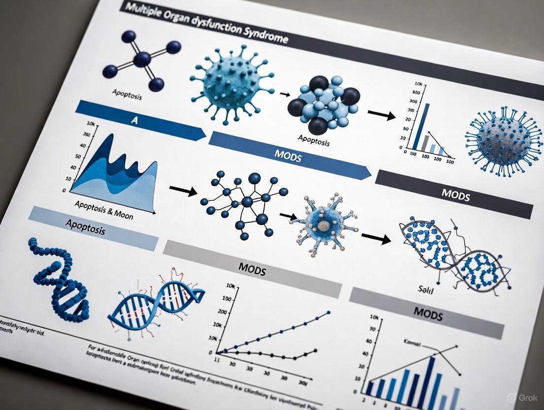
Apoptosis in MODS: Molecular Mechanisms, Biomarker Discovery, and Emerging Therapeutic Strategies
Multiple organ dysfunction syndrome (MODS) remains a leading cause of mortality in critically ill patients, with apoptosis occupying a central role in its pathogenesis.
Apoptosis in MODS: Molecular Mechanisms, Biomarker Discovery, and Emerging Therapeutic Strategies
Abstract
Multiple organ dysfunction syndrome (MODS) remains a leading cause of mortality in critically ill patients, with apoptosis occupying a central role in its pathogenesis. This comprehensive review synthesizes current understanding of stress-induced apoptotic pathways in MODS, from molecular mechanisms to clinical applications. We explore foundational concepts of injury-induced cell death, methodological approaches for identifying apoptosis-related biomarkers, strategies for therapeutic intervention targeting key regulators like BCL2A1 and S100A8/A9, and validation of novel targets through bioinformatics and experimental models. The content is specifically tailored for researchers, scientists, and drug development professionals seeking to advance diagnostic and therapeutic innovations for this devastating condition.
The Molecular Basis of Apoptosis in MODS Pathogenesis
Multiple organ dysfunction syndrome (MODS) is a clinical syndrome triggered by severe infections, trauma, burns, or other acute illnesses, manifesting as dysfunction or failure in two or more organs or systems [1]. The pathogenesis of MODS is intricate, featuring pathological damage that affects multiple organs, systems, levels, and targets. Even with advancements in life-support technologies, MODS continues to be characterized by high incidence rates, high mortality rates, and significant social and economic burdens [1]. When only two organs fail, mortality rates hover around 30%, but when three to four organs are impaired, mortality surges dramatically to between 50% and 70% [1]. Modern medicine has yet to discover fully effective prevention and treatment methods due to the complex and multifactorial nature of MODS [1].
At the cellular level, injurious stimuli trigger adaptive stress responses that include changes in gene expression. MODS represents the summation of these stress responses to severe systemic injury, integrated at the cellular, organ, and host levels [2]. We hypothesize that a complete understanding of the molecular mechanisms of stress responses induced by injury will aid in developing therapeutic strategies for treating MODS in critically ill patients. This review focuses particularly on stress-induced cell death by apoptosis and its central role in the progression from systemic injury to organ dysfunction [2]. Research suggests that the most important MODS-related pathophysiologic conditions known to date affect programmed cell death rates in almost all cell types [3]. Organ-specific cell death involving both parenchymal and microvasculature endothelial cells conceivably underlies organ dysfunction, providing a unifying theory for MODS pathophysiology [3].
Key Molecular Players in Apoptosis and MODS
Core Apoptosis-Related Genes in MODS
Recent bioinformatics analyses have identified specific apoptosis-related genes that play central roles in MODS pathogenesis. Through integrated analysis of MODS-related datasets from public databases including differential expression analysis, weighted gene co-expression network analysis (WGCNA), and machine learning algorithms, researchers have identified three key genes: S100A9, S100A8, and BCL2A1 [1]. These genes were consistently significantly highly expressed in MODS samples compared to controls and were found to jointly participate in the "oxidative phosphorylation" signaling pathway [1]. A nomogram prediction model constructed based on these key genes demonstrated excellent predictive ability for MODS, offering a novel approach for clinical diagnosis and potential targeted therapy [1].
Table 1: Key Apoptosis-Related Genes in MODS Pathogenesis
| Gene | Expression in MODS | Primary Function | Associated Pathway | Potential Therapeutic Target |
|---|---|---|---|---|
| S100A9 | Significantly upregulated | Calcium-binding protein, regulates inflammatory response | Oxidative phosphorylation | Curcumin |
| S100A8 | Significantly upregulated | Forms calprotectin with S100A9, innate immunity | Oxidative phosphorylation | Curcumin |
| BCL2A1 | Significantly upregulated | Anti-apoptotic BCL-2 family member, cell survival | Apoptosis inhibition | Small molecule inhibitors |
Apoptosis Signaling Kinases and Regulatory Mechanisms
A large body of evidence has revealed that numerous protein kinases serve as crucial regulators of apoptosis and cellular sensitivity to various proapoptotic signals [4]. These apoptosis signaling kinases generally act as crucial regulators of diverse cellular responses to a wide variety of stressors, beyond their specific roles in apoptosis regulation [4]. The dysregulation of these kinases has significant implications for health outcomes, particularly in severe stress conditions like MODS.
The mitochondrial pathway of apoptosis plays a particularly important role in MODS. Research has shown that chronic stress-induced apoptosis can be mitigated by young mitochondria transplantation, associated with down-regulation of Bax and Caspase-3 and up-regulation of Bcl-2 [5]. This suggests that mitochondrial dysfunction represents a core mechanism in stress-induced cellular suicide and organ dysfunction. In aged rat models subjected to chronic stress, young mitochondria administration reduced neuronal apoptosis in the prefrontal cortex, indicating the potential of mitotherapy for addressing mitochondrial dysfunction-induced apoptosis [5].
Methodological Approaches for Apoptosis Research in MODS
Experimental Workflow for Identification of Apoptosis-Related Genes
The comprehensive identification of key apoptosis-related genes in MODS involves a multi-step bioinformatics and experimental validation pipeline:
Data Acquisition: MODS-related datasets (GSE66099, GSE26440, GSE144406) are collected from Gene Expression Omnibus (GEO). These datasets typically use whole blood samples from MODS patients and controls [1].
Screening of Candidate Genes: Differential expression analysis is performed using the "limma" package (v 3.54.0) with criteria of |log2fold change (FC)| > 1 and adjusted p-value < 0.05. Weighted gene co-expression network analysis (WGCNA) is conducted to identify gene modules most correlated with MODS traits [1].
Functional Enrichment Analysis: Gene Ontology (GO) and Kyoto Encyclopedia of Genes and Genomes (KEGG) analyses are performed using the "clusterProfiler" package (v 4.7.1.003) to identify enriched biological processes, molecular functions, and pathways [1].
Protein-Protein Interaction Network Construction: The Search Tool for the Retrieval of Interacting Genes/Proteins (STRING) database is used to construct interaction networks, followed by hub gene identification using Cytoscape software (v 3.7.1) with cytoHubba plugin [1].
Machine Learning Validation: Multiple machine learning algorithms including least absolute shrinkage and selection operator (LASSO), support vector machine recursive feature elimination (SVM-RFE), and Boruta are applied to screen for key genes most predictive of MODS [1].
Experimental Validation: Final validation involves measuring gene expression in clinical MODS samples using techniques such as quantitative PCR, immunohistochemistry, or Western blotting [1].
Research Reagent Solutions for Apoptosis Studies
Table 2: Essential Research Reagents for Apoptosis and MODS Investigations
| Reagent/Category | Specific Examples | Research Application | Technical Notes |
|---|---|---|---|
| Cell Line Models | ESRE-bla HeLa reporter line | ER stress response monitoring | β-lactamase reporter under GRP78 promoter control [6] |
| Apoptosis Assays | Caspase-3 activity, Bax/Bcl-2 ratio, Cytochrome c release | Apoptosis quantification | Western blot, ELISA, immunohistochemistry [5] |
| Oxidative Stress Markers | Malondialdehyde (MDA) | Lipid peroxidation measurement | Indicator of oxidative damage in MODS [5] |
| Pathway Inhibitors/Activators | Salubrinal, Bortezomib, 17-AAG | ER stress modulation | Tool compounds for mechanistic studies [6] |
| High-Throughput Screening Platforms | qHTS in 1,536-well format | Drug discovery | NPC library screening for ER stress inducers [6] |
| Mitochondrial Function Assays | Mitochondrial transplantation, Membrane potential dyes | Mitochondrial health assessment | Direct mitochondrial transfer studies [5] |
Signaling Pathways and Regulatory Networks in MODS-Associated Apoptosis
Integrated Apoptosis Signaling in MODS Pathogenesis
The pathophysiology of MODS involves a complex interplay of inflammatory mediators, cellular stress pathways, and apoptotic signaling. Sepsis, a common precursor to MODS, has been referred to as a process of malignant intravascular inflammation [7]. Bacterial products such as Lipid A trigger the release of cytokines and other immune modulators that mediate the clinical manifestations of sepsis. Key mediators including interleukins, tumor necrosis factor (TNF)-α, interferon gamma (IFN-γ), and other colony-stimulating factors are produced rapidly within minutes or hours after interactions of monocytes and macrophages with bacterial components [7].
This inflammatory mediator release becomes a self-stimulating process, creating a state of destructive immunologic dissonance. Sepsis is described as an autodestructive process that permits extension of the normal pathophysiologic response to infection to involve otherwise normal tissues, resulting in MODS [7]. Significant microcirculatory dysfunction occurs in sepsis, characterized by a decrease in the number of perfused capillaries and mitochondrial dysfunction associated with reduced mitochondrial transmembrane potential gradients necessary to drive oxidative phosphorylation [7]. The end result is an apparent inability of end-organs to extract oxygen maximally, creating a condition termed microcirculatory and mitochondrial distress syndrome (MMDS) [7].
Organ-Specific Apoptosis Mechanisms in MODS
Different organ systems exhibit varying susceptibility to apoptosis in MODS, with distinct mechanistic pathways operating in each tissue type:
Pulmonary Dysfunction: Endothelial injury in the pulmonary vasculature leads to disturbed capillary blood flow and enhanced microvascular permeability, resulting in interstitial and alveolar edema. Neutrophil entrapment within the pulmonary microcirculation initiates and amplifies the injury to alveolar capillary membranes, leading to acute lung injury and acute respiratory distress syndrome (ARDS) [7].
Gastrointestinal Dysfunction: The GI tract may help propagate the injury of sepsis through bacterial translocation. Overgrowth of bacteria in the upper GI tract may be aspirated into the lungs, producing nosocomial pneumonia. The normal barrier function of the gut may be affected, allowing translocation of bacteria and endotoxins into the systemic circulation [7].
Myocardial Depression: Septic shock and SIRS are characterized by reversible myocardial depression resistant to catecholamine and fluid administration. Circulating "myocardial depressant factor"—probably representing the synergistic effects of TNF-α, IL-1β, other cytokines, and nitric oxide (NO)—is implicated in pathogenesis [7].
Hepatic and Renal Dysfunction: The liver and kidneys are frequent targets in MODS, with apoptosis playing a significant role in their dysfunction. Studies have shown that chronic stress-induced apoptosis affects these organs, particularly in aged subjects, and can be mitigated by mitochondrial transplantation [5].
Therapeutic Implications and Future Directions
Pharmacological Interventions Targeting Apoptosis
Several pharmacological approaches show promise for modulating apoptosis in MODS:
Dexmedetomidine (DEX), a potent selective α2-adrenergic receptor agonist, demonstrates protective effects against sepsis-induced organ injury. DEX mitigates injuries to the brain, lungs, kidneys, liver and immune system by regulating signaling pathways related to inflammation, apoptosis, pyroptosis, autophagy, and ferroptosis in sepsis treatment [8]. The drug exerts its effects through multiple mechanisms, including reduction of proinflammatory cytokine release, inhibition of apoptosis, and modulation of cellular stress responses.
Natural IgM antibodies represent another therapeutic avenue, acting as first-line defense in immune surveillance. These antibodies selectively kill aberrant cells by using different apoptotic stress mechanisms and can be isolated from patients or healthy donors using human hybridoma technology [9]. They represent components of innate immunity that induce stress-mediated apoptosis in malignant or dysregulated cells.
Other potential therapeutic compounds include curcumin, which has been predicted to target the key apoptosis genes S100A9 and S100A8, and mitotherapy approaches involving transplantation of young mitochondria to reverse mitochondrial dysfunction-induced apoptosis [1] [5].
High-Throughput Screening for ER Stress Inducers
Quantitative high-throughput screening (qHTS) platforms have been developed to identify drugs that induce endoplasmic reticulum stress response. The ESRE-bla HeLa reporter cell line, which stably expresses a β-lactamase reporter gene under the control of the ER stress response element (ESRE) present in the GRP78 gene promoter, enables screening of compound libraries for ER stress inducers [6]. This assay has been optimized and miniaturized into a 1,536-well plate format, allowing efficient screening of thousands of compounds.
Screening of the NIH Chemical Genomics Center Pharmaceutical Collection (NPC library) containing approximately 2,800 drugs has identified several known ER stress inducers, such as 17-AAG (via HSP90 inhibition), as well as novel inducers such as AMI-193 and spiperone [6]. These compounds induce ER stress through different mechanisms, including disruption of protein folding, calcium homeostasis, and activation of the unfolded protein response. The confirmed drugs can be further studied for their effects on phosphorylation of eukaryotic initiation factor 2α (eIF2α), X-box-binding protein (XBP1) splicing, and GRP78 gene expression [6].
Table 3: Therapeutic Approaches Targeting Apoptosis in MODS
| Therapeutic Approach | Mechanism of Action | Development Status | Key Findings |
|---|---|---|---|
| Dexmedetomidine (DEX) | α2-adrenergic receptor agonist, inhibits apoptosis and inflammation | Clinical use for sedation, investigational for MODS | Protects against sepsis-induced organ injury; regulates multiple cell death pathways [8] |
| Natural IgM Antibodies | Induce apoptosis in aberrant cells via stress mechanisms | Preclinical research | Isolated using human hybridoma technology; tumor-specific apoptosis induction [9] |
| Mitochondrial Transplantation | Replaces dysfunctional mitochondria, reduces apoptosis | Experimental models | Improves cognitive and motor performance in aged mice; reduces cytochrome c release [5] |
| ER Stress Inducers (e.g., 17-AAG, Bortezomib) | Activate unfolded protein response, induce apoptosis in stressed cells | Some approved for cancer, investigational for MODS | Bortezomib clinically applied for multiple myeloma; induces severe ER stress leading to apoptosis [6] |
| Curcumin | Predicted to target S100A8/S100A9 | Preclinical research | Identified through computational prediction; requires experimental validation [1] |
Stress-induced apoptosis plays a central role in the pathophysiology of multiple organ dysfunction syndrome, serving as a critical mechanism linking systemic injury to cellular suicide and organ failure. The identification of key apoptosis-related genes S100A9, S100A8, and BCL2A1 provides specific molecular targets for diagnostic and therapeutic development. The complex interplay between inflammatory mediators, cellular stress pathways, and apoptotic signaling creates a self-amplifying cycle that drives organ dysfunction.
Future research directions should focus on validating these key apoptosis genes across diverse MODS populations, developing targeted therapies that modulate specific apoptotic pathways, and exploring combination treatments that address both the inflammatory and apoptotic components of MODS. The use of advanced screening platforms, including high-throughput ER stress assays and mitochondrial function assessments, will accelerate the identification of novel therapeutic compounds. Ultimately, a precision medicine approach that targets specific apoptosis pathways based on individual patient profiles holds promise for reducing the unacceptably high mortality rates associated with this devastating clinical syndrome.
Multiple organ dysfunction syndrome (MODS) represents a life-threatening condition frequently precipitated by sepsis and systemic inflammatory response, carrying significant mortality in critical care settings. Emerging research has firmly established that dysregulated apoptosis, or programmed cell death, occupies a central position in MODS pathogenesis. This technical review examines the converging mechanisms of the extrinsic (death receptor) and intrinsic (mitochondrial) apoptotic pathways in MODS, highlighting the critical cross-talk between these systems that amplifies cellular destruction. We synthesize current molecular understanding with identification of key apoptotic genes S100A9, S100A8, and BCL2A1 as significantly upregulated in MODS patients. The comprehensive analysis presented herein aims to provide researchers and drug development professionals with mechanistic insights and methodological frameworks for advancing targeted therapeutic strategies against MODS.
Multiple organ dysfunction syndrome represents a final common pathway for numerous critical illnesses, with sepsis standing as its most prevalent trigger. The syndrome is characterized by progressive, potentially reversible physiological dysfunction across multiple organ systems requiring pharmacological support to maintain homeostasis. Within the complex pathophysiology of MODS, apoptosis has emerged as a critical mechanism contributing to organ failure [10]. Under normal physiological conditions, apoptosis functions as a regulated process for eliminating damaged or unnecessary cells without inciting inflammatory responses. However, in MODS, this precise regulation becomes disrupted, leading to excessive and pathological cell loss in vital tissues [11].
The significance of apoptotic processes in MODS is demonstrated by several key observations: circulating markers of apoptosis are elevated in MODS patients; experimental models demonstrate correlation between apoptosis inhibition and improved outcomes; and specific apoptotic gene expression patterns are altered in critical illness [12] [10]. Particularly noteworthy is the discovery that nucleosomes—fragments of chromatin released during apoptotic DNA fragmentation—serve as measurable markers of apoptosis in sepsis and MODS, providing a window into the magnitude of this process in clinical settings [10]. The following sections delineate the molecular machinery of apoptosis and its dysregulation in MODS.
Molecular Mechanisms of Apoptotic Pathways
The Death Receptor Pathway
The extrinsic apoptotic pathway initiates when extracellular death ligands bind to transmembrane death receptors belonging to the tumor necrosis factor (TNF) receptor superfamily. Key death receptors include Fas (CD95/Apo-1), TNF receptors, and TRAIL receptors [13]. Following ligand binding, these receptors aggregate at the cell surface, typically forming trimers that recruit intracellular adaptor molecules via their death domains (DD) [14] [13].
The fundamental signaling complex formed upon receptor activation is the death-inducing signaling complex (DISC). This multi-protein complex assembly involves the recruitment of the adaptor protein FADD (Fas-associated death domain), which in turn recruits procaspase-8 through interactions between death effector domains (DED) [13]. Within the DISC, caspase-8 undergoes proximity-induced activation and subsequent autoproteolytic processing [13]. The activated caspase-8 then directly cleaves and activates executioner caspases, particularly caspase-3, initiating the proteolytic cascade that dismantles cellular structures and executes cell death [14] [13].
Table 1: Major Death Receptors and Their Ligands in Apoptotic Signaling
| Death Receptor | Ligand | Primary Adaptor Protein | Cellular Context |
|---|---|---|---|
| Fas (CD95/Apo-1) | FasL | FADD | Immune regulation, lymphocyte deletion |
| TNFR1 | TNF-α | TRADD/FADD | Inflammation, infection response |
| TRAIL-R1/DR4 | TRAIL | FADD | Immune surveillance, tumor cell killing |
| TRAIL-R2/DR5 | TRAIL | FADD | Immune surveillance, tumor cell killing |
In certain cell types classified as Type I cells, the death receptor pathway activates executioner caspases sufficiently to induce apoptosis without mitochondrial involvement. This direct activation pathway is particularly prominent in thymocytes and certain lymphocytes [13].
The Mitochondrial Pathway
The intrinsic apoptotic pathway centers on mitochondrial events and serves as a primary sensor of cellular stress. This pathway activates in response to diverse intracellular insults including DNA damage, oxidative stress, growth factor withdrawal, and endoplasmic reticulum stress [11] [15]. The B-cell lymphoma 2 (BCL-2) protein family constitutes the crucial regulatory network controlling mitochondrial pathway activation [11] [15].
BCL-2 family proteins segregate into three functional groups: (1) Anti-apoptotic members (e.g., BCL-2, BCL-xL, MCL-1) that preserve mitochondrial integrity; (2) Pro-apoptotic effector proteins (BAX, BAK, BOK) that directly mediate mitochondrial outer membrane permeabilization (MOMP); and (3) BH3-only proteins (BID, BIM, PUMA, NOXA, BAD) that function as upstream sensors of cellular damage and stress [11] [15]. In healthy cells, BAX resides predominantly in the cytosol while BAK integrates into the mitochondrial membrane. Upon apoptotic activation, both proteins undergo conformational changes, oligomerize, and form pores in the mitochondrial outer membrane [15].
The pivotal event in mitochondrial apoptosis is MOMP, which permits the release of numerous proteins from the mitochondrial intermembrane space into the cytosol [16]. Among these proteins, cytochrome c stands as particularly significant for caspase activation. Once released into the cytosol, cytochrome c binds to APAF-1 (apoptotic protease activating factor-1), promoting ATP/dATP-dependent oligomerization of APAF-1 into the wheel-like apoptosome complex [15] [16]. The apoptosome recruits and activates procaspase-9, which then initiates the caspase cascade by activating executioner caspases-3 and -7 [16].
Table 2: Key Mitochondrial Intermembrane Space Proteins Released During Apoptosis
| Protein | Function in Apoptosis | Mechanism of Action |
|---|---|---|
| Cytochrome c | Caspase activation | Binds APAF-1 to form apoptosome |
| Smac/DIABLO | IAP antagonism | Binds and neutralizes XIAP, cIAP1, cIAP2 |
| Omi/HtrA2 | IAP antagonism & proteolysis | Inhibits IAPs; serine protease activity |
| AIF | Caspase-independent death | Chromatin condensation, DNA fragmentation |
| Endonuclease G | DNA fragmentation | Cleaves nuclear DNA |
Following MOMP, additional mitochondrial proteins including Smac (second mitochondria-derived activator of caspases) and Omi enhance caspase activation by neutralizing inhibitor of apoptosis proteins (IAPs), particularly XIAP, which constitutively suppresses caspase activity in healthy cells [16].
Pathway Convergence and Cross-Talk
While the death receptor and mitochondrial pathways represent distinct initiation mechanisms, significant cross-talk exists between them, particularly in cell types designated as Type II cells [13]. In these cells, which include hepatocytes and many epithelial cells, death receptor signaling alone proves insufficient for complete apoptosis activation, requiring mitochondrial amplification to execute cell death [13].
The molecular nexus of this pathway convergence is the BH3-only protein BID. Upon activation by death receptors, caspase-8 cleaves full-length BID into truncated tBID, which subsequently translocates to mitochondria [15]. At the mitochondrial membrane, tBID activates BAX and BAK, either directly or indirectly through inhibition of anti-apoptotic BCL-2 proteins [15]. This engagement of the mitochondrial pathway results in enhanced MOMP and more robust caspase activation through simultaneous cytochrome c/Apaf-1-mediated caspase-9 activation and Smac-mediated IAP inhibition [13] [15].
The following diagram illustrates the convergence between death receptor and mitochondrial pathways:
Apoptotic Dysregulation in MODS
Clinical Evidence of Apoptosis in MODS
Substantial clinical evidence supports the role of apoptotic dysregulation in MODS pathogenesis. Studies of circulating nucleosomes—fragments of chromatin released during apoptotic DNA fragmentation—have demonstrated their utility as markers of apoptosis in sepsis and MODS patients [10]. Additionally, investigations of leukocyte apoptosis in MODS patients have revealed profound suppression of neutrophil apoptosis, which contributes to persistent inflammation through extended release of toxic metabolites [17].
Key apoptosis-related genes demonstrate altered expression patterns in MODS. Recent research identified S100A9, S100A8, and BCL2A1 as significantly upregulated in MODS patients, with all three genes participating in oxidative phosphorylation signaling pathways [12]. A nomogram prediction model constructed based on these key genes demonstrated excellent predictive capability for MODS, highlighting their potential clinical utility [12].
Organ-Specific Apoptotic Responses
The consequences of apoptotic dysregulation in MODS manifest differently across organ systems:
- Lymphoid tissues: Extensive apoptosis occurs in lymphocytes and dendritic cells, potentially contributing to the immunosuppressive phase of MODS [10].
- Pulmonary system: Delayed neutrophil apoptosis in the lungs perpetuates inflammatory responses and tissue damage [17].
- Hepatic system: Hepatocyte apoptosis represents a hallmark of inflammatory shock models, preceding overt liver failure [10].
- Vascular endothelium: Endothelial apoptosis disrupts vascular integrity, promoting increased permeability and microvascular thrombosis [10].
The following table summarizes quantitative findings from MODS apoptosis research:
Table 3: Apoptosis-Related Findings in MODS Clinical and Experimental Studies
| Parameter Measured | Finding in MODS | Study Type | Significance |
|---|---|---|---|
| Circulating nucleosomes | Elevated | Clinical study | Marker of apoptosis in sepsis [10] |
| Neutrophil apoptosis | Delayed/suppressed | Clinical study | Persistence of inflammation [17] |
| S100A9 expression | Significantly increased | Gene expression analysis | Key apoptosis-related gene [12] |
| S100A8 expression | Significantly increased | Gene expression analysis | Key apoptosis-related gene [12] |
| BCL2A1 expression | Significantly increased | Gene expression analysis | Key apoptosis-related gene [12] |
| Lymphocyte apoptosis | Markedly increased | Sepsis study | Contributes to immunosuppression [10] |
Experimental Methodologies for MODS Apoptosis Research
Gene Expression Analysis
Comprehensive analysis of apoptosis-related genes in MODS involves a multi-step methodological approach:
- Dataset Acquisition: Obtain MODS-related datasets from public genomic repositories (e.g., GEO database) including both MODS and control samples.
- Differential Expression Analysis: Identify disparately expressed genes between MODS and control groups using appropriate statistical thresholds (e.g., fold change >2, adjusted p-value <0.05).
- Weighted Gene Co-Expression Network Analysis (WGCNA): Construct co-expression networks to identify gene modules most significantly associated with MODS status.
- Intersection with Apoptosis Genes: Cross-reference MODS-associated genes with established apoptosis-related genes (ARGs) from databases such as GeneOntology or Reactome.
- Machine Learning Integration: Apply machine learning algorithms (e.g., random forest, SVM) to identify optimal predictive gene signatures.
- Clinical Validation: Verify key gene expression changes in clinical samples using RT-qPCR or other validation methodologies [12].
Assessment of Mitochondrial Apoptosis
Experimental evaluation of mitochondrial pathway activation requires multi-parameter assessment:
- Cytochrome c Release: Employ subcellular fractionation followed by Western blotting to detect cytochrome c translocation from mitochondria to cytosol. Alternatively, utilize immunofluorescence microscopy with mitochondrial and cytochrome c co-staining.
- BAX/BAK Activation: Detect conformational changes in BAX/BAK using immunoprecipitation with conformation-specific antibodies. Monitor mitochondrial translocation of BAX via cell fractionation or confocal microscopy of GFP-BAX fusion proteins.
- Caspase Activation: Measure caspase-9 and caspase-3 activity using fluorogenic substrate assays (e.g., LEHD-AFC for caspase-9, DEVD-AFC for caspase-3). Confirm via Western blotting for characteristic cleavage fragments.
- MOMP Assessment: Quantify mitochondrial membrane permeability using potentiometric dyes (e.g., JC-1, TMRE) that detect loss of mitochondrial membrane potential (ΔΨm).
- Apoptosome Formation: Analyze APAF-1 oligomerization and caspase-9 recruitment via gel filtration chromatography or native gel electrophoresis [15] [16].
Death Receptor Pathway Analysis
Methodologies for evaluating death receptor signaling include:
- DISC Immunoprecipitation: Isolate the death-inducing signaling complex using immunoprecipitation with anti-receptor or anti-FADD antibodies followed by Western blotting for associated proteins (caspase-8, FADD, receptor).
- Receptor Activation Assays: Quantify receptor activation using antibodies specific for activated conformations or measure ligand binding via flow cytometry with fluorescently-tagged ligands.
- Membrane and Cytosolic Fractionation: Separate membrane and cytosolic fractions to evaluate BID translocation and processing via Western blotting.
- Caspase-8 Activity Assays: Utilize fluorometric substrates (IETD-AFC) or Western blotting for cleavage fragments to quantify caspase-8 activation.
Research Reagent Solutions
Table 4: Essential Research Reagents for MODS Apoptosis Investigation
| Reagent Category | Specific Examples | Research Application | Technical Notes |
|---|---|---|---|
| Death Receptor Ligands | Recombinant FasL, TRAIL, TNF-α | Activation of extrinsic pathway | Use with cross-linking enhancers for optimal activity |
| BCL-2 Family Inhibitors | ABT-737 (BCL-2/BCL-xL inhibitor), ABT-199 (venetoclax) | Targeting anti-apoptotic BCL-2 proteins | Assess platelet toxicity for BCL-xL inhibitors |
| Caspase Inhibitors | Z-VAD-FMK (pan-caspase), Z-IETD-FMK (caspase-8) | Determining caspase-dependence | Use appropriate controls for inhibitor specificity |
| Mitochondrial Dyes | JC-1, TMRE, MitoTracker | Assessing mitochondrial membrane potential | Validate with CCCP depolarization controls |
| Antibodies for WB/IHC | Anti-cytochrome c, anti-cleaved caspase-3, anti-BAX | Protein localization and activation | Compare multiple antibodies for key targets |
| Activity Assays | Caspase fluorogenic substrates, Annexin V apoptosis detection | Quantifying apoptosis and caspase activity | Establish kinetic measurement parameters |
| Gene Expression Tools | RT-qPCR primers for S100A8/S100A9/BCL2A1 | Validating MODS-associated genes | Normalize to multiple reference genes |
Therapeutic Implications and Future Directions
The convergence of death receptor and mitochondrial pathways in MODS presents both challenges and opportunities for therapeutic intervention. Several strategic approaches have emerged from mechanistic studies:
Direct Caspase Inhibition: Broad-spectrum caspase inhibitors have demonstrated efficacy in experimental sepsis models, particularly in reducing lymphocyte apoptosis and improving survival [10]. However, clinical translation requires careful consideration of timing and cell-specific effects.
Death Receptor Modulation: Targeting specific death receptors, particularly with TRAIL receptor agonists or Fas pathway inhibitors, offers potential for selective apoptosis modulation in specific cell populations.
BCL-2 Family Targeting: The identification of BCL2A1 as a key upregulated gene in MODS suggests potential for BH3 mimetics that selectively target specific anti-apoptotic BCL-2 family members [12].
IAP Antagonists: SMAC mimetics that neutralize XIAP and other inhibitor of apoptosis proteins may lower the threshold for apoptosis execution and sensitize cells to death signals [16].
Gene Expression-Based Stratification: Utilization of key apoptotic genes like S100A9, S100A8, and BCL2A1 as biomarkers for patient stratification and treatment selection represents a promising precision medicine approach [12].
Future research directions should focus on temporal aspects of apoptotic activation in MODS, cell-type specific responses, and the interplay between apoptosis and other cell death modalities such as necroptosis and pyroptosis. Additionally, the development of more sophisticated animal models that recapitulate human MODS pathophysiology will be essential for preclinical validation of apoptosis-targeted therapies.
The convergence of death receptor and mitochondrial apoptotic pathways represents a fundamental mechanism in MODS pathogenesis. The intricate cross-talk between these systems, particularly through BID-mediated amplification, creates a self-reinforcing cycle of cellular destruction that drives multi-organ failure. Recent identification of key apoptosis-related genes S100A9, S100A8, and BCL2A1 in MODS patients provides both mechanistic insights and potential diagnostic biomarkers. The experimental methodologies outlined in this review offer standardized approaches for investigating these pathways in MODS contexts. Moving forward, therapeutic strategies that selectively modulate specific components of these convergent pathways hold significant promise for improving outcomes in this devastating syndrome.
Apoptosis, a form of programmed cell death, plays a complex and dual role in critical illness. While essential for maintaining cellular homeostasis and eliminating damaged cells, dysregulated apoptosis is increasingly recognized as a key contributor to the pathogenesis of Multiple Organ Dysfunction Syndrome (MODS). This review examines the mechanisms by which apoptosis transitions from a protective process to a pathological driver in critically ill patients. We explore the intricate signaling pathways involved, highlight potential biomarkers for clinical monitoring, and discuss therapeutic strategies targeting apoptotic regulation. Within the context of MODS research, understanding this delicate balance is paramount for developing novel interventions aimed at mitigating organ failure and improving patient outcomes in intensive care settings.
In critical illness, the body's response to severe insults like sepsis, trauma, or massive hemorrhage is characterized by a complex interplay of inflammatory and cellular processes. Apoptosis, a highly regulated form of programmed cell death, sits at the crossroads of these pathways [18]. Under physiological conditions, apoptosis is crucial for development, tissue homeostasis, and the immune response, eliminating unwanted or damaged cells without inducing inflammation [18] [19]. This self-destructive process is characterized by distinct cellular changes including membrane blebbing, chromatin condensation, DNA fragmentation, and the formation of apoptotic bodies that are efficiently phagocytosed by neighboring cells [18].
However, in the hypermetabolic, inflammatory environment of critical illness, this normally protective process can become dysregulated. Excessive apoptosis can lead to the loss of parenchymal cells in vital organs, while insufficient apoptosis can perpetuate inflammation by allowing damaged cells to persist [20] [19]. This dysregulation is now considered a hallmark in the progression toward Multiple Organ Dysfunction Syndrome (MODS), a clinical syndrome characterized by progressive and potentially reversible physiologic dysfunction in two or more organs induced by various acute insults [21] [7]. The maladaptive role of apoptosis in MODS provides a unifying theory for organ dysfunction, where organ-specific cell death involving both parenchymal and microvasculature endothelial cells underlies clinical organ failure [3].
Apoptosis is mediated through two primary signaling pathways: the extrinsic (death receptor) pathway and the intrinsic (mitochondrial) pathway. Both converge on the activation of caspases, proteolytic enzymes that systematically dismantle the cell in an orderly manner [18] [19].
The Extrinsic Pathway
The extrinsic pathway is triggered by extracellular signals that bind to specific trans-membrane receptors belonging to the TNF/NGF family, collectively known as death receptors (DR) [19]. All death receptors function similarly: upon ligand binding, receptor molecules oligomerize and undergo conformational changes allowing assembly of a multi-protein complex known as the Death Initiation Signalling Complex (DISC). In the FAS/CD95 signaling complex, FAS recruits an adaptor molecule, Fas-associated protein with a death domain (FADD), through a highly conserved 80 amino acid death domain (DD). FADD then binds caspase-8 through homologous death effector domains (DED), leading to its activation [19]. Active caspase-8 then activates downstream effector caspases such as caspase-3, executing the cell death program [19].
The Intrinsic Pathway
The intrinsic pathway is activated in response to various cellular stresses including DNA damage, oxidative stress, and ischemia/reperfusion injury [19]. These stresses converge on the mitochondria, determining mitochondrial outer membrane permeabilization (MOMP), which results in dissipation of the mitochondrial membrane potential and release of proteins that promote caspase activation [19]. The Bcl-2 family proteins are essential regulators of this pathway, classified into anti-apoptotic members (Bcl-2, Bcl-xL, Bcl-w, Mcl-1) and pro-apoptotic members (Bax, Bak, Bid, Bim, Noxa, Puma) [20] [19]. Following MOMP, cytochrome c is released and binds to APAF-1, inducing formation of the apoptosome complex that recruits and activates caspase-9, which then activates downstream effector caspases [19].
The two pathways are interconnected; in some cells, activation of caspase-8 results in cleavage of the BH3-only protein Bid, generating truncated Bid (tBid) that permeabilizes mitochondria, thereby amplifying the death signal through the intrinsic pathway [19].
Table 1: Key Components of Apoptotic Signaling Pathways
| Pathway Component | Type | Function in Apoptosis |
|---|---|---|
| Death Receptors | Trans-membrane receptors | Initiate extrinsic apoptosis upon ligand binding |
| Caspase-8 | Initiator caspase | Key protease in extrinsic pathway activation |
| Caspase-9 | Initiator caspase | Key protease in intrinsic pathway activation |
| Caspase-3 | Effector caspase | Executes cell dismantling in both pathways |
| Bcl-2 | Anti-apoptotic protein | Regulates MOMP by inhibiting pro-apoptotic members |
| Bax/Bak | Pro-apoptotic proteins | Mediate mitochondrial outer membrane permeabilization |
| Cytochrome c | Mitochondrial protein | Activates apoptosome formation after release |
| APAF-1 | Adaptor protein | Forms apoptosome complex with cytochrome c |
Figure 1: Core Apoptotic Signaling Pathways. The extrinsic (yellow) and intrinsic (red) pathways converge on the execution phase (green), with caspase-8-mediated tBid formation providing cross-talk between pathways.
The Pathological Transition: From Protective Apoptosis to MODS Driver
In critical illness, the shift of apoptosis from a protective mechanism to a pathological driver involves complex interactions between systemic inflammation, immune dysregulation, and direct cellular injury. The transition often begins with the systemic inflammatory response syndrome (SIRS), characterized by overwhelming immune responses that lead to free radical generation and cellular hypoperfusion causing hypoxia [21]. These conditions collectively result in profound intracellular oxidative stress and mitochondrial damage, creating an environment where apoptotic regulation is compromised [21].
Sepsis, a primary cause of MODS, creates a state of "malignant intravascular inflammation" where microbial products trigger excessive release of cytokines including tumor necrosis factor (TNF)-α, interleukin (IL)-1, IL-6, and other mediators [7]. These inflammatory mediators not only cause direct tissue injury but also modulate apoptotic thresholds in various cell types. For instance, TNF-α can directly activate death receptors, while reactive oxygen species associated with ischemia/reperfusion injury can trigger the intrinsic pathway [3]. This results in a vicious cycle where inflammation promotes apoptosis, and apoptotic cells may further stimulate inflammation despite apoptosis typically being non-inflammatory [7].
The gastrointestinal tract plays a particularly important role in this transition. In critical illness, the normal barrier function of the gut may be compromised, allowing translocation of bacteria and endotoxins into the systemic circulation [7]. This not only fuels systemic inflammation but also exposes distant organs to apoptotic stimuli. Furthermore, emerging evidence suggests that endoplasmic reticulum (ER) stress and the unfolded protein response (UPR) contribute to parenchymal cell apoptosis in organs such as the liver and heart following trauma/hemorrhagic shock [22].
Table 2: Apoptotic Triggers in Critical Illness and Their Mechanisms
| Trigger | Clinical Context | Primary Apoptotic Pathway | Target Cells/Tissues |
|---|---|---|---|
| Pro-inflammatory Cytokines | Sepsis, SIRS | Extrinsic (Death Receptors) | Immune cells, endothelial cells |
| Reactive Oxygen Species | Ischemia/Reperfusion, Hypoperfusion | Intrinsic (Mitochondrial) | Parenchymal cells of various organs |
| Bacterial Products/Endotoxin | Severe Infection | Both Extrinsic and Intrinsic | Hepatocytes, endothelial cells |
| Endoplasmic Reticulum Stress | Trauma/Hemorrhagic Shock | UPR-mediated Intrinsic | Hepatocytes, cardiomyocytes |
| Glucocorticoids | Stress Response | Intrinsic (Mitochondrial) | Lymphocytes |
Organ-Specific Apoptotic Mechanisms in MODS
The manifestation of MODS varies significantly across organ systems, reflecting differences in cellular susceptibility, microenvironment, and regenerative capacity. Understanding these organ-specific mechanisms is crucial for developing targeted therapies.
Hepatic Dysfunction
The liver is a central organ in MODS pathogenesis, with hepatocyte apoptosis being a key feature in sepsis and trauma/hemorrhagic shock (T/HS) [22]. Studies in rodent T/HS models have demonstrated significant hepatocyte apoptosis, marked by increased histone-associated DNA fragments (nucleosomes) and TUNEL-positive staining [22]. Global transcriptome analysis has revealed that T/HS significantly alters the unfolded protein response (UPR) in the liver, with chaperones Heat Shock Protein 70 (25.6-fold) and Heat Shock Protein 40 (5.9-fold) being among the most dysregulated genes [22]. Intervention with IL-6 at resuscitation has been shown to prevent hepatocyte apoptosis through a Stat3-dependent mechanism, further augmenting Hsp70 and Hsp40 expression, suggesting these chaperones contribute to an adaptive, protective response [22].
Cardiac Dysfunction
Cardiomyocyte apoptosis is a significant contributor to sepsis-induced myocardial depression in MODS [22]. Similar to hepatic response, T/HS induces cardiomyocyte apoptosis that can be prevented by IL-6 administration, mediated in part by Stat3 [22]. The heart also demonstrates organ-specific UPR transcriptome changes following T/HS, though distinct from the liver profile, indicating tissue-specific regulation of apoptotic pathways [22]. Circulating "myocardial depressant factors"—likely synergistic effects of TNF-α, IL-1β, other cytokines, and nitric oxide—are implicated in pathogenesis, characterized by impaired adrenergic responsiveness and diastolic dysfunction [7].
Pulmonary Dysfunction
The lungs are frequently the first organ to fail in MODS, with acute lung injury and acute respiratory distress syndrome (ARDS) being common manifestations [7]. Endothelial injury in the pulmonary vasculature leads to disturbed capillary blood flow and enhanced microvascular permeability, resulting in interstitial and alveolar edema [7]. Neutrophil entrapment within the pulmonary microcirculation initiates and amplifies injury to alveolar capillary membranes through the release of proteases and reactive oxygen species, creating an environment that promotes endothelial and epithelial apoptosis [7].
Neurological Dysfunction
In acute brain injuries such as stroke and traumatic brain injury, caspase-3-mediated apoptosis contributes significantly to neuronal and neurovascular damage [23]. Specific biomarkers of this process have been identified in cerebrospinal fluid and peripheral blood, including caspase-3 itself and its cleavage products such as caspase-cleaved cytokeratin-18, caspase-cleaved tau, and a caspase-specific 120 kDa αII-spectrin breakdown product [23]. These biomarkers not only indicate apoptosis but may also help identify patients at risk for developing chronic neurodegenerative diseases following acute brain injuries [23].
Research Methodologies and Experimental Protocols
Detection and Quantification of Apoptosis
Accurate measurement of apoptosis is fundamental to research in this field. The ideal biomarker should be specific, accurately quantifiable in clinical samples with sufficient dynamic range, provide rapid and reliable measurement, and be measurable in readily obtainable clinical samples [24].
Nucleosome Detection Protocol: The nucleosome quantification assay measures histone-associated DNA fragments. Following the referenced methodology [22]:
- Sample Preparation: Tissue homogenates or serum samples are prepared using lysis buffer to release nuclear material.
- Nucleosome Capture: Anti-histone antibodies are immobilized on microplate wells to capture nucleosomes.
- Detection: Captured nucleosomes are detected using anti-DNA antibodies conjugated to a reporter enzyme (e.g., horseradish peroxidase).
- Quantification: Enzyme activity is measured colorimetrically after substrate addition, with absorbance directly proportional to nucleosome concentration.
TUNEL Staining Protocol: Terminal deoxynucleotidyl transferase dUTP nick end labeling (TUNEL) identifies apoptotic cells in tissue sections by labeling the 3'-hydroxyl termini of DNA fragments.
- Tissue Fixation: Paraffin-embedded tissue sections are deparaffinized and rehydrated.
- Permeabilization: Proteinase K treatment (20 μg/mL for 15 minutes) permeabilizes tissue and exposes DNA.
- Enzyme Incubation: Sections are incubated with terminal deoxynucleotidyl transferase (TdT) and fluorescently-labeled dUTP for 60 minutes at 37°C.
- Visualization: After stopping the reaction, sections are counterstained and analyzed by fluorescence microscopy.
Caspase-Cleaved Cytokeratin-18 (M30 Apoptosense) Protocol: This ELISA specifically detects a caspase-cleaved neo-epitope on CK18, an epithelial cell-specific protein [23] [24].
- Sample Collection: Serum or plasma samples are collected and stored at -80°C.
- Assay Procedure: Samples are added to microplate wells pre-coated with capture antibody specific for the M30 epitope.
- Detection: After washing, a detector antibody is added, followed by enzyme-conjugated secondary antibody.
- Quantification: Colorimetric development is measured, with intensity proportional to caspase-cleaved CK18.
Table 3: Biomarkers of Apoptosis in Critical Illness Research
| Biomarker | Detection Method | Biological Significance | Advantages | Limitations |
|---|---|---|---|---|
| Nucleosomes | ELISA | Indicator of DNA fragmentation in late apoptosis | Detectable in serum, quantitative | Not specific to apoptosis (also in necrosis) |
| Caspase-3 | Western blot, IHC, ELISA | Key executioner caspase in both pathways | Direct measure of apoptotic activation | Short half-life, rapid clearance |
| Caspase-cleaved CK18 (M30) | ELISA (M30 Apoptosense) | Epithelial-specific apoptosis marker | Specific for caspase-dependent apoptosis | Limited to epithelial-derived cells |
| Cytokeratin Fragments (M65) | ELISA | Total cell death (apoptosis + necrosis) | When combined with M30, distinguishes death mechanisms | Does not differentiate apoptosis from necrosis |
| Cytochrome c | Western blot, ELISA, IHC | Indicator of mitochondrial pathway activation | Early marker of intrinsic apoptosis | Requires cell fractionation for cellular localization |
Experimental Models of Apoptosis in Critical Illness
Rodent Trauma/Hemorrhagic Shock (T/HS) Model: This well-established model recapitulates the apoptotic responses seen in human critical illness [22].
- Hemorrhagic Shock Induction: Animals are bled to a predetermined mean arterial pressure (typically 30-35 mmHg) and maintained in shock for a specified period (e.g., 90 minutes).
- Resuscitation: Shed blood or crystalloid/colloid solutions are reinfused, simulating clinical resuscitation.
- Therapeutic Interventions: Test compounds (e.g., IL-6) are administered during resuscitation to assess protective effects.
- Tissue Analysis: Organs are harvested at predetermined endpoints for molecular, histological, and biochemical analysis of apoptosis.
Endotoxemia Model: Administration of bacterial lipopolysaccharide (LPS) to animals induces a systemic inflammatory response mimicking sepsis.
- LPS Administration: LPS from E. coli or other gram-negative bacteria is administered intravenously or intraperitoneally.
- Time Course Analysis: Animals are sacrificed at various time points to characterize the evolution of apoptotic responses in different organs.
- Pharmacological Modulation: Caspase inhibitors, anti-cytokine therapies, or other modulators can be tested for efficacy in preventing apoptosis and organ dysfunction.
Figure 2: Experimental Workflow for Studying Apoptosis in Trauma/Hemorrhagic Shock. The established model progresses from shock induction through resuscitation to comprehensive apoptosis analysis using multiple complementary techniques.
The Scientist's Toolkit: Research Reagent Solutions
Table 4: Essential Research Reagents for Apoptosis Studies in MODS
| Reagent/Category | Specific Examples | Research Application | Technical Notes |
|---|---|---|---|
| Caspase Inhibitors | Z-VAD-FMK (pan-caspase), Z-DEVD-FMK (caspase-3) | Pathway inhibition studies, therapeutic potential | Cell-permeable, irreversible inhibitors; use appropriate controls for specificity |
| Cytokine Modulators | Recombinant IL-6, Anti-TNF-α antibodies | Mechanistic studies of cytokine-mediated apoptosis | IL-6 shows Stat3-dependent protection in T/HS models [22] |
| ELISA Kits | M30 Apoptosense, Nucleosome ELISA, M65 ELISA | Quantification of apoptotic biomarkers in fluids | M30/M65 combination distinguishes apoptosis from necrosis [24] |
| Antibodies for IHC/Western | Anti-cleaved caspase-3, Anti-Bcl-2 family, Anti-cytochrome c | Tissue localization and protein expression | Subcellular fractionation required for cytochrome c localization |
| Apoptosis Inducers | Staurosporine, Actinomycin D, TNF-α + Cycloheximide | Positive controls for assay validation | Concentration and time course must be optimized for each cell type |
| Stat3 Inhibitors | GQ oligonucleotide (T40214) | Mechanism of action studies | Confirms Stat3-dependency of IL-6 protection in T/HS [22] |
| UPR Modulators | Tunicamycin, Thapsigargin | ER stress-induced apoptosis studies | Induce ER stress through distinct mechanisms (glycosylation inhibition, Ca2+ disruption) |
Therapeutic Implications and Future Directions
The recognition of apoptosis as a key mechanism in MODS pathogenesis has opened promising avenues for therapeutic intervention. Strategies generally aim to either inhibit excessive apoptosis in parenchymal cells or enhance apoptosis in hyperinflammatory immune cells.
Caspase Inhibition: Broad-spectrum caspase inhibitors like Z-VAD-FMK have shown efficacy in preclinical models of sepsis and ischemia/reperfusion injury [19]. However, clinical translation faces challenges due to the dual role of caspases in both pathological and physiological apoptosis. More targeted approaches aiming at specific caspases or upstream regulators may offer better therapeutic windows.
Cytokine-Targeted Therapies: As demonstrated in T/HS models, IL-6 administration at resuscitation prevents hepatocyte and cardiomyocyte apoptosis through a Stat3-dependent mechanism [22]. This highlights the potential of cytokine modulation, though timing and patient selection are critical given the complex, phase-dependent nature of cytokine responses in critical illness.
BH3 Mimetics: These small molecules inhibit anti-apoptotic Bcl-2 family proteins, potentially countering the pathological anti-apoptotic state in certain immune cells that contributes to immunosuppression in later stages of sepsis [20] [19]. Drugs like ABT-263 (navitoclax) and ABT-199 (venetoclax) are primarily developed for cancer but may find application in modulating immune cell apoptosis in persistent inflammation-immunosuppression and catabolism syndrome (PICS) [25].
Hsp70 Modulation: The dramatic upregulation of Hsp70 in protective responses to T/HS suggests this chaperone as a potential therapeutic target [22]. Strategies to enhance Hsp70 expression or function might provide organ protection without globally disrupting apoptotic pathways.
The future of apoptosis-targeted therapies in MODS will likely involve personalized approaches based on biomarker profiles to identify patients in specific phases of the apoptotic dysregulation continuum. Combination therapies targeting both inflammatory and apoptotic pathways simultaneously may yield better outcomes than single-target approaches.
Multiple Organ Dysfunction Syndrome (MODS) is a critical clinical condition characterized by the progressive failure of two or more organ systems following severe insults such as sepsis, trauma, or burns. With mortality rates surging from approximately 30% with two organ failures to 50-70% with three to four organ impairments, MODS represents a significant challenge in intensive care medicine [1]. Apoptosis, or programmed cell death, occupies a central position in MODS pathogenesis, acting as a "double-edged sword" [1]. While appropriate apoptosis helps clear damaged cells and pathogens in early disease stages, dysregulated apoptotic pathways can lead to excessive cell death in vital organs or delayed immune cell apoptosis, resulting in persistent inflammation and tissue damage [1]. This technical review examines the core molecular regulators of apoptosis—BCL-2 family proteins, caspases, and Inhibitor of Apoptosis Proteins (IAPs)—within the context of MODS research, providing experimental methodologies and pathway visualizations to facilitate therapeutic development.
BCL-2 Protein Family: Mitochondrial Apoptosis Regulators
The BCL-2 family represents crucial arbiters of the intrinsic (mitochondrial) apoptotic pathway, comprising three functional classes: multidomain anti-apoptotic proteins (e.g., Bcl-2, Bcl-XL, Mcl-1, A1), multidomain pro-apoptotic executioners (Bax, Bak), and BH3-only pro-apoptotic proteins (Bid, Bim, Bad, Noxa, Puma) [26]. These proteins control a critical step in commitment to apoptosis by regulating permeabilization of the mitochondrial outer membrane (MOM), which leads to cytochrome c release and caspase activation [26].
Molecular Mechanisms and Regulatory Models
Several models explain BCL-2 family interactions. The Direct Activation Model posits that activator BH3 proteins (Bim, tBid, Puma) directly bind and activate Bax/Bak, while sensitizer BH3 proteins (Bad, Noxa) neutralize anti-apoptotic members [26]. The Displacement Model suggests Bax and Bak are constitutively active and must be inhibited by anti-apoptotic proteins, with BH3 proteins functioning to displace them [26]. The Embedded Together Model incorporates the membrane as the locus of action, where conformational changes govern binding affinities [26]. The Unified Model builds upon this, distinguishing two inhibition modes: sequestering activator BH3 proteins (mode 1) and sequestering active Bax/Bak (mode 2) [26].
Table 1: BCL-2 Family Protein Interactions and Selective Binding Patterns
| Anti-apoptotic Protein | Binds Executioner Proteins | Binds Activator BH3 Proteins | Binds Sensitizer BH3 Proteins |
|---|---|---|---|
| Bcl-2 | Bax, Bid | Bim, Puma | Bmf, Bad |
| Bcl-XL | Bax, Bak, Bid | Bim, Puma | Bmf, Bad, Bik, Hrk |
| Bcl-w | Bax, Bak, Bid | Bim, Puma | Bmf, Bad, Bik, Hrk |
| Mcl-1 | Bak, Bid | Bim, Puma | Noxa, Hrk |
| A1 | Bak, Bid | Bim, Puma | Noxa, Bik, Hrk |
BCL-2 Proteins in MODS Pathogenesis
In MODS, aberrant expression of BCL-2 family members contributes to pathological cell death. A study on peripheral blood mononuclear cells (PBMCs) from MODS patients revealed significantly decreased Bcl-2 mRNA expression compared to healthy volunteers (0.11±0.09 vs. 0.19±0.06, P<0.05) [27]. This reduced anti-apoptotic protection coincided with increased PBMC apoptosis rates (25.4±9.2% in MODS vs. 15.9±6.8% in controls, P<0.01) [27]. Similarly, in trauma patients with sepsis development, decreased expression of the anti-apoptotic proteins A1 and Mcl-1 was linked to impaired intrinsic apoptosis resistance in neutrophils [28].
A recent bioinformatics analysis identified BCL2A1 (encoding A1 protein) as one of three key apoptosis-related genes in MODS, with significantly elevated expression in patient samples [1]. This suggests a complex regulatory landscape where different BCL-2 family members play distinct temporal roles in MODS progression.
Caspases: The Executioners of Apoptosis
Caspases, a family of cysteine proteases, serve as the primary executioners of apoptosis, cleaving hundreds of cellular substrates to orchestrate controlled cell dismantling. They exist as inactive zymogens and undergo proteolytic activation during apoptotic signaling.
Caspase Activation Pathways
The extrinsic pathway initiates with extracellular death ligands (e.g., FasL, TRAIL) binding transmembrane receptors, leading to caspase-8 activation [29]. The intrinsic pathway triggers caspase-9 activation through cytochrome c release from mitochondria and apoptosome formation [29]. Both pathways converge on executioner caspases (caspase-3, -6, -7) that mediate the terminal phase of apoptosis [29].
Caspase Dysregulation in MODS
Altered caspase expression and activity significantly contribute to MODS pathology. In acute pancreatitis, which often progresses to MODS, delayed neutrophil apoptosis is associated with decreased procaspase 3 expression [30]. This caspase dysregulation creates a pro-inflammatory state through prolonged neutrophil survival.
Experimental MODS models demonstrate that HSP70 administration significantly reduces caspase-3 expression in lung epithelial cells, correlating with improved survival [31]. This therapeutic approach highlights the potential of caspase modulation in MODS treatment.
Inhibitor of Apoptosis Proteins (IAPs): Caspase Regulators
IAPs comprise a family of eight proteins in humans (NAIP, cIAP1, cIAP2, XIAP, Survivin, Bruce/Apollon, ML-IAP/Livin, ILP-2) defined by the presence of one to three baculovirus IAP repeat (BIR) domains [29] [32]. While best known for caspase inhibition, IAPs also regulate immune signaling, cell cycle, and ubiquitin-dependent pathways through their E3 ligase activities [32] [33].
Molecular Mechanisms of IAP Action
The best-characterized IAP, XIAP, directly binds and inhibits caspase-3, -7, and -9 through its BIR2 and BIR3 domains [29]. IAPs containing RING domains (XIAP, cIAP1, cIAP2) function as E3 ubiquitin ligases, modifying themselves and substrate proteins with ubiquitin chains that influence protein stability and signaling [32]. IAP activity is antagonized by mitochondrial proteins like Smac/DIABLO and Omi/HtrA2, which are released during intrinsic apoptosis and displace caspases from IAPs through IBM (IAP Binding Motif) interactions [29].
IAPs in MODS and Therapeutic Targeting
IAP expression patterns significantly impact MODS progression. A systematic analysis across 32 cancer types revealed that IAPs regulate the intrinsic apoptotic pathway in 35.7% of cases and the extrinsic pathway in 29.0% [33]. Specific IAPs show pathway preferences: BIRC3 and NAIP predominantly regulate the extrinsic pathway, while BIRC6 and XIAP heavily influence the intrinsic pathway [33].
Small-molecule IAP antagonists (Smac mimetics) have demonstrated therapeutic potential by promoting caspase activation and cell death. Drugs like LCL161 and Xevinapant have entered clinical trials, showing mixed but promising results [29]. In MODS contexts, IAP inhibition may help restore apoptotic balance, though timing and cell-type specificity present challenges.
Experimental Approaches for Apoptosis Research in MODS
Methodologies for Apoptosis Assessment
Table 2: Core Methodologies for Apoptosis Analysis in MODS Research
| Method | Application | Key Output Parameters | Technical Considerations |
|---|---|---|---|
| Flow cytometry with propidium iodide DNA staining | Quantification of neutrophil apoptosis [30] | Apoptosis rate (%) | Requires fresh cell isolation; gates based on DNA content |
| Acridine orange-ethidium bromide staining with fluorescence microscopy | Morphological identification of apoptotic PBMCs [27] | Apoptotic cells per high-power field | Distinguishes live (green) from dead (orange) cells |
| DNA agarose gel electrophoresis | Detection of DNA fragmentation [27] | DNA ladder pattern | Qualitative assessment of internucleosomal cleavage |
| RT-PCR and quantitative PCR for Bcl-2 and p53 [27] | mRNA expression quantification | Relative mRNA expression (2^-ΔΔCt) | Requires RNA stabilization; normalization to housekeeping genes |
| Western blot for caspase, GST, and Mcl-1 expression [30] | Protein expression and cleavage analysis | Band intensity relative to controls | Antibody specificity critical for procaspase vs. cleaved forms |
| ELISA for IL-1β and GM-CSF [30] | Serum cytokine measurement | Concentration (pg/mL) | Serial dilutions for high-abundance samples |
| Immunohistochemistry for Cyt c, Bax, Caspase-3 [31] | Tissue localization and expression | Staining intensity (0-3+) | Antigen retrieval critical for formalin-fixed tissues |
Experimental Workflow for MODS Apoptosis Studies
Research Reagent Solutions for MODS Apoptosis Studies
Table 3: Essential Research Reagents for Apoptosis Studies in MODS
| Reagent/Category | Specific Examples | Research Application | Key Function in MODS Studies |
|---|---|---|---|
| Apoptosis Inducers/Inhibitors | LPS (E. coli 055:B5) [31], HSP70 [31], Staurosporine [28] | MODS modeling, therapeutic testing | Induce sepsis-like responses or modulate apoptosis |
| Antibodies for Detection | Anti-Bax, Anti-Cyt c, Anti-Caspase-3 [31], Anti-Bcl-2 [27], Anti-Mcl-1 [28] | Western blot, IHC, Flow cytometry | Target protein quantification and localization |
| Molecular Biology Kits | RT-PCR/qPCR kits [27] [28], ELISA kits (IL-1β, GM-CSF) [30] | Gene expression, cytokine profiling | mRNA and protein biomarker measurement |
| Cell Isolation Kits | Percoll gradient centrifugation [28] | Neutrophil/PBMC isolation | Obtain primary cells from patient blood |
| siRNA Systems | Mcl-1 siRNA [28] | Gene knockdown studies | Investigate specific gene function |
| Cell Culture Media | RPMI 1640 with human serum [28] | Ex vivo cell maintenance | Support neutrophil survival during experiments |
Apoptosis Signaling Pathways in MODS
Integrated Apoptosis Pathway Dysregulation
Therapeutic Targeting Strategies
Current therapeutic strategies focus on re-establishing apoptotic balance in MODS. HSP70 administration (200 μg/kg) significantly reduces expression of Cyt c, Bax, and Caspase-3 in lung epithelial cells, decreasing mortality in MODS models [31]. Smac mimetics that antagonize IAP proteins promote apoptosis in cancer models and may have applications in MODS [29] [33]. Additionally, targeting specific BCL-2 family members represents a promising approach, particularly for modulating immune cell survival.
The intricate interplay between BCL-2 family proteins, caspases, and IAPs creates a sophisticated regulatory network for apoptosis control in MODS. The dysregulation of these key molecular players contributes significantly to organ dysfunction through both excessive apoptosis in parenchymal cells and delayed apoptosis in inflammatory cells. Future therapeutic strategies must account for this complexity, with timing, cell-type specificity, and pathway redundancy presenting both challenges and opportunities for intervention. The experimental methodologies and pathway analyses presented here provide a foundation for advancing our understanding of apoptotic mechanisms in MODS and developing targeted therapies for this devastating condition.
Cellular Adaptation and Maladaptive Responses to Severe Systemic Injury
Severe systemic injury, such as that precipitated by sepsis or major trauma, triggers a complex cascade of cellular responses initially aimed at adaptation and survival. However, when dysregulated, these same mechanisms become maladaptive, contributing to the pathogenesis of multiple organ dysfunction syndrome (MODS). Apoptosis, or programmed cell death, occupies a central position in this transition from adaptive to maladaptive responses. This whitepaper synthesizes current research elucidating the molecular mechanisms of apoptosis in MODS, highlights key biomarkers and signaling pathways, details experimental methodologies for their investigation, and explores emerging therapeutic strategies targeting apoptotic regulation. The overarching thesis is that a profound understanding of apoptosis-related genes (ARGs) and their regulatory networks is critical for developing targeted diagnostics and interventions for MODS, ultimately bridging a critical gap in critical care medicine.
Multiple organ dysfunction syndrome represents a final common pathway for mortality in critically ill patients, particularly those with severe sepsis. Its pathogenesis is characterized by an incremental assault on multiple organ systems, driven by a complex interplay of inflammatory, endothelial, microcirculatory, and cellular processes [34]. Within this complex landscape, apoptosis has emerged as a critical mechanistic bridge between the initial systemic insult and the eventual failure of vital organs.
The role of apoptosis in MODS is complex and context-dependent. Initially, apoptosis serves as a regulated process for removing damaged or infected cells, thereby modulating immune responses and potentially limiting inflammation [1]. However, in sustained severe systemic injury, this normally protective process becomes maladaptive. Excessive apoptosis, particularly of parenchymal cells in vital organs and immune cells, contributes directly to organ dysfunction and failure [1] [34]. In the lymphoid tissue and intestinal epithelium, for instance, accelerated apoptosis leads to immunosuppression and loss of barrier function, respectively, fueling a vicious cycle of injury and inflammation [34].
Research has demonstrated that the progression from adaptive cellular responses to maladaptive organ failure is closely linked to the dysregulation of apoptosis-related genes and their protein products. Understanding these molecular determinants provides not only insights into MODS pathogenesis but also opportunities for diagnostic, prognostic, and therapeutic interventions.
Molecular Mechanisms: Signaling Pathways Linking Apoptosis to MODS
The execution of apoptosis in MODS occurs primarily through two well-defined pathways—the extrinsic and intrinsic pathways—which converge on a common execution phase. A third, more recently characterized form of regulated cell death, necroptosis, also contributes to the cellular injury pattern.
Extrinsic Apoptotic Pathway
The extrinsic pathway is initiated by the binding of extracellular death ligands (e.g., FasL, TRAIL, TNF-α) to their corresponding death receptors (FAS, TRAIL-R, TNFR1) on the cell surface [35]. This binding triggers receptor clustering and recruitment of adapter proteins like FADD, forming the Death-Inducing Signaling Complex (DISC). The DISC then activates initiator caspase-8, which can directly cleave and activate effector caspases such as caspase-3, leading to apoptosis [35]. In sepsis and MODS, elevated levels of circulating death ligands, particularly soluble Fas (sFas), have been identified as indirect markers of apoptosis activation [10].
Intrinsic Apoptotic Pathway
The intrinsic pathway is activated by intracellular stressors common in systemic injury, including DNA damage, oxidative stress, and hypoxia [35]. These stimuli cause mitochondrial outer membrane permeabilization (MOMP), controlled by the balance between pro-apoptotic (e.g., Bax, Bak) and anti-apoptotic (e.g., Bcl-2, Bcl-xL) Bcl-2 family proteins [36]. MOMP leads to cytochrome c release into the cytosol, where it binds Apaf-1 and forms the apoptosome, activating caspase-9 and subsequently effector caspases [35].
Regulatory Networks and Cross-Talk
Significant cross-talk exists between these pathways. For example, in some cell types, caspase-8 activated via the extrinsic pathway can cleave the BH3-only protein Bid to its active form (tBid), which then translocates to mitochondria, amplifying death signaling through the intrinsic pathway [35]. Furthermore, inflammatory mediators central to sepsis, such as TNF-α, can activate both apoptotic and survival pathways (e.g., NF-κB), with the cellular outcome determined by the prevailing signals and cellular context [34].
The following diagram illustrates the core apoptotic signaling pathways and their interconnections:
Key Apoptosis-Related Genes and Biomarkers in MODS
Recent research leveraging bioinformatics and machine learning has identified specific apoptosis-related genes that are central to MODS pathogenesis, offering potential as diagnostic biomarkers and therapeutic targets.
Identification and Validation of Key MODS Genes
A 2025 integrative bioinformatics study analyzed disparate datasets (GSE66099, GSE26440, GSE144406) from the Gene Expression Omnibus, combining differential expression analysis, weighted gene co-expression network analysis (WGCNA), and machine learning algorithms to identify key apoptosis-related genes in MODS [12] [1]. The analysis identified three key genes—S100A9, S100A8, and BCL2A1—that were significantly highly expressed in MODS patients compared to controls [12] [1]. These genes were consistently validated across independent patient cohorts and in clinical samples, confirming their robust association with MODS [1].
Table 1: Key Apoptosis-Related Genes in MODS
| Gene | Full Name | Expression in MODS | Primary Function | Potential Role in MODS |
|---|---|---|---|---|
| S100A9 | S100 Calcium Binding Protein A9 | Significantly Upregulated | Damage-Associated Molecular Pattern (DAMP); regulates inflammation and immune cell migration | Amplifies inflammatory response, correlates with immune cell infiltration [12] [1] |
| S100A8 | S100 Calcium Binding Protein A8 | Significantly Upregulated | Damage-Associated Molecular Pattern (DAMP); forms calprotectin with S100A9 | Promotes oxidative stress and pro-inflammatory signaling [12] [1] |
| BCL2A1 | BCL2-Related Protein A1 | Significantly Upregulated | Anti-apoptotic BCL-2 family member; inhibits pro-apoptotic proteins | Confers resistance to cellular stress, potentially enabling survival of dysfunctional cells [12] [1] |
Functional enrichment analyses revealed that these key genes are jointly involved in the "oxidative phosphorylation" signaling pathway, suggesting a central role in the metabolic derangements characteristic of MODS [12] [1]. Furthermore, a nomogram prediction model constructed based on the expression levels of S100A9, S100A8, and BCL2A1 demonstrated excellent predictive ability for MODS, highlighting their clinical translational potential [12].
Established and Emerging Biomarkers of Apoptosis
Beyond specific gene signatures, the process of apoptosis releases characteristic molecules that can serve as measurable biomarkers in tissues and biological fluids. These are crucial for monitoring disease progression and therapeutic response in a minimally invasive manner.
Table 2: Current and Emerging Biomarkers of Apoptosis in Human Disease
| Biomarker | Matrix | Analysis Platform | Clinical/Research Relevance |
|---|---|---|---|
| Circulating Nucleosomes | Serum, Plasma | ELISA | Marker of chromatin fragmentation during late-stage apoptosis; elevated in sepsis and MODS [36] [10] |
| Caspase-3 | Tissue, Serum | IHC, ELISA, Flow Cytometry | Key effector caspase; executioner of apoptosis; potential biomarker for myocardial injury [35] |
| Cytokeratin-18 (CK-18) & Fragments | Serum, Plasma | ELISA (M30/M65) | Caspase-cleaved CK-18 (M30) is a specific marker of epithelial apoptosis [36] |
| sFas / Fas Ligand | Serum, Plasma | ELISA | Marker of extrinsic pathway activation; levels correlate with organ failure in MODS [10] [35] |
| Bcl-2/Bcl-xL | Tissue, Cells | IHC, ELISA, Flow Cytometry | Anti-apoptotic proteins; overexpression contributes to chemoresistance; potential predictor of therapy response [36] [35] |
| MicroRNAs (e.g., hsa-let-7d-5p) | Serum, Cells | RNA Sequencing, qPCR | Regulate apoptosis-related gene expression; predicted to target key MODS genes [12] [1] |
An ideal apoptosis biomarker should be quantifiable in readily obtainable clinical samples (e.g., blood), specific to the cell death process, and exhibit a dynamic range that reflects changes in disease status or treatment response [36]. The trend is moving toward multiplexed assays that can simultaneously analyze panels of biomarkers, providing a more comprehensive picture of the apoptotic activity.
Experimental Protocols for Apoptosis and MODS Research
Investigating the role of apoptosis in MODS requires a multidisciplinary approach, combining bioinformatics, molecular biology, and clinical validation. The following workflow and detailed protocols are adapted from recent seminal research [1].
Integrated Workflow for Identification and Validation of Key Genes
The following diagram outlines a comprehensive experimental strategy for identifying and validating key apoptosis-related genes in MODS:
Detailed Methodologies
Data Acquisition and Preprocessing
- Data Sources: MODS-related transcriptomic datasets (e.g., GSE66099, GSE26440, GSE144406) are sourced from public repositories like the Gene Expression Omnibus (GEO) [1]. Sample types typically include whole blood from MODS patients (often defined as septic shock or sepsis patients) and healthy controls.
- Data Curation: A comprehensive list of Apoptosis-Related Genes (ARGs) is compiled from existing literature, resulting in a non-redundant set of genes (e.g., 802 ARGs) for subsequent analysis [1].
Screening of Candidate Genes
- Differential Expression Analysis: Using the
limmapackage (v 3.54.0) in R, differentially expressed genes (DEGs) between MODS and control groups are identified with thresholds of |log2 fold change (FC)| > 1 and adjusted p-value (adj. p) < 0.05 [1]. - Weighted Gene Co-expression Network Analysis (WGCNA): The
WGCNApackage (v 1.70.3) is used to identify gene modules highly correlated with the MODS phenotype. Genes from the most significantly correlated module (|correlation| > 0.3, p < 0.05) are selected as WGCNA genes [1]. - Candidate Gene Identification: The intersection of DEGs, WGCNA genes, and the pre-defined ARGs is taken to obtain a refined list of candidate genes for further analysis [1].
Functional Enrichment Analysis
- Gene Ontology (GO) and Kyoto Encyclopedia of Genes and Genomes (KEGG) Analysis: The
clusterProfilerpackage (v 4.7.1.003) is used to perform GO (covering Biological Processes, Cellular Components, and Molecular Functions) and KEGG pathway enrichment analyses on the candidate genes. A significance threshold of p < 0.05 is typically applied to identify overrepresented functions and pathways [1].
Identification of Hub and Key Genes
- Protein-Protein Interaction (PPI) Network: Candidate genes are input into the STRING database to construct a PPI network, and discrete proteins are removed. The resulting network is analyzed in Cytoscape software (v 3.7.1) [1].
- Hub Gene Identification: The cytoHubba plugin in Cytoscape is used with multiple algorithms (MCC, dNNC, degree) to calculate gene importance. The top 10 genes from each algorithm are intersected to define hub genes [1].
- Machine Learning for Key Gene Screening: Multiple machine learning algorithms are applied to the original expression matrix of the training set (e.g., GSE66099) to identify the most characteristic genes:
- LASSO Regression: Implemented via the
glmnetpackage (v 4.1-1) to perform variable selection and regularization [1]. - SVM-RFE: Support Vector Machine Recursive Feature Elimination is used to rank genes by importance.
- Boruta: A random forest-based algorithm for feature selection. The key genes are finalized by combining the results of these algorithms and verifying their differential expression in the training set [1].
- LASSO Regression: Implemented via the
Validation and Clinical Correlation
- Expression Validation: The expression levels of the identified key genes are validated in independent datasets (e.g., GSE26440 as validation set 1) [1].
- Clinical Sample Validation: Final confirmation is performed by measuring key gene expression in clinical samples from MODS patients, often using techniques like qRT-PCR [12] [1].
- Additional Analyses: The validated key genes form the basis for further exploratory analyses, including immune infiltration analysis (e.g., via CIBERSORT), SUMOylation site prediction, regulatory network construction (miRNA-lncRNA-mRNA), and drug prediction [1].
The Scientist's Toolkit: Research Reagent Solutions
The following table details essential reagents and resources for conducting research on apoptosis in MODS, as derived from the experimental protocols cited.
Table 3: Essential Research Reagents and Resources for Apoptosis and MODS Investigation
| Reagent / Resource | Function and Application | Specific Examples / Catalog Considerations |
|---|---|---|
| Transcriptomic Datasets | Provide genome-wide expression data for bioinformatic analysis of MODS vs. controls. | GEO Datasets: GSE66099, GSE26440, GSE144406 [1] |
| Apoptosis-Related Gene (ARG) List | A predefined list of genes involved in apoptosis for candidate gene screening. | Curated list of 802 non-reduplicate ARGs from literature [1] |
| R Statistical Software & Packages | The primary environment for bioinformatic analysis, statistical testing, and visualization. | limma (DEG analysis), WGCNA (co-expression), clusterProfiler (enrichment), glmnet (LASSO) [1] |
| Cytoscape with cytoHubba Plugin | Software platform for visualizing PPI networks and identifying hub genes via topological algorithms. | Used with STRING database; algorithms: MCC, dNNC, degree [1] |
| ELISA Kits | Quantify soluble biomarkers of apoptosis (e.g., nucleosomes, cytokeratins) in serum/plasma. | Kits for nucleosomes, M30 (caspase-cleaved CK-18), sFas [36] [10] |
| STRING Database | Online resource for predicting and visualizing functional protein association networks. | Used to construct PPI network for candidate genes (confidence score = 0.15) [1] |
Therapeutic Targeting and Future Directions
The delineation of specific apoptotic pathways and key genes in MODS opens promising avenues for therapeutic intervention. Strategies range from directly inhibiting apoptosis to modulating upstream regulators and exploring entirely new drug modalities.
Targeting Apoptosis Directly and Indirectly
- Potential Small Molecule Inhibitors: Computational drug prediction analyses suggest that natural compounds like curcumin may target key MODS genes such as S100A9 and S100A8, potentially mitigating their pro-inflammatory and pro-apoptotic effects [12] [1].
- Regulation of BCL-2 Family Proteins: While not yet applied in MODS, modulating the balance between pro- and anti-apoptotic BCL-2 family members (e.g., BCL2A1) is a validated strategy in oncology and could be explored in cellular survival during critical illness [36] [35].
- Caspase Inhibition: Broad-spectrum caspase inhibitors have shown efficacy in experimental sepsis models by reducing apoptosis in lymphocytes and organ parenchyma, thereby improving survival [34]. Translating these findings to the clinic remains a challenge.
Emerging Drug Modalities and Platforms
The broader biopharmaceutical landscape is witnessing rapid growth in new therapeutic modalities, some of which hold promise for targeting apoptosis in complex syndromes like MODS.
- Antibody-Based Therapies: Monoclonal antibodies (mAbs), antibody-drug conjugates (ADCs), and bispecific antibodies (BsAbs) continue to see robust pipeline growth [37] [38]. While currently dominant in oncology, their high specificity makes them potential candidates for neutralizing specific death ligands (e.g., TNF-α) or receptors in MODS.
- Recombinant Proteins: The success of GLP-1 agonists highlights the potential of recombinant proteins. In MODS, recombinant versions of endogenous anti-apoptotic or immunomodulatory proteins could be developed [37].
- Novel Small Molecule Platforms: New approaches are emerging to target previously "undruggable" proteins. Biomolecular condensate modulators, like Dewpoint Therapeutics' DPTX3186, represent a first-in-class approach to sequestering oncogenic proteins (e.g., β-catenin) in drug-induced condensates [39]. This innovative modality could potentially be adapted to modulate the activity of key apoptotic regulators in MODS.
- Cell and Gene Therapies: Although primarily focused on oncology and rare diseases, cell therapies (CAR-T, TCR-T) and gene therapies are expanding their reach. In the future, such technologies could be envisioned to reprogram specific cellular responses in systemic injury [37] [38].
The progression of these novel modalities from concept to clinic is facilitated by regulatory mechanisms like the FDA's Fast Track designation, which aims to expedite the development and review of drugs for serious conditions with unmet medical needs, a category that squarely includes MODS [39].
The maladaptive cellular responses in severe systemic injury, culminating in MODS, are profoundly influenced by the dysregulation of apoptosis. The transition from a potentially adaptive, controlled cell death process to a widespread, destructive phenomenon is a pivotal event in the pathogenesis of organ failure. The integration of bioinformatics, functional genomics, and clinical validation has identified key players, such as S100A9, S100A8, and BCL2A1, providing a more precise understanding of the molecular underpinnings of MODS. The future of MODS management lies in leveraging this mechanistic knowledge to develop targeted therapies that can intercept apoptotic signaling pathways and other maladaptive processes. As the drug development landscape evolves with novel modalities like condensate modulators and advanced antibody formats, new opportunities will emerge to translate our understanding of cellular adaptation and failure into effective treatments for this devastating syndrome.
Advanced Approaches for Apoptosis Biomarker Discovery and Target Identification
Multiple Organ Dysfunction Syndrome (MODS) represents a critical clinical condition with high mortality rates, particularly when three to four organs are impaired, where mortality can surge to between 50% and 70% [1]. The complex pathogenesis of MODS involves dysregulated apoptosis (programmed cell death) as a central mechanism, where excessive cellular suicide contributes to organ failure [1]. Modern bioinformatics approaches have enabled researchers to systematically unravel these complex molecular interactions through integrated analytical frameworks. The combination of Weighted Gene Co-expression Network Analysis (WGCNA), differential expression analysis, and machine learning algorithms has emerged as a powerful methodological paradigm for identifying key biomarkers and therapeutic targets in complex syndromes like MODS [1] [40] [41].
This integrated approach allows researchers to move beyond single-gene analysis to understand system-wide relationships across the entire transcriptome [42]. By examining correlation patterns rather than isolated expressions, these methods can identify clusters of functionally related genes and their association with clinical traits [42] [43]. The application of this bioinformatics strategy has proven particularly valuable in MODS research, where it has successfully identified critical apoptosis-related biomarkers, including S100A9, S100A8, and BCL2A1, which show significant overexpression in MODS patients and participate in crucial signaling pathways like "oxidative phosphorylation" [1].
Core Methodologies and Their Integration
Foundational Components of the Analytical Framework
The integrated bioinformatics framework relies on three complementary methodologies that form a comprehensive analytical pipeline for biomarker discovery and validation:
Differential Expression Analysis: This initial step identifies genes with statistically significant expression changes between MODS patients and control subjects. Using packages like "limma" in R, researchers apply thresholds such as |log2 fold change (FC)| > 1 and adjusted p-value (adj. p) < 0.05 to detect genuine biological signals amidst background variation [1]. This analysis typically reveals hundreds to thousands of differentially expressed genes (DEGs), providing the initial candidate pool for further investigation. For instance, in trauma-induced coagulopathy (a condition related to MODS), researchers identified 1,014 DEGs, with 711 upregulated and 303 downregulated [40].
Weighted Gene Co-expression Network Analysis (WGCNA): WGCNA moves beyond individual gene analysis to identify systems-level patterns by constructing correlation networks [42] [43]. Unlike unweighted networks that apply hard thresholding, WGCNA uses soft thresholding to preserve continuous correlation information, resulting in weighted networks that more accurately reflect biological continuity [43]. The methodology involves four main steps: (1) construction of weighted correlation networks between genes across samples; (2) identification of modules (clusters) of highly correlated genes using hierarchical clustering; (3) correlation of modules with external sample traits (e.g., disease status); and (4) identification of intramodular hub genes as potential key drivers of phenotypes [42].
Machine Learning Algorithms: Supervised machine learning methods refine biomarker candidates by identifying genes with the strongest predictive power for MODS classification. Three algorithms are particularly prevalent: (1) Least Absolute Shrinkage and Selection Operator (LASSO), which performs variable selection and regularization simultaneously to enhance prediction accuracy; (2) Support Vector Machine-Recursive Feature Elimination (SVM-RFE), which ranks genes by importance and uses cross-validation for error assessment; and (3) Random Forest (RF), which constructs multiple decision trees and selects features based on their mean decrease in Gini index (MDG) [1] [40] [41].
Integrated Workflow Logic
The power of this bioinformatics strategy lies in the sequential application and integration of these methodologies, where each step refines and validates the outputs of the previous one. The logical relationship between these components creates a robust filtering system that progresses from large-scale gene screening to precise biomarker identification, significantly reducing false discoveries while highlighting biologically relevant targets.
Figure 1: Integrated bioinformatics workflow for MODS biomarker discovery. The process progresses through four phases from data input to clinical application, with each stage refining the candidate gene list.
Detailed Experimental Protocols
Data Acquisition and Preprocessing
The initial phase focuses on assembling high-quality datasets and preparing them for analysis:
Data Source Identification: Researchers acquire MODS-related transcriptomic data from public repositories such as the Gene Expression Omnibus (GEO). A typical study might incorporate multiple datasets; for example, one investigation utilized GSE66099 as a training set (199 MODS samples, 47 controls), GSE26440 as validation set 1 (98 MODS, 32 controls), and GSE144406 as validation set 2 (23 MODS, 4 controls) [1]. All samples in this case were derived from whole blood, ensuring consistency in tissue source.
Apoptosis-Related Gene Compilation: A comprehensive list of apoptosis-related genes (ARGs) is compiled from literature reviews, typically resulting in 800+ non-duplicate genes that represent known players in programmed cell death pathways [1]. This gene set provides a biological context framework for subsequent analyses.
Data Preprocessing and Quality Control: Before analysis, researchers perform critical preprocessing steps: (1) removing genes with median absolute deviation (MAD) values in the bottom 50%; (2) using the
goodSamplesGenesfunction in WGCNA to eliminate unqualified genes and samples; (3) sample clustering withhclustto identify and remove outliers; and (4) batch effect correction using functions likeCombatfrom the "sva" R package when integrating multiple datasets [1] [41].
Differential Expression Analysis Protocol
The identification of differentially expressed genes follows a standardized protocol:
Statistical Analysis: Using the "limma" R package (v 3.54.0), researchers compare gene expression between MODS and control groups [1]. The model applies significance thresholds of |log2FC| > 1 and adjusted p-value < 0.05 to identify statistically significant DEGs while controlling for false discoveries.
Visualization: Results are visualized through volcano plots (using "ggplot2" v 3.4.3) that highlight the top 10 upregulated and downregulated DEGs based on |log2FC| values [1]. Heatmaps (using "ComplexHeatmap" v 2.14.0) display expression patterns across samples, facilitating pattern recognition.
Functional Enrichment: Initial functional insights are gained through Gene Ontology (GO) and Kyoto Encyclopedia of Genes and Genomes (KEGG) pathway analyses using the "clusterProfiler" package (v 4.7.1.003) [1]. This step helps contextualize DEGs within known biological processes, cellular components, molecular functions, and signaling pathways.
WGCNA Implementation Protocol
The WGCNA pipeline requires careful parameter selection and execution:
Network Construction: The co-expression similarity matrix is computed using pairwise correlations between genes across all samples. The absolute value of correlation coefficients is typically used, though alternatives like biweight midcorrelation or Spearman correlation are also implemented in the WGCNA package [43]. The similarity matrix is transformed into an adjacency matrix using a soft thresholding power that amplifies strong correlations while penalizing weak ones.
Soft Threshold Selection: A critical step involves selecting the optimal soft threshold power (β) to achieve a scale-free topology. Researchers choose the lowest power for which the scale-free topology fit index (R²) reaches a saturation point, typically above 0.80-0.90 [1] [41]. For instance, in hepatocellular carcinoma studies, β = 5 with R² = 0.9 has been used successfully [41].
Module Detection: Using the topological overlap matrix (TOM) and average linkage hierarchical clustering, genes with similar correlation patterns are grouped into modules. Parameters like
minModuleSize = 30-50andmergeCutHeight = 0.25control module granularity [1] [41]. Each module receives a color label, and module eigengenes (first principal components) are calculated to represent overall module expression patterns.Module-Trait Association: Pearson correlation analysis assesses relationships between module eigengenes and clinical traits (e.g., MODS status). Modules with high correlation coefficients (|cor| > 0.3) and statistical significance (p < 0.05) are selected for further investigation [1].
Hub Gene Identification: Within significant modules, hub genes are identified based on module membership (MM) measures, which quantify how closely a gene's expression correlates with the module eigengene [42] [43]. Genes with high MM values (>0.8) represent central players in their respective modules.
Machine Learning Validation Protocol
Machine learning algorithms provide robust biomarker validation through the following implementation steps:
LASSO Regression: Implemented using the "glmnet" package with the "binomial" family for classification. The algorithm applies L1 regularization to shrink less important coefficients to zero, effectively performing feature selection. Ten-fold cross-validation identifies the optimal lambda (λ) value that minimizes misclassification error [1] [40] [41].
SVM-Recursive Feature Elimination: Using the "e1071" package, SVM-RFE ranks genes by importance and recursively eliminates the least important features. The algorithm employs ten-fold cross-validation to estimate prediction error and determine the optimal feature subset size that balances model complexity and predictive accuracy [1] [41].
Random Forest: Implemented via the "randomForest" package, this algorithm constructs numerous decision trees and aggregates their predictions. Feature importance is measured by the Mean Decrease Gini (MDG), with genes having MDG > 1.0-2.0 typically considered important predictors [41].
Intersection Analysis: The final key biomarkers are identified as the intersection of important features from all three machine learning algorithms, ensuring robust selection across different methodological approaches.
Table 1: Key Parameters for Bioinformatics Analyses
| Analysis Type | Software/Tool | Critical Parameters | Typical Values | Purpose | ||
|---|---|---|---|---|---|---|
| Differential Expression | limma R package | log2FC threshold, adj. p-value | log2FC | > 1, adj. p < 0.05 | Identify significantly changed genes | |
| WGCNA Network Construction | WGCNA R package | Soft threshold power (β), minModuleSize, mergeCutHeight | β=5 (R²=0.9), minModuleSize=30, mergeCutHeight=0.25 | Construct biologically meaningful networks | ||
| Module Preservation | WGCNA R package | Module preservation Z-summary | Z-summary > 10 indicates strong preservation | Assess module robustness | ||
| LASSO Regression | glmnet R package | Alpha, lambda, family | alpha=1, lambda.1se, family="binomial" | Feature selection with regularization | ||
| SVM-RFE | e1071 R package | Kernel, cross-validation folds | kernel="linear", k=10 | Recursive feature elimination | ||
| Random Forest | randomForest R package | ntree, mtry, nodesize | ntree=500, mtry=sqrt(p), nodesize=5 | Ensemble-based feature importance |
Clinical Validation and Model Development
The final protocol phase focuses on translating biomarkers into clinically applicable tools:
Nomogram Construction: Using the "rms" R package, researchers build predictive models that incorporate expression levels of validated biomarkers. The nomogram visually represents the contribution of each biomarker to MODS risk prediction, enabling potential clinical deployment [1] [41].
Immunoinfiltration Analysis: Using tools like CIBERSORT or ESTIMATE, researchers examine immune cell composition differences between MODS and control samples, and assess correlations between key biomarkers and specific immune cell populations [1].
Drug Prediction: Computational approaches identify potential therapeutic compounds that might target the identified biomarkers. For example, molecular docking with AutoDockTools assesses binding affinity between candidate drugs and biomarker proteins [1] [41].
Application to MODS and Apoptosis Research
Key Findings in MODS Pathogenesis
The integrated bioinformatics approach has yielded significant insights into MODS pathogenesis, particularly regarding apoptotic mechanisms:
Identification of Apoptosis-Related Biomarkers: Application of this framework to MODS research identified three key apoptosis-related genes—S100A9, S100A8, and BCL2A1—that are significantly upregulated in MODS patients [1]. These biomarkers collectively participate in the "oxidative phosphorylation" signaling pathway, suggesting a mechanistic link between cellular energy metabolism and apoptosis in MODS progression.
Immune Cell Infiltration Patterns: Analysis revealed 15 distinct types of differentially infiltrated immune cells between MODS and control samples, with significant correlations to the identified key genes [1]. This finding highlights the interconnected nature of immune dysregulation and apoptotic pathways in MODS.
Regulatory Networks: Investigation of post-translational modifications identified multiple SUMOylation sites on each key biomarker, while regulatory network analysis predicted associated miRNAs (e.g., hsa-let-7d-5p) and lncRNAs (e.g., XIST) that potentially regulate these genes [1].
Therapeutic Implications: Computational drug prediction identified curcumin as a potential therapeutic agent targeting the key biomarkers, offering a promising avenue for intervention [1].
Signaling Pathways in MODS-Associated Apoptosis
The apoptotic process in MODS involves complex signaling relationships between key biomarkers and cellular pathways. The integration of bioinformatics analyses has helped elucidate these connections, revealing how identified biomarkers fit into broader molecular networks.
Figure 2: Apoptosis signaling pathways in MODS involving key biomarkers. The diagram illustrates how identified biomarkers interact with known apoptotic mechanisms and contribute to organ dysfunction.
Case Study: MODS Biomarker Discovery
A representative study demonstrates the practical application and outcomes of this integrated approach:
Experimental Design: Researchers analyzed three MODS datasets (GSE66099, GSE26440, GSE144406) containing 320 MODS samples and 83 controls total. After identifying 802 apoptosis-related genes from literature, they performed differential expression analysis and WGCNA to identify candidate genes [1].
Candidate Gene Identification: The intersection of DEGs (differentially expressed genes), WGCNA module genes, and apoptosis-related genes yielded candidate genes for further analysis. Protein-protein interaction networks constructed via STRING database and hub gene identification using cytoHubba plugin in Cytoscape further refined this list [1].
Machine Learning Validation: Three machine learning algorithms (LASSO, SVM-RFE, and Boruta) were applied to identify the most robust biomarkers. The intersection of results from all three methods identified S100A9, S100A8, and BCL2A1 as key biomarkers for MODS [1].
Clinical Model Development: Using these three biomarkers, researchers constructed a nomogram model that demonstrated excellent predictive ability for MODS diagnosis. Clinical validation confirmed significant overexpression of these genes in MODS patient samples compared to controls [1].
Table 2: Biomarkers Identified Through Integrated Bioinformatics Approaches in Different Studies
| Study Focus | Identified Biomarkers | Biological Functions | Validation Methods | Reference |
|---|---|---|---|---|
| MODS with Apoptosis Focus | S100A9, S100A8, BCL2A1 | Oxidative phosphorylation, immune regulation, apoptosis | LASSO, SVM-RFE, Boruta, clinical samples | [1] |
| Trauma-Induced Coagulopathy | TFPI, MMP9, ABCG5, TPSAB1, TK1, IGKV3D-11, SAMSN1, TIMP3, GZMB | Coagulation cascades, extracellular matrix organization, immune response | SVM-RFE, LASSO, Random Forest | [40] |
| Hepatocellular Carcinoma | CDKN3, PPIA, PRC1, GMNN, CENPW | Cell cycle regulation, chromosome segregation, peptidyl-prolyl isomerization | SVM-RFE, LASSO, Random Forest, single-cell validation | [41] |
Successful implementation of the integrated bioinformatics workflow requires specific computational tools and resources. The following table details essential research reagent solutions and their applications in MODS apoptosis research.
Table 3: Essential Research Reagent Solutions for Integrated Bioinformatics Analysis
| Resource Category | Specific Tool/Package | Primary Function | Application in MODS/Apoptosis Research | |
|---|---|---|---|---|
| Data Sources | GEO Database (NCBI) | Repository of functional genomics data | Source of MODS transcriptomic datasets (e.g., GSE66099, GSE26440) | [1] |
| Apoptosis Gene Sets | Literature-derived ARGs | Curated list of apoptosis-related genes | Reference set (802 genes) for candidate prioritization | [1] |
| Differential Expression | limma R package | Linear models for microarray data | Identify DEGs between MODS and controls | [1] |
| Network Analysis | WGCNA R package | Weighted correlation network analysis | Construct co-expression modules, identify hub genes | [42] [1] [43] |
| Protein Interactions | STRING Database | Protein-protein interaction networks | Validate functional relationships between candidate genes | [1] |
| Network Visualization | Cytoscape with cytoHubba | Network visualization and analysis | Identify hub genes from PPI networks | [1] |
| Machine Learning | glmnet, e1071, randomForest R packages | Feature selection algorithms | Refine biomarker lists (LASSO, SVM-RFE, Random Forest) | [1] [40] [41] |
| Functional Enrichment | clusterProfiler R package | GO and KEGG pathway analysis | Annotate biological functions of gene sets | [1] [41] |
| Clinical Modeling | rms R package | Regression modeling strategies | Develop diagnostic nomograms | [1] [41] |
| Molecular Docking | AutoDockTools | Molecular docking simulations | Predict drug-biomarker interactions | [1] [41] |
The integration of WGCNA, differential expression analysis, and machine learning represents a powerful bioinformatics strategy that has significantly advanced our understanding of MODS pathogenesis, particularly regarding apoptotic mechanisms. This approach has successfully identified reproducible biomarkers, elucidated their roles in relevant signaling pathways, and generated predictive models with clinical potential. The continued refinement of these methodologies, combined with emerging technologies like single-cell RNA sequencing and spatial transcriptomics, promises to further unravel the complexity of MODS and apoptosis, potentially leading to improved diagnostic capabilities and targeted therapeutic interventions for this devastating condition.
Multiple organ dysfunction syndrome (MODS) is a clinical syndrome triggered by severe infections, trauma, burns, or other acute illnesses, manifesting as dysfunction or failure in two or more organs or systems [1]. The pathogenesis of MODS is intricate, featuring pathological damage that affects multiple organs, systems, levels, and targets. Even with the advancement of life-support technologies today, MODS still features high incidence rates, high mortality rates, and significant social and economic pressures. When only two organs fail, the mortality rate is approximately 30%, but when three to four organs are impaired, the mortality rate surges to 50-70% [1].
Apoptosis, or programmed cell death, occupies a core position in the pathogenesis of MODS. This process represents active cell death under genetic control and has a major impact on embryonic advancement, morphogenesis, tissue stability, and immune responses [1]. In MODS, apoptosis acts as a double-edged sword. In early disease stages, it modulates immune response and promotes inflammation, aiding tissue repair. However, when apoptosis genes are overexpressed under sustained stress, excessive apoptosis leads to an overproduction of inflammatory mediators, exacerbating inflammatory response and contributing to organ failure progression [1]. The dysregulation of apoptosis shifts from a protective mechanism to a pathological contributor, emphasizing the importance of balancing cell death and survival signals in MODS prevention and management.
Despite recognition of apoptosis as a central mechanism in MODS, the specific expression patterns and regulatory roles of apoptosis-related genes (ARGs) remain incompletely understood. This technical guide comprehensively validates three key ARGs—S100A9, S100A8, and BCL2A1—in MODS, providing detailed methodologies, mechanistic insights, and practical resources for researchers investigating the apoptotic basis of multi-organ failure.
Identification and Validation of Key MODS Apoptosis Genes
Bioinformatic Discovery Approach
The identification of S100A9, S100A8, and BCL2A1 as key apoptosis-related genes in MODS resulted from a comprehensive bioinformatics analysis integrating multiple data sources and algorithmic approaches [1]. The discovery workflow encompassed several systematic phases:
Data Acquisition: MODS-related datasets (GSE66099, GSE26440, and GSE144406) were obtained from the Gene Expression Omnibus (GEO). These datasets represented whole blood samples from MODS patients and controls, with GSE66099 serving as the primary training set (199 MODS, 47 controls) [1].
Candidate Gene Screening: Differential expression analysis identified 802 non-duplicate apoptosis-related genes. Weighted gene co-expression network analysis (WGCNA) isolated modules most correlated with MODS status. Intersection of disparately expressed genes, WGCNA genes, and known ARGs yielded candidate genes for further investigation [1].
Machine Learning Integration: Three machine learning algorithms—least absolute shrinkage and selection operator (LASSO), support vector machine recursive feature elimination (SVM-RFE), and Boruta—were applied to screen for key genes most strongly associated with MODS from the candidate pool [1].
This multi-faceted analytical approach consistently identified S100A9, S100A8, and BCL2A1 as central apoptosis-related regulators in MODS pathogenesis.
Key Apoptosis-Related Genes in MODS
Table 1: Key Apoptosis-Related Genes Validated in MODS
| Gene | Expression in MODS | Primary Apoptotic Function | Related Pathways |
|---|---|---|---|
| S100A9 | Significantly upregulated | Promotes apoptosis via TLR4 signaling; regulates caspase 9/3 pathway | Oxidative phosphorylation, ROS-mediated cross-talk |
| S100A8 | Significantly upregulated | Forms calprotectin complex with S100A9; induces mitochondrial apoptosis | MAPK, PI3K-AKT, Bax/Bcl-2 balance |
| BCL2A1 | Significantly upregulated | Bcl-2 family member with anti-apoptotic activity | Mitochondrial apoptotic regulation |
The validation experiments confirmed that all three key genes were significantly highly expressed in MODS patient samples compared with controls [1]. These genes jointly participated in the "oxidative phosphorylation" signaling pathway, suggesting a shared mechanism of action in MODS pathogenesis.
Experimental Protocols and Methodologies
Gene Expression Validation Workflow
The following diagram illustrates the comprehensive experimental workflow used to validate key apoptosis-related genes in MODS:
Detailed Methodological Protocols
Differential Expression Analysis
Differentially expressed genes (DEGs) between MODS and control samples were identified using the "limma" package (v 3.54.0) in R [1]. The analysis employed the following parameters:
- Filtering criteria: |log₂ fold change (FC)| > 1 with adjusted p-value < 0.05
- Visualization: Volcano plots generated using "ggplot2" package (v 3.4.3)
- Expression heatmaps: Created with "ComplexHeatmap" package (v 2.14.0) to display top 10 upregulated and downregulated DEGs
Weighted Gene Co-expression Network Analysis (WGCNA)
WGCNA was performed to identify gene modules most correlated with MODS traits [1]:
- Data preprocessing: Genes with median absolute deviation (MAD) values in the bottom 50% were removed
- Network construction: Soft threshold determined based on scale-free topology criterion (R² > 0.85)
- Module identification: minModuleSize set to 50, mergeCutHeight set to 0.25
- Trait correlation: Pearson correlation calculated between modules and MODS/control status (|cor| > 0.3, p < 0.05)
Protein-Protein Interaction (PPI) Network Analysis
PPI networks were constructed using the STRING database followed by Cytoscape analysis [1]:
- Interaction confidence: Minimum required interaction score set to 0.15
- Hub gene identification: CytoHubba plugin employed with three algorithms (MCC, dMNC, and degree)
- Key gene selection: Intersection of top 10 candidates from all three algorithms identified final hub genes
Machine Learning Approaches
Three distinct machine learning algorithms were implemented for feature selection [1]:
- LASSO regression: Implemented using "glmnet" package (v 4.1-1) with ten-fold cross-validation
- SVM-RFE: Support vector machine with recursive feature elimination for feature ranking
- Boruta algorithm: Random forest-based feature selection method
All analyses were performed using the original expression matrix from GSE66099 after probe-to-gene mapping and filtering of missing values.
Molecular Mechanisms and Signaling Pathways
Apoptotic Signaling Pathways in MODS
The key genes S100A9, S100A8, and BCL2A1 regulate apoptosis in MODS through interconnected signaling pathways. The following diagram illustrates the core apoptotic mechanisms involved:
S100A8/A9 Heterodimer Mechanisms
The S100A8 and S100A9 proteins function as a heterodimeric complex known as calprotectin, which induces cytotoxicity and apoptosis through multiple molecular mechanisms [44] [45]:
ROS-Mediated Apoptosis: S100A8/A9 induces reactive oxygen species (ROS) generation, promoting cross-talk between mitochondria and lysosomes that involves BNIP3, a BH3-only pro-apoptotic Bcl-2 family member [45]. This pathway leads to both programmed cell death I (apoptosis) and programmed cell death II (autophagy).
Mitochondrial-Lysosomal Cross-talk: S100A8/A9 provokes BNIP3 translocation to mitochondria, decreasing mitochondrial transmembrane potential and increasing lysosomal activation [45]. Inhibition of either autophagy (with 3-methyladenine) or vacuolar H+-ATPase (with bafilomycin-A1) partially inhibits S100A8/A9-induced cell death.
Bax/Bcl-2 Regulation: Calprotectin (S100A8/S100A9) downregulates the anti-apoptotic protein Bcl-2 and upregulates the pro-apoptotic protein Bax in a time- and concentration-dependent fashion, altering the critical Bax/Bcl-2 expression ratio [44].
ERK Signaling Modulation: S100A8/A9 complex slightly upregulates expression of ERK2 while significantly decreasing levels of phospho-ERK in a time-dependent manner, indicating inhibition of ERK activation [44].
Cell-Type Specific Apoptotic Mechanisms
Research indicates that S100A8 and S100A9 promote apoptosis of chronic eosinophilic leukemia cells via TLR4, demonstrating cell-type specific mechanisms [46] [47]:
Surface TLR4 Modulation: S100A8 and S100A9 increase surface TLR4 expression while decreasing total TLR4 expression in eosinophilic cells [46].
Caspase Pathway Activation: These proteins trigger cell apoptosis by regulating caspase 9/3 pathway and Bcl family proteins, suppressing FIP1L1-PDGFRα-mediated signaling through downregulation of both mRNA and protein expression [46].
Therapeutic Potential: S100A8 and S100A9 induce apoptosis of imatinib-resistant leukemic cells and block tumor progression in xenograft models, suggesting potential therapeutic applications [46].
Research Reagent Solutions
Table 2: Essential Research Reagents for MODS Apoptosis Studies
| Reagent/Category | Specific Examples | Research Application |
|---|---|---|
| Bioinformatics Tools | limma R package (v3.54.0), WGCNA (v1.70-3), Cytoscape (v3.7.1) | Differential expression analysis, co-expression network construction, PPI visualization |
| Machine Learning Algorithms | LASSO (glmnet v4.1-1), SVM-RFE, Boruta | Feature selection and key gene identification |
| Cell Death Assays | Caspase-Glo 3/7 assay, Annexin V/PI staining, MTT assay | Apoptosis detection and quantification |
| Key Antibodies | Anti-S100A8 (sc-20174), Anti-S100A9 (sc-20173), Anti-BCL2A1, Cleaved caspase-3 (9664) | Protein expression validation by Western blot, IHC |
| Pathway Inhibitors | 3-Methyladenine (3-MA), Bafilomycin-A1 (Baf-A1), TAK-242 (TLR4i) | Mechanistic studies of autophagy and apoptosis pathways |
| Recombinant Proteins | His-tag S100A8/A9 (endotoxin <0.1 EU/μg) | Functional validation of apoptosis induction |
Quantitative Data Analysis
Analytical Performance Metrics
Table 3: Predictive Performance of MODS Diagnostic Models
| Model Type | Biomarkers/Components | AUC Value | Sensitivity/Specificity |
|---|---|---|---|
| Nomogram Prediction | S100A9, S100A8, BCL2A1 combined | Excellent predictive ability | Not specified |
| Mortality Prediction | Nucleosomes alone | 0.75 | Not specified |
| Mortality Prediction | APACHE II score alone | 0.81 | Not specified |
| Mortality Prediction | Nucleosomes + APACHE II | 0.84 | Improved predictive value |
| Diagnostic Model | Procalcitonin (PCT) alone | 0.64 | Not specified |
| Diagnostic Model | Three-parameter model (PCT + cfDNA + nucleosomes) | 0.74 | Enhanced diagnostic accuracy |
The nomogram constructed based on the key genes S100A9, S100A8, and BCL2A1 demonstrated excellent predictive ability for MODS [1]. In separate sepsis biomarker studies, nucleosomes showed a mortality prediction AUC of 0.75, which improved to 0.84 when combined with APACHE II scores [48]. Diagnostic models incorporating multiple apoptotic biomarkers (procalcitonin, cfDNA, and nucleosomes) achieved an AUC of 0.74 for distinguishing sepsis from non-infectious SIRS [48].
Immune Cell Infiltration Analysis
Analysis of immune cell infiltration patterns in MODS revealed significant differences between MODS and control subjects [1]. The study identified:
- 15 differentially infiltrated immune cell types between MODS and controls
- Significant correlations between key genes and immune cell populations
- Potential involvement of SUMOylation modifications, with each key gene possessing two or more SUMOylation sites
- Regulatory networks involving multiple miRNAs (e.g., hsa-let-7d-5p) and lncRNAs (e.g., XIST)
These findings suggest complex interactions between apoptotic gene expression and immune response in MODS pathogenesis.
Therapeutic Implications and Future Directions
Potential Targeted Therapies
Based on the validated key apoptosis-related genes and their mechanisms, several therapeutic approaches emerge as promising:
Curcumin: Potential therapeutic agent predicted based on key gene targets [1]. Curcumin has known anti-inflammatory and apoptosis-modulating properties that may counterbalance maladaptive apoptosis in MODS.
TLR4 Pathway Modulation: Given the role of S100A8/A9 in TLR4-mediated apoptosis, targeted inhibition of this pathway represents a promising therapeutic strategy [46].
BCL2A1-Targeted Approaches: As an anti-apoptotic Bcl-2 family member, BCL2A1 may be susceptible to BH3 mimetics or other targeted apoptosis modulators.
Diagnostic and Prognostic Applications
The validated key genes offer substantial potential for clinical translation:
Nomogram Integration: The constructed nomogram based on S100A9, S100A8, and BCL2A1 expression provides excellent predictive value for MODS diagnosis and prognosis [1].
Biomarker Panels: Incorporating these apoptosis-related genes into multi-marker panels may enhance early detection and risk stratification for MODS patients.
Treatment Response Monitoring: Serial assessment of these key genes may help track disease progression and therapeutic efficacy in MODS.
The comprehensive validation of S100A9, S100A8, and BCL2A1 as key apoptosis-related genes in MODS provides both mechanistic insights into disease pathogenesis and practical avenues for diagnostic and therapeutic development. These findings establish a foundation for targeting apoptotic pathways in the management of multiple organ dysfunction syndrome.
High-Throughput Screening and BH3 Profiling for Apoptosis Sensitivity Assessment
Multiple organ dysfunction syndrome (MODS) is a serious complication of critical illnesses, including severe sepsis and septic shock, carrying a high mortality rate. Within its pathogenesis, apoptosis, or programmed cell death, occupies a core position [12] [10]. While massive inflammatory reactions are a known trigger for MODS, targeting these mediators has not consistently improved patient outcomes. This suggests that downstream effects, such as the widespread induction of apoptosis in parenchymal and immune cells, are pivotal in the progression of organ failure [10]. Research has identified specific key genes related to apoptosis in MODS, including S100A9, S100A8, and BCL2A1, which are significantly highly expressed in MODS patients and are involved in signaling pathways like oxidative phosphorylation [12]. The critical role of apoptosis in MODS underscores the need for precise tools to measure the apoptotic propensity of cells.
Traditional, single-timepoint apoptosis assays often fail to capture the dynamic nature of cell death signaling and can be confounded by secondary necrosis. To overcome these limitations, the field is increasingly adopting functional assays that measure a cell's readiness to undergo apoptosis, a state known as "mitochondrial apoptotic priming" [49] [50]. BH3 profiling is a powerful technique that directly measures this priming, and when combined with high-throughput screening (HTS) platforms, it enables the rapid assessment of how genetic, metabolic, or therapeutic perturbations can alter a cell's sensitivity to death signals [51] [52]. This technical guide details the methodologies and applications of these advanced approaches within the context of MODS research.
Core Concepts: The Apoptotic Pathway and Its Measurement
The Mitochondrial (Intrinsic) Apoptosis Pathway
The BCL-2 protein family is the central regulator of the mitochondrial apoptosis pathway. The commitment to death is governed by the balance between pro-survival proteins (e.g., BCL-2, BCL-xL, MCL-1) and pro-apoptotic proteins. The latter group includes the effectors BAX and BAK, which upon activation, form pores in the mitochondrial outer membrane in a process known as mitochondrial outer membrane permeabilization (MOMP). MOMP is considered the 'point of no return' as it leads to the release of cytochrome c and the irreversible activation of caspases, executing cell death [49] [50]. Pro-apoptotic "sensitizer" proteins (e.g., BAD, NOXA, HRK) promote death by binding and inhibiting pro-survival proteins, thereby freeing activators like BIM and BID to directly stimulate BAX/BAK.
The following diagram illustrates the key components and interactions within this pathway.
A simplified representation of the intrinsic apoptosis pathway, showing how cellular stress signals are integrated by the BCL-2 protein family to determine cell fate.
What is BH3 Profiling?
BH3 profiling is a functional assay that measures the proximity of a cell to the apoptotic threshold, a state known as 'mitochondrial apoptotic priming' [49] [50]. The assay involves exposing mitochondria within permeabilized cells to synthetic peptides that mimic the death domains of native BH3-only proteins. The core principle is that the degree of mitochondrial response to these peptides reveals the cell's apoptotic state:
- Primed Cells: Require a low dose of BH3 peptide to undergo MOMP, indicating they are close to the apoptotic threshold and sensitive to stress.
- Unprimed Cells: Require a high dose of BH3 peptide, indicating they are buffered against death signals and are more resistant.
- Apoptosis Refractory Cells: Lack functional BAX/BAK and cannot undergo MOMP via this pathway [49].
Furthermore, by using peptides with specific binding profiles (e.g., BAD peptide for BCL-2/BCL-xL; NOXA peptide for MCL-1), the assay can identify which pro-survival proteins a cell is dependent on for survival, information that is critical for selecting targeted therapies like BH3 mimetics [49] [51].
High-Throughput Apoptosis Screening Methodologies
Real-Time Kinetic Apoptosis Imaging
Traditional Annexin V flow cytometry assays are endpoint, labor-intensive, and susceptible to artifacts from sample handling. Advanced live-cell imaging methods now enable real-time, kinetic analysis of apoptosis in high-throughput formats. This approach involves incubating cells with fluorescent recombinant Annexin V and a compatible viability dye (e.g., YOYO3) in multi-well plates, which are then imaged at regular intervals using a high-content imager [53].
Key Advantages:
- Superior Sensitivity: This method is reported to be 10-fold more sensitive than flow cytometry-based Annexin V detection [53].
- Kinetic Data: Provides single-cell and population-level resolution on the onset and progression of death, distinguishing early (Annexin V-positive) from late (viability dye-positive) events.
- Non-Toxic and Minimal Handling: Eliminates mechanical and chemical stress from harvesting, reducing artifacts. The reagents are non-toxic for prolonged incubation [53] [54].
- Multiplexing Capability: Can be adapted to control for variability in cell number and proliferation changes induced by treatments.
A critical finding is that using traditional Annexin Binding Buffer (ABB) can synergize with pro-apoptotic agents, artificially increasing the observed rates of apoptosis. Therefore, performing these assays in standard culture media (e.g., DMEM) is recommended for more physiologically relevant results [53].
High-Throughput Dynamic BH3 Profiling (HT-DBP)
BH3 profiling has been adapted for high-throughput to rapidly screen for compounds that alter apoptotic priming. Conventional BH3 profiling requires lifting cells and multiple centrifugation steps. The improved high-throughput BH3 profiling method is faster, scalable, and works with low cell numbers, making it suitable for precious samples like patient-derived cells [52].
Core Protocol for High-Throughput BH3 Profiling [52]:
- Culture and Treatment: Plate cells on an adhesive solid surface (e.g., ECM-coated multi-well plates) in culture medium with serum. Treat with the test agent(s) of interest.
- Processing: After the treatment period, wash the culture media from the cells in situ.
- BH3 Profiling Incubation: Contact the adherent cells directly with BH3 profiling buffer containing digitonin (to permeabilize the plasma membrane) and the desired pro-apoptotic BH3 domain peptide(s).
- MOMP Measurement: Quantify the induction of MOMP. This is typically done by measuring the loss of mitochondrial membrane potential using a fluorescent dye like JC-1, which can be read via a plate reader configured for fluorescence or by microscopy.
The workflow for this scalable profiling method is outlined below.
The streamlined workflow for high-throughput BH3 profiling, which allows for the direct testing of adherent cells in a multi-well plate format.
An extension of this, High-Throughput Dynamic BH3 Profiling (HT-DBP), involves screening libraries of small molecules to identify those that increase apoptotic priming and sensitize cells to BH3 mimetics. For instance, a screen of metabolism-perturbing agents identified NAMPT inhibitors as top candidates that sensitized triple-negative breast cancer cells to BH3 mimetics targeting MCL-1 [51].
A Multiplexed Approach for Discriminating Apoptosis and Necrosis
Distinguishing apoptosis from necrosis is vital in MODS research, as the two death modes have different implications for inflammation and tissue damage. A sensitive live-cell imaging method uses cells stably expressing two fluorescent probes:
- A FRET-based caspase sensor (e.g., ECFP-DEVD-EYFP), where caspase cleavage causes a loss of FRET.
- A non-soluble marker like Mito-DsRed targeted to mitochondria [55].
Cells are treated and imaged in real-time. Apoptotic cells show a loss of FRET (caspase activation) while retaining mitochondrial fluorescence. Necrotic cells lose the soluble cytosolic FRET probe due to membrane rupture without a preceding FRET loss, but retain the mitochondrial marker. This system provides a confirmatory, quantitative tool adaptable to high-throughput imaging platforms [55].
Quantitative Data and Research Reagents
Key Reagents for BH3 Profiling and Apoptosis Detection
Table 1: Essential Reagents for Apoptosis Screening Assays
| Reagent Category | Specific Examples | Function in the Assay |
|---|---|---|
| BH3 Profiling Peptides [49] | hBIM, hBID-Y, mBAD, mNoxaA, MS-1, Puma | Synthetic peptides that mimic native BH3-only proteins; used to stress mitochondria and measure priming/dependencies. |
| BH3 Mimetics (Drugs) [49] [51] | ABT-199 (BCL-2 inhibitor), A-1331852 (BCL-xL inhibitor), S63845 (MCL-1 inhibitor) | Small molecule inhibitors of pro-survival BCL-2 proteins; used for therapeutic targeting and dependency testing. |
| Apoptosis Detection Probes [53] [55] | Recombinant Annexin V-488/594, YOYO3, DRAQ7, FRET-based caspase sensor (DEVD) | Fluorescent markers to detect early apoptosis (PS exposure), late apoptosis/necrosis (membrane integrity), and caspase activity. |
| Buffers & Permeabilizers [49] [52] | MEB or Newmeyer Buffer, Digitonin | Provide a controlled ionic environment for the BH3 profiling assay; digitonin selectively permeabilizes the plasma membrane. |
Exemplary Data from MODS and Cancer Research
Table 2: Representative Quantitative Findings from Apoptosis Screening Studies
| Study Context | Key Finding | Measurement Method | Quantitative Result |
|---|---|---|---|
| MODS Patient Samples [12] | Identification of key apoptosis-related genes. | Gene expression analysis of clinical samples. | S100A9, S100A8, and BCL2A1 were significantly highly expressed in MODS. A nomogram model based on these genes showed excellent predictive ability. |
| Kinetic Apoptosis Imaging [53] | Comparison of Annexin V vs. viability dye detection. | Real-time high-content imaging in MEFs. | Annexin V positivity markedly preceded viability dye (DRAQ7) positivity, detecting apoptotic onset earlier. |
| Metabolic Sensitization in TNBC [51] | NAMPT inhibition sensitizes to BH3 mimetics. | High-Throughput Dynamic BH3 Profiling (HT-DBP). | NAMPT inhibitor (FK866) + MCL-1 antagonist (S63845) reduced tumor growth in a patient-derived xenograft model in vivo. |
Application in MODS Research and Therapeutic Development
The integration of these high-throughput screening methods opens new avenues for MODS research. By applying BH3 profiling to immune cells or organ-derived cells from MODS patients, researchers could stratify patients based on their apoptotic priming levels, potentially predicting organ failure progression or susceptibility to secondary infections [12]. Furthermore, HT-DBP can be used to screen for compounds that protect healthy cells from excessive apoptosis or sensitize dysfunctional cells to death, rebalancing survival and death signals.
The discovery that curcumin was predicted as a potential therapeutic agent for MODS via computational analysis of the key genes S100A9, S100A8, and BCL2A1 [12] provides a testable hypothesis. Researchers could now use high-throughput BH3 profiling and kinetic apoptosis assays to functionally validate whether curcumin, or other predicted compounds, can directly modulate the apoptotic threshold in relevant cellular models of MODS. This offers a novel approach and the potential for targeted therapies that precisely intervene in the dysregulated apoptosis central to MODS [12].
Multiple organ dysfunction syndrome (MODS) is a critical clinical condition with high mortality rates, often triggered by severe infections, trauma, or other acute illnesses. Apoptosis, or programmed cell death, occupies a central position in MODS pathogenesis, where dysregulation shifts this process from protective to pathological, exacerbating organ failure [1]. Post-translational modifications, particularly SUMOylation, have emerged as crucial regulatory mechanisms in cellular stress responses, immune regulation, and apoptosis [56] [57]. This technical guide explores integrated network analysis approaches to investigate the intersection of protein-protein interactions (PPIs), immune infiltration, and SUMOylation sites within the context of MODS and apoptosis, providing researchers with methodologies to uncover novel therapeutic targets.
Core Concepts and Biological Significance
SUMOylation: A Dynamic Post-Translational Modification
SUMOylation involves the covalent conjugation of small ubiquitin-like modifier (SUMO) proteins to target substrates, regulating various molecular and cellular processes including transcription, cell cycle progression, cell signaling, and DNA repair [56]. Key characteristics include:
- Reversible Process: SUMOylation is dynamically regulated through conjugation and deconjugation cycles
- Enzymatic Cascade: Requires E1 (activating), E2 (conjugating), and E3 (ligating) enzymes
- SUMO Paralogs: Five mammalian variants (SUMO1-5) with distinct functions and cellular localizations
- Consensus Motif: Typically targets ψ-K-X-D/E sequences (ψ = hydrophobic residue) on substrate proteins
The SUMOylation cycle consists of maturation, activation, conjugation, ligation, and deconjugation steps, mediated by specific enzymes including SENPs (SUMO-specific proteases) that reverse the modification [56] [57].
Apoptosis in MODS Pathogenesis
Apoptosis functions as a double-edged sword in MODS development. Initially protective by eliminating damaged cells, excessive apoptosis becomes maladaptive under sustained stress conditions, leading to amplified inflammatory responses and organ damage [1]. Recent bioinformatics analyses have identified S100A9, S100A8, and BCL2A1 as key apoptosis-related genes significantly upregulated in MODS, predominantly involved in oxidative phosphorylation signaling pathways that drive pathological progression [1].
Interplay Between SUMOylation and Apoptosis
SUMOylation regulates apoptotic pathways through multiple mechanisms:
- Modifying stability and activity of apoptosis-related transcription factors
- Regulating stress response pathways in immune cells
- Influencing protein interactions within death signaling complexes Cross-talk with other PTMs (ubiquitination, phosphorylation, acetylation) creates sophisticated regulatory networks that determine cell fate decisions under stress conditions characteristic of MODS [56].
Experimental Framework and Methodologies
Integrated Bioinformatics Workflow for MODS Research
Core Experimental Protocols
Differential Gene Expression Analysis
Purpose: Identify significantly dysregulated genes between MODS and control samples.
Protocol:
- Data Preparation: Obtain MODS datasets (e.g., GSE66099, GSE26440, GSE144406) from GEO database
- Preprocessing: Perform background correction, normalization, and log2 transformation
- Differential Analysis: Use limma package (v3.54.0) with thresholds of |log2FC| > 1 and adjusted p-value < 0.05
- Visualization: Generate volcano plots and heatmaps using ggplot2 (v3.4.3) and ComplexHeatmap (v2.14.0) packages
Validation: Confirm findings in independent validation sets and clinical samples [1].
Weighted Gene Co-expression Network Analysis (WGCNA)
Purpose: Identify modules of highly correlated genes associated with MODS traits.
Protocol:
- Data Filtering: Remove genes with median absolute deviation (MAD) in bottom 50%
- Network Construction: Determine optimal soft threshold power using scale-free topology criterion (R² > 0.85)
- Module Detection: Use hierarchical clustering with minModuleSize = 50 and mergeCutHeight = 0.25
- Module-Trait Association: Calculate Pearson correlations between module eigengenes and MODS traits (|cor| > 0.3, p < 0.05)
- Gene Selection: Extract genes from modules most significantly associated with MODS [1].
Protein-Protein Interaction Network Analysis
Purpose: Construct and analyze PPI networks to identify hub genes.
Protocol:
- Network Construction: Input candidate genes into STRING database (confidence score > 0.15)
- Hub Gene Identification: Apply maximal clique centrality (MCC), density of maximum neighborhood component (dMNC), and degree algorithms using cytoHubba plugin in Cytoscape (v3.7.1)
- Validation: Intersect top 10 candidate genes from all three algorithms to identify robust hub genes [1].
SUMOylation Site Prediction and Validation
Purpose: Identify and validate SUMOylation sites on key apoptotic proteins in MODS.
Protocol:
- In Silico Prediction:
- Scan for consensus motifs (ψ-K-X-D/E) using tools like GPS-SUMO
- Predict phosphorylation-dependent SUMOylation motifs (PDSM: ψ-K-X-D/E-X-X-S-P)
- Experimental Validation:
Immune Infiltration Analysis
Purpose: Characterize immune cell composition in MODS and correlate with SUMOylation patterns.
Protocol:
- Cell Type Deconvolution: Use CIBERSORT or similar algorithms to estimate immune cell fractions from bulk RNA-seq data
- Differential Analysis: Compare immune cell infiltration between MODS and controls
- Correlation Analysis: Assess relationships between key genes (S100A9, S100A8, BCL2A1) and immune cell abundances
- Validation: Confirm findings through flow cytometry or immunohistochemistry on clinical samples [1] [57].
Key Research Reagents and Computational Tools
Table 1: Essential Research Reagents for SUMOylation and Apoptosis Studies
| Category | Reagent/Resource | Specification/Function | Application in MODS Research |
|---|---|---|---|
| SUMOylation Reagents | SUMO E1 Enzyme (SAE1/SAE2) | ATP-dependent activating enzyme | SUMOylation cascade reconstitution |
| SUMO E2 Enzyme (Ubc9) | Conjugating enzyme, primary catalyst | Target protein SUMOylation assays | |
| SUMO E3 Ligases (PIAS family) | Specificity factors for substrate selection | Enhanced SUMOylation efficiency | |
| SENP Proteases (SENP1-7) | SUMO-specific deconjugating enzymes | SUMOylation dynamics and reversal studies | |
| Computational Tools | Cytoscape | Open-source network visualization and analysis | PPI network construction and hub gene identification [58] [59] |
| STRING Database | Protein-protein interaction resource | PPI network confidence scoring [1] | |
| limma R Package | Differential expression analysis | MODS vs. control gene expression profiling [1] | |
| WGCNA R Package | Weighted gene co-expression network analysis | Module identification and trait correlations [1] | |
| Database Resources | GEO Datasets | Gene Expression Omnibus repository | MODS transcriptome data (GSE66099, GSE26440) [1] |
| TCGA-BLCA | The Cancer Genome Atlas Bladder Cancer | SUMOylation pattern analysis in disease context [57] | |
| MSigDB | Molecular Signatures Database | SUMOylation-related gene sets for GSEA [57] |
Table 2: Key Apoptosis-Related Genes in MODS with SUMOylation Potential
| Gene Symbol | Expression in MODS | Protein Function | SUMOylation Sites Predicted | Immune Correlation |
|---|---|---|---|---|
| S100A9 | Significantly upregulated | Calcium-binding protein, inflammatory response | ≥2 SUMOylation sites [1] | Correlated with multiple immune cell types |
| S100A8 | Significantly upregulated | Calcium-binding protein, DAMPs release | ≥2 SUMOylation sites [1] | Correlated with multiple immune cell types |
| BCL2A1 | Significantly upregulated | Anti-apoptotic BCL-2 family member | ≥2 SUMOylation sites [1] | Correlated with multiple immune cell types |
SUMOylation Signaling Pathways in MODS
Data Analysis and Interpretation Framework
SUMOylation Pattern Classification in Disease
Recent studies in bladder cancer demonstrate methodologies applicable to MODS research. SUMOylation patterns can be classified through:
- Unsupervised Clustering: Based on expression of SUMOylation-related genes
- Pattern Characterization: Correlation with TME immune cell infiltration
- Survival Analysis: Association with patient prognosis and treatment response
- SUMO Scoring: Principal component analysis-based quantification of individual SUMOylation patterns [57]
Integration of Multi-Omics Data
Table 3: Multi-Omics Integration Framework for MODS Research
| Data Type | Analytical Approach | Key Outputs | Tools/Packages |
|---|---|---|---|
| Transcriptomics | Differential expression analysis | Dysregulated apoptosis and SUMOylation genes | limma, DESeq2 |
| Proteomics | Protein-protein interaction networks | Hub genes, protein complexes | STRING, Cytoscape |
| Post-translational Modifications | Motif prediction and enrichment | SUMOylation sites, modified pathways | GPS-SUMO, MotifFinder |
| Immune Profiling | Cell type deconvolution | Immune infiltration patterns | CIBERSORT, TIMER |
| Clinical Data | Survival and correlation analysis | Prognostic signatures, biomarkers | Survival R package |
Validation Strategies for Network Findings
Experimental Validation:
- Gene Expression: qRT-PCR and Western blot for key genes (S100A9, S100A8, BCL2A1) in clinical samples
- SUMOylation Status: Immunoprecipitation and Western blot using anti-SUMO antibodies
- Functional Assays: Knockdown/overexpression studies in cellular models under stress conditions
- Therapeutic Testing: Evaluate predicted drug candidates (e.g., curcumin) in preclinical MODS models [1]
Clinical Translation:
- Develop nomograms incorporating SUMO scores, age, gender, and clinical parameters for outcome prediction
- Validate prognostic value in independent patient cohorts
- Assess utility for treatment stratification and monitoring [1] [57]
Integrated network analysis of protein-protein interactions, immune infiltration, and SUMOylation sites provides powerful insights into MODS pathogenesis. The identification of key apoptosis-related genes (S100A9, S100A8, BCL2A1) with predicted SUMOylation sites establishes a molecular framework connecting stress responses, post-translational modifications, and immune dysregulation in MODS. Future research should focus on:
- Mechanistic Studies: Elucidating how SUMOylation of specific apoptotic regulators affects MODS progression
- Therapeutic Development: Targeting SUMOylation pathways or identified key genes with compounds like curcumin
- Biomarker Validation: Translating SUMOylation signatures into clinical diagnostic and prognostic tools
- Single-Cell Applications: Extending network analyses to single-cell resolution for cellular heterogeneity assessment
This integrated approach offers promising avenues for developing targeted interventions to modulate apoptosis and improve outcomes in MODS patients.
Computational Drug Prediction and Nomogram Modeling for Clinical Translation
Multiple organ dysfunction syndrome (MODS) is a devastating clinical condition triggered by severe infections, trauma, or other acute illnesses, manifesting as dysfunction or failure in two or more organs or systems. With mortality rates surging from approximately 30% with two organ failures to 50-70% with three to four organ impairments, MODS represents a significant challenge in critical care medicine [1]. Apoptosis, or programmed cell death, occupies a core position in MODS pathogenesis, functioning as a "double-edged sword" in disease progression [1]. In early stages, apoptosis modulates immune response and promotes inflammation beneficially; however, sustained stress conditions lead to maladaptive overexpression of apoptosis genes, causing excessive inflammatory mediator production that exacerbates tissue damage and organ failure [1].
The integration of computational drug discovery and nomogram modeling presents a transformative approach for identifying therapeutic targets and predicting patient-specific outcomes in MODS. Computational methods enable rapid identification and optimization of compounds targeting specific apoptotic pathways, while nomograms provide clinically accessible tools for visualizing complex predictive models, bridging the gap between computational prediction and bedside application [60]. This whitepaper provides a technical guide to these methodologies within the context of apoptosis-MODS research, detailing experimental protocols, visualization approaches, and clinical translation frameworks.
Computational Drug Prediction: Methods and Applications
Fundamental Approaches in Computer-Aided Drug Discovery
Computational drug discovery encompasses in silico methods that accelerate and economize drug discovery and development processes across multiple stages, from target identification to lead optimization [60]. These approaches have evolved significantly with advances in bioinformatics and machine learning, now constituting a fundamental component of modern drug development pipelines.
Key Methodological Frameworks:
Molecular docking predicts the preferred orientation of small molecule ligands when bound to their target biological macromolecules, enabling the characterization of binding behavior and the prediction of binding affinity [60]. Tools such as DOCK and AutoDock implement algorithmic approaches for efficient sampling of conformational space and scoring of ligand-receptor interactions.
Pharmacophore modeling identifies the essential steric and electronic features necessary for molecular recognition at a therapeutic target [60]. This approach facilitates virtual screening of compound databases when the target structure is unknown but key functional characteristics are understood.
De novo drug design generates novel molecular structures with desired pharmacological properties from scratch, often using generative AI models that explore chemical space without being constrained by existing compounds [61]. Recent advances include transformer-based language models for molecular generation and fragment-based approaches using graph attention mechanisms [61].
Virtual screening computationally evaluates large libraries of compounds for potential activity against a specific drug target, dramatically reducing the number of candidates requiring experimental validation [60]. Structure-based screening utilizes 3D structural information, while ligand-based methods employ similarity metrics to known active compounds.
Predicting Drug-Target Interactions with Multimodal Data
Accurate prediction of drug-target interactions (DTIs) is crucial for understanding drug action mechanisms, disease pathology, and potential side effects [62]. Modern DTI prediction incorporates multiple similarity measures for drugs and targets, capturing complementary information from various data modalities including chemical structure, gene expression patterns, drug side-effect profiles, and protein-protein interaction networks [62].
The MDADTI approach exemplifies contemporary DTI prediction by integrating multiple similarity measures through a multimodal deep autoencoder (MDA) [62]. This method applies random walk with restart (RWR) and positive pointwise mutual information (PPMI) to calculate topological similarity matrices that capture global structure information, then fuses these matrices using MDA to automatically learn low-dimensional features of drugs and targets, finally employing a deep neural network for interaction prediction [62].
Table 1: Similarity Measures for Enhanced Drug-Target Interaction Prediction
| Entity | Similarity Type | Data Source | Application in MODS |
|---|---|---|---|
| Drugs | Chemical structural | Molecular fingerprints | Identify compounds with structural similarity to known apoptosis modulators |
| Drugs | Side-effect-based | SIDER2 database | Predict novel apoptosis-targeting drugs with minimal adverse effects |
| Drugs | Gene expression-based | Transcriptomic profiles | Connect drugs with similar expression patterns to shared target proteins |
| Targets | Amino acid sequence | Protein databases | Identify novel targets with homology to known apoptosis-related proteins |
| Targets | Functional similarity | Gene Ontology annotations | Discover targets involved in apoptotic signaling pathways relevant to MODS |
| Targets | Protein interaction | HIPPIE database | Contextualize targets within apoptosis-related protein networks |
Target Identification for Apoptosis-Related MODS Treatment
Computational identification of apoptosis-related targets for MODS intervention leverages multi-omics data from public repositories such as the Gene Expression Omnibus (GEO) [1]. Differential expression analysis between MODS and control samples, combined with weighted gene co-expression network analysis (WGCNA), identifies gene modules strongly associated with MODS phenotypes [1]. Intersection with curated apoptosis-related genes (ARGs) yields candidate targets for therapeutic development.
A recent study identified S100A9, S100A8, and BCL2A1 as key apoptosis-related genes in MODS through integrated bioinformatics analysis [1]. These genes were significantly highly expressed in MODS and jointly participated in the "oxidative phosphorylation" signaling pathway, suggesting their central role in apoptosis dysregulation during MODS progression [1]. Computational drug prediction can now focus on these validated targets for specific therapeutic intervention.
Nomogram Modeling: Development and Clinical Application
Theoretical Foundations of Nomogram Construction
Nomograms are graphical calculating devices that provide two-dimensional diagrams for approximate graphical computation of mathematical functions, serving as visual representations of complex predictive models [63]. In clinical medicine, they transform multivariate regression equations into practical tools that healthcare providers can use without performing complex calculations, making sophisticated statistical models accessible for bedside decision-making.
The fundamental components of a nomogram include:
- Predictor axes: Scales representing each predictor variable in the model, with tick marks indicating values and corresponding points.
- Points axis: A scale where points from individual predictors are summed to obtain a total score.
- Outcome axis: A function scale that maps total points to predicted probabilities or survival estimates.
Nomograms can visualize various regression models, including logistic regression for binary outcomes, Cox proportional hazards models for time-to-event data, and ordinal logistic regression for categorical outcomes [63]. The performance of a nomogram directly depends on the underlying regression model, requiring rigorous assessment of discrimination, calibration, and validation before clinical implementation.
Developing a Nomogram for MODS Prediction
The construction of a nomogram for predicting MODS outcomes based on apoptosis-related biomarkers follows a systematic process:
Data Collection and Preprocessing:
- Collect demographic, clinical, laboratory, and biomarker data from MODS patients and controls
- Ensure data quality through missing value imputation and outlier detection
- Define the outcome variable (e.g., 28-day mortality, organ failure score)
Variable Selection and Model Building:
- Perform univariable analysis to identify candidate predictors with p-value <0.2
- Conduct multivariable analysis using logistic regression or Cox proportional hazards models
- Select independent predictors based on clinical relevance and statistical significance
- Check for multicollinearity using variance inflation factor (VIF) and tolerance statistics
Nomogram Construction and Validation:
- Use the
rmspackage in R software to build the nomogram [63] - Incorporate significant predictors such as age, HH grade, mFS, aneurysm location, and serum FFA levels for aSAH outcomes [64]
- Assess discriminative ability using the concordance index (C-index) or area under the ROC curve (AUC)
- Perform internal validation with bootstrap resampling and external validation with independent datasets
- Evaluate calibration using Hosmer-Lemeshow test and calibration curves
- Assess clinical utility with decision curve analysis (DCA) and clinical impact curves [64]
Table 2: Representative Predictive Features in MODS and aSAH Nomograms
| Predictor Category | Specific Variables | Clinical Application | Performance Metrics |
|---|---|---|---|
| Demographic factors | Advanced age | aSAH outcome prediction | OR=1.0, P=0.034 [64] |
| Disease severity | Hunt Hess (HH) grade | aSAH outcome prediction | OR=3.7, P<0.001 [64] |
| Radiological findings | Modified Fisher scale (mFS) | aSAH outcome prediction | OR=6.0, P<0.001 [64] |
| Anatomical features | Aneurysms in posterior circulation | aSAH outcome prediction | OR=4.4, P=0.019 [64] |
| Biochemical markers | Free fatty acid (FFA) levels | aSAH outcome prediction | OR=3.1, P=0.021 [64] |
| Apoptosis biomarkers | S100A9, S100A8, BCL2A1 expression | MODS prediction and stratification | High expression in MODS [1] |
Case Study: Nomogram for Aneurysmal Subarachnoid Hemorrhage Outcomes
A recently developed nomogram for predicting 3-month poor outcomes in patients with aneurysmal subarachnoid hemorrhage (aSAH) following endovascular therapy demonstrates the clinical utility of this approach [64]. The study included 213 patients with aSAH receiving endovascular therapy, with a poor outcome rate of 48.8% at 3-month follow-up [64].
Multivariable analysis identified five independent predictors of poor outcome: advanced age (P=0.034), poor Hunt Hess grade (OR=3.7, P<0.001), poor modified Fisher scale (OR=6.0, P<0.001), aneurysms in the posterior circulation (OR=4.4, P=0.019), and higher free fatty acid levels on admission (OR=3.1, P=0.021) [64]. The resulting nomogram demonstrated a C-index of 0.831, indicating excellent discriminative ability, with practical benefit validated through decision curve analysis [64].
This nomogram was translated into clinical practice through an online calculator based on R programming, enabling individualized prediction of 3-month poor outcome for each aSAH patient undergoing endovascular therapy [64]. This implementation strategy facilitates bedside application of complex predictive models without requiring statistical expertise from clinicians.
Integrated Workflow: From Computational Prediction to Clinical Translation
Comprehensive Workflow Diagram
The following diagram illustrates the integrated workflow connecting computational drug prediction, nomogram development, and clinical application in apoptosis-MODS research:
Signaling Pathways in Apoptosis-Related MODS
The following diagram illustrates key apoptotic signaling pathways in MODS and potential intervention points identified through computational approaches:
Experimental Protocols and Methodologies
Protocol 1: Identification of Apoptosis-Related Biomarkers for MODS
Objective: To identify and validate key apoptosis-related genes as biomarkers for MODS prediction and therapeutic targeting.
Materials and Reagents:
- MODS-related datasets (GSE66099, GSE26440, GSE144406) from GEO database
- Apoptosis-related genes (ARGs) compendium (802 non-duplicate ARGs)
- R software with packages: limma, WGCNA, clusterProfiler, ggplot2, ComplexHeatmap
- STRING database for protein-protein interaction analysis
- Cytoscape software with cytoHubba plugin for hub gene identification
Methodology:
- Differential Expression Analysis: Use "limma" package to identify DEGs between MODS and controls (|log2FC| > 1, adj.p < 0.05) [1]
- Weighted Gene Co-expression Network Analysis (WGCNA): Construct co-expression modules and identify modules most correlated with MODS traits (|cor| > 0.3, p < 0.05) [1]
- Candidate Gene Selection: Identify intersection between DEGs, WGCNA genes, and ARGs as candidate genes
- Functional Enrichment Analysis: Perform GO and KEGG pathway analysis on candidate genes using clusterProfiler
- Hub Gene Identification: Construct PPI network using STRING database and identify hub genes using MCC, dNNC, and degree algorithms in cytoHubba
- Machine Learning Validation: Apply LASSO, SVM-RFE, and Boruta algorithms to validate key genes
- Clinical Sample Validation: Verify key gene expression in clinical MODS samples using qRT-PCR
Expected Outcomes: Identification of 3-5 key apoptosis-related genes (e.g., S100A9, S100A8, BCL2A1) with validated expression patterns in MODS and potential as therapeutic targets [1].
Protocol 2: Development and Validation of a MODS Prediction Nomogram
Objective: To develop and validate a nomogram for predicting MODS outcomes based on clinical variables and apoptosis-related biomarkers.
Materials and Reagents:
- Clinical dataset with MODS patients and controls (minimum 200 patients)
- R software with rms, rmda, and survival packages
- Laboratory equipment for biomarker quantification (ELISA, qRT-PCR)
- Statistical software (SPSS, R) for data analysis
Methodology:
- Data Collection: Collect demographic, clinical, laboratory, and biomarker data
- Variable Selection: Perform univariable and multivariable logistic regression to identify independent predictors
- Model Building: Construct prediction model using significant predictors (p < 0.05)
- Nomogram Construction: Use nomogram() function in rms package with appropriate parameters (lp.at, fun, fun.at, conf.int) [63]
- Internal Validation: Assess discrimination using C-index/AUC and calibration using bootstrap resampling
- External Validation: Validate nomogram performance on independent patient cohort
- Clinical Utility Assessment: Perform decision curve analysis to evaluate net benefit across threshold probabilities
Expected Outcomes: A validated nomogram with C-index >0.8, good calibration, and demonstrated clinical utility for predicting MODS outcomes, potentially incorporating apoptosis-related biomarkers such as S100A9, S100A8, and BCL2A1 [1].
Table 3: Key Research Reagent Solutions for Computational MODS Research
| Resource Category | Specific Tools/Platforms | Application in MODS Research | Key Features |
|---|---|---|---|
| Bioinformatics Databases | GEO (GSE66099, GSE26440) | MODS transcriptomic data source | Whole blood samples from MODS patients and controls [1] |
| Protein Interaction Databases | STRING database | PPI network construction | Confidence score >0.15 for interaction reliability [1] |
| Drug-Target Databases | DrugBank, TTD, PDTD | Target identification and validation | Comprehensive drug and target information [60] |
| Computational Platforms | R software with rms package | Nomogram development and visualization | datadist(), lrm(), nomogram() functions [63] |
| Network Analysis Tools | Cytoscape with cytoHubba | Hub gene identification | MCC, dNNC, and degree algorithms [1] |
| Molecular Docking Software | DOCK, AutoDock, Glide | Drug-target interaction prediction | Scoring functions for binding affinity estimation [60] |
| Machine Learning Platforms | Python scikit-learn, R caret | Predictive model development | LASSO, SVM-RFE, random forest implementations [1] |
| Clinical Data Management | IBM SPSS Statistics, R | Statistical analysis and data management | Regression models, hypothesis testing [64] |
The integration of computational drug prediction and nomogram modeling represents a powerful paradigm for advancing apoptosis-targeted therapies in MODS. Computational approaches enable rapid identification of therapeutic candidates targeting specific apoptotic pathways, while nomograms facilitate the translation of complex predictive models into clinically accessible tools. The identification of S100A9, S100A8, and BCL2A1 as key apoptosis-related genes in MODS provides a foundation for targeted therapeutic development, with curcumin emerging as a potential modulator based on computational predictions [1].
Future directions should focus on validating computational predictions in experimental models of MODS, refining nomograms through prospective multicenter studies, and developing integrated platforms that seamlessly connect computational prediction with clinical decision support. As these methodologies continue to evolve, they hold significant promise for personalizing MODS therapy and improving outcomes for this critically ill patient population.
Overcoming Challenges in Apoptosis-Targeted Therapeutic Development
Within the pathogenesis of multiple organ dysfunction syndrome (MODS), apoptosis occupies a central position, acting as a double-edged sword. This technical review examines the critical balance between pro- and anti-apoptotic signaling pathways, with particular emphasis on temporal progression and tissue-specific manifestations in MODS. Drawing on recent bioinformatic discoveries and experimental models, we detail how the identification of key apoptosis-related genes (ARGs) such as S100A9, S100A8, and BCL2A1 offers novel diagnostic and therapeutic opportunities. For researchers and drug development professionals, this whitepaper provides both a mechanistic framework and practical experimental methodologies for investigating apoptotic dysregulation in MODS, with the goal of facilitating the development of targeted interventions that respect both timing and tissue context.
Multiple organ dysfunction syndrome represents a devastating clinical condition with mortality rates escalating from approximately 30% with two failing organs to 50-70% with three to four impaired organs [1]. The syndrome's intricate pathology affects multiple organs, systems, and molecular targets, with apoptosis serving as a core mechanism in its pathogenesis [12] [1]. While apoptosis functions as a beneficial process in tissue homeostasis and development, its dysregulation in MODS creates a maladaptive response where excessive cell death contributes to organ failure [1].
The dual nature of apoptosis in MODS presents a complex therapeutic challenge. Initially, apoptosis modulates immune responses and promotes inflammation to clear damaged cells and pathogens. However, under sustained stress conditions, overexpression of apoptosis genes leads to excessive inflammatory mediator production, tissue damage, and ultimately organ failure [1]. This transition from adaptive to pathological apoptosis underscores the necessity for precisely balanced interventional strategies that account for both timing and tissue-specific considerations.
Molecular Mechanisms of Apoptotic Signaling
Core Apoptotic Pathways
The execution of apoptosis occurs through two principal pathways that converge on common effector mechanisms:
Extrinsic Pathway (Death Receptor-Mediated): Initiated by extracellular ligands including tumor necrosis factor (TNF), Fas ligand (FasL), and TNF-related apoptosis-inducing ligand (TRAIL) binding to transmembrane death receptors. This interaction recruits adapter proteins (FADD/TRADD) and initiator pro-caspase-8/10 to form the death-inducing signaling complex (DISC), leading to caspase activation and cellular demolition [65] [66]. The extrinsic pathway demonstrates particular significance in immune cell regulation and inflammatory responses relevant to MODS progression.
Intrinsic Pathway (Mitochondrial-Mediated): Activated by intracellular stressors including DNA damage, oxidative stress, and hypoxia. These stimuli trigger mitochondrial outer membrane permeabilization (MOMP) through the coordinated action of Bcl-2 family proteins, resulting in cytochrome c release, apoptosome formation, and caspase-9 activation [65] [66]. The intrinsic pathway serves as a key sensor of cellular damage in MODS, integrating multiple stress signals into the apoptotic decision.
These pathways frequently intersect through molecular mediators such as Bid, which when cleaved by caspase-8 (t-Bid), translocates to mitochondria and amplifies the apoptotic signal through the intrinsic pathway [65].
Key Regulatory Networks
The Bcl-2 protein family constitutes the fundamental regulatory network governing the intrinsic apoptotic pathway through three functionally distinct subgroups:
- Anti-apoptotic multi-domain proteins (Bcl-2, Bcl-xL, Bcl-W, Mcl-1) containing three to four BH domains that prevent mitochondrial membrane permeabilization [65]
- Pro-apoptotic multi-domain proteins (Bax, Bak, Bok) containing three BH domains that promote cytochrome c release [65]
- Pro-apoptotic BH3-only proteins (Bid, Bim, Bad, Puma, Noxa) that sense cellular damage and activate downstream effectors [65]
The balance between these competing family members determines cellular fate following apoptotic stimuli, with their expression and activity varying significantly across tissue types and pathological conditions.
Table 1: Bcl-2 Protein Family Classification and Functions
| Subgroup | Representative Members | BH Domain Composition | Primary Function |
|---|---|---|---|
| Anti-apoptotic Multi-domain | Bcl-2, Bcl-xL, Mcl-1 | BH1-4 | Inhibit MOMP, sequester pro-apoptotic members |
| Pro-apoptotic Multi-domain | Bax, Bak, Bok | BH1-3 | Promote mitochondrial membrane permeabilization |
| Pro-apoptotic BH3-only | Bid, Bim, Bad, Puma, Noxa | BH3 only | Sense cellular stress, activate Bax/Bak |
Key Apoptotic Regulators in MODS: Recent Discoveries
Identification of MODS-Specific ARGs
Advanced bioinformatic approaches applied to MODS-related datasets have identified three key apoptosis-related genes with significant implications for diagnosis and treatment:
- S100A9: Demonstrates significant overexpression in MODS patients and participates in oxidative phosphorylation signaling pathways. This calcium-binding protein modulates inflammatory responses and cellular survival decisions [12] [1].
- S100A8: Forms a heterodimeric complex with S100A9 (calprotectin) and shows coordinated upregulation in MODS. This protein facilitates immune cell activation and contributes to the amplification of inflammatory tissue damage [12] [1].
- BCL2A1: A potent anti-apoptotic Bcl-2 family member that is significantly upregulated in MODS. This regulator confers resistance to mitochondrial-mediated apoptosis, potentially contributing to immune cell dysregulation [12] [1].
These key genes were validated through comprehensive bioinformatics analysis combining disparate expression analysis, weighted gene co-expression network analysis (WGCNA), and machine learning algorithms, with subsequent confirmation in clinical samples [1].
Functional Significance in MODS Pathology
The identified key genes participate in coordinated molecular networks that drive MODS progression:
- Oxidative Phosphorylation Signaling: All three key genes jointly participate in oxidative phosphorylation pathways, suggesting profound mitochondrial involvement in MODS-associated apoptosis [1].
- Immune Cell Infiltration: Correlation analysis revealed 15 types of differentially infiltrated immune cells between MODS and controls, with significant correlations to S100A9, S100A8, and BCL2A1 expression levels [1].
- Post-Translational Modification: Each key gene possesses two or more SUMOylation sites, indicating sophisticated regulation through small ubiquitin-like modifier pathways [1].
- Non-Coding RNA Regulation: Multiple miRNAs (including hsa-let-7d-5p) and lncRNAs (including XIST) were predicted to regulate the expression of these key apoptotic genes in MODS [1].
Table 2: Key Apoptosis-Related Genes in MODS and Their Characteristics
| Gene | Expression in MODS | Protein Function | Primary Pathway | Regulatory Mechanisms |
|---|---|---|---|---|
| S100A9 | Significantly upregulated | Calcium-binding protein, inflammatory mediator | Oxidative phosphorylation | SUMOylation, miRNA regulation |
| S100A8 | Significantly upregulated | Calcium-binding protein, forms calprotectin with S100A9 | Oxidative phosphorylation | SUMOylation, miRNA regulation |
| BCL2A1 | Significantly upregulated | Anti-apoptotic Bcl-2 family member | Mitochondrial apoptosis regulation | SUMOylation, miRNA regulation |
Critical Timing Considerations in Apoptotic Interventions
Temporal Progression of Apoptotic Signaling
The initiation and execution of apoptosis follow precise temporal patterns that significantly impact therapeutic efficacy:
- Cell Cycle Dependence: Apoptosis predominantly occurs during specific cell cycle phases that vary based on the nature of the insult. Research demonstrates that leukemia cells enter apoptosis at different cell cycle phases depending on the trigger (X-ray or UV in G1-phase; camptothecin in S-phase; arsenic, TNF, or Fas ligand in G1/S phases) [67].
- Delayed Onset in Therapeutic Response: Experimental models of chemotherapy-induced apoptosis reveal significant increases in 99mTc-annexin V accumulation (indicating phosphatidylserine externalization) at 20 hours post-treatment, but not at 4 or 12 hours, demonstrating the extended timeframe required for apoptotic commitment [68].
- Phased Molecular Events: Apoptosis proceeds through sequential biochemical stages including initial caspase activation, phosphatidylserine externalization, and eventual DNA fragmentation, with each phase offering distinct therapeutic windows [69].
Implications for MODS Treatment
The temporal dimension of apoptosis necessitates carefully timed interventions in MODS management:
- Therapeutic Window Identification: Effective detection of apoptotic responses with 99mTc-annexin V requires approximately 20 hours after cyclophosphamide treatment in experimental models, providing guidance for clinical imaging timelines [68].
- Dynamic Gene Expression: The expression patterns of S100A9, S100A8, and BCL2A1 evolve throughout MODS progression, suggesting that interventions targeting these molecules must account for disease stage [1].
- Treatment Scheduling: The cell cycle dependence of apoptosis indicates that synchronizing apoptotic inducers with susceptible cell cycle phases may enhance therapeutic efficacy while reducing off-target effects [67].
Tissue-Specific Apoptotic Regulation
Apoptotic signaling demonstrates remarkable tissue specificity that profoundly influences MODS manifestations:
- Cell Type-Specific Responses: Peripheral blood lymphocytes exhibit different apoptotic patterns compared to lymphocytic leukemia cells, with varying sensitivity to intrinsic and extrinsic triggers [67].
- Organ-Specific Vulnerability: Variations in death receptor expression, caspase activation thresholds, and Bcl-2 family member composition contribute to the differential organ susceptibility observed in MODS progression.
- Immune Cell Dynamics: The identification of 15 differentially infiltrated immune cell types in MODS, correlated with key apoptotic gene expression, highlights the immune system's central role in tissue-specific apoptotic regulation [1].
Experimental Approaches and Methodologies
Detection and Quantification Methods
Accurate assessment of apoptotic dynamics requires multimodal experimental approaches:
- Annexin V Staining: Fluorochrome-conjugated annexin V binding to externalized phosphatidylserine enables flow cytometric detection of early apoptosis. The 99mTc-labeled annexin V variant permits in vivo imaging of apoptotic progression, with optimal signal detection at approximately 20 hours post-insult in experimental models [68].
- Caspase Activity Assays: Immunostaining of activated caspase-3 provides specific indication of apoptotic commitment, with significantly higher rates observed in treated models compared to controls [68].
- TUNEL Assay: Terminal deoxynucleotidyl transferase-mediated deoxyuridine triphosphate nick-end labeling identifies DNA fragmentation in late apoptosis, showing increased positive cells at 8.3% ± 1.6% in treated groups versus 4.6% ± 0.7% in controls at 20 hours post-chemotherapy [68].
- Bioinformatic Analysis: Integration of disparate expression analysis, WGCNA, and machine learning algorithms (LASSO, SVM-RFE, Boruta) enables identification of key apoptotic regulators from genomic datasets [1].
MODS-Specific Research Protocols
Comprehensive apoptosis assessment in MODS research requires standardized methodologies:
MODS Apoptosis Research Workflow
Table 3: Essential Research Reagents for Apoptosis Studies in MODS
| Reagent/Category | Specific Examples | Research Application | Technical Considerations |
|---|---|---|---|
| Apoptosis Detection Reagents | Annexin V (FITC/PE/99mTc conjugates), Caspase-3/8/9 activity assays, TUNEL assay kits | Quantification of apoptotic progression using flow cytometry, IHC, or in vivo imaging | 99mTc-annexin V shows optimal tumor accumulation 20h post-chemotherapy [68] |
| Genomic Analysis Tools | Microarray platforms (GPL570, GPL18573), Apoptosis-focused PCR arrays, RNA-seq | Genome-wide expression profiling of apoptosis-related genes | Combined analysis of GSE66099, GSE26440, GSE144406 datasets identified MODS key genes [1] |
| Bioinformatic Software | limma, WGCNA, clusterProfiler, Cytoscape with cytoHubba plugin, glmnet, SVM-RFE | Identification and validation of apoptosis-related signatures | Machine learning algorithms applied to original expression matrices without Z-score normalization [1] |
| Pathway Modulators | Curcumin, TRAIL receptor agonists, Bcl-2 inhibitors (venetoclax), Caspase inhibitors | Experimental manipulation of apoptotic pathways for mechanistic studies | Curcumin identified as potential therapeutic for MODS via key gene modulation [12] [1] |
Therapeutic Implications and Future Directions
Apoptosis-Targeted Interventions
The identification of key apoptotic regulators in MODS enables development of targeted therapeutic strategies:
- Nomogram Predictive Models: Construction of nomograms based on key gene expression (S100A9, S100A8, BCL2A1) demonstrates excellent predictive ability for MODS progression, offering clinical decision support tools [1].
- Natural Compound Applications: Curcumin has been identified as a potential therapeutic agent targeting the key apoptotic genes in MODS, highlighting the promise of multi-target approaches [1].
- TRAIL-Based Therapeutics: Recombinant TRAIL and DR4/5 agonists selectively induce apoptosis in transformed cells while sparing normal cells, though clinical development faces challenges related to resistance mechanisms [70].
- BH3 Mimetics: Small molecule inhibitors that antagonize anti-apoptotic Bcl-2 family proteins (e.g., venetoclax) show promise in restoring apoptotic sensitivity in resistant cells [65] [70].
Integration of Timing and Tissue-Specific Considerations
Future therapeutic development must incorporate critical temporal and tissue-specific parameters:
- Chronotherapeutic Approaches: Administration schedules should align with optimal apoptotic induction windows, such as the 20-hour timeframe identified for maximal annexin V signal detection [68].
- Tissue-Targeted Delivery: Nanocarrier systems and tissue-specific ligands may enhance therapeutic precision while minimizing off-target effects in less vulnerable organs.
- Combination Strategies: Rational pairing of apoptosis modulators with conventional therapies may overcome resistance mechanisms while respecting tissue-specific apoptotic thresholds.
Therapeutic Development Strategy
The strategic balance between pro- and anti-apoptotic interventions in MODS requires sophisticated integration of timing considerations and tissue-specific regulatory mechanisms. The recent identification of S100A9, S100A8, and BCL2A1 as key apoptosis-related genes in MODS provides both validated biomarkers and promising therapeutic targets. Successful clinical translation will depend on respecting the temporal progression of apoptotic signaling and the tissue-specific manifestations of cell death pathways. Future research should prioritize the development of chronotherapeutic approaches that align with natural apoptotic timelines while incorporating tissue-targeting technologies to maximize efficacy and minimize collateral damage. Through continued investigation of these carefully balanced strategies, researchers and drug development professionals may ultimately transform the prognosis for MODS patients facing this devastating condition.
Addressing Therapeutic Resistance Mechanisms in MODS Pathology
Multiple organ dysfunction syndrome (MODS) represents a complex clinical challenge with persistently high mortality rates, particularly due to therapeutic resistance mechanisms that limit treatment efficacy. This whitepaper examines the molecular underpinnings of MODS pathology, with specific emphasis on dysregulated apoptotic pathways and their contribution to treatment resistance. By integrating recent discoveries of key apoptotic regulators with advanced computational modeling approaches, this analysis provides a framework for developing targeted therapeutic strategies to overcome resistance barriers. The identification of S100A9, S100A8, and BCL2A1 as central mediators in MODS-associated apoptosis offers promising avenues for diagnostic and therapeutic innovation, potentially enabling more effective, personalized interventions for this devastating syndrome.
Multiple organ dysfunction syndrome (MODS) is a clinical syndrome triggered by severe infections, trauma, burns, or other acute illnesses, manifesting as dysfunction or failure in two or more organs or systems [1]. The pathogenesis of MODS is intricate, featuring pathological damage that affects multiple organs, systems, levels, and targets. When only two organs fail, the mortality rate hovers around approximately 30%, but when three to four organs are impaired, the mortality rate surges to a range of 50% to 70% [1]. Modern medicine has yet to discover fully effective prevention and treatment methods due to the complex and multifactorial nature of MODS.
Apoptosis, or programmed cell death, occupies a core position in the pathogenesis of MODS [12] [2]. This process represents a carefully regulated mechanism that cells employ to actively pursue death upon receiving certain stimuli [71]. In normal physiological conditions, apoptosis plays crucial roles in tissue homeostasis, embryonic development, and immune system regulation. However, in MODS, stress-induced apoptosis becomes maladaptive, contributing significantly to organ damage and dysfunction. Research indicates that the pathophysiology of MODS is associated with stress-induced apoptosis, and inhibiting this stress-induced cell death may improve survival after severe injury [2].
The dual nature of apoptosis in MODS pathogenesis presents a particular challenge for therapeutic intervention. In early disease stages, apoptosis serves as a critical mechanism that modulates the immune response and promotes inflammation, helping activate the immune system to clear damaged cells and pathogens. However, when apoptosis genes are overexpressed under sustained stress conditions, this process becomes destructive. Excessive apoptosis leads to an overproduction of inflammatory mediators, further exacerbating the inflammatory response and significantly damaging tissues and organs, thereby worsening the overall condition and contributing to the progression of organ failure [1]. This delicate balance between protective and pathological apoptosis underscores the complexity of developing effective treatments for MODS.
Fundamental Apoptotic Pathways in MODS
Extrinsic (Death Receptor) Pathway
The extrinsic apoptotic pathway initiates when specific death ligands bind to corresponding death receptors on the cell surface. The best-characterized death receptors include type 1 TNF receptor (TNFR1) and Fas (CD95), with their respective ligands being TNF and FasL [71]. These death receptors feature an intracellular death domain that recruits adapter proteins such as TNF receptor-associated death domain (TRADD) and Fas-associated death domain (FADD), as well as cysteine proteases like caspase 8 [71]. The binding of death ligand to death receptor results in the formation of a death-inducing signalling complex (DISC), which initiates the assembly and activation of pro-caspase 8 [71] [65]. The activated caspase-8 then serves as an initiator caspase, cleaving other downstream executioner caspases to propagate the death signal [71].
In MODS, this pathway can be activated by elevated levels of inflammatory cytokines and death ligands, particularly in response to severe systemic inflammation. The persistent activation of death receptors contributes to the excessive cellular destruction characteristic of MODS pathology, creating therapeutic challenges due to the sustained inflammatory environment that maintains this apoptotic signaling.
Intrinsic (Mitochondrial) Pathway
The intrinsic apoptotic pathway is initiated within the cell in response to internal stimuli such as irreparable genetic damage, hypoxia, extremely high concentrations of cytosolic Ca²⁺, and severe oxidative stress [71] [65]. These triggers result in increased mitochondrial permeability and the release of pro-apoptotic molecules such as cytochrome c into the cytoplasm [71]. This pathway is tightly regulated by the Bcl-2 family of proteins, which consists of both pro-apoptotic (e.g., Bax, Bak, Bad, Bid) and anti-apoptotic members (e.g., Bcl-2, Bcl-xL, Mcl-1) [71] [65].
The balance between these opposing Bcl-2 family members determines mitochondrial membrane integrity and the subsequent release of apoptotic factors. Following mitochondrial outer membrane permeabilization (MOMP), cytochrome c binds to Apaf-1, forming a complex known as the apoptosome, which activates caspase-9 [65]. This initiator caspase then activates executioner caspases-3, -6, and -7, leading to the characteristic biochemical and morphological changes of apoptosis [65]. In MODS, cellular stressors such as ischemia-reperfusion injury and oxidative stress prominently activate the intrinsic pathway, contributing significantly to organ damage.
Convergent Pathways and Amplification Mechanisms
The extrinsic and intrinsic apoptotic pathways converge at the level of executioner caspases, particularly caspase-3, which cleaves vital cellular substrates, including the inhibitor of caspase-activated deoxyribonuclease, leading to systematic cellular dismantling [71]. Importantly, cross-talk exists between these pathways, providing amplification mechanisms that can exacerbate apoptotic signaling. For instance, in some cell types, caspase-8 activated through the extrinsic pathway cleaves the BH3-only protein Bid, generating truncated Bid (tBid), which translocates to mitochondria and activates the intrinsic pathway, thereby amplifying the death signal [65].
This amplification loop is particularly relevant in MODS, where initial death receptor signaling can be significantly reinforced through mitochondrial engagement, creating a therapeutic challenge by effectively bypassing interventions that target only one pathway. The integrated nature of these apoptotic pathways necessitates multi-targeted therapeutic approaches to effectively modulate cell death in MODS.
Key Molecular Mediators of Therapeutic Resistance in MODS
Identified Key Apoptotic Genes in MODS
Recent research has identified specific apoptotic genes that play central roles in MODS pathology and contribute significantly to therapeutic resistance. Through comprehensive bioinformatics analyses integrating differential expression analysis, weighted gene co-expression network analysis (WGCNA), and machine learning algorithms, three key genes have emerged as critically involved in MODS-associated apoptosis: S100A9, S100A8, and BCL2A1 [12] [1].
Table 1: Key Apoptotic Genes in MODS Pathology
| Gene | Expression in MODS | Primary Function | Role in Therapeutic Resistance | Associated Pathways |
|---|---|---|---|---|
| S100A9 | Significantly upregulated | Calcium-binding protein, pro-inflammatory mediator | Promotes oxidative stress and sustained inflammation | Oxidative phosphorylation, immune activation |
| S100A8 | Significantly upregulated | Forms calprotectin with S100A9, damage-associated molecular pattern | Enhances inflammatory signaling and tissue damage | Oxidative phosphorylation, innate immunity |
| BCL2A1 | Significantly upregulated | Anti-apoptotic Bcl-2 family member | Confers resistance to mitochondrial apoptosis | Intrinsic apoptotic pathway, cellular survival |
These genes were consistently and significantly highly expressed across multiple MODS patient datasets and were found to jointly participate in the "oxidative phosphorylation" signaling pathway, suggesting a coordinated mechanism of action [12] [1]. The persistent overexpression of these genes contributes substantially to the resilience of apoptotic signaling in MODS, even in the face of therapeutic interventions.
Bcl-2 Family Proteins in Apoptotic Resistance
The Bcl-2 family of proteins serves as a critical regulatory node in apoptosis execution and represents a major mechanism of therapeutic resistance in MODS. This protein family is categorized into three functional subgroups [65] [72]:
- Anti-apoptotic multi-domain proteins (Bcl-2, Bcl-xL, Bcl-W, Mcl-1, A1, Bcl-B) containing three to four BH domains that prevent mitochondrial outer membrane permeabilization
- Pro-apoptotic multi-domain proteins (Bax, Bak, Bok) containing three BH domains that promote cytochrome c release
- Pro-apoptotic BH3-only proteins (Bid, Bim, Bad, Puma, Noxa) that sense cellular damage and initiate the apoptotic cascade
In MODS, the balance between these opposing family members is disrupted, with noted upregulation of the anti-apoptotic BCL2A1 gene [12] [1]. This imbalance creates resistance to apoptosis-inducing signals, allowing damaged cells to persist and continue contributing to the inflammatory milieu. Additionally, the dynamic interactions between Bcl-2 family members create robust regulatory networks that can compensate for therapeutic inhibition of individual components, presenting a significant challenge for targeted therapies.
Non-Apoptotic Resistance Mechanisms
Beyond the core apoptotic machinery, several additional mechanisms contribute to therapeutic resistance in MODS:
Immune Cell Infiltration Alterations Analysis of MODS samples has revealed 15 distinct types of differentially infiltrated immune cells between MODS patients and controls, all demonstrating correlations with the key apoptotic genes S100A9, S100A8, and BCL2A1 [12]. This altered immune landscape creates a microenvironment that sustains apoptosis and inflammation while simultaneously resisting immunomodulatory therapies.
Post-Translational Modification Systems Bioinformatic analyses indicate that each of the key MODS genes possesses two or more small ubiquitin-like modifier (SUMO)ylation sites, suggesting significant post-translational regulation [12] [1]. SUMOylation can alter protein function, stability, and interactions, potentially reinforcing apoptotic signaling and contributing to therapeutic resistance even when gene expression is targeted.
Regulatory Network Complexity The key apoptotic genes in MODS are embedded in complex regulatory networks involving multiple miRNAs (e.g., hsa-let-7d-5p) and lncRNAs (e.g., XIST) [12]. These regulatory interactions create buffering capacity against therapeutic interventions, as modulation of one network component may be compensated for by redundant or alternative regulatory pathways.
Computational Modeling of Resistance Mechanisms
Systems Biology Approaches to MODS Pathology
Computational modeling has emerged as a powerful approach for understanding the complex, systems-level properties of apoptotic signaling in MODS and its contribution to therapeutic resistance. Systems biology methodologies have enhanced our quantitative and kinetic understanding of apoptosis signal transduction while revealing systems-emanating functions that are crucial determinants of cell fate decisions [73]. These features include molecular thresholds, cooperative protein functions, feedback loops, functional redundancies, and signaling topologies that allow ultrasensitive or switch-like responses [73].
Kinetic systems models that successfully recapitulate biological signal transduction observed in living cells have enabled translational studies that validate model predictions in clinical contexts [73]. These models capture the dynamic behavior of apoptotic networks, revealing how specific perturbations—whether from disease processes or therapeutic interventions—propagate through the signaling network to ultimately determine cellular fate. Bottom-up strategies that combine detailed pathway models with higher-level modeling at the tissue, organ, and whole-body levels hold significant potential for delivering a new generation of systems-based diagnostic tools for personalized and predictive medicine approaches in MODS [73].
Modeling Intrinsic Apoptosis and Resistance Pathways
The intrinsic apoptosis pathway has been particularly amenable to computational modeling due to the wealth of quantitative data available on its components and interactions. Mathematical models of this pathway have provided crucial insights into how specific molecular alterations can generate resistance to apoptotic stimuli. Key modeling achievements include [73] [74]:
Quantification of MOMP Control - Models have elucidated how the balance between pro- and anti-apoptotic Bcl-2 family proteins creates a threshold for mitochondrial outer membrane permeabilization, explaining how modest changes in this balance can dramatically alter cellular sensitivity to apoptotic stimuli.
Feedback and Bistability - Computational approaches have revealed how positive feedback loops in caspase activation create bistable systems that resist small perturbations but can undergo complete switching once a threshold is crossed, contributing to the "all-or-nothing" nature of apoptosis commitment.
Stochastic Modeling of Heterogeneity - Single-cell variability in apoptosis sensitivity has been explained through stochastic models that account for cell-to-cell differences in protein expression, helping to understand why subpopulations of cells resist therapy even in genetically identical populations.
These modeling approaches are particularly valuable for understanding therapeutic resistance in MODS, as they enable researchers to simulate how interventions targeting specific pathway components might affect system-level behavior and identify compensatory mechanisms that could undermine therapeutic efficacy.
Multiscale Modeling for Therapeutic Prediction
The most advanced computational approaches for addressing therapeutic resistance in complex diseases like MODS involve multiscale modeling frameworks that integrate molecular-level events with tissue-level and organ-level consequences. These models aim to capture how cellular resistance mechanisms scale to produce clinical phenotypes of treatment failure. Key elements of these multiscale approaches include [73] [74]:
Molecular Dynamics Simulations - Modeling atomic-level interactions to understand how genetic mutations affect drug-target binding and efficacy.
Kinetic Models of Molecular Networks - Capturing the dynamic interactions between apoptotic pathway components to predict how targeted interventions will affect signaling outcomes.
Cellular Population Models - Simulating heterogeneous cell populations to understand how subpopulations with different resistance mechanisms emerge and expand under therapeutic pressure.
Tissue and Organ-Level Models - Integrating cellular behavior with tissue-level physiology to predict how molecular resistance mechanisms manifest as organ dysfunction.
Pharmacokinetic-Pharmacodynamic Models - Linking drug administration, distribution, and target engagement to biological effects and treatment outcomes.
These computational approaches enable researchers to simulate clinical scenarios and predict potential resistance mechanisms before they manifest in patients, potentially allowing for preemptive targeting of resistance pathways in MODS treatment strategies.
Experimental Models and Methodologies for MODS Research
Bioinformatics Workflow for MODS Apoptosis Studies
Recent research has established comprehensive bioinformatics pipelines for identifying and validating key apoptotic genes in MODS. The following workflow illustrates the methodological approach used in current MODS apoptosis research [1]:
Table 2: Experimental Workflow for MODS Apoptosis Gene Discovery
| Step | Methodology | Key Parameters | Output |
|---|---|---|---|
| Data Acquisition | GEO dataset retrieval (GSE66099, GSE26440, GSE144406) | Whole blood samples, 199 MODS vs 47 controls | Expression matrices for analysis |
| Candidate Gene Screening | Differential expression analysis (adj.p < 0.05, |log2FC| > 1) + WGCNA network analysis | Module-trait correlation |cor| > 0.3, p < 0.05 | Candidate genes from DEGs, WGCNA, and known ARGs intersection |
| Functional Analysis | GO and KEGG enrichment analysis | p < 0.05 for significance | Functional pathways and biological processes |
| Hub Gene Identification | Protein-protein interaction network + cytoHubba algorithms (MCC, dMNC, degree) | Top 10 genes from each algorithm | Consensus hub genes from intersection |
| Machine Learning Validation | LASSO, SVM-RFE, and Boruta algorithms | Model-specific tuning parameters | Validated key genes (S100A9, S100A8, BCL2A1) |
| Experimental Confirmation | Clinical sample validation | Expression level comparison | Confirmed overexpression in MODS patients |
This integrated approach combines statistical, network-based, and machine learning methods to overcome limitations of individual methodologies and provide robust identification of key apoptotic mediators in MODS. The workflow emphasizes cross-validation across multiple datasets and methodological approaches to ensure the reliability of findings.
The Scientist's Toolkit: Essential Research Reagents and Platforms
Table 3: Essential Research Tools for MODS Apoptosis Studies
| Category | Specific Tools/Reagents | Application in MODS Research | Key Features |
|---|---|---|---|
| Bioinformatics Platforms | GEO database, STRING database, Cytoscape with cytoHubba | Data retrieval, network analysis, hub gene identification | Integration of multiple data types, visualization capabilities |
| Computational Packages | limma (v3.54.0), WGCNA (v1.70.3), clusterProfiler (v4.7.1.003) | Differential expression, co-expression networks, functional enrichment | Statistical rigor, specialized algorithms for omics data |
| Machine Learning Algorithms | LASSO (glmnet), SVM-RFE, Boruta | Feature selection, key gene identification | Dimensionality reduction, identification of most predictive features |
| Experimental Validation Tools | qPCR reagents, antibodies for S100A9/S100A8/BCL2A1, flow cytometry kits | Confirmation of key gene expression, protein level validation | Quantitative assessment, single-cell resolution |
| Pathway Analysis Resources | GO, KEGG, Wikipathways | Functional interpretation of gene lists | Curated pathway information, statistical enrichment methods |
These tools collectively enable researchers to navigate the complexity of MODS pathology, from initial discovery to mechanistic validation. The integration of computational and experimental approaches is particularly important for addressing therapeutic resistance, as it allows for both system-level understanding and targeted intervention development.
Therapeutic Implications and Future Directions
Targeting Key Apoptotic Regulators
The identification of specific apoptotic mediators in MODS opens promising avenues for targeted therapeutic interventions. Computational drug prediction analyses have identified several potential therapeutic compounds, with curcumin emerging as a particularly promising candidate targeting the key MODS apoptotic genes S100A9, S100A8, and BCL2A1 [12] [1]. This natural compound has demonstrated anti-inflammatory and anti-apoptotic properties in various disease models and may represent a multi-target approach to modulating the dysregulated apoptosis in MODS.
Beyond specific compound identification, several strategic approaches show promise for overcoming therapeutic resistance in MODS:
Combination Targeting - Simultaneously addressing multiple components of the apoptotic machinery to prevent compensatory mechanisms that drive resistance. For instance, combining S100A8/A9 inhibitors with BCL2A1 antagonists may more effectively restore apoptotic balance than single-target approaches.
Pathway-Specific Interventions - Targeting critical nodal points in apoptotic signaling where multiple pathways converge, such as the executioner caspases or key transcriptional regulators, to overcome resistance arising from redundant activation mechanisms.
Immunomodulatory Approaches - Addressing the altered immune cell infiltration associated with MODS apoptotic gene expression to modify the tissue microenvironment that sustains resistance.
Personalized Medicine Strategies - Using nomogram models constructed from key gene expression patterns to predict individual patient responses and resistance liabilities, enabling tailored therapeutic approaches [12] [1].
Advanced Modeling for Clinical Translation
The future of addressing therapeutic resistance in MODS lies in advancing computational approaches toward clinical application. Several promising directions are emerging [73] [74]:
Patient-Specific Model Parameterization - Using individual patient data to parameterize computational models of apoptotic signaling, enabling prediction of personalized resistance mechanisms and optimal intervention strategies.
Dynamic Biomarker Development - Identifying and validating computational biomarkers that can detect emerging resistance before clinical manifestation, allowing for preemptive therapeutic adjustments.
Drug Combination Optimization - Using computational models to simulate the effects of drug combinations on apoptotic networks and identify synergistic pairings that effectively overcome resistance while minimizing toxicity.
Multiscale Clinical Forecasting - Developing integrated models that connect molecular resistance mechanisms to clinically relevant outcomes, enabling improved prognostication and treatment selection.
These advanced computational approaches, combined with continued experimental validation, hold significant promise for breaking the therapeutic resistance that has long plagued MODS treatment. By simultaneously targeting multiple facets of the dysregulated apoptosis characteristic of MODS, these integrated strategies may finally yield effective interventions for this devastating syndrome.
Therapeutic resistance in MODS represents a formidable challenge rooted in the complex, multi-layered regulation of apoptotic pathways. The recent identification of S100A9, S100A8, and BCL2A1 as key mediators of MODS-associated apoptosis provides specific molecular targets for intervention, while systems biology approaches offer powerful methods for understanding and predicting resistance mechanisms. Success in addressing MODS therapeutic resistance will require integrated strategies that combine multi-target pharmacological approaches with computational modeling to anticipate and circumvent resistance pathways. By leveraging these advanced tools and insights, researchers and clinicians can develop more effective, personalized therapeutic regimens that ultimately improve outcomes for MODS patients.
Optimizing Caspase Inhibitor Specificity and Delivery in Critical Care Settings
Multiple Organ Dysfunction Syndrome (MODS) is a critical condition characterized by progressive, potentially reversible physiological dysfunction in two or more organ systems, and it remains a major cause of mortality in intensive care units worldwide [21] [7]. The pathophysiology of MODS is complex, with dysregulated immune responses and cellular hypoperfusion acting as key drivers [21]. Within this pathological context, apoptosis, or programmed cell death, has been identified as a crucial mechanism underlying organ dysfunction [3]. Research has demonstrated that specific MODS-related conditions—including increased inflammatory cytokines, oxygen free radicals, glucocorticoid elevation, and bacterial product release—can accelerate apoptotic rates in various cell types, contributing to organ failure [3].
Caspases, an evolutionarily conserved family of cysteine-dependent proteases, serve as central executioners of the apoptotic process and key mediators of inflammation [75] [76]. The excessive apoptosis observed in lymphocytes and other cell types during sepsis and MODS has been linked to immune suppression and disease progression [77] [3]. Consequently, caspase inhibition has emerged as a promising therapeutic strategy to interrupt this destructive cascade, potentially preserving immune function and preventing organ failure [77] [75]. This whitepaper examines current approaches to optimizing caspase inhibitor specificity and delivery, with particular emphasis on applications in critical care settings where MODS predominates.
Caspase Biology and Molecular Mechanisms in MODS
Caspase Classification and Functions
Caspases are typically classified based on their primary biological functions, with inflammatory caspases (caspase-1, -4, -5, -11, -12) primarily involved in cytokine maturation and apoptotic caspases further divided into initiators (caspase-2, -8, -9, -10) and executioners (caspase-3, -6, -7) [75] [76]. All caspases cleave their substrates after aspartic acid residues within specific tetrapeptide motifs, providing a unique recognition signature that distinguishes them from other proteases [76].
Table 1: Caspase Classification and Substrate Preferences
| Group | Caspases Included | Primary Function | Substrate Preference |
|---|---|---|---|
| Group I | Caspase-1, -4, -5 | Inflammation | WEHD motif |
| Group II | Caspase-2, -3, -7 | Apoptosis (Execution) | DEXD motif |
| Group III | Caspase-6, -8, -9, -10 | Apoptosis (Initiation) | (I/L/V)EXD motif |
In MODS, the malignant intravascular inflammation characteristic of sepsis triggers a self-stimulating process of inflammatory mediator release [7]. This leads to continued activation of immune cells and creates a state of "immunologic dissonance" where normally protective processes become destructive to host tissues [7]. The resulting endothelial injury and microcirculatory dysfunction contribute to organ failure through both direct cellular damage and induction of apoptotic pathways [7].
Apoptosis Signaling Pathways in MODS Pathogenesis
The following diagram illustrates key apoptotic signaling pathways implicated in MODS pathogenesis and potential inhibition points:
Caspase Inhibitor Design and Specificity Optimization
Strategic Approaches to Caspase Inhibition
The development of caspase inhibitors has evolved through multiple generations, from broad-spectrum compounds to increasingly targeted agents. Peptide-based inhibitors were the first synthetic caspase inhibitors developed, featuring an aspartic acid residue at the P1 position modified with reactive electrophilic groups ("warheads") that covalently bind the catalytic cysteine residue [75]. These early inhibitors faced significant limitations including poor membrane permeability, limited stability, and inadequate specificity [75] [76].
Peptidomimetic inhibitors represent a more advanced approach, addressing pharmacological drawbacks through backbone cyclization, peptide bond reduction, and warhead optimization [75] [76]. Key warhead strategies include:
- Reversible inhibitors: Aldehyde, ketone, or nitrile groups that bind catalytic cysteine without permanent enzyme alteration
- Irreversible inhibitors: Chloro-(CMK) or fluoro-(FMK) methyl ketones that form thiomethyl ketones with the catalytic cysteine
- Advanced warheads: Acylomethyl ketones, phosphinyloxy methyl ketones, epoxides, and Michael acceptors
Experimental Protocols for Assessing Inhibitor Specificity
Enzyme Inhibition Assays
Comprehensive specificity profiling requires testing candidate inhibitors against multiple caspase family members and related proteases. The following protocol is adapted from established methodologies [77]:
- Enzyme Preparation: Source active recombinant caspases (caspase-1, -2, -3, -4, -6, -7, -8, -9, -10) through commercial vendors or recombinant expression systems.
- Reaction Conditions: Conduct assays at 37°C in appropriate buffer systems. For fluorogenic assays, use substrates such as Ac-DEVD-AFC (for caspase-3) or Ac-WEHD-AFC (for caspase-1) at optimal concentrations.
- Inhibition Kinetics: Pre-incubate caspases with varying concentrations of inhibitor (typically 0.1 nM-100 μM) for 30 minutes before adding substrate.
- Data Analysis: Monitor fluorescence continuously (excitation 390 nm, emission 460 nm) using a plate reader. Calculate IC50 values and determine inhibition mechanism (reversible/irreversible) through kinetic analysis.
- Selectivity Assessment: Counter-screen against related proteases (cathepsin B, granzyme B) and broader panels of enzymes, ion channels, and receptors to identify off-target effects.
Cellular Apoptosis Assays
Cell-based systems provide critical data on membrane permeability and functional efficacy:
- Cell Culture: Maintain Jurkat cells (for lymphocyte apoptosis) or Human Aorta Endothelial Cells (HAEC) in appropriate media.
- Apoptosis Induction: Trigger apoptosis using:
- Anti-Fas antibody CH-11 (10 ng/mL) for receptor-mediated apoptosis
- TNF-α (20 ng/mL) with cycloheximide (112 ng/mL)
- Staurosporine (350 ng/mL) for stress-mediated apoptosis
- Serum deprivation for endothelial cells
- Inhibitor Treatment: Add caspase inhibitors at varying concentrations (typically 0.1-100 μM) concurrently with or prior to apoptosis induction.
- Apoptosis Quantification: After 18-48 hours incubation, measure apoptosis using:
- Annexin V staining with flow cytometric or fluorimetric detection
- Cell Death Detection ELISA for histone-associated DNA fragments
- Inflammatory Marker Assessment: In PBMCs stimulated with LPS (50 ng/mL) or SAC, measure IL-1β and IL-18 release via ELISA to assess inflammatory caspase inhibition.
Quantitative Profiling of Caspase Inhibitors
Table 2: Experimental Characterization of Caspase Inhibitors
| Inhibitor | Primary Target | Enzyme IC50 | Cellular EC50 | Key Specificity Findings |
|---|---|---|---|---|
| VX-166 | Pan-caspase | Second-order inactivation constants determined for multiple caspases [77] | Potent anti-apoptotic activity in Jurkat cells [77] | Inhibited IL-1β and IL-18 release; moderate effect on T-cell activation [77] |
| VX-740 (Pralnacasan) | Caspase-1 | 1.3 nM [76] | N/D | Micromolar inhibition of caspase-3 and -8; withdrawn due to liver toxicity [76] |
| VX-765 (Belnacasan) | Caspase-1 | ~2-fold lower than VX-740 [76] | Significant potency in inflammatory models [76] | Phase II trials demonstrated cytokine reduction; development discontinued for liver toxicity [76] |
| Emricasan (IDN-6556) | Pan-caspase | N/D | Ameliorated liver fibrosis [76] | Investigated for chronic HCV and liver transplantation; development terminated [76] |
| Q-VD-OPh | Pan-caspase | N/D | Effective at 500-1000 μM without toxicity [75] | Enhanced permeability and reduced toxicity; maintained T cell ratios in SIV model [75] |
Delivery Strategies for Critical Care Applications
Formulation and Administration Considerations
Effective delivery of caspase inhibitors in critical care settings presents unique challenges, including the need for rapid onset of action, dose titration capability, and hemodynamic compatibility. The successful application of VX-166 in sepsis models provides instructive insights into potential delivery approaches [77]:
- Intravenous Administration: For immediate effect in unstable patients, IV formulation ensures complete bioavailability and rapid distribution.
- Continuous Infusion: Maintains consistent therapeutic levels for sustained caspase inhibition, particularly important given the irreversible nature of many caspase inhibitors.
- Loading Dose Followed by Maintenance: An initial bolus to achieve therapeutic concentrations quickly, followed by continuous infusion to maintain effect.
- Implantable Osmotic Pumps: As demonstrated in the CLP model, continuous subcutaneous delivery via mini-osmotic pumps can provide sustained inhibition with single intervention [77].
Dosing Protocols from Preclinical Models
The following experimental workflow illustrates a comprehensive approach to evaluating caspase inhibitors in MODS-relevant models:
Key dosing strategies from successful preclinical studies include:
- Post-insult Administration: VX-166 demonstrated efficacy when administered 3-8 hours after CLP, supporting the therapeutic (rather than preventive) potential of caspase inhibition [77].
- Multiple Dosing Regimens: In LPS-induced endotoxic shock, VX-166 administered at 0, 4, 8, and 12 hours post-LPS significantly improved survival in a dose-dependent manner [77].
- Dose-Response Relationship: Both survival benefit and biochemical markers (thymic atrophy, lymphocyte apoptosis, plasma endotoxin) showed dose-dependent improvement with VX-166 treatment [77].
The Scientist's Toolkit: Research Reagent Solutions
Table 3: Essential Research Reagents for Caspase Inhibitor Development
| Reagent Category | Specific Examples | Research Application | Key Considerations |
|---|---|---|---|
| Recombinant Caspases | Caspase-1, -3, -8 (commercial sources) [77] | Enzyme inhibition assays | Verify specific activity; confirm activation status |
| Fluorogenic Substrates | Ac-DEVD-AFC (caspase-3), Ac-WEHD-AFC (caspase-1) [77] | Kinetic inhibition studies | Optimize substrate concentration for linear kinetics |
| Cell Lines | Jurkat E6-1, Human Aorta Endothelial Cells [77] [78] | Cellular efficacy assessment | Select relevant cell types for disease model |
| Apoptosis Inducers | Anti-Fas antibody, TNF-α + cycloheximide, staurosporine [77] | Cellular model validation | Use multiple induction mechanisms |
| Detection Reagents | Annexin V, Cell Death Detection ELISA [77] | Apoptosis quantification | Combine methods for verification |
| Animal Models | Murine LPS model, Rat CLP model [77] | In vivo efficacy testing | CLP provides polymicrobial sepsis relevance |
| Cytokine Assays | IL-1β, IL-18 ELISAs [77] | Inflammatory caspase monitoring | Measure both cellular and systemic levels |
Clinical Translation Challenges and Future Directions
Addressing Toxicity and Specificity Concerns
The clinical development of caspase inhibitors has faced significant challenges, primarily related to toxicity profiles and inadequate specificity. Multiple promising candidates have been withdrawn from clinical trials due to adverse effects, particularly hepatotoxicity, as observed with VX-740 and VX-765 [75] [76]. Emerging strategies to overcome these limitations include:
- Structure-Based Drug Design: Utilizing crystallographic data to optimize interactions with caspase-specific binding pockets, particularly the S4 pocket which varies significantly among caspases [76].
- Allosteric Inhibition: Developing compounds that target regulatory sites rather than the active site, potentially enhancing specificity [76].
- Prodrug Approaches: Designing inactive precursors that are converted to active inhibitors specifically at sites of inflammation or apoptosis.
- Combination Therapies: Employing caspase inhibitors at lower doses alongside complementary agents to reduce toxicity while maintaining efficacy.
Biomarker-Guided Patient Selection
The successful clinical application of caspase inhibitors in MODS will likely require careful patient stratification based on biomarkers of apoptotic activation. Potential approaches include:
- Lymphocyte Apoptosis Quantification: Flow cytometric analysis of apoptotic markers in circulating lymphocytes to identify patients with excessive immune cell death.
- Caspase Activity Profiling: Measurement of circulating caspase levels or activity as indicators of systemic apoptotic activation.
- Inflammatory Cytokine Panels: Assessment of IL-1β, IL-18, and other caspase-dependent cytokines to identify patients with caspase-mediated inflammation.
The experience with VX-166 demonstrates that caspase inhibition can significantly improve survival even when initiated after the establishment of sepsis, with treatment 3 hours post-CLP improving survival from 40% to 92% (P = 0.009) and even delayed treatment at 8 hours post-CLP providing a survival benefit (40% to 66%, P = 0.19) [77]. These findings support the therapeutic potential of optimized caspase inhibitors in critical care settings where MODS predominates.
The optimization of caspase inhibitor specificity and delivery represents a promising frontier in the management of MODS in critical care settings. While challenges remain in balancing efficacy, specificity, and safety, recent advances in understanding caspase biology and inhibitor design provide a roadmap for future development. The successful application of caspase inhibitors in MODS will require continued refinement of compound specificity, innovative delivery strategies tailored to critically ill patients, and careful patient selection based on apoptotic biomarkers. As research progresses, caspase inhibition may emerge as a valuable therapeutic approach to modulate the excessive apoptosis that contributes to organ dysfunction and mortality in this vulnerable population.
The pathophysiology of multiple organ dysfunction syndrome (MODS) in critically ill patients has evolved from a simplistic biphasic inflammatory model to a complex understanding of simultaneous pro-inflammatory and immunosuppressive processes. This whitepaper examines the crucial role of apoptotic regulation within the Systemic Inflammatory Response Syndrome (SIRS) and Compensatory Anti-inflammatory Response Syndrome (CARS) continuum, providing researchers and drug development professionals with technical insights into experimental methodologies and therapeutic targeting. We present a detailed analysis of how dysregulated apoptosis contributes to organ dysfunction through both excessive parenchymal cell loss and immune cell exhaustion, framing apoptosis modulation as a promising immunotherapeutic strategy for MODS.
The Evolving Conceptual Framework
The contemporary understanding of sepsis and MODS has undergone significant refinement. Initially conceptualized as sequential phases, where SIRS was thought to be followed by CARS, current evidence indicates these processes occur simultaneously from the earliest stages of critical illness [79]. SIRS represents the overwhelming pro-inflammatory response to infection or tissue injury, characterized by the release of cytokines such as tumor necrosis factor-alpha (TNF-α) and interleukins (IL-1, IL-6) [80] [81]. CARS constitutes the counter-regulatory immunosuppressive response, mediated by anti-inflammatory cytokines like IL-10, which leads to lymphocyte apoptosis and immune paralysis [80] [79]. The Persistent Inflammation, Immunosuppression, and Catabolism Syndrome (PICS) framework has emerged as the dominant pathophysiology replacing late-onset MODS, describing patients who experience concurrent ongoing inflammation and immunosuppression during prolonged intensive care unit stays [80].
Apoptosis as a Unifying Mechanism in MODS
The hypothesis that increased apoptotic rates contribute to organ dysfunction provides a unifying theory for MODS pathophysiology [3]. Research confirms that most MODS-related pathophysiologic conditions affect programmed cell death rates in virtually all cell types [3]. Organ-specific cell death involving both parenchymal and microvasculature endothelial cells underlies organ dysfunction, with apoptosis serving as a critical mechanism at the intersection of SIRS and CARS [2] [81]. The delicate balance between pro-apoptotic and anti-apoptotic signals determines whether homeostasis is restored or progressive organ failure ensues.
Molecular Mechanisms of Apoptosis in MODS
Core Apoptotic Pathways
2.1.1 Intrinsic (Mitochondrial) Pathway The intrinsic apoptotic pathway is primarily regulated by the B-cell lymphoma 2 (BCL-2) protein family, which controls mitochondrial outer membrane permeabilization (MOMP) [82] [83]. Following MOMP, cytochrome c is released into the cytoplasm, activating the apoptosome complex and initiator caspase-9, which subsequently activates effector caspases-3, -6, and -7 [82] [36]. This pathway is triggered by cellular stress signals, including oxidative stress, DNA damage, and ischemia-reperfusion injury—common features in MODS pathophysiology [81] [3].
2.1.2 Extrinsic (Death Receptor) Pathway The extrinsic pathway is initiated by the binding of death ligands (Fas-L, TNF, TRAIL) to their corresponding death receptors (Fas, TNFR, DR4/5) on the plasma membrane [82] [36]. This interaction leads to the formation of the death-inducing signaling complex (DISC), which activates initiator caspase-8 [82]. In some cell types, caspase-8 directly activates effector caspases, while in others it engages the intrinsic pathway through Bid cleavage [36]. Dysregulation of this pathway, particularly through decreased DR4/5 activity or overexpression of decoy receptors, contributes to immune dysfunction in MODS [82].
2.1.3 Execution Phase Both pathways converge on the activation of executioner caspases-3, -6, and -7, which mediate the proteolytic cleavage of cellular components, leading to characteristic apoptotic morphology—chromatin condensation, DNA fragmentation, membrane blebbing, and formation of apoptotic bodies [82] [36]. Inhibitor of Apoptosis Proteins (IAPs) constitute a conserved family that regulates this execution phase by binding to active caspase subunits, while SMAC (second mitochondrial activator of caspase) released during MOMP counteracts IAP activity [82].
Organ-Specific Apoptotic Mechanisms in MODS
The vulnerability of different organ systems to apoptotic mechanisms varies significantly in MODS. Lymphoid tissues experience extensive apoptosis of B and T lymphocytes, contributing to the immunosuppressive CARS state [79] [81]. Hepatic apoptosis occurs through both TNF-α-mediated and Fas-mediated pathways, while renal tubular epithelial cells are particularly susceptible to ischemia-reperfusion induced apoptosis [81] [3]. Endothelial cell apoptosis disrupts microvascular integrity, promoting tissue edema and hypoperfusion, which further exacerbates organ dysfunction [81].
Table 1: Apoptotic Pathways and Their Role in MODS
| Pathway | Initiating Stimuli | Key Mediators | MODS Manifestations |
|---|---|---|---|
| Intrinsic | Cellular stress, oxidative stress, DNA damage, ischemia-reperfusion | BCL-2 family, cytochrome c, caspase-9 | Parenchymal cell loss, mitochondrial dysfunction |
| Extrinsic | Death ligands (Fas-L, TNF, TRAIL), microbial products | Death receptors, caspase-8, FADD | Immune cell depletion, endothelial injury |
| Execution | Caspase cascade activation | Caspases-3, -6, -7, ICAD, PARP | Nuclear fragmentation, membrane blebbing, apoptotic bodies |
Experimental Methodologies for Apoptosis Research in MODS
In Vitro Modeling of SIRS-CARS Dynamics
Cell Culture Systems: Established cell lines (e.g., HepG2 for hepatic function) and primary cells (hepatic stellate cells, endothelial cells) are utilized to model tissue-specific responses [84]. Co-culture systems incorporating immune cells and parenchymal cells better replicate the immune-parenchymal interactions relevant to MODS.
Experimental Protocol: LPS-Induced Apoptosis in HepG2 Cells
- Culture HepG2 cells in DMEM supplemented with 10% FBS at 37°C in 5% CO₂
- At 80% confluence, treat with LPS (100 ng/mL-1 μg/mL) to simulate SIRS conditions
- For CARS modeling, add IL-10 (20-50 ng/mL) 2 hours prior to LPS exposure
- Harvest cells at 6, 12, 24, and 48 hours for apoptosis assessment
- Analyze via flow cytometry (Annexin V/PI staining), caspase-3/7 activity assays, and Western blotting for BCL-2 family proteins [84]
Assessment Techniques for Apoptotic Markers
Flow Cytometry: Annexin V/propidium iodide (PI) staining distinguishes early apoptotic (Annexin V+/PI-), late apoptotic (Annexin V+/PI+), and necrotic (Annexin V-/PI+) cells [36]. Mitochondrial membrane potential can be assessed using JC-1 or TMRM dyes.
Caspase Activity Assays: Fluorometric or colorimetric substrates detect caspase activation (e.g., DEVD-pNA for caspase-3) [36]. Multiplex caspase activity profiling provides simultaneous measurement of multiple caspases.
Biomarker Detection in Biological Fluids: ELISA-based detection of circulating apoptotic markers including cytokeratins, nucleosomal DNA, and caspase-cleaved products enables serial monitoring without invasive tissue sampling [36].
Table 2: Key Apoptosis Biomarkers and Detection Methods
| Biomarker | Biological Significance | Detection Methods | Utility in MODS Research |
|---|---|---|---|
| Annexin V | Binds externalized phosphatidylserine | Flow cytometry, fluorescence microscopy | Early apoptosis detection |
| Activated Caspases | Execution phase mediators | Fluorometric assays, Western blot, IHC | Quantification of apoptosis progression |
| Cytochrome c | Mitochondrial pathway activation | ELISA, Western blot | Intrinsic pathway activation |
| Nucleosomal DNA | DNA fragmentation | ELISA, DNA laddering | Late apoptosis marker |
| BCL-2/BAX Ratio | Apoptosis regulation | Western blot, qPCR, IHC | Prognostic indicator, therapeutic target |
| Cytokeratin Fragments | Epithelial cell apoptosis | ELISA | Epithelial injury assessment |
In Vivo Models of MODS
Animal models of sepsis (cecal ligation and puncture, LPS infusion) and trauma-hemorrhage replicate the SIRS-CARS continuum and enable investigation of temporal patterns of apoptosis in different organ systems [3]. Transgenic models with tissue-specific overexpression or deletion of apoptotic regulators (e.g., BCL-2, caspases) help elucidate organ-specific vulnerability.
Therapeutic Targeting of Apoptosis in the SIRS-CARS Continuum
Caspase Inhibition Strategies
Broad-spectrum caspase inhibitors (e.g., Z-VAD-FMK) and more specific inhibitors (e.g., DEVD-CHO for caspase-3) have demonstrated protective effects in experimental MODS, particularly in reducing lymphocyte apoptosis and improving survival [84] [3]. The caspase-3 inactivation has shown protective effects against hepatic cell death and ameliorates fibrogenesis in diet-induced NASH models [84].
BCL-2 Family Protein Modulation
The BCL-2 inhibitor venetoclax, approved for hematologic malignancies, represents a paradigm for targeting anti-apoptotic proteins [82]. In MODS contexts, however, the therapeutic goal may involve either inhibition or enhancement of BCL-2 function depending on the cell type and disease phase.
Death Receptor Pathway Modulation
Agonistic antibodies against TRAIL receptors (e.g., conatumumab, lexatumumab) and recombinant TRAIL (dulanermin) have been investigated for their potential to selectively induce apoptosis in activated immune cells [82]. Second-generation TRAIL therapeutics like TLY012 exhibit prolonged half-life (12-18 hours) and enhanced efficacy [82].
IAP Antagonists
SMAC mimetics that antagonize IAPs promote apoptosis in cancer cells and may have applications in MODS by sensitizing cells to death receptor-mediated apoptosis [82]. These agents counter the anti-apoptotic effects of XIAP, cIAP1, and cIAP2.
Non-Apoptotic Regulated Cell Death Pathways
Emerging evidence indicates cross-talk between apoptosis and other regulated cell death pathways including pyroptosis, necroptosis, and ferroptosis [83]. Therapeutic strategies may target multiple cell death modalities to achieve optimal immunomodulation.
Research Reagent Solutions
Table 3: Essential Research Reagents for Apoptosis-MODS Investigations
| Reagent Category | Specific Examples | Research Application | Technical Notes |
|---|---|---|---|
| Apoptosis Inducers | LPS, TNF-α, Fas agonist antibodies, staurosporine | SIRS modeling, death receptor activation | Dose-response essential; timing varies by cell type |
| Caspase Inhibitors | Z-VAD-FMK (pan-caspase), DEVD-CHO (caspase-3) | Mechanism studies, therapeutic targeting | Cell permeability varies; assess toxicity |
| BCL-2 Family Modulators | Venetoclax (BCL-2 inhibitor), ABT-737 (BH3 mimetic) | Intrinsic pathway regulation | Context-dependent effects |
| Death Receptor Agonists | Recombinant TRAIL, conatumumab (DR5 agonist) | Extrinsic pathway activation | Cell-specific sensitivity |
| Detection Kits | Annexin V/PI kits, caspase activity assays, nucleosomal DNA ELISA | Apoptosis quantification | Multiplex approaches recommended |
| Antibodies | Anti-active caspase-3, anti-BCL-2, anti-BAX, anti-cytochrome c | Western blot, IHC, flow cytometry | Validation in specific models required |
Signaling Pathway Visualizations
Diagram 1: Apoptosis Regulation in SIRS-CARS Continuum. This map illustrates the interplay between pro-inflammatory (SIRS) and anti-inflammatory (CARS) processes in modulating apoptotic pathways, alongside potential therapeutic intervention points.
Diagram 2: Experimental Workflow for Apoptosis-MODS Research. This chart outlines a comprehensive approach for investigating apoptotic mechanisms in MODS, from initial modeling to therapeutic validation.
The regulation of apoptosis represents a critical control point within the SIRS-CARS continuum, offering promising therapeutic targets for MODS intervention. Future research should focus on temporal-specific and cell type-specific modulation of apoptotic pathways, recognizing that therapeutic strategies may need to differ based on the dominant phase (SIRS vs. CARS) and affected organ systems. The development of biomarker panels that can accurately identify a patient's position within the SIRS-CARS continuum will be essential for personalized apoptotic-directed therapies. Combining apoptosis modulation with other immunomodulatory approaches, such as PD-1/PD-L1 pathway inhibition, may provide synergistic benefits in reversing sepsis-induced immunosuppression while controlling excessive inflammation [79]. As our understanding of non-apoptotic regulated cell death pathways expands, exploring the cross-talk between these mechanisms in MODS may reveal novel therapeutic opportunities for this challenging patient population.
Multiple organ dysfunction syndrome (MODS) is a critical clinical condition triggered by severe insults such as infection, trauma, or burns, manifesting as the progressive dysfunction of two or more organ systems. With mortality rates escalating from approximately 30% with two failing organs to 50-70% with three to four impaired organs, MODS represents a significant challenge in intensive care medicine [1]. The pathogenesis of MODS is multifactorial, with apoptosis, or programmed cell death occupying a central position in the underlying mechanisms driving organ failure [12] [1]. Apoptosis functions as a double-edged sword in MODS development: while it plays a beneficial role in immune modulation and removal of damaged cells during early disease stages, dysregulated excessive apoptosis under sustained stress conditions becomes maladaptive, contributing to widespread tissue damage and organ failure [1]. This technical guide examines the validation of preclinical models for studying apoptosis in MODS, tracing the pathway from rodent studies to human clinical sample validation, with particular emphasis on recently identified key apoptotic genes and their implications for diagnostic and therapeutic development.
Model Systems for Apoptosis Research in MODS
Rodent Models of MODS
Rodent models provide fundamental platforms for investigating the role of apoptosis in MODS pathogenesis. These models typically employ various induction methods including sepsis models (e.g., cecal ligation and puncture), trauma-haorrhage models, and ischemia-reperfusion injury models that simulate the clinical triggers of MODS in humans. In cerebral ischemia/reperfusion injury models, researchers have observed the concurrent activation of pyroptosis, apoptosis, and necroptosis (PANoptosis), suggesting complex cell death pathway interactions in MODS-relevant pathologies [85]. These models enable controlled investigation of apoptotic mechanisms while allowing for genetic and therapeutic manipulations not feasible in human subjects.
In Vitro Models and Cell Death Discrimination
Complementing in vivo models, in vitro systems utilizing cell lines and primary cells provide platforms for mechanistic studies of apoptosis. The accurate discrimination between apoptosis and necrosis is essential for these investigations. Advanced methodologies employing FRET-based caspase sensors coupled with mitochondrially-targeted fluorescent proteins (e.g., Mito-DsRed) enable real-time discrimination of apoptotic versus necrotic cell death at single-cell resolution [55]. This approach detects caspase activation through loss of FRET upon cleavage of the DEVD linker while simultaneously monitoring mitochondrial integrity, allowing clear identification of three distinct cell populations: apoptotic cells (FRET loss with retained mitochondrial fluorescence), necrotic cells (loss of FRET probe without cleavage and retained mitochondrial fluorescence), and live cells (intact FRET probe and mitochondrial fluorescence) [55].
The transition from rodent models to human relevance requires validation using clinical samples. MODS research utilizes human whole blood samples from patients and controls, with data often obtained from public repositories such as the Gene Expression Omnibus (GEO). Key datasets include GSE66099 (199 MODS, 47 controls), GSE26440 (98 MODS, 32 controls), and GSE144406 (23 MODS, 4 controls) [1]. These datasets enable large-scale analysis of apoptosis-related gene expression patterns and validation of findings from experimental models.
Validation Methodologies and Workflows
Integrated Bioinformatics and Experimental Validation
A comprehensive validation workflow for apoptosis-related targets in MODS combines bioinformatics approaches with experimental verification. The process begins with differential expression analysis and weighted gene co-expression network analysis (WGCNA) to identify MODS-associated genes, followed by intersection with known apoptosis-related genes (ARGs) to derive candidate genes [1]. Subsequent protein-protein interaction network analysis using the STRING database and centrality algorithms (MCC, dNNC, degree) through Cytoscape identifies hub genes [1]. Machine learning approaches including LASSO regression, support vector machine recursive feature elimination (SVM-RFE), and Boruta algorithms further refine key apoptotic genes [1]. Final validation involves experimental verification in clinical samples using techniques such as quantitative PCR and immunohistochemistry to confirm expression patterns.
Mathematical Modeling of Apoptotic Signaling
Computational approaches provide powerful tools for understanding the dynamics of apoptotic signaling in MODS. Mathematical modeling formalisms including ordinary differential equations (ODEs) and Boolean networks enable quantitative analysis of the death receptor-induced apoptosis pathway [86]. These models simulate the stoichiometry of death-inducing signaling complex (DISC) formation, caspase activation kinetics, and the type I/type II apoptotic signaling dichotomy [86]. For MODS research, such modeling approaches can help predict system behaviors under various therapeutic interventions and identify critical control points in the apoptotic cascade that may be targeted for therapeutic benefit.
Table 1: Key Apoptosis-Related Genes Validated in MODS
| Gene Symbol | Full Name | Expression in MODS | Validated Functions | Therapeutic Associations |
|---|---|---|---|---|
| S100A9 | S100 Calcium Binding Protein A9 | Significantly upregulated | Oxidative phosphorylation; inflammatory response | Curcumin predicted as potential modulator |
| S100A8 | S100 Calcium Binding Protein A8 | Significantly upregulated | Oxidative phosphorylation; inflammatory response | Curcumin predicted as potential modulator |
| BCL2A1 | BCL2 Related Protein A1 | Significantly upregulated | Anti-apoptotic activity; oxidative phosphorylation | Curcumin predicted as potential modulator |
Analytical Techniques for Cell Death Discrimination
Accurate assessment of apoptosis in MODS models requires sophisticated analytical approaches. High-throughput screening platforms adapted for apoptosis/necrosis discrimination utilize automated fluorescence imaging systems (e.g., BD Pathway 435 Bio-imager) capable of quantifying FRET ratio changes alongside mitochondrial fluorescence [55]. Flow cytometric applications of the FRET/mitochondrial sensor system enable population-level analysis of cell death modalities. Confocal microscopy with time-lapse imaging provides single-cell resolution of death kinetics, revealing that cells typically transition from apoptotic to secondary necrotic states within 45 minutes to 3 hours after caspase activation in drug-treated models [55]. These temporal dynamics inform appropriate sampling intervals for MODS studies.
Signaling Pathways in MODS-Related Apoptosis
The extrinsic and intrinsic apoptotic pathways converge in MODS pathogenesis. The extrinsic pathway initiates through death receptor (e.g., CD95, TRAIL-R) activation, leading to DISC formation, caspase-8 activation, and subsequent effector caspase-3/7 activation [86]. The intrinsic pathway triggers through mitochondrial outer membrane permeabilization (MOMP), cytochrome c release, and caspase-9 activation [86]. In MODS, the recently identified key genes S100A9, S100A8, and BCL2A1 participate in the "oxidative phosphorylation" signaling pathway, connecting metabolic dysfunction with apoptotic regulation [12] [1]. Additionally, PANoptosis—the coordinated activation of pyroptosis, apoptosis, and necroptosis—may contribute to the cell death landscape in MODS, as evidenced by studies in cerebral ischemia/reperfusion models [85].
Diagram 1: Apoptotic Signaling Pathways in MODS. This diagram illustrates the extrinsic and intrinsic apoptotic pathways in MODS, highlighting the position of key validated genes (S100A9, S100A8, BCL2A1) in the oxidative phosphorylation pathway that influences mitochondrial function.
Experimental Protocols
Protocol: Identification and Validation of Apoptosis-Related Genes in MODS
Objective: To identify and validate key apoptosis-related genes in MODS using integrated bioinformatics and experimental approaches.
Materials:
- Rodent models of MODS (sepsis, trauma, or ischemia-reperfusion models)
- Human whole blood samples from MODS patients and healthy controls
- RNA extraction kit (e.g., Qiagen RNeasy)
- Microarray or RNA-seq platform for transcriptomic profiling
- Bioinformatics software: R packages (limma, WGCNA, clusterProfiler, glmnet), Cytoscape, STRING database
- qPCR equipment and reagents for expression validation
Procedure:
- Data Acquisition and Preprocessing
- Obtain MODS-related transcriptomic datasets from public repositories (GEO)
- Annotate and normalize expression data using R/Bioconductor packages
- Combine septic shock and sepsis samples as MODS group versus controls
Candidate Gene Screening
- Perform differential expression analysis using "limma" package (adj.p < 0.05, |log2FC| > 1)
- Conduct WGCNA to identify co-expression modules correlated with MODS
- Intersect differentially expressed genes, WGCNA genes, and known apoptosis-related genes
Hub Gene Identification
- Construct protein-protein interaction network using STRING database
- Calculate node importance using MCC, dNNC, and degree algorithms in cytoHubba
- Identify hub genes by intersecting top candidates from multiple algorithms
Machine Learning Validation
- Apply LASSO regression for feature selection and regularization
- Utilize SVM-RFE to identify optimal gene subsets
- Implement Boruta algorithm for all-relevant feature selection
- Intersect results from all three algorithms to identify key genes
Experimental Validation
- Validate key gene expression in rodent MODS models using qPCR
- Confirm expression in human clinical samples (whole blood from MODS patients)
- Perform statistical analysis of expression differences (t-tests with multiple testing correction)
Protocol: Real-Time Discrimination of Apoptosis and Necrosis
Objective: To distinguish apoptosis from necrosis in real-time using FRET-based caspase sensor and mitochondrial marker.
Materials:
- Cell lines stably expressing FRET-based caspase sensor (ECFP-DEVD-EYFP)
- Mito-DsRed construct for mitochondrial labeling
- Fluorescence microscope with time-lapse capability or flow cytometer with FRET detection
- Apoptosis inducers: doxorubicin, valinomycin, CCCP
- Necrosis inducers: H2O2
Procedure:
- Cell Line Development
- Stably transduce cells with FRET-based caspase sensor (ECFP-DEVD-EYFP)
- Subsequently transduce with Mito-DsRed targeted to mitochondria
- Select single-cell clones with homogeneous expression of both probes
Real-Time Imaging Setup
- Plate cells in imaging-compatible plates and treat with death inducers
- For microscopy: Set up time-lapse imaging with appropriate filters for ECFP, EYFP, and DsRed
- Acquire images at 15-30 minute intervals for 24-48 hours
- For flow cytometry: Analyze cells at various time points post-treatment
Data Analysis and Cell Death Classification
- Calculate FRET ratio (ECFP/EYFP) for each cell over time
- Classify cell death modalities:
- Apoptotic cells: Increased FRET ratio (caspase activation) with retained Mito-DsRed
- Necrotic cells: Loss of FRET fluorescence without ratio change, retained Mito-DsRed
- Live cells: No FRET ratio change, intact FRET probe, retained Mito-DsRed
- Quantify the percentage of each population over time
Table 2: Research Reagent Solutions for Apoptosis Studies in MODS
| Reagent/Category | Specific Examples | Function/Application | Technical Specifications |
|---|---|---|---|
| FRET-Based Caspase Sensors | ECFP-DEVD-EYFP construct | Real-time detection of caspase activation | DEVD linker specific for effector caspases; requires 380nm excitation |
| Mitochondrial Markers | Mito-DsRed | Visualize mitochondrial integrity and necrosis | Targeted to mitochondrial matrix; excitation 557nm, emission 579nm |
| Apoptosis Inducers | Doxorubicin, Valinomycin, CCCP | Induce mitochondrial apoptosis pathways | Various concentrations and exposure times required |
| Necrosis Inducers | H2O2 | Indce primary necrosis | Typically 0.5-2mM for several hours |
| Bioinformatics Tools | limma R package, WGCNA, Cytoscape | Identify differentially expressed genes and networks | Requires R v3.4+; specific package versions for reproducibility |
| Machine Learning Algorithms | LASSO, SVM-RFE, Boruta | Feature selection for key gene identification | Implemented in R with caret, e1071, Boruta packages |
Validation Framework and Translation
Cross-Species Validation Framework
A robust validation framework for MODS apoptosis research requires integration across model systems and human data. The process begins with gene discovery in rodent models of MODS, proceeds through in vitro mechanistic studies using the identified targets, and culminates in validation in human clinical samples [1]. This framework recently identified S100A9, S100A8, and BCL2A1 as key apoptosis-related genes in MODS, demonstrating consistent upregulation across rodent models and human patients [12] [1]. These genes collectively participate in oxidative phosphorylation signaling and show associations with immune cell infiltration patterns in MODS, suggesting both diagnostic and therapeutic implications [12].
Analytical Validation Considerations
Methodological rigor requires attention to several key considerations in MODS apoptosis studies. Temporal dynamics of cell death must be accounted for, with evidence suggesting apoptotic to secondary necrotic transitions within 45 minutes to 3 hours post-caspase activation [55]. Cell type-specific responses to apoptotic stimuli vary significantly, necessitating examination of relevant cell populations for MODS pathogenesis. Analytical thresholds for differential expression (typically |log2FC| > 1, adj.p < 0.05) and network construction parameters must be rigorously applied and reported [1]. Additionally, the discrimination between primary and secondary necrosis is essential for accurate interpretation of cell death mechanisms in MODS.
Diagram 2: Preclinical Validation Workflow for MODS Apoptosis Research. This diagram outlines the integrated workflow from initial rodent studies to human clinical validation, incorporating bioinformatics, in vitro assays, and machine learning approaches.
The validation of preclinical models for apoptosis research in MODS requires a multidisciplinary approach integrating rodent studies, in vitro models, and human clinical sample validation. The recent identification of S100A9, S100A8, and BCL2A1 as key apoptosis-related genes in MODS through such integrated approaches highlights the power of this methodology [12] [1]. These advances provide not only improved understanding of MODS pathogenesis but also practical diagnostic tools such as nomogram models with excellent predictive ability and potential therapeutic interventions including predicted targeting compounds like curcumin [12]. As modeling techniques evolve to incorporate more sophisticated readouts including single-cell analyses and real-time death pathway discrimination, the translational potential of preclinical MODS apoptosis research continues to expand, offering promising avenues for addressing this challenging clinical syndrome.
Evaluating Apoptosis-Targeted Interventions: From Bench to Bedside
Comparative Analysis of Apoptosis Modulation Strategies Across Disease States
Apoptosis, or programmed cell death, is a fundamental biological process essential for maintaining cellular homeostasis, eliminating damaged or unnecessary cells, and ensuring proper development and immune function [87] [88]. This highly regulated process is characterized by distinct morphological changes, including cell shrinkage, chromatin condensation, DNA fragmentation, and the formation of apoptotic bodies [87]. The core apoptotic machinery consists of a cascade of cysteine proteases called caspases, which are responsible for the systematic dismantling of the cell [87] [88]. Apoptosis proceeds primarily through two signaling pathways: the intrinsic (mitochondrial) pathway, triggered by internal cellular stress signals such as DNA damage or oxidative stress, and the extrinsic (death receptor) pathway, initiated by external signals binding to death receptors on the cell surface [87]. Both pathways converge to activate executioner caspases that carry out the final stages of cell death.
The precise modulation of apoptosis is critical for health, and its dysregulation is a hallmark of numerous diseases. Insufficient apoptosis can lead to uncontrolled cell proliferation and cancer, whereas excessive apoptosis is implicated in neurodegenerative disorders and tissue damage [87]. This whitepaper provides a comparative analysis of apoptosis modulation strategies across different disease states, with a particular focus on their relevance to Multiple Organ Dysfunction Syndrome (MODS) research. MODS is a severe, often fatal, clinical syndrome characterized by the progressive dysfunction of two or more organ systems following acute insults like severe infection or trauma [12] [1]. Recent evidence firmly establishes the central role of dysregulated apoptosis in MODS pathogenesis, making it a critical area for the development of targeted therapeutic interventions [12] [1] [10].
Molecular Mechanisms of Apoptosis
Core Apoptotic Pathways
The intrinsic apoptosis pathway is initiated within the cell in response to internal stress signals, such as DNA damage, oxidative stress, or growth factor deprivation. These stresses lead to the transcriptional upregulation or post-translational activation of BH3-only proteins, which are pro-apoptotic members of the B-cell lymphoma-2 (Bcl-2) protein family [89]. BH3-only proteins then activate the pro-apoptotic effector proteins Bax and Bak, which oligomerize to form pores in the outer mitochondrial membrane. This process, known as Mitochondrial Outer Membrane Permeabilization (MOMP), leads to the release of cytochrome c and other pro-apoptotic factors into the cytosol [89]. Cytochrome c then binds to Apaf-1 to form the apoptosome, which activates caspase-9, ultimately leading to the activation of executioner caspases-3 and -7 [87] [89].
The extrinsic apoptosis pathway is triggered by the binding of specific death ligands (e.g., FasL, TRAIL) to their corresponding death receptors on the cell surface. This receptor-ligand interaction leads to the formation of the Death-Inducing Signaling Complex (DISC) and the activation of caspase-8. Caspase-8 can then directly activate executioner caspases or cleave the BH3-only protein Bid to cBid, which amplifies the death signal through the intrinsic mitochondrial pathway [87].
The following diagram illustrates the key components and interactions of these core apoptotic pathways:
Key Regulatory Proteins and Their Functions
The Bcl-2 protein family serves as the critical regulatory node in the intrinsic apoptosis pathway, comprising both pro-apoptotic and anti-apoptotic members that interact to determine cellular fate [89]. Anti-apoptotic proteins (e.g., Bcl-2, Bcl-xL, Mcl-1) preserve mitochondrial integrity by binding and neutralizing pro-apoptotic family members. Pro-apoptotic proteins are divided into multi-domain effectors (Bax, Bak) and BH3-only sensors (Bim, Bid, Puma, Bad, Noxa). The BH3-only proteins act as cellular sentinels that respond to specific stress signals; 'activator' BH3-only proteins (Bim, Bid, Puma) can directly engage and activate Bax/Bak, while 'sensitizer' BH3-only proteins (Bad, Noxa) neutralize anti-apoptotic proteins, thereby promoting Bax/Bak activation [89].
The caspase family of cysteine proteases executes the apoptotic program. Initiator caspases (caspase-8, -9, -10) are activated early in the process and propagate the death signal, while executioner caspases (caspase-3, -6, -7) are responsible for the proteolytic cleavage of numerous cellular substrates, leading to the characteristic morphological changes of apoptosis [87].
Table 1: Key Regulatory Proteins in Apoptosis
| Protein Category | Representative Members | Primary Function | Regulatory Role |
|---|---|---|---|
| Anti-apoptotic Bcl-2 | Bcl-2, Bcl-xL, Mcl-1 | Preserves mitochondrial integrity, inhibits Bax/Bak | Cell survival |
| Pro-apoptotic BH3-only | Bim, Bid, Puma, Bad, Noxa | Sensors of cellular stress, initiate apoptosis signaling | Apoptosis initiation |
| Pro-apoptotic Effectors | Bax, Bak | Mediates MOMP, releases cytochrome c | Apoptosis commitment |
| Initiator Caspases | Caspase-8, Caspase-9 | Initiate and amplify apoptotic signaling | Signal transduction |
| Executioner Caspases | Caspase-3, Caspase-7 | Cleave cellular substrates, execute cell death | Programmed dismantling |
Apoptosis in Disease Pathogenesis
The Role of Apoptosis in Multiple Organ Dysfunction Syndrome (MODS)
MODS represents a complex and life-threatening clinical condition often triggered by severe infections, trauma, or burns, with mortality rates escalating from approximately 30% with two failing organs to 50-70% with three to four impaired organs [1]. Apoptosis occupies a central position in MODS pathogenesis, acting as a "double-edged sword" [1]. In the early stages of the disease, apoptosis can serve a beneficial role by modulating the immune response and promoting the clearance of damaged cells. However, under sustained stress conditions, the overexpression of apoptosis-related genes leads to excessive and maladaptive cell death, particularly in lymphoid tissues and vulnerable organ parenchyma [1] [10]. This exaggerated apoptotic response contributes to immunosuppression and the progressive failure of multiple organs.
Recent translational research has identified specific apoptotic genes critically involved in MODS. A 2025 study integrated data from public databases and employed multiple bioinformatics approaches to identify S100A9, S100A8, and BCL2A1 as key apoptosis-related genes in MODS [12] [1]. These genes were significantly overexpressed in MODS patients and were found to jointly participate in the "oxidative phosphorylation" signaling pathway. The study also constructed a nomogram model based on these key genes that demonstrated excellent predictive ability for MODS, offering a novel approach for diagnosis and potential targeted therapy [12] [1].
Comparative Analysis of Apoptosis Across Disease States
Dysregulated apoptosis is a common pathogenic mechanism across a spectrum of diseases, though the specific manifestations and consequences vary considerably.
In cancer, there is typically a deficit in apoptosis, enabling malignant cells to survive and proliferate uncontrollably. This is often driven by overexpression of anti-apoptotic proteins (e.g., Bcl-2, Bcl-xL) or mutations in pro-apoptotic genes (e.g., p53) [87] [88]. In contrast, neurodegenerative diseases such as Alzheimer's and Parkinson's are characterized by excessive apoptosis, leading to the progressive loss of post-mitotic neurons that cannot be replaced [87] [88]. In autoimmune diseases, impaired apoptosis can result in the failure to eliminate self-reactive immune cells, leading to attacks on the body's own tissues [88].
Table 2: Apoptosis Dysregulation Across Different Disease States
| Disease Category | Nature of Apoptosis Dysregulation | Key Molecular Players | Cellular Consequences |
|---|---|---|---|
| MODS | Excessive apoptosis in parenchymal and immune cells | S100A9, S100A8, BCL2A1 [1] | Organ failure, immunosuppression |
| Cancer | Defective apoptosis, evasion of cell death | Bcl-2, Bcl-xL, mutated p53 [87] [88] | Uncontrolled proliferation, tumor survival |
| Neurodegenerative Diseases | Excessive neuronal apoptosis | Caspase-3, Bax, p53 [87] | Progressive neuronal loss, functional decline |
| Autoimmune Diseases | Impaired apoptosis of self-reactive lymphocytes | Fas, FasL, Bcl-2 [88] | Immune attack on self-tissues, inflammation |
Experimental Approaches for Apoptosis Analysis
Quantitative and Kinetic Analysis of Apoptosis
Advanced methodologies have been developed to enable sensitive, accurate, and kinetic analysis of apoptosis. Traditional flow cytometry-based Annexin V assays, while considered a gold standard, have limitations including extensive sample handling, requirement for significant cell numbers, and provision of only end-point data [53]. A robust high-throughput alternative integrates real-time high-content live-cell imaging with Annexin V labeling. This approach provides highly sensitive kinetic data of apoptosis induction at both single-cell and population-level resolutions, outperforming viability dyes or caspase-activation reporters [53].
This method typically employs recombinant Annexin V conjugated to fluorophores (e.g., Annexin V-488, Annexin V-594) to detect phosphatidylserine externalization—an early event in apoptosis. For simultaneous detection of late apoptotic events, compatible viability dyes such as YOYO3 are incorporated, enabling real-time tracking of apoptotic progression from early to late stages without toxic effects on cells [53]. The sensitivity of this method allows for the use of Annexin V concentrations as low as 0.25 μg/ml (7 nM), approximately 10-fold lower than concentrations used in traditional flow cytometry [53].
Protocol for Flow Cytometry-Based Apoptosis Analysis
For researchers requiring multiparametric analysis, a detailed protocol for flow cytometry-based quantification of apoptosis combines Annexin V-FITC/propidium iodide (PI) staining with antibody labeling for specific proteins [90]. This approach allows for simultaneous assessment of apoptosis induction and tracking of protein expression changes in defined cell subpopulations.
Materials and Reagents:
- Annexin V-FITC conjugate
- Propidium Iodide (PI) solution
- APC-conjugated antibody for protein of interest (e.g., anti-CD44)
- Annexin Binding Buffer (ABB)
- Cell culture medium and washing buffers
- Flow cytometer equipped with appropriate lasers and filters
Procedure:
- Cell Preparation and Treatment: Harvest cells after experimental treatment, wash with cold PBS, and resuspend in ABB at a concentration of 1×10^6 cells/mL.
- Staining: Incubate 100 μL of cell suspension with Annexin V-FITC, PI, and APC-conjugated antibody for 15 minutes in the dark at room temperature.
- Dilution and Analysis: Add 400 μL of ABB to each tube and analyze by flow cytometry within 1 hour.
- Flow Cytometry Setup: Use appropriate filter sets—FITC (488 nm excitation, 530/30 nm emission), PI (488 nm excitation, 585/42 nm emission), APC (640 nm excitation, 660/20 nm emission). Implement proper compensation controls using single-stained samples.
- Gating Strategy:
- Exclude debris based on forward and side scatter properties.
- Identify viable cells (Annexin V−/PI−), early apoptotic cells (Annexin V+/PI−), and late apoptotic/necrotic cells (Annexin V+/PI+).
- Analyze protein expression via APC fluorescence within each apoptotic subpopulation.
This protocol enables the correlation of apoptosis induction with changes in specific protein biomarkers, providing insights into signaling regulation and mechanisms underlying apoptotic responses to cytotoxic treatments [90].
The following workflow diagram illustrates the key steps in apoptosis analysis:
Research Reagent Solutions
Table 3: Essential Research Reagents for Apoptosis Analysis
| Reagent Category | Specific Examples | Primary Function | Application Notes |
|---|---|---|---|
| Early Apoptosis Detectors | Annexin V-FITC, Annexin V-488, Annexin V-594 | Binds phosphatidylserine exposed on outer membrane leaflet | Early apoptosis marker; requires calcium [53] [90] |
| Viability Dyes | Propidium Iodide (PI), DRAQ7, YOYO3 | Labels cells with compromised membrane integrity | Late apoptosis/necrosis marker; YOYO3 preferred for live imaging [53] [90] |
| Caspase Activity Reporters | Fluorogenic caspase substrates (e.g., DEVD) | Detects caspase activation via cleavage | Can be cleaved by non-caspase proteases [53] |
| Antibodies for Key Proteins | Anti-Bcl-2, Anti-Bax, Anti-S100A8/A9, Anti-BCL2A1 | Detects expression of apoptosis regulators | Used in Western blot, flow cytometry, IHC [1] [90] |
| BH3 Profiling Reagents | Synthetic BH3 peptides (Bim, Bad, Noxa) | Measures mitochondrial priming | Functional assay for apoptosis susceptibility |
Therapeutic Targeting of Apoptosis
Current Apoptosis-Targeting Therapies
The strategic modulation of apoptotic pathways has emerged as a promising therapeutic approach across multiple disease domains. In oncology, the primary goal is to restore or enhance apoptosis in cancer cells. Venetoclax, a BH3 mimetic that selectively inhibits the anti-apoptotic protein Bcl-2, has been approved for certain hematological malignancies [88]. Other strategies include recombinant TRAIL receptor agonists to activate the extrinsic pathway and proteasome inhibitors that disrupt protein homeostasis, leading to apoptosis induction [87].
For conditions like MODS and neurodegenerative diseases where excessive apoptosis contributes to pathology, therapeutic approaches aim to inhibit cell death. This includes caspase inhibitors, agents that modulate the expression or function of key regulators like S100A9/S100A8 in MODS, and neuroprotective factors in neurodegenerative contexts [1] [87]. Curcumin has been computationally predicted as a potential therapeutic agent for MODS, potentially targeting the identified key genes S100A9, S100A8, and BCL2A1 [1].
Table 4: Apoptosis-Targeting Therapeutic Strategies
| Therapeutic Strategy | Mechanism of Action | Representative Agents | Target Diseases |
|---|---|---|---|
| BH3 Mimetics | Inhibit anti-apoptotic Bcl-2 proteins | Venetoclax, Navitoclax [88] | CLL, other hematologic cancers |
| Death Receptor Agonists | Activate extrinsic apoptosis pathway | Recombinant TRAIL, TRAIL receptor agonists | Solid tumors |
| Caspase Inhibitors | Block executioner caspase activity | Emricasan (in clinical trials) [88] | Liver disease, neurodegenerative disorders |
| S100A8/A9 Inhibitors | Modulate inflammatory apoptosis | Curcumin (predicted) [1] | MODS (potential) |
| IAP Antagonists | Neutralize caspase-inhibiting proteins | Smac mimetics | Cancer |
Challenges and Future Perspectives
Despite significant progress, several challenges remain in the therapeutic targeting of apoptosis. A primary hurdle is achieving cell-type specificity to minimize off-target effects on healthy tissues [88]. Additionally, the development of resistance to apoptosis-inducing therapies, particularly in cancer, and potential toxicity associated with these interventions present substantial obstacles [88].
Future directions in apoptosis research and therapy development include the advancement of systems medicine approaches. Computational modeling of the intrinsic apoptosis pathway has already enhanced our quantitative and kinetic understanding of signal transduction and has identified systems-emanating functions crucial for cell fate decisions [89]. These models show potential for predicting tumor responsiveness to genotoxic chemotherapy and could contribute to personalized medicine approaches [89]. For MODS specifically, further validation of key genes like S100A9, S100A8, and BCL2A1, and exploration of their regulatory networks including SUMOylation sites and non-coding RNAs (e.g., hsa-let-7d-5p, XIST) may unlock novel diagnostic and therapeutic opportunities [1].
This comparative analysis underscores the dual nature of apoptosis in human health and disease. While essential for maintaining tissue homeostasis and eliminating damaged cells, the dysregulation of apoptotic processes contributes significantly to the pathogenesis of diverse conditions, including MODS, cancer, and neurodegenerative disorders. The identification of key apoptosis-related genes in MODS, particularly S100A9, S100A8, and BCL2A1, highlights the potential for novel diagnostic and therapeutic approaches for this severe syndrome.
The ongoing development of sophisticated experimental techniques, from high-content live-cell imaging to multiparametric flow cytometry, continues to enhance our ability to quantitatively analyze apoptotic processes. Coupled with computational modeling and systems biology approaches, these methodologies provide increasingly powerful tools for deciphering the complex regulation of cell death. As our understanding of the molecular mechanisms governing apoptosis deepens, so too does the potential for developing targeted therapies that can selectively modulate these pathways to achieve therapeutic benefit across a spectrum of diseases, with particular promise for addressing the significant clinical challenge of MODS.
Multiple Organ Dysfunction Syndrome represents a complex clinical condition characterized by progressive, potentially reversible physiological dysfunction in two or more organ systems following acute insults such as severe infection, trauma, or ischemia [7]. The pathophysiology of MODS involves a maladaptive systemic inflammatory response that leads to widespread tissue injury and organ failure. Within this context, dysregulated apoptosis, or programmed cell death, has emerged as a critical mechanism underlying the cellular damage observed in MODS [3]. Under normal physiological conditions, apoptosis maintains tissue homeostasis by eliminating damaged or unnecessary cells in a controlled manner. However, in MODS, this balance is disrupted, leading to either excessive or insufficient cell death across different tissue types, which perpetuates organ dysfunction [3] [7].
Therapeutically targeting apoptotic pathways offers a promising strategy for MODS intervention. This whitepaper provides a comprehensive technical analysis of three major classes of apoptosis-targeting agents—caspase inhibitors, BCL-2 family modulators, and Inhibitor of Apoptosis Proteins (IAP) antagonists—with focus on their mechanisms of action, clinical trial evidence, and experimental protocols for research applications. Understanding the intricate regulation of apoptosis is fundamental to developing effective therapies for MODS, as the syndrome represents a state of destructive immunologic dissonance where normal protective responses become maladaptive, causing tissue injury rather than protection [7].
Caspase Inhibitors: Direct Intervention in Apoptosis Execution
Molecular Mechanisms and Therapeutic Rationale
Caspases, a family of cysteine-dependent aspartate-specific proteases, function as the primary executioners of apoptosis by cleaving hundreds of cellular substrates, leading to the characteristic morphological changes of apoptotic cell death [91]. These enzymes are synthesized as inactive zymogens (procaspases) that undergo proteolytic activation in response to apoptotic signals. Caspases are broadly categorized into initiator caspases (including caspases-8, -9, and -10), which initiate the apoptosis cascade, and effector caspases (including caspases-3, -6, and -7), which carry out the dismantling of cellular structures [91]. The molecular mechanism of caspase activation involves conformational changes that follow proteolytic cleavage, particularly the release of the L2' loop, which provides crucial support for the formation of the catalytic site [91].
In the context of MODS, caspase inhibition aims to mitigate excessive apoptotic cell death in parenchymal tissues, which can contribute to organ dysfunction. Research has demonstrated that specific pathophysiologic conditions associated with MODS onset, including increased inflammatory cytokines, oxygen free radical production, and elevated glucocorticoid concentrations, can significantly increase apoptotic rates in various cell types [3]. Caspase inhibitors are designed to counteract this pathological cell loss by directly targeting the enzymatic activity of caspases.
Table 1: Selected Caspase Inhibitors in Development
| Compound Name | Target Specificity | Clinical Status | Key Characteristics |
|---|---|---|---|
| Z-VAD-FMK | Pan-caspase inhibitor | Preclinical | Cell-permeable, irreversible; inhibits all apoptotic characteristics [92] |
| Emricasan (IDN-6556) | Pan-caspase inhibitor | Phase III (completed) | Irreversible inhibitor; studied for liver diseases [93] [92] |
| Q-VD-OPh | Pan-caspase inhibitor | Preclinical | Broad-spectrum; inhibits caspases 1, 3, 8, 9; reduces DNA fragmentation [92] |
| Belnacasan (VX-765) | Caspase-1 inhibitor | Phase II | Selective for inflammatory caspases; Ki=0.8 nM [92] |
| Z-DEVD-FMK | Caspase-3/7 inhibitor | Preclinical | Irreversible; also inhibits caspases 6, 8, 10 [92] |
Clinical and Preclinical Evidence Base
The therapeutic potential of caspase inhibition extends beyond simply preventing cell death. In a pressure overload-induced cardiac dysfunction model, caspase inhibition with Z-Asp-CH2-DCB preserved left ventricular function not primarily by protecting against myocyte apoptosis, but rather by reducing non-myocyte apoptosis, diminishing fibrosis, augmenting myocyte contractility, and promoting angiogenesis [94]. This suggests that the benefits of caspase inhibition in organ dysfunction may involve complex paracrine mechanisms and tissue remodeling effects beyond mere cell preservation.
Several caspase inhibitors have advanced to clinical trials, though development has faced challenges. Emricasan (IDN-6556) reached Phase III trials for liver diseases but has not gained FDA approval to date [93]. The clinical translation of caspase inhibitors has been hampered by issues such as poor membrane permeability, instability in biological systems, and relatively low potency of early compounds [93]. Additionally, the fundamental biological challenge remains that caspases regulate multiple cell death and inflammatory pathways, creating potential for off-target effects and complicating therapeutic dosing strategies.
Experimental Protocol: Assessing Caspase Inhibition in Disease Models
Title: Protocol for Evaluating Caspase Inhibitor Efficacy in Pressure Overload-Induced Cardiac Dysfunction
Background: This method details the administration of a caspase inhibitor in a transverse aortic constriction (TAC) mouse model to assess its impact on cardiac structure and function, particularly distinguishing effects on myocyte versus non-myocyte apoptosis [94].
Materials and Reagents:
- Caspase inhibitor: Z-Asp-2,6-dichlorobenzoyloxymethylketone (Z-Asp-CH2DCB)
- C57BL/6 mice (3-4 months old)
- Micro-osmotic pumps (Alzet, model 1002)
- Anesthetics: Ketamine, xylazine, acepromazine
- Antibodies for immunohistochemistry: F4/80 (macrophages), HSP47 (fibroblasts), Isolectin IB4 (endothelial cells), Troponin I (myocytes), Ki67 (proliferation marker), cleaved caspase-3 (apoptosis marker)
- TUNEL assay kit for apoptosis detection
- Picrosirius red stain for collagen deposition
Methodology:
- Surgical Procedure: Perform TAC surgery to induce pressure overload. Administer a bolus of caspase inhibitor (10 mg/kg) or vehicle immediately post-surgery.
- Chronic Drug Administration: Implant micro-osmotic pumps subcutaneously for continuous delivery of caspase inhibitor (10 mg/kg/day) or vehicle. Replace pumps at 2 weeks for extended studies.
- Functional Assessment: At designated endpoints (e.g., 1 week, 3 weeks), evaluate cardiac function using echocardiography to measure LV dimensions, fractional shortening, and ejection fraction. Perform cardiac catheterization to measure hemodynamic parameters.
- Tissue Collection: Euthanize animals and collect heart tissue for analysis. Fix samples in formalin for histology or process for myocyte isolation.
- Apoptosis Quantification: Perform TUNEL assay combined with cell-type-specific immunostaining to distinguish apoptotic events in myocytes versus non-myocytes. Confirm apoptosis detection with cleaved caspase-3 immunohistochemistry.
- Additional Endpoints: Assess fibrosis using picrosirius red staining, measure angiogenesis via endothelial cell markers, evaluate myocyte proliferation using Ki67 staining, and isolate adult cardiac myocytes for contractility measurements.
BCL-2 Family Modulators: Regulating the Mitochondrial Apoptotic Pathway
Molecular Mechanisms and Therapeutic Rationale
The BCL-2 protein family serves as a critical regulator of the mitochondrial (intrinsic) apoptotic pathway, functioning as a decisive checkpoint in the cell death process [95]. This protein family includes both anti-apoptotic members (such as BCL-2, BCL-XL, and MCL-1) that promote cell survival and pro-apoptotic members (such as BAX, BAK, BIM, and BID) that initiate apoptosis. The balance between these opposing factions determines cellular fate. Anti-apoptotic BCL-2 proteins function by capturing and sequestering pro-apoptotic proteins, thereby preventing them from initiating the apoptotic cascade [96]. In many cancers and potentially in MODS-related cellular stress, this balance is disrupted, with frequent overexpression of anti-apoptotic BCL-2 family members that confer resistance to cell death stimuli [95].
BCL-2 inhibitors represent a class of targeted therapeutics designed to counteract this imbalance. As their name implies, BCL-2 inhibitors work by directly binding to anti-apoptotic BCL-2 family proteins, displacing the captured pro-apoptotic proteins and allowing them to trigger apoptosis [96]. In the context of MODS, the therapeutic application of these agents is complex, as their primary use has been in oncology to eliminate cancer cells. However, understanding their mechanism is crucial for comprehending the full spectrum of apoptotic regulation in disease states, including the potential for modulating cell survival in specific tissue compartments during critical illness.
Clinical and Preclinical Evidence Base
Venetoclax stands as the pioneering BCL-2 inhibitor, having received FDA approval in 2016 for the treatment of certain hematological malignancies [96]. Its success has catalyzed the development of additional BCL-2 family inhibitors currently in various stages of clinical investigation. Current research efforts focus on several key areas: (1) developing BCL-2 inhibitors for specific cancer types, (2) identifying predictive biomarkers to determine which patients are most likely to respond to treatment, and (3) addressing the challenge of drug resistance, wherein tumors develop mechanisms to evade BCL-2 inhibition over time [96].
Research into resistance mechanisms has revealed that cancer cells can develop BCL-2 hyperphosphorylation, which reduces drug efficacy. Interestingly, laboratory studies have demonstrated that the drug fingolimod can remove phosphate groups from BCL-2 proteins, potentially restoring sensitivity to venetoclax [96]. Additionally, researchers are exploring combination therapies that simultaneously target BCL-2 and other critical survival pathways, such as the JAK/STAT signaling cascade, to enhance therapeutic efficacy and overcome resistance [96].
Table 2: Selected BCL-2 Inhibitors in Development
| Compound Name | Target Specificity | Clinical Status | Key Characteristics |
|---|---|---|---|
| Venetoclax | BCL-2 | FDA-approved (AML, CLL) | First-in-class; combined with chemotherapy/other drugs [96] |
| BGB-11417 | BCL-2 | Clinical trials | Investigated for Waldenström's macroglobulinemia [96] |
| Navitoclax | BCL-2/BCL-XL/BCL-w | Clinical trials | Being tested with trametinib for KRAS/NRAS-mutated solid tumors [96] |
| Lisaftoclax (APG-2575) | BCL-2 | Clinical trials | Combined with acalabrutinib for CLL [96] |
Experimental Protocol: Evaluating BCL-2 Inhibitor Efficacy and Resistance
Title: Protocol for Investigating BCL-2 Inhibitor Resistance Mechanisms
Background: This methodology outlines approaches for identifying and overcoming resistance to BCL-2 inhibitors like venetoclax in hematological malignancies, with potential relevance to understanding apoptotic regulation in MODS [96].
Materials and Reagents:
- BCL-2 inhibitor (e.g., venetoclax)
- Cell lines resistant to BCL-2 inhibitors (e.g., diffuse large B-cell lymphoma cell lines)
- Fingolimod (for phosphatase activity)
- Phosphorylation state-specific antibodies for BCL-2 family proteins
- DNA methylation and histone modification analysis tools
- Cell viability assay kits (e.g., MTT, CellTiter-Glo)
- Apoptosis detection reagents (Annexin V, caspase activity assays)
Methodology:
- Resistance Model Development: Generate BCL-2 inhibitor-resistant cell lines through chronic exposure to increasing drug concentrations or obtain clinically derived resistant samples.
- Epigenetic Analysis: Investigate epigenetic modifications by analyzing DNA methylation patterns and histone modifications in regulatory regions of BCL-2 family genes using chromatin immunoprecipitation (ChIP) and bisulfite sequencing.
- Phosphorylation Profiling: Assess phosphorylation status of BCL-2 family proteins using phospho-specific antibodies via Western blot and immunoprecipitation techniques.
- Resistance Reversal Experiments: Treat resistant cells with fingolimod to assess its ability to reverse BCL-2 hyperphosphorylation and restore drug sensitivity.
- Combination Therapy Screening: Test BCL-2 inhibitors in combination with targeted agents affecting complementary pathways (e.g., JAK/STAT inhibitors) to identify synergistic effects.
- Functional Validation: Measure cell viability, apoptosis induction (via Annexin V/PI staining and caspase activation), and mitochondrial membrane potential in response to single-agent and combination treatments.
- In Vivo Validation: Implement patient-derived xenograft models to confirm resistance mechanisms and test combination therapies in vivo.
IAP Antagonists: SMAC Mimetics in Apoptosis Regulation
Molecular Mechanisms and Therapeutic Rationale
The Inhibitor of Apoptosis Proteins family comprises crucial cellular regulators that function as endogenous brakes on the apoptotic process. Key members include XIAP (X-linked IAP), cIAP1, and cIAP2, which exert their anti-apoptotic effects through direct inhibition of caspase activity and modulation of cell signaling pathways, particularly NF-κB [97] [98]. XIAP is recognized as the most potent direct caspase inhibitor among IAP family members, capable of binding to and suppressing the activity of caspases 3, 7, and 9 [97]. Meanwhile, cIAP1 and cIAP2 function as E3 ubiquitin ligases that regulate NF-κB signaling and influence the stability of TNF receptor family members [97].
IAP antagonists, commonly referred to as SMAC mimetics, are designed to counteract the protective effects of IAP proteins. These compounds mimic the function of the endogenous protein SMAC (Second Mitochondria-derived Activator of Caspases), which is released from mitochondria during apoptosis and contains a conserved N-terminal tetrapeptide motif (Ala-Val-Pro-Ile, AVPI) that binds to BIR domains of IAP proteins [97] [98]. By structurally mimicking this AVPI motif, SMAC mimetics competitively disrupt IAP-caspase interactions, thereby releasing caspases from inhibition and permitting apoptosis to proceed [98]. Additionally, SMAC mimetics promote the degradation of cIAP1 and cIAP2, further enhancing their pro-apoptotic effects [97].
Clinical and Preclinical Evidence Base
IAP antagonists have demonstrated particular promise in the treatment of head and neck squamous cell carcinomas (HNSCC), where genomic alterations in IAP pathways are frequently observed [97]. Approximately 60% of HNSCC cases exhibit alterations in components of IAP signaling pathways, including FADD, BIRC2/3 (genes encoding cIAP1/2), and caspase 8, rendering these tumors especially susceptible to IAP antagonism [97]. Preclinical studies have revealed that SMAC mimetics can effectively sensitize cancer cells to conventional treatments like radiation therapy by simultaneously inhibiting both intrinsic and extrinsic apoptosis pathways [97].
The therapeutic efficacy of IAP antagonists appears to involve multiple mechanisms. In addition to directly promoting caspase-mediated apoptosis, these agents can stimulate immunogenic cell death and enhance T-cell activation within the tumor microenvironment [97]. Furthermore, they can promote alternative forms of cell death such as necroptosis, which may potentially induce more robust immune responses compared to classical apoptosis [97]. Several IAP antagonists, including birinapant, tolinapant, and xevinapant, have progressed to Phase I/II clinical trials, with early results supporting their continued investigation, particularly in combination with radiation therapy [97].
Experimental Protocol: IAP Antagonist Testing in Cancer Models
Title: Protocol for Evaluating IAP Antagonists with Radiation Therapy
Background: This methodology describes the assessment of IAP antagonists, particularly in combination with radiation therapy, for head and neck squamous cell carcinoma models, examining both molecular and immune-mediated mechanisms [97].
Materials and Reagents:
- IAP antagonists/SMAC mimetics (birinapant, tolinapant, xevinapant)
- HNSCC cell lines (HPV-positive and HPV-negative)
- Radiation source for in vitro and in vivo irradiation
- Antibodies for: IAP proteins (XIAP, cIAP1, cIAP2), cleaved caspases, NF-κB pathway components
- Immune cell markers (CD3, CD4, CD8, dendritic cell markers)
- Cytokine detection multiplex assays
- Western blot, qPCR, and immunohistochemistry supplies
- Syngeneic or xenograft mouse models
Methodology:
- In Vitro Sensitivity Screening: Treat a panel of HNSCC cell lines with increasing concentrations of IAP antagonists as single agents and in combination with radiation.
- Viability and Apoptosis Assays: Assess cell viability using ATP-based assays and quantify apoptosis via Annexin V/propidium iodide staining and caspase-3/7 activity assays.
- Mechanistic Studies: Evaluate IAP protein degradation and caspase activation by Western blotting. Analyze effects on NF-κB signaling using reporter assays and assessment of NF-κB target gene expression.
- Immune Response Evaluation: In co-culture systems, assess T-cell activation markers and cytokine production following IAP antagonist treatment. Measure immunogenic cell death markers (e.g., calreticulin exposure, ATP release, HMGB1 secretion).
- In Vivo Efficacy Studies: Implement patient-derived xenograft or syngeneic models to evaluate tumor growth inhibition with IAP antagonists alone and with radiation therapy.
- Immunophenotyping of Tumors: Analyze tumor-infiltrating lymphocytes and immune cell populations by flow cytometry and immunohistochemistry in treated versus control tumors.
- Biomarker Correlation: Correlate baseline IAP pathway component expression (e.g., FADD, BIRC2/3, caspase-8) with treatment response using genomic and proteomic approaches.
Comparative Analysis and Research Applications
Integrated View of Apoptosis-Targeting Therapeutics
The three classes of apoptosis-targeting agents—caspase inhibitors, BCL-2 modulators, and IAP antagonists—operate at distinct yet interconnected nodes within the apoptotic signaling network, offering complementary therapeutic approaches for conditions involving dysregulated cell death like MODS. While caspase inhibitors directly block the execution phase of apoptosis, BCL-2 family modulators influence the upstream decision point at mitochondria, and IAP antagonists target endogenous caspase inhibitors to promote apoptosis. The selection of appropriate apoptotic modulators depends on the specific apoptotic pathways activated in a given pathological context and the cellular compartment primarily affected.
In MODS, the therapeutic goal may involve either inhibiting excessive apoptosis in parenchymal cells (potentially using caspase inhibitors) or promoting apoptosis in hyperactivated inflammatory cells (possibly using IAP antagonists or BCL-2 inhibitors), depending on the cell type and disease phase. This nuanced approach requires careful consideration of the temporal aspects of MODS progression and cell-type-specific effects of apoptotic modulation. The finding that caspase inhibition protects cardiac function primarily by reducing non-myocyte rather than myocyte apoptosis underscores the importance of understanding which specific cell populations are being targeted by these interventions [94].
Research Reagent Solutions
Table 3: Key Research Reagents for Apoptosis Modulation Studies
| Reagent Category | Specific Examples | Research Applications | Technical Notes |
|---|---|---|---|
| Caspase Inhibitors | Z-VAD-FMK (pan-caspase), Z-DEVD-FMK (caspase-3/7), Q-VD-OPh (broad-spectrum) | In vitro and in vivo apoptosis inhibition; determining caspase involvement in cell death pathways [92] | Q-VD-OPh offers improved specificity and reduced cellular toxicity compared to older inhibitors [92] |
| BCL-2 Family Modulators | Venetoclax (BCL-2 specific), Navitoclax (BCL-2/BCL-xL/BCL-w) | Studying mitochondrial apoptosis regulation; combination therapy screening; resistance mechanism investigations [96] | Venetoclax demonstrates specificity for BCL-2 over BCL-xL, reducing platelet toxicity [96] |
| IAP Antagonists | Birinapant, Tolinapant, Xevinapant | Restoring apoptosis in IAP-overexpressing models; radiation/chemotherapy sensitization; immunogenic cell death studies [97] | These SMAC mimetics induce rapid degradation of cIAP1/2 while antagonizing XIAP [97] |
| Activity Assays | Caspase-Glo kits, Annexin V/Propidium iodide, TUNEL assay, Mitochondrial membrane potential dyes | Quantifying apoptosis induction; screening compound efficacy; mechanistic studies | Combine multiple assays for comprehensive apoptosis assessment (early vs. late stages) |
| Pathway Analysis Tools | Phospho-specific BCL-2 antibodies, IAP protein antibodies, Cleaved caspase antibodies | Evaluating signaling pathway activation; biomarker development; resistance mechanism studies | Hyperphosphorylation-specific BCL-2 antibodies can detect resistant states [96] |
The therapeutic targeting of apoptotic pathways represents a promising yet complex approach for addressing Multiple Organ Dysfunction Syndrome. Caspase inhibitors, BCL-2 family modulators, and IAP antagonists each offer distinct mechanisms for intervening in the cell death processes that contribute to organ dysfunction. Current evidence suggests that the clinical application of these agents will require careful patient stratification, optimized timing of administration, and potentially combination approaches that address the multifaceted nature of MODS pathophysiology.
Future research directions should focus on several key areas: (1) developing biomarker strategies to identify which patients are most likely to benefit from specific apoptotic modulators; (2) elucidating the temporal dynamics of apoptosis activation in different organ systems during MODS progression; (3) exploring combination therapies that simultaneously target multiple nodes in cell death and survival pathways; and (4) advancing our understanding of how these agents influence immune responses and tissue repair processes beyond their direct effects on cell survival. As our knowledge of apoptotic regulation in critical illness continues to expand, so too will opportunities for developing more effective, targeted interventions for this devastating clinical syndrome.
Multiple organ dysfunction syndrome (MODS) represents a life-threatening condition with high mortality rates, often arising from severe infections, trauma, or other acute insults. Within the broader thesis investigating the link between apoptosis and MODS, this technical guide delineates the validation of key apoptotic targets in clinical MODS samples. Recent transcriptomic and bioinformatic analyses have identified S100A9, S100A8, and BCL2A1 as crucial apoptosis-related genes (ARGs) significantly overexpressed in MODS patients. This whitepaper provides an in-depth examination of their expression profiles, functional significance in apoptosis and oxidative phosphorylation pathways, immune cell correlations, and regulatory mechanisms. We further present structured experimental protocols for validating these targets, essential research reagents, and visual workflows to facilitate translational research efforts aimed at developing diagnostic nomograms and targeted therapeutic interventions for MODS.
Multiple organ dysfunction syndrome (MODS) is a clinical syndrome characterized by progressive, potentially reversible physiological dysfunction in two or more organs induced by severe insults like sepsis, trauma, or burns [81] [7]. It represents a critical condition with mortality rates escalating from approximately 30% with two failing organs to 50-70% with three to four impaired organs [1]. Within the complex pathophysiology of MODS, characterized by malignant intravascular inflammation, microcirculatory dysfunction, and mitochondrial impairment, apoptosis occupies a central position [12] [3] [81]. Apoptosis, or programmed cell death, is a highly regulated process essential for development and tissue homeostasis, but its dysregulation can contribute to disease pathogenesis [18]. In MODS, excessive apoptosis in parenchymal and endothelial cells is hypothesized to underlie organ dysfunction, providing a unifying theory for its pathophysiology [3] [2]. The septic milieu triggers endothelial cell (EC) injury through coordinated activation of programmed death modalities, initiating a lethal triad of cytokine storm amplification, immunothrombotic dysregulation, and barrier disintegration, culminating in capillary leakage, distributive shock, and tissue hypoxia [99]. This technical guide validates key apoptotic targets in clinical MODS samples, framing the findings within the broader thesis of apoptosis as a core mechanism in MODS, and provides detailed methodologies for their experimental investigation.
Key Validated Apoptotic Targets in MODS
Comprehensive bioinformatic analyses of MODS-related datasets from public repositories (e.g., GEO accession GSE66099, GSE26440), integrating differential expression, weighted gene co-expression network analysis (WGCNA), and machine learning algorithms, have consistently identified three pivotal apoptosis-related genes (ARGs) as central to MODS pathogenesis: S100A9, S100A8, and BCL2A1 [12] [1]. These genes were not only identified through computational screens but also subsequently validated in independent clinical samples from MODS patients, confirming their significant overexpression compared to controls [12] [1].
Table 1: Key Validated Apoptotic Targets in MODS
| Gene Symbol | Full Name | Expression in MODS | Primary Function | Role in Apoptosis |
|---|---|---|---|---|
| S100A9 | S100 Calcium Binding Protein A9 | Significantly Upregulated [1] | Damage-Associated Molecular Pattern (DAMP); Calcium sensor; Inflammation amplifier [99] | Modulates inflammatory apoptosis; Regulates oxidative stress pathways [12] |
| S100A8 | S100 Calcium Binding Protein A8 | Significantly Upregulated [1] | Damage-Associated Molecular Pattern (DAMP); Forms calprotectin with S100A9 [99] | Modulates inflammatory apoptosis; Regulates oxidative stress pathways [12] |
| BCL2A1 | BCL2 Related Protein A1 | Significantly Upregulated [1] | Anti-Apoptotic Protein; BCL-2 Family Member [18] | Inhibits mitochondrial apoptosis pathway; Promotes cell survival [12] [18] |
The S100A8/A9 complex functions as a potent damage-associated molecular pattern (DAMP) that amplifies inflammatory responses by activating pattern recognition receptors like Toll-like receptor 4 (TLR4) and the receptor for advanced glycation end products (RAGE) [99]. This activation perpetuates a cycle of cytokine release, oxidative stress, and further cellular injury. In contrast, BCL2A1 is a member of the BCL-2 protein family that acts as a potent inhibitor of the mitochondrial (intrinsic) apoptosis pathway [18]. Its overexpression enhances cellular survival against various stressors, potentially contributing to the immune dysregulation observed in MODS.
Functional Significance and Integrated Pathogenesis
The validated key genes S100A9, S100A8, and BCL2A1 are not merely biomarkers but active contributors to MODS pathogenesis through their integrated roles in apoptosis, inflammation, and metabolism.
Signaling Pathway and Network Interactions
Gene set enrichment analysis (GSEA) reveals that these three key genes jointly participate in the oxidative phosphorylation signaling pathway, indicating a profound impact on cellular energy metabolism during MODS [12] [1]. This dysfunction in energy production is intricately linked to apoptosis induction. Furthermore, a regulatory network constructed around these key genes identified multiple regulatory miRNAs (e.g., hsa-let-7d-5p) and lncRNAs (e.g., XIST), suggesting complex post-transcriptional and epigenetic regulation [1]. Each key gene was also predicted to possess two or more small ubiquitin-like modifier (SUMO)ylation sites, indicating potential post-translational regulation that could influence their stability, activity, or subcellular localization [1].
Figure 1: Integrated Apoptotic Pathway in MODS Pathogenesis. This diagram illustrates the central role of S100A8/A9 and BCL2A1 in linking initial insults to organ failure via apoptosis and inflammation.
Immune Infiltration and Microenvironment
Analysis of immune cell infiltration in MODS samples using bioinformatic algorithms (e.g., CIBERSORT) identified 15 types of differentially infiltrated immune cells between MODS and control samples [1]. The key genes S100A9, S100A8, and BCL2A1 showed significant correlations with the abundances of these immune cells, highlighting the interplay between apoptotic processes and the immune microenvironment in MODS. This dysregulated immune response, characterized by simultaneous pro-inflammatory and anti-inflammatory up-regulation, contributes to the failure of host defense homeostasis, which is the final pathway from sepsis to MODS [7].
Therapeutic and Diagnostic Potential
A nomogram model constructed based on the expression levels of S100A9, S100A8, and BCL2A1 demonstrated excellent predictive ability for MODS, offering a novel approach for clinical diagnosis and risk stratification [1]. Furthermore, drug prediction analyses identified several potential therapeutic compounds, including curcumin, which may target the pathways regulated by these key genes [1]. This suggests that these validated targets not only illuminate disease mechanisms but also provide tangible avenues for intervention.
Experimental Protocols for Target Validation
This section details essential methodologies for validating the expression, functional significance, and regulatory mechanisms of S100A9, S100A8, and BCL2A1 in clinical MODS samples.
Clinical Sample Processing and RNA Extraction
Objective: To obtain high-quality RNA from clinical MODS samples (typically whole blood or specific tissue biopsies) for subsequent gene expression analysis. Materials: PAXgene Blood RNA Tubes or RNAlater stabilization solution; PAXgene Blood RNA Kit or equivalent; EDTA or citrate as anticoagulant (for blood); Nanodrop spectrophotometer or Agilent Bioanalyzer. Protocol:
- Sample Collection: Draw venous blood directly into PAXgene Blood RNA Tubes from MODS patients and matched controls. Invert tubes 8-10 times immediately after collection. Alternatively, snap-freeze tissue biopsies in liquid nitrogen and store at -80°C.
- Sample Storage: Store PAXgene tubes at -20°C for up to 5 days or at -80°C for long-term storage before RNA extraction.
- RNA Extraction: Use the PAXgene Blood RNA Kit following manufacturer's instructions. Briefly, this includes:
- Cell lysis and homogenization.
- DNase digestion to remove genomic DNA contamination.
- RNA binding to a silica membrane column.
- Washing with ethanol-based buffers.
- Elution with nuclease-free water.
- RNA Quantification and Quality Control: Measure RNA concentration using a Nanodrop spectrophotometer. Assess RNA integrity number (RIN) using an Agilent Bioanalyzer. Proceed only with samples having A260/A280 ratios between 1.8-2.1 and RIN > 7.0 [1].
Quantitative Real-Time PCR (qRT-PCR)
Objective: To quantitatively validate the overexpression of S100A9, S100A8, and BCL2A1 in MODS samples. Materials: High-Capacity cDNA Reverse Transcription Kit; TaqMan Gene Expression Master Mix; TaqMan probes (e.g., Hs00610058m1 for S100A9, Hs00374263m1 for S100A8, Hs00187845_m1 for BCL2A1); Real-time PCR system. Protocol:
- cDNA Synthesis: Reverse transcribe 1 µg of total RNA to cDNA using the High-Capacity cDNA Reverse Transcription Kit in a 20 µL reaction volume. Cycling conditions: 25°C for 10 min, 37°C for 120 min, 85°C for 5 min.
- qPCR Setup: Prepare reactions in triplicate containing 10 µL TaqMan Master Mix, 1 µL TaqMan probe, 2 µL cDNA (diluted 1:10), and 7 µL nuclease-free water.
- Amplification: Run plates on a real-time PCR system using the following cycling parameters: 50°C for 2 min, 95°C for 10 min, followed by 40 cycles of 95°C for 15 sec and 60°C for 1 min.
- Data Analysis: Calculate relative gene expression using the 2^(-ΔΔCt) method, normalizing to stable housekeeping genes (e.g., GAPDH, ACTB) and relative to the control group [1].
Protein Level Validation by Western Blot
Objective: To confirm overexpression of key targets at the protein level. Materials: RIPA lysis buffer; Protease inhibitor cocktail; BCA Protein Assay Kit; SDS-PAGE gels; Nitrocellulose/PVDF membranes; Anti-S100A8, Anti-S100A9, Anti-BCL2A1 primary antibodies; HRP-conjugated secondary antibodies; Chemiluminescence detection system. Protocol:
- Protein Extraction: Lyse cells or tissue samples in RIPA buffer with protease inhibitors. Centrifuge at 14,000 x g for 15 min at 4°C. Collect supernatant.
- Quantification: Determine protein concentration using the BCA assay.
- Electrophoresis: Load 20-30 µg protein per lane on a 4-20% gradient SDS-PAGE gel. Run at 120 V for 1-2 hours.
- Transfer: Transfer proteins to a nitrocellulose membrane at 100 V for 1 hour on ice.
- Blocking and Incubation: Block membrane with 5% non-fat milk in TBST for 1 hour. Incubate with primary antibodies (1:1000 dilution) overnight at 4°C. Wash and incubate with HRP-conjugated secondary antibody (1:5000) for 1 hour at room temperature.
- Detection: Develop blots using chemiluminescence substrate and image. Use β-actin as a loading control.
Functional Validation via Gene Silencing
Objective: To investigate the functional consequence of inhibiting key gene expression on apoptosis and inflammatory responses. Materials: siRNA targeting S100A9, S100A8, BCL2A1; Non-targeting siRNA control; Lipofectamine RNAiMAX transfection reagent; Cell culture medium; Apoptosis detection kit (Annexin V/PI). Protocol:
- Cell Seeding: Seed appropriate cell lines (e.g., THP-1 monocytes, HUVECs) in 12-well plates at 2.5 x 10^5 cells/well.
- Transfection: Dilute 50 nM siRNA and 5 µL Lipofectamine RNAiMAX in Opti-MEM separately. Combine and incubate for 20 min at RT. Add complexes to cells.
- Incubation: Incubate cells for 48-72 hours at 37°C.
- Stimulation: Stimulate cells with relevant stressors (e.g., LPS at 100 ng/mL for 24 hours) to mimic MODS conditions.
- Apoptosis Assay: Harvest cells, wash with PBS, and resuspend in 1X Binding Buffer. Add Annexin V-FITC and Propidium Iodide (PI). Incubate for 15 min in the dark. Analyze by flow cytometry within 1 hour.
Figure 2: Experimental Workflow for Target Validation. A sequential pipeline from clinical sample collection to functional analysis and data integration.
The Scientist's Toolkit: Research Reagent Solutions
The following table compiles essential reagents and resources required for the experimental validation of key apoptotic targets in MODS.
Table 2: Essential Research Reagents for MODS Target Validation
| Reagent/Resource | Specific Example/Catalog Number | Primary Function in Validation |
|---|---|---|
| RNA Stabilization Tubes | PAXgene Blood RNA Tubes (BD) | Stabilizes intracellular RNA in blood samples immediately upon drawing, preserving the transcriptomic profile [1]. |
| Total RNA Extraction Kit | PAXgene Blood RNA Kit (Qiagen) | Isoles high-quality, DNA-free total RNA from whole blood for downstream qRT-PCR or sequencing [1]. |
| qRT-PCR Assays | TaqMan Gene Expression Assays (Thermo Fisher); Hs00610058_m1 (S100A9) | Provides pre-optimized primers and probes for highly specific and sensitive quantification of target gene expression [1]. |
| Primary Antibodies | Anti-S100A8 (ab92331, Abcam), Anti-S100A9 (ab74275, Abcam), Anti-BCL2A1 (ab45419, Abcam) | Binds specifically to target proteins for detection and quantification via Western Blot or immunohistochemistry [1]. |
| Gene Silencing Reagents | ON-TARGETplus siRNA (Horizon Discovery), Lipofectamine RNAiMAX (Thermo Fisher) | Enables sequence-specific knockdown of target genes to study their functional role in apoptosis and inflammation [1]. |
| Apoptosis Detection Kit | Annexin V-FITC Apoptosis Detection Kit (BioLegend) | Differentiates between live, early apoptotic, late apoptotic, and necrotic cells via flow cytometry [1]. |
| Bioinformatics Tools | STRING database, Cytoscape with cytoHubba plugin, "limma" & "WGCNA" R packages | Facilitates PPI network construction, hub gene identification, and differential expression analysis from transcriptomic data [1]. |
The validation of S100A9, S100A8, and BCL2A1 as key apoptosis-related targets in clinical MODS samples substantiates the central role of dysregulated programmed cell death in MODS pathogenesis. These genes orchestrate a pathological network involving oxidative phosphorylation, immune cell infiltration, and inflammatory signaling, ultimately contributing to organ failure. The experimental frameworks and reagents detailed herein provide a robust foundation for researchers to further explore these mechanisms. Future work should focus on elucidating the precise role of predicted regulatory elements like miRNAs and lncRNAs, validating the efficacy of predicted therapeutics such as curcumin in preclinical models, and prospectively assessing the clinical utility of the multi-gene nomogram in diverse patient cohorts. By bridging computational discovery with rigorous experimental validation, this research paradigm offers a promising path toward novel diagnostic and therapeutic strategies for MODS.
Within biomedical research, and particularly in the investigation of the link between dysregulated apoptosis and multiple organ dysfunction syndrome (MODS), the rigorous evaluation of biomarker performance is paramount. This technical guide provides an in-depth analysis of the core metrics—sensitivity, specificity, and predictive value—used to assess the clinical validity and utility of biomarkers. Framed within the context of apoptosis and MODS, this whitepaper details the statistical foundations, experimental protocols for validation, and advanced imaging methodologies that researchers and drug development professionals must employ to translate promising biomarkers from discovery to clinical application. The accurate characterization of apoptotic biomarkers is presented as a critical tool for early detection, prognosis prediction, and therapeutic monitoring in complex syndromes like MODS.
A biomarker is formally defined as "a characteristic that is objectively measured and evaluated as an indicator of normal biological processes, pathogenic processes, or pharmacologic responses to a therapeutic intervention" [100]. In the context of apoptosis (programmed cell death) and multiple organ dysfunction syndrome (MODS), biomarkers serve as crucial quantifiable indicators of biological events. Apoptosis is a fundamental biological process, and its dysregulation—either excessive or insufficient cell death—is a hallmark of numerous human diseases [35]. In MODS, a life-threatening condition characterized by the progressive failure of two or more organ systems, aberrant apoptosis is increasingly recognized as a key pathological mechanism. The reliable detection and quantification of apoptotic activity through specific biomarkers can provide invaluable insights for early diagnosis, prognostic stratification, and monitoring of therapeutic efficacy.
The journey of a biomarker from discovery to clinical use is long and arduous, requiring meticulous validation at every stage [101]. This guide focuses on the essential statistical and methodological frameworks required to assess biomarker performance, with illustrations drawn from the field of apoptosis research. Understanding these core concepts is a prerequisite for developing robust biomarkers that can elucidate the role of apoptosis in MODS and improve patient outcomes.
Core Metrics for Biomarker Performance
The performance of a diagnostic or prognostic biomarker is primarily evaluated using a set of inter-related metrics derived from a 2x2 contingency table that compares the biomarker test results with a known "gold standard" or true disease status. The following core metrics are indispensable [36] [101] [102].
- Sensitivity: The proportion of subjects with the disease (e.g., MODS) who test positive with the biomarker assay. A highly sensitive biomarker is effective at correctly identifying individuals who have the condition, minimizing false negatives.
- Formula: Sensitivity = True Positives / (True Positives + False Negatives)
- Specificity: The proportion of subjects without the disease who test negative with the biomarker assay. A highly specific biomarker is effective at correctly ruling out the condition in healthy individuals, minimizing false positives.
- Formula: Specificity = True Negatives / (True Negatives + False Positives)
- Positive Predictive Value (PPV): The probability that a subject with a positive biomarker test result actually has the disease. This value is highly dependent on the prevalence of the disease in the population being tested.
- Formula: PPV = True Positives / (True Positives + False Positives)
- Negative Predictive Value (NPV): The probability that a subject with a negative biomarker test result truly does not have the disease.
- Formula: NPV = True Negatives / (True Negitives + False Negatives)
- Receiver Operating Characteristic (ROC) Curve and AUC: The ROC curve is a graphical plot that illustrates the diagnostic ability of a biomarker by plotting its sensitivity against (1 - Specificity) across all possible threshold values. The Area Under the Curve (AUC) provides a single measure of the biomarker's overall ability to discriminate between diseased and non-diseased states. An AUC of 0.5 indicates no discriminative ability (equivalent to a coin flip), while an AUC of 1.0 represents perfect discrimination [101].
Table 1: Core Metrics for Biomarker Performance Assessment
| Metric | Definition | Interpretation | Formula |
|---|---|---|---|
| Sensitivity | Ability to correctly identify diseased individuals | Low sensitivity leads to many missed cases (False Negatives) | TP / (TP + FN) |
| Specificity | Ability to correctly identify healthy individuals | Low specificity leads to many false alarms (False Positives) | TN / (TN + FP) |
| Positive Predictive Value (PPV) | Probability that a positive test indicates true disease | Highly dependent on disease prevalence | TP / (TP + FP) |
| Negative Predictive Value (NPV) | Probability that a negative test indicates no disease | Highly dependent on disease prevalence | TN / (TN + FN) |
| Area Under the Curve (AUC) | Overall measure of discriminatory power | AUC = 0.5 (No value), AUC = 1.0 (Perfect) | Area under the ROC curve |
The relationship between a biomarker's continuous measurements and the clinical endpoint, and the subsequent derivation of these metrics, can be visualized as a workflow. The following diagram outlines the process from data collection and threshold determination to performance evaluation and clinical application, which is vital for apoptosis research in MODS.
Biomarkers of Apoptosis: Relevance to MODS
Apoptosis occurs via several well-defined pathways, primarily the extrinsic (death receptor-mediated) and intrinsic (mitochondria-mediated) pathways, culminating in the activation of executioner caspases [36] [35]. The detection of molecules involved in these pathways provides a rich source of potential biomarkers. In MODS, where uncontrolled cell death in various tissues can drive organ failure, quantifying these biomarkers offers a window into the underlying pathophysiology.
Table 2: Current and Emerging Apoptosis Biomarkers with MODS Relevance
| Biomarker | Full Name | Function / Pathway | Potential Role in MODS |
|---|---|---|---|
| CASP3 | Caspase-3 | Key executioner caspase; cleaves cellular substrates [35] | Quantifying active caspase-3 could indicate the level of ongoing apoptotic activity in target organs. |
| Circulating CK-18 | Cytokeratin-18 | Structural protein cleaved by caspases during apoptosis; measurable in serum [36] | A non-invasive serum marker for epithelial cell death (e.g., in liver or lung injury). |
| DR5 | Death Receptor 5 | Cell surface receptor for TRAIL; triggers extrinsic apoptosis [103] | Upregulation may reflect activation of death receptor pathways in immune cells or parenchymal tissues. |
| DcR3 | Decoy Receptor 3 | Soluble receptor that inhibits FasL-mediated apoptosis [104] | Elevated levels may represent a counter-regulatory mechanism to limit excessive apoptosis in infection or inflammation. |
| M30/M65 | – | ELISA assays detecting different caspase-cleaved and total CK-18 fragments [36] | The M30/M65 ratio can provide specificity for apoptosis versus overall cell death. |
| cIAP1/2 | Cellular Inhibitor of Apoptosis Protein 1 & 2 | Regulate caspase activity and cell survival pathways [103] | Downregulation could predispose cells to apoptosis, while overexpression may contribute to immune cell survival and persistent inflammation. |
The intricate interplay of these biomarkers within the apoptotic signaling network is complex. The following pathway diagram synthesizes key elements from the search results to illustrate potential biomarker measurement points in the context of MODS research, highlighting the extrinsic, intrinsic, and execution phases of apoptosis.
Experimental Protocols for Apoptosis Biomarker Validation
To ensure that apoptosis biomarkers are reliable and clinically useful, their measurement must be embedded within rigorously designed experimental protocols. The following section outlines key methodologies cited in the literature for biomarker validation.
Gene Expression Profiling via qRT-PCR
This protocol is adapted from a study investigating a 5-gene apoptotic panel in colorectal cancer, which identified significant dysregulation of genes like DR5, XIAP, and Survivin [103].
- Objective: To quantify the mRNA expression levels of apoptosis-related genes (e.g., DR4, DR5, cIAP1, cIAP2, XIAP, BIRC5/Survivin) in patient tissue samples (e.g., from biopsies of potentially affected organs in MODS) and compare them to normal control tissues.
- Materials:
- Sample: RNA extracted from paired cancerous-normal tissue specimens (e.g., 100 pairs).
- Reagents: Specific primers and probes for target genes and housekeeping genes (e.g., GAPDH, HPRT); qRT-PCR master mix.
- Equipment: Real-time PCR instrument.
- Methodology:
- RNA Extraction & Quantification: Extract total RNA from tissue samples and quantify its concentration and purity.
- Reverse Transcription: Convert RNA into complementary DNA (cDNA).
- Quantitative PCR: Amplify target genes and housekeeping genes simultaneously using a real-time PCR system.
- Data Analysis: Calculate the relative fold-change in gene expression using the 2^(-ΔΔCt) method. Normalize the expression of target genes in diseased tissue to their expression in matched normal tissue and to the housekeeping genes.
- Statistical Validation: Perform Wilcoxon Signed Ranks test to compare gene expression between groups. Use ROC analysis to determine the discriminatory power (AUC) of individual genes and gene panels.
Serum Biomarker Analysis via Multiplex Immunoassay
This protocol is based on studies that measured apoptosis-associated serum biomarkers like DcR3, PGE2, and lipoxin to discriminate between different stages of tuberculosis infection [104].
- Objective: To simultaneously measure the concentrations of multiple apoptosis-related proteins (e.g., DcR3, cytokines, chemokines) in serum or plasma from MODS patients and controls.
- Materials:
- Sample: Serum or plasma collected from patients (e.g., at MODS diagnosis and serially during treatment) and matched controls.
- Reagents: Multiplex bead-based antibody panels (e.g., Bio-Plex Pro Human Cytokine Assays); detection antibodies; streptavidin-PE; calibration standards.
- Equipment: Multiplex array reader (e.g., Bio-Plex Suspension Array System).
- Methodology:
- Sample Preparation: Dilute serum/plasma samples as optimized.
- Assay Procedure: Incubate samples with antibody-coupled magnetic beads. After washing, add biotinylated detection antibody followed by streptavidin-PE.
- Data Acquisition: Read the plate on the array reader, which identifies each analyte by its bead region and quantifies it based on fluorescent signal intensity.
- Data Analysis: Generate a standard curve for each analyte using provided standards and calculate the concentration in samples. Compare biomarker levels between patient groups using ANOVA or t-tests. Use multivariate logistic regression to identify independent biomarkers associated with disease status or outcome.
In Vivo Apoptosis Imaging with Caspase-Targeted PET Radiotracers
This protocol is derived from research validating the caspase-3/7 targeted radiotracer 18F-ICMT-11 in a genetic model of cell death [105].
- Objective: To non-invasively detect and quantify caspase activation (a key event in apoptosis) in vivo, allowing for longitudinal monitoring of disease progression or treatment response in MODS models.
- Materials:
- Animal Model: Pre-clinical model of MODS (e.g., murine model).
- Radiotracer:
18F-ICMT-11(a caspase-3/7-specific isatin sulfonamide radiotracer) or18F-ML-10. - Equipment: PET scanner; gamma counter; equipment for immunohistochemistry.
- Methodology:
- Radiotracer Administration: Inject the radiotracer intravenously into the animal model.
- PET Imaging: Perform static or dynamic PET imaging at predetermined time points post-injection.
- Ex Vivo Biodistribution: Euthanize animals after imaging, collect tissues of interest (e.g., organs affected in MODS), and measure radioactivity with a gamma counter to confirm PET findings.
- Validation: Perform immunohistochemistry on tissue sections for cleaved caspase-3 to histologically validate the PET signal.
- Image & Data Analysis: Calculate standardized uptake values (SUVs) in regions of interest. Statistically compare tracer uptake between experimental and control groups.
The Scientist's Toolkit: Essential Research Reagents
The following table catalogs key reagents and their functions, as utilized in the experimental protocols cited above, providing a resource for researchers designing studies on apoptosis in MODS.
Table 3: Research Reagent Solutions for Apoptosis Biomarker Studies
| Research Reagent | Function / Application | Example from Literature |
|---|---|---|
| qRT-PCR Assays | Quantification of mRNA expression for apoptosis-related genes (e.g., DR5, IAPs). | Used to profile DR4, DR5, cIAP1, cIAP2, XIAP, and BIRC5/Survivin in colorectal cancer [103]. |
| Multiplex Bead Assays | Simultaneous measurement of multiple soluble proteins (cytokines, chemokines, death receptors) from a single small-volume sample. | Used to measure DcR3, PGE2, lipoxin, IL-6, MCP-1, etc., in serum from patients with TB [104]. |
Caspase-Specific Radiotracers (e.g., 18F-ICMT-11) |
Non-invasive in vivo PET imaging of caspase-3/7 activity to detect and monitor apoptosis. | Validated for specific detection of caspase-3 activation in a genetic death-switch tumour model [105]. |
| Caspase-Glo 3/7 Assay | Luminescent assay for measuring caspase-3 and caspase-7 activity in cultured cells. | Used to confirm caspase-3/7 activation in B16ovaRevC3 cells after death-switch induction [105]. |
| Specific ELISA Kits | Quantification of specific apoptotic proteins or their cleaved products (e.g., M30 for caspase-cleaved CK-18). | Used for individual measurement of lipoxin, PGE2, and DcR3 in studies of tuberculosis [104]. |
| siRNA / shRNA | Gene knockdown to functionally validate the role of a specific apoptotic gene (e.g., BAK1, CSE1L) in disease processes. | Knockdown of BAK1 and CSE1L inhibited proliferation and promoted apoptosis in HCC cells [106]. |
Statistical Considerations and Reporting Standards
The discovery and validation of biomarkers require stringent statistical practices to avoid bias and ensure reproducibility [101] [102].
- Study Design and Power: The intended use of the biomarker (diagnostic, prognostic, predictive) and the target population must be defined a priori. A pre-planned analysis plan and an a priori power calculation are essential to ensure the study is adequately sized [101].
- Avoiding Bias: Bias can enter during patient selection, specimen collection, and analysis. Randomization of sample processing and blinding of personnel to clinical outcomes are two critical tools to mitigate bias [101].
- ROC Curve Analysis: This is the standard method for evaluating the discriminatory ability of a biomarker and for selecting an optimal cut-off value that balances sensitivity and specificity [103] [101] [104].
- Multivariate Analysis and Biomarker Panels: Information from a panel of biomarkers often achieves better performance than a single biomarker. Multivariate Cox regression and logistic regression are used to construct prognostic and diagnostic models, respectively [103] [106]. Using biomarkers in their continuous form retains maximal information for model development [101].
- Reporting Standards: Incomplete reporting hinders external validation. Studies must provide comprehensive information on data pre-processing, assay protocols, statistical methods, and, crucially, a discussion of the study's limitations [102].
The precise assessment of biomarker performance using sensitivity, specificity, predictive values, and AUC is a cornerstone of translational research. In the complex field of apoptosis and MODS, where multiple cell death pathways contribute to pathology, the application of these metrics, coupled with robust experimental protocols—from gene expression and serum assays to advanced molecular imaging—is essential. By adhering to rigorous statistical standards and reporting guidelines, researchers can develop and validate reliable apoptotic biomarkers. These tools hold the promise of transforming our understanding of MODS pathogenesis, enabling earlier diagnosis, accurate prognosis, and the development of therapies targeted to the molecular drivers of organ dysfunction.
Multiple Organ Dysfunction Syndrome (MODS) represents a pivotal endpoint in critical illness, with apoptosis occupying a core position in its pathogenesis [1]. The syndrome reflects the convergence of uncontrolled systemic inflammation, impaired microcirculation, mitochondrial dysfunction, and maladaptive host responses, culminating in progressive organ failure and mortality rates that range from 27% to nearly 100% depending on organ failure severity [107]. Within this complex pathophysiology, programmed cell death (PCD) operates as a critical mechanism, with multiple forms beyond apoptosis—including necroptosis, pyroptosis, autophagy, and ferroptosis—implicated in sepsis-induced MODS [108].
The "therapeutic window" concept refers to the optimal dosage range where a drug produces a therapeutic response without unacceptable toxicity [109]. In the context of apoptosis modulation for MODS, this balance is particularly crucial, as apoptosis acts as a double-edged sword: while essential for removing damaged cells, its dysregulation can drive organ failure. The challenge lies in selectively modulating pathological apoptosis without compromising its physiological functions, a task complicated by the dynamic and heterogeneous nature of MODS [1] [108].
Mechanistic Foundations: Apoptosis Pathways in MODS Pathogenesis
Key Apoptosis Pathways in MODS
The core apoptotic machinery in MODS involves two principal pathways that converge on executive caspases:
Intrinsic (Mitochondrial) Pathway: Activated by cellular stress (e.g., DNA damage, hypoxia), this pathway leads to oligomerization of B-cell lymphoma-2 (BCL-2) family proteins, crucial for mitochondrial outer membrane permeabilization (MOMP) and cytochrome C release [108]. Cytochrome C then binds to apoptotic protease activating factor-1 (APAF1) and caspase-9 to form the apoptosome complex. Research has identified BCL2A1 as a key anti-apoptotic regulator in MODS, with significant overexpression in clinical samples [1].
Extrinsic (Death Receptor) Pathway: Initiated by death ligands (FasL, TNF, TRAIL) binding to death receptors (Fas, TNFR1, TNFR2, TRAIL receptors DR4/DR5), triggering recruitment of Fas-associated death domain protein (FADD) and activation of caspase-8/10 [108]. Both pathways converge to activate executioner caspases 3/6/7, which cleave various intracellular proteins, leading to cell contraction, nuclear fragmentation, and membrane vesicle formation [108].
Organ-Specific Apoptosis Manifestations in MODS
Table 1: Apoptosis in MODS-Affected Organs and Experimental Evidence
| Organ | Clinical/Experimental Findings | Experimental Models | Key Molecular Regulators |
|---|---|---|---|
| Intestine | Increased IEC apoptosis in septic patients; barrier function loss [108]. | Human autopsy; CLP; P. aeruginosa-induced murine sepsis [108]. | Bcl-2, Fas/FasL |
| Lung | Fas/FasL upregulation in sepsis-related ARDS; PMVEC dysfunction [108]. | Hemorrhage-induced ALI; CLP; Human pulmonary endothelial cell line [108]. | Fas/FasL, FADD, BAX, caspases |
| Heart | Cardiomyocyte apoptosis with ROS increase in systolic dysfunction [108]. | LPS-induced murine sepsis [108]. | BAX, Bcl-2, Caspase-3 |
| Liver | Increased hepatocyte apoptosis and caspase 3 activity [108]. | LPS-induced murine sepsis [108]. | BAX, Bcl-2, Caspase-3 |
| Kidney | Apoptosis in tubular and endothelial cells during S-AKI [108]. | Plasma from S-AKI patients applied to cultured cells [108]. | Not specified |
The calcium-binding proteins S100A8 and S100A9 have been identified as key MODS biomarkers, showing significant overexpression and participation in oxidative phosphorylation signaling pathways [1]. These molecules represent promising targets for therapeutic intervention within the apoptosis regulatory network.
Figure 1: Apoptosis Signaling Pathways in MODS Pathogenesis. The diagram illustrates the convergence of extrinsic and intrinsic apoptotic pathways in MODS, culminating in organ dysfunction.
Methodological Framework: Evaluating the Therapeutic Window
Quantitative Assessment of Apoptosis in MODS
Table 2: Methodologies for Apoptosis Quantification in MODS Research
| Methodology | Measured Parameters | Utility in MODS | Technical Considerations |
|---|---|---|---|
| Gene Expression Analysis | mRNA levels of ARGs (e.g., S100A9, S100A8, BCL2A1) [1]. | Identification of key apoptotic regulators; biomarker discovery. | Requires fresh tissue/blood; RNA preservation critical. |
| Immunohistochemistry | Protein localization/expression of caspases, BCL-2 family, Fas/FasL [108]. | Tissue-specific apoptosis assessment; spatial distribution. | Semi-quantitative; requires specific validated antibodies. |
| ELISA-based Serology | Circulating biomarkers: cytochrome c, cytokeratins, nucleosomal DNA [36]. | Minimally invasive serial monitoring; pharmacokinetic studies. | May not reflect tissue-specific changes; dynamic range limitations. |
| Flow Cytometry | Annexin V/PI staining; activated caspases; mitochondrial membrane potential [36]. | Quantitative single-cell analysis; multiparameter assessment. | Requires single-cell suspensions; equipment-intensive. |
| Caspase Activity Assays | Cleavage of specific fluorogenic substrates [36]. | Functional assessment of apoptosis execution. | Does not distinguish between apoptosis and other cell death forms. |
Advanced bioinformatics approaches have proven valuable for comprehensive apoptosis assessment. One established protocol involves:
- Data Acquisition: Obtain MODS-related datasets from public repositories (e.g., GEO) and apoptosis-related genes (ARGs) from literature compilations (typically 800+ genes) [1].
- Candidate Gene Screening: Identify differentially expressed genes (DEGs) between MODS and controls (|log2FC| > 1, adj.p < 0.05) and perform weighted gene co-expression network analysis (WGCNA) to find MODS-associated modules [1].
- Intersection Analysis: Take common genes among DEGs, WGCNA genes, and ARGs as candidate genes for further validation [1].
- Machine Learning Application: Apply multiple algorithms (LASSO, SVM-RFE, Boruta) to identify characteristic genes strongly associated with MODS [1].
Experimental Models for Efficacy-Toxicity Profiling
Table 3: Experimental Models for Apoptosis Modulation in MODS
| Model System | Key Applications | Advantages | Limitations |
|---|---|---|---|
| Cecal Ligation and Puncture (CLP) | Evaluating apoptotic pathways in intestine, lung, liver, kidney [108]. | Clinically relevant polymicrobial sepsis; gold standard. | High variability; technically demanding. |
| Lipopolysaccharide (LPS) Challenge | Studying hepatocyte, cardiomyocyte, and pulmonary apoptosis [108]. | Reproducible; controlled inflammatory response. | Does not fully replicate human sepsis complexity. |
| Human Primary Cells | Assessing cell-type specific apoptotic responses (e.g., pulmonary endothelial cells, hepatocytes) [108]. | Human-relevant pathophysiology; drug screening. | Limited availability; loss of in vivo context. |
| Patient-Derived Blood Samples | Validation of key genes (S100A9, S100A8, BCL2A1) in clinical MODS [1]. | Direct clinical translation; biomarker validation. | Inter-patient variability; sample processing critical. |
Figure 2: Therapeutic Window Evaluation Workflow. The diagram outlines the phased approach from in vitro screening to clinical translation for apoptosis-targeting therapies in MODS.
Research Reagent Solutions for Apoptosis Studies in MODS
Table 4: Essential Research Reagents for Apoptosis Modulation Studies
| Reagent Category | Specific Examples | Research Application | Functional Role |
|---|---|---|---|
| BCL-2 Family Inhibitors | Venetoclax (BCL-2 specific), BH3 mimetics [110]. | Restoring apoptosis in malignant/activated cells. | Block anti-apoptotic proteins to promote mitochondrial apoptosis. |
| Caspase Inhibitors | Z-VAD-FMK (pan-caspase), specific caspase inhibitors [111]. | Determining caspase-dependence; reducing apoptotic damage. | Irreversible binding to caspase active sites to inhibit apoptosis execution. |
| Death Receptor Agonists/Antagonists | Recombinant TRAIL, anti-FAS ligands, DR5 antibodies [111]. | Activating extrinsic pathway; blocking deleterious signaling. | Induce or inhibit death receptor-mediated apoptosis initiation. |
| IAP Antagonists | SMAC mimetics, XIAP inhibitors [111]. | Sensitizing cells to apoptosis; overcoming resistance. | Neutralize inhibitor of apoptosis proteins to facilitate caspase activation. |
| siRNA/shRNA Libraries | FADD siRNA, BCL2A1-targeting sequences [1] [108]. | Target validation; pathway dissection. | Gene-specific knockdown to assess functional importance in MODS. |
| Cytokine/Chemokine Panels | TNF-α, IL-1β, IL-6, IL-8 ELISA kits [34]. | Monitoring inflammatory context of apoptosis. | Quantify inflammatory mediators that modulate apoptotic signaling. |
| Phospho-Specific Antibodies | Anti-phospho-MLKL, anti-phospho-RIPK1 [108]. | Detecting activation of necroptosis pathways. | Identify specific post-translational modifications in cell death pathways. |
Clinical Translation: From Mechanistic Insights to Therapeutic Strategies
The transition from preclinical findings to clinical application requires careful consideration of several challenges:
Biomarker-Guided Therapeutic Strategies
The identification of S100A9, S100A8, and BCL2A1 as key apoptosis-related genes in MODS provides a foundation for biomarker-guided therapy [1]. These biomarkers can potentially identify patients most likely to benefit from apoptosis-modulating interventions, thereby narrowing the therapeutic window to maximize efficacy and minimize toxicity. The construction of nomograms based on these key genes has demonstrated excellent predictive ability for MODS outcomes [1].
Combination Therapy Approaches
The complexity of MODS pathophysiology suggests that targeting apoptosis as a standalone intervention may yield limited benefits. Combination approaches that address multiple pathological processes simultaneously represent a promising strategy:
- Apoptosis inhibitors with immunomodulators: Balancing suppression of excessive apoptosis while modulating the inflammatory response [34].
- BCL-2 inhibitors with targeted therapies: Venetoclax combinations showing promise in hematologic malignancies provide a template for MODS applications [110].
- Pathogen-specific approaches: Targeting opportunistic pathogens that directly induce PCD in parenchymal cells [108].
Temporal Considerations in Apoptosis Modulation
The concept of "apoptotic windows"—distinct temporal patterns of apoptosis activation in disease states—must be considered in therapeutic targeting [109]. Research in cardiovascular remodeling has demonstrated that apoptosis is dynamically regulated, with periods of both increased and decreased activity throughout disease progression [109]. Similar temporal patterns likely exist in MODS, necessitating careful timing of apoptosis-modulating interventions to coincide with pathologically relevant windows of opportunity.
The future of apoptosis-targeted therapies for MODS will likely involve personalized dosing regimens guided by real-time biomarker monitoring and multi-mechanism approaches that acknowledge the interplay between different cell death pathways [108] [112]. As our understanding of the complex apoptosis networks in MODS deepens, more precise therapeutic interventions can be developed to manipulate this critical cell death pathway within a safety window that improves patient outcomes without introducing unacceptable risks.
Conclusion
The intricate role of apoptosis in MODS pathophysiology presents both challenges and unprecedented opportunities for therapeutic intervention. The identification of key apoptosis-related genes S100A9, S100A8, and BCL2A1 through advanced bioinformatics provides validated targets for drug development. Future directions should focus on temporal modulation of apoptotic pathways to balance inflammatory and immunosuppressive phases of MODS, development of organ-specific delivery systems for apoptosis modulators, and personalized approaches based on individual apoptotic profiling. The integration of machine learning with multi-omics data and advancement of clinical trials targeting specific apoptotic components hold promise for finally improving outcomes in this devastating syndrome. Collaborative efforts between basic scientists, clinical researchers, and drug development professionals will be essential to translate these mechanistic insights into life-saving therapies.
References
- 1. Identification and validation of key genes related to apoptosis in... [https://eurjmedres.biomedcentral.com/articles/10.1186/s40001-025-03292-x]
- 2. Molecular biology of multiple organ syndrome: injury... dysfunction [https://pubmed.ncbi.nlm.nih.gov/12594891/]
- 3. Does programmed cell death (apoptosis) play a role in the development of multiple organ dysfunction in critically ill patients? a review and a theoretical framework - PubMed [https://pubmed.ncbi.nlm.nih.gov/10708197/]
- 4. Apoptosis signaling kinases: from stress response to health outcomes - PubMed [https://pubmed.ncbi.nlm.nih.gov/20969480/]
- 5. Chronic stress - induced is mitigated by young mitochondria... apoptosis [https://ijbms.mums.ac.ir/article_22167.html]
- 6. Identification of Known Drugs Targeting the Endoplasmic Reticulum Stress Response - PMC [https://pmc.ncbi.nlm.nih.gov/articles/PMC9945465/]
- 7. Multiple Organ Dysfunction Syndrome in Sepsis: Practice Essentials, Background, Pathophysiology [https://emedicine.medscape.com/article/169640-overview]
- 8. Research status and advances in dexmedetomidine for sepsis‑ induced ... [https://www.spandidos-publications.com/10.3892/ijmm.2025.5535]
- 9. Death by stress : natural IgM- induced apoptosis [https://pubmed.ncbi.nlm.nih.gov/15834451/]
- 10. In Sepsis And Apoptosis Multiple Organ Dysfunction Syndrome [https://link.springer.com/chapter/10.1007/978-3-642-18480-2_1]
- 11. Mechanisms and cross-talk of regulated cell death and their epigenetic... [https://molecular-cancer.biomedcentral.com/articles/10.1186/s12943-024-02172-y]
- 12. Identification and validation of key genes related to apoptosis in... [https://pubmed.ncbi.nlm.nih.gov/41204291/]
- 13. Death Receptor Signals to Mitochondria - PMC [https://www.ncbi.nlm.nih.gov/pmc/articles/PMC2941887/]
- 14. Apoptosis - Wikipedia [https://en.wikipedia.org/wiki/Apoptosis]
- 15. The Role of Mitochondria in Apoptosis - PMC [https://pmc.ncbi.nlm.nih.gov/articles/PMC4762029/]
- 16. The Mitochondrial Pathway of Apoptosis: Part I: MOMP and Beyond - PMC [https://pmc.ncbi.nlm.nih.gov/articles/PMC9159267/]
- 17. [PDF] Delayed neutrophil apoptosis in patients with multiple ... organ [https://www.semanticscholar.org/paper/Delayed-neutrophil-apoptosis-in-patients-with-organ-Sakka-Galley/3a75eb657829953e0ae364dc6bda6bccb418060f]
- 18. Apoptosis-Mechanisms, Regulation in Pathology, and Therapeutic Potential | IntechOpen [https://www.intechopen.com/online-first/1210078]
- 19. Role of Apoptosis in disease | Aging [https://www.aging-us.com/article/100459/text]
- 20. as anticancer mechanism: function and dysfunction of its... Apoptosis [https://pubmed.ncbi.nlm.nih.gov/27019364/]
- 21. Multiple organ dysfunction syndrome: Contemporary insights on the clinicopathological spectrum - PMC [https://pmc.ncbi.nlm.nih.gov/articles/PMC7884906/]
- 22. of the Unfolded Protein Response During Hepatocyte and... Modulation [https://www.nature.com/articles/srep01187?error=cookies_not_supported]
- 23. Prospective clinical biomarkers of caspase-mediated apoptosis ... [https://pubmed.ncbi.nlm.nih.gov/30276309/]
- 24. of Biomarkers | British Journal of Cancer apoptosis [https://www.nature.com/articles/6604519?error=cookies_not_supported&code=0b34e5f8-3860-4690-9037-ddee6d9e7eb3]
- 25. Sepsis Pathophysiology, Chronic Critical Illness, and Persistent Inflammation-Immunosuppression and Catabolism Syndrome - PubMed [https://pubmed.ncbi.nlm.nih.gov/27632674/]
- 26. Mechanisms of Action of Bcl-2 Family Proteins - PMC [https://pmc.ncbi.nlm.nih.gov/articles/PMC3683897/]
- 27. [Study of apoptosis of peripheral blood mononuclear cells in patients...] [https://pubmed.ncbi.nlm.nih.gov/16512642/]
- 28. Molecular Mechanisms Underlying Delayed Apoptosis in Neutrophils... [https://link.springer.com/article/10.2119/molmed.2011.00380]
- 29. Inhibitor of apoptosis - Wikipedia [https://en.wikipedia.org/wiki/Inhibitor_of_apoptosis]
- 30. Altered caspase expression results in delayed neutrophil apoptosis in... [https://pubmed.ncbi.nlm.nih.gov/10914485/]
- 31. The efficacy of 200 îœg/kgbw/IP heat shock PROTEIN-70... [https://journals.innovareacademics.in/index.php/ajpcr/article/view/29541]
- 32. Inhibitor of Apoptosis (IAP) Proteins–Modulators of Cell Death and Inflammation - PMC [https://pmc.ncbi.nlm.nih.gov/articles/PMC3552501/]
- 33. Comprehensive molecular characterization of inhibitors of apoptosis proteins (IAPs) for therapeutic targeting in cancer | BMC Medical Genomics | Full Text [https://bmcmedgenomics.biomedcentral.com/articles/10.1186/s12920-020-0661-x]
- 34. Sepsis-induced multi- organ —a mechanistic... dysfunction syndrome [https://jeccm.amegroups.org/article/view/3844/4493]
- 35. Current and Emerging Biomarkers of Cell Death in Human Disease - PMC [https://pmc.ncbi.nlm.nih.gov/articles/PMC4052120/]
- 36. Biomarkers of apoptosis - PMC [https://pmc.ncbi.nlm.nih.gov/articles/PMC2538762/]
- 37. The 2024 New Drug Modalities Report | BCG [https://www.bcg.com/publications/2024/new-drug-modalities-report]
- 38. Emerging New Drug Modalities in 2025 | BCG [https://www.bcg.com/publications/2025/emerging-new-drug-modalities]
- 39. FDA Grants Fast Track Designation to Dewpoint Therapeutics’ DPTX3186 for the Treatment of Gastric Cancer - BioSpace [https://www.biospace.com/press-releases/fda-grants-fast-track-designation-to-dewpoint-therapeutics-dptx3186-for-the-treatment-of-gastric-cancer]
- 40. Integrated analysis of WGCNA and machine learning identified diagnostic biomarkers in trauma-induced coagulopathy | Scientific Reports [https://www.nature.com/articles/s41598-025-10323-4]
- 41. Integrating single-cell RNA sequencing, WGCNA, and machine learning to identify key biomarkers in hepatocellular carcinoma | Scientific Reports [https://www.nature.com/articles/s41598-025-95493-x]
- 42. Tutorial: How It Works, Limitations, and Tools WGCNA [https://bigomics.ch/blog/introduction-to-wgcna-and-its-applications-in-gene-correlation-network-analysis/]
- 43. : an R package for weighted correlation network analysis WGCNA [https://bmcbioinformatics.biomedcentral.com/articles/10.1186/1471-2105-9-559]
- 44. Calprotectin ( S / 100 )-induced cytotoxicity and A in... 8 S 100 A 9 apoptosis [https://pubmed.ncbi.nlm.nih.gov/32167384/]
- 45. / S induces autophagy and 100 via ROS-mediated... A 8 A 9 apoptosis [https://www.nature.com/articles/cr2009129?error=cookies_not_supported]
- 46. Frontiers | S100A8 and S100A9 Promote Apoptosis of Chronic Eosinophilic Leukemia Cells [https://www.frontiersin.org/journals/immunology/articles/10.3389/fimmu.2020.01258/full]
- 47. S100A8 and S100A9 Promote Apoptosis of Chronic Eosinophilic Leukemia Cells - PMC [https://pmc.ncbi.nlm.nih.gov/articles/PMC7438788/]
- 48. Evaluating the discriminating capacity of cell death (apoptotic) biomarkers in sepsis | Journal of Intensive Care | Full Text [https://jintensivecare.biomedcentral.com/articles/10.1186/s40560-018-0341-5]
- 49. BH3 profiling: a functional assay to measure apoptotic priming and dependencies - PMC [https://pmc.ncbi.nlm.nih.gov/articles/PMC7325165/]
- 50. BH3 Profiling: A Functional Assay to Measure Apoptotic Priming and Dependencies - PubMed [https://pubmed.ncbi.nlm.nih.gov/30535998/]
- 51. Metabolic perturbations sensitize triple-negative breast cancers to... [https://pubmed.ncbi.nlm.nih.gov/34103421/]
- 52. US10761086B2 - High ... - Google Patents throughput BH 3 profiling [https://patents.google.com/patent/US10761086B2/en]
- 53. Robust high-throughput kinetic analysis of apoptosis with real-time high-content live-cell imaging | Cell Death & Disease [https://www.nature.com/articles/cddis2016332]
- 54. High-throughput live cell imaging of apoptosis - PubMed [https://pubmed.ncbi.nlm.nih.gov/20521231/]
- 55. A quantitative real-time approach for discriminating apoptosis and necrosis | Cell Death Discovery [https://www.nature.com/articles/cddiscovery2016101]
- 56. Mechanisms and functions of SUMOylation in health and disease... [https://jbiomedsci.biomedcentral.com/articles/10.1186/s12929-024-01003-y]
- 57. Frontiers | SUMOylation Pattern Predicts Prognosis and Indicates... [https://www.frontiersin.org/journals/immunology/articles/10.3389/fimmu.2022.864156/full]
- 58. libraryguides.mcgill.ca/humanities_ network _ analysis _ data ... [https://libraryguides.mcgill.ca/humanities_network_analysis_data_visualization]
- 59. .library.jhu.edu/datavisualization/ guides network [https://guides.library.jhu.edu/datavisualization/network]
- 60. Computational drug discovery | Acta Pharmacologica Sinica [https://www.nature.com/articles/aps2012109]
- 61. Computational methodology for drug discovery [https://www.nature.com/collections/dhcgiaigac]
- 62. A Novel Approach for Drug-Target Interactions Prediction Based on Multimodal Deep Autoencoder - PMC [https://pmc.ncbi.nlm.nih.gov/articles/PMC6997437/]
- 63. Drawing Nomograms with R: applications to categorical outcome and... [https://atm.amegroups.org/article/view/14736/html]
- 64. Development and Clinical of a Perioperative Translation ... Nomogram [https://pmc.ncbi.nlm.nih.gov/articles/PMC8354211/]
- 65. Apoptosis as anticancer mechanism: function and dysfunction of its modulators and targeted therapeutic strategies | Aging [https://www.aging-us.com/article/100934/text]
- 66. Understanding Apoptosis and Apoptotic Pathways Targeted Cancer Therapeutics - PMC [https://pmc.ncbi.nlm.nih.gov/articles/PMC6664112/]
- 67. Timing of apoptosis onset depends on cell cycle progression in peripheral blood lymphocytes and lymphocytic leukemia cells - PubMed [https://pubmed.ncbi.nlm.nih.gov/17487402/]
- 68. Time Course of Apoptotic Tumor Response After a Single Dose of Chemotherapy: Comparison with 99mTc-Annexin V Uptake and Histologic Findings in an Experimental Model | Journal of Nuclear Medicine [https://jnm.snmjournals.org/content/45/12/2083]
- 69. Apoptosis: A Review of Programmed Cell Death - PMC [https://pmc.ncbi.nlm.nih.gov/articles/PMC2117903/]
- 70. Novel Apoptosis-Inducing Agents for the Treatment of Cancer, a New Arsenal in the Toolbox - PMC [https://www.ncbi.nlm.nih.gov/pmc/articles/PMC6721450/]
- 71. in cancer: from Apoptosis to treatment | Journal of... pathogenesis [https://jeccr.biomedcentral.com/articles/10.1186/1756-9966-30-87]
- 72. Apoptosis as anticancer mechanism: function and dysfunction of its modulators and targeted therapeutic strategies - PMC [https://pmc.ncbi.nlm.nih.gov/articles/PMC4925817/]
- 73. From computational modelling of the intrinsic apoptosis pathway to a systems-based analysis of chemotherapy resistance: achievements, perspectives and challenges in systems medicine - PubMed [https://pubmed.ncbi.nlm.nih.gov/24874730/]
- 74. Mathematical modeling and computational prediction of cancer drug resistance - PMC [https://pmc.ncbi.nlm.nih.gov/articles/PMC6402530/]
- 75. A long way to go: caspase inhibitors in clinical use | Cell Death & Disease [https://www.nature.com/articles/s41419-021-04240-3]
- 76. The potential for caspases in drug discovery - PMC [https://www.ncbi.nlm.nih.gov/pmc/articles/PMC3289102/]
- 77. VX-166: a novel potent small molecule caspase as... inhibitor [https://ccforum.biomedcentral.com/articles/10.1186/cc8041]
- 78. of Inhibition Improves Non-Viral T Cell Receptor Editing Caspases [https://pubmed.ncbi.nlm.nih.gov/32933048/]
- 79. Frontiers | Immune Deregulation in Sepsis and Septic Shock... [https://www.frontiersin.org/journals/immunology/articles/10.3389/fimmu.2020.624279/full]
- 80. Persistent inflammation and immunosuppression: A common syndrome... [https://pmc.ncbi.nlm.nih.gov/articles/PMC3705923/]
- 81. Sepsis-induced multi-organ dysfunction syndrome—a mechanistic approach - Spapen - Journal of Emergency and Critical Care Medicine [https://jeccm.amegroups.org/article/view/3844/html]
- 82. Targeting apoptotic pathways for cancer therapy - PMC [https://pmc.ncbi.nlm.nih.gov/articles/PMC11245162/]
- 83. Frontiers | Advances in non-apoptotic regulated cell death: implications for malignant tumor treatment [https://www.frontiersin.org/journals/oncology/articles/10.3389/fonc.2025.1519119/full]
- 84. Effect of Umbilical Cord Mesenchymal Stem Cells with/without... [https://jurnal.uns.ac.id/smjim/article/view/80110]
- 85. Do pyroptosis, apoptosis , and necroptosis (PANoptosis) exist in... [https://pubmed.ncbi.nlm.nih.gov/35017436/]
- 86. Mathematical modeling of apoptosis | Cell Communication and Signaling | Full Text [https://biosignaling.biomedcentral.com/articles/10.1186/1478-811X-11-44]
- 87. -Mechanisms, Regulation in Pathology, and... | IntechOpen Apoptosis [https://www.intechopen.com/chapters/1210078]
- 88. Programmed Cell Death: A Molecular Perspective [https://www.numberanalytics.com/blog/programmed-cell-death-molecular-perspective]
- 89. From computational modelling of the intrinsic apoptosis pathway to a systems-based analysis of chemotherapy resistance: achievements, perspectives and challenges in systems medicine | Cell Death & Disease [https://www.nature.com/articles/cddis201436]
- 90. Flow cytometry-based quantitative analysis of cellular protein expression in apoptosis subpopulations: A protocol - ScienceDirect [https://www.sciencedirect.com/science/article/pii/S2405844024096968]
- 91. Molecular mechanisms of caspase regulation during apoptosis [https://www.nature.com/articles/nrm1496?error=cookies_not_supported&code=48439561-0b1a-4f9f-85bc-9d35e87f1610]
- 92. Caspase抑制剂 | Caspase Inhibitor [https://www.selleck.cn/Caspase.html]
- 93. sciencedirect.com/topics/pharmacology-toxicology-and-pharmaceutical... [https://www.sciencedirect.com/topics/pharmacology-toxicology-and-pharmaceutical-science/caspase-inhibitor]
- 94. Novel mechanisms for caspase protecting cardiac... inhibition [https://pmc.ncbi.nlm.nih.gov/articles/PMC3563433/]
- 95. the Modulating - bcl of 2 suppressors for potential... family apoptosis [https://pubmed.ncbi.nlm.nih.gov/16304385/]
- 96. BCL2抑制剂最新研究 | Dana-Farber Cancer Institute [https://blog.dana-farber.org/insight/zh-hans/bcl2%E6%8A%91%E5%88%B6%E5%89%82%E6%9C%80%E6%96%B0%E7%A0%94%E7%A9%B6]
- 97. The expanding role of IAP for the treatment of head and... antagonists [https://pmc.ncbi.nlm.nih.gov/articles/PMC10358233/]
- 98. The Mechanism of Apoptosis Regulation by IAP ... Antagonist [https://link.springer.com/chapter/10.1007/978-1-4757-5890-0_17]
- 99. Research advances on the role of programmed endothelial cell death in sepsis | Cell Death Discovery [https://www.nature.com/articles/s41420-025-02728-x]
- 100. What are Biomarkers? - PMC [https://pmc.ncbi.nlm.nih.gov/articles/PMC3078627/]
- 101. Biomarker Discovery and Validation: Statistical Considerations - PMC [https://pmc.ncbi.nlm.nih.gov/articles/PMC8012218/]
- 102. Challenges and Standards in Reporting Diagnostic and Prognostic Biomarker Studies - PMC [https://pmc.ncbi.nlm.nih.gov/articles/PMC5350675/]
- 103. Death receptor 5 (DR5) and a 5-gene apoptotic panel with... biomarker [https://www.nature.com/articles/srep36532?error=cookies_not_supported&code=102208c5-636a-42fe-8fce-7308de50a7ea]
- 104. -associated Apoptosis in tuberculosis: promising for... biomarkers [https://bmcinfectdis.biomedcentral.com/articles/10.1186/1471-2334-13-45]
- 105. Evaluation of apoptosis imaging biomarkers in a genetic model of cell death | EJNMMI Research | Full Text [https://ejnmmires.springeropen.com/articles/10.1186/s13550-019-0487-8]
- 106. Identifying Apoptosis -Related Transcriptomic Aberrations and... [https://pmc.ncbi.nlm.nih.gov/articles/PMC7931692/]
- 107. (PDF) Multiple Contemporary Insights... Organ Dysfunction Syndrome [https://www.academia.edu/143712265/Multiple_Organ_Dysfunction_Syndrome_Contemporary_Insights_on_the_Clinico_pathological_Spectrum]
- 108. The role of programmed cell death in organ dysfunction induced by opportunistic pathogens - PMC [https://pmc.ncbi.nlm.nih.gov/articles/PMC11761187/]
- 109. The time window of apoptosis: a new component in the therapeutic strategy for cardiovascular remodeling - PubMed [https://pubmed.ncbi.nlm.nih.gov/9120687/]
- 110. Oncology Apoptosis Modulators Market 2025-2035 [https://www.futuremarketinsights.com/reports/oncology-apoptosis-modulators-market]
- 111. Apoptosis-based therapies and drug targets | Cell Death & Differentiation [https://www.nature.com/articles/4401556]
- 112. Killing Me Softly—Future Challenges in Apoptosis Research - PMC [https://www.ncbi.nlm.nih.gov/pmc/articles/PMC3975365/]