Apoptosis Morphology Across Cell Types: A Comparative Guide for Biomedical Research
This article provides a comprehensive comparative analysis of apoptotic morphological features across diverse cell types, essential for researchers and drug development professionals.
Apoptosis Morphology Across Cell Types: A Comparative Guide for Biomedical Research
Abstract
This article provides a comprehensive comparative analysis of apoptotic morphological features across diverse cell types, essential for researchers and drug development professionals. It explores the fundamental hallmarks of apoptosis, including cell shrinkage, chromatin condensation, and membrane blebbing, and examines how these features manifest differently in neural, immune, and cancer cells. The content details state-of-the-art detection methodologies like flow cytometry and time-lapse video microscopy, addresses common troubleshooting scenarios in morphological interpretation, and provides a framework for validating findings through comparative analysis with other cell death mechanisms. This resource is designed to enhance the accuracy of apoptosis detection and interpretation in complex biological systems and drug screening assays.
Core Hallmarks and Cell-Type-Specific Variations of Apoptotic Morphology
Within the context of a broader thesis on comparing apoptosis morphology across cell types, defining the universal morphological stages of this programmed cell death is paramount. Apoptosis, a fundamental biological phenomenon first named in 1972, is characterized by a highly conserved sequence of structural changes that occur independently of the initiating stimulus [1] [2]. These morphological alterations represent a critical window into cellular health and fate, providing researchers and drug development professionals with tangible markers for identifying and quantifying this essential process. Unlike necrotic cell death, which follows a chaotic pathway of cellular swelling and lysis, apoptosis unfolds as a tightly orchestrated series of events that efficiently eliminate targeted cells without provoking an inflammatory response [1] [3] [4].
The universality of these morphological stages across diverse cell types and organisms underscores their fundamental role in maintaining tissue homeostasis. From embryonic development to adult tissue turnover, the visual manifestation of apoptosis follows a predictable pattern, beginning with initial condensation and culminating in the formation of membrane-bound apoptotic bodies [1] [2]. This morphological consistency provides a common language for scientists investigating cell death across different experimental systems and pathological conditions. The precise recognition of these stages is not merely an academic exercise but forms the foundation for developing therapeutic strategies that modulate apoptotic pathways in diseases such as cancer, neurodegenerative disorders, and autoimmune conditions [3] [5] [4].
The Universal Morphological Stages of Apoptosis
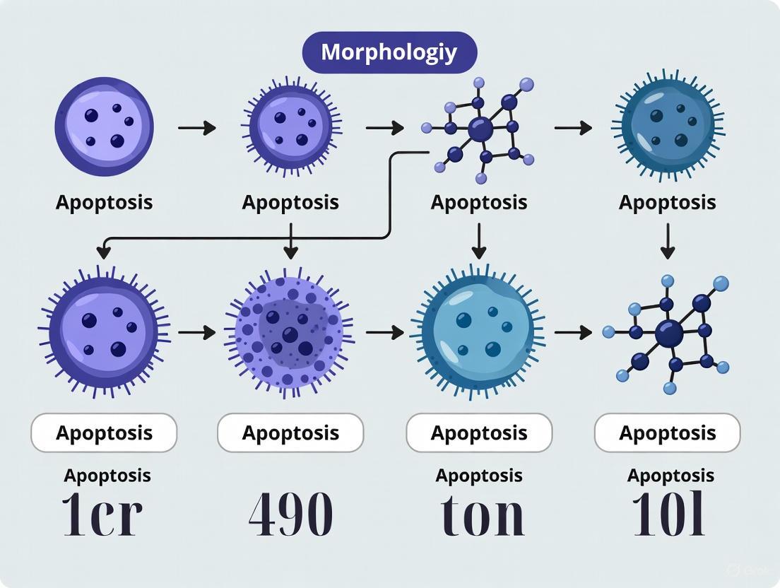
The process of apoptosis follows a conserved morphological sequence that can be consistently observed across different cell types and organisms. This predictable progression facilitates the accurate identification and study of programmed cell death in diverse research contexts.
Stage 1: Cell Shrinkage and Condensation
The initial morphological indicator of apoptosis is a rapid reduction in cellular volume and compaction of intracellular components. The cell, which typically maintains close connections with its neighbors, begins to detach and lose specialized surface structures such as microvilli [1] [2]. The cytoplasm becomes increasingly dense as organelles pack more tightly together, though their fundamental integrity remains intact at this stage. This condensation represents the first visible commitment to the apoptotic pathway and precedes more dramatic nuclear changes [1].
Stage 2: Nuclear Fragmentation (Pyknosis and Karyorrhexis)
Nuclear changes constitute the most characteristic morphological feature of apoptosis. The process begins with pyknosis, characterized by progressive condensation of nuclear chromatin into dense, featureless masses that aggregate peripherally beneath the nuclear membrane [1]. This is followed by karyorrhexis, where the pyknotic nucleus undergoes fragmentation into discrete membrane-bound bodies containing electron-dense chromatin [1] [2]. These nuclear alterations result from caspase-mediated activation of specific endonucleases that cleave DNA at internucleosomal sites, creating the classic DNA ladder pattern observed in gel electrophoresis [2].
Stage 3: Membrane Blebbing and Apoptotic Body Formation
In the penultimate stage, the cell undergoes extensive plasma membrane blebbing, forming dynamic protrusions that eventually separate from the main cell body [1] [6]. This "budding" process produces apoptotic bodies - spherical, membrane-bound vesicles containing tightly packed organelles, cytoplasmic components, and nuclear fragments [1] [2]. The formation of apoptotic bodies is mediated by caspase-mediated cleavage of cytoskeletal proteins, including ROCK1 kinase, which leads to reorganization of actin filaments and membrane blebbing [2]. These bodies vary in size and composition but are universally characterized by intact membrane integrity, which prevents the release of cellular contents into the extracellular environment [1].
Stage 4: Phagocytic Clearance
The final stage involves the recognition and engulfment of apoptotic bodies by neighboring phagocytic cells. Professional phagocytes (macrophages) or adjacent parenchymal cells rapidly identify apoptotic bodies through surface markers such as externalized phosphatidylserine [2]. This "eat-me" signal triggers efferocytosis, the process by which apoptotic bodies are internalized and degraded within phagolysosomes [1] [2]. The complete elimination of cellular material occurs without provoking an inflammatory response, distinguishing apoptotic clearance from necrotic cell death [1].
Table 1: Universal Morphological Stages of Apoptosis
| Stage | Key Morphological Features | Cellular Location | Molecular Triggers |
|---|---|---|---|
| Cell Shrinkage | Cytoplasmic condensation, organelle packing, loss of cell-cell contacts | Cytoplasm, membrane | Caspase activation, cytoskeletal degradation |
| Nuclear Fragmentation | Chromatin condensation (pyknosis), nuclear fragmentation (karyorrhexis) | Nucleus | Caspase-activated DNase (CAD), chromatin cleavage |
| Membrane Blebbing | Dynamic membrane protrusions, formation of apoptotic bodies | Plasma membrane, cytoskeleton | ROCK1 activation, actin reorganization |
| Phagocytic Clearance | Engulfment by phagocytes, lysosomal degradation | Extracellular environment | Phosphatidylserine externalization, "eat-me" signals |
Comparative Morphology: Apoptosis Versus Necrosis
Distinguishing apoptosis from other forms of cell death, particularly necrosis, is essential for accurate pathological assessment and experimental interpretation. While both processes result in cellular demise, their morphological sequences and physiological consequences differ fundamentally.
Apoptosis manifests as a controlled, energy-dependent process characterized by cell shrinkage, chromatin condensation, and preservation of membrane integrity until late stages [1] [6]. The formation of apoptotic bodies and their subsequent phagocytosis occurs without inflammatory activation, making apoptosis a "silent" mechanism of cell elimination [1] [7]. In contrast, necrosis follows an uncontrolled pathway initiated by severe cellular injury, featuring cell swelling, organelle disruption, and premature plasma membrane rupture [1] [3] [4]. The consequent release of intracellular components provokes a significant inflammatory response, contributing to tissue damage and pathological progression [1].
Table 2: Morphological Comparison of Apoptosis and Necrosis
| Characteristic | Apoptosis | Necrosis |
|---|---|---|
| Cellular Size | Cell shrinkage | Cell swelling |
| Plasma Membrane | Intact until late stages; blebbing | Early disruption; rupture |
| Nuclear Changes | Pyknosis and karyorrhexis | Karyolysis, pyknosis, and karyorrhexis |
| Cytoplasmic Contents | Retained in apoptotic bodies | Released extracellularly |
| Inflammatory Response | None | Significant |
| Tissue Response | Phagocytosis by adjacent cells | Infiltration of inflammatory cells |
| Energy Requirement | Energy-dependent (ATP-requiring) | Energy-independent |
| Molecular Regulation | Caspase-dependent, genetically programmed | Uncontrolled, accidental |
The concept of an "apoptosis-necrosis continuum" acknowledges that these processes are not always mutually exclusive [1]. Factors such as ATP depletion or caspase inhibition can convert an apoptotic signal into necrotic morphology, creating hybrid forms of cell death with overlapping characteristics [1] [8]. This continuum underscores the importance of using multiple detection methods to accurately classify cell death modalities in experimental systems.
Advanced Detection Methodologies and Protocols
Light and Electron Microscopy
Standard Protocol for Morphological Assessment:
- Sample Fixation: Process cells or tissues with appropriate fixatives (e.g., formaldehyde, glutaraldehyde) to preserve structural integrity [1].
- Staining: Apply histological stains such as hematoxylin and eosin (H&E) to highlight nuclear and cytoplasmic features [1] [9].
- Microscopic Analysis: Identify apoptotic cells by their characteristic morphology - single cells or small clusters with condensed eosinophilic cytoplasm and dense purple nuclear fragments [1].
For enhanced resolution, transmission electron microscopy provides definitive ultrastructural evidence of apoptosis, including chromatin margination, organelle compaction, and apoptotic body formation [1] [9]. While highly specific, these morphological methods may underestimate apoptosis in tissues with rapid clearance of apoptotic bodies and cannot detect early pre-morphological events [9].
Fluorescence-Based Detection Methods
SCAN (System for Counting and Analysis of Nuclei) Protocol: This automated system quantifies apoptosis in multicellular specimens by analyzing nuclear morphology [10].
- Sample Preparation: Stain cells with DNA-binding fluorophores (Hoechst 33342 or DAPI) to visualize nuclear structure [10].
- Image Acquisition: Capture z-stack images at multiple focal planes to account for all nuclei in three-dimensional space [10].
- Software Analysis: Utilize the Condensation Module to discriminate intact nuclei from condensed nuclei based on parameters including nucleolus presence, fluorescence distribution, nuclear size, and relative fluorescence per nuclear area [10].
TUNEL Assay Protocol: The Terminal deoxynucleotidyl transferase dUTP Nick End Labeling assay detects DNA fragmentation, a hallmark of apoptotic nuclei [10].
- Sample Fixation: Treat cells with cross-linking agents like paraformaldehyde [10].
- Permeabilization: Apply mild detergent (e.g., Triton X-100) to allow reagent penetration [10].
- Enzymatic Labeling: Incubate with terminal deoxynucleotidyl transferase (TdT) enzyme and fluorescently-labeled dUTP to tag DNA break sites [10].
- Quantification: Analyze using fluorescence microscopy or flow cytometry; the SCAN TUNEL module can automate this quantification [10].
Label-Free Imaging Technologies
Full-Field Optical Coherence Tomography (FF-OCT): This emerging technology enables high-resolution, label-free visualization of apoptotic morphological changes in living cells [7].
- System Configuration: Employ a Linnik-configured Michelson interferometer with identical water-immersion objectives in reference and sample arms [7].
- Image Acquisition: Use a broadband halogen light source to capture interferometric images; phase shifting techniques remove DC components to isolate sample reflection information [7].
- 3D Reconstruction: Stack sequential en face cross-sectional images to reconstruct three-dimensional cellular morphology, allowing quantitative analysis of surface changes during apoptosis [7].
FF-OCT can identify characteristic apoptotic features including echinoid spine formation, membrane blebbing, filopodia reorganization, and cell contraction without fluorescent labels or sample fixation [7].
Molecular Mechanisms Underlying Morphological Changes
The stereotypical morphological stages of apoptosis are executed by conserved molecular pathways that can be initiated through different triggers but converge on a common final pathway.
The Extrinsic (Death Receptor) Pathway
The extrinsic pathway initiates apoptosis in response to extracellular signals, particularly death receptor activation [6] [5] [2].
Diagram 1: Extrinsic apoptosis pathway (Max Width: 760px)
This pathway begins when extracellular death ligands (FasL, TRAIL, TNF-α) bind to their corresponding transmembrane death receptors, inducing receptor trimerization and intracellular death domain exposure [6] [2]. Adaptor proteins including FADD (Fas-associated death domain) or TRADD (TNF receptor-associated death domain) are recruited, forming the death-inducing signaling complex (DISC) [6] [5] [2]. The DISC activates initiator caspase-8, which directly cleaves and activates executioner caspases-3, -6, and -7, initiating the proteolytic cascade responsible for apoptotic morphology [6] [2].
The Intrinsic (Mitochondrial) Pathway
The intrinsic pathway responds to intracellular stress signals including DNA damage, oxidative stress, and growth factor withdrawal [6] [5] [2].
Diagram 2: Intrinsic apoptosis pathway (Max Width: 760px)
Cellular stressors activate BH3-only proteins which neutralize anti-apoptotic Bcl-2 family members, permitting the oligomerization of pro-apoptotic effectors BAX and BAK at the mitochondrial membrane [6] [2]. This triggers mitochondrial outer membrane permeabilization (MOMP), releasing cytochrome c and other intermembrane proteins into the cytosol [6] [5]. Cytochrome c binds to APAF-1, forming the apoptosome complex that activates caspase-9, which in turn activates the same executioner caspases as the extrinsic pathway [6] [2].
Execution Phase: From Molecular Events to Morphology
The executioner caspases (primarily caspases-3, -6, and -7) orchestrate the systematic dismantling of cellular structures through cleavage of specific substrate proteins [2]:
- Nuclear Fragmentation: Caspase-3 activates CAD (caspase-activated DNase) by cleaving its inhibitor ICAD, resulting in internucleosomal DNA cleavage and nuclear condensation [2].
- Membrane Blebbing: Caspase-mediated cleavage of ROCK1 kinase leads to actin cytoskeleton reorganization and membrane blebbing [2].
- Phosphatidylserine Externalization: Caspase-3 cleaves flippase ATP11 and activates scramblase Xkr8, promoting phosphatidylserine exposure on the outer leaflet as an "eat-me" signal for phagocytes [2].
- Apoptotic Body Formation: Caspase cleavage of structural and regulatory proteins facilitates the packaging of cellular contents into apoptotic bodies [2].
The Scientist's Toolkit: Essential Research Reagents
Table 3: Essential Research Reagents for Apoptosis Detection
| Reagent/Category | Specific Examples | Primary Function | Application Notes |
|---|---|---|---|
| DNA-Binding Dyes | Hoechst 33342, DAPI, Propidium Iodide (PI) | Nuclear staining for morphology assessment | Distinguish condensed chromatin; PI excludes viable cells |
| Phosphatidylserine Detection | FITC-Annexin V | Binds externalized PS on apoptotic cells | Used with viability dyes; requires calcium buffer |
| Caspase Activity Assays | Fluorogenic substrates (DEVD-AFC), FLICA kits | Measure caspase enzymatic activity | Quantitative but may not distinguish specific caspase roles |
| Antibody-Based Reagents | Anti-cytochrome c, anti-active caspase-3, anti-Bcl-2 family | Detect protein localization, activation, expression | IHC, IF, Western blot; confirm pathway activation |
| Mitochondrial Dyes | JC-1, TMRM, MitoTracker | Assess mitochondrial membrane potential | ΔΨm loss indicates intrinsic pathway activation |
| Cell Death Inducers | Staurosporine, Doxorubicin, Etoposide, TRAIL | Positive controls for apoptosis induction | Activate different pathways (intrinsic vs. extrinsic) |
| Caspase Inhibitors | z-VAD-fmk, Q-VD-OPh | Pan-caspase inhibition | Confirm caspase-dependent apoptosis |
Implications for Cross-Cell Type Morphological Comparisons
When comparing apoptotic morphology across different cell types, researchers must consider both the universal features and cell-type-specific variations. The core morphological sequence remains consistent, but timing, susceptibility to different pathways, and clearance mechanisms may vary significantly.
Epithelial cells typically demonstrate classic apoptotic morphology with well-defined apoptotic bodies, while neuronal cells may exhibit more condensed forms with extensive neurite fragmentation [1]. Hematopoietic cells often undergo rapid apoptosis with pronounced membrane blebbing, whereas fibroblasts may display prolonged early stages with gradual condensation [1]. These variations highlight the importance of establishing cell-type-specific baselines when quantifying apoptotic responses in experimental systems.
The conservation of apoptotic morphology across evolutionary distant organisms, from nematodes to mammals, underscores its fundamental role in multicellular biology [1] [2]. This conservation provides researchers with robust comparative models and validates the extrapolation of findings from experimental systems to human biology. However, subtle differences in regulatory mechanisms necessitate careful interpretation of cross-species comparisons, particularly in the context of therapeutic development where human-specific factors may be critical [5] [4].
Regulated cell death is a fundamental process governing the precise cellular remodeling required for proper brain formation. For decades, intrinsic apoptosis was considered the primary mechanism eliminating surplus cells in the developing nervous system [11] [4]. However, emerging evidence now reveals a more complex interplay where extrinsic apoptosis and necroptosis contribute significantly to shaping the embryonic brain [11] [12]. This paradigm shift challenges conventional understanding and necessitates a comparative analysis of how different cell death pathways coordinate neural development.
The telencephalon, the embryonic precursor to cerebral hemispheres, serves as an ideal model system due to its well-characterized developmental processes and extensive neural-vascular interactions [11]. Contemporary research leveraging advanced single-cell technologies has begun delineating the specific contributions of distinct cell death pathways across different neural cell populations. This review synthesizes current understanding of apoptosis morphology and signaling in telencephalic development, providing both quantitative comparisons and methodological frameworks for continued investigation in this evolving field.
Molecular Mechanisms of Apoptotic Pathways in Neural Development
Intrinsic and Extrinsic Apoptosis Signaling
Apoptosis occurs primarily through two core pathways that converge on executioner caspases:
Extrinsic Pathway: Initiated by extracellular death ligands (e.g., FasL, TNF-α) binding to cell surface death receptors (e.g., Fas/CD95, TNFR1). This triggers formation of the death-inducing signaling complex (DISC), leading to activation of initiator caspase-8 [11] [13] [4]. Active caspase-8 can directly cleave and activate executioner caspases (caspase-3, -6, -7) or engage the mitochondrial pathway via Bid cleavage.
Intrinsic Pathway: Activated by intracellular stressors including DNA damage and oxidative stress. This pathway is regulated by the Bcl-2 protein family, where pro-apoptotic members (Bax, Bak) promote mitochondrial outer membrane permeabilization (MOMP), enabling cytochrome c release [13] [4]. Cytochrome c forms the apoptosome with Apaf-1, activating caspase-9.
These pathways exhibit distinct yet interconnected functions during telencephalic development. While intrinsic apoptosis was historically considered dominant, genetic evidence now demonstrates significant roles for extrinsic signaling, particularly in specific neural progenitor populations [11].
Figure 1: Molecular Pathways of Apoptosis. The extrinsic (death receptor) and intrinsic (mitochondrial) pathways represent the two principal apoptosis mechanisms converging on caspase-3/7 activation. Cross-talk occurs via Bid cleavage, connecting both pathways during developmental cell death.
Necroptosis and Cross-pathway Regulation
Beyond classical apoptosis, necroptosis represents a regulated, inflammatory form of cell death mediated by RIPK1, RIPK3, and MLKL. Caspase-8 critically suppresses necroptosis by cleaving RIPK1 and RIPK3 [11]. In telencephalic development, this regulatory relationship is particularly important in endothelial cells, where Caspase-8 deficiency triggers vascular defects through unopposed necroptotic signaling [11].
The complex interplay between these pathways is evidenced by embryonic lethality in Caspase-8-deficient mice, which is rescued by concurrent deletion of RIPK3. This demonstrates the critical balance required between apoptotic and necroptotic pathways during brain development [11].
Quantitative Analysis of Cell Death in Telencephalic Models
Genetic Models Reveal Pathway-specific Contributions
Recent single-cell mass cytometry (CyTOF) studies of mouse telencephalon have quantified the relative contributions of different death pathways using knockout models:
Table 1: Quantitative Effects of Cell Death Pathway Disruption in Telencephalic Development
| Genetic Model | Total Cell Count Change | Key Affected Cell Populations | Primary Pathway Affected |
|---|---|---|---|
| Wild-type (WT) | Baseline (reference) | Normal distribution across all lineages | Balanced apoptosis/necroptosis |
| RIPK3 KO | Moderate increase | Endothelial cells | Necroptosis impaired |
| Caspase-8 KO | Embryonic lethal (E11.5) | Vascular/endothelial cells | Necroptosis hyperactivation |
| RIPK3/Caspase-8 DKO | +12.6% vs. WT | Tbr2⁺ intermediate progenitors, endothelial cells | Extrinsic apoptosis & necroptosis impaired |
The 12.6% increase in total cell count observed in RIPK3/Caspase-8 double knockout (DKO) mice demonstrates the combined contribution of extrinsic apoptosis and necroptosis to developmental cell elimination [11] [12]. This challenges the historical predominance assigned to intrinsic apoptosis alone.
Temporal Dynamics and Cell Type-specific Vulnerability
Analysis of dying cells across developmental stages (E13-P4) reveals progressive increases in both cleaved Caspase-3-positive (apoptotic) and Cisplatin-positive (membrane-compromised) populations, with CC3+ cells increasing by 203.0% and Cisplatin+ cells by 129.9% over this period [11]. Different neural populations show distinct vulnerability to specific death pathways:
- Tbr2⁺ intermediate progenitors: Selectively enriched in DKO mice, indicating significant regulation by extrinsic apoptosis and/or necroptosis [11]
- Endothelial cells: Particularly dependent on Caspase-8-mediated necroptosis suppression for vascular homeostasis [11]
- Immature neurons: Exhibit heterogeneous death marker profiles suggesting multiple active elimination mechanisms
Table 2: Cell Death Marker Profiles and Interpretations in Neural Development
| Marker Profile | Morphological Interpretation | Proposed Death Mechanism | Detection Methods |
|---|---|---|---|
| CC3+Cisplatin− | Apoptotic cells with intact membranes | Early-stage apoptosis | Immunofluorescence, CyTOF [11] |
| CC3−Cisplatin+ | Cells with compromised membranes | Non-apoptotic death (necroptosis, accidental) | Viability dyes, membrane integrity assays [11] [14] |
| CC3+Cisplatin+ | Mixed apoptotic/necroptotic features | Later-stage apoptosis with secondary necrosis | Multiparameter cytometry [11] |
| Annexin V+PI− | Early apoptosis (PS externalization) | Apoptosis initiation | Flow cytometry [15] |
| DNA fragmentation | Nuclear condensation and cleavage | Late-stage apoptosis | TUNEL assay, gel electrophoresis [15] |
Methodological Framework for Apoptosis Analysis
Single-cell Mass Cytometry (CyTOF) Workflow
The application of CyTOF to telencephalic development involves several critical steps enabling high-dimensional analysis of cell death across diverse populations:
Figure 2: Single-cell Mass Cytometry Workflow for Cell Death Analysis. This protocol enables simultaneous quantification of multiple cell death markers across diverse neural populations, including traditionally excluded dying cells by removing viability gating [11].
The Scientist's Toolkit: Essential Reagents and Methods
Table 3: Key Research Reagents and Methods for Neural Apoptosis Studies
| Reagent/Method | Primary Function | Application in Neural Cell Death | Technical Considerations |
|---|---|---|---|
| Anti-cleaved Caspase-3 | Detects activated caspase-3 in apoptotic cells | Gold standard for apoptosis identification in tissue sections and single cells [11] [15] | Requires appropriate fixation; does not distinguish intrinsic vs. extrinsic pathways |
| Cisplatin viability dye | Identifies cells with compromised membranes | Distinguishes apoptotic (Cisplatin−) from necrotic/necroptotic (Cisplatin+) cells [11] | Short 30-second exposure before fixation prevents induction of apoptosis |
| Annexin V conjugates | Binds phosphatidylserine exposed on outer membrane | Early apoptosis detection before membrane rupture [15] | Requires calcium-containing buffer; not suitable for fixed cells |
| TUNEL assay | Labels DNA strand breaks in apoptotic nuclei | Late-stage apoptosis detection; histochemical validation [15] | High sensitivity but risk of false positives in necrotic cells |
| Genetic models (RIPK3 KO, Caspase-8 DKO) | Disrupt specific cell death pathways | Pathway-specific functional analysis in telencephalic development [11] [12] | Embryonic lethality of single Caspase-8 KO requires conditional models or DKO approaches |
| Quantitative Phase Imaging (QPI) | Label-free analysis of morphological changes | Distinguishes apoptosis from lytic death based on cell dynamics [14] | Enables real-time tracking of death progression without biochemical markers |
Multiparameter Assessment of Cell Death Morphology
Accurate classification of cell death modalities requires integrated assessment of multiple morphological and biochemical features:
- Membrane Alterations: Phosphatidylserine externalization (Annexin V binding) precedes membrane blebbing and eventual rupture in late-stage death [15]
- Nuclear Changes: Chromatin condensation progresses to nuclear fragmentation and DNA cleavage, detectable via TUNEL staining and nuclear morphology [15]
- Cytoplasmic Events: Cell shrinkage, apoptotic body formation, and mitochondrial transmembrane potential collapse represent key hallmarks [4] [15]
- Proteolytic Cascade: Caspase activation (particularly caspase-3) and cleavage of substrates like PARP provide biochemical confirmation of apoptosis [4] [15]
Advanced techniques like Quantitative Phase Imaging (QPI) enable label-free distinction of apoptosis from lytic death based on dynamic morphological parameters, including cell density and membrane dynamics [14]. This approach can achieve 75.4% prediction accuracy for caspase-dependent versus independent death subroutines.
Discussion and Future Perspectives
The comparative analysis of neural cell apoptosis in telencephalic models reveals unexpected complexity in developmental cell elimination. The selective enrichment of Tbr2⁺ intermediate progenitors in RIPK3/Caspase-8 DKO mice indicates cell type-specific roles for extrinsic death pathways that extend beyond historical understanding [11]. These findings suggest potential mechanistic links to neurodevelopmental disorders characterized by aberrant cell death or survival.
Future research directions should include:
- Temporal-specific and cell type-restricted genetic manipulations to resolve spatial and temporal functions of death pathways
- High-resolution live imaging to correlate dynamic morphological changes with molecular pathway activation
- Investigation of potential compensatory mechanisms between intrinsic apoptosis, extrinsic apoptosis, and necroptosis
- Translational studies exploring how dysregulation of developmental death pathways contributes to neurological disorders
The integrated methodological approach combining genetic models, single-cell technologies, and multidimensional death marker analysis provides a powerful framework for advancing understanding of how coordinated cell elimination shapes the developing brain.
Apoptosis, a form of programmed cell death, is fundamental to immune system homeostasis, development, and function. The morphological execution of apoptosis is largely conserved, yet emerging evidence reveals critical nuances in its manifestation between the two principal lineages of immune cells: lymphocytes and myeloid cells. Understanding these distinct "morphological fingerprints" is essential for researchers and drug development professionals dissecting immune responses, inflammatory diseases, and cancer immunotherapy. This guide provides an in-depth technical comparison of apoptotic morphology in these lineages, framed within the context of cell death research, and details the experimental methodologies for their precise characterization.
Core Morphological Hallmarks of Apoptosis
Apoptosis is executed in a highly regulated manner, leading to a series of characteristic morphological changes distinct from other forms of cell death like necrosis or pyroptosis [16] [13]. The key stages are universally recognized, though their presentation can vary by cell type.
Table 1: Universal Morphological Stages of Apoptosis
| Stage | Key Characteristics | Technical Detection Methods |
|---|---|---|
| Cell Shrinkage | Reduction in cell volume and organelle condensation. | Flow cytometry (FSC/SSC parameters), microscopy. |
| Chromatin Condensation | Chromatin margination, nuclear condensation (pyknosis), and DNA fragmentation. | Fluorescent DNA dyes (DAPI, Hoechst), TUNEL assay. |
| Membrane Blebbing | Dynamic bulging of the plasma membrane, driven by actomyosin contraction. | Time-lapse microscopy, scanning electron microscopy (SEM). |
| Apoptotic Body Formation | Separation of the cell into small, membrane-bound vesicles containing intact organelles and nuclear fragments. | Flow cytometry, microscopy. |
| Preservation of Membrane Integrity | The plasma membrane remains intact until late stages, preventing inflammatory content release. | Annexin V/PI staining, impermeability to vital dyes. |
The process is predominantly mediated by a family of cysteine proteases called caspases, which are responsible for the proteolytic cleavage that leads to these structural dismantlements [17]. The intrinsic (mitochondrial) and extrinsic (death receptor) pathways converge on the activation of executioner caspases-3, -6, and -7, which orchestrate the morphological endpoint [17] [13].
Lineage-Specific Morphological Comparisons
While the core apoptotic program is shared, the execution and functional consequences display notable lineage-specific characteristics, particularly between lymphocytes and myeloid cells.
Lymphocyte Apoptosis
Lymphocytes, including T and B cells, are characterized by a rapid and efficient apoptotic response, which is critical for immune homeostasis and the termination of an immune response.
- Morphological Features: Lymphocytes typically exhibit pronounced cell shrinkage and generate a high number of small, uniform apoptotic bodies [18]. The condensed chromatin often forms dense, spherical masses.
- Functional Context: The propensity for rapid apoptosis is a key mechanism of immunosenescence. Studies on elderly post-COVID individuals revealed a significantly elevated proportion of apoptotic CD4+ and CD8+ T-cells, characterized by mitochondrial depolarization and increased Bax/Bcl-2 ratios, indicating a shift toward the intrinsic apoptotic pathway [18] [19]. This underscores the role of apoptotic dysregulation in age-related immune decline.
Myeloid Cell Apoptosis
Myeloid cells, such as macrophages, neutrophils, and dendritic cells, display distinct apoptotic features, often linked to their roles in phagocytosis and inflammation.
- Morphological Features: Macrophages and other myeloid cells can generate large apoptotic bodies (ApoBDs). Research on human bone marrow mesenchymal stromal cells (MSCs) has shown that large ApoBDs (~700 nm) exhibit superior immunomodulatory capacity compared to smaller ones (~500 nm) [20]. These large vesicles are more effectively taken up by macrophages and polarize them toward an anti-inflammatory M2 phenotype [20].
- Functional Context: The formation of large, immunomodulatory ApoBDs by myeloid cells represents a critical communication mechanism within the immune system. Furthermore, in the context of cancer, apoptotic cells—including myeloid cells—can paradoxically promote metastasis. Apoptotic cells externalize phosphatidylserine (PS), which increases the activity of Tissue Factor, triggering coagulation and the formation of platelet clots that protect circulating tumor cells [21].
Table 2: Comparative Morphological Fingerprints: Lymphocytes vs. Myeloid Cells
| Characteristic | Lymphocytes | Myeloid Cells |
|---|---|---|
| Primary Physiological Role | Immune memory, adaptive immunity | Phagocytosis, innate immunity, antigen presentation |
| Typical Apoptotic Body Size | Small, uniform [18] | Can form large (~700 nm) ApoBDs [20] |
| Key Immunological Outcome | Homeostatic control, avoidance of autoimmunity | Immunomodulation (e.g., M2 macrophage polarization) [20] |
| Pathological Dysregulation | Immunosenescence, post-viral T-cell depletion [18] [19] | Contribution to chronic inflammation, pro-tumoral effects [21] |
| Notable Surface Exposure/Release | Phosphatidylserine (PS) exposure | PS exposure; release of large immunomodulatory ApoBDs [20] [21] |
Detailed Experimental Protocols for Morphological Assessment
Accurate assessment of these morphological fingerprints relies on a suite of well-established experimental techniques.
Multiparametric Flow Cytometry for Apoptotic Quantification
This is a cornerstone technique for quantifying early and late apoptosis in specific immune cell populations.
Workflow:
- Cell Preparation: Isolate Peripheral Blood Mononuclear Cells (PBMCs) or purified immune cell populations.
- Staining:
- Resuspend cells in Annexin V binding buffer.
- Add Fluorochrome-conjugated Annexin V to detect phosphatidylserine (PS) exposure, a marker for early apoptosis.
- Add Propidium Iodide (PI) or a similar viability dye to detect loss of membrane integrity, a marker for late apoptosis/necrosis.
- Optional: Include fluorescent antibodies for cell surface markers (e.g., CD3 for T cells, CD19 for B cells, CD11b/CD14 for monocytes/macrophages) to gate on specific lineages.
- Incubation & Analysis: Incubate for 15-20 minutes in the dark and analyze immediately on a flow cytometer. Cells are categorized as:
- Viable: Annexin V-, PI-
- Early Apoptotic: Annexin V+, PI-
- Late Apoptotic: Annexin V+, PI+ [18]
Microscopic Techniques for Morphological Confirmation
Flow cytometry data should be complemented with imaging for direct visual confirmation of morphology.
Protocol for Fluorescence Microscopy:
- Culture cells on glass coverslips and induce apoptosis.
- Fix cells with 4% paraformaldehyde.
- Permeabilize with 0.1% Triton X-100 and stain with DAPI or Hoechst 33342 to visualize nuclear condensation and fragmentation.
- Counterstain with Phalloidin to observe actin cytoskeleton remodeling and membrane blebbing.
- Image using a fluorescence microscope. Apoptotic cells show bright, punctate, or fragmented nuclei [13].
Protocol for Scanning Electron Microscopy (SEM):
- Fix cell pellets with glutaraldehyde (2.5%) in cacodylate buffer.
- Dehydrate through a graded series of ethanol.
- Critical-point dry the samples to preserve structure.
- Sputter-coat with a thin layer of gold/palladium.
- Image under SEM to reveal exquisite surface details like membrane blebbing and apoptotic body formation [22].
Key Signaling Pathways in Apoptosis
The morphological changes are the direct result of the activation of two main apoptotic signaling pathways. The following diagram illustrates the intrinsic and extrinsic pathways and their convergence.
The Scientist's Toolkit: Essential Research Reagents
A successful investigation into immune cell apoptosis relies on a carefully selected toolkit of reagents and assays.
Table 3: Key Research Reagent Solutions for Apoptosis Studies
| Reagent / Assay | Function / Target | Specific Application |
|---|---|---|
| Annexin V (FITC, PE, APC) | Binds to externalized Phosphatidylserine (PS) | Flow cytometry detection of early apoptosis [18]. |
| Propidium Iodide (PI) / 7-AAD | Intercalates into DNA of membrane-compromised cells | Flow cytometry viability stain; identifies late apoptotic/necrotic cells [18]. |
| Caspase Inhibitors (e.g., Z-VAD-FMK) | Pan-caspase inhibitor | To confirm caspase-dependent apoptosis [17]. |
| Anti-active Caspase-3 Antibody | Detects cleaved, active caspase-3 | Immunofluorescence or flow cytometry to confirm apoptotic execution [18]. |
| TUNEL Assay Kit | Labels fragmented DNA (double-strand breaks) | Microscopy or flow cytometry detection of late-stage nuclear apoptosis [13]. |
| JC-1 Dye / TMRE | Measures mitochondrial membrane potential (ΔΨm) | Flow cytometry/fluorometry to detect early intrinsic apoptosis [18]. |
| BH3 Mimetics (e.g., ABT-199/Venetoclax) | Inhibit anti-apoptotic Bcl-2 proteins | Induce intrinsic apoptosis; key therapeutic agents [17]. |
| SMAC Mimetics | Antagonize Inhibitor of Apoptosis Proteins (IAPs) | Promote caspase activation and sensitize cells to apoptosis [17]. |
Integrated Experimental Workflow
A robust analysis of morphological fingerprints requires an integrated approach, combining the techniques and reagents detailed above. The following diagram outlines a recommended workflow from sample preparation to data interpretation.
Aberrant morphology in cancer cells is a direct visual manifestation of profound internal molecular dysfunction, serving as a critical bridge between cellular transformation and the clinical challenge of therapeutic resistance. This morphological dysregulation extends from subcellular disturbances in organelles to dramatic alterations in overall cell shape and size, frequently correlating with aggressive disease and poor patient outcomes [23]. Within the context of a broader thesis comparing apoptosis morphology across cell types, it is crucial to understand that cancer cells co-opt the very morphological programs of regulated cell death (RCD), such as apoptosis, to foster survival and resistance [17]. The evasion of apoptosis, a classically defined process with distinct morphological hallmarks, is a cornerstone of carcinogenesis. However, tumor cells exhibit remarkable death pathway plasticity, often shifting between apoptotic and non-apoptotic RCD mechanisms like ferroptosis, necroptosis, and autophagy in response to therapeutic pressure [17] [23]. This review provides an in-depth technical analysis of how aberrant morphology underpins drug resistance and outlines advanced, morphology-informed screening platforms that are essential for developing more effective cancer therapeutics.
Morphological Hallmarks of Cancer Cell Death and Their Functional Implications
The classification of cell death is fundamentally rooted in observable morphological criteria. Accidental cell death (ACD) is a rapid, chaotic process resulting from extreme physicochemical injury, leading to unregulated cellular dissolution [23]. In contrast, regulated cell death (RCD), including apoptosis, is an intricate, signal-driven process characterized by specific, conserved morphological changes [23]. Cancer cells dysregulate these precise morphological sequences to survive.
Table 1: Morphological and Molecular Features of Key Regulated Cell Death Types
| Cell Death Type | Key Morphological Hallmarks | Key Molecular Regulators | Implications in Cancer Drug Resistance |
|---|---|---|---|
| Apoptosis | Cell shrinkage, chromatin condensation (pyknosis), nuclear fragmentation (karyorrhexis), dynamic membrane blebbing, formation of apoptotic bodies [17] [23]. | Caspases, Bcl-2 family proteins, Cytochrome c, APAF-1, Phosphatidylserine externalization [24] [25]. | Overexpression of anti-apoptotic proteins (e.g., Bcl-2, Bcl-xL) inhibits the process; cancer cells avoid the classic apoptotic morphology, leading to treatment failure [24] [17]. |
| Necroptosis | Cellular and organellar swelling, plasma membrane rupture, release of intracellular contents, minimal chromatin condensation [23]. | RIPK1, RIPK3, MLKL [17]. | A backup death mechanism when apoptosis is blocked; its lytic nature can be immunogenic, potentially altering the tumor microenvironment [17]. |
| Ferroptosis | Smaller mitochondria, reduced mitochondrial cristae, intact plasma membrane, absence of chromatin condensation [17]. | GPX4, Glutathione, Lipid peroxides, ACSL4 [17]. | Resistance to therapies that induce apoptosis; regulated by pathways like p53/xCT/GPX4; its induction can overcome apoptotic resistance [17]. |
| Autophagy | Appearance of double-membrane autophagosomes engulfing cytoplasmic material, subsequent lysosomal degradation [17] [23]. | ATG proteins, LC3, Beclin-1, p62 [17]. | Can promote cell survival under stress; its role as a cell death mechanism is context-dependent and can contribute to therapy resistance [17]. |
A critical and paradoxical finding is that the morphology of apoptosis itself can be co-opted to promote cancer progression. Recent research demonstrates that circulating apoptotic cells, characterized by phosphatidylserine externalization, can robustly enhance metastasis. These apoptotic cells recruit platelets to circulating tumor cells (CTCs) by increasing the activity of the coagulation initiator Tissue Factor, forming protective emboli that shield CTCs from shear stress and immune surveillance, thereby promoting their survival and seeding at distant sites [25]. This indicates that the morphological signature of apoptosis is not always a terminal endpoint but can be a dynamic state that actively contributes to the metastatic niche.
Molecular Mechanisms Linking Aberrant Morphology to Drug Resistance
Dysregulation of Apoptotic Signaling Pathways
The failure to execute the morphological program of apoptosis is a primary mechanism of drug resistance. This evasion is mediated through the disruption of two core pathways:
The Intrinsic (Mitochondrial) Pathway: This pathway is activated by intracellular stressors like DNA damage, oxidative stress, and oncogene activation. The key morphological event is Mitochondrial Outer Membrane Permeabilization (MOMP), which is controlled by the balance between pro-apoptotic (e.g., Bax, Bak) and anti-apoptotic (e.g., Bcl-2, Bcl-xL) proteins [24] [17]. MOMP leads to the release of cytochrome c into the cytosol, triggering the formation of the apoptosome and the activation of caspase-9, which then initiates the execution phase. Cancer cells often overexpress anti-apoptotic Bcl-2 family members, preventing MOMP and the ensuing morphological breakdown, thus conferring resistance to a wide range of chemotherapeutics [24] [17].
The Extrinsic (Death Receptor) Pathway: This pathway is initiated by the binding of extracellular ligands (e.g., Fas-L, TRAIL) to death receptors on the cell surface. This leads to the formation of the Death-Inducing Signaling Complex (DISC), which activates initiator caspases-8 and -10 [24] [17]. A key regulatory protein, c-FLIP, can inhibit DISC formation, preventing the cascade that leads to the executioner phase and characteristic apoptotic morphology [24].
The following diagram illustrates the interconnected molecular circuitry of these core apoptotic pathways and their points of dysregulation in cancer.
Non-Apoptotic Death Pathways and Tumor Microenvironment (TME)
When apoptosis is blocked, cancer cells may leverage other RCD pathways, each with unique morphological signatures. The tumor microenvironment (TME) plays a crucial role in regulating this "death pathway plasticity." Factors such as hypoxia, oxidative stress, and interactions with cancer-associated fibroblasts (CAFs) and immune cells can determine which death modality is activated, allowing cancer cells to adapt and resist therapy [17] [26]. For instance, extracellular vesicles from drug-resistant cells can transfer resistance-related proteins to sensitive cells, altering their functional morphology and promoting survival [26].
Advanced Screening Methodologies for Morphology and Resistance
Modern drug discovery integrates high-throughput screening technologies with computational biology to decode the complex relationships between cell morphology, signaling networks, and drug response.
Integrated Technological Pillars for Drug Development
Table 2: Core Technologies in Modern Cancer Drug Screening
| Technology | Application in Screening | Key Limitations |
|---|---|---|
| Omics Strategies (Genomics, Proteomics, Metabolomics) | Provides foundational data on disease-related molecular characteristics; identifies potential drug targets and biomarkers [27]. | Data heterogeneity and lack of standardization can lead to biased predictions [27]. |
| Bioinformatics | Processes and analyzes complex biological data from omics studies; aids in target identification and mechanism elucidation [27]. | Prediction accuracy is highly algorithm-dependent, which can affect result reliability [27]. |
| Network Pharmacology (NP) | Studies drug-target-disease networks to reveal multi-target therapy opportunities, moving beyond single-target models [27]. | May oversimplify biological complexity (e.g., protein expression variations), leading to false positives [27]. |
| Molecular Dynamics (MD) Simulation | Examines atomic-level interactions between drugs and target proteins, enhancing design precision [27]. | High computational cost; accuracy is sensitive to force field parameters [27]. |
The synergy of these technologies is essential for confronting tumor heterogeneity, a decisive factor in drug resistance. Intratumoral heterogeneity, driven by genomic instability and clonal evolution, results in morphologically and functionally diverse cell populations. Under therapeutic pressure, this diversity allows for the selection of resistant sub-clones [26]. Advanced screening, particularly single-cell RNA sequencing, is critical for dissecting this heterogeneity and understanding its role in resistance and recurrence [26].
AI-Driven Predictive Models and Experimental Workflows
Artificial intelligence (AI) is revolutionizing the prediction of drug response based on complex input data. Models like PharmaFormer use a Transformer-based architecture and transfer learning. They are pre-trained on vast datasets from 2D cell lines (e.g., GDSC) and then fine-tuned with more physiologically relevant but data-limited patient-derived organoid (PDO) models. This approach significantly improves the accuracy of predicting clinical drug responses from bulk RNA-seq data of patient tumors [28].
The following diagram outlines a representative integrated workflow for screening and validating compounds that can overcome resistance by modulating cell death pathways.
A key case study demonstrating this workflow involved the investigation of Formononetin (FM) for liver cancer. Researchers used network pharmacology to screen FM's targets, analyzed differentially expressed genes from The Cancer Genome Atlas (TCGA), and then used molecular docking and MD simulation to confirm FM's stable binding to a key target, GPX4. Subsequent in vitro and in vivo experiments showed that FM induces ferroptosis, a non-apoptotic cell death, by regulating the p53/xCT/GPX4 pathway, thereby inhibiting liver cancer progression [27]. This exemplifies how targeting an alternative death pathway can overcome apoptotic resistance.
The Scientist's Toolkit: Essential Research Reagents and Materials
Table 3: Key Research Reagent Solutions for Investigating Cell Death and Resistance
| Reagent / Material | Function in Experimental Protocols |
|---|---|
| Caspase-8/9 FKBPF36V Dimerization Domains | Enables precise, ligand-controlled induction of extrinsic or intrinsic apoptosis to study morphological outcomes and their functional impact [25]. |
| Patient-Derived Organoids (PDOs) | 3D culture models that retain tumor morphology, heterogeneity, and drug sensitivity of primary tissues; used for high-fidelity drug response testing [28]. |
| SMAC Mimetics | Small molecule inhibitors of IAP proteins; used in experiments to sensitize cancer cells to apoptosis and overcome resistance [17]. |
| BH3 Mimetics (e.g., Bcl-2 inhibitors) | Compounds that antagonize anti-apoptotic proteins to promote MOMP and induce apoptotic morphology; used to target the intrinsic pathway [17]. |
| Phosphatidylserine (PS) Blocking Agents | Antibodies or proteins (e.g., Annexin V) used to detect or inhibit externalized PS on apoptotic cells; crucial for studying the pro-metastatic role of apoptosis [25]. |
| Ferroptosis Inducers/Inhibitors (e.g., Erastin, Liproxstatin-1) | Tools to manipulate the ferroptotic pathway, characterized by distinct mitochondrial morphology, to explore non-apoptotic death routes [17]. |
| Tissue Factor (TF) Inhibitors / Anticoagulants | Used to investigate the role of coagulation in metastasis, particularly how apoptotic cell-driven platelet clots protect CTCs [25]. |
Aberrant cellular morphology in cancer is not a passive consequence of transformation but an active and dynamic determinant of drug resistance and disease progression. The failure to undergo the classical morphological sequence of apoptosis, coupled with a adaptive plasticity to shift between different modes of regulated cell death, constitutes a major barrier to successful treatment. Future research and drug development must adopt an integrated approach that leverages advanced screening technologies, AI-driven modeling, and a deep understanding of death pathway crosstalk. By focusing on the morphological and molecular vulnerabilities exposed by this integrated view, the field can move towards more effective, patient-specific therapeutic strategies that overcome the formidable challenge of cancer drug resistance.
Cell death is a fundamental biological process, crucial for development, homeostasis, and the elimination of damaged or infected cells. Historically, cell death was simplistically categorized as either apoptosis (programmed) or necrosis (accidental). Apoptosis was defined as a genetically encoded, orderly process characterized by specific morphological features, while necrosis was viewed as an unregulated, passive consequence of extreme injury [1]. However, groundbreaking research over recent decades has revealed that certain forms of necrosis, including necroptosis, are also molecularly regulated [29] [30]. This discovery led to the concept of "regulated cell death" (RCD) and shed light on the extensive crosstalk and coordination between different death pathways [31].
This review focuses on the intricate morphological and molecular interplay between apoptosis, necroptosis, and other RCD pathways. We will dissect how these processes are not isolated but exist in a dynamic network, often modulating each other through mutual inhibitory mechanisms and serving as backup routes when the primary death pathway is compromised [29]. Understanding this crosstalk is paramount for researchers and drug development professionals, as it opens new avenues for therapeutic intervention in diseases such as cancer and neurodegenerative disorders, where cell death regulation is fundamentally disrupted [31] [32].
Morphological Hallmarks of Apoptosis and Necroptosis
The distinct morphological signatures of different cell death modalities remain a cornerstone for their identification and remain a critical tool for researchers [33]. The following table provides a structured comparison of the core morphological features of apoptosis and necroptosis.
Table 1: Morphological Comparison of Apoptosis and Necroptosis
| Feature | Apoptosis | Necroptosis |
|---|---|---|
| Cell Size & Shape | Cell shrinkage, rounding up, decreased cellular volume [29] [1] | Increased cell volume (oncosis) culminating in disruption of the plasma membrane [29] [30] |
| Nucleus | Chromatin condensation (pyknosis), nuclear fragmentation (karyorrhexis) [29] [1] | Chromatin decondensation; less obvious nuclear pyknosis [29] [30] |
| Plasma Membrane | Membrane blebbing and shedding of apoptotic bodies with intact membrane [29] [33] | Loss of membrane integrity, rupture, and release of cellular contents [30] [32] |
| Organelles | Minimal ultrastructural modification; organelles packed into apoptotic bodies [29] | Swelling of organelles, translucent cytoplasm [29] |
| Elimination & Inflammation | Rapid engulfment by phagocytes; no inflammatory response [1] | Potent inflammatory response due to release of DAMPs [32] |
Detailed Morphological Assessment of Apoptosis
The apoptotic process is a sequence of highly coordinated morphological events. It begins with cell shrinkage and pseudopod retraction, driven by controlled movements of ions and water [33]. The cytoplasm becomes dense, and organelles are tightly packed. A key early event is chromatin condensation, where nuclear material aggregates peripherally under the nuclear membrane [1]. This is followed by extensive plasma membrane blebbing, a process dependent on actin cytoskeleton rearrangement and activation of myosin light-chains via ROCK-I [33]. In the final stages, the cell undergoes karyorrhexis (nuclear fragmentation) and separates into multiple, sealed, membrane-wrapped vesicles called apoptotic bodies [1]. These bodies are rapidly phagocytosed by neighboring cells or macrophages, preventing an inflammatory response [1] [33].
Objective Quantification of Apoptotic Morphology
Modern image analysis software allows for the objective quantification of these morphological changes. Studies using ImageJ software have demonstrated that caspase-3 positive apoptotic cells show a smaller average nuclear area and circumference, along with a larger nuclear form factor (a measure of circularity) compared to healthy cells [34]. A novel morphological indicator, the nuclear circumference divided by form factor, has shown the strongest correlation with caspase-3 expression, providing a sensitive and quantifiable metric for identifying apoptosis [34]. These methods are vital for accurate and reproducible assessment in both basic research and drug discovery.
Molecular Mechanisms and Signaling Pathways
The morphological differences between apoptosis and necroptosis are a direct result of their distinct underlying molecular machineries.
The Apoptotic Signaling Cascade
Apoptosis proceeds via two main pathways that converge on a common execution phase:
- The Extrinsic (Death Receptor) Pathway: This pathway is initiated by the binding of extracellular ligands (e.g., FasL, TNF) to death receptors on the plasma membrane. This binding recruits the adapter protein FADD and pro-caspase-8 to form the Death-Inducing Signaling Complex (DISC), leading to caspase-8 activation [30] [32].
- The Intrinsic (Mitochondrial) Pathway: This pathway is triggered by intracellular stresses like DNA damage or growth factor deprivation. It is tightly regulated by the BCL-2 protein family, which controls Mitochondrial Outer Membrane Permeabilization (MOMP). MOMP leads to the release of cytochrome c into the cytosol, where it binds to APAF-1 and forms the "apoptosome," activating caspase-9 [30] [32].
Both pathways culminate in the activation of executioner caspases (caspase-3 and -7), which systematically cleave hundreds of cellular substrates, leading to the characteristic morphological demise of the cell [30] [1].
The Necroptotic Signaling Cascade
Necroptosis serves as a backup death pathway when apoptotic signaling, particularly caspase-8 activity, is inhibited [29] [32]. It can be initiated by the same death receptors that trigger apoptosis. When caspase-8 is inactive, RIPK1 and RIPK3 interact through their RHIM domains, forming a filamentous complex. This necrosome complex leads to the phosphorylation and activation of MLKL by RIPK3. Activated MLKL oligomerizes and translocates to the plasma membrane, where it forms pores, disrupting ionic homeostasis and causing the necrotic phenotype of cell swelling and membrane rupture [29] [32]. The release of intracellular components, known as Damage-Associated Molecular Patterns (DAMPs), then drives inflammation [32].
Diagram 1: Molecular Crosstalk in PANoptosis
Intracellular Crosstalk and the Emergence of PANoptosis
The traditional view of independent death pathways has been superseded by evidence of a complex, interconnected network. Key molecular nodes facilitate this crosstalk:
- Caspase-8 as a Master Switch: Caspase-8 sits at a critical crossroads. When active, it promotes apoptosis by cleaving and activating executioner caspases. Simultaneously, it cleaves pro-necroptotic proteins like CYLD and RIPK1, thereby inhibiting necroptosis. When caspase-8 is inhibited, the cell can default to the necroptotic pathway [29] [31].
- RIPK1's Dual Function: Depending on the cellular context, RIPK1 can form a complex that promotes NF-κB-mediated survival, apoptosis via FADD and caspase-8, or necroptosis via RIPK3 and MLKL [29] [32].
- Reactive Oxygen Species (ROS) as Universal Connectors: ROS, including superoxide anions and hydrogen peroxide, act as nodal regulators that can initiate, modulate, or suppress multiple RCD pathways. By exceeding critical thresholds, ROS can promote crosstalk and switches between apoptosis, necroptosis, and other forms of death like ferroptosis, functioning as a "molecular bridge" between different stress signals and death outcomes [35].
This integrative crosstalk is formally recognized in the concept of PANoptosis, defined as a unique inflammatory RCD pathway triggered by specific stimuli (e.g., pathogens, cytokines) and governed by a multi-protein complex called the PANoptosome [31]. The PANoptosome simultaneously recruits machinery from apoptosis (caspases), necroptosis (RIPK1/RIPK3/MLKL), and pyroptosis (inflammasomes/GSDMD), creating a robust, redundant death signaling platform that is difficult for pathogens to evade [31].
Experimental Protocols for Morphological and Biochemical Assessment
Accurate assessment of cell death requires a combination of morphological, biochemical, and functional assays. Below is a core methodology for quantifying apoptosis via morphological changes.
Protocol: Quantifying Apoptosis by Nuclear Morphology using ImageJ
This protocol is adapted from studies that objectively correlated nuclear morphology with caspase-3 activation [34].
1. Cell Culture and Induction:
- Culture cells (e.g., ARPE-19 or other relevant cell lines) under standard conditions.
- Induce apoptosis using a suitable agent (e.g., 1 µM Staurosporine for 24 hours). Include an untreated control.
2. Staining and Imaging:
- Fix cells with methanol or 4% paraformaldehyde for 15 minutes at room temperature.
- Permeabilize and block with PBS containing 1% BSA and 0.2% Triton X-100 for 30 minutes.
- Stain nuclei by incubating with DAPI (1 µg/mL) for 5 minutes.
- Acquire fluorescent images using a microscope with a DAPI filter set at a standardized magnification (e.g., 200x). Maintain constant exposure time and gain across all samples.
3. Image Analysis with ImageJ:
- Open the 16-bit DAPI image in ImageJ. Convert to 8-bit (Image > Type > 8-bit).
- Auto-threshold the image to create a binary mask (Process > Binary > Make Binary).
- Separate touching nuclei using the "Watershed" function (Process > Binary > Watershed).
- Analyze particles (Analyze > Analyze Particles). Set a size threshold to exclude debris.
- The software will output quantitative data for each nucleus, including:
- Area
- Circumference (Perimeter)
- Form Factor (4π*Area/Perimeter²). A perfect circle has a form factor of 1.
4. Data Interpretation:
- Compare the average Nuclear Area and Circumference between treated and control groups. A significant decrease indicates cell shrinkage.
- Compare the average Form Factor. An increase (closer to 1) indicates more circular, condensed nuclei.
- Calculate the novel indicator Circumference divided by Form Factor, which has shown a strong negative correlation (r ≈ -0.475) with caspase-3 expression [34].
Table 2: The Scientist's Toolkit - Key Reagents for Cell Death Research
| Reagent / Assay | Function / Target | Application in Research |
|---|---|---|
| Staurosporine | Broad-spectrum protein kinase inhibitor [34] | Commonly used positive control for inducing intrinsic apoptosis in vitro. |
| zVAD-fmk | Pan-caspase inhibitor [29] | Used to inhibit apoptosis and unmask alternative death pathways like necroptosis. |
| Necrostatin-1 (Nec-1) | RIPK1 inhibitor [29] | Selective inhibitor of necroptosis; used to confirm RIPK1-dependent death. |
| Hoechst 33342 / DAPI | Cell-permeable DNA dyes [33] [34] | Fluorescent staining of nuclei to assess chromatin condensation and nuclear fragmentation via microscopy. |
| Anti-Cleaved Caspase-3 Antibody | Detects activated caspase-3 [34] | Gold-standard immunohistochemical/immunofluorescent marker for committed apoptosis. |
| Annexin V Probes | Binds to phosphatidylserine (PS) [30] | Detects PS externalization, an early event in apoptosis, typically measured by flow cytometry. |
| Propidium Iodide (PI) | DNA dye, membrane impermeant [33] | Distinguishes live cells (PI-negative) from dead cells with compromised membranes (PI-positive). |
| Anti-pMLKL Antibody | Detects phosphorylated MLKL [32] | Key biomarker for the commitment to necroptosis. |
Implications for Disease and Therapeutic Development
The crosstalk between apoptosis and necroptosis has profound implications for human disease and drug discovery, particularly in cancer and neurodegeneration.
- Cancer Therapy Resistance: Many cancers develop resistance to pro-apoptotic chemotherapeutic agents. Understanding the molecular switches that control the apoptotic-necroptotic balance offers a strategy to overcome this resistance. For instance, inducing necroptosis in apoptosis-resistant tumor cells, or combining apoptosis inducers with caspase inhibitors to shift death modality, represents a promising therapeutic approach [29] [32]. Furthermore, the immunogenic nature of necroptosis can stimulate anti-tumor immunity, providing a synergistic effect with immunotherapy [32].
- Neurodegenerative Diseases: In contrast to cancer, neurodegenerative diseases like Alzheimer's (AD) and Parkinson's (PD) are characterized by excessive cell death. PANoptosis has been implicated in these pathologies. In AD, Aβ oligomers can activate the NLRP3 inflammasome and caspase-8, driving pyroptosis and apoptosis in neurons [31]. Similarly, in PD, α-synuclein fibrils can activate the NLRP3/ASC axis to trigger PANoptosis in dopaminergic neurons [31]. Targeting key nodes in this network, such as the NLRP3 inflammasome, may provide neuroprotection.
The global apoptosis assays market, projected to grow from USD 4.90 billion in 2024 to USD 9.20 billion by 2032, underscores the continued importance of this field in basic research and drug development [36]. The integration of artificial intelligence for high-throughput data analysis and the development of multi-parametric assays are accelerating our ability to dissect these complex death networks and identify novel therapeutic targets [36].
The morphological and molecular crosstalk between apoptosis and necroptosis illustrates the remarkable plasticity of cell death. These pathways are not simple linear routes but are embedded in a complex, redundant network with shared components and mutual regulation, as exemplified by the PANoptosis concept. For researchers in comparative morphology and drug development, a holistic understanding of this interplay is no longer optional but essential. Future research must continue to elucidate the precise regulatory networks, develop specific modulators of these pathways, and translate these insights into targeted therapies that can selectively manipulate cell fate in a range of human diseases.
Advanced Techniques for Detecting and Quantifying Apoptotic Morphology
Flow cytometry leverages the interaction between cells and laser light to extract critical morphological information through two fundamental parameters: forward scatter (FSC) and side scatter (SSC). When a cell passes through a laser beam, it scatters light in both forward and lateral directions. FSC, measured approximately along the axis of the laser beam, correlates strongly with cell size and cell surface area. SSC, collected at approximately 90 degrees to the laser beam, provides information on internal granularity and cytoplasmic complexity [37]. These light scatter properties form a foundational, label-free method for assessing cellular morphology in real-time at high throughput speeds of thousands of cells per second [38] [39].
Within apoptosis research, light scatter parameters undergo characteristic and reproducible changes that serve as initial indicators of cellular demise. Early in apoptosis, cells typically undergo shrinkage and chromatin condensation, leading to a measurable decrease in FSC. Concurrently, the internal complexity increases due to nuclear fragmentation and organelle reorganization, resulting in a transient increase in SSC [38]. In late apoptosis and secondary necrosis, both FSC and SSC typically decrease dramatically as cells lose internal content and integrity [39]. This morphological fingerprint provides researchers with a rapid, reagent-free method for initial apoptosis screening and population gating before applying more specific fluorescent probes.
Core Principles: Deciphering Light Scatter Signatures
Physical Principles of Light Scatter
The interaction between cells and laser light follows the principles of light scattering physics, primarily Mie scattering for FSC (which is sensitive to cell size and membrane properties) and Rayleigh scattering for SSC (which provides information about smaller internal structures and granularity). The FSC ratio (the ratio between signal intensities of forward scatter area and height) has recently been identified as a highly sensitive parameter for distinguishing single cells from cellular multiplets or aggregates, enhancing the accuracy of morphological assessments [40].
Morphological Interpretation of Scatter Patterns
The combination of FSC and SSC measurements creates a morphological fingerprint that enables discrimination of major leukocyte populations in peripheral blood and identification of aberrant cell states. The table below summarizes key morphological correlates of light scatter parameters:
Table 1: Morphological Correlates of Light Scatter Parameters in Flow Cytometry
| Light Parameter | Primary Morphological Correlates | Apoptotic Changes | Affected Cellular Features |
|---|---|---|---|
| Forward Scatter (FSC) | Cell size, surface area, cell diameter | Decrease (cell shrinkage) | Membrane blebbing, cytoplasmic condensation |
| Side Scatter (SSC) | Internal granularity, cytoplasmic complexity, nuclear structure | Initial increase, then decrease | Chromatin condensation, nuclear fragmentation, organelle reorganization |
| FSC/SSC Ratio | Discrimination of single cells from multiplets | Altered profile | Cell aggregation, formation of apoptotic bodies |
The analytical power of light scatter extends beyond simple population discrimination. Through careful calibration and experimental design, researchers can detect subtle morphological shifts indicating early-stage apoptosis, cellular activation, or pathological transformations. For instance, in the context of apoptosis comparison across cell types, lymphocytes typically show more pronounced FSC reduction than monocytes during early apoptosis, reflecting their different structural compositions and death kinetics [39].
Methodological Applications in Apoptosis Research
Standardized Light Scatter Gating Protocol
A reproducible protocol for morphological assessment of apoptosis using light scatter properties includes the following critical steps:
Instrument Optimization: Perform daily instrument calibration using standardized beads to ensure consistent light scatter measurements across experiments. Adjust photomultiplier tube (PMT) voltages to place the live cell population appropriately on scale [41].
Sample Preparation: Prepare single-cell suspensions at appropriate density (approximately 1×10⁶ cells/mL) to avoid swarm effects and ensure single-cell analysis. Maintain consistent handling and temperature conditions across samples [38].
Data Acquisition: Acquire a minimum of 10,000 events per sample using a low flow rate (e.g., 12-35 μL/min for BD instruments) to enhance resolution. Record both FSC-A and FSC-H parameters for doublet discrimination [40].
Viable Cell Gating: Initially gate on FSC-A versus SSC-A to exclude debris and dead cells. Subsequently, apply FSC-A versus FSC-H gating to exclude cell doublets and aggregates [40].
Apoptosis Analysis: Identify the apoptotic subpopulation based on decreased FSC and typically increased SSC characteristics compared to the viable cell population.
Data Interpretation: Calculate the percentage of cells in the apoptotic region and report alongside fluorescence-based apoptosis markers for validation.
This methodology enables rapid, label-free screening for apoptotic cells, providing a cost-effective approach for initial experiments or for monitoring apoptosis kinetics in time-course studies.
Advanced Analytical Approaches
Recent technological advances have enhanced the application of light scatter for morphological assessment. Imaging flow cytometry (IFC) represents a particularly significant innovation, combining the high-throughput capability of conventional flow cytometry with high-resolution morphological imaging [37]. This technology allows direct visualization of cells that show characteristic light scatter changes, confirming through imaging that decreased FSC indeed corresponds to apoptotic cell shrinkage and membrane blebbing.
The FSC ratio has emerged as a particularly powerful parameter in advanced applications. Recent research demonstrates that thresholding of the FSC ratio, particularly using Otsu's method, enables robust identification of cellular multiplets with F1 scores between 0.50-0.84, providing a data-driven approach for scatter-based discrimination of physically interacting cells [40]. This application is particularly valuable for studying immune synapses and cellular interactions in immunotherapy research.
Table 2: Research Reagent Solutions for Apoptosis Assessment via Flow Cytometry
| Reagent/Category | Specific Example | Primary Function in Apoptosis Assessment |
|---|---|---|
| Mitochondrial Potential Probes | TMRM (Tetramethylrhodamine methyl ester) | Detection of early apoptosis via loss of ΔΨm [38] |
| Caspase Activity Probes | FLICA (FAM-VAD-FMK) | Fluorochrome-labeled caspase inhibitors bind active enzymes [38] |
| Plasma Membrane Probes | Annexin V-FITC/APC | Binds phosphatidylserine externalization [38] |
| DNA Binding Dyes | Propidium Iodide (PI) | Assesses membrane integrity & identifies late apoptotic/necrotic cells [38] |
| Viability Stains | TO-PRO family dyes | Distinguish viable from compromised cells [39] |
| Morphological Standards | Size-calibrated microbeads | Instrument calibration for consistent light scatter measurements [41] |
Technological Integration and Future Directions
The integration of light scatter analysis with fluorescence measurements represents the standard approach in modern apoptosis research. This multiparameter strategy enables researchers to correlate morphological changes with specific biochemical events, such as caspase activation or phosphatidylserine externalization [38]. For instance, researchers can simultaneously measure decreased FSC (morphological change), increased Annexin V binding (membrane alteration), and increased caspase activity (enzymatic activation) within the same cell, providing comprehensive insight into the apoptotic process.
Artificial intelligence and machine learning approaches are increasingly being applied to flow cytometry data, including light scatter parameters [37] [42]. These computational methods can identify subtle, multivariate patterns in light scatter data that may escape conventional gating strategies. For example, generalized linear models (GLMs) can analyze the complex relationships between light scatter parameters and experimental variables such as time post-treatment, drug concentration, and cell type, accommodating the non-normal distributions typical of cytometric data [42].
The following workflow diagram illustrates the integration of light scatter analysis with other apoptotic markers in a comprehensive assessment strategy:
Integrated Workflow for Morphological Apoptosis Assessment
Emerging technologies continue to expand the applications of light scatter in morphological assessment. Spectral flow cytometry provides enhanced resolution of scatter parameters by capturing the full scatter spectrum rather than discrete wavelengths [37]. Laser scanning cytometry enables correlative analysis of light scatter signatures with precise subcellular localization. These technological advances ensure that light scatter analysis will remain a cornerstone of morphological assessment in apoptosis research, continually evolving to provide deeper insights into cellular dynamics.
Light scatter analysis in flow cytometry provides an indispensable, label-free method for morphological assessment in apoptosis research across diverse cell types. The integration of FSC and SSC measurements with fluorescent biomarkers creates a powerful multidimensional analytical platform that captures both structural and biochemical facets of programmed cell death. As cytometry technologies evolve toward higher-parameter systems and incorporate artificial intelligence-driven analysis, the fundamental principles of light scatter continue to provide critical morphological context. For researchers comparing apoptotic morphology across cell types, light scatter parameters offer a consistent, quantitative framework for identifying conserved and cell-type-specific aspects of death mechanisms, ultimately advancing both basic biological understanding and therapeutic development in diseases characterized by dysregulated apoptosis.
Within the broader context of comparing apoptosis morphology across cell types, the translocation of phosphatidylserine (PS) from the inner to the outer leaflet of the plasma membrane stands as a critical early molecular event, preceding the characteristic morphological hallmarks of programmed cell death [43] [44]. This loss of membrane asymmetry serves as a universal "eat-me" signal for phagocytes and represents a pivotal point of convergence in the study of apoptotic pathways across diverse cellular systems. The calcium-dependent binding of Annexin V to externalized PS provides a powerful tool for researchers, allowing for the sensitive detection of apoptosis before membrane integrity is lost [45]. This technical guide delves into the methodology of Annexin V staining, framing it as an essential technique for correlating this initial biochemical signal with the subsequent, defining morphological changes that occur as cells from various tissues commit to apoptosis. Accurate detection is paramount in fields such as cancer research and drug development, where quantifying cell death is essential for evaluating therapeutic efficacy [45].
The Scientific Principle: PS Exposure and Apoptotic Morphology
Apoptosis is a tightly regulated process characterized by a cascade of biochemical events leading to distinct cellular alterations. Among the earliest changes is the rapid redistribution of phosphatidylserine (PS), a phospholipid normally confined to the inner leaflet of the plasma membrane by ATP-dependent translocases [44]. During the initial stages of apoptosis, this enzymatic activity is suppressed, and a scramblase is activated, resulting in the exposure of PS on the cell surface [45]. This externalized PS acts as a key ligand for macrophage receptors, facilitating the prompt recognition and engulfment of the dying cell without inciting an inflammatory response—a fundamental difference from necrotic cell death [45].
The exposure of PS closely coincides with other early morphological changes, such as chromatin condensation and cell shrinkage [43]. Importantly, PS externalization occurs while the plasma membrane remains intact, a crucial distinction that allows for the differentiation between early apoptosis and late-stage apoptosis or necrosis. As apoptosis progresses to later stages, the membrane integrity is lost, a process often referred to as secondary necrosis [45].
Annexin V, a 35-36 kDa phospholipid-binding protein, exhibits a high affinity for PS in the presence of calcium ions (Ca²⁺) [44]. When conjugated to a fluorochrome such as Fluorescein Isothiocyanate (FITC), it serves as a sensitive probe for detecting PS externalization on the surface of living cells. This binding is reversible and calcium-dependent, requiring precise buffer conditions for optimal staining [44]. To distinguish between intact and compromised membranes, the membrane-impermeable DNA-binding dye Propidium Iodide (PI) is frequently used in conjunction with Annexin V. PI is excluded from viable and early apoptotic cells but penetrates and stains the nucleic acids of late apoptotic and necrotic cells [45]. This dual-staining approach enables the quantitative discrimination of cell populations via flow cytometry.
It is noteworthy that while PS exposure is a hallmark of apoptosis, it is not entirely exclusive to this form of cell death. For instance, recent research indicates that phosphatidylserine exposure also occurs during ferroptosis, another regulated cell death modality, though often accompanied by distinct membrane perforations [46].
Key Apoptotic Stages Detectable by Annexin V/PI Staining
The diagram below illustrates the correlation between PS exposure, key morphological changes, and their detection using Annexin V and PI staining across the continuum of cell death.
Experimental Protocols
Core Annexin V Staining Protocol for Flow Cytometry
This protocol is optimized for the detection of early apoptotic cells in both suspension and adherent cultures using flow cytometry, based on established methodologies [45] [44].
Materials Needed:
- Cells: Cultured cells or cell suspension from tissues (1–5 x 10⁵ cells per sample).
- Annexin V Conjugate: Fluorescently labeled Annexin V (e.g., Annexin V-FITC).
- Propidium Iodide (PI): Stock solution, typically at 50 µg/mL.
- Binding Buffer: Calcium-containing buffer (e.g., 10 mM HEPES, 140 mM NaCl, 2.5 mM CaCl₂, pH 7.4).
- Flow Cytometer: Equipped with appropriate lasers and filters.
- Controls: Unstained cells, Annexin V-only, PI-only, and apoptosis-induced positive control.
Step-by-Step Procedure:
Cell Preparation and Harvesting:
- Induce apoptosis using the desired method (e.g., chemical inducer like staurosporine or serum deprivation).
- For adherent cells: Detach cells gently using non-enzymatic methods (e.g., EDTA) or mild trypsinization to preserve membrane integrity. Wash with serum-containing media to inactivate trypsin [44].
- For suspension cells: Collect cells directly.
- Centrifuge all cells at 300 x g for 5 minutes at room temperature. Discard the supernatant.
- Wash cells once with cold PBS to remove residual media and proteins.
- Resuspend the cell pellet in binding buffer to a concentration of 1 x 10⁶ cells/mL.
Staining:
Analysis:
- Add 400 µL of binding buffer to each tube.
- Keep samples on ice and analyze by flow cytometry within 1 hour.
- Use an excitation wavelength of 488 nm. Measure Annexin V-FITC fluorescence in the FL1 (FITC) detector and PI fluorescence in the FL2 (phycoerythrin) or FL3 detector.
- Use single-stained controls to set up proper compensation and gating.
Critical Controls and Data Interpretation
The following table outlines the essential controls required to validate the Annexin V/PI staining experiment and guide accurate data interpretation.
Table 1: Essential Controls for Annexin V/PI Staining
| Control Type | Purpose | Annexin V | PI | Expected Population |
|---|---|---|---|---|
| Unstained | To set baseline fluorescence and voltage | No | No | All cells in Q3 (double negative) |
| Annexin V Only | For compensation and gate setting | Yes | No | Cells in Q4 (Annexin V positive) |
| PI Only | For compensation and gate setting | No | Yes | Cells in Q1 (PI positive) |
| Viable/Negative | Baseline for healthy cells | Yes | Yes | Majority in Q3 |
| Induced/Positive | Protocol validation | Yes | Yes | Significant population in Q2 and Q4 |
Data interpretation is performed using a flow cytometry dot plot with Annexin V-FITC on the x-axis and PI on the y-axis, dividing the cell population into four distinct quadrants [45]:
- Q3: Annexin V⁻ / PI⁻ → Viable, healthy cells. These cells have not externalized PS and possess an intact membrane.
- Q4: Annexin V⁺ / PI⁻ → Early apoptotic cells. This population has externalized PS but maintains membrane integrity, preventing PI uptake.
- Q2: Annexin V⁺ / PI⁺ → Late apoptotic (and sometimes necrotic) cells. These cells have both PS on the surface and a compromised membrane.
- Q1: Annexin V⁻ / PI⁺ → Necrotic cells or cellular debris. This population typically represents cells that have undergone primary necrosis, losing membrane integrity without significant PS externalization.
The Scientist's Toolkit: Key Research Reagents
A successful Annexin V staining experiment relies on a set of specific, high-quality reagents. The table below details the essential components and their functions.
Table 2: Essential Reagents for Annexin V Staining Assays
| Reagent | Function / Role in the Assay | Critical Notes |
|---|---|---|
| Annexin V Conjugate | Binds to externalized phosphatidylserine on the cell surface in a Ca²⁺-dependent manner. | Must be conjugated to a fluorochrome (e.g., FITC, PE). Binding is reversible and calcium-dependent [44]. |
| Propidium Iodide (PI) | Membrane-impermeable DNA dye; distinguishes cells with compromised membranes. | Penetrates late apoptotic/necrotic cells. Viable and early apoptotic cells are PI negative [45]. |
| Annexin V Binding Buffer | Provides the optimal ionic and pH environment; supplies Ca²⁺ ions essential for Annexin V binding. | Typically contains 2.5 mM CaCl₂. The absence of calcium will abrogate binding [45] [44]. |
| Apoptosis Inducer | Positive control to validate the staining protocol and induce PS exposure. | Common agents: staurosporine, chemotherapeutic drugs, UV irradiation. |
| Enzymatic Detachment Solution | For harvesting adherent cells without inducing artificial PS exposure. | Non-enzymatic (EDTA) or mild trypsinization is preferred over harsh trypsin to preserve membrane integrity [44]. |
Correlation with Broader Apoptosis Research
Integrating Annexin V staining into a multifaceted research approach is crucial for a comprehensive understanding of cell death. This technique is highly effective for quantifying early apoptosis but provides limited insight into the upstream signaling events that trigger PS externalization. Therefore, correlating Annexin V data with other morphological and biochemical analyses is a cornerstone of robust apoptosis research, especially in comparative studies across different cell types.
Morphological Correlations:
- Chromatin Condensation: This hallmark of apoptosis can be visualized using DNA-binding dyes like Hoechst 33342 or DAPI in conjunction with Annexin V staining. Researchers can correlate Annexin V positivity with nuclear condensation and fragmentation under fluorescence microscopy [43].
- Membrane Blebbing: Advanced imaging techniques like live-cell time-lapse microscopy can be used to observe the dynamic process of membrane blebbing, linking these morphological changes to the timing of PS exposure.
Biochemical Correlations:
- Caspase Activation: The Annexin V assay can be combined with fluorescent inhibitors of caspases (FLICA) or antibodies against activated caspases to investigate the relationship between PS exposure and the activation of key executioner enzymes in the apoptotic pathway.
- Mitochondrial Membrane Potential (ΔΨm): Staining with JC-1 or TMRE allows for the assessment of mitochondrial health. The collapse of ΔΨm is a common early event in intrinsic apoptosis and can be analyzed in parallel with PS exposure to delineate the apoptotic pathway involved.
- DNA Fragmentation: The TUNEL (Terminal deoxynucleotidyl transferase dUTP nick end labeling) assay specifically detects DNA strand breaks, a late-stage apoptotic event. Combining Annexin V and TUNEL can help map the temporal sequence from PS exposure to nuclear degradation [43].
The following diagram summarizes a generalized apoptotic signaling pathway and highlights where Annexin V staining serves as a key detection point relative to other assays.
Quantitative Data and Analysis
The quantitative output from an Annexin V/PI experiment is typically the percentage of cells residing in each quadrant. The table below provides a hypothetical data set from a drug efficacy experiment, illustrating how results can be structured and interpreted.
Table 3: Example Quantitative Data from a Drug Treatment Experiment (Flow Cytometry)*
| Cell Type / Treatment | Viable Cells\nAnnexin V⁻ / PI⁻ (%) | Early Apoptotic Cells\nAnnexin V⁺ / PI⁻ (%) | Late Apoptotic Cells\nAnnexin V⁺ / PI⁺ (%) | Necrotic Cells\nAnnexin V⁻ / PI⁺ (%) |
|---|---|---|---|---|
| Primary Fibroblasts (Control) | 92.5 ± 2.1 | 3.1 ± 1.0 | 2.5 ± 1.2 | 1.9 ± 0.8 |
| Primary Fibroblasts (+Drug) | 85.4 ± 3.5 | 8.7 ± 2.3 | 4.1 ± 1.5 | 1.8 ± 0.9 |
| Burkitt Lymphoma Cells (Control) | 88.3 ± 3.0 | 5.2 ± 1.8 | 4.0 ± 1.4 | 2.5 ± 1.1 |
| Burkitt Lymphoma Cells (+Drug) | 45.6 ± 5.2 | 25.8 ± 4.1 | 25.1 ± 3.8 | 3.5 ± 1.5 |
Data presented as mean percentage ± standard deviation (SD) from n=3 experiments. This hypothetical data demonstrates a cell-type-specific response, with lymphoma cells showing greater susceptibility to the pro-apoptotic drug than primary fibroblasts.
Troubleshooting Common Issues:
- Weak Annexin V Signal: Check reagent expiration and ensure the binding buffer contains sufficient Ca²⁺ [44].
- High Background Staining: Optimize washing steps to remove unbound dye and avoid over-digestion during cell harvesting [44].
- High Percentage of Late Apoptotic/Necrotic Cells: If unexpected, consider that the apoptosis-inducing treatment may be too harsh or prolonged, leading to secondary necrosis. Titrate the inducer and reduce the time between staining and analysis [45].
Time-Lapse Video Microscopy (TLVM) and Kinetic Assays for Real-Time Morphological Tracking
Time-Lapse Video Microscopy (TLVM) represents a transformative technological advancement for conducting kinetic assays that enable real-time morphological tracking of dynamic cellular processes. This non-invasive imaging methodology allows for the continuous monitoring of live cells within stable incubator environments by automatically capturing images at regular intervals without removing them from their optimal growth conditions [47] [48]. The application of TLVM has proven particularly valuable for comparing apoptosis morphology across different cell types, as it facilitates the documentation and evaluation of subtle morphological changes and the precise timing of developmental events through continuous live image tracking [47]. The study of embryo kinetics using time-lapse imaging has given rise to powerful new markers for cell selection, creating predictive models of cellular outcomes based on morphokinetic parameters [47]. For apoptosis research specifically, TLVM provides an unparalleled window into the highly ordered demise of cells, allowing researchers to characterize cell shrinkage, membrane blebbing, and other apoptotic events as they unfold across different cellular contexts [21] [49].
The fundamental advantage of holographic time-lapse microscopy lies in its quantitative imaging capabilities, where the created quantitative phase images remain focused when viewed, not when recorded [48]. This technical feature makes systems like the HoloMonitor time-lapse cytometer ideal for long-term imaging and analysis of living cells, as unfocused images caused by focus drift can be refocused computationally by recreating the phase image from the recorded hologram [48]. When deployed to compare apoptosis across cell types, this technology can identify each individual cell while providing data for analysis of more than 30 morphological parameters, with the true power emerging when the same cells are monitored over time to establish kinetic profiles of apoptotic progression [48].
Key Morphological Markers of Apoptosis Across Cell Types
Universal Apoptotic Morphological Features
The process of apoptosis manifests through a conserved sequence of morphological events that can be quantitatively tracked using TLVM. According to current understanding of cell death mechanisms, apoptotic cells typically display cell shrinkage, membrane blebbing, and phosphatidylserine externalization [21] [49]. These morphological hallmarks result from the activation of caspase enzymes, whose protease activity targets specific cellular components to execute the highly ordered demise of the cell [21]. Caspase-8 activation follows extrinsic stimuli, while caspase-9 activation occurs downstream of mitochondrial permeabilization during intrinsic apoptosis, with both pathways converging on the activation of executioner caspases 3 and 7 [21]. The externalization of phosphatidylserine (PS) from the inner to the outer leaflet of the plasma membrane serves as a key eat-me signal for phagocytic cells and represents one of the most reliably quantifiable morphological markers detectable via TLVM when combined with appropriate fluorescent probes [21] [49].
Cell-Type Specific Variations in Apoptotic Morphology
Different cell types exhibit notable variations in their apoptotic morphological signatures, which can be systematically characterized using TLVM kinetic assays. In mature erythrocytes, for instance, a unique apoptotic-like cell death process termed "eryptosis" occurs, triggered by Ca2+ ionophores and associated with apoptosis-like morphological signs including cell shrinkage, membrane blebbing, and phosphatidylserine externalization [49]. However, this process is distinguished from classical apoptosis by the lack of apoptotic mitochondrial machinery in these terminally differentiated, organelle-lacking cells [49]. Conversely, nucleated cells typically demonstrate more complex apoptotic morphologies, including nuclear fragmentation and the formation of apoptotic bodies, which can be readily quantified through TLVM approaches [21]. The morphological progression of apoptosis also varies significantly between cell types in terms of kinetics, with some cells completing the process within hours while others require considerably longer timeframes, highlighting the critical importance of real-time tracking across multiple cell types for comprehensive comparative analysis.
Table 1: Comparative Apoptotic Morphological Features Across Cell Types
| Morphological Feature | Nucleated Cells | Erythrocytes (Eryptosis) | Immune Cells |
|---|---|---|---|
| Cell Shrinkage | Present | Present | Present |
| Membrane Blebbing | Present | Present | Variable |
| Phosphatidylserine Externalization | Present | Present | Present |
| Nuclear Fragmentation | Present | Absent (no nucleus) | Present |
| Mitochondrial Involvement | Key role in intrinsic pathway | Absent (no mitochondria) | Key role |
| Apoptotic Body Formation | Present | Absent | Present |
| Typical Time Course | 1-24 hours | 2-48 hours | 1-12 hours |
Quantitative Morphokinetic Parameters for Apoptosis Analysis
Time-lapse imaging enables the quantification of specific morphokinetic parameters that serve as valuable biomarkers for comparing apoptotic progression across different cell types. These parameters document both the morphological characteristics and the precise timing of developmental events through continuous live image tracking [47]. For apoptosis research, the most informative parameters include temporal metrics (time from apoptosis induction to specific morphological events), spatial metrics (degree of cell shrinkage, membrane blebbing intensity), and molecular translocation events (phosphatidylserine externalization). The HoloMonitor system, for example, provides data for analysis of more than 30 morphological parameters, with the true power of time-lapse cytometry emerging when the same cells are monitored over time [48]. This approach allows researchers to extract and kinetically analyze live cell population data based on individual cell measurements, including cell count, cell morphology, cell velocity, and cell division rate—all from the same experimental time-lapse data without requiring additional experiments [48].
The parameters with the greatest discriminatory power for comparing apoptosis across cell types include the time interval between apoptosis induction and the first appearance of membrane blebbing, the rate of cell volume reduction, the timing of phosphatidylserine externalization relative to other morphological changes, and the duration of the process from initiation to complete cellular disintegration. These metrics can be combined to create apoptotic signature profiles that are characteristic of specific cell types and death stimuli. Research has demonstrated that the morphokinetics of cells during in vitro development can be described using parameters indicative of different events capable of predicting the ability to reach key developmental stages, and this same principle applies to tracking the progression through apoptotic stages [47].
Table 2: Key Quantitative Parameters for Apoptosis Morphokinetic Analysis
| Parameter Category | Specific Metrics | Measurement Method | Significance in Apoptosis Comparison |
|---|---|---|---|
| Temporal Parameters | Time to membrane blebbing | Hours from induction | Reveals initiation kinetics of execution phase |
| Time to PS externalization | Hours from induction | Indicates "eat-me" signal presentation timing | |
| Time to complete fragmentation | Hours from induction | Measures overall apoptosis duration | |
| Spatial Parameters | Cell volume reduction rate | Percentage decrease per hour | Quantifies shrinkage dynamics |
| Membrane blebbing intensity | Blebs per cell surface area | Measures cytoskeletal disruption severity | |
| Nuclear condensation index | Fluorescence intensity (if stained) | Specific to nucleated cells | |
| Molecular Parameters | Phosphatidylserine exposure | Annexin V binding kinetics | Early marker comparison across cell types |
| Caspase activation timing | FRET reporter signals (if used) | Execution phase initiation |
Experimental Protocols for TLVM-Based Apoptosis Tracking
Core Workflow for Multi-Cell Type Apoptosis Comparison
Implementing robust experimental protocols is essential for generating comparable TLVM data when assessing apoptotic morphology across different cell types. The following workflow outlines a standardized approach:
Cell Preparation and Plating: Culture each cell type under optimal conditions and plate at appropriate densities in TLVM-compatible vessels. For comparative studies, ensure consistent confluency (typically 30-50%) across all cell types to minimize density-dependent effects on apoptosis. Include sufficient replicates for statistical power.
Apoptosis Induction and Staining: Apply identical apoptotic stimuli to all cell types at time zero. For kinetic profiling of phosphatidylserine externalization, add fluorescent Annexin V conjugates (or similar probes) to the media according to manufacturer recommendations. For caspase activation tracking, incorporate caspase-sensitive fluorescent reporters if available for your system.
TLVM Image Acquisition: Place culture vessels in the time-lapse imaging system maintained at appropriate physiological conditions (37°C, 5% CO2). Program image capture intervals based on the anticipated apoptosis kinetics—typically every 5-15 minutes for most mammalian cell types. Continue acquisition until all cells in positive controls have completed apoptotic morphology or for a predetermined maximum duration (often 24-72 hours).
Image Analysis and Parameter Quantification: Use automated tracking software to follow individual cells across frames. Quantify key morphokinetic parameters including: time from induction to first morphological change, rate of cell shrinkage, timing of membrane blebbing initiation and duration, phosphatidylserine externalization kinetics, and time to complete fragmentation.
Specialized Protocol for Eryptosis Analysis
For specialized cell types like erythrocytes, modified protocols are required due to their unique characteristics. For eryptosis analysis in erythrocytes [49]:
Erythrocyte Preparation: Isolate fresh erythrocytes from blood samples and wash thoroughly to remove plasma components and platelets. Adjust to standardized cell density in appropriate buffer.
Eryptosis Induction: Apply Ca2+ ionophores or other eryptotic stimuli while maintaining appropriate calcium concentrations in the medium. Include negative controls without inducing agents.
Morphological Tracking: Utilize holographic time-lapse microscopy to track cell shrinkage and membrane alterations without the need for staining [48]. For phosphatidylserine externalization assessment, include Annexin V staining as with nucleated cells.
Kinetic Analysis: Quantify the percentage of cells exhibiting morphological changes associated with eryptosis at each time point. Compare kinetics to nucleated cell apoptosis under identical imaging conditions.
Essential Research Reagent Solutions
Implementing effective TLVM for apoptosis comparison requires specific research reagents and materials tailored to capture the key morphological events of cell death across different cellular contexts. The following toolkit represents essential components for these investigations:
Table 3: Essential Research Reagent Solutions for TLVM Apoptosis Analysis
| Reagent Category | Specific Examples | Function in Apoptosis Tracking |
|---|---|---|
| Viability Markers | Propidium iodide, 7-AAD | Distinguishes late apoptotic/necrotic cells with permeable membranes |
| Phosphatidylserine Detectors | FITC-Annexin V, PE-Annexin V | Detects PS externalization as early apoptotic marker |
| Caspase Reporters | FLICA reagents, caspase-GFP fusions | Visualizes caspase activation kinetics in live cells |
| Membrane Integrity Probes | CellTracker dyes, CMFDA | Tracks individual cells over time despite morphological changes |
| Nuclear Stains | Hoechst 33342, DAPI (fixed cells only) | Visualizes nuclear fragmentation in nucleated cells |
| Apoptosis Inducers | Staurosporine, Actinomycin D, TNF-α | Provides controlled initiation of apoptotic signaling |
| Specialized Media | Phenol-red free imaging media | Reduces background fluorescence during time-lapse capture |
| Inhibitors | Z-VAD-FMK (pan-caspase inhibitor), Necrostatin-1 | Controls for specific death pathway involvement |
Signaling Pathways in Apoptosis and Eryptosis
The morphological events tracked by TLVM manifest from specific underlying biochemical pathways that vary significantly across cell types. Understanding these pathways provides crucial context for interpreting morphokinetic data. In nucleated cells, apoptosis proceeds through two main pathways: the extrinsic (death receptor) pathway initiated by extracellular signals such as FasL or TNF-α, and the intrinsic (mitochondrial) pathway triggered by intracellular stress signals [21]. Both converge on executioner caspases (caspase-3 and -7) that mediate the characteristic morphological changes including cell shrinkage, membrane blebbing, and phosphatidylserine externalization [21]. In contrast, erythrocytes undergo "eryptosis" through a unique pathway that is critically dependent on Ca2+ signaling and associated with similar morphological features (cell shrinkage, membrane blebbing, and phosphatidylserine externalization) but occurs without the classical apoptotic mitochondrial machinery [49].
The interconnection between these pathways extends to regulatory components, as evidence suggests that apoptosis and necroptosis may exhibit mutual exclusivity in erythrocytes similar to nucleated cells, indicating that even these simplified cells maintain sophisticated death pathway regulation [49]. When comparing apoptosis across cell types using TLVM, these pathway differences manifest as variations in the kinetics and specific sequence of morphological events. For instance, the timing between apoptosis induction and phosphatidylserine externalization may differ significantly between cell types with strong mitochondrial involvement versus those relying primarily on calcium-mediated signaling.
Data Analysis and Visualization Approaches
The rich datasets generated by TLVM require specialized analytical approaches to extract meaningful comparative information about apoptotic morphology across cell types. Effective analysis strategies include both quantitative morphokinetic parameter extraction and sophisticated data visualization techniques. For parameter extraction, automated cell tracking software enables the quantification of key metrics from time-lapse sequences, including cell volume changes, circularity indices, membrane blebbing dynamics, and the timing of specific apoptotic events [48]. These parameters can be compiled into comparative tables that highlight similarities and differences in apoptotic progression between cell types.
For data visualization, multiple chart types serve distinct purposes in presenting TLVM-derived apoptosis data. Bar charts and column charts effectively compare the duration of specific apoptotic stages across different cell types [50] [51]. Line charts illustrate the kinetic progression of apoptosis, showing the percentage of cells exhibiting specific morphological features over time [51]. Scatter plots can reveal correlations between different morphokinetic parameters, potentially identifying unique apoptotic signatures characteristic of particular cell types or death stimuli [51]. When designing these visualizations, careful color selection with sufficient contrast ensures accessibility and clarity, with tools available to verify that color combinations meet WCAG contrast requirements [52] [53]. The specific color palette of #4285F4 (blue), #EA4335 (red), #FBBC05 (yellow), #34A853 (green), and #FFFFFF (white) provides distinctive visual differentiation while maintaining professional appearance [54] [55].
The application of these analytical approaches enables researchers to move beyond qualitative descriptions of apoptotic morphology to generate quantitative apoptotic profiles that capture both the spatial and temporal dimensions of cell death across different cellular contexts. This quantitative framework facilitates systematic comparison of apoptotic mechanisms between normal and diseased tissues, between different cell lineages, and in response to various therapeutic agents, ultimately advancing our understanding of cell death regulation in health and disease.
Single-cell Mass Cytometry by Time-of-Flight (CyTOF) represents a transformative technology that enables highly multiplexed protein measurement at single-cell resolution. This advanced platform combines the cellular analysis principles of traditional fluorescence-based flow cytometry with the quantitative detection capabilities of inductively coupled plasma mass spectrometry (ICP-MS). The fundamental innovation lies in the use of antibodies conjugated to stable isotopes of rare earth metals instead of fluorochromes, which virtually eliminates spectral overlap and allows simultaneous detection of over 40 cellular parameters from a single sample [56]. This high-dimensional capacity makes CyTOF particularly powerful for dissecting cellular heterogeneity in complex tissues, enabling researchers to identify rare cell populations and characterize subtle phenotypic variations that underlie biological processes and disease states.
Within the context of apoptosis research, CyTOF provides a unique platform for comparing morphological and biochemical features of programmed cell death across diverse cell types within heterogeneous tissues. Unlike conventional methods that capture limited apoptotic markers, CyTOF enables simultaneous quantification of caspase activation, mitochondrial alterations, surface phosphatidylserine exposure, cell cycle status, and lineage-specific markers in millions of individual cells [57] [56]. This comprehensive profiling allows researchers to determine how different cell types within the same tissue respond to apoptotic stimuli, revealing cell-type-specific death pathways and regulatory mechanisms that would be obscured in bulk measurements.
Technical Foundations and Workflows
Core Technological Principles
The CyTOF platform operates on fundamentally different detection principles compared to conventional flow cytometry. In traditional flow cytometry, antibody-fluorophore conjugates are excited by lasers, and emitted light is detected through optical filters, with practical limitations arising from fluorescent spectral overlap. Mass cytometry circumvents this limitation by using metal-tagged antibodies, where rare earth metals are chelated by polymer chains attached to specific antibodies. During analysis, single cells are nebulized into a stream of droplets and introduced into an argon plasma torch operating at approximately 7,000K, which completely vaporizes the cells and atomizes the metal tags [56].
The resulting ion cloud is then analyzed by a time-of-flight mass spectrometer, which separates ions based on their mass-to-charge ratio and detects them with extremely high sensitivity. This approach provides several distinct advantages: minimal signal compensation between channels due to minimal overlap between isotopic masses, essentially zero background from biological samples (as rare earth metals are virtually absent in biological systems), and the capacity to measure dozens of parameters simultaneously without significant compromise on data quality [56]. The current state-of-the-art mass cytometers can measure up to 100 different stable isotopes simultaneously, though practical experimental panels typically include 40-60 parameters to maintain cell throughput and data quality [56] [58].
Experimental Workflow
The standard CyTOF workflow encompasses multiple critical stages from sample preparation to data acquisition, each requiring specific optimization to ensure high-quality results. Table 1 outlines the key stages in a typical CyTOF experiment for apoptosis studies.
Table 1: Key Experimental Stages in CyTOF Apoptosis Analysis
| Stage | Key Procedures | Technical Considerations |
|---|---|---|
| Sample Preparation | Tissue dissociation, cell isolation, viability staining | Gentle enzymatic digestion to preserve surface epitopes; use of cisplatin for live/dead discrimination [59] |
| Antibody Staining | Surface antigen staining, fixation, intracellular staining | Metal-tagged antibody panels; Fc receptor blocking; permutation of antibody combinations to minimize interference [59] |
| Barcoding | Palladium-based barcoding of multiple samples | Enables sample multiplexing, reduces technical variation, and minimizes antibody consumption [60] |
| Data Acquisition | Introduction to CyTOF instrument, normalization | Use of normalization beads; acquisition rate optimization (typically 300-500 cells/second) [59] |
| Data Preprocessing | Debris removal, doublet discrimination, normalization | Event length and DNA content gating; barium-based doublet exclusion [60] |
A critical component of CyTOF panel design involves careful selection of metal isotopes to minimize potential interference. Heavier metals are typically assigned to more abundant markers, as they generally provide stronger signals. Proper titration of each metal-conjugated antibody is essential to ensure optimal staining index, and validation against known positive and negative control cells is necessary to confirm specificity. For apoptosis studies, incorporation of metal-tagged antibodies targeting cleaved caspases, Bcl-2 family proteins, phospho-histone H2AX, and cell cycle regulators enables comprehensive profiling of cell death pathways alongside phenotypic markers [60] [56].
Visualizing the Experimental Workflow
The following diagram illustrates the complete CyTOF workflow from sample preparation to data analysis, specifically tailored for apoptosis studies:
Reagents and Research Tools
Essential Research Reagents
The successful implementation of CyTOF requires a specialized set of reagents and materials designed specifically for metal-tagging and mass spectrometry detection. Table 2 provides a comprehensive overview of the essential research reagent solutions used in CyTOF experiments, particularly those focused on apoptosis analysis.
Table 2: Essential Research Reagent Solutions for CyTOF Apoptosis Studies
| Reagent Category | Specific Examples | Function and Application |
|---|---|---|
| Metal-Conjugated Antibodies | Anti-cleaved caspase-3, Annexin V, phospho-histone H3, Ki-67 | Target-specific detection of apoptosis markers, cell cycle proteins, and signaling molecules [60] |
| Cell Staining Reagents | Cisplatin (viability dye), DNA intercalators (Iridium-191/193) | Discrimination of live/dead cells; quantification of DNA content for cell cycle analysis [60] [59] |
| Cell Barcoding Reagents | Palladium isotopes (102Pd, 104Pd, 105Pd, 106Pd, 108Pd, 110Pd) | Multiplexing of multiple samples; reduces technical variability and instrument time [60] |
| Signal Normalization Beads | EQ Four Element Calibration Beads | Standardization of signal detection across samples and between different runs [59] |
| Tissue Processing Reagents | Collagenase/DNase mixtures, PBS with BSA, Fc receptor blocking antibodies | Tissue dissociation and single-cell suspension preparation; reduction of non-specific antibody binding [61] |
The selection of appropriate metal-conjugated antibodies represents the most critical aspect of panel design for apoptosis studies. Commercially available conjugation kits now enable researchers to custom-label antibodies with specific lanthanide metals, providing flexibility in panel design. For comprehensive apoptosis profiling, panels should include markers for early apoptosis (e.g., Annexin V binding), execution phase (e.g., activated caspases), and late apoptosis (e.g., loss of membrane integrity), alongside cell identity markers to enable cell-type-specific analysis of death pathways [60] [56]. Additionally, incorporation of cell cycle markers allows researchers to investigate the relationship between proliferation status and apoptotic susceptibility across different cell populations within heterogeneous tissues.
Data Analysis Approaches
Computational Frameworks
The high-dimensional nature of CyTOF data requires specialized computational approaches for visualization and interpretation. Numerous algorithms have been developed to extract meaningful biological insights from these complex datasets. Dimensionality reduction techniques such as t-Distributed Stochastic Neighbor Embedding (t-SNE) and Uniform Manifold Approximation and Projection (UMAP) create two-dimensional maps that preserve high-dimensional relationships, allowing visualization of cellular heterogeneity [59]. For automated population identification, clustering algorithms including PhenoGraph, FlowSOM, and X-shift group cells based on similarity across multiple parameters, enabling discovery of novel cell states [59] [58].
In the context of apoptosis research, these computational approaches enable researchers to identify distinct cell death states and determine how they distribute across different cell types within complex tissues. For example, PhenoGraph clustering can reveal subpopulations of cells exhibiting coordinated expression of apoptotic markers, while trajectory inference algorithms can reconstruct temporal sequences of apoptotic progression [59]. When comparing apoptosis across cell types, these methods can identify both conserved and cell-type-specific features of cell death execution, potentially revealing specialized regulatory mechanisms in different cellular contexts.
Visualizing Apoptosis Signaling Pathways
The following diagram illustrates key apoptosis signaling pathways that can be simultaneously monitored using CyTOF, demonstrating how multiplexed protein mapping captures coordinated cell death mechanisms:
Applications in Apoptosis Research
Mapping Cell-Type-Specific Apoptosis
Single-cell mass cytometry has revealed remarkable heterogeneity in apoptotic responses across different cell types within complex tissues. In a study investigating silver nanoparticle (AgNP)-induced toxicity in a 3D alveolar tetra-culture model, CyTOF analysis demonstrated that PMA-differentiated THP-1 cells, A549 alveolar epithelial cells, and EA.hy926 endothelial cells exhibited distinct response patterns despite identical exposure conditions [58]. Specifically, PMA-differentiated THP-1 and A549 cells showed high AgNP association but limited cytotoxicity, indicating activation of stress-mitigation pathways, while undifferentiated THP-1 cells displayed early inflammatory activation despite minimal AgNP association [58]. This cell-type-specific pattern of stress response and apoptosis would have been obscured in bulk measurements.
The technology has similarly illuminated heterogeneity in pathological cell death processes. In multiple sclerosis research, CyTOF analysis of active lesions from progressive MS donors revealed distinct microglial subpopulations with varying apoptotic susceptibility, including homeostatic microglia that were relatively resistant to cell death and activated microglial states with enhanced vulnerability [61]. This application demonstrates how CyTOF can identify not only which cells are undergoing apoptosis but also the phenotypic characteristics that predispose specific subpopulations to cell death within diseased tissues.
Pharmacological Applications
Mass cytometry has emerged as a powerful tool for evaluating pharmacodynamic responses to therapeutic agents, particularly those designed to modulate apoptotic pathways in diseases like cancer. The technology enables comprehensive mapping of drug effects across diverse cell populations within complex tissues, revealing both intended on-target effects and potential off-target consequences [56]. For apoptosis-inducing anticancer therapies, CyTOF can simultaneously monitor drug-induced caspase activation, changes in Bcl-2 family proteins, cell cycle arrest, and DNA damage responses across multiple cell lineages, providing a systems-level view of drug mechanism of action.
In drug development workflows, CyTOF facilitates the assessment of how therapeutic agents alter apoptotic thresholds in different cell types, potentially identifying cell populations with inherent resistance mechanisms. This application is particularly valuable for characterizing immunomodulatory drugs, where the goal may be to selectively induce apoptosis in specific immune cell subsets while sparing others [56]. The capacity to measure dozens of parameters simultaneously also makes CyTOF ideal for biomarker discovery, identifying protein signatures that predict apoptotic susceptibility and treatment response across diverse cellular contexts.
Comparative Methodological Analysis
Advantages Over Conventional Methods
When comparing CyTOF to traditional apoptosis assessment methods, several distinct advantages emerge, particularly for heterogeneous tissue analysis. Conventional techniques such as annexin V/propidium iodide staining by flow cytometry, TUNEL assays for DNA fragmentation, and Western blotting for caspase cleavage provide valuable but limited information, typically focusing on one or two aspects of the apoptotic process [57]. In contrast, CyTOF integrates multidimensional assessment of multiple apoptotic features simultaneously while maintaining single-cell resolution and cellular phenotype information.
Table 3 compares the analytical capabilities of different methodological approaches for apoptosis assessment, highlighting CyTOF's unique strengths for heterogeneous tissue analysis.
Table 3: Comparative Analysis of Apoptosis Assessment Methods
| Method | Key Readouts | Multiplexing Capacity | Single-Cell Resolution | Suitability for Heterogeneous Tissues |
|---|---|---|---|---|
| Annexin V/PI Staining | Phosphatidylserine exposure, membrane integrity | Low (2-3 parameters) | Yes | Limited without additional phenotypic markers [57] |
| TUNEL Assay | DNA fragmentation | Low (1-2 parameters) | Yes (microscopy) | Limited without counterstaining [57] |
| Western Blotting | Cleaved caspases, PARP cleavage | Medium (4-6 targets) | No | Not applicable for heterogeneous samples [57] |
| Microscopy (Immunofluorescence) | Morphology, limited protein markers | Medium (4-6 channels) | Yes | Moderate, limited by antibody panel size [57] |
| Mass Cytometry (CyTOF) | 30+ proteins, modifications, DNA content | High (40+ parameters) | Yes | Excellent, enables cell-type-specific analysis [60] [56] |
This comparative analysis demonstrates that CyTOF provides unprecedented capacity for contextualizing apoptosis within the cellular heterogeneity of complex tissues. While traditional methods remain valuable for specific applications, CyTOF offers a systems-level approach that captures the multidimensional nature of cell death processes across diverse cellular populations.
Future Perspectives
The ongoing advancement of mass cytometry technology continues to expand its applications in apoptosis research and beyond. Emerging developments include increased multiplexing capacity through new metal tags and isotope detection methods, improved sample preparation techniques for rare and delicate cell populations, and enhanced computational frameworks for integrating CyTOF data with other single-cell modalities such as transcriptomics and epigenomics [56]. These innovations will further strengthen the technology's utility for mapping apoptotic pathways across diverse cellular contexts.
Particularly promising is the growing integration of CyTOF with complementary spatial proteomics approaches, which will enable researchers to not only identify cell-type-specific apoptosis patterns but also situate these events within their tissue architectural context. As these technologies mature, they will provide increasingly comprehensive understanding of how apoptotic regulation contributes to tissue homeostasis, disease pathogenesis, and therapeutic responses, ultimately advancing both basic biological knowledge and clinical translation in cell death research.
The precise identification of apoptotic cells within their tissue context is a cornerstone of biomedical research, crucial for understanding development, homeostasis, and disease pathogenesis. This technical guide focuses on two established methodologies: the detection of cleaved caspase-3 (CC3), a key effector caspase in the apoptosis execution phase, and Terminal deoxynucleotidyl transferase dUTP nick-end labeling (TUNEL), which identifies DNA fragmentation. While both techniques mark apoptotic cells, they capture distinct biochemical events in the cell death cascade. Their integration with detailed morphological analysis allows researchers to not only confirm apoptosis but also contextualize it within the tissue architecture, enabling insights into cell-type-specific susceptibility, death mechanisms, and tissue remodeling processes. Framed within a broader thesis on comparing apoptotic morphology across cell types, this whitepaper provides researchers and drug development professionals with a current, in-depth comparison of these techniques, including detailed protocols, quantitative performance data, and guidance for their synergistic application.
Molecular Mechanisms and Key Biomarkers of Apoptosis
Apoptosis, or Type I programmed cell death, is characterized by a cascade of molecular events leading to the orderly dismantling of the cell. Morphologically, it is defined by cell shrinkage, chromatin condensation (pyknosis), nuclear fragmentation (karyorrhexis), plasma membrane blebbing, and the formation of apoptotic bodies that are phagocytosed by neighboring cells without inducing inflammation [30].
The molecular pathway converges on the activation of caspase proteases. Apoptosis can be initiated via the extrinsic (death receptor) pathway or the intrinsic (mitochondrial) pathway. The extrinsic pathway is triggered by the ligation of death receptors (e.g., Fas, TNFR), leading to the formation of the Death-Inducing Signaling Complex (DISC) and activation of initiator caspase-8 [30] [11]. The intrinsic pathway is activated by intracellular stress signals (e.g., DNA damage, ER stress), resulting in mitochondrial outer membrane permeabilization (MOMP) and the release of cytochrome c, which promotes the assembly of the apoptosome and activation of initiator caspase-9 [30]. Both pathways culminate in the activation of executioner caspases, primarily caspase-3 and caspase-7.
Cleaved Caspase-3 (CC3) is the activated form of caspase-3, generated via proteolytic cleavage of its pro-form. Its activation is often considered a "point of no return" in the apoptotic cascade, as it cleaves numerous cellular substrates, including the DNA fragmentation factor (DFF), leading to the systematic disassembly of the cell [30] [62]. Consequently, CC3 is a highly specific biomarker for apoptosis.
The TUNEL assay detects another hallmark of late-stage apoptosis: DNA fragmentation. This process, catalyzed by specific endonucleases like CAD (Caspase-Activated DNase), creates a multitude of DNA breaks with 3'-hydroxyl ends. The TUNEL assay uses the enzyme terminal deoxynucleotidyl transferase (TdT) to incorporate labeled dUTPs into these DNA ends, allowing their visualization [63] [62]. It is critical to note that while TUNEL is a classic apoptosis marker, it can also label cells undergoing other forms of cell death, such as necrosis, if DNA fragmentation is present [62].
The following diagram illustrates the core apoptotic pathways and the stage at which these key biomarkers are generated:
Technical Comparison of Cleaved Caspase-3 and TUNEL Assays
Choosing between CC3 immunohistochemistry (IHC) and the TUNEL assay requires a clear understanding of their technical strengths and limitations. The table below provides a structured comparison to guide this decision.
Table 1: Technical Comparison of Cleaved Caspase-3 and TUNEL Assays
| Feature | Cleaved Caspase-3 (CC3) IHC/IF | TUNEL Assay |
|---|---|---|
| Target | Activated (cleaved) form of caspase-3 protein [62] | DNA strand breaks with 3'-OH ends [63] [62] |
| Specificity for Apoptosis | High; marks a central event in the canonical apoptotic pathway [62] | Moderate; can also stain necrotic cells and sometimes non-apoptotic cells with DNA damage [62] |
| Stage of Detection | Mid-stage apoptosis (execution phase) [62] | Mid-to-late stage apoptosis (after DNA fragmentation) [62] |
| Morphological Context | Excellent; compatible with high-resolution multiplexed imaging for spatial proteomics [63] | Excellent, but traditional ProK antigen retrieval can damage protein epitopes [63] |
| Key Technical Consideration | Dependent on antibody specificity and affinity. | Antigen retrieval method is critical; Proteinase K (ProK) degrades protein antigens, while pressure cooker treatment preserves them [63]. |
| Compatibility with Multiplexing | High; readily integrated into iterative IF protocols (e.g., MILAN, CyCIF) [63] | Possible with protocol optimization; antibody-based TUNEL with pressure cooker retrieval is compatible with MILAN [63]. |
| Quantitative Performance | Robust but sensitive to image processing parameters; thresholding must be carefully optimized [62] | Robust and less sensitive to variations in image processing parameters compared to CC3 [62] |
Detailed Experimental Protocols and Workflows
Protocol for Cleaved Caspase-3 Immunofluorescence
This protocol is adapted from studies on Drosophila and mammalian tissues, highlighting its broad applicability [62].
- Tissue Fixation: Fix tissue in 3.7% formaldehyde in phosphate-buffered saline (PBS) for 20 minutes at room temperature.
- Permeabilization and Blocking: Wash tissue in PBST (PBS with 0.3% Triton X-100). Incubate in a blocking solution (e.g., PBST with 2% BSA) for 1 hour.
- Primary Antibody Incubation: Incubate with anti-cleaved caspase-3 (or anti-cleaved Dcp-1 for Drosophila) antibody diluted in blocking solution overnight at 4°C. A common dilution is 1:100 [62].
- Secondary Antibody Incubation: After washing, incubate with fluorophore-conjugated secondary antibody (e.g., Alexa Fluor 612) for 2 hours at room temperature.
- Mounting and Imaging: Wash and mount slides using an anti-fade mounting medium (e.g., ProLong Diamond). Acquire images using a confocal microscope.
Protocol for TUNEL Staining
This protocol contrasts traditional ProK-based retrieval with a more multiplexing-friendly pressure cooker method [63].
- Sample Preparation and Fixation: Use formalin-fixed, paraffin-embedded (FFPE) tissue sections. Deparaffinize and rehydrate through a graded series of ethanol to water.
- Antigen Retrieval (Critical Step):
- Proteinase K Method (Traditional): Digest sections with Proteinase K (e.g., 15-20 µg/mL) for 15-30 minutes at room temperature. Note: This step significantly reduces subsequent protein antigenicity [63].
- Pressure Cooker Method (Recommended for Multiplexing): Perform heat-induced epitope retrieval in a pressure cooker using an appropriate buffer (e.g., citrate buffer). This method preserves TUNEL signal without compromising protein epitopes [63].
- TUNEL Reaction: Apply the TUNEL reaction mixture containing TdT enzyme and fluorescently-labeled dUTP according to the manufacturer's instructions. Incubate in a humidified chamber for 60 minutes at 37°C.
- Counterstaining and Mounting: Wash slides and apply a nuclear counterstain (e.g., DAPI). Mount and image.
Integrated Workflow for Combined Morphological and Molecular Analysis
The following diagram outlines a generalized workflow for processing tissue samples to analyze apoptosis using CC3, TUNEL, and morphological assessment, incorporating the critical decision point for antigen retrieval.
Quantitative Data and Analysis
Performance Comparison in Model Systems
A systematic comparison of CC3 and TUNEL staining in Drosophila wing imaginal discs provides valuable quantitative insights into their performance.
Table 2: Quantitative Comparison of CC3 and TUNEL Readouts in a Genetic Model of Apoptosis
| Genotype / Condition | Cleaved Caspase-3 (Dcp-1) Readout | TUNEL Readout | Key Interpretation |
|---|---|---|---|
| vg > rbf1 (High apoptosis) | High stained area / object count [62] | High stained area / object count [62] | Both assays reliably detect strong genetic induction of apoptosis. |
| vg > rbf1, debclE26 (Partially suppressed apoptosis) | Significant reduction in stained area / object count [62] | Significant reduction in stained area / object count [62] | Both assays are sensitive enough to detect a partial reduction in cell death. |
| Image Processing Robustness | Signal quantification is more sensitive to image processing parameters (e.g., thresholding) [62] | Signal quantification is robust across a wider range of image processing parameters [62] | TUNEL may provide more consistent quantitative results across different imaging setups or analysis pipelines. |
Advanced and Integrated Detection Methodologies
Moving beyond traditional endpoint assays, recent technological advances offer more dynamic and multiplexed approaches to studying apoptosis.
Table 3: Advanced Tools for Real-Time and Integrated Apoptosis Detection
| Technology / Assay | Principle | Application and Key Features |
|---|---|---|
| Incucyte Apoptosis Assays | Kinetic, live-cell imaging using Caspase-3/7 Green dye or Annexin V dyes [64] | Real-time, high-throughput quantification of apoptosis in response to pharmacological treatments; can be multiplexed with cytotoxicity and proliferation markers [64]. |
| Fluorescent Reporter Cells (ZipGFP) | Stable cell lines expressing a caspase-3/7 biosensor (DEVD-ZipGFP) and a constitutive mCherry marker [65] | Enables real-time, single-cell tracking of caspase activation dynamics in 2D and 3D cultures (e.g., spheroids, organoids); useful for studying asynchronous cell death [65]. |
| Bright-to-Dark GFP Reporter | An EGFP mutant containing an inserted caspase-3 cleavage motif (DEVD); fluorescence decreases upon cleavage [66] | Offers a "signal-off" readout reported to have higher sensitivity than "signal-on" reporters; applicable across various cell types and species [66]. |
| Full-Field Optical Coherence Tomography (FF-OCT) | Label-free, interferometric imaging technique providing high-resolution 3D cellular tomography [7] | Visualizes apoptotic morphology (cell shrinkage, membrane blebbing) and necrotic morphology (membrane rupture) without stains, enabling non-invasive monitoring [7]. |
| Single-Cell Mass Cytometry (CyTOF) | Measures metal-tagged antibodies at single-cell level, allowing deep immunophenotyping [11] | Simultaneously quantifies CC3, membrane integrity (Cisplatin viability stain), and proliferation (Ki67) across dozens of cell populations in developing tissues [11]. |
Table 4: Key Research Reagent Solutions for Apoptosis Detection
| Reagent / Resource | Function / Application | Example Sources / Citations |
|---|---|---|
| Anti-Cleaved Caspase-3 (Asp175) Antibody | Primary antibody for specific detection of activated caspase-3 in IHC/IF. | Cell Signaling Technology [62] |
| ApopTag Red In Situ Apoptosis Kit | Commercial TUNEL assay kit for fluorescence-based detection. | Merck-Millipore [62] |
| Click-iT Plus TUNEL Assay Kit | Commercial TUNEL assay using Click chemistry, often considered a gold standard. | Invitrogen [63] |
| Incucyte Caspase-3/7 Dye | Non-fluorescent, cell-permeable substrate for real-time live-cell imaging of caspase-3/7 activity. | Sartorius [64] |
| Incucyte Annexin V Dye | Fluorescently-conjugated Annexin V for real-time detection of phosphatidylserine exposure. | Sartorius [64] |
| ZipGFP-based Caspase-3/7 Reporter | Lentiviral construct for generating stable cell lines with a caspase-activatable fluorescent biosensor. | As described in Cell Death Discovery [65] |
| Pressure Cooker (Antigen Retriever) | Critical hardware for heat-induced epitope retrieval that preserves tissue antigenicity for multiplexing. | Standard laboratory supplier [63] |
The integration of cleaved caspase-3 detection and TUNEL staining, complemented by advanced morphological analysis, provides a powerful, multi-faceted approach for the precise identification and contextualization of apoptotic cells. While CC3 offers high specificity for the apoptotic pathway and TUNEL detects its characteristic downstream DNA fragmentation, the choice of assay and protocol is critical. The recent demonstration that pressure cooker retrieval can harmonize TUNEL with multiplexed spatial proteomics [63] is a significant advancement, overcoming a major historical limitation. For researchers comparing apoptosis across cell types, a combined approach—leveraging the specificity of CC3, the robustness of TUNEL, and the dynamic, label-free morphological insights from technologies like FF-OCT [7]—will yield the most comprehensive and reliable data. This integrated methodology is essential for deepening our understanding of cell death in development, homeostasis, and disease, ultimately informing more effective therapeutic strategies.
Resolving Challenges in Morphological Interpretation and Assay Selection
Pitfalls in Distinguishing Apoptosis from Necroptosis and Accidental Necrosis
Within the broader thesis comparing apoptotic morphology across cell types, a fundamental challenge persists in experimental cell biology: the accurate differentiation between apoptosis, necroptosis, and accidental necrosis. While each represents a distinct mode of cell death with unique morphological and biochemical signatures, significant overlaps and transitional states create considerable diagnostic pitfalls. This in-depth technical guide examines the critical challenges researchers face in distinguishing these pathways, focusing on morphological, biochemical, and methodological complexities. As cell death research increasingly reveals intricate crosstalk among different cell death mechanisms [67], the need for precise discrimination becomes paramount for basic research, drug discovery, and therapeutic development. Misclassification can lead to flawed experimental conclusions, misinterpretation of drug mechanisms, and incorrect assessment of cellular responses in disease models, particularly in cancer and neurodegenerative research where these pathways play crucial roles [68] [69].
Morphological and Biochemical Hallmarks
The classical morphological features of each cell death type provide the initial diagnostic framework. However, these characteristics can exhibit significant overlap or appear sequentially in dying cells, creating interpretation challenges.
Classical Definitions and Characteristics
Apoptosis, or type I programmed cell death, demonstrates a highly organized process characterized by cell shrinkage, chromatin condensation (pyknosis), nuclear fragmentation (karyorrhexis), and plasma membrane blebbing [70]. The process concludes with the formation of membrane-enclosed apoptotic bodies that are swiftly phagocytosed by neighboring cells without provoking an inflammatory response [70] [71]. This elegant disposal mechanism represents the physiological standard for programmed cell elimination.
Necroptosis represents a regulated form of necrosis that morphologically resembles accidental necrosis but occurs through a definable molecular pathway [68] [69]. Cells undergoing necroptosis exhibit organelle swelling, plasma membrane rupture, and release of intracellular contents that trigger inflammatory responses [72]. Despite its necrotic appearance, necroptosis is genetically encoded and can be inhibited by specific interventions such as necrostatin-1 targeting RIPK1 [69].
Accidental necrosis occurs due to overwhelming physicochemical injury and represents an uncontrolled process [71] [72]. It features rapid metabolic collapse, ATP depletion, generalized membrane damage, and cellular swelling leading to lytic disintegration [72]. The process invariably elicits a significant inflammatory response due to the uncontrolled release of cellular components.
Table 1: Comparative Morphological Features of Cell Death Types
| Feature | Apoptosis | Necroptosis | Accidental Necrosis |
|---|---|---|---|
| Cell Size | Shrinkage | Swelling | Swelling |
| Nucleus | Chromatin condensation, fragmentation | Condensation (variable) | Karyolysis |
| Plasma Membrane | Blebbing, intact integrity | Rupture in late stages | Early rupture |
| Mitochondria | Cytochrome c release | Permeability transition | Swelling, disintegration |
| Inflammatory Response | None | High | High |
| Caspase Dependence | Dependent | Independent | Independent |
| Energy Requirement | ATP-dependent | ATP-independent | ATP-independent |
Key Molecular Regulators
The molecular machinery governing each cell death pathway provides more definitive discrimination criteria, though significant crosstalk occurs at critical nodal points.
Apoptosis proceeds through two main pathways. The extrinsic pathway initiates through death receptor ligation (Fas, TNFR) forming the Death-Inducing Signaling Complex (DISC) that activates caspase-8 and caspase-10 [68] [67]. The intrinsic pathway triggers through mitochondrial stress, leading to Bcl-2 family-mediated cytochrome c release, apoptosome formation, and caspase-9 activation [70] [67]. Both pathways converge on caspase-3 activation, which executes the apoptotic program through cleavage of specific cellular substrates [68].
Necroptosis activates through death receptors when caspase-8 is inhibited, initiating a phosphorylation cascade involving RIPK1 and RIPK3 formation of a heterodimeric scaffold [68] [69]. This necrosome recruits and phosphorylates MLKL, which oligomerizes and translocates to the plasma membrane, executing membrane disruption through pore formation [69].
Table 2: Key Molecular Regulators and Inhibitors
| Pathway | Key Activators | Key Executioners | Specific Inhibitors |
|---|---|---|---|
| Apoptosis | Caspase-8, Caspase-9, Bax/Bak | Caspase-3, Caspase-7 | Z-VAD-FMK (pan-caspase), Bcl-2 overexpression |
| Necroptosis | RIPK1, RIPK3 | p-MLKL | Necrostatin-1 (RIPK1), GSK'872 (RIPK3) |
| Accidental Necrosis | Cellular trauma, ischemia, toxins | None specific | None (unregulated) |
Critical Technical Challenges and Pitfalls
Morphological Overlap and Sequential Transitions
A primary pitfall in cell death classification arises from the dynamic nature of the dying process. High-resolution imaging studies reveal that morphological features can overlap significantly between pathways. For example, early necroptosis may demonstrate cytoplasmic vacuolization that resembles autophagy, while late apoptosis may show secondary necrosis with membrane disintegration that mimics necrotic phenotypes [7] [72].
Advanced imaging using Full-Field Optical Coherence Tomography (FF-OCT) has captured detailed morphological transitions in single cells. Apoptotic cells exhibit echinoid spine formation, controlled membrane blebbing, and filopodia reorganization [7]. In contrast, necrotic cells demonstrate rapid membrane rupture and abrupt loss of adhesion structures [7]. However, these distinct patterns become blurred when analyzing heterogeneous cell populations or when death pathways engage in crosstalk.
The recently discovered "FOotprint Of Death" (FOOD) phenomenon further complicates morphological classification. This membrane-encased, F-actin-rich structure forms during apoptotic cell retraction and vesicularizes into large extracellular vesicles that mark the site of cell death [73]. While initially characterized in apoptosis, similar structures might occur in other death modalities, potentially leading to misclassification.
Pathway Crosstalk and Molecular Switching
The crosstalk among different cell death mechanisms represents perhaps the most significant challenge for clear discrimination [67]. Key molecular switches determine cellular fate, with caspase-8 acting as a critical decision point. When active, caspase-8 promotes apoptosis while cleaving RIPK1 to inhibit necroptosis; when inhibited, the cell shifts toward necroptosis through RIPK1-RIPK3 activation [68].
This molecular interplay creates a death triad where inhibition of one pathway may activate another [70]. For example, cancer cells frequently develop apoptosis resistance through caspase-8 downregulation or FLIP overexpression, potentially redirecting cell death toward necroptosis when stimulated [68] [67]. Experimental interventions targeting one pathway must therefore account for potential engagement of alternative death mechanisms.
Technical Limitations in Assessment Methods
Widely used assessment methods each carry inherent limitations that can compromise accurate discrimination:
Morphological profiling often relies on Cell Painting assays that capture changes across multiple cellular compartments [74]. However, standardized methodologies and analytical frameworks for distinguishing death modalities remain limited. Variations in staining protocols, image acquisition parameters, and analysis algorithms can significantly impact classification accuracy [75].
Flow cytometry using Annexin V/propidium iodide staining struggles to distinguish necroptosis from accidental necrosis, as both exhibit phosphatidylserine exposure and membrane permeability at different stages [72]. The dynamic nature of these markers means that temporal assessment is critical, yet often overlooked in endpoint assays.
Biochemical markers like caspase activation provide more specific apoptosis indication, but caspase-independent apoptosis-like pathways can yield false negatives [67]. Similarly, RIPK1/RIPK3/MLKL phosphorylation indicates necroptosis, but expression levels vary across cell types, and secondary phosphorylation events can occur in other contexts [69].
Experimental Approaches for Accurate Discrimination
Multimodal Assessment Strategy
To overcome these pitfalls, researchers should implement a multimodal assessment strategy that combines complementary techniques:
- Temporal live-cell imaging using label-free techniques like FF-OCT [7] or quantitative phase microscopy to track morphological dynamics without staining artifacts
- Multiparameter flow cytometry incorporating caspase activity assays alongside viability markers and phospho-MLKL detection
- Biochemical confirmation through Western blot analysis of key pathway components (cleaved caspases, RIPK1-RIPK3 interaction, MLKL phosphorylation)
- Genetic validation using siRNA/shRNA knockdown of critical regulators (caspase-8, RIPK1, RIPK3, MLKL) to establish pathway dependence
Standardized Imaging and Analysis Protocols
The development of standardized imaging and analysis protocols is essential for reproducible discrimination. The Cell Analysis Working Group (CAWG) and ISO committees are working to establish Critical Quality Attributes (CQAs) for morphological profiling that would improve cross-study comparability [75].
Recommended parameters for consistent imaging include:
- Confocal microscopy with Z-stack acquisition for three-dimensional structural analysis
- Standardized fixation protocols to minimize artifacts (when using fixed samples)
- Automated image analysis tools with validated algorithms for feature extraction
- Reference materials and controls to enable cross-laboratory standardization
Advanced Imaging Techniques
Label-free imaging technologies offer significant advantages for distinguishing cell death modalities by minimizing artifacts associated with chemical staining:
Full-Field Optical Coherence Tomography (FF-OCT) provides high-resolution interferometric imaging that enables visualization of cellular structural changes without labels [7]. This technique can distinguish apoptotic spine formation from necrotic membrane rupture through three-dimensional surface topography mapping.
Quantitative Phase Microscopy (QPM) measures phase shifts in transmitted light to map density distribution and refractive index variations within intracellular structures [7]. This approach can detect subtle structural differences between death pathways but requires complex mathematical processing and calibration.
Lattice Light Sheet Microscopy (LLSM) has revealed novel death structures like the FOOD (FOotprint Of Death) phenomenon, where apoptotic cells leave behind membrane-encased, F-actin-rich footprints that vesicularize into large extracellular vesicles [73]. Such discoveries highlight how advanced imaging continues to refine our understanding of death morphology.
Detailed Experimental Protocols
Multiparameter Flow Cytometry for Death Pathway Discrimination
This protocol enables simultaneous assessment of multiple death parameters in a single sample:
Reagents Required:
- Annexin V binding buffer
- Fluorochrome-conjugated Annexin V
- Propidium iodide (PI) or 7-AAD
- Cell-permeable caspase activity probe (e.g., FAM-VAD-FMK)
- Fixation buffer (if intracellular staining required)
- Anti-phospho-MLKL antibody (conjugated)
Procedure:
- Harvest cells and wash in cold PBS
- Resuspend in Annexin V binding buffer at 1×10^6 cells/mL
- Add Annexin V conjugate and incubate 15 minutes in the dark
- Add caspase activity probe according to manufacturer instructions
- Add PI immediately before acquisition
- Analyze using flow cytometry with minimum 10,000 events per sample
- Use uncompensated controls for each fluorochrome
Gating Strategy:
- Viable cells: Annexin V-negative, PI-negative, caspase-negative
- Early apoptosis: Annexin V-positive, PI-negative, caspase-positive
- Late apoptosis: Annexin V-positive, PI-positive, caspase-positive
- Necroptosis: Annexin V-positive (variable), PI-positive, caspase-negative, p-MLKL-positive
- Accidental necrosis: Annexin V-negative (early) or positive (late), PI-positive, caspase-negative, p-MLKL-negative
Live-Cell Imaging of Apoptotic vs. Necrotic Morphology
This protocol uses label-free FF-OCT to track morphological dynamics:
Equipment Required:
- Custom-built time-domain FF-OCT system [7]
- Broadband halogen light source (center wavelength: 650 nm)
- Linnik-configured Michelson interferometer with 40× water-immersion objectives
- Precision linear stage for Z-axis control
- CCD camera (1024 × 1024 pixels, 12 bits, 20 fps)
- Environmental chamber maintaining 37°C and 5% CO2
Cell Preparation:
- Culture HeLa cells or other adherent lines in appropriate medium
- Seed cells on imaging-compatible dishes 24 hours before experiment
- For apoptosis induction: Add 5 μmol/L doxorubicin [7]
- For necrosis induction: Add 99% ethanol [7]
- Initiate imaging immediately after treatment
Image Acquisition Parameters:
- Acquisition interval: 20 minutes for up to 180 minutes
- Field of view: Minimum 5 cells per condition
- Z-stack resolution: 1 μm slices
- Image processing: Remove DC component, isolate sample reflection
Morphological Analysis:
- Apoptotic features: Echinoid spines, cell contraction, membrane blebbing, filopodia reorganization
- Necrotic features: Rapid membrane rupture, intracellular content leakage, abrupt adhesion loss
- Quantitative measures: Cell area reduction, membrane roughness, adhesion footprint area
Research Reagent Solutions
Table 3: Essential Research Reagents for Cell Death Discrimination
| Reagent Category | Specific Examples | Function/Application | Key Considerations |
|---|---|---|---|
| Caspase Inhibitors | Z-VAD-FMK (pan-caspase), Z-DEVD-FMK (caspase-3) | Apoptosis inhibition, pathway validation | Confirm specificity; high concentrations may have off-target effects |
| Necroptosis Inhibitors | Necrostatin-1 (RIPK1), GSK'872 (RIPK3) | Necroptosis inhibition, pathway validation | Necrostatin-1 may not inhibit all RIPK1 mutants; concentration optimization required |
| Death Inducers | Doxorubicin (apoptosis), TNF-α + Z-VAD (necroptosis), Ethanol (necrosis) | Pathway-specific death induction | Titrate for cell type-specific response; verify pathway engagement |
| Detection Reagents | Annexin V conjugates, caspase substrates, anti-pMLKL antibodies | Pathway-specific marker detection | Optimize for multiparameter assays; address spectral overlap |
| Cell Lines | Bax-/-Bak-/- MEFs (apoptosis-resistant), FADD-deficient cells | Genetic validation of pathways | Verify genotype regularly; consider compensatory mechanisms |
Accurate discrimination between apoptosis, necroptosis, and accidental necrosis remains challenging due to morphological overlap, pathway crosstalk, and technical limitations in assessment methods. Researchers must implement multimodal strategies that combine temporal imaging, multiparameter flow cytometry, biochemical confirmation, and genetic validation to overcome these pitfalls. Standardized protocols and advanced label-free imaging technologies continue to improve our classification capabilities. As research increasingly reveals the clinical relevance of these death pathways in cancer, neurodegeneration, and inflammatory diseases, precise discrimination becomes not merely an academic exercise but a necessity for therapeutic development and mechanistic understanding of disease processes.
Apoptosis, or programmed cell death, is a fundamental biological process essential for maintaining tissue homeostasis, development, and eliminating damaged cells [13] [39]. This regulated cell death (RCD) pathway is characterized by a cascade of biochemical events and distinctive morphological changes, including cell shrinkage, chromatin condensation, DNA fragmentation, membrane blebbing, and formation of apoptotic bodies [39] [76]. The kinetic progression of these morphological features varies significantly across different cell types and in response to different apoptotic inducers, creating a complex landscape for researchers studying cell death mechanisms. Understanding these variable time-windows is critical for accurate experimental interpretation, especially within the broader context of comparing apoptosis morphology across cell types.
Apoptosis can be triggered through two principal pathways: the extrinsic (death receptor-mediated) pathway and the intrinsic (mitochondria-mediated) pathway [13]. The extrinsic pathway initiates when tumor necrosis factor (TNF) superfamily receptors engage, leading to death-inducing signaling complex (DISC) assembly and caspase-8 activation. The intrinsic pathway activates in response to intracellular stressors like genotoxic damage or metabolic crisis, culminating in mitochondrial outer membrane permeabilization (MOMP) and caspase-9 activation [13]. Both pathways converge on the activation of executioner caspases-3 and -7, which systematically dismantle the cell through cleavage of structural and regulatory proteins [65]. The timing and manifestation of apoptotic morphology are influenced by multiple factors, including the initiating stimulus, cell type-specific expression of regulatory proteins, and cellular microenvironment.
Kinetic Profiling of Apoptosis Across Inducers and Cell Types
Quantitative Kinetics of Different Cell Death Modalities
The kinetic signatures of apoptosis vary dramatically depending on the induction method and cellular context. Advanced real-time imaging technologies have enabled precise quantification of these temporal differences, revealing distinct kinetic profiles that can serve as fingerprints for specific cell death pathways.
Table 1: Kinetic Parameters of Different RCD Modalities in NIH-3T3 Fibroblasts
| RCD Modality | Inducing Agent | Force Reduction Onset | Force Collapse Duration | Stabilization Phase | Key Morphological Features |
|---|---|---|---|---|---|
| Apoptosis | Staurosporine (1 μM) | ~10 minutes | ~20-30 minutes | Near-zero stabilization by ~30 min | Extreme, rapid force collapse; actin disruption |
| Cuproptosis | Elesclomol-CuCl₂ | ~10-20 minutes | ~40-60 minutes | Sustained low level by ~60 min | Swift, sustained force decrease |
| Ferroptosis | RSL3 (10 μM) | ~20-40 minutes | ~3 hours | Progressive decline | Sustained force reduction |
| Ferroptosis | Erastin (10 μM) | ~20 minutes (subtle) | ~6 hours | Delayed major decline from ~120 min | Biphasic force decrease |
Recent nanomechanical profiling using traction force microscopy (TFM) has revealed that different RCD modalities exhibit unique kinetic signatures in NIH-3T3 fibroblasts [77]. Apoptosis induced by staurosporine demonstrates an extremely rapid force collapse, initiating within approximately 10 minutes and reaching near-zero traction forces by 20-30 minutes post-induction. This rapid kinetic profile contrasts sharply with ferroptosis induced by Erastin, which shows a biphasic response with an early subtle decrease beginning around 20 minutes, followed by a major decline initiating approximately 120 minutes post-induction and spanning about 6 hours [77]. Cuproptosis induced by Elesclomol-CuCl₂ displays an intermediate kinetic profile, initiating within 10-20 minutes and stabilizing at low levels by 40-60 minutes [77].
Cell-Type Specific Kinetic Variations
The progression of apoptosis exhibits significant cell-type dependent variability, influenced by factors such as metabolic activity, expression levels of regulatory proteins, and tissue-specific functions.
Table 2: Cell-Type Specific Apoptosis Kinetics
| Cell Type | Inducing Agent | Key Kinetic Observations | Experimental Platform | Morphological Hallmarks |
|---|---|---|---|---|
| Human Mammary Epithelial Cells | Not specified | Nuclear shrinkage; increased chromatin condensation; early fragmentation | Live-cell imaging with nuclear marker | Chromatin condensation, nuclear shrinkage, apoptotic bodies |
| HT-1080 Fibrosarcoma | Cisplatin (12.5 μM) | Kinetic Annexin V signal increase over 72 hours; morphological changes align with fluorescence | Incucyte Live-Cell Analysis | Membrane blebbing, cell shrinkage, PS externalization |
| A549 Cancer Cells | Camptothecin (0.16-10 μM) | Concentration-dependent apoptotic response; maximal effect at 72 hours | Incucyte Annexin V NIR Dye | Dose-dependent morphological changes |
| MSC-derived Apoptotic Bodies | Staurosporine | Generation of large (~700 nm) and small (~500 nm) ApoBDs with immunomodulatory functions | Flow cytometry, macrophage co-culture | Phosphatidylserine exposure, vesicle formation |
In epithelial cells, which have high cellular turnover, apoptosis manifests through distinct nuclear changes including progressive nuclear shrinkage and chromatin condensation [76]. Live-cell imaging of human mammary epithelial cells expressing nuclear fluorescent markers has enabled precise tracking of these morphological transitions, revealing heterogeneity in apoptotic progression including early nuclear fragmentation and extrusion through different dimensions [76]. In contrast, immune cells like polymorphonucleated leukocytes (neutrophils and eosinophils) exhibit apoptosis characterized by cytoplasmic and membrane changes, including membrane blebbing and apoptotic body formation, with a median duration of morphological changes encompassing approximately 8 frames in time-lapse imaging [76].
The kinetic response to pharmacological agents also demonstrates significant cell-type specificity. For instance, A549 cancer cells treated with camptothecin show a concentration-dependent apoptotic response measurable over 72 hours, while similar experiments in HT-1080 fibrosarcoma cells reveal progressive phosphatidylserine externalization aligning with morphological changes including cell shrinkage and membrane blebbing [64]. These variations underscore the importance of contextualizing apoptosis kinetics within specific experimental systems.
Advanced Methodologies for Kinetic Analysis
Real-Time Imaging and Fluorescent Reporter Systems
Modern apoptosis research employs sophisticated reporter systems that enable dynamic tracking of apoptotic events at single-cell resolution. A prominent approach involves fluorescent reporters based on caspase-3/-7 activation. One such system utilizes a ZipGFP-based caspase-3/-7 reporter incorporating a DEVD cleavage motif, where caspase-mediated cleavage enables GFP reconstitution and fluorescence recovery [65]. This design provides high specificity, irreversible signal accumulation, and minimal background, facilitating long-term imaging in both 2D monolayers and complex 3D culture environments [65].
The development of novel fluorescent reporters continues to enhance kinetic analysis. Recent work describes a caspase-3 biosensor created by inserting the DEVDG cleavage sequence into GFP structure, resulting in fluorescence switch-off at apoptosis initiation [78]. This simplified design offers enhanced sensitivity and accuracy for real-time apoptosis monitoring across diverse cell models, providing a powerful tool for evaluating drug-induced cytotoxicity and therapeutic efficacy [78].
Experimental Protocol: Stable Caspase Reporter Cell Generation
- Vector Construction: Clone ZipGFP caspase-3/-7 reporter containing DEVD cleavage motif into lentiviral expression vector.
- Constitutive Marker: Incorporate mCherry cassette for normalization and cell presence tracking.
- Virus Production: Package lentiviral particles using HEK-293T cells and standard packaging plasmids.
- Cell Transduction: Infect target cells with lentivirus and select with appropriate antibiotics.
- Clonal Selection: Isolate single-cell clones and validate reporter functionality via inducer treatment.
- Application: Implement for real-time apoptosis tracking in 2D, 3D spheroid, or organoid models [65].
Label-Free Detection and Computational Approaches
Beyond fluorescent reporters, label-free methods provide alternative pathways for kinetic apoptosis analysis. The TFM-CRIM (traction force microscopy with confocal reflection interference microscopy) platform enables label-free, quantitative assessment of nanomechanical responses during early RCD [77]. This integrated approach quantifies cellular traction forces with nanonewton sensitivity, revealing force collapse signatures specific to different cell death modalities.
Experimental Protocol: TFM-CRIM for Nanomechanical Profiling
- Substrate Preparation: Fabricate elastic polyacrylamide substrates embedded with fluorescent beads, coated with collagen I.
- Cell Plating: Seed NIH-3T3 fibroblasts on functionalized substrates and allow adherence.
- Death Induction: Treat with RCD inducers: staurosporine (1 μM) for apoptosis, Erastin/RSL3 (10 μM) for ferroptosis, Elesclomol-CuCl₂ for cuproptosis.
- Real-Time Imaging: Acquire simultaneous TFM (bead displacement) and CRIM (cell boundary) data over appropriate time course.
- Force Calculation: Compute traction forces from substrate deformation using inversion algorithms.
- Data Analysis: Generate kinetic traction force profiles and compare across RCD modalities [77].
Artificial intelligence approaches now offer automated apoptosis detection with remarkable accuracy. The ADeS (Apoptosis Detection System) utilizes a transformer deep learning architecture to identify apoptotic events in live-cell imaging data [76]. Trained on extensive datasets containing over 10,000 apoptotic instances collected both in vitro and in vivo, ADeS achieves classification accuracy above 98% and can detect location and duration of multiple apoptotic events in full microscopy time-lapses, surpassing human performance in the same task [76].
Signaling Pathways and Experimental Workflows
Apoptosis Signaling Pathways
The morphological changes observed during apoptosis result from precisely regulated signaling pathways. The following diagram illustrates the core apoptotic signaling cascades and their connection to morphological outcomes:
Apoptosis Signaling Pathways and Morphological Outcomes
Integrated Experimental Workflow for Kinetic Analysis
A comprehensive approach to apoptosis kinetics requires integration of multiple methodologies. The following workflow outlines a strategic framework for characterizing temporal patterns across cell types and inducers:
Integrated Workflow for Apoptosis Kinetic Analysis
Research Reagent Solutions and Essential Materials
The experimental approaches discussed require specific reagents and tools optimized for apoptosis kinetic studies. The following table summarizes key research solutions for investigating variable time-windows in apoptosis morphology.
Table 3: Essential Research Reagents for Apoptosis Kinetic Studies
| Reagent/Material | Function | Example Application | Key Features |
|---|---|---|---|
| Caspase-3/7 Fluorescent Reporters | Detection of executioner caspase activation | Real-time apoptosis tracking in 2D/3D cultures | DEVD cleavage motif; minimal background; signal accumulation |
| Annexin V Conjugates | Phosphatidylserine externalization detection | Early apoptosis marker in live-cell imaging | High affinity for PS; multiple fluorophore options |
| Staurosporine | Protein kinase inhibitor; apoptosis inducer | Positive control for rapid apoptosis induction | Activates caspase-3/7; ~10 min onset in fibroblasts |
| Erastin/RSL3 | System Xc⁻/GPX4 inhibitors; ferroptosis inducers | Delayed apoptosis kinetics comparison | Iron-dependent cell death; slower kinetics (3-6 hours) |
| ZipGFP-based Reporter System | Caspase-3/7 activity biosensor | Single-cell apoptosis dynamics in live cells | Split-GFP design; irreversible activation; high specificity |
| Incucyte Caspase-3/7 Dye | Non-fluorescent substrate for caspase activity | Kinetic quantification in multiwell plates | Cell-permeable; fluorogenic upon cleavage; no-wash protocol |
| TFM-CRIM Platform | Nanomechanical force measurement | Label-free RCD discrimination | Collagen-coated PAA substrates; bead displacement tracking |
| ADeS Deep Learning Algorithm | Automated apoptosis detection in microscopy | High-throughput screening of time-lapse data | Transformer architecture; >98% accuracy; multi-cell tracking |
The North American apoptosis assay market, valued at USD 2.7 billion in 2024, reflects the growing importance of these research tools, with consumables representing the largest segment at USD 1.5 billion in 2024 [79]. This growth is driven by increasing demand for high-performance reagents and assay kits that support scalable, reproducible apoptosis analysis across pharmaceutical R&D and academic research [79].
The kinetic progression of apoptosis exhibits significant variability across cell types and inducers, creating both challenges and opportunities for researchers comparing apoptotic morphology. The temporal windows for characteristic morphological events range from minutes in staurosporine-induced apoptosis to hours in Erastin-induced ferroptosis, with additional variation introduced by cell-type specific factors. Advanced methodologies including fluorescent reporter systems, nanomechanical profiling, and deep learning approaches now enable precise characterization of these kinetic patterns, providing powerful tools for drug discovery, toxicology assessment, and fundamental cell death research. As these technologies continue to evolve, integrating multi-parametric data from different platforms will be essential for comprehensive understanding of apoptosis kinetics within the broader context of comparative cell death research.
Overcoming Limitations of Endpoint Assays with Kinetic and Multi-Parameter Approaches
In the investigation of complex biological processes such as apoptosis and its morphological variations across cell types, researchers have traditionally relied heavily on endpoint assays. These conventional methods measure the amount of product formed after a fixed reaction period or provide a single snapshot of cellular status at a predetermined time point [80]. While endpoint assays offer advantages in simplicity and scalability for high-throughput screening, they fundamentally lack the temporal resolution needed to capture dynamic cellular events [80] [81]. This limitation becomes particularly problematic when studying apoptosis, where the sequence of morphological and biochemical events unfolds over time and varies significantly between cell types and experimental conditions.
The transition from endpoint to kinetic and multi-parameter approaches represents a paradigm shift in biological research methodology. Kinetic assays provide real-time monitoring of biological processes, enabling researchers to capture the dynamics of cellular responses rather than merely their final states [80] [81]. When combined with multi-parameter strategies that simultaneously track multiple biomarkers, these approaches offer unprecedented insight into the complex interplay of cellular events that characterize processes like apoptosis [82]. This technical guide explores the limitations of traditional endpoint methodologies and presents advanced kinetic and multi-parameter strategies that are transforming apoptosis morphology research across diverse cell types.
Fundamental Limitations of Endpoint Assays in Cell Death Research
Temporal Blind Spots and Assumption-Based Limitations
Endpoint assays are designed to measure the amount of product formed after the reaction has been allowed to proceed for a set period, after which the reaction is typically terminated using a stop solution [80]. This approach operates on the critical assumption that the chosen timepoint falls within the linear portion of the progress curve, ensuring that the endpoint signal accurately reflects the initial reaction rate [81]. However, this assumption frequently breaks down in practical research scenarios, particularly when studying time-dependent inhibition or complex cellular processes like apoptosis that may follow different kinetic profiles across cell types.
The fundamental weakness of endpoint methodologies lies in their inability to capture the progression of cellular events between time zero and the measurement endpoint. For apoptosis research, this temporal blind spot means researchers may miss crucial information about the sequence of morphological changes, the rate of caspase activation, or the point at which membrane integrity becomes compromised [83]. Furthermore, endpoint assays provide no information about enzyme stability or inactivation over time, potentially leading to misinterpretation of experimental results [80]. The absence of real-time data also means that researchers cannot adjust experimental parameters mid-course based on emerging response patterns, reducing both flexibility and efficiency in experimental design.
Incomplete Mechanistic Understanding in Apoptosis Studies
When investigating apoptosis morphology across different cell types, endpoint assays often fail to provide sufficient mechanistic insight. For example, an endpoint measurement showing caspase activation cannot distinguish between rapid, synchronous apoptosis initiation versus a slow, asynchronous process spanning many hours [30]. Similarly, a single timepoint measurement of phosphatidylserine externalization using Annexin V staining cannot reveal the kinetics of this transitional event or its correlation with other apoptotic markers [83].
The limitation becomes even more pronounced when studying heterogeneous cell populations or comparing apoptotic responses across different cell types. Without kinetic data, researchers may overlook important differences in the timing and sequence of apoptotic events, potentially missing cell-type-specific regulatory mechanisms [30] [13]. Furthermore, endpoint approaches typically require larger sample sizes and multiple experimental replicates to establish time courses, increasing material costs and introducing additional inter-experiment variability [80] [81].
Kinetic Approaches: Capturing Dynamic Biological Processes
Fundamental Principles and Methodological Advantages
Kinetic assays continuously monitor biological reactions over time, providing real-time data on reaction rates and progression [80]. In the context of enzyme activity studies, this approach enables direct observation of substrate conversion and product formation without interrupting the reaction [80] [81]. For apoptosis research, kinetic monitoring allows researchers to track the temporal sequence of morphological and biochemical events as they unfold in living cells, preserving the natural context of the dying process.
The advantages of kinetic approaches are particularly evident when studying dynamic cellular processes. Continuous monitoring provides detailed insight into catalytic properties and is especially valuable for understanding how different variables, such as substrate concentration or enzyme inhibitors, affect enzymatic activity [80]. In apoptosis studies, kinetic approaches can reveal how specific perturbations alter the rate of cell death initiation, the sequence of execution events, and the ultimate mode of cell death [83] [7]. This temporal dimension is essential for distinguishing between primary apoptotic events and secondary necrosis, or for identifying transitional states that might be missed in endpoint snapshots.
Technical Implementation Strategies
Implementing kinetic approaches requires both appropriate instrumentation and optimized experimental design. For biochemical studies, real-time continuous read modalities that permit serial reading of the same reaction vessel are ideal, enabling multiple time point measurements from the same assay plate [84]. Technologies such as fluorescence or bioluminescence resonance energy transfer are particularly useful for this continuous read modality [84].
For cellular studies, automated live-cell imaging systems such as the Incucyte Live-Cell Analysis System enable kinetic monitoring of apoptosis within standard tissue culture incubators [83]. These systems typically employ multi-well plates and time-lapse imaging to track morphological changes and biomarker expression over extended periods, from hours to several days [83]. When designing kinetic experiments, researchers should ensure sufficient time points are collected to properly define the curve, particularly the rise phase and plateau, with some iteration often required to identify the optimal temporal range [84].
Table 1: Comparison of Endpoint and Kinetic Assay Approaches
| Parameter | Endpoint Assays | Kinetic Assays |
|---|---|---|
| Temporal Resolution | Single time point measurement | Continuous real-time monitoring |
| Data Output | Snapshot of activity at fixed time | Progress curves showing activity over time |
| Throughput | High (suitable for large-scale screening) | Variable (typically medium throughput) |
| Mechanistic Insight | Limited to final outcome | Detailed kinetic parameters (rates, transitions) |
| Assumption Dependency | High (assumes linearity at chosen endpoint) | Low (direct measurement of progression) |
| Time-Dependent Effects | Often missed or mischaracterized | Directly observable |
| Resource Requirements | Lower per data point, but may require more replicates | Higher per experiment, but more information rich |
| Optimal Application | Well-characterized systems, initial screening | Mechanistic studies, complex or variable systems |
Multi-Parameter Strategies: Comprehensive Profiling of Cell Death
The Rationale for Multi-Parameter Approaches in Apoptosis Research
Apoptosis is not a single event but rather a complex process involving multiple biochemical and morphological changes that may vary across cell types [30] [13]. These include caspase activation, phosphatidylserine externalization, chromatin condensation, DNA fragmentation, membrane blebbing, and eventual breakdown of membrane integrity [30] [83]. A single-parameter approach, even when implemented kinetically, provides only a partial view of this coordinated process, potentially leading to incomplete or misleading conclusions.
Multi-parameter strategies address this limitation by simultaneously monitoring multiple aspects of the apoptotic process, enabling researchers to establish correlations between different events and build a more comprehensive understanding of the cell death process [82] [83]. This approach is particularly valuable when comparing apoptosis across different cell types, as it can reveal variations in the sequence or dependence of specific events, potentially reflecting cell-type-specific regulation of the death process [30] [13]. Furthermore, multi-parameter assessment helps distinguish between different modes of cell death (apoptosis, necrosis, pyroptosis, etc.) that may share some features but differ in others [30] [13].
Implementation of Multi-Parameter Assessment
Modern multi-parameter approaches often combine live-cell imaging with multiplexed biomarker detection. For example, the Incucyte platform enables simultaneous monitoring of nuclear morphology (using Nuclight reagents), caspase activation (using caspase-3/7 dyes), phosphatidylserine exposure (using Annexin V dyes), and membrane integrity (using cytotox dyes) within the same cell population over time [83]. This multiparametric live-cell analysis preserves temporal information while providing multiple perspectives on the cell death process.
Flow cytometry represents another powerful platform for multi-parameter assessment, allowing simultaneous measurement of multiple biomarkers at single-cell resolution [82]. When combined with automated samplers, flow cytometric approaches can provide semi-kinetic information across multiple time points. For fixed-cell imaging, multiplexed immunofluorescence using antibodies with different fluorophores enables spatial mapping of multiple biomarkers within the context of cellular and tissue architecture.
Table 2: Key Biomarkers for Multi-Parameter Apoptosis Assessment
| Biomarker Category | Specific Markers | Detection Method | Biological Significance |
|---|---|---|---|
| Early Apoptosis Markers | Phosphatidylserine externalization | Annexin V binding [83] | Loss of membrane asymmetry |
| Caspase-3/7 activation | Fluorogenic substrates [83] | Execution phase initiation | |
| Mitochondrial membrane potential | JC-1, TMRM dyes | Intrinsic pathway activation | |
| Mid/Late Apoptosis Markers | Chromatin condensation | Nuclear dyes (Hoechst, DAPI) [7] | Nuclear remodeling |
| DNA fragmentation | TUNEL assay | Irreversible commitment to death | |
| Membrane blebbing | Phase-contrast imaging [7] | Cytoskeletal reorganization | |
| Necrosis Markers | Loss of membrane integrity | Propidium iodide, cytotox dyes [83] | Terminal event in necrosis |
| Cellular swelling | Phase-contrast imaging [7] | Osmotic dysregulation | |
| Cell-Type-Specific Markers | Cell surface markers | Antibody staining | Identification of cell types in mixed populations |
| Intracellular proteins | Immunofluorescence | Cell-type-specific signaling |
Integrated Workflow: Combining Kinetic and Multi-Parameter Approaches
Experimental Design Considerations
Implementing an integrated kinetic multi-parameter approach requires careful experimental planning. Researchers must first define the key questions being addressed and select parameters that collectively provide a comprehensive view of the apoptotic process while being technically compatible for simultaneous measurement. The choice of temporal resolution and experiment duration should reflect the expected kinetics of the process under study, with pilot experiments often necessary to establish appropriate timing parameters [84].
Cell culture conditions and experimental vessels must support long-term viability while accommodating the imaging requirements of the chosen platform. For comparative studies across cell types, researchers should establish baseline kinetic profiles for each cell type under control conditions to identify inherent differences in apoptotic timing and progression [7]. Appropriate controls for assay validation, including positive inducers of apoptosis and inhibitors of specific death pathways, should be incorporated into the experimental design [83].
Technical Implementation of Integrated Approaches
The following diagram illustrates a generalized workflow for implementing an integrated kinetic multi-parameter approach to apoptosis research:
Integrated Kinetic Multi-Parameter Workflow
Advanced platforms like the Incucyte system exemplify this integrated approach, allowing automated image acquisition and analysis of multiple parameters within standard cell culture incubators [83]. These systems typically employ mix-and-read reagents that do not require washing, fixing, or lifting of cells, thereby minimizing perturbation of the natural cell death process [83]. The combination of label-free morphological analysis with specific fluorescence biomarkers enables both targeted and discovery-based approaches within the same experiment.
For biochemical studies, platforms like the Sensor-Integrated Proteome On Chip (SPOC) technology enable high-throughput kinetic screening of thousands of protein interactions simultaneously [85]. This approach combines cell-free protein expression in nanowells with real-time label-free analysis using surface plasmon resonance (SPR), generating kinetic data for numerous interactions in parallel [85].
Research Reagent Solutions for Advanced Apoptosis Studies
Successful implementation of kinetic multi-parameter approaches depends on appropriate reagent systems designed specifically for real-time monitoring and multiplexing. The following table summarizes key research tools and their applications in advanced apoptosis studies:
Table 3: Essential Research Reagents for Kinetic Multi-Parameter Apoptosis Studies
| Reagent Category | Specific Examples | Primary Function | Compatibility Considerations |
|---|---|---|---|
| Viability/Cytotoxicity Probes | Incucyte Cytotox Dyes [83] | Label dying cells based on membrane integrity | Compatible with live cells; multiple wavelengths available |
| Propidium Iodide | Standard membrane integrity marker | Requires no wash protocols for kinetic use | |
| Apoptosis-Specific Reporters | Incucyte Caspase-3/7 Dyes [83] | Detect execution caspase activation | Fluorogenic substrates for minimal background |
| Annexin V conjugates [83] | Detect phosphatidylserine externalization | Calcium-dependent binding | |
| Nuclear Markers | Incucyte Nuclight Reagents [83] | Label nuclei for proliferation and morphology | Lentiviral delivery for stable expression |
| Hoechst stains | Conventional DNA staining | Potential cytotoxicity at high concentrations | |
| Cell-Type Identification | Cell surface antibody panels | Identify specific cell types in mixed cultures | Conjugates with far-red fluorophores minimize spectral overlap |
| Cell tracker dyes | Label specific cell populations prior to co-culture | Should not interfere with apoptosis markers | |
| Metabolic Activity Reporters | AlamarBlue, MTT | Measure metabolic function | Endpoint use only; not suitable for kinetics |
| Biosensor Platforms | SPOC technology [85] | High-throughput kinetic screening of protein interactions | Requires specialized instrumentation |
Signaling Pathways in Apoptosis and Their Kinetic Assessment
Understanding the molecular pathways governing apoptosis is essential for designing appropriate kinetic multi-parameter experiments. The following diagram illustrates the major apoptotic pathways and key nodes where kinetic parameters can be monitored:
Apoptosis Signaling Pathways and Monitoring Nodes
The extrinsic apoptosis pathway initiates through death receptor activation (e.g., Fas, TNFR) leading to formation of the death-inducing signaling complex (DISC) and subsequent activation of caspase-8 [30] [13]. The intrinsic pathway triggers mitochondrial outer membrane permeabilization (MOMP) in response to cellular stress, resulting in cytochrome c release and activation of caspase-9 through the apoptosome complex [30] [13]. Both pathways converge on execution caspases (caspase-3/7) that mediate the proteolytic events responsible for characteristic apoptotic morphology [30].
Kinetic multi-parameter approaches can monitor key events along these pathways, including:
- Early pathway activation: Caspase-8 or caspase-9 activation
- Execution phase: Caspase-3/7 activation
- Intermediate events: Phosphatidylserine externalization, mitochondrial membrane potential loss
- Late morphological changes: Membrane blebbing, nuclear condensation, cell fragmentation
Simultaneous monitoring of multiple nodes within these pathways enables researchers to establish temporal relationships and causal sequences in different cell types and under various experimental conditions.
Data Analysis and Interpretation Strategies
Processing High-Dimensional Kinetic Data
The rich datasets generated by kinetic multi-parameter approaches require specialized analysis strategies. For imaging-based data, automated image analysis algorithms segment cells and classify them based on multiple parameters, generating time-course data for each measured feature [83]. These high-dimensional datasets can be visualized using various strategies, including parallel coordinate plots, hierarchical clustering, principal component analysis, and dimensionality reduction techniques such as t-distributed stochastic neighbor embedding (t-SNE) or uniform manifold approximation and projection (UMAP) [82].
Kinetic parameters derived from these analyses typically include:
- Lag time: Delay between stimulus application and response initiation
- Rate: Speed of response progression (e.g., rate of caspase activation)
- Maximum response: Peak level of a given parameter
- Timing of coordinated events: Temporal relationships between different parameters
- Population heterogeneity: Variation in timing and extent of response across cells
Cross-Cell Type Comparative Analysis
When comparing apoptosis across different cell types, normalization strategies become particularly important. Time-course data may be aligned based on specific reference points, such as the time of initial response detection or the time to half-maximal response [7]. Population analysis at single-cell resolution can reveal whether differences in overall kinetics reflect shifts in the entire population or the presence of distinct subpopulations with different response patterns [83].
Data visualization strategies should facilitate comparison across multiple cell types and conditions. Spider plots can effectively display multiple kinetic parameters simultaneously, while small-multiple line graphs enable direct comparison of time courses across conditions [82]. Heatmaps with temporal ordering can reveal patterns in the sequence of apoptotic events across different cell types.
The limitations of traditional endpoint assays in apoptosis research are effectively addressed through integrated kinetic and multi-parameter approaches. By capturing the dynamic progression of cell death and simultaneously monitoring multiple biomarkers, these advanced methodologies provide unprecedented insight into the temporal organization and cell-type-specific variations of apoptotic processes. The experimental strategies and reagent solutions outlined in this technical guide empower researchers to overcome the constraints of snapshot measurements, enabling more comprehensive characterization of cell death mechanisms across diverse cellular contexts. As these approaches continue to evolve, they will undoubtedly yield new discoveries about the fundamental regulation of apoptosis and its implications for health and disease.
Interpreting Mixed Morphological Phenotypes in Co-culture and In Vivo Systems
Within the broader thesis of comparing apoptosis morphology across diverse cell types, a significant challenge arises in complex biological systems: the presence of mixed morphological phenotypes. In co-culture systems and in vivo environments, cells of different lineages interact, leading to heterogeneous responses to stimuli. A single cell type may exhibit multiple, concurrent death phenotypes (e.g., apoptotic, necroptotic, pyroptotic), or different cell types within the same microenvironment may undergo morphologically distinct forms of cell death. This technical guide details the frameworks and methodologies for deconvoluting these mixed phenotypes to enable accurate interpretation of cell fate in physiologically relevant contexts.
Core Apoptotic Morphological Hallmarks Across Cell Types
While apoptosis is characterized by conserved features, their manifestation can vary significantly between cell types. The following table summarizes key quantitative morphological metrics and their cell-type-specific variations.
Table 1: Quantitative Metrics of Apoptotic Morphology Across Cell Types
| Morphological Feature | Epithelial Cells (e.g., HeLa) | Fibroblasts (e.g., NIH/3T3) | Neurons (Primary) | Immune Cells (e.g., Jurkat T-Cells) |
|---|---|---|---|---|
| Cell Shrinkage (% Reduction in Area) | 40-60% | 30-50% | 20-40% (complex neurite network) | 50-70% |
| Chromatin Condensation (Intensity Increase) | >2.5-fold (Hoechst) | >2.0-fold (Hoechst) | Rapid, focal condensation | >3.0-fold (Hoechst) |
| Nuclear Fragmentation (# of Fragments) | 3-7 | 2-5 | 2-4 (larger fragments) | 4-10 (highly fragmented) |
| Membrane Blebbing (Duration) | 30-90 minutes | 60-120 minutes | Minimal to absent | 20-60 minutes |
| Apoptotic Body Formation | Frequent, large bodies | Infrequent, irregular bodies | Rare; neurite beading | Frequent, small bodies |
Experimental Workflow for Phenotype Deconvolution
The following diagram outlines a integrated experimental approach for identifying and quantifying mixed cell death phenotypes in complex systems.
Workflow for Phenotype Analysis
Detailed Methodologies
Protocol: Multiplex Immunofluorescence for Co-culture Systems
This protocol allows for the simultaneous identification of cell type and cell death morphology.
- Cell Seeding and Treatment: Seed cells in a co-culture system (e.g., 1:1 ratio of fibroblasts and epithelial cells) in a µ-Slide chamber. Treat with 1 µM Staurosporine for 6 hours to induce apoptosis.
- Fixation and Permeabilization: Aspirate media. Rinse with 1x PBS. Fix with 4% Paraformaldehyde (PFA) for 15 minutes at room temperature (RT). Rinse 3x with PBS. Permeabilize with 0.1% Triton X-100 in PBS for 10 minutes at RT.
- Blocking: Incubate with blocking buffer (5% Bovine Serum Albumin (BSA) in PBS) for 1 hour at RT.
- Primary Antibody Staining:
- Apply a mixture of primary antibodies diluted in blocking buffer for 2 hours at RT or overnight at 4°C.
- Mouse anti-Cytokeratin 18 (1:500) for epithelial cells.
- Rabbit anti-Vimentin (1:1000) for mesenchymal/fibroblast cells.
- Rabbit anti-cleaved Caspase-3 (Asp175) (1:400) for apoptotic cells.
- Apply a mixture of primary antibodies diluted in blocking buffer for 2 hours at RT or overnight at 4°C.
- Secondary Antibody Staining:
- Rinse 3x with PBS + 0.1% Tween-20 (PBST).
- Apply a mixture of fluorescently conjugated secondary antibodies and dyes in blocking buffer for 1 hour at RT in the dark.
- Goat anti-Mouse IgG-Alexa Fluor 488 (1:1000) for Cytokeratin.
- Goat anti-Rabbit IgG-Alexa Fluor 568 (1:1000) for Vimentin.
- Goat anti-Rabbit IgG-Alexa Fluor 647 (1:1000) for cleaved Caspase-3.
- Hoechst 33342 (1 µg/mL) for nuclear staining.
- Imaging: Rinse 3x with PBS. Acquire high-resolution z-stack images using a confocal microscope with a 40x or 60x oil objective. Use separate laser lines for 405 nm (Hoechst), 488 nm (Cytokeratin), 561 nm (Vimentin), and 640 nm (cleaved Caspase-3).
Protocol: In Vivo Analysis via Intravital Microscopy
For visualizing mixed phenotypes in real-time within living organisms.
- Model Preparation: Use a dorsal skinfold window chamber model in mice or a zebrafish xenograft model.
- Cell Labeling: Label two different cell populations with distinct, stable fluorescent proteins (e.g., GFP for tumor cells, RFP for endothelial cells).
- Staining Agent Administration: Inject a fluorescent Annexin V conjugate (e.g., Annexin V-Cy5, 5 µg in 100 µL saline) intravenously to label phosphatidylserine externalization.
- Image Acquisition: Anesthetize the animal and place it on the microscope stage. Acquire time-lapse images every 10-15 minutes for 4-8 hours post-treatment with a chemotherapeutic agent (e.g., 10 mg/kg Doxorubicin). Use multi-channel acquisition for GFP, RFP, and Cy5.
- Analysis: Track individual cells over time. A GFP+ cell that becomes Annexin V-Cy5+ is identified as an apoptotic tumor cell.
Integrated Cell Death Signaling Pathways
In mixed populations, crosstalk can trigger different death modalities. The following diagram illustrates key pathways and their interactions.
Cell Death Pathway Crosstalk
The Scientist's Toolkit: Essential Research Reagents
Table 2: Key Reagents for Analyzing Mixed Morphological Phenotypes
| Reagent / Material | Function / Application |
|---|---|
| Hoechst 33342 / DAPI | Cell-permeable/-impermeable nuclear stains to assess chromatin condensation and nuclear fragmentation. |
| Annexin V (FITC, Alexa Fluor conjugates) | Binds to phosphatidylserine (PS) exposed on the outer leaflet of the plasma membrane in early apoptosis. Used in flow cytometry and microscopy. |
| Propidium Iodide (PI) / 7-AAD | Cell-impermeable DNA dyes that stain cells with compromised plasma membranes, indicating late apoptosis/necrosis. |
| Antibody: Cleaved Caspase-3 | Gold-standard immunohistochemical marker for detecting committed apoptotic cells. |
| Antibody: Phospho-MLKL (Ser358) | Specific marker for necroptosis, indicating activation of the key executioner protein. |
| CellTracker Dyes (CM-Dil, CFSE) | Fluorescent cytoplasmic labels for long-term tracking of specific cell populations in co-culture. |
| z-VAD-FMK (pan-Caspase Inhibitor) | Pharmacological inhibitor to block apoptotic signaling and potentially shift cell fate to necroptosis. |
| Necrostatin-1 (Nec-1) | Specific inhibitor of RIPK1, used to suppress the necroptotic pathway. |
| µ-Slide Co-culture (Ibidi) | Chambered slides with patterned surfaces for establishing defined, adjacent co-cultures. |
| Incucyte Live-Cell Analysis System | Enables automated, long-term live-cell imaging and analysis of confluence and fluorescence in a standard incubator. |
Optimizing Sample Preparation and Fixation to Preserve Fragile Morphological Features
Within the context of a broader thesis comparing apoptosis morphology across different cell types, the integrity of morphological data is paramount. Apoptosis, a fundamental programmed cell death mechanism, is characterized by a sequence of distinct and often fragile morphological changes [30] [4]. These include cell shrinkage, chromatin condensation, nuclear fragmentation, membrane blebbing, and the formation of apoptotic bodies [86]. Preserving these delicate features from the moment of cell harvesting through to final imaging is a significant technical challenge. Inconsistent or suboptimal sample preparation can introduce artifacts, obscure genuine phenotypic differences, and compromise the validity of cross-cell-type comparisons. This guide provides detailed protocols and quantitative insights to help researchers standardize their sample preparation and fixation workflows, ensuring that the subtle and dynamic morphological signatures of apoptosis are accurately captured for robust scientific analysis.
Fundamental Morphological Characteristics of Apoptosis
A precise understanding of the morphological endpoints is essential for optimizing the processes that preserve them. Apoptosis progresses through a series of well-defined structural changes that distinguish it from other forms of cell death, such as necrosis.
The following table summarizes the key morphological features and how they contrast with necrotic cell death.
Table 1: Morphological Hallmarks of Apoptosis versus Necrosis
| Cellular Feature | Apoptosis | Necrosis |
|---|---|---|
| Cell Size and Shape | Cell shrinkage, contraction, and rounding [4] [7]. | Cell and organelle swelling (oncosis) [4]. |
| Plasma Membrane | Membrane blebbing and blistering; integrity maintained until late stages [30] [4]. | Rapid membrane rupture and loss of integrity [7]. |
| Nuclear Morphology | Chromatin condensation (pyknosis), nuclear fragmentation (karyorrhexis) [30] [87]. | Dissolution or fragmentation of the nucleus (karyolysis) [30]. |
| DNA Fragmentation | Cleavage into oligonucleosomal fragments (DNA ladder) [88]. | Random, non-specific digestion [88]. |
| Cellular Contents | Packaged into apoptotic bodies for phagocytosis [30]. | Leakage of intracellular contents into surroundings [4]. |
| Inflammatory Response | No associated inflammation [4]. | Triggers a significant inflammatory response [4]. |
These morphological changes are a consequence of a tightly regulated molecular machinery. The diagram below illustrates the core signaling pathways that lead to the execution of apoptosis.
Quantitative Morphological Analysis of Apoptosis
Advanced imaging and analysis enable the quantification of these morphological changes, providing objective data for comparison. Three-dimensional (3D) reconstruction of confocal image stacks allows for precise measurement of parameters that are hallmarks of apoptosis.
Table 2: Quantitative 3D Morphological Parameters in Apoptotic MCF-7 Cells
| Morphological Parameter | Change in Apoptosis | Measurement Technique | Biological Significance |
|---|---|---|---|
| Nuclear Volume & Density | Decreased volume; Increased density [87]. | Confocal Z-stacks with DNA dyes (e.g., Syto-61) [87]. | Indicates chromatin condensation and pyknosis. |
| Nuclear Fragmentation | Increased number of discrete nuclear clusters [87]. | Voxel clustering analysis of 3D reconstructed nuclei [87]. | Marks nuclear breakdown (karyorrhexis). |
| Cell Volume | Significant reduction (cell shrinkage) [87]. | 3D reconstruction of cell membrane/cytoplasm [87]. | A key early indicator of apoptotic commitment. |
| Mitochondrial Membrane Potential | Loss of potential (depolarization) [89]. | Fluorescence intensity of potentiometric dyes (e.g., MitoView) [89]. | Indicator of intrinsic pathway activation; loss of health. |
| Caspase-3/7 Activity | Significant increase [89]. | Fluorescence intensity of caspase substrates (e.g., NucView 488) [89]. | Direct marker of executioner caspase activation. |
Optimized Protocols for Sample Preparation and Fixation
The following protocols are designed to minimize artifacts and preserve the fragile morphological features detailed in the previous sections.
Protocol 1: Standard Fixation for Adherent Cells for Apoptosis Analysis
This protocol is optimized for adherent cell lines (e.g., HeLa, MCF-7) to preserve membrane blebs, apoptotic bodies, and nuclear details.
Research Reagent Solutions:
- Phosphate-Buffered Saline (PBS), pH 7.4: A physiological buffer for washing and reagent dilution.
- Paraformaldehyde (PFA), 4% in PBS: A cross-linking fixative that provides excellent structural preservation.
- Triton X-100, 0.1-0.5% in PBS: A detergent used for permeabilizing cell membranes to allow antibody entry.
- Glycine, 100 mM in PBS: A quenching agent to neutralize unreacted PFA.
- Bovine Serum Albumin (BSA), 1-3% in PBS: A blocking agent to reduce non-specific antibody binding.
Detailed Methodology:
- Culture and Induction: Grow cells on appropriate imaging-grade dishes (e.g., glass-bottom dishes). Induce apoptosis using your chosen method (e.g., 5 μM Doxorubicin for 20-48 hours [87] [7]).
- Washing: Gently aspirate the culture medium. Wash the monolayer twice with pre-warmed (37°C) PBS to remove serum proteins and dead cells. Avoid forceful pipetting directly onto the cells.
- Fixation: Immediately add freshly prepared, pre-warmed 4% PFA in PBS to the cells. Incubate for 15-20 minutes at room temperature. Pre-warming the fixative prevents "thermal shock" that can induce artifactual contraction or blebbing.
- Quenching: Aspirate the PFA and wash the cells three times with PBS. Incubate with 100 mM Glycine in PBS for 5 minutes to quench residual aldehyde groups.
- Permeabilization and Blocking: Incubate cells with a solution of 0.1% Triton X-100 and 1-3% BSA in PBS for 20 minutes at room temperature. This step permeabilizes the membranes and blocks non-specific sites.
- Staining: Proceed with your chosen staining protocol for markers like active Caspase-3 [89], TUNEL assay [88], or nuclear dyes.
Protocol 2: Handling and Fixation of Non-Adherent or Detached Cells
Apoptotic cells often detach from the substrate. Including these cells in the analysis is critical to avoid bias.
Research Reagent Solutions:
- Annexin V Binding Buffer: A specific calcium-containing buffer for Annexin V staining.
- Propidium Iodide (PI) or 7-AAD: Membrane-impermeant DNA dyes to identify necrotic cells.
- Cytospin Funnels and Filter Cards: Equipment for concentrating non-adherent cells onto slides.
Detailed Methodology:
- Harvesting: Collect the culture medium containing detached cells. For adherent cells that may be loosely attached, use gentle pipetting or a non-enzymatic cell dissociation solution instead of trypsin, which can damage surface markers like phosphatidylserine.
- Combining Fractions: Combine the harvested medium with the trypsinized (if necessary) and pooled adherent cell fraction. Centrifuge at 300-500 x g for 5 minutes.
- Annexin V Staining (Live Cell Assay): For detecting early apoptosis (phosphatidylserine exposure), resuspend the cell pellet in Annexin V Binding Buffer. Add fluorescently labeled Annexin V and a viability dye like PI. Incubate for 15 minutes in the dark before analysis by flow cytometry or fluorescence microscopy [88] [86].
- Fixation for Imaging: After any live-cell assays, pellet the cells and resuspend gently in 4% PFA for 15 minutes. For microscopy, use a Cytospin centrifuge to concentrate and attach the fixed cells onto glass slides before staining.
The workflow for deciding on the optimal sample preparation path, especially when dealing with mixed populations of adherent and non-adherent cells, is summarized below.
Advanced Label-Free Imaging for Morphological Validation
While fluorescence-based methods are widespread, label-free techniques offer a powerful way to visualize apoptosis without potential staining artifacts. Full-Field Optical Coherence Tomography (FF-OCT) is one such high-resolution, interferometric technique that enables non-invasive, 3D visualization of apoptotic morphological changes in living cells [7].
Key Advantages for Apoptosis Research:
- Label-Free: Eliminates potential artifacts from fluorescent dyes or fixation [7].
- Non-Invasive: Allows for continuous, real-time monitoring of the same cells throughout the apoptotic process [7].
- High-Resolution 3D: Provides tomographic views and surface topography mapping, clearly showing features like echinoid spine formation, membrane blebbing, and cell contraction in apoptosis, versus membrane rupture in necrosis [7].
Application: This technique is ideal for validating that the morphological features observed in fixed, stained samples are genuine and not preparation-induced. It provides a robust reference for comparing apoptotic progression across different cell types under various experimental conditions.
Benchmarking Apoptotic Morphology Against Molecular and Functional Readouts
Within the broader research thesis comparing apoptotic morphology across cell types, a critical challenge persists: definitively correlating the classic morphological features of apoptosis with the specific biochemical events that drive them. Apoptosis, or programmed cell death, is a fundamental process characterized by a series of distinctive morphological changes, including cell shrinkage, nuclear fragmentation, and formation of apoptotic bodies [90]. These morphological hallmarks are orchestrated by a conserved biochemical machinery, central to which are the activation of caspase proteases and the fragmentation of nuclear DNA [90] [91]. This whitepaper provides an in-depth technical guide to the mechanisms linking these biochemical hallmarks to their morphological counterparts and details advanced methodologies for their simultaneous detection, with particular emphasis on insights gained from cross-cell type comparisons.
Morphological and Biochemical Hallmarks of Apoptosis
Defining the Key Features of Apoptosis
Apoptosis is distinguished from other forms of cell death, such as necrosis, by a unique set of morphological and biochemical characteristics. The sequence of morphological events is highly coordinated, resulting in the orderly disposal of cellular components without inducing an inflammatory response [30] [90].
Morphological Hallmarks: The key morphological changes include:
- Cell Shrinkage and Condensation: The cell undergoes a reduction in volume and its cytoplasm becomes denser.
- Chromatin Condensation and Pyknosis: Nuclear chromatin aggregates into compact, well-delimited masses beneath the nuclear envelope.
- Nuclear Fragmentation (Karyorrhexis): The nucleus breaks up into multiple discrete fragments.
- Plasma Membrane Blebbing: The cell membrane exhibits dynamic, outward blebbing.
- Formation of Apoptotic Bodies: The cell fragments into membrane-bound vesicles containing intact organelles and nuclear fragments.
- Phagocytosis: The apoptotic bodies are rapidly engulfed and degraded by neighboring phagocytic cells [30] [90].
Biochemical Hallmarks: These morphological changes are driven by specific biochemical events:
- Caspase Activation: A cascade of activation in a family of cysteine proteases that cleave after aspartic acid residues.
- DNA Fragmentation: Activation of endonucleases that cleave genomic DNA into oligonucleosomal fragments (DNA laddering).
- Phosphatidylserine (PS) Externalization: The phospholipid PS, normally confined to the inner leaflet of the plasma membrane, is translocated to the outer surface, serving as an "eat-me" signal for phagocytes [30] [92].
The following table summarizes the core morphological features and their primary biochemical correlates.
Table 1: Correlation of Apoptotic Morphological Features with Biochemical Hallmarks
| Morphological Feature | Primary Biochemical Correlate | Key Molecular Effectors |
|---|---|---|
| Cell Shrinkage & Blebbing | Cleavage of cytoskeletal and cytoplasmic proteins | Caspase-3-mediated cleavage of gelsolin, fodrin, and ROCK I [91] |
| Chromatin Condensation | Caspase-mediated histone modification and inactivation of DNA repair enzymes | Caspase-3, CAD/DFF40 activation [91] |
| DNA Fragmentation | Activation of specific Ca2+/Mg2+-dependent endonucleases | Caspase-3-dependent cleavage of DFF45/ICAD, releasing active DFF40/CAD [93] [91] |
| Formation of Apoptotic Bodies | Membrane blebbing and cellular fragmentation | Caspase-3-mediated cleavage of cytoskeletal targets [93] |
| Phosphatidylserine Exposure | Loss of phospholipid asymmetry; Scramblase activation | Caspase-3-mediated cleavage of Xkr8 [30] |
The Central Pathway: Caspase Activation Leading to DNA Fragmentation
The morphological disintegration of the cell is executed by caspases. These proteases are synthesized as inactive zymogens (pro-caspases) and are activated through proteolytic cleavage, often by other caspases, creating an amplifying cascade [30] [91]. The pathway culminates in the activation of effector caspases, primarily caspase-3, which is responsible for the cleavage of hundreds of cellular substrates, leading to the systematic dismantling of the cell.
A critical substrate of caspase-3 is the DFF45/ICAD (DNA Fragmentation Factor 45/Inhibitor of Caspase-Activated DNase) protein. In healthy cells, DFF45/ICAD binds to and inhibits the endonuclease DFF40/CAD. Upon apoptotic stimulation, caspase-3 cleaves DFF45/ICAD, liberating the active DFF40/CAD enzyme, which then translocates to the nucleus and degrades chromosomal DNA into the characteristic oligonucleosomal fragments [91]. This biochemical event is directly responsible for the morphological observation of nuclear condensation and fragmentation.
Diagram 1: Biochemical pathway linking caspase activation to DNA fragmentation and morphological change.
Comparative Analysis Across Cell Types: The Critical Role of Caspase-3
A key finding from comparative research is that the dependency of morphological changes on specific caspases, particularly caspase-3, can vary between cell types. While the core apoptotic pathway is conserved, studies on caspase-3-deficient cells have revealed striking cell-type-specific differences in the manifestation of morphological endpoints.
Table 2: Phenotypic Consequences of Caspase-3 Deficiency in Different Cell Types
| Cell Type | Susceptibility to Death | Cytoplasmic Blebbing | Nuclear Fragmentation | DNA Laddering | Key Findings |
|---|---|---|---|---|---|
| MCF-7 (Breast Carcinoma) | Maintained [93] | Absent [93] | Not Reported | Absent [93] | Re-introduction of caspase-3 restores blebbing and DNA fragmentation. |
| Hepatocytes | Maintained [91] | Absent [91] | Absent/Delayed [91] | Delayed [91] | Death occurs without classic apoptotic morphology. |
| Thymocytes | Maintained [91] | Altered [91] | Altered/Delayed [91] | Delayed [91] | Aberrant morphology and delayed substrate cleavage. |
Evidence from MCF-7 breast carcinoma cells, which lack functional caspase-3, demonstrates that while these cells can still undergo death in response to stimuli like tumor necrosis factor (TNF) or staurosporine, they do not exhibit characteristic DNA fragmentation or cellular blebbing [93]. Re-introduction of the caspase-3 gene into MCF-7 cells restores both biochemical and morphological hallmarks, confirming its non-redundant role in these processes [93].
Similarly, studies in primary caspase-3-deficient hepatocytes and thymocytes showed that these cells remain susceptible to Fas-mediated death. However, the morphological presentation is drastically altered, with a significant absence of cytoplasmic blebbing and nuclear fragmentation, alongside delayed DNA laddering [91]. This underscores that while caspase-3 is not always essential for cell death per se, it is a critical mediator for the classic apoptotic phenotype. The eventual death of these cells may proceed with a necrotic-like morphology, highlighting the interplay and potential overlap between different cell death pathways.
Advanced Experimental Protocols for Correlation
Correlating morphology with biochemistry requires sophisticated assays that allow for simultaneous, real-time observation of both parameters. The following protocols outline two key approaches.
Real-Time Imaging of Caspase Activation and Morphological Dynamics
This protocol uses a FRET (Förster Resonance Energy Transfer)-based genetically encoded caspase sensor to visualize caspase activity concurrently with morphological changes in live cells [92].
Workflow:
- Cell Line Engineering: Generate a stable cell line (e.g., neuroblastoma U251) expressing a FRET-based caspase sensor (e.g., ECFP-DEVD-EYFP). Optionally, co-express a fluorescent protein targeted to an organelle like mitochondria (Mito-DsRed) to track cellular integrity and morphology [92].
- Treatment and Imaging Plate Setup: Seed cells into multi-well imaging plates and allow to adhere. Introduce apoptotic inducers (e.g., doxorubicin, staurosporine) or negative controls.
- Time-Lapse Quantitative Phase and Fluorescence Imaging: Place the plate on a live-cell imaging system (e.g., multimodal holographic microscope, confocal microscope) maintaining 37°C, 5% CO₂. Acquire images at regular intervals (e.g., every 15-30 minutes) for 24-48 hours [14] [92].
- Channel 1: Donor fluorescence (ECFP excitation/emission).
- Channel 2: Acceptor fluorescence (EYFP excitation/emission).
- Channel 3: Morphology/viability marker (e.g., Mito-DsRed).
- Channel 4: Quantitative Phase Imaging (QPI) to monitor subtle changes in cell mass, density, and membrane dynamics [14].
- Image and Data Analysis:
- Caspase Activation: Calculate the ECFP/EYFP emission ratio pixel-by-pixel. A decrease in FRET (increase in ratio) indicates caspase-3/7-mediated cleavage of the probe [92].
- Morphological Classification:
- Viable Cell: No ratio change, intact morphology, retains Mito-DsRed.
- Apoptotic Cell: Increased FRET ratio, cell shrinkage, membrane blebbing, but retains Mito-DsRed.
- Necrotic Cell: No FRET ratio change, sudden loss of cytosolic FRET probe, cell swelling, loss of membrane integrity, but may retain Mito-DsRed temporarily [92].
- Quantification: Use segmentation and tracking software to quantify the percentage of cells in each category over time.
Diagram 2: Experimental workflow for real-time correlation of caspase activity and morphology.
Correlative Assessment of DNA Fragmentation and Nuclear Morphology
This endpoint protocol combines the TUNEL assay for detecting DNA fragmentation with nuclear staining to visualize morphological changes in fixed samples.
Workflow:
- Cell Culture and Apoptosis Induction: Grow cells on glass coverslips. Treat with apoptotic inducers and include appropriate controls.
- Cell Fixation and Permeabilization: At desired time points, wash cells with PBS and fix with 4% paraformaldehyde for 15-20 minutes at room temperature. Permeabilize cells with a detergent solution (e.g., 0.1% Triton X-100 in PBS) for 5-10 minutes.
- Terminal Deoxynucleotidyltransferase-Mediated dUTP Nick-End Labeling (TUNEL) Assay:
- Incubate fixed and permeabilized cells with the TUNEL reaction mixture, which contains terminal deoxynucleotidyl transferase (TdT) and fluorescently labeled dUTP (e.g., FITC-dUTP).
- TdT enzymatically adds the labeled nucleotides to the 3'-ends of fragmented DNA.
- Incubate for 60 minutes at 37°C in a humidified dark chamber [90].
- Nuclear Counterstaining and Visualization:
- After TUNEL labeling, stain nuclei with a DNA-binding dye such as DAPI (4',6-diamidino-2-phenylindole) or Hoechst 33342 to assess overall nuclear morphology.
- Mount coverslips onto glass slides using an anti-fade mounting medium.
- Confocal Microscopy and Analysis:
- Image cells using a confocal or fluorescence microscope.
- TUNEL Signal (FITC channel): Green fluorescence indicates cells with DNA fragmentation.
- Nuclear Morphology (DAPI channel): Blue fluorescence reveals nuclear condensation, pyknosis, and karyorrhexis.
- Correlative Analysis: Score cells for co-localization of TUNEL positivity with apoptotic nuclear morphology. A cell is confirmed apoptotic if it displays both fragmented DNA (TUNEL+) and a condensed/fragmented nucleus [90].
The Scientist's Toolkit: Essential Reagents and Tools
Table 3: Key Research Reagent Solutions for Apoptosis Correlation Studies
| Reagent / Tool | Function & Application | Key Characteristics |
|---|---|---|
| FRET-Based Caspase Sensor (e.g., ECFP-DEVD-EYFP) | Live-cell, real-time detection of caspase-3/7 activity. Ratio-metric change (loss of FRET) upon cleavage [92]. | Genetically encoded; enables kinetic studies in single cells; adaptable to HTS. |
| Mito-DsRed / Organelle-Specific Fluorophores | Live-cell morphological marker. Labels mitochondria/ organelles to track integrity and morphology during death [92]. | Non-soluble; retained during early apoptosis but lost in late necrosis; aids in distinguishing death subtypes. |
| TUNEL Assay Kits | End-point biochemical detection of DNA fragmentation in fixed cells by labeling 3'-OH DNA ends [90]. | High sensitivity; specific for apoptotic DNA cleavage; can be combined with ICC. |
| Caspase Inhibitors (e.g., z-VAD-fmk) | Pan-caspase inhibitor. Used to confirm caspase-dependent apoptosis and probe mechanisms [91]. | Cell-permeable, irreversible; validates role of caspases in observed morphology. |
| Annexin V-FITC / Propidium Iodide (PI) | Flow cytometry/imaging to detect PS exposure (early apoptosis) and loss of membrane integrity (necrosis/late apoptosis) [92]. | Standard for distinguishing early/late apoptotic and necrotic populations. |
| Quantitative Phase Imaging (QPI) Microscopy | Label-free measurement of morphological and biophysical parameters (cell mass, density, dynamics) [14]. | Non-invasive; quantifies subtle pre-apoptotic changes and classifies death via morphology. |
The precise correlation between biochemical hallmarks and morphological changes is fundamental to apoptosis research. Evidence consistently shows that caspase-3 activation is a pivotal link, directly responsible for producing key morphological features like DNA fragmentation and membrane blebbing through the cleavage of specific cellular substrates. However, as comparative studies across cell types reveal, the reliance on caspase-3 can vary, leading to divergent morphological outcomes despite a common initiating death signal. The advancement of real-time, multimodal imaging technologies, particularly those combining FRET-based biosensors with label-free quantitative morphology, provides researchers and drug development professionals with powerful tools to dissect these complex relationships with unprecedented temporal and spatial resolution. This capability is critical for accurately classifying cell death mechanisms, evaluating the efficacy of novel therapeutics, and understanding cell-type-specific responses in disease and treatment.
The precise characterization of programmed cell death, or apoptosis, represents a critical challenge in cancer research and therapeutic development. Traditional morphological assessments of apoptosis often prove inadequate in complex tissues, where cellular heterogeneity obscures clear analysis. This case study explores the integration of microRNA (miRNA) profiling as a powerful tool to deconvolute cell death mechanisms within intricate biological systems. Framed within a broader thesis investigating the comparison of apoptosis morphology across different cell types, this work demonstrates how miRNA signatures can serve as precise, quantitative biomarkers to dissect cell-type-specific apoptotic pathways in complex microenvironments like tumors. The approach is particularly valuable for understanding therapeutic response and resistance mechanisms, offering a higher-resolution alternative to conventional methods.
Theoretical Foundation: miRNAs as Regulators of Apoptosis
miRNA Biogenesis and Function in Cellular Homeostasis
MicroRNAs (miRNAs) are small non-coding RNA molecules, approximately 17-25 nucleotides in length, that regulate gene expression at the post-transcriptional level. They typically bind to complementary sequences in the 3' untranslated regions (UTRs) of target messenger RNAs (mRNAs), leading to mRNA degradation or translational repression. Through this mechanism, miRNAs fine-tune critical biological processes including cell proliferation, differentiation, and apoptosis. Their dysregulation is implicated in various human diseases, particularly cancer, where they can function as either oncogenes (oncomiRs) or tumor suppressors [94].
miRNA Dysregulation in Cancer and Apoptosis
In cancer biology, miRNAs have emerged as pivotal regulators of apoptotic pathways. Different cancer types exhibit distinct miRNA dysregulation patterns:
- Oncogenic miRNAs (e.g., miR-21, miR-221, miR-32-5p) are frequently overexpressed and inhibit pro-apoptotic genes. For instance, miR-21 is upregulated in breast, lung, gastric, and brain cancers, where it targets tumor suppressors like PTEN, PDCD4, and TPM1, thereby enhancing tumor growth, metastasis, and chemoresistance [94].
- Tumor-suppressive miRNAs (e.g., miR-34a, let-7 family, miR-143/145) are often downregulated, removing inhibitory effects on oncogenic pathways. The miR-34 family, significantly reduced across diverse cancers, plays a crucial role in promoting apoptosis through p53 network interactions [94].
Table 1: Key Apoptosis-Regulating miRNAs in Various Cancers
| Cancer Type | Oncogenic miRNAs (Promoting Survival) | Tumor-Suppressive miRNAs (Promoting Apoptosis) |
|---|---|---|
| Breast Cancer | miR-21, miR-155, miR-32-5p [95] [94] | miR-34a, miR-125b, let-7 [94] |
| Glioblastoma | miR-17/92 cluster, miR-21 [94] | miR-7, miR-34a, miR-124 [94] |
| Lung Cancer | miR-21, miR-31, miR-155 [94] | miR-34a, let-7 family, miR-126 [94] |
| Colorectal Cancer | miR-17-5p, miR-21, miR-155 [94] | miR-34a, miR-143, miR-145 [94] |
Experimental Approach: miRNA Profiling in Breast Cancer Model
Study Design and Rationale
This case study focuses on investigating the regulatory relationship between miR-32-5p and the c-MYC oncogene in MCF-7 breast cancer cells. The selection of miR-32-5p was based on several compelling factors: its established oncogenic activity in other malignancies, preliminary bioinformatics evidence suggesting potential binding sites within c-MYC regulatory regions, and the critical knowledge gap regarding its function in breast cancer specifically. With approximately 70% of breast cancers exhibiting c-MYC overexpression and current treatments inadequately addressing c-MYC-driven proliferation, identifying novel upstream regulators like miRNAs presents significant therapeutic potential [95].
Methodology
Cell Culture and Transfection
MCF-7 breast cancer cells were maintained under standard culture conditions. For inhibition of miR-32-5p, a locked nucleic acid (LNA)-based antisense oligonucleotide inhibitor was transfected into cells. LNA technology provides remarkable stability, enzymatic resistance, non-toxicity, and minimal immune response, making it an effective tool for antisense applications and potential gene therapy [95].
Viability and Apoptosis Assessment
- Cell Viability: Evaluated using MTT assays at 24, 48, and 72 hours post-transfection to measure metabolic activity [95].
- Apoptosis Quantification: Conducted using Annexin V labeling with propidium iodide (PI) staining to differentiate apoptotic and necrotic cell populations. This method leverages the property that Annexin V binds to phosphatidylserine exposed on the outer leaflet of the plasma membrane during early apoptosis, while PI permeates late apoptotic/necrotic cells with compromised membrane integrity [95] [96].
Gene Expression Analysis
- miRNA and mRNA Quantification: Expression levels of miR-32-5p and c-MYC mRNA were evaluated using quantitative Real-Time PCR before and after transfection with the miR-32-5p inhibitor [95].
Key Experimental Results
The experimental findings demonstrated:
- Inhibition of miR-32-5p significantly reduced MCF-7 cell viability at 48 hours post-transfection (P < 0.002)
- Increased apoptotic cells to approximately 17% compared to 0.3% in controls (P < 0.05)
- Significant downregulation of c-MYC mRNA expression (P < 0.006)
- Lowest miR-32-5p expression levels observed at 48 hours following transfection, with gradual recovery by 72 hours [95]
Table 2: Quantitative Experimental Findings Following miR-32-5p Inhibition
| Parameter | Control Group | miR-32-5p Inhibition Group | Statistical Significance |
|---|---|---|---|
| Cell Viability (48h) | 100% (reference) | Significantly reduced | P < 0.002 |
| Apoptotic Cells | 0.3% | 17% | P < 0.05 |
| c-MYC mRNA Expression | Baseline | Significantly downregulated | P < 0.006 |
| miR-32-5p Expression (48h) | Baseline | Lowest level | N/A |
Technical Framework for miRNA-Based Apoptosis Deconvolution
Integrated Workflow for Complex Tissue Analysis
The following diagram illustrates the comprehensive workflow for deconvoluting cell death mechanisms in complex tissues using miRNA profiling:
Advanced Detection Methodologies
Live-Cell Kinetic Analysis
The Incucyte Live-Cell Analysis System enables automated, real-time measurement of apoptosis through two primary detection methods:
- Caspase-3/7 Activation: Uses non-fluorescent, inert substrates that freely cross cell membranes and are cleaved by activated caspase-3/7 to release fluorescent DNA-binding labels [64].
- Phosphatidylserine Exposure: Employ Annexin V dyes conjugated to bright, photostable cyanine fluorescent dyes that bind to exposed PS on apoptotic cells [64].
This platform facilitates multiplexed measurements of proliferation and apoptosis when combined with nuclear labeling reagents, enabling comprehensive assessment of compound effects over time with minimal background signal [64].
Flow Cytometry-Based Apoptosis Analysis
Advanced flow cytometry techniques utilize dual staining with annexin V and propidium iodide (PI) to differentiate viable (annexin V-/PI-), early apoptotic (annexin V+/PI-), and late apoptotic/necrotic (annexin V+/PI+) populations. This approach can be enhanced by incorporating fluorochrome-conjugated antibodies to track protein expression changes in defined cell subpopulations during apoptosis, providing key insights into signaling regulation and mechanisms underlying apoptotic responses to cytotoxic treatments [96].
Molecular Apoptosis Detection
ApoqPCR represents a novel approach that integrates ligation-mediated PCR and qPCR to generate an absolute value for the amount of apoptotic DNA per cell population. This method offers a 1000-fold linear dynamic range with sensitivity to distinguish subtle low-level changes, and requires minimal sample material while enabling archival or longitudinal studies with high-throughput capability [97].
Research Reagent Solutions for miRNA Apoptosis Studies
Table 3: Essential Research Tools for miRNA and Apoptosis Analysis
| Reagent/Tool | Function | Application in Apoptosis Research |
|---|---|---|
| LNA-based miRNA Inhibitors | Specifically inhibit miRNA function with high stability and binding affinity | Functional studies of oncomiRs; miR-32-5p inhibition shown to increase apoptosis in breast cancer cells [95] |
| Incucyte Apoptosis Assays | No-wash, mix-and-read reagents for kinetic quantification | Real-time measurement of caspase-3/7 activity or PS exposure in response to pharmacological treatments [64] |
| Annexin V Conjugates | Bind exposed phosphatidylserine on apoptotic cells | Flow cytometry or fluorescence imaging to detect early apoptosis; often used with PI to distinguish late apoptosis/necrosis [96] |
| Caspase-3/7 Fluorogenic Substrates | Non-fluorescent substrates cleaved by active caspases | Detection of mid-to-late stage apoptosis; cleaved products generate fluorescent signal proportional to caspase activity [64] |
| ApoqPCR Reagents | Ligation-mediated PCR for apoptotic DNA quantification | Absolute measurement of apoptotic DNA fragments; highly sensitive with wide dynamic range [97] |
| Multiplexed Nuclear Labels | Fluorescent tags for cell tracking and proliferation | Enable simultaneous monitoring of apoptosis and proliferation in same sample [64] |
miRNA Regulatory Networks in Complex Tissues
Cell-Type-Specific miRNA Networks in Glioblastoma
Single-cell RNA sequencing studies in glioblastoma multiforme (GBM) have revealed intricate, cell-type-specific miRNA networks that regulate apoptosis resistance. Research has identified unique alternative polyadenylation (APA) profiles that signify transitional phases between neoplastic cells and oligodendrocyte precursor cells (OPCs), highlighting APA as an independent regulatory mechanism that modulates miRNA-binding sites. This APA-driven alteration of miRNA binding sites affects genes crucial for maintaining stem cell characteristics and DNA repair, ultimately influencing apoptotic sensitivity and therapeutic resistance [98].
Integrative miRNA-mRNA-lncRNA Networks
Machine learning frameworks that integrate miRNA-mRNA-long non-coding RNA (lncRNA) interaction networks have demonstrated remarkable capability in classifying tumors by tissue of origin, achieving 99% accuracy across 14 cancer types. This approach reveals that miRNAs central to these networks (e.g., miR-21-5p, miR-93-5p, miR-10b-5p) correlate with patient survival and drug response, and are significantly involved in pathways such as TGF-beta signaling, epithelial-mesenchymal transition, and immune modulation - all processes intimately connected to apoptosis regulation [99].
Conceptual Framework of miRNA-Mediated Apoptosis Regulation
The following diagram illustrates the complex regulatory relationships between miRNAs and apoptotic pathways in cancer cells:
Discussion and Future Perspectives
This case study demonstrates that miRNA profiling provides a powerful methodological framework for deconvoluting cell death mechanisms in complex tissues. The approach offers several significant advantages over traditional morphological assessment of apoptosis:
Technical Advantages
- Specificity: miRNA signatures can identify cell-type-specific apoptosis patterns within heterogeneous tissues
- Sensitivity: Modern profiling techniques can detect subtle changes in apoptotic regulation before morphological manifestations
- Multiplexing Capability: Simultaneous monitoring of multiple cell death pathways and interactions
- Kinetic Resolution: Live-cell imaging and continuous monitoring enabled by advanced platforms [64]
Clinical Translation Potential
The North American apoptosis assay market, projected to grow from USD 3 billion in 2025 to USD 6.1 billion by 2034, reflects increasing recognition of the importance of precise cell death analysis in drug development [79]. miRNA-based apoptosis profiling aligns with several key market trends:
- Growing demand for personalized medicine approaches
- Need for companion diagnostics in targeted therapies
- Integration of artificial intelligence for data analysis
- Expansion into three-dimensional cell culture models [79]
Integration with Multi-Omics Approaches
Future developments will likely focus on integrating miRNA apoptosis profiling with other omics technologies, including:
- Single-cell sequencing to resolve cellular heterogeneity
- Spatial transcriptomics to map apoptosis patterns within tissue architecture
- Proteomic validation to confirm functional outcomes of miRNA regulation
- Computational modeling to predict apoptotic responses to therapeutic interventions [98] [99]
This multi-dimensional approach will further enhance our ability to deconvolute cell death mechanisms in physiological and pathological contexts, ultimately advancing both basic research and clinical applications in oncology and beyond.
Apoptosis, or programmed cell death, is a fundamental biological process critical for maintaining cellular homeostasis, ensuring proper development, and eliminating damaged cells [100]. Its precise detection is paramount in diverse fields, from basic biological research to drug discovery and development, particularly in oncology and neurodegenerative disease research [79] [101]. The selection of an appropriate detection method is complicated by the existence of multiple techniques, each with unique principles, outputs, and limitations. This whitepaper provides an in-depth technical guide comparing three classical and widely used apoptosis detection methods: morphological assessment, Annexin V staining, and sub-G1 DNA content analysis. Framed within a broader thesis on comparing apoptosis morphology across cell types, this analysis aims to equip researchers and drug development professionals with the knowledge to select the optimal methodological output for their specific experimental context, interpret data accurately, and understand the constraints inherent in each approach.
Core Principles and Technical Basis of Each Method
Morphological Assessment
Morphological assessment is the foundational method for identifying apoptosis, rooted in the original description of the process by Kerr, Wyllie, and Currie in 1972 [33]. This approach relies on observing the characteristic structural changes a cell undergoes during the orderly process of apoptotic death. Key features include cell shrinkage and rounding, chromatin condensation (pyknosis), nuclear fragmentation (karyorrhexis), plasma membrane blebbing, and the formation of apoptotic bodies [100] [33]. These morphological hallmarks are best visualized using microscopy techniques. Staining with hematoxylin and eosin (H&E) or Romanowski-type stains (e.g., Giemsa) under light microscopy allows for the identification of condensed nuclei and apoptotic bodies [33]. Fluorescence microscopy with DNA-binding dyes like Hoechst 33342, DAPI, or propidium iodide provides superior visualization of nuclear condensation and fragmentation, with apoptotic nuclei appearing brighter and more condensed due to chromatin compaction [33]. The highest level of detail is achieved through electron microscopy, which can reveal ultrastructural changes such as the preservation of organelle integrity until late stages and the disintegration of the nuclear envelope [33]. A critical advantage of morphological assessment is its ability to distinguish apoptosis from other forms of cell death, such as necrosis, which is characterized by cell and organelle swelling and loss of membrane integrity [100] [33].
Annexin V Staining
The Annexin V staining method is a biochemical assay that detects an early event in apoptosis: the externalization of phosphatidylserine (PS). In viable cells, PS is predominantly located on the inner leaflet of the plasma membrane. During the early stages of apoptosis, this phospholipid is translocated to the outer leaflet, where it becomes accessible for binding [101]. Annexin V is a calcium-dependent phospholipid-binding protein with a high affinity for PS. By conjugating Annexin V to a fluorochrome (e.g., FITC), the early apoptotic cells can be detected via flow cytometry or fluorescence microscopy [79]. A critical component of this assay is the simultaneous use of a membrane-impermeant DNA dye, such as propidium iodide (PI), which is excluded from viable and early apoptotic cells with intact membranes. Thus, the classic staining pattern distinguishes:
- Annexin V-/PI-: Viable, non-apoptotic cells.
- Annexin V+/PI-: Early apoptotic cells.
- Annexin V+/PI+: Late apoptotic or necrotic cells (as the membrane integrity is lost).
This method is highly sensitive for detecting the initiation phase of apoptosis but requires careful interpretation, as PS externalization can also occur in other processes, such as cellular activation and necrosis [101].
Sub-G1 DNA Content Analysis
The sub-G1 DNA content analysis is a flow cytometry-based method that identifies apoptotic cells based on the loss of DNA content. This technique leverages the characteristic DNA fragmentation that occurs in the later stages of apoptosis, where endonucleases cleave DNA between nucleosomes, generating fragments of ~180-200 base pairs [102] [33]. In this protocol, cells are fixed and permeabilized, then stained with a DNA-binding dye like PI. Following staining, the cellular DNA content is analyzed by flow cytometry. Cells in the G0/G1, S, and G2/M phases of the cell cycle exhibit distinct DNA content peaks. Apoptotic cells, having lost a significant portion of their DNA due to fragmentation and subsequent leakage during the washing and permeabilization steps, display a reduced DNA content and appear as a distinct "sub-G1" peak on the DNA histogram [102]. It is crucial to note that this sub-G1 population, while typically associated with apoptosis, can also represent a unique state. Recent research has identified a phenomenon termed "senoptosis," where cells, particularly human diploid fibroblasts, undergo non-lethal DNA cleavage, resulting in a stable, viable sub-G1 population that does not proceed to full apoptosis [102]. This finding underscores the importance of not equating the sub-G1 fraction exclusively with cell death without further validation.
Diagram 1: Temporal Sequence of Apoptotic Events and Associated Detection Methods. This workflow illustrates the sequence of key apoptotic events and the corresponding methodologies—Annexin V staining, morphological assessment, and sub-G1 analysis—used for their detection.
Comparative Analysis of Methodological Outputs
A critical understanding of apoptosis detection requires a direct comparison of the technical parameters, advantages, and limitations of each method. The following tables provide a structured, side-by-side analysis to guide researchers in their selection.
Table 1: Technical and Operational Comparison of Apoptosis Detection Methods
| Parameter | Morphological Assessment | Annexin V Staining | Sub-G1 DNA Content |
|---|---|---|---|
| Primary Target | Cellular & nuclear structure [33] | Phosphatidylserine (PS) on outer membrane leaflet [101] | DNA content / integrity [102] |
| Stage Detected | Mid to late apoptosis (after commitment) [33] | Early apoptosis (before loss of membrane integrity) [101] | Late apoptosis (after DNA fragmentation) [102] |
| Key Readout | Cell shrinkage, chromatin condensation, apoptotic bodies [33] | Annexin V+ / PI- (early apoptotic) [101] | Fraction of cells with hypodiploid (sub-G1) DNA content [102] |
| Quantification | Semi-quantitative (can be laborious) [33] | Highly quantitative (flow cytometry) [79] [101] | Highly quantitative (flow cytometry) [102] |
| Throughput | Low to medium | High (with flow cytometry) [79] | High (with flow cytometry) |
| Viability Context | Can be combined with viability dyes | Built-in viability (PI co-staining) [101] | No inherent viability measure; fixed cells |
Table 2: Experimental Considerations and Limitations of Apoptosis Detection Methods
| Consideration | Morphological Assessment | Annexin V Staining | Sub-G1 DNA Content |
|---|---|---|---|
| Key Advantages | - Gold standard for definitive identification [33]- Distinguishes apoptosis from necrosis [100] [33]- No specialized reagents required | - Detects initiation phase- Allows for cell sorting- High sensitivity and statistical power | - Technically simple and inexpensive- Can be combined with cell cycle analysis- Robust for many cell types |
| Key Limitations & Pitfalls | - Subjective and time-consuming [33]- Low throughput and statistical power- Requires expertise in morphology | - PS externalization can be reversible or non-apoptotic [101]- Requires careful handling to avoid artefactual staining- Cannot be used on fixed tissues | - Not specific for apoptosis (also picks up necrotic cells, mechanical damage, senoptosis) [102]- Misses early apoptotic cells- DNA staining patterns can vary by cell type |
| Best-Suited Applications | - Definitive confirmation of apoptosis [33]- Studies of novel cell death inducers- Histopathological analysis of tissues | - Kinetic studies of apoptosis onset- Analysis of rare cell populations- High-throughput drug screening [79] | - Large-scale screening of apoptosis inducers [101]- Initial, low-cost assessment of cell death |
Detailed Experimental Protocols
To ensure reproducibility and provide a practical laboratory resource, detailed protocols for each method are outlined below. Adherence to these steps is critical for generating reliable and interpretable data.
Protocol for Morphological Assessment using Fluorescence Microscopy
This protocol details the staining of adherent cells with Hoechst 33342 for clear visualization of nuclear morphology [33].
- Cell Seeding and Treatment: Seed cells onto sterile glass coverslips placed in a culture dish. Allow cells to adhere and reach the desired confluency before applying apoptotic inducers.
- Staining Solution Preparation: Prepare a working solution of Hoechst 33342 in pre-warmed culture medium or PBS at a final concentration of 1-10 µg/mL. Note: Hoechst 33342 is membrane-permeant.
- Staining Incubation: Following treatment, remove the culture medium from the cells. Add enough Hoechst 33342 working solution to cover the coverslip. Incubate for 15-20 minutes at 37°C protected from light.
- Washing: Gently aspirate the staining solution and rinse the coverslip with 1X PBS to remove excess dye.
- Mounting: Mount the coverslip onto a glass microscope slide using a small drop of anti-fade mounting medium (e.g., Mowiol). Gently press to remove air bubbles and seal the edges with clear nail polish if necessary.
- Visualization and Analysis: Observe the cells under a fluorescence microscope with a DAPI/UV filter set. Viable, non-apoptotic nuclei will display diffuse and faint blue fluorescence. Apoptotic nuclei are identified by intensely stained, condensed chromatin, often marginated at the nuclear periphery, and/or nuclear fragmentation [33]. Count at least 200-300 cells per sample to determine the percentage of apoptotic nuclei.
Protocol for Annexin V/Propidium Iodide (PI) Staining for Flow Cytometry
This is a standard protocol for the simultaneous detection of PS externalization and membrane integrity [101].
- Cell Harvesting: Gently harvest both adherent and suspension cells. For adherent cells, use mild enzymatic (e.g., trypsin without EDTA) or non-enzymatic dissociation methods to avoid artifactual PS exposure. Wash cells once with cold 1X PBS.
- Binding Buffer Preparation: Prepare 1X Annexin V Binding Buffer (often supplied with kits) or use a homemade buffer (10 mM HEPES/NaOH, pH 7.4, 140 mM NaCl, 2.5 mM CaCl2).
- Staining: Resuspend the cell pellet (approximately 1 x 10^5 to 1 x 10^6 cells) in 100 µL of Annexin V Binding Buffer.
- Add the recommended volume of fluorochrome-conjugated Annexin V (e.g., Annexin V-FITC).
- Add PI to a final concentration of 0.5-1.0 µg/mL.
- Gently vortex the cells and incubate for 15-20 minutes at room temperature (or as per kit instructions) in the dark.
- Dilution and Analysis: Without washing, add 400 µL of Annexin V Binding Buffer to the tube. Analyze the cells by flow cytometry within 1 hour.
- Flow Cytometry Setup and Gating: Use flow cytometry with FL1 (FITC) and FL2 or FL3 (PI) detectors. Create a dot plot of Annexin V-FITC vs. PI. Establish quadrants using unstained cells, cells stained with Annexin V only (compensation control), and cells stained with PI only.
- Lower Left (Annexin V-/PI-): Viable cells.
- Lower Right (Annexin V+/PI-): Early apoptotic cells.
- Upper Right (Annexin V+/PI+): Late apoptotic or necrotic cells.
Protocol for Sub-G1 DNA Content Analysis by Flow Cytometry
This protocol describes the ethanol fixation and PI staining of cells for DNA content analysis [102].
- Cell Harvesting and Washing: Harvest cells and wash once with cold 1X PBS. The cell count and viability should be noted prior to fixation.
- Fixation: Gently resuspend the cell pellet in 0.5-1.0 mL of cold 1X PBS. While vortexing at a low speed, slowly add 3-5 mL of ice-cold 70% ethanol dropwise to the cell suspension. Fix cells for at least 2 hours at -20°C (or overnight for best results).
- Washing Post-Fixation: Centrifuge the fixed cells (at least 300 x g for 5-10 minutes) and carefully decant the ethanol. Wash the cell pellet once with cold 1X PBS to remove residual ethanol.
- RNase Treatment and Staining: Resuspend the cell pellet in 0.5-1.0 mL of a PI/RNase staining solution (e.g., PBS containing 50 µg/mL PI and 100 µg/mL RNase A). Incubate for 30-45 minutes at 37°C in the dark.
- Flow Cytometry Analysis: Analyze the cells by flow cytometry using a FL2 or FL3 detector (for PI fluorescence). Collect a minimum of 10,000 events per sample. On the resulting histogram (DNA content vs. cell count), gate the singlet population to avoid doublets. The sub-G1 population, representing cells with hypodiploid DNA content, will appear as a distinct peak to the left of the G0/G1 peak.
The Scientist's Toolkit: Essential Research Reagents
Successful execution of the described protocols relies on high-quality reagents and instruments. The following table catalogs key materials and their functions in apoptosis detection.
Table 3: Key Reagents and Instruments for Apoptosis Detection
| Reagent / Instrument | Function / Application |
|---|---|
| Hoechst 33342 | Cell-permeant fluorescent DNA dye used in morphological assessment to visualize nuclear condensation and fragmentation by microscopy [33]. |
| Propidium Iodide (PI) | Cell-impermeant DNA dye used to distinguish dead/necrotic cells (PI+) in Annexin V assays and to stain DNA content in fixed cells for sub-G1 analysis [101] [33]. |
| Annexin V-FITC Conjugate | Fluorescently tagged protein that binds to externalized phosphatidylserine, enabling detection of early apoptotic cells by flow cytometry or microscopy [79] [101]. |
| RNase A | Enzyme used in sub-G1 protocol to degrade RNA, preventing false-positive staining from PI binding to double-stranded RNA, ensuring DNA-specific signal. |
| Flow Cytometer | Essential instrument for the high-throughput, quantitative analysis of Annexin V/PI-stained cells and sub-G1 DNA content [79] [101]. |
| Fluorescence Microscope | Instrument required for visualizing and capturing images of cells stained with fluorescent dyes (Hoechst, Annexin V-FITC) for morphological assessment. |
Diagram 2: Method Selection Decision Tree. This guide helps researchers select the most appropriate apoptosis detection method based on their specific experimental needs, such as the need for definitive identification, early detection, or high-throughput analysis.
The comparative analysis presented in this whitepaper clearly demonstrates that morphology, Annexin V staining, and sub-G1 DNA content analysis provide complementary, rather than interchangeable, methodological outputs. Each technique interrogates a different biochemical or morphological facet of the apoptotic cascade, resulting in distinct strengths and limitations. Morphological assessment remains the definitive gold standard for confirming apoptosis, as it visualizes the unique structural endpoint of the process [33]. Its primary role within a research workflow, especially in studies comparing apoptosis across cell types, is for validation. Relying solely on Annexin V positivity or a sub-G1 peak can be misleading; confirming the classic morphological hallmarks provides an essential layer of certainty. In contrast, Annexin V staining is the superior tool for kinetic studies and detecting the initial phases of cell death, offering high quantitative power through flow cytometry [79] [101]. The sub-G1 assay serves as a cost-effective and high-throughput method for initial screening of apoptosis inducers or for large-scale experiments, though its lack of specificity means its findings should be interpreted with caution and ideally confirmed with another method [102].
The choice among these methods is not a matter of identifying the "best" technique, but rather of selecting the most appropriate tool for the specific research question and context. For a comprehensive analysis within a thesis investigating apoptosis across diverse cell types, a multi-parametric approach is highly recommended. A robust strategy might involve using Annexin V/propidium iodide flow cytometry for quantitative, time-course experiments to determine the rate and extent of apoptosis induction. Subsequently, follow-up experiments using morphological assessment (e.g., Hoechst staining) on key samples can provide definitive confirmation and reveal any cell-type-specific morphological nuances. This synergistic application of methodologies leverages the strengths of each output, ensuring that data is not only quantitative but also specific and biologically verified, leading to more reliable and insightful conclusions in apoptosis research.
The validation of novel therapeutic agents in leukemia models relies on the precise assessment of apoptotic morphology and its correlation with treatment efficacy. Apoptosis, a fundamental process of programmed cell death, is characterized by distinct morphological stages—including chromatin condensation, membrane blebbing, and apoptotic body formation—that serve as critical biomarkers for evaluating drug mechanism of action and therapeutic potential. This technical guide provides a comprehensive framework for researchers to quantitatively link morphological features of apoptosis to functional treatment outcomes in leukemia models, with particular emphasis on standardized detection methodologies, analytical workflows, and interpretation criteria. By establishing robust validation protocols that integrate multimodal assessment of cell death parameters, researchers can more accurately predict clinical efficacy and advance the development of targeted therapies for diverse leukemia subtypes.
Apoptosis represents a genetically programmed, active process of cellular elimination that is characterized by specific biochemical and morphological events distinct from other forms of cell death [30]. In leukemia, the evasion of apoptosis is a recognized hallmark of pathogenesis, making the reactivation of apoptotic pathways a central therapeutic strategy [17]. The morphological features of apoptosis occur in a sequential continuum, beginning with chromatin condensation and nuclear fragmentation, progressing to cell shrinkage and membrane blebbing, and culminating in the formation of membrane-bound apoptotic bodies that are rapidly cleared by phagocytes [30]. These visible changes are the execution phase of a molecular cascade mediated primarily by caspase activation, which can be triggered through either the intrinsic (mitochondrial) or extrinsic (death receptor) pathways [13].
The critical importance of apoptosis validation in leukemia research stems from its role as a key indicator of therapeutic mechanism and efficacy. Targeted therapies such as venetoclax (a BCL-2 inhibitor) achieve their antileukemic effects specifically by overcoming the apoptotic blockade in malignant cells [103] [104]. Consequently, accurately documenting the morphological and biochemical features of apoptosis provides essential validation of both drug mechanism and biological activity in disease models. This guide establishes standardized approaches for linking these morphological changes to therapeutic outcomes across various leukemia subtypes, enabling more robust preclinical to clinical translation.
Core Morphological Features of Apoptosis
Hierarchical Progression of Apoptotic Morphology
The morphological progression of apoptosis follows a conserved sequence of cellular changes that can be quantitatively assessed at subcellular, cellular, and population levels. These alterations represent the phenotypic manifestation of underlying molecular execution events and provide a multi-parameter framework for validation.
Table 1: Key Morphological Markers of Apoptosis in Leukemia Models
| Morphological Stage | Cellular Features | Subcellular Alterations | Primary Detection Methods |
|---|---|---|---|
| Early Apoptosis | Cell shrinkage, loss of cell-cell contacts | Chromatin condensation, ribosome dissociation from RER | Annexin V staining, Hoechst staining |
| Intermediate Apoptosis | Membrane blebbing, cytoplasmic vacuolization | Nuclear fragmentation (pyknosis), organelle compaction | PI exclusion testing, caspase activation assays |
| Late Apoptosis | Apoptotic body formation, phosphatidylserine externalization | Mitochondrial depolarization, DNA fragmentation | TUNEL assay, MitoTracker staining |
| Secondary Necrosis | Loss of membrane integrity, cellular swelling | Organelle disintegration, complete nuclear dissolution | PI uptake, LDH release assays |
The earliest detectable morphological changes in apoptosis include cell shrinkage and chromatin condensation, characterized by compacted genetic material that stains more intensely with DNA-binding dyes [30]. This is followed by progressive nuclear fragmentation (pyknosis) and the appearance of membrane protrusions (blebbing) as the cytoskeleton is systematically dismantled by caspase-mediated cleavage [30] [13]. The culmination of this process is the formation of apoptotic bodies—small, membrane-bound cellular fragments containing intact organelles and nuclear material. These morphological landmarks distinguish apoptosis from other cell death modalities such as necroptosis (which displays necrosis-like features including cellular swelling and membrane rupture) or autophagy-dependent cell death (characterized by abundant vacuolization) [30].
Molecular Pathways Executing Apoptotic Morphology
The stereotypical morphological changes observed during apoptosis result from the coordinated activation of specific molecular pathways. The intrinsic (mitochondrial) pathway is regulated by BCL-2 family proteins, which control mitochondrial outer membrane permeabilization (MOMP), leading to cytochrome c release and apoptosome formation [17] [13]. The extrinsic pathway initiates through death receptor engagement (e.g., Fas, TNFR) and formation of the death-inducing signaling complex (DISC) [13]. Both pathways converge on executioner caspases (caspase-3, -6, -7) that mediate the proteolytic cleavage of hundreds of cellular substrates, including structural proteins such as nuclear lamins and cytoskeletal components, directly responsible for the characteristic morphological alterations [17] [30].
Quantitative Detection Methodologies
Multiparametric Flow Cytometry Approaches
Flow cytometry represents the cornerstone technology for quantitative apoptosis assessment in leukemia models, enabling simultaneous measurement of multiple parameters at single-cell resolution. The Annexin V/propidium iodide (PI) assay remains the gold standard for distinguishing early apoptotic (Annexin V+/PI-), late apoptotic (Annexin V+/PI+), and necrotic (Annexin V-/PI+) cell populations [18] [101]. This methodology capitalizes on the translocation of phosphatidylserine to the outer leaflet of the plasma membrane during early apoptosis, which exposes this phospholipid for Annexin V binding, while PI penetration indicates loss of membrane integrity associated with later stages of cell death.
Advanced multiparametric panels incorporate additional parameters including:
- Caspase activation: Using fluorogenic substrates or cleavage-specific antibodies
- Mitochondrial membrane potential (ΔΨm): Assessed with JC-1 or TMRE dyes
- DNA fragmentation: Evaluated via TUNEL assay or sub-G1 peak analysis
- BCL-2 family protein expression: Quantified with specific antibodies in permeabilized cells
A representative protocol for comprehensive flow cytometric analysis in leukemia cell lines involves collecting 1×10^6 cells per condition, staining with Annexin V-FITC and PI according to manufacturer specifications, followed by acquisition on a flow cytometer with appropriate fluorescence compensation [18]. For intracellular targets like activated caspase-3, cells must be fixed and permeabilized prior to antibody staining. This approach enables researchers to not only quantify the percentage of apoptotic cells but also to characterize the molecular pathway responsible for cell death execution.
Imaging-Based Morphological Analysis
High-content imaging platforms provide complementary morphological data that validates and extends flow cytometric findings. These systems capture detailed cellular and subcellular changes while maintaining spatial context, allowing for visual confirmation of classic apoptotic features.
Table 2: Quantitative Imaging Parameters for Apoptosis Validation
| Morphological Parameter | Detection Method | Measurement Output | Association with Apoptotic Stage |
|---|---|---|---|
| Nuclear condensation | Hoechst/DAPI staining | Nuclear intensity, area | Early apoptosis |
| Membrane blebbing | Phase-contrast microscopy | Membrane texture analysis | Intermediate apoptosis |
| Apoptotic body formation | Bright-field imaging | Object count, size distribution | Late apoptosis |
| Mitochondrial morphology | MitoTracker staining | Network fragmentation | Early-mid apoptosis |
| Phosphatidylserine exposure | Annexin V conjugates | Surface fluorescence intensity | Early apoptosis |
Standardized imaging protocols for leukemia models typically involve plating cells in optical-grade multiwell plates, treating with therapeutic compounds, and fixing at predetermined timepoints (e.g., 6, 24, 48 hours) to capture the dynamic progression of apoptotic morphology [105]. For live-cell imaging, cells are maintained in physiological buffer systems with vital dyes that monitor real-time changes without fixation artifacts. Automated image analysis algorithms then quantify parameters such as nuclear intensity, cell area, and membrane roughness, generating objective, reproducible metrics of apoptotic progression that correlate with therapeutic efficacy.
Therapeutic Targeting and Efficacy Assessment
Apoptosis-Targeted Therapies in Leukemia
The strategic reactivation of apoptotic pathways represents a validated therapeutic approach across multiple leukemia subtypes. BCL-2 inhibition with venetoclax has demonstrated remarkable efficacy in chronic lymphocytic leukemia (CLL) and acute myeloid leukemia (AML), particularly when combined with hypomethylating agents or low-dose cytarabine [103] [104]. This targeted approach directly engages the apoptotic machinery by displacing pro-apoptotic proteins from their inhibitory interactions with BCL-2, thereby permitting mitochondrial outer membrane permeabilization and caspase activation.
Other apoptosis-targeting strategies in clinical development include:
- SMAC mimetics: Counteract inhibitor of apoptosis proteins (IAPs) to facilitate caspase activation
- MDM2 inhibitors: Reactivate p53 signaling in wild-type TP53 leukemias
- TRAIL receptor agonists: Engage the extrinsic apoptosis pathway
- BCL-2 family pan-inhibitors: Target multiple anti-apoptotic BCL-2 family members
The efficacy of these targeted agents is intrinsically linked to their ability to induce characteristic apoptotic morphology in leukemia cells, making morphological validation an essential component of both preclinical development and clinical response assessment.
Correlating Morphological Changes with Therapeutic Outcomes
Quantitative assessment of apoptotic morphology provides critical insights into treatment mechanism and efficacy across different leukemia models. Morphological features typically emerge in a dose- and time-dependent manner following effective therapeutic intervention, with characteristic signatures for different classes of agents.
Table 3: Therapeutic Efficacy Correlation with Apoptotic Morphology
| Therapeutic Class | Representative Agents | Primary Molecular Target | Characteristic Morphological Signature | Efficacy Correlation |
|---|---|---|---|---|
| BCL-2 Inhibitors | Venetoclax | BCL-2 | Rapid mitochondrial depolarization, symmetric chromatin condensation | High correlation with response in CLL/AML |
| TKI + Antibody Combinations | Dasatinib + Blinatumomab | BCR-ABL1 + CD19 | Sequential caspase activation, uniform apoptotic body formation | Predictive of minimal residual disease negativity in Ph+ ALL |
| Hypomethylating Agents + BCL-2 Inhibitors | Azacitidine + Venetoclax | DNMT1 + BCL-2 | Delayed but synchronized apoptosis across population | Associated with composite complete response in elderly AML |
| FLT3 Inhibitors | Gilteritinib | FLT3-ITD | Heterogeneous response with subpopulations of resistant cells | Morphological heterogeneity predicts relapse risk |
In venetoclax-treated CLL models, for example, effective BCL-2 inhibition produces synchronized apoptotic morphology characterized by rapid mitochondrial depolarization within 2-4 hours, followed by phosphatidylserine externalization at 4-8 hours, and culminating in widespread apoptotic body formation by 24 hours [103]. This morphological progression correlates directly with therapeutic response and long-term outcomes in clinical studies. Similarly, in acute lymphoblastic leukemia (ALL) models, the combination of tyrosine kinase inhibitors with immunotherapeutic antibodies produces distinctive morphological patterns that predict depth of response and potential for treatment-free remission [103].
The Scientist's Toolkit: Essential Research Reagents
Table 4: Key Research Reagent Solutions for Apoptosis Validation
| Reagent Category | Specific Products | Primary Application | Technical Considerations |
|---|---|---|---|
| Viability & Apoptosis Dyes | Annexin V conjugates, Propidium Iodide, 7-AAD | Flow cytometry detection of phosphatidylserine exposure and membrane integrity | Requires calcium-containing buffer; early apoptotic marker |
| Caspase Activity Assays | Fluorogenic substrates (DEVD-AFC), cleaved caspase antibodies | Specific detection of caspase activation; immunohistochemistry and flow cytometry | Distinguishes between initiator and executioner caspases |
| Mitochondrial Function Probes | JC-1, TMRE, MitoTracker dyes | Measurement of mitochondrial membrane potential (ΔΨm) | JC-1 exhibits emission shift from green to red with high ΔΨm |
| DNA Fragmentation Assays | TUNEL assay, sub-G1 analysis, Hoechst stains | Detection of nuclear apoptosis characteristics | TUNEL labels DNA strand breaks; requires fixation/permeabilization |
| BCL-2 Family Antibodies | Anti-BCL-2, BAX, BAK, BIM | Protein expression analysis by Western blot, flow cytometry | Permeabilization required; conformation-specific antibodies available |
| High-Content Imaging Kits Multiparameter apoptosis kits (Nuclear, cytoplasmic, mitochondrial markers) | Automated morphological analysis | Optimized for specific imaging platforms; validated for reproducibility |
The selection of appropriate reagent combinations should be guided by the specific apoptotic pathway being investigated and the experimental model system. For targeted therapy validation in leukemia models, a minimum of two complementary methods (e.g., Annexin V/PI flow cytometry combined with caspase activation assessment) provides robust confirmation of apoptotic induction. Recent technological advances include brighter fluorophore conjugates that enable higher multiplexing panels, antibodies specific for activated caspase cleavage forms, and live-cell compatible dyes that permit real-time tracking of morphological dynamics without fixation artifacts [101]. These tools collectively empower researchers to establish quantitative links between therapeutic intervention and apoptotic morphological outcomes with high precision and biological relevance.
The validation of therapeutic efficacy in leukemia models through rigorous assessment of apoptotic morphology represents an essential bridge between basic research and clinical translation. By implementing standardized, multimodal methodologies that quantify both biochemical and morphological features of apoptosis, researchers can more accurately predict clinical performance of novel therapeutic agents. The continuing refinement of detection technologies—particularly in multiparametric flow cytometry and high-content imaging—promises enhanced sensitivity for detecting subtle morphological alterations and heterogeneous responses within complex leukemia populations. As the field advances toward increasingly personalized therapeutic approaches, the precise linkage of morphological signatures to specific molecular mechanisms will remain fundamental to optimizing treatment strategies across diverse leukemia subtypes.
Establishing a Gold-Standard Framework for Multi-Parameter Apoptosis Validation
The accurate detection and validation of apoptotic cell death constitutes a fundamental requirement across diverse biological research disciplines, from basic cell biology to translational drug discovery. Apoptosis, or programmed cell death, represents a genetically encoded, actively regulated cell death mechanism governed by stringent molecular checkpoints [13]. This process is characterized by distinct morphological changes and energy-dependent biochemical mechanisms that differ significantly from other forms of cell death, particularly necrosis [1]. Traditional single-parameter approaches to apoptosis detection have proven insufficient for capturing the complexity of cell death pathways, often leading to misinterpretation of results due to the overlapping features between different death modalities and the existence of alternative regulated cell death pathways [13] [92].
The establishment of a gold-standard framework for multi-parameter apoptosis validation addresses critical challenges in contemporary cell death research. First, it accounts for the temporal progression of apoptotic events, from early initiator phases to late execution stages. Second, it enables discrimination between true apoptosis and other cell death mechanisms that may exhibit similar biochemical features at isolated time points. Third, it provides researchers with standardized benchmarks for comparing results across different experimental systems and laboratories. This framework is particularly vital within the context of comparing apoptosis morphology across cell types, where intrinsic differences in biochemical machinery and kinetic progression may lead to divergent interpretations without proper validation standards [1] [92].
This technical guide presents a comprehensive multi-parameter approach that integrates complementary detection methodologies spanning morphological, biochemical, and functional assays. By establishing rigorous validation criteria and implementation protocols, this framework provides researchers with a standardized foundation for unambiguous apoptosis identification, overcoming the limitations of single-parameter assays while accommodating the diverse requirements of different experimental systems and research applications.
Fundamental Hallmarks of Apoptosis: Morphological and Biochemical Foundations
Morphological Hallmarks
The definitive identification of apoptosis begins with recognition of its characteristic morphological features, which manifest in a temporally regulated sequence. During early apoptosis, cell shrinkage and pyknosis (chromatin condensation) become visible by light microscopy [1]. With cell shrinkage, cells exhibit reduced cytoplasmic volume, densely packed organelles, and aggregated nuclear material that often accumulates peripherally under the nuclear membrane [1]. These initial changes progress to extensive plasma membrane blebbing followed by karyorrhexis (nuclear fragmentation) and separation of cell fragments into apoptotic bodies during "budding" [1]. These apoptotic bodies consist of cytoplasm with tightly packed organelles with or without nuclear fragments, all enclosed within intact plasma membranes [1].
The morphological progression of apoptosis stands in stark contrast to necrotic cell death. While apoptosis affects individual cells or small clusters with maintained organelle integrity and intact cell membranes until late stages, necrosis typically affects contiguous cell fields with disrupted cell membranes, cytoplasmic release, and accompanying inflammation [1]. The table below summarizes the key morphological distinctions:
Table 1: Comparative Morphological Features of Apoptosis versus Necrosis
| Parameter | Apoptosis | Necrosis |
|---|---|---|
| Cellular Scope | Single cells or small clusters | Often contiguous cells |
| Cell Size | Cell shrinkage and convolution | Cell swelling |
| Nucleus | Pyknosis and karyorrhexis | Karyolysis, pyknosis, and karyorrhexis |
| Cell Membrane | Intact until late stages | Disrupted early |
| Cytoplasmic Fate | Retained in apoptotic bodies | Released extracellularly |
| Inflammatory Response | Essentially none | Usually present |
Advanced imaging technologies now enable high-resolution, label-free visualization of these morphological changes. Full-field optical coherence tomography (FF-OCT) has recently demonstrated the capability to resolve apoptotic features such as echinoid spine formation, cell contraction, membrane blebbing, and filopodia reorganization at the single-cell level, providing non-invasive methods for continuous monitoring of morphological transitions [7].
Biochemical Hallmarks
The morphological manifestations of apoptosis result from the activation of specific biochemical pathways. The exposure of phosphatidylserine (PS) on the outer leaflet of the plasma membrane represents one of the earliest detectable biochemical events, occurring through caspase-mediated cleavage of lipid flippases that normally maintain PS asymmetry [106]. This PS externalization creates an "eat-me" signal for phagocytic cells and serves as the binding site for Annexin V [106].
Caspase activation constitutes the central biochemical event in apoptosis execution. Both intrinsic (mitochondrial) and extrinsic (death receptor) pathways converge on the activation of executioner caspases (caspase-3, -6, and -7) that orchestrate the systematic dismantling of cellular structures through cleavage of specific substrates [38] [13]. Mitochondrial outer membrane permeabilization (MOMP) represents a commitment point in intrinsic apoptosis, regulated by the Bcl-2 protein family hierarchy, leading to cytochrome c release and apoptosome formation [13]. Late apoptotic events include internucleosomal DNA fragmentation through activation of specific endonucleases, creating the characteristic DNA laddering pattern [97].
The diagram below illustrates the core apoptotic signaling pathways and their interconnections:
Multi-Parameter Technical Approaches: Establishing Correlative Validation
Real-Time Live-Cell Imaging Methodologies
Advanced live-cell imaging platforms now enable kinetic analysis of apoptosis progression with single-cell resolution, providing significant advantages over traditional endpoint assays. Real-time high-content live-cell imaging integrated with Annexin V labelling represents a highly sensitive, accurate approach to quantify apoptosis in response to both extrinsic and intrinsic inducers [106]. This method outperforms previous high-throughput methodologies using viability dyes or caspase-activation reporters, demonstrating a 10-fold increase in sensitivity compared to flow cytometry-based Annexin V detection while eliminating extensive sample handling and processing [106].
The implementation of dual-reporter systems combining Annexin V with compatible viability dyes such as YOYO3 enables simultaneous detection of early and late apoptotic events. In this configuration, Annexin V rapidly detects apoptotic cells through PS externalization, while YOYO3 incorporation indicates subsequent loss of membrane integrity [106]. Critical to this approach is the selection of non-toxic reagents compatible with prolonged incubation, as traditional dyes like propidium iodide exhibit toxicity with extended exposure [106].
For superior discrimination between apoptosis and necrosis, FRET-based caspase sensors coupled with organelle-targeted fluorescent proteins provide definitive validation. Cells stably expressing caspase-cleavable FRET probes (e.g., ECFP-DEVD-EYFP) together with mitochondrial-targeted DsRed enable simultaneous monitoring of caspase activation and cell membrane integrity [92]. This system identifies three distinct populations: apoptotic cells (FRET ratio change with retained mitochondrial fluorescence), necrotic cells (loss of FRET probe without ratio change but retained mitochondrial fluorescence), and live cells (intact FRET probe without ratio change) [92]. The experimental workflow for this approach is illustrated below:
Flow Cytometry-Based Multiparameter Assays
Flow cytometry represents the most widely used technology for analyzing apoptosis, with its multiparametric capability allowing simultaneous assessment of multiple apoptotic characteristics in a single sample [38] [107]. A comprehensive flow cytometric panel should incorporate measurements spanning early, intermediate, and late apoptotic stages to capture the dynamic progression of cell death.
Annexin V binding combined with viability dyes constitutes the most common flow cytometry approach for apoptosis detection. This assay discriminates between early apoptotic (Annexin V+/dye-), late apoptotic (Annexin V+/dye+), and necrotic (Annexin V-/dye+) populations [38]. Critical methodological considerations include avoiding traditional Annexin Binding Buffer (ABB) that can artificially increase basal apoptosis rates, and instead using standard culture media like DMEM that contains sufficient calcium (1.8 mM) for Annexin V binding without inducing synergistic stress [106].
Caspase activity monitoring using fluorochrome-labeled inhibitors (FLICA) provides specific detection of apoptosis execution. FLICA reagents covalently bind active caspase enzymes, enabling retention during cell fixation and permeability for subsequent multiparameter analysis [38] [107]. When combined with plasma membrane permeability markers like propidium iodide, FLICA staining discriminates between caspase-active apoptotic cells and caspase-inactive necrotic cells [38].
Mitochondrial transmembrane potential (Δψm) assessment using potentiometric dyes like TMRM detects early apoptotic events preceding caspase activation. The extent of TMRM uptake, measured by fluorescence intensity, is proportional to cellular Δψm status, with apoptotic cells showing diminished accumulation [38]. This parameter can be combined with other markers in multiparameter panels to establish temporal sequences of apoptotic progression.
DNA fragmentation analysis through sub-G1 fraction detection provides measurement of late apoptotic events. This method quantifies the percentage of cells with reduced DNA content resulting from internucleosomal cleavage, though it should be combined with earlier markers to confirm apoptotic mechanism rather than other causes of DNA damage [38].
Table 2: Flow Cytometry Parameters for Apoptosis Staging
| Stage | Parameter | Detection Method | Interpretation |
|---|---|---|---|
| Early | PS Externalization | Annexin V-FITC/APC | Annexin V+/PI- |
| Mitochondrial Depolarization | TMRM, JC-1 | Decreased fluorescence | |
| Intermediate | Caspase Activation | FLICA (FAM-VAD-FMK) | FLICA+ |
| Membrane Integrity | PI exclusion | PI- | |
| Late | DNA Fragmentation | Sub-G1 analysis | Reduced DNA content |
| Loss of Membrane Integrity | PI incorporation | PI+ | |
| Alternative Death | Necrotic Index | Annexin V-/PI+ | Primary necrosis |
Advanced Label-Free Imaging Technologies
Emerging label-free technologies provide complementary approaches for apoptosis validation without potential artifacts introduced by fluorescent probes or staining procedures. Full-field optical coherence tomography (FF-OCT) enables high-resolution interferometric imaging that visualizes cellular structural changes associated with apoptosis, including echinoid spine formation, cell contraction, membrane blebbing, and filopodia reorganization [7]. This technique employs a broadband light source with high numerical aperture objectives to achieve sub-micrometer resolution, allowing detailed 3D reconstruction of apoptotic morphological transitions [7].
Quantitative phase microscopy (QPM) represents another label-free approach that measures phase shifts in transmitted light to map density distribution and refractive index variations within intracellular structures [7]. While QPM enables visualization of apoptosis-associated structural changes, limitations include reduced resolution when internal structural boundaries are indistinct and complex mathematical processing requirements [7].
These label-free methodologies are particularly valuable for long-term kinetic studies where prolonged exposure to fluorescent probes might compromise cell viability or function, and for providing orthogonal validation of findings from fluorescence-based assays.
Integrated Experimental Protocols: From Assay Design to Data Interpretation
Protocol 1: Kinetic Analysis of Apoptosis by Real-Time High-Content Imaging
Principle: This protocol enables quantitative kinetic analysis of apoptosis progression by monitoring phosphatidylserine externalization in live cells using Annexin V conjugates compatible with extended imaging [106].
Reagents and Equipment:
- Cell culture appropriate for experimental system
- Recombinant Annexin V-488 or Annexin V-594 (0.25 μg/mL in DMEM)
- YOYO3 iodide (1 nM in DMEM) for viability assessment
- Real-time live-cell imaging system with environmental control (37°C, 5% CO₂)
- 96-well or 384-well imaging-optimized microplates
Procedure:
- Plate cells in imaging microplates at optimized density (e.g., 5,000-10,000 cells/well for 96-well format) and culture for 24 hours.
- Prepare imaging medium by supplementing DMEM with Annexin V fluorophore conjugate (0.25 μg/mL) and YOYO3 (1 nM).
- Replace culture medium with imaging medium containing experimental treatments.
- Initiate time-lapse imaging immediately, acquiring images every 30-60 minutes for duration of experiment (typically 24-48 hours).
- Acquire images in appropriate fluorescence channels: FITC/GFP (Annexin V-488), TRITC/DSRed (Annexin V-594), and Cy5 (YOYO3).
- Analyze images using cell segmentation and classification algorithms to quantify the percentage of Annexin V-positive cells over time.
Critical Considerations:
- Avoid traditional Annexin V binding buffers that increase basal apoptosis; use complete culture media instead [106].
- Include control wells for background fluorescence (no Annexin V), basal apoptosis (vehicle treatment), and positive control (apoptosis inducer).
- Optimize imaging intervals to capture rapid apoptotic transitions while minimizing phototoxicity.
- Validate apoptosis morphology through parallel brightfield or phase-contrast imaging.
Protocol 2: Multiparametric Flow Cytometry for Apoptosis Staging
Principle: This protocol simultaneously assesses multiple apoptotic parameters—phosphatidylserine exposure, caspase activation, and membrane integrity—to discriminate stages of apoptosis and confirm apoptotic mechanism [38] [107].
Reagents and Equipment:
- Annexin V-FITC or Annexin V-APC
- FLICA reagent (FAM-VAD-FMK)
- Propidium iodide (PI) stock solution (50 μg/mL)
- Flow cytometry buffer: PBS with 1.8 mM CaCl₂
- Flow cytometer with 488 nm and 633 nm lasers
Procedure:
- Harvest cells by gentle trypsinization or non-enzymatic dissociation and wash with PBS.
- Resuspend 2.5×10⁵ - 1×10⁶ cells in 100 μL flow cytometry buffer.
- Add FLICA working solution (3 μL per 100 μL cells) and incubate 60 minutes at 37°C protected from light.
- Wash cells twice with 2 mL PBS to remove unbound FLICA.
- Resuspend cells in 100 μL flow cytometry buffer containing Annexin V conjugate (5 μL/test) and PI (1 μL of 50 μg/mL stock).
- Incubate 15 minutes at room temperature protected from light.
- Add 400 μL flow cytometry buffer and analyze by flow cytometry within 1 hour.
- Acquire data using standard filter sets: FITC (FLICA), PE/APC (Annexin V), and PerCP-Cy5.5 (PI).
Data Analysis and Interpretation:
- Create bivariate plots of Annexin V vs. FLICA and Annexin V vs. PI.
- Identify populations: Viable (Annexin V-/FLICA-/PI-), early apoptotic (Annexin V+/FLICA+/PI-), late apoptotic (Annexin V+/FLICA+/PI+), and necrotic (Annexin V-/FLICA-/PI+).
- Calculate percentages in each population and perform kinetic analysis if multiple timepoints are available.
The Scientist's Toolkit: Essential Reagents and Materials
Table 3: Research Reagent Solutions for Apoptosis Detection
| Category | Reagent/Material | Function | Key Considerations |
|---|---|---|---|
| PS Exposure | Annexin V-488/594/APC | Binds externalized phosphatidylserine | Use low concentrations (0.25 μg/mL); calcium-dependent [106] |
| Viability Probes | YOYO3 iodide | Labels cells with compromised membranes | Lower toxicity than PI for long-term imaging [106] |
| DRAQ7 | Far-red DNA dye for viability | Compatible with multiplex assays [106] | |
| Caspase Detection | FLICA (FAM-VAD-FMK) | Binds active caspases | Covalent binding allows fixation [38] |
| FRET-based caspase probes | Reports caspase activity in live cells | Requires stable expression [92] | |
| Mitochondrial Probes | TMRM | Measures mitochondrial membrane potential | Concentration-dependent accumulation [38] |
| Mito-DsRed | Labels mitochondrial structure | Enables organelle tracking [92] | |
| DNA Staining | Propidium iodide | Labels DNA in permeable cells | Toxic for long-term imaging [106] |
| DAPI | Nuclear counterstain | Use after fixation/permeabilization | |
| Specialized Media | DMEM with 1.8 mM Ca²⁺ | Provides calcium for Annexin V binding | Superior to specialized buffers [106] |
| Imaging Substrates | Glass-bottom microplates | High-resolution imaging | #1.5 thickness for oil objectives |
Validation Criteria and Data Interpretation Framework
Establishing Apoptosis Validation Thresholds
Definitive apoptosis validation requires meeting multiple criteria across complementary assay systems. The following thresholds provide guidelines for confirmatory apoptosis assessment:
Morphological Criteria:
- ≥3-fold increase in cells exhibiting chromatin condensation and nuclear fragmentation compared to vehicle control [1]
- Progressive cell shrinkage with maintenance of membrane integrity until late stages [1]
- Formation of apoptotic bodies in ≥5% of cell population [1]
Biochemical Criteria:
- ≥2-fold increase in Annexin V-positive cells with appropriate kinetics [106]
- ≥60% of Annexin V-positive cells showing concurrent caspase activation [38] [92]
- Temporal progression from early (Annexin V+/PI-) to late (Annexin V+/PI+) apoptotic stages [106] [38]
Molecular Criteria:
- Caspase activation preceding loss of membrane integrity [92]
- Inhibition by pan-caspase inhibitors (e.g., Z-VAD-FMK) confirming caspase dependence
- For intrinsic pathway: Mitochondrial depolarization preceding caspase activation [38]
Discrimination from Alternative Cell Death Mechanisms
A critical function of the multi-parameter framework is distinguishing apoptosis from other regulated cell death modalities. The table below provides key discriminatory criteria:
Table 4: Discrimination Between Apoptosis and Alternative Cell Death Mechanisms
| Death Mechanism | Morphological Features | Biochemical Markers | Discriminatory Assays |
|---|---|---|---|
| Apoptosis | Cell shrinkage, chromatin condensation, apoptotic bodies | Caspase activation, PS exposure, DNA fragmentation | Annexin V/FLICA positivity, caspase dependence |
| Necrosis | Cell swelling, membrane rupture, organelle disintegration | No caspase activation, ATP depletion | Annexin V-/PI+, caspase independence [92] |
| Necroptosis | Necrotic morphology with regulated initiation | RIPK1/RIPK3 activation, MLKL phosphorylation | Inhibition by necrostatin-1, caspase independence |
| Ferroptosis | Mitochondrial shrinkage, normal nucleus | Lipid peroxidation, GPX4 inhibition | Inhibition by ferrostatin-1, iron dependence [13] |
| Autophagy | Autophagosome formation, cytoplasmic vacuolization | LC3-I to LC3-II conversion, p62 degradation | LC3 puncta formation, inhibition by 3-MA [13] |
Quality Control and Experimental Design Considerations
Robust apoptosis validation requires implementation of stringent quality control measures:
Controls and Standards:
- Include vehicle-treated controls for basal apoptosis assessment
- Implement positive controls (e.g., staurosporine for intrinsic pathway, anti-Fas for extrinsic pathway)
- Use caspase inhibitors to confirm caspase dependence
- Include necrotic controls (e.g., ethanol fixation) for assay discrimination [7]
Kinetic Considerations:
- Establish time courses covering early to late apoptotic events
- Account for cell-type specific variations in apoptosis progression
- Consider asynchronous population responses in data interpretation
Technical Replicates and Statistical Analysis:
- Perform minimum of three biological replicates with technical triplicates
- Apply appropriate statistical tests for multiple comparisons in time-course experiments
- Report both absolute percentages and fold-changes relative to control
The establishment of a gold-standard framework for multi-parameter apoptosis validation represents a critical advancement in cell death research methodology. By integrating complementary techniques spanning morphological, biochemical, and functional assessments, this framework provides researchers with a standardized approach for unambiguous apoptosis identification. The implementation of correlative validation across multiple detection platforms addresses the inherent limitations of single-parameter assays while accommodating the diverse requirements of different experimental systems.
For the specific research context of comparing apoptosis morphology across cell types, this multi-parameter framework enables rigorous characterization of cell-type-specific variations in apoptotic progression while controlling for technical artifacts. The standardized criteria and thresholds facilitate direct comparison between different cellular models, advancing our understanding of how apoptotic mechanisms diverge across physiological and pathological contexts.
In drug development applications, this comprehensive validation approach supports more accurate assessment of compound mechanisms and therapeutic potential, distinguishing true pro-apoptotic agents from those inducing alternative cell death mechanisms. The integration of real-time kinetic analyses further enables precise determination of treatment efficacy and temporal progression of cell death responses.
As apoptosis research continues to evolve alongside discoveries of novel regulated cell death pathways, this multi-parameter framework provides a flexible foundation for incorporating additional validation parameters while maintaining methodological rigor. Through widespread adoption of these standardized approaches, the scientific community can advance toward more reproducible, reliable, and physiologically relevant apoptosis research with enhanced translational potential.
Conclusion
The comparative analysis of apoptotic morphology across cell types reveals a complex landscape where universal hallmarks are uniquely modulated by cellular context. This synthesis underscores that accurate interpretation requires integrating morphological assessment with molecular and kinetic data, moving beyond single-timepoint assays. The persistent challenge of distinguishing apoptosis from other death mechanisms like necroptosis highlights the need for multi-parameter approaches. Future directions in biomedical research will be shaped by advanced deconvolution algorithms for complex tissues, the development of real-time morphological tracking in vivo, and the exploitation of morphological insights to overcome therapy resistance, as exemplified by mitochondrial remodeling in leukemia. A nuanced, cell-type-informed understanding of apoptotic morphology remains a critical pillar for advancing drug discovery and therapeutic efficacy.
References
- 1. Apoptosis: A Review of Programmed Cell Death - PMC [https://pmc.ncbi.nlm.nih.gov/articles/PMC2117903/]
- 2. 50 years on and still very much alive: 'Apoptosis [https://www.nature.com/articles/s41416-022-02020-0]
- 3. Apoptosis: A Comprehensive Overview of Signaling Pathways ... [https://pmc.ncbi.nlm.nih.gov/articles/PMC11592877/]
- 4. Apoptosis: A Comprehensive Overview of Signaling ... [https://www.mdpi.com/2073-4409/13/22/1838]
- 5. Overcoming cancer drug resistance: insights into Apoptotic ... [https://www.frontiersin.org/journals/oncology/articles/10.3389/fonc.2025.1677380/full]
- 6. Apoptosis Overview | Thermo Fisher Scientific - US [https://www.thermofisher.com/us/en/home/life-science/antibodies/antibodies-learning-center/antibodies-resource-library/antibody-methods/apoptosis-overview.html]
- 7. High-Resolution Imaging of Morphological Changes ... [https://www.mdpi.com/2079-6374/15/8/522]
- 8. Distinction between Apoptosis and Oncosis | PLOS One [https://journals.plos.org/plosone/article?id=10.1371/journal.pone.0121674]
- 9. Guidelines for the use and interpretation of assays ... [https://pmc.ncbi.nlm.nih.gov/articles/PMC2757140/]
- 10. Measurement of apoptosis by SCAN©, a system for counting ... [https://pmc.ncbi.nlm.nih.gov/articles/PMC5349136/]
- 11. Extrinsic apoptosis and necroptosis in telencephalic ... [https://www.nature.com/articles/s41418-025-01594-5]
- 12. a single-cell mass cytometry study - PubMed - NIH [https://pubmed.ncbi.nlm.nih.gov/41120619/]
- 13. Comprehensive landscape of cell death mechanisms [https://www.frontiersin.org/journals/cell-and-developmental-biology/articles/10.3389/fcell.2025.1611055/full]
- 14. The Quantitative-Phase Dynamics of Apoptosis and Lytic ... [https://pmc.ncbi.nlm.nih.gov/articles/PMC6994697/]
- 15. Apoptosis [https://www.abcam.com/en-us/technical-resources/guides/cell-health-guide/apoptosis]
- 16. Damage-associated molecular patterns (DAMPs) in diseases [https://pmc.ncbi.nlm.nih.gov/articles/PMC12394712/]
- 17. Reactivating apoptotic pathways in cancer: A review of ... [https://www.sciencedirect.com/science/article/abs/pii/S0014299925007198]
- 18. Investigating apoptosis in peripheral blood mononuclear cells ... [https://pmc.ncbi.nlm.nih.gov/articles/PMC12577001/]
- 19. Mechanistic insights and biomarker discovery in immune ... [https://www.frontiersin.org/journals/immunology/articles/10.3389/fimmu.2025.1637191/full]
- 20. Comparison of in-vitro immunomodulatory capacity ... [https://www.sciencedirect.com/science/article/pii/S1567576925004709]
- 21. Apoptotic cells promote circulating tumor cell survival and ... [https://pmc.ncbi.nlm.nih.gov/articles/PMC12307979/]
- 22. Micro- and nano-plastics induce inflammation and cell ... [https://www.frontiersin.org/journals/immunology/articles/10.3389/fimmu.2025.1528502/full]
- 23. Cell death classification: A new insight based on molecular ... [https://www.sciencedirect.com/science/article/abs/pii/S0014482723004111]
- 24. Overcoming cancer drug resistance: insights into Apoptotic ... [https://pmc.ncbi.nlm.nih.gov/articles/PMC12583045/]
- 25. Apoptotic cells promote circulating tumor cell survival and ... [https://www.nature.com/articles/s42003-025-08541-7]
- 26. Unveiling the mechanisms and challenges of cancer drug ... [https://biosignaling.biomedcentral.com/articles/10.1186/s12964-023-01302-1]
- 27. Strategies for the drug development of cancer therapeutics [https://pmc.ncbi.nlm.nih.gov/articles/PMC12518319/]
- 28. PharmaFormer predicts clinical drug responses through ... [https://www.nature.com/articles/s41698-025-01082-6]
- 29. Molecular crosstalk between apoptosis, necroptosis, and survival signaling - PMC [https://pmc.ncbi.nlm.nih.gov/articles/PMC4905361/]
- 30. Research progress on morphology and mechanism of ... [https://www.nature.com/articles/s41419-024-06712-8]
- 31. PANoptosis: Cross-Talk Among Apoptosis, Necroptosis, and ... [https://pmc.ncbi.nlm.nih.gov/articles/PMC12184785/]
- 32. Targeting regulated cell death: Apoptosis, necroptosis, pyroptosis, ferroptosis, and cuproptosis in anticancer immunity - PMC [https://pmc.ncbi.nlm.nih.gov/articles/PMC11921819/]
- 33. Morphological assessment of apoptosis [https://www.sciencedirect.com/science/article/abs/pii/S1046202307002150]
- 34. Objective assessment of changes in nuclear morphology and ... [https://diagnosticpathology.biomedcentral.com/articles/10.1186/1746-1596-9-92]
- 35. Reactive Oxygen Species Across Death Pathways [https://www.mdpi.com/1422-0067/26/20/10240]
- 36. How will Apoptosis Assays Market Growth Accelerate ... [https://www.linkedin.com/pulse/how-apoptosis-assays-market-growth-accelerate-between-dpztf/]
- 37. Frontiers | Imaging flow cytometry: from high - resolution morphological imaging to innovation in high - throughput multidimensional biomedical analysis [https://www.frontiersin.org/journals/bioengineering-and-biotechnology/articles/10.3389/fbioe.2025.1580749/full]
- 38. Flow cytometry-based apoptosis detection - PMC [https://pmc.ncbi.nlm.nih.gov/articles/PMC3863590/]
- 39. Apoptosis and Beyond: Cytometry in Studies of Programmed Cell Death - PMC [https://pmc.ncbi.nlm.nih.gov/articles/PMC3263828/]
- 40. Ultra-high-scale cytometry-based cellular interaction ... [https://www.nature.com/articles/s41592-025-02744-w]
- 41. ICCS Quality & Standards Committee Modules [https://www.cytometry.org/web/quality.php]
- 42. Generalized linear modeling of flow cytometry data to ... [https://www.nature.com/articles/s41540-025-00572-4]
- 43. Annexin V for Flow Cytometric Detection of ... [https://www.sciencedirect.com/science/article/pii/S0006497120776081]
- 44. Annexin V staining assay protocol for apoptosis [https://www.abcam.com/en-us/technical-resources/protocols/annexin-v-for-apoptosis]
- 45. Annexin V PI Staining Guide for Apoptosis Detection [https://www.bosterbio.com/blog/post/methods/flow-cytometry-with-annexin-v-pi-staining?srsltid=AfmBOooF2W3uWSWWucuyiCUVXVoyxRiwn9LTiEOJ6Mk2D4tnXOtcyV9g]
- 46. Phosphatidylserine exposure and plasma membrane ... [https://pmc.ncbi.nlm.nih.gov/articles/PMC12500960/]
- 47. Morphokinetic analysis of in vitro fertilization outcomes [https://www.sciencedirect.com/science/article/pii/S0015028223006180]
- 48. Imaging Cytometry by Holographic Time-lapse Microscopy [https://phiab.com/holomonitor/technology/holographic-time-lapse-cytometry/]
- 49. Current understanding of eryptosis [https://www.nature.com/articles/s41419-025-07784-w]
- 50. 80 types of charts & graphs for data visualization (with ... [https://www.datylon.com/blog/types-of-charts-graphs-examples-data-visualization]
- 51. 24 Types of Charts And Graphs For Data Visualization [https://www.thoughtspot.com/data-trends/data-visualization/types-of-charts-graphs]
- 52. Text has enhanced contrast | ACT Rule | WAI [https://www.w3.org/WAI/standards-guidelines/act/rules/09o5cg/]
- 53. Contrast Checker [https://webaim.org/resources/contrastchecker/]
- 54. Selective Yellow – Chateau Green – White Color scheme [https://icolorpalette.com/4285f4_ea4335_fbbc05_34a853_ffffff]
- 55. Google New Color Palette [https://www.color-hex.com/color-palette/51381]
- 56. Mass Cytometry: A Highly Multiplexed Single-Cell ... [https://www.sciencedirect.com/science/article/abs/pii/S0090955624103418]
- 57. Comparative Analysis of Different Methodological ... [https://pmc.ncbi.nlm.nih.gov/articles/PMC1867030/]
- 58. Single-cell mass cytometry reveals cell type - RSC Publishing [https://pubs.rsc.org/en/content/articlehtml/2025/en/d5en00439j]
- 59. A Beginner's Guide To Analyzing and Visualizing Mass ... [https://pmc.ncbi.nlm.nih.gov/articles/PMC5765874/]
- 60. A deep single cell mass cytometry approach to capture ... [https://www.nature.com/articles/s41467-025-63883-4]
- 61. Single-cell mass cytometry reveals complex myeloid cell ... [https://pmc.ncbi.nlm.nih.gov/articles/PMC7437178/]
- 62. Apoptosis Quantification in Tissue: Development of a Semi- ... [https://www.mdpi.com/2218-273X/11/10/1523]
- 63. Harmonizing TUNEL with multiplexed iterative ... [https://pmc.ncbi.nlm.nih.gov/articles/PMC12146639/]
- 64. Kinetic quantification of apoptosis using Incucyte® live-cell ... [https://www.news-medical.net/whitepaper/20251027/Kinetic-quantification-of-apoptosis-using-Incucytec2ae-live-cell-imaging-assays.aspx]
- 65. Integrated real-time imaging of executioner caspase ... [https://www.nature.com/articles/s41420-025-02662-y]
- 66. Designing an apoptosis reporter by mutagenesis-based ... [https://www.sciencedirect.com/science/article/pii/S2090123225004795]
- 67. Insights on the crosstalk among different cell death ... [https://www.nature.com/articles/s41420-025-02328-9]
- 68. Differences of Key Proteins between Apoptosis and Necroptosis - PMC [https://pmc.ncbi.nlm.nih.gov/articles/PMC8684821/]
- 69. Research trends, hot spots and prospects for necroptosis in ... [https://pmc.ncbi.nlm.nih.gov/articles/PMC8323674/]
- 70. Cell death triad: Integrating apoptotic, autophagic, and ... [https://www.sciencedirect.com/science/article/pii/S1877117325001048]
- 71. Cell Necrosis Vs. Apoptosis: Differences Between Cell Deaths [https://www.akadeum.com/blog/necrosis-vs-apoptosis-processes-necrotic-cell-death-apoptosis-steps/]
- 72. Necrosis vs Necroptosis vs Apoptosis [https://www.antibodiesinc.com/pages/necrosis-vs-necroptosis-vs-apoptosis]
- 73. The formation of the 'footprint of death' as a mechanism for ... [https://www.nature.com/articles/s41467-025-64206-3]
- 74. Morphological profiling data resource enables prediction of ... [https://www.sciencedirect.com/science/article/pii/S2589004225007060]
- 75. Evaluation of image analysis tools for the measurement of ... [https://pmc.ncbi.nlm.nih.gov/articles/PMC12119626/]
- 76. Transformer-based spatial-temporal detection of apoptotic ... [https://elifesciences.org/reviewed-preprints/90502v2/pdf]
- 77. Real-time nanomechanical visualization and profiling for ... [https://www.sciencedirect.com/science/article/abs/pii/S0039914025014924]
- 78. "Reading cell death with light: Real-time visualization of ... [https://www.eurekalert.org/news-releases/1096590]
- 79. North America Apoptosis Assay Market Trends 2025–2034 [https://www.gminsights.com/industry-analysis/north-america-apoptosis-assay-market]
- 80. Endpoint vs Kinetic Enzyme Activity Assays: When to Use ... [https://synapse.patsnap.com/article/endpoint-vs-kinetic-enzyme-activity-assays-when-to-use-each]
- 81. Continuous vs. Endpoint Kinase Assays: What You Need to ... [https://www.assayquant.com/blog/continuous-vs.-endpoint-kinase-assays-what-you-need-to-know]
- 82. Visualization Strategies to Aid Interpretation of High ... [https://pmc.ncbi.nlm.nih.gov/articles/PMC11178453/]
- 83. Incucyte® Cytotoxicity Assays for Live-Cell Analysis [https://www.sartorius.com/en/applications/life-science-research/live-cell-assays/cytotoxicity]
- 84. Analyzing Kinetic Binding Data - Assay Guidance Manual - NCBI [https://www.ncbi.nlm.nih.gov/books/NBK569501/]
- 85. Multiplexed proteomic biosensor platform for label-free real ... [https://www.nature.com/articles/s42003-025-07844-z]
- 86. Comparison of Several Techniques for the Detection ... [https://www.creative-bioarray.com/support/comparison-of-several-techniques-for-the-detection-of-apoptotic-cells.htm]
- 87. Quantitative analysis and comparison of 3D morphology ... [https://pmc.ncbi.nlm.nih.gov/articles/PMC5590996/]
- 88. Comparison of several techniques for the detection ... [https://pmc.ncbi.nlm.nih.gov/articles/PMC6495494/]
- 89. 4.4. Apoptotic Cell Death Detection [https://bio-protocol.org/exchange/minidetail?id=10398330&type=30]
- 90. Morphologic and biochemical hallmarks of apoptosis [https://pubmed.ncbi.nlm.nih.gov/10728374/]
- 91. Caspase-3 controls both cytoplasmic and nuclear events ... [https://pmc.ncbi.nlm.nih.gov/articles/PMC24868/]
- 92. A quantitative real-time approach for discriminating ... [https://www.nature.com/articles/cddiscovery2016101]
- 93. Caspase-3 Is Required for DNA Fragmentation and ... [https://www.sciencedirect.com/science/article/pii/S002192581846139X]
- 94. MicroRNA in cancer therapy: breakthroughs and ... [https://pmc.ncbi.nlm.nih.gov/articles/PMC12010629/]
- 95. Targeting miR-32-5p suppresses c-MYC-driven ... [https://pmc.ncbi.nlm.nih.gov/articles/PMC12296842/]
- 96. Flow cytometry-based quantitative analysis of cellular ... [https://www.sciencedirect.com/science/article/pii/S2405844024096968]
- 97. A new way of measuring apoptosis by absolute quantitation of ... [https://pmc.ncbi.nlm.nih.gov/articles/PMC3424536/]
- 98. Unveiling cell-type-specific microRNA networks through ... [https://bmcbiol.biomedcentral.com/articles/10.1186/s12915-024-02104-8]
- 99. Tumor tissue-of-origin classification using miRNA-mRNA ... [https://www.frontiersin.org/journals/bioinformatics/articles/10.3389/fbinf.2025.1571476/full]
- 100. Apoptosis-Mechanisms, Regulation in Pathology, and ... [https://www.intechopen.com/chapters/1210078]
- 101. Apoptosis Testing Market Size, Share, Growth [https://market.us/report/apoptosis-testing-market/]
- 102. non-lethal DNA cleavage as a route to deep senescence [https://pmc.ncbi.nlm.nih.gov/articles/PMC5458157/]
- 103. A tour of leukemia progress in 2025, viewed through the MD ... [https://pmc.ncbi.nlm.nih.gov/articles/PMC12548704/]
- 104. Emerging strategies and novel therapeutic targets in acute ... [https://biomarkerres.biomedcentral.com/articles/10.1186/s40364-025-00839-2]
- 105. A Promising Therapeutic Approach for the Treatment of ... [https://www.mdpi.com/1424-8247/18/8/1203]
- 106. Robust high-throughput kinetic analysis of apoptosis with real-time high-content live-cell imaging | Cell Death & Disease [https://www.nature.com/articles/cddis2016332]
- 107. Multiparametric Analysis of Apoptosis by Flow Cytometry | SpringerLink [https://link.springer.com/protocol/10.1007/978-1-61737-950-5_10]