Beyond Specificity: Understanding Cleaved Caspase-3 Cross-Reactivity for Robust Biomedical Research
This article provides a comprehensive analysis of cleaved caspase-3 antibody cross-reactivity, a critical consideration for researchers and drug development professionals.
Beyond Specificity: Understanding Cleaved Caspase-3 Cross-Reactivity for Robust Biomedical Research
Abstract
This article provides a comprehensive analysis of cleaved caspase-3 antibody cross-reactivity, a critical consideration for researchers and drug development professionals. It explores the foundational mechanisms behind cross-reactivity, including recognition of shared neo-epitopes and species-specific variations, as demonstrated in Drosophila models. The content covers methodological best practices for accurate detection and interpretation across techniques like Western blot and immunohistochemistry, alongside common troubleshooting scenarios and optimization strategies. Finally, it outlines rigorous validation approaches and comparative analyses with other apoptotic markers to ensure data reliability, offering a complete guide for navigating the complexities of caspase-3 signaling in experimental and clinical contexts.
The Molecular Basis of Cross-Reactivity: Shared Epitopes and Structural Mimicry
{/* Main content for technical guide on neo-epitopes and cleaved caspase-3 antibodies */}
Defining the Neo-Epitope: How Cleaved Caspase-3 Antibodies Target Exposed C-Termini
The specific detection of apoptotic cells hinges on the precise recognition of proteolytically generated neo-epitopes. This whitepaper delineates the molecular architecture of the neo-epitope defined by caspase-3 cleavage, focusing on the critical exposed C-terminus as the antibody's primary target. We detail the structural basis of antibody specificity, validated experimental methodologies for confirming neo-epitope recognition, and the implications of this specific targeting for apoptosis research and biomarker development. This foundational knowledge is critical for interpreting experimental data on caspase-3 activation and for designing novel reagents that minimize cross-reactivity in research and diagnostic applications, forming a core component of a broader thesis investigating cleaved caspase-3 antibody cross-reactivity.
Neo-epitopes are novel antigenic determinants that are not present in native, uncleaved proteins but are exposed or generated as a direct result of proteolytic cleavage [1]. In the context of programmed cell death (apoptosis), the executioner caspases, including caspase-3, -6, and -7, are responsible for the proteolytic dismantling of the cell. These enzymes cleave their substrate proteins after specific aspartic acid residues, creating new polypeptide termini that were previously inaccessible within the protein's structure [2] [3]. Antibodies engineered to recognize these caspase-cleaved products, known as Neo-epitope Antibodies (NEAs), are therefore powerful tools because they provide a highly specific signature of caspase activity and apoptosis itself [1]. Their specificity is paramount, as it allows researchers to distinguish the cleaved, active form of a protein from its inactive precursor, a common requirement for assessing the efficacy of chemotherapeutic agents and other apoptosis-inducing therapies.
The Molecular Architecture of a Caspase-3-Generated Neo-Epitope
Caspase-3 has a well-defined substrate specificity, with a strong preference for the tetrapeptide sequence DEVD (Asp-Glu-Val-Asp), where it cleaves after the C-terminal aspartic acid residue [4] [3]. This cleavage event severs the protein backbone, resulting in two fragments: an N-terminal fragment and a C-terminal fragment.
The critical neo-epitope targeted by many "cleaved caspase-3" antibodies is located on the new C-terminal fragment. The proteolytic cleavage generates a new, exposed C-terminus with the aspartic acid (D) as the terminal residue. The antibody's binding pocket is structurally complementary to this exposed C-terminal peptide sequence. The antigenic determinant often encompasses not just the terminal aspartate but also the immediate upstream amino acids that form the characteristic structure or "shape" recognized by the antibody [1]. While the primary sequence is important, research indicates that the specificity of some broad-spectrum NEAs is based on the shared three-dimensional structure of the caspase-cleaved "ends" of proteins, which must all fit into the same active site, conferring a common antigenic shape [1]. This explains how an antibody raised against one DXXD sequence (e.g., DEVD) can sometimes recognize other DXXD sequences (e.g., DALD), as demonstrated by the immunoprecipitation of cleaved cytokeratin 18 (cleavage site DALD) using antibodies generated against a different set of eight tetrapeptides [1].
Table 1: Key Caspase-3 Substrate Sequences and Resulting Neo-Epitopes
| Substrate Protein | Caspase-3 Cleavage Site (P1-P4) | Exposed C-Terminal Neo-Epitope | Biological Consequence of Cleavage |
|---|---|---|---|
| PARP | DEVD | ...-G-DEVD* | Inactivation of DNA repair [1] |
| Caspase-6 | DVVD | ...-G-DVVD* | Activation of the effector caspase [1] |
| Cytokeratin 18 | DALD | ...-S-DALD* | Cytoskeletal breakdown [1] |
| CAD/DFF40 | DMQD | ...-S-DMQD* | Activation of DNAse, DNA fragmentation [3] |
| GSDME | DMPD | ...-G-DMPD* | Release of N-terminal domain to induce pyroptosis [5] |
The following diagram illustrates the transformation of a canonical caspase-3 substrate, such as PARP, during cleavage and the subsequent exposure of the neo-epitope that is targeted by specific antibodies.
Experimental Protocols for Validating Neo-Epitope Antibody Specificity
Validating that an antibody specifically recognizes the caspase-generated neo-epitope, and not the uncleaved protein, requires a multi-pronged experimental approach. The following methodologies, drawn from foundational research, are essential for confirmation.
Direct Peptide ELISA
Purpose: To confirm that the purified antibody binds specifically to the C-terminal peptide sequence and not to internal sequences or linker peptides used in immunization [1].
- Procedure:
- Coat ELISA plates with two types of peptides: the target "DXXD" peptide (e.g., Scramble 3-DEVD) and a control peptide with an internal "DXXD" sequence followed by two additional amino acids (e.g., DXXDZZ).
- Incubate with the purified neo-epitope antibody.
- Measure binding affinity. A valid NEA will show high binding to the C-terminal DXXD peptide and significantly lower binding to the internal control peptide [1].
- Key Validation: This test directly proves that the antibody requires the DXXD motif to be positioned at the very C-terminus, a condition only met after caspase cleavage.
Immunoprecipitation and Western Blot with Apoptotic Lysates
Purpose: To demonstrate that the antibody can isolate and detect known caspase-cleaved proteins from a complex biological mixture, such as a lysate from apoptotic cells.
- Procedure:
- Induce apoptosis in a cell line (e.g., HCT116) using a combination of agents like 5-fluorouracil (5-FU) and TRAIL [1] [6].
- Prepare cell lysates from both apoptotic and non-apoptotic (control) cells. A portion of the apoptotic culture should be pre-treated with a pan-caspase inhibitor (e.g., QVD-OPH) to confirm caspase-dependence [1].
- Perform immunoprecipitation (IP) using the neo-epitope antibody.
- Subject the IP eluates to SDS-PAGE and Western blot, probing with commercially available antibodies against known caspase substrates (e.g., PARP, caspase-6) that recognize both cleaved and uncleaved forms.
- Expected Outcome: The Western blot should detect only the cleaved forms of the substrates in the IP fraction from apoptotic cells, with no bands present in the caspase-inhibited or control IP samples [1]. This confirms the antibody's specificity for the neo-epitope in situ.
In Vitro Cleavage and Pull-Down Assay
Purpose: To establish a direct causal link between caspase-3 cleavage and antibody recognition in a purified system.
- Procedure:
- Incubate a recombinant substrate protein (e.g., Cytokeratin 18 or CAD) with recombinant caspase-3 in the presence or absence of a caspase inhibitor (QVD-OPH) [1] [6].
- Following the cleavage reaction, perform a pull-down assay with the neo-epitope antibody.
- Detect the captured protein using a cleavage-specific antibody (e.g., M30 for Cytokeratin 18) [1].
- Expected Outcome: The cleaved substrate is only captured by the NEA in the reaction containing active caspase-3, providing definitive proof that the antibody target is a product of caspase-3 proteolysis [1].
The workflow below summarizes the key experimental steps for generating and validating a neo-epitope antibody, from immunization to final application.
The Scientist's Toolkit: Key Research Reagents
The following table catalogues essential reagents and their functions for researching caspase-3 neo-epitopes, as featured in the cited studies.
Table 2: Essential Research Reagents for Caspase-3 Neo-Epitope Investigation
| Reagent / Tool | Function / Description | Example Use in Experimental Context |
|---|---|---|
| Neo-Epitope Antibodies (NEAs) | Purified antibodies specific for the exposed C-terminal DXXD motif of caspase-cleaved proteins [1]. | Immunoprecipitation and Western blot detection of cleaved PARP and caspase-6 from apoptotic HCT116 cell lysates [1]. |
| Caspase-3 Inhibitor (e.g., QVD-OPH) | A potent, cell-permeable pan-caspase inhibitor that blocks caspase-3 activity and prevents substrate cleavage [1]. | Served as a critical negative control to confirm that protein cleavage and subsequent antibody detection are caspase-dependent [1]. |
| Recombinant Active Caspase-3 | Purified, active caspase-3 enzyme for in vitro cleavage assays. | Used in a defined system to cleave recombinant substrate proteins (e.g., CAD, Cytokeratin 18) prior to pull-down with NEAs [1] [6]. |
| Chemiluminescent Caspase-3 Probe (Ac-DEVD-CL) | A substrate probe that releases a chemiluminescent signal upon cleavage by caspase-3, offering high sensitivity and low background [4]. | Real-time imaging and quantification of caspase-3 activity in live cells (e.g., 4T1 cancer cells treated with cisplatin) [4]. |
| Apoptosis Inducers (e.g., 5-FU, TRAIL) | Chemical or biological agents that trigger the intrinsic and/or extrinsic apoptotic pathways, leading to caspase-3 activation [1] [6]. | Induction of apoptosis in cell cultures (e.g., HCT116, MKN45) to generate lysates containing caspase-cleaved neo-epitopes for assay validation [1] [6]. |
Discussion: Implications for Research and Cross-Reactivity
The precise targeting of the exposed C-terminus by cleaved caspase-3 antibodies is a cornerstone of reliable apoptosis detection. However, this very mechanism is a source of potential cross-reactivity, a central theme of the broader thesis this whitepaper supports. Antibodies, especially polyclonal preparations, raised against a canonical DEVD sequence may exhibit off-target binding to other proteins that share a similar C-terminal DXXD structure, such as other cleaved caspases (e.g., caspase-6, DVVD) or substrates (e.g., Cytokeratin 18, DALD) [1]. This structural-based recognition, while useful for broad neo-epitope discovery, complicates the attribution of a signal to a single specific protein in complex samples.
Therefore, rigorous validation, as outlined in Section 3, is non-negotiable. Furthermore, understanding this potential for cross-reactivity is critical for the development of next-generation antibodies and chemical probes. The advent of highly specific chemiluminescent probes like Ac-DEVD-CL, which boast a 5,000-fold signal increase upon cleavage and minimal cross-reactivity with other proteases, represents a significant advance in achieving specific caspase-3 detection [4]. For antibody-based work, employing multiple validation methods, including caspase inhibition and the use of knockout cell lines, is essential to ensure that observed signals are truly indicative of the target protein and not a cross-reactive artifact. This level of rigor is paramount in both basic research and drug development, where the accurate measurement of caspase-3 activation directly impacts the assessment of therapeutic efficacy.
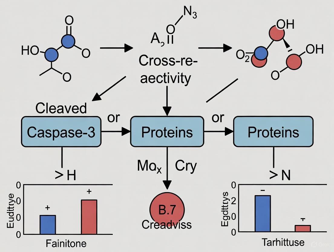
Within the context of broader thesis research on cleaved caspase-3 cross-reactivity, this case study examines a critical specificity challenge encountered in Drosophila apoptosis research. The cleaved caspase-3 (Asp175) antibody from Cell Signaling Technology has become a popular tool for detecting dying cells in Drosophila [7]. This polyclonal antibody was raised against a peptide adjacent to the Asp175 cleavage site in human caspase-3 and was initially assumed to specifically detect cleaved forms of the caspase-3-like effector caspases DRICE and DCP-1 in Drosophila [7]. However, rigorous genetic testing has revealed this assumption to be incorrect, exposing unexpected complexities in the apoptotic pathway and raising important questions about antibody specificity in experimental models. This case study systematically investigates the cross-reactivity of this antibody, identifies the unknown DRONC-dependent proteins it detects, and discusses the implications for interpreting apoptosis experiments in Drosophila systems.
Background: The Drosophila Apoptotic Pathway
Core Apoptotic Machinery
The apoptotic pathway in Drosophila is evolutionarily conserved and consists of key regulatory components. The initiator caspase DRONC (caspase-9-like) is activated through formation of an apoptosome complex with the Apaf-1-related protein ARK [7]. Once activated, DRONC proteolytically processes and activates the effector caspases DRICE and DCP-1 (caspase-3-like), which then execute the cell death program by cleaving various cellular substrates [8]. This cascade is regulated by inhibitor of apoptosis proteins (IAPs), particularly DIAP1, which binds to and inhibits multiple caspases including DRONC [9].
Effector Caspase Functional Relationships
Genetic analyses reveal that DRICE and DCP-1 have partially overlapping functions in the apoptotic pathway [8]. Studies using mutant alleles demonstrate that while some cells (type I) strictly require drICE for apoptosis, other cells (type II) require either drICE or dcp-1, indicating functional redundancy in specific cellular contexts [8]. This partial redundancy complicates the interpretation of single-mutant phenotypes and necessitates careful genetic analysis to delineate specific caspase functions.
Table 1: Key Caspases in Drosophila Apoptosis
| Caspase | Type | Mammalian Homolog | Key Functions | Regulator |
|---|---|---|---|---|
| DRONC | Initiator | Caspase-9 | Apical caspase activated in apoptosome complex | DIAP1 |
| DRICE | Effector | Caspase-3 | Key executioner caspase with partially redundant functions | DIAP1, p35 |
| DCP-1 | Effector | Caspase-3 | Executioner caspase with overlapping functions with DRICE | DIAP1, p35 |
| DCP-2/DREDD | Initiator | Caspase-8 | Involved in specific death pathways | DIAP1 |
Figure 1: Drosophila Apoptotic Signaling Pathway. The pathway depicts key regulatory steps from RHG protein induction to apoptotic execution, highlighting caspase activation hierarchy.
The Discovery: Unexpected Antibody Cross-Reactivity
Initial Observations in Mutant Models
The specificity issue emerged when researchers investigated cleaved caspase-3 antibody labeling in genetic mutants. Surprisingly, eye imaginal discs doubly mutant for the null alleles dcp-1Prev and drICEΔ1 still showed strong immunoreactivity with the cleaved caspase-3 antibody [7]. In wild-type third instar larval eye discs, the antibody typically labels a few scattered dying cells, while in the double mutants, labeling occurred in clusters, similar to patterns observed when cell death was blocked by caspase inhibitor p35 expression [7].
Critical Experiments in GMR-hid Models
To further test antibody specificity under controlled apoptotic conditions, researchers used GMR-hid transgenes that induce apoptosis in two distinct waves in the posterior half of the developing eye [7]. When GMR-hid eye discs were made doubly mutant for dcp-1 and drICE, TUNEL-positive apoptosis was completely abrogated, confirming the essential role of these effector caspases in cell death execution [7]. Remarkably, however, dcp-1 drICE double mutant GMR-hid eye discs still showed strong immunoreactivity with the cleaved caspase-3 antibody [7]. The labeling pattern changed from two distinct waves to filling the entire posterior compartment, suggesting persistent epitope exposure in the absence of cell death.
Table 2: Genetic Evidence for Antibody Cross-Reactivity
| Genetic Background | TUNEL Staining | Cleaved Caspase-3 Antibody Labeling | Interpretation |
|---|---|---|---|
| Wild-type eye disc | Scattered positive cells | Scattered positive cells | Normal apoptosis |
| dcp-1 drICE double mutant | No staining | Persistent clustered labeling | Antibody detects non-effector caspase targets |
| GMR-hid (wild-type) | Two distinct waves | Two distinct waves | Normal induced apoptosis |
| GMR-hid; dcp-1 drICE double mutant | Completely absent | Strong signal throughout posterior compartment | Antibody recognizes non-apoptotic epitopes |
| dronc mutant GMR-hid | Absent | Absent | Labeling requires DRONC activity |
| ark mutant GMR-hid | Absent | Absent | Labeling requires apoptosome function |
Identification of DRONC as the Critical Factor
Genetic Dependency Studies
To determine whether the persistent antibody labeling represented non-specific detection or recognition of upstream apoptotic components, researchers examined GMR-hid eye discs mutant for apoptosome components. In contrast to the dcp-1 drICE double mutants, both dronc and ark mutant GMR-hid eye discs showed complete absence of both TUNEL and cleaved caspase-3 antibody labeling [7]. This demonstrated that the antibody detects a genuine apoptotic epitope, but one that depends on DRONC activity rather than effector caspase function.
Molecular Basis of Cross-Reactivity
Through peptide blocking experiments and sequence analysis, researchers identified the molecular basis for the cross-reactivity. Alignment of the caspase-3 peptide used to generate the antibody with Drosophila caspase sequences revealed that the C-terminal residues "ETD" are conserved in DRICE and DCP-1, representing their activation cleavage sites [7]. However, the N-terminal two-thirds of the caspase-3 peptide showed highest similarity to DRONC, with six out of nine residues conserved [7]. Although DRONC doesn't require cleavage between its large and small subunits for activity, this region appears to be recognized by the antibody in a DRONC-dependent manner.
The Unknown DRONC-Dependent Proteins
Evidence for Additional Substrates
The persistence of cleaved caspase-3 antibody labeling in dcp-1 drICE double mutants, coupled with its complete dependence on DRONC, suggests the antibody detects at least one additional unknown protein that is processed in a DRONC-dependent manner [7]. This unknown substrate may be involved in non-apoptotic functions of DRONC, potentially explaining why the labeling pattern changes in the absence of effector caspases—cells maintain the epitope because they cannot complete apoptosis [7].
Potential Candidates and Implications
While the specific identity of the unknown DRONC-dependent protein(s) remains to be fully characterized, their existence has important implications. In mammalian systems, caspase-3 cleaves numerous substrates beyond effector caspases, including metabolic enzymes like CAD (carbamoyl-phosphate synthetase 2, aspartate transcarbamoylase, and dihydroorotase) [6] and immune signaling components such as NF-κB members [10]. The conservation of this regulatory mechanism suggests Drosophila may possess similar non-apoptotic caspase substrates.
Experimental Protocols for Investigating Cross-Reactivity
Genetic Epistasis Analysis Protocol
To validate antibody specificity and identify unknown caspase targets, the following genetic epistasis protocol can be employed:
Generate mutant tissue using FLP-FRT system to create somatic clones homozygous for various caspase mutations in the developing eye imaginal disc [7] [8].
Induce apoptosis using GMR-hid or similar apoptotic drivers to ensure consistent death signaling across genotypes.
Fix and stain third instar larval eye discs with:
- Primary antibodies: cleaved caspase-3 (1:100-1:200) and appropriate cell type markers
- Secondary antibodies: fluorescently-labeled (e.g., Alexa Fluor 488, 568)
- TUNEL assay to detect DNA fragmentation
- DAPI for nuclear counterstaining
Image and quantify using confocal microscopy with consistent settings across samples. Count positive cells in specific regions (e.g., posterior eye disc) for statistical comparison.
Analyze patterns noting both the presence/absence and distribution of labeling across genotypes.
Peptide Blocking Experiments Protocol
To determine the precise epitope recognized by the antibody in Drosophila:
Synthesize peptides corresponding to:
- The original human caspase-3 immunogen (CQAC(ETD↓SG)
- DRICE cleavage site region
- DCP-1 cleavage site region
- DRONC regions with high similarity
Pre-incubate antibody with 10-fold molar excess of each peptide for 1 hour at room temperature before applying to tissue sections.
Perform immunohistochemistry as normal with peptide-blocked antibodies.
Score staining intensity compared to unblocked controls to identify which peptides effectively compete for antibody binding.
Validate findings by testing peptides with scrambled sequences as negative controls.
Figure 2: Experimental Workflow for Cross-Reactivity Studies. The diagram outlines parallel genetic and biochemical approaches to characterize antibody specificity.
The Scientist's Toolkit: Essential Research Reagents
Table 3: Key Research Reagents for Drosophila Apoptosis Studies
| Reagent/Tool | Type | Key Application | Considerations |
|---|---|---|---|
| Cleaved Caspase-3 (Asp175) Antibody | Rabbit polyclonal antibody | Detection of apoptotic cells in Drosophila | Recognizes DRONC-dependent epitopes beyond effector caspases [7] |
| dcp-1Prev | Null mutant allele | Genetic removal of DCP-1 function | Complete loss of function; used in combination with drICE mutants [7] |
| drICEΔ1 | Null mutant allele | Genetic removal of DRICE function | When combined with dcp-1, eliminates effector caspase activity [7] |
| dronc mutants | Loss-of-function alleles | Testing DRONC dependence | Essential for establishing hierarchy in caspase activation [7] |
| GMR-hid | Transgenic line | Induced apoptosis in eye disc | Provides consistent apoptotic stimulus for comparative studies [7] |
| TUNEL Assay Kit | Biochemical assay | Detection of DNA fragmentation | Gold standard for confirming apoptotic cell death [7] |
| p35 | Baculovirus caspase inhibitor | Broad effector caspase inhibition | Blocks DRICE and DCP-1 but not DRONC [9] |
Implications for Research and Drug Development
Reinterpretation of Existing Data
The findings from this case study necessitate careful reinterpretation of previous studies that relied exclusively on cleaved caspase-3 antibody for detecting apoptosis in Drosophila. Rather than representing effector caspase activity, the antibody should be considered a marker for DRONC activity [7]. This distinction is crucial because DRONC activation can occur without subsequent cell death execution, particularly when effector caspases are inhibited or dysfunctional.
Considerations for Therapeutic Development
For drug development professionals, these findings highlight the importance of understanding pathway specificity when targeting apoptotic components. Compounds designed to modulate cell death based on caspase-3-like activity measurements may require reevaluation if the detection methods cannot distinguish between initiator and effector caspase activities. The existence of unknown DRONC-dependent proteins further suggests potential alternative pathways that could contribute to treatment resistance or off-target effects.
This case study demonstrates that the cleaved caspase-3 antibody cross-reacts with unknown DRONC-dependent proteins beyond its intended targets DRICE and DCP-1 in Drosophila. Through systematic genetic analysis, researchers have established that immunoreactivity with this antibody requires DRONC activity but persists in the absence of both major effector caspases [7]. This discovery has profound implications for apoptosis research, suggesting previously unappreciated complexity in the Drosophila apoptotic pathway and highlighting the critical importance of validating reagent specificity in model organisms.
Future research should focus on identifying the unknown DRONC-dependent proteins recognized by the antibody, characterizing their functions in both apoptotic and non-apoptotic processes, and developing more specific reagents for distinguishing between different caspase activities in experimental models.
The canonical DXXD motif has long been recognized as the signature cleavage site for caspase proteases. However, emerging evidence suggests that caspase-cleaved proteins share common three-dimensional antigenic shapes that transcend their primary amino acid sequences. This whitepaper examines the structural basis for antibody cross-reactivity with cleaved caspase substrates, with particular emphasis on implications for cleaved caspase-3 research. We explore the molecular mechanisms underlying this phenomenon, present experimental approaches for its investigation, and discuss its significant impact on biomarker development and therapeutic discovery.
Caspases (cysteine-dependent aspartate-specific proteases) constitute a family of cysteine proteases that orchestrate programmed cell death and inflammation through precise proteolytic cleavage of cellular substrates [11]. These enzymes exhibit stringent specificity for aspartic acid residues at the P1 position, with additional recognition determinants extending across adjacent residues [12].
Historical Classification of Caspase Cleavage Motifs
Traditional classification systems group caspases based on their tetrapeptide substrate preferences:
- Group I (caspase-1, -4, -14): Preference for (W/L/Y)EHD
- Group II (caspase-2, -3, -7): Preference for DEXD
- Group III (caspase-6, -8, -9, -10): Preference for (L/V/I)EXD [12]
Table 1: Classical Caspase Substrate Specificity Profiles
| Group | Representative Caspases | Preferred Tetrapeptide Motif | Primary Function |
|---|---|---|---|
| I | Caspase-1, -4, -14 | (W/L/Y)EHD | Inflammation |
| II | Caspase-2, -3, -7 | DEXD | Apoptotic execution |
| III | Caspase-6, -8, -9, -10 | (L/V/I)EXD | Apoptotic initiation |
Beyond these classical motifs, recent investigations have identified novel caspase recognition patterns, including the AEAD motif validated as a bona fide caspase cleavage site [13]. This expanding repertoire of recognition sequences raises fundamental questions about the structural constraints governing caspase-substrate interactions.
The Structural Hypothesis: Common Antigenic Shapes in Caspase-Cleaved Proteins
The Neo-Epitope Antibody Approach
A groundbreaking investigation tested the hypothesis that caspase-cleaved proteins share conserved three-dimensional features at their cleavage sites, irrespective of their exact amino acid sequences [1]. Researchers immunized rabbits with a cocktail of the eight most prevalent C-terminal tetrapeptide sequences exposed after caspase cleavage, collectively termed 'DXXD' motifs [1].
The resulting neo-epitope antibodies (NEAs) demonstrated remarkable specificity for exposed C-terminal peptides, selectively recognizing caspase-cleaved forms of known substrates like caspase-6 and PARP from apoptotic cell lysates [1]. Crucially, these antibodies exhibited conformational cross-reactivity, successfully immunoprecipitating caspase-cleaved cytokeratin 18 (CK-18) despite its DALD cleavage site differing from the eight tetrapeptides used for immunization [1].
Structural Basis for Cross-Reactivity
The three-dimensional architecture of caspase active sites imposes strict steric and electrostatic constraints on substrate binding. Although variations exist among caspase family members, the fundamental requirement for accommodating an aspartic acid residue at the P1 position creates conserved features in cleaved products [11]. The catalytic mechanism employing a histidine-cysteine dyad further constrains substrate orientation, potentially generating shared antigenic determinants across different cleavage products [12].
Figure 1: Mechanism of Antibody Recognition of Common Antigenic Shapes in Caspase-Cleaved Proteins
Experimental Evidence and Methodologies
Key Experimental Workflow for Neo-Epitope Antibody Development
The seminal study employed a sophisticated immunization and purification strategy [1]:
- Immunogen Design: Rabbits were immunized with a cocktail of eight peptides containing the most prevalent C-terminally exposed tetrapeptide sequences following caspase cleavage
- Cross-linking Strategy: Each peptide was coupled to two different linker sequences ("Scramble 1" and "Scramble 2") to minimize antibody generation against non-relevant regions
- Affinity Purification: Antibodies were purified using sulfolink affinity resin conjugated with eight novel peptides containing the DXXD motifs but different linker sequences ("Scramble 3")
- Specificity Validation: Purified antibodies were validated through direct ELISAs demonstrating high binding affinity to Scramble 3-DXXD peptides but minimal recognition of similar peptides with internal DXXD motifs
Critical Validation Experiments
The functional validation of neo-epitope antibodies encompassed multiple experimental approaches [1]:
- Immunoprecipitation-Western Blot: NEAs specifically pulled down cleaved forms of caspase-6 (DVVD site) and PARP (DEVD site) from apoptotic HCT116 cell lysates
- Caspase Inhibition Control: Treatment with pan-caspase inhibitor QVD-OPH abolished immunoprecipitation of cleaved substrates, confirming caspase dependence
- Cross-Reactivity Testing: NEAs successfully immunoprecipitated caspase-cleaved cytokeratin 18 containing the non-immunized DALD sequence, demonstrating structural recognition beyond sequence specificity
Table 2: Experimental Evidence for Structural Cross-Reactivity
| Experimental Approach | Key Finding | Implication |
|---|---|---|
| Peptide ELISA | High affinity for C-terminal DXXD, low affinity for internal DXXD | Specificity for neo-epitopes, not linear sequences |
| Immunoprecipitation of known substrates | Selective pull-down of caspase-cleaved PARP and caspase-6 | Recognition of apoptosis-specific cleavage events |
| Cross-reactivity with CK-18 | Recognition of DALD sequence not used in immunization | Specificity based on 3D structure, not exact amino acid sequence |
| In vitro cleavage assay | Capture of recombinant CK-18 only after caspase-3 cleavage | Confirmation of caspase-dependent epitope generation |
Implications for Cleaved Caspase-3 Cross-Reactivity Research
The Caspase-3 Cross-Reactivity Paradigm
Research in Drosophila models revealed that the widely-used cleaved caspase-3 antibody detects multiple proteins in a DRONC (caspase-9-like)-dependent manner, not solely cleaved effector caspases DRICE and DCP-1 [14]. This cross-reactivity persists in drICE and dcp-1 double mutants but is abolished in DRONC and ARK (Apaf-1 related) mutants [14]. These findings position the cleaved caspase-3 antibody as a marker for DRONC activity rather than specifically detecting caspase-3-like proteins.
Broader Biological Implications
The recognition of common antigenic shapes across caspase-cleaved proteins extends beyond antibody cross-reactivity to impact fundamental biological processes:
- Secondary Necrosis: Caspase-3 cleavage of DFNA5 after Asp270 generates a necrotic N-terminal fragment that targets the plasma membrane, mediating progression to secondary necrotic/pyroptotic cell death [15]
- Extracellular Functions: Recent evidence identifies extracellular roles for caspases-3 and -7, which can cleave extracellular domains of membrane-bound proteins upon release during secondary necrosis [16]
- Non-Apoptotic Roles: Caspase-3 participates in diverse non-apoptotic processes including dendritic cell maturation, T-cell activation, and anti-tumor immunity [17]
Research Reagent Solutions
Table 3: Essential Research Tools for Investigating Caspase Cleavage Events
| Reagent / Method | Specific Function | Key Application |
|---|---|---|
| Neo-epitope antibodies (NEAs) | Detection of common structural features in caspase-cleaved proteins | Identification of novel caspase substrates; Apoptosis biomarker detection |
| Pan-caspase inhibitors (zVAD-FMK) | Irreversible inhibition of caspase activity | Experimental controls to confirm caspase-dependent processes |
| Novel caspase inhibitors (Z-AEAD-FMK) | Broad-spectrum caspase inhibition based on newly identified AEAD motif [13] | Inhibition of caspase-mediated cell death pathways triggered by viral infection |
| Site-directed mutagenesis of cleavage sites | Validation of specific caspase recognition motifs | Determination of cleavage site necessity and sufficiency |
| Combination Annexin V/PI staining | Discrimination of apoptotic stages and secondary necrosis | Correlation of caspase activation with cell death progression |
Future Directions and Technical Considerations
Advancing Structural Understanding
Future research should prioritize high-resolution structural studies of caspase-cleaved neo-epitopes in complex with recognizing antibodies. Such investigations would elucidate the precise atomic interactions governing cross-reactivity and facilitate the engineering of antibodies with tailored specificity profiles.
Methodological Recommendations
Researchers employing caspase cleavage detection methodologies should:
- Implement multiple orthogonal approaches to verify caspase-dependent cleavage events
- Utilize caspase inhibition controls to confirm specificity
- Exercise caution when interpreting cleaved caspase-3 immunoreactivity, particularly in non-mammalian systems or when investigating non-apoptotic cellular processes
- Consider potential extracellular caspase functions when designing experimental paradigms
The investigation of common antigenic shapes beyond DXXD motifs represents a paradigm shift in caspase biology. The structural cross-reactivity observed across caspase-cleaved proteins underscores the importance of three-dimensional architecture in caspase substrate recognition and antibody development. This understanding critically informs research on cleaved caspase-3 cross-reactivity while opening new avenues for biomarker discovery, therapeutic development, and fundamental exploration of cell death mechanisms. As research continues to unravel the complex roles of caspases in health and disease, recognition of these shared structural principles will prove essential for accurate experimental design and interpretation.
The Role of Initiator Caspase Activity in Epitope Exposure
In caspase biology research, the detection of specific protein fragments through antibodies is a cornerstone technique. This whitepaper addresses the critical, yet often overlooked, principle that initiator caspase activity is the fundamental prerequisite for the exposure of many epitopes detected in apoptosis research, particularly those associated with effector caspases. The commonly used cleaved caspase-3 antibody (Asp175) is frequently interpreted as a direct marker of effector caspase activity. However, substantial evidence demonstrates that its immunoreactivity primarily serves as a marker for initiator caspase activity, revealing a more complex signaling cascade than generally appreciated [7]. This distinction is not merely academic; it fundamentally shapes the interpretation of experimental data concerning cell death signaling pathways, their activation states, and the validation of caspase-specific reagents. Within a broader thesis on cleaved caspase-3 cross-reactivity, this document establishes that initiator caspases, including caspase-9 and its orthologs, act as the master regulators of neoepitope generation, a concept with profound implications for drug development and diagnostic biomarker design.
The Fundamental Principle: Initiator Caspases Gate Epitope Exposure
The prevailing model of caspase activation involves a hierarchical cascade where initiator caspases (caspase-8, -9, -10) proteolytically activate effector caspases (caspase-3, -6, -7), which then execute the dismantling of the cell [18]. A logical extension of this model is that antibodies targeting cleavage sites on effector caspases directly report on the activity of those effectors. This assumption is technically incorrect in many experimental contexts.
Seminal research in Drosophila models provided a paradigm-shifting insight: the widely used cleaved caspase-3 antibody (raised against the human caspase-3 neoepitope at Asp175) requires the activity of the initiator caspase DRONC (the mammalian caspase-9 ortholog) for its immunoreactivity [7]. Genetic experiments demonstrated that immunoreactivity persisted even in mutants doubly null for the primary caspase-3-like effector caspases, drICE and dcp-1 [7]. In stark contrast, mutations in the apoptosome components dronc and ark (Apaf-1 related) completely abrogated antibody labeling [7]. This genetic evidence compellingly argues that the antibody detects epitopes whose exposure is dependent on DRONC, not the effector caspases themselves. The epitope is likely generated through cleavage of an unknown substrate by active DRONC, independently of DRICE and DCP-1 [7]. Therefore, in this context, the cleaved-caspase-3 antibody is more accurately described as a marker for DRONC activity.
This principle extends to mammalian systems. Initiator caspases-8 and -9 are active during the prolonged delay that precedes mitochondrial outer membrane permeabilization (MOMP) and full effector caspase activation [19]. During this pre-MOMP state, initiator caspases begin processing substrates, but effector caspase activity is restrained by regulatory systems involving XIAP and proteasome-dependent degradation [19]. This creates a cellular state where initiator caspase-dependent cleavage events can occur without the full commitment to apoptosis, highlighting the potential for initiator caspases to expose epitopes prior to, or independent of, massive effector caspase activation.
Key Experimental Evidence and Data
The link between initiator caspase activity and epitope exposure is supported by quantitative studies across multiple model systems. The following table summarizes critical experimental findings that underpin this relationship.
Table 1: Key Experimental Evidence Linking Initiator Caspases to Epitope Exposure
| Experimental Context | Key Perturbation/Finding | Impact on Epitope Detection | Interpretation | Source |
|---|---|---|---|---|
| Drosophila eye disc (GMR-hid) | Double mutant for effector caspases drICE and dcp-1 (null alleles) | Strong immunoreactivity with cleaved caspase-3 antibody persists. | Epitope detection is independent of canonical effector caspases. | [7] |
| Drosophila eye disc (GMR-hid) | Mutant for initiator caspase dronc or apoptosome component ark | Immunoreactivity with cleaved caspase-3 antibody is completely blocked. | Epitope exposure is strictly dependent on the initiator caspase DRONC. | [7] |
| Mammalian Axon Degeneration (DRG neurons) | NGF withdrawal-induced degeneration | Cleavage of caspase-9 observed in axons; XIAP overexpression suppresses caspase-3 activation and degeneration. | Initiator caspase-9 activation is a proximal event in axonal caspase cascade; regulated by XIAP. | [20] |
| Cisplatin-Resistant Mesothelioma (P31res1.2 cells) | Acquired chemoresistance | Basal fragmentation of caspase-8 and -9 without concomitant proteolytic activity. | Caspase fragmentation (epitope exposure) can be uncoupled from catalytic activity, complicating interpretation. | [21] |
| Necrosis-Induced Regeneration (Drosophila wing disc) | DCGluR1-induced necrosis | Caspase activity (NiA) in distant cells is dependent on initiator caspase Dronc. | Non-apoptotic initiator caspase signaling can drive epitope exposure in regenerative contexts. | [22] |
Quantitative data further enriches this narrative. In studies of cisplatin resistance, the acquisition of resistance in P31res1.2 cells led to a clear uncoupling of caspase fragmentation from activity. While these cells exhibited basal fragmentation of initiator caspases-8 and -9, this did not translate to a corresponding increase in their proteolytic function [21]. This finding is critical for drug development, as it demonstrates that detecting a cleaved caspase fragment via antibody binding is not a reliable standalone indicator of enzymatic activity or commitment to apoptosis.
Table 2: Analytical Measurements of Caspase Activity and Fragmentation
| Cell Line / Condition | Caspase-3/7 Proteolytic Activity (DEVD-AFC cleavage) | Caspase-3 Fragmentation (Western Blot) | PARP Cleavage | Interpretation |
|---|---|---|---|---|
| P31 (Parental) | Low basal activity | Not detected under control conditions | Low | Baseline state. |
| P31res1.2 (Cisplatin-Resistant) | Increased basal activity [21] | Not detected by WB; but detected by proteome array [21] | Increased under control conditions [21] | Effector caspase activity is elevated, but initiator caspase fragments may be inactive. |
| P31 + Cisplatin (24h) | Increased | Detected | Increased | Canonical apoptotic response. |
| P31res1.2 + Cisplatin (24h) | Increased | Detected | Increased | Cell death response remains intact despite basal alterations. |
Molecular and Structural Mechanisms
The molecular explanation for initiator caspase-dependent epitope exposure lies in the specificity of protease cleavage. Initiator and effector caspases have distinct, albeit overlapping, substrate specificities defined by the amino acid sequence N-terminal to the cleavage site (the P4-P1 positions) [23].
- Effector Caspases (e.g., caspase-3): Prefer the consensus sequence DEVD [19] [23].
- Initiator Caspases (e.g., caspase-9): Caspase-9 helps dismantle intermediate filaments via direct cleavage of vimentin and contributes to axonal degeneration by cleaving caspase-6 [18]. Its activity creates specific neoepitopes, such as the cleavage of caspase-3 at D175, which is the very epitope detected by the popular cleaved caspase-3 antibody [19].
The generation of a neoepitope requires the proteolytic separation of protein domains, which often unmasks a previously inaccessible peptide sequence. Antibodies like the cleaved caspase-3 antibody are designed to recognize these neoepitopes—in this case, the new C-terminus created at Asp175 [7]. However, the production of this specific neoepitope is catalyzed by an initiator caspase (caspase-8, -9, or -10). The structural basis for antibody specificity can be shape-based rather than strictly sequence-based. Antibodies generated against a cocktail of C-terminal "DXXD" tetrapeptides (a common caspase cleavage product) can recognize a variety of caspase-cleaved proteins, even those with sequences not explicitly used in the immunization, provided they share a similar three-dimensional structure at the cleaved end [23]. This broad cross-reactivity further underscores that a positive signal reflects the upstream activating protease's activity.
Diagram 1: Caspase activation and epitope exposure pathway. The critical role of the initiator caspase in creating the detectable neoepitope is the central, often overlooked, step.
Regulatory mechanisms add further complexity. Proteins like the X-linked Inhibitor of Apoptosis (XIAP) can bind to and inhibit both initiator and effector caspases [18] [20]. In degenerating axons, XIAP-mediated inhibition of caspase-3 and caspase-9 is a major regulatory point, and a reduction in axonal XIAP levels is necessary for caspase activation and degeneration to proceed [20]. Furthermore, caspase-9 itself is regulated by inhibitory phosphorylation and nitrosylation, which interfere with its binding to the activating apoptosome [18]. Therefore, the detection of a neoepitope is the net result of a balance between protease activation and endogenous inhibition.
Research Reagent Solutions and Experimental Protocols
A critical task for researchers is selecting and validating reagents that accurately report on caspase activity. The following toolkit details essential reagents, highlighting their utility and potential pitfalls.
Table 3: Research Reagent Toolkit for Studying Caspase Activity and Epitope Exposure
| Reagent / Tool | Function / Target | Key Consideration / Limitation | Application in This Context |
|---|---|---|---|
| Cleaved Caspase-3 (Asp175) Antibody | Detects neoepitope on cleaved caspase-3. | A positive signal indicates initiator caspase activity has occurred, not necessarily effector caspase activity. Cross-reacts with other DRONC-dependent epitopes [7]. | Widely used for IHC, IF, WB. Interpretation must be cautious. |
| Neoepitope Antibodies (NEA) [23] | Polyclonal antibodies against C-terminal "DXXD" motifs. | Broadly detects multiple caspase-cleaved substrates. Specificity is structure-based, not sequence-specific [23]. | Ideal for identifying novel caspase substrates and global caspase activity. |
| Caspase-9 Neoepitope Antibodies | Specific for caspase-9 cleaved at D315 or D330 [18]. | D315 cleavage is susceptible to XIAP inhibition, while D330 indicates concurrent caspase-3 activity [18]. | Provides direct insight into initiator caspase-9 activation and its regulation. |
| Live-Cell Caspase Reporters (e.g., IC-RP, EC-RP) [19] | FRET-based reporters for initiator (IETD) or effector (DEVD) caspase activity. | Allows real-time, single-cell kinetics of caspase activation. IETD-based IC-RP is a direct readout of initiator caspase activity [19]. | Essential for dissecting the temporal dynamics between initiator and effector caspase activation. |
| XIAP Modulators | Overexpression or knockdown of XIAP. | XIAP is a key endogenous regulator of caspase-3 and -9 [20]. Modulating it tests the restraint on the caspase cascade. | Functional tests to validate if epitope exposure translates to full activation. |
| Genetic Caspase Mutants (e.g., dronc, drICE, dcp-1) [7] | Complete genetic ablation of specific caspases. | Provides definitive evidence of specificity and dependency, as seen in Drosophila studies. | Gold standard for controlling and interpreting antibody cross-reactivity. |
Detailed Experimental Protocol: Validating Initiator Caspase-Dependent Epitope Exposure
This protocol outlines a combined genetic and pharmacological approach to confirm that antibody detection of a neoepitope is dependent on initiator caspase activity, using the cleaved caspase-3 antibody as a primary example.
Workflow Overview:
Diagram 2: Experimental validation workflow.
Step-by-Step Methodology:
Induction of Apoptosis:
Genetic and Pharmacological Perturbations:
- Genetic Knockdown/Knockout: Generate cell lines or use models (like Drosophila) deficient in key caspases.
- Pharmacological Inhibition: Pre-treat cells with a specific initiator caspase inhibitor (where available) alongside the pan-caspase inhibitor.
Sample Preparation and Analysis:
- Prepare cell lysates at various time points post-stimulation.
- Western Blotting:
- Probe membranes with the cleaved caspase-3 antibody.
- Also probe for total/cleaved forms of the perturbed caspases (e.g., caspase-9) to confirm efficacy of knockdown/knockout [21].
- Include a loading control (e.g., GAPDH, Actin).
- Neoepitope Antibody Immunoprecipitation (Optional):
- Use purified neoepitope antibodies (NEA) to immunoprecipitate caspase-cleaved proteins from apoptotic lysates [23].
- Detect pulled-down proteins by western blotting with antibodies against known caspase substrates (e.g., PARP, caspase-6).
- Caspase Activity Assay:
Data Interpretation:
- Expected Outcome (Validating the Principle): Loss of initiator caspase (Group 1) should abolish or drastically reduce the cleaved caspase-3 antibody signal. Loss of effector caspase (Group 2) may have little to no effect on the signal, demonstrating its dependence on the initiator [7].
- Correlation with Activity: A positive cleaved caspase-3 antibody signal should be correlated with a measurable increase in DEVDase activity. A discrepancy (signal without activity) suggests regulated inhibition, as seen in cisplatin-resistant cells [21].
Implications for Research and Drug Development
The misinterpretation of caspase antibody data carries significant consequences. In the context of chemotherapeutic development, a drug candidate that induces cleaved caspase-3 staining might be misinterpreted as successfully activating the apoptotic execution phase. However, if this signal is not coupled with downstream effector caspase activity due to regulatory blocks (e.g., high XIAP), the cells may not die, leading to "undead" cells that can contribute to genomic instability and therapy resistance [19] [21]. The finding that caspase-9 drives neurovascular injury through non-apoptotic endothelial cell dysfunction further emphasizes that caspase activity and neoepitope exposure can signify non-lethal signaling functions, opening new avenues for therapeutic intervention in vascular diseases [18].
For biomarker development, the cross-reactivity of the cleaved caspase-3 antibody with other DRONC-dependent epitopes [7] and the broad specificity of neoepitope antibodies [23] suggest that serum or urine biomarkers detecting caspase-cleaved products are reporting on the upstream initiator caspase activity that initiated the cleavage cascade. This is valuable information, but it must be correctly interpreted. Assays targeting specific neoepitopes, like the caspase-9 cleavage site at D315, could provide more pathway-specific biomarkers for diagnostic or prognostic use [18]. A precise understanding of the caspase cascade and its associated neoepitopes is therefore not just an academic exercise but a fundamental requirement for accurate disease modeling, drug discovery, and clinical translation.
Accurate Detection: Methodologies and Interpreting Cross-Reactive Signals
Application Guidelines for Western Blot, IHC, and Immunofluorescence
The study of precise molecular events, such as the cleavage of caspase-3 during apoptosis, relies heavily on robust protein detection techniques. Western Blot (WB), Immunohistochemistry (IHC), and Immunofluorescence (IF) are cornerstone methods for researchers investigating cleaved caspase-3 cross-reactivity and function. These techniques provide complementary information, from confirming the presence of the specific cleaved protein fragment to revealing its spatial distribution within a tissue or cell. This guide provides a detailed application framework for these techniques within the context of caspase-3 research, addressing the critical need for specificity to ensure accurate interpretation and avoid false-positive results from cross-reactive antibodies.
Technique Selection Guide
Choosing the appropriate technique depends on the specific research question. The table below summarizes the core capabilities of WB, IHC, and IF for apoptosis research.
Table 1: Comparison of Key Protein Detection Techniques for Apoptosis Research
| Feature | Western Blot (WB) | Immunohistochemistry (IHC) | Immunofluorescence (IF) |
|---|---|---|---|
| Primary Information | Protein presence, molecular weight, and relative quantification [24] | Protein localization within the tissue architecture and cellular context [24] | Protein localization and co-localization with other targets, subcellular detail [25] |
| Protein State | Denatured [24] | In situ, but fixed [24] | In situ, but fixed [25] |
| Multiplexing Capacity | Limited (typically 2-4 targets with fluorescent detection) [24] | Moderate (chromogenic) to High (multiplex IF) [24] | High (easily 4+ targets with spectral unmixing) [24] [25] |
| Sensitivity | High [24] | Medium [24] | Medium [25] |
| Specificity Control | Molecular weight confirmation [26] | Cellular and morphological context [24] | Co-localization with organelle markers [25] |
| Best for Caspase-3 Research | Confirming cleavage by detecting ~17 kDa (p17) and ~12 kDa (p12) fragments [27] | Visualizing spatial heterogeneity of apoptosis within tumor tissue sections [6] | Quantifying apoptotic cells and analyzing subcellular activation patterns (e.g., cytoplasmic) [27] |
Detailed Experimental Protocols
Western Blot for Detecting Cleaved Caspase-3
The following protocol is adapted for the specific detection of the activated, cleaved form of caspase-3.
Stage 1: Sample Preparation [26]
- Lysis: Lyse cells or homogenize tissue samples in a suitable ice-cold lysis buffer (e.g., RIPA buffer) supplemented with protease and phosphatase inhibitors to prevent protein degradation and preserve post-translational modifications.
- Quantification: Determine the protein concentration of the supernatant using a Bradford or BCA assay.
- Denaturation: Dilute lysates in Laemmli loading buffer containing Dithiothreitol (DTT)
- Denaturation: Dilute lysates in Laemmli loading buffer containing Dithiothreitol (DTT), boil at 100°C for 10 minutes to fully denature proteins. A typical load is 10-40 µg of total protein per lane [26].
Stage 2: Gel Electrophoresis and Transfer [26]
- Gel Selection: For cleaved caspase-3 (p17 fragment, ~17 kDa), a 4-12% Bis-Tris gel with MOPS buffer is recommended [26].
- Electrophoresis: Load samples and a pre-stained protein ladder. Run the gel at constant voltage until the dye front approaches the bottom.
- Transfer: Transfer proteins from the gel to a nitrocellulose or PVDF membrane using a wet or semi-dry transfer system.
Stage 3: Immunodetection of Cleaved Caspase-3
- Blocking: Incubate the membrane in a blocking solution (e.g., 5% non-fat dry milk or BSA in TBST) for 1 hour at room temperature to prevent non-specific antibody binding [28].
- Primary Antibody Incubation: Incubate membrane with a cleaved caspase-3 (Asp175) specific primary antibody (e.g., Cell Signaling Technology #9579 or its conjugates) diluted in blocking buffer or TBST overnight at 4°C [27].
- Washing: Wash membrane several times with TBST.
- Secondary Antibody Incubation: Incubate with an HRP-conjugated or fluorescently-labeled secondary antibody for 1 hour at room temperature.
- Detection: Develop using chemiluminescent substrate for HRP and image with a CCD system, or directly scan for fluorescence [26].
Immunofluorescence (IF) / Immunohistochemistry (IHC) for Cleaved Caspase-3
The protocols for IF and IHC share many steps, with the key difference being the sample type: cultured cells (IF/ICC) or tissue sections (IHC).
Stage 1: Sample Preparation and Fixation [24] [29]
- Cell Culture (IF): Grow cells on glass coverslips. For cleaved caspase-3, a pre-extraction step before fixation can help reduce background by removing soluble proteins.
- Fixation: Fix cells or tissue sections with freshly prepared 4% Paraformaldehyde (PFA) for 15-20 minutes at room temperature. Note: Methanol fixation can also be used but may destroy some epitopes [24].
- Tissue Preparation (IHC): Perfuse-fix animal with 4% PFA, cryoprotect in sucrose, and embed in OCT medium for frozen sections. Alternatively, paraffin-embed after fixation for formalin-fixed paraffin-embedded (FFPE) sections, which requires subsequent deparaffinization and antigen retrieval [29].
Stage 2: Permeabilization and Blocking [29]
- Permeabilization: Incubate samples in 0.1-0.5% Triton X-100 in PBS for 10-15 minutes to allow antibody access to intracellular targets.
- Blocking: Block samples in 5% serum (from the same species as the secondary antibody host) with 0.05% Triton X-100 for 30-60 minutes at room temperature to minimize non-specific staining.
Stage 3: Antibody Staining and Detection [27] [29]
- Primary Antibody Incubation: Apply the anti-cleaved caspase-3 (Asp175) antibody directly conjugated to a fluorophore (e.g., Alexa Fluor 555) or an unconjugated antibody followed by a conjugated secondary. Incubate for 1-2 hours at room temperature or overnight at 4°C in a humidified chamber. For IHC, a chromogenic detection system like DAB can be used instead [27].
- Multiplexing (Optional): To co-stain with other markers (e.g., cell type-specific proteins), repeat the primary and secondary antibody steps using antibodies from different host species and fluorophores with non-overlapping emission spectra.
- Nuclear Counterstain and Mounting: Incubate with DAPI to label nuclei. Wash and mount slides with an anti-fade mounting medium. Seal coverslips with nail polish [29].
Caspase-3 Activation & Detection
Successful detection of cleaved caspase-3 requires carefully selected, high-quality reagents.
Table 2: Key Research Reagent Solutions for Cleaved Caspase-3 Detection
| Reagent / Resource | Function / Description | Key Considerations for Caspase-3 Research |
|---|---|---|
| Anti-Cleaved Caspase-3 (Asp175) Antibody | Specifically binds the large fragment (p17) of activated caspase-3, but not full-length protein [27]. | Critical for specificity. Verify lack of cross-reactivity with other proteins or cleaved caspases. Validate using positive/negative controls [30]. |
| Protease Inhibitor Cocktail | Added to lysis buffer to prevent protein degradation during sample preparation [26]. | Essential to preserve the cleaved caspase-3 fragment and prevent further degradation by other cellular proteases. |
| Phosphatase Inhibitor Cocktail | Added to lysis buffer to preserve phosphorylation states of proteins [26]. | Important if investigating cross-talk between phosphorylation and caspase-3 cleavage/activity [31]. |
| Fluorophore-Conjugated Antibodies | Antibodies linked to fluorescent dyes (e.g., Alexa Fluor 555) for direct or indirect IF detection [27]. | Enable multiplexing. Choose fluorophores compatible with your microscope's filter sets and with minimal spectral overlap. |
| Mounting Medium with Anti-fade Agent | Preserves fluorescence and protects samples from photobleaching during microscopy [29]. | Crucial for maintaining signal intensity, especially for low-abundance targets like cleaved caspase-3. |
Critical Validation and Controls for Cross-Reactivity Research
A core challenge in cleaved caspase-3 research is ensuring antibody specificity to avoid misinterpretation.
- Positive and Negative Controls: Always include a positive control (e.g., lysates or cells from a known apoptotic sample like staurosporine-treated cells) and a negative control (untreated, healthy cells). For IF/IHC, an isotype control or secondary-only control is essential to identify non-specific binding [27].
- Genetic Validation: Use Caspase-3 Knockout (KO) cells or tissues. Staining or a band in the KO sample indicates non-specific cross-reactivity of the antibody [30].
- Multiple Technique Correlation: Correlate findings across techniques. A signal in WB at the correct molecular weight for cleaved caspase-3, combined with appropriate cellular staining in IF/IHC, strongly supports specific detection [24] [25].
- Mass Spectrometry Verification: For definitive identification, use immunoprecipitation followed by mass spectrometry to confirm the identity of the protein bound by the antibody [31] [30].
Multi-Technique Validation Workflow
Strategies for Differentiating Specific Signal from Non-Specific Background
In research focused on cleaved caspase-3, the accurate differentiation of its specific signal from non-specific background is a fundamental challenge with direct implications for data reliability and experimental conclusions. Caspase-3, a key executioner protease in apoptosis, cleaves after aspartic acid residues and is synthesized as an inactive zymogen that requires proteolytic activation [32]. This activation generates catalytically active subunits, which are typically detected using cleavage-specific antibodies. However, the high cellular abundance of caspase-3 (approximately 200 nM) and the presence of structurally similar proteins create substantial risk for cross-reactivity and background interference [33]. Within the context of cleaved caspase-3 cross-reactivity research, this guide outlines definitive strategies, protocols, and tools to achieve superior signal specificity in molecular detection.
Non-specific background in cleaved caspase-3 detection arises from several technical and biological factors:
- Antibody Cross-Reactivity: Primary antibodies may recognize unrelated proteins that share minimal linear epitopes or three-dimensional conformational motifs with the target caspase-3 cleavage site. This is particularly problematic with some polyclonal antibodies [34].
- Cellular Autofluorescence: Intracellular molecules such as NADPH and flavoproteins emit fluorescence in common detection channels, obscuring specific signals, especially in flow cytometry and immunofluorescence [32].
- Incomplete Blocking: Non-specific binding of antibodies to cellular components occurs when blocking steps are insufficient.
- Endogenous Enzyme Activity: In enzymatic assays, non-caspase proteases may cleave reporter substrates, generating false-positive signals [32].
- Non-Specific Protein-Protein Interactions: Charged or hydrophobic interactions can cause assay reagents to adhere non-specifically to membranes or cellular structures.
Research Reagent Solutions for Enhanced Specificity
The cornerstone of specific detection lies in selecting and validating high-quality reagents. The table below summarizes essential tools and their strategic applications.
Table 1: Key Research Reagent Solutions for Cleaved Caspase-3 Detection
| Reagent | Specific Function | Key Feature for Specificity | Example Applications |
|---|---|---|---|
| Cleavage-Specific Caspase-3 (Asp175) (D3E9) Rabbit mAb [34] | Detects caspase-3 only when cleaved at Asp175 | Minimal cross-reactivity with full-length procaspase-3 or other caspases | IHC, Flow Cytometry, IF (Highly Recommended) [34] |
| Cleavage-Specific Caspase-3 (Asp175) (5A1E) Rabbit mAb [34] | Detects caspase-3 cleaved at Asp175 | Suitable for Western Blot (WB) and Immunoprecipitation (IP) | WB, IP (Very Highly Recommended) [34] |
| Caspase-3/7 Activatable MRI Probe (C-SNAM) [35] | MRI contrast agent activated by caspase-3/7 | Dual activation (reduction + enzyme cleavage) minimizes off-target contrast | In vivo MR Imaging of apoptosis |
| Fluorogenic/Luminescent Substrates (e.g., DEVD-ase) [32] | Caspase-3/7 substrate that emits light upon cleavage | The DEVD sequence provides specificity for caspase-3/7 over other proteases | High-throughput screening, kinetic assays |
| Caspase Inhibitors (e.g., zVAD-fmk, DEVD-CHO) | Irreversibly or reversibly binds caspase active site | Serves as a critical negative control to confirm signal dependence on caspase activity | Control experiments across all methods |
Experimental Protocols for Signal Verification
Protocol: Specificity Validation for Cleaved Caspase-3 Antibodies in Western Blot
This protocol is critical for confirming that an antibody signal represents the true target.
- Step 1: Sample Preparation. Generate lysates from cells under three conditions: (1) Untreated control, (2) Apoptosis-induced (e.g., 1-10 µM Staurosporine for 4-6 hours), and (3) Apoptosis-induced in the presence of a pan-caspase inhibitor (e.g., 20 µM zVAD-fmk).
- Step 2: Blocking with Antigenic Peptide. Pre-incubate the primary antibody (e.g., Cleavage-Specific Caspase-3 (Asp175) Antibody #9661) with a 5-10 fold molar excess of the immunizing peptide for 1 hour at room temperature before applying it to the membrane. This should completely compete away the specific band.
- Step 3: Western Blot Execution. Perform standard SDS-PAGE and western blotting. Include a molecular weight marker to confirm the expected size for cleaved caspase-3 fragments (~17 kDa and ~19 kDa for large and small subunits, and ~12 kDa for the SBDP120 fragment from αII-spectrin breakdown) [33].
- Step 4: Specificity Analysis. A specific signal is confirmed if: (a) It appears only in the apoptotic sample, (b) It is absent in the caspase-inhibited sample, (c) It is blocked by the pre-incubation with the peptide, and (d) Its molecular weight matches the expected size.
Protocol: Knockdown/CRISPR-Cas9 Control for Immunofluorescence
Genetic validation provides the highest level of specificity confirmation.
- Step 1: Generate Caspase-3 Deficient Cells. Use CRISPR-Cas9 to knock out the CASP3 gene in your cell line of interest. Validate the knockout via Western blot and functional assays.
- Step 2: Stimulate and Stain. Treat both wild-type and caspase-3 knockout cells with an apoptosis inducer. Perform standard immunofluorescence for cleaved caspase-3.
- Step 3: Image and Quantify. Acquire images using identical exposure settings between cell lines. Any remaining signal in the knockout cell line under apoptotic conditions is definitively non-specific and can be used to set thresholds for background subtraction in wild-type cells.
Protocol: Orthogonal Validation with Mass Spectrometry
Mass spectrometry (MS) can unequivocally identify caspase-3 cleavage events and its specific substrates, moving beyond antibody-based reliance [32].
- Step 1: In Vitro Cleavage Assay. Incubate recombinant caspase-3 with a candidate protein or cellular lysate. Use a catalytically inactive caspase-3 (C163A mutant) as a negative control.
- Step 2: Sample Preparation for MS. Separate proteins via SDS-PAGE, excise the protein band of interest, and digest it in-gel with trypsin.
- Step 3: LC-MS/MS Analysis. Analyze the resulting peptides using liquid chromatography coupled to tandem mass spectrometry (LC-MS/MS).
- Step 4: Data Analysis. Search the MS/MS spectra against a protein database, specifying caspase-3 cleavage specificity (C-terminal to aspartic acid). The identification of a specific cleavage product, such as the DFNA5-N fragment generated by cleavage after Asp270, provides definitive evidence of caspase-3 activity and substrate identity [15].
Quantitative and Kinetic Approaches
Quantitative kinetics provide a powerful layer of specificity. Different cleavage sites on the same substrate can have vastly different catalytic efficiencies, which can be measured to confirm specific caspase-3 activity.
Table 2: Kinetic Parameters for Caspase-3 Cleavage of αII-Spectrin
| Cleavage Site in αII-Spectrin | Catalytic Efficiency (kcat/KM) | Resulting Breakdown Product (SBDP) | Implication for Specificity |
|---|---|---|---|
| After D1185 [33] | 40,000 M⁻¹s⁻¹ | SBDP150 | Unusually efficient; a dominant, specific signal in apoptosis. |
| After D1478 [33] | 3,000 M⁻¹s⁻¹ | SBDP120 | More typical efficiency; requires verification against background. |
The data in Table 2 shows that measuring the rate of SBDP150 formation provides a more specific indicator of caspase-3 activity than SBDP120, as its high catalytic efficiency makes it less likely to be generated by non-specific proteolysis [33]. This kinetic profiling can be applied using fluorogenic substrates in real-time assays.
Visualizing Signaling Pathways and Experimental Workflows
The following diagrams illustrate the key pathways and a consolidated experimental strategy for ensuring signal specificity.
Caspase-3 Activation and Cross-Talk in Cell Death
Experimental Workflow for Specific Signal Verification
Differentiating specific cleaved caspase-3 signal from non-specific background is not achieved by a single method but through a convergent strategy. This involves using highly validated, cleavage-specific reagents, implementing rigorous controls including pharmacological inhibition and genetic knockout, and employing orthogonal detection methods like mass spectrometry. Furthermore, leveraging quantitative kinetic data can provide an additional, powerful layer of validation. As research into caspase-3 cross-reactivity advances, the adoption of these comprehensive strategies will be paramount in generating reliable, reproducible data that accurately reflects biological reality and drives the development of effective therapeutic interventions.
Leveraging Broad-Spectrum Neo-Epitope Antibodies for Apoptosis Detection
The detection of apoptosis, a fundamental process in development, homeostasis, and disease, relies heavily on identifying specific molecular markers that signify cell death activation. Among these markers, caspase-3 stands as a critical executioner protease whose activation serves as a definitive point of commitment to apoptotic cell death. Traditional antibody-based detection methods face challenges with specificity, particularly due to cross-reactivity concerns with structurally similar proteins. The emergence of neo-epitope antibody technology offers a transformative approach for detecting specific proteolytic events with high precision. These antibodies are designed to recognize unique peptide sequences—neo-epitopes—that are exposed only after precise proteolytic cleavage events, such as caspase-mediated substrate processing. This technical guide explores the application of broad-spectrum neo-epitope antibodies for specific apoptosis detection within the broader context of cleaved caspase-3 cross-reactivity research, providing detailed methodologies and analytical frameworks for researchers and drug development professionals.
Computational Identification of Apoptosis-Associated Neo-Epitopes
The targeted discovery of apoptosis-specific neo-epitopes requires sophisticated computational pipelines that integrate genomic, proteomic, and structural data. These pipelines identify proteolytic cleavage sites and generate antibodies against the newly exposed peptide sequences.
Neo-Epitope Source Characterization
Apoptosis-associated neo-epitopes originate from specific proteolytic cleavage events, primarily mediated by caspases. The table below summarizes the primary sources and characteristics of these neo-epitopes.
Table 1: Sources and Characteristics of Apoptosis-Associated Neo-Epitopes
| Source Type | Description | Key Features | Relevant Caspases |
|---|---|---|---|
| Caspase Substrates | Proteins cleaved during apoptosis execution | Exposed C-termini after aspartic acid residues; highly specific to activated caspases | Caspase-3, -7, -6, -8 |
| Structural Variations | Genomic alterations generating novel protein sequences | Not derived from normal proteome; completely tumor-specific | N/A |
| Post-Translational Modifications | Modified proteins creating novel immune epitopes | Can include phosphorylation, acetylation, or oxidation | N/A |
Bioinformatics Workflow for Neo-Epitope Prediction
The identification of optimal neo-epitopes follows a multi-stage computational pipeline:
Whole Exome Sequencing: Identifies somatic mutations and potential novel protein sequences through comparison of tumor and normal tissue [36] [37]. This approach typically yields 243-665 non-synonymous mutations per sample, with approximately 50.7% being expressed [36].
HLA Binding Prediction: Utilizes algorithms such as NetMHCpan and IEDB to predict binding affinity between candidate peptides and patient-specific HLA molecules [38] [37]. Peptides with IC50 mutant <500 nM and IC50 mutant < IC50 wild type are typically classified as high-affinity binders.
Molecular Dynamics Simulation: Models the structural accessibility of candidate neo-epitopes following caspase cleavage, prioritizing epitopes with stable presentation.
Figure 1: Computational pipeline for neo-epitope identification and validation
Research Reagent Solutions for Neo-Epitope-Based Apoptosis Detection
The experimental validation of apoptosis-associated neo-epitopes requires specialized reagents and systems. The table below details essential research tools and their applications.
Table 2: Research Reagent Solutions for Neo-Epitope Apoptosis Detection
| Reagent Category | Specific Examples | Function & Application | Key Features |
|---|---|---|---|
| Detection Antibodies | Anti-CLEAVED caspase-3 (Asp175); Neo-epitope specific mAbs | Specific detection of apoptosis-activated caspases; identifies unique cleavage products | High specificity for neo-epitopes; minimal cross-reactivity with full-length proteins |
| Activity-Based Probes | Ac-DEVD-CL chemiluminescent probe [4] | Direct measurement of caspase-3 enzymatic activity in live cells | 5000-fold signal increase upon activation; 100x lower detection limit vs. fluorescent probes |
| Cell Culture Models | HL-60; HGC27; HCT116; SW480 [6] [39] | Apoptosis induction and response quantification | Well-characterized apoptosis pathways; chemosensitivity profiles |
| Stimulation Systems | Artificial antigen-presenting cells (aAPCs) [37] | T-cell activation and neoantigen immunogenicity validation | HLA-A2-coated systems for peptide presentation |
| Apoptosis Inducers | 5-FU; Oxaliplatin; Actinomycin D; Cytochrome c/dATP [6] [39] | Controlled induction of apoptotic pathways | Activation of intrinsic and extrinsic apoptosis pathways |
Experimental Protocols for Caspase-3 Neo-Epitope Validation
Protocol: Caspase Activation Assay in Cell-Free System
This cell-free protocol enables controlled study of caspase activation mechanisms without confounding cellular processes.
Materials:
- Cell extraction buffer (25 mM HEPES, pH 7.4, 5 mM MgCl₂, 1 mM EGTA)
- Cytochrome c (10 µM final concentration)
- dATP (1 mM final concentration)
- GSSG (0-1 mM for inhibition studies) [39]
- Caspase assay buffer (50 mM HEPES, pH 7.4, 100 mM NaCl, 1 mM EDTA, 10% glycerol, 0.1% CHAPS)
Procedure:
- Prepare cellular extracts from HL-60 or other appropriate cell lines by homogenization in caspase activation buffer.
- Centrifuge extracts at 14,000 × g for 15 minutes at 4°C.
- Pre-incubate aliquots (100 µg protein) with varying concentrations of GSSG (0-1 mM) for 90 minutes at room temperature.
- Initiate caspase activation by adding cytochrome c and dATP to the extracts.
- Incubate at 37°C for 60 minutes to allow procaspase processing.
- Assess activation by Western blot analysis using neo-epitope-specific antibodies or measure caspase activity using colorimetric substrates (Ac-DEVD-pNA for caspase-3).
Protocol: Immunogenicity Validation of Neo-Epitopes
This protocol validates the immune recognition of identified neo-epitopes, crucial for therapeutic antibody development.
Materials:
- HLA-A2-coated artificial antigen-presenting cells (aAPCs)
- Dimeric HLA-A2 molecules produced via baculovirus expression [37]
- CD8+ T cells from healthy donors
- IFN-γ ELISA kit
- Candidate neo-epitope peptides
Procedure:
- Generate HLA-A2/peptide complexes by incubating HLA-A2 molecules with candidate neo-epitope peptides.
- Coat artificial APCs with HLA-A2/peptide complexes.
- Co-culture coated aAPCs with CD8+ T cells from multiple donors.
- Monitor T-cell proliferation over 5-7 days using flow cytometry or tritiated thymidine incorporation.
- Measure IFN-γ secretion in supernatants using ELISA to quantify T-cell activation.
- Compare responses to mutant peptides versus wild-type peptides to confirm specificity.
Caspase-3 Cross-Reactivity: Molecular Mechanisms and Solutions
Structural Basis of Caspase-3 Specificity and Cross-Reactivity
Caspase-3 belongs to the group II caspases with preference for DEXD substrate recognition motifs [12]. The enzyme contains a conserved pentapeptide sequence QACXG with a critical cysteine residue at the active site that is essential for catalytic activity. Structural analyses reveal that:
- The caspase-3 active site cysteine (Cys163) is susceptible to post-translational modifications, including glutathionylation at Cys135 and Cys45, which inhibits both activation and activity [39].
- Neo-epitope antibodies targeting the cleavage site of caspase-3 (e.g., after Asp175) provide superior specificity compared to antibodies recognizing the full-length protein.
- The functional continuum model suggests caspase-3 exhibits different functions based on activity gradients and spatiotemporal localization [40], complicating detection with conventional antibodies.
Caspase-3 in Cell Death Pathways
Understanding caspase-3's role in multiple cell death pathways is essential for appropriate detection strategy selection.
Figure 2: Caspase-3 functions in multiple cell death pathways
Quantitative Assessment of Caspase-3 Detection Methods
The table below compares the performance characteristics of different caspase-3 detection methodologies, highlighting the advantages of neo-epitope approaches.
Table 3: Performance Comparison of Caspase-3 Detection Methods
| Detection Method | Principle | Limit of Detection | Advantages | Cross-Reactivity Concerns |
|---|---|---|---|---|
| Traditional Western Blot | Antibody recognition of caspase-3 protein | ~10 ng | Semi-quantitative; well-established | High with procaspase-3 and other caspases |
| Neo-Epitope Western | Antibody specific to cleaved form | ~5 ng | High specificity for activated caspase-3 | Minimal with proper epitope mapping |
| Fluorescent Probes (Ac-DEVD-AMC) | Enzyme activity measurement | ~100 pM | Real-time monitoring in live cells | Cross-reactivity with caspase-7, -8 |
| Chemiluminescent Probes (Ac-DEVD-CL) [4] | Enzymatic cleavage triggering light emission | ~1 pM | 100x lower LOD vs. fluorescence; minimal background | Specific to DEVD-cleaving caspases |
| Neo-Epitope Flow Cytometry | Cell-surface detection of cleavage products | ~100 events | Single-cell resolution; multiparameter analysis | Dependent on antibody quality |
Discussion and Future Perspectives
The development of broad-spectrum neo-epitope antibodies for apoptosis detection represents a significant advancement in cell death research and drug discovery. The specificity afforded by these reagents addresses longstanding challenges in caspase-3 cross-reactivity, particularly important in the context of:
- Therapeutic Monitoring: Evaluating response to chemotherapeutic agents where CAD cleavage by caspase-3 determines chemosensitivity [6].
- Immunotherapy Development: Combining neo-epitope detection with neoantigen-targeted therapies [41] for enhanced cancer treatment strategies.
- Disease Mechanism Elucidation: Understanding the role of sublethal caspase-3 activation in neural plasticity, immune regulation, and metabolic reprogramming [40].
Future directions should focus on expanding the neo-epitope antibody toolkit to cover more caspase substrates, developing standardized validation protocols, and creating multiplexed detection platforms that can simultaneously monitor multiple apoptosis markers in complex biological systems. The integration of these approaches with emerging technologies in spatial biology and single-cell analysis will further enhance our understanding of apoptotic processes in health and disease.
Correlating Cleaved Caspase-3 Signal with Other Apoptotic Markers
Caspase-3 is a critical executioner protease that mediates the final stages of apoptosis, playing an indispensable role in the systematic dismantling of cellular structures [32]. This enzyme is synthesized as an inactive zymogen that requires proteolytic cleavage for activation, typically at aspartic acid residues, to form the active heterotetramer consisting of two large and two small subunits [42]. The detection of cleaved caspase-3 serves as a definitive indicator of apoptotic commitment, as this activated form specifically targets key cellular substrates including poly(ADP-ribose) polymerase (PARP), cytokeratins, and various structural proteins [43]. Within the context of broader research on caspase-3 cross-reactivity, understanding the correlation between cleaved caspase-3 signals and other apoptotic markers becomes paramount for accurate interpretation of experimental data, particularly in drug development and therapeutic efficacy studies [44].
The molecular activation of caspase-3 occurs downstream of both intrinsic (mitochondrial) and extrinsic (death receptor) apoptotic pathways [45]. In the intrinsic pathway, cellular stress signals trigger mitochondrial outer membrane permeabilization, leading to cytochrome c release and formation of the apoptosome complex, which activates caspase-9, which in turn cleaves and activates caspase-3 [45]. In the extrinsic pathway, death receptor engagement activates caspase-8, which can directly process caspase-3 [44]. Once activated, caspase-3 amplifies the apoptotic cascade by cleaving numerous cellular substrates, ultimately leading to the characteristic morphological changes associated with apoptosis, including chromatin condensation, DNA fragmentation, and formation of apoptotic bodies [45].
Apoptotic Signaling Pathways and Key Molecular Markers
Integrated Cell Death Signaling Network
The following diagram illustrates the central position of caspase-3 within the apoptotic signaling network and its relationships with other key markers:
Key Apoptotic Markers for Correlation Studies
Table 1: Core Apoptotic Markers for Correlation with Cleaved Caspase-3
| Marker Category | Specific Marker | Biological Significance | Detection Methods |
|---|---|---|---|
| Upstream Initiators | Activated Caspase-8 | Extrinsic pathway initiation | Western blot, IHC, FRET |
| Activated Caspase-9 | Intrinsic pathway initiation | Western blot, IHC | |
| Cytochrome c release | Mitochondrial commitment | IHC, fluorescence imaging | |
| Parallel Execution | Activated Caspase-7 | Redundant effector function | Western blot, fluorescent reporters |
| Cleaved PARP | DNA repair disruption | Western blot, ELISA (M30) | |
| Downstream Substrates | Cleaved Cytokeratin-18 | Structural protein breakdown | ELISA (M30) |
| DNA fragmentation | Nuclear disintegration | TUNEL assay | |
| Cellular Responses | Phosphatidylserine exposure | "Eat-me" signal | Annexin V staining |
| Membrane permeability | Loss of integrity | Propidium iodide uptake |
Current Detection Methodologies and Technologies
Established Caspase-3 Detection Platforms
The landscape of caspase-3 detection methodologies has evolved significantly from traditional antibody-based approaches to sophisticated real-time imaging platforms [32]. Classical techniques including Western blotting, immunohistochemistry (IHC), and enzyme-linked immunosorbent assays (ELISAs) provide fundamental insights into caspase-3 expression and activation but offer limited temporal resolution [43]. Western blotting remains the gold standard for confirming the presence of cleaved caspase-3 based on molecular weight shifts, with the cleaved active form typically detected at ~17/19 kDa compared to the ~32 kDa pro-form [32]. Immunohistochemistry enables spatial localization within tissue architectures but is predominantly semi-quantitative and requires careful validation to address cross-reactivity concerns [43].
More recent advancements have introduced genetically encoded fluorescent biosensors that enable real-time monitoring of caspase-3 activation dynamics in live cells [46] [47]. These systems typically employ cleavage-sensitive constructs where caspase-3-mediated separation of fluorescent protein fragments allows reconstitution of fluorescence signals [46]. The ZipGFP-based reporter system represents one such innovation, utilizing a split-GFP architecture with a DEVD cleavage motif that prevents proper folding until caspase-3/-7-mediated cleavage occurs, resulting in minimal background fluorescence and rapid signal amplification upon activation [47]. This technology has been successfully adapted for both 2D cultures and complex 3D models including spheroids and patient-derived organoids, providing unprecedented spatial and temporal resolution of apoptotic events [47].
Experimental Workflow for Multi-Parameter Apoptosis Assessment
Table 2: Correlation Experiment Workflow and Methodologies
| Experimental Phase | Key Procedures | Technical Considerations | Cross-Validation Requirements |
|---|---|---|---|
| Sample Preparation | Cell treatment with apoptosis inducers; Primary tissue processing; Time-course sampling | Optimal fixation for epitope preservation; Avoidance of accidental necrosis; Appropriate positive controls | Consistent handling across all assays; Multiple biological replicates |
| Parallel Detection | Western blot for cleaved caspase-3; M30 ELISA for cleaved CK18; TUNEL for DNA fragmentation; Annexin V/PI flow cytometry | Antibody validation for cross-reactivity; Signal linearity confirmation; Compensation controls for multiplex flow | Internal reference standards; Normalization to protein/cell count |
| Real-Time Monitoring | Live-cell imaging with fluorescent reporters; IncuCyte systems; High-content screening platforms | Background fluorescence minimization; Environmental control; Phototoxicity avoidance | Control reporters with mutated cleavage sites; Caspase inhibitor validation |
| Data Integration | Multiplexed data analysis; Temporal correlation assessment; Spatial mapping in tissues | Normalization strategies; Statistical power analysis; Batch effect correction | Cross-platform correlation coefficients; Independent method confirmation |
Multiparameter Apoptosis Detection Workflow
The following diagram outlines a comprehensive experimental approach for correlating cleaved caspase-3 with other apoptotic markers:
Research Reagent Solutions and Experimental Tools
Table 3: Essential Research Reagents for Caspase-3 Correlation Studies
| Reagent Category | Specific Examples | Application Function | Validation Requirements |
|---|---|---|---|
| Activation Inducers | Carfilzomib (proteasome inhibitor); Oxaliplatin (DNA damage); TNF-α + cycloheximide (extrinsic) | Induction of specific apoptotic pathways; Positive controls for assay validation | Dose-response optimization; Time-course establishment |
| Caspase Inhibitors | Z-VAD-FMK (pan-caspase); Z-DEVD-FMK (caspase-3/7 specific); Q-VD-OPh (broad-spectrum) | Specificity controls; Pathway inhibition studies; Background determination | Titration for complete inhibition; Cytotoxicity assessment |
| Validated Antibodies | Anti-cleaved caspase-3 (Asp175); Anti-cleaved PARP (Asp214); M30 anti-CK18 (Asp396) | Immunodetection in fixed samples; Specific epitope recognition | Cross-reactivity screening with related caspases; Knockout validation |
| Fluorescent Reporters | ZipGFP-DEVD constructs; FRET-based SCAT probes; NucView 488 substrates | Real-time activation kinetics; Single-cell analysis; High-throughput screening | Background fluorescence assessment; Cleavage specificity confirmation |
| Detection Kits | M30/M65 ELISA kits; TUNEL assay kits; Annexin V/PI staining kits | Multiplexed endpoint analysis; Standardized protocols across labs | Linearity and dynamic range; Inter-assay reproducibility |
Interpretation Framework and Technical Considerations
Correlation Analysis and Data Interpretation
Establishing robust correlation between cleaved caspase-3 and other apoptotic markers requires careful consideration of temporal dynamics, as various markers appear at different stages of the apoptotic process [43]. Cleaved caspase-3 typically emerges after initiator caspase activation (caspase-8 or -9) but before complete biochemical and morphological changes such as DNA fragmentation and apoptotic body formation [45]. The M30 epitope (cleaved cytokeratin-18) generally appears contemporaneously with or shortly after caspase-3 activation, while DNA fragmentation detectable by TUNEL staining often represents a later event [43]. Understanding these temporal relationships is crucial for accurate experimental design and interpretation, particularly when assessing the effects of novel therapeutic agents.
Quantitative correlation analysis should account for the different dynamic ranges and detection sensitivities of various apoptotic assays [32]. For instance, fluorescent reporter systems may detect caspase-3 activation earlier than Western blotting due to their amplification capabilities and single-cell resolution [46]. Similarly, flow cytometric analysis of Annexin V binding detects phosphatidylserine externalization, which can occur prior to caspase-3 activation in certain cell types or under specific conditions [43]. These methodological considerations must be incorporated into the interpretation framework to avoid erroneous conclusions about apoptotic progression and therapeutic efficacy.
Addressing Cross-Reactivity and Specificity Challenges
Within the context of caspase-3 cross-reactivity research, specificity validation remains paramount [42]. Caspase-3 shares significant structural homology with other executioner caspases, particularly caspase-7, which also cleaves DEVD-based substrates and can potentially confound results if detection methods lack sufficient specificity [32] [47]. Multiple control strategies should be employed, including caspase-specific inhibitors, genetic knockdown approaches, and the use of caspase-3-deficient cell lines (such as MCF-7 cells) to verify signal specificity [46] [47].
Additional validation should include assessment of potential cross-reactivity with inflammatory caspases, particularly in models involving immune activation or tissue injury [42]. The growing recognition of caspase-3's role in non-apoptotic processes, including cellular differentiation and immunogenic cell death, further complicates interpretation and necessitates careful experimental design with appropriate contextual markers [42] [47]. Multiparameter approaches that simultaneously assess multiple apoptotic indicators provide the most robust framework for accurately correlating cleaved caspase-3 signals with authentic apoptotic commitment, thereby advancing both basic research and drug development applications.
Resolving Ambiguity: Troubleshooting False Positives and Optimizing Specificity
Identifying and Mitigating Non-Specific Labeling in Healthy Cells
The detection of apoptotic cells via cleaved caspase-3 is a cornerstone of biomedical research, particularly in cancer biology, neurobiology, and immunology. However, the reliability of this detection is fundamentally compromised by non-specific labeling in healthy, non-apoptotic cells—a methodological pitfall that can significantly distort experimental interpretations and therapeutic evaluations. This technical guide examines the molecular basis of cleaved caspase-3 cross-reactivity and provides evidence-based strategies to identify and mitigate non-specific labeling. Within the broader context of caspase-3 cross-reactivity research, understanding these limitations is paramount for accurate biomarker interpretation, especially in drug development where off-target effects and therapeutic efficacy assessments depend on precise apoptosis quantification. The following sections detail experimental protocols, validation methodologies, and advanced techniques to enhance detection specificity, providing researchers with a comprehensive framework for improving assay reliability.
Understanding the Molecular Basis of Cross-Reactivity
The cleaved caspase-3 antibody (Asp175), a widely used apoptosis marker, demonstrates concerning cross-reactivity that extends beyond its intended targets. Originally raised against a peptide adjacent to Asp175 in human caspase-3, this antibody was designed to detect the activated large fragment (17/19 kDa) of the effector caspase while ignoring the full-length, inactive zymogen [48]. However, rigorous genetic studies in Drosophila models reveal that strong immunoreactivity persists in apoptotic cells even when genes for the caspase-3-like effector enzymes DRICE and DCP-1 are doubly mutated [7]. This finding fundamentally challenges the antibody's presumed specificity.
Further investigation demonstrates that immunoreactivity is completely abolished in mutants lacking the apoptosome components DRONC (Caspase-9-like) or ARK (Apaf-1 related), which function upstream of effector caspases [7]. This genetic evidence indicates that the cleaved caspase-3 antibody does not exclusively detect cleaved effector caspases but instead recognizes multiple proteins in a DRONC-dependent manner. The antibody appears to detect an apoptotic epitope that requires DRONC activity for exposure but persists independently of the primary effector caspases DRICE and DCP-1 [7]. Consequently, researchers should interpret positive signals as indicators of DRONC activity rather than definitive evidence of effector caspase activation, a critical distinction when investigating apoptotic pathways and cellular stress responses.
Detection Methods and Their Limitations
Established Detection Platforms
Multiple detection platforms utilizing cleaved caspase-3 antibodies are employed in research settings, each with distinct advantages and limitations for specificity assessment. Flow cytometry applications using fixed and permeabilized cells typically employ antibody dilutions of 1:50 for direct detection of the endogenous protein [48]. This approach enables quantitative analysis at single-cell resolution but remains susceptible to cross-reactivity issues inherent to the antibody. Western blotting provides molecular weight verification, potentially distinguishing the authentic 17/19 kDa caspase-3 fragment from cross-reacting proteins, while immunohistochemistry offers spatial context within tissues but may amplify non-specific signals through secondary detection systems.
The standard indirect detection method utilizes an unlabeled primary antibody followed by a labeled secondary antibody, providing signal amplification but increasing the risk of non-specific binding through secondary antibody interactions with cellular components [49]. Direct methods using pre-labeled primary antibodies eliminate secondary antibody cross-reactivity but offer less signal amplification and are commercially available for limited targets [50]. For cleaved caspase-3 detection, the Alexa Fluor 488-conjugated antibody represents a commercially available direct option validated for flow cytometry in human cells [48].
Comparative Analysis of Detection Methods
Table 1: Comparison of Cleaved Caspase-3 Detection Methods
| Method | Key Advantage | Primary Limitation | Specificity Concerns |
|---|---|---|---|
| Flow Cytometry | Quantitative single-cell resolution | Requires cell fixation/permeabilization | Cross-reactivity with non-target proteins of similar size |
| Western Blot | Molecular weight verification | Loses cellular context | May not distinguish cross-reactive proteins of similar molecular weight |
| Immunohistochemistry | Preserves tissue architecture | Semi-quantitative at best | Signal amplification can magnify non-specific binding |
| Direct Detection | Eliminates secondary antibody artifacts | Limited commercial availability | Same cross-reactivity issues as unconjugated primary antibody |
| Indirect Detection | High signal amplification | Secondary antibody non-specificity | Combined primary and secondary non-specific interactions |
Experimental Protocols for Specificity Validation
Blocking Peptide Assays
Peptide blocking experiments represent a fundamental approach for validating antibody specificity. These assays determine whether the observed immunoreactivity can be competed away by pre-incubating the antibody with the antigenic peptide used for immunization.
Protocol:
- Prepare blocking solution: Reconstitute the immunizing peptide at 1-10 mM concentration in PBS or antibody dilution buffer.
- Pre-absorb antibody: Mix cleaved caspase-3 antibody at working concentration with a 5-10 molar excess of blocking peptide.
- Incubate: Rotate mixture for 2-4 hours at room temperature or overnight at 4°C.
- Apply to samples: Use the pre-absorbed antibody solution alongside untreated antibody on parallel samples.
- Compare signals: Significant reduction in staining intensity with pre-absorbed antibody confirms specificity, while persistent signal suggests cross-reactivity.
Research indicates that the tripeptide sequence ETD (Glu-Thr-Asp) represents the core apoptotic epitope detected by cleaved caspase-3 antibody in Drosophila models [7]. Blocking with peptides containing this sequence can help distinguish specific from non-specific binding events.
Genetic Controls for Specificity Assessment
Genetic approaches provide the most rigorous specificity validation by directly eliminating target protein expression.
Protocol for Genetic Validation:
- Select model system: Utilize CRISPR/Cas9 or RNAi to generate caspase-3 knockout or knockdown cells.
- Validate knockout: Confirm complete absence of target protein via Western blot.
- Apply antibody: Process wild-type and knockout cells in parallel using identical detection protocols.
- Compare signals: Complete absence of signal in knockout cells confirms antibody specificity, while residual staining indicates cross-reactivity.
As noted in Drosophila studies, "the cleaved-Caspase-3 antibody still labels cells in eye discs doubly mutant for the null alleles dcp-1Prev and drICEΔ1" [7], demonstrating that genetic controls can reveal unexpected cross-reactivities even when the presumed targets are eliminated.
Dual-Labeling Approaches to Identify Non-Specific Binding
Dual-labeling strategies enable researchers to distinguish specifically labeled cells from those with non-specific uptake capacity.
Protocol for Dual-Labeling:
- Prepare antibodies: Conjugate specific antibody (e.g., anti-cleaved caspase-3) and isotype control antibody with distinct fluorophores (e.g., Alexa Fluor 488 and Alexa Fluor 647).
- Co-inject/co-apply: Administer both labeled antibodies simultaneously to cells or experimental animals.
- Detect signals: Use a two-channel detection system to identify single-positive (specifically labeled) versus double-positive (non-specifically labeled) cells.
- Quantitate: Calculate the ratio of specific to non-specific labeling under experimental conditions.
This approach is particularly valuable for in vivo flow cytometry, where "cells with such non-specific uptake capacities will be falsely counted as labeled cells" [51]. The best way to eliminate such false positives is through dual-labeling schemes where "specifically labeled cells (single positive) can then be distinguished from the nonspecifically labeled cells (double positive)" [51].
Research Reagent Solutions for Enhanced Specificity
Essential Reagents for Non-Specificity Assessment
Table 2: Key Research Reagents for Mitigating Non-Specific Labeling
| Reagent/Category | Function | Application Notes |
|---|---|---|
| Isotype Control Antibodies | Distinguish specific from non-specific binding | Must match the host species, isotype, and conjugation of primary antibody |
| Blocking Peptides | Confirm antibody specificity through competition | Should correspond to the immunogen sequence; use in 5-10× molar excess |
| Fc Block (anti-CD16/32) | Reduce Fc receptor-mediated non-specific binding | Critical for immune cell studies; pre-incubate before antibody application |
| Biotin/Avidin Systems | Enable signal amplification with minimal non-specificity | NeutrAvidin recommended over Streptavidin to avoid bacterial recognition sequence background [49] |
| PolySpecificity Particle (PSP) Assay | High-sensitivity detection of antibody polyspecificity | Uses Protein A beads to capture antibodies; detects non-specific interactions with defined reagents like ovalbumin [52] |
Advanced Labeling Technologies
Innovative labeling strategies can circumvent traditional antibody-based detection limitations. Metabolic glycoengineering approaches, for example, enable highly specific cell tracking without the phagocytosis-associated signal distortion that plagues conventional methods [53]. This technique utilizes tetra-acetylated N-azidoacetyl-D-mannosamine (Ac4ManNAz) to generate unnatural sialic acids with azide groups on target cell surfaces, followed by administration of dibenzyl cyclooctyne-conjugated Cy5 (DBCO-Cy5) that specifically binds azide groups via bioorthogonal click chemistry [53]. This method demonstrates 100% labeling efficacy and significantly reduces false signals from macrophage phagocytosis compared to traditional DiD labeling [53].
For caspase-3 detection specifically, the Alexa Fluor 488-conjugated cleaved caspase-3 antibody provides a validated direct detection option that eliminates secondary antibody cross-reactivity [48]. This reagent is purified by protein A and peptide affinity chromatography and conjugated with optimal fluorophore-to-protein (F/P) ratios of 2-6 to maintain both brightness and antigen recognition [48].
Workflow for Systematic Specificity Validation
Diagram 1: Systematic workflow for identifying and mitigating non-specific labeling, incorporating essential controls and validation strategies.
Non-specific labeling in healthy cells presents a significant challenge in apoptosis research and drug development, particularly when relying on cleaved caspase-3 as a biomarker. The cross-reactivity of commonly used antibodies with non-target proteins necessitates rigorous validation using the systematic approaches outlined in this guide. Through appropriate control experiments, genetic validation, and advanced detection strategies, researchers can significantly improve the reliability of their apoptosis assessments. The implementation of these methodologies will enhance data quality in both basic research and therapeutic development, ultimately leading to more accurate interpretation of cellular responses in experimental and clinical contexts.
Optimizing Buffer Conditions, Titers, and Blocking Protocols
Accurate detection of cleaved caspase-3, a key executioner protease in apoptosis, is fundamental to research in cancer, neurodegenerative diseases, and drug development. However, a significant challenge in this field is the documented cross-reactivity of popular antibodies with non-target proteins, which can compromise experimental validity. A foundational study demonstrated that the widely used cleaved caspase-3 (Asp175) antibody exhibits strong immunoreactivity in Drosophila models even in the absence of its presumed targets, the caspase-3-like effector caspases DRICE and DCP-1 [7]. This cross-reactivity was traced to an unknown protein substrate dependent on the initiator caspase DRONC, indicating that the antibody functions more accurately as a marker for DRONC activity rather than specifically for effector caspase activity in this model system [7]. This finding underscores the critical importance of rigorous optimization of buffer conditions, antibody titers, and blocking protocols to minimize off-target binding and ensure data reliability in cleaved caspase-3 research. This guide provides detailed methodologies to achieve this specificity, framed within the context of mitigating cross-reactivity concerns.
Core Principles: Blocking Buffers and Buffer Systems
Effective blocking is essential to prevent nonspecific antibody binding, which is a primary source of false-positive signals in immunoassays. The choice of blocking buffer and the consistency of the buffer system throughout the experimental protocol are two paramount considerations.
Choosing a Blocking Buffer and Buffer System
The selection of an appropriate blocking agent depends on the specific application and the detection method. No single blocking agent works optimally for all assays, requiring empirical testing [54].
- Normal Serum: Normal serum (1-5% w/v) is commonly used because it contains antibodies that bind to reactive sites, preventing the nonspecific binding of secondary antibodies. A critical rule is to use serum from the species in which the secondary antibody was raised, not the primary antibody species [54].
- Protein Solutions: Bovine serum albumin (BSA), gelatin, or non-fat dry milk at 1-5% (w/v) are inexpensive options that compete with primary antibodies for nonspecific binding sites. A crucial limitation is that non-fat dry milk contains biotin and must be avoided in detection systems that use biotin-streptavidin complexes [54].
- Commercial Blocking Buffers: These proprietary formulations often provide more consistent results and longer shelf lives than homemade preparations. They can contain highly purified proteins or protein-free compounds designed for specific applications [54].
- Buffer System Consistency: The buffering system (e.g., TBS or PBS) must be consistent across all steps of a protocol, including blocking, antibody dilution, and washing. This consistency prevents artifacts caused by buffer exchange. Furthermore, the choice of system has biochemical implications; for instance, TBS-based buffers are generally recommended for detecting phospho-proteins because the phosphate in PBS may competitively inhibit antibody binding to phospho-epitopes [55].
The Challenge of Cross-Reactivity with Cleaved Caspase-3 Antibodies
The cleaved caspase-3 antibody is a popular tool for apoptosis detection. However, genetic studies in Drosophila reveal significant limitations in its specificity. Research shows strong immunoreactivity persists in apoptotic cells even when the genes for the primary target effector caspases (DRICE and DCP-1) are knocked out. In contrast, this signal is abolished in mutants for the apoptosome components DRONC (Caspase-9-like) and ARK [7]. This indicates the antibody's immunoreactivity is more accurately a marker of the initiator caspase DRONC's activity. Peptide blocking experiments further identified that the antibody recognizes the tripeptide ETD, a sequence found in multiple proteins cleaved during apoptosis [7]. This underscores that a positive signal with this antibody may not solely indicate active caspase-3 but could reflect the cleavage of other, unknown substrates, highlighting the non-negotiable need for optimized and controlled conditions.
Experimental Protocol: Buffer and Blocker Optimization for Western Blotting
The following section details a standardized protocol for empirically determining the optimal blocking conditions for a specific experimental system, adapted from a general blocking buffer optimization protocol [55].
Workflow for Blocking Buffer Optimization
The diagram below outlines the experimental workflow for systematic optimization.
Required Reagents
Table 1: Key Research Reagent Solutions for Buffer Optimization
| Reagent | Function/Explanation | Example (from search results) |
|---|---|---|
| Intercept (TBS) Blocking Buffer | A commercial, protein-based blocking buffer in Tris-buffered Saline (TBS). Used as a standard comparator. [55] | Intercept (TBS) Blocking Buffer (927-60001) [55] |
| Intercept (PBS) Blocking Buffer | A commercial, protein-based blocking buffer in Phosphate-buffered Saline (PBS). Used to compare buffer systems. [55] | Intercept (PBS) Blocking Buffer (927-70001) [55] |
| Intercept (TBS) Protein-Free Blocking Buffer | A commercial, protein-free blocking buffer. Tested to assess performance vs. protein-based blockers. [55] | Intercept (TBS) Protein-Free Blocking Buffer (927-80001) [55] |
| Alternative Blocking Buffer | A researcher's choice of blocker (e.g., BSA, milk, serum). Allows comparison to established in-lab methods. [55] | Blocking buffer of your choice (milk, BSA, etc.) [55] |
| IRDye Secondary Antibodies | Fluorescently-labeled antibodies for detection on Odyssey imaging systems. [55] | IRDye Secondary Antibodies [55] |
| Tween 20 | A non-ionic detergent added to buffers to reduce non-specific hydrophobic binding. [55] | Tween 20 [55] |
| Odyssey Nitrocellulose Membrane | A low-fluorescence membrane optimized for use with fluorescent detection methods. [55] | Odyssey Nitrocellulose (926-31090, 926-31092) [55] |
Step-by-Step Methodology
Step 1: Load Gel and Transfer
- Prepare a gel with your sample lysate loaded in a serial dilution (e.g., from 313 ng to 10 µg) in duplicate and in random order. Include primary antibody (5-10 ng) as a positive control and an appropriate protein molecular weight marker [55].
- After electrophoresis, transfer proteins to a nitrocellulose or PVDF membrane using standard procedures [55].
Step 2: Prepare Membrane
- Dry the membrane completely using one of the following methods: air-dry on the benchtop for ~1 hour, in a 37°C oven for ~10 minutes, or between filter paper overnight [55].
- Cut the membrane into four individual strips, each containing a full set of the serial dilution and controls [55].
- Wet the membrane strips in the appropriate buffer (TBS or PBS) for 5 minutes. If using PVDF, a brief 30-second methanol wetting step is required first [55].
Step 3: Block and Incubate with Antibodies
- Block each of the four membrane strips with 10 mL of a different blocking buffer for 1 hour at room temperature with gentle shaking [55].
- Box 1: Intercept (TBS) Blocking Buffer
- Box 2: Intercept (PBS) Blocking Buffer
- Box 3: Intercept (TBS) Protein-Free Blocking Buffer
- Box 4: Your chosen blocking buffer (e.g., 5% BSA/TBS)
- Prepare the primary antibody diluent for each box by adding Tween 20 to the respective blocking buffer to a final concentration of 0.2% [55].
- Dilute the primary antibody (e.g., cleaved caspase-3) in the respective diluents according to the vendor's recommendations.
- Carefully pour off the blocking buffer and incubate the blots in the primary antibody solutions for 1-4 hours at room temperature or overnight at 4°C with gentle shaking [55].
Step 4: Wash and Detect
- Wash each blot four times for 5 minutes each with vigorous shaking, using a wash buffer that matches the blocking buffer system (e.g., TBS-T for TBS-based blockers, PBS-T for PBS-based blockers) [55].
- Dilute the fluorescently-labeled secondary antibody (e.g., IRDye) in the corresponding blocking buffer diluent (with 0.2% Tween 20). A starting dilution of 1:20,000 is often recommended. For PVDF membranes, also add 0.01% SDS to the secondary antibody diluent [55].
- Incubate the blots in the secondary antibody solution for 1 hour at room temperature with gentle shaking, protected from light.
- Perform a final series of four washes as before.
- Image the blots on an appropriate imaging system, such as an Odyssey family imager [55].
Data Analysis and Interpretation
Table 2: Quantitative Analysis of Buffer Performance
| Blocking Buffer | Signal Intensity (Target Band) | Background Noise | Signal-to-Noise Ratio | Recommended for Cleaved Caspase-3? |
|---|---|---|---|---|
| Intercept (TBS) | High | Low | High | Yes, pending validation |
| Intercept (PBS) | High | Medium | Medium | Not for phospho-proteins |
| Intercept (PF-TBS) | Medium | Very Low | High | Yes, excellent candidate |
| 5% BSA/TBS | Medium | Low | High | Yes, cost-effective option |
Analyze the acquired images to determine the optimal blocking condition. The ideal buffer will produce the highest signal-to-noise ratio—that is, strong specific signal at the expected molecular weight for cleaved caspase-3 (~17 kDa, ~19 kDa) with minimal background staining across the membrane. The use of a serial dilution is critical, as it helps identify the condition that provides the greatest sensitivity (detection of low-abundance target) and the lowest background. The condition that allows detection of the target in the greatest number of dilution lanes with the cleanest background should be selected for future experiments.
Advanced Detection Modalities for Caspase-3
While Western blotting is a cornerstone technique, other detection methods offer unique advantages. The following probe technologies are relevant for validating caspase-3 activity independently of antibody-based detection.
Fluorogenic and Chemiluminescent Substrates and Probes
Fluorogenic and chemiluminescent probes are valuable tools for monitoring caspase-3 activity in real-time, especially in live-cell applications.
- Fluorogenic Substrates: These are typically based on the DEVD peptide sequence and employ Förster Resonance Energy Transfer (FRET). Cleavage by caspase-3 separates the fluorophore from the quencher, generating a fluorescent signal. Modifications to the DEVD sequence (e.g., using Asp-Leu-Pro-Asp) have been shown to increase selectivity for caspase-3 over the highly similar caspase-7 [56].
- Chemiluminescent Probes: A recent advancement is the development of chemiluminescent probes like Ac-DEVD-CL. This probe utilizes a phenoxy-dioxetane luminophore that, upon cleavage by caspase-3 and a subsequent self-immolation reaction, emits a green photon. This method eliminates the need for an external light source, thereby avoiding problems of autofluorescence and light scattering. Probe Ac-DEVD-CL demonstrated a 5,000-fold increase in signal upon activation and a limit of detection 100-fold lower than a comparable fluorescent probe [57].
The relationship between probe design and signal generation is summarized below.
Optimizing buffer conditions, antibody titers, and blocking protocols is not a mere procedural formality but a critical step in ensuring the validity of research on cleaved caspase-3. The documented cross-reactivity of popular antibodies with non-target proteins, including non-caspase substrates in a DRONC-dependent manner, makes rigorous methodological optimization a scientific imperative [7]. By employing systematic optimization protocols—comparing different blocking buffers and buffer systems, using serial dilutions to objectively assess signal-to-noise ratio, and integrating controls—researchers can significantly enhance the specificity and reliability of their data. Furthermore, leveraging advanced probe technologies like chemiluminescent substrates can provide orthogonal validation of caspase-3 activity, strengthening conclusions in apoptosis research, drug discovery, and therapeutic monitoring.
The Critical Role of Peptide Blocking Experiments for Confirmation
In biomedical research, the specific binding of an antibody to its intended target epitope is fundamental to generating accurate and interpretable data. However, non-specific binding—where an antibody binds to proteins or antigens other than its target—complicates data interpretation by increasing background signal and potentially leading to false conclusions [58]. This challenge is particularly acute when studying proteins with multiple isoforms, cleavage products, or high homology across family members, such as cleaved caspase-3 in apoptosis research. Peptide blocking experiments serve as a critical control to validate antibody specificity, providing researchers with a definitive method to distinguish specific signal from non-specific background [59].
The technical foundation of this method lies in the use of a blocking peptide, a synthetic peptide comprising the exact amino acid sequence corresponding to the epitope recognized by the antibody [58]. When pre-incubated with the antibody, this peptide occupies the antigen-binding sites, preventing subsequent binding to the target protein in the experimental sample. The powerful simplicity of this approach—comparing results with and without peptide pre-absorption—makes it an indispensable tool for researchers across applications from western blotting to immunohistochemistry, especially when characterizing reagents for detecting critical apoptotic markers like cleaved caspase-3 [60] [14].
Core Principles and Mechanism of Peptide Blocking
Fundamental Concepts and Definitions
- Blocking Peptide: A short, synthetic peptide sequence identical to the immunizing epitope used to generate the antibody. Its high-affinity binding to the antibody is the cornerstone of the blocking experiment [58] [59].
- Immunizing Epitope: The specific region of an antigen (typically 10-15 amino acids) against which an antibody is raised. The blocking peptide mimics this precise region [60].
- Antibody Neutralization: The process of pre-incubating an antibody with an excess of blocking peptide, which saturates the antibody's antigen-binding sites and renders it unable to bind to the target protein in the sample [58].
Mechanistic Workflow
The mechanism of a peptide blocking experiment follows a logical sequence where the binding affinity between the antibody and its epitope is exploited to confirm specificity.
The core principle is competitive binding. The blocking peptide and the target epitope within the sample compete for the same paratope on the antibody. When the peptide is present in excess, it wins this competition. The experimental readout is straightforward: a signal that disappears when using the blocked antibody is specific to the antibody-epitope interaction, whereas persistent signal indicates non-specific binding [58] [59].
Detailed Experimental Protocol
A generalized, step-by-step protocol for performing a peptide blocking experiment is outlined below. While specific conditions may require optimization, this framework applies to various applications, including western blot (WB), immunohistochemistry (IHC), and immunocytochemistry (ICC) [58].
Stage 1: Preparation and Incubation
- Determine Antibody Concentration: Identify the optimal working concentration of your antibody that provides a clear positive signal in your established protocol [58].
- Prepare Antibody Solution: Dilute the antibody in an appropriate blocking buffer (e.g., TBST with BSA for WB; PBS with BSA for IHC) to the final volume needed for two identical experiments [58].
- Split Solution: Divide the diluted antibody equally into two tubes:
- Control Tube: Add an equivalent volume of buffer only.
- Blocked Tube: Add a five-fold excess (by weight) of the immunizing/blocking peptide to the antibody solution. For example, if using 1 µg of antibody, add 5 µg of peptide [58].
- Incubate: Agitate both tubes to mix. Incubate at room temperature for 30 minutes or overnight at 4°C to allow the peptide to fully saturate the antibody's binding sites [58].
Stage 2: Application and Analysis
- Perform Staining Protocol: Apply the control antibody and the blocked antibody to two identical samples (e.g., adjacent western blot membrane strips, serial tissue sections, or identical cell culture preparations). Process them in parallel using your standard staining protocol [58].
- Compare and Analyze Results: Compare the signals between the two samples.
Key Reagents and Materials
The essential materials required to perform a standard peptide blocking experiment are summarized in the table below.
Table 1: Essential Reagents for Peptide Blocking Experiments
| Item | Function/Description | Example/Comment |
|---|---|---|
| Primary Antibody | The reagent whose specificity is being tested. | e.g., Cleaved Caspase-3 (Asp175) Antibody [60]. |
| Blocking (Immunizing) Peptide | The synthetic peptide used to neutralize the primary antibody. | Sequence should match the epitope used for immunization [58] [60]. |
| Blocking Buffer | Buffer used to dilute the antibody and peptide. | TBST with 3% BSA or 5% milk for WB; PBS with 1% BSA for IHC [58]. |
| Matched Sample Pairs | Two identical biological samples for side-by-side comparison. | e.g., Two western blot lanes from the same transfer, or serial tissue sections [58]. |
Peptide Blocking in Caspase-3 Cross-Reactivity Research
The Specificity Challenge with Cleaved Caspase-3 Antibodies
Antibodies against cleaved caspase-3 are widely used as definitive markers for apoptotic cells. The product specification for a common cleaved caspase-3 (Asp175) antibody states it detects the "large fragment (17/19 kDa) of activated caspase-3" and is not supposed to recognize full-length caspase-3 or other cleaved caspases [60]. However, the same datasheet also includes critical notes on potential cross-reactivity, explicitly mentioning that the antibody "detects non-specific caspase substrates by western blot" and that "non-specific labeling may be observed by immunofluorescence" in certain healthy cell types [60]. This acknowledged potential for off-target binding makes rigorous validation via peptide blocking imperative for conclusive apoptosis research.
A Case Study in Drosophila Research
The power of peptide blocking to redefine scientific understanding is powerfully illustrated by research in Drosophila. The cleaved caspase-3 antibody is a popular tool for detecting apoptosis in the fruit fly, under the assumption it primarily recognizes the cleaved, active forms of the effector caspases DRICE and DCP-1 [14]. However, when researchers used genetic models and peptide blocking, they made a surprising discovery.
Peptide blocking experiments were crucial in demonstrating that the immunoreactivity was specific, yet not solely due to the expected caspases. The study concluded that the cleaved caspase-3 antibody recognizes multiple proteins in a manner dependent on the initiator caspase DRONC (the Drosophila caspase-9 homolog). Therefore, in Drosophila, this antibody serves better as a marker for DRONC activity rather than as a specific indicator for effector caspase activity [14]. This critical reinterpretation, confirmed by peptide blocking, prevents misinterpretation of apoptotic signaling in this model organism.
Advanced Research Context and Quantitative Data
Quantitative Insights from Proteomic Screens and Novel Probes
Research on caspase regulation underscores the complexity that makes specificity controls like peptide blocking so vital. An unbiased proteomic screen investigating cross-talk between phosphorylation and caspase cleavage identified several proteins where caspase-mediated cleavage was either inhibited or promoted by phosphorylation [31]. This hierarchical regulation suggests that the caspase degradome is highly context-dependent. Furthermore, the development of novel, highly sensitive detection tools like the Ac-DEVD-CL chemiluminescent probe for caspase-3 highlights the field's push for precision. This probe showed a 5,000-fold increase in signal upon activation and a limit of detection 100-fold lower than a comparable fluorescent probe [57]. As detection methods become more powerful, confirming that the signal originates from the intended target via controls like peptide blocking becomes even more critical.
Peptide Blocking Data Interpretation Guide
Correct interpretation of peptide blocking experiments is key to validating antibody specificity. The following table outlines the possible outcomes and their meanings.
Table 2: Interpreting Results of a Peptide Blocking Experiment
| Observation | Interpretation | Action |
|---|---|---|
| The target band/signal is abolished with the blocked antibody. | The antibody binding is highly specific for the target epitope. [58] [59] | The antibody is validated for use. |
| Multiple bands/signals disappear with the blocked antibody. | These bands share the antigenic determinant. They could be protein fragments, splice variants, or members of a complex containing the antigen. [58] | The antibody is specific for the epitope, but care is needed to identify the correct protein band. |
| The target band/signal is unchanged with the blocked antibody. | The binding is non-specific and not directed against the purported epitope. | The antibody cannot be used to reliably detect the target. |
| The target band/signal is reduced but not eliminated. | A mix of specific and non-specific binding. | The antibody requires stringent optimization of conditions or may not be suitable for quantitative work. |
The Scientist's Toolkit: Research Reagent Solutions
For researchers designing or validating experiments involving caspase-3 or other specific targets, a curated toolkit of reagents and methods is essential.
Table 3: Research Reagent Solutions for Specificity Confirmation
| Tool / Reagent | Primary Function | Application in Specificity Confirmation |
|---|---|---|
| Blocking Peptides | Competitively inhibit antibody binding to its target epitope in a sample. | Gold-standard negative control for confirming antibody specificity in immunoassays. [58] [59] |
| Cleaved Caspase-3 Antibodies | Detect the activated (cleaved) form of caspase-3, an apoptosis executioner. | Key reagent for identifying apoptotic cells; requires validation to confirm absence of cross-reactivity with other proteins. [60] [14] |
| Caspase-3 Inhibitors (e.g., Ac-DEVD-CHO) | Specifically inhibit caspase-3 enzymatic activity. | Serves as a functional control to confirm that a signal or phenotype is dependent on caspase-3 activity. [57] |
| Chemiluminescent Probes (e.g., Ac-DEVD-CL) | Produce light upon caspase-3-mediated cleavage. | Highly sensitive tool for detecting caspase-3 activity; inhibition by caspase-3 inhibitors confirms signal specificity. [57] |
Peptide blocking experiments remain a foundational, robust, and accessible technique for confirming antibody specificity, a cornerstone of reproducible biomedical science. The methodology provides a direct path to distinguishing authentic signal from experimental artifact, which is not merely a technical formality but a critical step for accurate data interpretation. As demonstrated in the re-evaluation of cleaved caspase-3 antibody specificity in Drosophila, this control can reshape the understanding of fundamental biological pathways [14]. Integrating peptide blocking as a standard practice, particularly in complex research areas like apoptosis involving caspases with multiple isoforms and cleavage states, ensures scientific rigor, enhances data reliability, and ultimately fortifies the conclusions drawn from antibody-based assays.
Utilizing Genetic Controls (e.g., Caspase-Deficient Models) for Validation
Within the broader context of investigating cleaved caspase-3 cross-reactivity with other proteins, genetic controls, particularly caspase-deficient models, have emerged as an indispensable methodology for validating research findings. The discovery that commercially available antibodies against cleaved caspase-3 can recognize multiple proteins in a DRONC (Caspase-9-like)-dependent manner, rather than being specific to effector caspases, fundamentally reshapes validation requirements in apoptosis research [7]. This cross-reactivity issue, initially characterized in Drosophila models, underscores a pervasive challenge in caspase detection: reliance on antibody-based methods without adequate genetic validation can yield misleading interpretations of caspase activation and function [32] [7].
The imperative for genetic validation extends beyond addressing antibody specificity concerns. Contemporary research has revealed that caspases, particularly caspase-3, exhibit multifaceted roles that transcend their classical apoptotic functions. These include regulation of genetic instability, cancer cell motility, immune cell function, and developmental processes [61] [62] [17]. These non-apoptotic functions, often involving sublethal or structurally distinct caspase activation, further complicate the interpretation of conventional caspase detection assays and necessitate rigorous genetic controls to establish causal relationships beyond correlative observations.
Caspase Cross-Reactivity: A Validation Imperative
The Cleaved Caspase-3 Antibody Specificity Challenge
The cleaved caspase-3 (Asp175) antibody, raised against a peptide from the large subunit of human caspase-3, has become a widely used tool for detecting apoptotic cells across model organisms. However, genetic validation experiments in Drosophila have revealed critical limitations in its specificity. When researchers applied this antibody to dcp-1 drICE double null mutants (lacking Caspase-3-like effector caspases), strong immunoreactivity persisted in apoptotic models, demonstrating that the antibody recognizes epitopes beyond its intended targets [7].
Further investigation identified that immunoreactivity consistently depended on the apoptosome components DRONC (Caspase-9-like) and ARK (Apaf-1 related), rather than on the effector caspases themselves. Through peptide blocking experiments, researchers established that the antibody detects the tripeptide ETD, a sequence conserved in multiple caspase proteins. This finding fundamentally repositions the cleaved-caspase-3 antibody as a marker for DRONC activity rather than specifically detecting effector caspase activity in Drosophila models [7]. These findings highlight the necessity of genetic controls for interpreting antibody-based detection methods accurately.
Table 1: Key Findings on Cleaved Caspase-3 Antibody Cross-Reactivity
| Genetic Model | Antibody Reactivity | Interpretation | Key Experimental Evidence |
|---|---|---|---|
| dcp-1 drICE double null mutants | Persistent strong labeling | Antibody detects non-effector caspase epitopes | TUNEL-negative cells still show immunoreactivity |
| dronc and ark mutants | Abolished labeling | Epitope generation requires apoptosome activity | Loss of signal in GMR-hid eye discs |
| Peptide blocking assays | ETD sequence critical | Recognition of conserved tripeptide motif | N-terminal part of Caspase-3 peptide bears similarity to DRONC |
Implications for Mammalian Caspase Research
While the initial cross-reactivity characterization occurred in Drosophila models, the implications extend directly to mammalian caspase research. The conservation of caspase domains and activation mechanisms across species suggests similar specificity challenges may affect mammalian studies [32] [63]. The demonstration that a popular caspase detection antibody recognizes multiple proteins in a manner dependent on upstream activation components necessitates a paradigm shift in validation approaches, moving beyond reliance on single method detection.
The cross-reactivity findings are particularly relevant when investigating non-apoptotic caspase functions, where conventional activation markers may not follow canonical patterns. For instance, caspase-3 has been shown to promote genetic instability and carcinogenesis in surviving cells after radiation exposure, functioning in motility regulation in melanoma cells, and modulating dendritic cell function—all contexts where traditional apoptosis markers may provide incomplete or misleading information [61] [62] [17].
Caspase-Deficient Models: A Technical Guide
Genetic Knockout Systems
Complete caspase knockout models provide the most comprehensive approach for establishing the specific contributions of individual caspases to biological processes. The generation of Caspase-3 knockout (Casp3-KO) mice has revealed unexpected functions beyond apoptosis, including roles in dendritic cell maturation, anti-tumor immunity, and tissue development [17]. These models enable researchers to distinguish between caspase-dependent and independent processes with high specificity.
In practice, Casp3-KO bone marrow-derived dendritic cells (BMDCs) exhibit impaired maturation, characterized by diminished dendritic arborization and reduced expression of surface markers (CD80, CD86, CXCR4, MHCI, MHCII) [17]. Migration assays further revealed that Caspase-3 deficiency impairs BMDC motility both in vitro and in vivo. Most significantly, Casp3-KO BMDCs showed compromised capabilities in phagocytosing tumor antigens and activating naïve T cells, demonstrating the critical non-apoptotic functions of this enzyme in immune responses [17].
Table 2: Phenotypic Characterization of Caspase-3 Deficient Models
| Biological System | Key Phenotypic Changes | Functional Consequences | Experimental Evidence |
|---|---|---|---|
| Melanoma cells | Disorganized F-actin fibers, reduced focal adhesions | Impaired cell migration and invasion | IncuCyte live cell imaging, chemotaxis assays |
| Bone marrow-derived dendritic cells | Reduced surface marker expression, impaired maturation | Attenuated anti-tumor immunity, poor T cell activation | Flow cytometry, mixed lymphocyte reaction |
| Irradiated mammalian cells | Persistent DNA damage, mitochondrial membrane depolarization | Enhanced oncogenic transformation | Clonogenic survival assays, γH2AX staining |
| Renal scarring model | Reduced apoptosis, inflammation and fibrosis | Attenuated disease progression | TUNEL assay, caspase activity fluorometric assay |
Conditional and Cell-Type Specific Knockouts
For studying caspases with essential developmental functions or tissue-specific roles, conditional knockout systems provide superior alternatives to complete knockout models. The embryonic lethality associated with caspase-8 deficiency, for instance, can be circumvented through tissue-specific deletion or compound mutant strategies [64]. In severe COVID-19 models, compound mutant mice (C8-/-/R3-/-) lacking both caspase-8 and RIPK3 enabled researchers to dissect the inflammatory functions of caspase-8 independent of its apoptotic and necroptotic roles [64].
These sophisticated genetic approaches revealed that caspase-8 drives pathological inflammation during SARS-CoV-2 infection through modulation of IL-1β levels and NF-κB signaling, rather than through induction of apoptosis [64]. This finding was facilitated by the genetic separation of caspase-8's inflammatory function from its cell death roles, demonstrating the power of tailored genetic controls for dissecting complex caspase biology.
CRISPR-Cas9 Mediated Gene Editing
Recent advances in CRISPR-Cas9 technology have enabled more accessible and targeted generation of caspase-deficient cells lines for validation studies. In melanoma research, CRISPR-Cas9-mediated CASP3 knockout cells provided crucial insights into the non-apoptotic functions of caspase-3 in regulating coronin 1B activity and actin polymerization [62]. These isogenic cell lines allow for clean genetic comparisons while controlling for background genetic variation.
The protocol for generating caspase-3 deficient melanoma cells involves transfection with CASP3-specific guide RNAs and Cas9 protein, followed by single-cell cloning and validation of knockout through sequencing and Western blotting [62]. Functional validation includes assessment of migration and invasion capabilities using IncuCyte live cell imaging systems, which demonstrated that caspase-3 deficiency significantly impairs melanoma cell motility independently of its apoptotic function [62].
Experimental Protocols for Genetic Validation
Protocol 1: Validating Antibody Specificity Using Genetic Knockouts
Purpose: To determine the specificity of caspase antibodies using genetic knockout models. Materials: Wild-type and caspase-deficient cells or tissues, target antibody, secondary antibodies, blocking peptides, immunohistochemistry/immunofluorescence reagents.
- Sample Preparation: Generate parallel samples from wild-type and caspase-knockout models under identical treatment conditions. For in vivo studies, use age- and sex-matched animals [7].
- Immunostaining: Perform standard immunohistochemistry or immunofluorescence procedures on both wild-type and knockout samples using the caspase antibody of interest.
- Blocking Experiments: Pre-incubate the antibody with its immunizing peptide (10-20× molar excess) for 1 hour at room temperature before applying to samples [7].
- Image Acquisition and Analysis: Capture images using identical exposure and processing parameters for direct comparison. Quantify staining intensity in relevant cellular compartments.
- Interpretation: Specific staining demonstrates significant reduction in knockout samples and effective blocking by immunizing peptide. Persistent staining in knockouts indicates cross-reactivity with non-target proteins.
Protocol 2: Live-Cell Imaging of Caspase Activation Using Genetic Reporters
Purpose: To monitor caspase activation dynamics in single live cells using cross-correlation analysis. Materials: Caspase activation reporter (e.g., EGFP-Luciferase fusion with caspase cleavage site), fluorescence cross-correlation spectroscopy (FCCS) system, live-cell imaging chamber.
Reporter Design:
- Construct a chimeric protein with tandemly fused EGFP and mRFP separated by a caspase-3 cleavage sequence (DEVD) [65].
- Incorporate a proteasome-targeting domain (polyubiquitin) downstream of the cleavage site to ensure low baseline fluorescence.
Cell Transduction:
- Stably transduce the caspase-3 reporter gene into target cells (e.g., MCF10A human mammary epithelial cells) [61].
- Validate reporter functionality through apoptosis induction and fluorescence monitoring.
Cross-Correlation Analysis:
- Subject transduced cells to experimental treatments (e.g., ionizing radiation, chemical inducers).
- Perform dual-color fluorescence cross-correlation spectroscopy to monitor cleavage-dependent separation of EGFP and mRFP [65].
- Measure decreases in cross-correlation amplitude as an indicator of caspase-3 activation.
Data Interpretation:
- Correlate cross-correlation changes with cellular fate through time-lapse imaging.
- Identify sublethal caspase activation events in surviving cells [61].
Protocol 3: Functional Rescue Experiments in Caspase-Deficient Models
Purpose: To confirm phenotype specificity through genetic rescue. Materials: Caspase-deficient cells, expression vectors for wild-type and mutant caspases, transfection reagents, functional assay systems.
Vector Design:
- Clone wild-type caspase cDNA into appropriate expression vectors.
- Generate catalytic dead mutants (e.g., C163A for caspase-3) by site-directed mutagenesis [62].
Reconstitution:
- Transfect caspase-deficient cells with wild-type, mutant, or empty vector controls.
- Select stable pools or clones with comparable expression levels.
Phenotypic Assessment:
- Subject rescued cells to functional assays relevant to the studied phenotype (migration, invasion, survival, etc.).
- Compare rescue efficiency between wild-type and catalytic dead mutants.
Validation:
- Confirm that wild-type, but not catalytic dead, caspase restores the observed phenotype.
- Verify proper localization and processing of reconstituted caspases.
The Scientist's Toolkit: Essential Research Reagents
Table 3: Key Research Reagents for Caspase Validation Studies
| Reagent/Category | Specific Examples | Function/Application | Validation Considerations |
|---|---|---|---|
| Genetic Models | Casp3-KO mice, dcp-1 drICE double mutants, CRISPR-Cas9 knockout cells | Establishing causal relationships beyond correlation | Background strain effects, compensatory mechanisms |
| Antibodies | Cleaved Caspase-3 (Asp175) Antibody, CM1 (historical) | Detection of activated caspases in fixed samples | Require peptide blocking and knockout validation for specificity [7] |
| Live-Cell Reporters | Casp3EGFP-Luc reporter, EGFP-mRFP cross-correlation constructs | Dynamic monitoring of caspase activation in live cells | Reporter expression level effects, cleavage specificity [61] [65] |
| Activity Assays | Fluorometric substrate cleavage assays, FRET-based sensors | Quantitative measurement of caspase enzymatic activity | Substrate specificity, cellular permeability limitations [32] [66] |
| Pharmacologic Inhibitors | Emricasan, Z-VAD-FMK, DEVD-CHO | Acute caspase inhibition for functional studies | Off-target effects, incomplete inhibition, concentration optimization [64] |
Signaling Pathways and Experimental Workflows
Caspase-3 Cross-Reactivity Validation Workflow
Non-Apoptotic Caspase-3 Signaling in Cancer
The utilization of genetic controls, particularly caspase-deficient models, represents a fundamental requirement for rigorous caspase research, especially within investigations of cleaved caspase-3 cross-reactivity with other proteins. The demonstrated cross-reactivity of popular caspase antibodies, coupled with the expanding repertoire of non-apoptotic caspase functions, necessitates a validation paradigm that moves beyond correlative antibody-based detection toward causal genetic evidence.
Successful integration of genetic controls requires thoughtful experimental design that incorporates multiple complementary approaches: complete and conditional knockout models, CRISPR-mediated gene editing, live-cell reporters, and functional rescue experiments. The consistent application of these rigorous genetic validation standards across caspase research will enhance reproducibility, clarify contradictory findings, and advance our understanding of both classical and non-canonical caspase functions in health and disease.
Ensuring Specificity: Validation Techniques and Marker Correlations
Within apoptosis research, particularly the study of cleaved caspase-3 cross-reactivity, robust validation of tools and methods is paramount. Unverified findings can derail scientific progress, especially when antibodies exhibit unexpected cross-reactivity with non-target proteins, as is documented with the popular cleaved caspase-3 antibody [14]. This technical guide details three essential validation methodologies—KO validation, mass spectrometry, and multiplex assays—framed within the context of ensuring reliable research on caspase-3 and its interactors. We provide detailed protocols and data presentation formats to empower researchers in generating definitive, reproducible data.
KO Validation in Caspase-3 Research
Conceptual Foundation and Workflow
KO validation is a foundational method for testing the specificity of antibodies and biological reagents. In caspase research, this involves using genetically modified cell lines or organisms where specific caspase genes (e.g., drICE or dcp-1 in Drosophila) have been deactivated. By comparing experimental results in these knockout models to wild-type controls, researchers can determine if an antibody's signal is truly specific to the intended target.
The process below outlines the key steps for using KO validation to test an antibody's specificity for cleaved caspase-3, which is critical given its documented cross-reactivity [14]:
Key Experimental Protocol
Validating Cleaved Caspase-3 Antibody Specificity Using Drosophila KO Models
This protocol is adapted from genetic studies that revealed the cleaved caspase-3 antibody detects not only DRICE and DCP-1 but also other DRONC-dependent proteins [14].
Cell Lysate Preparation:
- Cultivate wild-type and caspase-knockout Drosophila cell lines (e.g., doubly mutant for drICE and dcp-1).
- Induce apoptosis using a validated method (e.g., UV irradiation).
- Lyse cells in RIPA buffer supplemented with protease and phosphatase inhibitors.
Immunoblotting:
- Separate proteins via SDS-PAGE and transfer to a PVDF membrane.
- Block the membrane with 5% non-fat milk in TBST.
- Probe with the primary cleaved caspase-3 antibody according to the manufacturer's instructions.
- Incubate with an HRP-conjugated secondary antibody.
- Develop using an enhanced chemiluminescence substrate.
Analysis: A specific antibody will show no signal in the knockout lysates. Persistent signal indicates cross-reactivity.
Mass Spectrometry for Identifying Cross-Reactive Targets
Workflow for Unbiased Proteomic Screening
Liquid chromatography-tandem mass spectrometry (LC-MS/MS) is a powerful tool for identifying unknown proteins that cross-react with antibodies. Its high sensitivity and specificity make it ideal for analyzing complex protein mixtures from immunoprecipitation experiments.
The following workflow is tailored for identifying proteins cross-reacting with a cleaved caspase-3 antibody, incorporating principles from an unbiased proteomic screen for caspase substrates [31]:
Key Experimental Protocol and Data
LC-MS/MS Method for Protein Identification [31] [67]
Sample Preparation:
- Perform immunoprecipitation on apoptotic cell lysates using the cleaved caspase-3 antibody.
- On-bead, reduce proteins with DTT, alkylate with iodoacetamide, and digest with trypsin overnight at 37°C.
Liquid Chromatography:
- Desalt peptides using a C18 precolumn.
- Separate peptides on a nanoflow LC system using a C18 analytical column with a 40-240 minute gradient from 5% to 40% solvent B (0.1% formic acid in acetonitrile).
Mass Spectrometry Analysis:
- Analyze eluted peptides with a high-resolution mass spectrometer (e.g., LTQ Orbitrap Velos).
- Acquire data in a data-dependent mode, with full MS scans followed by MS/MS fragmentation of the most intense ions.
Data Processing:
- Search MS/MS spectra against a relevant protein database (e.g., Swiss-Prot) using search engines like Mascot or Sequest.
- Validate protein identifications using strict false discovery rate thresholds.
Table 1: LC-MS/MS Method Validation Parameters for Quantitative Analysis (Representative Data) [67]
| Validation Parameter | Result | Acceptance Criteria |
|---|---|---|
| Analytical Range | 75 - 25,000 ng/mL | Linear with R² > 0.99 |
| Precision (CV) | < 12% | Typically < 15% |
| Accuracy | 96% - 107% | 85% - 115% |
| Recovery | Consistently High | Consistent and reproducible |
| Matrix Effect | Not Observed | No significant suppression/enhancement |
Multiplex Assays for High-Throughput Validation
Multiplexed LC-MS/MS Assay Workflow
Multiplex assays enable the simultaneous measurement of multiple analytes from a single sample, maximizing data output while conserving precious material. In the context of enzyme activity profiling for lysosomal storage diseases, this approach has been successfully implemented using LC-MS/MS [68]. This principle can be adapted to study caspase activities and their modulation.
The following workflow is based on a validated 5-plex assay for mucopolysaccharidoses (MPS) [68], illustrating the general principles of a multiplexed LC-MS/MS approach:
Key Experimental Protocol and Data
Multiplexed Enzyme Activity Assay using LC-MS/MS [68]
Sample Preparation:
- Punch a 3.2 mm disk from a dried blood spot (DBS) sample.
- Incubate the disk in a single well with a cocktail containing substrates and internal standards for five enzymes (e.g., IDUA, I2S, NAGLU, GALNS, ARSB) in an appropriate assay buffer.
Liquid Chromatography:
- Use a chromatographic column (e.g., Octadecyl-silica) to separate reaction products from their respective substrates and from each other. This is critical to avoid in-source fragmentation and ensure accurate quantification [68].
- Employ a gradient elution with mobile phases such as water and methanol, both containing ammonium formate.
Mass Spectrometry:
- Operate the mass spectrometer in selected reaction monitoring (SRM) mode for high sensitivity and specificity.
- Monitor specific precursor-product ion transitions for each enzymatic product and its corresponding internal standard.
Data Analysis:
- Quantify enzyme activity by calculating the ratio of the product peak area to the internal standard peak area.
- Express activity as micromoles of product generated per hour per liter of blood (µM/h).
Table 2: Enzyme Activities in Control vs. MPS Patient DBS Samples (Mean ± SD) [68]
| Enzyme (MPS Type) | Control (n=672) (µM/h) | 99.5th Percentile (µM/h) | Patient (µM/h) | Clear Separation from Controls? |
|---|---|---|---|---|
| IDUA (MPS I) | 4.19 ± 1.53 | 1.77 | 0.428 (n=2) | Yes |
| I2S (MPS II) | 8.39 ± 2.82 | 1.08 | 0.085 (n=14) | No (Overlap) |
| NAGLU (MPS IIIB) | 1.96 ± 0.57 | 0.95 | 0.046 (n=1) | Yes |
| GALNS (MPS IVA) | 0.50 ± 0.20 | 0.17 | 0.031 (n=4) | No (Overlap) |
| ARSB (MPS VI) | 2.64 ± 1.01 | 0.95 | 0.232 (n=2) | Yes |
The Scientist's Toolkit: Research Reagent Solutions
Table 3: Essential Research Reagents for Caspase and Validation Studies
| Reagent / Material | Function / Application | Example / Note |
|---|---|---|
| Cleaved Caspase-3 Antibody | Detects activated caspase-3; primary reagent for IHC, WB. | Known to cross-react with other proteins in a DRONC-dependent manner; requires rigorous validation [14]. |
| Caspase Knockout Models | Gold standard control for testing antibody specificity. | e.g., drICE and dcp-1 double mutant Drosophila models [14]. |
| λ Phosphatase | Investigates cross-talk between phosphorylation and caspase cleavage. | Used in degradome studies to identify phosphorylation-regulated caspase substrates [31]. |
| LC-MS/MS System | High-sensitivity identification and quantification of proteins and peptides. | Used for proteomic screens and multiplexed enzyme activity assays [68] [31]. |
| Multiplex Enzyme Substrate Cocktail | Enables simultaneous measurement of multiple enzyme activities from a single sample. | Critical for high-throughput newborn screening of lysosomal storage diseases [68]. |
| Irreversible Caspase Inhibitor (z-VAD-fmk) | Terminates caspase reactions to control experimental timing. | Used in degradome preparation to stop catalysis after a set period [31]. |
Within the intricate machinery of apoptotic cell death, executioner caspases function as the primary demolitions team, orchestrating the controlled disassembly of the cell. For decades, caspase-3, -6, and -7 were often viewed as serving redundant functions due to overlapping substrate preferences. However, advanced genetic and biochemical analyses have revealed that these proteases perform distinct, non-redundant roles in both cell death and non-apoptotic processes. This whitepaper provides a comparative analysis of cleaved caspase-3 against other executioner caspases, with a specific emphasis on experimental contexts where antibody cross-reactivity can complicate data interpretation. A precise understanding of their unique functions, activation mechanisms, and substrate profiles is critical for both basic research and the development of targeted therapeutic agents.
Apoptosis, or programmed cell death, is a fundamental process regulated by the caspase family of cysteine proteases. The executioner caspases—caspase-3, -6, and -7—occupy a critical position in the apoptotic cascade, responsible for the proteolytic cleavage of hundreds of cellular substrates, leading to the characteristic morphological changes of apoptosis [69] [70]. These enzymes are synthesized as inactive zymogens (procaspases) that reside in the cytosol as pre-formed homodimers. Activation occurs via proteolytic cleavage by upstream initiator caspases (e.g., caspase-8, -9, -10) at specific aspartic residues, which removes an inhibitory linker region and allows for the formation of a catalytically competent enzyme [69].
While caspase-3 and caspase-7 share a high degree of structural similarity and an optimal peptide recognition sequence (DEXD), a growing body of evidence underscores their significant functional differences [69] [71]. Caspase-6, while also an executioner, demonstrates distinct preferences and roles. This analysis delves into these differences, providing a framework for researchers to accurately dissect their unique contributions to cellular physiology and pathology.
Structural and Functional Distinctions
Structural Basis for Differential Activity
Although caspase-3 and caspase-7 share 54% amino acid identity and a highly similar three-dimensional structure, key structural variations dictate their functional divergence [71]. Research using chimeric caspases has identified specific amino acid regions that are responsible for the differences in protease activity and homodimer-forming specificity between caspase-3 and -7.
- In Vitro vs. In Cellulo Activity: The much stronger cleaving activity of caspase-3 against low molecular weight substrates in vitro is dependent on four specific amino acid regions. However, within cells, an additional three regions are required for caspase-3 to exert its superior protease activity against cellular substrates [71].
- Structural Localization: These critical regions form two distinct three-dimensional structures located at the interface of the procaspase-7 homodimer on opposite sides, influencing both protease activity and specific homodimer-forming activity within cells [71].
The table below summarizes the key structural and functional differences between the primary executioner caspases.
Table 1: Comparative Profile of Executioner Caspases
| Feature | Caspase-3 | Caspase-7 | Caspase-6 |
|---|---|---|---|
| Primary Structure Homology | 54% identity with caspase-7 [71] | 54% identity with caspase-3 [71] | Distinct |
| Optimal Peptide Motif | DEXD [71] | DEXD [71] | VEHD [72] |
| Protease Activity | Stronger cleaving activity both in vitro and within cells [71] | More selective, weaker activity [69] [71] | Not as comprehensively characterized |
| Role in Apoptosis | Primary executioner; essential for DNA fragmentation, PARP-1 cleavage, and nuclear collapse [70] | Important for specific substrate cleavage (e.g., p23, PARP) [69] [70] | Minimal impact in some systems; role is context-dependent [70] |
| Non-Apoptotic Roles | Stress adaptation, differentiation [73] | Inflammation, restriction of intracellular pathogens [69] | Less defined |
Non-Redundant Functional Roles in Apoptosis
The generation of caspase-deficient mice has been instrumental in elucidating the non-redundant physiological roles of these enzymes.
- Caspase-3 is the Primary Executioner: Studies using cell-free extracts immunodepleted of specific caspases demonstrated that caspase-3 is essential for the cleavage of the majority of key apoptotic substrates, including fodrin, DFF45/ICAD, and lamin B. It is also critical for morphological hallmarks like DNA fragmentation and nuclear collapse [70]. Mouse embryonic fibroblasts (MEFs) lacking caspase-3 are less sensitive to intrinsic death stimuli, confirming its central role in killing efficiency [74].
- Distinct Roles for Caspase-7: While caspase-7-deficient MEFs show resistance to certain apoptotic stimuli, its role is more specialized. It is indispensable for the cleavage of specific substrates like the cochaperone p23 and PARP under certain conditions [69] [70]. Furthermore, caspase-7 activation, unlike caspase-3, can be dependent on caspase-1 inflammasomes under inflammatory conditions [69].
- Limited Role for Caspase-6: Surprisingly, immunodepletion of caspase-6 (or caspase-7) had a minimal impact on substrate cleavage and nuclear apoptosis in a cell-free system, calling into question its precise role as a primary executioner during the demolition phase [70].
The following diagram illustrates the distinct activation pathways and functional roles of the executioner caspases within the apoptotic signaling cascade.
Experimental Analysis and the Cross-Reactivity Challenge
A significant technical challenge in this field is the specificity of research reagents, particularly antibodies. The cleaved-Caspase-3 (Asp175) antibody is widely used as a marker for apoptosis. However, its reliability can be compromised by cross-reactivity.
The Case for Caution: Antibody Cross-Reactivity
- Cross-Reactivity in Drosophila Models: In Drosophila research, the cleaved-Caspase-3 antibody is a popular apoptosis marker, presumed to detect cleaved Caspase-3-like effector caspases DRICE and DCP-1. However, strong immunoreactivity persists in apoptotic cells doubly mutant for drICE and dcp-1. This signal depends on the initiator caspase DRONC (Caspase-9-like). Evidence suggests the antibody recognizes multiple proteins in a DRONC-dependent manner, making it a marker for DRONC activity rather than solely effector caspase activity [7].
- Implication for Mammalian Research: This finding highlights that an antibody raised against a specific cleaved-caspase epitope can detect other proteins that share a similar epitope or whose cleavage and epitope exposure are dependent on upstream protease activity. Researchers using these antibodies must include appropriate genetic controls (e.g., caspase knockout cells) and validate findings with complementary methods to avoid misinterpretation [7].
Detailed Experimental Protocol: Differentiating Caspase Activity
To accurately dissect the specific contributions of each executioner caspase, researchers have developed sophisticated genetic and biochemical protocols. The following workflow, adapted from studies that generated caspase-3/7 double knockdown (DKD) cells, provides a robust methodology [71].
Table 2: Research Reagent Solutions for Caspase Analysis
| Reagent / Tool | Function / Application | Example & Notes |
|---|---|---|
| shRNA Lentiviral Vectors | Stable down-regulation of endogenous caspase expression. | Target sequences: GATCGTTGTAGAAGTCTAA (caspase-3), GTACCGTCCCTCTTCAGTA (caspase-7) [71]. |
| cDNAs with Silent Mutations | Re-expression of exogenous wild-type or mutant caspases in DKD cells. | Bypasses shRNA targeting, allowing for functional rescue experiments [71]. |
| Chimeric Caspase Constructs | Mapping functional regions by swapping domains between caspases. | Identifies amino acid regions critical for activity and dimerization [71]. |
| Colorimetric DEVDase Assay | In vitro measurement of caspase-3/7-like activity. | Uses DEVD-p-nitroanilide (pNA) substrate; caspase-3 shows stronger activity [71]. |
| Cleaved Caspase-3 (Asp175) Antibody | Immunofluorescence detection of activated caspase-3. | Cell Signaling Technology #8172; requires validation due to potential cross-reactivity [75] [7]. |
| Caspase-Specific Inhibitors | Pharmacological dissection of caspase contributions. | Lack of absolute specificity can be a limitation; genetic knockout is preferred [76]. |
Protocol Steps:
- Generation of DKD Cells: Use lentiviral vectors expressing short hairpin RNAs (shRNAs) targeting caspase-3 and caspase-7 to stably down-regulate their expression in human cell lines (e.g., HeLa, Jurkat) [71].
- Validation of Knockdown: Confirm the efficient reduction of target caspase proteins by Western blotting. Apoptotic signaling in these DKD cells should be significantly impaired upon stimulation (e.g., with Fas ligand) [71].
- Reconstitution with Cysteine Mutants: Introduce cDNAs encoding caspase-3, caspase-7, or chimeric proteins containing silent mutations that make them resistant to the shRNAs. This allows for the expression of near-physiological levels of exogenous caspases in a null background [71].
- Induction of Apoptosis and Analysis: Subject the reconstituted cell lines to apoptotic stimuli. Compare the efficiency of apoptosis restoration by:
- Colorimetric Assay: Measure the cleavage of the synthetic substrate DEVD-pNA to quantify in vitro DEVDase activity [71].
- Western Blotting: Assess the cleavage of specific endogenous substrates (e.g., lamin A, SETβ) to evaluate activity within the cellular context [71].
- Phenotypic Assays: Quantify apoptosis through DNA laddering assays and morphological analysis [71].
Broader Implications for Research and Therapeutics
The distinct roles of executioner caspases extend beyond classical apoptosis into stress adaptation and inflammation, with direct implications for drug discovery.
- Non-Apoptotic Functions in Stress Adaptation: Quantitative proteomic analysis reveals that exposure to non-lethal stresses induces discrete cleavage of numerous cellular proteins. Strikingly, this entire proteolytic landscape in stressed, viable cells depends solely on caspase-3 and caspase-7, suggesting these "executioners" fulfill important stress adaptive responses distinct from their role in cell death [73].
- Therapeutic Targeting: The differential involvement of caspases in pathologies presents opportunities for targeted therapy. For instance, caspase-7 deficient mice are resistant to endotoxemia, while caspase-3 deficient mice are not, suggesting that specifically inhibiting caspase-7 could hold therapeutic value for treating inflammatory ailments without broadly blocking apoptosis [69]. Conversely, promoting caspase-3 activation remains a goal in cancer therapy to overcome chemoresistance.
Executioner caspases-3, -6, and -7 are not functionally redundant components of the apoptotic machinery. Caspase-3 serves as the primary and most potent executioner, caspase-7 handles a more selective set of substrates and plays a unique role in inflammation, and caspase-6's role is more context-dependent. A critical consideration for all researchers is the potential for cross-reactivity of common reagents, such as cleaved-caspase-3 antibodies, which necessitates rigorous experimental validation using genetic controls. A refined understanding of the unique functions and regulation of each executioner caspase, as detailed in this analysis, is paramount for accurately interpreting cellular death pathways and for developing precise caspase-modulating therapeutics.
The cleaved caspase-3 antibody (Asp175) has long been established as a gold-standard biomarker for detecting apoptotic cells. However, emerging evidence reveals this reagent exhibits significant cross-reactivity with non-apoptotic epitopes, challenging traditional interpretations of apoptosis assays. This technical guide explores how this cross-reactivity, rather than representing an experimental limitation, can be leveraged as a sophisticated tool for investigating novel proteolytic activities in non-apoptotic processes. Within the broader context of cleaved caspase-3 cross-reactivity research, we provide a comprehensive framework for designing experiments, interpreting complex signaling data, and validating findings relevant to developmental biology, cancer research, and therapeutic development.
Caspase-3 is a crucial executioner protease in the apoptotic pathway, responsible for orchestrating the dismantling of cellular components during programmed cell death [77]. Synthesized as an inactive zymogen, caspase-3 is activated by upstream initiator caspases through proteolytic cleavage at aspartic acid residues, generating characteristic 17 kDa and 12 kDa fragments that form the active enzyme [77] [60]. The cleaved caspase-3 antibody (Asp175), one of the most widely used reagents in apoptosis research, was specifically developed to detect the large fragment (17/19 kDa) of activated caspase-3 resulting from cleavage adjacent to Asp175 [60].
Traditional interpretation assumes that positive immunoreactivity with this antibody specifically indicates caspase-3 activation and therefore apoptotic cell death. However, rigorous genetic studies have demonstrated that this antibody recognizes multiple proteins in a DRONC (caspase-9-like)-dependent manner, not just cleaved effector caspases DRICE and DCP-1 [7]. This cross-reactivity reveals a more complex picture of caspase signaling networks and presents an opportunity to investigate novel proteolytic events in non-apoptotic contexts, including embryonic development, stem cell differentiation, synaptic plasticity, and compensatory proliferation [77] [47].
Molecular Basis of Cross-Reactivity: Structural and Functional Insights
Antibody Recognition Patterns
The cleaved caspase-3 antibody from Cell Signaling Technology (#9661) is a polyclonal antibody produced by immunizing animals with a synthetic peptide corresponding to amino-terminal residues adjacent to Asp175 in human caspase-3 [60]. While this antibody does not recognize full-length caspase-3 or other cleaved caspases, the manufacturer explicitly notes that it "detects non-specific caspase substrates by Western blot" and may show "non-specific labeling in specific sub-types of healthy cells" [60].
Research indicates the antibody's cross-reactivity stems from structural similarities in caspase cleavage sites. Caspase-3 recognizes tetra-peptide sequences with the motif Asp-x-x-Asp (DXXD), where the C-terminal aspartic acid is absolutely required for recognition [77] [23]. The catalytic mechanism involves the thiol group of Cys-163 and the imidazole ring of His-121, which stabilize the peptide bond cleavage after aspartic acid residues [77].
Evidence from Genetic Studies
Seminal research investigating this antibody's performance in Drosophila models revealed surprising cross-reactivity patterns. In apoptotic cells doubly mutant for the caspase-3-like effector caspases drICE and dcp-1, strong immunoreactivity persisted with the cleaved caspase-3 antibody [7]. In contrast, mutants of the apoptosome components DRONC (Caspase-9-like) and ARK (Apaf-1 related) showed complete absence of labeling [7].
This genetic evidence demonstrates that the cleaved caspase-3 antibody detects epitopes generated through DRONC activity, independently of the canonical effector caspases DRICE and DCP-1. The antibody requires DRONC for immunoreactivity but not the effector caspases themselves, suggesting it recognizes unknown DRONC substrates potentially involved in non-apoptotic processes [7].
Figure 1: Cleaved Caspase-3 Antibody Recognition Pathways. The antibody detects both canonical effector caspase cleavage (apoptotic pathway) and unknown DRONC substrates (non-apoptotic processes), enabling its use as a detection tool for novel proteolytic activities.
Quantitative Assessment of Cross-Reactivity
Cleavage Efficiency at Different Sites
Studies of caspase-3 catalyzed αII-spectrin breakdown provide quantitative insights into cleavage efficiency variations that may contribute to cross-reactivity patterns. Research demonstrates dramatic differences in catalytic efficiency at various cleavage sites, which could influence antibody recognition under different physiological conditions [33].
Table 1: Kinetic Parameters of Caspase-3 Cleavage at αII-Spectrin Sites
| Cleavage Site | kcat/KM Value (M⁻¹sec⁻¹) | Resulting Fragment | Relative Efficiency |
|---|---|---|---|
| D1185 | 40,000 | SBDP150 | Exceptionally high |
| D1478 | 3,000 | SBDP120 | Typical |
| D888 | Not cleaved | - | - |
| D1340 | Not cleaved | - | - |
| D1475 | Not cleaved | - | - |
Data adapted from quantitative studies of caspase-3 catalyzed αII-spectrin breakdown [33]
The unusually high catalytic efficiency for cleavage after D1185 (40,000 M⁻¹sec⁻¹) compared to the more typical efficiency after D1478 (3,000 M⁻¹sec⁻¹) demonstrates that caspase-3 exhibits significant sequence preference, which may influence antibody cross-reactivity with different protein fragments [33].
Neo-Epitope Antibody Specificity Profiles
Research into broadly reactive caspase cleavage antibodies provides additional quantitative framework for understanding cross-reactivity. Antibodies developed against the eight most prevalent exposed C-terminal tetrapeptide sequences following caspase cleavage show distinct recognition patterns that can be systematically quantified [23].
Table 2: Cross-Reactivity Profile of Neo-Epitope Antibodies
| Tetrapeptide Sequence | Relative Antibody Affinity | Example Substrate | Structural Requirement |
|---|---|---|---|
| DEVD | High | PARP | C-terminal exposure |
| DVVD | High | Caspase-6 | C-terminal exposure |
| DALD | Moderate | Cytokeratin-18 | C-terminal exposure |
| DXXD (internal) | Low/None | - | Requires cleavage |
| XXXD | Low/None | Caspase-7, Lamin A | P4 position aspartic acid |
Data derived from studies of caspase cleavage neo-epitope antibodies [23]
These studies demonstrate that antibody specificity is based on the three-dimensional structure of caspase-cleaved 'ends' of proteins rather than absolute sequence specificity, explaining why the cleaved caspase-3 antibody can recognize proteins beyond its intended target [23].
Experimental Approaches for Leveraging Cross-Reactivity
Validating Non-Apoptotic Signaling
When investigating non-apoptotic processes, researchers should employ comprehensive validation strategies to distinguish true caspase-3 activation from cross-reactive signals:
Genetic Validation:
- Utilize caspase-3 deficient cell lines (e.g., MCF-7 cells) to confirm caspase-7-mediated signals [47]
- Employ RNA interference to selectively knock down candidate caspases
- Implement genetic mutants of putative cleavage sites to verify specificity
Pharmacological Inhibition:
- Use pan-caspase inhibitors (zVAD-FMK) at varying concentrations
- Apply specific caspase-3 inhibitors (DEVD-FMK) to distinguish between caspase activities
- Combine inhibition with temporal analysis to identify primary versus secondary cleavage events
Orthogonal Assays:
- Combine antibody detection with fluorescent biosensors (DEVD-based)
- Correlate with TUNEL staining for DNA fragmentation
- Implement Annexin V staining for phosphatidylserine externalization
Protocol for Distinguishing Apoptotic vs. Non-Apoptotic Signals
Materials Required:
- Cleaved Caspase-3 (Asp175) Antibody (#9661, Cell Signaling Technology) [60]
- Stable reporter cells expressing DEVD-based biosensor [47]
- Pan-caspase inhibitor (zVAD-FMK)
- Apoptosis-inducing positive control (e.g., carfilzomib)
- Cell culture reagents for appropriate model system
Methodology:
- Experimental Setup: Divide cells into three treatment groups: (1) experimental condition, (2) positive control for apoptosis (carfilzomib), (3) experimental condition + zVAD-FMK.
- Time-Course Analysis: Collect samples at multiple time points (0, 6, 12, 24, 48 hours) to track signal progression.
- Parallel Detection: Process samples simultaneously for:
- Western blotting with cleaved caspase-3 antibody
- Fluorescence imaging of biosensor activation
- Flow cytometry for Annexin V/PI staining
- Signal Quantification: Normalize cleaved caspase-3 signal to housekeeping proteins and compare across conditions.
- Morphological Assessment: Evaluate cell morphology for classic apoptotic features (membrane blebbing, chromatin condensation).
Interpretation Guidelines:
- True Apoptosis: Strong cleaved caspase-3 signal that increases over time, correlates with biosensor activation, is inhibited by zVAD-FMK, and associates with apoptotic morphology.
- Non-Apoptotic Processing: Persistent cleaved caspase-3 signal in the presence of zVAD-FMK, absence of apoptotic morphology, and dissociation from other apoptosis markers.
- Cross-Reactive Artifact: Signal present in caspase-3 deficient cells or under conditions where all proteolytic activity is inhibited.
Research Reagent Solutions Toolkit
Table 3: Essential Reagents for Investigating Caspase-3 Cross-Reactivity
| Reagent | Specific Function | Application Examples | Key Considerations |
|---|---|---|---|
| Cleaved Caspase-3 (Asp175) Antibody #9661 [60] | Detects 17/19 kDa fragment of activated caspase-3 | Western blot, IHC, IF, Flow cytometry | Detects non-specific caspase substrates; validate with controls |
| Recombinant human Cleaved Caspase-3 protein (Active) [78] | Positive control for caspase-3 activity | Enzyme assays, inhibitor screening | Specific activity: ≥15,000 units/mg; cleaves DEVD-pNA |
| ZipGFP-based caspase-3/7 reporter [47] | Real-time visualization of caspase activity | Live-cell imaging, 3D culture models | Minimal background fluorescence; irreversible signal |
| Pan-caspase inhibitor zVAD-FMK [47] | Broad-spectrum caspase inhibition | Specificity controls, pathway analysis | Can inhibit non-apoptotic caspase functions at high concentrations |
| MCF-7 caspase-3 deficient cell line [47] | Model for distinguishing caspase-3 vs. caspase-7 activity | Specificity validation | Retains caspase-7 activity; useful for DEVD cleavage studies |
Advanced Applications and Methodologies
Real-Time Imaging in Complex Model Systems
Modern caspase detection has evolved from endpoint assays to dynamic, real-time imaging approaches. Fluorescent biosensors based on DEVD cleavage motifs enable researchers to monitor caspase activity with high spatiotemporal resolution in physiologically relevant systems [79] [47].
The ZipGFP-based caspase-3/7 reporter represents a significant advancement, utilizing a split-GFP architecture where the GFP molecule is divided into two parts tethered via a flexible linker containing the DEVD cleavage motif [47]. Before caspase activation, forced proximity of the β-strands prevents proper folding, resulting in minimal background. Upon caspase-3 or -7 activation, cleavage at the DEVD site separates the β-strands, allowing spontaneous refolding into fluorescent GFP [47].
Protocol for 3D Culture Imaging:
- Generate stable reporter cells expressing the ZipGFP biosensor.
- Culture cells in appropriate 3D matrix (e.g., Cultrex for spheroids).
- Image using confocal microscopy with environmental control (37°C, 5% CO₂).
- Quantify fluorescence intensity normalized to constitutive marker (e.g., mCherry).
- Correlate localized caspase activation with morphological features.
This approach has been successfully applied to patient-derived organoid models, revealing heterogeneous caspase activation patterns within complex tissue structures [47].
Mass Spectrometry-Based Substrate Identification
When cleaved caspase-3 antibody signals appear in non-apoptotic contexts without correlation to cell death, mass spectrometry provides a powerful method to identify the actual proteins being detected:
Workflow for Substrate Identification:
- Immunoprecipitation: Use cleaved caspase-3 antibody to pull down cross-reactive proteins from samples showing non-apoptotic signals.
- Sample Preparation: Digest pulled-down proteins with trypsin and label with appropriate tags for quantification.
- LC-MS/MS Analysis: Separate peptides by liquid chromatography followed by tandem mass spectrometry.
- Data Analysis: Identify proteins enriched in experimental conditions compared to appropriate controls.
- Validation: Confirm identified substrates through orthogonal methods (Western blot, genetic manipulation).
This approach has the potential to uncover novel caspase substrates involved in non-apoptotic processes, expanding our understanding of caspase functions beyond cell death.
The cross-reactivity of the cleaved caspase-3 antibody, once considered a limitation, represents a valuable opportunity for discovering novel proteolytic events in non-apoptotic processes. By applying rigorous validation frameworks and advanced technologies, researchers can transform this cross-reactivity from an experimental confounder into a discovery tool.
Future research directions should focus on:
- Systematic identification of cross-reactive proteins in different physiological contexts
- Development of more specific antibodies targeting unique neo-epitopes
- Integration of real-time imaging with endpoint antibody-based detection
- Exploration of cross-reactivity patterns in human disease tissues
- Investigation of conserved cross-reactivity across model organisms
As we continue to unravel the complexity of caspase signaling networks, the strategic application of cross-reactivity as a detection tool will undoubtedly yield new insights into the non-apoptotic functions of caspase-family proteases and their roles in development, homeostasis, and disease.
Cleaved caspase-3 (CC3) serves as a cornerstone biomarker for detecting apoptotic cell death in human tissue analysis, with widespread applications across basic research, clinical diagnostics, and drug development. Its detection signifies the activation of the executive phase of apoptosis, providing critical insights into tissue homeostasis, disease pathogenesis, and therapeutic response. However, the interpretation of CC3 immunopositivity is complicated by significant technical and biological considerations, most notably antibody cross-reactivity with unrelated proteins bearing similar epitopes. Within the context of a broader thesis on cleaved caspase-3 cross-reactivity, this technical guide examines the biomarker's established prognostic value, details the pitfalls associated with its detection, and provides standardized methodologies to ensure accurate data interpretation for researchers, scientists, and drug development professionals. A critical understanding of these factors is essential for leveraging CC3 as a reliable tool in human tissue analysis.
Prognostic Significance of Cleaved Caspase-3 in Human Diseases
The detection of cleaved caspase-3 in human tissues provides valuable prognostic information across a spectrum of diseases, particularly in oncology and forensic pathology. Its presence or absence can indicate therapeutic efficacy, disease aggressiveness, and underlying pathophysiological mechanisms.
Table 1: Prognostic Value of Cleaved Caspase-3 in Human Tissue Analysis
| Disease Context | Tissue Type | Prognostic Significance | Reference/Context |
|---|---|---|---|
| Hepatocellular Carcinoma (HCC) | Tumor Tissue | Low apoptosis levels (indicated by low CC3) often associated with poor response to therapy and worse survival. | [80] |
| SARS-CoV-2 Infection | Peripheral Blood Mononuclear Cells (PBMCs) | Elevated CASP3 gene expression and caspase-3/7 activity in CD4⁺ T and B cells linked to infection and clinical symptoms. | [81] |
| Cancer Therapy Response | Various Tumors | CC3 detection used to confirm apoptosis induction by chemotherapeutic agents; level of activity can predict treatment efficacy. | [82] [83] |
| Asphyxial Death (Hanging) | Compressed Skin (Ligature Mark) | Significant CC3 immunopositivity serves as a reliable supravitality marker, indicating antemortem injury. | [84] |
In cancer research, the level of apoptosis, as measured by CC3, is a crucial indicator of tumor biology and treatment response. Many chemotherapeutic agents and targeted therapies exert their cytotoxic effects primarily by inducing apoptosis [82] [83]. Consequently, a high level of CC3 in tumor tissue post-treatment often correlates with a positive response to therapy, while low levels may indicate resistance. Furthermore, the baseline level of apoptosis can also have prognostic value, as dysregulated cell death is a hallmark of cancer [80].
Beyond oncology, CC3 has emerged as a vital marker in other fields. In forensic pathology, the detection of CC3 in the compressed skin of hanging victims provides conclusive evidence of a supravital reaction—that is, the injury occurred while the individual was still alive. The significantly higher CC3 immunopositivity in compressed skin compared to healthy skin underscores its role as a marker of vital tissue response to mechanical stress [84]. Similarly, in infectious disease, increased caspase-3 activity in immune cells during SARS-CoV-2 infection points to the role of apoptotic pathways in the immune response and pathogenesis of the disease [81].
Critical Pitfalls: Cross-Reactivity and Interpretation Challenges
A primary and significant challenge in using CC3 as a biomarker is that antibodies raised against the human CC3 neo-epitope can cross-react with other proteins, leading to potential false-positive results and misinterpretation of data.
The most well-documented cross-reactivity occurs with effector caspases from other species. For instance, the commonly used CC3 antibody from Cell Signaling Technology (#9661), while raised against the human cleaved caspase-3 neo-epitope, is known to cross-react with Drosophila effector caspases DrICE and Dcp-1 [85]. Surprisingly, this cross-reactivity relies on only three conserved C-terminal residues (ETD) within the epitope, which are part of the cleavage site for the initiator caspase Dronc [85]. This has led to a critical re-interpretation of the antibody's specificity in some models; it may function more as a marker of Dronc (initiator caspase) activity rather than specifically indicating effector caspase activity [85].
Perhaps more confounding is the finding that the CC3 antibody can detect an unknown non-apoptotic protein (or proteins) bearing a similar epitope in Drosophila [85]. This finding highlights a substantial risk: positive CC3 immunolabeling is not an absolute confirmation of apoptosis. It may also signify non-apoptotic caspase activity, which has been implicated in processes like cell differentiation, compensatory proliferation, and migration [85]. Therefore, in human tissue analysis, it is imperative to confirm apoptotic cell death through complementary techniques and not rely solely on CC3 immunohistochemistry.
Table 2: Key Pitfalls in Cleaved Caspase-3 Tissue Analysis
| Pitfall | Underlying Cause | Impact on Interpretation | Mitigation Strategy | |
|---|---|---|---|---|
| Cross-species Reactivity | Conservation of short epitope sequences (e.g., ETD) across different caspases. | False positive signal in non-human tissues or cell lines. | Validate antibody specificity in the specific model organism; use species-specific controls. | |
| Detection of Unknown Proteins | Antibody recognition of non-apoptotic proteins with structurally similar epitopes. | Misattribution of non-apoptotic signals to apoptosis. | Correlate with morphological markers of apoptosis (e.g., chromatin condensation). | |
| Non-apoptotic Caspase Functions | Caspase activation in cell processes not leading to full apoptosis (e.g., cell signaling). | Overestimation of cell death levels in a tissue sample. | Use multiple apoptosis assays (e.g., TUNEL, morphological analysis). | |
| Regulation by Post-translational Modification | Oxidation and glutathionylation of the caspase-3 active site cysteine (e.g., by GSSG). | Reversible inhibition of caspase-3 activity, masking apoptosis. | Ensure proper sample handling under reducing conditions; assess activity. | [39] |
Additional pitfalls extend beyond immunology. The catalytic activity of caspase-3 is dependent on a reduced cysteine residue in its active site. This site is susceptible to post-translational modification, such as glutathionylation, which occurs under oxidative stress conditions. Glutathionylation, the formation of a mixed disulfide with glutathione, reversibly inhibits both the activation and activity of caspase-3 [39]. This means that in tissues under significant oxidative stress, apoptotic commitment may exist, yet CC3 activity may be undetectable by activity-based assays, creating a false-negative scenario.
Essential Methodologies for Accurate Detection
Robust and reproducible detection of cleaved caspase-3 requires meticulous sample preparation and validation. The following protocols are adapted from established methods for immunohistochemistry in human and model organism tissues [85].
Sample Preparation and Fixation for Immunohistochemistry
Principle: Proper tissue preservation is critical to maintain antigen integrity and tissue morphology while preventing post-collection degradation.
Materials:
- Phosphate-Buffered Saline (PBS): 137 mM NaCl, 2.7 mM KCl, 8 mM Na₂HPO₄, 2 mM KH₂PO₄, pH 7.2.
- PBT: 1x PBS with 0.3% (v/v) Triton X-100.
- 4% Paraformaldehyde (PFA) in PBS: Prepared fresh from 16% stock and kept on ice.
- Fine forceps (#5 and #55 Dumont style).
- Glass multi-well spot plates or microcentrifuge tubes.
Protocol:
- Dissection: Dissect the target human or model organism tissue in cold PBS to slow metabolic processes.
- Cleaning: Carefully remove associated tissue structures like fat body or glandular tissue that can release degradative enzymes.
- Fixation: Immediately transfer the tissue to ice-cold 4% PFA. Fix for 30 minutes at room temperature on a rotating platform. Avoid over-fixation, which can mask epitopes.
- Washing: Remove the fixative and wash the tissue in PBT three times for 5 minutes each at room temperature to remove residual PFA. Samples can be stored in PBT at 4°C for short periods.
Immunolabeling for Cleaved Caspase-3
Principle: Use of a specific primary antibody to bind the cleaved caspase-3 neo-epitope, followed by a fluorophore- or enzyme-conjugated secondary antibody for detection.
Materials:
- Blocking Solution: 1x PBT with 2% (v/v) Normal Donkey Serum (or serum matching the host of the secondary antibody).
- Primary Antibody: Rabbit anti-Cleaved Caspase-3 (Asp175) (Cell Signaling Technology, #9661).
- Secondary Antibody: Fluorophore-conjugated anti-rabbit antibody (e.g., Alexa Fluor 488).
- Mounting Media: Vectashield with or without DAPI.
Protocol:
- Blocking: Pre-incubate samples in blocking solution for a minimum of 15 minutes at room temperature to reduce non-specific binding.
- Primary Antibody Incubation: Incubate samples with the anti-CC3 antibody diluted in fresh blocking solution (recommended dilution 1:100 to 1:500) overnight at 4°C on a rotating platform.
- Washing: Wash samples in PBT three times for 5 minutes each at room temperature to remove unbound primary antibody.
- Secondary Antibody Incubation: Incubate with the fluorophore-conjugated secondary antibody (diluted as per manufacturer's instructions in blocking solution) for a minimum of 2 hours up to 6 hours at 4°C in the dark.
- Final Washes: Wash twice in PBT for 15 minutes, followed by one wash in PBS for 15 minutes.
- Mounting: Incubate samples in Vectashield mounting media for at least one hour before mounting on glass slides for visualization under a fluorescence microscope [85].
Western Blot Analysis for Apoptosis Detection
Principle: To separate and identify specific apoptotic proteins, including cleaved caspase-3, based on molecular weight, providing confirmation of proteolytic cleavage.
Materials:
- Cell Lysis Buffer (e.g., RIPA buffer).
- SDS-PAGE Gel (4-20% gradient recommended).
- Primary Antibodies: Anti-cleaved caspase-3, anti-PARP, anti-β-actin (loading control).
- Secondary Antibodies: HRP-conjugated anti-rabbit or anti-mouse.
- Chemiluminescent Substrate.
Protocol:
- Protein Extraction: Lyse tissue or cells in an appropriate lysis buffer containing protease inhibitors.
- Quantification: Determine protein concentration using a standard assay (e.g., BCA).
- Electrophoresis: Load equal amounts of protein (20-40 µg) onto an SDS-PAGE gel and run to separate proteins by size.
- Transfer: Transfer proteins from the gel to a nitrocellulose or PVDF membrane.
- Blocking: Block the membrane with 5% non-fat milk in TBST for 1 hour.
- Antibody Incubation: Incubate with primary antibodies (e.g., CC3 at 1:1000) overnight at 4°C. Wash and incubate with HRP-conjugated secondary antibody for 1 hour at room temperature.
- Detection: Develop the blot using chemiluminescent substrate and image. The cleaved caspase-3 fragment is typically observed at ~17 kDa and ~12 kDa, while full-length PARP (116 kDa) and its cleaved fragment (89 kDa) serve as complementary markers [83].
The Scientist's Toolkit: Key Research Reagents
Table 3: Essential Reagents for Cleaved Caspase-3 Research
| Reagent | Specification/Example | Critical Function in Analysis | |
|---|---|---|---|
| Anti-Cleaved Caspase-3 Antibody | Rabbit monoclonal, Cell Signaling #9661 (recognizes human and cross-reactive epitopes). | Primary tool for specific detection of the activated caspase-3 neo-epitope in IHC and Western blot. | [85] |
| Caspase Activity Assay Kits | Fluorometric or colorimetric kits using substrates like Ac-DEVD-pNA. | Measures enzymatic activity of caspase-3, providing functional validation beyond immunodetection. | [39] |
| Apoptosis Western Blot Cocktail | Pre-mixed antibodies (e.g., ab136812: pro/p17-caspase-3, cleaved PARP, actin). | Streamlines detection of multiple apoptotic markers in a single assay, confirming apoptosis. | [83] |
| PARP Antibody | Anti-PARP (cleaved and full-length). | Detection of PARP cleavage (a key caspase-3 substrate) serves as a secondary confirmation of apoptosis. | [83] |
| Oxidizing/Reducing Agents | GSSG (oxidized glutathione) and DTT (dithiothreitol). | Used to study redox regulation of caspase-3; GSSG inhibits via glutathionylation, DTT reverses it. | [39] |
Signaling Pathways and Experimental Workflow
The role of caspase-3 lies at the convergence of multiple cell death pathways. Its activation can lead to traditional apoptosis or, in the presence of specific substrates, inflammatory pyroptosis.
Caspase-3 acts as a switch between apoptosis and pyroptosis. The expression level of GSDME determines the cell death pathway following caspase-3 activation [82].
The following diagram outlines a generalized experimental workflow for detecting and validating cleaved caspase-3 in tissue samples, integrating steps to mitigate common pitfalls.
A multi-faceted approach is essential for accurate interpretation of cleaved caspase-3 data, combining visualization with functional and biochemical confirmation [85] [83] [84].
Cleaved caspase-3 remains an indispensable, yet complex, biomarker in human tissue analysis. Its detection provides critical prognostic information in cancer, forensic science, and other pathologies. However, a sophisticated approach is required to navigate the significant pitfalls, particularly antibody cross-reactivity with non-apoptotic proteins and the regulation of caspase-3 by oxidative modifications. Reliable conclusions depend on a rigorous methodological framework that includes optimized immunohistochemistry and Western blot protocols, coupled with confirmatory assays. By acknowledging these complexities and employing a multi-technique validation strategy, researchers and clinicians can confidently utilize cleaved caspase-3 analysis to generate robust, translatable findings in both basic research and clinical applications.
Conclusion
Cross-reactivity of cleaved caspase-3 antibodies is not merely a technical artifact but a complex phenomenon with significant implications for data interpretation. Acknowledging that these antibodies can detect a wider range of caspase-derived neo-epitopes, as evidenced in Drosophila and through structural studies, is crucial for accurate biological inference. Future research should focus on developing next-generation antibodies with refined specificity, establishing standardized validation protocols across experimental systems, and further exploring the biological significance of non-apoptotic, caspase-dependent processes identified through these cross-reactive signals. For biomedical and clinical research, this refined understanding is essential for developing reliable biomarkers, improving diagnostic accuracy, and creating more effective therapeutic strategies that target apoptotic pathways.
References
- 1. Generation and characterization of antibodies specific for ... [https://pmc.ncbi.nlm.nih.gov/articles/PMC3186909/]
- 2. Caspase structure, proteolytic substrates, and function ... [https://www.nature.com/articles/4400598.pdf]
- 3. Caspase substrates | Cell Death & Differentiation [https://www.nature.com/articles/4402059]
- 4. Super-Sensitive Chemiluminescent Probe for the Detection ... [https://pubmed.ncbi.nlm.nih.gov/40340439/]
- 5. Triclabendazole Induces Pyroptosis by Activating Caspase ... [https://www.frontiersin.org/journals/pharmacology/articles/10.3389/fphar.2025.1583920/abstract]
- 6. Cleavage of CAD by caspase-3 determines the cancer cell ... [https://www.nature.com/articles/s41467-025-60144-2]
- 7. The cleaved-Caspase-3 antibody is a marker of ... [https://pmc.ncbi.nlm.nih.gov/articles/PMC2822068/]
- 8. The effector caspases drICE and dcp-1 have partially ... [https://www.nature.com/articles/4401920]
- 9. The Drosophila caspase DRONC is regulated by DIAP1 [https://pmc.ncbi.nlm.nih.gov/articles/PMC305598/]
- 10. Apoptotic caspase inhibits innate immune signaling by ... [https://www.nature.com/articles/s41419-022-05156-2]
- 11. The protein structures that shape caspase activity, specificity ... [https://pmc.ncbi.nlm.nih.gov/articles/PMC1134104/]
- 12. structural and molecular mechanisms and functions in cell ... [https://www.nature.com/articles/s41421-025-00791-3]
- 13. Identification of a novel caspase cleavage motif AEAD [https://www.sciencedirect.com/science/article/pii/S1995820X24001159]
- 14. The cleaved-Caspase-3 antibody is a marker of ... [https://www.nature.com/articles/cdd2009185]
- 15. Cleavage of DFNA5 by caspase-3 during apoptosis ... [https://www.nature.com/articles/ncomms14128]
- 16. Apoptotic Caspases-3 and -7 Cleave Extracellular ... [https://www.mdpi.com/1422-0067/26/8/3466]
- 17. Caspase-3 deficiency inhibits the development and anti- ... [https://link.springer.com/article/10.1007/s12672-025-04118-2]
- 18. -9: A Multimodal Therapeutic Target With Diverse Cellular... Caspase [https://pmc.ncbi.nlm.nih.gov/articles/PMC8299054/]
- 19. Quantitative analysis of pathways controlling extrinsic ... [https://pmc.ncbi.nlm.nih.gov/articles/PMC2858979/]
- 20. XIAP Regulates Caspase Activity in Degenerating Axons [https://www.sciencedirect.com/science/article/pii/S2211124713003628]
- 21. Resistance to caspase -8 and -9 fragments in a malignant pleural... [https://www.nature.com/articles/cddis201054?error=cookies_not_supported&code=ff6aeedb-9bce-40f5-b774-173727e45db6]
- 22. Regeneration following tissue necrosis is mediated by non-apoptotic... [https://elifesciences.org/reviewed-preprints/101114v1]
- 23. Generation and characterization of antibodies specific for ... [https://www.nature.com/articles/cddis201191]
- 24. Immunohistochemistry (IHC): The Complete Guide [https://www.antibodies.com/applications/immunohistochemistry]
- 25. Immunocytochemistry/Immunofluorescence Guide - ICC/IF ... [https://www.antibodies.com/applications/immunofluorescence]
- 26. Western blot protocol [https://www.abcam.com/en-us/technical-resources/protocols/western-blot]
- 27. Cleaved Caspase-3 (Asp175) (D3E9) Rabbit Monoclonal ... [https://www.cellsignal.com/products/antibody-conjugates/cleaved-caspase-3-asp175-d3e9-rabbit-monoclonal-antibody-alexa-fluor-555-conjugate/9604?srsltid=AfmBOorzB8vXiazQemJGa_GKlTNrF_KX8157z415PD_A5Vm2T0mRsIhB]
- 28. Western Blot Resources [https://www.cellsignal.com/applications/western-blot-immunoblot/western-blot-resources?srsltid=AfmBOoru5pMmffRAC4zNhqrg_ZggCd3wnbXKzzSzcemelrFHUJL8S2bD]
- 29. Fluorescent IHC Staining of Frozen Tissue Protocol [https://www.novusbio.com/support/support-by-application/immunohistochemistry-frozen/protocol.html?srsltid=AfmBOorc5UO90e6e2wwJLgPMqTmoi2UaUj1aoMAjQTG5emsbCUBrQ8F4]
- 30. A Comprehensive Exploration of Caspase Detection Methods [https://www.mdpi.com/1422-0067/25/10/5460]
- 31. An Unbiased Proteomic Screen Reveals Caspase ... [https://pmc.ncbi.nlm.nih.gov/articles/PMC4014278/]
- 32. A Comprehensive Exploration of Caspase Detection Methods [https://pmc.ncbi.nlm.nih.gov/articles/PMC11121653/]
- 33. Quantitative Studies of Caspase-3 Catalyzed αII-Spectrin ... [https://pmc.ncbi.nlm.nih.gov/articles/PMC3786445/]
- 34. Caspase-3 Antibody Comparison Table [https://www.cellsignal.com/science-resources/caspase-3-signaling/caspase-3-comparison?srsltid=AfmBOorwV1PQt2Fp9GKnBzMSoQladYXPbse9YzCRUkDAdGmCRNdF9RPP]
- 35. Caspase-responsive smart gadolinium-based contrast ... [https://pmc.ncbi.nlm.nih.gov/articles/PMC4241271/]
- 36. Personalized, autologous neoantigen-specific T cell ... [https://www.nature.com/articles/s41591-024-03418-4]
- 37. In silico neoantigen screening and HLA multimer-based ... [https://pmc.ncbi.nlm.nih.gov/articles/PMC11666526/]
- 38. Computation strategies and clinical applications in neoantigen ... [https://biomarkerres.biomedcentral.com/articles/10.1186/s40364-025-00808-9]
- 39. Inhibition Of Caspase-3 Activity and Activation by Protein ... [https://pmc.ncbi.nlm.nih.gov/articles/PMC2453044/]
- 40. The multifunctionality of the caspase family of proteases [https://www.sciencedirect.com/science/article/abs/pii/S0098299725000755]
- 41. Cancer therapy via neoepitope-specific monoclonal antibody ... [https://pubmed.ncbi.nlm.nih.gov/40448719/]
- 42. Caspases: structural and molecular mechanisms and ... [https://pmc.ncbi.nlm.nih.gov/articles/PMC12052993/]
- 43. Biomarkers of apoptosis | British Journal of Cancer [https://www.nature.com/articles/6604519]
- 44. A Systematic Review of Apoptosis in Correlation With Cancer [https://pmc.ncbi.nlm.nih.gov/articles/PMC9514374/]
- 45. Insights on the crosstalk among different cell death ... [https://www.nature.com/articles/s41420-025-02328-9]
- 46. Visualization of caspase-3-like activity in cells using a ... [https://www.nature.com/articles/ncomms3157]
- 47. Integrated real-time imaging of executioner caspase ... [https://www.nature.com/articles/s41420-025-02662-y]
- 48. Cleaved Caspase-3 (Asp175) Antibody (Alexa Fluor ® 488 ... [https://www.cellsignal.com/products/antibody-conjugates/cleaved-caspase-3-asp175-antibody-alexa-fluor-488-conjugate/9669?srsltid=AfmBOooH_nRlOdQ-dGHWVrnIOUnAna553HuapwMuVJQRBb688GjvpWfN]
- 49. Overview of Detection Probes | Thermo Fisher Scientific - US [https://www.thermofisher.com/us/en/home/life-science/protein-biology/protein-biology-learning-center/protein-biology-resource-library/pierce-protein-methods/overview-detection-probes.html]
- 50. How to label antibodies | MBL Life Sience -GLOBAL- [https://www.mblbio.com/bio/g/support/method/conjugated_howto.html]
- 51. Cell labeling approaches for fluorescence-based in vivo flow cytometry - PMC [https://pmc.ncbi.nlm.nih.gov/articles/PMC3686107/]
- 52. Highly sensitive detection of antibody nonspecific ... [https://pmc.ncbi.nlm.nih.gov/articles/PMC8317921/]
- 53. Cell Labeling and Tracking Method without Distorted Signals by Phagocytosis of Macrophages - PMC [https://pmc.ncbi.nlm.nih.gov/articles/PMC3936294/]
- 54. Blocking Strategies for IHC [https://www.thermofisher.com/kr/ko/home/life-science/protein-biology/protein-biology-learning-center/protein-biology-resource-library/pierce-protein-methods/blocking-strategies-ihc.html]
- 55. Blocking Buffer Optimization Protocol [https://www.licorbio.com/support/contents/applications/western-blots/blocking-buffer-optimization-protocol.html]
- 56. Fluorogenic Substrates for In Situ Monitoring of Caspase - 3 Activity in... [https://journals.plos.org/plosone/article?id=10.1371/journal.pone.0153209]
- 57. Super-Sensitive Chemiluminescent Probe for the Detection of... [https://pmc.ncbi.nlm.nih.gov/articles/PMC12100654/]
- 58. Blocking with immunizing peptide protocol [https://www.abcam.com/en-us/technical-resources/protocols/blocking-with-immunizing-peptides]
- 59. Peptide Blocking [https://www.novusbio.com/support/blocking-with-immunizing-peptide?srsltid=AfmBOoqES16ei_m_zPIopEeDTjyNobwhDOzTxv1kL5nIkI_cKE807say]
- 60. - Cleaved (Asp175) Antibody... | Cell Signaling Technology Caspase 3 [https://www.cellsignal.com/products/primary-antibodies/cleaved-caspase-3-asp175-antibody/9661]
- 61. Caspase 3 promotes genetic instability and carcinogenesis [https://pmc.ncbi.nlm.nih.gov/articles/PMC4408780/]
- 62. Atypical contribution of caspase-3 to melanoma cancer cell ... [https://www.nature.com/articles/s41419-025-07952-y]
- 63. Caspases as master regulators of programmed cell death [https://www.nature.com/articles/s12276-025-01470-9]
- 64. Non-apoptotic caspase-8 is critical for orchestrating ... [https://pmc.ncbi.nlm.nih.gov/articles/PMC12615602/]
- 65. Direct detection of caspase-3 activation in single live cells ... [https://pubmed.ncbi.nlm.nih.gov/15474505/]
- 66. Caspase-3 and apoptosis in experimental chronic renal ... [https://www.sciencedirect.com/science/article/pii/S0085253815480573]
- 67. Development and validation of an LC-MS/MS method for ... [https://pubmed.ncbi.nlm.nih.gov/41104540/]
- 68. Validation of Liquid Chromatography-Tandem Mass ... [https://www.mdpi.com/1422-0067/21/6/2025]
- 69. Caspase-7: a protease involved in apoptosis and inflammation [https://pmc.ncbi.nlm.nih.gov/articles/PMC2787741/]
- 70. Executioner Caspase-3, -6, and -7 Perform Distinct, Non- ... [https://www.sciencedirect.com/science/article/pii/S0021925819671826]
- 71. Identification of Functional Regions Defining Different Activity ... [https://pmc.ncbi.nlm.nih.gov/articles/PMC2919105/]
- 72. Substrate Specificities of Caspase Family Proteases [https://www.sciencedirect.com/science/article/pii/S0021925818408010]
- 73. The proteolytic landscape of cells exposed to non-lethal ... [https://www.nature.com/articles/s41420-021-00539-4]
- 74. Caspase-9, caspase-3 and caspase-7 have distinct roles ... [https://bmcmolcellbiol.biomedcentral.com/articles/10.1186/1471-2121-14-32]
- 75. Cleaved Caspase-3 (Asp175) (D3E9) Rabbit Monoclonal ... [https://www.cellsignal.com/products/8172/datasheet?images=1&protocol=0&srsltid=AfmBOoqooWNn4jzotSilMZP1jFKkkpvliyP-HZzmbatZiTvo0d13eYxK]
- 76. Overlapping cleavage motif selectivity of caspases [https://www.nature.com/articles/4402260]
- 77. Caspase 3 - Wikipedia [https://en.wikipedia.org/wiki/Caspase_3]
- 78. Recombinant human Cleaved Caspase-3 protein (Active) (ab52101) | Abcam [https://www.abcam.com/en-us/products/proteins-peptides/recombinant-human-cleaved-caspase-3-protein-active-ab52101]
- 79. Visualization of caspase-3-like activity in cells using a ... [https://pubmed.ncbi.nlm.nih.gov/23857461/]
- 80. Independent prognostic significance of tissue and ... [https://pmc.ncbi.nlm.nih.gov/articles/PMC11890906/]
- 81. CASPASE 3 GENE MODULATION MAY HAVE RELEVANT ... [https://www.sciencedirect.com/science/article/pii/S2772613425000162]
- 82. The caspase-3/GSDME signal pathway as a switch ... [https://www.nature.com/articles/s41420-020-00349-0]
- 83. Apoptosis western blot guide [https://www.abcam.com/en-us/knowledge-center/western-blot/western-blot-analysis-of-apoptosis]
- 84. Evaluation of Apoptotic Caspase-3 Immunopositivity in ... [https://www.mdpi.com/1422-0067/26/7/3317]
- 85. Detecting Caspase Activity in Drosophila Larval Imaginal ... [https://pmc.ncbi.nlm.nih.gov/articles/PMC4443691/]