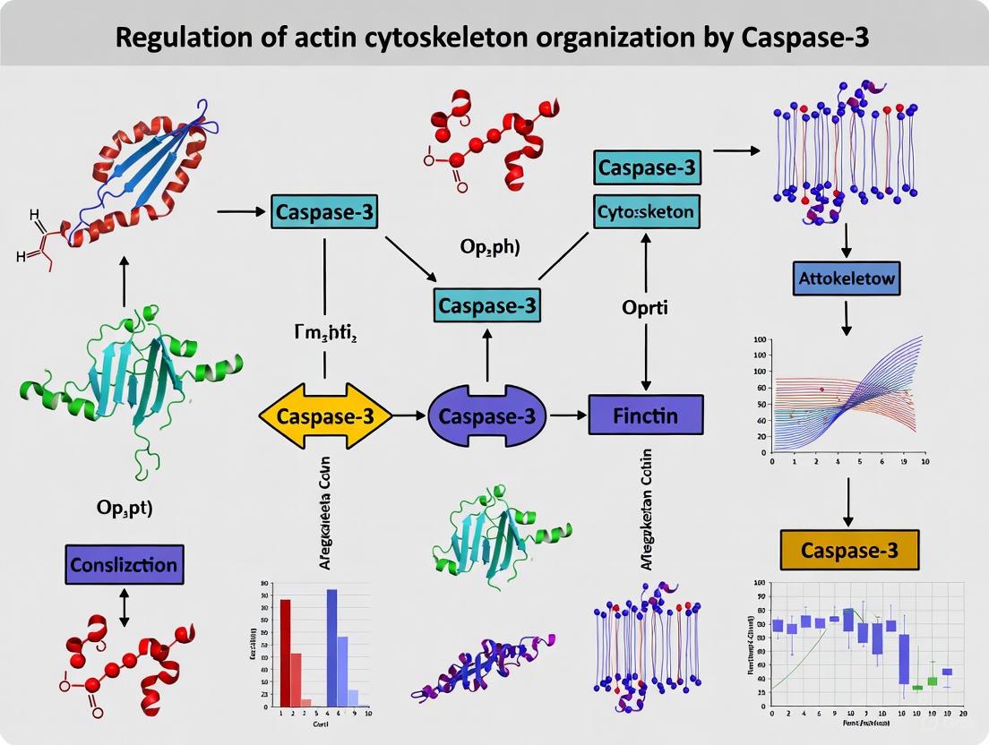
Caspase-3 in Actin Cytoskeleton Remodeling: Unveiling Non-Apoptotic Roles in Cancer and Inflammation
This article synthesizes cutting-edge research revealing the multifaceted roles of caspase-3 beyond apoptosis, focusing on its direct regulation of actin cytoskeleton organization.
Caspase-3 in Actin Cytoskeleton Remodeling: Unveiling Non-Apoptotic Roles in Cancer and Inflammation
Abstract
This article synthesizes cutting-edge research revealing the multifaceted roles of caspase-3 beyond apoptosis, focusing on its direct regulation of actin cytoskeleton organization. For researchers and drug development professionals, we explore foundational mechanisms where caspase-3 interacts with actin-binding proteins like coronin 1B and influences cell motility, particularly in melanoma. The content covers methodological approaches for studying these interactions, troubleshooting for experimental challenges, and comparative analyses of caspase-3's functions across different cell death pathways. By integrating recent findings on how caspase-3 modulates cytoskeletal dynamics to drive cancer metastasis and inflammatory responses, this review aims to provide a comprehensive framework for developing novel therapeutic strategies targeting caspase-3's non-apoptotic functions.
Beyond Cell Death: Foundational Concepts of Caspase-3 in Cytoskeletal Dynamics
Caspase-3, a well-characterized executioner protease in apoptosis, is increasingly recognized as a key regulator of actin cytoskeleton organization in both physiological and pathological contexts. This whitepaper synthesizes current research demonstrating caspase-3's non-apoptotic functions in cellular remodeling, highlighting its role in regulating cell migration, adhesion, and cytoskeletal dynamics through specific cleavage of structural and signaling proteins. We provide comprehensive experimental methodologies, quantitative analyses, and visualization tools to facilitate research into caspase-3's dual functionalities, with particular relevance to cancer biology and therapeutic development.
Caspase-3 is a cysteine-aspartate protease that serves as a primary executioner caspase in apoptotic pathways, catalyzing the specific cleavage of numerous cellular proteins to orchestrate programmed cell death [1]. Traditionally, caspase-3 activation occurs downstream of both intrinsic (mitochondrial) and extrinsic (death receptor) apoptotic pathways, where it systematically dismantles cellular components through limited proteolysis [2]. However, emerging evidence reveals that caspase-3 also participates in vital non-apoptotic processes, particularly in regulating actin cytoskeleton organization and dynamics [3] [4]. This paradigm shift positions caspase-3 as a multifunctional enzyme capable of influencing cell motility, adhesion, and structural remodeling independent of cell death—functions especially relevant in neuronal development, immune function, and cancer progression.
The non-apoptotic functions of caspase-3 occur at sub-lethal activation levels and are characterized by restricted proteolytic activity that avoids triggering cell death [4]. In melanoma and other aggressive cancers, unexpectedly high caspase-3 expression correlates with enhanced metastatic potential rather than apoptosis induction [3]. This whitepaper examines the molecular mechanisms underlying caspase-3's cytoskeletal regulatory functions, providing methodological guidance for researchers investigating this emerging aspect of caspase biology.
Molecular Mechanisms of Caspase-3 in Cytoskeletal Regulation
Direct Cleavage of Cytoskeletal Proteins
Caspase-3 regulates cytoskeletal organization through direct proteolytic cleavage of key structural and regulatory proteins. The table below summarizes primary cytoskeletal targets and functional consequences.
Table 1: Caspase-3 Cytoskeletal Substrates and Functional Outcomes
| Substrate | Cleavage Site | Functional Consequence | Biological Context |
|---|---|---|---|
| PTP-PEST | 549DSPD motif [5] | Enhanced catalytic activity; altered scaffolding | Apoptosis; cell adhesion |
| Coronin 1B | Not fully characterized [3] | Modulates actin polymerization | Melanoma cell migration |
| Actin | Specific aspartate residues [4] | Generates 15 kDa fragment; cytoskeletal remodeling | Neuronal development; aging |
| Spectrin | Unknown | Alters cytoskeleton for neurite outgrowth [4] | Axonal guidance |
| Gap43 | Unknown | Regulates growth cones [4] | Neuronal development |
| NCAM/NgCAM | Unknown | Modifies extracellular vesicle cargo [4] | Auditory brainstem development |
Regulation of Actin-Binding Proteins and Signaling Networks
Beyond direct cleavage, caspase-3 modulates actin dynamics through interactions with actin-binding proteins. In melanoma cells, caspase-3 forms constitutive associations with coronin 1B, a key regulator of actin polymerization, thereby promoting cell migration independently of its apoptotic protease function [3]. The interactome of caspase-3 in melanoma cells shows significant enrichment for proteins containing actin-binding domains, with gene ontology analysis revealing clusters related to "actin filament organization" and "positive regulation of cytoskeleton organization" [3].
Caspase-3 also regulates scaffolding functions through cleavage of PTP-PEST (protein tyrosine phosphatase with PEST domain), which modulates interactions with paxillin, leupaxin, Shc, and PSTPIP [5]. This cleavage facilitates cellular detachment during apoptosis but may also influence motility in non-apoptotic contexts. The generation of specific PTP-PEST fragments with increased catalytic activity demonstrates how caspase-3 can alter signaling networks controlling cytoskeletal dynamics.
Experimental Approaches for Studying Caspase-3 Cytoskeletal Functions
Methodologies for Protein-Protein Interaction Mapping
Comprehensive Interactome Analysis via Immunoprecipitation-Mass Spectrometry
Objective: Identify caspase-3 interacting partners in cytoskeletal regulation.
Protocol:
- Generate stable cell lines expressing GFP-tagged caspase-3 (WM793 and WM852 melanoma cells) [3]
- Culture cells under standard conditions (DMEM, 10% FBS, 37°C, 5% CO2)
- Lyse cells using mild lysis buffer (20 mM Tris-HCl pH 7.5, 150 mM NaCl, 1% Triton X-100, protease inhibitors)
- Immunoprecipitate caspase-3-GFP complexes using anti-GFP nanobodies coupled to magnetic agarose beads
- Wash beads extensively with lysis buffer containing 300 mM NaCl
- Elute bound proteins using SDS-PAGE sample buffer or low pH glycine buffer
- Analyze eluates by liquid chromatography-tandem mass spectrometry (LC-MS/MS)
- Process data using gene ontology classification for actin filament and cytoskeleton organization terms
Key Reagents:
- Anti-GFP nanobodies (ChromoTek)
- Magnetic agarose beads (Pierce)
- Protease inhibitor cocktail (Roche)
- LC-MS/MS system (Thermo Fisher)
Functional Assays for Cytoskeletal Organization
Quantitative F-actin Anisotropy Measurement
Objective: Quantify caspase-3-mediated changes in actin fiber organization.
Protocol:
- Seed cells on glass coverslips and transfer to serum-free medium 24 hours post-transfection with caspase-3 siRNA
- Fix cells with 4% paraformaldehyde for 15 minutes at room temperature
- Permeabilize with 0.1% Triton X-100 in PBS for 5 minutes
- Stain F-actin with phalloidin conjugated to Alexa Fluor 488 (1:100 dilution) for 1 hour
- Mount coverslips using antifade mounting medium
- Acquire confocal images using 63x oil immersion objective
- Process images using FibrilTool plugin in ImageJ to measure anisotropy
- Compare anisotropy values between control and caspase-3 depleted cells [3]
Key Controls:
- Cytochalasin D treatment (actin polymerization inhibitor) as positive control for disruption
- Non-targeting siRNA as negative control
- Multiple imaging fields (>10 per condition) for statistical power
Cell Migration and Invasion Assays
IncuCyte Live-Cell Imaging for Migration and Invasion
Objective: Quantitatively assess caspase-3 role in melanoma cell motility.
Protocol for Migration Assay:
- Seed cells in 96-well ImageLock plates at 20,000 cells/well
- Create uniform wound using WoundMaker tool
- Wash cells twice to remove debris
- Add fresh medium containing mitomycin C (1 μg/mL) to inhibit proliferation
- Place plate in IncuCyte Live-Cell Analysis System
- Acquire images every 2 hours for 24-48 hours
- Analyze wound closure using IncuCyte software
- Express results as relative wound density over time [3]
Protocol for Invasion Assay:
- Coat 96-well ImageLock plates with 50 μL Matrigel (2 mg/mL)
- Polymerize Matrigel at 37°C for 30 minutes
- Seed cells on top of Matrigel layer at 30,000 cells/well
- Monitor invasion through Matrigel using IncuCyte system
- Quantify invaded cell count using built-in analysis algorithms
Quantitative Analysis of Caspase-3 Cytoskeletal Functions
Research findings demonstrate consistent quantitative effects of caspase-3 on cytoskeletal parameters across multiple experimental systems.
Table 2: Quantitative Effects of Caspase-3 on Cytoskeletal and Motility Parameters
| Parameter Measured | Experimental System | Effect Size | Significance |
|---|---|---|---|
| F-actin anisotropy | WM793 melanoma cells, caspase-3 knockdown [3] | Dramatic decrease | p < 0.001 |
| Focal adhesion number | WM793 cells, paxillin staining [3] | Significant reduction | p < 0.01 |
| Cell adhesion | Matrigel-coated substrate, caspase-3 knockdown [3] | Clear impairment | p < 0.01 |
| Migration rate | IncuCyte assay, WM793 caspase-3 knockdown [3] | Significant inhibition | p < 0.001 |
| Invasion capacity | Matrigel invasion, caspase-3 knockdown [3] | Marked reduction | p < 0.001 |
| Chemotaxis | Transwell assay, caspase-3 depletion [3] | Impaired response | p < 0.01 |
| Caspase-3 cytoskeletal association | Subcellular fractionation [3] | Consistent proportion | N/A |
| PTP-PEST cleavage | In vitro caspase-3 assay [5] | Specific at 549DSPD | N/A |
Research Reagent Solutions
Table 3: Essential Research Reagents for Caspase-3 Cytoskeletal Studies
| Reagent/Category | Specific Examples | Function/Application |
|---|---|---|
| Cell Lines | WM793, WM852 melanoma cells [3]; U2OS osteosarcoma [6] | Models for migration and cytoskeletal studies |
| Antibodies | Anti-caspase-3 (Cell Signaling); anti-GFP (Clonetech); anti-paxillin (BD Transduction) [5] | Detection, immunoprecipitation |
| Chemical Inhibitors | Z-DEVD-FMK (caspase-3 inhibitor); cytochalasin D (actin polymerization) [7] | Functional perturbation |
| Expression Vectors | pcDNA3.1/Zeo-PTP-PEST; pEGFP-C2-PTP-PEST; pEBG-PSTPIP [5] | Molecular manipulation |
| Activity Assays | Recombinant active caspase-3 (Upstate) [5]; fluorogenic substrates | Enzymatic activity measurement |
| Cytoskeletal Markers | Phalloidin conjugates (F-actin) [3] [6] | Cytoskeletal visualization |
| Apoptosis Inducers | Etoposide [6]; mitomycin C [6] | Caspase-3 activation |
Signaling Pathways and Experimental Workflows
Caspase-3 Signaling in Apoptosis and Cytoskeletal Regulation
Experimental Workflow for Caspase-3 Cytoskeletal Analysis
The emerging role of caspase-3 as a regulator of actin cytoskeleton organization represents a significant expansion of its biological functions beyond apoptotic execution. The experimental approaches and reagents outlined in this whitepaper provide researchers with comprehensive tools to investigate caspase-3's dual functionalities in diverse biological contexts. Future research should focus on elucidating the precise mechanisms that determine whether caspase-3 activation leads to apoptosis or cytoskeletal remodeling, including the identification of threshold effects, spatial regulation, and competing signaling pathways. Therapeutic targeting of caspase-3's cytoskeletal functions holds particular promise for anti-metastatic strategies in cancers with elevated caspase-3 expression, such as melanoma and colorectal carcinoma [3]. As research progresses, caspase-3 continues to exemplify the complexity of protease biology, where context-dependent regulation enables participation in both life-death decisions and vital cellular processes.
The actin cytoskeleton is a dynamic, three-dimensional grid structure composed of filamentous actin (F-actin) that fills the cytoplasmic space and maintains cell shape. This network, interwoven with microtubules and intermediate filaments, mediates essential processes including cell migration, adhesion, division, and intracellular transport [8]. The dynamic equilibrium between globular actin (G-actin) monomers and F-actin polymers, a process known as "treadmilling," allows for rapid remodeling of the cytoskeleton in response to cellular cues [9]. This remodeling is controlled by a vast repertoire of actin-binding proteins (ABPs) that nucleate, sever, cap, cross-link, and depolymerize actin filaments.
Among these regulators, the Arp2/3 complex, cofilin, and coronins represent a core set of highly conserved proteins that orchestrate the generation and turnover of branched actin networks. Recent research has placed these regulators within a broader signaling context, revealing that their activity is subject to sophisticated control, including proteolytic regulation by enzymes such as caspase-3 during processes like apoptosis. This guide provides an in-depth technical overview of these key proteins, framed within the context of caspase-3-mediated cytoskeletal reorganization, to serve as a resource for researchers and drug development professionals.
Core Components of Actin Cytoskeleton Regulation
Arp2/3 Complex: The Master Nucleator
The Arp2/3 complex is a central actin nucleator composed of seven subunits, including two actin-related proteins, ARP2 and ARP3. It drives the formation of branched actin networks by nucleating new filaments as branches off the sides of existing filaments at a characteristic ~70° angle [10] [11]. These branched networks generate the protrusive forces necessary for processes like lamellipodial formation, endocytosis, and intracellular pathogen motility [12].
Regulatory Mechanism: The complex's activity is precisely controlled by numerous regulators. Nucleation Promoting Factors (NPFs), such as proteins from the WASP (Wiskott-Aldrich syndrome protein) family, bind to both the Arp2/3 complex and actin monomers or filaments to activate nucleation [12]. Recent structural studies have revealed that inhibitors like GMF (Glial Maturation Factor) bind to the barbed end of Arp2, overlapping with the proposed WASP binding site, thereby inhibiting nucleation and promoting debranching [12]. This suggests a competitive binding mechanism for regulation.
Table 1: Key Regulators of the Arp2/3 Complex
| Regulator | Type | Function | Mechanistic Insight |
|---|---|---|---|
| WASP/WAVE | Activator (NPF) | Activates nucleation; links to signaling | Binds Arp2/3 complex and G-actin [12] |
| Cortactin | Activator (NPF) | Activates nucleation; stabilizes branches | Binds Arp2/3 complex and F-actin [12] |
| GMF | Inhibitor | Inhibits nucleation; promotes debranching | Binds barbed end of Arp2, competing with WASP [12] |
| Coronin | Modulator | Recruits Arp2/3 to filament sides; promotes cofilin activity | Binds Arp2/3 via coiled-coil domain [10] [11] |
Cofilin: The Actin Severing Protein
Cofilin, a member of the ADF/cofilin superfamily, is a potent actin-binding protein that severs and depolymerizes actin filaments, generating free barbed ends for polymerization and promoting actin subunit turnover [8]. Its activity is critical for cell migration, cytokinesis, and synaptic plasticity.
Regulatory Mechanism: Cofilin activity is primarily regulated by phosphorylation at serine 3. Phosphorylation by LIM kinase (LIMK) inactivates cofilin, while dephosphorylation by phosphatases like Slingshot (SSH) reactivates it [13]. The balance between phosphorylated (inactive) and dephosphorylated (active) cofilin is crucial for regulating actin dynamics. In Alzheimer's disease (AD), this pathway is dysregulated, with studies reporting conflicting findings of either elevated LIMK1 activity leading to cofilin inactivation or elevated cofilin activity, suggesting divergent regulatory mechanisms depending on the disease stage [13]. Cofilin is also a component of pathological Hirano bodies and cofilin-actin rods found in AD brains [13].
Coronins: The Spatial Coordinators
Coronins are a conserved family of actin-binding proteins that promote cell motility by coordinating the activities of Arp2/3 complex and cofilin [10] [11]. Most coronins share a characteristic structure: an N-terminal WD40-repeat β-propeller domain, a highly variable "unique" region, and a C-terminal coiled-coil domain [10].
Regulatory Mechanism: Coronins function as spatial regulators within actin networks. At the leading edge, coronin binds to ATP/ADP+Pi F-actin, protecting new filaments from premature disassembly by cofilin and simultaneously recruiting Arp2/3 complex to filament sides to promote branching and network expansion [10]. At the rear of networks, coronin synergizes with cofilin to dismantle aged ADP-rich filaments [10]. This spatial targeting of Arp2/3 and cofilin to opposite ends of actin networks accelerates polarized actin subunit flux, increasing network plasticity and replenishing the G-actin pool.
Table 2: Functional Domains of Type I Coronins
| Domain | Structure | Key Functions | Key Interactions |
|---|---|---|---|
| β-Propeller | Seven-bladed WD40 repeats | Primary F-actin binding; prefers ATP/ADP+Pi F-actin | F-actin (via conserved residues like Arg30) [11] |
| Unique Region | Variable in length and sequence | Species-specific functions; some isoforms bind microtubules | Microtubules (e.g., in S. cerevisiae Crn1) [10] |
| Coiled-Coil | 35-50 residues, 4-7 heptad repeats | Homo-oligomerization; secondary F-actin binding; Arp2/3 binding | Arp2/3 complex; F-actin; self [10] |
Caspase-3 Regulation of Actin Cytoskeleton Organization
Caspase-3, a key executioner protease in apoptosis, actively contributes to the dismantling of the actin cytoskeleton, facilitating morphological changes and cellular detachment. Beyond cleaving structural components, caspase-3 directly targets and modulates the activity of actin regulatory proteins.
Cleavage of Regulatory Proteins
Research has demonstrated that PTP-PEST, a protein tyrosine phosphatase involved in regulating cell migration and adhesion, is a specific substrate of caspase-3 [5]. During apoptosis, caspase-3 cleaves PTP-PEST at the 549DSPD motif, generating fragments with altered activity and scaffolding functions [5]. This cleavage disrupts PTP-PEST's interactions with partners like paxillin, leupaxin, Shc, and PSTPIP, thereby modulating downstream signaling and contributing to the loss of adhesion during cell death [5].
Translocation and Activation at the Cytoskeleton
Evidence from human platelets indicates that caspase activation is spatially regulated. Thrombin stimulation induces the translocation of both procaspase-3 and procaspase-9 from the cytosol to the actin cytoskeleton, where they are activated [14]. This process depends on PKC activity and actin polymerization, as it is inhibited by Ro-31-8220 and cytochalasin D, respectively [14]. The association with the reorganizing actin cytoskeleton appears to be important for full caspase activation, linking cytoskeletal dynamics directly to the amplification of the apoptotic signal.
The following diagram illustrates the integrated signaling pathways involving the key actin regulators and their modulation by caspase-3.
Experimental Methodologies for Key Findings
Mapping Protein-Protein Interactions: GMF and Arp2/3 Complex
Objective: To determine the structural basis of GMF binding to the Arp2/3 complex and identify critical interfacial residues [12].
Protocol:
- Protein Complex Formation: Co-crystallize bovine Arp2/3 complex with mouse GMFγ in the presence of ATP and calcium.
- Structure Determination: Collect X-ray diffraction data. Use the structure of unliganded Arp2/3 complex (1K8K.pdb) as a starting model for molecular replacement to solve the phases.
- Mutagenesis and Binding Assays:
- Generate GMF mutants based on structural data (e.g., Δ1-7, M102A, D128K, R124A).
- Perform GST pull-down assays to quantify binding affinity of wild-type and mutant GMF to Arp2/3 complex.
- Compare band intensities to assess relative binding strength.
Investigating Caspase-3 Substrate Cleavage: PTP-PEST
Objective: To identify caspase-3 as the primary protease responsible for PTP-PEST cleavage during apoptosis and map the cleavage site [5].
Protocol:
- In Vitro Cleavage Assay: Incubate purified, recombinant PTP-PEST with active caspase-3. Use specific caspase inhibitors (e.g., for caspase-6, -7, -8) in control reactions to establish specificity.
- Cleavage Site Mapping:
- Generate a series of PTP-PEST point mutants where putative caspase recognition motifs (e.g., 549DSPD552, 604ADS607) are altered to alanines (e.g., 549ASPA552).
- Test the cleavage resistance of these mutants in both in vitro assays and transfected cells induced to undergo apoptosis.
- Functional Consequences:
- Use co-immunoprecipitation (Co-IP) to assess how cleavage of PTP-PEST affects its interactions with known partners like paxillin, Shc, and PSTPIP.
- Monitor cellular detachment in cells expressing cleavage-resistant PTP-PEST versus wild-type during apoptosis.
Analyzing Caspase Translocation to the Cytoskeleton
Objective: To study the translocation and activation of caspases at the actin cytoskeleton in human platelets [14].
Protocol:
- Cell Fractionation: Stimulate human platelets with thrombin (1 U/mL). At timed intervals, lyse cells and separate fractions by centrifugation:
- Cytosolic fraction (supernatant)
- Cytoskeletal fraction (Triton X-100 insoluble pellet)
- Activity and Detection:
- Measure caspase-3 and -9 activity in each fraction using fluorogenic substrates (e.g., Ac-DEVD-AMC for caspase-3).
- Detect procaspase and active caspase fragments in each fraction by Western blotting using specific monoclonal antibodies.
- Pharmacological Inhibition: Pre-treat platelets with:
- PKC inhibitor (Ro-31-8220) to test PKC dependency.
- Actin polymerization inhibitor (Cytochalasin D) to test cytoskeleton dependency.
- Caspase inhibitors (Z-DEVD-CMK for caspase-3) to assess effects on translocation.
The Scientist's Toolkit: Key Research Reagents
Table 3: Essential Reagents for Studying Actin Regulators and Caspase-3 Effects
| Reagent / Assay | Specific Example | Function in Research |
|---|---|---|
| Recombinant Proteins | Bovine Arp2/3 complex, mouse GMFγ [12] | For in vitro binding, nucleation, and debranching assays; co-crystallization. |
| Caspase-3 Enzyme | Recombinant active caspase-3 (Upstate) [5] | For in vitro cleavage assays to identify and validate novel substrates. |
| Fluorogenic Substrates | Ac-DEVD-AMC (for caspase-3) [14] | To quantitatively measure caspase-3 enzyme activity in cell lysates or fractions. |
| Pharmacological Inhibitors | Cytochalasin D (actin polymerization) [14]; Ro-31-8220 (PKC) [14]; Z-DEVD-CMK (caspase-3) [14] | To dissect the functional contribution of specific proteins/pathways in cellular processes. |
| Site-Directed Mutagenesis Kits | QuikChange Kit (Stratagene) [5] | To generate cleavage-resistant or binding-deficient mutants of proteins like PTP-PEST. |
| Specific Antibodies | Monoclonal anti-caspase-3 (8G10) [14]; anti-caspase-9 (C9) [14]; PTP-PEST polyclonal (2528, 2530) [5] | For Western blot detection of full-length and cleaved proteins; immunoprecipitation. |
Integrated View of Actin Regulation and Proteolytic Control
The following workflow summarizes the experimental approach for investigating caspase-3-mediated regulation of an actin regulatory protein, integrating the methodologies described above.
The interplay between the actin cytoskeleton and apoptotic machinery represents a critical juncture in cell biology. The Arp2/3 complex, cofilin, and coronins form a core regulatory module that controls the architecture and dynamics of actin networks. Caspase-3 emerges as a key upstream modulator of this system, fine-tuning the activity and interactions of regulatory proteins like PTP-PEST and becoming activated in a cytoskeleton-dependent manner. Understanding these connections provides not only fundamental biological insights but also reveals potential therapeutic nodes for manipulating cell fate in diseases such as cancer and neurodegeneration. The experimental frameworks outlined here provide a roadmap for further elucidating the complex crosstalk that governs cytoskeletal organization and disassembly.
Caspase-3, a well-characterized executioner protease in apoptosis, plays multifaceted roles in regulating actin cytoskeleton organization through direct molecular interactions with actin and key actin-binding proteins. Beyond its apoptotic functions, caspase-3 mediates crucial cleavage events that directly impact actin filament dynamics, severing, and reorganization. This whitepaper synthesizes current mechanistic insights into how caspase-3 directly cleaves actin, gelsolin, PTP-PEST, and coronin 1B, thereby influencing cellular processes ranging from programmed cell death to cancer cell motility. We provide comprehensive experimental protocols, quantitative data analyses, and molecular visualization tools to facilitate further research and therapeutic development targeting caspase-3-actin interactions in disease pathologies.
Caspase-3 is a cysteine-aspartic acid protease traditionally recognized for its executioner role in apoptosis, where it cleaves cellular targets to orchestrate cell death [15]. Emerging research has revealed that caspase-3 also participates in non-apoptotic processes, particularly through direct interactions with components of the actin cytoskeleton. The actin cytoskeleton, comprising globular (G)-actin monomers and filamentous (F)-actin polymers, provides structural integrity and enables cellular processes like morphogenesis, membrane blebbing, and intracellular transport [16]. Actin-binding proteins (ABPs) precisely regulate the dynamic equilibrium between G-actin and F-actin. Caspase-3 directly cleaves specific ABPs and actin itself, thereby modulating actin organization and function in both apoptotic and non-apoptotic contexts [17] [18] [16]. This whitepaper delineates the direct molecular interactions between caspase-3, actin, and ABPs, framing these interactions within the broader thesis of caspase-3-mediated cytoskeletal regulation.
Direct Cleavage of Actin by Caspase-3
Mechanistic Insights and Functional Consequences
Caspase-3 directly cleaves actin, producing characteristic proteolytic fragments that facilitate subsequent protein degradation. This cleavage serves as an initial step in muscle protein loss during catabolic conditions such as uremia, cancer, and sepsis [17]. The cleavage of actomyosin complexes by caspase-3 yields a characteristic ≈14-kDa actin fragment and other proteins that become substrates for degradation by the ATP-ubiquitin-proteasome (Ub-P'some) system [17]. This limited cleavage event increases protein degradation by the Ub-P'some system by 125%, indicating its catalytic role in priming actomyosin for destruction [17].
The functional significance of actin cleavage extends to membrane blebbing during apoptosis. Caspase-3-mediated cleavage generates two primary actin fragments: a mitochondria-targeted N-myristoylated 15-kDa fragment (tActin) and an N-terminal 32-kDa fragment (Fractin) [16]. Research indicates that tActin, rather than Fractin, specifically induces morphological changes resembling apoptosis, highlighting the functional specificity of distinct cleavage products [16].
Experimental Evidence and Detection Methods
Cell Culture and Treatments: Utilize L6 skeletal muscle cells maintained in DMEM with 10% FCS. To induce actin cleavage, employ serum deprivation by changing to media containing 2% horse serum. Insulin treatment can be applied to observe inhibition of cleavage via a PI3K-dependent mechanism [17].
In Vitro Cleavage Assay: Incubate actomyosin with recombinant active caspase-3 in buffer containing 20 mM HEPES (pH 7.5), 10 mM MgCl2, 1 mM EDTA, 1 mM EGTA, and 1 mM DTT at 37°C for 2-3 hours [17].
Detection of Cleavage Products:
- Prepare cell extracts using ice-cold hypotonic buffer with protease inhibitors
- Separate proteins by SDS-PAGE and transfer to membranes
- Detect actin fragments by Western blotting using an affinity-purified anti-actin antibody recognizing the C-terminal 11 amino acids of α-actin
- The ≈14-kDa fragment is characteristic of caspase-3-mediated cleavage [17]
Table 1: Quantitative Data on Caspase-3-Mediated Actin Cleavage
| Experimental Condition | Actin Fragment Generated | Effect on Proteolysis | Cellular Context |
|---|---|---|---|
| Recombinant caspase-3 + actomyosin | ≈14-kDa fragment | 125% increase in Ub-P'some degradation | In vitro |
| Serum deprivation of L6 cells | Actin fragments | Increased proteolysis | Muscle cells |
| Serum deprivation + insulin | Reduced fragments | Inhibition of proteolysis | PI3K-dependent mechanism |
| Diabetic or uremic rat muscle | Actin fragments | Accelerated protein degradation | In vivo disease model |
| Diabetes + caspase-3 inhibitor | Reduced fragments | Suppressed proteolysis | In vivo intervention |
Interactions with Actin-Binding Proteins
Gelsolin
Gelsolin, a calcium-activated F-actin severing and capping protein, represents a critical caspase-3 substrate [19]. During apoptosis, caspase-3 cleaves gelsolin, producing a constitutively active fragment that severs actin filaments [16]. Structural analyses reveal that calcium-bound gelsolin adopts an extended conformation when bound to the barbed end of F-actin, with its six domains (G1-G6) wrapping around the filament [19]. Caspase-3 cleavage of gelsolin generates a G1-G3 fragment that binds both sides of F-actin, potentially enhancing severing activity [19].
The molecular chaperone CCT (TRiC) directly interacts with gelsolin, sequestering it and protecting it from caspase-3 cleavage [20]. Cryo-EM structures demonstrate that gelsolin binds deep within the CCT chaperonin cavity, distinct from the binding sites for other CCT substrates like actin and tubulin [20]. This interaction represents a regulatory mechanism controlling gelsolin availability for caspase-3 cleavage.
Experimental Protocol for Gelsolin-Caspase-3 Interaction:
- Complex Formation: Incubate gelsolin with CCT in appropriate buffer to form complexes
- Caspase-3 Cleavage Assay: Treat CCT-gelsolin complexes with recombinant active caspase-3
- Controls: Include gelsolin without CCT protection
- Analysis: Use SDS-PAGE and Western blotting with anti-gelsolin antibodies to detect cleavage patterns
- Visualization: Employ cryo-EM and single-particle 3D reconstruction to characterize complex structures [20]
Coronin 1B
In melanoma cells, caspase-3 directly interacts with coronin 1B, a key regulator of actin polymerization, thereby promoting cell migration and invasion independently of its apoptotic function [18]. Caspase-3 forms constitutive associations with the cytoskeleton in aggressive cancers, where it colocalizes with coronin 1B at the leading edge of migrating cells.
Experimental Evidence:
- Interactome Analysis: Immunoprecipitation of caspase-3-GFP fusion proteins followed by mass spectrometry identifies coronin 1B as a binding partner
- Functional Assays: Caspase-3 knockdown impairs F-actin organization, reduces focal adhesions, and decreases cell migration and invasion
- Clinical Correlation: High caspase-3 expression correlates with poor prognosis in metastatic melanoma [18]
PTP-PEST
Caspase-3 directly cleaves the protein tyrosine phosphatase PTP-PEST, which regulates actin cytoskeleton organization [5]. Cleavage occurs at the 549DSPD552 motif, generating fragments with altered catalytic activity and scaffolding functions [5]. This cleavage event facilitates cellular detachment during apoptosis by modulating interactions with cytoskeletal regulators like paxillin, leupaxin, Shc, and PSTPIP.
Research Reagent Solutions
Table 2: Essential Research Reagents for Studying Caspase-3-Actin Interactions
| Reagent | Specific Example/Catalog | Function/Application |
|---|---|---|
| Recombinant active caspase-3 | Upstate Biotechnology Inc. | In vitro cleavage assays |
| Anti-actin antibody | Sigma-Aldrich (C-terminal specific) | Detection of actin fragments by Western blot |
| Caspase-3 inhibitor | Ac-DEVD-CHO (Calbiochem) | Inhibition of caspase-3 activity in functional assays |
| Cell lines | L6 skeletal muscle cells (ATCC) | Study of actin cleavage in muscle proteolysis |
| Fluorogenic substrate | DEVD-AMC (Calbiochem) | Measurement of caspase-3 activity |
| Anti-GFP nanobodies | For immunoprecipitation | Isolation of caspase-3-protein complexes |
| Inducible expression system | Doxycycline-inducible pCW vectors | Controlled expression of caspase-3 mutants |
Methodologies for Key Experiments
Measuring Caspase-3 Activity in Muscle Tissue
- Homogenize frozen muscle tissue in buffer containing 100 mM HEPES (pH 7.5), 10% sucrose, 0.1% NP-40, and 10 mM DTT
- Subject homogenates to freeze-thaw cycles and centrifugation
- Incubate supernatant with fluorogenic substrate DEVD-AMC (50 μM)
- Measure fluorescence (excitation 360 nm, emission 460 nm) to calculate caspase-3 activity [17]
Evaluating Protein Degradation in Cell Lysates
- Harvest cells and dialyze lysates to remove accumulated tyrosine
- Incubate aliquots with ATP-generating system
- Stop reactions with trichloroacetic acid
- Measure free tyrosine fluorometrically to calculate protein degradation rates [17]
Interactome Analysis of Caspase-3 Binding Partners
- Express caspase-3-GFP fusion proteins in target cells
- Immunoprecipitate using anti-GFP nanobodies coupled to magnetic beads
- Analyze immunoprecipitated complexes by mass spectrometry
- Perform gene ontology classification of interacting proteins [18]
Visualization of Molecular Interactions
Caspase-3 Actin Interactions
Signaling Pathways and Functional Outcomes
Caspase-3 Signaling Pathways
Discussion and Therapeutic Implications
The direct molecular interactions between caspase-3 and actin/ABPs represent a crucial regulatory node connecting proteolytic signaling to cytoskeletal dynamics. In catabolic conditions, caspase-3 activation initiates muscle protein degradation by cleaving actomyosin, generating fragments for proteasomal degradation [17]. In apoptosis, caspase-3-mediated cleavage of gelsolin and other ABPs contributes to characteristic morphological changes, including membrane blebbing [16]. Beyond cell death, caspase-3 regulates cell motility in cancer through non-apoptotic interactions with coronin 1B and other cytoskeletal regulators [18].
Therapeutically, inhibiting caspase-3-mediated actin cleavage may ameliorate muscle wasting in catabolic diseases [17]. Conversely, promoting caspase-3 interaction with specific ABPs could potentially inhibit cancer metastasis [18]. The structural insights from caspase-3-gelsolin interactions and CCT-mediated protection offer opportunities for developing small-molecule modulators [20]. Further research should explore tissue-specific and context-dependent regulation of these interactions to develop targeted therapies for conditions ranging from muscle atrophy to cancer metastasis.
Caspase-3, traditionally recognized as an executioner protease in apoptosis, has emerged as a critical regulator of non-apoptotic cellular processes, particularly in cancer biology. Recent research has revealed an atypical role for caspase-3 in regulating actin cytoskeleton dynamics and cell motility through its interaction with coronin 1B, a key actin-binding protein. This technical review comprehensively examines the molecular mechanism whereby caspase-3 regulates coronin 1B activity to modulate actin polymerization, thereby promoting melanoma cell migration and invasion. We synthesize findings from cellular, molecular, and functional studies that delineate this pathway, with particular emphasis on its implications for metastatic progression and potential therapeutic targeting. The mechanistic insights presented herein reframe our understanding of caspase-3 functionality beyond cell death and establish its significance in cytoskeletal reorganization and cancer cell motility.
The caspase family of cysteine-aspartic proteases has been extensively characterized for its fundamental role in programmed cell death. Caspase-3, in particular, has been considered a primary executioner caspase, responsible for the proteolytic cleavage of numerous cellular substrates during apoptosis [21] [22]. However, accumulating evidence demonstrates that caspase-3 regulates diverse physiological processes independent of its apoptotic function, including cellular differentiation, synaptic plasticity, and cell motility [3] [23]. This functional expansion is especially relevant in cancer biology, where caspase-3 is paradoxically highly expressed in certain aggressive cancers, including melanoma and colon cancer, despite its pro-apoptotic role [3].
The actin cytoskeleton represents a dynamic network essential for maintaining cellular structure, enabling migration, and facilitating invasion. Coronin 1B, a member of the coronin family of actin-binding proteins, serves as a crucial regulator of actin dynamics by coordinating the activities of the Arp2/3 complex and cofilin, thereby controlling actin filament nucleation, branching, and turnover [24]. The emerging connection between caspase-3 and coronin 1B establishes a novel mechanistic link between the protease and cytoskeletal remodeling, providing a plausible explanation for the high caspase-3 expression observed in motile cancer cells. This review systematically examines the experimental evidence supporting this mechanism and its functional consequences in cancer pathophysiology.
Molecular Mechanism: Caspase-3-Coronin 1B-Actin Polymerization Axis
Caspase-3 Expression and Localization in Melanoma
In melanoma, caspase-3 demonstrates unexpectedly high expression levels despite its apoptotic function. Comprehensive genomic analyses reveal that CASP3 is mutated in only approximately 2% of melanoma cases, significantly less than major drivers like BRAF and NRAS, suggesting evolutionary pressure to maintain its expression [3]. Transcriptomic data from the Cancer Cell Line Encyclopedia shows substantial CASP3 expression across numerous melanoma cell lines. Clinically, CASP3 expression levels significantly differentiate primary from metastatic melanoma tumors, with higher expression correlating with advanced disease [3].
Critically, subcellular localization studies demonstrate that a fraction of caspase-3 constitutively associates with the cytoskeleton in melanoma cells, positioning it to directly influence cytoskeletal dynamics [3]. Immunofluorescence and cellular fractionation experiments confirm caspase-3's proximity to the plasma membrane and F-actin at the cellular cortex, a pattern distinct from the diffuse cytoplasmic localization of the related executioner caspase, caspase-7 [3].
Coronin 1B as a Regulator of Actin Dynamics
Coronin 1B functions as a central coordinator of actin cytoskeleton remodeling through multiple mechanisms:
- Arp2/3 Complex Regulation: Coronin 1B directly interacts with the Arp2/3 complex, inhibiting actin filament nucleation and branching under basal conditions [24].
- Cofilin Activation Pathway: Coronin 1B simultaneously binds Slingshot phosphatase (SSH1L), directing it to lamellipodia where it activates cofilin through dephosphorylation, thereby promoting actin filament severing and turnover [24].
- Spatiotemporal Coordination: By physically and functionally linking these processes, coronin 1B ensures coupled actin filament formation and disassembly, which is essential for effective lamellipodial protrusion and cell migration [24].
Table 1: Coronin 1B Functions in Actin Cytoskeleton Regulation
| Function | Molecular Mechanism | Cellular Outcome |
|---|---|---|
| Arp2/3 Inhibition | Binds Arp2/3 complex and attenuates nucleation | Controls branched actin network formation |
| Cofilin Activation | Recruits SSH1L phosphatase to dephosphorylate cofilin | Enhances actin filament disassembly |
| Myosin Regulation | Fine-tunes ROCK signaling pathway | Modulates actomyosin contractility |
| Junction Remodeling | Regulates actin at endothelial cell-cell junctions | Controls barrier integrity and tube formation |
Functional Interaction Between Caspase-3 and Coronin 1B
The caspase-3-coronin 1B interaction represents a non-apoptotic signaling axis that directly influences actin polymerization and cell motility:
- Direct Protein Interaction: Immunoprecipitation and mass spectrometry analyses demonstrate that caspase-3 physically interacts with coronin 1B and other proteins involved in actin filament organization [3].
- Cytoskeletal Association: Caspase-3 is constitutively associated with the cytoskeleton and resides in close proximity to F-actin at the cell cortex, facilitating its regulation of coronin 1B activity [3].
- Protease-Independent Function: Caspase-3 modulates coronin 1B activity and promotes melanoma cell motility independently of its canonical apoptotic protease function, suggesting a structural or scaffolding role [3].
- Transcriptional Regulation: Specificity protein 1 (SP1) acts as a transcriptional regulator of CASP3 expression, and SP1 inhibition reduces caspase-3 levels and impairs melanoma cell migration [3].
Experimental Evidence and Functional Validation
Key Experimental Approaches and Findings
Multiple experimental approaches have been employed to delineate the caspase-3-coronin 1B functional relationship:
Diagram 1: Experimental workflow for establishing caspase-3's role in cytoskeletal regulation and cell motility.
- Interactome Analysis: Caspase-3-GFP fusion proteins were stably expressed in melanoma cell lines (WM793 and WM852), followed by immunoprecipitation using anti-GFP nanobodies and mass spectrometry analysis. Gene ontology classification revealed significant enrichment of caspase-3-interacting proteins involved in actin filament and cytoskeletal organization [3].
- Cytoskeletal Organization Assessment: Caspase-3 depletion resulted in substantial disorganization of F-actin fibers and reduced anisotropy, indicating loss of parallel fiber alignment. Focal adhesion number was also decreased following caspase-3 knockdown, as evidenced by paxillin immunostaining [3].
- Functional Migration and Invasion Assays: Live-cell imaging using IncuCyte technology demonstrated that caspase-3 depletion significantly impaired melanoma cell migration and invasion in vitro. Cellular tomography revealed that caspase-3-deficient cells exhibited defective attachment and polarization capacity [3].
Table 2: Quantitative Effects of Caspase-3 Depletion on Melanoma Cell Behavior
| Parameter | Effect of Caspase-3 Knockdown/KO | Experimental System |
|---|---|---|
| Cell Adhesion | Significantly impaired | Matrigel-coated substrate assay |
| F-actin Organization | Dramatically disorganized, reduced anisotropy | Phalloidin staining and quantification |
| Focal Adhesions | Decreased number | Paxillin immunostaining |
| 2D Migration | Inhibited | IncuCyte live-cell imaging |
| 3D Invasion | Impaired | Matrigel invasion assay |
| Chemotaxis | Reduced directional movement | Chemotaxis assay |
| In Vivo Metastasis | Impaired (based on associated gene signature) | Mouse models and patient data correlation |
Detailed Experimental Protocols
Caspase-3 Interactome Analysis
This protocol outlines the methodology for identifying caspase-3 interacting proteins, including coronin 1B, in melanoma cells.
Cell Line Preparation
- Utilize melanoma cell lines (e.g., WM793, WM852) representing different disease stages
- Stably express GFP-tagged caspase-3 or GFP alone as control using lentiviral transduction
- Maintain cells in appropriate melanoma cell culture medium with selection antibiotics
Immunoprecipitation
- Lyse cells in mild detergent buffer (e.g., 1% NP-40 or Triton X-100) to preserve protein complexes
- Incubate lysates with anti-GFP nanobodies coupled to magnetic agarose beads for 2-4 hours at 4°C
- Wash beads extensively with lysis buffer to remove non-specifically bound proteins
- Elute bound protein complexes using low-pH buffer or GFP peptide competition
Mass Spectrometry Analysis
- Digest eluted proteins with trypsin following standard proteomic protocols
- Analyze peptides by liquid chromatography-tandem mass spectrometry (LC-MS/MS)
- Process raw data using database search algorithms (e.g., MaxQuant, Proteome Discoverer)
- Validate interactions through co-immunoprecipitation and western blotting
Functional Migration and Invasion Assays
These protocols detail the methods for assessing the functional consequences of caspase-3 depletion on cell motility.
IncuCyte Live-Cell Migration Assay
- Seed control and caspase-3-depleted melanoma cells in 96-well ImageLock plates
- Create uniform wounds using the WoundMaker tool according to manufacturer's protocol
- Add mitomycin C (1-2 μg/mL) to suppress proliferation and isolate migration effects
- Monitor wound closure every 2-4 hours using the IncuCyte live-cell imaging system
- Quantify relative wound density using integrated metric analysis software
Matrigel Invasion Assay
- Coat Transwell inserts (8μm pore size) with diluted Matrigel matrix (200-300μg/filter)
- Seed serum-starved cells in the upper chamber in serum-free medium
- Add complete medium with 10% FBS as chemoattractant in the lower chamber
- Incubate for 24-48 hours at 37°C to allow invasion through Matrigel
- Fix cells with 4% formaldehyde and stain with 0.1% crystal violet
- Count invaded cells in multiple microscope fields per filter
Data Analysis
- Normalize invasion/migration data to control conditions
- Perform statistical analyses using Student's t-test or ANOVA with post-hoc testing
- Conduct at least three independent experiments with technical replicates
The Scientist's Toolkit: Essential Research Reagents
Table 3: Key Research Reagents for Studying Caspase-3 - Coronin 1B Axis
| Reagent/Category | Specific Examples | Function/Application |
|---|---|---|
| Cell Lines | WM793, WM852 melanoma cells | Model systems for studying melanoma progression |
| Molecular Tools | Caspase-3-GFP fusion construct | Visualizing localization and interaction studies |
| Knockdown Approaches | siRNA targeting CASP3, CRISPR/Cas9 KO | Functional validation of caspase-3 roles |
| Detection Antibodies | Anti-caspase-3, anti-coronin 1B, anti-paxillin | Protein localization and expression analysis |
| Cytoskeletal Markers | Phalloidin conjugates | F-actin visualization and organization assessment |
| Live-Cell Imaging | IncuCyte system | Quantitative migration and invasion kinetics |
| Invasion Matrices | Matrigel, collagen I | 3D microenvironment for invasion assays |
Integrated Pathway and Therapeutic Implications
The mechanistic relationship between caspase-3 and coronin 1B represents a signaling axis with significant implications for understanding cancer metastasis and developing therapeutic strategies.
Diagram 2: Integrated signaling pathway from caspase-3 transcription to functional cell motility outcomes, with potential therapeutic intervention points.
The caspase-3-coronin 1B regulatory axis represents a promising target for anti-metastatic therapy. Several strategic intervention points emerge from the elucidated mechanism:
- SP1 Inhibition: Targeting the transcription factor SP1 reduces caspase-3 expression and impairs melanoma cell migration, representing an upstream therapeutic approach [3].
- Protein-Protein Interaction Disruption: Developing small molecules or peptides that specifically disrupt the caspase-3-coronin 1B interaction without affecting apoptotic caspase-3 function could selectively inhibit metastasis.
- Coronin 1B Activity Modulation: Compounds that modulate coronin 1B's ability to coordinate Arp2/3 complex and cofilin activities may normalize actin dynamics in metastatic cells.
This mechanistic understanding reframes caspase-3 as a multimodal regulator of cellular homeostasis with context-dependent functions. In melanoma and potentially other aggressive cancers, the non-apoptotic, motility-promoting functions of caspase-3 may dominate, explaining its paradoxical high expression in these malignancies. Future therapeutic strategies must account for this functional duality when considering caspase-3 modulation in cancer treatment.
Morphological changes at the cellular level serve as critical indicators of physiological and pathological states. Among the most significant are membrane blebbing, cell rounding, and adhesion changes—three interconnected hallmarks that frequently manifest during processes ranging from normal cell migration to apoptosis and metastatic cancer progression. Membrane blebs are defined as spherical protrusions of the plasma membrane that occur upon its detachment from the underlying actin cortex, typically ranging from 1 to 5 micrometers in diameter [25]. These structures were traditionally viewed primarily as features of apoptotic cell death, where they precede the formation of apoptotic bodies [26]. However, contemporary research has established that blebbing also constitutes a dynamic feature of dramatic cellular reorganization in numerous physiological contexts, including cell spreading, mitosis, and both in vivo and in vitro cell motility [25].
Cell rounding is a ubiquitous characteristic of programmed cell death, occurring in almost all instances of apoptosis independent of the initiating stimulus [26]. This process involves significant cell shrinkage and loss of contact with neighboring cells or the extracellular matrix (ECM). The primary determinant of this volume change is the movement of water, controlled by alterations in osmotically active particles such as K+, Na+, and Cl− ions [26]. Concurrent with cell rounding are profound adhesion changes, where cells undergo detachment from their substrate and disassemble focal adhesions—the physical connections that anchor cells to the ECM. These morphological transitions are not isolated events but are mechanistically coupled through the dynamic reorganization of the actin cytoskeleton and its regulatory proteins, including the unexpected involvement of traditional apoptotic enzymes like caspase-3 in non-apoptotic contexts [18].
Table 1: Functional Contexts of Morphological Hallmarks
| Morphological Hallmark | Apoptotic Context | Non-Apoptotic Context |
|---|---|---|
| Membrane Blebbing | Execution-phase of apoptosis; precedes apoptotic body formation [26] | Cell spreading, motility, mitosis; pressure regulation after rapid detachment [25] |
| Cell Rounding | Early event involving cell shrinkage and ion flux; detachment from neighbors/ECM [26] | Transition to amoeboid migration mode in 3D environments; metastatic dissemination [27] |
| Adhesion Changes | Loss of focal adhesions; detachment from substrate [26] | Mesenchymal-amoeboid transition; invasive migration through tissues [27] [18] |
Caspase-3 Regulation of Actin Cytoskeleton Organization
The discovery of non-apoptotic functions for caspase-3 has fundamentally expanded our understanding of its role in cellular physiology, particularly in regulating the actin cytoskeleton. In aggressive cancers such as melanoma, caspase-3 is unexpectedly highly expressed despite its pro-apoptotic function, suggesting it confers advantages unrelated to cell death [18]. Molecular interactome analyses using caspase-3-GFP fusion proteins and immunoprecipitation coupled with mass spectrometry have revealed that caspase-3 interacts with a network of proteins involved in actin filament and cytoskeletal organization, with significant enrichment in actin-binding domains [18]. This interaction is specific to caspase-3, as the executioner caspase-7 does not show similar association with the cytoskeletal fraction [18].
Mechanistically, caspase-3 interacts with and modulates the activity of coronin 1B, a key regulator of actin polymerization, thereby promoting melanoma cell motility independently of its canonical apoptotic protease function [18]. This pathway represents a paradigm shift in understanding how traditional executioners of cell death can directly influence cellular architecture and behavioral states. The functional consequences of this regulation are profound: depletion of caspase-3 in melanoma cells leads to significant disorganization of F-actin fibers, reduces the parallel alignment (anisotropy) of actin structures, decreases focal adhesion number, and impairs cell adhesion, migration, and invasion in vitro and in vivo [18]. This cytoskeletal role for caspase-3 provides a mechanistic explanation for its high expression in metastatic tumors and its association with poor patient prognosis.
Table 2: Experimental Evidence for Caspase-3 in Cytoskeletal Regulation
| Experimental Approach | Key Findings | Functional Consequences |
|---|---|---|
| Interactome Analysis (GFP-IP/MS) | Caspase-3 interacts with actin-binding proteins and complexes involved in "actin filament organization" and "positive regulation of cytoskeleton organization" [18] | Identifies direct physical connection between caspase-3 and cytoskeletal machinery |
| Immunofluorescence & Subcellular Fractionation | Caspase-3 localizes at the cellular cortex with F-actin and associates with the cytoskeletal fraction (unlike caspase-7) [18] | Demonstrates spatial coordination between caspase-3 and actin networks |
| Caspase-3 Depletion (siRNA) | Disorganized F-actin fibers, reduced focal adhesions, impaired lamellipodia function [18] | Establishes necessary role in maintaining cytoskeletal architecture |
| Migration/Invasion Assays | Reduced migration speed, impaired chemotaxis, decreased invasive capacity through matrices [18] | Links caspase-3 cytoskeletal function to cell behavioral outputs |
Experimental Protocol: Caspase-3 Interactome Analysis
To investigate the non-apoptotic interactions of caspase-3 with the cytoskeleton, researchers have developed a comprehensive interactome analysis protocol [18]:
Cell Line Selection: Utilize metastatic melanoma cell lines such as WM793 and WM852 that endogenously express high levels of caspase-3.
Stable Expression System: Generate cell lines stably expressing GFP-tagged caspase-3 fusion proteins or GFP alone as a control using lentiviral transduction and antibiotic selection.
Immunoprecipitation: Harvest cells and lyse using a mild detergent buffer (e.g., 1% Triton X-100 in PBS with protease inhibitors) to preserve protein complexes. Incubate lysates with anti-GFP nanobodies coupled to magnetic agarose beads for 2-4 hours at 4°C with gentle rotation.
Mass Spectrometry Sample Preparation: Wash beads extensively with lysis buffer to remove non-specifically bound proteins. Elute bound proteins using low-pH buffer or direct denaturation in SDS-containing buffer. Digest proteins with trypsin and desalt peptides using C18 columns.
Liquid Chromatography-Tandem Mass Spectrometry (LC-MS/MS): Analyze peptides using a high-resolution mass spectrometer coupled to a nanoflow liquid chromatography system. Perform database searching against the human proteome to identify significantly enriched proteins in caspase-3-GFP samples compared to GFP controls.
Bioinformatic Analysis: Process raw data using computational pipelines such as MaxQuant. Perform gene ontology (GO) enrichment analysis using tools like DAVID or PANTHER to identify biological processes and molecular functions significantly represented in the caspase-3 interactome.
This protocol typically requires 5-7 days from cell culture to preliminary bioinformatic analysis and enables the unbiased identification of caspase-3 interacting partners independent of its apoptotic function.
Membrane Blebbing: Mechanisms and Regulation
Biophysical Basis of Bleb Formation
Membrane blebbing occurs through a well-defined mechanical process initiated by the detachment of the plasma membrane from the underlying actin cortex. This detachment creates a cytosol-filled bulge that expands rapidly (within approximately 30 seconds) due to intracellular pressure [25]. Bleb expansion typically stalls through one of two mechanisms: either through a drop in intracellular pressure or, more commonly, through the de novo assembly of an actin cortical layer on the bleb membrane [25]. This assembly occurs in a defined sequence: first, transmembrane actin-binding proteins localize within the membrane of the bleb, followed by actin polymerization at the bleb membrane, and finally, the localization of motor proteins—particularly myosin II—which facilitates bleb retraction toward the cell body within a few minutes [25].
The mechanical homeostasis of the cell membrane plays a crucial role in bleb dynamics. Plasma membrane tension, which is primarily determined by membrane-to-cortex attachment (MCA) regulated by ERM proteins (ezrin, radixin, and moesin), creates a tensional homeostasis that can suppress bleb formation [27]. Epithelial cells maintain higher PM tension than their metastatic counterparts, and this high tension potently inhibits membrane curvature and bleb formation [27]. When MCA is disrupted—either through genetic interference with ERM proteins or through physiological processes like epithelial-mesenchymal transition—the resultant decrease in membrane tension facilitates bleb formation and promotes a transition to amoeboid migration modes [27].
Experimental Protocol: Quantifying Blebbing Dynamics
To investigate blebbing activity in response to cellular detachment, researchers have established a standardized protocol using interference reflection microscopy (IRM) [25]:
Cell Culture and Substrate Preparation: Culture bovine aortic endothelial cells (BAECs) in Dulbecco's Modified Eagle Medium supplemented with 10% bovine serum. Coat glass-bottom dishes with 100 µg/mL fibronectin for 2 hours at room temperature, then rinse three times with phosphate-buffered saline before cell addition.
Cell Detachment: Plate cells on fibronectin-coated plates for 24 hours to reach adhesion saturation. For rapid detachment, use 0.25% trypsin with EDTA; for slow detachment, use trypsin with EDTA diluted 20:1 with BAEC medium.
Microscopy and Imaging: Observe cells using an inverted microscope fitted with a 100× oil immersion objective lens and a mercury lamp. Maintain cells in a closed microscope chamber at 37°C, 5% CO₂, and 50% humidity during imaging. Capture one frame every 3-5 seconds for both detachment and spreading experiments.
Image Analysis: Identify cells using an algorithm based on fitting of intensity histograms implemented in IGOR-Pro data analysis software. Manually trace cell boundaries in IRM images using ImageJ software and calculate area using built-in routines. Count blebs from multiple bright-field snapshots taken at each time point to ensure identification of blebs on both basal and apical surfaces.
Pharmacological Inhibition: To test dynamin-dependence of blebbing decay, treat cells with 80 µM dynasore for 10 minutes before plating and maintain the drug concentration throughout the experiment.
This protocol typically reveals that blebs begin to appear on both basal and apical surfaces of detaching cells after approximately 78% ± 11% of the total adhered area has detached [25].
Cell Rounding: Mechanisms and Functional Consequences
Molecular Basis of Cell Rounding
Cell rounding during apoptosis represents a coordinated physiological process driven by ion flux and cytoskeletal reorganization. Early in apoptosis, transient increases in intracellular Na+ control initial signaling events that ultimately lead to cell shrinkage [26]. As shrinkage proceeds, cells experience loss of both Na+ and K+ ions, and inhibition of the Na+/K+-ATPase and Ca2+-dependent potassium channels can reduce this shrinkage event, indicating that ion movement plays a regulatory role in the apoptotic process beyond simply controlling volume changes [26]. The morphological changes are accompanied by rearrangement of the actin cytoskeleton and phosphorylation of myosin light chains by ROCK-I, a Rho effector protein [26]. Inhibition of ROCK-I prevents membrane blebbing but does not impair phagocytosis of apoptotic cells by macrophages, indicating that blebs themselves are not essential for recognition and engulfment [26].
In non-apoptotic contexts, cell rounding facilitates transitions between migration modes, particularly the shift from mesenchymal to amoeboid motility in three-dimensional environments [27]. This rounded morphology enables cells to navigate through narrow constrictions in the extracellular matrix without requiring proteolytic degradation of matrix components. The mechanical properties of rounded cells differ significantly from their spread counterparts, with decreased cell stiffness correlating with increased invasive capability [27]. This relationship between decreased stiffness and increased invasiveness represents a fundamental "mechanical signature" of malignant cells that has been observed across multiple cancer types.
Adhesion Changes: From Focal Adhesions to Detachment
Adhesion Remodeling in Motility and Death
Focal adhesions are multi-protein complexes that connect the intracellular actin cytoskeleton to the extracellular matrix, serving as both mechanical anchors and signaling hubs. During apoptosis, cells undergo detachment from their substrate through the disassembly of these adhesion structures, which facilitates the rounding and eventual fragmentation of the dying cell [26]. In metastatic cancer cells, adhesion changes are more nuanced—focal adhesions become more dynamic, allowing for rapid attachment and detachment cycles that facilitate migration [18]. Caspase-3 plays a surprising role in this process, as its depletion in melanoma cells leads to reduced numbers of focal adhesions and impaired cell adhesion to matrigel-coated substrates [18]. This suggests that caspase-3 contributes to the regulation of adhesion turnover in addition to its roles in cytoskeletal organization.
The relationship between adhesion strength and migration mode represents a critical determinant of invasive capacity. Mesenchymal migration typically involves strong adhesion and actomyosin contractility, while amoeboid migration employs weaker adhesions and relies more on membrane blebbing for propulsion [27]. Cancer cells exhibit remarkable plasticity in switching between these modes based on environmental constraints, and adhesion dynamics play a central role in these transitions. Experimental evidence demonstrates that reducing membrane-to-cortex attachment (and consequently plasma membrane tension) through ERM protein knockdown is sufficient to induce a mesenchymal migratory phenotype in epithelial cells, even in the absence of classical epithelial-mesenchymal transition (EMT) programs [27].
Experimental Protocol: Analyzing Focal Adhesions and Actin Organization
To quantify changes in actin organization and focal adhesion dynamics, researchers have developed sophisticated image analysis approaches [28] [29]:
Cell Staining and Fixation: Culture cells on glass coverslips. Fix with 2-4% paraformaldehyde for 15 minutes, permeabilize with 0.1-0.5% Triton X-100 for 5 minutes, and block with 2% bovine serum albumin. Stain F-actin with fluorescent phalloidin (e.g., Phalloidin-TRITC at 0.1 M) and focal adhesion proteins with specific antibodies (e.g., anti-paxillin).
High-Resolution Confocal Microscopy: Image cells using a laser scanning confocal microscope with a 60× or 100× oil immersion objective. Acquire z-stacks at 0.2-0.3 µm intervals to capture the entire cellular volume. Maintain identical laser power, gain, and offset settings across all experimental conditions.
Image Reconstruction and Quantification: Use commercial software (e.g., Imaris, Volocity) or custom algorithms (e.g., SFEX, FSegment) to reconstruct 3D models from z-stacks. For actin organization, quantify parameters including fiber alignment (anisotropy), density, and orientation. For focal adhesions, quantify number, size, aspect ratio, and distribution.
Statistical Analysis: Analyze a minimum of 15-20 cells per condition across multiple independent experiments. Use appropriate statistical tests (e.g., Student's t-test for two conditions, ANOVA for multiple comparisons) to determine significance.
This protocol enables quantitative assessment of cytoskeletal and adhesion changes in response to genetic manipulations or pharmacological treatments, providing insights into the molecular mechanisms regulating these morphological hallmarks.
Visualization of Caspase-3 Signaling in Cytoskeletal Regulation
The diagram above illustrates the non-apoptotic signaling pathway through which caspase-3 regulates actin cytoskeleton organization and cell motility. This pathway operates independently of caspase-3's proteolytic activity in apoptosis and involves direct interaction with cytoskeletal regulators [18].
The Scientist's Toolkit: Essential Research Reagents
Table 3: Essential Research Reagents for Studying Morphological Hallmarks
| Reagent/Category | Specific Examples | Function/Application |
|---|---|---|
| Actin Visualization | Fluorescent phalloidin (e.g., TRITC, FITC conjugates); Phalloidin-TRITC at 0.1 M [25] [28] | Selective F-actin staining for fluorescence microscopy; high affinity binding to filamentous actin |
| Live-Cell Actin Probes | GFP fusions to G-actin; fusions to actin-binding proteins/peptides; live dyes (e.g., SiR-actin) [28] | Real-time visualization of actin dynamics in living cells without fixation |
| Caspase-3 Tools | Caspase-3-GFP fusion constructs; anti-caspase-3 antibodies; CASP3 siRNA [18] | Investigation of caspase-3 localization, interactions, and functional roles through manipulation and detection |
| Focal Adhesion Markers | Anti-paxillin antibodies; anti-phospho-ERM antibodies; anti-vinculin antibodies [28] [27] | Identification and quantification of focal adhesion structures and membrane-cortex attachment proteins |
| Cytoskeletal Drugs | Dynasore (80 μM) [25]; Cytochalasin D [18]; ROCK inhibitors (e.g., Y-27632) [26] | Pharmacological perturbation of specific cytoskeletal processes: dynamin inhibition, actin polymerization blockade, ROCK pathway inhibition |
| Detachment Reagents | 0.25% trypsin with EDTA; diluted trypsin (20:1) for slow detachment; EDTA alone [25] | Controlled cell detachment from substrates to study adhesion changes and blebbing dynamics |
| Image Analysis Software | SFEX (Stress Fiber Extractor); FSegment; SFALab; ImageJ with custom macros [28] | Quantitative analysis of actin structures, stress fibers, and focal adhesions from microscopy images |
The morphological hallmarks of membrane blebbing, cell rounding, and adhesion changes represent integrated responses to fundamental cellular processes ranging from physiological motility to pathological transformation. The emerging role of caspase-3 in regulating actin cytoskeleton organization independent of apoptosis reveals the remarkable functional versatility of traditional cell death enzymes and provides new insights into the mechanisms underlying cancer metastasis. These morphological transitions are not merely passive consequences of cellular states but active mechanical adaptations that enable cells to navigate diverse environmental challenges. The experimental methodologies and reagents outlined in this review provide researchers with comprehensive tools to investigate these processes at molecular, cellular, and biophysical levels, potentially opening new avenues for therapeutic intervention in cancer and other diseases characterized by dysregulated cell morphology and motility.
Research Tools and Techniques: Studying Caspase-3 Cytoskeletal Functions
Caspase-3, a key executioner protease in apoptosis, is traditionally recognized for cleaving cellular targets to execute cell death. However, emerging research has revealed non-apoptotic functions of caspase-3, particularly in regulating actin cytoskeleton organization. This dual functionality positions caspase-3 as a critical molecule in cellular remodeling, with implications for cancer metastasis and neuronal development. In catabolic conditions, caspase-3 activation serves as an initial step triggering accelerated muscle proteolysis by cleaving actomyosin complexes, producing fragments that are subsequently degraded by the ubiquitin-proteasome system [30]. Beyond cell death, caspase-3 directly interacts with cytoskeletal components, as evidenced by its constitutive association with the cytoskeleton in melanoma cells, where it regulates cell migration and invasion by modulating coronin 1B activity, a key regulator of actin polymerization [31]. Furthermore, research demonstrates that caspases 3 and 9 translocate to the cytoskeleton in human platelets upon thrombin stimulation, with this translocation requiring protein kinase C (PKC) activity and actin polymerization [14]. These findings underscore the necessity of employing sophisticated interactome analysis techniques to comprehensively identify caspase-3 binding partners and elucidate its non-canonical functions in cytoskeletal regulation.
Co-immunoprecipitation (co-IP) Principles and Advancements
Co-immunoprecipitation is a powerful technique for studying protein-protein interactions under native conditions, allowing researchers to capture protein complexes directly from cell lysates. The standard approach involves using a specific antibody against the protein of interest (bait) to pull it down along with its associated partners (prey) from a solution. However, traditional single-step co-IP suffers from limitations, including coprecipitated contaminants that can confound results. To address this, advanced methodologies like the Two-Step Coimmunoprecipitation (TIP) have been developed, which enable sequential coimmunoprecipitations of endogenous protein complexes for highly selective enrichment [32].
The TIP methodology can be performed with a broad range of mono- and polyclonal antibodies targeting either a single protein or different components of a given complex. This approach results in substantially reduced background contamination compared to single-step co-IPs, making it particularly valuable for downstream applications such as mass spectrometry analysis. When benchmarked for identifying interacting proteins in primary human CD4+ T cells, TIP successfully recapitulated all major known interactors while enabling proteomic discovery of novel interaction partners [32].
Mass Spectrometry (MS) in Protein Complex Analysis
Mass spectrometry provides the analytical power to identify and quantify the components of protein complexes isolated through co-IP. Recent advancements have expanded MS applications in caspase research, enabling identification of caspase substrates, cleavage products, and post-translational modifications, while also unveiling complex regulatory networks [33]. The recruitment of mass spectrometry techniques in investigating caspases has significantly expanded the repertoire of tools available for comprehensive interactome analysis.
When combined with co-IP, mass spectrometry allows researchers to move beyond simple interaction cataloging to functional characterization of protein complexes. For instance, in a systematic proteomics analysis of PCDH-γ-associated protein complexes, researchers identified 154 non-redundant proteins, providing insights into the molecular composition and function of these complexes in neural development [34].
Experimental Design and Workflow
Workflow Diagram for Caspase-3 Interactome Analysis
Critical Experimental Considerations
Cell Culture and Caspase-3 Activation
For studying caspase-3 interactions relevant to actin cytoskeleton organization, researchers should consider appropriate cellular models. Melanoma cell lines have proven valuable, as caspase-3 is constitutively associated with the cytoskeleton in these cells and regulates motility through coronin 1B interaction [31]. Primary neurons or platelet systems also provide relevant models, as demonstrated by studies showing caspase-3 translocation to the cytoskeleton in thrombin-stimulated platelets [14].
Caspase-3 activation can be achieved through various stimuli, including:
- Serum withdrawal to induce intrinsic apoptosis pathway [35]
- Staurosporine treatment (typically 0.5-2 μM for 4-24 hours) [36]
- Specific death receptor ligands for extrinsic pathway activation
- Cellular stressors relevant to pathological conditions being investigated
Lysis Conditions for Preserving Cytoskeletal Associations
Appropriate lysis conditions are crucial for maintaining caspase-3 interactions with cytoskeletal components while ensuring sufficient solubilization of protein complexes. Key considerations include:
- Use mild non-ionic detergents (e.g., 1-4% Triton X-100) to solubilize membrane-bound proteins while preserving protein interactions [34]
- Implement cytoskeletal stabilization buffers when studying actin-associated complexes
- Include protease and phosphatase inhibitors to maintain complex integrity
- Consider crosslinking approaches for transient or weak interactions
For specific investigation of caspase-3 cytoskeletal associations, researchers may employ subcellular fractionation methods to isolate cytoskeletal components before co-IP, as demonstrated in platelet studies where caspases 3 and 9 were found in cytoskeletal fractions following thrombin stimulation [14].
Detailed Methodological Protocols
Two-Step Coimmunoprecipitation (TIP) for Caspase-3 Complexes
The TIP protocol significantly enhances specificity for isolating caspase-3 interaction partners compared to single-step co-IP [32]:
Prepare native cell lysates from approximately 5×10^7 cells using lysis buffer (50 mM Tris-HCl, pH 7.5, 150 mM NaCl, 1 mM EDTA, 10 mM NaF, 10 mM Na₃VO₄) supplemented with protease inhibitors and 1% Triton X-100.
Pre-clear lysate with control IgG and protein A/G beads for 1 hour at 4°C.
First immunoprecipitation: Incubate pre-cleared lysate with anti-caspase-3 antibody (2-5 μg) overnight at 4°C with gentle rotation.
Capture complexes by adding protein A/G agarose beads (50 μL slurry) and incubating for 2-4 hours at 4°C.
Wash beads extensively with lysis buffer (4-5 washes, 5 minutes each).
Elute complexes from beads using mild elution conditions (0.5 M NaCl or low pH glycine buffer).
Second immunoprecipitation: Dilute eluate to lower salt concentration and repeat immunoprecipitation with fresh anti-caspase-3 antibody or antibodies against different caspase-3 domains.
Final wash and preparation for downstream analysis.
Note: For caspase-3 complexes associated with actin cytoskeleton, consider including cytoskeleton-stabilizing agents (e.g., phalloidin) in lysis and wash buffers.
Mass Spectrometry Sample Preparation and Analysis
Protein denaturation and reduction: Resuspend final co-IP pellets in denaturing buffer (6 M urea, 2 M thiourea, 10 mM DTT) and incubate at 56°C for 30 minutes.
Alkylation: Add iodoacetamide to 25 mM final concentration and incubate in darkness for 20 minutes.
Tryptic digestion: Dilute samples 4-fold with 50 mM ammonium bicarbonate, add trypsin (1:50 enzyme-to-substrate ratio), and incubate overnight at 37°C.
Peptide desalting: Use C18 StageTips or similar solid-phase extraction columns.
LC-MS/MS analysis:
- Liquid chromatography: Use nanoflow LC systems with C18 columns (75 μm × 25 cm) and 60-120 minute gradients from 5% to 35% acetonitrile in 0.1% formic acid.
- Mass spectrometry: Operate instruments in data-dependent acquisition mode, with full MS scans (300-1500 m/z) followed by fragmentation of top N most intense ions.
Data processing: Search MS/MS data against appropriate protein databases using search engines like MaxQuant or Proteome Discoverer with caspase-3 sequence included for reference.
Key Research Reagents and Tools
Table 1: Essential Research Reagents for Caspase-3 Interactome Studies
| Reagent Category | Specific Examples | Application/Function | Considerations |
|---|---|---|---|
| Caspase-3 Antibodies | Monoclonal anti-caspase-3 (8G10) [14] | Immunoprecipitation and detection | Validate for IP efficiency; check species reactivity |
| Caspase Activity Assays | CellEvent Caspase-3/7 Green [36] | Monitor caspase activation in live cells | Enables real-time, no-wash detection |
| Caspase Inhibitors | Caspase-3/7 Inhibitor I (DEVD-based) [36] | Specific inhibition of caspase-3/7 activity | Use for control experiments to verify specificity |
| Actin Visualization | Phalloidin conjugates | Label actin filaments for microscopy | Essential for correlating interactions with cytoskeletal changes |
| Protein A/G Beads | Agarose or magnetic beads | Capture antibody-protein complexes | Magnetic beads facilitate gentle washing steps |
| MS-Grade Enzymes | Trypsin, Lys-C | Protein digestion for mass spectrometry | Essential for generating peptides for LC-MS/MS |
| Lysis Buffers | Triton X-100 containing buffers [34] | Solubilize membrane and cytoskeletal proteins | Concentration optimization needed for different cell types |
Expected Results and Data Interpretation
Quantitative Analysis of Caspase-3 Interactions
Table 2: Expected Caspase-3 Interaction Partners in Cytoskeletal Regulation
| Protein Category | Specific Interactors | Functional Significance | Validation Priority |
|---|---|---|---|
| Direct Cytoskeletal Regulators | Coronin 1B [31] | Regulation of actin polymerization | High |
| Cytoskeletal Components | Actin, Myosin [30] | Structural components cleaved by caspase-3 | High |
| Upstream Regulators | PKC isoforms [14] | Regulate caspase translocation to cytoskeleton | Medium |
| Apoptotic Machinery | Caspase-9, Apaf-1 [14] | Traditional apoptotic cascade components | Low (known interactions) |
| Novel Partners | PPM1G, IPO7 [32] | Potential new regulatory mechanisms | High |
Caspase-3 Cytoskeletal Interactions and Regulatory Network
Functional Validation Approaches
Following identification of caspase-3 interaction partners through co-IP/MS, several validation strategies should be employed:
Reciprocal co-IP: Confirm interactions by reversing bait-prey relationships.
Colocalization studies: Use immunofluorescence and confocal microscopy to visualize spatial relationships between caspase-3 and identified partners in the context of actin cytoskeleton.
Functional assays: Assess the impact of interactions on cellular phenotypes such as:
- Cell migration and invasion (for coronin 1B interactions) [31]
- Actin polymerization dynamics
- Apoptotic sensitivity
Mutational analysis: Identify critical interaction domains through truncation mutants and point mutations, particularly focusing on the caspase-3 prodomain which regulates its activation [35].
Troubleshooting and Technical Considerations
Common Challenges and Solutions
Low yield of caspase-3 complexes:
- Potential cause: Insufficient caspase-3 activation or expression
- Solution: Optimize activation stimuli; verify caspase-3 expression and activation using Western blotting for both full-length (35 kDa) and cleaved (17 kDa) forms [14]
High background contamination:
- Potential cause: Non-specific antibody binding or insufficient washing
- Solution: Implement TIP methodology [32]; increase wash stringency; include appropriate controls (IgG control, caspase-3 knockout cells)
Failure to detect cytoskeleton-associated interactions:
- Potential cause: Disruption of cytoskeletal associations during lysis
- Solution: Optimize lysis conditions; include cytoskeleton-stabilizing agents; consider subcellular fractionation prior to co-IP [14]
Inconsistent MS results:
- Potential cause: Incomplete tryptic digestion or sample degradation
- Solution: Standardize digestion protocols; include quality control steps; use fresh protease inhibitors
This comprehensive guide provides researchers with the methodological foundation for conducting robust interactome analysis of caspase-3, with particular emphasis on its emerging role in actin cytoskeleton regulation. The integration of advanced co-IP techniques with sensitive mass spectrometry enables the discovery of novel interaction partners that may reveal new therapeutic targets for conditions involving aberrant cell death and cytoskeletal dynamics.
The traditional understanding of caspase-3 as solely an executioner protease in apoptosis has been fundamentally reshaped by emerging evidence of its non-apoptotic functions, particularly in regulating actin cytoskeleton organization. In the context of a broader thesis on caspase-3 regulation of actin cytoskeleton organization, this technical guide details the imaging approaches essential for investigating this relationship. Caspase-3 actively contributes to cellular remodeling by cleaving specific cytoskeletal substrates and interacting with actin-regulating proteins, thereby influencing processes from lamellipodia retraction during apoptosis to promoting cancer cell motility in metastasis [5] [3]. In platelets, caspase-3 translocates to the cytoskeleton upon thrombin stimulation, a process dependent on protein kinase C (PKC) and actin polymerization [14]. Furthermore, in melanoma cells, a fraction of caspase-3 constitutively associates with the cytoskeleton and co-localizes with F-actin at the cellular cortex, where it crucially regulates cell migration and invasion [3]. This guide provides a detailed framework for visualizing these intricate interactions through a combination of fixed and live-cell imaging techniques, enabling researchers to decipher the spatial and temporal dynamics of caspase-3 in cytoskeletal remodeling.
Key Molecular Relationships and Signaling Pathways
The diagram below illustrates the core signaling pathways and molecular interactions through which caspase-3 influences actin cytoskeleton organization, integrating both apoptotic and non-apoptotic functions.
Experimental Protocols for Visualizing Caspase-3 and Actin
Immunofluorescence Staining for Co-localization Analysis
This protocol is optimized for visualizing the spatial relationship between caspase-3 and F-actin in fixed cells, providing a snapshot of their co-localization at cortical structures.
Materials Required:
- Cultured cells of interest (e.g., WM793 melanoma cells, platelets, primary neurons)
- Paraformaldehyde (4% in PBS) for fixation
- Triton X-100 (0.1-0.5% in PBS) for permeabilization
- Primary antibodies: Anti-cleaved caspase-3 (Cell Signaling Technology), anti-paxillin (BD Transduction Laboratories) for focal adhesions
- Secondary antibodies: Fluorescently-labeled (e.g., Alexa Fluor conjugates)
- Phalloidin conjugates: Alexa Fluor-labeled phalloidin for F-actin staining
- Mounting medium with DAPI for nuclear counterstaining
- High-resolution confocal microscope
Detailed Procedure:
- Cell Culture and Preparation: Plate cells on glass coverslips coated with appropriate extracellular matrix (e.g., matrigel, poly-L-lysine) and culture until desired confluence is reached.
- Experimental Stimulation: Apply relevant apoptotic or non-apoptotic stimuli based on research goals:
- For apoptosis induction: Treat with death receptor ligands (e.g., FasL, TNF-α) or other apoptotic inducers.
- For non-apoptotic motility studies: Utilize serum stimulation or chemotactic agents.
- For thrombin-mediated activation: Treat platelets with 1 U/ml thrombin for 1 hour [14].
- Fixation and Permeabilization: Rinse cells with warm PBS and fix with 4% paraformaldehyde for 15 minutes at room temperature. Permeabilize with 0.1% Triton X-100 in PBS for 10 minutes.
- Immunostaining: Block with 5% BSA in PBS for 1 hour. Incubate with primary antibody against cleaved caspase-3 (diluted in blocking buffer) overnight at 4°C. Wash and incubate with appropriate secondary antibody for 1 hour at room temperature.
- F-actin Labeling: Incubate with fluorescently-labeled phalloidin (1:200-1:500 dilution) for 30 minutes at room temperature to visualize F-actin architecture.
- Mounting and Imaging: Mount coverslips using antifade mounting medium containing DAPI. Image using a high-resolution confocal microscope with appropriate laser lines and filter sets. Acquire z-stacks for three-dimensional reconstruction if needed.
Technical Considerations: For optimal preservation of cortical actin structures, some protocols may omit Triton X-100 permeabilization or use milder detergents such as saponin (0.05-0.1%). Include controls without primary antibody to assess non-specific staining. To quantify co-localization, calculate Pearson's correlation coefficient or Mander's overlap coefficient using image analysis software [3].
Subcellular Fractionation for Biochemical Validation
This biochemical approach complements imaging data by providing quantitative assessment of caspase-3 association with cytoskeletal components.
Materials Required:
- Fractionation buffer: 10 mM HEPES (pH 7.4), 150 mM NaCl, 1% Triton X-100, plus protease and phosphatase inhibitors
- Cytoskeleton-stabilizing buffer: 10 mM PIPES (pH 6.8), 100 mM NaCl, 300 mM sucrose, 3 mM MgCl₂, 1 mM EGTA, 0.5% Triton X-100
- Ultracentrifuge and appropriate rotors
- Protein assay kit (e.g., BCA assay)
- SDS-PAGE and western blotting equipment
- Primary antibodies: Anti-caspase-3 (8G10), anti-caspase-7 (for specificity comparison), anti-β-actin (loading control for cytoskeletal fraction)
Detailed Procedure:
- Cell Harvesting: Collect cells by gentle scraping or trypsinization, followed by centrifugation at 500 × g for 5 minutes.
- Cytosolic and Cytoskeletal Fractionation:
- Resuspend cell pellet in cytoskeleton-stabilizing buffer and incubate on ice for 5-10 minutes.
- Centrifuge at 16,000 × g for 10 minutes at 4°C to separate Triton X-100-soluble (cytosolic) and -insoluble (cytoskeletal) fractions.
- Alternatively, use a sequential extraction protocol with buffers of increasing stringency to isolate different cytoskeletal compartments.
- Protein Quantification and Analysis: Determine protein concentration of each fraction using BCA assay. Prepare equal amounts of protein from each fraction for SDS-PAGE and western blotting.
- Western Blotting: Separate proteins by SDS-PAGE, transfer to PVDF membrane, and probe with antibodies against caspase-3, caspase-7 (as a control for specificity), and cytoskeletal markers (e.g., β-actin, tubulin).
- Quantification: Use densitometry to quantify the relative distribution of caspase-3 between cytosolic and cytoskeletal fractions under different experimental conditions.
Technical Considerations: The association of caspase-3 with the cytoskeleton is dependent on PKC activity and actin polymerization, as demonstrated by significant reduction in cytoskeletal caspase-3 in Ro-31-8220 (PKC inhibitor) or cytochalasin D (actin polymerization inhibitor) treated cells [14]. Always include quality controls for fractionation efficiency, such as probing for specific markers of different subcellular compartments [3] [14].
Live-Cell Imaging of Caspase-3 Activation Dynamics
This protocol utilizes FRET-based biosensors to monitor spatiotemporal dynamics of caspase-3 activation in living cells, particularly valuable for capturing transient, localized activation events.
Materials Required:
- mSCAT3 FRET biosensor: A monomeric caspase-3 sensor with DEVD cleavage sequence between mECFP and mVenus [37]
- Synaptophysin-mSCAT3: For presynaptic targeting in neuronal studies
- Appropriate expression system: Adenoviral or lentiviral vectors, or transfection reagents
- Live-cell imaging chamber with environmental control (temperature, CO₂)
- Confocal or epifluorescence microscope with FRET capability
- Image analysis software (e.g., ImageJ with FRET analysis plugins)
Detailed Procedure:
- Biosensor Expression: Introduce mSCAT3 or its targeted variants into cells using appropriate method (transfection, viral transduction). Allow 24-48 hours for expression.
- Experimental Setup: Plate transfected cells on glass-bottom dishes compatible with live-cell imaging. For neuronal cultures, utilize neuron-glia coculture systems to preserve physiological context [37].
- Image Acquisition:
- Maintain cells at 37°C with 5% CO₂ during imaging.
- Acquire time-lapse images of both mECFP and mVenus channels before and after experimental stimulation.
- For neuronal activity studies, utilize DREADD technology (hM3Dq receptor with CNO ligand) to precisely control neuronal firing [37].
- FRET Ratio Calculation: Calculate mECFP/mVenus ratio for each time point. Cleavage of the DEVD sequence by caspase-3 decreases FRET efficiency, increasing the mECFP/mVenus ratio.
- Data Analysis: Define caspase-3 activation as an mECFP/mVenus ratio ≥1, based on validation with cleaved caspase-3 immunostaining [37]. Track changes in FRET ratio at specific subcellular locations (e.g., presynapses, lamellipodia, cortical actin).
Technical Considerations: The mSCAT3 probe incorporates a monomeric mutation to prevent aggregation when fused to targeting sequences like synaptophysin. Include control experiments with synaptophysin-mSCAT3DEVG (catalytically inactive) to verify specificity. For studies linking caspase-3 activation to mitochondrial signaling, co-image with mito-DsRed to correlate caspase activity with mitochondrial accumulation [37].
Quantitative Data Compilation
Caspase-3 Localization and Cytoskeletal Association
Table 1: Quantitative assessment of caspase-3 association with cytoskeletal components across experimental systems
| Experimental System | Stimulus/Condition | Caspase-3 Localization | Quantitative Measure | Technical Approach | Citation |
|---|---|---|---|---|---|
| WM793 Melanoma Cells | Constitutive (Non-apoptotic) | Cortical F-actin, cytoskeletal fraction | Proportion associated with cytoskeleton | Subcellular fractionation + Western blot | [3] |
| Human Platelets | Thrombin (1 U/ml, 1 hr) | Cytoskeletal fraction | Significant increase in active caspase-3 in cytoskeleton | Subcellular fractionation + Western blot | [14] |
| Human Platelets | Thrombin + Cytochalasin D | Cytoskeletal fraction | Inhibition of translocation and activation | Pharmacological inhibition + Fractionation | [14] |
| Human Platelets | Thrombin + Ro-31-8220 (PKC inhibitor) | Cytoskeletal fraction | Reduced activation and cytoskeletal association | Pharmacological inhibition + Fractionation | [14] |
| Neuron-Glia Coculture | hM3Dq + CNO (Neuronal activation) | Presynaptic compartments | Proportion of presynapses with caspase-3 activation: ~2.1% (Pre) to increased post-stimulation | synaptophysin-mSCAT3 FRET imaging | [37] |
Functional Consequences of Caspase-3 on Cytoskeletal Organization
Table 2: Functional metrics of caspase-3-mediated cytoskeletal reorganization and cellular behaviors
| Cellular Process | Experimental Manipulation | Observed Effect | Quantitative Impact | Assessment Method | Citation |
|---|---|---|---|---|---|
| Cell Adhesion | Caspase-3 knockdown in melanoma | Impaired adhesion to matrigel | Significant reduction in attached cells | Cell adhesion assay | [3] |
| Cell Migration | Caspase-3 knockdown in WM793 | Inhibited migration | Significant reduction in migration rate | IncuCyte live-cell imaging | [3] |
| Cell Invasion | Caspase-3 knockdown in WM852 | Impaired invasion through matrix | Significant reduction in invasion | IncuCyte invasion assay | [3] |
| F-actin Organization | Caspase-3 downregulation | Disorganization of F-actin fibers | Dramatic decrease in anisotropy | F-actin anisotropy measurement | [3] |
| Focal Adhesions | Acute caspase-3 reduction | Reduced number of focal adhesions | Lower paxillin-positive foci | Paxillin immunostaining | [3] |
| Lamellipodia Retraction | Apoptosis induction | PTP-PEST cleavage facilitates detachment | Correlation with caspase-3 activation | Time-lapse microscopy | [5] |
Experimental Workflow for Comprehensive Analysis
The following diagram outlines an integrated experimental workflow from sample preparation through data analysis for studying caspase-3 and actin cytoskeleton interactions.
Research Reagent Solutions
Table 3: Essential research reagents and tools for investigating caspase-3 and actin cytoskeleton interactions
| Reagent/Tool | Specific Example | Function/Application | Experimental Use | Citation |
|---|---|---|---|---|
| Anti-Caspase-3 Antibodies | Cleaved Caspase-3 (Cell Signaling) | Detects activated caspase-3 | Immunofluorescence, Western blot | [3] |
| F-actin Probes | Alexa Fluor-phalloidin | Labels filamentous actin | F-actin visualization by IF | [3] |
| FRET Biosensors | mSCAT3, synaptophysin-mSCAT3 | Live-cell caspase-3 activity monitoring | Real-time activation imaging | [37] |
| Caspase Inhibitors | Z-DEVD-FMK, Z-DEVD-CMK | Specific caspase-3 inhibition | Control for caspase-dependent effects | [3] [14] |
| Actin Modulators | Cytochalasin D | Inhibits actin polymerization | Test actin dependence of translocation | [14] |
| PKC Inhibitors | Ro-31-8220 | Protein kinase C inhibition | Assess PKC role in caspase activation | [14] |
| Caspase Activation Inducers | Thrombin, TNF-α + CHX | Stimulate caspase-3 activation | Experimental caspase-3 activation | [14] [37] |
| Focal Adhesion Markers | Anti-paxillin antibody | Labels focal adhesions | Assess cell-matrix adhesion sites | [3] |
| Mitochondrial Probes | Mito-DsRed | Labels mitochondria | Correlate mitochondrial position with caspase activation | [37] |
| DREADD System | hM3Dq + CNO | Chemogenetic neuronal activation | Controlled neuronal activity induction | [37] |
In the study of cellular dynamics, particularly in cancer research and developmental biology, the processes of cell migration, invasion, and adhesion represent fundamental phenotypes. These processes are not only crucial for physiological events such as embryogenesis, immune responses, and tissue repair but are also hallmarks of pathological conditions, most notably cancer metastasis [38]. Within this context, the regulation of the actin cytoskeleton has emerged as a critical control point for cellular motility. Recent research has unveiled that proteins beyond traditional structural components, including the apoptotic executioner caspase-3, actively participate in cytoskeletal organization, thereby creating an unexpected intersection between cell death and cell motility pathways [5] [3]. This technical guide provides a comprehensive overview of the established and emerging functional assays used to quantify migration, invasion, and adhesion, while framing them within the specific research context of caspase-3's role in regulating the actin cytoskeleton. The methodologies outlined herein are essential for researchers aiming to dissect the molecular mechanisms driving cell movement and for drug development professionals screening for novel anti-metastatic therapeutics.
Core Assay Principles and Quantitative Comparison
The assessment of cell migration, invasion, and adhesion relies on a suite of complementary assays, each designed to capture distinct aspects of motile behavior. The table below summarizes the key in vitro and in vivo assays, their underlying principles, and the primary data they generate.
Table 1: Summary of Core Functional Assays for Cell Migration, Invasion, and Adhesion
| Assay Type | Assay Name | Principle | Measured Parameters | Context in Caspase-3/Cytoskeleton Research |
|---|---|---|---|---|
| Migration | Scratch/Wound Healing [38] | Create a "wound" in a cell monolayer and monitor cell migration to close the gap over time. | Wound closure rate, Cell front velocity. | To study how caspase-3 knockdown impairs lamellipodia formation and collective cell migration [3]. |
| Migration | Individual Cell Tracking [38] | Live-cell imaging of individual cells followed by trajectory analysis. | Accumulated distance, velocity, directionality. | To analyze the role of caspase-3 in persistent, mesenchymal-type migration. |
| Migration & Chemotaxis | Boyden Chamber/Transwell [38] [39] | Cells migrate through a porous membrane toward a chemoattractant gradient. | Number of migrated cells. | To quantify the chemotactic defect in caspase-3-depleted cells [3]. |
| Invasion | Transwell with ECM Matrix [38] [39] | Cells must degrade and invade through a basement membrane matrix (e.g., Matrigel) coated on a transwell insert. | Number of invaded cells. | To assess the ability of cells to overcome physical barriers, dependent on caspase-3-mediated regulation of coronin 1B [3]. |
| Adhesion | Cell Spreading/Adhesion Assay [38] | Seed cells on an ECM-coated surface and quantify attachment and spreading over time. | Percentage of adherent cells, spreading area, focal adhesion number. | To test the finding that caspase-3 knockdown impairs adhesion to Matrigel and reduces focal adhesions [3]. |
| In Vivo | Metastasis Models | Injection of cancer cells into model organisms (e.g., mice) to monitor distant organ colonization. | Number and size of metastatic nodules. | To validate in vitro findings on caspase-3's role in promoting metastasis [3]. |
Detailed Experimental Protocols
Two-Dimensional Wound Healing (Scratch) Assay
The scratch assay is a straightforward and widely used method to study collective cell migration in two dimensions [38].
- Materials: Cell line of interest (e.g., WM793 melanoma cells [3]), complete culture medium, sterile PBS, culture inserts (e.g., Ibidi μ-Dish) or pipette tips, time-lapse microscope or regular microscope, imaging software (e.g., ImageJ/Fiji).
- Procedure:
- Cell Seeding: Seed cells in a multi-well plate. If using culture inserts, place the insert in the well and seed cells in the two reservoirs. Incubate until a confluent monolayer forms.
- Wound Creation: Remove the culture insert to create a uniform, cell-free gap. Alternatively, for the traditional method, use a sterile pipette tip to scratch a straight line through the monolayer.
- Washing: Gently wash the well with PBS to remove detached cells and add fresh medium, optionally containing treatments or inhibitors.
- Image Acquisition: Place the plate on a pre-warmed microscope stage. Mark multiple positions along the wound. Acquire images at regular intervals (e.g., every 2 hours) for 24-48 hours using a 4x or 10x objective.
- Data Analysis: Use ImageJ/Fiji to measure the area of the wound at each time point. Calculate the wound closure rate as: (Initial wound area - Wound area at time T) / Initial wound area × 100%. Alternatively, measure the distance between the two wound edges over time.
Transwell Migration and Invasion Assay
This assay quantitatively measures the directed migration (chemotaxis) or invasive capacity of individual cells toward a chemoattractant [38] [39].
- Materials: Transwell inserts (e.g., Corning, 8.0 μm pore size for most epithelial and fibroblast cells [39]), 24-well plate, chemoattractant (e.g., 10% FBS), serum-free medium, cell culture reagents, Calcein-AM or crystal violet for staining, Matrigel (for invasion assay).
- Procedure for Migration:
- Preparation: Add chemoattractant diluted in medium to the lower chamber of the 24-well plate.
- Cell Seeding: Harvest, count, and resuspend cells in serum-free medium. Seed a defined number of cells (e.g., 5 × 10^4) into the upper chamber of the Transwell insert.
- Incubation: Incubate the plate for 6-24 hours at 37°C to allow cells to migrate through the pores toward the lower chamber.
- Cell Staining and Counting: After incubation, remove non-migratory cells from the top of the membrane by swabbing. Fix and stain the cells that have migrated to the lower side of the membrane with Calcein-AM or 0.1% crystal violet. For Calcein-AM, measure fluorescence with a plate reader. For crystal violet, elute the dye and measure absorbance, or directly count the cells in several microscope fields.
- Procedure for Invasion: Coat the Transwell membrane with a thin, uniform layer of Matrigel (e.g., 50-100 μg/insert) and allow it to polymerize at 37°C for 1-2 hours before seeding cells. The subsequent steps are identical to the migration assay, but the incubation time is typically longer (24-48 hours) to allow for matrix degradation and invasion.
Cell Adhesion and Spreading Assay
This protocol tests the capacity of cells to adhere to an extracellular matrix (ECM) and is crucial for studying focal adhesion dynamics [38] [3].
- Materials: ECM protein (e.g., fibronectin, Matrigel), PBS, BSA, multi-well plates, paraformaldehyde (PFA), fluorescent dyes (e.g., phalloidin for F-actin, anti-paxillin for focal adhesions).
- Procedure:
- Surface Coating: Coat the wells of a multi-well plate with an ECM protein (e.g., 10 μg/mL fibronectin) for 1-2 hours at 37°C. Block non-specific binding sites with 1% BSA for 30 minutes.
- Cell Seeding: Harvest and resuspend cells in serum-free medium. Seed cells onto the coated wells and allow them to adhere for a predetermined time (e.g., 15, 30, 60, 120 minutes).
- Washing and Fixation: At each time point, gently wash the wells with PBS to remove non-adherent cells. Fix the adherent cells with 4% PFA for 15 minutes.
- Staining and Imaging: Permeabilize cells, stain F-actin with phalloidin and focal adhesion proteins with specific antibodies (e.g., paxillin). Image using a fluorescence or confocal microscope.
- Data Analysis:
- Adhesion: Count the number of adherent cells relative to the initially seeded number.
- Spreading: Quantify the average cell surface area using ImageJ.
- Focal Adhesions: Quantify the number, size, and distribution of focal adhesions per cell.
Integration with Caspase-3 and Actin Cytoskeleton Research
The functional assays described above become particularly powerful when applied to investigate specific molecular pathways. A prime example is the emerging non-apoptotic role of caspase-3 in regulating the actin cytoskeleton and cell motility.
Caspase-3 as a Novel Regulator of Motility
Contrary to its classical pro-apoptotic function, caspase-3 is highly expressed in some aggressive cancers, including melanoma, where its expression correlates with metastasis and poor prognosis [3]. Interactome analyses have revealed that caspase-3 associates with proteins involved in actin filament and cytoskeletal organization, such as coronin 1B [3]. Subcellular fractionation and immunofluorescence confirm that a pool of caspase-3 constitutively localizes to the cytoskeleton and cell cortex, where it co-localizes with F-actin [3]. This physical association is functionally critical: genetic knockdown or knockout of caspase-3 leads to disorganized F-actin fibers, a reduced number of focal adhesions (visualized by paxillin staining), and impaired cell adhesion, polarization, and lamellipodia formation [3]. These cytoskeletal defects directly translate into functional deficiencies, as demonstrated by the consistent impairment in migration (in scratch and transwell assays) and invasion (in Matrigel transwell assays) observed in caspase-3-depleted melanoma cells, both in vitro and in vivo [3]. Furthermore, during apoptosis, caspase-3 cleaves the protein tyrosine phosphatase PEST (PTP-PEST) at a specific DSPD motif, modulating its catalytic and scaffolding functions, which in turn facilitates cellular detachment—a key step in the apoptotic regression of cells [5] [40].
Diagram 1: Caspase-3 in cytoskeleton regulation and motility. This diagram illustrates the molecular mechanisms by which caspase-3 influences cell motility and adhesion, which are quantified using the functional assays described.
Application of Functional Assays in the Study of Caspase-3
The following workflow demonstrates how the standard assays can be applied specifically to investigate caspase-3's role.
Diagram 2: Experimental workflow for studying caspase-3. This workflow outlines the process from genetic manipulation of caspase-3 to functional assays and subsequent molecular analysis.
The Scientist's Toolkit: Key Reagent Solutions
The successful execution of these functional assays relies on a suite of reliable reagents and tools. The following table details essential items for setting up these experiments.
Table 2: Key Research Reagent Solutions for Functional Assays
| Reagent/Tool | Function/Application | Examples/Specifications |
|---|---|---|
| Cell Culture Inserts | To create a two-chamber system for transwell migration/invasion and culture-insert based wound healing. | Corning Transwell inserts (e.g., 8.0 μm pores for most cells) [39]; Ibidi μ-Dish culture inserts [38]. |
| Basement Membrane Matrix | To simulate a physiological barrier for invasion assays; also used for coating surfaces in adhesion assays. | Matrigel Basement Membrane Matrix [38]. |
| ECM Proteins | To coat surfaces and study specific integrin-mediated adhesion and migration pathways. | Fibronectin, Collagen I/IV [38] [39]. |
| siRNA/miRNA Tools | For targeted gene knockdown to study gene function (e.g., caspase-3) in motility phenotypes. | Predefined and custom siRNAs for in vitro use [41]. |
| CRISPR/Cas9 Tools | For complete gene knockout to validate findings from knockdown studies. | Used to generate CASP3 KO cell lines [3]. |
| Live-Cell Imaging Dyes | For staining cells in tracking and adhesion assays without fixation. | HOECHST 33342 (nucleus), Calcein-AM (viability) [38]. |
| Caspase Inhibitors | Pharmacological tools to dissect apoptotic vs. non-apoptotic roles of caspases in functional assays. | Z-DEVD-FMK (caspase-3 inhibitor) [14]. |
| Actin Modulators | Control compounds to validate cytoskeletal dependencies in assays. | Cytochalasin D (inhibitor of actin polymerization) [14] [3]. |
The comprehensive suite of functional assays for measuring cell migration, invasion, and adhesion provides an indispensable toolkit for modern cell biology and translational cancer research. When applied to the study of non-canonical regulatory pathways—such as the emerging role of caspase-3 in actin cytoskeleton organization—these assays bridge the gap between molecular interactions and complex cellular behaviors. The integration of robust quantitative methods (e.g., live-cell imaging, transwell assays) with sophisticated molecular tools (e.g., siRNA, CRISPR) allows researchers to deconstruct the mechanisms driving metastasis with high precision. As the field continues to evolve, the standardization of these protocols and the careful application within relevant physiological contexts will be paramount for the discovery and validation of novel therapeutic targets aimed at curbing pathological cell invasion.
The advent of sophisticated genetic manipulation tools has revolutionized functional genomics, enabling researchers to precisely dissect gene function with unprecedented specificity. Within the context of cytoskeleton research, two powerful technologies—CRISPR/Cas9-mediated gene knockout and RNA interference (RNAi)-mediated gene knockdown—have emerged as cornerstone methodologies for investigating fundamental biological processes. The study of caspase-3's non-apoptotic functions in regulating actin cytoskeleton organization provides an ideal framework for examining the complementary applications of these technologies. As researchers explore non-canonical roles of executioner caspases in cellular processes like migration and adhesion, the strategic selection between permanent gene knockout and transient gene silencing becomes critically important for generating mechanistically insightful data [3] [5].
This technical guide examines the operational principles, experimental workflows, and strategic considerations for employing CRISPR/Cas9 and RNAi technologies in functional studies, with particular emphasis on their application in cytoskeletal research. We will explore how these tools have illuminated the surprising dual functionality of caspase-3, traditionally known as an executioner protease in apoptosis, which has been found to directly interact with actin regulatory proteins and influence cell motility pathways in melanoma models [3]. The parallel development of both technologies has followed remarkably similar trajectories, with CRISPR/Cas9 benefiting from many lessons learned during the maturation of RNAi technology [42].
Technology Comparison: CRISPR/Cas9 versus RNAi
Fundamental Mechanisms of Action
RNAi (RNA interference) operates at the transcriptional level by leveraging the endogenous eukaryotic RNA-induced silencing complex (RISC). This process utilizes small RNA molecules (approximately 21 nucleotides in length), including small interfering RNAs (siRNAs) or short hairpin RNAs (shRNAs), which bind to complementary messenger RNA (mRNA) transcripts through base-pairing interactions. The argonaute protein within RISC then cleaves the targeted mRNA or stalls its translation, resulting in reduced protein expression without altering the underlying DNA sequence. This technology essentially mimics the natural microRNA-based gene regulation pathway present in eukaryotic cells [43].
CRISPR/Cas9 (Clustered Regularly Interspaced Short Palindromic Repeats/CRISPR-associated protein 9) functions at the genomic level through a ribonucleoprotein complex consisting of the Cas9 endonuclease and a single-guide RNA (sgRNA). This complex creates double-strand breaks in DNA at precise locations specified by the sgRNA sequence and adjacent to a protospacer-adjacent motif (PAM). The cellular repair of these breaks typically occurs through error-prone non-homologous end joining (NHEJ), which often results in insertions or deletions (indels) that disrupt the reading frame and generate premature stop codons, effectively abolishing gene function at the DNA level [43] [44].
Table 1: Fundamental Characteristics of RNAi and CRISPR/Cas9
| Feature | RNAi (Knockdown) | CRISPR/Cas9 (Knockout) |
|---|---|---|
| Target Level | mRNA | DNA |
| Molecular Machinery | Endogenous RISC complex, Dicer | Bacterial-derived Cas9 nuclease, sgRNA |
| Mechanism | mRNA degradation/translational inhibition | DNA double-strand break with NHEJ repair |
| Effect Duration | Transient (days to weeks) | Permanent, heritable |
| Efficiency Range | Typically 70-90% mRNA reduction | Variable; 5-65% indel formation in unenriched populations |
| Reversibility | Reversible | Irreversible |
| Key Components | siRNA, shRNA, endogenous miRNA machinery | Cas9, sgRNA, PAM sequence |
Practical Implementation Considerations
The practical implementation of these technologies reveals significant differences in experimental design, timing, and interpretation. RNAi typically produces a partial reduction in gene expression (knockdown), which allows researchers to study genes whose complete absence would be lethal, and enables verification of phenotypic effects through restoration of protein expression in the same cells [43]. In contrast, CRISPR/Cas9 generates complete and permanent gene disruption (knockout), eliminating potential confounding effects from residual low-level protein expression that might persist after RNAi-mediated knockdown [43].
The specificity profiles of these technologies also differ substantially. RNAi is notorious for sequence-dependent and sequence-independent off-target effects, where siRNAs may target mRNAs with limited complementarity or trigger interferon responses in certain cell types [43]. While early CRISPR/Cas9 systems also demonstrated some off-target effects, advances in sgRNA design, chemical modifications to enhance stability, and the use of computational prediction tools have significantly reduced these concerns [43] [45]. A recent comparative analysis confirmed that CRISPR/Cas9 exhibits substantially fewer off-target effects than RNAi, contributing to its rapid adoption for most research applications [43].
Application in Caspase-3 and Cytoskeleton Research
Elucidating Non-Apoptotic Caspase-3 Functions
The investigation of caspase-3's role in regulating actin cytoskeleton organization provides an excellent case study for the complementary application of RNAi and CRISPR/Cas9 technologies. Initial studies leveraging RNAi-mediated knockdown revealed that caspase-3 interacts with proteins involved in cytoskeletal organization and actin filament dynamics, with depletion of caspase-3 leading to significant disorganization of F-actin fibers and reduced focal adhesion points in melanoma cells [3]. These findings were particularly surprising given caspase-3's established role as an executioner protease in apoptosis, and highlighted the importance of its non-apoptotic functions in cellular motility.
Follow-up studies employing CRISPR/Cas9-generated caspase-3 knockout cell lines provided permanent validation of these findings, demonstrating that complete ablation of caspase-3 expression impaired melanoma cell migration and invasion in vitro and reduced metastatic potential in vivo [3]. The consistency of phenotypic outcomes across both technological approaches strengthened the conclusion that caspase-3 plays a fundamental role in cytoskeletal dynamics independent of its apoptotic function. Mechanistically, caspase-3 was found to interact with and modulate the activity of coronin 1B, a key regulator of actin polymerization, thereby promoting cell motility through a protease-independent scaffolding function [3].
Integration with Cytoskeletal Regulatory Networks
The genetic manipulation of caspase-3 expression has revealed its position within broader cytoskeletal regulatory networks, particularly those involving the Arp2/3 complex—an evolutionarily conserved molecular machinery that nucleates branched actin networks [46]. The Arp2/3 complex regulates actin cytoskeleton dynamics by binding to the lateral face of pre-existing actin filaments and nucleating daughter filament assembly, generating the branched actin networks essential for forming lamellipodia and other migratory structures [46]. In caspase-3 deficient cells, genes involved in regulating lamellipodia function were significantly altered, suggesting that caspase-3 participates in the coordination of actin polymerization dynamics at the leading edge of invading cancer cells [3].
Table 2: Experimental Outcomes from Caspase-3 Manipulation in Melanoma Models
| Experimental Approach | Cytoskeletal Phenotype | Functional Outcome | Molecular Insights |
|---|---|---|---|
| RNAi Knockdown | Disorganized F-actin fibers, reduced focal adhesions | Impaired cell adhesion and polarization | Caspase-3 associates with cytoskeletal fraction and cortical F-actin |
| CRISPR/Cas9 Knockout | Inability to form lamellipodia, cortical F-actin defects | Reduced migration and invasion in vitro and in vivo | Interaction with coronin 1B independent of proteolytic activity |
| Rescue Experiments | Restored actin organization and focal adhesions | Recovered migratory capacity | Confirmed specificity of genetic manipulation |
Experimental Design and Workflows
RNAi Knockdown Methodology
The implementation of RNAi-based approaches requires careful consideration of reagent design, delivery methods, and validation strategies. The standard workflow begins with the design of highly specific siRNA or shRNA sequences targeting the gene of interest, typically focusing on regions with minimal homology to other genes to reduce off-target effects [43]. These RNAi reagents can be introduced into cells via various methods, including plasmid vectors, synthetic siRNAs, PCR products, or in vitro transcribed RNAs, with lipid-based transfection being the most common delivery approach [43].
Following delivery, successful gene silencing is typically validated through multiple approaches, including quantitative RT-PCR to measure mRNA transcript levels, immunoblotting or immunofluorescence to assess protein reduction, and phenotypic analysis to confirm functional consequences [43]. In the case of caspase-3 studies, RNAi-mediated knockdown proved particularly valuable for establishing initial structure-function relationships, as the partial reduction in expression allowed researchers to distinguish between the protease-dependent and scaffolding functions of caspase-3 in cytoskeletal organization [3].
CRISPR/Cas9 Knockout Protocol
The development of robust CRISPR/Cas9 workflows has enabled the generation of permanent knockout cell lines with remarkable efficiency. A comprehensive protocol for creating CRISPR-mediated knockout cancer cell lines involves nine distinct parts encompassing 74 detailed steps [47]:
Design of Effective Guide RNAs: This critical initial step employs bioinformatics tools to select sgRNAs with high predicted on-target efficiency and minimal off-target effects. Tools like Benchling, CCTop, and others help identify target sequences within the gene of interest, typically focusing on early exons to maximize disruption of the coding sequence [47] [45].
Preparation of CRISPR Components: The selected sgRNA can be delivered as in vitro transcribed RNA (IVT-sgRNA) or chemically synthesized and modified (CSM-sgRNA). Chemical synthesis with 2'-O-methyl-3'-thiophosphonoacetate modifications at both 5' and 3' ends enhances sgRNA stability within cells [45].
Delivery Methods: The CRISPR components can be introduced into cells as plasmids, mRNA, or, most effectively, as preassembled ribonucleoprotein (RNP) complexes. The RNP format, consisting of the Cas9 protein complexed with the sgRNA, enables the highest editing efficiencies and most reproducible results [43] [45].
Isolation and Validation: Following delivery, monoclonal cell populations are isolated through limiting dilution or fluorescence-activated cell sorting. Successful knockout validation typically involves DNA sequencing to confirm indel formation, coupled with Western blot analysis to verify complete absence of the target protein [47] [45].
Technical Considerations and Optimization
Efficiency and Specificity Enhancements
Both RNAi and CRISPR/Cas9 technologies have undergone extensive optimization to improve their efficiency and specificity. For RNAi, this has included the development of rational design algorithms that achieve approximately 75% correlation and >80% positive predictive power in identifying functional siRNAs, along with chemical modifications to reduce off-target effects [42]. Similarly, the CRISPR/Cas9 field has rapidly advanced through the systematic optimization of critical parameters including sgRNA design, delivery methods, and cellular tolerance to nucleofection stress [45].
Recent optimization efforts for CRISPR/Cas9 have demonstrated that stable indel efficiencies of 82-93% for single-gene knockouts, over 80% for double-gene knockouts, and up to 37.5% homozygous knockout efficiency for large DNA fragment deletions can be achieved through careful parameter refinement [45]. These improvements include optimizing cell-to-sgRNA ratios, utilizing chemically modified sgRNAs to enhance stability, and implementing repeated nucleofection strategies to increase editing efficiency in resistant cell types.
The Scientist's Toolkit: Essential Research Reagents
Table 3: Key Research Reagents for Genetic Manipulation Studies
| Reagent Category | Specific Examples | Function and Application |
|---|---|---|
| CRISPR Vectors | pSpCas9(BB)-2A-Puro (PX459) | All-in-one vector expressing Cas9, sgRNA, and puromycin resistance for selection [47] |
| Delivery Reagents | Lipofectamine 3000, Cationic lipid nanoparticles | Facilitate intracellular delivery of CRISPR components or RNAi reagents [47] |
| Selection Agents | Puromycin, Geneticin (G418) | Enable selection of successfully transfected cells based on antibiotic resistance markers |
| Validation Tools | T7 Endonuclease I, ICE Analysis, TIDE Analysis | Detect and quantify genome editing efficiency through mismatch cleavage or sequencing trace deconvolution [45] |
| Cell Culture Reagents | Opti-MEM, Reduced-serum media | Enhance transfection efficiency during reverse transfection protocols |
The complementary application of CRISPR/Cas9 and RNAi technologies has dramatically accelerated functional genomics research, providing powerful and precise methods for interrogating gene function in diverse biological contexts. The investigation of caspase-3's role in cytoskeletal organization exemplifies how these tools can uncover unexpected biological functions that challenge conventional understanding of protein functionality. As both technologies continue to evolve, their integration with high-throughput screening approaches, organoid models, and single-cell technologies will further enhance our ability to dissect complex biological processes.
For researchers studying cytoskeletal dynamics and related cellular processes, the strategic selection between RNAi knockdown and CRISPR/Cas9 knockout approaches should be guided by the specific experimental questions being addressed. RNAi remains invaluable for studying essential genes, conducting rapid preliminary assessments, and investigating acute functional consequences, while CRISPR/Cas9 provides definitive validation through complete gene disruption and enables the study of long-term adaptations to permanent genetic loss. The continued optimization of both technologies, particularly in enhancing specificity and delivery efficiency, promises to further expand their utility in both basic research and therapeutic development.
Caspase-3, traditionally recognized as a key executioner protease in apoptosis, has emerged as a significant regulator of actin cytoskeleton organization in both physiological and pathological contexts. Recent research has revealed that caspase-3 exhibits non-apoptotic functions, particularly in regulating cell motility, adhesion, and cytoskeletal dynamics [3]. In aggressive cancers such as melanoma, caspase-3 is highly expressed and contributes to tumor cell migration and invasion through direct interactions with actin-regulating proteins [3]. This technical guide provides a comprehensive overview of pharmacological tools for modulating caspase-3 activity, with specific application to research investigating its role in actin cytoskeleton regulation.
Beyond its classical apoptotic functions, caspase-3 actively participates in cytoskeletal organization by cleaving specific substrates that influence actin dynamics. Studies demonstrate that a pool of caspase-3 localizes to the cell periphery and associates with the cytoskeletal fraction, where it interacts with proteins involved in actin filament organization [3] [14]. This caspase-3-cytoskeleton association is functionally significant, as evidenced by experiments showing that caspase-3 depletion leads to disorganized F-actin fibers, reduced focal adhesions, and impaired cell adhesion and migration [3]. These findings establish caspase-3 as a crucial regulatory component in cytoskeletal reorganization, necessitating specialized pharmacological approaches for its investigation.
Chemical Inhibitors of Caspase-3: Mechanisms and Applications
Comprehensive Table of Caspase-3 Inhibitors
Table 1: Characterized Caspase-3 Inhibitors and Their Properties
| Inhibitor Name | CAS Number | Mechanism of Action | Specificity | Key Applications in Cytoskeleton Research |
|---|---|---|---|---|
| Z-DEVD-FMK | 210344-95-9 | Irreversible inhibitor, binds active site | Caspase-3 selective | Reduces actin cleavage and sarcomere disruption; decreases catalytic activity of caspase-3 in cytoskeletal fractions [48] [49] |
| Q-VD-OPH | 1135695-98-5 | Broad-spectrum caspase inhibitor, pan-caspase | Caspase-3, -8, -9 | Confers long-lasting neuroprotection; reduces neuronal damage with enhanced cell permeability and lower toxicity [48] [49] |
| Z-VAD(OMe)-FMK | 187389-52-2 | Irreversible broad-spectrum inhibitor | Pan-caspase | Decreases myocardial dysfunction in endotoxemia models; prevents lamellipodia retraction in cytoskeletal studies [48] [49] |
| Ac-ATS010-KE | Not specified | Activity-based probe with KE warhead | Caspase-3 selective (154-fold vs caspase-7) | Advanced imaging probe for apoptosis detection; optimized binding kinetics for caspase-3 visualization [50] |
| Caspase-3 Inhibitor III | 285570-60-7 | Irreversible active site binding | Caspase-3 selective | Used to elucidate non-apoptotic functions of caspase-3 in cell migration and cytoskeletal organization [49] |
Experimental Protocols for Inhibitor Applications
Protocol 1: Assessing Caspase-3 Mediated Actin Cleavage in Cellular Models
This protocol evaluates caspase-3's role in generating the characteristic 14-kDa actin fragment during muscle atrophy or cytoskeletal reorganization [48].
Reagents Required:
- Appropriate cell culture system (e.g., myotubes, melanoma cells)
- Caspase-3 inhibitor (e.g., Z-DEVD-FMK at working concentration of 20-50 μM)
- Apoptosis inducer (e.g., staurosporine, TNF-α) or cytoskeletal stressor
- Lysis buffer (RIPA buffer supplemented with protease inhibitors)
- SDS-PAGE and Western blot equipment
- Primary antibody against actin (to detect full-length and 14-kDa fragment)
- Primary antibody against cleaved caspase-3
Methodology:
- Culture cells to 70-80% confluence in appropriate growth medium.
- Pre-treat experimental groups with caspase-3 inhibitor (Z-DEVD-FMK) for 1-2 hours prior to induction of apoptosis or cytoskeletal stress.
- Apply apoptosis inducer or cytoskeletal stressor for predetermined time course (typically 4-24 hours).
- Harvest cells and lyse in RIPA buffer. Centrifuge at 12,000 × g for 15 minutes to remove insoluble material.
- Quantify protein concentration using Bradford or BCA assay.
- Separate 20-30 μg of total protein by SDS-PAGE (12-15% gel) and transfer to PVDF membrane.
- Probe membrane with anti-actin antibody to detect both full-length (42-kDa) and caspase-3-cleaved (14-kDa) fragments.
- Re-probe membrane with anti-cleaved caspase-3 antibody to confirm inhibition efficiency.
- Quantify band intensities using densitometry software and normalize to loading controls.
Data Interpretation: Effective caspase-3 inhibition should significantly reduce the 14-kDa actin fragment generation while maintaining full-length actin levels, indicating suppression of caspase-3-mediated cytoskeletal degradation.
Protocol 2: Evaluating Caspase-3 Inhibition in Cell Migration and Invasion Assays
This protocol examines how caspase-3 inhibition affects melanoma cell motility and invasion, processes dependent on actin cytoskeleton reorganization [3].
Reagents Required:
- Metastatic melanoma cell line (e.g., WM793, WM852)
- Caspase-3 inhibitor (Z-DEVD-FMK or caspase-3-specific siRNA)
- Matrigel-coated invasion chambers
- IncuCyte live-cell imaging system or equivalent
- Crystal violet staining solution
- Transwell migration chambers
Methodology:
- Seed melanoma cells in appropriate growth medium and allow to adhere overnight.
- Treat cells with caspase-3 inhibitor or transfert with caspase-3-specific siRNA according to established protocols.
- For migration assays: Seed treated cells in serum-free medium into upper chamber of Transwell inserts. Place complete medium with serum in lower chamber as chemoattractant.
- For invasion assays: Coat Transwell inserts with diluted Matrigel (1:20 in serum-free medium) prior to cell seeding.
- Incubate chambers for 12-48 hours at 37°C in 5% CO₂.
- For endpoint analysis: Remove non-migrated/invaded cells from upper chamber with cotton swab. Fix migrated/invaded cells on lower membrane surface with 4% paraformaldehyde and stain with 0.1% crystal violet.
- Count cells in 5 random fields per membrane under light microscope (20× objective).
- For real-time analysis: Use IncuCyte live-cell imaging system to track cell migration and invasion every 2 hours over 48-hour period.
Data Interpretation: Caspase-3 inhibition should significantly reduce both migration and invasion capacities, demonstrating its essential role in cytoskeleton-mediated motility processes. Complementary immunofluorescence staining for F-actin (phalloidin) and focal adhesion markers (paxillin) can further characterize cytoskeletal alterations.
Caspase-3 Activators and Activity-Based Probes
Research Reagent Solutions for Caspase-3 Studies
Table 2: Essential Research Reagents for Caspase-3 Cytoskeleton Research
| Reagent Category | Specific Examples | Research Application |
|---|---|---|
| Chemical Activators | Staurosporine, Fas Ligand, TNF-α with cycloheximide | Induce caspase-3 activation through intrinsic or extrinsic apoptotic pathways to study downstream cytoskeletal cleavage events [48] |
| Activity-Based Probes | [¹⁸F]MICA-316, Ac-ATS010-KE derivatives | Enable visualization and tracking of active caspase-3 in live cells; particularly useful for monitoring spatiotemporal activation during cytoskeletal remodeling [50] |
| Cytoskeletal Stains | Phalloidin (F-actin), DNase I (G-actin), Paxillin antibodies | Assess cytoskeletal organization and focal adhesion dynamics in response to caspase-3 modulation [3] [51] |
| Subcellular Fractionation Kits | Cytosolic/cytoskeletal fractionation kits | Isolate cytoskeleton-associated caspase-3 to determine its specific localization and activity in different cellular compartments [3] [14] |
| Caspase-3 Activity Assays | Ac-DEVD-AMC fluorogenic substrate, Western blot for cleaved caspase-3 | Quantify caspase-3 enzymatic activity and activation status in response to various stimuli or inhibitor treatments [14] |
Advanced Molecular Tools and Experimental Approaches
Activity-Based Probes for Caspase-3 Imaging
Recent advances in caspase-3 detection have yielded highly selective activity-based probes (ABPs) designed for precise imaging applications. These tools are particularly valuable for investigating the spatial activation of caspase-3 in relation to cytoskeletal elements [50]. The second-generation probe Ac-ATS010-KE incorporates a pentafluorophenylalanine residue that confers 154-fold increased efficiency in caspase-3 inactivation compared to earlier versions, with ninefold higher selectivity for caspase-3 over the highly homologous caspase-7 [50]. These probes utilize a ketoester (KE) warhead that covalently binds the active site cysteine of caspase-3, enabling specific labeling and detection. For live-cell imaging, these ABPs can be conjugated to fluorescent tags, while for PET imaging applications, they can be radiolabeled with ¹⁸F for in vivo tracking of caspase-3 activation [50].
Genetic Manipulation Approaches
Beyond pharmacological tools, genetic approaches provide complementary methods for investigating caspase-3 functions:
- CRISPR/Cas9 Knockout: Generation of caspase-3 null cell lines (particularly in melanoma models) to study cytoskeletal organization and cell migration in complete absence of caspase-3 [3].
- RNA Interference: Transient knockdown using siRNA or stable knockdown using shRNA to reduce caspase-3 expression and assess effects on F-actin anisotropy, focal adhesion dynamics, and lamellipodia formation [3].
- Mutagenesis Strategies: Introduction of point mutations at caspase-3 cleavage sites in cytoskeletal regulators (e.g., PTP-PEST at 549DSPD motif) to study pathway-specific functions [5].
Signaling Pathways and Experimental Visualization
Caspase-3 Cytoskeletal Regulation Pathways
Figure 1: Caspase-3 Regulation of Actin Cytoskeleton Dynamics. This pathway illustrates the dual localization of active caspase-3 in both cytosolic and cytoskeletal compartments, where it cleaves specific substrates (PTP-PEST, actin) to regulate focal adhesion dynamics and cell motility. Inhibitors such as Z-DEVD-FMK block these activities at the level of caspase-3 activation.
Experimental Workflow for Pharmacological Studies
Figure 2: Experimental Workflow for Caspase-3 Cytoskeleton Research. This workflow outlines key methodological approaches for investigating caspase-3's role in cytoskeletal regulation, incorporating pharmacological modulation, subcellular localization studies, and functional outcomes.
The pharmacological tools summarized in this technical guide provide robust methods for investigating the dual roles of caspase-3 in apoptosis and cytoskeletal regulation. The inhibitors, activators, and specialized probes detailed herein enable researchers to dissect complex signaling networks connecting caspase activation to actin dynamics, focal adhesion turnover, and cell motility. Particularly in cancer research contexts such as melanoma studies, these tools help elucidate the paradoxical observation that caspase-3 expression promotes rather than suppresses tumor aggressiveness through its cytoskeletal functions [3]. As research advances, the continued development of increasingly selective caspase-3 modulators will further refine our understanding of its non-apoptotic functions and potential as a therapeutic target in cytoskeleton-related pathologies.
Research Challenges and Solutions: Overcoming Experimental Hurdles
The conventional view of apoptotic proteins has been fundamentally reshaped by a growing body of evidence demonstrating their vital roles in non-lethal cellular processes. Apoptosis-regulatory proteins participate in physiological processes as diverse as cellular differentiation, cytoskeletal reorganization, inflammatory signaling, and metabolism, functioning completely independently of their cell death functions [52]. This functional duality presents a significant technical challenge for researchers aiming to dissect the specific contributions of these proteins in different contexts. In the specific field of caspase-3 regulation of actin cytoskeleton organization, distinguishing between its apoptotic and non-apoptotic functions is not merely an academic exercise but a fundamental prerequisite for accurate data interpretation. This technical guide provides a structured framework for researchers to experimentally distance apoptotic from non-apoptotic functions, with particular emphasis on the caspase-3/cytoskeleton paradigm.
Conceptual Framework: Key Differentiating Principles
The mechanistic basis for differentiating apoptotic from non-apoptotic functions rests on three established principles that govern how the same protein can mediate distinct biological outcomes.
Spatial Restriction of Protein Activity
Compartmentalization of apoptotic proteins within specific subcellular locations can restrict their activity to non-apoptotic functions. A documented example includes the activation of caspase-9 during thrombopoiesis, which occurs specifically in perinuclear, granular structures rather than diffused throughout the cytosol [52]. This spatial restriction limits the repertoire of available substrates and prevents widespread proteolysis that would lead to cell death. In the context of actin cytoskeleton regulation, caspase-3 activity may be localized to specific adhesion complexes or membrane protrusions, thereby exclusively targeting cytoskeletal regulators rather than apoptotic substrates.
Temporal Restriction of Protein Activity
The duration and timing of protein activation can determine functional outcomes. Transient, low-level activation may trigger differentiation or remodeling programs, while sustained activation typically leads to apoptosis. Research on early erythropoiesis has documented a transient wave of caspase-3 activation that does not culminate in cell death but is essential for differentiation [52]. Similarly, in cytoskeletal remodeling, caspase-3 activation may occur as a brief, localized pulse in response to migration signals, insufficient to trigger apoptosis but adequate to process specific cytoskeletal substrates.
Substrate Specificity Through Protective Mechanisms
The presence of chaperone proteins and structural barriers can shield apoptotic substrates from proteolysis while permitting cleavage of non-apoptotic targets. During erythropoiesis, heat-shock protein 70 (HSP70) protects the transcription factor GATA-1 from caspase-mediated degradation, enabling differentiation to proceed [52]. In actin cytoskeleton regulation, specific scaffolding complexes may similarly direct caspase-3 activity toward cytoskeletal targets while protecting classical apoptotic substrates.
Table 1: Fundamental Principles Differentiating Apoptotic from Non-Apoptotic Functions
| Principle | Mechanism | Example | Experimental Implications |
|---|---|---|---|
| Spatial Restriction | Compartmentalization of activity to specific subcellular locations | Perinuclear caspase-9 activation during thrombopoiesis [52] | Subcellular fractionation; compartment-specific activity reporters |
| Temporal Restriction | Transient versus sustained activation kinetics | Transient caspase-3 wave during early erythropoiesis [52] | Live-cell imaging with temporal resolution; kinetic activity assays |
| Substrate Specificity | Protective chaperones and structural barriers | HSP70 protection of GATA-1 during differentiation [52] | Proximity ligation assays; substrate accessibility studies |
Experimental Approaches for Functional Distinction
Monitoring Apoptotic Commitment While Studying Non-Apoptotic Processes
A critical technical requirement is implementing parallel assays that continuously monitor apoptotic commitment while investigating putative non-apoptotic functions.
Detailed Protocol: Simultaneous Assessment of Caspase-3 Activity and Morphological Apoptosis
- Cell Preparation: Plate cells on glass-bottom dishes suitable for high-resolution microscopy. For migration studies, use substrates with defined adhesive patterns if investigating directionality.
- Staining Protocol: Load cells with:
- NucView 488 caspase-3 substrate (5 µM) for real-time caspase-3 activity monitoring
- MitoTracker Deep Red (100 nM) for mitochondrial integrity assessment
- CellMask Deep Red (1:1000 dilution) for plasma membrane integrity
- SYTOX Green (50 nM) for nuclear permeability (apoptotic indicator)
- Image Acquisition: Acquire time-lapse images every 15-30 minutes using a confocal microscope equipped with environmental control (37°C, 5% CO₂).
- Analysis Parameters: Quantify caspase-3 activation kinetics (NucView 488 intensity), mitochondrial network integrity (MitoTracker pattern), and nuclear fragmentation (SYTOX Green/Hoechst pattern). True non-apoptotic activation should show caspase-3 activity without subsequent nuclear fragmentation or mitochondrial disintegration.
Technical Validation: Include positive controls (apoptotic inducers) and caspase-3 null cells to verify signal specificity. For actin cytoskeleton studies, simultaneously monitor membrane blebbing (apoptotic) versus lamellipodial dynamics (migration).
Molecular Tools for Differentiating Apoptotic and Non-Apoptotic Signaling
Caspase-3 Substrate Mutagenesis to Determine Functional Cleavage
- Rationale: Caspase-3 cleaves substrates at specific aspartic acid residues within recognizable motifs [53]. Identifying cleavage sites in cytoskeletal proteins enables generation of non-cleavable mutants to test functional necessity.
- Protocol for Cleavage Site Identification:
- Perform in vitro cleavage assays with recombinant active caspase-3 and candidate cytoskeletal proteins (e.g., PTP-PEST, gelsoiln)
- Resolve cleavage products by SDS-PAGE and identify by mass spectrometry
- Mutate candidate aspartate residues (D→A) in putative cleavage motifs
- Express wild-type and non-cleavable mutants in caspase-3 null cells and assess phenotypic rescue
- Application Example: PTP-PEST is cleaved by caspase-3 at the 549DSPD552 motif during apoptosis, generating fragments with altered catalytic activity and scaffolding functions [5]. Mutation of this site disrupts apoptosis-associated cleavage while potentially preserving non-apoptotic functions.
Structural Guidance: When mutagenizing cleavage sites, consider that caspase-3 recognizes tetrapeptide sequences (P4-P3-P2-P1) with preference for DEVD-like sequences, where the P1 position is absolutely an aspartic acid residue [53].
Measuring Multiple Cell Death Parameters
Relying on a single apoptosis assay is insufficient for functional distinction. A multi-parameter approach is essential for confident classification.
Table 2: Multiparametric Assessment of Apoptotic Commitment
| Parameter | Apoptotic Profile | Non-Apoptotic Profile | Detection Method |
|---|---|---|---|
| Caspase-3/7 Activity | Sustained, increasing activation | Transient, low-level activation | FRET-based live cell substrates (e.g., DEVD-NucView) |
| Mitochondrial Outer Membrane Permeabilization (MOMP) | Extensive cytochrome c release | No significant release | Cytochrome c-GFP redistribution; IMARIS analysis |
| Nuclear Morphology | Chromatin condensation; nuclear fragmentation | Normal nuclear architecture | Hoechst 33342 staining; high-content imaging |
| Plasma Membrane Integrity | Maintained until late stages (no SYTOX uptake) | Maintained throughout | SYTOX Green exclusion assay |
| Phosphatidylserine Externalization | Early, persistent exposure | Minimal or transient exposure | Annexin V staining kinetics |
| Cellular Morphology | Membrane blebbing, cell shrinkage | Normal or specific remodeling (e.g., lamellipodia) | Phase-contrast time-lapse microscopy |
Technical Visualization of Key Concepts
The following diagrams illustrate the core conceptual and methodological frameworks for distinguishing apoptotic from non-apoptotic functions.
Decision Framework for Functional Distinction
Caspase-3 in Cytoskeletal Regulation Versus Apoptosis
The Scientist's Toolkit: Essential Research Reagents
Table 3: Essential Reagents for Distinguishing Apoptotic and Non-Apoptotic Functions
| Reagent Category | Specific Examples | Function/Application | Considerations for Use |
|---|---|---|---|
| Caspase Inhibitors | Z-VAD-FMK (pan-caspase); Z-DEVD-FMK (caspase-3 specific) | Inhibit catalytic activity; test functional requirement | Use at minimal effective concentrations (10-50 µM) to avoid off-target effects |
| Activity Reporters | NucView 488 (caspase-3); FRET-based SCAT3 (caspase-3 sensor) | Real-time visualization of caspase activation kinetics | Validate specificity with caspase-3 null cells or RNAi controls |
| Apoptosis Inducers | Staurosporine (1 µM); Actinomycin D (500 nM); TNF-α + cycloheximide (10 ng/mL + 10 µg/mL) | Positive controls for apoptotic pathway activation | Titrate for cell type-specific response; establish kinetic profiles |
| Viability Assays SYTOX Green; Annexin V conjugates; MitoTracker dyes | Multiparametric death assessment | Combine multiple dyes for simultaneous readouts of different death parameters | |
| Protein Cleavage Tools | Active recombinant caspase-3; Cleavage site antibodies (e.g., anti-cleaved PTP-PEST) | In vitro cleavage assays; detection of specific cleavage events | Include catalytically dead caspase-3 (C163A) as negative control |
| Genetic Tools | CRISPR/Cas9 knockout cells; siRNA/shRNA; Non-cleavable mutants (D→A) | Specific pathway disruption; functional testing of cleavage sites | Verify complete knockout at protein level; monitor for compensatory mechanisms |
Special Considerations for Caspase-3 and Actin Cytoskeleton Research
The investigation of caspase-3 in actin cytoskeleton organization presents unique technical challenges that require specialized approaches.
Simultaneous Monitoring of Cytoskeletal and Apoptotic Dynamics
Advanced Live-Cell Imaging Protocol:
- Transfection: Co-transfect cells with:
- mCherry-LifeAct (1 µg) for F-actin visualization
- GFP-CAAX (1 µg) for membrane轮廓
- H2B-Cerulean (0.5 µg) for nuclear visualization
- Caspase-3 Monitoring: Add IncuCyte Caspase-3/7 Green Reagent (1:1000) for kinetic activity measurement
- Image Acquisition: Use confocal microscope with environmental chamber, acquiring images every 10 minutes for 24-48 hours
- Analysis: Quantify lamellipodial dynamics (LifeAct intensity fluctuations), membrane blebbing (GFP-CAAX deformation), and caspase-3 activation kinetics
Biochemical Separation of Functional Complexes
Subcellular Fractionation for Caspase-3 Activity:
- Prepare subcellular fractions (cytosol, membrane, nuclear, cytoskeletal)
- Measure caspase-3 activity in each fraction using DEVD-AFC cleavage assay
- Compare specific activity (per mg protein) across compartments
- Perform immunoblotting for localization of caspase-3 and its cytoskeletal substrates
The rigorous distinction between apoptotic and non-apoptotic functions of proteins like caspase-3 requires a multifaceted experimental approach that integrates spatial, temporal, and biochemical parameters. In the context of actin cytoskeleton regulation, failure to implement these controls risks misinterpretation of fundamental biological mechanisms and potentially confounds drug development efforts. The technical framework presented here provides a foundation for researchers to design experiments that can confidently assign observed phenotypes to specific functional modalities, thereby advancing our understanding of the dual-life proteins that regulate both cellular structure and survival.
Optimizing Conditions for Preserving Non-Apoptotic Caspase-3 Complexes
Caspase-3 has traditionally been characterized as a key executioner protease in apoptotic cell death. However, emerging research has revealed that this enzyme also plays vital non-apoptotic roles in cellular processes, particularly in regulating actin cytoskeleton organization and dynamics. The investigation of these non-lethal functions requires specific methodological approaches to preserve and study caspase-3 complexes that operate outside the context of cell death. This technical guide provides a comprehensive framework for optimizing experimental conditions to maintain these delicate complexes, enabling researchers to advance our understanding of caspase-3's non-canonical functions in cytoskeletal regulation and cellular physiology.
Biological Context: Non-Apoptotic Caspase-3 and the Actin Cytoskeleton
A growing body of evidence demonstrates that caspase-3 participates in non-apoptotic processes, especially those involving the actin cytoskeleton. In melanoma cells, caspase-3 localizes to the cytoskeleton and regulates cell migration and invasion by modulating the activity of coronin 1B, a key regulator of actin polymerization [3]. This interaction occurs independently of caspase-3's apoptotic protease function and is crucial for maintaining proper cytoskeletal organization [3]. Similarly, in human platelets, caspase-3 translocates to the cytoskeleton upon thrombin stimulation, with this process requiring intact actin polymerization [14].
The preservation of non-apoptotic caspase-3 complexes presents unique challenges, as these complexes are often transient and exist in a delicate balance between activation and full apoptotic commitment. Research in Drosophila has revealed that survival following caspase activation is widespread during development, with distinct spatial and temporal patterns across different tissues [54]. In mammalian systems, preconditioning strategies can maintain caspase-3 in a non-apoptotic state, as demonstrated in uterine myocytes where unfolded protein response preconditioning conserves non-apoptotic caspase-3 activity essential for pregnancy maintenance [55]. Understanding these biological contexts is fundamental to developing effective preservation strategies.
Key Strategies for Complex Preservation
Inhibition of Apoptotic Amplification
Preventing the cascade to full apoptosis is paramount when studying non-apoptotic caspase-3 complexes. Several targeted approaches have proven effective:
PARP1 Activity Modulation: Caspase-3 and caspase-7 promote cytoprotective autophagy in response to non-lethal stress by modulating PARP1 activity in breast cancer cells [56]. Maintaining a balance in PARP1 function helps prevent apoptotic commitment.
Pharmacological Preconditioning: Lipopolysaccharide (LPS) preconditioning attenuates pro-inflammatory responses and promotes cytoprotective effects via pre-activation of Toll-like receptor-4, leading to inhibition of the caspase-3/nuclear factor-kappa B pathway [57]. This approach establishes a cellular state resistant to apoptotic conversion.
Chemical Chaperones: Administration of phenyl butyric acid (PBA), a chemical chaperone, can modulate stress-mediated unfolded protein response preconditioning effects, helping to maintain caspase-3 in a non-apoptotic state [55].
Stabilization of Cytoskeletal Interactions
Since non-apoptotic caspase-3 frequently associates with actin regulatory complexes, stabilizing these interactions is crucial:
Cytoskeletal Fractionation Preservation: A significant proportion of caspase-3 associates with the cytoskeletal fraction in melanoma cells, unlike the executioner caspase-7 [3]. Preservation buffers should maintain cytoskeletal architecture without disrupting protein-protein interactions.
Coronin 1B Interaction Maintenance: Caspase-3 interacts with and modulates coronin 1B activity to promote melanoma cell motility [3]. Stabilizing this interaction requires maintaining appropriate ionic strength and avoiding excessive detergent use that might dissociate this complex.
The table below summarizes key strategies for preserving non-apoptotic caspase-3 complexes:
Table 1: Preservation Strategies for Non-Apoptotic Caspase-3 Complexes
| Preservation Target | Strategy | Experimental Evidence | Considerations |
|---|---|---|---|
| Apoptotic Amplification | PARP1 activity modulation | Maintains cytoprotective autophagy in breast cancer cells [56] | Requires careful titration to avoid complete PARP1 inhibition |
| Inflammatory Signaling | LPS preconditioning | Inhibits caspase-3/NF-κB pathway in PC12 cells [57] | Cell-type specific responses; optimal timing varies |
| ER Stress Management | Chemical chaperones (PBA) | Maintains uterine myocyte quiescence during pregnancy [55] | May alter multiple signaling pathways beyond caspase regulation |
| Cytoskeletal Association | Cytoskeletal stabilization | Preserves caspase-3-coronin 1B interaction in melanoma [3] | Buffer composition critical; mechanical disruption must be minimized |
| Proteolytic Activity | Calpain inhibition | Prevents non-canonical CASP7 processing in breast cancer [56] | Calpain has broad substrate specificity; off-target effects possible |
Experimental Conditions and Parameters
Critical Parameters for Complex Stability
Optimizing specific physical and chemical parameters is essential for maintaining non-apoptotic caspase-3 complexes in their functional state:
Temporal Considerations: Research demonstrates that non-apoptotic caspase-3 functions occur within specific timeframes. In SKBR3 breast cancer cells, autophagy induction through caspase activity was observed at 8- and 24-hour starvation timepoints without signs of cell death [56]. Establishing appropriate observation windows is critical for capturing these transient complexes.
Calcium Homeostasis: Maintenance of calcium flux is crucial, as calpain-mediated cleavage can generate stable CASP7-p29/p30 fragments in non-lethal cell stress conditions [56]. Calcium chelators or calpain inhibitors may be necessary to preserve specific caspase complexes.
Energy Status: Non-apoptotic caspase-3 functions often occur under specific metabolic conditions. Amino acid starvation induces cytoprotective autophagy in a caspase-dependent manner [56], suggesting that metabolic status directly influences these complexes.
Quantitative Experimental Parameters
The table below summarizes key quantitative parameters derived from recent research on non-apoptotic caspase-3 complexes:
Table 2: Experimentally-Defined Parameters for Complex Preservation
| Parameter | Optimal Condition | Biological System | Observed Outcome | Citation |
|---|---|---|---|---|
| Starvation Duration | 8-24 hours | SKBR3 breast cancer cells | Autophagic flux increase without cell death markers [56] | [56] |
| LPS Preconditioning | 3 μg/mL for 12 hours | Differentiated PC12 cells | Protection against apoptotic LPS concentration (0.75 mg/mL) [57] | [57] |
| TM Preconditioning | 0.1 μg/ml for 24 hours | hTERT-HM uterine myocytes | GRP78 induction without apoptosis or inflammation [55] | [55] |
| Thaps Preconditioning | 10 nM for 24 hours | hTERT-HM uterine myocytes | Reduced PARP cleavage despite caspase-3 activation [55] | [55] |
| Thrombin Activation | 1 U/ml for 1 hour | Human platelets | Caspase-3/9 activation and translocation to cytoskeleton [14] | [14] |
Experimental Workflows and Methodologies
Preconditioning Workflow for Non-Apoptotic Caspase Preservation
Preconditioning approaches have proven highly effective for maintaining caspase-3 in non-apoptotic states. The following workflow, adapted from studies on uterine myocytes and neuronal cells, provides a robust methodology:
Diagram 1: Preconditioning workflow for maintaining non-apoptotic caspase-3.
This workflow emphasizes the critical timing elements identified in multiple studies. The preconditioning phase utilizes sublethal stress stimuli (e.g., low-dose tunicamycin, thapsigargin, or LPS) to establish a cellular environment conducive to non-apoptotic caspase function [55] [57]. The recovery period allows for the establishment of protective mechanisms without triggering apoptosis. Experimental validation should include assessment of canonical apoptotic markers (particularly PARP cleavage) to confirm non-apoptotic status while verifying caspase-3 activation through specific activity assays or cleavage state analysis [55].
Caspase-3-Cytoskeleton Complex Isolation
For studies specifically investigating caspase-3's role in cytoskeletal regulation, the following methodology optimizes complex preservation:
Diagram 2: Isolation workflow for caspase-3-cytoskeleton complexes.
This workflow highlights two complementary approaches: subcellular fractionation to isolate cytoskeleton-associated caspase-3 [3] and co-immunoprecipitation to identify direct interaction partners. The cytoskeleton-stabilizing lysis buffer is critical and should contain appropriate cytoskeletal preservatives (e.g., phalloidin to stabilize F-actin) while avoiding detergents that disrupt protein-protein interactions. Research demonstrates that a significant fraction of caspase-3, but not caspase-7, associates with the cytoskeleton in melanoma cells [3], emphasizing the specificity of this association.
The Scientist's Toolkit: Essential Research Reagents
The table below catalogues essential reagents and their applications in studying non-apoptotic caspase-3 complexes:
Table 3: Research Reagent Solutions for Non-Apoptotic Caspase-3 Studies
| Reagent/Category | Specific Examples | Function/Application | Experimental Notes |
|---|---|---|---|
| Preconditioning Agents | Tunicamycin (0.1 μg/mL), Thapsigargin (10 nM), LPS (3 μg/mL) | Induce protective cellular states without triggering apoptosis [55] [57] | Concentration critical; cell-type specific optimization required |
| Caspase Inhibitors | z-DEVD-fmk, z-VAD-fmk | Define caspase-dependent processes; control for apoptotic diversion [57] | Use at minimum effective concentrations to avoid off-target effects |
| Cytoskeletal Drugs | Cytochalasin D, Latrunculin A | Disrupt actin polymerization; test caspase-3-cytoskeleton dependency [3] [14] | Titration essential as complete disruption causes cytotoxicity |
| Protein Stabilizers | Phenyl Butyric Acid (PBA), Protease Inhibitor Cocktails | Maintain complex integrity during isolation [55] | Include calpain inhibitors where appropriate [56] |
| Actin Binding Reagents | Phalloidin, Jasplakinolide | Stabilize F-actin structures; preserve cytoskeletal associations [3] | Phalloidin useful for stabilization but not functional studies |
| Apoptosis Markers | PARP cleavage antibodies, Annexin V, Cell viability assays | Verify non-apoptotic conditions [56] [55] | Multiple markers recommended for confirmation |
| Complex Isolation Tools | Crosslinkers (e.g., DSS), Co-IP grade caspase-3 antibodies | Stabilize transient interactions for analysis [3] | Crosslinking conditions require optimization for each system |
Analytical and Validation Methods
Verification of Non-Apoptotic Status
Confirming the non-apoptotic nature of caspase-3 activation is essential for proper interpretation of results. Multiple verification approaches should be employed:
PARP Cleavage Analysis: Monitor for the characteristic ~89 kDa cleavage fragment of PARP, a hallmark of apoptotic commitment. Its absence indicates maintained non-apoptotic status, as demonstrated in starvation-induced autophagy studies [56].
Cell Viability Assessment: Employ multiple viability assays (trypan blue exclusion, MTT assays) to confirm cell health despite caspase-3 activation [55] [57].
Morphological Examination: Assess cellular morphology for apoptotic characteristics (membrane blebbing, cell shrinkage). Non-apoptotic caspase-3 activation in melanoma cells maintains normal cellular architecture while influencing motility [3].
Functional Interaction Mapping
To validate the functional significance of preserved caspase-3 complexes, several analytical approaches are recommended:
Interactome Analysis: As demonstrated in melanoma studies, caspase-3 interactome characterization through immunoprecipitation coupled with mass spectrometry reveals partners involved in actin filament organization and regulation of actin-based processes [3].
Localization Studies: Immunofluorescence and subcellular fractionation determine caspase-3 localization patterns. Non-apoptotic caspase-3 frequently shows distinct localization at the cell cortex and cytoskeletal interfaces [3].
Activity Profiling: Employ fluorogenic substrates and activity probes to measure caspase-3 activity levels, distinguishing between the low-level, localized activity characteristic of non-apoptotic functions and the widespread activity associated with apoptosis.
The preservation of non-apoptotic caspase-3 complexes represents a methodological challenge that requires careful optimization of cellular conditions, stabilization strategies, and analytical approaches. By implementing the preconditioning protocols, cytoskeletal stabilization methods, and verification procedures outlined in this guide, researchers can effectively study caspase-3's non-apoptotic functions, particularly its emerging roles in actin cytoskeleton regulation. These technical advances will facilitate deeper investigation into the dual nature of caspase-3 in cellular physiology and potentially reveal new therapeutic opportunities that target its non-apoptotic functions in conditions ranging from cancer metastasis to neurological disorders.
Addressing Cell-Type Specific Variations in Caspase-3 Expression and Localization
Caspase-3, traditionally recognized as a key executioner protease in apoptosis, is increasingly understood to play vital non-apoptotic roles in cellular remodeling and differentiation. Within the context of a broader thesis on caspase-3 regulation of actin cytoskeleton organization, this review addresses a critical and often overlooked aspect: the significant variations in its expression and subcellular localization across different cell types. These variations are not merely biological curiosities; they fundamentally influence cellular responses to caspase-3 activity, determining whether a cell undergoes programmed death or utilizes the protease for specialized functions like cytoskeletal rearrangement. Understanding these cell-type specific nuances is paramount for researchers and drug development professionals aiming to target caspase-3 in diseases such as cancer, neurodegenerative disorders, and autoimmune conditions, where its dysregulation is a hallmark feature.
Quantitative Analysis of Cell-Type Specific Caspase-3 Expression
The expression and activity of caspase-3 are not uniform across tissues or cell types. This variation is driven by differential transcriptional regulation, post-translational modifications, and the presence of endogenous inhibitors. The following quantitative data, synthesized from recent studies, highlights these disparities.
Table 1: Caspase-3 Expression and Activity Across Cell and Tissue Types
| Cell / Tissue Type | Experimental Context | Key Finding on Caspase-3 | Measurement Method | Citation |
|---|---|---|---|---|
| PBMCs (CD4⁺ T & B cells) | SARS-CoV-2 infection | Caspase 3/7 activity significantly increased in infected individuals. | Flow Cytometry | [58] |
| Triple-Negative Breast Cancer (TNBC) | Tumor vs. normal adjacent tissue | Average CASP3 gene expression slightly decreased in tumor tissue (Mean logRQ = -0.017). | RT-qPCR | [59] |
| Forensic Skin Samples | Ligature marks in hanging | Caspase-3 immunopositivity significantly higher in compressed skin vs. healthy skin (p < 0.005). | Immunohistochemistry | [60] |
| Fibroblasts (in vitro) | Receptor-mediated apoptosis | Identified as a primary regulator of PTP-PEST processing, linking it to cytoskeletal dismantling. | Immunoblotting / in vitro cleavage assays | [5] |
Diagram 1: Cell-type specific caspase-3 activation pathways.
The data reveals that caspase-3 dynamics are highly context-dependent. In immune cells, an acute viral infection drives both gene expression and enzymatic activity [58]. Conversely, in the tumor microenvironment of TNBC, there is a slight but consistent downregulation of CASP3 gene expression, which may reflect an evasion of apoptosis, a common hallmark of cancer [59]. In a forensic context, mechanical stress on skin tissue induces a clear supravital caspase-3 response, confirming its role as a marker of vital tissue reaction [60].
Detailed Experimental Protocols for Assessing Caspase-3
To reliably study these variations, robust and specific experimental protocols are required. Below are detailed methodologies for key techniques used in the cited studies.
Gene Expression Analysis via RT-qPCR
This protocol is adapted from studies profiling caspase family genes in patient tissues [59].
- Sample Preparation: Obtain tissue samples (e.g., tumor and matched adjacent normal tissue). Homogenize and extract total RNA using a commercial kit (e.g., TRIzol reagent). Assess RNA purity and integrity via spectrophotometry (A260/A280 ratio ~1.8-2.0) and agarose gel electrophoresis.
- Reverse Transcription: Convert 1 µg of total RNA into complementary DNA (cDNA) using a reverse transcription kit with oligo(dT) and/or random hexamer primers. Include a no-reverse transcriptase control (-RT) to monitor genomic DNA contamination.
- Quantitative PCR: Prepare reactions containing SYBR Green PCR master mix, gene-specific forward and reverse primers (e.g., for CASP3), and cDNA template. Use a reference gene (e.g., GAPDH, β-actin) for normalization.
- Primer Sequence Example (Human CASP3):
- Forward: 5'-CATGGAAGCGAATCAATGGACT-3'
- Reverse: 5'-CTGTACCAGACCGAGATGTCA-3'
- Primer Sequence Example (Human CASP3):
- Thermocycling Conditions:
- Initial Denaturation: 95°C for 10 minutes.
- 40 Cycles of:
- Denaturation: 95°C for 15 seconds.
- Annealing/Extension: 60°C for 1 minute.
- Data Analysis: Calculate the fold change in gene expression using the 2^(-ΔΔCt) method, where ΔCt = Ct(target gene) - Ct(reference gene).
Protein Activity and Localization via Flow Cytometry
This protocol is used to measure caspase activity in specific immune cell populations [58].
- Cell Isolation and Staining: Isolate Peripheral Blood Mononuclear Cells (PBMCs) from fresh blood using density gradient centrifugation (e.g., Ficoll-Paque). Stain the cells with fluorochrome-conjugated antibodies against surface markers (e.g., anti-CD4-FITC for T cells, anti-CD19-APC for B cells) for 30 minutes at 4°C in the dark.
- Caspase Activity Assay: Use a fluorogenic caspase-3/7 substrate (e.g., CellEvent Caspase-3/7 Green Detection Reagent). This reagent is non-fluorescent until cleaved by active caspase-3/7, upon which it binds to DNA and produces a bright green fluorescence.
- Resuspend the stained PBMCs in culture media containing the caspase-3/7 substrate.
- Incubate for 30-60 minutes at 37°C, protected from light.
- Flow Cytometric Acquisition and Analysis: Analyze the cells on a flow cytometer. First, gate on the lymphocyte population based on forward and side scatter. Then, within this gate, identify CD4+ T cell and CD19+ B cell populations. Measure the median fluorescence intensity (MFI) of the caspase-3/7 green signal within each cell population. Compare the MFI between infected and uninfected samples.
Protein Cleavage and Interaction Assays
This protocol is based on research identifying caspase-3 substrates like PTP-PEST [5].
- In Vitro Cleavage Assay:
- Substrate Preparation: Express and purify a recombinant substrate protein (e.g., GST-tagged PTP-PEST) from bacteria.
- Cleavage Reaction: Incubate 1 µg of the purified substrate with 0.1-0.5 µg of active recombinant caspase-3 in a reaction buffer (e.g., 20 mM HEPES pH 7.5, 100 mM NaCl, 10% sucrose, 0.1% CHAPS, 10 mM DTT) for 1-2 hours at 37°C.
- Analysis: Stop the reaction by adding SDS-PAGE loading buffer. Separate the proteins by SDS-PAGE and visualize cleavage fragments by Coomassie Blue staining or immunoblotting with an antibody against the substrate.
- Co-Immunoprecipitation (Co-IP):
- Cell Lysis: Lyse cells in a mild, non-denaturing lysis buffer (e.g., containing 1% NP-40) to preserve protein-protein interactions.
- Immunoprecipitation: Incubate the cell lysate with an antibody specific to the protein of interest (e.g., anti-PTP-PEST) overnight at 4°C. Then, add Protein A/G agarose beads to capture the antibody-antigen complex.
- Washing and Elution: Wash the beads extensively with lysis buffer to remove non-specifically bound proteins. Elute the bound proteins by boiling in SDS-PAGE sample buffer.
- Detection: Analyze the eluates by immunoblotting to detect co-precipitated interaction partners (e.g., paxillin, leupaxin, Shc).
Caspase-3 in Actin Cytoskeleton Regulation: Mechanisms and Pathways
A pivotal non-apoptotic function of caspase-3 is its direct role in regulating the actin cytoskeleton, a mechanism that underscores the importance of its precise localization. The key molecular link in this process is the cleavage of the protein tyrosine phosphatase PTP-PEST [5].
Diagram 2: Caspase-3 cleaves PTP-PEST to regulate cytoskeleton.
The cleavage of PTP-PEST by caspase-3 is a multi-faceted regulatory event [5]:
- Enhanced Catalytic Activity: Cleavage generates PTP-PEST fragments, some of which display increased phosphatase activity, potentially amplifying downstream signals.
- Altered Scaffolding Function: Caspase-3 processing directly regulates PTP-PEST's interactions with key cytoskeletal and signaling partners like paxillin, leupaxin, Shc, and PSTPIP. This disrupts normal complexes and forms new ones.
- Facilitation of Cellular Detachment: The dismantling of adhesion structures through PTP-PEST cleavage is a critical step in the morphological changes of apoptosis, facilitating the retraction of lamellipodia and eventual cell detachment.
This mechanism provides a direct molecular pathway connecting an apoptotic signal to the structural collapse of the cell, orchestrated by caspase-3. It highlights that the localization of active caspase-3 to specific subcellular compartments, such as adhesion complexes, is essential for its function in cytoskeletal reorganization.
The Scientist's Toolkit: Essential Research Reagents
Table 2: Key Reagents for Caspase-3 and Cytoskeleton Research
| Reagent / Material | Function / Application | Example from Research |
|---|---|---|
| Fluorogenic Caspase-3/7 Substrate | Flow cytometric or fluorescent microscopic detection of active caspase-3/7. | CellEvent Caspase-3/7 Green reagent used in PBMC studies [58]. |
| Anti-Caspase-3 Antibodies | Immunoblotting, Immunoprecipitation, Immunohistochemistry (IHC). | Detection of cleaved caspase-3 in forensic skin IHC [60]. |
| Recombinant Active Caspase-3 | In vitro cleavage assays to identify and validate direct substrates. | Used to confirm PTP-PEST as a direct substrate [5]. |
| PTP-PEST Specific Antibodies | Detection of full-length and cleaved PTP-PEST in immunoblots. | Monoclonal (AG25) and polyclonal antibodies used in cleavage studies [5]. |
| Cell Permeable Caspase Inhibitors (e.g., Z-DEVD-FMK) | To inhibit caspase-3 activity in cell-based assays and confirm caspase-dependent phenotypes. | Validating the role of caspase-3 in specific cellular processes. |
| Actin Staining Probes (e.g., Phalloidin conjugates) | Visualizing filamentous actin (F-actin) structures to assess cytoskeletal morphology. | Correlating caspase-3 activation with cytoskeletal changes. |
The cell-type specific variations in caspase-3 expression and localization are not mere background noise but are central to its diverse functions. The dichotomy of its role—orchestrating cell death in one context and fine-tuning cytoskeletal dynamics in another—is governed by this specificity. For drug development, this presents both a challenge and an opportunity. Targeting caspase-3 in a disease like cancer requires strategies that can discriminate between its pro-apoptotic role in healthy cells and its potential dysregulation in tumor cells or its non-apoptotic functions in metastasis. A deeper understanding of the mechanisms underlying its localization, particularly its role in processes like PTP-PEST-mediated cytoskeletal reorganization, opens the door to novel therapeutic interventions that can modulate specific caspase-3 functions without triggering widespread apoptosis. Future research must continue to elucidate the complex regulatory networks that determine caspase-3's context-dependent behavior across the diverse landscape of human tissues and cell types.
Research over the past decade has fundamentally transformed our understanding of caspase-3, revealing functions that extend far beyond its classical role as an executioner protease in apoptosis. In the specific context of actin cytoskeleton organization, studies have demonstrated that caspase-3 can interact directly with cytoskeletal components and regulate cell motility, independently of its apoptotic function [3]. These non-apoptotic roles are particularly relevant in aggressive cancers like melanoma, where caspase-3 is inexplicably highly expressed [3]. This expanded functional repertoire makes the validation of genetic manipulation specificity not merely a technical formality, but a fundamental research necessity. Without rigorous controls, observed phenotypes may be incorrectly attributed to the intended target, leading to flawed mechanistic interpretations. This guide provides a comprehensive framework for controlling off-target effects in genetic manipulations, specifically tailored for research investigating caspase-3's regulation of the actin cytoskeleton.
Key Validation Approaches: A Comparative Framework
A robust validation strategy employs multiple orthogonal methods to confirm that observed phenotypes result from specific modulation of the intended target. The following table summarizes the core approaches, each with distinct advantages and applications.
Table 1: Core Experimental Approaches for Validating Specificity in Genetic Manipulations
| Validation Method | Key Principle | Primary Application | Major Strength | Key Limitation |
|---|---|---|---|---|
| Multiple Distinct Effectors [3] | Using >1 siRNA/shRNA with non-overlapping sequences or CRISPR guides to target the same gene. | All loss-of-function studies (knockdown/knockout). | High confidence when consistent phenotype is observed across different effectors. | Does not rule out off-targets within the same biological pathway. |
| Rescue Experiments [3] | Re-introducing a wild-type or modified version of the target gene to reverse the phenotype. | Confirming causal relationship between gene loss and phenotype. | Provides the strongest evidence for specificity and causality. | Technically challenging; rescue construct may not replicate endogenous expression. |
| Pharmacological Inhibition [48] | Applying a small-molecule inhibitor (e.g., caspase-3 inhibitor DEVD) to corroborate genetic data. | Complementary validation for targetable proteins like caspase-3. | Orthogonal method (small molecule vs. genetic); can be temporally controlled. | Potential lack of absolute specificity in the inhibitor itself. |
| Interaction Partner Analysis [3] | Identifying direct binding partners (e.g., via mass spectrometry) to confirm expected molecular context. | Characterizing novel, non-apoptotic roles of proteins like caspase-3. | Provides mechanistic insight and validates expected protein complexes. | Identifies proximity, not necessarily functional necessity. |
The Scientist's Toolkit: Essential Reagents for Caspase-3 Cytoskeletal Research
Selecting appropriate reagents is critical for successfully probing the non-apoptotic functions of caspase-3. The table below details key tools, their functions, and important considerations for their use.
Table 2: Research Reagent Solutions for Caspase-3 and Cytoskeleton Studies
| Research Reagent | Function / Purpose | Example & Notes |
|---|---|---|
| Caspase-3 Inhibitors [48] | Chemically inhibit caspase-3 proteolytic activity to distinguish apoptotic from non-apoptotic functions. | z-DEVD-fmk: A cell-permeable caspase-3 specific inhibitor. Q-VD-OPh: A broad-spectrum, pan-caspase inhibitor with reduced cellular toxicity. |
| siRNA / shRNA [3] | Knock down CASP3 gene expression at the mRNA level via the RNAi pathway. | Use a pool of multiple distinct sequences targeting different regions of the CASP3 transcript to mitigate off-target effects. |
| CRISPR/Cas9 Systems [3] | Create permanent knockout of the CASP3 gene in the cellular genome. | Requires validation via sequencing and Western blot to confirm complete gene disruption and protein loss. |
| Expression Plasmids [3] | Express wild-type or mutant proteins (e.g., caspase-3-GFP) for rescue experiments or localization studies. | Caspase-3-GFP: Used for subcellular localization and interactome studies via immunoprecipitation. Catalytically Inactive Mutant (C285A): Used to separate proteolytic function from scaffolding roles. |
| Actin Visualization Tools | Visualize the organization of the actin cytoskeleton using high-resolution microscopy. | Phalloidin Conjugates: Fluorescently-labeled phalloidin binds specifically to F-actin, outlining stress fibers and cortical actin. |
Detailed Experimental Protocols for Key Assays
Validating Caspase-3 Knockdown and Knockout
Procedure:
- Genetic Manipulation: Perform transfection (for siRNA) or viral transduction (for shRNA/CRISPR) on melanoma cells (e.g., WM793, WM852) following standard protocols [3].
- Confirmation of Knockdown/Knockout:
- Western Blotting: Harvest cell lysates 48-72 hours post-transfection (siRNA) or after selection (shRNA/CRISPR). Probe membranes with antibodies against caspase-3 and a loading control (e.g., GAPDH, β-actin).
- Quantitative PCR (qPCR): Isolve RNA and perform qPCR with primers specific for CASP3 mRNA, normalizing to a housekeeping gene (e.g., GAPDH, HPRT1).
- Phenotypic Analysis: Proceed to functional assays (e.g., migration, invasion, cytoskeletal analysis) only using cells with confirmed reduction (>70%) or ablation of caspase-3.
Interactome Analysis via Immunoprecipitation and Mass Spectrometry
Procedure: [3]
- Cell Line Generation: Stably express a caspase-3-GFP fusion protein or a GFP-only control in your chosen melanoma cell line.
- Protein Complex Isolation: Lyse cells using a mild, non-denaturing lysis buffer. Incubate the clarified lysate with anti-GFP nanobodies conjugated to magnetic agarose beads to immunoprecipitate caspase-3-GFP and its associated protein complexes.
- Mass Spectrometry (MS): Wash the beads extensively, elute the bound proteins, and digest them with trypsin. Analyze the resulting peptides by liquid chromatography-tandem mass spectrometry (LC-MS/MS).
- Bioinformatic Analysis: Identify proteins from the MS data and perform Gene Ontology (GO) enrichment analysis (e.g., for "actin filament organization," "cytoskeleton") to validate the biological relevance of the interactions.
Functional Rescue Experiments
Procedure: [3]
- Create Caspase-3 Deficient Cells: Knock down or knock out endogenous caspase-3 using validated siRNA or CRISPR/Cas9.
- Re-express the Transgene: Co-transfect the caspase-3 deficient cells with a plasmid expressing either:
- Wild-type caspase-3: To test for full phenotypic rescue.
- Catalytically inactive caspase-3 (C285A): To determine if the proteolytic activity is required for the cytoskeletal function.
- Assess Phenotype: Perform the relevant functional assay (e.g., IncuCyte migration, actin anisotropy measurement) and compare the results between the rescue groups and the control (e.g., caspase-3 deficient cells expressing an empty vector). A successful rescue demonstrates the phenotype is specific to the loss of caspase-3.
Visualization of Experimental Workflows and Signaling Pathways
Caspase-3 Interactome Validation Workflow
The following diagram outlines the key steps involved in validating caspase-3 interactions with the actin cytoskeleton, from genetic manipulation to final confirmation.
Caspase-3 in Apoptosis vs. Cytoskeletal Regulation
This diagram contrasts the classical apoptotic pathway with the emerging non-apoptotic, cytoskeleton-regulating pathway of caspase-3, highlighting the critical juncture where validation of specificity is paramount.
Standardizing Cytoskeletal Organization Metrics and Quantification Methods
The actin cytoskeleton is a dynamic filamentous network whose organization into higher-order structures dictates critical cellular processes, including migration, division, and signal transduction. Quantitative analysis of this organization provides essential metrics for understanding cellular physiology and pathology. This whitepaper synthesizes current methodologies for quantifying actin architecture, focusing on metrics such as filament orientation, length, bundling, and network morphology. Within the broader context of caspase-3 regulation of the actin cytoskeleton, we emphasize how proteolytic cleavage of key regulatory proteins, such as PTP-PEST, can alter cytoskeletal dynamics and metric outputs. We provide standardized experimental protocols, computational workflows, and a curated toolkit of research reagents to facilitate reproducibility and cross-study comparisons in cytoskeleton research, ultimately aiming to bridge quantitative image analysis with molecular mechanistic dissection.
The actin cytoskeleton assembles into a diverse set of specialized structures—including stress fibers, lamellipodia, filopodia, and the cortical meshwork—that enable cells to carry out essential functions. The versatility of the actin cytoskeleton arises from the ability of actin filaments to assemble into these higher-order structures through interactions with a vast repertoire of regulatory proteins [61] [28]. Quantifying the size, abundance, orientation, and distribution of different actin structures provides invaluable insights into the physiological state of a cell and the mechanisms of disease [28] [29]. For instance, stress fiber density is proportional to a cell’s ability to spread and generate contractile forces, while an increase in filopodia number and length is associated with increased metastatic potential [28].
However, the quantification of actin organization remains challenging due to the dynamic and interdependent nature of the reactions involved. The accuracy of measurements is often complicated by filament overlapping, rapid polymerization/depolymerization kinetics, and the dense, complex architecture of the networks [61] [28]. This whitepaper addresses these challenges by consolidating and standardizing the key metrics and methodologies for assessing cytoskeletal organization. Furthermore, we frame this discussion within the context of caspase-3-mediated regulation, illustrating how apoptotic signaling cascades directly cleave and modulate actin-binding proteins to orchestrate large-scale cytoskeletal reorganization during programmed cell death [5].
Core Metrics for Quantifying Cytoskeletal Organization
A range of metrics has been developed to capture different aspects of cytoskeletal architecture. The selection of an appropriate metric depends on the specific actin structure being investigated and the biological question at hand.
Table 1: Core Metrics for Actin Cytoskeleton Quantification
| Metric Category | Specific Metric | Description | Biological Significance |
|---|---|---|---|
| Orientation & Order | Orientational Order Parameter (OOP) | Quantifies the degree of alignment of a single construct (e.g., actin filaments) from 0 (isotropic) to 1 (perfectly aligned) [62] [63]. | Correlates with tissue function; used in cardiac and muscle research [63]. |
| Co-orientational Order Parameter (COOP) | Measures the correlation and consistency in orientation between two different constructs (e.g., actin and Z-lines) from 0 to 1 [62] [63]. | Reveals organizational relationships between different cytoskeletal components [63]. | |
| Morphology & Architecture | Filament Length & Number | Measures the number and length distribution of individual actin filaments from fluorescence micrographs [61]. | Reports on the rates of filament nucleation, elongation, and severing [61]. |
| Network Pore Analysis | Quantifies parameters like pore area, pore edge length, and filament density in meshworks [64]. | Describes the architecture and density of cortical actin meshworks [64]. | |
| Stress Fiber Metrics | Quantifies stress fiber width, length, orientation, and shape from reconstructed images [28]. | Indicator of cellular contractility and mechanical state [28]. | |
| Assembly & Bundling | Bundling Kinetics | Tracks the increase in fluorescence intensity along actin filaments over time as crosslinkers bind [61]. | Provides kinetic parameters of actin-crosslinking protein interactions [61]. |
| F-actin/G-actin Ratio | Measures the relative amount of filamentous (F) to globular (G) actin, often via biochemical assay or image reconstruction [29]. | Indicates the overall polymerization state of the actin cytoskeleton [29]. |
The Co-Orientational Order Parameter (COOP) for Multi-Component Correlation
The Co-orientational Order Parameter (COOP) is a powerful metric for quantifying how the orientation of one biological construct correlates with another. Developed based on the mathematical framework of the classical Orientational Order Parameter (OOP), the COOP is designed to be symmetric with a period of π for both vectors, making it ideal for pseudo-vectors like cytoskeletal components [63].
The COOP is calculated from two sets of pseudo-vectors, P and Q (e.g., representing actin filaments and sarcomeric Z-lines). A new field F is defined as the angle between the two constructs at each location. The COOP is then derived from the order tensor of this field:
Analytically, the COOP can be expressed as: [ \text{COOP} = \langle 2 \cos^2(\theta{\vec{p}i, \vec{q}i} - \phi0) - 1 \rangle ] where (\theta{\vec{p}i, \vec{q}i}) is the angle between the two constructs at point *i*, and (\phi0) is the director, representing the mean angle between them [63]. This metric successfully demonstrated perfect correlation between actin filaments and Z-lines in cardiac tissues, as expected from their known biology [62] [63].
Experimental Protocols for Cytoskeletal Quantification
Protocol: Quantification of Actin Filament Numbers and Lengths Using MATLAB
This protocol details the use of MATLAB-based programs to count and measure actin filaments in fluorescence micrographs, incorporating manual error correction for overlapping filaments [61].
Workflow Overview:
Detailed Steps:
- Sample Preparation and Imaging: Incubate purified actin monomers (e.g., 2 µM) in polymerization conditions for 2 hours to reach equilibrium. Stabilize and label filaments with fluorescein-isothiocyanate (FITC) phalloidin. Immobilize filaments on a glass coverslip and visualize by Total Internal Reflection Fluorescence (TIRF) microscopy [61].
- Image Pre-processing: Input a single micrograph or a time-series stack. Process the image by applying noise filtering and background subtraction using 2D Gaussian filters with user-defined standard deviations. Normalize the image by setting each pixel's intensity to a value between 0 and 1 [61].
- Thresholding and Binarization: Identify filamentous actin by selecting pixels whose intensity exceeds a user-defined minimum threshold. Apply a thresholding algorithm (e.g., MATLAB's
graythreshandimbinarize) to convert the grayscale image into a binary image [61]. - Skeletonization: Reduce each two-dimensional filament in the binary image to a line with a width of one pixel using a skeletonization algorithm [61].
- Error Detection and Manual Correction: Assess each contiguous object. Objects with more than two endpoints or at least one branch point are flagged as containing "errors" from overlapping filaments. Use an interactive interface to resolve these errors by manually selecting segments to be recorded as standalone filaments or combined into a single filament. This step is critical, as automated removal of overlaps shortens average length measurements, while no correction increases variability and overestimation [61].
- Length Quantification and Output: Calculate filament lengths by dividing the perimeter of each skeletonized object by two. Convert lengths from pixels to micrometers using a conversion factor determined by the camera and magnification. Display results in a histogram and an exportable data table [61].
Protocol: Quantitation of F-actin in Cytoskeletal Reorganization via Confocal Microscopy and 3D Reconstruction
This protocol uses high-magnification confocal microscopy and 3D reconstruction to quantitatively estimate F-actin reorganization upon signaling events, overcoming limitations of simple intensity-based assays [29].
Detailed Steps:
- Cell Culture and Staining: Plate cells on appropriate coverslips. Upon treatment (e.g., induction of apoptosis or other signaling), fix cells and permeabilize following standard protocols. Stain F-actin with fluorescently conjugated phalloidin. Optionally, stain G-actin using DNase I [29].
- Confocal Microscopy: Use a laser-scanning confocal microscope with a high-magnification objective (e.g., 60x or 100x oil immersion). Acquire z-stacks through the entire cell volume with optimal step sizes (e.g., 0.2-0.5 µm) to ensure sufficient resolution for 3D reconstruction. The confocal pinhole eliminates out-of-focus light, providing high-resolution optical sections [29].
- 3D Image Reconstruction: Import the z-stack series into 3D reconstruction software (e.g., Imaris, Volocity, or open-source alternatives). Reconstruct a 3D model of the cell's actin cytoskeleton. This step transitions the data from a set of 2D images to a quantifiable 3D object, mitigating issues of intensity variation and filament fragmentation [29].
- Quantitative Analysis: The reconstructed 3D model allows for the calculation of the total volume of F-actin structures. This volume, relative to the total cell volume or the volume of G-actin staining, serves as a robust quantitative parameter for F-actin content that is less susceptible to staining efficiency and instrument setting variations than total fluorescence intensity [29].
Caspase-3 Regulation of the Actin Cytoskeleton: A Mechanistic and Metric Perspective
Caspase-3, a key executioner protease in apoptosis, directly cleaves and regulates actin-binding proteins to facilitate the dramatic morphological changes characteristic of programmed cell death. Understanding this regulation provides a critical context for interpreting changes in cytoskeletal metrics during apoptosis.
Key Mechanistic Insight: Cleavage of PTP-PEST The protein tyrosine phosphatase PTP-PEST, a regulator of cell adhesion and migration, is a direct substrate of caspase-3. During apoptosis, caspase-3 cleaves PTP-PEST at a specific motif (549DSPD), generating fragments with altered activity and scaffolding functions [5].
Functional Consequences for the Cytoskeleton:
- Altered Scaffolding: Cleavage of PTP-PEST modulates its interactions with key partners like paxillin, leupaxin, Shc, and PSTPIP. This disrupts normal signaling complexes that maintain adhesion structures [5].
- Cellular Detachment: Cleavage of PTP-PEST and other substrates like p130Cas facilitates the dismantling of focal adhesions, leading to loss of cellular attachment (anoikis) and retraction of membrane protrusions [5].
- Metric Correlates: In an experimental setup investigating caspase-3 activation, one would expect to see quantifiable changes in cytoskeletal metrics, such as:
The Scientist's Toolkit: Research Reagent Solutions
Table 2: Essential Reagents and Tools for Cytoskeletal Quantification
| Reagent / Tool | Function | Application Example |
|---|---|---|
| Phalloidin (FITC, TRITC, etc.) | High-affinity stain for F-actin. Gold standard for fixed-cell actin imaging [28] [29]. | Visualizing and quantifying overall F-actin architecture in fixed cells. |
| Live-cell Actin Probes | GFP-tagged actin, actin-binding domains (e.g., LifeAct, Utrophin), or live dyes (e.g., SiR-actin) [28]. | Tracking actin dynamics and reorganization in live cells. |
| Recombinant Active Caspase-3 | Purified enzyme to induce direct cleavage of substrates in vitro or in permeabilized cells [5]. | Studying direct effects of caspase-3 on actin regulators like PTP-PEST. |
| PTP-PEST Mutants (e.g., 549ASP552A) | Caspase-3 cleavage-resistant mutant [5]. | Validating the specific role of PTP-PEST cleavage in apoptotic cytoskeletal remodeling. |
| MATLAB with Image Processing Toolbox | Platform for running custom scripts for filament detection, length measurement, and bundling kinetics [61]. | Quantifying filament numbers and lengths from TIRF microscopy images. |
| Meshworks Analyzer Software | Open-source software for quantifying network architecture (pore area, filament density) from super-resolution images [64]. | Analyzing the nanoscale architecture of the actin cortex. |
| Stress Fiber Extractor (SFEX) | Open-source image processing software specifically designed to reconstruct and quantify stress fibers [28]. | Measuring stress fiber width, length, and orientation in response to mechanical cues. |
Advanced Computational Frameworks: Deep Learning for Actin Analysis
Recent advances have incorporated deep learning to overcome challenges in segmenting complex, dense actin structures. Convolutional Neural Networks (CNNs), particularly U-net architectures, have demonstrated remarkable success.
Application Example: Microridge Segmentation A deep learning framework was developed for segmenting actin microridges in zebrafish epidermis, achieving ~95% pixel-level accuracy [65]. The workflow involves:
- Training Set Generation: Creating a training set from raw fluorescence images and corresponding annotated images produced by an automated segmentation pipeline.
- Data Augmentation and Hyperparameter Optimization: Augmenting the data (rotations, flips) to improve model generalizability. Optimizing hyperparameters like image size (e.g., 256² pixels), learning rate (e.g., 10⁻⁴), mini-batch size, and maximum epochs.
- Performance Evaluation: Evaluating the trained model on a withheld test set using metrics like pixel-wise accuracy and the Mean Intersection over Union (IOU) score. A high IOU (e.g., >95%) indicates excellent overlap between predicted and ground-truth segmentations [65].
This approach allows for large-scale, accurate analysis of intricate actin structures that are difficult to segment with traditional methods, enabling the estimation of biophysical parameters like persistence length and the analysis of mechanical stress flows within the network [65].
Functional Validation and Cross-Pathway Analysis
Caspase-3, traditionally recognized as an executioner protease in apoptosis, demonstrates paradoxical overexpression in aggressive melanoma tumors. Emerging research reveals that caspase-3 contributes to melanoma progression through non-apoptotic functions, particularly by regulating actin cytoskeleton organization and cellular motility. This whitepaper synthesizes current evidence establishing caspase-3 as a critical regulator of melanoma cell migration, invasion, and metastasis. We examine the molecular mechanisms whereby caspase-3 interfaces with cytoskeletal components, analyze its clinical correlations with disease progression, and discuss emerging therapeutic strategies targeting caspase-3's non-canonical functions. The compiled data positions caspase-3 as a novel therapeutic target for impeding melanoma metastasis through inhibition of motility pathways rather than induction of cell death.
The elevated expression of caspase-3 in metastatic melanoma presents a compelling biological paradox. As a primary executioner caspase, its traditional role centers on mediating apoptotic cell death through proteolytic cleavage of cellular substrates [3]. One would therefore expect negative selection against caspase-3 expression in cancer cells. However, transcriptomic analyses reveal that CASP3 expression is significantly elevated in metastatic melanoma tumors compared to primary lesions [3]. Furthermore, the CASP3 gene is mutated in only approximately 2% of melanoma cases, contrasting sharply with the high mutation frequencies of driver genes like BRAF (>50%) and NRAS (>20%) [3]. This conservation suggests caspase-3 provides a selective advantage independent of its apoptotic function, potentially through regulation of the actin cytoskeleton—a mechanism explored in this technical guide.
Molecular Mechanisms: Caspase-3 Regulation of Actin Cytoskeleton
Caspase-3 Interaction with Cytoskeletal Components
Caspase-3 directly associates with the cellular cytoskeleton in melanoma cells, forming the basis for its non-apoptotic functions. Immunofluorescence staining demonstrates that a significant fraction of caspase-3 localizes near the plasma membrane and F-actin at the cellular cortex [3]. Subcellular fractionation experiments confirm that caspase-3, but not the related executioner caspase-7, associates with the cytoskeletal fraction [3]. Systematic interactome analyses using caspase-3-GFP immunoprecipitation coupled with mass spectrometry revealed that caspase-3 interacts with proteins containing actin-binding domains and participates in protein complexes involved in "actin filament organization" and "positive regulation of cytoskeleton organization" [3].
Table 1: Caspase-3 Interacting Partners in Cytoskeletal Regulation
| Protein Category | Specific Components | Functional Consequence |
|---|---|---|
| Actin Regulators | Coronin 1B | Modulation of actin polymerization dynamics |
| Focal Adhesion Proteins | Paxillin | Regulation of adhesion turnover |
| Actin Nucleation Complex | ARP2/3 | Potential influence on branched actin networks |
| Tyrosine Phosphatases | PTP-PEST | Regulation of adhesion disassembly |
Mechanism of Coronin 1B Regulation
The molecular mechanism whereby caspase-3 influences cytoskeletal dynamics involves direct interaction with coronin 1B, a key regulator of actin polymerization. Caspase-3 interacts with and modulates the activity of coronin 1B, thereby promoting actin remodeling necessary for cell motility [3] [31]. This interaction occurs independently of caspase-3's apoptotic protease function, representing a structurally distinct functionality [3]. Through this mechanism, caspase-3 participates in the formation of pro-migratory structures such as lamellipodia at the leading edge of invading melanoma cells [3].
Transcriptional Regulation of CASP3 Expression
The transcription factor Specificity Protein 1 (SP1) regulates CASP3 expression in melanoma cells [3]. Inhibition of SP1 reduces caspase-3 expression and subsequently impairs melanoma cell migration, establishing a direct link between transcriptional control of caspase-3 expression and metastatic behavior [3]. This regulatory pathway presents a potential therapeutic target for modulating caspase-3's non-apoptotic functions in melanoma.
Functional Consequences on Melanoma Cell Behavior
Impact on Cell Motility and Invasion
Caspase-3 expression is functionally required for efficient melanoma cell migration and invasion. Knockdown studies demonstrate that caspase-3 depletion significantly impairs melanoma cell migration and invasion in both 2D and 3D models [3]. Caspase-3-deficient cells exhibit reduced adhesion to matrigel-coated substrates and impaired attachment and polarization capacity [3]. Cellular tomography reveals that while control cells attach completely, spread effectively, and expand lamellipodia, caspase-3-knockdown cells display defective attachment and polarization [3]. These findings establish caspase-3 as a critical determinant of the invasive phenotype in melanoma.
Cytoskeletal Organization and Focal Adhesions
Caspase-3 significantly influences cytoskeletal architecture in melanoma cells. CASP3 knockdown induces disorganization of F-actin fibers and dramatically decreases the anisotropy (parallel alignment) of F-actin networks [3]. This disorganization parallels the effects of cytochalasin D, a known inhibitor of actin polymerization, though to a lesser degree [3]. Additionally, caspase-3 downregulation reduces focal adhesion number, as evidenced by paxillin immunostaining, indicating compromised cell-matrix adhesion [3]. These structural alterations directly impact the mechanical properties and motile capacity of melanoma cells.
Table 2: Functional Consequences of Caspase-3 Manipulation in Melanoma Models
| Experimental Manipulation | In Vitro Phenotype | In Vivo Correlation |
|---|---|---|
| CASP3 Knockdown (siRNA/CRISPR) | Impaired migration & invasion | Reduced metastatic potential |
| Reduced cell adhesion | ||
| Disorganized F-actin fibers | ||
| Decreased focal adhesions | ||
| SP1 Inhibition | Reduced CASP3 expression | Not reported |
| Impaired cell migration | ||
| Caspase-3 Chemical Inhibition | Impaired motility | Not reported |
| No effect on apoptosis |
Experimental Methodologies for Investigating Caspase-3 in Melanoma
Interactome Mapping Protocol
To characterize caspase-3 interactions with cytoskeletal proteins:
- Stable Expression: Express GFP or caspase-3-GFP fusion proteins in melanoma cell lines (WM793, WM852)
- Immunoprecipitation: Use anti-GFP nanobodies coupled to magnetic agarose beads to isolate protein complexes
- Mass Spectrometry Analysis: Identify co-immunoprecipitating proteins using LC-MS/MS
- Bioinformatic Analysis: Perform gene ontology (GO) classification of interacting partners to identify enriched categories, particularly those related to actin filament and cytoskeletal organization [3]
Functional Migration and Invasion Assays
IncuCyte Live-Cell Imaging Analysis:
- Seed melanoma cells in specialized plates (migration vs. Matrigel-coated invasion)
- Monitor cell movement every 2-4 hours using IncuCyte imaging system
- Quantify cell migration: percentage of wound confluence over time
- Quantify cell invasion: percentage of Matrigel invasion area over time
- Compare caspase-3 proficient versus deficient cells across multiple replicates [3]
Chemotaxis Assay:
- Utilize transwell chamber systems with chemoattractant gradients
- Fix and stain migrated cells after 6-24 hours
- Quantify migrated cells per field across multiple fields [3]
Cytoskeletal Organization Assessment
Immunofluorescence and Anisotropy Analysis:
- Culture cells on glass coverslips and fix with paraformaldehyde
- Stain F-actin with phalloidin conjugates and caspase-3 with specific antibodies
- Acquire high-resolution confocal images
- Quantify F-actin anisotropy using directionality algorithms in ImageJ/Fiji
- Perform co-localization analysis between caspase-3 and actin signals [3]
Focal Adhesion Quantification:
- Immunostain for paxillin or other focal adhesion markers
- Acquire z-stack images at cell-substrate interface
- Use automated analysis to identify and count focal adhesions per cell
- Measure focal adhesion size and distribution [3]
Clinical Correlations and Therapeutic Implications
Clinical Prognostic Significance
Caspase-3 expression demonstrates significant clinical correlations with melanoma progression. Analysis of The Cancer Genome Atlas (TCGA) melanoma dataset reveals that CASP3 expression differentiates primary from metastatic melanoma tumors, with higher expression in metastases [3]. A gene signature derived from CASP3-depleted melanoma cells identifies patients with better prognosis when the "CASP3-repressed" signature is highly expressed in tumors [3]. These clinical correlations support the functional data implicating caspase-3 in melanoma progression.
Caspase-3 as a Therapeutic Target
The non-apoptotic functions of caspase-3 present novel therapeutic opportunities for metastatic melanoma. Rather than conventional approaches seeking to activate caspase-3 for apoptosis induction, emerging strategies aim to specifically inhibit caspase-3's role in cytoskeletal regulation and cell motility [3]. This approach could limit metastatic dissemination without triggering resistance mechanisms related to apoptotic evasion. Combination therapies incorporating caspase-3 motility inhibitors with standard care (BRAF/MEK inhibitors or immunotherapy) may provide synergistic benefits for advanced melanoma patients.
The Scientist's Toolkit: Essential Research Reagents
Table 3: Key Research Reagents for Investigating Caspase-3 in Melanoma
| Reagent Category | Specific Examples | Research Application |
|---|---|---|
| Cell Lines | WM793, WM852, A375, RPMI-7951 | In vitro migration and invasion models |
| Caspase-3 Modulators | siRNA oligos, CRISPR/Cas9 constructs | Genetic manipulation of caspase-3 expression |
| Antibodies | Anti-caspase-3, anti-coronin 1B, anti-paxillin | Immunofluorescence, Western blot |
| Fluorescent Probes | Phalloidin conjugates, caspase-3 activity probes | Cytoskeletal and protease activity visualization |
| Inhibitors | SP1 inhibitors, caspase-3 specific inhibitors | Functional perturbation studies |
| Migration Assay Tools | IncuCyte system, transwell chambers | Quantitative motility measurement |
Caspase-3 exemplifies the expanding repertoire of protein functions beyond their traditional roles in cancer biology. Its contribution to melanoma progression through regulation of actin cytoskeleton organization and cellular motility establishes a new paradigm in metastasis research. The molecular mechanisms involving coronin 1B regulation and the clinical correlations with advanced disease position caspase-3 as both a biomarker and therapeutic target. Future research should focus on developing selective inhibitors that specifically target caspase-3's motile functions while sparing its apoptotic activity, potentially offering a novel strategy to combat metastatic melanoma with reduced toxicity. The integration of caspase-3 motility inhibitors with existing targeted and immunotherapies represents a promising frontier in melanoma treatment.
Caspases, a family of cysteine-aspartate proteases, are universally recognized for their central role as executioners of apoptotic cell death. However, emerging research has unveiled their critical involvement in a range of non-apoptotic, vital cellular processes, with the regulation of the cytoskeleton being a particularly dynamic area of investigation [66]. Among these proteases, caspase-3, a primary effector caspase, demonstrates unique and multifaceted functions in orchestrating actin cytoskeleton dynamics. These functions extend from facilitating cell death-associated morphological changes to directly regulating cellular motility and structural integrity in living cells [3] [67]. This review provides a comparative analysis of the mechanisms by which caspase-3 and other caspases regulate the cytoskeleton. Framed within broader research on caspase-3's governance of actin organization, this article synthesizes current knowledge for a scientific audience, highlighting experimental methodologies, key regulatory networks, and the emerging therapeutic potential of targeting these pathways in diseases such as cancer and neurodegeneration.
Caspases are traditionally categorized based on their primary roles in apoptosis or inflammation, but a more informative classification for cytoskeletal regulation considers the structure of their pro-domains, which dictates their activation complexes and upstream regulators [68] [69].
- CARD-Domain Containing Caspases: This group includes caspase-1, -2, -4, -5, -9, -11, and -12. They utilize a Caspase Activation and Recruitment Domain (CARD) for homotypic interactions within activating complexes, such as the apoptosome (caspase-9) or the inflammasome (caspase-1) [68].
- DED-Domain Containing Caspases: This group includes caspase-8 and -10, which feature Death Effector Domains (DED). These domains facilitate their recruitment to signaling complexes like the DISC (Death-Inducing Signaling Complex) downstream of death receptor activation [68].
- Short/No Pro-Domain Caspases: This group includes the effector caspases -3, -6, and -7. They are typically activated by upstream initiator caspases and are responsible for the proteolytic cleavage of a vast array of cellular substrates, including numerous cytoskeletal proteins [69].
The table below summarizes the primary cytoskeletal functions of different caspases, underscoring the distinct and central role of caspase-3.
Table 1: Cytoskeletal Functions of Selected Caspases
| Caspase | Pro-Domain | Primary Role in Cell Death | Key Documented Functions in Cytoskeletal Regulation |
|---|---|---|---|
| Caspase-3 | Short/None | Apoptosis executioner, Pyroptosis | Cleaves actin, tubulin, coronin 1B, gelsolin; promotes lamellipodia formation and cell migration; associates with cytoskeleton in non-apoptotic cells [3] [70] [20]. |
| Caspase-6 | Short/None | Apoptosis executioner | Cleaves actin and tubulin during axon degeneration; activates caspase-8 to drive BID-dependent apoptosis [70] [68]. |
| Caspase-7 | Short/None | Apoptosis executioner | Cleaves GSDMD at a non-canonical site to suppress pyroptosis; functionally distinct from caspase-3 in cytoskeletal association [68] [3]. |
| Caspase-8 | DED | Extrinsic apoptosis initiation | Acts as molecular switch between cell death pathways; cleaves GSDMC; promotes calpain cleavage for focal adhesion turnover in cell migration [68] [3]. |
| Caspase-9 | CARD | Intrinsic apoptosis initiation | Activates caspase-3/7; indirect cytoskeletal effects via effector caspases; can inhibit necroptosis by cleaving RIPK1 [68]. |
| Caspase-1 | CARD | Pyroptosis initiation | Inflammasome-activated; cleaves GSDMD to initiate pyroptotic pore formation; can induce apoptosis in absence of GSDMD [68]. |
Molecular Mechanisms of Caspase-3 in Cytoskeletal Regulation
Caspase-3 exerts its influence on the cytoskeleton through several distinct, yet potentially interconnected, molecular mechanisms. Its activity is precisely controlled by transcriptional regulation and subcellular localization, ensuring specific outcomes.
Transcriptional Regulation and Subcellular Localization
In aggressive cancers like melanoma, caspase-3 is highly expressed with few mutations, suggesting a non-apoptotic selective advantage [3]. Specificity protein 1 (SP1) has been identified as a key transcriptional regulator of CASP3 expression in this context. Crucially, a significant fraction of caspase-3 localizes to the cellular cortex and cytoskeletal fractions, a distribution not observed for the related executioner caspase-7. This specific targeting is fundamental to its non-apoptotic functions [3].
Direct Cleavage of Cytoskeletal Targets
As an effector protease, caspase-3 directly cleaves a wide range of cytoskeletal and cytoskeleton-associated proteins, often at specific aspartate residues.
Table 2: Key Cytoskeletal Substrates of Caspase-3
| Substrate | Function of Substrate | Consequence of Caspase-3 Cleavage | Biological Context |
|---|---|---|---|
| β-Actin [70] | Core component of microfilaments | Generation of a specific neoepitope; used as a marker for cytoskeletal degradation | Axon degeneration, neuronal apoptosis. |
| α-Tubulin [70] | Core component of microtubules | Generation of a specific neoepitope; disrupts microtubule integrity | Axon degeneration, neuronal apoptosis. |
| Gelsolin [20] | Actin filament severing and capping | Constitutive activation, leading to enhanced actin severing activity | Apoptosis; regulated by CCT chaperone sequestration in non-apoptotic cells. |
| Coronin 1B [3] | Regulator of actin polymerization | Promotes actin polymerization and dynamics at the leading edge | Non-apoptotic melanoma cell migration and invasion. |
| Gasdermin E (GSDME) [68] | Pore-forming protein | Cleavage releases N-terminal fragment that executes pyroptosis | Switching from apoptosis to inflammatory pyroptosis. |
Non-Apoptotic Regulation via Protein-Protein Interactions
Beyond proteolysis, caspase-3 can regulate the cytoskeleton through non-proteolytic mechanisms. In melanoma cells, caspase-3 interacts with coronin 1B, a key regulator of the Arp2/3 complex, and modulates its activity to promote actin polymerization and cell motility independently of caspase-3's apoptotic protease function [3]. This interaction is a cornerstone of its non-canonical, pro-motility role.
Comparative Mechanisms of Other Caspases in Cytoskeletal Dynamics
While caspase-3 is a central player, other caspases also contribute significantly to cytoskeletal remodeling, often through distinct pathways.
- Caspase-6 in Neurodegeneration: Caspase-6 has a non-redundant role in developmental axon pruning and degeneration following nerve growth factor deprivation. It cleaves both β-actin and α-tubulin, and its activation is a key feature in pathological axon degeneration [70].
- Caspase-8 in Cell Migration and Adhesion: Caspase-8 is essential for the migration of neuroblastoma and other cells. It localizes to cellular adhesions and promotes the turnover of focal adhesion components, potentially through the calpain pathway, facilitating cell movement [3] [66].
- Inflammatory Caspases and Gasdermin-Mediated Pyroptosis: Caspases-1, -4, -5, and -11 are initiators of pyroptosis. They cleave Gasdermin D (GSDMD), releasing its N-terminal fragment, which forms pores in the plasma membrane. This process involves profound plasma membrane rupture and blebbing, representing a catastrophic and inflammatory form of cytoskeletal disintegration [68].
The following diagram illustrates the core signaling pathways through which different caspases converge on cytoskeletal regulation.
Caspase Pathways in Cytoskeletal Regulation
Experimental Approaches and Methodologies
Investigating the nuanced roles of caspases in cytoskeletal regulation requires a combination of sophisticated cellular, biochemical, and imaging techniques.
Key Experimental Workflows
1. Defining the Non-Apoptotic Interactome and Functional Role of Caspase-3: This workflow, used to elucidate caspase-3's role in melanoma cell motility, involves several key steps [3]:
- Stable Expression: Generate melanoma cell lines (e.g., WM793, WM852) stably expressing GFP-tagged caspase-3.
- Interactome Pull-Down: Use anti-GFP nanobodies coupled to magnetic beads to immunoprecipitate caspase-3-GFP protein complexes from cell lysates.
- Mass Spectrometry (MS) Analysis: Identify co-precipitated proteins using MS. Subsequent Gene Ontology (GO) analysis of interacting partners reveals enrichment in actin filament and cytoskeleton organization.
- Functional Validation:
- Genetic Knockdown/Knockout: Reduce CASP3 expression using RNA interference (siRNA) or CRISPR/Cas9 gene editing.
- Phenotypic Assays: Utilize IncuCyte live-cell imaging systems to perform migration and invasion assays (e.g., wound healing, Boyden chamber). Assess cell adhesion on matrigel-coated substrates.
- Cytoskeletal Analysis: Perform immunofluorescence staining for F-actin (e.g., phalloidin) and focal adhesion components (e.g., paxillin). Quantify F-actin fiber anisotropy and focal adhesion number.
2. Visualizing Caspase-Mediated Cytoskeletal Cleavage in Neurodegeneration: This methodology is used to detect caspase-mediated cleavage of cytoskeletal components in neuronal models [70]:
- Model Establishment:
- In Vitro: Use compartmentalized microfluidic chambers to culture sympathetic neurons. Induce selective axon degeneration via local Nerve Growth Factor (NGF) deprivation.
- In Vivo: Employ models like ethanol-induced neuronal apoptosis in postnatal day 7 mice.
- Caspase Activity Manipulation: Treat systems with pan-caspase inhibitors (e.g., ZVAD) or specific inhibitors to delineate the roles of caspase-3 and -6.
- Detection of Cleaved Substrates: Use antibodies specific for caspase-generated neoepitopes of β-actin and α-tubulin for immunohistochemistry and western blotting. These antibodies only recognize the proteins after they have been cleaved by caspases, providing a direct readout of activity.
3. Characterizing the F-Actin-rich Apoptotic Territory: This approach identifies a specific cytosolic compartment where apoptosome assembly and caspase activation are coordinated [6]:
- Induction of Apoptosis: Treat cells (e.g., U2OS osteosarcoma) with DNA-damaging agents like etoposide.
- High-Resolution Imaging: Use immunofluorescence and structured illumination microscopy (SIM) to visualize the co-localization of endogenous JMY (an actin nucleation factor), F-actin (phalloidin stain), cytochrome c, Apaf-1, and activated caspase-3.
- Genetic Knockdown: Deplete JMY or WHAMM using siRNA to assess the requirement of actin nucleation for the formation of this territory and efficient caspase-3 activation.
The Scientist's Toolkit: Essential Research Reagents
Table 3: Key Reagents for Studying Caspases and the Cytoskeleton
| Reagent / Tool | Function / Target | Key Application in Research |
|---|---|---|
| CRISPR/Cas9 & siRNA | Gene knockout/knockdown (e.g., CASP3, JMY) | Validating the specific, non-redundant functions of caspases and actin nucleators in cytoskeletal dynamics [3] [6]. |
| Anti-GFP Nanobodies | High-affinity binding to GFP tags | Isolation of protein complexes for interactome studies via immunoprecipitation-mass spectrometry (IP-MS) [3]. |
| Neoepitope Antibodies | Caspase-cleaved fragments of β-actin/α-tubulin | Specific detection of caspase-mediated cytoskeletal cleavage in apoptosis and neurodegeneration [70]. |
| ZVAD-fmk (Pan-caspase inhibitor) | Broad-spectrum caspase inhibitor | Determining the caspase-dependence of a cellular process, such as axon degeneration or cell death [70]. |
| Caspase-3 Specific Inhibitors (e.g., DEVD-fmk) | Active site of caspase-3 | Differentiating the role of caspase-3 from other caspases in apoptotic and non-apoptotic processes. |
| IncuCyte Live-Cell Analysis | Automated, label-free cell imaging | Kinetic analysis of cell migration, invasion, and confluence in response to genetic or chemical perturbation [3]. |
| Cytoskeletal Drugs (Cytochalasin D, Latrunculin A) | F-actin disruption (polymerization inhibition) | Probing the functional role of the actin cytoskeleton in caspase activation and cell death [3] [51]. |
Pathophysiological Implications and Therapeutic Targeting
The intersection of caspase activity and cytoskeletal regulation has profound implications for human disease, offering novel therapeutic avenues.
- Cancer Metastasis: The non-apoptotic role of caspase-3 in promoting melanoma cell migration and invasion reveals a potential target for anti-metastatic therapy [3]. Inhibiting the specific interaction between caspase-3 and coronin 1B, or targeting the SP1-mediated transcription of CASP3, could curb metastasis without triggering apoptosis.
- Neurodegenerative Disorders: The involvement of caspase-3 and -6 in cleaving actin and tubulin during axon degeneration positions them as therapeutic targets for conditions like Alzheimer's disease and peripheral neuropathies [70]. Inhibiting these caspases could slow the axonal destruction that underlies clinical symptoms.
- Inflammatory Diseases and Leukemia: In Chronic Lymphocytic Leukemia (CLL), where cells can be resistant to apoptotic (Type I) death, engaging caspase-independent Type III PCD via disruption of the F-actin cytoskeleton presents an alternative therapeutic strategy to eliminate malignant cells [51].
- Modulation of Actin Nucleators: The discovery that JMY and WHAMM-mediated actin polymerization creates a territory that enhances apoptosome assembly and caspase-3 activation suggests that actin nucleators can be targeted to modulate cellular sensitivity to DNA-damaging chemotherapeutics [6].
Caspase-3 stands apart from its caspase relatives as a master regulator of the cytoskeleton, wielding influence through both canonical proteolytic degradation and non-canonical signaling and scaffolding functions. Its ability to directly cleave structural components, activate actin-severing proteins, and modulate the activity of regulators like coronin 1B allows it to govern processes ranging from apoptotic cell dismantling to pro-metastatic motility in living cells. While other caspases like caspase-6 and -8 contribute to cytoskeletal dynamics in specific contexts, the breadth and depth of caspase-3's mechanisms are unique. Future research will need to further elucidate the spatiotemporal control that confines caspase-3's activity to specific subcellular compartments, preventing unintended cell death while enabling its vital functions. Understanding these intricate relationships will be paramount for developing next-generation therapeutics that selectively target the pro-tumorigenic or degenerative functions of caspase-3 while sparing its essential roles in homeostasis and controlled cell death.
Programmed cell death (PCD) is essential for organismal development, tissue homeostasis, and host defense. For decades, apoptosis, pyroptosis, and necroptosis were studied as independent pathways with distinct molecular mechanisms and physiological functions. However, emerging research reveals extensive crosstalk and coordination among these pathways, leading to the identification of a novel unified cell death modality termed PANoptosis. This integrated cell death pathway is regulated by multifaceted protein complexes called PANoptosomes, which simultaneously activate key effectors from multiple PCD pathways in response to pathogen infection, cellular damage, or homeostatic perturbations [71] [72] [73]. The convergence of these pathways creates a robust innate immune mechanism that enables cells to counteract pathogenic evasion strategies that might otherwise block a single death pathway.
This technical guide explores the molecular architecture of PANoptosis, with particular emphasis on the emerging role of caspase-3 as a potential integrator between cell death execution and cytoskeletal reorganization—a finding with significant implications for both infectious disease and cancer biology. We present a comprehensive analysis of the core mechanisms, experimental approaches, and therapeutic implications of this unified cell death pathway for researchers and drug development professionals working at the intersection of cell death, immunology, and cytoskeletal dynamics.
Molecular Mechanisms of Apoptosis, Pyroptosis, and Necroptosis
Apoptosis: The Canonical Pathway of Programmed Cell Removal
Apoptosis is characterized by a non-lytic, controlled cellular dismantling that avoids inflammatory responses. It proceeds through two main pathways:
- Extrinsic Pathway: Triggered by extracellular death ligands (e.g., TNF-α, FasL) binding to death receptors, leading to formation of the death-inducing signaling complex (DISC) containing FADD and caspase-8. Active caspase-8 directly activates executioner caspases-3/7 [72].
- Intrinsic Pathway: Initiated by intracellular stresses (e.g., DNA damage, oxidative stress) causing mitochondrial outer membrane permeabilization (MOMP) and cytochrome c release. Cytochrome c forms the apoptosome with Apaf-1, activating caspase-9, which then activates executioner caspases [72].
Both pathways converge on caspase-3 activation, which cleaves hundreds of cellular substrates including structural proteins, signaling molecules, and DNA repair enzymes, leading to controlled cellular dismantling [74].
Pyroptosis: Inflammatory Cell Death
Pyroptosis is a lytic, inflammatory form of PCD primarily activated by pathogenic insults. Its key features include:
- Inflammasome Activation: Molecular platforms (e.g., NLRP3, AIM2) assemble in response to PAMPs/DAMPs, recruiting ASC and pro-caspase-1 [71] [72].
- Caspase-1 Activation: Cleaves gasdermin D (GSDMD), releasing its N-terminal pore-forming domain and pro-inflammatory cytokines IL-1β and IL-18 [72].
- Membrane Permeabilization: GSDMD-N fragments form plasma membrane pores, causing ionic imbalance, osmotic lysis, and inflammatory mediator release [72].
The non-canonical pathway involves human caspases-4/5 or mouse caspase-11 directly binding intracellular LPS, activating GSDMD independently of caspase-1 [72].
Necroptosis: Regulated Necrosis
Necroptosis represents a countermeasure against pathogens that inhibit apoptosis. Its regulation involves:
- Kinase Activation: When caspase-8 activity is suppressed, RIPK1 and RIPK3 form the necrosome complex through RHIM domain interactions [72] [73].
- MLKL Phosphorylation: RIPK3 phosphorylates mixed-lineage kinase domain-like protein (MLKL), inducing conformational changes and oligomerization [72].
- Membrane Disruption: MLKL oligomers translocate to and disrupt plasma membrane integrity, causing necrotic death and DAMP release [72].
Table 1: Core Components of Individual PCD Pathways
| Pathway | Initiators | Adaptors | Effectors | Key Substrates | Morphological Features |
|---|---|---|---|---|---|
| Apoptosis | Caspase-8/-9, Bcl-2 proteins | FADD, Apaf-1, cytochrome c | Caspase-3/-7, CAD | PARP, gelsolin, lamin | Cell shrinkage, membrane blebbing, nuclear fragmentation |
| Pyroptosis | NLRP3, AIM2, caspase-4/5/11 | ASC, caspase-1 | GSDMD, caspase-1 | Pro-IL-1β, pro-IL-18 | Plasma membrane pores, cellular swelling, lytic death |
| Necroptosis | RIPK1, RIPK3, ZBP1 | - | MLKL, RIPK3 | - | Organelle swelling, plasma membrane rupture |
PANoptosis: A Unified Cell Death Paradigm
PANoptosome Complexes: The Molecular Hubs of PANoptosis
PANoptosis is coordinated through multimeric complexes termed PANoptosomes, which serve as cellular command centers that integrate signals from multiple PCD pathways. These complexes exhibit context-dependent composition:
- ZBP1-PANoptosome: Activated by viral nucleic acids, recruits RIPK3, caspase-8, and ASC to simultaneously activate caspase-3 (apoptosis), MLKL (necroptosis), and GSDMD (pyroptosis) [71] [73].
- NLRP3-PANoptosome: Engaged by β-amyloid or α-synuclein in neurodegenerative diseases, recruits ASC, pro-caspase-1, and caspase-8 to drive inflammatory death [71].
- AIM2-PANoptosome: Triggered by cytoplasmic DNA, assembles components for all three PCD pathways [73].
- RIPK1-PANoptosome: Activated by TNF signaling, integrates death receptor signaling with inflammatory cell death [73].
These complexes typically contain sensors (e.g., ZBP1, NLRP3), adaptors (ASC, FADD), enzymes (caspases, RIPKs), and effectors (GSDMD, MLKL) that collectively determine cell fate [71] [72] [73].
Molecular Crosstalk and Key Regulatory Nodes
Extensive molecular crosstalk enables PANoptosis to function as a robust defense mechanism:
- Caspase-8 as a Master Regulator: Functions as a molecular switch directing cell fate decisions; it can initiate apoptosis, cleave GSDMD to promote pyroptosis, or when inhibited, enable necroptosis [71] [73].
- Caspase-3 as a Multifunctional Effector: Beyond its apoptotic function, caspase-3 can cleave GSDME to drive secondary pyroptosis and regulate cytoskeletal dynamics through substrates like gelsolin [74] [72] [18].
- RIPK1 as an Integration Node: Scaffolds complex formation, with its kinase activity promoting necroptosis while its scaffolding function supports apoptosis [71].
- Shared Inhibitor Targets: Pathogens often deploy inhibitors targeting central nodes (e.g., viral caspase-8 inhibitors), but PANoptosis provides redundancy to overcome such evasion [73].
Table 2: PANoptosis-Associated PANoptosomes and Their Components
| PANoptosome Type | Activating Stimuli | Core Sensors | Adaptor Proteins | Effector Molecules | Associated Pathologies |
|---|---|---|---|---|---|
| ZBP1-PANoptosome | Viral RNA/DNA, MCMV, IAV | ZBP1 | ASC, FADD, RIPK1 | Caspase-3, MLKL, GSDMD | Influenza, SARS-CoV-2, HSV-1 |
| AIM2-PANoptosome | Cytosolic DNA, Francisella | AIM2 | ASC, FADD, RIPK1 | Caspase-3, MLKL, GSDMD | Bacterial infections, autoinflammation |
| NLRP3-PANoptosome | Aβ oligomers, α-synuclein | NLRP3 | ASC, FADD | Caspase-1, caspase-8, GSDMD | Alzheimer's disease, Parkinson's disease |
| RIPK1-PANoptosome | TNF-α, viral inhibitors | RIPK1 | FADD, caspase-8 | Caspase-3, MLKL, GSDMD | Inflammatory pathologies |
Visualization 1: PANoptosis Signaling Integration. The diagram illustrates how diverse stimuli activate specific sensors that nucleate PANoptosome formation, leading to coordinated activation of apoptosis, pyroptosis, and necroptosis effectors.
Caspase-3 as a Regulatory Node Between Cell Death and Cytoskeletal Organization
Non-Apoptotic Functions of Caspase-3 in Cytoskeletal Regulation
Beyond its well-established role as an executioner caspase, emerging evidence reveals that caspase-3 regulates actin cytoskeleton organization, particularly in pathological contexts:
- Caspase-3 Localization: In melanoma cells, caspase-3 constitutively associates with the cytoskeletal fraction and co-localizes with F-actin at the cell cortex, suggesting direct cytoskeletal interactions [18].
- Cytoskeletal Protein Cleavage: Caspase-3 cleaves actin-severing proteins like gelsolin, generating N-terminal fragments that contribute to actin cytoskeletal collapse during apoptosis [74].
- Migration and Invasion Regulation: Caspase-3 interacts with coronin 1B to regulate actin polymerization, promoting melanoma cell migration and invasion independently of its apoptotic function [18].
- Transcriptional Regulation: Specificity protein 1 (SP1) regulates CASP3 expression in melanoma, linking caspase-3 levels to motility pathways [18].
Mechanistic Insights into Caspase-3 Cytoskeletal Functions
The molecular mechanisms underlying caspase-3's cytoskeletal regulation include:
- Direct Protein Interactions: Caspase-3 interactome analyses reveal enrichment for actin-binding proteins and regulators of actin filament organization [18].
- Focal Adhesion Dynamics: Caspase-3 depletion reduces focal adhesion number and impairs cell-matrix adhesion, compromising migratory capacity [18].
- Cytoskeletal Architecture Maintenance: Caspase-3 supports F-actin fiber anisotropy and organization, with its depletion causing dramatic cytoskeletal disassembly [18].
- Platelet Activation Paradigm: In thrombin-stimulated platelets, caspases-3 and -9 translocate to and are activated at the reorganizing actin cytoskeleton in a PKC-dependent manner [14].
Experimental Approaches for PANoptosis Research
Methodologies for PANoptosis Detection and Characterization
Comprehensive PANoptosis analysis requires multimodal assessment of all three death pathways:
- Multiparameter Cell Death Assays: Simultaneously measure caspase-3/7 activity (apoptosis), GSDMD cleavage (pyroptosis), and MLKL phosphorylation (necroptosis) in the same cellular context [75] [76].
- PANoptosis Stimuli: Employ established inducters including influenza A virus (IAV), TNF-α plus caspase inhibition, or specific PAMP/DAMP combinations [75] [73] [76].
- Morphological Analysis: Use scanning electron microscopy to identify mixed morphological features of different PCD forms within the same visual field [76].
- Complex Immunoprecipitation: Isolate endogenous PANoptosomes using co-immunoprecipitation of core components (ZBP1, ASC, caspase-8, RIPK1) under native conditions [71] [76].
Specific Protocols for PANoptosis Induction and Assessment
Materials:
- MC3T3-E1 pre-osteoblast cells
- Recombinant TNF-α (dissolved in DMSO)
- NLRP3 inhibitor CY-09 (10 μM in DMSO)
- Osteogenic differentiation medium: MEM-α, 10% FBS, 10 mM β-glycerol phosphate, 50 mM ascorbic acid
- Cell death staining kit (propidium iodide or similar)
- LDH quantification assay kit
Procedure:
- Culture MC3T3-E1 cells in osteogenic differentiation medium until 70% confluent
- Pre-treat with 10 μM CY-09 for 30 minutes to specifically inhibit NLRP3 component
- Stimulate with TNF-α (10-50 ng/mL) for 72 hours
- Assess cell death by:
- Propidium iodide staining and fluorescence microscopy
- LDH release quantification
- Western blot for PANoptosis markers: cleaved caspase-3, GSDMD-N, pMLKL
- Evaluate osteogenic differentiation impairment via:
- Alkaline phosphatase activity assay
- Calcium ion quantification
- RUNX2 expression analysis by Western blot
Validation:
- Confirm PANoptosis occurrence by detecting all three death pathways simultaneously
- Verify rescue of osteogenic differentiation with NLRP3 inhibition
Materials:
- Metastatic melanoma cell lines (WM793, WM852)
- Caspase-3-GFP fusion construct
- Anti-GFP nanobodies coupled to magnetic agarose beads
- Cytoskeletal buffer: 10 mM PIPES pH 6.8, 100 mM NaCl, 300 mM sucrose, 3 mM MgCl₂, 1 mM EGTA, 0.5% Triton X-100
- Protease and phosphatase inhibitors
- Mass spectrometry system
Procedure:
- Stably express caspase-3-GFP in melanoma cells via lentiviral transduction
- Fractionate cells to isolate cytoskeletal components using cytoskeletal buffer
- Immunoprecipitate caspase-3-GFP complexes using anti-GFP nanobeads
- Analyze interacting proteins by mass spectrometry
- Validate interactions by co-immunoprecipitation and immunofluorescence
- Assess functional consequences by:
- siRNA-mediated caspase-3 knockdown
- IncuCyte live-cell migration and invasion assays
- F-actin staining and anisotropy measurement
- Focal adhesion analysis via paxillin immunostaining
Visualization 2: PANoptosis Experimental Workflow. The diagram outlines a comprehensive approach for inducing, analyzing, and validating PANoptosis, incorporating both standard multiparameter assessment and advanced characterization techniques.
The Scientist's Toolkit: Essential Research Reagents
Table 3: Key Research Reagents for PANoptosis and Caspase-3 Cytoskeletal Studies
| Reagent Category | Specific Examples | Function/Application | Key Experimental Uses |
|---|---|---|---|
| PANoptosis Inducers | TNF-α + z-VAD; Influenza A Virus (IAV); Salmonella Typhimurium; Aβ oligomers | Activate multiple cell death pathways simultaneously | PANoptosis model establishment; pathway crosstalk studies |
| Pathway-Specific Inhibitors | CY-09 (NLRP3 inhibitor); Nec-1s (RIPK1 inhibitor); Q-VD-OPh (pan-caspase inhibitor); Disulfiram (GSDMD inhibitor) | Selective blockade of specific PCD pathways | Mechanism dissection; therapeutic potential assessment |
| Caspase-3 Tools | Active caspase-3 antibodies; Caspase-3-GFP constructs; Caspase-3 substrates (DEVD-AMC); Caspase-3 knockout cells | Caspase-3 activity detection and manipulation | Apoptosis measurement; non-apoptotic function studies |
| Cytoskeletal Reagents | Latrunculin B (actin depolymerizer); Cytochalasin D (actin polymerization inhibitor); Phalloidin (F-actin stain); Coronin 1B antibodies | Cytoskeletal manipulation and visualization | Caspase-3 cytoskeletal interaction studies; migration/invasion assays |
| Complex Isolation Tools | Anti-ASC antibodies; Anti-ZBP1 antibodies; Anti-GFP nanobodies; BioID proximity labeling system | PANoptosome isolation and characterization | Molecular complex identification; interaction mapping |
Therapeutic Implications and Future Directions
PANoptosis in Disease Pathogenesis
PANoptosis contributes to various pathological conditions through different mechanisms:
- Neurodegenerative Diseases: In Alzheimer's disease, β-amyloid oligomers activate ZBP1-PANoptosome in microglia, causing neuronal mitochondrial ROS bursts and tau hyperphosphorylation [71]. In Parkinson's disease, α-synuclein fibrils activate NLRP3/ASC axis-driven PANoptosis in dopaminergic neurons [71].
- Cancer: PANoptosis can eliminate transformed cells, but cancer cells may develop evasion mechanisms. Conversely, in melanoma, caspase-3 promotes migration and invasion through cytoskeletal interactions [18].
- Inflammatory Disorders: In cytokine storm syndromes, sepsis, and Aicardi-Goutières syndrome (AGS), excessive PANoptosis drives pathological inflammation and tissue damage [73].
- Orthopedic Conditions: TNF-α-induced PANoptosis inhibits osteogenic differentiation in bone infection models, suggesting novel therapeutic targets for bone regeneration [76].
Therapeutic Targeting Strategies
Targeting PANoptosis offers innovative therapeutic approaches:
- Small Molecule Inhibitors: MCC950 (NLRP3 inhibitor), OLT1177 (NLRP3 inhibitor), and emricasan (pan-caspase inhibitor) show efficacy in preclinical models of neurological diseases [71].
- Combination Therapies: Simultaneously targeting multiple PANoptosis components may enhance efficacy while reducing resistance.
- Cell-Type Specific Delivery: Nanoparticle-based delivery systems could enable cell-type-specific PANoptosis modulation while limiting off-target effects [72].
- Contextual Modulation: Therapeutic strategies must balance PANoptosis's dual roles—promoting it in cancer while inhibiting it in neurodegenerative diseases.
PANoptosis represents a paradigm shift in our understanding of inflammatory cell death, revealing previously unappreciated integration among apoptosis, pyroptosis, and necroptosis. The discovery of PANoptosomes as molecular hubs coordinating these pathways provides a new framework for understanding host defense, inflammation, and disease pathogenesis. Furthermore, the emerging role of caspase-3 as a regulator of actin cytoskeleton organization highlights the multifaceted functions of cell death components beyond traditional death execution.
Future research should focus on structural characterization of various PANoptosomes, development of more specific modulators, and clinical translation of PANoptosis-targeting therapies. The integration of PANoptosis concepts with cytoskeletal dynamics presents particularly promising avenues for understanding cancer metastasis, neurological disease progression, and developmental processes. As our knowledge of these integrated pathways grows, so too will our ability to precisely manipulate them for therapeutic benefit across a spectrum of human diseases.
Caspase-3, traditionally recognized as a key executioner protease in apoptosis, has emerged as a critical regulator of cytoskeletal organization in both physiological and pathological contexts. Beyond its canonical role in cell death, caspase-3 mediates limited cleavage of specific cytoskeletal components during early apoptosis, fundamentally altering cell morphology, motility, and signaling capabilities [77]. This caspase-3-cytoskeleton axis represents a promising therapeutic target in multiple disease states, particularly cancer, where it influences critical processes including metastasis, drug resistance, and tumor microenvironment remodeling. The molecular interplay between caspase-3 activation and cytoskeletal proteins forms a sophisticated signaling network that determines cell fate in response to therapeutic interventions [15] [69]. This technical guide comprehensively examines the therapeutic validation of this axis across disease models, providing detailed methodologies, quantitative analyses, and visualization tools to advance research in this emerging field.
Molecular Mechanisms: Caspase-3 Interactions with Cytoskeletal Components
Direct Caspase-3 Substrates in the Cytoskeletal Network
Caspase-3 demonstrates selective proteolytic activity toward multiple cytoskeletal proteins, with cleavage outcomes ranging from activation to functional disruption. The table below summarizes key validated caspase-3 substrates within the cytoskeletal machinery and their functional consequences.
Table 1: Key Cytoskeletal Proteins as Direct Caspase-3 Substrates
| Cytoskeletal Component | Cleavage Site/ Domain | Functional Consequence | Disease Relevance |
|---|---|---|---|
| α-Fodrin (Spectrin) | D1185 (between subunits II and III) | Membrane blebbing, loss of structural integrity | Early apoptotic morphology [77] |
| Gelsolin | D352, D371 | Permanent activation, actin filament severing | Enhanced membrane fragility [77] |
| Gasdermin E (GSDME) | Multiple sites | Pore formation, pyroptosis induction | Inflammation, secondary necrosis [15] [69] |
| PAK2 | Caspase-3 specific cleavage | Altered cell motility, membrane blebbing | Cancer cell dissemination [77] |
Interplay with Programmed Cell Death Pathways
Caspase-3 serves as a molecular switch between distinct cell death modalities through its actions on cytoskeletal and gasdermin family proteins:
Apoptosis-Pyroptosis Transition: Caspase-3-mediated cleavage of GSDME converts non-inflammatory apoptosis to pro-inflammatory pyroptosis, with profound implications for tumor immunogenicity and therapy response [15] [69]. This cleavage generates an active N-terminal fragment that oligomerizes to form plasma membrane pores, triggering lytic cell death and inflammatory mediator release.
Crosstalk with Other PCD Pathways: Emerging evidence positions caspase-3 at the intersection of apoptosis, necroptosis, and pyroptosis. While caspase-3 primarily executes apoptosis, it can suppress pyroptosis through non-canonical cleavage of GSDMD at D87, preventing pore formation [15]. This sophisticated regulatory network enables context-dependent determination of cellular fate.
Therapeutic Targeting Strategies and Validation Models
Direct Caspase-3 Activation in Oncology
Recent therapeutic development has focused on procaspase-3 activation as a strategy to bypass apoptotic resistance mechanisms in aggressive malignancies. The most clinically advanced approach involves the procaspase-3 activator SM-1 (fu马酸奥比特嗪肠溶微丸胶囊), which has demonstrated compelling efficacy in early-phase trials.
Table 2: Therapeutic Validation of SM-1 in Recurrent High-Grade Glioma (rHGG) Clinical Trial
| Parameter | Baseline Cohort | Therapeutic Outcome | Statistical Significance |
|---|---|---|---|
| Patient Population | 24 rHGG patients (heavily pretreated) | Completed treatment cycles | N/A |
| Objective Response Rate (ORR) | N/A | 29.2% (7/24 patients) | Clinically significant in rHGG |
| Disease Control Rate (DCR) | N/A | 50.0% (12/24 patients) | Promising disease stabilization |
| Complete Response (CR) | N/A | 2 patients (8.3%) | Tumor eradication achieved |
| Partial Response (PR) | N/A | 5 patients (20.8%) | >50% volume reduction |
| Safety Profile | N/A | Favorable toxicity spectrum | Improved therapeutic window |
The 2025 ESMO-reported Phase I trial (NCT unreported) evaluated SM-1 combined with temozolomide in rHGG patients who had exhausted conventional therapies [78]. The procaspase-3 activating mechanism demonstrated particular efficacy in this treatment-resistant population, with nearly one-third achieving objective response and half achieving disease control. International experts highlighted SM-1's novel mechanism of action and its potential to address aberrant apoptosis regulation in therapy-resistant malignancies [78].
CAD-Caspase-3 Axis in Chemotherapy Resistance
Groundbreaking research has identified the CAD (carbamoyl-phosphate synthetase II/aspartate transcarbamylase/dihydroorotase) enzyme as a critical caspase-3 substrate that determines chemotherapy response in gastrointestinal cancers. The caspase-3-CAD axis represents a promising therapeutic vulnerability:
Diagram 1: CAD-Caspase-3 Resistance Axis. This pathway illustrates how caspase-3 cleavage of CAD at Asp1371 regulates cell fate during chemotherapy, and how RMY-186 targets resistant CAD mutants.
The mechanistic basis involves caspase-3-mediated cleavage of CAD at Asp1371, which disrupts pyrimidine synthesis and promotes apoptosis during chemotherapy. However, CAD-Asp1371 mutations confer resistance by evading caspase-3 cleavage, maintaining nucleotide pools for survival. The small molecule RMY-186 overcomes this resistance by recruiting the E3 ligases BIRC6/UBE3C to induce ubiquitin-mediated degradation of both wild-type and mutant CAD, simultaneously triggering nucleotide exhaustion and GSDME-dependent pyroptosis [79].
Experimental Methodologies for Axis Validation
In Vitro Assessment Protocols
Protocol 1: Caspase-3 Activation and Cytoskeletal Cleavage Analysis
Cell Treatment: Expose target cells (e.g., HCT116, HT29, DLD-1 for CRC models) to therapeutic agents (SM-1: 0.1-10μM; RMY-186: 0.5-5μM; chemotherapeutics: concentration gradients based on IC50) for 2-48 hours depending on assay endpoint [78] [79].
Caspase-3 Activity Measurement: Lyse cells in CHAPS buffer (10mM HEPES, 2mM EDTA, 0.1% CHAPS, pH 7.4) and incubate with caspase-3 substrate Ac-DEVD-pNA (200μM) at 37°C for 2 hours. Measure absorbance at 405nm against pNA standards. Include Z-VAD-fmk (20μM) as caspase inhibitor control [80].
Western Blot for Cytoskeletal Targets: Separate proteins (20-50μg) on 4-12% Bis-Tris gels, transfer to PVDF, and probe with antibodies against: α-fodrin (1:1000, cleaved fragment ~120kDa), gelsolin (1:2000, cleaved ~40kDa), GSDME (1:1000, cleaved ~35kDa), CAD (1:1000, monitor Asp1371 cleavage), and β-actin (1:5000 loading control) [77] [79].
Immunofluorescence Co-localization: Fix cells with 4% PFA, permeabilize (0.1% Triton X-100), block (5% BSA), and incubate with primary antibodies against active caspase-3 (1:200) and cytoskeletal markers (phalloidin for F-actin, 1:500; α-tubulin, 1:1000). Use Alexa Fluor-conjugated secondaries (1:1000) and image with confocal microscopy (63x oil objective). Quantify co-localization with Pearson's correlation coefficient [81].
Protocol 2: Functional Cytoskeletal Dynamics Assessment
Transwell Migration/Invasion Assay: Seed 5×10⁴ cells in serum-free medium into upper chambers (8μm pores; Matrigel-coated for invasion, uncoated for migration). Place complete medium in lower chamber as chemoattractant. After 24-48 hours, fix migrated cells (4% PFA), stain (0.1% crystal violet), and count in 5 random fields (10x objective) [82] [79].
Membrane Blebbing Quantification: Capture time-lapse images (5-minute intervals for 4 hours) post-treatment using phase-contrast microscopy (20x objective). Score cells with pronounced membrane blebbing as percentage of total population (>50 blebs/cell considered positive) [77].
Atomic Force Microscopy (AFM) for Mechanical Properties: Measure cell stiffness and Young's modulus in live cells using AFM cantilevers (0.1nN force) pre- and post-caspase-3 activation. Correlate mechanical changes with cleavage events [81].
In Vivo Therapeutic Validation Models
Model 1: Subcutaneous Xenograft for Pharmacodynamic Analysis
Cell Inoculation: Inject 5×10⁶ HCT116-luc or patient-derived GI cancer cells suspended 1:1 in Matrigel into flanks of 6-8 week old BALB/c nude or NSG mice (n=8-10/group) [82] [79].
Treatment Protocol: Initiate dosing when tumors reach 100-150mm³. Administer SM-1 (10mg/kg oral gavage, 5×weekly), RMY-186 (3mg/kg IP, 3×weekly), combination therapies, or vehicle control. Monitor tumor volume (caliper measurements) and body weight 3×weekly.
Endpoint Analysis: Harvest tumors at study endpoint (1000mm³ maximum volume). Process for: (1) Western blot of caspase-3 activation and cleavage targets; (2) IHC for cleaved caspase-3, Ki-67, TUNEL; (3) TEM for cytoskeletal and mitochondrial ultrastructure [79].
Model 2: Orthotopic and Metastatic Models for Microenvironment Effects
Intrasplenic Injection for Liver Metastasis: Inject 1×10⁶ luciferase-tagged cancer cells into spleen of anesthetized mice, followed by splenectomy 5 minutes post-injection. Monitor metastasis weekly via IVIS imaging after D-luciferin injection (150mg/kg IP) [82].
Treatment and Analysis: Initiate therapy 7 days post-implantation upon confirmation of metastatic establishment. Quantify metastatic burden through bioluminescent flux (photons/sec/cm²/sr). Analyze liver sections for caspase-3 activation and cytoskeletal remodeling within metastatic niches.
Research Reagent Solutions Toolkit
Table 3: Essential Research Reagents for Caspase-3-Cytoskeleton Axis Investigation
| Reagent/Category | Specific Examples | Research Application | Commercial Sources |
|---|---|---|---|
| Caspase-3 Activators | SM-1 (Procaspase-3 agonist) | Direct caspase-3 activation studies | Preclinical development [78] |
| Caspase Inhibitors | Z-VAD-fmk (pan-caspase), Z-DEVD-fmk (caspase-3 specific) | Control for caspase-specific effects | Sigma, MedChemExpress |
| CAD-Targeting Molecules | RMY-186 (CAD degrader) | Overcoming chemotherapy resistance | Research use [79] |
| Antibodies for Detection | Cleaved caspase-3 (Asp175), α-fodrin (cleaved), GSDME (cleaved), CAD (Asp1371) | Western, IHC, and IF applications | Cell Signaling, Abcam |
| Live-Cell Imaging Reagents | CellEvent Caspase-3/7 Green, SiR-actin, Phalloidin conjugates | Real-time caspase activation and cytoskeletal dynamics | Thermo Fisher, Cytoskeleton Inc |
| Animal Models | BALB/c nude, NSG mice, CDX/PDX models | In vivo therapeutic validation | Jackson Laboratory, Charles River |
| Specialized Assay Kits | Caspase-3 Colorimetric/Luminescent kits, Transwell migration plates | Functional activity assessment | Promega, Corning, Abcam |
Therapeutic targeting of the caspase-3-cytoskeleton axis represents a paradigm shift in overcoming treatment resistance in aggressive malignancies. The validation approaches outlined herein provide a comprehensive framework for evaluating novel interventions that modulate this critical cellular axis. As research advances, several emerging areas warrant particular attention: the development of more specific caspase-3 activators with reduced off-target effects, biomarker strategies to identify patients most likely to benefit from these approaches, and combination therapies that simultaneously target multiple nodes in the caspase-3-cytoskeleton network. The integration of these targeted approaches with conventional therapies holds significant promise for overcoming treatment resistance across multiple disease contexts.
Caspase-3, a cysteine-aspartic acid protease traditionally recognized as an executioner of apoptosis, has emerged as a key regulator of actin cytoskeleton organization across diverse biological contexts. This whitepaper synthesizes current research illuminating the complex, often non-apoptotic functions of caspase-3 in cytoskeletal dynamics, highlighting both conserved mechanisms and species-specific adaptations. Understanding these multifaceted roles is critical for developing targeted therapeutic strategies, particularly in cancer and degenerative diseases where cytoskeletal integrity is compromised. The following sections provide a comprehensive analysis of caspase-3's functional divergence, detailed experimental methodologies, essential research tools, and integrated signaling pathways governing its cytoskeletal functions.
Conserved and Divergent Roles of Caspase-3 in Actin Regulation
Non-Apoptotic Caspase-3 Functions in Mammalian Systems
Recent studies have revealed that caspase-3 regulates actin dynamics and cell motility through mechanisms independent of its apoptotic function. In human melanoma cells, caspase-3 constitutively associates with the cytoskeleton, interacting with proteins involved in actin filament organization and regulating coronin 1B activity to promote cell migration and invasion [3]. This non-apoptotic role is supported by the identification of a caspase-3 interactome enriched with actin-binding proteins and regulators of actin-based processes [3]. Furthermore, in various human cell types, including normal and malignant mammary cells, caspase-3 is essential for cell survival and proliferation, with its prodomain—not its catalytic activity—being sufficient to mediate these effects and regulate intracellular protein aggregate clearance [83].
Evolutionary Divergence in Caspase-3/7 Function
Comparative analyses across species reveal significant functional divergence between caspase-3 and caspase-7, which share high sequence similarity but have distinct roles. While human caspase-3 cleaves gasdermin E (GSDME), human caspase-7 lacks this capability due to a key residue difference (S234) in the p10 subunit [84]. Interestingly, pufferfish (Takifugu rubripes) GSDME is cleaved by both pufferfish caspase-3/7 and human caspase-3/7, whereas human GSDME is resistant to human caspase-7 [84]. This indicates evolutionary specialization in mammalian caspases, potentially enabling more restricted regulatory functions in different cell death pathways.
Table 1: Functional Divergence of Caspase-3 and Caspase-7 Across Species
| Species | Caspase-3 GSDME Cleavage | Caspase-7 GSDME Cleavage | Key Determinant |
|---|---|---|---|
| Human | Yes [84] | No [84] | Residue S234 in caspase-7 p10 subunit [84] |
| Pufferfish | Yes [84] | Yes [84] | Different key residue in caspase-7 p10 subunit [84] |
| Mouse | Yes [84] | Cleaves human GSDME but not mouse GSDME [84] | Species-specific GSDME C-terminus [84] |
Tissue-Specific Roles in Cytoskeletal Organization
Caspase-3 exhibits tissue-specific functions in actin cytoskeleton regulation. In melanoma cells, caspase-3 localizes to the plasma membrane and F-actin at the cellular cortex, with its knockdown causing disorganization of F-actin fibers, reduced focal adhesion number, and impaired cell adhesion and polarization [3]. In muscle cells undergoing atrophy from catabolic conditions like diabetes or uremia, caspase-3 cleaves actomyosin to produce fragments that are subsequently degraded by the ubiquitin-proteasome system, initiating muscle protein loss [17]. During apoptosis, caspase-3 cleaves actin-binding proteins including cortactin, HS1, and HIP-55, disrupting connections between actin-binding domains and SH3 domains, which may contribute to morphological changes in dying cells [85].
Table 2: Tissue-Specific Caspase-3 Functions in Actin Regulation
| Tissue/Cell Type | Caspase-3 Function | Molecular Target/Mechanism | Biological Outcome |
|---|---|---|---|
| Melanoma cells [3] | Promotes migration and invasion | Interacts with coronin 1B; regulates actin polymerization at leading edge [3] | Enhanced cell motility and metastasis |
| Skeletal muscle [17] | Initiates protein degradation | Cleaves actomyosin; generates fragments for proteasomal degradation [17] | Muscle atrophy in catabolic conditions |
| Multiple cell types during apoptosis [85] | Indces morphological changes | Cleaves cortactin, HS1, HIP-55 [85] | Dissociation of actin signaling complexes |
| Endothelial cells [5] | Regulates cellular detachment | Cleaves PTP-PEST at DSPD549 motif [5] | Alters focal adhesion dynamics |
Experimental Approaches for Studying Caspase-3 in Cytoskeletal Regulation
Interactome Analysis and Localization Studies
To investigate caspase-3's interaction with cytoskeletal components, researchers have employed comprehensive interactome analyses using stable expression of caspase-3-GFP fusion proteins in melanoma cell lines (WM793 and WM852), followed by immunoprecipitation with anti-GFP nanobodies coupled to magnetic agarose beads and mass spectrometry identification of interacting partners [3]. This approach revealed caspase-3's association with proteins involved in actin filament organization. Subcellular fractionation and immunofluorescence staining have further demonstrated that a portion of caspase-3 protein associates with the cytoskeletal fraction and localizes near the plasma membrane and F-actin at the cellular cortex [3]. These techniques require caspase-3-GFP constructs, anti-GFP nanobodies, magnetic agarose beads, mass spectrometry instrumentation, cytoskeletal fractionation buffers, and antibodies for specific cytoskeletal markers.
Functional Assays for Cell Motility and Adhesion
Multiple experimental approaches have been developed to assess caspase-3's role in cell motility and adhesion:
- IncuCyte Live-Cell Imaging: For real-time monitoring of cell migration and invasion in caspase-3 knockdown or knockout cells [3]
- Chemotaxis Assays: To evaluate directional migration in response to chemical gradients [3]
- Cellular Tomography: For detailed analysis of cell attachment and polarization states [3]
- Adhesion Assays: Using matrigel-coated substrates to quantify cell attachment capability [3]
These assays require specific instrumentation (IncuCyte system), modified Boyden chambers or similar devices for chemotaxis, matrigel-coated plates, and appropriate imaging and analysis software.
Molecular Manipulation Techniques
Genetic manipulation approaches are crucial for establishing causal relationships between caspase-3 and cytoskeletal regulation:
- RNA Interference: Transient or stable knockdown using lentiviral short hairpin (sh) RNAs targeting CASP3 [3] [83]
- CRISPR/Cas9 Gene Editing: Generation of CASP3 knockout cell lines [3]
- Inducible Expression Systems: Doxycycline-inducible vectors for controlled expression of caspase-3 mutants [35]
- Site-Directed Mutagenesis: For structure-function studies of specific caspase-3 domains [35] [5]
These techniques require lentiviral packaging systems, shRNA constructs, CRISPR/Cas9 components, inducible expression vectors, and mutagenesis kits.
Biochemical Characterization of Cleavage Events
To identify direct caspase-3 substrates involved in cytoskeletal regulation, researchers employ:
- In Vitro Cleavage Assays: Incubation of recombinant active caspase-3 with potential substrate proteins [17] [85] [5]
- Immunodepletion Experiments: To confirm caspase-specific cleavage events [5]
- Mass Spectrometry Analysis: For identifying cleavage sites and proteomic changes [83]
- Western Blotting: Using antibodies specific for cleaved fragments [17] [85]
These approaches require recombinant active caspase-3, specific caspase inhibitors, protein electrophoresis equipment, and antibodies recognizing full-length and cleaved proteins.
The Scientist's Toolkit: Essential Research Reagents
Table 3: Key Research Reagents for Studying Caspase-3 in Cytoskeletal Regulation
| Reagent/Category | Specific Examples | Function/Application |
|---|---|---|
| Genetic Manipulation Tools | CASP3-targeting shRNAs [3] [83], CASP3 CRISPR/Cas9 constructs [3], Doxycycline-inducible caspase-3 expression vectors [35], Caspase-3 prodomain deletion mutants [35] | Modulating caspase-3 expression and function in cellular models |
| Cell Culture Models | Melanoma cell lines (WM793, WM852) [3], Triple-negative breast cancer cells (MDA-MB-231, BT-20) [83], Immortalized mammary epithelial cells (MCF10A) [83], Primary human mammary basal and luminal progenitor cells [83] | Studying tissue-specific caspase-3 functions in relevant biological contexts |
| Activity Assays | DEVDase colorimetric/fluorometric assays [86], Fluorogenic substrate DEVD-AMC [17], APOPCYTO caspase-3 colorimetric assay kit [86] | Measuring caspase-3 enzymatic activity in vitro and in cell lysates |
| Cytoskeletal Analysis Reagents | Phalloidin (F-actin staining) [3], Anti-paxillin antibodies (focal adhesions) [3], Anti-coronin 1B antibodies [3] | Visualizing and quantifying cytoskeletal organization and dynamics |
| Key Antibodies | Anti-caspase-3 [83] [5], Anti-GFP [3], Anti-actin [17], Anti-cleaved substrate antibodies [85] [5] | Detecting protein expression, localization, and cleavage events |
| Caspase-3 Mutants | Catalytically inactive (C163A/S) [35], Prodomain deletion mutants (Δ28, Δ10, Δ19) [35], Point mutants (D9A, D28A) [35] | Structure-function studies of caspase-3 domains |
Caspase-3 Regulation of Actin Cytoskeleton: Integrated Signaling Pathways
The following diagram illustrates the core molecular mechanisms through which caspase-3 regulates actin cytoskeleton organization across different cellular contexts, highlighting both apoptotic and non-apoptotic functions:
The regulation of actin cytoskeleton organization by caspase-3 represents a sophisticated biological mechanism that exhibits both remarkable conservation and significant divergence across species and tissue types. While core functions in cytoskeletal remodeling are maintained, specific molecular interactions, substrate preferences, and activation thresholds have evolved to meet particular physiological needs. The experimental approaches and research tools detailed in this whitepaper provide a foundation for further investigating these complex regulatory networks. As our understanding of caspase-3's non-apoptotic functions continues to expand, so too will opportunities for therapeutic intervention in diseases characterized by aberrant cytoskeletal dynamics, from metastatic cancer to muscular atrophy. Future research should focus on elucidating the precise structural determinants of caspase-3's cytoskeletal functions and developing strategies to selectively modulate these activities without disrupting apoptotic pathways.
Conclusion
The emerging paradigm of caspase-3 as a direct regulator of actin cytoskeleton organization represents a fundamental shift in understanding cell biology, with profound implications for cancer and inflammatory diseases. Key takeaways include the identification of specific molecular interactions with actin-binding proteins like coronin 1B, the functional consequences for cell motility and metastasis, and the methodological frameworks for studying these non-apoptotic functions. Future research should focus on elucidating the structural basis of caspase-3 interactions with cytoskeletal components, developing specific modulators that can selectively target its non-apoptotic functions, and exploring the therapeutic potential of manipulating this axis in metastatic cancer and chronic inflammatory conditions. The integration of caspase-3's dual roles in cell death and cytoskeletal remodeling opens new avenues for innovative therapeutic strategies that extend beyond traditional apoptosis-focused approaches.
References
- 1. Emerging roles of caspase-3 in apoptosis [https://www.nature.com/articles/4400476.pdf]
- 2. Caspases in Cell Death, Inflammation, and Gasdermin ... [https://pmc.ncbi.nlm.nih.gov/articles/PMC7190443/]
- 3. Atypical contribution of caspase-3 to melanoma cancer cell ... [https://www.nature.com/articles/s41419-025-07952-y]
- 4. Non-Canonical Roles of Apoptotic Caspases in the ... [https://www.frontiersin.org/journals/cell-and-developmental-biology/articles/10.3389/fcell.2022.840023/full]
- 5. Caspase-3 Regulates Catalytic Activity and Scaffolding ... [https://pmc.ncbi.nlm.nih.gov/articles/PMC1800677/]
- 6. F-actin–rich territories coordinate apoptosome assembly and ... [https://pmc.ncbi.nlm.nih.gov/articles/PMC10162420/]
- 7. Caspases 3 and 9 are translocated to the cytoskeleton and ... [https://pubmed.ncbi.nlm.nih.gov/16300929/]
- 8. The role of actin cytoskeleton CFL1 and ADF/cofilin ... [https://pmc.ncbi.nlm.nih.gov/articles/PMC11303188/]
- 9. The Actin Regulators Involved in the Function and Related ... [https://pmc.ncbi.nlm.nih.gov/articles/PMC8965893/]
- 10. Coronin: The Double-Edged Sword of Actin Dynamics - NCBI [https://www.ncbi.nlm.nih.gov/books/NBK6492/]
- 11. Progress towards understanding the Coronin family of actin ... [https://pmc.ncbi.nlm.nih.gov/articles/PMC3163407/]
- 12. Structural basis for regulation of Arp2/3 complex by GMF [https://pmc.ncbi.nlm.nih.gov/articles/PMC3766443/]
- 13. Role of LIMK1-cofilin-actin axis in dendritic spine dynamics ... [https://www.nature.com/articles/s41419-025-07741-7]
- 14. Caspases 3 and 9 are translocated to the cytoskeleton and ... [https://www.sciencedirect.com/science/article/abs/pii/S0898656805002639]
- 15. Caspases as master regulators of programmed cell death [https://www.nature.com/articles/s12276-025-01470-9]
- 16. Involvement of the Actin Machinery in Programmed Cell ... [https://www.frontiersin.org/journals/cell-and-developmental-biology/articles/10.3389/fcell.2020.634849/full]
- 17. Activation of caspase-3 is an initial step triggering ... [https://pmc.ncbi.nlm.nih.gov/articles/PMC300763/]
- 18. Atypical contribution of caspase-3 to melanoma cancer cell ... [https://pmc.ncbi.nlm.nih.gov/articles/PMC12500911/]
- 19. Mechanism of Actin Filament Severing and Capping by ... [https://pmc.ncbi.nlm.nih.gov/articles/PMC11832341/]
- 20. The Molecular Chaperone CCT Sequesters Gelsolin and ... [https://www.sciencedirect.com/science/article/pii/S0022283621006367]
- 21. Caspase 3 [https://en.wikipedia.org/wiki/Caspase_3]
- 22. structural and molecular mechanisms and functions in cell ... [https://www.nature.com/articles/s41421-025-00791-3]
- 23. Neuronal caspase-3 signaling: not only cell death [https://pubmed.ncbi.nlm.nih.gov/19960023/]
- 24. Coronin 1B Coordinates Arp2/3 Complex and Cofilin ... [https://www.sciencedirect.com/science/article/pii/S0092867407001328]
- 25. Cell Blebbing and Membrane Area Homeostasis in ... [https://pmc.ncbi.nlm.nih.gov/articles/PMC2944031/]
- 26. Morphological assessment of apoptosis [https://www.sciencedirect.com/science/article/abs/pii/S1046202307002150]
- 27. Homeostatic membrane tension constrains cancer cell ... [https://www.nature.com/articles/s41467-021-26156-4]
- 28. Quantifying cytoskeletal organization from optical ... [https://pmc.ncbi.nlm.nih.gov/articles/PMC10792062/]
- 29. Quantitation of F-actin in cytoskeletal reorganization [https://www.sciencedirect.com/science/article/abs/pii/S1046202324001713]
- 30. Activation of caspase-3 is an initial step triggering accelerated ... [https://pubmed.ncbi.nlm.nih.gov/14702115/]
- 31. Atypical contribution of caspase-3 to melanoma cancer cell ... [https://pubmed.ncbi.nlm.nih.gov/41053010/]
- 32. Two-Step Coimmunoprecipitation (TIP) Enables Efficient ... [https://www.sciencedirect.com/science/article/pii/S1535947620324725]
- 33. A Comprehensive Exploration of Caspase Detection Methods [https://pmc.ncbi.nlm.nih.gov/articles/PMC11121653/]
- 34. Proteomics Analysis Reveals Overlapping Functions of ... [https://pmc.ncbi.nlm.nih.gov/articles/PMC2808268/]
- 35. The prodomain of caspase-3 regulates its own removal ... [https://www.nature.com/articles/s41420-019-0142-1]
- 36. Caspase Assays | Thermo Fisher Scientific - US [https://www.thermofisher.com/us/en/home/life-science/cell-analysis/cell-viability-and-regulation/apoptosis/caspase-assays.html]
- 37. Nonapoptotic caspase-3 guides C1q-dependent synaptic ... [https://pmc.ncbi.nlm.nih.gov/articles/PMC11754728/]
- 38. In vitro Cell Migration, Invasion, and Adhesion Assays [https://www.frontiersin.org/journals/cell-and-developmental-biology/articles/10.3389/fcell.2019.00107/full]
- 39. Cell Migration & Invasion Assays [https://www.sigmaaldrich.com/US/en/technical-documents/technical-article/cell-culture-and-cell-culture-analysis/cell-based-assays/cell-migration-invasion-assays?srsltid=AfmBOoo9_r_qqlGevJbXX4eGRehJIs5F0jCrvTKU1-0_WTz4XlzrG8Bp]
- 40. Caspase-3 regulates catalytic activity and scaffolding ... [https://pubmed.ncbi.nlm.nih.gov/17130234/]
- 41. In Vitro & In Vivo Studies [https://www.thermofisher.com/us/en/home/life-science/rnai/functional-genomics-screening-workflow/in-vitro-in-vivo-studies.html]
- 42. Advances in CRISPR-Cas9 genome engineering: lessons ... [https://pmc.ncbi.nlm.nih.gov/articles/PMC4402539/]
- 43. RNAi vs. CRISPR: Guide to Selecting the Best Gene ... [https://www.synthego.com/blog/rnai-vs-crispr-guide/]
- 44. CRISPR/Cas9 therapeutics: progress and prospects [https://www.nature.com/articles/s41392-023-01309-7]
- 45. Optimization of gene knockout approaches and sgRNA ... [https://www.nature.com/articles/s41598-025-24184-4]
- 46. The role of actin cytoskeleton component Arp2/3 in ... [https://www.frontiersin.org/journals/cell-and-developmental-biology/articles/10.3389/fcell.2025.1631187/full]
- 47. Step-by-Step Protocol for Generating CRISPR-Mediated ... [https://www.protocols.io/view/step-by-step-protocol-for-generating-crispr-mediat-dhg433yw.html]
- 48. Caspase 3 Inhibitor - an overview | ScienceDirect Topics [https://www.sciencedirect.com/topics/medicine-and-dentistry/caspase-3-inhibitor]
- 49. caspase-3 Inhibitors | SCBT - Santa Cruz Biotechnology [https://www.scbt.com/browse/caspase-3-inhibitors]
- 50. Development of caspase-3-selective activity-based probes for ... [https://ejnmmipharmchem.springeropen.com/articles/10.1186/s41181-024-00291-x]
- 51. Caspase-independent type III programmed cell death ... [https://haematologica.org/article/view/5219]
- 52. Non-apoptotic functions of apoptosis-regulatory proteins [https://pmc.ncbi.nlm.nih.gov/articles/PMC3321152/]
- 53. Mechanisms of Apoptosis - PMC - NIH [https://pmc.ncbi.nlm.nih.gov/articles/PMC1885741/]
- 54. CasExpress reveals widespread and diverse patterns of cell survival of... [https://elifesciences.org/articles/10936]
- 55. Preconditioning the uterine unfolded protein response maintains... [https://www.nature.com/articles/s41419-018-1000-4?error=cookies_not_supported&code=08dcfb47-d545-4775-b11f-544a93fb5ef0]
- 56. and caspase 7 promote cytoprotective autophagy and the... Caspase 3 [https://pmc.ncbi.nlm.nih.gov/articles/PMC11882052/]
- 57. Frontiers | Lipopolysaccharide Pre- conditioning Attenuates... [https://www.frontiersin.org/journals/cellular-neuroscience/articles/10.3389/fncel.2020.598453/full]
- 58. CASPASE 3 GENE MODULATION MAY HAVE RELEVANT ... [https://www.sciencedirect.com/science/article/pii/S2772613425000162]
- 59. Profiling the Expression Level of a Gene from the Caspase ... [https://pmc.ncbi.nlm.nih.gov/articles/PMC12347209/]
- 60. Evaluation of Apoptotic Caspase-3 Immunopositivity in Human ... [https://pmc.ncbi.nlm.nih.gov/articles/PMC11989430/]
- 61. Computational tools for quantifying actin filament numbers ... [https://pmc.ncbi.nlm.nih.gov/articles/PMC10924227/]
- 62. Metrics for assessing cytoskeletal orientational correlations ... [https://pubmed.ncbi.nlm.nih.gov/25849553/]
- 63. Metrics for Assessing Cytoskeletal Orientational Correlations ... [https://journals.plos.org/ploscompbiol/article?id=10.1371/journal.pcbi.1004190]
- 64. Meshworks Analyzer: Quantitative analysis software for ... [https://www.sciencedirect.com/science/article/pii/S2665963821000646]
- 65. A deep learning framework for quantitative analysis of actin ... [https://www.nature.com/articles/s41540-023-00276-7]
- 66. Caspase-dependent non-apoptotic processes in ... [https://www.nature.com/articles/cdd201736]
- 67. The actin cytoskeleton as a sensor and mediator of apoptosis [https://pmc.ncbi.nlm.nih.gov/articles/PMC3414384/]
- 68. Caspases as master regulators of programmed cell death [https://pmc.ncbi.nlm.nih.gov/articles/PMC12229594/]
- 69. Caspases: structural and molecular mechanisms and ... [https://pmc.ncbi.nlm.nih.gov/articles/PMC12052993/]
- 70. Caspase-mediated cleavage of actin and tubulin is a common ... [https://pmc.ncbi.nlm.nih.gov/articles/PMC3996144/]
- 71. PANoptosis: Cross-Talk Among Apoptosis, Necroptosis, and ... [https://pmc.ncbi.nlm.nih.gov/articles/PMC12184785/]
- 72. PANoptosis: bridging apoptosis, pyroptosis, and ... [https://www.nature.com/articles/s41417-024-00765-9]
- 73. Wiring and rewiring PANoptosis: Molecular vulnerabilities ... [https://www.sciencedirect.com/science/article/abs/pii/S1359610125001315]
- 74. Caspase-3-induced gelsolin fragmentation contributes to actin ... [https://pubmed.ncbi.nlm.nih.gov/9930654/]
- 75. A comparative study of apoptosis, pyroptosis, necroptosis, and ... [https://pubmed.ncbi.nlm.nih.gov/38412164/]
- 76. Identification of the pyroptosis, apoptosis, and necroptosis ... [https://eurjmedres.biomedcentral.com/articles/10.1186/s40001-025-02702-4]
- 77. Mechanisms of Caspase-3 Regulation in the Execution ... [https://www.semanticscholar.org/paper/Mechanisms-of-Caspase-3-Regulation-in-the-Execution-Malavez/d48b6ce04f0cc69f08f36a3ff227f1192a849ca1]
- 78. 重磅首发!真兴医药SM-1 治疗复发高级别脑胶质瘤I 期临床 ... [https://www.fdx-fund.com/wap/news-detail-2858.html]
- 79. 李福男团队与合作者成功开发出可靶向降解耐药靶点CAD的小分子 [https://pharm.xmu.edu.cn/info/1046/103871.htm]
- 80. Therapeutic Targeting of Apoptosis, Autophagic Cell Death ... [https://pmc.ncbi.nlm.nih.gov/articles/PMC12293011/]
- 81. Cytoskeletal dynamics in breast cancer: mechanistic ... [https://www.sciencedirect.com/science/article/pii/S2667005425001188]
- 82. 靶向CAD的肿瘤微环境响应型纳米异质结协同诱导铜死亡并 ... [https://www.ebiotrade.com/newsf/2025-11/20251123002830653.htm]
- 83. Dependence of human cell survival and proliferation on the ... [https://www.nature.com/articles/s41420-024-01826-6]
- 84. The molecular mechanism and evolutionary divergence of ... [https://elifesciences.org/reviewed-preprints/89974]
- 85. Caspase-Mediated Cleavage of Actin-Binding and SH3- ... [https://www.sciencedirect.com/science/article/abs/pii/S0006291X01958620]
- 86. Identification of Functional Regions Defining Different Activity ... [https://pmc.ncbi.nlm.nih.gov/articles/PMC2919105/]