Cellular Stress and Intrinsic Apoptosis: Molecular Triggers, Detection Methods, and Therapeutic Applications
This article provides a comprehensive analysis of the diverse cellular stressors that activate the intrinsic apoptotic pathway, a crucial mechanism for maintaining tissue homeostasis and a key target in disease...
Cellular Stress and Intrinsic Apoptosis: Molecular Triggers, Detection Methods, and Therapeutic Applications
Abstract
This article provides a comprehensive analysis of the diverse cellular stressors that activate the intrinsic apoptotic pathway, a crucial mechanism for maintaining tissue homeostasis and a key target in disease therapy. Tailored for researchers, scientists, and drug development professionals, it details the molecular machinery—from Bcl-2 family regulation to caspase activation—initiated by triggers such as DNA damage, oxidative stress, and ER stress. The scope encompasses foundational mechanisms, state-of-the-art methodological approaches for detection, common experimental challenges with optimization strategies, and validation techniques for differentiating apoptosis from other cell death modalities. The synthesis aims to bridge fundamental knowledge with translational applications, particularly in developing novel cancer therapeutics that target apoptotic evasion.
The Molecular Circuitry of Stress-Induced Intrinsic Apoptosis
The intrinsic apoptosis pathway, also known as the mitochondrial pathway, represents a fundamental programmed cell death mechanism essential for multicellular organism development, tissue homeostasis, and the elimination of damaged or stressed cells [1] [2]. This genetically encoded, highly regulated process functions as a critical cellular quality control mechanism that is initiated in response to diverse intracellular stressors, including DNA damage, oxidative stress, hypoxia, metabolic crisis, and oncogene activation [3] [4] [2]. Unlike its extrinsic counterpart, which is triggered by extracellular death ligands, the intrinsic pathway is characterized by its mitochondrial-centric regulation, wherein mitochondria serve as both sensors of cellular stress and executioners of the cell death program through the release of apoptogenic factors [5] [2].
From a therapeutic perspective, the intrinsic apoptosis pathway represents a crucial target for drug development, particularly in oncology, where its frequent dysregulation contributes to both tumorigenesis and chemoresistance [4] [2]. Most conventional chemotherapeutic agents and emerging targeted cancer therapies exert their cytotoxic effects primarily through activation of the intrinsic pathway, leveraging its capacity to initiate the point-of-no-return in cellular demise—mitochondrial outer membrane permeabilization (MOMP) [4] [2]. This technical guide provides a comprehensive examination of the molecular machinery, regulatory networks, and experimental methodologies defining the intrinsic apoptotic pathway, with particular emphasis on its relevance to cellular stress response research and therapeutic intervention.
Core Mechanism of the Intrinsic Apoptotic Pathway
The intrinsic apoptosis pathway operates through a precisely orchestrated molecular cascade that culminates in mitochondrial permeabilization and cellular dismantling. The process can be conceptually divided into three distinct phases: initiation signaling, mitochondrial commitment, and execution.
Initiation and Stress Sensing
The intrinsic pathway initiates when cells experience irreparable internal damage or stress. Key activating stimuli include DNA damage (detected by p53), severe oxidative stress, growth factor deprivation, endoplasmic reticulum stress, metabolic disturbances, and cytotoxic damage [3] [4] [2]. These stress signals are integrated primarily through the B-cell lymphoma-2 (BCL-2) protein family, which constitutes the central regulatory node of the pathway [5] [2]. The BCL-2 family functions as a sophisticated signaling hub that interprets diverse death signals and determines whether a cell undergoes MOMP—the critical commitment point to apoptosis [2].
Mitochondrial Outer Membrane Permeabilization (MOMP)
MOMP represents the biochemical point-of-no-return in intrinsic apoptosis and is governed by complex interactions between pro- and anti-apoptotic BCL-2 family members [5] [2]. The current model of BCL-2 family regulation posits that cellular stress activates "activator" BH3-only proteins (such as BIM and tBID), which directly engage and activate the pro-apoptotic effectors BAX and BAK [5] [2]. Simultaneously, "sensitizer" BH3-only proteins (including BAD, PUMA, and NOXA) neutralize anti-apoptotic family members (BCL-2, BCL-XL, MCL-1), thereby releasing their inhibition on BAX and BAK [2].
Upon activation, BAX and BAK undergo conformational changes and oligomerize to form pores in the mitochondrial outer membrane [2]. This process is facilitated by mitochondrial membrane lipid dynamics, particularly the oxidation of cardiolipin, a mitochondria-specific phospholipid that serves as an activation platform for BAX and BAK [5]. Cytochrome c, normally bound to cardiolipin in the mitochondrial intermembrane space, is released upon cardiolipin oxidation and pore formation [5]. MOMP also involves structural alterations in mitochondrial cristae, regulated by the GTPase OPA1, which facilitates the release of cytochrome c and other intermembrane proteins into the cytosol [5].
Caspase Activation and Execution Phase
Following MOMP, cytochrome c released into the cytosol binds to apoptotic protease activating factor-1 (APAF-1), triggering an ATP/dATP-dependent conformational change that enables APAF-1 oligomerization into a wheel-like signaling complex known as the apoptosome [5] [4] [2]. The apoptosome recruits and activates procaspase-9 through caspase recruitment domain (CARD) interactions, forming the "apoptosome complex" [5] [6]. Activated caspase-9 then cleaves and activates the executioner caspases-3 and -7, which systematically dismantle the cell by cleaving hundreds of cellular substrates, including structural proteins, DNA repair enzymes, and regulatory molecules [6] [2].
Concurrently, MOMP leads to the release of other pro-apoptotic factors from the mitochondrial intermembrane space, including SMAC/DIABLO and HTRA2/OMI, which counteract inhibitor of apoptosis proteins (IAPs) and further promote caspase activation [4] [2]. This irreversible proteolytic cascade culminates in characteristic apoptotic morphology—cell shrinkage, chromatin condensation, DNA fragmentation, and formation of apoptotic bodies that are efficiently cleared by phagocytes without provoking inflammation [1] [2].
Diagram 1: Molecular Regulation of the Intrinsic Apoptosis Pathway. This diagram illustrates the sequential process from initial stress signals to caspase activation, highlighting the central role of BCL-2 family proteins and mitochondrial outer membrane permeabilization.
Key Components and Molecular Regulators
The BCL-2 Protein Family: Gatekeepers of MOMP
The BCL-2 protein family constitutes the critical regulatory network that governs MOMP and serves as the primary determinant of cellular commitment to apoptosis. This family is categorized into three functionally distinct subgroups based on their BCL-2 homology (BH) domains and apoptotic functions [5] [2].
Table 1: BCL-2 Protein Family Classification and Functions
| Subgroup | Representative Members | BH Domain Profile | Function | Mechanism of Action |
|---|---|---|---|---|
| Anti-apoptotic | BCL-2, BCL-XL, MCL-1, BCL-W | BH1-4 | Inhibit apoptosis | Sequester activators (BIM, tBID) and effectors (BAX, BAK); maintain mitochondrial integrity |
| Multi-domain Pro-apoptotic | BAX, BAK, BOK | BH1-3 | Execute MOMP | Oligomerize to form pores in mitochondrial outer membrane; mediate cytochrome c release |
| BH3-only Pro-apoptotic | BIM, tBID, PUMA | BH3 only | Activate effectors | Directly activate BAX/BAK; initiate MOMP |
| BH3-only Pro-apoptotic | BAD, NOXA, BIK, BMF, HRK | BH3 only | Sensitize to death | Neutralize anti-apoptotic proteins; displace activators/effectors |
The anti-apoptotic members (BCL-2, BCL-XL, MCL-1) preserve mitochondrial integrity by binding and sequestering both the activator BH3-only proteins and the activated forms of BAX and BAK [2]. Their expression is often upregulated in cancer cells, conferring resistance to apoptotic stimuli [4] [2]. The pro-apoptotic BH3-only proteins function as sentinels that detect specific stress signals and transmit them to the core apoptotic machinery [2]. The "activator" BH3-only proteins (BIM, tBID) directly engage BAX and BAK to induce conformational activation, while "sensitizer" BH3-only proteins (BAD, NOXA, PUMA) function by neutralizing specific anti-apoptotic members, thereby unleashing the activators and effectors [2]. The effector proteins BAX and BAK, upon activation, undergo extensive conformational changes that enable their insertion into the mitochondrial outer membrane and subsequent oligomerization into proteolipid pores [5] [2].
The Apoptosome Complex: Caspase Activation Platform
The apoptosome represents the central caspase activation platform in the intrinsic pathway. This ~1.4 MDa heptameric complex is composed of APAF-1, cytochrome c, and procaspase-9, assembled in an ATP/dATP-dependent manner [5] [2]. Cytochrome c binding induces conformational changes in APAF-1 that expose its nucleotide-binding and CARD domains, promoting oligomerization into a wheel-like structure with seven-fold symmetry [5]. The CARD domains of oligomerized APAF-1 then recruit procaspase-9 through homotypic CARD-CARD interactions, facilitating its activation through proximity-induced autocatalysis [5] [6]. Once activated, caspase-9 remains bound to the apoptosome, where it exhibits dramatically enhanced catalytic activity toward executioner caspases, functioning as a holoenzyme complex [5].
Mitochondrial Dynamics and Regulation
Emerging evidence indicates that mitochondrial dynamics and ultrastructure play crucial modulatory roles in intrinsic apoptosis [5]. Mitochondrial fusion and fission processes, regulated by dynamin-related GTPases (OPA1, MFN1/2, DRP1), influence apoptotic susceptibility by modulating the organization of mitochondrial cristae and the availability of cytochrome c for release [5]. Additionally, mitochondrial lipid composition, particularly cardiolipin, facilitates BAX and BAK activation and pore formation [5]. During apoptosis, cardiolipin undergoes oxidation by mitochondrial reactive oxygen species (ROS) and cytochrome c itself, which promotes its translocation to the outer membrane and facilitates BAX/BAK insertion and oligomerization [5].
Regulatory Networks and Crosstalk
Oxidative Stress and Redox Regulation
Reactive oxygen species (ROS) serve as both initiators and amplifiers of intrinsic apoptosis through multiple mechanisms [5]. Moderately elevated ROS levels can directly activate BH3-only proteins and induce conformational changes in BAX through oxidation of critical cysteine residues [5]. Additionally, ROS-mediated cardiolipin oxidation promotes cytochrome c release from mitochondrial membranes, while also inactivating antioxidant defense systems [5]. The intimate connection between oxidative stress and intrinsic apoptosis creates a positive feedback loop wherein mitochondrial damage generates additional ROS, further amplifying the apoptotic signal [5].
Interplay with Other Cell Death Modalities
The intrinsic apoptosis pathway exhibits extensive molecular crosstalk with other programmed cell death mechanisms, creating a complex regulatory network that determines cellular fate under stress conditions [7] [6]. Caspase-8, the initiator caspase in extrinsic apoptosis, cleaves the BH3-only protein BID to generate tBID, which directly engages the intrinsic pathway by activating BAX and BAK at mitochondria [6] [2]. Similarly, crosstalk with necroptosis occurs through caspase-8-mediated cleavage of key necroptosis regulators (RIPK1 and RIPK3), thereby suppressing necroptosis when caspase-8 is active [6]. Emerging evidence also indicates connections with ferroptosis, although this form of cell death is generally considered caspase-independent [7] [6].
p53 as a Master Regulator
The tumor suppressor p53 serves as a critical integrator of stress signals that activate the intrinsic pathway [3]. In response to DNA damage and other cellular stresses, p53 transcriptionally activates multiple pro-apoptotic BCL-2 family members, including BAX, PUMA, and NOXA, while repressing anti-apoptotic genes [3]. Additionally, cytoplasmic p53 can directly engage BCL-2 family proteins at mitochondria to promote BAX activation and MOMP [3]. The critical role of p53 in intrinsic apoptosis explains its frequent mutation in cancer, which enables tumor cells to evade cell death in response to genotoxic stress [4].
Experimental Methods and Research Tools
Monitoring MOMP and Cytochrome c Release
Several well-established experimental approaches enable researchers to monitor the key events in intrinsic apoptosis. For visualizing MOMP and cytochrome c release in real-time, fluorescent protein fusions targeted to the mitochondrial intermembrane space (IMS-RP) provide dynamic readouts of mitochondrial integrity [8]. IMS-RP typically consists of RFP or GFP fused to the mitochondrial targeting sequence of proteins such as Smac or cytochrome c [8]. Under normal conditions, IMS-RP displays punctate mitochondrial localization, which transitions to diffuse cytosolic fluorescence upon MOMP [8]. This translocation event typically occurs 6-9 minutes before the appearance of overt apoptotic morphology and can be quantified using automated image analysis algorithms [8].
Additional methodologies for assessing MOMP include:
- Cytochrome c immunofluorescence: Fixed-cell staining for cytochrome c combined with mitochondrial markers reveals its redistribution from mitochondria to cytosol during apoptosis.
- Mitochondrial membrane potential assays: Fluorometric dyes (JC-1, TMRM, TMRE) detect the collapse of ΔΨm that frequently accompanies MOMP.
- BAX/BAK conformation-specific antibodies: Antibodies that recognize activated, oligomerized forms of BAX and BAK enable direct visualization of effector activation at mitochondria.
Caspase Activity Assessment
Monitoring caspase activation provides crucial information about apoptotic progression downstream of MOMP. Live-cell caspase reporters include:
- Effector Caspase Reporter (EC-RP): A FRET-based construct containing CFP and YFP connected by a linker with the caspase-3/7-specific cleavage sequence DEVDR. Caspase-3/7-mediated cleavage disrupts FRET, increasing CFP/YFP ratio [8].
- Initiator Caspase Reporter (IC-RP): Similar FRET construct with caspase-8/9-specific cleavage sequence IETD to monitor initiator caspase activity [8].
For fixed-cell applications, antibodies against cleaved, activated caspases (particularly caspase-3 and caspase-9) enable histological assessment of apoptosis in tissue samples. Additionally, fluorogenic caspase substrate assays using cell lysates provide quantitative measurement of caspase enzymatic activity.
BCL-2 Family Interactions
Investigating BCL-2 family protein interactions is essential for understanding intrinsic pathway regulation. Key methodologies include:
- Co-immunoprecipitation: Assess protein-protein interactions between BCL-2 family members.
- BH3 profiling: Functional assay that measures mitochondrial sensitivity to synthetic BH3 peptides, predicting apoptotic priming and dependence on anti-apoptotic proteins.
- Bimolecular fluorescence complementation (BiFC): Visualizes and quantifies protein interactions in live cells.
Table 2: Essential Research Reagents for Intrinsic Apoptosis Investigation
| Reagent Category | Specific Examples | Research Application | Technical Considerations |
|---|---|---|---|
| Live-cell Reporters | IMS-RP (e.g., Smac-RFP), EC-RP (DEVD-based FRET), IC-RP (IETD-based FRET) | Real-time monitoring of MOMP and caspase activation in living cells | Validate specificity with RNAi; optimize expression levels to avoid artifacts |
| Chemical Activators | Staurosporine, Doxorubicin, ABT-263 (Navitoclax), ABT-199 (Venetoclax) | Induce intrinsic apoptosis through DNA damage or BCL-2 inhibition | Use dose-response studies; confirm mechanism with genetic approaches |
| Chemical Inhibitors | Z-VAD-FMK (pan-caspase inhibitor), Q-VD-OPh (caspase inhibitor), Necrostatin-1 (necroptosis inhibitor) | Discern apoptosis from other death mechanisms; establish caspase dependence | Assess potential off-target effects; use multiple inhibitors when possible |
| Antibodies | Anti-cytochrome c, anti-cleaved caspase-3, anti-BAX (6A7, conformation-specific), anti-BCL-2 | Immunodetection of apoptotic markers in fixed cells or tissues | Validate antibodies for specific applications; optimize staining conditions |
| siRNA/shRNA | BAX/BAK double knockdown, BH3-only gene targeting, APAF-1 silencing | Genetic validation of component necessity in intrinsic pathway | Use multiple targeting sequences; confirm knockdown efficiency |
| Mitochondrial Dyes | JC-1, TMRM, MitoTracker Red CMXRos, MitoSOX Red | Assess mitochondrial membrane potential and mitochondrial ROS | Consider dye toxicity and photostability; use appropriate controls |
Quantitative Phase Imaging for Label-free Apoptosis Detection
Recent advances in quantitative phase imaging (QPI) enable label-free detection and classification of cell death modalities based on morphological and dynamic parameters [9]. This approach quantifies subtle changes in cell mass distribution, density, and dynamics that characterize different cell death subroutines [9]. Key parameters include:
- Cell density: Measured in picograms per pixel, decreases during apoptosis due to cell shrinkage and blebbing.
- Cell Dynamic Score (CDS): Quantifies temporal changes in pixel intensity, reflecting membrane dynamics and blebbing.
- Morphological features: Cell shrinkage, nuclear condensation, and apoptotic body formation can be quantified through QPI.
QPI-based classification achieves approximately 75-76% accuracy in distinguishing caspase-dependent apoptosis from caspase-independent lytic cell death modalities, providing a powerful label-free alternative to fluorescent reporters [9].
Diagram 2: Experimental Workflow for Intrinsic Apoptosis Research. This diagram outlines a comprehensive approach for investigating the intrinsic pathway, from induction methods to analytical techniques.
Therapeutic Targeting and Research Applications
Cancer Therapeutics and BH3 Mimetics
The intrinsic apoptosis pathway represents a promising therapeutic target, particularly in oncology, where its dysregulation contributes to tumorigenesis and treatment resistance [4] [2]. BH3 mimetics constitute a novel class of targeted therapeutics that specifically engage anti-apoptotic BCL-2 family proteins to reactivate apoptosis in malignant cells [4] [2]. Venetoclax (ABT-199), a selective BCL-2 inhibitor, has demonstrated remarkable efficacy in hematological malignancies, particularly chronic lymphocytic leukemia, where it promotes mitochondrial priming and restores apoptotic sensitivity [4]. Additional BH3 mimetics targeting BCL-XL (A-1331852) and MCL-1 (S63845) are under active investigation, both as monotherapies and in rational combination strategies [4] [2].
Predictive Biomarkers and Resistance Mechanisms
Identifying predictive biomarkers for intrinsic apoptosis activation remains an active research area. Functional assays such as BH3 profiling measure "mitochondrial priming" - the proximity of mitochondria to the apoptotic threshold - which correlates with clinical response to chemotherapeutics [4]. Additionally, expression patterns of BCL-2 family proteins, particularly the anti-apoptotic members, can predict therapeutic vulnerability [4]. Common resistance mechanisms include upregulation of alternative anti-apoptotic proteins (e.g., MCL-1 upregulation in response to BCL-2 inhibition), mutations in BAX/BAK, and impaired caspase activation [4] [2]. Understanding these resistance pathways informs rational combination therapies that co-target complementary apoptotic regulators.
Research Applications Beyond Cancer
While cancer research represents the primary application for intrinsic apoptosis modulation, this pathway also features prominently in other pathological contexts. In neurodegenerative diseases, excessive intrinsic apoptosis contributes to neuronal loss, suggesting potential therapeutic applications for caspase inhibitors and BCL-2 agonists [1] [7]. Conversely, in autoimmune disorders, impaired apoptosis enables survival of self-reactive lymphocytes, indicating potential utility for pro-apoptotic agents [1] [7]. The expanding understanding of intrinsic apoptosis regulation continues to reveal novel therapeutic opportunities across diverse disease contexts.
The intrinsic apoptosis pathway represents a sophisticated mitochondrial-centric process that integrates diverse stress signals to determine cellular fate. Its precise regulation through BCL-2 family interactions, mitochondrial dynamics, and caspase activation ensures appropriate elimination of damaged cells while avoiding unnecessary tissue loss. Continued investigation of this pathway, leveraging the experimental tools and methodologies outlined in this technical guide, will further elucidate its complexity and therapeutic potential across human diseases. The ongoing development of targeted apoptosis modulators, particularly BH3 mimetics, represents a promising frontier in precision medicine that leverages fundamental insights into mitochondrial regulation of cell survival and death.
The B-cell lymphoma 2 (BCL-2) protein family constitutes the essential regulatory network that governs the intrinsic apoptotic pathway, functioning as a critical cellular stress sensor that determines cellular life or death decisions. This family maintains a delicate balance between pro-survival and pro-apoptotic signals, ensuring tissue homeostasis while eliminating damaged cells. Dysregulation of this balance represents a fundamental hallmark of cancer and other pathologies, making the BCL-2 family a pivotal focus for therapeutic development. Recent advances in BH3-mimetics and structural biology have illuminated novel regulatory mechanisms and therapeutic opportunities, positioning this protein family at the forefront of apoptosis research and drug discovery.
The BCL-2 protein family functions as the central regulatory switch for the intrinsic apoptotic pathway, determining cellular fate in response to diverse stress signals including DNA damage, cytokine deprivation, and oncogenic activation [10]. This family controls the critical commitment point at the mitochondrial outer membrane (MOM), where permeabilization leads to the irreversible release of cytochrome c and activation of the caspase cascade [11] [12]. The founding member, BCL-2, was initially discovered in 1984 through its involvement in the t(14;18) chromosomal translocation characteristic of follicular lymphoma, representing the first oncogene shown to promote cancer by inhibiting cell death rather than enhancing proliferation [11]. Subsequent research has expanded this family to approximately 20 members in humans, all characterized by structural motifs known as BCL-2 homology (BH) domains [11] [13].
The BCL-2 family operates as a tripartite regulatory cassette that integrates stress signals and executes the decision for mitochondrial apoptosis [10]. This precise regulation is essential for developmental processes, immune system function, and tissue homeostasis, with dysregulation contributing profoundly to cancer pathogenesis, autoimmune disorders, and neurodegenerative conditions [11] [10]. The critical balance between opposing family members dictates cellular susceptibility to apoptosis, establishing the BCL-2 family as both a fundamental biological regulator and a promising therapeutic target.
Core Members and Functional Classification
The BCL-2 family is structurally and functionally categorized into three principal subgroups based on their BH domain composition and apoptotic function. These subgroups engage in complex interactions that ultimately determine whether a cell survives or undergoes mitochondrial apoptosis.
Table 1: Classification of Major BCL-2 Family Proteins
| Subgroup | Representative Members | BH Domains | Primary Function |
|---|---|---|---|
| Anti-apoptotic | BCL-2, BCL-XL, MCL-1, BCL-W, BFL-1/A1, BCL-B | BH1, BH2, BH3, BH4 | Guard mitochondrial integrity; sequester pro-apoptotic members [11] [13] |
| Pro-apoptotic BH3-only | BIM, BID, PUMA, BAD, NOXA, BMF, HRK | BH3 only | Sense cellular stress; inhibit anti-apoptotic proteins or activate effectors [11] [10] [13] |
| Pro-apoptotic Effectors | BAX, BAK, BOK | BH1, BH2, BH3 | Execute MOM permeabilization (MOMP); form apoptotic pores [11] [10] |
Anti-apoptotic Guardians
The anti-apoptotic proteins (BCL-2, BCL-XL, MCL-1, BCL-W, BFL-1/A1, and BCL-B) function as the primary guardians of cellular survival. These globular α-helical proteins share extensive structural similarity, featuring a conserved hydrophobic groove formed by their BH1, BH2, and BH3 domains that serves as a receptor for the BH3 domains of pro-apoptotic family members [11] [12]. They typically localize to the mitochondrial outer membrane via a C-terminal transmembrane domain, where they prevent mitochondrial outer membrane permeabilization (MOMP) by sequestering activated BH3-only proteins and the effectors BAX and BAK [11] [13]. Each anti-apoptotic member exhibits distinct binding specificities for pro-apoptotic partners; for instance, BCL-2 preferentially binds BIM, PUMA, and BAD, while MCL-1 shows preference for NOXA and BIM [13]. This selective binding creates tissue-specific and stress-specific survival dependencies in different physiological and pathological contexts.
Pro-apoptotic BH3-only Sensors
BH3-only proteins function as critical sentinels that sense and integrate diverse intracellular stress signals, including DNA damage, endoplasmic reticulum stress, and cytokine withdrawal [10]. They are further subdivided based on their binding mechanisms and functional capabilities:
- Activators (BIM, tBID, PUMA): These "direct activator" proteins can bind and conformationally activate the effector proteins BAX and BAK, and also engage all anti-apoptotic members with high affinity [10] [13]. PUMA is particularly notable as the most potent inducer in this subclass, capable of neutralizing all major anti-apoptotic proteins (BCL-2, BCL-XL, MCL-1, BCL-W) [14].
- Sensitizers (BAD, NOXA, BMF, HRK): These "sensitizer" proteins lack direct activator capability but promote apoptosis by selectively neutralizing specific anti-apoptotic guardians. For example, BAD specifically inhibits BCL-2, BCL-XL, and BCL-W, while NOXA selectively targets MCL-1 and A1 [10] [13].
The traditional division between activators and sensitizers is increasingly recognized as fluid, with emerging evidence suggesting overlapping functions under certain conditions [13].
Pro-apoptotic Effector Proteins
BAX and BAK serve as the essential executioners of mitochondrial apoptosis. In healthy cells, they exist as inactive monomers restrained through interactions with anti-apoptotic proteins and conformational constraints that bury their BH3 domains [10]. Upon activation by BH3-only proteins, they undergo profound conformational changes, leading to homo-oligomerization and integration into the mitochondrial outer membrane where they form permeabilizing pores [12]. This process of mitochondrial outer membrane permeabilization (MOMP) allows the irreversible release of cytochrome c and other intermembrane space proteins, triggering caspase activation and cellular demolition [11] [10]. Cells lacking both BAX and BAK are profoundly resistant to most intrinsic apoptotic stimuli, underscoring their non-redundant essential function [10].
Molecular Mechanisms of Apoptotic Regulation
The Interaction Network and Affinity Model
The BCL-2 family proteins engage in a complex interaction network where their relative concentrations and binding affinities ultimately dictate cell fate. The "embedded together" model emphasizes that these interactions primarily occur at intracellular membranes, particularly the mitochondrial outer membrane, which actively facilitates structural changes that alter protein affinities and functions [12]. The affinities between family members vary significantly, with BIM, PUMA, and tBID displaying high-affinity binding (nanomolar range) to all anti-apoptotic members, while other BH3-only proteins show more selective interaction patterns [10] [12].
Table 2: Selective Binding Interactions Between Key BCL-2 Family Members
| Anti-apoptotic Protein | Primary Pro-apoptotic Binding Partners |
|---|---|
| BCL-2 | BIM, PUMA, BAD, BAX [13] |
| BCL-XL | BIM, BAD, BAX, BAK [13] |
| MCL-1 | NOXA, BIM, PUMA, BAK [13] |
| BCL-W | BAD, BAX, BAK [13] |
| BCL2A1 (BFL-1) | BIM, BID, NOXA [13] |
The indirect activation model posits that BH3-only proteins primarily function by engaging and neutralizing their pro-survival relatives, thereby preventing these guardians from constraining BAX and BAK [10]. In this model, apoptosis occurs by default when BAX and BAK are freed from inhibition, rather than requiring direct activation by specific BH3-only proteins [10]. This is supported by evidence that cells lacking putative "activator" proteins (BIM, BID, PUMA) can still undergo robust apoptosis in response to various stimuli [10].
Diagram 1: BCL-2 Family Regulation of Intrinsic Apoptosis. Cellular stress activates BH3-only proteins, which neutralize anti-apoptotic members, releasing BAX/BAK to oligomerize and permeabilize mitochondria.
Mitochondrial Outer Membrane Permeabilization (MOMP)
MOMP represents the point of irreversible commitment to intrinsic apoptosis. Once activated, BAX and BAK undergo conformational changes that expose their N-termini and BH3 domains, leading to their integration into the mitochondrial outer membrane and formation of oligomeric pores [12]. These pores permit the release of cytochrome c and other pro-apoptotic factors from the mitochondrial intermembrane space into the cytosol. Cytochrome c then facilitates the formation of the apoptosome complex, which activates caspase-9 and initiates the proteolytic caspase cascade that executes cellular dismantling [11]. MOMP is typically rapid and complete, with cytochrome c release occurring from most mitochondria within minutes and full caspase activation within 15-30 minutes [12].
Experimental Approaches and Research Methodologies
Key Experimental Protocols
Research elucidating BCL-2 family function employs sophisticated biochemical, biophysical, and cell biological approaches:
Protein-Protein Interaction Analysis:
- Surface Plasmon Resonance (SPR) and Isothermal Titration Calorimetry (ITC): These quantitative biophysical techniques determine binding affinities (KD values) between purified BCL-2 family proteins, providing crucial data for understanding the interaction network [12].
- Co-immunoprecipitation and Cross-linking Studies: Used to detect endogenous protein complexes in cell lysates, validating interactions under physiological conditions [10].
- Nuclear Magnetic Resonance (NMR) Spectroscopy: Employed for structural studies of BCL-2 family proteins, mapping interaction surfaces and conformational changes, and facilitating structure-based drug design as demonstrated in the development of ABT-737 [11].
Functional Apoptosis Assays:
- Reconstituted Liposome Systems: Purified BAX/BAK proteins are incorporated with mitochondrial lipid compositions to study pore formation mechanisms in a controlled environment, quantifying membrane permeabilization kinetics [10] [12].
- Cytochrome c Release Assays: Isolated mitochondria are treated with recombinant BH3-only proteins or peptides to measure MOMP induction, typically detected by Western blotting or ELISA of supernatant fractions [12].
- BH3 Profiling: A functional precision medicine technique that measures mitochondrial depolarization in response to specific BH3 peptides, classifying tumors based on their "priming" status and dependencies on specific anti-apoptotic proteins [13].
Cellular Death Assays:
- Flow Cytometry with Annexin V/Propidium Iodide: Standard method for quantifying apoptosis in cell cultures treated with BH3-mimetics or other death inducers [15].
- Caspase-3/7 Activity assays: Luminescent or fluorescent measurements of effector caspase activation as an apoptosis marker [15].
- Clonogenic Survival Assays: Determine long-term reproductive cell death after transient drug exposure, assessing durable cytotoxic effects [16].
The Scientist's Toolkit: Essential Research Reagents
Table 3: Key Research Reagents for BCL-2 Family Studies
| Reagent / Tool | Composition / Type | Primary Research Application | Key Function |
|---|---|---|---|
| BH3 Peptides | Synthetic peptides corresponding to BH3 domains | BH3 profiling, competitive binding studies | Measure mitochondrial priming & anti-apoptotic dependencies [13] |
| Recombinant BCL-2 Family Proteins | Full-length or truncated purified proteins | In vitro binding & structural studies | Determine interaction affinities & structural mechanisms [12] |
| BH3-Mimetics (Venetoclax, etc.) | Small molecule inhibitors | Mechanistic studies & therapeutic testing | Specifically inhibit anti-apoptotic BCL-2 members [11] [13] |
| Genetic Models (Knockout/Knockdown) | siRNA, CRISPR/Cas9, transgenic mice | Functional validation in cellular & animal models | Establish essential roles of specific BCL-2 family members [10] |
Diagram 2: Experimental Workflow for BCL-2 Research. Multidisciplinary approaches integrate in vitro, cellular, and in vivo methods to elucidate BCL-2 family function.
Therapeutic Targeting and Clinical Translation
BH3-Mimetics: From Concept to Clinic
BH3-mimetics represent a paradigm shift in cancer therapeutics, designed to structurally mimic the BH3 domain of pro-apoptotic proteins and selectively inhibit anti-apoptotic BCL-2 family members [11] [13]. The development trajectory began with NMR-based screening and structure-based design, leading to ABT-737, the first specific and potent tool compound that inhibited BCL-2, BCL-XL, and BCL-w [11]. Its orally available derivative, navitoclax (ABT-263), progressed to clinical trials but demonstrated dose-limiting thrombocytopenia due to BCL-XL inhibition [11]. This prompted the development of venetoclax (ABT-199), the first selective BCL-2 inhibitor, which received FDA approval in 2016 and has transformed treatment for several hematologic malignancies [11].
Next-Generation BCL-2 Inhibitors and Combination Strategies
Recent advances have yielded novel BH3-mimetics with improved properties and specificities:
- Lisaftoclax (APG-2575): A novel BCL-2 inhibitor that has demonstrated promising efficacy and manageable tolerability in clinical trials. Notably, early data suggests potential activity in some venetoclax-refractory patients, indicating a possible differentiated clinical profile [17].
- Sonrotoclax (BGB-11417): A next-generation BCL-2 inhibitor characterized by high potency and a short half-life, potentially offering pharmacokinetic advantages. It has received FDA Priority Review for relapsed/refractory mantle cell lymphoma [18].
- Combination with p53 activation: Preclinical studies demonstrate synthetic lethality when BCL-2 inhibition is combined with p53 activation. Mechanistically, p53 activation negatively regulates the Ras/Raf/MEK/ERK pathway and promotes GSK3-mediated MCL-1 degradation, overcoming a key resistance mechanism to BCL-2 inhibition [16].
Challenges and Future Directions
Despite remarkable progress, significant challenges remain in targeting the BCL-2 family therapeutically. Resistance mechanisms include upregulation of alternative anti-apoptotic proteins (particularly MCL-1 and BCL-XL), mutations in BCL-2 itself, and the "double-bolt locking" mechanism that confers structural resistance to BH3-mimetics [13]. Toxicity concerns, particularly thrombocytopenia for BCL-XL inhibitors and cardiotoxicity for MCL-1 inhibitors, have limited clinical development of broader-spectrum agents [11]. Emerging strategies include proteolysis targeting chimeras (PROTACs), antibody-drug conjugates (ADCs), and tissue-specific delivery approaches to enhance therapeutic indices [11] [13]. Beyond oncology, BH3-mimetics show expanding potential in autoimmune diseases, fibrosis, and infectious diseases where pathological cell survival contributes to disease pathogenesis [13].
The BCL-2 protein family embodies the critical balance governing cellular life and death decisions in response to stress signals. Its sophisticated regulation through protein-protein interactions, membrane integration, and post-translational modifications provides a robust yet tunable switch for intrinsic apoptosis. Continued elucidation of its molecular mechanisms, coupled with advances in therapeutic targeting, promises to expand clinical applications beyond current successes. For researchers investigating cellular stress responses, the BCL-2 family remains a rich landscape for fundamental discovery and translational innovation, offering profound insights into both cellular physiology and therapeutic intervention strategies for cancer and other diseases characterized by apoptotic dysregulation.
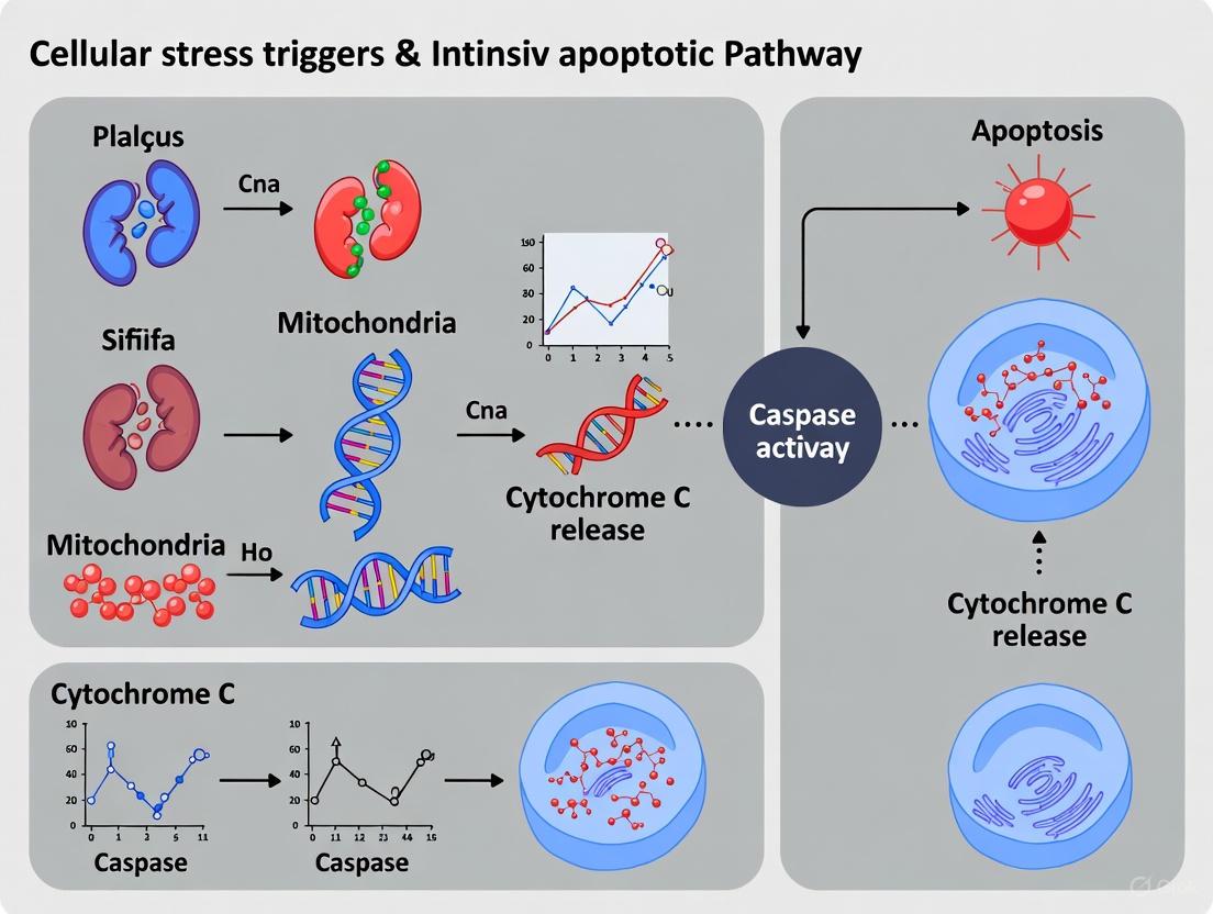
Cellular stress triggers are fundamental to understanding the intrinsic apoptotic pathway, a cornerstone of programmed cell death research. DNA damage, oxidative stress, and hypoxia represent three core stressors that converge on mitochondrial-mediated apoptosis, driving cellular fate decisions in physiological processes and disease pathologies. These triggers are interconnected through shared signaling networks that determine whether cells survive, undergo programmed death, or progress toward pathological states such as cancer and neurodegeneration. This technical guide examines the molecular mechanisms through which these stressors initiate the intrinsic apoptotic pathway, providing researchers with structured data, experimental protocols, and visualization tools to advance therapeutic interventions targeting cell death regulation.
DNA Damage as a Cellular Stress Trigger
Types of DNA Damage and Detection Methodologies
Genomic instability from DNA damage constitutes a primary activator of the intrinsic apoptotic pathway. Different types of DNA lesions engage distinct detection and repair mechanisms, with the failure of these systems leading to mitochondrial-mediated apoptosis.
Table 1: Types of DNA Damage and Associated Repair Pathways
| Damage Type | Primary Causes | Detection Sensors | Repair Pathway | Apoptotic Outcome |
|---|---|---|---|---|
| Double-strand breaks (DSBs) | Ionizing radiation, radiomimetic chemicals | MRN complex, ATM kinase | NHEJ, HR | High apoptotic potential |
| Single-strand breaks (SSBs) | UV radiation, oxidative stress | PARP1 | Base excision repair | Moderate apoptotic potential |
| Base lesions | Oxidation, alkylation | DNA glycosylases | Base excision repair | Context-dependent |
| Bulky adducts | UV light, chemicals | XPC, XPE complexes | Nucleotide excision repair | Limited unless extensive |
| Clustered lesions | Ionizing radiation | Multiple sensors | Complex, often error-prone | Very high apoptotic potential |
DSBs represent the most lethal form of DNA damage and are primarily repaired through two major pathways: non-homologous end joining (NHEJ) and homologous recombination (HR) [19]. The MRE11-RAD50-NBS1 (MRN) complex serves as the primary sensor for DSBs, recruiting and activating ataxia-telangiectasia mutated (ATM) kinase, which initiates a signaling cascade that coordinates DNA repair with cell fate decisions [19]. When repair fails, persistent DSBs activate pro-apoptotic signaling through mitochondrial permeabilization.
Experimental Protocol: Assessing DNA Damage-Induced Apoptosis
- Damage Induction: Treat cells with 2-10 Gy ionizing radiation or 1-5 µM etoposide to induce DSBs
- Damage Verification: Immunofluorescence staining for γH2AX foci (DSB marker) at 1-4 hours post-treatment
- Repair Monitoring: Track recruitment of repair proteins (e.g., RAD51 for HR, Ku70/80 for NHEJ) via live-cell imaging
- Apoptosis Assessment: Measure mitochondrial membrane potential (ΔΨm) with JC-1 dye and caspase-3/7 activation with fluorescent substrates
- Cell Fate Tracking: Use FUCCI cell cycle reporters to correlate damage phase with death outcomes [20]
From Nuclear Damage to Cytoplasmic Signaling
Unrepaired DNA damage triggers a transition from nuclear DNA damage response (DDR) to cytoplasmic apoptotic signaling through multiple mechanisms. Persistent nuclear DNA damage promotes structural nuclear envelope disruptions through DDR-mediated phosphorylation of lamin A/C via ATM and ATR signaling pathways [21]. This leads to micronuclei formation and cytoplasmic chromatin fragment (CCF) release, particularly in senescent cells [21]. The cGAS-STING pathway then detects this cytoplasmic DNA, initiating inflammatory signaling that can converge with apoptotic pathways [21]. Additionally, DNA damage-associated transcription stress generates R-loops that can be cleaved by endonucleases like XPG and XPF, releasing immunostimulatory nucleic acids into the cytoplasm [21].
Diagram 1: DNA damage to apoptosis signaling (52x20px)
Oxidative Stress and Redox Signaling
Oxidative stress represents an imbalance between reactive oxygen species (ROS) production and antioxidant defense mechanisms, leading to cellular damage and activation of stress response pathways. The major ROS sources in cells include mitochondrial electron transport chain leakage, NADPH oxidase (NOX) family enzymes, endoplasmic reticulum oxidative protein folding, and peroxisomal fatty acid oxidation [22]. At physiological levels, ROS function as signaling molecules regulating growth, differentiation, and survival through reversible oxidation of redox-sensitive cysteine residues on target proteins [22]. However, excessive ROS causes oxidative damage to cellular components including DNA, proteins, and lipids, activating stress response pathways including apoptosis.
Table 2: Primary ROS Sources and Their Characteristics
| ROS Source | Cellular Location | Primary ROS Produced | Physiological Functions | Pathological Consequences |
|---|---|---|---|---|
| Mitochondrial ETC | Inner mitochondrial membrane | O₂⁻, H₂O₂ | Metabolic signaling, hypoxia adaptation | ATP depletion, apoptosis initiation |
| NOX family | Plasma membrane, phagosomes | O₂⁻, H₂O₂ | Immune defense, cell signaling | Chronic inflammation, tissue damage |
| Endoplasmic reticulum | ER lumen | H₂O₂ | Disulfide bond formation in proteins | ER stress, unfolded protein response |
| Peroxisomes | Peroxisomal matrix | H₂O₂ | Fatty acid oxidation, ether lipid synthesis | Metabolic dysfunction, oxidative damage |
| Cytoplasmic enzymes | Cytosol | Varies by enzyme | Specific metabolic pathways | Enzyme-specific pathologies |
Oxidative Stress and Apoptotic Activation
Excessive ROS directly activates the intrinsic apoptotic pathway through multiple mechanisms. ROS induces mitochondrial outer membrane permeabilization (MOMP) by activating pro-apoptotic BCL-2 family proteins and triggering mitochondrial permeability transition [22] [23]. This leads to cytochrome c release and apoptosome formation, activating caspase-9 and the downstream executioner caspases-3 and -7 [22] [24]. Additionally, ROS causes oxidative DNA damage, activating the DDR pathway and p53-mediated apoptosis [22]. ROS also disrupts calcium homeostasis and impairs protein folding in the endoplasmic reticulum, contributing to ER stress-induced apoptosis [22].
Experimental Protocol: Measuring ROS-Induced Apoptosis
- ROS Induction: Treat cells with 100-500 µM H₂O₂ or 1-10 µM menadione for 2-6 hours
- ROS Detection: Use fluorescent probes (DCFDA for general ROS, MitoSOX for mitochondrial superoxide)
- Antioxidant Modulation: Pre-treat with N-acetylcysteine (1-10 mM) or SOD mimetics to validate ROS-specific effects
- Mitochondrial Assessment: Monitor ΔΨm with TMRE, cytochrome c release via immunofluorescence, and MOMP with real-time imaging
- Oxidative Damage Markers: Measure 8-oxo-dG (DNA oxidation), protein carbonylation, and lipid peroxidation products
Diagram 2: Oxidative stress to apoptosis pathway (52x20px)
Hypoxia and Oxygen Sensing
HIF Signaling and Cellular Adaptation
Hypoxia activates sophisticated oxygen-sensing mechanisms centered on hypoxia-inducible factors (HIFs), which orchestrate both adaptive survival responses and maladaptive apoptotic signaling depending on severity and duration. Under normoxic conditions, HIF-α subunits undergo prolyl hydroxylation by prolyl hydroxylase domain proteins (PHDs), leading to von Hippel-Lindau (pVHL)-mediated ubiquitination and proteasomal degradation [25]. During hypoxia, PHD activity decreases due to substrate (O₂) limitation, resulting in HIF-α stabilization, nuclear translocation, dimerization with HIF-1β, and transcription of hypoxia-responsive genes [25]. While acute HIF activation promotes adaptive responses including angiogenesis and metabolic reprogramming, chronic hypoxia induces apoptotic signaling through multiple mechanisms.
Hypoxia-Induced Apoptotic Signaling
Sustained hypoxia transitions from adaptive survival responses to intrinsic apoptosis activation through interconnected pathways. Hypoxia induces mitochondrial dysfunction through oxidative stress generated at complex III of the electron transport chain, promoting MOMP and cytochrome c release [25]. HIF-1α directly upregulates pro-apoptotic BNIP3 and NIX, which disrupt BCL-2/BCL-xL interactions with Beclin-1 and activate BAX/BAK-mediated apoptosis [25]. Hypoxia also impairs protein folding in the endoplasmic reticulum, activating the unfolded protein response and CHOP-mediated apoptosis [26]. Additionally, hypoxia creates an immunosuppressive microenvironment that can enhance survival of damaged cells while simultaneously inducing apoptosis in specific cell types.
Experimental Protocol: Modeling Hypoxia-Induced Apoptosis
- Hypoxia Induction: Use specialized chambers (0.1-1% O₂) or chemical mimetics (100-400 µM CoCl₂)
- HIF Verification: Monitor HIF-1α stabilization via western blotting and nuclear translocation by immunofluorescence
- Gene Expression Analysis: Quantify BNIP3, NIX, VEGF, and GLUT1 mRNA levels by qRT-PCR
- Metabolic Assessment: Measure extracellular acidification rate (glycolysis) and oxygen consumption rate (mitochondrial function)
- Apoptosis Quantification: Assess Annexin V/propidium iodide staining, caspase activation, and mitochondrial depolarization
Table 3: Hypoxia Severity and Cellular Responses
| Oxygen Level | Physiological/Pathological Context | Primary Signaling Response | Cell Fate Decision |
|---|---|---|---|
| 1-5% O₂ (Physioxia) | Normal tissue oxygenation | Baseline HIF signaling, metabolic homeostasis | Survival and proliferation |
| 0.5-1% O₂ (Mild hypoxia) | Ischemic border zones, solid tumors | HIF-1α dominant, glycolytic shift | Adaptive survival, angiogenesis |
| <0.5% O₂ (Severe hypoxia) | Ischemic core, tumor necrotic areas | HIF-1α/HIF-2α, ER stress, BNIP3 induction | Context-dependent apoptosis/autophagy |
| Anoxia (0% O₂) | Acute infarction, complete ischemia | ATP depletion, metabolic collapse | Rapid necrosis |
Diagram 3: Hypoxia signaling to apoptosis (52x20px)
Integration of Stress Pathways and Apoptotic Activation
Convergence on the Intrinsic Apoptotic Pathway
DNA damage, oxidative stress, and hypoxia demonstrate significant pathway crosstalk while ultimately converging on mitochondrial outer membrane permeabilization (MOMP) as the commitment point to intrinsic apoptosis. The BCL-2 protein family serves as the central integration point for these stress signals, with interactions between pro-apoptotic (Bax, Bak, BIM, PUMA) and anti-apoptotic (BCL-2, BCL-xL, MCL-1) members determining mitochondrial permeability [27] [24]. Following MOMP, cytochrome c release enables apoptosome formation with Apaf-1 and caspase-9, initiating the caspase cascade that executes apoptosis through cleavage of cellular substrates [24].
Experimental Protocol: Comprehensive Stress Pathway Analysis
- Multi-Stress Exposure: Apply combined stress conditions (e.g., sublethal hypoxia + low-dose etoposide)
- BH3 Profiling: Use peptide tools to measure mitochondrial priming and BCL-2 family dependencies
- Live-Cell Imaging: Track stress signaling kinetics with FRET biosensors and fluorescent protein reporters
- Multiplexed Pathway Assessment: Simultaneously measure DDR (γH2AX), oxidative stress (ROS sensors), and hypoxia (HIF reporters)
- Computational Integration: Develop mathematical models predicting cell fate decisions from early stress signaling events
The Scientist's Toolkit: Research Reagent Solutions
Table 4: Essential Reagents for Stress and Apoptosis Research
| Reagent Category | Specific Examples | Primary Research Application | Key Experimental Considerations |
|---|---|---|---|
| DNA damage inducers | Etoposide, Camptothecin, Bleomycin, IR | Activate DDR and study repair-death balance | Dose-response critical; cell cycle effects significant |
| ROS modulators | H₂O₂, Menadione, NAC, SOD mimetics | Investigate redox signaling and oxidative damage | Timing crucial; consider compensatory mechanisms |
| Hypoxia mimetics | CoCl₂, DFX, DMOG | Simulate HIF signaling without specialized equipment | May not fully recapitulate hypoxic metabolism |
| Apoptosis inducers | Staurosporine, ABT-737 (BCL-2 inhibitor) | Positive controls for apoptotic pathways | Confirm mechanism of action for your system |
| DDR inhibitors | KU-60019 (ATM), NU7441 (DNA-PKcs) | Dissect repair pathway contributions | Potential off-target effects require validation |
| HIF stabilizers | PHD inhibitors (FG-4592) | Specific HIF pathway activation | Monitor duration of effect and adaptive responses |
| Caspase substrates | DEVD-AMC (caspase-3/7), IETD-AFC (caspase-8) | Quantify apoptosis execution | Combine with inhibition to confirm specificity |
| Mitochondrial dyes | JC-1, TMRM, MitoTracker | Assess mitochondrial health and function | Optimize loading conditions and controls |
| BH3 mimetics | Venetoclax (ABT-199), A-1331852 (BCL-xL) | Target anti-apoptotic BCL-2 proteins | Cell type-specific dependencies exist |
| Live-cell reporters | FUCCI (cell cycle), ROSA26-H2B-GFP (chromatin) | Real-time fate tracking | Consider phototoxicity in long-term imaging |
DNA damage, oxidative stress, and hypoxia represent interconnected cellular stress triggers that converge on the intrinsic apoptotic pathway through both shared and distinct mechanisms. The complex crosstalk between these pathways creates a sophisticated stress response network that determines cellular fate decisions in health and disease. Advanced experimental approaches that simultaneously monitor multiple stress signaling pathways, combined with computational modeling and single-cell analysis, are advancing our understanding of how these triggers integrate to control apoptosis. This knowledge provides the foundation for developing novel therapeutic strategies that modulate apoptotic signaling in cancer, neurodegenerative disorders, and other conditions characterized by dysregulated cell death.
The endoplasmic reticulum (ER) is a critical organelle responsible for the synthesis, folding, and post-translational modification of approximately one-third of the cellular proteome, along with lipid synthesis and calcium storage [28]. When cellular conditions disrupt ER function, an accumulation of unfolded or misfolded proteins occurs, leading to a state known as ER stress. To counteract this stress and restore proteostasis, cells activate an evolutionarily conserved signaling network called the unfolded protein response (UPR) [29] [28].
The UPR is coordinated by three ER-transmembrane sensors: IRE1 (inositol-requiring enzyme 1), PERK (PKR-like ER kinase), and ATF6 (activating transcription factor 6). Under normal conditions, these sensors are kept in an inactive state through association with the chaperone protein BiP (GRP78). During ER stress, BiP dissociates to bind misfolded proteins, allowing the activation of these sensors [29] [30] [28]. The primary goal of the UPR is to adapt to stress by reducing global protein translation, increasing the production of ER chaperones, and enhancing the degradation of misfolded proteins. However, if these adaptive measures fail to resolve the stress within a certain timeframe, the UPR initiates apoptotic signaling to eliminate the damaged cell [29] [31]. This switch from pro-survival to pro-death signaling is a critical juncture in cellular fate, with significant implications in pathophysiology.
Molecular Mechanisms Linking ER Stress to Apoptosis
When ER stress is severe or prolonged, the UPR transitions from its adaptive role to initiating apoptosis through several well-characterized molecular pathways. The key mediators of this fatal decision are the PERK-ATF4-CHOP axis, the IRE1-TRAF2-JNK pathway, and the regulation of BCL-2 family proteins.
The PERK-ATF4-CHOP Axis
The PERK pathway is a major contributor to ER-stress-induced apoptosis. Upon activation, PERK phosphorylates the α-subunit of eukaryotic translation initiation factor 2 (eIF2α), which attenuates global protein synthesis, thereby reducing the protein-folding load on the stressed ER. Paradoxically, this phosphorylation simultaneously promotes the translation of select mRNAs, including that of activating transcription factor 4 (ATF4) [29] [28]. ATF4 then upregulates the expression of the pro-apoptotic transcription factor C/EBP-homologous protein (CHOP, also known as GADD153) [29] [30] [28].
CHOP induces apoptosis through multiple mechanisms:
- Downregulation of Bcl-2: CHOP transcriptionally represses the anti-apoptotic protein Bcl-2, thereby sensitizing cells to death signals [30] [32].
- Induction of GADD34: CHOP upregulates GADD34, a regulatory subunit of a phosphatase complex that dephosphorylates eIF2α. This action reverses the translational attenuation imposed by PERK, potentially leading to a fatal resurgence of protein synthesis and oxidative stress [29] [30].
- Death Receptor 5 (DR5) Induction: CHOP has been shown to induce the expression of TRAIL Death Receptor 5 (DR5), sensitizing cells to extrinsic apoptosis pathways [33] [30].
Table 1: Key Pro-Apoptotic Molecules in ER Stress
| Molecule | Full Name | Primary Function in ER-Stress-Induced Apoptosis |
|---|---|---|
| CHOP/GADD153 | C/EBP Homologous Protein / Growth Arrest- and DNA Damage-inducible gene 153 | Master pro-apoptotic transcription factor; downregulates Bcl-2, induces GADD34 and DR5. |
| GADD34 | Growth Arrest and DNA Damage-inducible protein 34 | Regulatory phosphatase subunit; dephosphorylates eIF2α, restoring protein translation and promoting stress. |
| TRAIL-R2/DR5 | TNF-Related Apoptosis-Inducing Ligand Receptor 2 / Death Receptor 5 | Death receptor; activated in a TRAIL-independent manner during ER stress to initiate caspase-8 cleavage. |
| JNK | Jun N-terminal Kinase | Kinase; phosphorylates and inactivates anti-apoptotic Bcl-2 proteins, promoting mitochondrial apoptosis. |
The IRE1-TRAF2-JNK Pathway
The IRE1 branch of the UPR also plays a dual role in cell fate. IRE1α, the ubiquitously expressed isoform, possesses both kinase and endoribonuclease activities. Its primary pro-survival function is the unconventional splicing of XBP1 mRNA, generating a potent transcription factor that drives the expression of ER chaperones and components of ER-associated degradation (ERAD) [29] [28]. However, under persistent stress, IRE1α can trigger apoptosis.
The primary apoptotic mechanism of IRE1 involves the recruitment of the adaptor protein TRAF2 (TNF receptor-associated factor 2) to the phosphorylated cytosolic domain of IRE1α. This complex then activates ASK1 (Apoptosis Signal-regulating Kinase 1), which in turn phosphorylates and activates JNK (c-Jun N-terminal kinase) [29] [31]. Sustained JNK activation promotes apoptosis by phosphorylating and inactivating anti-apoptotic members of the Bcl-2 family, such as Bcl-2 and Bcl-xL, thereby unleashing the pro-apoptotic proteins Bax and Bak [29]. Furthermore, under high stress levels, IRE1's RNase activity can become promiscuous, engaging in Regulated IRE1 Dependent Decay (RIDD) of a broad set of membrane-associated mRNAs. This process can degrade vital mRNAs, further contributing to cell death [29].
Cross-Talk with the Mitochondrial Apoptotic Pathway
The ER stress-induced apoptotic pathways converge on the mitochondrial (intrinsic) apoptotic pathway, which is governed by the BCL-2 protein family. The pro-apoptotic signals from CHOP and JNK tip the balance in favor of the pro-apoptotic BCL-2 members like Bim, Bax, and Bak [34] [32]. Furthermore, direct physical interaction between IRE1α and Bak/Bax at the ER membrane has been reported, which is important for IRE1α activation and the propagation of the apoptotic signal [29]. The culmination of these events leads to mitochondrial outer membrane permeabilization (MOMP), release of cytochrome c, activation of caspase-9, and finally, the execution of apoptosis via caspase-3 [34] [35].
Advanced Concepts and Emerging Signaling Pathways
Recent research has uncovered more sophisticated layers of regulation connecting ER stress to apoptosis, particularly in the context of disease microenvironments.
Mechanical Sensing via YAP/TAZ and Control of the DR5 Pathway
A 2025 study revealed that mechanical signals from the extracellular matrix (ECM) rigidity, transmitted through the transcriptional co-activators YAP (Yes-associated protein) and TAZ (Transcriptional coactivator with PDZ-binding motif), play a critical role in regulating ER-stress-induced apoptosis [33]. Tumor cells grown on a soft ECM (mimicking some in vivo conditions) showed nuclear exclusion and inactivation of YAP/TAZ, resulting in heightened sensitivity to ER-stress-induced apoptosis. Conversely, cells on a rigid ECM or expressing a constitutively active YAP mutant were highly resistant.
The molecular link for this mechanical regulation is the TRAIL-R2/DR5 pathway. YAP/TAZ activity exerts a dual control to suppress apoptosis:
- Preventing DR5 Clustering: It inhibits the intracellular clustering of DR5 that is necessary for caspase-8 activation.
- Inhibiting cFLIP Downregulation: It blocks the down-regulation of the caspase-8 inhibitor cFLIP during ER stress [33].
This mechanism demonstrates how physical cues from the tumor microenvironment can be integrated with proteotoxic stress to determine cell fate.
Viral Infections and ER Stress
Viral infections often place a heavy demand on the host cell's ER for viral protein synthesis and folding, thereby inducing ER stress. For instance, infection with Duck Hepatitis A Virus Type 1 (DHAV-1) was shown to activate the PERK-eIF2α-ATF4-CHOP pathway, leading to upregulation of pro-apoptotic GADD34, suppression of anti-apoptotic Bcl-2, and activation of caspase-3 [30]. Inhibition of PERK activity suppressed both CHOP activation and viral replication, highlighting the complex interplay where the host's ER-stress-induced apoptotic response can also function as an antiviral mechanism [30].
Experimental Analysis of ER-Stress-Induced Apoptosis
This section provides a detailed methodological framework for investigating the linkage between ER stress and apoptosis, as cited in the literature.
Key Research Reagent Solutions
Table 2: Essential Reagents for Studying ER-Stress-Induced Apoptosis
| Reagent / Tool | Function / Target | Experimental Application |
|---|---|---|
| Thapsigargin | SERCA pump inhibitor; disrupts ER calcium homeostasis. | A classic and potent ER stress inducer used to trigger the UPR and subsequent apoptosis [33] [30]. |
| Tunicamycin | Inhibits N-linked glycosylation; causes accumulation of unfolded proteins. | A widely used ER stressor to study UPR activation and CHOP-dependent apoptosis [33] [30]. |
| 4-PBA (4-Phenylbutyric acid) | Chemical chaperone that aids protein folding. | Used to alleviate ER stress and test the dependency of apoptosis on the UPR [30]. |
| GSK2606414 | Potent and selective PERK inhibitor. | Used to specifically inhibit the PERK branch of the UPR to delineate its role in apoptosis [30]. |
| siRNA/shRNA (YAP/TAZ) | Knocks down expression of YAP and TAZ transcriptional co-activators. | Used to study the role of mechanical signaling in regulating ER-stress-induced apoptosis, particularly via the DR5 pathway [33]. |
| Caspase-8 Inhibitor (e.g., Z-IETD-FMK) | Selective inhibitor of caspase-8. | Used to determine the contribution of the extrinsic apoptotic pathway (DR5-caspase-8) to cell death [33]. |
| Antibodies (p-eIF2α, CHOP, BiP/GRP78, cleaved Caspase-3) | Detect specific protein levels and post-translational modifications. | Essential for Western Blot and immunofluorescence analysis to monitor UPR activation and apoptotic execution. |
Detailed Experimental Protocol: Modulating ECM Rigidity to Assess YAP/TAZ Role
Objective: To investigate how matrix stiffness and YAP/TAZ activity regulate ER-stress-induced apoptosis via the TRAIL-R2/DR5 pathway [33].
Workflow:
Methodology:
Substrate Preparation and Cell Culture:
- Use acrylamide hydrogels of tunable stiffness (e.g., 0.5 kPa for "soft" and 50 kPa for "stiff") to mimic different mechanical microenvironments. Tissue culture plastic serves as a high-rigidity control.
- Seed appropriate tumor cell lines (e.g., A549 lung carcinoma, HeLa) onto these substrates and allow them to adhere and acclimatize for 24-48 hours.
Genetic Manipulation:
- Knockdown: Transfect cells with pooled siRNAs targeting YAP and TAZ to deplete their expression. Use non-targeting siRNA as a negative control.
- Overexpression: Generate stable cell lines expressing a constitutively active YAP mutant (YAP5SA) that is resistant to Hippo pathway inhibition. Use an inducible system for wild-type YAP to control the timing of expression.
ER Stress Induction and Drug Treatment:
- Treat cells with a predetermined optimal concentration of an ER stress inducer like Thapsigargin (e.g., 1 µM) or Tunicamycin (e.g., 5 µg/mL) for a time course (e.g., 6-24 hours).
- In parallel, use specific inhibitors (e.g., PERK inhibitor GSK2606414) or caspase-8 inhibitors to dissect the contribution of specific pathways.
Apoptosis and Pathway Analysis:
- Quantify Apoptosis: Use flow cytometry with Annexin V/propidium iodide (PI) staining to quantify the percentage of apoptotic cells.
- Analyze Caspase Activation: Perform Western Blot analysis on cell lysates to detect the cleavage (activation) of caspase-8 and caspase-3.
- Assess DR5 Activation: Use immunofluorescence or subcellular fractionation to monitor the intracellular clustering of TRAIL-R2/DR5.
- Measure Gene/Protein Expression: Use RT-qPCR and Western Blotting to analyze the expression levels of key genes like CHOP, cFLIP, and GADD34.
Expected Outcomes: Cells on soft substrates or with YAP/TAZ knockdown should exhibit significantly higher levels of ER-stress-induced apoptosis, DR5 clustering, and caspase-8 activation compared to cells on rigid substrates or expressing active YAP.
Pathophysiological Relevance and Therapeutic Implications
Dysregulated ER-stress-induced apoptosis is implicated in the pathogenesis of numerous human diseases.
- Neurodegenerative Diseases: Conditions like Alzheimer's and Parkinson's are characterized by the accumulation of misfolded proteins, chronic ER stress, and neuronal loss via CHOP-mediated apoptosis [29] [31].
- Diabetes: Pancreatic beta cells are highly specialized for insulin secretion, making them particularly susceptible to ER stress. Chronic stress can lead to beta cell apoptosis, contributing to the pathogenesis of diabetes [29] [31].
- Atherosclerosis: In atherosclerotic plaques, macrophage apoptosis driven by ER stress contributes to plaque necrosis and instability, which can trigger acute clinical events [31].
- Cancer: Tumor microenvironments often feature nutrient deprivation, hypoxia, and acidosis, all of which induce ER stress. Cancer cells co-opt the UPR to survive these conditions, but the pro-apoptotic arm remains a therapeutic target. The newly discovered YAP/TAZ-DR5 link provides a novel angle for targeting therapy-resistant tumors by manipulating mechanical signals or downstream effectors [33] [28].
The molecular players in ER-stress-induced apoptosis represent promising therapeutic targets. Strategies are being developed to either protect cells from apoptosis in degenerative diseases by inhibiting CHOP or the JNK pathway, or to push cancer cells over the brink from adaptation to apoptosis by enhancing the pro-death outputs of the UPR.
Integrated Signaling Pathway Visualization
The core molecular mechanisms linking ER stress to apoptosis, including the emerging role of mechanical signaling, are summarized in the following integrated pathway diagram.
Mitochondrial outer membrane permeabilization (MOMP) is recognized as the decisive commitment point in numerous forms of apoptotic cell death, particularly within the intrinsic apoptotic pathway [36] [37]. This process represents a physiological event wherein the mitochondrial outer membrane becomes permeable to specific molecules, enabling the passage of proteins crucial for apoptosis execution from the mitochondrial intermembrane space into the cytosol [38] [39]. As the central regulatory step in the mitochondrial pathway of apoptosis, MOMP is triggered in response to diverse cellular stresses, including severe DNA damage and protein turnover dysfunction, which are frequently induced by chemotherapeutic agents [40] [41].
The permeabilization event transforms the mitochondrial outer membrane from a barrier that is normally permeable to molecules smaller than 5 kDa to one that can accommodate proteins larger than 100 kDa [41]. This dramatic increase in permeability allows the release of pro-apoptotic proteins such as cytochrome c and SMAC (Second mitochondria-derived activator of caspase), which activate the proteolytic cascade that dismantles the cell [39] [41]. Once MOMP occurs, the cell is fated to die even in the absence of caspase activity, as the resulting mitochondrial dysfunction alone can eventually lead to cell death [42]. This "point of no return" characteristic makes MOMP a critical control point in cellular fate decisions and a focal point for therapeutic interventions in diseases such as cancer [36] [37].
Molecular Mechanisms of MOMP Execution
Core Components of the MOMP Machinery
The fundamental machinery responsible for MOMP centers on the BCL-2 protein family, which consists of three functionally distinct subgroups that interact to regulate mitochondrial membrane integrity [41] [42]. The pro-apoptotic effector proteins Bax and Bak are essential components, as cells deficient in both proteins demonstrate profound resistance to most intrinsic apoptotic stimuli [42]. These multidomain proteins directly mediate the formation of pores in the mitochondrial outer membrane. A third pro-apoptotic effector, Bok, shares structural similarities with Bax and Bak, though its role appears to be more context-dependent [41].
Opposing these pro-apoptotic effectors are the anti-apoptotic proteins including Bcl-2, Bcl-xL, and Mcl-1, which preserve mitochondrial integrity by binding and neutralizing Bax/Bak activation [40] [41]. The third subgroup consists of BH3-only proteins (such as Bim, Bid, Puma, Noxa, and Bad), which function as sentinels that sense diverse cellular stress signals and initiate the apoptotic cascade by engaging the other BCL-2 family members [40] [42].
Pore Formation and Activation Mechanism
The current model of Bax/Bak activation involves a multi-step conformational change process [42]. In healthy cells, Bax predominantly resides in the cytosol or is loosely associated with membranes, while Bak is integrated into the mitochondrial outer membrane. Following an apoptotic stimulus, Bax undergoes translocation to the mitochondria and inserts its C-terminal transmembrane domain into the outer membrane [42]. Both proteins then experience significant conformational changes that include exposure of their N-terminal epitopes and, critically, the transient exposure of their BH3 domains [42].
The exposed BH3 domain of one activated Bax or Bak molecule subsequently binds to the hydrophobic surface groove of another activated molecule, forming a novel symmetric homodimer through BH3:groove interactions [42]. These dimers then further associate via interfaces outside the BH3 domain to form higher-order oligomers that constitute the apoptotic pore complex in the outer mitochondrial membrane [42]. Although the precise architecture of these pores remains under investigation, they are sufficient to permit the passage of large proteins like cytochrome c (approximately 15 kDa) and SMAC from the intermembrane space to the cytosol [41].
Table 1: BCL-2 Protein Family Members Regulating MOMP
| Protein Category | Representative Members | Primary Function in MOMP Regulation |
|---|---|---|
| Anti-apoptotic | Bcl-2, Bcl-xL, Mcl-1 | Bind and neutralize activated Bax/Bak; sequester BH3-only proteins |
| Pro-apoptotic Effectors | Bax, Bak | Form oligomeric pores in mitochondrial outer membrane |
| BH3-only Proteins | Bim, Bid, Puma, Noxa, Bad | Sense cellular stress; initiate apoptosis by engaging anti-apoptotic proteins and/or directly activating Bax/Bak |
Dynamics and Coordination of MOMP
The permeabilization of individual mitochondria within a single cell occurs in a coordinated manner, though the onset timing varies between individual mitochondria [43] [41]. High-resolution cellular imaging has revealed that MOMP at the single mitochondrion level is a rapid process lasting only seconds, while complete permeabilization of all mitochondria within a cell typically requires approximately five minutes [43] [41]. Interestingly, studies using sibling HeLa cell pairs have demonstrated synchronous apoptosis execution, suggesting clonal influences on MOMP timing [43].
In many instances, MOMP propagation through the cytosol follows a wave-like pattern [43]. Computational modeling using partial differential equations suggests that this wave-like propagation can be sufficiently explained by diffusion-adsorption velocities of locally generated permeabilization inducers [43]. While some evidence links this wave propagation to ER calcium channels, elevation of intracellular calcium is not universally required for MOMP execution [41].
Figure 1: Molecular Regulation of MOMP in Intrinsic Apoptosis
MOMP as a Cellular Decision Point
The "Point of No Return" Concept
MOMP earns its designation as the "point of no return" in apoptosis through two distinct but complementary mechanisms that ensure cellular demise [42]. First, the release of cytochrome c into the cytosol triggers the formation of the apoptosome complex, which activates caspase-9 and subsequently the executioner caspases-3 and -7, initiating a proteolytic cascade that systematically dismantles cellular structures [39] [42]. Second, MOMP compromises essential mitochondrial functions, including oxidative phosphorylation, leading to bioenergetic failure irrespective of caspase activation [42]. This dual mechanism provides a fail-safe ensuring that once MOMP occurs, cell death proceeds even if downstream elements of the apoptotic cascade are disrupted.
The critical nature of MOMP is demonstrated by experiments showing that genetic ablation of both Bax and Bak renders cells highly resistant to diverse apoptotic stimuli, whereas deletion of individual components downstream of MOMP (such as Apaf-1 or caspase-9) only delays but does not prevent eventual cell death [42]. This evidence confirms that mitochondrial outer membrane integrity represents the true commitment point in the apoptotic pathway.
Variations in MOMP Completeness
While MOMP is typically considered an all-or-nothing event at the cellular level, recent research has revealed more nuanced scenarios where MOMP occurs incompletely [41]. Two distinct variations have been characterized:
Incomplete MOMP (iMOMP): Occurs when most but not all mitochondria within a cell undergo permeabilization [36] [41]. Cell survival in this scenario depends on the absence or inhibition of caspase activity, potentially allowing recovery if the initiating stress is resolved.
Minority MOMP (miniMOMP): Involves permeabilization of only a small fraction of mitochondria in response to sublethal stress [36] [41]. This limited MOMP can generate sublethal caspase activity that promotes DNA damage and other non-apoptotic signaling functions, potentially contributing to oncogenic transformation [41].
These partial MOMP phenomena demonstrate that the cellular response to mitochondrial permeabilization exists on a spectrum, with implications for both physiological signaling and pathological processes such as carcinogenesis.
Table 2: MOMP Variants and Their Consequences
| MOMP Type | Mitochondria Affected | Caspase Activation | Cell Fate | Potential Consequences |
|---|---|---|---|---|
| Complete MOMP | All or nearly all mitochondria | Robust, widespread | Apoptotic cell death | Normal development; tissue homeostasis; chemotherapy-induced killing |
| Incomplete MOMP (iMOMP) | Majority of mitochondria | Variable, potentially inhibited | Survival possible without caspase activity | Potential recovery; altered signaling |
| Minority MOMP (miniMOMP) | Small fraction of mitochondria | Sublethal, localized | Cell survival with sublethal signaling | DNA damage; oncogenic transformation |
Experimental Assessment of MOMP
Key Methodologies and Reagents
Research into MOMP mechanisms employs a diverse toolkit of chemical agents, recombinant proteins, and genetic approaches that allow precise dissection of the apoptotic machinery. Key experimental approaches include:
Cellular Stress Inducers: Chemotherapeutic agents such as doxorubicin (induces severe DNA damage) and bortezomib (proteasome inhibitor causing protein turnover dysfunction) serve as robust initiators of intrinsic apoptosis [40]. These compounds generate cellular stress that converges on mitochondria to trigger MOMP.
BH3-mimetics: Small molecule inhibitors including ABT-263 (Navitoclax) and Venetoclax that bind anti-apoptotic BCL-2 family proteins, neutralizing their protective function and promoting Bax/Bak activation [40] [41]. These compounds allow researchers to bypass upstream signaling events and directly probe the core apoptotic machinery.
Genetic Manipulation: Knockdown approaches (e.g., siRNA against Bim) and gene knockout cells (e.g., Bax/Bak double knockout cells) enable functional validation of specific components in the MOMP pathway [40] [42].
High-Speed Cellular Imaging: Advanced microscopy techniques that monitor the temporal and spatial dynamics of MOMP in living cells, often using fluorescently tagged cytochrome c or other IMS proteins [43].
The Researcher's Toolkit: Essential Reagents
Table 3: Key Research Reagents for MOMP Investigation
| Reagent / Tool | Category | Primary Research Application | Mechanistic Insight |
|---|---|---|---|
| ABT-263 (Navitoclax) | BH3-mimetic | Inhibits Bcl-2/Bcl-xL; tests dependence on specific anti-apoptotic proteins | Demonstrates BCL-2 family interactions; reveals therapeutic vulnerabilities |
| Venetoclax | BH3-mimetic | Selective Bcl-2 inhibitor; clinical correlation | High specificity for Bcl-2; minimal effect on Mcl-1 dependencies |
| Doxorubicin (DOX) | DNA damaging agent | Induces severe DNA damage stress; p53 activation studies | Triggers intrinsic apoptosis; reveals p53-dependent and independent pathways |
| Bortezomib (BTZ) | Proteasome inhibitor | Causes protein turnover dysfunction stress | Induces p53-independent apoptosis; ER stress connections |
| Bim siRNA | Genetic tool | Knocks down BH3-only protein Bim | Tests necessity of specific BH3-only proteins in MOMP initiation |
| Cytochrome c-GFP | Imaging reporter | Visualizes MOMP timing and spread in live cells | Reveals MOMP dynamics; wave propagation patterns |
Experimental Protocol: Assessing MOMP in Cellular Models
A standard approach for evaluating MOMP involvement in apoptotic pathways involves combining stress inducers with BH3-mimetics in genetically defined cell systems [40]. The following protocol exemplifies this strategy:
Cell Model Selection: Utilize matched cell pairs differing in specific genetic components (e.g., p53-wildtype LNCaP and p53-null PC3 prostate cancer cells) [40].
Stress Application: Treat cells with increasing concentrations of doxorubicin (0.1-5 μM) or bortezomib (5-100 nM) for 12-48 hours to induce graded cellular stress [40].
BH3-mimetic Combination: Co-treat with BH3-mimetics such as ABT-263 (0.1-1 μM) to test for synergistic effects that indicate BCL-2 family involvement [40].
Apoptosis Assessment: Quantify apoptosis using flow cytometry with Annexin V/propidium iodide staining and monitor MOMP execution via cytochrome c release assays (immunofluorescence or subcellular fractionation) [40] [43].
Mechanistic Validation: Employ genetic tools (e.g., Bim siRNA) to confirm the specific contribution of individual BCL-2 family members to the observed MOMP phenotype [40].
This experimental approach demonstrated that severe cellular stress can induce apoptosis through a dual control pathway where Bim functions as a sensitizer to release Bax/Bak from Bcl-xL, while p53 may act as an activator to directly activate Bax/Bak [40].
Figure 2: Experimental Workflow for MOMP Mechanism Investigation
MOMP in Disease and Therapeutic Targeting
Pathophysiological Implications
MOMP regulation has significant implications for human disease, particularly in cancer and degenerative conditions [38] [41]. In cancer, defective MOMP execution contributes to tumor development and treatment resistance, while excessive MOMP can drive degenerative diseases [41]. Cancer cells often exploit multiple mechanisms to evade MOMP, including:
p53 Mutations: Abrogation of p53 function eliminates a key activator of BH3-only proteins like Puma and Noxa, raising the threshold for apoptosis initiation [40].
Anti-apoptotic Protein Overexpression: Elevated levels of Bcl-2, Bcl-xL, or Mcl-1 in many cancers effectively buffer pro-apoptotic signals, preventing Bax/Bak activation [40] [41].
Altered BH3-only Protein Expression: Downregulation of specific BH3-only proteins such as Bim can disconnect cellular stress signals from the core apoptotic machinery [40].
Interestingly, recent evidence suggests that partial MOMP events (miniMOMP) may promote oncogenic transformation by causing sublethal caspase activation and DNA damage, potentially creating genomic instability that drives tumor evolution [41].
Therapeutic Opportunities
The central role of MOMP in apoptosis control makes it an attractive target for therapeutic intervention, particularly in oncology [41] [42]. Several targeted approaches have emerged:
BH3-mimetics: Small molecules that bind and inhibit specific anti-apoptotic BCL-2 family proteins [41]. Venetoclax (selective Bcl-2 inhibitor) has demonstrated clinical efficacy in hematological malignancies, particularly chronic lymphocytic leukemia [41].
Combination Therapies: BH3-mimetics combined with conventional chemotherapeutic agents can overcome treatment resistance by lowering the threshold for MOMP induction [40]. Studies in prostate cancer, glioblastoma, and osteosarcoma models show synergistic effects when ABT-263 is combined with doxorubicin or bortezomib, even in p53-null cells [40].
BH3 Profiling: Functional assessment of mitochondrial priming to predict chemotherapy sensitivity, potentially enabling personalized treatment regimens based on a tumor's dependence on specific anti-apoptotic proteins [41].
The development of Mcl-1-specific inhibitors represents an active area of investigation, as Mcl-1 overexpression can confer resistance to existing BH3-mimetics that primarily target Bcl-2 and Bcl-xL [41]. As our understanding of MOMP regulation advances, so too will opportunities to manipulate this critical cellular process for therapeutic benefit across a spectrum of human diseases.
The intrinsic apoptotic pathway is a fundamental process of programmed cell death essential for maintaining tissue homeostasis and eliminating damaged or stressed cells [44] [45]. This pathway is characterized by a meticulously orchestrated series of molecular events that commence within the cell in response to severe internal stressors, including DNA damage, oxidative stress, growth factor withdrawal, and endoplasmic reticulum stress [46]. The central event in this pathway is mitochondrial outer membrane permeabilization (MOMP), which leads to the release of key pro-apoptotic proteins from the mitochondrial intermembrane space into the cytosol [44] [46]. Among these proteins, cytochrome c plays a pivotal role as it initiates the formation of a critical molecular complex known as the apoptosome, which subsequently activates a cascade of proteolytic enzymes called caspases that ultimately execute cell death [47] [48]. This technical guide provides a comprehensive overview of the molecular mechanisms, experimental methodologies, and regulatory networks governing these downstream execution events, with particular emphasis on their relevance to cellular stress responses and therapeutic targeting.
Cytochrome c Release and Regulation
The Central Role of Cytochrome c in Apoptosis Execution
Cytochrome c is a hemoprotein normally located in the mitochondrial intermembrane space, where it serves as an essential component of the electron transport chain during cellular respiration [44] [48]. However, upon induction of the intrinsic apoptotic pathway by cellular stressors, cytochrome c is released into the cytosol, where it assumes a completely different function as a critical initiator of the apoptotic cascade [48] [46]. This release represents a point of no return in the commitment to cell death and is primarily triggered by mitochondrial outer membrane permeabilization (MOMP) [46].
Once in the cytosol, cytochrome c interacts with apoptotic protease activating factor-1 (Apaf-1) in the presence of dATP/ATP to form the apoptosome complex, which serves as a molecular platform for caspase activation [47] [48]. Recent research has revealed that cytochrome c possesses additional regulatory functions beyond Apaf-1 activation. Specifically, it can interact with 14-3-3ε, a cytosolic inhibitor of Apaf-1, thereby blocking 14-3-3ε-mediated Apaf-1 inhibition and indirectly promoting caspase activation [48]. This dual functionality positions cytochrome c as a master regulator of the intrinsic apoptotic pathway, capable of both directly activating pro-apoptotic machinery and simultaneously relieving intrinsic inhibitory mechanisms.
Molecular Regulation of Cytochrome c Release
The release of cytochrome c from mitochondria is primarily governed by the Bcl-2 family of proteins, which consists of both pro-apoptotic and anti-apoptotic members that delicately balance cell survival and death decisions [44] [47] [46]. This protein family can be categorized into three functional groups:
- Anti-apoptotic proteins (e.g., Bcl-2, Bcl-xL, Mcl-1) that preserve mitochondrial integrity and prevent cytochrome c release [46]
- Pro-apoptotic effector proteins (e.g., Bax, Bak) that directly mediate MOMP [47] [46]
- BH3-only proteins (e.g., Bid, Bim, Puma, Noxa) that function as sensors of cellular stress and initiate the activation of Bax and Bak [46]
In response to cellular stressors such as DNA damage, the tumor suppressor protein p53 transcriptionally upregulates several pro-apoptotic BH3-only proteins, including Puma and Noxa [46]. Additionally, caspase-8 can cleave the BH3-only protein Bid to generate truncated Bid (tBid), which translocates to mitochondria and activates Bax and Bak, representing a key point of crosstalk between the extrinsic and intrinsic apoptotic pathways [47] [46].
Upon activation, Bax and Bak undergo conformational changes and oligomerize to form pores in the mitochondrial outer membrane, leading to MOMP and the consequent release of cytochrome c and other pro-apoptotic factors [47] [46]. Anti-apoptotic Bcl-2 family proteins counteract this process by binding to and neutralizing activated Bax, Bak, and BH3-only proteins, thereby maintaining mitochondrial integrity [46].
Table 1: Bcl-2 Protein Family Members Regulating Cytochrome c Release
| Protein Class | Representative Members | Function in Cytochrome c Release |
|---|---|---|
| Anti-apoptotic | Bcl-2, Bcl-xL, Mcl-1 | Bind and inhibit pro-apoptotic members; maintain mitochondrial membrane integrity |
| Pro-apoptotic Effectors | Bax, Bak | Oligomerize to form mitochondrial pores; mediate MOMP |
| BH3-only Proteins | Bid, Bim, Puma, Noxa, Bad | Sense cellular stress; activate Bax/Bak or inhibit anti-apoptotic proteins |
Alternative Mechanisms of Mitochondrial Membrane Permeabilization
Beyond Bax/Bak-mediated MOMP, cytochrome c release can occur through mitochondrial permeability transition (MPT), which involves the opening of a multiprotein channel known as the permeability transition pore complex (PTPC) at contact sites between the mitochondrial inner and outer membranes [46]. The PTPC comprises several proteins, including the voltage-dependent anion channel (VDAC) in the outer membrane, adenine nucleotide translocase (ANT) in the inner membrane, and cyclophilin D in the matrix [46]. MPT is triggered by specific lethal stimuli such as cytosolic Ca²⁺ overload and excessive reactive oxygen species generation, leading to osmotic swelling of the mitochondrial matrix and subsequent rupture of the outer membrane [46]. Genetic studies have demonstrated that cyclophilin D deficiency can limit pathological cell death in various contexts, highlighting the physiological relevance of this mechanism [46].
Apoptosome Formation and Structure
Molecular Composition and Assembly
The apoptosome is a large multiprotein complex that serves as the molecular platform for initiating the caspase activation cascade in the intrinsic apoptotic pathway [47] [48]. Its formation is triggered when cytochrome c is released from mitochondria and binds to apoptotic protease activating factor-1 (Apaf-1) in the cytosol in the presence of dATP/ATP [47] [48]. Apaf-1 is a multidomain protein consisting of an N-terminal caspase recruitment domain (CARD), a central nucleotide-binding and oligomerization domain, and a C-terminal regulatory region containing multiple WD40 repeats that mediate cytochrome c binding [48].
Upon cytochrome c binding, Apaf-1 undergoes a conformational change that promotes its oligomerization into a wheel-like structure with seven-fold symmetry [47] [48]. This oligomeric complex, the apoptosome, then recruits and activates procaspase-9 through CARD-CARD interactions between Apaf-1 and the caspase [47]. The fully assembled apoptosome consists of Apaf-1, cytochrome c, and caspase-9, forming a proteolytically active complex often described as a "caspase-activating machine" [48].
Regulatory Mechanisms of Apoptosome Function
Apoptosome formation and activity are subject to multiple layers of regulation to ensure precise control over the cell death decision. The 14-3-3ε protein has been identified as a direct inhibitor of Apaf-1 that prevents apoptosome assembly and subsequent caspase activation [48]. This inhibitory function is enhanced when Apaf-1 is phosphorylated at Ser268 by the p90kDa ribosomal S6 kinase-1 (Rsk-1), which is activated through the mitogen-activated protein kinase (MAPK) cascade [48]. Interestingly, cytochrome c can counteract this inhibition by binding to 14-3-3ε, thereby blocking its interaction with Apaf-1 and promoting caspase activation [48]. This reveals a novel regulatory mechanism wherein cytochrome c functions not only as an apoptosome assembly factor but also as a reliever of intrinsic inhibition.
The heme status of cytochrome c also significantly influences its ability to activate Apaf-1. The apo form of cytochrome c (lacking heme) can bind to Apaf-1 but fails to induce apoptosome formation, instead acting as a competitive inhibitor that blocks holo-cytochrome c-dependent caspase activation [49]. This suggests that the heme group is essential for the proper conformational changes required for Apaf-1 activation.
Table 2: Key Components of the Apoptosome Complex
| Component | Structure/Features | Function in Apoptosome |
|---|---|---|
| Apaf-1 | CARD domain, nucleotide-binding domain, WD40 repeats | Oligomerizes to form platform; recruits and activates caspase-9 |
| Cytochrome c | Heme-containing protein | Binds WD40 domain of Apaf-1; triggers conformational change and oligomerization |
| Caspase-9 | CARD domain, protease domain | Initiator caspase; activated within apoptosome; cleaves executioner caspases |
| dATP/ATP | Nucleotides | Cofactors required for apoptosome assembly and activation |
Caspase Activation Cascade
Caspase Classification and Activation Mechanisms
Caspases are a family of cysteine proteases that cleave their substrates at specific aspartic acid residues, serving as the primary executioners of apoptosis [6]. Based on their function and position in the apoptotic cascade, caspases are broadly classified into two categories: initiator caspases (caspases-2, -8, -9, and -10) and executioner caspases (caspases-3, -6, and -7) [44] [47] [6]. Initiator caspases possess long prodomains containing protein-protein interaction motifs (CARD or DED domains) that enable their recruitment to and activation within specific signaling complexes [6]. In the intrinsic pathway, caspase-9 is the primary initiator caspase, activated within the apoptosome complex [47] [6].
Executioner caspases have short prodomains and exist in the cytosol as inactive dimers [47]. They are activated through proteolytic cleavage by initiator caspases, which generates the mature enzyme consisting of large and small subunits [47]. Once activated, executioner caspases cleave numerous cellular substrates, leading to the characteristic morphological and biochemical changes associated with apoptosis [47].
The Caspase Cascade in Intrinsic Apoptosis
In the intrinsic apoptotic pathway, the caspase activation cascade begins when caspase-9 is recruited to the apoptosome via CARD-CARD interactions with Apaf-1 [47] [6]. Within this complex, caspase-9 undergoes activation through proximity-induced dimerization and conformational change [6]. Activated caspase-9 then proteolytically cleaves and activates the executioner caspases, primarily caspase-3 and caspase-7 [47] [6].
Caspase-3, once activated, serves as the primary executioner caspase and amplifies the death signal by cleaving numerous cellular substrates, including structural proteins, DNA repair enzymes, and cell cycle regulators [47]. One critical substrate is ICAD (inhibitor of caspase-activated DNase), whose cleavage releases CAD (caspase-activated DNase) to migrate to the nucleus and fragment DNA [50] [47]. Caspase-3 also cleaves components of the cytoskeleton, nuclear envelope proteins such as lamins, and other caspases, including caspase-6, creating a positive feedback loop that further amplifies the apoptotic signal [47] [6].
Regulation of Caspase Activity
Caspase activity is tightly regulated at multiple levels to prevent inappropriate cell death. The inhibitor of apoptosis proteins (IAPs), including XIAP, cIAP1, and cIAP2, directly bind to and inhibit active caspases [44] [47]. XIAP is particularly potent in inhibiting caspases-3, -7, and -9 through its BIR domains [47]. This inhibition is counteracted by mitochondrial proteins such as Smac/DIABLO and HtrA2/OMI, which are released along with cytochrome c during MOMP [44] [47]. These proteins promote caspase activation by binding to and neutralizing IAPs, thereby relieving caspase inhibition [44] [47].
Additionally, caspases themselves can regulate their own activity through feedback mechanisms. For instance, caspase-3-mediated cleavage of anti-apoptotic Bcl-2 family proteins such as Bcl-2 and Bcl-xL inactivates them and promotes further mitochondrial permeabilization, creating a positive feedback loop that amplifies the apoptotic signal [50]. Conversely, caspase-9 can cleave RIPK1, thereby inhibiting necroptosis and ensuring the cell undergoes apoptosis rather than alternative forms of cell death [6].
Experimental Analysis of Downstream Execution Events
Methodologies for Monitoring Cytochrome c Release
The release of cytochrome c from mitochondria is a critical early event in the intrinsic apoptotic pathway that can be detected using several experimental approaches:
Subcellular Fractionation and Western Blotting: Cells are fractionated into cytosolic and mitochondrial components using digitonin permeabilization and differential centrifugation [50]. Briefly, cells are suspended in permeabilization buffer (75 mM KCl, 1 mM NaH₂PO₄, 8 mM Na₂HPO₄, 250 mM sucrose) containing 150 μg/ml digitonin and protease inhibitors, then fractionated by centrifugation at 16,000 × g [50]. The resulting cytosolic and particulate fractions are analyzed by Western blotting using antibodies against cytochrome c, with mitochondrial markers (e.g., VDAC1) and cytosolic markers (e.g., α-tubulin) serving as fractionation controls [50].
Immunocytochemistry and Fluorescence Microscopy: Cells are stained with fluorescent dyes such as MitoTracker Deep Red to visualize mitochondria, followed by fixation and immunostaining with anti-cytochrome c antibodies [50]. The redistribution of cytochrome c from a punctate mitochondrial pattern to a diffuse cytosolic pattern indicates release. Cells are counterstained with DAPI to visualize nuclear morphology and analyzed by fluorescence microscopy [50].
Flow Cytometric Analysis of Mitochondrial Membrane Potential: Changes in mitochondrial membrane potential (ΔΨm) often precede or accompany cytochrome c release. This can be measured using potentiometric dyes such as tetramethylrhodamine ethyl ester (TMRE) [50]. Cells are incubated with 25 nM TMRE for 20 minutes, then analyzed by flow cytometry. Decreased TMRE fluorescence indicates loss of ΔΨm, which is associated with MOMP and cytochrome c release [50].
Techniques for Studying Apoptosome Formation
Caspase Activation Assays: Apoptosome activity is typically assessed by measuring the activation of its downstream effector, caspase-9, and the executioner caspases-3 and -7 [50]. Commercially available luminescent or fluorescent substrates are commonly used. For example, the Caspase-Glo 3/7 assay provides a luminescent readout proportional to caspase-3/7 activity [50]. Cells are treated with experimental conditions, then incubated with Caspase-Glo reagent for 1 hour before measuring luminescence [50]. Similarly, caspase-9 activity can be measured using specific substrates or activity assays.
Reconstitution Experiments with Purified Components: The molecular details of apoptosome formation can be studied using purified recombinant proteins [48]. Cytochrome c, Apaf-1, and caspase-9 are incubated in the presence of dATP/ATP to reconstitute the apoptosome in vitro [48]. Complex formation can be analyzed by size exclusion chromatography, while caspase activation is measured using fluorogenic substrates [48]. This approach was instrumental in demonstrating that apo-cytochrome c (lacking heme) binds Apaf-1 but fails to activate caspase-9, instead acting as a competitive inhibitor [49].
Co-Immunoprecipitation and Protein Interaction Studies: Protein-protein interactions within the apoptosome can be analyzed by co-immunoprecipitation [48]. For example, the interaction between cytochrome c and 14-3-3ε was established by immunoprecipitating cytochrome c from cytosolic extracts and probing for co-precipitated 14-3-3ε by Western blotting [48]. Similarly, interactions between Apaf-1 and caspase-9 can be studied using tagged versions of these proteins expressed in cell lines.
Advanced Techniques for Caspase Analysis
Live-Cell Imaging of Caspase Activation: Fluorescent biosensors based on FRET (Förster Resonance Energy Transfer) or fluorophore translocation enable real-time monitoring of caspase activation in living cells. These reporters typically consist of caspase cleavage sites linking two fluorescent proteins or a fluorescent protein and a quenching domain. Upon caspase-mediated cleavage, changes in fluorescence intensity or localization occur, allowing kinetic analysis of caspase activation.
Activity-Based Protein Profiling (ABPP): This chemical proteomics approach uses reactive, directed probes that covalently label active caspases, enabling their enrichment and identification by mass spectrometry. ABPP provides a global view of caspase activity in complex proteomes and can detect altered caspase regulation in disease states.
Flow Cytometric Analysis of Apoptotic Markers: Multiple apoptotic parameters can be simultaneously analyzed by flow cytometry using combination staining. Common approaches include:
- Annexin V/propidium iodide staining to detect phosphatidylserine externalization (early apoptosis) and membrane integrity (late apoptosis/necrosis)
- Antibodies against active caspases for direct detection of caspase activation
- DNA content analysis using propidium iodide to detect sub-G1 population (apoptotic cells with fragmented DNA) [50]
Research Reagent Solutions
Table 3: Essential Research Reagents for Studying Downstream Apoptotic Events
| Reagent Category | Specific Examples | Applications and Functions |
|---|---|---|
| Antibodies for Detection | Anti-cytochrome c, anti-Apaf-1, anti-caspase-9, anti-active caspase-3 | Western blotting, immunoprecipitation, immunocytochemistry to detect protein localization, activation, and interactions |
| Caspase Activity Assays | Caspase-Glo 3/7, Caspase-Glo 9, fluorogenic substrates (e.g., DEVD-AFC) | Quantitative measurement of caspase activation using luminescent or fluorescent readouts |
| Chemical Inhibitors/Activators | z-VAD-fmk (pan-caspase inhibitor), ABT-737 (BH3 mimetic), Smac mimetics | Tool compounds to probe apoptotic pathway function; potential therapeutic agents |
| Mitochondrial Dyes | MitoTracker Deep Red, TMRE, JC-1 | Assessment of mitochondrial mass, membrane potential, and integrity |
| Recombinant Proteins | Recombinant cytochrome c, Apaf-1, caspase-9 | Reconstitution experiments, in vitro studies of protein interactions and complex formation |
| Cell Death Detection Kits | Annexin V staining kits, TUNEL assay kits | Detection of apoptotic markers: phosphatidylserine exposure, DNA fragmentation |
Signaling Pathway Visualizations
Figure 1: Intrinsic Apoptotic Pathway Signaling Cascade. This diagram illustrates the sequential molecular events from cellular stress initiation to apoptotic execution, highlighting key regulatory points and feedback mechanisms.
Figure 2: Apoptosome Formation and Regulatory Mechanisms. This diagram details the molecular events in apoptosome assembly and highlights the regulatory role of cytochrome c in relieving 14-3-3ε-mediated inhibition of Apaf-1.
Figure 3: Experimental Workflow for Analyzing Downstream Apoptotic Events. This diagram outlines key methodological approaches for studying cytochrome c release, apoptosome formation, and caspase activation.
Therapeutic Implications and Future Perspectives
The precise regulation of cytochrome c release, apoptosome formation, and caspase activation presents numerous therapeutic opportunities, particularly in oncology where malignant cells often exhibit dysregulated apoptosis [44] [45]. Several targeted approaches are currently under investigation:
BH3 Mimetics: Small molecule inhibitors that mimic the function of BH3-only proteins by binding to and neutralizing anti-apoptotic Bcl-2 family proteins such as Bcl-2, Bcl-xL, and Mcl-1 [44] [45]. These compounds promote MOMP and cytochrome c release in cancer cells that depend on specific anti-apoptotic proteins for survival [44].
Smac Mimetics: Compounds that mimic the function of Smac/DIABLO by antagonizing IAP proteins, thereby relieving caspase inhibition and promoting apoptotic execution [50]. These agents are particularly effective in combination with other pro-apoptotic stimuli and are being evaluated in clinical trials [50].
Direct Caspase Activators: Approaches aimed at directly activating the caspase cascade, bypassing upstream regulatory mechanisms that may be defective in cancer cells [44]. However, achieving tumor specificity with such approaches remains challenging.
The safety and effectiveness of apoptosis-targeting drugs are currently being assessed in ongoing preclinical and clinical trials (phase I-III), opening the door for more effective therapeutic approaches and improved patient outcomes [44]. Future research directions include developing more specific biomarkers to identify patients most likely to respond to apoptosis-targeted therapies, understanding and overcoming resistance mechanisms, and identifying optimal combination strategies with conventional chemotherapy, radiotherapy, and emerging immunotherapies [44] [45].
In conclusion, the downstream execution events of the intrinsic apoptotic pathway—cytochrome c release, apoptosome formation, and caspase activation—represent a sophisticated cellular machinery for controlled self-destruction. Continued elucidation of the molecular details and regulatory networks governing these processes will undoubtedly yield new insights into both fundamental biology and novel therapeutic strategies for cancer and other diseases characterized by apoptotic dysregulation.
Detecting and Measuring Stress-Mediated Apoptosis in Research
Within the context of intrinsic apoptotic pathway research, mitochondrial health serves as a critical barometer of cellular stress. The initiation of the intrinsic apoptosis pathway is often precipitated by internal stressors such as DNA damage, oxidative stress, or metabolic disturbances, which converge primarily on the mitochondria [44] [2]. Two key parameters for assessing mitochondrial function in this pathway are the Mitochondrial Membrane Potential (ΔΨm), an essential component of the electrochemical gradient that drives ATP production, and Mitochondrial Outer Membrane Permeabilization (MOMP), a decisive and often irreversible step in the commitment to cell death [51] [2]. Disruption of ΔΨm is an early indicator of mitochondrial dysfunction, while MOMP represents a point-of-no-return, leading to the release of pro-apoptotic factors like cytochrome c and the activation of caspases [44] [2]. This technical guide details the methodologies for assessing these pivotal events, providing researchers with the tools to investigate cellular stress responses and apoptotic triggers.
The Role of Mitochondria in the Intrinsic Apoptotic Pathway
The intrinsic apoptosis pathway is a caspase-dependent, programmed cell death process initiated in response to internal cellular stressors, including DNA damage, hypoxia, metabolic stress, and excessive reactive oxygen species (ROS) [44]. Mitochondria are central to the regulation and execution of this pathway.
- Key Event - MOMP: The pivotal event in intrinsic apoptosis is Mitochondrial Outer Membrane Permeabilization (MOMP) [2]. This process is meticulously regulated by the B-cell lymphoma-2 (BCL-2) family of proteins, which includes both pro-apoptotic (e.g., BAX, BAK, BIM, BID) and anti-apoptotic members (e.g., BCL-2, BCL-XL, MCL-1) [2]. In response to apoptotic stimuli, activators like BIM and BID directly activate the executioner proteins BAX and BAK. Once activated, BAX and BAK oligomerize to form pores in the outer mitochondrial membrane [2].
- Consequence of MOMP: MOMP leads to the release of several apoptogenic factors from the mitochondrial intermembrane space into the cytosol [44] [2]. These include:
- Cytochrome c: Once released, it binds to Apaf-1 and procaspase-9 to form the "apoptosome," which activates caspase-9 and subsequently the executioner caspases-3, -6, and -7 [2].
- SMAC/DIABLO: Counteracts the activity of Inhibitor of Apoptosis Proteins (IAPs), thereby promoting caspase activation [2].
- Relationship with ΔΨm: The integrity of the inner mitochondrial membrane and its electrochemical potential (ΔΨm) is often compromised during this process, though it can be an early event preceding MOMP. A loss of ΔΨm indicates a collapse of the proton gradient and severe mitochondrial dysfunction, which can trigger MOMP or occur as a consequence of it [52].
The following diagram illustrates this core signaling pathway and the central role of MOMP.
Assessing Mitochondrial Membrane Potential (ΔΨm)
The mitochondrial membrane potential (ΔΨm) is a key indicator of mitochondrial health, reflecting the efficiency of the electron transport chain in maintaining the electrochemical gradient essential for ATP production [51] [52]. A loss of ΔΨm (depolarization) is an early event in mitochondrial dysfunction and can precede MOMP, serving as a sensitive marker for initial cellular stress [52] [53].
Detection Methods and Reagents
Fluorescent dyes are the primary tools for detecting changes in ΔΨm. They can be broadly categorized into ratiometric and single-emission probes.
Table 1: Common Fluorescent Dyes for Mitochondrial Membrane Potential Assays
| Dye Name | Detection Method | Readout Principle | Key Features | Instrument Platforms | Compatibility with Fixation |
|---|---|---|---|---|---|
| JC-1 [52] [54] | Ratiometric | Forms red fluorescent J-aggregates in high ΔΨm; remains green monomers in low ΔΨm. The red/green ratio indicates ΔΨm. | Sensitive; allows for qualitative and quantitative analysis. | Fluorescence microscopy, Flow cytometry, Microplate readers | No [52] |
| TMRM / TMRE [52] [53] | Single-emission | Accumulates in mitochondria in a ΔΨm-dependent manner; fluorescence intensity decreases with depolarization. | Reversible binding; suitable for kinetic measurements in live cells. | Fluorescence microscopy, Flow cytometry, Microplate readers | No [52] |
| DiOC₂(3) [52] | Ratiometric (Flow) | Fluorescence emission shifts to far-red at higher concentrations in active mitochondria. | Can be used as a ratiometric probe in flow cytometry. | Flow cytometry | No [52] |
| Rhodamine 123 [54] | Single-emission | Accumulation in active mitochondria correlates with ΔΨm; fluorescence decreases with depolarization. | An early and sensitive dye. | Fluorescence microscopy, Flow cytometry | No |
| m-MPI [51] | Ratiometric | Similar to JC-1; forms red aggregates in healthy mitochondria and green monomers upon depolarization. | Water-soluble; optimized for high-throughput screening. | Microplate readers, High-content imaging systems | No |
Detailed Experimental Protocol: MMP Assay in a 1536-Well Plate Format
This protocol, adapted for high-throughput screening, uses a ratiometric dye like m-MPI or JC-1 to assess compound-induced mitochondrial toxicity [51].
Materials & Reagents:
- Human HepG2 cells (or other relevant cell line)
- Culture medium (e.g., Eagle's Minimum Essential Medium with fetal bovine serum)
- Trypsin-EDTA (0.05%) for cell detachment
- DPBS (without calcium and magnesium)
- Mitochondrial Membrane Potential Indicator (e.g., m-MPI or JC-1)
- Control compounds: FCCP (a mitochondrial uncoupler, for depolarization) and Oligomycin A (an ATP synthase inhibitor, for hyperpolarization) [51] [53]
- CellTiter-Glo Luminescent Cell Viability Assay (for multiplexing with viability) [51]
- 1536-well black wall/clear bottom plates
Procedure:
- Cell Plating: Harvest HepG2 cells from culture flasks at 80–90% confluency using Trypsin-EDTA. Centrifuge the cells and resuspend in culture medium. Plate cells at a density of 2,000 cells per well in 5 µL of culture medium into a 1536-well plate using a multidispenser [51].
- Incubation: Incubate the assay plates overnight at 37°C under 5% CO₂ to allow for cell adhesion [51].
- Compound Treatment: Treat adhered cells with 23 nL of test compounds or controls (e.g., FCCP dose titration for a positive depolarization control, DMSO as a vehicle control) using a pintool workstation [51].
- Compound Incubation: Incubate the plates at 37°C for a desired period (e.g., 1 h or 5 h) to observe compound effects [51].
- Dye Loading: After incubation, add 5 µL of a 2x dye-loading solution (prepared in MMP assay buffer, equilibrated to room temperature) to each well using a flying reagent dispenser [51].
- Dye Incubation: Incubate the plates at 37°C for 30 minutes to allow for dye accumulation in the mitochondria [51].
- Fluorescence Measurement: Read the fluorescence intensity using a compatible plate reader.
- For m-MPI/JC-1: Ex/Em for monomers: ~485/535 nm; Ex/Em for aggregates: ~540/590 nm [51].
- Data Analysis: Express the data as the ratio of fluorescence at 590 nm (aggregates) to 535 nm (monomers). A decrease in this ratio indicates a loss of ΔΨm. Calculate IC₅₀ values for compounds using the FCCP control curve as a reference [51].
- Multiplexing with Viability (Optional): Following the MMP read, add CellTiter-Glo reagent to the same well to measure ATP content as a indicator of cell viability, allowing for the distinction between cytotoxic and non-cytotoxic mitochondrial disruptors [51].
The workflow for this multiplexed assay is summarized below.
Assessing Mitochondrial Outer Membrane Permeabilization (MOMP)
MOMP is a critical, often irreversible step in the intrinsic apoptotic pathway. Its detection is essential for confirming a cell's commitment to apoptosis.
Key Assays for Detecting MOMP
Unlike ΔΨm, MOMP is not measured by a single direct assay but is inferred through the detection of subsequent events, primarily the release of intermembrane space proteins.
Table 2: Key Methods for Assessing MOMP and its Consequences
| Method / Assay Target | What is Measured | Technique | Significance in MOMP Context |
|---|---|---|---|
| Cytochrome c Release [2] | Translocation of cytochrome c from mitochondria to cytosol. | Immunofluorescence (IF), Western Blotting (WB) of subcellular fractions [55] | Direct evidence of outer membrane integrity loss. IF allows spatial visualization in single cells. |
| BAX/BAK Oligomerization [2] | Activation and oligomerization of pro-apoptotic proteins BAX/BAK on mitochondria. | Cross-linking + WB, Proximity Ligation Assay, IF colocalization | Direct detection of the pore-forming machinery that executes MOMP. |
| SMAC/DIABLO Release [2] | Release of SMAC/DIABLO into cytosol. | WB of subcellular fractions | Corroborates cytochrome c release; its function is to antagonize IAPs. |
| Caspase-3/7 Activation [44] [2] | Cleavage and activation of executioner caspases. | Fluorogenic caspase substrates, Antibodies against active caspase (IF/WB) [53] | A key downstream consequence of cytochrome c release and apoptosome formation. |
Detailed Experimental Protocol: Cytochrome c Release by Immunofluorescence
This protocol allows for the direct visualization of MOMP at a single-cell level by tracking the subcellular localization of cytochrome c.
Materials & Reagents:
- Cells grown on glass coverslips
- Apoptosis-inducing agent (e.g., Staurosporine)
- Phosphate-Buffered Saline (PBS)
- Fixative (e.g., 4% Paraformaldehyde in PBS)
- Permeabilization Buffer (e.g., 0.1% Triton X-100 in PBS)
- Blocking Buffer (e.g., 1-5% BSA in PBS)
- Primary Antibody against Cytochrome c
- Fluorescently-labeled Secondary Antibody
- Mitochondrial Marker (e.g., antibody against TOM20, or a mitotracker dye used prior to fixation)
- Nuclear Counterstain (e.g., Hoechst 33342 or DAPI)
- Mounting Medium
Procedure:
- Cell Treatment: Treat cells grown on coverslips with the apoptotic inducer for a predetermined time.
- Fixation: Wash cells briefly with warm PBS and fix with 4% PFA for 15-20 minutes at room temperature.
- Permeabilization: Wash fixed cells with PBS and permeabilize with 0.1% Triton X-100 for 10 minutes.
- Blocking: Incubate cells with Blocking Buffer for 30-60 minutes to reduce non-specific antibody binding.
- Primary Antibody Staining: Incubate coverslips with a primary antibody against cytochrome c (and if multiplexing, with an antibody against a mitochondrial marker like TOM20) diluted in Blocking Buffer for 1-2 hours at room temperature or overnight at 4°C.
- Washing: Wash coverslips thoroughly with PBS (3 x 5 minutes) to remove unbound primary antibody.
- Secondary Antibody Staining: Incubate coverslips with appropriate fluorescently-labeled secondary antibodies (e.g., Alexa Fluor 488 for cytochrome c, Alexa Fluor 555 for TOM20) diluted in Blocking Buffer for 45-60 minutes at room temperature in the dark.
- Washing and Counterstaining: Wash coverslips with PBS (3 x 5 minutes). Incubate with Hoechst 33342 (or DAPI) for 5-10 minutes to stain nuclei.
- Mounting: Wash coverslips with PBS and distilled water. Mount onto glass slides using an anti-fade mounting medium.
- Image Acquisition and Analysis: Visualize using a fluorescence or confocal microscope.
- Healthy Cells: Cytochrome c staining shows a punctate pattern that overlaps perfectly with the mitochondrial marker.
- Cells after MOMP: Cytochrome c staining displays a diffuse, cytosolic pattern and no longer co-localizes with the mitochondrial marker.
The Scientist's Toolkit: Essential Research Reagents
Table 3: Key Research Reagent Solutions for Mitochondrial Health and Apoptosis Assays
| Reagent / Kit | Function / Application | Key Features |
|---|---|---|
| JC-1 Dye / MitoProbe JC-1 Assay Kit [52] | Ratiometric detection of ΔΨm. | Sensitive; suitable for microscopy and flow cytometry; included control (CCCP) validates assay performance. |
| TMRM / TMRE Reagents [52] [53] | Quantitative measurement of ΔΨm in live cells. | Reversible binding; ideal for kinetic measurements; compatible with multiplexing. |
| m-MPI (Mitochondrial Membrane Potential Indicator) [51] | Ratiometric ΔΨm detection in high-throughput screening. | Water-soluble; optimized for 1536-well plate formats; red/green fluorescence ratio. |
| Incucyte MMP Orange Dye [53] | Real-time, kinetic measurement of ΔΨm in live cells within an incubator. | Enables automated, long-term monitoring without disturbing cells; can be multiplexed with other cell health dyes. |
| CellTiter-Glo Luminescent Cell Viability Assay [51] | Measurement of ATP content as a surrogate for cell viability. | Ideal for multiplexing after MMP assays to distinguish specific mitochondrial toxicity from general cytotoxicity. |
| Caspase-3/7 Apoptosis Assays [53] | Detection of executioner caspase activity. | Fluorogenic or luminescent substrates provide a direct readout of apoptosis commitment post-MOMP. |
| FCCP [51] [52] | Chemical uncoupler; positive control for ΔΨm depolarization. | Collapses the proton gradient, causing immediate and complete loss of ΔΨm. |
| Oligomycin A [53] | ATP synthase inhibitor; control for ΔΨm hyperpolarization. | Inhibits proton flow through ATP synthase, leading to a transient increase in ΔΨm. |
| Staurosporine [52] | Broad-spectrum protein kinase inducer of intrinsic apoptosis. | Commonly used positive control for triggering MOMP and downstream apoptotic events. |
The coordinated assessment of mitochondrial membrane potential and MOMP provides researchers with a powerful approach to dissect the role of cellular stress in initiating the intrinsic apoptotic pathway. While a loss of ΔΨm serves as an early warning signal of mitochondrial insult, the occurrence of MOMP typically marks a decisive commitment to cell death. The methodologies detailed in this guide—from high-throughput fluorescent assays to precise immunofluorescence techniques—enable the quantitative and qualitative evaluation of these critical events. Integrating these assays with other cell health parameters, such as overall viability and caspase activation, allows for a comprehensive understanding of compound mechanisms and toxicities, which is indispensable for basic research and drug development.
Cellular stress, whether from DNA damage, oxidative stress, or endoplasmic reticulum (ER) stress, triggers the mitochondrial-mediated intrinsic apoptotic pathway. This pathway is characterized by mitochondrial outer membrane permeabilization (MOMP), leading to cytochrome c release and formation of the apoptosome complex, which activates initiator caspase-9 [56] [6] [57]. Caspase-9 then processes and activates the executioner caspases-3 and -7, which systematically cleave cellular proteins to execute apoptosis [6] [57]. The intrinsic pathway is a primary focus for therapeutic intervention, as its dysregulation is implicated in cancer, neurodegenerative disorders, and other diseases [6]. Accurate tracking of caspase activation from initiators to executioners is therefore paramount for understanding fundamental biology and developing novel treatments.
Caspase Classification and Hierarchy
Caspases are cysteine-dependent aspartate-specific proteases that are synthesized as inactive zymogens. They are broadly categorized based on their function and position in the proteolytic cascade [6] [57]. Initiator caspases (caspase-2, -8, -9, -10) possess long prodomains and are activated through proximity-induced dimerization in multi-protein complexes. Executioner caspases (caspase-3, -6, -7) have short prodomains and are activated by cleavage from initiator caspases; they are responsible for the proteolytic dismantling of the cell. A third group, inflammatory caspases (caspase-1, -4, -5, -11, -12, -14), primarily function in cytokine maturation and inflammation [57].
Table 1: Key Caspases in the Intrinsic Apoptotic Pathway
| Caspase | Role/Type | Activation Complex | Primary Substrate Motif | Key Functions in Intrinsic Apoptosis |
|---|---|---|---|---|
| Caspase-9 | Initiator | Apoptosome (APAF-1/cytochrome c) | LEHD [58] | Initiates the cascade; cleaves and activates executioner caspases-3 and -7 [6]. |
| Caspase-3 | Executioner | Activated by caspase-8, -9, -10 | DEVD [58] [59] | Principal executioner; cleaves hundreds of substrates (e.g., PARP, ICAD); also cleaves gasdermins to induce pyroptosis [6]. |
| Caspase-7 | Executioner | Activated by caspase-8, -9, -10 | DEVD [58] | Executioner; functionally overlaps with caspase-3; cleaves PARP and can suppress pyroptosis via non-canonical GSDMD cleavage [6]. |
| Caspase-6 | Executioner | Activated by caspase-3, -7, -8, -10 | VEID [59] | Executioner; cleaves lamin A and other structural proteins; implicated in neurodegenerative diseases [59]. |
| Caspase-2 | Initiator | PIDDosome | VDVAD [58] | Activated in response to DNA damage and ER stress; can cleave BID to induce MOMP [6] [57]. |
Figure 1: The Intrinsic Apoptotic Pathway. Cellular stress triggers a mitochondrial pathway leading to executioner caspase activation.
Methodologies for Tracking Caspase Activation
A diverse toolkit has been developed to detect and measure caspase activity, ranging from traditional bulk assays to sophisticated real-time imaging techniques that provide spatial and temporal resolution.
Antibody-Based Detection Methods
Antibody-based methods were foundational in early caspase research and remain widely used for their specificity and semi-quantitative data [57].
- Western Blotting: This method detects caspase protein levels and cleavage events (e.g., pro-caspase versus cleaved active forms). For example, cleavage of PARP by caspase-3 is a classic apoptosis marker [58] [6]. While useful, it provides limited kinetic data and is an endpoint measurement.
- Immunohistochemistry (IHC) / Immunofluorescence (IF): These techniques allow for the spatial localization of active caspases within tissue sections or cells. Using antibodies against cleaved (active) caspase-3 provides morphological context but is still typically an endpoint assay [56] [57].
Fluorescent Reporter Systems for Live-Cell Imaging
Recent advances enable real-time, dynamic tracking of caspase activity in live cells, even in complex 3D models like spheroids and patient-derived organoids [58] [60].
- FRET-Based Reporters: These sensors contain a fluorophore pair linked by a caspase-cleavable sequence (e.g., DEVD). Caspase cleavage separates the fluorophores, causing a loss of FRET signal that can be quantified to measure activity [57].
- ZipGFP-Based Reporters: A state-of-the-art system using a split-GFP architecture where two fragments are tethered by a linker containing a caspase-specific DEVD motif. Caspase cleavage allows GFP refolding and fluorescence recovery. This design offers low background, high signal-to-noise ratio, and irreversible marking of apoptotic events, making it ideal for long-term imaging [58] [60]. A constitutive marker like mCherry is often co-expressed for cell presence normalization.
Figure 2: ZipGFP Caspase Reporter Mechanism. Caspase cleavage triggers GFP reconstitution for real-time detection.
Proteomic and N-Terminomic Approaches
For a global, unbiased identification of caspase substrates and activity, mass spectrometry-based approaches are powerful.
- Quantitative N-Terminomics: This method uses enzymes like subtiligase to selectively label and enrich for N-terminal peptides from proteolyzed proteins. When combined with quantitative techniques like Selected Reaction Monitoring (SRM), it allows for the identification of hundreds of caspase cleavage sites and their kinetics across different cell types and drug treatments [61]. This revealed that ~500 caspase cleavage products vary dramatically by cell type and stressor, providing a "fingerprint" of the apoptotic response [61].
Activity Assays with Synthetic and Protein Subrates
- Peptide Substrate Assays: These colorimetric or fluorogenic assays use short peptides (e.g., DEVD for caspase-3/7, VEID for caspase-6) linked to a reporter molecule. Cleavage releases the reporter, generating a signal proportional to activity. A key limitation is potential cross-reactivity; for example, the VEID peptide can be cleaved by other proteases besides caspase-6 [59].
- Protein Substrate-Based Assays: To overcome specificity issues, assays using full-length protein substrates have been developed. For instance, an electrochemiluminescence ELISA using lamin A, which is cleaved specifically by caspase-6 at amino acid 230, provides a highly specific and quantitative measure of caspase-6 activity, even in complex apoptotic extracts [59].
Table 2: Comparison of Caspase Detection Methodologies
| Methodology | Principle | Key Reagents / Tools | Advantages | Limitations |
|---|---|---|---|---|
| Western Blot | Immunodetection of caspase cleavage | Antibodies against cleaved caspases or substrates (e.g., cleaved PARP) | Semi-quantitative, widely accessible, specific | Endpoint analysis, no kinetic data, low throughput |
| Live-Cell Imaging (ZipGFP) | Caspase-induced reconstitution of fluorescent protein | Lentiviral reporter (ZipGFP-DEVD), constitutive mCherry | Real-time kinetics, single-cell resolution, works in 3D cultures | Requires genetic manipulation, potential photobleaching |
| Fluorogenic Peptide Assay | Cleavage of peptide substrate releases fluorophore | DEVD-Afc (for caspase-3/7), VEID-Afc (for caspase-6) | Quantitative, relatively high-throughput, kinetic | Can lack specificity; peptide context differs from native protein |
| Quantitative N-Terminomics | Mass spectrometry identification of neo-N-termini | Subtiligase, SILAC labeling, SRM | Global, unbiased identification of cleavage events | Technically complex, requires specialized equipment/expertise |
| Protein Substrate ELISA (e.g., Lamin A) | Immunodetection of a specific protein cleavage fragment | Neo-epitope antibody against caspase-6-cleaved lamin A | Highly specific and sensitive, quantitative | Focused on a single caspase, endpoint measurement |
Detailed Experimental Protocols
Protocol: Real-Time Caspase-3/7 Activity Imaging with ZipGFP Reporter
This protocol enables dynamic tracking of apoptosis in 2D and 3D culture models [58] [60].
- Cell Line Generation: Generate stable reporter cell lines via lentiviral transduction with a construct encoding the ZipGFP-DEVD cassette and a constitutive mCherry marker.
- Model Establishment: Seed reporter cells in 2D culture or form 3D spheroids/organoids in appropriate extracellular matrix (e.g., Cultrex).
- Treatment and Imaging: Treat models with the agent of interest (e.g., chemotherapeutic drug like carfilzomib). Place the culture in a live-cell imaging chamber (37°C, 5% CO₂).
- Image Acquisition: Acquire GFP and mCherry fluorescence images at regular intervals (e.g., every 2-4 hours) over the desired timeframe (e.g., 48-120 hours) using a high-content microscope.
- Data Analysis:
- Quantify the GFP/mCherry fluorescence intensity ratio over time to normalize for cell presence.
- Use the IncuCyte AI Cell Health Module or similar software to automatically count GFP-positive (apoptotic) and mCherry-positive (total) cells.
- For AIP studies, pre-label cells with a proliferation dye (e.g., CFSE) and track dye dilution in non-apoptotic neighboring cells following induction of apoptosis in a subset of the population.
Protocol: Specific Assessment of Caspase-6 Activity via Lamin A Cleavage ELISA
This protocol provides a highly specific and quantitative measure of caspase-6 activity, avoiding the cross-reactivity of VEID-based peptides [59].
- Sample Preparation: Lyse cells in a appropriate buffer (e.g., RIPA) following treatment. Clarify lysates by centrifugation.
- Substrate Incubation: Incubate cell lysates with purified, full-length lamin A protein substrate for 1-2 hours at 37°C.
- ELISA Procedure:
- Coat an ELISA plate with a capture antibody that recognizes the neo-epitope of lamin A cleaved at amino acid 230.
- Block the plate to prevent non-specific binding.
- Add the reaction mixture (lysate + lamin A) to the plate. The cleaved lamin A fragment will be captured.
- Add a detection antibody against lamin A, followed by an HRP-conjugated secondary antibody.
- Signal Detection and Quantification: Develop the plate with an electrochemiluminescent substrate and read the signal. Quantify caspase-6 activity by comparing signals to a standard curve generated with recombinant active caspase-6.
The Scientist's Toolkit: Essential Research Reagents
Table 3: Key Reagents for Caspase Activation Research
| Reagent / Tool | Function / Application | Example / Specification |
|---|---|---|
| Caspase Inhibitors | Pan-caspase or specific caspase inhibition to confirm caspase-dependent processes | zVAD-FMK (pan-caspase inhibitor) [58] |
| Activation Inducers | Induce intrinsic apoptosis to study caspase activation pathways | Staurosporine, Bortezomib, Doxorubicin, Oxaliplatin [61] [58] |
| Fluorescent Reporters | Real-time, live-cell imaging of caspase activity | ZipGFP-based caspase-3/7 reporter (DEVD motif) [58] [60] |
| Specific Antibodies | Detect cleavage events (WB, IHC, IF) and for specific assay platforms (ELISA) | Anti-cleaved caspase-3, Anti-cleaved PARP, Neo-epitope anti-cleaved Lamin A (for caspase-6) [56] [59] |
| Fluorogenic Peptides | Quantitative activity measurement in cell lysates | DEVD-Afc (for casp-3/7), VEID-Afc (for casp-6) [59] [57] |
| 3D Culture Matrices | Create physiologically relevant models for studying apoptosis | Cultrex, Matrigel for spheroid and organoid culture [58] [60] |
Advanced Concepts and Non-Apoptotic Functions
Tracking caspase activation has revealed that their roles extend beyond cell death execution. Sublethal caspase activation is now recognized as a critical regulator of cellular processes. A landmark study using a transgenic mouse lineage tracing system (mCasExpress) demonstrated that executioner caspases are activated in many hepatocytes during liver regeneration after partial hepatectomy, but the majority of these cells survive and proliferate. Inhibition of this sublethal activation impaired regeneration, revealing an essential, apoptosis-independent role for executioner caspases in promoting proliferation via the JAK/STAT3 pathway [56].
Furthermore, caspases act as molecular switches between different cell death modalities. For example, caspase-8 is a key regulator of extrinsic apoptosis, but when inhibited, it can promote necroptosis. Caspase-3 can cleave gasdermin E (GSDME) to induce a switch from apoptosis to pyroptosis, an inflammatory form of cell death [6]. The cellular context, including mechanical signals from the extracellular matrix and transcriptional co-activators like YAP/TAZ, can also regulate ER stress-induced, caspase-mediated cell fate decisions, adding another layer of complexity to caspase signaling networks [33].
This technical guide provides researchers and drug development professionals with comprehensive methodologies for detecting phosphatidylserine (PS) externalization, a hallmark event in the early stages of the intrinsic apoptotic pathway. Within the context of cellular stress trigger research, we detail optimized Annexin V staining protocols for flow cytometry, including critical experimental considerations, reagent specifications, and troubleshooting guidance to ensure accurate quantification of apoptotic populations. The protocols presented enable precise discrimination between viable, early apoptotic, and late apoptotic/necrotic cells, providing essential techniques for investigating cellular stress responses and evaluating therapeutic efficacy in drug development.
Biochemical Basis of Phosphatidylserine Externalization
In viable cells, phosphatidylserine (PS) is predominantly restricted to the inner leaflet of the plasma membrane through ATP-dependent enzymatic activity [62]. During the early phases of apoptosis, particularly in the intrinsic pathway triggered by cellular stresses such as DNA damage, oxidative stress, or chemotherapeutic agents, this membrane asymmetry is lost through a caspase-dependent process. This involves both the inhibition of flippase activity (mediated by ATP11C/CDC50A complexes) and the activation of scramblase activity (mediated by Xkr8) [63]. The resulting externalization of PS to the outer leaflet of the plasma membrane serves as a primary "eat-me" signal for phagocytic clearance of dying cells [64].
Annexin V Binding Mechanism
Annexin V is a 35-36 kDa human vascular anticoagulant protein that binds with high affinity to PS in a calcium-dependent manner [64]. When conjugated to fluorochromes, it enables sensitive detection of PS externalization, typically showing approximately 100-fold higher fluorescence intensity in apoptotic versus non-apoptotic cells as measured by flow cytometry [64]. This binding characteristic makes it an exceptionally robust tool for identifying cells in the early stages of apoptosis, before membrane integrity is compromised.
Relationship to Intrinsic Apoptosis
The intrinsic apoptotic pathway, initiated by diverse cellular stresses, converges on mitochondrial outer membrane permeabilization (MOMP), leading to caspase activation. PS externalization represents a key downstream event in this cascade, making it a valuable marker for detecting apoptosis triggered by cellular stress [63]. However, researchers should note that PS externalization is not absolutely specific to apoptosis and has been reported in other forms of regulated cell death, including necroptosis [65]. Therefore, appropriate experimental controls and complementary viability assessments are essential for accurate interpretation.
Experimental Principles and Considerations
Critical Experimental Parameters
Successful detection of PS externalization requires careful attention to several critical parameters. The calcium-dependent nature of Annexin V binding necessitates the inclusion of 2.5 mM CaCl₂ in binding buffers and the strict avoidance of calcium chelators such as EDTA [66] [67]. Maintaining membrane integrity throughout the staining process is equally crucial, as compromised membranes allow Annexin V to access internal PS pools, generating false-positive signals [64]. Additionally, researchers must consider the temporal aspects of apoptosis, as cells progress from early to late stages and eventually to secondary necrosis.
Viability Assessment and Population Discrimination
Combining Annexin V with membrane-impermeant viability dyes enables precise discrimination of cellular states. Propidium iodide (PI) and 7-AAD are commonly used for this purpose, as they are excluded from viable and early apoptotic cells but penetrate cells with compromised membranes [68] [62]. This dual-staining approach allows resolution of four distinct populations:
- Viable cells: Annexin V negative, viability dye negative
- Early apoptotic cells: Annexin V positive, viability dye negative
- Late apoptotic/necrotic cells: Annexin V positive, viability dye positive
- Necrotic cells (if processing artifacts occur): Annexin V negative, viability dye positive
It is important to note that PI and 7-AAD must remain in the buffer during flow cytometry acquisition and washing steps should be avoided after their addition [66].
Specificity Controls and Validation
Appropriate controls are essential for validating Annexin V staining specificity. These should include unstained cells, single-stained controls (Annexin V only and viability dye only) for compensation, and cells with known apoptosis induction for positive controls [68]. For definitive specificity validation, Annexin V blocking experiments can be performed by pre-incubating samples with unconjugated Annexin V to saturate binding sites before adding fluorescent Annexin V [68]. This control demonstrates the specificity of PS binding by showing reduced fluorescent signal in pre-blocked samples.
Materials and Reagent Specifications
Essential Reagents and Solutions
The following table summarizes the critical reagents required for Annexin V staining protocols:
Table 1: Essential Reagents for Annexin V Staining
| Reagent | Composition/Specifications | Function |
|---|---|---|
| 1X Binding Buffer | 10 mM HEPES (pH 7.4), 140 mM NaCl, 2.5 mM CaCl₂ [67] | Provides optimal calcium-dependent Annexin V-PS binding conditions |
| Annexin V Conjugates | Fluorophore-conjugated Annexin V (FITC, PE, APC, Alexa Fluor dyes, etc.) [64] | Primary detection reagent for externalized PS |
| Viability Dyes | Propidium iodide (PI), 7-AAD, or Fixable Viability Dyes [66] [68] | Discrimination of membrane integrity status |
| Wash Buffer | 1X PBS, preferably azide- and serum/protein-free for viability dye compatibility [66] | Cell washing without interfering with staining |
Research Reagent Solutions
Table 2: Research Reagent Solutions for PS Externalization Detection
| Reagent Category | Specific Examples | Function & Application Notes |
|---|---|---|
| Annexin V Conjugates | Annexin V-FITC, Annexin V-PE, Annexin V-APC, Annexin V-Alexa Fluor dyes [64] | PS detection with varying excitation/emission profiles for flow cytometry panel compatibility |
| Viability Dyes | Propidium Iodide (PI), 7-AAD, SYTOX Green, Fixable Viability Dyes (eFluor series) [66] [64] | Membrane integrity assessment; fixable dyes required for subsequent intracellular staining |
| Binding Buffers | 1X Annexin Binding Buffer (commercial or prepared) [67] [68] | Optimized calcium-containing environment for specific Annexin V-PS interaction |
| Apoptosis Induction Controls | Camptothecin, Actinomycin D, Dexamethasone [64] [62] | Positive control generation for protocol validation and instrument setup |
Detailed Experimental Protocols
Standard Annexin V/Propidium Iodide Staining Protocol
This fundamental protocol is suitable for most apoptosis detection applications using flow cytometry and requires analysis within 1 hour of staining [67] [68].
- Cell Preparation: Harvest and wash cells twice with cold PBS, then once with 1X binding buffer. Resuspend cells in 1X binding buffer at a concentration of 1-5 × 10⁶ cells/mL [66].
- Staining: Transfer 100 µL cell suspension to flow cytometry tube. Add 5 µL fluorochrome-conjugated Annexin V, mix gently, and incubate for 15 minutes at room temperature protected from light [68].
- Viability Staining: Add 2-5 µL propidium iodide (optimal concentration may require titration for specific cell types) [68]. Incubate for 5-15 minutes on ice or at room temperature. Do not wash after PI addition.
- Analysis: Add 400 µL 1X binding buffer and analyze immediately by flow cytometry using appropriate excitation/emission settings for the chosen fluorophores [67].
Protocol with Fixable Viability Dyes
This advanced protocol enables subsequent intracellular staining or delayed analysis and is particularly valuable for complex experimental designs.
- Cell Preparation: Wash cells twice with azide-free and serum/protein-free PBS. Resuspend at 1-10 × 10⁶ cells/mL in the same buffer [66].
- Viability Staining: Add 1 µL Fixable Viability Dye per 1 mL cell suspension, vortex immediately, and incubate for 30 minutes at 2-8°C protected from light.
- Washing: Wash cells twice with Flow Cytometry Staining Buffer or equivalent, then once with 1X binding buffer.
- Annexin V Staining: Resuspend cells in 1X binding buffer at 1-5 × 10⁶ cells/mL. Add 5 µL fluorochrome-conjugated Annexin V to 100 µL cell suspension. Incubate 10-15 minutes at room temperature protected from light.
- Analysis: Add 2 mL 1X binding buffer, centrifuge, discard supernatant, resuspend in 200 µL 1X binding buffer, and analyze by flow cytometry [66].
Integrated Surface and Intracellular Staining Protocol
For experiments requiring simultaneous analysis of PS externalization and intracellular markers, this comprehensive protocol maintains detection sensitivity while enabling complex immunophenotyping.
- Surface Staining: Begin with standard cell surface antigen staining using antibodies against extracellular markers of interest.
- Viability Staining: Wash cells twice in azide-free and serum/protein-free PBS, then stain with Fixable Viability Dye as described in section 4.2.
- Annexin V Staining: Wash cells twice with Flow Cytometry Staining Buffer, then once with 1X binding buffer. Perform Annexin V staining as described in section 4.2.
- Intracellular Staining: After Annexin V staining, wash cells once with 1X binding buffer, then proceed with standard intracellular staining protocols using fixation and permeabilization buffers [66].
- Analysis: Analyze by flow cytometry, using compensation controls for all fluorophores including Annexin V and viability dyes.
Signaling Pathways and Experimental Workflows
Intrinsic Apoptosis Signaling Pathway
The following diagram illustrates the key molecular events in the intrinsic apoptotic pathway, highlighting the point at which PS externalization occurs and can be detected by Annexin V staining:
Experimental Workflow for Annexin V Staining
This workflow outlines the key decision points and procedural steps for successful Annexin V staining experiments:
Data Interpretation and Analysis
Flow Cytometry Gating Strategy
Proper gating and data interpretation are critical for accurate apoptosis quantification. The standard approach involves creating a biparametric plot of Annexin V fluorescence versus viability dye fluorescence, typically divided into four quadrants:
- Lower left quadrant: Viable cells (Annexin V negative, viability dye negative)
- Lower right quadrant: Early apoptotic cells (Annexin V positive, viability dye negative)
- Upper right quadrant: Late apoptotic/necrotic cells (Annexin V positive, viability dye positive)
- Upper left quadrant: Potentially necrotic cells or staining artifacts (Annexin V negative, viability dye positive)
Quantitative Analysis and Normalization
For meaningful comparisons between experimental conditions, apoptosis should be quantified as the percentage of cells in early and late apoptotic populations combined. Basal apoptosis levels in untreated control samples should be subtracted from induced samples to determine specific apoptosis [68]. When comparing multiple experiments, normalization to positive controls (e.g., camptothecin-treated cells) can account for inter-experimental variability.
Troubleshooting and Technical Considerations
Common Issues and Solutions
Table 3: Troubleshooting Guide for Annexin V Staining
| Problem | Potential Causes | Solutions |
|---|---|---|
| High background in untreated controls | Excessive cell death during processing, insufficient washing, non-specific binding | Optimize cell handling to minimize stress, increase wash steps, include viability dye to identify dead cells |
| Weak Annexin V signal | Insufficient apoptosis induction, incorrect buffer composition (Ca²⁺ concentration), expired reagents | Include positive control, verify binding buffer pH and calcium concentration, use fresh reagents |
| Excessive viability dye staining | Overly harsh processing causing membrane damage, incorrect dye concentration | Titrate viability dye concentration, gentler cell harvesting methods |
| Inconsistent results between replicates | Variable cell concentrations, inconsistent incubation times, light exposure during staining | Standardize cell counting methods, precisely time incubations, ensure complete light protection |
Limitations and Alternative Approaches
While Annexin V staining represents a robust method for detecting PS externalization, researchers should be aware of several limitations. PS exposure is not absolutely specific to apoptosis and has been observed in other cell death modalities, including necroptosis [65]. Additionally, certain cell types may exhibit constitutive PS exposure under normal conditions [63]. The calcium dependence of Annexin V binding restricts its use in conditions where calcium chelators are necessary. For these specific scenarios, alternative approaches such as the fluorogenic probe P-IID, which functions independently of calcium and provides turn-on fluorescence, may be considered [69].
Applications in Cellular Stress Research
In the context of intrinsic apoptotic pathway research, Annexin V staining serves as a powerful tool for quantifying cellular responses to diverse stress triggers. The methodology enables evaluation of chemotherapeutic efficacy in oncology research, assessment of toxicological responses in drug development, investigation of oxidative stress in neurodegenerative disease models, and analysis of DNA damage response pathways. When combined with additional markers of apoptosis, such as caspase activation or mitochondrial membrane potential assessment, Annexin V staining provides a comprehensive view of the apoptotic cascade initiated by cellular stress.
In the study of cellular stress, the intrinsic apoptotic pathway is a fundamental process triggered by factors such as DNA damage, oxidative stress, and cytokine deprivation. A critical biochemical hallmark of this pathway is internucleosomal DNA fragmentation, where cellular endonucleases cleave chromosomal DNA into fragments of approximately 180 to 200 base pairs and their multiples [70] [71]. This specific cleavage pattern serves as a definitive indicator of programmed cell death. Accurately identifying this phenomenon is therefore crucial for researchers and drug development professionals investigating mechanisms of cell death, the efficacy of chemotherapeutic agents, or the toxicity profile of drug candidates.
This technical guide provides an in-depth comparison of two key methodologies for detecting DNA fragmentation: the TUNEL assay and the DNA Laddering technique. It outlines their core principles, detailed protocols, and applications within the context of intrinsic apoptotic pathway research, providing the necessary tools for implementing these assays in a laboratory setting.
Core Principles of Detection Methods
TUNEL Assay (Terminal deoxynucleotidyl transferase dUTP Nick End Labeling)
The TUNEL assay is a well-established in situ method that allows for the visualization and quantification of apoptotic cells within a sample, preserving spatial context. Its principle relies on the enzyme terminal deoxynucleotidyl transferase (TdT), which catalyzes the attachment of modified deoxynucleotides to the 3'-hydroxyl (3'-OH) termini of DNA breaks [72] [73]. These incorporated nucleotides are tagged with a label, such as a fluorescent dye or biotin, enabling the detection of cells with extensive DNA fragmentation [72]. A key advantage of the TUNEL assay is its high sensitivity and its applicability to various sample formats, including fixed cells, tissue sections, and cultured cells, for analysis by flow cytometry, fluorescence microscopy, or high-content analysis [74].
DNA Laddering Assay
The DNA Laddering assay is a classical biochemical technique that detects the characteristic internucleosomal DNA cleavage pattern of apoptosis. In this method, DNA is extracted from a population of cells and separated by agarose gel electrophoresis [70] [73]. In apoptotic samples, this results in a distinctive "ladder" pattern of DNA fragments, where each band is separated by approximately 180–200 base pairs [70] [71]. In contrast, non-apoptotic DNA appears as a high-molecular-weight smear, while necrotic cells may produce a more diffuse smear due to random DNA digestion. Although this method lacks the spatial context of TUNEL and requires a relatively large number of apoptotic cells for clear visualization, it remains a robust technique for confirming the biochemical hallmark of apoptosis.
Table 1: Core Characteristics of DNA Fragmentation Detection Methods
| Feature | TUNEL Assay | DNA Laddering Assay |
|---|---|---|
| Core Principle | Enzymatic labeling of DNA strand breaks in situ [72] | Gel electrophoresis of extracted DNA to visualize fragment sizes [70] [73] |
| Key Readout | Fluorescent or colorimetric signal at DNA break sites | Discrete "ladder" pattern of DNA fragments on a gel |
| Spatial Resolution | Yes (can identify individual cells) | No (analyzes a cell population homogenate) |
| Sensitivity | High (can detect single cells) [71] | Lower (requires ~5-10% apoptotic cells) |
| Quantification | Yes (via flow cytometry or image analysis) [75] | Semi-quantitative |
| Primary Application | Localization and quantification of apoptosis in situ | Biochemical confirmation of apoptotic DNA cleavage |
Comparative Workflow and Apoptotic Pathway Integration
The following diagram illustrates the procedural workflow for both the TUNEL assay and DNA laddering, and situates them within the broader context of the intrinsic apoptotic pathway, which is triggered by cellular stress.
Detailed Experimental Protocols
TUNEL Assay Protocol
The following section provides a detailed methodology for performing the TUNEL assay, based on both standard and advanced commercial kits.
Sample Preparation and Fixation
- Cell Culture: For adherent cells, grow on sterile glass coverslips. For suspension cells, prepare cytospins. Wash cells gently with phosphate-buffered saline (PBS).
- Tissue Sections: For formalin-fixed paraffin-embedded (FFPE) tissues, deparaffinize and rehydrate sections using xylene and a graded ethanol series.
- Fixation: Fix cells or tissues using a cross-linking fixative like 4% paraformaldehyde in PBS for 15-30 minutes at room temperature [75]. Avoid over-fixation, which can mask DNA breaks.
- Permeabilization: Permeabilize samples to allow reagent entry. Treat with a permeabilization solution such as 0.1% Triton X-100 in citrate buffer for several minutes on ice [74]. Alternatively, for FFPE tissues, an antigen retrieval step is critical. Recent evidence suggests that pressure cooker-based retrieval is superior to proteinase K digestion, as the latter can massively degrade protein antigenicity, preventing future multiplexing with other antibodies [76].
TUNEL Reaction and Detection
The core labeling step can be accomplished through different detection strategies, as summarized in the table below.
Table 2: Common TUNEL Detection Strategies and Reagent Kits
| Detection Method | Core Principle | Key Reagent(s) | Function of Key Reagent | Example Kits & Compatibility |
|---|---|---|---|---|
| Direct Labeling | Nucleotide pre-conjugated to fluorophore | Fluorescein-dUTP | TdT incorporates this directly; single-step detection [72] | Simple, fast protocols; suitable for imaging and flow cytometry |
| BrdU Indirect Labeling | TdT incorporates BrdUTP, detected by antibody | BrdUTP, Anti-BrdU Antibody | BrdUTP is incorporated; Alexa Fluor-conjugated antibody provides signal [70] [74] | Apo-BrdU Kit [70]; Can produce brighter signal [72] |
| Click Chemistry | TdT incorporates EdUTP, detected via bioorthogonal reaction | EdUTP, Azide-Dye | Copper-catalyzed "click" reaction links azide-dye to alkyne-EdUTP [74] | Click-iT TUNEL Assays [74]; Allows multiplexing with fluorescent proteins [74] |
| Colorimetric IHC | Biotinylated nucleotide detected by enzyme-conjugate | Biotin-dUTP, Streptavidin-HRP | Streptavidin-HRP binds biotin; DAB substrate produces brown precipitate [72] [77] | Compatible with brightfield microscopy and standard histology counterstains |
The procedure for a standard BrdU-based kit typically involves:
- Preparation of TUNEL Reaction Mixture: Combine TdT enzyme, reaction buffer, and labeled nucleotide (e.g., BrdUTP) according to the manufacturer's instructions. Cobalt ions are often required as a cofactor in the buffer [71].
- Incubation: Apply the reaction mixture to the fixed and permeabilized samples. Incubate in a humidified chamber at 37°C for 60-90 minutes.
- Detection: For indirect methods (e.g., BrdU), incubate with a fluorophore-conjugated anti-BrdU antibody for an additional period at room temperature, protected from light [70] [74].
- Counterstaining and Mounting: Counterstain nuclei with DAPI or Hoechst 33342 to visualize all cells [74] [75]. Mount slides with an anti-fade mounting medium.
- Analysis: Analyze samples using fluorescence microscopy, confocal microscopy, or flow cytometry. Include critical controls: a positive control (e.g., sample treated with DNase I to induce DNA breaks) and a negative control (e.g., sample incubated without the TdT enzyme) [72] [73].
DNA Laddering Protocol
This protocol describes the classic method for detecting the apoptotic DNA ladder.
DNA Extraction:
- Harvest and lyse cells (approximately 1-5 x 10^6 cells) using a lysis buffer containing EDTA, Triton X-100, and a proteinase K.
- Incubate at 50°C for several hours or overnight to digest proteins.
- Extract DNA using phenol/chloroform/isoamyl alcohol and precipitate with ethanol and sodium acetate. Resuspend the DNA pellet in TE buffer or nuclease-free water.
Gel Electrophoresis:
- Prepare a standard 1.5 - 2% agarose gel in TAE or TBE buffer, supplemented with a nucleic acid stain.
- Mix the DNA samples with a loading dye and load onto the gel alongside a DNA molecular weight marker (e.g., 100 bp ladder).
- Run the gel at 5-8 V/cm until the dye front has migrated sufficiently.
Visualization and Analysis:
- Visualize the DNA fragments under UV light. A positive apoptotic result is indicated by a characteristic ladder of bands, each separated by approximately 180–200 base pairs [70] [71]. A negative, non-apoptotic sample will show only a high-molecular-weight band, while necrosis may manifest as a diffuse smear.
The Scientist's Toolkit: Essential Research Reagents
Selecting the appropriate reagents is critical for the success of any experiment. The following table details key materials used in DNA fragmentation assays.
Table 3: Essential Reagents for DNA Fragmentation Analysis
| Item | Function | Technical Notes & Considerations |
|---|---|---|
| Terminal Deoxynucleotidyl Transferase (TdT) | Core enzyme that adds labeled nucleotides to 3'-OH DNA ends [72] [73] | Critical for TUNEL; requires a cobalt-containing buffer for optimal activity [71] |
| Labeled Nucleotides (dUTP) | Substrate for TdT; provides the detectable signal | Choices: Fluorescein-dUTP (direct), BrdUTP (indirect/antibody), EdUTP (click chemistry), Biotin-dUTP (colorimetric) [72] [74] |
| Anti-BrdU Antibody | Detects incorporated BrdUTP in indirect TUNEL assays [70] | Often conjugated to bright fluorophores (e.g., Alexa Fluor 488); enables signal amplification |
| Click Chemistry Reagents (EdUTP, Azide-Dye) | For bioorthogonal detection of incorporated nucleotides; avoids antibodies [74] | Offers flexibility and multiplexing potential; Click-iT Plus kits use optimized copper levels to preserve fluorescent proteins [74] |
| Proteinase K / Antigen Retrieval Reagents | Exposes DNA breaks by digesting proteins; required for FFPE tissues [76] | Note: Proteinase K can degrade protein antigens. For multiplexing with IF, use pressure cooker retrieval instead [76] |
| DNase I | Induces DNA strand breaks for the positive control [72] [76] | Essential validation control to confirm assay is working correctly |
| Nuclear Counterstain (DAPI, Hoechst, PI) | Stains all nuclei; allows for localization and calculation of apoptotic index [74] [75] | DAPI/Hoechst for microscopy; Propidium Iodide (PI) is common for flow cytometry |
Advanced Applications and Contemporary Considerations
Quantitative Analysis of TUNEL Data
For robust quantification, especially in tissue sections, manual counting can be supplemented with automated image analysis. A validated multichannel thresholding (MCT) method in ImageJ improves accuracy by requiring co-localization of the TUNEL signal with a nuclear counterstain like DAPI, thus avoiding staining artifacts [75]. This method has shown strong correlation with established quantitation methods and is particularly useful for analyzing large datasets or dense "hotspot" TUNEL regions [75].
Harmonizing TUNEL with Multiplexed Spatial Proteomics
A significant recent advancement is the integration of TUNEL with modern spatial proteomic methods like Multiplexed Iterative Labeling by Antibody Neodeposition (MILAN) and Cyclic Immunofluorescence (CycIF) [76]. This allows for the rich spatial contextualization of cell death alongside the expression of dozens of protein markers on a single tissue specimen. The key to this compatibility lies in replacing the traditional proteinase K retrieval step with pressure cooker-based antigen retrieval, which preserves protein antigenicity for subsequent iterative antibody staining without compromising TUNEL sensitivity [76]. This harmonized protocol is a powerful tool for elucidating cell-type-specific death and mechanistic relationships within complex tissues.
Both the TUNEL assay and DNA laddering are indispensable techniques for identifying DNA fragmentation in cellular stress and intrinsic apoptotic pathway research. The choice between them depends on the specific research question: DNA laddering provides straightforward biochemical confirmation of apoptosis, while the TUNEL assay offers superior sensitivity and spatial resolution for quantifying and localizing apoptotic cells within heterogeneous samples. Furthermore, the ongoing evolution of TUNEL, particularly its newfound compatibility with high-plex spatial proteomics, is transforming it from a standalone endpoint assay into a powerful component of multidimensional analyses, promising deeper insights into the spatial context of cell death in health and disease.
The intrinsic apoptotic pathway represents a fundamental cellular response to stress, culminating in mitochondrial outer membrane permeabilization (MOMP) and the release of pro-apoptotic factors like cytochrome c. Western blot analysis of core regulators Bcl-2, Bax, and cytochrome c provides critical insights into the balance between cell survival and death, with profound implications for understanding disease mechanisms and developing therapeutic interventions. This technical guide details the methodologies and interpretations for researchers investigating these key protein players within cellular stress responses, offering a standardized framework for generating reproducible, high-quality data in intrinsic apoptotic pathway research.
Biological Roles of the Key Proteins
The Bcl-2 protein family functions as the central regulator of the intrinsic apoptotic pathway, determining cellular fate through complex protein interactions that govern mitochondrial integrity.
- Bcl-2 (B-cell lymphoma 2): An anti-apoptotic protein that localizes to the outer mitochondrial membrane where it preserves membrane integrity and prevents cytochrome c release by binding and neutralizing pro-apoptotic family members [78].
- Bax (Bcl-2-associated X protein): A pro-apoptotic protein that remains largely cytosolic in healthy cells. Upon apoptotic activation, Bax undergoes conformational change and translocates to mitochondria where it oligomerizes to form pores in the outer mitochondrial membrane, facilitating cytochrome c release into the cytosol [78] [79].
- Cytochrome c: A mitochondrial hemoprotein crucial for electron transport in healthy cells. Following MOMP, cytochrome c release into the cytosol triggers apoptosome formation, leading to caspase-9 and caspase-3 activation, and execution of apoptosis [78].
The critical balance between pro-apoptotic and anti-apoptotic signals is often represented by the Bax/Bcl-2 ratio, which serves as a rheostat for cellular susceptibility to apoptosis. Research demonstrates that a high Bax/Bcl-2 ratio characterizes apoptosis-sensitive cells, while a low ratio is found in resistant populations [79].
Quantitative Analysis and Data Interpretation
Accurate quantification of Western blot data provides essential insights into apoptotic regulation. The tables below summarize key quantitative relationships and representative findings.
Table 1: Key Protein Ratios and Relationships in Apoptosis Regulation
| Ratio/Relationship | Biological Significance | Experimental Interpretation |
|---|---|---|
| Bax/Bcl-2 Ratio | Determines cellular susceptibility to apoptosis; acts as a central rheostat for mitochondrial function [79]. | High ratio indicates apoptosis sensitivity; Low ratio indicates apoptosis resistance [79]. |
| Cytochrome c Redistribution | Marker of mitochondrial outer membrane permeabilization (MOMP) [78]. | Compare mitochondrial vs. cytosolic fractions; Increased cytosolic cytochrome c confirms MOMP. |
| Bcl-2/Bax Interaction | Bcl-2 can inhibit Bax-mediated apoptosis downstream of cytochrome c release [78]. | Co-immunoprecipitation assays can confirm functional interaction independent of cytochrome c blockade. |
Table 2: Representative Experimental Findings from Key Studies
| Experimental Context | Bcl-2 Effect | Bax Effect | Cytochrome c Release | Apoptotic Outcome |
|---|---|---|---|---|
| Bax-transfected Cells [78] | Does not prevent Bax-induced cytochrome c release [78]. | Localizes to mitochondria and induces cytochrome c release [78]. | Yes, rapid release induced by Bax [78]. | Cell death occurs unless Bcl-2 co-expressed [78]. |
| Bcl-2 & Bax Co-expression [78] | Blocks apoptosis despite cytochrome c presence in cytoplasm [78]. | Cytochrome c release still occurs [78]. | Yes, but apoptosis blocked downstream [78]. | Cell survival with cytoplasmic cytochrome c [78]. |
| CD95/Fas-sensitive Melanoma [79] | Low expression in sensitive cells [79]. | High expression in sensitive cells [79]. | Defective release essential for resistance [79]. | Susceptibility determined by Bax/Bcl-2 ratio [79]. |
Experimental Protocol for Western Blot Analysis
Sample Preparation
Begin with cell lysates isolated from your experimental conditions (e.g., treated with cytotoxic compounds, growth factor deprivation, or other cellular stressors). For cytochrome c localization studies, separate mitochondrial and cytosolic fractions using differential centrifugation before lysis. Perform protein quantification using Bradford, BCA, or similar assays to ensure equal loading across samples [80].
SDS-PAGE and Western Blotting
- Gel Selection: Use 12-15% Tris-Glycine gels for optimal separation of Bax (20 kDa), Bcl-2 (26 kDa), and cytochrome c (12 kDa).
- Protein Loading: Load 20-50 μg of total protein per lane, using prestained molecular weight markers for accurate identification.
- Transfer: Employ standard wet or semi-dry transfer systems to nitrocellulose or PVDF membranes.
- Blocking: Incubate membranes with 5% non-fat dry milk or BSA in TBST for 1 hour at room temperature to prevent non-specific antibody binding [80].
Antibody Incubation and Detection
- Primary Antibodies: Dilute antibodies in blocking solution according to manufacturer recommendations. Incubate membranes overnight at 4°C with gentle agitation.
- Target-Specific Conditions:
- Bcl-2: Use anti-Bcl-2 monoclonal antibodies.
- Bax: Use anti-Bax monoclonal antibodies.
- Cytochrome c: Use anti-cytochrome c monoclonal antibodies.
- Loading Controls: Include antibodies for housekeeping proteins (β-actin, GAPDH, or tubulin) from the same sample to normalize protein expression levels [80].
- Washing: Wash membranes 3-5 times for 5 minutes each with TBST.
- Secondary Antibodies: Incubate with appropriate HRP-conjugated secondary antibodies for 1 hour at room temperature.
- Detection: Use enhanced chemiluminescence (ECL) or similar detection methods, ensuring signal falls within the linear range of detection [80].
Associated Signaling Pathways
The intrinsic apoptotic pathway integrates with multiple cellular stress response systems, including the PINK1/Parkin mitophagy pathway which represents a critical mitochondrial quality control mechanism.
Diagram 1: Intrinsic Apoptosis and Mitophagy Pathways
This diagram illustrates the interconnected nature of mitochondrial stress responses. Cellular insults activate both apoptotic signaling through Bax translocation and mitochondrial quality control via PINK1/Parkin-mediated mitophagy. The PINK1/Parkin pathway can be activated by mitochondrial depolarization, leading to PINK1 accumulation on the outer mitochondrial membrane where it phosphorylates both Parkin and ubiquitin to initiate mitophagy [81] [82] [83]. This pathway serves as a protective mechanism to remove damaged mitochondria, potentially counteracting apoptotic induction. Notably, Bcl-2 family proteins regulate the critical step of mitochondrial outer membrane permeabilization, determining whether cytochrome c is released to activate the caspase cascade [78] [79].
The Scientist's Toolkit: Essential Research Reagents
Table 3: Key Research Reagents for Apoptosis Western Blot Analysis
| Reagent/Category | Specific Examples | Function/Application |
|---|---|---|
| Primary Antibodies | Anti-Bcl-2, Anti-Bax, Anti-Cytochrome c [80] [78] | Specific detection of target proteins in Western blotting. |
| Loading Controls | Anti-β-actin, Anti-GAPDH, Anti-tubulin [80] | Normalization for protein loading and transfer efficiency. |
| Apoptosis Inducers | Staurosporine, Betulinic Acid, UV irradiation [79] | Positive controls for activating intrinsic apoptotic pathway. |
| Caspase Substrates | DEVD-peptide (caspase-3/7 substrate) [78] | Confirm functional apoptosis execution downstream of cytochrome c. |
| Fractionation Kits | Mitochondrial/Cytosolic Fractionation Kits | Study cytochrome c redistribution during apoptosis [78]. |
| Enhanced Detection | HRP-conjugated secondaries, ECL reagents [80] | Visualization of protein bands with high sensitivity. |
Technical Considerations and Troubleshooting
Several methodological challenges can impact data quality when analyzing these apoptotic regulators by Western blot.
- Protein Localization: For cytochrome c analysis, subcellular fractionation is essential to distinguish cytosolic release from mitochondrial retention. Improper fractionation can lead to misinterpretation of release kinetics [78].
- Antibody Specificity: Validate antibodies using appropriate positive and negative controls. Some Bcl-2 family members share epitopes, requiring careful antibody selection and optimization.
- Quantification Approach: Normalize band intensity to housekeeping proteins (e.g., β-actin, GAPDH) using densitometry software such as ImageJ. Calculate Bax/Bcl-2 ratios from the same blot when possible to minimize technical variability [80] [79].
- Dynamic Range: Ensure signals fall within the linear detection range, particularly for highly abundant proteins like cytochrome c, which may require adjusted exposure times or sample loading concentrations.
- Simultaneous Analysis: Consider using antibody cocktails that target multiple apoptosis regulators (e.g., caspase-3, PARP, Bcl-2) to maximize data acquisition from limited samples while ensuring consistent processing conditions [80].
Western blot analysis of Bcl-2, Bax, and cytochrome c provides a powerful methodological approach for investigating the intrinsic apoptotic pathway in cellular stress research. The Bax/Bcl-2 ratio serves as a critical biomarker for cellular susceptibility to apoptosis, while cytochrome c redistribution confirms mitochondrial commitment to cell death. When properly executed with appropriate controls and quantification methods, this technique yields invaluable insights into fundamental biological processes and therapeutic mechanisms, contributing significantly to drug development and disease modeling in cancer, neurodegeneration, and beyond.
High-Content and High-Throughput Screening for Apoptosis Inducers
The intrinsic apoptotic pathway is a fundamental process triggered by cellular stress, playing a critical role in normal development, homeostatic control, and various disease states. Research into cellular stress triggers for the intrinsic apoptotic pathway relies heavily on robust screening methodologies to identify and characterize potential apoptosis-inducing compounds. High-throughput screening (HTS) and high-content screening (HCS) have emerged as powerful technologies that enable the systematic evaluation of thousands to millions of compounds for their ability to induce programmed cell death. HTS involves rapidly testing large chemical libraries in parallel using automated assays, typically measuring a single parameter such as caspase activation [84]. In contrast, HCS combines automated microscopy with multiparameter image analysis, allowing for the quantification of complex phenotypic changes in cells, including those characteristic of apoptosis [85]. These approaches are particularly valuable for identifying novel therapeutic candidates that can modulate the intrinsic apoptotic pathway, which is characterized by mitochondrial outer membrane permeabilization (MOMP), cytochrome c release, and caspase-9 activation [5].
Within the context of cellular stress research, both HTS and HCS provide unique insights into how stress signals converge on the intrinsic pathway. The intrinsic pathway, also known as the mitochondrial pathway, is regulated by BCL-2 family proteins and is activated in response to intracellular stressors including DNA damage, oxidative stress, and growth factor withdrawal [5]. By employing screening technologies that can detect subtle changes in this pathway, researchers can identify compounds that specifically trigger apoptosis through stress-mediated mechanisms, offering potential therapeutic strategies for conditions where apoptosis is dysregulated, such as cancer and degenerative diseases.
Apoptosis Signaling Pathways: Mechanisms and Key Components
The Intrinsic Apoptosis Pathway
The intrinsic apoptosis pathway represents a crucial mechanism through which cells respond to internal stress signals. This pathway is characterized by mitochondrial involvement and is tightly regulated by the BCL-2 protein family, which includes both pro-apoptotic and anti-apoptotic members [5]. When cells experience stress signals such as DNA damage, oxidative stress, or growth factor withdrawal, pro-apoptotic BH3-only proteins are activated. These proteins then antagonize anti-apoptotic family members like BCL-2 and BCL-XL, or directly activate the executioner proteins BAX and BAK [5]. Once activated, BAX and BAK oligomerize to form pores in the mitochondrial outer membrane, leading to mitochondrial outer membrane permeabilization (MOMP). This pivotal event results in the release of cytochrome c and other pro-apoptotic factors from the mitochondrial intermembrane space into the cytoplasm [5].
Following its release into the cytosol, cytochrome c binds to apoptotic protease-activating factor-1 (Apaf-1), triggering ATP/dATP-dependent oligomerization of Apaf-1 into a complex known as the apoptosome [5]. The apoptosome then recruits and activates the initiator caspase, caspase-9, through caspase recruitment domains (CARDs) present in both proteins. Active caspase-9 subsequently cleaves and activates the executioner caspases-3 and -7, which mediate the proteolytic cleavage of numerous cellular substrates, leading to the characteristic morphological changes of apoptosis [86]. These changes include chromatin condensation, nuclear fragmentation, plasma membrane blebbing, cell shrinkage, and eventual breakdown of the cell into apoptotic bodies [86] [87].
Key Regulatory Nodes in the Intrinsic Pathway
Several critical regulatory nodes exist within the intrinsic apoptosis pathway that serve as potential intervention points for therapeutic development. The 14-3-3 proteins represent one such node, functioning as molecular scaffolds that regulate cell survival by sequestering pro-apoptotic proteins like BAD in the cytoplasm [88]. When BAD is phosphorylated at specific serine residues (Ser112 and Ser136), it binds to 14-3-3 proteins, preventing its translocation to mitochondria and subsequent pro-apoptotic activity. Under cellular stress conditions, phosphatases dephosphorylate BAD, leading to its dissociation from 14-3-3 and translocation to mitochondria, where it promotes apoptosis by displacing pro-apoptotic BAX from anti-apoptotic BCL-2 and BCL-xL [88].
Reactive oxygen species (ROS) represent another crucial regulator of the intrinsic pathway, influencing multiple components through both direct and indirect mechanisms. Hydrogen peroxide can induce BAX dimerization through direct formation of a disulfide bond, promoting its translocation to mitochondria [5]. Additionally, ROS can oxidize cardiolipin, a phospholipid in the mitochondrial inner membrane that normally binds cytochrome c. Oxidation of cardiolipin causes cytochrome c to dissociate, facilitating its release through BAX/BAK pores during MOMP [5]. Furthermore, cristae remodeling controlled by OPA1 represents an additional regulatory step, as the opening of cristae junctions is required for complete cytochrome c release into the cytoplasm [5].
Diagram Title: Intrinsic Apoptosis Pathway and Regulation
Screening Platform Technologies: HTS vs HCS Comparative Analysis
High-Throughput Screening (HTS) Platforms
High-throughput screening represents an automated approach that enables the rapid testing of thousands to millions of compounds in biological assays. HTS platforms typically utilize multimode plate readers to measure a single specific parameter or a limited set of parameters in a population of cells [84]. These systems are optimized for speed and efficiency, making them ideal for primary screening campaigns where large compound libraries need to be evaluated. Modern HTS platforms can perform assays in 96-, 384-, or 1536-well plate formats, with the latter enabling significant miniaturization and reduced reagent costs [84]. The detection methods commonly employed in HTS for apoptosis detection include luminescence, fluorescence, and absorbance measurements, with luminescence-based assays generally offering the highest sensitivity and suitability for ultra-HTS applications [84].
For apoptosis detection specifically, HTS platforms frequently target key biochemical events in the cell death process. The most common markers adapted for HTS include caspase-3/7 activity and phosphatidylserine (PS) exposure on the outer leaflet of the cell membrane [84]. Caspase-3/7 activity is typically measured using consensus tetrapeptide substrates (such as DEVD sequences) that generate a fluorescent, luminescent, or colorimetric signal upon cleavage. Particularly sensitive luminogenic caspase substrates have been developed that can detect caspase activity in as few as 100 cells per well in 1536-well format, representing about 20-50-fold greater sensitivity than fluorogenic versions [84]. For PS exposure, traditional flow cytometry approaches using fluorescently-tagged annexin V have been adapted to plate-based formats through the development of no-wash enzyme complementation approaches that utilize recombinant annexin V fusion proteins containing subunits of shrimp-derived luciferase [84].
High-Content Screening (HCS) Platforms
High-content screening, also known as high-content analysis (HCA) or cellomics, represents a more detailed approach that combines automated digital microscopy with sophisticated image analysis algorithms to extract multiparametric data at the single-cell level [85]. Unlike HTS, which typically provides population-averaged data, HCS can resolve subcellular events and capture heterogeneous responses within a cell population. HCS instruments are essentially automated fluorescence microscopes that can be equipped with various detection modalities including confocal imaging, brightfield, phase contrast, and FRET capabilities [85]. These systems are integrated with robotic handling for automated cell plating, compound addition, and staining procedures, as well as IT systems for the storage and analysis of the large image datasets generated.
The primary advantage of HCS for apoptosis detection lies in its ability to simultaneously quantify multiple morphological and biochemical features associated with programmed cell death. This includes measurement of nuclear fragmentation and condensation, plasma membrane blebbing, cell shrinkage, mitochondrial network integrity, and changes in the distribution of specific proteins [89] [85]. Through the use of multiple fluorescent tags with different spectral properties, HCS can analyze several different cell components in parallel, providing a systems-level view of the apoptotic process. For example, a single HCS assay can simultaneously quantify mitochondrial membrane potential, activation and localization of caspases, nuclear morphology, and plasma membrane integrity [85]. This multiparameter approach enables not only the detection of apoptosis but also can provide insights into the specific mechanisms and pathways through which compounds induce cell death.
Comparative Analysis of HTS and HCS Approaches
Table 1: Comparison of HTS and HCS Platforms for Apoptosis Screening
| Parameter | High-Throughput Screening (HTS) | High-Content Screening (HCS) |
|---|---|---|
| Throughput | Very high (thousands to millions of compounds) | Moderate to high (hundreds to thousands of compounds) |
| Data Content | Low (typically 1-3 parameters) | High (multiple parameters per cell) |
| Cellular Resolution | Population average | Single-cell resolution |
| Primary Readouts | Luminescence, fluorescence, absorbance | Multiparameter image analysis |
| Key Apoptosis Assays | Caspase-3/7 activity, PS exposure [84] | Nuclear morphology, mitochondrial distribution, membrane blebbing [89] |
| Instrumentation | Multimode plate readers | Automated microscopy systems [85] |
| Data Complexity | Low to moderate | High (requires specialized analysis) |
| Typical Applications | Primary compound screening, large-scale profiling | Mechanism of action studies, phenotypic screening [85] |
| Cost per Compound | Lower | Higher |
Experimental Protocols for Apoptosis Detection
HTS Caspase-3/7 Activity Assay
The measurement of caspase-3/7 activity represents one of the most widely used HTS approaches for apoptosis detection, as these executioner caspases are activated in the majority of apoptotic pathways and serve as a point of convergence for both intrinsic and extrinsic apoptosis signaling [84]. The following protocol outlines a luminescence-based caspase-3/7 assay suitable for HTS applications:
Materials and Reagents:
- Caspase-Glo 3/7 reagent (or similar luminogenic caspase substrate)
- Opaque-walled white microplates (96-, 384-, or 1536-well format)
- Cell suspension (appropriate density for assay format)
- Test compounds and controls
- Multimode plate reader capable of luminescence detection
Procedure:
- Plate cells in opaque-walled white plates at an optimal density for the specific cell line and well format. For most mammalian cell lines, this typically ranges from 1,000-5,000 cells per well in 384-well format or 5,000-20,000 cells per well in 96-well format.
- Incubate cells under appropriate conditions (37°C, 5% CO₂) for the desired attachment and recovery period (typically 24 hours).
- Treat cells with test compounds, positive controls (e.g., staurosporine at 1 μM), and vehicle controls. Include appropriate dilution series for dose-response studies.
- Incubate for the desired treatment period (typically 4-24 hours, depending on the model system and compounds).
- Equilibrate Caspase-Glo 3/7 reagent to room temperature and add equal volume to each well. For 384-well plates, add 25 μL reagent to 25 μL cell culture medium.
- Mix contents gently using a plate shaker for 30 seconds to 1 minute.
- Incubate at room temperature for 30-120 minutes to allow signal development.
- Measure luminescence using a plate-reading luminometer with integration time of 0.1-1 second per well.
Validation and Optimization: The luminescent caspase-3/7 assay has been validated in the 1536-well format using various cell lines including HepG2, Jurkat, and HEK293, as demonstrated by screening data available through the PubChem database (AID654-AID667) [84]. The assay performance is generally robust across different cell types, though optimal cell numbers and incubation times should be empirically determined for each specific cell line. The assay chemistry is not significantly affected by DMSO concentrations up to 1%, though higher concentrations may increase background signal [84].
HCS Morphological Analysis of Apoptosis
High-content screening enables the detection of apoptosis based on characteristic morphological changes, providing a multiparameter assessment of cell death without requiring specific biochemical markers. The following protocol describes an HCS approach for quantifying apoptotic morphology:
Materials and Reagents:
- Cell culture plates suitable for imaging (e.g., 96-well or 384-well optical-bottom plates)
- Fixative (e.g., 4% formaldehyde in PBS)
- Permeabilization buffer (e.g., 0.1% Triton X-100 in PBS)
- Blocking buffer (e.g., 1-5% BSA in PBS)
- Nuclear stain (e.g., Hoechst 33342 or DAPI)
- Cytoplasmic or membrane stain (e.g., Phalloidin for F-actin)
- Optional: Antibodies for specific apoptotic markers (e.g., cleaved caspase-3, phospho-histone H2AX)
- High-content imaging system (e.g., Thermo Scientific CellInsight platforms) [90]
Procedure:
- Plate cells in optical-bottom plates at an appropriate density to prevent overcrowding while allowing sufficient cells for analysis (typically 5,000-20,000 cells per well for 96-well format).
- Incubate cells under standard conditions (37°C, 5% CO₂) for attachment (typically 24 hours).
- Treat cells with test compounds and controls for the desired duration.
- Fix cells with 4% formaldehyde for 15-20 minutes at room temperature.
- Permeabilize cells with 0.1% Triton X-100 for 10 minutes.
- Block with 1-5% BSA for 30-60 minutes to reduce nonspecific staining.
- Stain with nuclear stain (e.g., Hoechst 33342 at 1 μg/mL) and other desired fluorescent markers for 30-60 minutes.
- Acquire images using an automated microscope with appropriate filters and objectives (typically 20x or 40x magnification).
- Analyze images using integrated analysis software to quantify multiple morphological parameters including:
- Nuclear size and intensity (condensation and fragmentation)
- Cell size and shape (shrinkage and blebbing)
- Mitochondrial morphology (fragmentation and membrane potential)
- Specific marker localization or intensity
Validation and Data Analysis: Park et al. (2025) demonstrated that HCS morphological descriptors show significant correlations (correlation coefficients ranging from 0.64 to 0.98) with apoptosis rates measured by flow cytometry following staurosporine treatment [89]. The study identified 13 morphological descriptors that showed significant correlations with apoptosis, validating the approach for detecting apoptosis based solely on cellular morphological changes. For different cell types and treatments, the specific descriptors showing the strongest correlation with apoptosis may vary and should be validated using established apoptosis inducers as positive controls.
BRET-Based Protein-Protein Interaction Disruption Assay
A more specialized approach for identifying compounds that disrupt specific protein-protein interactions involved in apoptosis regulation utilizes bioluminescence resonance energy transfer (BRET). This method is particularly valuable for targeting specific nodes in the apoptotic pathway, such as the interaction between 14-3-3ζ and the pro-apoptotic protein BAD [88]:
Materials and Reagents:
- Plasmids encoding fusion proteins: 14-3-3ζ-Rluc8 and BAD-mCitrine
- Appropriate cell line (e.g., NIH-3T3 fibroblasts or cancer cell lines)
- Transfection reagent
- BRET substrate (coelenterazine h or similar)
- White opaque-walled microplates
- Plate reader capable of detecting luminescence and fluorescence
Procedure:
- Culture cells in appropriate medium and plate in white opaque-walled plates.
- Transfect cells with balanced amounts of 14-3-3ζ-Rluc8 and BAD-mCitrine constructs using standard transfection methods.
- 24-48 hours post-transfection, treat cells with test compounds for the desired duration.
- Following treatment, add BRET substrate according to manufacturer's instructions.
- Measure luminescence at two emission wavelengths: 475 nm (Rluc8 donor emission) and 530 nm (mCitrine acceptor emission).
- Calculate BRET ratio as (emission at 530 nm / emission at 475 nm).
- Normalize data to vehicle control to determine disruption of protein-protein interaction.
Validation and Applications: This BRET-based screening approach was used to identify disruptors of 14-3-3ζ:BAD interactions from a library of 1,971 compounds, with a validated Z'-score of 0.52, indicating a robust assay for HTS [88]. The screen identified 101 initial hits, which were subsequently evaluated for their ability to induce cell death, resulting in 41 confirmed apoptosis-inducing compounds. This approach demonstrates how targeted screening strategies can identify compounds that specifically modulate defined regulatory nodes within the intrinsic apoptosis pathway.
Research Reagent Solutions for Apoptosis Screening
Table 2: Essential Research Reagents for Apoptosis Screening Assays
| Reagent Category | Specific Examples | Function in Apoptosis Detection | Application Notes |
|---|---|---|---|
| Caspase Substrates | DEVD-aminoluciferin, DEVD-AMC, DEVD-AFC, DEVD-pNA, (Z-DEVD)₂-R110 [84] | Measures caspase-3/7 activity through cleavage of specific peptide sequences | Luminogenic substrates offer highest sensitivity; fluorogenic substrates allow multiplexing |
| Membrane Asymmetry Probes | Annexin V-FITC, Annexin V-APC, recombinant annexin V luciferase fusion proteins [84] [87] | Binds phosphatidylserine exposed on outer membrane leaflet during early apoptosis | Often combined with viability dyes (PI) to distinguish apoptosis from necrosis |
| Mitochondrial Dyes | TMRM, TMRE, JC-1, MitoTracker dyes [87] | Measures mitochondrial membrane potential (Δψm) dissipation | TMRM particularly useful for multiparameter assays; sensitive early apoptotic marker |
| Nuclear Stains | Hoechst 33342, DAPI, propidium iodide [87] | Detects nuclear condensation and fragmentation; assesses membrane integrity | Propidium iodide excluded from viable cells; enters apoptotic late-stage and necrotic cells |
| Viability Indicators | Propidium iodide, 7-AAD, SYTOX dyes [87] | Distinguishes live from dead cells based on membrane integrity | Essential for flow cytometry and HCS to differentiate apoptosis from necrosis |
| Caspase Activity Probes | FLICA reagents (FAM-VAD-FMK) [87] | Fluorochrome-labeled caspase inhibitors that bind active caspase enzymes | Allows detection of caspase activation in intact cells by flow cytometry or microscopy |
| Protein Interaction Tools | BRET constructs (Rluc8, mTurquoise, mCitrine fusions) [88] | Measures disruption of specific protein-protein interactions in live cells | Enables targeted screening of specific apoptotic regulators like 14-3-3ζ:BAD interactions |
Data Analysis and Interpretation in Apoptosis Screening
Quantitative Analysis of Screening Data
The analysis of screening data for apoptosis inducers requires careful consideration of multiple parameters to accurately identify and prioritize hits. For HTS campaigns using single-parameter assays such as caspase activation, data is typically normalized to positive (e.g., staurosporine) and negative (vehicle-treated) controls, with hits identified based on statistical thresholds such as Z-score or Z'-factor [84]. The Z'-factor, which measures the separation between positive and negative controls, is particularly important for validating assay quality, with values above 0.5 generally indicating a robust assay suitable for screening [88]. For dose-response studies, IC₅₀ or EC₅₀ values are calculated to determine compound potency.
In HCS approaches, data analysis becomes more complex due to the multiparameter nature of the readouts. Advanced image analysis algorithms are employed to segment individual cells and quantify features such as nuclear size and intensity, cell area, mitochondrial morphology, and specific marker intensities [89] [85]. These multiple parameters can be analyzed individually or combined using machine learning approaches to classify cells into different states (viable, early apoptotic, late apoptotic, necrotic). Park et al. (2025) demonstrated that specific morphological descriptors quantified by HCS show significant correlations with apoptosis rates measured by flow cytometry, with correlation coefficients ranging from 0.64 to 0.98 for different descriptors following staurosporine treatment [89]. This validation is crucial for ensuring that the morphological changes detected by HCS accurately reflect apoptotic processes.
Integration of Multiple Apoptosis Parameters
A significant advantage of both HTS and HCS approaches is the ability to integrate multiple apoptosis parameters to gain a more comprehensive understanding of compound effects. For example, combining caspase activation with phosphatidylserine exposure or mitochondrial membrane potential measurements can help distinguish between different stages of apoptosis and identify compounds that act through specific mechanisms [87]. In HCS, the simultaneous measurement of nuclear morphology, mitochondrial distribution, and membrane integrity provides a multiparametric profile that can fingerprint specific mechanisms of action.
The integration of data from different screening approaches is particularly powerful. For instance, initial HTS campaigns using single-parameter caspase assays can identify apoptosis inducers from large compound libraries, followed by secondary HCS analysis to characterize morphological features and potential mechanisms. This cascaded screening approach efficiently balances throughput and content, enabling the prioritization of the most promising candidates for further mechanistic studies. Additionally, target-based screening approaches such as the BRET-based assay for 14-3-3ζ:BAD disruption can identify compounds that act through specific molecular mechanisms, which can then be validated using functional apoptosis assays [88].
Diagram Title: Apoptosis Screening Workflow
Applications in Drug Discovery and Development
Case Studies in Cancer Therapeutics
The application of HTS and HCS for identifying apoptosis inducers has yielded significant advances in cancer drug discovery. A prominent example is the development of Venetoclax (ABT-199), a specific inhibitor of the anti-apoptotic protein BCL-2, which was developed to treat chronic lymphocytic leukemia and acute myeloid leukemia [88]. This success has motivated ongoing efforts to target other regulators of the intrinsic apoptosis pathway. Recent research has focused on the 14-3-3ζ protein, a molecular scaffold that sequesters pro-apoptotic proteins like BAD in the cytoplasm, thereby inhibiting apoptosis [88]. Overexpression of 14-3-3ζ has been observed in various cancers and is associated with chemotherapy resistance, making it an attractive target for therapeutic intervention.
A screening approach utilizing BRET-based detection of 14-3-3ζ:BAD interactions identified several compounds with potential for repurposing as cancer therapeutics, including terfenadine (a withdrawn antihistamine), penfluridol (an antipsychotic), and lomitapide (a cholesterol-lowering medication) [88]. These compounds were shown to disrupt 14-3-3ζ:BAD interactions and induce apoptosis in colorectal cancer cell lines, demonstrating how screening approaches can identify new therapeutic applications for existing compounds. Similarly, the natural compound neocarzilin A (NCA) was found to induce apoptosis through mitochondrial disturbance and endoplasmic reticulum stress by targeting reticulon 4, representing a novel mechanism for apoptosis induction [91].
Integration with Emerging Technologies
The future of apoptosis screening lies in the integration of HTS and HCS with emerging technologies such as artificial intelligence, CRISPR screening, and advanced organoid models. AI-driven image analysis can extract subtle morphological features that may not be apparent through traditional analysis, potentially identifying novel patterns associated with specific mechanisms of apoptosis induction [92]. The combination of CRISPR screening with apoptosis readouts enables systematic identification of genetic modifiers of cell death, revealing new potential targets for therapeutic intervention. Meanwhile, the use of more physiologically relevant models such as 3D spheroids and organoids in screening campaigns may improve the translation of findings from in vitro models to clinical applications.
Advances in detection technologies continue to enhance both the throughput and content of apoptosis screening. New fluorescent and luminescent probes with improved sensitivity and spectral properties enable more multiplexed assays, while developments in instrumentation such as confocal HCS systems and acoustic dispensing for nanoliter-scale compound addition push the boundaries of what can be achieved in screening campaigns [85] [92]. Furthermore, the integration of biochemical and morphological assays with omics technologies (transcriptomics, proteomics) provides a systems-level understanding of how apoptosis inducers alter cellular pathways, facilitating both target identification and mechanism of action studies.
Overcoming Challenges in Apoptosis Research and Experimental Design
Distinguishing Apoptosis from Necroptosis, Pyroptosis, and Ferroptosis
Regulated cell death (RCD) is a fundamental biological process essential for maintaining tissue homeostasis and eliminating damaged or infected cells [93] [94]. While apoptosis has been extensively studied as a classic non-inflammatory programmed cell death pathway, recent decades have unveiled other regulated necrotic pathways—necroptosis, pyroptosis, and ferroptosis—that play crucial roles in physiology and disease [93] [95]. These cell death modalities differ significantly in their morphological features, biochemical mechanisms, and functional consequences, particularly in their capacity to trigger inflammation and immune responses [94] [96]. Understanding these distinctions is especially critical in the context of cellular stress research, where the intrinsic apoptotic pathway serves as a primary response mechanism to internal stressors like DNA damage, oxidative stress, and mitochondrial dysfunction [97] [94]. This technical guide provides a comprehensive comparison of these four RCD pathways, with emphasis on their molecular mechanisms, experimental detection methodologies, and implications for therapeutic development.
Key Characteristics and Mechanisms
Table 1: Comparative Analysis of Cell Death Modalities
| Feature | Apoptosis | Necroptosis | Pyroptosis | Ferroptosis |
|---|---|---|---|---|
| Morphological Features | Cell shrinkage, chromatin condensation, membrane blebbing, apoptotic bodies [93] [94] | Organelle swelling, loss of plasma membrane integrity, cell rupture [95] [94] | Cell swelling, plasma membrane pore formation, cell lysis [95] [96] | Intact cell membrane, normal nucleus, shrunken mitochondria with reduced cristae [95] |
| Primary Physiological Role | Development, homeostasis, non-inflammatory cell removal [93] [96] | Alternative cell death when apoptosis is blocked, defense against viruses [95] [94] | Host defense against pathogens, inflammatory response [95] [96] | Tumor suppression, oxidative stress response [98] [95] |
| Key Initiators/Stimuli | DNA damage, growth factor deprivation, ER stress (intrinsic); Death ligands (extrinsic) [94] | TNF-α, TLR ligands, viral infection (especially when caspases are inhibited) [95] [94] | Pathogen-associated molecular patterns (PAMPs), damage-associated molecular patterns (DAMPs) [95] [96] | Glutathione depletion, GPX4 inhibition, iron overload, lipid peroxidation [98] [95] |
| Core Executioners | Caspases (3, 6, 7, 8, 9), Bcl-2 family proteins [93] [94] | RIPK1, RIPK3, MLKL [95] [94] | Caspase-1/4/5/11, Gasdermin family (primarily GSDMD) [95] [96] | Lipid peroxides, reactive oxygen species (ROS) [98] [95] |
| Inflammatory Response | Anti-inflammatory; minimal immune activation [94] [96] | Pro-inflammatory; release of DAMPs [95] [94] | Highly pro-inflammatory; release of IL-1β, IL-18 [95] [96] | Immunogenic; release of DAMPs which can enhance antitumor immunity [98] [94] |
| Key Regulatory Proteins | Bcl-2, Bcl-xL, Mcl-1 (anti-apoptotic); Bax, Bak, Bid (pro-apoptotic) [93] [94] | RIPK1, RIPK3, MLKL [95] [94] | NLRP3, AIM2, ASC, Caspase-1, GSDMD [95] [96] | GPX4, SLC7A11, FSP1, NRF2, TFR1 [98] [95] |
Mitochondrial Involvement Across Cell Death Pathways
Table 2: Mitochondrial Dynamics in Different Cell Death Pathways
| Pathway | Mitochondrial Role | Key Mitochondrial Events | Regulatory Proteins |
|---|---|---|---|
| Apoptosis (Intrinsic) | Central executioner | Mitochondrial outer membrane permeabilization (MOMP), cytochrome c release, apoptosome formation [94] | Bcl-2 family proteins (Bax, Bak, Bcl-2, Bcl-xL) [93] [94] |
| Necroptosis | Indirect involvement | Metabolic regulation, ROS production; not directly involved in execution [94] | Not primarily mitochondrial |
| Pyroptosis | Inflammatory amplification | Mitochondrial ROS can activate NLRP3 inflammasome [95] | Not primarily mitochondrial |
| Ferroptosis | Organelle vulnerability | Mitochondrial shrinkage, increased membrane density, reduced cristae; site of lipid peroxidation [99] [95] | Not primarily mitochondrial |
Molecular Mechanisms and Signaling Pathways
Apoptosis: The Prototypical Regulated Cell Death
Apoptosis occurs through two main pathways: the intrinsic (mitochondrial) and extrinsic (death receptor) pathways [94]. The intrinsic pathway is triggered by intracellular stressors such as DNA damage, oxidative stress, or growth factor deprivation, leading to mitochondrial outer membrane permeabilization (MOMP) controlled by BCL-2 family proteins [93] [94]. This results in cytochrome c release, which forms the apoptosome with APAF-1 and activates caspase-9, subsequently triggering the executioner caspases-3, -6, and -7 [93] [94]. The extrinsic pathway initiates when death ligands (FasL, TNF-α, TRAIL) bind their receptors, recruiting FADD and caspase-8 to form the death-inducing signaling complex (DISC), which directly activates executioner caspases [94] [96]. Both pathways converge on caspase activation, leading to controlled cellular dismantling without inflammation [94].
Necroptosis: A Regulated Necrotic Pathway
Necroptosis represents a backup cell death pathway when apoptosis is blocked, particularly during viral infections that encode caspase inhibitors [95] [94]. Initiated by death receptors (TNFR1), TLRs, or viral sensors, necroptosis requires RIPK1 and RIPK3 kinase activity, which form a amyloid signaling complex through RHIM domain interactions [94]. This complex phosphorylates MLKL, causing its oligomerization and translocation to the plasma membrane where it forms pores, leading to membrane rupture and release of damage-associated molecular patterns (DAMPs) that trigger inflammation [95] [94].
Pyroptosis: Inflammatory Cell Death
Pyroptosis is characterized by gasdermin family protein-mediated pore formation, particularly GSDMD, which is cleaved by inflammatory caspases [95] [96]. The canonical pathway involves pathogen recognition by inflammasome sensors (NLRP3, AIM2), which recruit ASC and activate caspase-1, while the non-canonical pathway is triggered by cytosolic LPS directly activating caspase-4/5/11 [95] [96]. Both pathways cleave GSDMD, releasing its N-terminal fragment that oligomerizes and forms plasma membrane pores, allowing release of pro-inflammatory cytokines IL-1β and IL-18, and ultimately causing cell lysis [95] [96].
Ferroptosis: Iron-Dependent Cell Death
Ferroptosis is characterized by iron-dependent accumulation of lipid peroxides that overwhelms cellular antioxidant defenses [98] [95]. Key regulators include system Xc- (SLC7A11), which imports cystine for glutathione synthesis, and GPX4, which uses glutathione to reduce lipid hydroperoxides [98] [95]. Inhibition of system Xc- or GPX4 leads to glutathione depletion and lipid peroxide accumulation. Additional pathways include FSP1, which reduces coenzyme Q10 as an alternative antioxidant system, and NRF2, which activates antioxidant response elements [95]. Iron accumulation through transferrin receptors fuels Fenton chemistry, generating hydroxyl radicals that initiate lipid peroxidation, ultimately causing membrane damage and cell death [98] [95].
Experimental Methodologies and Detection Approaches
Standardized Detection Protocols
Morphological Assessment:
- Apoptosis: Visualize using phase-contrast microscopy for cell shrinkage, membrane blebbing, and apoptotic body formation. Nuclear staining with Hoechst 33342 or DAPI reveals chromatin condensation and nuclear fragmentation [93] [96].
- Necroptosis: Identify using light microscopy for cellular and organellar swelling, followed by plasma membrane rupture. Propidium iodide (PI) staining confirms loss of membrane integrity [95] [94].
- Pyroptosis: Detect via microscopy for cell swelling and large bubble-like protrusions. Scanning electron microscopy reveals plasma membrane pores [95] [96].
- Ferroptosis: Utilize transmission electron microscopy to identify shrunken mitochondria with increased membrane density and reduced/absent cristae, while maintaining normal nuclear morphology [95].
Biochemical Assays:
- Apoptosis Detection: Employ Annexin V-FITC/PI staining to detect phosphatidylserine externalization (early apoptosis) and membrane integrity. Measure caspase activation using fluorogenic substrates or Western blotting for cleaved caspases and PARP [100] [96].
- Necroptosis Verification: Confirm through Western blot analysis of phosphorylated MLKL and RIPK3. Use specific inhibitors (Nec-1s for RIPK1) to validate pathway specificity [94].
- Pyroptosis Analysis: Detect cleaved GSDMD and active caspase-1 by Western blot. Measure IL-1β and IL-18 release via ELISA. Utilize GSDMD knockout controls to confirm specificity [96].
- Ferroptosis Assessment: Measure lipid peroxidation using C11-BODIPY 581/591 or Liperfluo probes. Verify with GPX4 inhibition and rescue with ferroptosis inhibitors (ferrostatin-1, liproxstatin-1) [98] [99].
Advanced Multiparameter Approaches
Contemporary research increasingly employs integrated detection strategies that combine multiple parameters for definitive pathway identification [100] [101]. High-content screening platforms coupled with artificial intelligence enable automated image analysis and classification of cell death subtypes based on morphological and biochemical features [100] [101]. Flow cytometry panels incorporating Annexin V, PI, caspase activation markers, and lipid peroxidation probes allow simultaneous assessment of multiple cell death pathways in heterogeneous populations [100]. Real-time kinetic assays using impedance-based systems or fluorescent probes can monitor temporal progression of distinct cell death modalities, revealing unique kinetic signatures for each pathway [100].
The Scientist's Toolkit: Essential Research Reagents
Table 3: Key Research Reagents for Cell Death Studies
| Category | Specific Reagents | Primary Function | Application Notes |
|---|---|---|---|
| Small Molecule Inducers | Staurosporine, ABT-263 (apoptosis); TNF-α + Z-VAD (necroptosis); Nigericin, LPS + ATP (pyroptosis); Erastin, RSL3, ML162 (ferroptosis) [99] [94] | Selective pathway activation | Z-VAD is typically combined with TNF-α to block apoptosis and unlock necroptosis; LPS priming required for nigericin-induced pyroptosis [94] |
| Inhibitors | Z-VAD-FMK, Q-VD-OPh (pan-caspase); Necrostatin-1 (RIPK1); Disulfiram (MLKL); VX-765 (caspase-1); Ferrostatin-1, Liproxstatin-1 (ferroptosis) [99] [94] | Pathway-specific inhibition | Necrostatin-1 specificity varies between species; Ferrostatin-1 requires fresh preparation due to instability in solution [99] |
| Detection Reagents | Annexin V-FITC/PI kits; Fluorogenic caspase substrates (e.g., DEVD-AMC); C11-BODIPY 581/591; Antibodies against cleaved caspases, pMLKL, GSDMD, 4-HNE [99] [100] | Detection and quantification | Annexin V binding requires calcium; C11-BODIPY is light-sensitive and requires optimized loading conditions [100] |
| Cell Lines & Models | HT-29 (apoptosis); L929, FADD-deficient Jurkat (necroptosis); THP-1, BMDMs (pyroptosis); GPX4-knockout cells (ferroptosis) [99] | Pathway-specific models | FADD-deficient Jurkat cells are prone to necroptosis; GPX4-knockout cells require continuous expression or inducible systems [99] |
Interconnections and Pathway Crosstalk
Emerging evidence reveals complex crosstalk between different cell death modalities, creating regulatory networks that determine cellular fate [93] [99]. For instance, caspase-8 serves as a molecular switch that suppresses necroptosis by cleaving RIPK1 and RIPK3; when caspase-8 is inhibited, cells default to necroptosis [94]. Similarly, recent studies demonstrate that ferroptosis and apoptosis can intersect, with cells undergoing GPX4 inhibition displaying both lipid peroxidation and apoptotic features including cytochrome c release and caspase activation [99]. BH3-mimetics targeting anti-apoptotic BCL-2 proteins can synergize with ferroptosis inducers to enhance cell death, though some BH3-mimetics unexpectedly suppress ferroptosis through antioxidant activities at certain concentrations [99]. p53 activation can promote ferroptosis through inhibition of SLC7A11 while simultaneously activating apoptotic pathways, demonstrating how key regulators can influence multiple death modalities [95]. These interactions create a complex regulatory network where cellular context, stress intensity, and metabolic status determine the ultimate cell death outcome.
The precise discrimination between apoptosis, necroptosis, pyroptosis, and ferroptosis is essential for understanding their distinct roles in physiology and disease. While apoptosis represents a silent, anti-inflammatory clearance mechanism, necroptosis and pyroptosis constitute pro-inflammatory pathways with important functions in host defense, and ferroptosis represents a metabolically-driven death modality with emerging roles in cancer and neurodegeneration. Each pathway exhibits characteristic morphological and biochemical features, involves unique molecular components, and elicits specific immune consequences. The growing appreciation of crosstalk between these pathways highlights the complexity of cellular fate decisions and presents both challenges and opportunities for therapeutic intervention. For researchers investigating intrinsic apoptotic pathway triggers, understanding how these stimuli might simultaneously engage or be modulated by other cell death pathways is crucial for comprehensive experimental design and data interpretation. The continued development of specific detection reagents and experimental approaches will further enhance our ability to discriminate these pathways and exploit them for therapeutic benefit.
Addressing Off-Target Effects in Pharmacological Apoptosis Induction
The targeted induction of apoptosis in cancer cells represents a cornerstone of modern oncology drug discovery. However, the inherent complexity of cellular signaling networks often leads to off-target effects, where small molecules interact with proteins or pathways beyond their intended targets. Within the context of intrinsic apoptotic pathway research, these off-target interactions can confound experimental results, contribute to dose-limiting toxicities in clinical trials, and ultimately lead to drug development failures. A comprehensive analysis of clinical-stage oncology drugs revealed that a significant proportion fail due to efficacy and toxicity issues stemming from mischaracterized mechanisms of action [102]. This whitepaper provides a technical examination of off-target effects in apoptosis induction, offering mechanistic insights, standardized experimental protocols, and analytical frameworks to advance more precise therapeutic development targeting cellular stress pathways.
Mechanisms of Off-Target Induced Apoptosis
Case Studies in Off-Target Apoptosis Induction
Table 1: Documented Cases of Off-Target Apoptosis Induction
| Compound | Intended Target | Off-Target Mechanism | Experimental Model | Key Apoptotic Markers |
|---|---|---|---|---|
| Loratadine | Histamine receptor H1 (HRH1) | PP2A activation; JNK, p38, STAT3 deactivation [103] | LUAD cells (H23, PC9); xenograft model [103] | ↑ Caspase-3, ↑ PARP cleavage, ↑ cytochrome c release [103] |
| OTS964 | Reported: MELK Actual: CDK11 [102] | CDK11 inhibition identified via genetic target-deconvolution [102] | Multiple cancer cell lines; CRISPR/Cas9 validation [102] | CRISPR-resistant to MELK knockout but sensitive to CDK11 inhibition [102] |
| Raptinal | Unknown discovery | Mitochondrial disturbance; rapid cytochrome c release [104] | U-937, HL-60, multiple cancer/non-cancer lines [104] | ↑ Caspase-3 activation within minutes; phosphatidylserine externalization [104] |
| Neocarzilin A (NCA) | VAT-1, BST-2 | Reticulon 4-mediated ER stress; mitochondrial calcium overload [91] | HeLa cells; proteomic ABPP validation [91] | ↑ Caspase-8, -9, -3; Bid processing; cytochrome c release; PARP cleavage [91] |
The case studies in Table 1 demonstrate that off-target apoptosis induction occurs through diverse mechanisms. The repurposing of loratadine for lung adenocarcinoma treatment reveals how completely unrelated primary targets (HRH1) can yield clinically relevant apoptosis through PP2A activation and critical signaling pathway deactivation [103]. Similarly, the OTS964 example highlights how comprehensive genetic validation using CRISPR/Cas9 can correct mischaracterized mechanisms, revealing CDK11 rather than MELK as the true therapeutic target [102].
The Raptinal case is particularly instructive for intrinsic pathway research, as it demonstrates unprecedented kinetics of apoptosis induction, triggering mitochondrial cytochrome c release within minutes rather than hours [104]. This rapid onset suggests direct engagement with core apoptotic machinery rather than upstream signaling events, making it a valuable tool compound for studying the intrinsic pathway's fundamental mechanics.
Signaling Pathways in Off-Target Apoptosis
The following diagram illustrates the key signaling pathways involved in off-target apoptosis induction as identified in the case studies:
This integrated pathway visualization demonstrates how chemically diverse compounds converge on core apoptotic machinery through distinct off-target interactions. The diagram highlights three primary entry points for off-target effects: (1) PP2A activation leading to stress kinase deactivation, (2) ER stress induction causing mitochondrial calcium overload, and (3) direct mitochondrial disturbance impairing electron transport chain function [103] [91]. These initiating events ultimately converge on mitochondrial outer membrane permeabilization (MOMP), representing the critical commitment point to intrinsic apoptosis [105].
Experimental Approaches for Detection and Validation
Kinetic Apoptosis Quantification Protocols
Table 2: Comparative Analysis of Apoptosis Detection Methodologies
| Method | Detection Principle | Key Readouts | Advantages | Limitations |
|---|---|---|---|---|
| Incucyte Live-Cell Analysis [106] | Caspase-3/7 substrate cleavage or Annexin V binding | Real-time kinetic fluorescence; object count | No-wash protocol; continuous monitoring; high-throughput compatible | Equipment-specific; fluorescent background over time |
| AV/PI Flow Cytometry [104] [107] | Phosphatidylserine exposure & membrane integrity | Early vs. late apoptotic populations; necrotic cells | Quantitative; well-established standards | Single timepoint; requires cell lifting potentially losing fragile cells |
| Western Blot Apoptotic Markers [103] [91] | Cleavage of specific protein substrates | PARP, caspase-3, -8, -9 cleavage; cytochrome c release | Mechanistic insight; multiple targets from same sample | Semi-quantitative; endpoint measurement only |
| CRISPR Competition Assay [102] | Gene knockout impact on cell fitness | GFP+ cell depletion over time; competitive growth | Functional validation of target essentiality | Requires specialized CRISPR delivery systems |
The experimental workflow for comprehensive off-target apoptosis investigation involves sequential application of these methodologies:
This workflow emphasizes the critical importance of kinetic analysis in characterizing off-target apoptosis induction. Traditional endpoint measurements (e.g., 24-hour MTT assays) may miss rapid-onset apoptosis characteristic of direct intrinsic pathway activators like Raptinal, which triggers caspase-3 activation within 60 minutes [104]. The Incucyte platform addresses this limitation by enabling continuous, label-free monitoring of apoptosis progression in response to compound treatment, generating rich kinetic data that can distinguish between direct and indirect apoptosis inducers [106].
Detailed Experimental Protocol: Kinetic Apoptosis Assay
Materials and Reagents:
- Incucyte Caspase-3/7 Green Dye (Sartorius, #4717) OR Annexin V NIR Dye (Sartorius, #46415)
- Cell culture medium appropriate for cell line
- 96-well or 384-well tissue culture-treated microplates
- Test compounds prepared in DMSO or aqueous buffer
- Incucyte Live-Cell Analysis System
Procedure:
- Cell Seeding: Seed adherent cells at optimal density (e.g., 2,000-5,000 cells/well for 96-well format) in complete medium and incubate overnight (18-24 hours) at 37°C, 5% CO₂ to establish proper attachment [106].
- Dye Addition: Prepare working solution of apoptosis dye diluted in pre-warmed culture medium. For Caspase-3/7 Green Dye, use 1:1000 dilution; for Annexin V NIR Dye, use 1:200 dilution [106].
- Compound Treatment: Add test compounds at desired concentrations using precision liquid handling to minimize well-to-well variability. Include appropriate controls:
- Dye Incorporation: Add prepared dye solution directly to each well (no washing required). For 96-well format, add 20 µL dye solution to 180 µL existing medium [106].
- Real-Time Data Acquisition: Place plate in Incucyte instrument and program scanning protocol:
- Scan interval: Every 2-4 hours
- Scan duration: 48-72 hours
- Objective: 20x (for high-content morphological analysis)
- Data Analysis: Use integrated software to quantify fluorescent object count per well over time. Normalize data to vehicle control and generate kinetic apoptosis curves.
Troubleshooting Notes:
- High background fluorescence: Optimize dye concentration; ensure proper cell washing before assay if background is excessive.
- Poor kinetic resolution: Increase scan frequency to hourly intervals for rapid inducers like Raptinal.
- Cell type variability: Adapt cell seeding density to ensure 70-90% confluence at end of experiment for optimal signal-to-noise ratio [106].
The Scientist's Toolkit: Essential Research Reagents
Table 3: Research Reagent Solutions for Apoptosis Mechanism Studies
| Reagent/Category | Specific Examples | Research Application | Key Considerations |
|---|---|---|---|
| Caspase Substrates | Incucyte Caspase-3/7 Green Dye [106] | Real-time caspase activation kinetics | Cell-permeable; non-fluorescent until cleaved; compatible with live cells |
| Phosphatidylserine Detection | Annexin V conjugates (FITC, NIR, Red) [106] [107] | Early apoptosis detection via membrane asymmetry | Requires calcium-containing buffer; often combined with PI for viability assessment |
| CRISPR/Cas9 Components | Guide RNA vectors targeting putative drug targets [102] | Genetic validation of target essentiality | Use multiple guides per target; confirm knockout via Western blot |
| Mitochondrial Function Probes | MitoSOX Red, TMRE, JC-1 [91] | ROS production & membrane potential assessment | Potential phototoxicity; requires appropriate loading controls |
| Apoptosis Inducers (Controls) | Staurosporine (1-10 µM), Camptothecin (1-10 µM) [106] [107] | Positive controls for intrinsic pathway activation | Kinetics vary by cell line; staurosporine is slower than Raptinal [104] |
| Caspase Inhibitors | Q-VD-OPh (50 µM), Z-VAD-FMK [104] [107] | Specificity confirmation for caspase-dependent apoptosis | Pre-incubation (1-2h) required before apoptotic stimulus |
| Western Blot Antibodies | Anti-PARP, anti-cleaved caspase-3, -8, -9 [103] [91] | Mechanistic pathway elucidation | Always probe for both cleaved and full-length forms |
Discussion and Future Perspectives
The systematic investigation of off-target apoptosis induction requires integration of multiple complementary approaches. The case studies presented demonstrate that off-target effects are not merely experimental artifacts but can reveal biologically significant interactions with therapeutic potential. The repurposing of loratadine for lung adenocarcinoma illustrates how comprehensive mechanistic understanding of off-target effects can transform a common antihistamine into a candidate oncology therapeutic with a novel PP2A-dependent mechanism [103].
A critical insight from these studies is that stringent genetic validation of putative drug targets using CRISPR/Cas9 technology can correct mischaracterized mechanisms of action before costly clinical trial investments [102]. The finding that multiple cancer drugs in clinical development effectively kill cells through off-target mechanisms despite target knockout underscores the importance of incorporating these validation steps early in drug discovery pipelines.
Future research directions should focus on leveraging proteomic approaches like activity-based protein profiling (ABPP) to systematically identify protein targets of apoptosis-inducing compounds, as successfully demonstrated with neocarzilin A [91]. Combined with genetic screening approaches, these methods provide powerful toolkits for comprehensive target deconvolution that can distinguish primary targets from secondary effectors in apoptotic cascades.
Furthermore, the death pathway plasticity observed in cancer cells underscores the need to understand how off-target effects might engage backup cell death mechanisms when primary apoptosis pathways are compromised [105]. This understanding is particularly relevant for overcoming resistance in advanced malignancies, where engaging non-apoptotic cell death pathways through off-target effects might provide therapeutic advantages.
In conclusion, rather than viewing off-target effects purely as liabilities, researchers should approach them as opportunities for discovering novel biology and potentially repurposing existing compounds. The integration of kinetic apoptosis assays, genetic validation tools, and comprehensive target identification approaches provides a robust framework for transforming serendipitous off-target observations into mechanistically understood therapeutic strategies targeting the intrinsic apoptotic pathway.
Optimizing Assay Conditions to Avoid False Positives and Negatives
In the field of cellular stress and intrinsic apoptotic pathway research, the reliability of experimental data is paramount. The accurate identification of compounds that genuinely modulate key apoptotic regulators—such as components of the BCL-2 family, DR5, or the integrated stress response (ISR)—is complicated by numerous factors that can generate misleading results [27] [108] [109]. False positives, wherein an inactive compound appears to have an effect, and false negatives, wherein a truly active compound goes undetected, can significantly derail research progress and drug development pipelines [110]. This guide details the primary sources of these errors within apoptosis research and provides a structured framework of experimental strategies and validation protocols to mitigate them, thereby enhancing the accuracy and reproducibility of your findings.
Understanding False Positives and Negatives in Apoptosis Research
In the context of cellular stress and apoptosis, false results often stem from the complex biology of cell death pathways and the inherent limitations of in vitro assay systems.
- False Positives in Antagonism Assays: A primary challenge is distinguishing true receptor-mediated antagonism from artifactual signal reduction. A decrease in a reporter signal, such as caspase activation or Vtg mRNA expression, can occur through multiple mechanisms unrelated to the target. These include general cytotoxicity, changes in assay media pH, precipitate formation, or non-specific chemical interactions that compromise cellular transcriptional or translational viability [108]. For instance, a chemical that acidifies the media or directly causes cell death will produce a false-positive readout in an assay designed to find DR5 or ER antagonists [108].
- False Negatives from Compensatory Pathways and Resistance Mechanisms: Cancer cells frequently exhibit profound resistance to apoptosis, which can lead to false negatives in drug-screening campaigns. These resistance mechanisms include the overexpression of anti-apoptotic proteins like BCL-2 and XIAP, loss of pro-apoptotic effectors such as BAX and BAK, insufficient cytochrome c release, and defects in death receptor signaling [27]. Furthermore, the engagement of one apoptotic pathway may be buffered by compensatory signals; for example, during ER stress, the kinase IRE1 cleaves DR5 mRNA to degrade it, thereby temporarily suppressing apoptosis [109]. A screening assay might therefore fail to identify a pro-apoptotic compound if these redundant survival pathways are active.
- Assay Interference: Chemical properties of the test compounds themselves can interfere with readouts. For example, auto-fluorescent compounds can cause false positives in fluorescence-based reporter assays. Similarly, compounds that quench fluorescence or absorb light at critical wavelengths can lead to false negatives or skewed dose-response curves [108] [110]. Non-specific binding or aggregation of compounds can also produce misleading results in binding assays like ELISA [110].
Table 1: Common Sources of Error in Apoptosis and Stress Pathway Assays
| Source of Error | Impact | Example in Apoptosis Research |
|---|---|---|
| Cytotoxicity [108] | False Positive in antagonism assays | A test compound kills cells via off-target toxicity, mimicking the reduced signal of true receptor antagonism. |
| pH Change [108] | False Positive/False Negative | A compound alters media pH, affecting enzyme activity in reporter systems (e.g., luciferase) or cellular health. |
| Overexpression of IAPs [27] | False Negative | High levels of XIAP in cancer cells block caspase activation, leading to resistance against TRAIL-induced apoptosis. |
| Non-Specific Binding [110] | False Positive | A compound aggregates or binds non-specifically to assay components, giving a signal that mimics target engagement. |
| Defective Death Receptor Signaling [27] | False Negative | Colorectal cancer cells with decreased DR4/5 activity fail to undergo apoptosis in response to TRAIL receptor agonists. |
Experimental Design for Robust Assays
A well-designed experiment is the first and most crucial defense against false results. This involves the strategic use of controls and replicates.
Controls
Including appropriate controls is non-negotiable for meaningful data interpretation [111].
- Positive Controls: These define the maximum assay response and are used to calculate assay quality metrics. In a screen for pro-apoptotic compounds, this could be a known inducer like a BH3 mimetic (e.g., venetoclax for the intrinsic pathway) or an ISR activator (e.g., thapsigargin) [27] [109]. For antagonism assays, use established inhibitors like tamoxifen for ER or specific ISR inhibitors [108].
- Negative Controls: These define the baseline or minimal assay response, typically cells treated with a vehicle (e.g., DMSO). The difference between positive and negative controls establishes the assay's dynamic range [111].
- Control Placement: To control for spatial biases like "edge effects" in multi-well plates, alternate positive and negative controls across the available control wells (e.g., in the first and last columns) instead of grouping them together [111].
Replicates
Replicates reduce variability and provide estimates of experimental error, which is essential for distinguishing true hits from background noise [111].
- Purpose: Replicate measurements lower variability, allow for direct estimation of data spread, and reduce false negatives without increasing false positives [111].
- Practical Application: Most high-content screening (HCS) is performed in duplicate due to cost and reagent constraints. While more replicates (e.g., 3-4) are ideal, a standard practice is to perform an initial screen in duplicate, followed by confirmation assays on hit compounds with a higher number of replicates and dose-response studies [111].
Validation and Quality Control Protocols
Rigorous validation is required to ensure an assay is robust and measuring what it is designed to measure.
Assay Quality Metrics
Quantitative metrics are used to evaluate the performance and readiness of an assay for screening.
- Z'-Factor: This is a widely used metric for assessing assay quality. It accounts for the dynamic range between positive and negative controls and the data variation of both control groups [111].
- Calculation: Z' = 1 - [3(σp + σn) / |μp - μn| ], where σp and σn are the standard deviations of the positive and negative controls, and μp and μn are their means [111].
- Interpretation: A Z'-factor between 0.5 and 1.0 is considered an excellent assay. For complex phenotypic assays common in apoptosis research (e.g., measuring cytochrome c release or caspase activation), a Z'-factor between 0 and 0.5 may still be acceptable, as biologically relevant hits can be more subtle [111].
Table 2: Interpretation of the Z'-Factor Metric [111]
| Z'-Factor Value | Assay Quality Assessment |
|---|---|
| 1.0 | Ideal assay (not realistic) |
| 0.5 < Z' < 1.0 | Excellent assay |
| 0 < Z' ≤ 0.5 | A moderate assay; may be acceptable for complex HCS phenotypes |
| Z' = 0 | The separation between controls is negligible. |
| Z' < 0 | There is significant overlap between the control populations. |
Specific Validation Strategies for Apoptosis Research
Beyond general quality metrics, specific strategies are needed to deconvolute complex biology.
- Distinguishing True Antagonism: To confirm that a compound is a true competitive antagonist and not merely causing signal reduction via toxicity or other off-target effects, a dual-concentration approach is recommended. Test the compound in combination with two different concentrations of the native agonist (e.g., estradiol for ER, TRAIL for DR5). A true competitive antagonist will show a dose-dependent rightward shift in the agonist's curve, and the degree of inhibition will be sensitive to the concentration of the agonist [108].
- Counter-Screens for Cytotoxicity: Any compound showing activity in an apoptosis assay should be run in parallel in a viability assay (e.g., ATP content, propidium iodide staining) to rule out that the effect is simply due to cell death [108] [109]. For instance, in a screen for ISR-mediated apoptosis, the protection offered by the ISR inhibitor ISRIB helps confirm that cell death is relayed through the ISR core pathway and not a general poison [109].
- Monitoring Physicochemical Parameters: Simple checks for media pH and precipitate formation should be a routine part of hit confirmation, as these are common causes of false antagonism signals [108].
The Scientist's Toolkit: Key Reagents and Materials
The following table details essential reagents and their applications in developing robust assays for cellular stress and apoptotic pathway research.
Table 3: Research Reagent Solutions for Apoptosis and Stress Pathway Assays
| Reagent / Material | Function and Application |
|---|---|
| Venetoclax (ABT-199) [27] | A BH3 mimetic and positive control for intrinsic apoptosis; inhibits BCL-2, leading to BAX/BAK activation and apoptosis. |
| TLY012 [27] | A PEGylated, long-half-life TRAIL analog; positive control for extrinsic apoptosis via DR4/5 receptor activation. |
| Tamoxifen & ICI-182,780 [108] | Model true competitive antagonists (e.g., for estrogen receptor); used as positive controls in antagonism assay development. |
| ISRIB [109] | Integrated Stress Response inhibitor; used as a negative control to confirm that cell death is mediated by the ISR pathway. |
| Thapsigargin [109] | ER stress inducer that activates the PERK branch of the UPR; positive control for ISR-mediated apoptosis. |
| SimpleStep ELISA Kits [112] | Streamlined ELISA technology that reduces assay time and minimizes wash steps, improving reproducibility for quantifying biomarkers. |
| I.DOT Liquid Handler [110] | Automated liquid handling system that enhances accuracy, precision, and throughput while minimizing human error in assay setup. |
Signaling Pathways and Experimental Workflows
Core Apoptotic Pathways and the ISR
The following diagram illustrates the key signaling pathways in apoptosis, highlighting the points of convergence with the Integrated Stress Response (ISR) and common sources of assay interference.
Diagram 1: Apoptosis Pathways and ISR Integration. This diagram shows how the Integrated Stress Response (yellow) converges on the intrinsic (green) and extrinsic (red) apoptotic pathways via DR5 upregulation. Common sources of false results, such as cellular resistance mechanisms (blue octagon) and assay interference (blue octagon), are indicated at their key points of impact.
Experimental Workflow for Antagonism Assay Validation
The following diagram outlines a robust experimental workflow, based on validated strategies, for confirming true competitive antagonism while controlling for artifactual false positives.
Diagram 2: Antagonism Assay Validation Workflow. A stepwise protocol to distinguish true competitive antagonists from false positives caused by cytotoxicity or physicochemical artifacts [108].
By integrating these rigorous experimental design principles, validation protocols, and a clear understanding of the underlying biology, researchers can significantly enhance the accuracy and reliability of their findings in the complex field of cellular stress and apoptosis.
Apoptosis, or programmed cell death, is a fundamental process essential for maintaining cellular homeostasis and eliminating damaged or malignant cells. The dysregulation of apoptotic pathways is a recognized hallmark of cancer, enabling tumor cells to evade death and develop resistance to therapies [27]. The two principal pathways governing apoptosis are the intrinsic (mitochondrial) pathway and the extrinsic (death receptor) pathway. The intrinsic pathway is critically regulated by the B-cell lymphoma 2 (BCL-2) family of proteins, which sense intracellular stress signals and decide cell fate. The extrinsic pathway is triggered by extracellular ligands binding to death receptors on the cell surface [27]. A third pathway, the endoplasmic reticulum (ER) stress pathway, can also initiate apoptosis through disturbances in ER function and calcium homeostasis [91].
Understanding these pathways has enabled the development of sophisticated strategies to modulate apoptosis for therapeutic purposes. Two of the most prominent approaches are CRISPR-Cas9 gene editing, which allows for the precise deletion or modification of key apoptotic regulators, and BH3 mimetics, a class of small-molecule inhibitors that target anti-apoptotic proteins to reactivate cell death in malignant cells [113] [114] [115]. This guide provides an in-depth technical examination of these strategies, detailing their mechanisms, applications, and specific experimental protocols, framed within the context of cellular stress triggers for intrinsic apoptotic pathway research.
Molecular Mechanisms of the Intrinsic Apoptotic Pathway
The intrinsic apoptotic pathway is primarily controlled by the intricate balance between pro-survival and pro-apoptotic members of the BCL-2 protein family. This pathway is typically initiated in response to internal cellular stresses, such as DNA damage, oxidative stress, or oncogene activation [27] [115].
The BCL-2 Protein Family: The BCL-2 family can be functionally divided into three groups:
- Anti-apoptotic guardians (e.g., BCL-2, BCL-xL, MCL-1, BCL-W, BFL-1): These proteins possess four BH domains (BH1-BH4) and function to promote cell survival by binding and sequestering pro-apoptotic members [115] [116].
- Pro-apoptotic effectors (BAX, BAK, and to some extent, BOK): These proteins contain multiple BH domains and are the direct executors of mitochondrial outer membrane permeabilization (MOMP) [115] [27].
- BH3-only proteins (e.g., BIM, BID, PUMA, BAD, NOXA): These are sensitizers and activators that respond to specific death signals. Activators (BIM, BID, PUMA) can directly engage and activate BAX/BAK, while sensitizers (BAD, NOXA) neutralize anti-apoptotic proteins, thereby displacing the activators [115] [116].
Mitochondrial Outer Membrane Permeabilization (MOMP): The pivotal event in the intrinsic pathway is MOMP. When the balance shifts in favor of apoptosis, BAX and BAK undergo conformational changes, oligomerize, and form pores in the mitochondrial outer membrane. This leads to the release of cytochrome c and other pro-apoptotic factors into the cytosol [115] [27]. Cytochrome c then facilitates the formation of the apoptosome, which activates caspase-9, ultimately leading to the cleavage and activation of executioner caspases (e.g., caspase-3 and -7) and the systematic dismantling of the cell [27].
The following diagram illustrates the key components and interactions within the intrinsic apoptotic pathway and the mechanisms of BH3 mimetics.
CRISPR-Cas9 Gene Editing for Apoptosis Research
CRISPR-Cas9 technology enables precise, permanent knockout of specific genes, allowing researchers to definitively determine the functional role of apoptotic regulators in cancer cell survival, proliferation, and metastasis.
Protocol: CRISPR-Cas9-Mediated Knockout of MADD in Thyroid Cancer Cells
The following protocol details the methodology for knocking out the MADD (MAP-kinase-activating death domain-containing protein) gene, a pro-survival protein, in anaplastic thyroid cancer (ATC) cells [113].
- Objective: To investigate the functional consequences of MADD deletion on ATC cell viability, apoptosis, migration, and in vivo tumor growth.
- Experimental Workflow: The comprehensive workflow for this investigation is summarized in the diagram below.
Key Reagents and Materials:
- Cell Lines: Three ATC cell lines with distinct mutational backgrounds: 8505C (BRAFV600E, TP53R248G), C643 (HRASQ61R, TP53R248Q), and HTH7 (NRASQ61R, TP53R273H) [113].
- Plasmids: Lenti-iCas9 plasmid (#84232, Addgene) for inducible Cas9 expression. sgRNAs (sgRNA1: 5'-AGTATACAAACACTCTCGGA-3', sgRNA2: 5'-AAGAAACTGGGCATCCCTCG-3') targeting exon 3 of the human MADD gene [113].
- Critical Reagent: 4-Hydroxytamoxifen (4HT) to activate the inducible Cas9-ERT2 system [113].
- Transfection Reagent: Lipofectamine 3000.
- Culture Media: RPMI 1640 supplemented with 10% FBS and 1% penicillin/streptomycin/amphotericin B.
Detailed Experimental Steps:
- sgRNA Design and Cloning: Design sgRNAs targeting a conserved exon (e.g., exon 3) expressed in all known MADD isoforms using online tools (e.g., IDT Alt-R CRISPR-Cas9 guide RNA design). Ligate double-stranded oligonucleotides of the sgRNA sequences into the SacI-linearized Lenti-iCas9 plasmid [113].
- Cell Culture and Transfection: Culture ATC cells in complete RPMI 1640 medium. Seed cells in 6-well plates at a density of 2 × 10^5 cells/well and transfect 24 hours later using Lipofectamine 3000 with the sgRNA-Cas9 plasmid DNA complex. Include a non-targeting control sgRNA [113].
- Induction of Knockout: After 24-48 hours post-transfection, add 1 µM 4-Hydroxytamoxifen (4HT) to the culture medium to induce nuclear translocation of Cas9 and gene editing. Maintain 4HT treatment for several days to ensure complete protein turnover [113].
- Validation of Knockout:
- Genomic DNA: Perform Sanger sequencing or T7 Endonuclease I assay on the target region to confirm indels.
- mRNA: Use RT-qPCR with primers flanking the target exon to detect aberrant splicing or reduction in mRNA.
- Protein: Confirm loss of MADD protein expression by Western blotting using an anti-MADD antibody [113].
Functional Assays and Key Findings:
- Cell Viability (MTT Assay): MADD knockout significantly reduced viability across all three ATC cell lines [113].
- Apoptosis Assay (Annexin V/PI Staining): MADD deletion led to a substantial increase in Annexin V-positive apoptotic cells. It also induced G0/G1 cell cycle arrest and impaired cell migration [113].
- In Vivo Validation: In an orthotopic ATC mouse model, MADD knockout dramatically suppressed tumor growth, reduced lung metastases, and prolonged survival [113].
- Transcriptomic Analysis (RNA-seq): Revealed alterations in genes related to cell survival, proliferation, and metastasis, providing mechanistic insights [113].
The Scientist's Toolkit: Key Reagents for Apoptosis Modulation Research
Table 1: Essential research reagents for apoptosis modulation studies.
| Reagent Category | Specific Example | Function/Application | Key Experimental Context |
|---|---|---|---|
| Inducible CRISPR System | Lenti-iCas9-ERT2 plasmid + 4-HT | Enables temporal control of Cas9 activity for knocking out essential genes. | MADD knockout in ATC cells [113]. |
| Apoptosis Inducers | Venetoclax (ABT-199) | BH3 mimetic; selectively inhibits BCL-2 to trigger intrinsic apoptosis. | AML and CLL therapy [114] [27]. |
| TLY012 (PEGylated rhTRAIL) | Activates extrinsic apoptosis; engineered for longer half-life. | CRC and fibrotic cell models [27]. | |
| Natural Compounds | Neocarzilin A (NCA) | Induces ER stress and mitochondrial apoptosis; targets Reticulon 4. | Studying unconventional cell death [91]. |
| Resveratrol | Induces apoptosis via p53/p27 upregulation and p21 downregulation. | GBM studies in combination with Temozolomide [117]. | |
| Cell Viability Assay | MTT Assay | Measures cellular metabolic activity as a proxy for viability. | Standard post-treatment viability check [117] [113]. |
| Apoptosis Detection | Annexin V/Propidium Iodide | Distinguishes early (Annexin V+/PI-) and late (Annexin V+/PI+) apoptosis. | Gold-standard for apoptosis quantification [117] [113] [118]. |
| Gene Expression Analysis | RT-qPCR | Quantifies mRNA expression of apoptotic genes (e.g., p53, p21, p27). | Analyzing pathway activation [117]. |
BH3 Mimetics: Targeting the BCL-2 Family in Cancer
BH3 mimetics are a groundbreaking class of small molecules designed to mimic the function of native BH3-only proteins. They bind to the hydrophobic groove of specific anti-apoptotic BCL-2 proteins, displacing pro-apoptotic partners like BIM and BAX, thereby initiating MOMP and apoptosis [115] [116].
Key BH3 Mimetics and Their Specificities
Table 2: Clinically relevant and developmental BH3 mimetics.
| BH3 Mimetic | Primary Target(s) | Key Clinical/Preclinical Applications | Development Status |
|---|---|---|---|
| Venetoclax (ABT-199) | BCL-2 | Acute Myeloid Leukemia (AML), Chronic Lymphocytic Leukemia (CLL) | FDA-approved [114] [27]. |
| Navitoclax (ABT-263) | BCL-2, BCL-xL, BCL-w | Solid tumors, lymphoid malignancies | Clinical trials [115] [116]. |
| Eftozanermin alfa (ABBV-621) | TRAIL receptor agonists | Solid tumors | Clinical trials [27]. |
| MCL-1 Inhibitors (e.g., S63845) | MCL-1 | Multiple Myeloma, AML, solid tumors (e.g., NSCLC, breast cancer) | Preclinical/Clinical development [115] [116]. |
| BCL-xL Inhibitors (e.g., A-1331852) | BCL-xL | Solid tumors | Preclinical/Clinical development [115]. |
Protocol: Evaluating Efficacy of BH3 Mimetics with Apoptosis Assays
- Objective: To assess the potency and mechanism of action of a BH3 mimetic (e.g., Venetoclax) in inducing apoptosis in cancer cells.
- Cell Preparation: Culture target cells (e.g., AML cell lines) in appropriate medium. Seed cells in multi-well plates and allow to adhere overnight.
- Treatment: Treat cells with a concentration gradient of the BH3 mimetic (e.g., 1 nM - 10 µM) for 24-48 hours. Include a vehicle control (e.g., DMSO).
- Apoptosis Quantification:
- Harvesting: Collect both adherent and floating cells.
- Staining: Resuspend cell pellet in Annexin V binding buffer. Add Annexin V-FITC and Propidium Iodide (PI) according to manufacturer's protocol (e.g., using the Tali Apoptosis Kit). Incubate for 15-20 minutes at room temperature in the dark [117] [119].
- Analysis: Analyze samples immediately using flow cytometry or a Tali cytometer. Quantify the percentages of viable (Annexin V-/PI-), early apoptotic (Annexin V+/PI-), late apoptotic (Annexin V+/PI+), and necrotic (Annexin V-/PI+) cells [117].
- Downstream Validation:
- Western Blotting: Analyze cleavage of executioner caspases (Caspase-3, PARP) and changes in BCL-2 family protein levels (e.g., MCL-1, BIM) [91] [116].
- Mitochondrial Assays: Use JC-1 or TMRE dyes to measure loss of mitochondrial membrane potential (ΔΨm), a key event downstream of BAX/BAK activation [91].
Combination Therapies and Emerging Resistance Mechanisms
Monotherapies often face challenges due to innate or acquired resistance. Combining agents that target different pathways can overcome these limitations and enhance therapeutic efficacy.
Rationale for Combination Strategies
- Overcoming Resistance: Cancer cells frequently overexpress other anti-apoptotic proteins (e.g., MCL-1 or BCL-xL) to compensate for BCL-2 inhibition. This leads to a "double-bolt locking" mechanism, where inhibiting a single guardian is insufficient to trigger apoptosis [115]. Co-targeting BCL-2 and MCL-1 can synergistically induce cell death in resistant models [115] [116].
- Synergistic Drug Interactions: The combination of Venetoclax with standard-of-care therapies has shown remarkable success. In AML, Venetoclax combined with hypomethylating agents or low-dose cytarabine has become a standard for older or chemotherapy-ineligible patients, leading to improved response rates and survival [114]. Similarly, the combination of Resveratrol and Temozolomide in Glioblastoma cells demonstrated synergistic suppression of proliferation and enhanced apoptosis via p53 pathway activation [117].
Protocol: Assessing Synergy in Drug Combinations
- Objective: To determine if the interaction between two drugs (e.g., Resveratrol and Temozolomide) is additive, synergistic, or antagonistic.
- Experimental Design: Treat cells with a matrix of serial dilutions of both Drug A and Drug B alone and in combination.
- Viability Assay: Measure cell viability using an MTT assay or similar after 48-72 hours of treatment [117].
- Data Analysis - Combination Index (CI): Calculate the CI using the Chou-Talalay method [117].
- Formula: ( \text{CI} = \frac{D1}{D{x1}} + \frac{D2}{D{x2}} )
- ( D1 ) and ( D2 ) are the doses in the combination that achieve a specific effect (e.g., 50% inhibition).
- ( D{x1} ) and ( D{x2} ) are the doses of each drug alone required to achieve the same effect.
- Interpretation: CI < 1 indicates synergy; CI = 1 indicates additivity; CI > 1 indicates antagonism [117].
- Formula: ( \text{CI} = \frac{D1}{D{x1}} + \frac{D2}{D{x2}} )
The strategic modulation of apoptosis represents a cornerstone of modern cancer therapeutics. CRISPR-Cas9 technology provides an unparalleled tool for functional genomics, enabling the identification and validation of novel apoptotic targets like MADD. Concurrently, BH3 mimetics, exemplified by Venetoclax, have successfully translated basic knowledge of BCL-2 family biology into clinically effective drugs.
Future research will focus on several key areas:
- Next-Generation Mimetics: Developing mimetics with broader specificity or higher affinity for challenging targets like MCL-1 and BFL-1 [115] [116].
- Advanced Delivery Systems: Utilizing nanocarriers, extracellular vesicles, and other platforms to improve the bioavailability and reduce the on-target/off-tumor toxicity of apoptotic modulators [116].
- Expanding Beyond Oncology: Exploring the application of BH3 mimetics in autoimmune diseases, fibrosis, and regenerative medicine, where modulating cell survival is critical [115].
- Understanding Non-Apoptotic Roles: Investigating the non-canonical functions of BCL-2 family proteins in cellular processes like metabolism and autophagy to fully comprehend the implications of their inhibition [115].
The continued integration of precise genetic tools like CRISPR with rational drug design promises to unlock further breakthroughs in targeting apoptosis for therapeutic benefit.
Interpreting Conflicting Results in Multi-Stress Paradigms
In intrinsic apoptotic pathway research, the application of multi-stress paradigms—experimental systems that expose cells to multiple simultaneous or sequential stressors—often generates contradictory findings. These conflicting results, rather than representing mere experimental noise, frequently illuminate the complex logic and regulatory networks governing cell fate. This whitepaper provides a comprehensive technical framework for designing, executing, and interpreting multi-stress experiments, with a specific focus on reconciling disparate outcomes. We detail standardized protocols, visualization strategies for signaling crosstalk, and a curated toolkit of research reagents to enhance reproducibility and mechanistic insight in preclinical drug development.
The intrinsic apoptotic pathway is a central executioner of programmed cell death, triggered by diverse cellular insults including DNA damage, oxidative stress, and endoplasmic reticulum (ER) stress. Researchers often employ multi-stress paradigms to better model the pathophysiological environment of diseases like cancer or neurodegeneration. However, these experiments are notoriously prone to yielding conflicting results, where the same combination of stressors produces divergent apoptotic outcomes in different cellular contexts or experimental setups [120].
Understanding the source of these conflicts is not a methodological nuisance but a scientific imperative. The differential effects of stress paradigms on cellular outcomes are influenced by a complex interplay of factors including stressor type, duration, intensity, and sequence, as well as the cell's prior stress history and adaptive capacity [120]. For instance, the same stressor can promote either survival or apoptosis depending on its temporal pattern and the integrity of feedback mechanisms in signaling networks. This guide provides a structured approach to navigate this complexity, transforming conflicting data into discoverable insights.
Core Principles of Multi-Stress Paradigm Design
The design of a multi-stress experiment is foundational to its interpretability. The principles below are critical for generating meaningful, rather than merely confounding, results.
- Stressor Characteristic Mapping: Precisely define the nature of each stressor used. Key characteristics include its molecular target (e.g., DNA, mitochondria, ER), known signaling pathways it activates, and its temporal dynamics (acute vs. chronic). This mapping allows for the intelligent design of stressor combinations that probe specific network interactions.
- Temporal Sequencing Logic: The order of stress application is a critical variable. Sequential stresses can probe adaptive responses and priming effects, where an initial, sub-lethal stress alters the cell's response to a subsequent challenge. Simultaneous stresses test the capacity of the cell to integrate multiple damage signals. Explicitly stating the hypothesis behind the chosen sequence is essential.
- Contextual Dependency Accounting: The cellular context—including cell type, metabolic state, and genetic background—profoundly influences stress outcomes. For example, the same stress paradigm that induces robust apoptosis in a p53-wildtype cell line may fail in a p53-null line, a variable that must be controlled for or explicitly investigated [121].
- Defined Readout Windows: Apoptotic signaling unfolds over time, with early commitment phases often preceding late execution events like DNA fragmentation. Measuring readouts at multiple, well-justified time points is necessary to distinguish a delayed response from a failed one.
Experimental Protocols for Key Multi-Stress Investigations
The following section provides detailed methodologies for experiments designed to dissect common nodes of conflict in multi-stress-induced apoptosis.
Protocol: Assessing Mitochondrial-ER Crosstalk via Calcium Flux
Objective: To determine if synergistic apoptosis induction by ER and metabolic stressors is mediated by calcium transfer at Mitochondrial-ER Contact Sites (MERCS).
Materials:
- Cell line of interest (e.g., HeLa, MCF-7)
- ER stressor: Tunicamycin (5 µg/mL) or Neocarzilin A (NCA, 10-50 µM) [91]
- Metabolic stressor: 2-Deoxy-D-glucose (2-DG, 10 mM)
- Calcium chelator: BAPTA-AM (10 µM)
- Fluorescent dyes: Fluo-4 AM (cytosolic Ca²⁺), Rhod-2 AM (mitochondrial Ca²⁺)
- Mitochondrial membrane potential dye: TMRE (50 nM)
- Caspase-3/7 activity assay kit
Procedure:
- Cell Seeding: Seed cells in appropriate multi-well plates (e.g., 96-well for imaging, 6-well for immunoblotting) and culture for 24 hours to reach 70-80% confluence.
- Pre-treatment: Pre-incubate one group with BAPTA-AM for 1 hour to chelate intracellular calcium.
- Stress Application:
- Group 1: Vehicle control (DMSO)
- Group 2: ER stressor alone (e.g., NCA, 6 hours)
- Group 3: Metabolic stressor alone (2-DG, 6 hours)
- Group 4: Co-treatment with ER stressor and 2-DG
- Group 5: BAPTA-AM + co-treatment (from step 2)
- Real-time Calcium Imaging:
- Load cells with Fluo-4 AM and Rhod-2 AM according to manufacturer specifications.
- Image every 5 minutes for 1 hour post-stress onset using a live-cell imaging system. Quantify fluorescence intensity in the cytosol and mitochondrial regions.
- Endpoint Assays (24 hours post-stress):
- Mitochondrial Membrane Potential (ΔΨm): Stain cells with TMRE for 30 minutes, wash, and measure fluorescence by flow cytometry or plate reader. A decrease indicates depolarization.
- Caspase Activation: Perform caspase-3/7 activity assay according to kit instructions.
- Immunoblotting: Analyze markers of intrinsic apoptosis (e.g., cleaved caspase-9, caspase-3, PARP, cytochrome c release) and ER stress (e.g., BiP, CHOP).
Interpretation: Co-treatment (Group 4) that shows a significant increase in mitochondrial calcium, ΔΨm loss, and caspase activation compared to single stressors indicates synergistic apoptosis. Abrogation of this effect in the BAPTA-AM group (Group 5) confirms calcium dependence.
Protocol: Evaluating Apoptotic Competence After Stress Priming
Objective: To test if a pre-conditioning (priming) stress alters the cell's sensitivity to a second, distinct stressor by modulating the expression of anti-apoptotic proteins.
Materials:
- Cell lines with varying p53 status (e.g., HCT116 p53+/+ vs. HCT116 p53-/-)
- Priming stressor: Ultraviolet C (UVC) radiation (20 J/m²) for DNA damage
- Challenging stressor: Staurosporine (1 µM) for mitochondrial stress
- Antibodies for: p53, BCL-2, BCL-xL, MCL-1, BIM, PUMA, cleaved PARP
Procedure:
- Priming Phase: Expose cells to a low dose of UVC radiation. Include an un-irradiated control.
- Recoery Window: Allow cells to recover for 12 hours.
- Challenge Phase: Treat both primed and unprimed cells with a dose of Staurosporine for 6 hours.
- Analysis:
- Immunoblotting: Harvest protein lysates at the end of the challenge phase. Analyze the levels of pro- and anti-apoptotic BCL-2 family proteins.
- Viability Assay: Measure cell viability using an ATP-based assay (e.g., CellTiter-Glo) at 24 hours post-challenge.
Interpretation: Conflicting results between cell lines can often be explained by p53 status. p53-competent cells may upregulate pro-apoptotic PUMA and BIM after priming, leading to enhanced apoptosis upon challenge. p53-null cells may instead upregulate anti-apoptotic MCL-1, leading to desensitization.
Quantitative Data Synthesis and Analysis
Structuring quantitative outcomes from multi-stress experiments is vital for comparison and meta-analysis. The table below synthesizes key apoptotic markers and how their interpretation can resolve conflicts.
Table 1: Key Apoptotic Markers in Multi-Stress Paradigms and Their Interpretation
| Marker Category | Specific Marker | Technical Assay | Interpretation of Result | Potential Source of Conflict |
|---|---|---|---|---|
| Early Initiation | Mitochondrial Ca²⁺ overload | Rhod-2 AM fluorescence [91] | Induces Permeability Transition; pro-apoptotic | Cell-type specific MERCS density & IP3R-VDAC coupling |
| DRP1 phosphorylation (Ser616) | Phospho-specific immunoblotting | Promotes mitochondrial fission; often pro-apoptotic | Fission can sometimes precede mitophagy, a survival response | |
| Commitment & Execution | BAX/BAK oligomerization | Cross-linking & immunoblotting | Direct activator of MOMP; definitive commitment | Balanced by anti-apoptotics (BCL-2, BCL-xL); levels vary by context |
| Cytochrome c release | Cytosolic fractionation / immunofluorescence | Consequences of MOMP; activates caspase-9 | Can be incomplete; threshold effects may cause variable caspase activation | |
| Caspase-3/7 cleavage & activity | Immunoblotting / Fluorogenic substrate assay | Final execution step of apoptosis | Can be inhibited by IAP proteins; does not measure caspase-independent death | |
| Integrated Stress Response | CHOP (DDIT3) expression | qPCR / Immunoblotting | Indicator of unresolved ER stress; pro-apoptotic | Can promote both apoptosis and autophagy; outcome is context-dependent |
| p53 stabilization & translocation | Immunoblotting / Nuclear immunofluorescence | Integrates DNA damage & oncogenic stress signals | Wild-type vs. mutant p53 status leads to diametrically opposed outcomes [121] |
Visualization of Signaling Pathways and Experimental Workflows
Visualizing the complex interactions within multi-stress paradigms is essential for generating testable hypotheses. The following diagrams, generated with Graphviz DOT language, map core concepts.
Multi-Stress Pathway Crosstalk
This diagram illustrates the molecular crosstalk between key stress pathways leading to intrinsic apoptosis, highlighting potential conflict points.
Experimental Decision Workflow
This flowchart outlines a systematic approach for diagnosing the source of conflicting results in multi-stress experiments.
The Scientist's Toolkit: Research Reagent Solutions
A selection of critical reagents for implementing the protocols and investigations described in this guide.
Table 2: Essential Research Reagents for Multi-Stress Apoptosis Studies
| Reagent / Tool | Primary Function | Example Application | Key Considerations |
|---|---|---|---|
| Neocarzilin A (NCA) | Induces ER stress and mitochondrial dysfunction via Rtn4 [91] | Probing ER-mitochondrial calcium coupling and intrinsic apoptosis. | Novel target (Rtn4); effects on mitochondrial network are rapid and pronounced. |
| BAPTA-AM | Cell-permeable calcium chelator | Distinguishing calcium-dependent and independent apoptosis mechanisms. | Critical for validating the role of calcium flux in stressor synergy. |
| 2-Deoxy-D-Glucose (2-DG) | Glycolysis inhibitor; induces metabolic stress | Modeling energy stress and combining with ER stressors. | Can activate pro-survival AMPK; outcome is dose and context-dependent. |
| TMRE / JC-1 Dyes | Fluorescent indicators of mitochondrial membrane potential (ΔΨm) | Measuring early mitochondrial commitment to apoptosis. | Loss of ΔΨm is a key event but can be reversible; use with other MOMP markers. |
| qPCR/Immunoblot Assays for p53, CHOP, BCL-2 | Quantifying expression of critical stress and apoptosis regulators | Determining the molecular predisposition of a cell line to undergo apoptosis. | Essential for controlling for cellular context, a major source of conflict [121]. |
| Caspase-3/7 Fluorogenic Substrates | Quantifying executioner caspase activity | A definitive, functional readout of apoptotic commitment. | Does not measure caspase-independent cell death; can be inhibited by IAPs. |
Validating Findings and Contextualizing Apoptosis Among Cell Death Pathways
Genetic validation, the process of confirming a gene's function through experimental perturbation, is a cornerstone of modern molecular biology, particularly in the study of complex processes like the intrinsic apoptotic pathway. This pathway, activated by cellular stresses such as DNA damage or oxidative stress, is characterized by mitochondrial outer membrane permeabilization (MOMP) and the release of apoptogenic factors like cytochrome c [122]. Accurately delineating the roles of specific genes within this pathway requires precise genetic tools. Researchers primarily employ three powerful technologies for loss-of-function studies: small interfering RNA (siRNA), short hairpin RNA (shRNA), and the clustered regularly interspaced short palindromic repeats (CRISPR)-Cas9 system. These methodologies enable the systematic interrogation of gene function by silencing or disrupting target genes, allowing scientists to observe subsequent phenotypic consequences in apoptotic signaling. The choice among these tools is critical, as each operates through distinct mechanisms, offers different levels of potency and permanence, and is susceptible to unique experimental artifacts. This guide provides an in-depth technical comparison of these approaches, with a specific focus on their application in intrinsic apoptosis research triggered by cellular stress.
The three core genetic validation technologies function via fundamentally different biological mechanisms, leading to their characteristic outcomes of either gene knockdown (siRNA/shRNA) or knockout (CRISPR-Cas9).
RNA Interference (RNAi): siRNA and shRNA The RNAi pathway, utilized by both siRNA and shRNA, mediates post-transcriptional gene silencing. This process begins with the introduction of double-stranded RNA (dsRNA) into the cell. For siRNA, this is a synthetic, duplex RNA molecule, while shRNA is typically encoded by a DNA vector and transcribed intracellularly into a hairpin structure that is subsequently processed. The RNase III enzyme Dicer cleaves these dsRNA molecules into short fragments approximately 21 nucleotides in length. These small RNAs are then loaded into the RNA-induced silencing complex (RISC). Within RISC, the passenger strand is removed, and the guide strand directs the complex to complementary messenger RNA (mRNA) sequences. The Argonaute protein within RISC then cleaves the target mRNA, preventing its translation into protein. The key difference between siRNA and shRNA is their persistence; siRNA transfections typically result in transient knockdown lasting several days, whereas shRNA expressed from integrated vectors can enable long-term, stable gene silencing [123].
CRISPR-Cas9 The CRISPR-Cas9 system functions as a programmable DNA endonuclease, creating permanent genetic modifications. The core system requires two components: a Cas9 nuclease and a guide RNA (gRNA). The gRNA, a chimeric RNA molecule, directs the Cas9 nuclease to a specific genomic locus complementary to a 20-nucleotide spacer sequence within the gRNA. Upon binding, Cas9 induces a double-strand break (DSB) in the DNA. The cell repairs this break primarily through the error-prone non-homologous end joining (NHEJ) pathway, which often results in small insertions or deletions (indels) at the cut site. When these indels occur within a protein-coding exon, they can disrupt the reading frame, leading to a premature stop codon and effective gene knockout. This results in the complete and permanent abolition of functional protein production from the targeted allele [123].
Table 1: Core Mechanisms and Molecular Outcomes
| Technology | Mechanism of Action | Level of Intervention | Genetic Outcome | Permanence |
|---|---|---|---|---|
| siRNA | mRNA degradation via RISC | Post-transcriptional (mRNA) | Knockdown | Transient (days) |
| shRNA | mRNA degradation via RISC | Post-transcriptional (mRNA) | Knockdown | Stable (weeks+) |
| CRISPR-Cas9 | DNA cleavage via Cas9 nuclease | Genomic (DNA) | Knockout | Permanent |
Comparative Analysis of Performance Characteristics
Direct comparisons between RNAi and CRISPR-Cas9 screens have revealed critical differences in their performance, which are essential for experimental planning and data interpretation.
Precision and Off-Target Effects A systematic comparison of shRNA and CRISPR/Cas9 screens for identifying essential genes found that both technologies can achieve high performance, with area under the curve (AUC) of the receiver operating characteristic (ROC) curve exceeding 0.90 [124]. However, a significant finding is that the results from the two screening types often show little correlation, suggesting they may identify distinct sets of essential genes and biological processes [124]. For instance, one study found that genes involved in the electron transport chain were strongly enriched in a CRISPR screen, whereas subunits of the chaperonin-containing T-complex were preferentially identified in a parallel shRNA screen [124].
A primary concern with RNAi technology is its propensity for off-target effects. These can occur when the siRNA or shRNA partially hybridizes to non-target mRNAs with sequence complementarity, leading to their unintended degradation or translational inhibition [123]. Furthermore, siRNAs can trigger sequence-independent interferon responses in certain cell types [123]. While optimized design and chemical modifications have mitigated these issues, off-target effects remain a significant challenge for RNAi. While CRISPR-Cas9 can also have sequence-specific off-target effects, advanced gRNA design tools and the use of modified, high-fidelity Cas9 variants have substantially reduced this problem. A comparative analysis concluded that CRISPR generally exhibits far fewer off-target effects than RNAi [123].
Phenotypic Discrepancies and Compensatory Mechanisms A crucial consideration is that knockdown and knockout of the same gene do not always produce congruent phenotypes. These discrepancies can arise from technical artifacts like off-target effects, but also from bona fide biological phenomena, such as compensatory mechanisms. There are documented cases where genetic knockout via CRISPR-Cas9 triggers a compensatory response that masks the phenotypic effect of gene loss, a response not observed in the more acute, but incomplete, suppression achieved by RNAi [125]. Conversely, an RNAi phenotype that is not recapitulated by a genetic knockout may indicate an off-target effect. Therefore, a combined methodology, using both RNAi and CRISPR-Cas9, can help differentiate true on-target effects from artifacts [125]. This approach was effectively used to demonstrate that an shRNA-targeting Sema4B caused reduced cell proliferation via an off-target effect, as the phenotype was not observed with CRISPR-Cas9-mediated knockout [125].
Table 2: Performance Characteristics in Genetic Screens
| Characteristic | RNAi (siRNA/shRNA) | CRISPR-Cas9 |
|---|---|---|
| Correlation between Technologies | Low correlation with CRISPR screens [124] | Low correlation with RNAi screens [124] |
| Primary Artifact | Off-target effects from miRNA-like seed regions [123] | Off-target editing at near-cognate sites |
| Key Biological Insight | Can identify distinct essential processes (e.g., chaperonin complex) [124] | Can identify distinct essential processes (e.g., electron transport chain) [124] |
| Compensation | Less likely to induce compensatory gene expression | Can induce compensatory mechanisms that mask phenotype [125] |
| Combined Utility | Phenotype not seen with CRISPR may indicate RNAi off-target effect [125] | Phenotype not seen with RNAi may indicate compensation [125] |
Application in Intrinsic Apoptosis Research
The study of the intrinsic apoptotic pathway, which is initiated by cellular stress, presents unique challenges and opportunities for genetic validation. This pathway is tightly regulated by the Bcl-2 family of proteins, which includes both pro-apoptotic (e.g., Bax, Bak) and anti-apoptotic (e.g., Bcl-2, Bcl-xL) members. Cellular stresses, such as the compound bigelovin, can disrupt cellular redox homeostasis and trigger this pathway by concurrently inhibiting the glutathione (GSH) and thioredoxin reductase (TrxR) systems, leading to oxidative stress-mediated apoptosis [126]. This makes the pathway ripe for interrogation using siRNA, shRNA, and CRISPR-Cas9.
Validating Key Apoptotic Regulators A common research goal is to identify which members of the Bcl-2 family are critical for a particular cell death trigger. For example, to test if the anti-apoptotic protein Bcl-2 is essential for cell survival under stress, researchers could use siRNA for a rapid, transient knockdown or CRISPR-Cas9 to generate a complete knockout. The knockdown might reveal increased sensitivity to stress, whereas the knockout could be lethal, preventing the study of the gene's role in post-mitotic cells. In such cases, inducible shRNA or CRISPR systems (e.g., CRISPRi) are valuable alternatives, allowing controlled gene suppression only after the cells are established. Furthermore, as apoptosis is a dynamic process, the transient nature of siRNA can be advantageous for studying the timing and order of events in the signaling cascade.
Addressing Redundancy and Complex Interactions The intrinsic pathway often features redundancy, such as between Bax and Bak. CRISPR-Cas9 is exceptionally powerful for addressing this, as it allows for the sequential generation of single, double, and even triple knockouts to dissect complex genetic interactions—a process known as synthetic lethality screening. This approach can identify genes that are uniquely essential in the context of specific apoptotic triggers or genetic backgrounds, revealing novel therapeutic targets.
Considerations for Stress Pathway Studies When applying these tools to stress pathways, the timing of perturbation is critical. For CRISPR-Cas9, allowing sufficient time for protein turnover after gene editing is essential, as pre-existing proteins may persist and obscure the phenotypic consequences of gene disruption. In contrast, siRNA and shRNA directly target the mRNA, leading to a more rapid reduction in protein levels, which can be advantageous for studying acute stress responses. Furthermore, the choice of delivery method (e.g., lipid nanoparticles for primary cells, lentivirus for stable cell lines) must be optimized for the specific cellular model used in stress research.
Detailed Experimental Protocols
shRNA-Mediated Knockdown for Apoptosis Studies
This protocol details the process of creating stable, long-term gene knockdown using lentiviral shRNA, suitable for studying prolonged cellular stress.
Reagents and Materials:
- MISSION shRNA library vectors or custom-designed shRNA sequences in a lentiviral backbone.
- Lentiviral packaging plasmids (psPAX2, pMD2.G).
- HEK-293T cells for virus production.
- Polybrene or similar transduction enhancer.
- Puromycin or appropriate antibiotic for selection.
Procedure:
- shRNA Selection and Virus Production: Select 3-5 distinct shRNA sequences targeting your gene of interest (e.g., a Bcl-2 family member) from a validated library like TRC (MISSION shRNA) to control for off-target effects [125]. Co-transfect HEK-293T cells with the shRNA plasmid and packaging plasmids using a standard transfection reagent. Harvest the lentivirus-containing supernatant at 48 and 72 hours post-transfection.
- Cell Transduction: Seed your target cells (e.g., a fibrosarcoma cell line). The next day, replace the medium with the harvested supernatant containing the lentivirus and polybrene (e.g., 8 µg/mL). Centrifuge the plates to enhance infection (spinoculation).
- Selection and Validation: 24 hours post-transduction, replace the medium with fresh medium containing puromycin (e.g., 2 µg/mL, concentration must be predetermined by a kill curve) to select for successfully transduced cells. Maintain selection for 3-5 days.
- Knockdown Validation: 72-96 hours after transduction, harvest cells and validate knockdown efficiency using quantitative RT-PCR to measure mRNA levels and western blotting to assess protein reduction [125]. A successful knockdown should achieve 70-90% reduction in target expression [125].
- Phenotypic Assay: Induce intrinsic apoptosis by applying the cellular stress trigger (e.g., bigelovin to cause oxidative stress [126]). Monitor apoptotic markers 48-168 hours after stress induction, including:
CRISPR-Cas9-Mediated Knockout for Apoptosis Studies
This protocol describes the generation of clonal cell lines with a targeted gene knockout, enabling the study of complete gene loss in the intrinsic pathway.
Reagents and Materials:
- Synthetic sgRNA(s) targeting an early exon of the gene of interest.
- Purified Cas9 protein or a Cas9-expression plasmid.
- Lipofectamine CRISPRMAX or similar RNP-transfection reagent.
- FACS sorter or limiting dilution equipment.
Procedure:
- sgRNA Design and Complex Formation: Design 2-3 sgRNAs with high on-target and low off-target scores using tools like Synthego's guide design tool. For the ribonucleoprotein (RNP) method, complex the synthetic sgRNA with the Cas9 protein to form the RNP complex. The RNP format is preferred for high editing efficiency and reduced off-target effects [123].
- Cell Transfection: Transfect the target cells with the pre-formed RNP complex using a suitable transfection reagent.
- Validation of Editing Efficiency: 48-72 hours post-transfection, harvest a portion of the cells. Isolate genomic DNA and assess the editing efficiency at the target locus using a T7 Endonuclease I assay or tracking of indels by decomposition (TIDE) analysis.
- Single-Cell Cloning: If editing efficiency is high, dilute the transfected cell population and seed into 96-well plates to obtain single-cell-derived clones. Alternatively, use fluorescence-activated cell sorting (FACS) to deposit single cells into wells.
- Clone Screening and Validation: Allow clonal colonies to expand over 2-3 weeks. Screen clones for the desired knockout by isolating genomic DNA and performing PCR amplification of the target region, followed by Sanger sequencing to identify frameshift mutations. Confirm the absence of the target protein by western blotting.
- Phenotypic Analysis: Subject the validated knockout clones and isogenic control cells to the cellular stressor. Compare the apoptotic response using the assays listed in the shRNA protocol. The use of isogenic controls (e.g., a non-targeting sgRNA clone) is critical for attributing phenotypes directly to the gene knockout.
Research Reagent Solutions
The following table outlines essential reagents and tools for implementing the genetic validation approaches discussed.
Table 3: Essential Research Reagents and Tools
| Reagent / Tool | Function | Example & Notes |
|---|---|---|
| Arrayed CRISPR Libraries | High-throughput knockout screening | Synthego Arrayed CRISPR Libraries; enable confident screening with minimal off-targets in an easy-to-deconvolute format [123]. |
| Validated shRNA Libraries | High-throughput knockdown screening | MISSION shRNA library (TRC); provides a resource for studying loss-of-function of human or mouse genes [125]. |
| Synthetic sgRNA | Guide RNA for CRISPR editing | Chemically modified, high-quality sgRNA for RNP delivery; increases editing efficiency and reduces off-target effects compared to plasmid-based systems [123]. |
| Lipid Nanoparticles (LNPs) | In vivo delivery of RNA and CRISPR components | Efficient delivery vehicle for systemic administration; has a natural affinity for the liver and is used in clinical trials for in vivo CRISPR therapy [127]. |
| Anti-CRISPR Proteins | Inhibition of Cas9 activity to reduce off-target effects | Engineered cell-permeable systems (e.g., LFN-Acr/PA) can shut down Cas9 activity after editing is complete, improving safety and specificity [128]. |
| Apoptosis Assay Kits | Measure key apoptotic markers | Commercial kits for caspase-3/7 activity, Annexin V/propidium iodide staining, and mitochondrial membrane potential (e.g., JC-1) are widely available. |
Workflow and Pathway Visualization
Experimental Workflow Diagram
The following diagram illustrates the key decision points and steps in a genetic validation workflow for studying apoptosis.
Intrinsic Apoptosis Signaling Pathway
This diagram maps the intrinsic apoptosis pathway, highlighting key regulatory nodes that can be targeted for genetic validation using siRNA, shRNA, or CRISPR-Cas9.
Pharmacological validation is a cornerstone of molecular biology, enabling researchers to decipher causal relationships within signaling pathways by using specific chemical tools to modulate protein function. In the context of cellular stress and the intrinsic apoptotic pathway, this approach is indispensable for confirming the roles of specific proteins, identifying therapeutic targets, and understanding mechanisms of drug resistance. The intrinsic apoptotic pathway, a primary focus in oncology research, is initiated by cellular stress signals such as DNA damage, oxidative stress, and oncogene activation. These stimuli converge on the BCL-2 protein family at the mitochondrial outer membrane, which acts as a critical decision point for cell survival [27] [44] [24]. The pathway culminates in mitochondrial outer membrane permeabilization (MOMP), the release of cytochrome c, and the activation of caspases that execute cell death [44] [129]. This guide provides an in-depth technical resource for researchers and drug development professionals, detailing the use of inhibitors and activators to validate targets within this pathway, complete with structured data, experimental protocols, and visualization to support rigorous experimental design.
Core Concepts of the Intrinsic Apoptotic Pathway
The intrinsic apoptosis pathway is a tightly regulated process essential for maintaining tissue homeostasis and eliminating damaged cells. Cellular stress triggers, including DNA damage, growth factor withdrawal, and endoplasmic reticulum stress, initiate this pathway by disrupting the delicate balance between pro-survival and pro-apoptotic members of the BCL-2 protein family [44] [24]. The pivotal event is MOMP, which is controlled by the interactions of three classes of BCL-2 proteins. Multi-domain pro-apoptotic proteins like BAX and BAK are the ultimate effectors of MOMP. Upon activation, they oligomerize to form pores in the mitochondrial outer membrane [27]. The anti-apoptotic proteins, including BCL-2, BCL-XL, and MCL-1, preserve mitochondrial integrity by binding and neutralizing the pro-apoptotic members [27] [129]. The BH3-only proteins, the cellular sentinels, sense stress and propagate the death signal by either activating BAX/BAK directly or by neutralizing the anti-apoptotic proteins, thereby unleashing the activators [27].
Once MOMP occurs, proteins such as cytochrome c and SMAC are released from the mitochondrial intermembrane space. Cytochrome c forms the apoptosome in the cytosol with APAF-1 and procaspase-9, leading to the activation of the initiator caspase-9. This, in turn, activates the executioner caspases-3 and -7, which systematically dismantle the cell [44] [24]. Concurrently, SMAC neutralizes the Inhibitor of Apoptosis Proteins (IAPs) like XIAP, which normally suppress caspase activity, thereby further promoting the apoptotic cascade [27] [130]. The following diagram illustrates the key components and sequence of events in the intrinsic apoptotic pathway.
Pharmacological Toolkit for Targeting Intrinsic Apoptosis
The development of specific pharmacological agents has been transformative for probing the intrinsic apoptotic pathway. These tools, which include small molecule inhibitors and activators, allow for precise intervention at key regulatory nodes, facilitating target validation and the assessment of therapeutic potential.
BH3 Mimetics: Targeting Anti-Apoptotic Proteins
BH3 mimetics are small molecules that mimic the function of native BH3-only proteins by binding to the hydrophobic grooves of anti-apoptotic BCL-2 family proteins. This binding displaces pro-apoptotic proteins like BIM, leading to the activation of BAX and BAK and the induction of MOMP [27]. Their specificity makes them powerful tools for dissecting dependencies and for therapeutic development.
Table 1: Key BH3 Mimetics for Pharmacological Validation
| Agent Name | Primary Target | Mechanism of Action | Key Research/Clinical Context |
|---|---|---|---|
| Venetoclax (ABT-199) | BCL-2 | Selectively inhibits BCL-2, releasing pro-apoptotic proteins to trigger apoptosis [27]. | FDA-approved for CLL and AML; used to validate BCL-2 dependency [27]. |
| Navitoclax (ABT-263) | BCL-2, BCL-XL, BCL-w | Pan-inhibitor of BCL-2 family proteins; shows broader activity but can cause thrombocytopenia due to BCL-XL inhibition [129]. | Used preclinically to assess overall anti-apoptotic dependency. |
| A-1155463 | BCL-XL | Potent and selective BCL-XL inhibitor; used to probe BCL-XL-specific survival roles [129]. | Research tool; highlights BCL-XL's role in solid tumors and platelet survival. |
| S63845 / S63541 | MCL-1 | Selective MCL-1 inhibitor; disrupts MCL-1 interactions with pro-apoptotic proteins [129]. | Research tool; validates MCL-1 as a critical survival protein in many cancers. |
SMAC Mimetics: Antagonizing IAP Proteins
SMAC mimetics are designed to replicate the N-terminal tetrapeptide of the endogenous SMAC protein. By antagonizing IAPs such as XIAP, cIAP1, and cIAP2, these compounds promote apoptosis by relieving the inhibition of caspases-9, -3, and -7 [27] [130]. They are particularly useful for studying the resistance mechanisms mediated by IAP overexpression.
Table 2: Selected SMAC Mimetics and Caspase Inhibitors
| Agent Name | Primary Target | Mechanism of Action | Key Research/Clinical Context |
|---|---|---|---|
| LCL161 | cIAP1, XIAP | Promotes degradation of cIAPs and antagonizes XIAP to sensitize cells to apoptosis [130]. | Evaluated in clinical trials for solid tumors; used to validate IAP role in survival. |
| Xevinapant | XIAP, cIAP1/2 | Dual antagonist that potently inhibits multiple IAPs, enhancing caspase activity [130]. | In Phase III trials for squamous cell carcinoma. |
| z-VAD-FMK | Pan-caspase | Irreversible broad-spectrum caspase inhibitor; blocks the execution phase of apoptosis [9]. | Essential control tool to confirm caspase-dependent cell death. |
Assay Kits and Reagents for Detection
Validating the effects of these pharmacological agents requires robust functional and biochemical assays. Key reagents include:
- Annexin V-FITC/PI Apoptosis Detection Kit: A standard flow cytometry-based assay to detect phosphatidylserine externalization (early apoptosis) and loss of membrane integrity (late apoptosis/necrosis) [131].
- CellEvent Caspase-3/7 Green Detection Reagent: A fluorogenic substrate that becomes activated upon cleavage by executioner caspases, allowing for live-cell imaging and flow cytometric analysis of apoptosis [9].
- BH3 Profiling Peptides: A set of synthetic BH3 peptides (e.g., BIM, BAD, HRK, MS1) used to functionally assess mitochondrial priming and dependencies on specific anti-apoptotic proteins in live cells [129].
Experimental Approaches and Workflows
BH3 Profiling for Functional Dependency Mapping
BH3 profiling is a powerful functional assay that measures a cell's proximity to the apoptotic threshold, termed "mitochondrial priming," and identifies its specific dependencies on anti-apoptotic proteins. The experimental workflow begins with the isolation of viable cells from tissue or culture, followed by permeabilization to allow synthetic BH3 peptides access to the mitochondria. These peptides are then applied, each with known specificity for different anti-apoptotic proteins (e.g., BAD for BCL-2/BCL-XL; HRK for BCL-XL; MS1 for MCL-1). The degree of cytochrome c release, quantified by flow cytometry or immunofluorescence, serves as a direct measure of MOMP induction [129]. A high response to a specific peptide indicates a functional dependency on the corresponding anti-apoptotic protein. This workflow is summarized in the following diagram.
Co-immunofluorescence Assay for Apoptotic DNA Damage
Distinguishing between DNA damage caused directly by a genotoxic agent and damage that occurs as a consequence of apoptosis is critical for understanding a compound's mechanism of action. A co-immunofluorescence assay that simultaneously detects γH2AX (a marker for DNA double-strand breaks) and cleaved caspase-3 associated with membrane blebbing (CC3(bleb)) can achieve this. Cells are treated with the agent of interest, fixed, and stained with antibodies against γH2AX and cleaved caspase-3. The plasma membrane marker Na+/K+-ATPase can be used to identify the blebbing morphology [132]. Analysis via fluorescence microscopy then allows for the enumeration of distinct cell populations:
- γH2AX+/CC3(bleb)+: Cells undergoing apoptosis, where DNA damage is a result of the apoptotic process.
- γH2AX+/CC3(bleb)-: Cells with direct, drug-induced DNA damage that have not yet committed to apoptosis [132].
This assay has been validated in xenograft models and canine clinical trial biopsies, demonstrating its clinical utility [132].
Detailed Experimental Protocol: Combination Treatment in Glioma Models
Integrated molecular and functional characterization has revealed that standard-of-care therapy, such as ionizing radiation (IR), can rewire apoptotic signaling in a genotype-specific manner. The following protocol details how to validate a resulting therapeutic vulnerability in TP53 wild-type glioma using pharmacological tools [129].
Background and Objective
In TP53 wild-type glioblastoma (GBM), ionizing radiation induces a shift from dual dependency on BCL-XL and MCL-1 to an exclusive survival dependency on BCL-XL. This creates a therapeutic window where adding a BCL-XL inhibitor to radiation can synergistically induce apoptosis. The objective of this protocol is to pharmacologically validate this BCL-XL dependency.
Materials and Reagents
- Cell Model: Patient-derived gliomaspheres, molecularly characterized (e.g., TP53 WT status confirmed).
- Pharmacological Agents: BCL-XL inhibitor (e.g., A-1155463), MCL-1 inhibitor (e.g., S63541), pan-caspase inhibitor (z-VAD-FMK).
- Induction Agent: Ionizing radiation (IR).
- Assay Kits: Annexin V-FITC/PI Apoptosis Detection Kit, CellEvent Caspase-3/7 Green Detection Reagent.
Step-by-Step Procedure
Cell Preparation and Treatment:
- Culture patient-derived gliomaspheres in appropriate conditions.
- Divide cells into the following treatment groups:
- Group 1: Vehicle control (DMSO)
- Group 2: IR alone (e.g., 5-10 Gy)
- Group 3: BCL-XL inhibitor (A-1155463) alone
- Group 4: IR + BCL-XL inhibitor
- Group 5: IR + BCL-XL inhibitor + z-VAD-FMK (caspase inhibitor)
- For Group 4 and 5, pre-treat with the BCL-XL inhibitor for 1-2 hours before IR exposure.
Apoptosis Assessment (48-72 hours post-treatment):
- Flow Cytometry: Harvest cells and stain with Annexin V-FITC and Propidium Iodide (PI) according to kit instructions. Analyze by flow cytometry to quantify early apoptotic (Annexin V+/PI-) and late apoptotic/dead (Annexin V+/PI+) populations.
- Live-Cell Imaging: Seed cells in an imaging-compatible plate. Treat as above and add CellEvent Caspase-3/7 reagent to the medium. Monitor and quantify caspase activation over time using a live-cell imaging system.
Functional Validation with Dynamic BH3 Profiling (Optional):
- Perform Dynamic BH3 profiling on treated and untreated cells from each group to confirm the shift to BCL-XL dependency. Expose permeabilized cells to the HRK peptide (specific for BCL-XL) and measure cytochrome c release.
Expected Results and Interpretation
- In TP53 WT gliomaspheres, the combination of IR and BCL-XL inhibitor (Group 4) should show a significant increase in Annexin V positivity and caspase-3/7 activation compared to either treatment alone.
- This apoptotic response should be abolished by the addition of the pan-caspase inhibitor z-VAD-FMK (Group 5), confirming the death is caspase-mediated apoptosis.
- Dynamic BH3 profiling should show a pronounced increase in cytochrome c release in response to the HRK peptide specifically in the IR-treated group, functionally validating the acquired BCL-XL dependency.
- As a control, TP53 mutant gliomaspheres should not show this synergistic effect, maintaining their dual dependency and resistance to the IR/BCL-XLi combination [129].
Pharmacological validation using specific inhibitors and activators provides an unambiguous method to establish the functional role of targets within the intrinsic apoptotic pathway. As demonstrated in glioma models, the integration of these tools with functional assays like BH3 profiling can reveal context-specific vulnerabilities and rationally guide combination therapies. This approach, central to modern drug development, enables the transition from observing correlative molecular associations to defining causative mechanisms, ultimately paving the way for more effective, mechanism-based cancer therapeutics.
Apoptosis, or programmed cell death, is a fundamental process crucial for tissue homeostasis, development, and the removal of damaged or infected cells [133] [134]. This highly regulated form of cell death occurs via two principal signaling pathways: the intrinsic and extrinsic pathways [27] [3]. While both pathways culminate in the activation of caspases and the systematic dismantling of the cell, they are initiated by distinct triggers and involve unique molecular machinery [93] [134]. The intrinsic pathway responds to internal cellular insults, such as DNA damage and oxidative stress, whereas the extrinsic pathway is activated by external death signals delivered to the cell surface [133] [3]. A detailed understanding of these pathways is not only essential for basic cell biology but also for developing novel therapeutic strategies, particularly in cancer research, where evasion of apoptosis is a hallmark of the disease [27] [135]. This review provides a comparative analysis of the intrinsic and extrinsic apoptosis pathways, with a specific focus on cellular stress as a trigger for intrinsic apoptosis research.
The intrinsic and extrinsic apoptotic pathways, while converging on similar execution phases, are characterized by unique initiators, signaling complexes, and regulatory proteins. The table below provides a structured comparison of their core characteristics.
Table 1: Comparative Overview of Intrinsic and Extrinsic Apoptotic Pathways
| Feature | Intrinsic Pathway | Extrinsic Pathway |
|---|---|---|
| Also Known As | Mitochondrial Pathway [136] [93] | Death Receptor Pathway [93] |
| Initiating Stimuli | Internal cellular stress: DNA damage, hypoxia, oncogene activation, oxidative stress, survival factor deprivation [3] [137] | External ligands: FasL, TNF-α, TRAIL binding to cell surface death receptors [133] [93] |
| Key Initiator Caspase | Caspase-9 [133] [134] | Caspase-8 [133] [134] |
| Key Regulatory Complex | Apoptosome (Apaf-1 + Cytochrome c) [133] [3] | DISC (Death-Inducing Signaling Complex) [133] [3] |
| Key Regulatory Proteins | Bcl-2 Family Proteins (e.g., Bax, Bak, Bcl-2, Bcl-xL) [133] [27] | Death Receptors (e.g., Fas, TNFR1, DR4/5) and Adaptors (e.g., FADD) [133] [3] |
| Point of No Return | Mitochondrial Outer Membrane Permeabilization (MOMP) [133] | Death Receptor oligomerization and DISC formation [133] |
The Intrinsic Apoptosis Pathway
The intrinsic apoptosis pathway is primarily activated in response to severe cellular stress. DNA damage, hypoxia, and other stressors stabilize the tumor suppressor protein p53, which transcriptionally upregulates pro-apoptotic BH3-only proteins like Puma and Noxa [133] [3]. These proteins tip the balance within the Bcl-2 protein family, which is composed of both pro-apoptotic (e.g., Bax, Bak, Bid, Bim) and anti-apoptotic members (e.g., Bcl-2, Bcl-xL, Mcl-1) [133] [134]. The critical event is MOMP, a process tightly regulated by these Bcl-2 family interactions [133] [138]. Upon activation, the executioner proteins Bax and Bak form pores in the mitochondrial outer membrane, leading to the release of cytochrome c and other pro-apoptotic factors into the cytosol [133] [136]. Cytochrome c then binds to Apaf-1, forming the apoptosome complex, which recruits and activates the initiator caspase-9. Active caspase-9 then cleaves and activates the executioner caspase-3 and -7, leading to the proteolytic cleavage of cellular components and cell death [133] [3].
Diagram 1: The intrinsic apoptosis pathway is triggered by cellular stress.
The Extrinsic Apoptosis Pathway
The extrinsic apoptosis pathway is initiated outside the cell by the binding of specific death ligands (e.g., FasL, TRAIL) to their corresponding death receptors on the plasma membrane [133] [3]. This ligand-receptor interaction triggers the receptor to recruit an adaptor protein, such as FADD (Fas-Associated protein with Death Domain), which in turn recruits procaspase-8 molecules. This multi-protein structure is known as the Death-Inducing Signaling Complex (DISC) [133] [93]. Within the DISC, procaspase-8 undergoes autocatalytic activation. The active caspase-8 then directly cleaves and activates the executioner caspase-3, committing the cell to apoptosis [3] [134]. In some cell types (designated Type II cells), the signal from the DISC requires amplification through the intrinsic pathway. This is achieved via caspase-8-mediated cleavage of the protein Bid into its active, truncated form (tBid). tBid then translocates to the mitochondria, promoting MOMP and engaging the intrinsic pathway to ensure robust cell death [133] [93].
Diagram 2: The extrinsic apoptosis pathway is initiated by death ligands.
Cellular Stress as a Trigger for the Intrinsic Pathway
Cellular stress is a broad term encompassing various internal and external perturbations that disrupt homeostasis. The intrinsic pathway acts as a critical defense mechanism by eliminating cells that have sustained irreparable damage from such stressors [3] [137]. Key stressors include:
- Genotoxic Stress: DNA damage from sources like ultraviolet (UV) radiation or chemotherapeutic agents activates the DNA damage response, leading to p53 stabilization and transcriptional activation of pro-apoptotic targets like Bax, Puma, and Noxa [133] [3].
- Oxidative Stress: An imbalance between reactive oxygen species (ROS) and antioxidant defenses causes damage to lipids, proteins, and DNA, triggering the intrinsic pathway [137] [93]. For example, the oxysterol 25-Hydroxycholesterol has been shown to induce intrinsic apoptosis in neuroblastoma cells by increasing the Bax/Bcl-2 ratio and reducing mitochondrial membrane potential [136].
- Hypoxia and Metabolic Stress: Insufficient oxygen or nutrient supply can induce ER stress and disrupt mitochondrial function, leading to MOMP [137].
- Oncogenic Stress: Unscheduled proliferation driven by oncogenes can activate stress pathways that engage intrinsic apoptosis as a barrier to tumor development [3].
The central role of p53 in sensing multiple forms of cellular stress and translating them into an apoptotic signal underscores its importance as a tumor suppressor and a key node in intrinsic pathway research [133] [3].
Experimental Protocols for Apoptosis Research
Researchers employ a suite of assays to dissect the apoptotic pathways and evaluate the effects of cellular stressors or potential therapeutics. The following protocols are foundational to this field.
Cell Viability and Cytotoxicity Assay (CCK-8)
The Cell Counting Kit-8 (CCK-8) assay is a colorimetric method used to determine cell viability and proliferation in response to a stressor or drug.
Protocol:
- Cell Seeding: Plate cells in a 96-well microplate at a density optimized for logarithmic growth.
- Treatment: After cell attachment, treat with the test compound (e.g., a cellular stressor like 25-Hydroxycholesterol [136]) at varying concentrations. Include negative control (vehicle) and blank (media only) wells.
- Incubation: Incubate the plate for the desired treatment period (e.g., 24, 48, 72 hours) in a humidified CO~2~ incubator.
- CCK-8 Reagent Addition: Add 10 µL of the CCK-8 solution directly to each well.
- Signal Development: Incubate the plate for 1-4 hours at 37°C.
- Absorbance Measurement: Measure the absorbance at 450 nm using a microplate reader.
- Data Analysis: Calculate cell viability as a percentage of the negative control after subtracting the blank absorbance [136].
Annexin V/Propidium Iodide (PI) Staining for Flow Cytometry
This assay distinguishes between live, early apoptotic, late apoptotic, and necrotic cells by detecting phosphatidylserine (PS) externalization and membrane integrity.
Protocol:
- Cell Harvesting: Collect both adherent and floating cells. Wash with cold PBS.
- Staining: Resuspend the cell pellet (~1x10^5^ to 1x10^6^ cells) in 100 µL of 1X Annexin V Binding Buffer.
- Add Fluorophores: Add 5 µL of fluorescently conjugated Annexin V and 5 µL of Propidium Iodide (PI) staining solution to the cell suspension.
- Incubation: Incubate for 15 minutes at room temperature in the dark.
- Analysis: Add 400 µL of 1X Annexin V Binding Buffer and analyze by flow cytometry within 1 hour.
- Gating Strategy:
Western Blot Analysis for Apoptotic Proteins
Western blotting is used to detect changes in the expression and cleavage of key apoptotic proteins.
Protocol:
- Protein Extraction: Lyse cells in RIPA buffer supplemented with protease and phosphatase inhibitors.
- Protein Quantification: Determine protein concentration using a BCA or Bradford assay.
- Gel Electrophoresis: Separate equal amounts of protein (20-40 µg) by SDS-PAGE.
- Membrane Transfer: Transfer proteins from the gel to a PVDF or nitrocellulose membrane.
- Blocking: Block the membrane with 5% non-fat milk or BSA in TBST for 1 hour.
- Antibody Probing: Incubate with primary antibodies (e.g., anti-Bax, anti-Bcl-2, anti-cleaved Caspase-3, anti-PARP) overnight at 4°C. Wash and then incubate with an HRP-conjugated secondary antibody.
- Detection: Develop the blot using enhanced chemiluminescence (ECL) substrate and visualize with a digital imager. Analyze the band intensity to quantify protein levels, for example, by calculating the Bax/Bcl-2 ratio [135] [136].
The Scientist's Toolkit: Key Reagents and Assays
A wide array of reagents, kits, and instruments is essential for modern apoptosis research. The following table details key solutions used in the field.
Table 2: Key Research Reagent Solutions for Apoptosis Research
| Research Tool | Function / Target | Application Example |
|---|---|---|
| CCK-8 Assay Kits | Measures cell viability and proliferation based on metabolic activity. | Quantifying concentration- and time-dependent cytotoxicity of a chemical stressor like 25-Hydroxycholesterol [136]. |
| Annexin V Conjugates | Binds to phosphatidylserine (PS) exposed on the outer leaflet of the plasma membrane during early apoptosis. | Flow cytometry-based discrimination of early apoptotic cell populations when used with a viability dye like PI [134] [119]. |
| TUNEL Assay Kits | Labels the 3'-OH ends of fragmented DNA, a hallmark of late apoptosis. | Detecting and quantifying DNA fragmentation in tissue sections or cultured cells, e.g., during developmental tissue remodeling [134]. |
| Caspase Activity Assays | Fluorometric or luminescent kits to measure the enzymatic activity of initiator and executioner caspases. | Determining whether a drug activates the intrinsic (Caspase-9) or extrinsic (Caspase-8) pathway, and confirming executioner (Caspase-3/7) activation [135] [119]. |
| Mitochondrial Membrane Potential Dyes (e.g., TMRE) | Accumulates in active mitochondria; fluorescence loss indicates depolarization, an early event in intrinsic apoptosis. | Assessing the loss of mitochondrial membrane potential (ΔΨm) following treatment with a BH3 mimetic or cellular stressor [136] [134]. |
| Antibodies to Apoptotic Proteins | Detect specific proteins (e.g., Bcl-2 family, cleaved caspases, PARP) via Western blot, IF, or IHC. | Confirming the upregulation of Bax, downregulation of Bcl-2, and cleavage of PARP and Caspase-3 in response to a pro-apoptotic stimulus [135] [134]. |
| BH3 Mimetics (e.g., Venetoclax) | Small molecule inhibitors that mimic BH3-only proteins to antagonize anti-apoptotic Bcl-2 proteins. | Inducing intrinsic apoptosis in cancer cells, such as in chronic lymphocytic leukemia (CLL), by directly engaging the mitochondrial pathway [27] [134]. |
The intrinsic and extrinsic apoptosis pathways represent two sophisticated and evolutionarily conserved mechanisms for eliminating unwanted or damaged cells. While distinct in their initiation, they share common executioners and can interconnect through molecular bridging. The study of the intrinsic pathway is deeply intertwined with understanding cellular stress responses, as this pathway functions as a critical quality-control mechanism to preserve organismal health. Disruptions in this pathway are a cornerstone of cancer development and therapy resistance. Current and future research, supported by advanced experimental tools and a growing arsenal of targeted reagents like BH3 mimetics, continues to unravel the complexities of apoptotic signaling. This knowledge is paramount for developing novel and effective therapeutic strategies aimed at modulating cell death in human disease.
The intrinsic apoptotic pathway, a cornerstone of cellular homeostasis, does not operate in isolation. Its execution is profoundly influenced by complex molecular dialogues with other regulated cell death (RCD) pathways, namely autophagy and necroptosis. This cross-talk, integral to cellular stress responses, dictates cell fate decisions under pathological conditions, most notably in cancer. This whitepaper delineates the molecular mechanisms governing this intricate interplay, focusing on shared regulators and critical switches. Furthermore, it provides a detailed compendium of experimental methodologies for investigating these pathways, supported by structured data visualization and essential research tools. Understanding this cross-talk is paramount for developing novel therapeutic strategies that exploit these interconnected death mechanisms to overcome treatment resistance.
Cell death pathways were historically studied as linear, independent cascades. However, it is now evident that programmed cell death modalities, including apoptosis, autophagy, and necroptosis, engage in extensive molecular cross-talk, creating a sophisticated network that determines cellular fate [139] [140]. The intrinsic apoptotic pathway, often initiated by cellular stress such as DNA damage or growth factor withdrawal, is particularly intertwined with these other pathways. This interplay involves shared signaling components, reciprocal regulation, and context-dependent outcomes that can either promote cell survival or precipitate death [140] [141]. In the context of cancer, tumor cells frequently exploit this cross-talk to evade cell death, making its understanding a critical frontier for therapeutic development [142] [143]. This guide dissects the specific molecular interfaces between the intrinsic apoptotic pathway, autophagy, and necroptosis, providing researchers with the conceptual and experimental framework needed to navigate this complex biological landscape.
Molecular Mechanisms of Cross-Talk
Apoptosis and Autophagy
Autophagy, primarily a cytoplasmic recycling system, plays a dual role in relation to apoptosis. It can serve as a pro-survival mechanism by mitigating cellular stress, thereby suppressing apoptosis, or, under specific conditions, can itself execute cell death (autophagic cell death) or facilitate apoptosis [144] [140].
Shared Regulatory Proteins: Several key proteins act as nodes of cross-talk.
- BCL-2 Family: The anti-apoptotic protein BCL-2 binds to the autophagy protein Beclin-1 (BECN1), thereby inhibiting its pro-autophagic activity. During cellular stress, pro-apoptotic signals can displace Beclin-1 from BCL-2, thereby inducing autophagy and simultaneously priming the cell for apoptosis [139] [144].
- p53 and PUMA: The tumor suppressor p53, a potent inducer of intrinsic apoptosis, can also transcriptionally upregulate pro-autophagic genes. Furthermore, its downstream target PUMA (p53-upregulated modulator of apoptosis) can indirectly influence autophagy by engaging BCL-2 family members [93] [142].
- Caspase-Mediated Cleavage: Executioner caspases, such as caspase-3, can cleave essential autophagy proteins like Beclin-1 and ATG5. This cleavage inactivates autophagy and can generate C-terminal fragments that possess pro-apoptotic activity, amplifying the death signal [139] [141].
The Survival/Death Switch: The cellular decision between survival (via autophagy) and death (via apoptosis) often hinges on the intensity and duration of the stress signal. Mild stress typically induces protective autophagy, allowing the cell to recover. However, if the stress is severe or persistent, the apoptotic program is activated, and autophagy is often shut down via caspase cleavage [140]. In scenarios where apoptosis is genetically inhibited, excessive autophagy can transition from a protective to a lethal process, leading to autophagic cell death [144].
Apoptosis and Necroptosis
Necroptosis represents a programmed form of necrosis that serves as a backup death pathway when apoptotic signaling is compromised, particularly when caspase-8 activity is inhibited [140] [142].
The Critical Caspase-8 Switch: The enzyme caspase-8 is the master regulator at the crossroads of apoptosis and necroptosis. In its active state, caspase-8 cleaves and inactivates key necroptotic mediators like RIPK1 and RIPK3, thereby promoting apoptosis and suppressing necroptosis [140] [143]. When caspase-8 is inhibited, RIPK1 and RIPK3 form a filamentous signaling complex known as the necrosome. This leads to the phosphorylation and activation of the pseudokinase MLKL, which oligomerizes and translocates to the plasma membrane to execute necroptotic cell lysis [140] [142].
Mitochondrial Convergence: Both intrinsic apoptosis and necroptosis can involve mitochondrial dysfunction, albeit through different mechanisms. Apoptosis involves MOMP mediated by BAX/BAK, leading to cytochrome c release. Necroptotic signaling can also promote MOMP through the activity of JNK and other kinases, contributing to bioenergetic collapse and ROS production, which amplifies the necroptotic death signal [139] [141].
Autophagy and Necroptosis
The relationship between autophagy and necroptosis is one of indirect regulation. Autophagy generally serves to suppress necroptosis by maintaining cellular homeostasis.
- Selective Degradation of Necrosome Components: Autophagy can target necrosome components, such as RIPK1, for lysosomal degradation, thereby negatively regulating necroptosis [145].
- Mitochondrial Quality Control: By removing damaged mitochondria (mitophagy), autophagy limits the release of mitochondrial factors that could serve as damage-associated molecular patterns (DAMPs) and trigger inflammatory pathways, including those linked to necroptosis [144] [93]. Conversely, impaired autophagy can lead to the accumulation of damaged organelles and ROS, creating a cellular environment permissive for necroptosis.
The following diagram illustrates the core molecular relationships and key regulatory switches between these three pathways.
Experimental Protocols for Investigating Cross-Talk
Elucidating the cross-talk between RCD pathways requires a multifaceted approach, combining genetic, pharmacological, and biochemical techniques. The following workflows and protocols are standardized for robust investigation.
Protocol 1: Differentiating Apoptosis, Necroptosis, and Autophagy
This workflow is designed to dissect the contribution of each pathway to cell death induced by a specific stimulus.
Diagram Title: Cell Death Pathway Differentiation Workflow
Detailed Methodology:
Cell Treatment:
- Seed cells in multi-well plates and allow to adhere.
- Pre-treat with pathway-specific inhibitors for 1-2 hours:
- Apoptosis inhibition: 20 µM Z-VAD-FMK (pan-caspase inhibitor).
- Necroptosis inhibition: 10 µM Necrostatin-1 (RIPK1 inhibitor).
- Autophagy inhibition: 5 mM 3-Methyladenine (3-MA; Class III PI3K inhibitor).
- Apply the death stimulus (e.g., 50 ng/mL TNF-α, 100 nM SMAC mimetic, and 20 µM Z-VAD-FMK to induce necroptosis).
Cell Death Quantification (24-48 hours post-treatment):
- Flow Cytometry: Perform double staining with Annexin V-FITC and Propidium Iodide (PI) to distinguish apoptotic (Annexin V+/PI-) from necroptotic/necrotic (Annexin V+/PI+) cells.
- LDH Release Assay: Measure lactate dehydrogenase activity in the culture supernatant as a marker for plasma membrane rupture, characteristic of necroptosis.
Pathway Activation Analysis:
- Western Blotting:
- Apoptosis: Probe for cleaved caspase-3, cleaved caspase-8, and cleaved PARP.
- Necroptosis: Probe for phosphorylated RIPK1 (Ser166), RIPK3 (Ser227), and MLKL (Ser358/Thr357).
- Autophagy: Probe for LC3B-II (lipidated form) and p62/SQSTM1 (degradation indicates autophagic flux).
- Western Blotting:
Protocol 2: Assessing Autophagic Flux and Its Impact on Apoptosis
This protocol measures the dynamic process of autophagy and how its modulation influences apoptotic sensitivity.
Diagram Title: Autophagic Flux & Apoptosis Assay
Detailed Methodology:
Modulate Autophagy:
- Induction: Treat cells with 100 nM Rapamycin (mTOR inhibitor) for 12-24 hours.
- Inhibition: Use genetic (siRNA against ATG5 or ATG7) or pharmacological (100 nM Bafilomycin A1 for 4-6 hours, which inhibits lysosomal degradation) approaches.
Measure Autophagic Flux:
- Use an LC3B-GFP-RFP reporter construct. The GFP signal is quenched in the acidic autolysosome, while RFP is stable. A decrease in the GFP/RFP ratio indicates successful autophagic flux.
- Perform Western blotting for LC3-II in the presence and absence of Bafilomycin A1. An accumulation of LC3-II with Bafilomycin A1 treatment confirms active flux.
Challenge with Apoptotic Stimulus:
- Apply a chemotherapeutic agent (e.g., 5 µM Etoposide) to both autophagy-modulated and control cells.
- Quantify apoptosis after 24 hours using a Caspase-Glo 3/7 assay for luminescent readout or by flow cytometry with Annexin V staining.
The Scientist's Toolkit: Research Reagent Solutions
The following table catalogues essential reagents for probing the cross-talk between intrinsic apoptosis, autophagy, and necroptosis.
Table 1: Key Research Reagents for Investigating RCD Pathway Cross-Talk
| Reagent Name | Target/Function | Application in Cross-Talk Research | Key Experimental Consideration |
|---|---|---|---|
| Z-VAD-FMK | Pan-caspase inhibitor | Blocks apoptotic execution; used to unmask necroptosis or autophagy. | Can induce compensatory upregulation of alternative death pathways. Verify efficacy via caspase-3 cleavage. |
| Necrostatin-1 | RIPK1 inhibitor | Specifically inhibits necroptosis; confirms RIPK1-dependent death. | Validates necroptosis but not other forms of programmed necrosis. |
| 3-Methyladenine (3-MA) | Class III PI3K inhibitor | Inhibits autophagosome formation; tests autophagy-dependence. | Use for short-term treatments; can have off-target effects with prolonged use. |
| Bafilomycin A1 | V-ATPase inhibitor | Blocks autophagic flux by preventing lysosomal acidification; used in LC3-turnover assays. | Distinguishes between autophagosome formation and degradation. Cytotoxic at high concentrations. |
| Rapamycin | mTOR inhibitor | Induces autophagy by relieving mTOR-mediated suppression of ULK1 complex. | Assesses the effect of enhanced autophagy on apoptotic threshold. |
| LC3B Antibody | Marker of autophagosomes | Detects LC3-I to LC3-II conversion by Western blot; used for immunofluorescence to visualize puncta. | LC3-II levels correlate with autophagosome number. Always measure p62 for flux interpretation. |
| siRNA/CRISPR (vs. ATG5/7) | Genetic autophagy ablation | Creates stable autophagy-deficient models to study long-term cross-talk. | Confirm knockout/western blotting efficiency; be aware of potential developmental compensatory mechanisms. |
| Annexin V / PI Kit | PS externalization & membrane integrity | Differentiates early apoptosis (Annexin V+/PI-) from late apoptosis/necrosis (Annexin V+/PI+). | Use in combination with pathway inhibitors for definitive classification. |
| Caspase-Glo 3/7 Assay | Caspase-3/7 activity | Luminescent readout for apoptotic activation. | High-throughput compatible. Normalize to cell number. |
The interplay between pathways can be quantitatively assessed. The table below summarizes key molecular events and their measurable outputs.
Table 2: Quantitative Metrics for Assessing RCD Pathway Activity and Cross-Talk
| Pathway | Key Molecular Event | Measurable Readout | Technique | Interpretation |
|---|---|---|---|---|
| Intrinsic Apoptosis | Mitochondrial Outer Membrane Permeabilization (MOMP) | Cytochrome c release | Western Blot (cytosolic fraction), Immunofluorescence | Quantifies commitment to apoptotic death. |
| Caspase Activation | Cleaved Caspase-3, -9 | Western Blot, Fluorometric Activity Assay | Direct measure of apoptotic execution. | |
| Autophagy | Autophagic Flux | LC3-II turnover (with/without Bafilomycin A1) | Western Blot | Increased flux with inhibitor = active autophagy. |
| Substrate Degradation | p62/SQSTM1 degradation | Western Blot | Decreased p62 indicates successful autophagic degradation. | |
| Necroptosis | Necrosome Activation | Phospho-RIPK1, RIPK3, MLKL | Western Blot (Phospho-specific antibodies) | Confirms upstream pathway activation. |
| Plasma Membrane Rupture | LDH Release | Colorimetric Assay | Quantifies lytic cell death in culture supernatant. | |
| Cross-Talk | BCL-2/Beclin-1 Interaction | Co-Immunoprecipitation | IP/Co-IP | Disruption indicates induction of autophagy and apoptosis. |
| Caspase-8 Activity | Cleaved Caspase-8, Caspase-8 Activity Assay | Western Blot, Fluorometric Assay | High activity suppresses necroptosis; low activity permits it. |
The cross-talk between the intrinsic apoptotic pathway, autophagy, and necroptosis represents a critical layer of regulation in cellular stress responses. This complex network, governed by molecular switches like BCL-2/Beclin-1 and caspase-8, allows the cell to integrate diverse signals and execute an appropriate fate decision. For researchers, appreciating this interconnectivity is essential. Experimental design must move beyond studying pathways in isolation and employ the combinatorial pharmacological and genetic strategies outlined in this guide. The ability of cancer cells to dynamically shift between these death modalities is a major mechanism of therapeutic resistance. Therefore, targeting the nodes of this cross-talk—for instance, using BH3 mimetics to simultaneously disrupt BCL-2-mediated inhibition of both apoptosis and autophagy, or combining caspase inhibitors with necroptosis inducers—holds immense promise for next-generation cancer therapies that can overcome these adaptive resistance mechanisms.
α-Ketoglutarate (α-KG), a crucial intermediate in the tricarboxylic acid (TCA) cycle, has emerged as a significant therapeutic metabolite for managing inflammatory bowel conditions. This whitepaper validates the efficacy of α-KG in ameliorating experimental colitis through modulation of the intrinsic apoptotic pathway, endoplasmic reticulum (ER) stress, and inflammatory signaling. Research demonstrates that oral administration of α-KG at 1 g/kg body weight significantly alleviates colitis symptoms in a 2,4,6-trinitrobenzenesulfonic acid (TNBS)-induced rat model, restoring intestinal epithelial integrity and reducing disease activity indices. The molecular mechanisms involve suppression of pro-inflammatory cytokines, inhibition of NF-κB-mediated signaling, reduction of oxidative stress, and regulation of Bcl-2 family proteins. These findings position α-KG as a promising nutraceutical agent for targeting cellular stress pathways in colitis management, with implications for drug development focusing on metabolic interventions in inflammatory bowel disease.
Inflammatory bowel disease (IBD), encompassing both colitis and Crohn's disease, represents a significant global health burden with approximately 5 million cases worldwide and 156-290 new cases per 100,000 individuals annually [146]. The pathogenesis of colitis involves complex interactions between genetic predisposition, environmental factors, microbial dysbiosis, and dysregulated immune responses [146]. Within this pathophysiology, programmed cell death mechanisms play a crucial role, particularly the intrinsic apoptotic pathway activated by cellular stressors including oxidative damage, ER stress, and inflammatory mediators [44] [3].
The intrinsic apoptosis pathway, also known as the mitochondrial pathway, initiates when cells experience internal stress signals such as DNA damage, oxidative stress, or metabolic disturbances [3]. This pathway is characterized by increased mitochondrial outer membrane permeability regulated by Bcl-2 family proteins, leading to cytochrome c release, formation of the apoptosome complex, and subsequent caspase activation [44] [3]. In colitis, this pathway becomes dysregulated, contributing to excessive epithelial cell death, breakdown of mucosal barrier function, and disease progression [146].
α-Ketoglutarate, a key metabolic intermediate, demonstrates multifaceted bioactive properties including antioxidant capabilities, anti-inflammatory effects, and function as a cofactor for epigenetic modifications [147]. Recent evidence indicates that α-KG provides protection to intestinal epithelium under various pathophysiological conditions, positioning it as a compelling candidate for therapeutic investigation in colitis management [146] [147]. This whitepaper systematically evaluates the experimental evidence validating α-KG's modulation of colitis through apoptotic pathway regulation, providing technical guidance for researchers and drug development professionals.
Experimental Validation of α-KG in Colitis Management
In Vivo Colitis Model and α-KG Treatment Protocol
The therapeutic efficacy of α-KG was evaluated using a well-established TNBS-induced colitis model in Wistar rats, which closely mimics human IBD pathophysiology through robust activation of both inflammatory and apoptotic pathways [146].
- Animal Model: Male Wistar rats (4 weeks old, 150-200 g) were acclimatized for two weeks under standard laboratory conditions with 12-hour light/dark cycles [146].
- Colitis Induction: TNBS solution was administered intra-rectally via catheter insertion following a pre-sensitization phase on the earlobes to establish immune sensitization [146].
- α-KG Treatment: After 36 hours of TNBS administration, rats received oral α-KG solution at 1 g/kg body weight once daily for 5 consecutive days via gavage needle [146]. The control groups included untreated colitis animals (TNBS-only) and healthy controls (CON).
- Disease Assessment: Colitis progression was monitored using a Disease Activity Index (DAI) scoring system based on weight loss, stool consistency, bleeding, and activity levels [146].
Table 1: Disease Activity Index (DAI) Scoring System
| Score | Weight Loss | Stool Consistency | Bleeding | Activity/Behavior |
|---|---|---|---|---|
| 0 | None | Normal | No bleeding | Normal |
| 1 | 1-5% | Loose stools | No bleeding | Slightly reduced activity |
| 2 | 5-10% | Loose stools | Slight hemoccult | Mild reduction in activity |
| 3 | 10-15% | Slight diarrhea | Severe hemoccult | Moderate reduction |
| 4 | >15% | Watery diarrhea | Gross bleeding | Lethargy |
Quantitative Therapeutic Outcomes
The ameliorative effects of α-KG against TNBS-mediated colitis were confirmed through macroscopic inspection, histopathological evaluation, and molecular analysis [146].
Table 2: Therapeutic Effects of α-KG in TNBS-Induced Colitis
| Parameter | TNBS Group | TNBS + α-KG Group | Measurement Method |
|---|---|---|---|
| Macroscopic Damage | Inflamed colonic surface with ulcerations | Significant reduction in inflammation and ulcerations | Visual inspection of colon tissue |
| Crypt Structure | Severe alterations and disruption | Notable preservation of crypt architecture | Hematoxylin and eosin staining |
| Epithelial Integrity | Disruption in epithelial and mucosal layers | Improved epithelial and mucosal integrity | Histopathological observation |
| Pro-inflammatory Cytokines | Elevated levels (TNF-α, IL-1β, IL-18) | Significant reduction | Molecular techniques (ELISA, Western blot) |
| ER Stress Markers | Elevated GRP78 and stress-mediated cell death | Marked reduction | Western blot analysis |
| Apoptotic Activity | Activation of intrinsic apoptosis pathway | Significant suppression | Caspase activation assays |
The histopathological evaluation employed a standardized grading system assessing inflammation extent, regeneration status, crypt damage, and percent involvement [146]. The α-KG treated group demonstrated substantial improvement across all histological parameters compared to the TNBS-only group, confirming the protective effects at the tissue level.
Molecular Mechanisms: α-KG Modulation of Apoptotic Signaling
Intrinsic Apoptosis Pathway in Colitis Pathogenesis
The intrinsic apoptosis pathway represents a central execution mechanism in colitis pathophysiology, activated by various cellular stressors. This pathway initiates when cells experience internal damage signals, leading to mitochondrial outer membrane permeabilization (MOMP) regulated by Bcl-2 family proteins [44] [3]. Pro-apoptotic members Bax and Bak form pores in the mitochondrial membrane, facilitating cytochrome c release into the cytosol [3]. Cytochrome c then binds to Apaf-1, forming the apoptosome complex that activates caspase-9, which subsequently cleaves and activates executioner caspase-3 [3]. This cascade culminates in characteristic apoptotic events including DNA fragmentation, cytoskeletal degradation, and formation of apoptotic bodies [44].
In TNBS-induced colitis, this pathway becomes hyperactivated, resulting in excessive epithelial cell death, compromised barrier function, and disease progression [146]. Key molecular features include upregulation of pro-apoptotic Bcl-2 family members, increased mitochondrial permeability, caspase activation, and impaired cellular homeostasis.
Diagram 1: α-KG Modulation of Intrinsic Apoptosis in Colitis. The diagram illustrates how colitis-associated stressors activate the mitochondrial apoptosis pathway and the multi-faceted intervention points of α-KG through Nrf2-mediated antioxidant induction, Bcl-2 regulation, and NF-κB inhibition.
α-KG Mechanisms of Action
α-Ketoglutarate exerts its therapeutic effects through multiple interconnected mechanisms that target the core apoptotic and inflammatory pathways in colitis:
Inflammation Modulation: α-KG demonstrates potent anti-inflammatory activity by inhibiting NF-κB activation, a master regulator of pro-inflammatory cytokine production [146]. Molecular docking analysis reveals α-KG has high binding affinity for the NF-κB complex, potentially interfering with its transcriptional activity and reducing expression of TNF-α, IL-1β, and IL-18 [146] [148].
Oxidative Stress Reduction: As an antioxidant, α-KG enhances the activity of endogenous antioxidant enzymes including superoxide dismutase (SOD), glutathione (GSH), and catalase (CAT) [146]. It directly scavenges reactive oxygen species (ROS) through oxidative decarboxylation of H₂O₂ and activates the Nrf2 pathway, a critical regulator of cellular antioxidant responses [146].
ER Stress Amelioration: Colitis-associated endoplasmic reticulum stress activates the unfolded protein response (UPR) and ER stress-mediated cell death pathways. α-KG treatment reduces expression of GRP78 and other ER stress markers, preserving cellular homeostasis and reducing stress-induced apoptosis [146].
Apoptosis Regulation: α-KG modulates the intrinsic apoptosis pathway by regulating the balance of Bcl-2 family proteins, inhibiting mitochondrial permeability transition, and reducing caspase activation [146]. This preserves intestinal epithelial integrity by preventing excessive programmed cell death.
Research Reagent Solutions
Table 3: Essential Research Reagents for α-KG and Apoptosis Studies
| Reagent/Chemical | Function/Application | Supplier/Example |
|---|---|---|
| α-Ketoglutarate | Primary therapeutic compound for colitis intervention | Sigma-Aldrich [146] |
| TNBS (2,4,6-trinitrobenzenesulfonic acid) | Chemical inducer of experimental colitis | Sigma-Aldrich [146] |
| Caspase-3, -8, -9 Antibodies | Detection of apoptosis pathway activation | Cell Signaling Technology, Abcam [146] |
| Bcl-2 Family Antibodies (Bax, Bak, Bcl-2, Bcl-XL) | Analysis of mitochondrial apoptosis regulators | Cell Signaling Technology, Abcam, Novus Biologicals [146] |
| Pro-inflammatory Cytokine Antibodies (TNF-α, IL-1β, IL-18) | Inflammation assessment in colonic tissue | Cell Signaling Technology, Abcam [146] |
| ER Stress Markers (GRP78, CHOP) | Evaluation of endoplasmic reticulum stress | Cell Signaling Technology, Abcam [146] |
| NF-κB Pathway Components (p65, IκBα, IKK) | Analysis of inflammatory signaling | Cell Signaling Technology, Abcam [146] |
| Oxidative Stress Assays (SOD, GSH, CAT, MPO) | Quantification of redox status and neutrophil infiltration | Commercial assay kits [146] |
| Protease & Phosphatase Inhibitor Cocktails | Preservation of protein phosphorylation states during extraction | Sigma-Aldrich [146] |
Experimental Workflow and Technical Protocols
Comprehensive Experimental Workflow
The systematic investigation of α-KG's effects on colitis pathophysiology follows a structured experimental approach encompassing in vivo modeling, molecular analyses, and computational validation.
Diagram 2: Experimental Workflow for α-KG Colitis Research. The diagram outlines the sequential methodology from animal model establishment through data integration, highlighting key experimental phases including treatment, assessment, and multi-modal analysis.
Detailed Methodological Protocols
TNBS-Induced Colitis Model
- Prepare 1% (w/v) TNBS solution by combining 4 volumes of acetone and olive oil mixture (4:1 ratio) with 1 volume of 5% aqueous TNBS solution [146].
- Apply TNBS solution to the back surface of rat earlobes for pre-sensitization and monitor for 7 days [146].
- On day 8, anesthetize rats and administer 500 μL of TNBS solution [5% (w/v) in 1:1 ratio with ethanol] intra-rectally via catheter insertion [146].
- Confirm colitis development through physical symptoms including loose blood-mixed stool, apathy for food, and weight loss over 36 hours post-induction [146].
Histopathological Evaluation
- Fix colon tissues in 10% formalin buffer for 24 hours at room temperature [146].
- Process tissues through graded ethanol series, embed in paraffin, and section at 4-5μm thickness using a microtome [146].
- Stain sections with hematoxylin and eosin (H&E) following standard protocols [146].
- Examine slides under bright-field microscope (e.g., Leica Microsystem) and score histopathological changes using a standardized grading system evaluating inflammation extent, crypt damage, regeneration status, and percent involvement [146].
Molecular Docking Analysis
- Retrieve 3D structures of target proteins (NF-κB complex, IKK, TNF-R1) from Protein Data Bank [146].
- Prepare protein structures by removing water molecules, adding hydrogen atoms, and assigning partial charges using molecular modeling software [146].
- Optimize α-KG structure using energy minimization algorithms [146].
- Perform molecular docking simulations using automated docking software to predict binding affinities and interaction modes [146].
- Analyze docking poses to identify key binding residues and interaction types (hydrogen bonds, hydrophobic interactions, electrostatic interactions) [146].
This case study validation establishes α-Ketoglutarate as a potent modulator of colitis pathophysiology through targeted regulation of the intrinsic apoptotic pathway. The experimental evidence demonstrates that α-KG administration at 1 g/kg body weight significantly ameliorates TNBS-induced colitis through multi-mechanistic actions including suppression of NF-κB-mediated inflammation, reduction of oxidative and ER stress, and inhibition of mitochondrial apoptosis signaling.
The research findings have significant implications for drug development targeting metabolic pathways in inflammatory bowel disease. α-KG represents a promising nutraceutical agent that integrates effectively with cellular metabolism while providing targeted anti-apoptotic and anti-inflammatory effects. Future research directions should include clinical translation of these findings, investigation of α-KG in combination therapies, and exploration of its effects on gut microbiome composition and mucosal immunity. The experimental protocols and mechanistic insights presented provide a robust framework for advancing metabolic interventions in IBD treatment, offering new avenues for addressing the unmet therapeutic needs in colitis management.
This case study validates the natural compound Neocarzilin A (NCA) as a potent inducer of endoplasmic reticulum (ER) stress-mediated intrinsic apoptosis through its novel targeting of the reticulon 4 (Rtn4) protein. The compelling experimental data presented herein demonstrate that NCA triggers a well-defined cascade initiating with ER stress, propagating to mitochondrial dysfunction, and culminating in programmed cell death. This mechanism is highly relevant for targeted cancer therapy, particularly for malignancies resistant to conventional treatments. The validation is contextualized within a broader thesis on cellular stress triggers, positioning NCA as a promising chemical tool for probing the ER-mitochondria axis and a lead compound for developing novel anti-cancer agents.
The endoplasmic reticulum (ER) is a critical organelle for protein synthesis, folding, and cellular calcium homeostasis. The accumulation of unfolded or misfolded proteins within the ER lumen disturbs its functional integrity, leading to a state known as endoplasmic reticulum stress (ERS). In response, cells activate a complex signaling network termed the unfolded protein response (UPR), which initially aims to restore proteostatic balance. The UPR is primarily mediated by three ER-transmembrane sensors: PERK (protein kinase R-like ER kinase), IRE1α (inositol-requiring enzyme 1α), and ATF6 (activating transcription factor 6).
While a transient UPR is pro-survival, severe or prolonged ER stress irreversibly commits the cell to the intrinsic apoptotic pathway. A pivotal mechanism in this switch is the PERK-eIF2α-ATF4-mediated transcriptional upregulation of the CHOP (C/EBP homologous protein) pro-apoptotic transcription factor. CHOP in turn suppresses anti-apoptotic proteins like Bcl-2 and activates ER oxidase 1α, driving oxidative stress. Critically, ER stress-induced apoptosis often involves a crosstalk with mitochondria, leading to mitochondrial outer membrane permeabilization (MOMP), cytochrome c release, and activation of executioner caspases.
Within this framework, natural compounds that can selectively push cells beyond the ER stress tolerance threshold offer powerful tools for basic research and therapeutic development. Neocarzilin A represents one such compound.
Mechanistic Insights: NCA's Action on the Rtn4-ER-Mitochondria Axis
The central mechanism validated for Neocarzilin A involves its direct interaction with Reticulon 4 (Rtn4), a protein critical for maintaining high-curvature ER tubules and overall ER morphology [149].
Target Identification and Initial ER Insult
Activity-based protein profiling (ABPP) identified Rtn4 as a primary molecular target of NCA [149]. Subsequent experimental validation confirmed a direct interaction, establishing the initial ER insult. This interaction perturbs ER structure and function, inducing severe and sustained ER stress. The treatment triggers the UPR, specifically activating the PERK branch, as evidenced by increased levels of phosphorylated eIF2α and ATF4 [149]. This ER stress is visually manifested by a pronounced cytoplasmic vacuolization originating from the ER and nuclear envelope [149].
Table 1: Key ER Stress and Morphological Effects of NCA
| Parameter | Observed Effect | Experimental Method | Biological Significance |
|---|---|---|---|
| Primary Target | Reticulon 4 (Rtn4) | ABPP, co-staining, RNAi | Identifies novel druggable target; explains ER morphology defects |
| ER Morphology | Cytoplasmic vacuolization | Live-cell imaging (DsRed2-ER), TEM | Visual hallmark of severe ER stress and dilation |
| UPR Activation | PERK-eIF2α-ATF4 axis | Immunoblotting | Initiates pro-apoptotic signaling via CHOP |
| Cytosolic Calcium | Significant increase | Fluorescence-based assays | Provides link between ER stress and mitochondrial dysfunction |
Propagation to Mitochondrial Dysfunction
The ER stress signal is rapidly communicated to mitochondria, primarily via calcium signaling. NCA treatment causes a dramatic increase in cytosolic calcium, leading to mitochondrial calcium overload [149]. This has several catastrophic consequences for the organelle:
- Loss of Mitochondrial Membrane Potential (Δψm): A prominent dissipation of Δψm is observed, comparable to the effect of the uncoupler CCCP [149].
- Fragmentation of Mitochondrial Network: The mitochondrial architecture shifts from an interconnected network to fragmented, punctate structures [149]. This is associated with an increased ratio of short-to-long isoforms of OPA1, a protein regulating inner mitochondrial membrane fusion [149].
- Oxidative Stress: Mitochondria from NCA-treated cells exhibit excessive generation of reactive oxygen species (ROS), surpassing levels induced by antimycin A [149].
- Bioenergetic Collapse: The electron transfer chain, particularly Complex I activity, is impaired, leading to a stark reduction in ATP synthesis [149].
Table 2: Quantitative Data on NCA-Induced Mitochondrial Dysfunction
| Mitochondrial Parameter | Effect of NCA (vs. Control) | Measurement Method |
|---|---|---|
| Membrane Potential (Δψm) | ~80% dissipation (at highest dose) | Fluorescence-based assay (JC-1/TMRM) |
| Calcium Levels | Time- and dose-dependent increase | Mt-GCaMP6f sensor |
| ROS Production | Surpassed levels induced by Antimycin A | Flow cytometry (MitoSOX) |
| Complex I Activity | ~50% reduction | Enzymatic assay on isolated mitochondria |
| ATP Production | Significantly diminished (with 2-DG) | Luminescence assay |
Execution Phase: Apoptosis Activation
The confluence of ER stress and mitochondrial failure robustly activates the apoptotic machinery. NCA treatment initiates a cascade involving:
- Caspase-8 Activation: Upstream initiation, leading to enhanced Bid processing [149].
- MOMP and Cytochrome c Release: The permeabilized mitochondria release cytochrome c into the cytosol [149].
- Caspase-9 and -3 Activation: Formation of the apoptosome and activation of the key executioner caspase-3 [149].
- Apoptotic Hallmarks: Cleavage of PARP and DNA fragmentation, confirming the irreversible commitment to cell death [149].
Diagram 1: NCA-induced ER stress and intrinsic apoptosis pathway.
Detailed Experimental Protocols for Validation
This section outlines the key methodologies used to validate NCA's mechanism of action, providing a reproducible framework for researchers.
Target Identification and Validation
- Activity-Based Protein Profiling (ABPP): Incubate cell lysates (e.g., from HeLa cells) with NCA or a DMSO vehicle control. Use a functionalized NCA-derived probe (e.g., NC-4) bearing a bio-orthogonal tag (like an alkyne) for click chemistry-based conjugation to a reporter (e.g., biotin or a fluorophore). Enrich labeled proteins and identify bound targets via mass spectrometry [149].
- RNA Interference (RNAi) Knockdown: Transfert cells with siRNA or shRNA targeting Rtn4. A control group should use a non-targeting scrambled sequence. After 48-72 hours, treat cells with NCA and assess apoptosis (e.g., by caspase-3 activation). Reduced sensitivity to NCA in knockdown cells confirms Rtn4's functional role [149].
- Co-localization Studies: Co-stain NCA-treated cells with the NCA-target probe (e.g., NC-4) and a fluorescent antibody against Rtn4. Analyze via confocal microscopy for significant co-localization, confirming direct target engagement in a cellular context [149].
Assessing Mitochondrial Morphology and Function
- Network Analysis: Treat cells on glass coverslips with NCA. Stain mitochondria with MitoTracker Deep Red FM or immunostain for a mitochondrial protein like Hsp60. Image using a confocal microscope. Analyze images with tools like the MiNA (Mitochondrial Network Analysis) plugin for ImageJ to quantify footprint, branch length, and network complexity [149].
- Membrane Potential (Δψm) Assay: Treat cells with NCA in a 96-well plate. Add a potentiometric dye like JC-1 (which shifts from green to red fluorescence with high Δψm) or TMRM. Measure fluorescence using a plate reader. CCCP serves as a positive control for dissipation. Calculate the red/green fluorescence ratio as a measure of Δψm [149].
- ATP Production Assay: Seed cells in a white-walled 96-well plate. Treat with NCA, optionally in combination with the glycolysis inhibitor 2-deoxy-D-glucose (2-DG) to force oxidative phosphorylation. Lyse cells and measure ATP levels using a luciferase-based ATP assay kit, quantifying luminescence [149].
Apoptosis Detection
- Western Blot for Apoptotic Markers: Lyse NCA-treated cells and separate proteins via SDS-PAGE. Transfer to a membrane and probe with antibodies against cleaved caspase-3, cleaved caspase-9, cleaved PARP, and Bid. Antibody for β-actin or GAPDH serves as a loading control [149] [40].
- DNA Fragmentation Assay (TUNEL): Fix NCA-treated cells on coverslips. Perform the TUNEL (Terminal deoxynucleotidyl transferase dUTP Nick End Labeling) reaction according to the manufacturer's protocol to label fragmented DNA. Counterstain nuclei with DAPI and visualize via fluorescence microscopy [149].
The Scientist's Toolkit: Key Research Reagents
The following table compiles essential reagents and their applications for studying NCA and ER stress-mediated apoptosis.
Table 3: Research Reagent Solutions for Investigating NCA
| Reagent / Assay | Specific Example(s) | Primary Function in Research |
|---|---|---|
| Activity-Based Probe | NC-4 (NCA-derived probe) | Chemoproteomic target identification and cellular localization studies [149]. |
| ER Stress Inducer/Inhibitor | Tunicamycin, Thapsigargin; 4-PBA | Positive controls (inducers) or chemical chaperones to attenuate ER stress (inhibitor) [33] [30]. |
| Mitochondrial Dye | MitoTracker Deep Red FM, JC-1, TMRM, MitoSOX Red | Visualizing mitochondrial mass/network (MitoTracker), measuring membrane potential (JC-1, TMRM), and detecting mitochondrial superoxide (MitoSOX) [149]. |
| Caspase Activity Assay | Fluorogenic substrates (e.g., DEVD-AFC for caspase-3), Cleaved caspase antibodies | Quantifying enzymatic activity of executioner caspases or detecting their active forms via immunoblotting [149] [40]. |
| Calcium Indicator | Fura-2, Fluo-4; genetically encoded Mt-GCaMP6f | Measuring cytosolic calcium fluxes (Fura-2/Fluo-4) or specifically monitoring mitochondrial calcium levels (Mt-GCaMP6f) [149]. |
| UPR Antibodies | Anti-GRP78, anti-p-PERK, anti-p-eIF2α, anti-ATF4, anti-CHOP | Detecting activation of key UPR signaling pathways through Western blot analysis [149] [30]. |
Diagram 2: Experimental workflow for validating NCA's mechanism.
Discussion and Research Context
The validation of NCA's action through Rtn4 provides a novel and specific chemical tool to manipulate ER morphology and study the ensuing biological consequences. This is significant because reticulon proteins have been historically difficult to target with small molecules. The dual induction of both ER stress and mitochondrial dysfunction makes NCA a highly effective apoptosis inducer, potentially overcoming resistance mechanisms seen in cancers that evade single-pathway targeted therapies [150] [40].
This case study exemplifies a modern approach to natural product research, moving beyond phenotypic screens to deconvolution of precise molecular targets. The integration of ABPP with functional genetics and detailed cell biology provides a robust framework for validating a compound's mechanism of action. Furthermore, the findings highlight the therapeutic potential of targeting the ER-mitochondrial interface, a nexus for cell fate decisions. NCA and its future derivatives could be particularly useful in treating cancers dependent on ER homeostasis or those with elevated Rtn4 expression.
In conclusion, this validation solidifies Neocarzilin A's role as a precise inducer of ER stress-mediated apoptosis, offering both a powerful research tool for probing organelle communication and a promising lead compound for the development of a new class of anti-cancer therapeutics.
Conclusion
The activation of the intrinsic apoptotic pathway by cellular stress is a finely orchestrated process central to cellular fate decisions, with profound implications for health and disease. A deep understanding of its triggers—from genomic instability and metabolic crisis to proteotoxic ER stress—provides a roadmap for therapeutic intervention. The continued refinement of detection methodologies and robust validation strategies is paramount for accurately dissecting this pathway and its complex interplay with other cell death modalities. Future research must focus on translating this knowledge into clinical applications, particularly in oncology, where overcoming apoptotic resistance is a cornerstone of treatment. The development of targeted agents, such as BH3 mimetics, and the exploration of combination therapies that exploit stress-induced apoptosis, represent the most promising frontiers for next-generation therapeutics, ultimately aiming to restore controlled cell death in pathological conditions.
References
- 1. Apoptosis - Wikipedia [https://en.wikipedia.org/wiki/Apoptosis]
- 2. Intrinsic Apoptosis - an overview [https://www.sciencedirect.com/topics/biochemistry-genetics-and-molecular-biology/intrinsic-apoptosis]
- 3. Intrinsic and Extrinsic Pathways of Apoptosis [https://www.thermofisher.com/us/en/home/life-science/antibodies/antibodies-learning-center/antibodies-resource-library/cell-signaling-pathways/cellular-apoptosis-pathway.html]
- 4. Intrinsic Apoptosis Pathway - Creative Diagnostics [https://www.creative-diagnostics.com/intrinsic-apoptosis-pathway.htm]
- 5. Regulation of the Intrinsic Apoptosis Pathway by Reactive ... [https://pmc.ncbi.nlm.nih.gov/articles/PMC3717204/]
- 6. Caspases as master regulators of programmed cell death [https://www.nature.com/articles/s12276-025-01470-9]
- 7. Insights on the crosstalk among different cell death mechanisms [https://pmc.ncbi.nlm.nih.gov/articles/PMC11811070/]
- 8. Quantitative analysis of pathways controlling extrinsic ... [https://pmc.ncbi.nlm.nih.gov/articles/PMC2858979/]
- 9. The Quantitative-Phase Dynamics of Apoptosis and Lytic ... [https://www.nature.com/articles/s41598-020-58474-w]
- 10. The Bcl-2-regulated apoptosis switch: mechanism and ... [https://pmc.ncbi.nlm.nih.gov/articles/PMC2754308/]
- 11. The BCL2 family: from apoptosis mechanisms to new ... [https://www.nature.com/articles/s41392-025-02176-0]
- 12. BCL-2 family proteins: changing partners in the dance ... [https://www.nature.com/articles/cdd2017186]
- 13. Targeting the BCL2 Family: Advances and Challenges in BH3 ... [https://pmc.ncbi.nlm.nih.gov/articles/PMC12563009/]
- 14. Harnessing PUMA's lethal potential: BCL-2 family ... [https://www.sciencedirect.com/science/article/pii/S246829422500111X]
- 15. Next-Generation Bcl-2 Inhibitors: Design and Evaluation of ... [https://pubmed.ncbi.nlm.nih.gov/40963496/]
- 16. Synthetic Lethality of Combined Bcl-2 Inhibition and p53 ... [https://www.sciencedirect.com/science/article/pii/S1535610817305020]
- 17. Live from ASCO 2025 [https://www.ascentage.com/live-from-asco-2025-ascentage-pharma-presents-clinical-data-on-bcl-2-inhibitor-lisaftoclax-in-venetoclax-refractory-patients-in-oral-report/]
- 18. FDA Grants Priority Review for Next-Gen BCL2 Inhibitor ... [https://www.targetedonc.com/view/fda-grants-priority-review-for-next-gen-bcl2-inhibitor-sonrotoclax-in-mcl]
- 19. The interplay between DNA damage response and ... [https://pmc.ncbi.nlm.nih.gov/articles/PMC12580142/]
- 20. Homologous recombination promotes non-immunogenic ... [https://www.nature.com/articles/s41556-024-01557-x]
- 21. From Genome to Geroscience: How DNA Damage Shapes ... [https://pmc.ncbi.nlm.nih.gov/articles/PMC12455273/]
- 22. Oxidative cell death in the central nervous system [https://pmc.ncbi.nlm.nih.gov/articles/PMC12075549/]
- 23. Mitochondria in oxidative stress, inflammation and aging [https://www.nature.com/articles/s41392-025-02253-4]
- 24. Overcoming cancer drug resistance: insights into Apoptotic ... [https://www.frontiersin.org/journals/oncology/articles/10.3389/fonc.2025.1677380/full]
- 25. Hypoxia, oxidative stress, and the interplay of HIFs and ... [https://www.nature.com/articles/s12276-024-01180-8]
- 26. Interplay of hypoxia, immune dysregulation, and metabolic ... [https://pmc.ncbi.nlm.nih.gov/articles/PMC12174378/]
- 27. Targeting apoptotic pathways for cancer therapy - PMC [https://pmc.ncbi.nlm.nih.gov/articles/PMC11245162/]
- 28. Endoplasmic reticulum stress induced autophagy in cancer ... [https://www.sciencedirect.com/science/article/abs/pii/S016748892400212X]
- 29. Integrating the mechanisms of apoptosis induced by ... [https://pmc.ncbi.nlm.nih.gov/articles/PMC3107571/]
- 30. Role of endoplasmic reticulum stress in cell apoptosis ... [https://www.frontiersin.org/journals/immunology/articles/10.3389/fimmu.2025.1567540/full]
- 31. Integrating the mechanisms of apoptosis induced by ... [https://www.nature.com/articles/ncb0311-184]
- 32. The endoplasmic reticulum in apoptosis and autophagy [https://pubmed.ncbi.nlm.nih.gov/18955970/]
- 33. Regulation of ER stress-induced apoptotic and ... [https://www.nature.com/articles/s41420-025-02335-w]
- 34. ER-stress and apoptosis: molecular mechanisms and ... [https://www.sciencedirect.com/science/article/pii/S1286457914001142]
- 35. Endoplasmic reticulum stress and associated apoptosis ... [https://www.sciencedirect.com/science/article/pii/S0032579124006825]
- 36. Mitochondrial outer membrane permeabilization [https://en.wikipedia.org/wiki/Mitochondrial_outer_membrane_permeabilization]
- 37. Mitochondrial outer membrane permeabilization during apoptosis : the... [https://www.nature.com/articles/4401963?error=cookies_not_supported&code=b3508462-8102-4cad-affc-49ba55043c68]
- 38. Permeabilization of the outer mitochondrial membrane [https://www.sciencedirect.com/science/article/pii/S0925443924003107]
- 39. Mitochondria in cell death [https://pubmed.ncbi.nlm.nih.gov/20533903/]
- 40. Severe cellular stress drives apoptosis through a dual ... [https://pmc.ncbi.nlm.nih.gov/articles/PMC9184497/]
- 41. MOMP, cell suicide as a BCL-2 family business [https://www.nature.com/articles/cdd2017179]
- 42. Mechanisms by which Bak and Bax permeabilise ... [https://pmc.ncbi.nlm.nih.gov/articles/PMC2736138/]
- 43. Dynamics of Outer Mitochondrial Membrane ... [https://pubmed.ncbi.nlm.nih.gov/19136937/]
- 44. Apoptosis: A Comprehensive Overview of Signaling Pathways ... [https://pmc.ncbi.nlm.nih.gov/articles/PMC11592877/]
- 45. Comprehensive landscape of cell death mechanisms [https://www.frontiersin.org/journals/cell-and-developmental-biology/articles/10.3389/fcell.2025.1611055/full]
- 46. Intrinsic Apoptosis - an overview [https://www.sciencedirect.com/topics/neuroscience/intrinsic-apoptosis]
- 47. Apoptosis Signaling Pathway: Stages, Types and Key ... [https://geneglobe.qiagen.com/us/knowledge/pathways/apoptosis-signaling]
- 48. speeds up Cytochrome cascade c by blocking... caspase activation [https://www.nature.com/articles/s41419-018-0408-1?error=cookies_not_supported&code=ae742f51-8529-496b-9c94-38a8eb78879d]
- 49. Apo cytochrome inhibits c by preventing caspases ... apoptosome [https://pubmed.ncbi.nlm.nih.gov/15184073/]
- 50. Promotion of Caspase Activation by Caspase-9-mediated ... [https://pmc.ncbi.nlm.nih.gov/articles/PMC3607397/]
- 51. Mitochondrial Membrane Potential Assay - PMC [https://pmc.ncbi.nlm.nih.gov/articles/PMC5375165/]
- 52. Assays for Mitochondria Function [https://www.thermofisher.com/us/en/home/life-science/cell-analysis/cell-viability-and-regulation/apoptosis/mitochondria-function.html]
- 53. Mitochondrial Membrane Potential Assay | Sartorius [https://www.sartorius.com/en/applications/life-science-research/live-cell-assays/mitochondrial-membrane-potential]
- 54. Comprehensive Guide to Mitochondrial Function Assays [https://www.elabscience.com/resources/cell-metabolism-technical-topics/1954?srsltid=AfmBOoqjdbSqfgtKmtVeVX_1KiAwENHkvnzHK6D9yZVmQWZUqrfLlm7h]
- 55. Methods to Study the Mitochondria - qRT-PCR, Western ... [https://pmc.ncbi.nlm.nih.gov/articles/PMC9955455/]
- 56. Sublethal executioner caspase activation in hepatocytes ... [https://journals.plos.org/plosbiology/article?id=10.1371/journal.pbio.3003357]
- 57. A Comprehensive Exploration of Caspase Detection Methods [https://pmc.ncbi.nlm.nih.gov/articles/PMC11121653/]
- 58. Integrated real-time imaging of executioner caspase dynamics ... [https://pmc.ncbi.nlm.nih.gov/articles/PMC12328661/]
- 59. A Quantitative Method for the Specific Assessment of Caspase ... [https://journals.plos.org/plosone/article?id=10.1371/journal.pone.0027680]
- 60. Integrated real-time imaging of executioner caspase ... [https://www.nature.com/articles/s41420-025-02662-y]
- 61. Quantitative profiling of caspase-cleaved substrates ... [https://pmc.ncbi.nlm.nih.gov/articles/PMC3412033/]
- 62. Phosphatidylserine Externalization in Apoptosis [https://www.novusbio.com/research-topics/apoptosis/phosphatidylserine-externalization?srsltid=AfmBOooRhGtYrVyulSXMleNbHOz0pMkrL6yLpfdlrKNe8OMin9Y-gblt]
- 63. Phosphatidylserine externalization by apoptotic cells is ... [https://pmc.ncbi.nlm.nih.gov/articles/PMC9234239/]
- 64. Annexin V Staining | Thermo Fisher Scientific - US [https://www.thermofisher.com/us/en/home/life-science/cell-analysis/cell-viability-and-regulation/apoptosis/annexin-v-staining.html]
- 65. Flipping the dogma – phosphatidylserine in non-apoptotic cell ... [https://biosignaling.biomedcentral.com/articles/10.1186/s12964-019-0437-0]
- 66. Annexin V Staining Protocol for Flow Cytometry [https://www.thermofisher.com/us/en/home/references/protocols/cell-and-tissue-analysis/protocols/annexin-v-staining-flow-cytometry.html]
- 67. Annexin V and PI Staining for Apoptosis by Flow Cytometry [https://www.bio-techne.com/resources/protocols-troubleshooting/apoptosis-flow-annexin-v-protocol]
- 68. Annexin V Staining Protocol [https://www.bdbiosciences.com/en-us/resources/protocols/annexin-v-staining-protocol]
- 69. Detection of cell-surface phosphatidylserine using the ... [https://pubmed.ncbi.nlm.nih.gov/32560803/]
- 70. 【TUNEL】BrdU DNA Fragmentation 細胞凋亡分析—Biovision [https://biopioneer.com.tw/?p=1582]
- 71. Visualizing Apoptosis with AAT Bioquest's TUNEL Assays [https://resources.biomol.com/biomol-blog/cellular-self-destruction-visualizing-apoptosis-with-aat-bioquests-tunel-assays]
- 72. TUNEL staining [https://www.abcam.com/en-us/technical-resources/guides/cell-health-guide/tunel-staining-or-tunel-assay]
- 73. TUNEL assay – Encyclopedia of Biological Methods [https://rwu.pressbooks.pub/encyclopediaofbiologicalmethods/chapter/tunel-assay/]
- 74. TUNEL Assays | Thermo Fisher Scientific - US [https://www.thermofisher.com/us/en/home/life-science/cell-analysis/cell-viability-and-regulation/apoptosis/tunel-assays.html]
- 75. A New Multichannel Method Quantitating TUNEL in Detached ... [https://pmc.ncbi.nlm.nih.gov/articles/PMC6215716/]
- 76. Harmonizing TUNEL with multiplexed iterative ... [https://pmc.ncbi.nlm.nih.gov/articles/PMC12146639/]
- 77. TUNEL检测原理 [https://www.rndsystems.com/cn/resources/technical/tunel-assay-principle]
- 78. Bcl-2 prolongs cell survival after Bax-induced release of ... [https://pubmed.ncbi.nlm.nih.gov/9461218/]
- 79. The Bax/Bcl-2 Ratio Determines the Susceptibility of ... [https://www.sciencedirect.com/science/article/pii/S0022202X15413326]
- 80. Apoptosis western blot guide [https://www.abcam.com/en-us/knowledge-center/western-blot/western-blot-analysis-of-apoptosis]
- 81. Hypothermia regulates mitophagy and apoptosis via PINK1 ... [https://pubmed.ncbi.nlm.nih.gov/39920327/]
- 82. BL-918 activates PINK1/Parkin signaling pathway to ... [https://pmc.ncbi.nlm.nih.gov/articles/PMC11345547/]
- 83. PINK1-mediated phosphorylation of the Parkin ubiquitin- ... [https://www.nature.com/articles/srep01002]
- 84. Apoptosis Marker Assays for HTS - NCBI [https://www.ncbi.nlm.nih.gov/books/NBK572437/]
- 85. High-content screening [https://en.wikipedia.org/wiki/High-content_screening]
- 86. Mechanisms of Apoptosis - PMC - PubMed Central - NIH [https://pmc.ncbi.nlm.nih.gov/articles/PMC1885741/]
- 87. Flow cytometry-based apoptosis detection - PMC [https://pmc.ncbi.nlm.nih.gov/articles/PMC3863590/]
- 88. repurposing drugs to identify novel disruptors of 14-3 ... [https://www.nature.com/articles/s41419-025-08150-6]
- 89. High-content screening morphological analysis to evaluate ... [https://pubmed.ncbi.nlm.nih.gov/40222580/]
- 90. High-Content Screening (HCS) and ... [https://www.thermofisher.com/us/en/home/life-science/cell-analysis/cellular-imaging/hcs-hca.html]
- 91. Neocarzilin A induces apoptosis and mitochondrial ... [https://www.nature.com/articles/s41420-025-02560-3]
- 92. Top High Throughput Screening Companies & How to ... [https://www.linkedin.com/pulse/top-high-throughput-screening-companies-how-g93be]
- 93. Insights on the crosstalk among different cell death ... [https://www.nature.com/articles/s41420-025-02328-9]
- 94. Targeting regulated cell death: Apoptosis, necroptosis ... [https://pmc.ncbi.nlm.nih.gov/articles/PMC11921819/]
- 95. Induction Mechanism of Ferroptosis, Necroptosis, and Pyroptosis: A Novel Therapeutic Target in Nervous System Diseases - PMC [https://pmc.ncbi.nlm.nih.gov/articles/PMC10299044/]
- 96. What is the difference between pyroptosis and apoptosis? [https://www.ptglab.com/news/blog/what-is-the-difference-between-pyroptosis-and-apoptosis/?srsltid=AfmBOoqtOPeVf12tHVG3Bvdp2sxne1lWn2jyeDuzhUu5cgH_OiWAEknR]
- 97. A Comprehensive Insight into Apoptosis [https://pubmed.ncbi.nlm.nih.gov/39760426/]
- 98. Frontiers | Targeting ferroptosis, pyroptosis and necroptosis for cancer immunotherapy in melanoma: mechanistic insights and clinical perspectives [https://www.frontiersin.org/journals/immunology/articles/10.3389/fimmu.2025.1629620/full]
- 99. Interplay of ferroptotic and apoptotic cell death and its ... [https://www.nature.com/articles/s41418-025-01514-7]
- 100. Apoptosis Assay Market Size Report, 2025 – 2034 [https://www.gminsights.com/industry-analysis/apoptosis-assay-market]
- 101. How will Apoptosis Assays Market Growth Accelerate ... [https://www.linkedin.com/pulse/how-apoptosis-assays-market-growth-accelerate-between-dpztf/]
- 102. Off-target toxicity is a common mechanism of action ... [https://pmc.ncbi.nlm.nih.gov/articles/PMC7717492/]
- 103. The off‑target effect of loratadine triggers autophagy ... [https://pubmed.ncbi.nlm.nih.gov/39886963/]
- 104. A Small Molecule that Induces Intrinsic Pathway Apoptosis ... [https://pmc.ncbi.nlm.nih.gov/articles/PMC4683402/]
- 105. Reactivating apoptotic pathways in cancer: A review of ... [https://www.sciencedirect.com/science/article/abs/pii/S0014299925007198]
- 106. Kinetic quantification of apoptosis [https://www.news-medical.net/whitepaper/20251027/Kinetic-quantification-of-apoptosis-using-Incucytec2ae-live-cell-imaging-assays.aspx]
- 107. Induction of apoptosis in cells [https://www.abcam.com/en-us/technical-resources/protocols/induce-apopotosis]
- 108. Avoiding False Positives and Optimizing Identification ... [https://pmc.ncbi.nlm.nih.gov/articles/PMC6104401/]
- 109. The integrated stress response engages a cell- ... [https://www.nature.com/articles/s41419-025-07403-8]
- 110. Boosting Accuracy and Efficiency: Assay Development ... [https://dispendix.com/blog/boosting-accuracy-and-efficiency-assay-development-and-validation-process-for-drug-discovery]
- 111. Advanced Assay Development Guidelines for Image-Based ... [https://www.ncbi.nlm.nih.gov/books/NBK126174/]
- 112. Optimizing your ELISA Assays [https://www.bmglabtech.com/en/blog/optimizing-your-elisa-assays/]
- 113. CRISPR/Cas9-mediated deletion of MADD induces cell ... [https://www.nature.com/articles/s41598-025-22907-1]
- 114. Apoptosis-targeting BH3 mimetics: transforming treatment ... [https://pubmed.ncbi.nlm.nih.gov/40890352/]
- 115. Advances and Challenges in BH3 Mimetic-Based Therapies [https://www.mdpi.com/1422-0067/26/20/9859]
- 116. BH3-mimetics: recent developments in cancer therapy [https://jeccr.biomedcentral.com/articles/10.1186/s13046-021-02157-5]
- 117. Resveratrol and temozolomide induce apoptosis and ... [https://pmc.ncbi.nlm.nih.gov/articles/PMC12333572/]
- 118. Apoptotic cells promote circulating tumor cell survival and ... [https://www.nature.com/articles/s42003-025-08541-7]
- 119. Apoptosis Testing Market Size, Share, Growth [https://market.us/report/apoptosis-testing-market/]
- 120. Differential effects of stress paradigms on the morphology ... [https://www.sciencedirect.com/science/article/pii/S2352289515000314]
- 121. TP53 apoptosis biomarkers in breast cancer [https://pubmed.ncbi.nlm.nih.gov/41135852/]
- 122. Apoptosis: A Comprehensive Overview of Signaling ... [https://www.mdpi.com/2073-4409/13/22/1838]
- 123. RNAi vs. CRISPR: Guide to Selecting the Best Gene ... [https://www.synthego.com/blog/rnai-vs-crispr-guide/]
- 124. Systematic comparison of CRISPR-Cas9 and RNAi ... [https://pmc.ncbi.nlm.nih.gov/articles/PMC4900911/]
- 125. Combined shRNA over CRISPR/cas9 as a methodology to ... [https://www.nature.com/articles/s41598-017-18551-z]
- 126. Disruption of cellular redox homeostasis by bigelovin ... [https://pubmed.ncbi.nlm.nih.gov/41175726/]
- 127. CRISPR Clinical Trials: A 2025 Update [https://innovativegenomics.org/news/crispr-clinical-trials-2025/]
- 128. A boost for the precision of genome editing [https://news.mit.edu/2025/boost-precision-genome-editing-0820]
- 129. Integrated molecular and functional characterization of the ... [https://www.nature.com/articles/s41467-024-54138-9]
- 130. Inhibitor of apoptosis [https://en.wikipedia.org/wiki/Inhibitor_of_apoptosis]
- 131. North America Apoptosis Assay Market Trends 2025–2034 [https://www.gminsights.com/industry-analysis/north-america-apoptosis-assay-market]
- 132. Development of a quantitative pharmacodynamic assay for ... [https://pmc.ncbi.nlm.nih.gov/articles/PMC5908309/]
- 133. Your Complete Guide to Intrinsic & Extrinsic Pathways [https://www.assaygenie.com/blog/apoptosis-unveiled-your-complete-guide-to-intrinsic-extrinsic-pathways/?srsltid=AfmBOoq7L6cvwjubr_20xw2v59u6leMRRDpr9GcyFmo9dpO_pENioTTw]
- 134. Mechanisms of Cell Death: Apoptosis - CST Blog [https://blog.cellsignal.com/mechanisms-of-cell-death-apoptosis]
- 135. Restoring apoptosis in breast cancer via peptide mediated ... [https://www.nature.com/articles/s41598-025-24772-4]
- 136. 25-Hydroxycholesterol Induces Intrinsic Apoptosis via ... [https://www.mdpi.com/1422-0067/26/16/8012]
- 137. Exploring Stressors: Impact on Cellular Organelles and ... [https://www.mdpi.com/2673-7140/5/2/26]
- 138. Mitochondria and cell death signalling [https://www.sciencedirect.com/science/article/pii/S0955067425000481]
- 139. Crosstalk between apoptosis, necrosis and autophagy [https://pubmed.ncbi.nlm.nih.gov/23770045/]
- 140. The independence of and associations among apoptosis ... [https://www.nature.com/articles/s41392-018-0018-5]
- 141. Crosstalk between apoptosis, necrosis and autophagy [https://www.sciencedirect.com/science/article/pii/S0167488913002243]
- 142. Mechanisms and cross-talk of regulated cell death ... [https://molecular-cancer.biomedcentral.com/articles/10.1186/s12943-024-02172-y]
- 143. Apoptosis, autophagy, necroptosis, and cancer metastasis [https://molecular-cancer.biomedcentral.com/articles/10.1186/s12943-015-0321-5]
- 144. The complex interplay between autophagy, apoptosis, and ... [https://pmc.ncbi.nlm.nih.gov/articles/PMC2966323/]
- 145. Crosstalk between autophagy and other forms of ... [https://www.sciencedirect.com/science/article/pii/S0014299925001682]
- 146. α-ketoglutarate ameliorates colitis through modulation ... - PMC [https://pmc.ncbi.nlm.nih.gov/articles/PMC11780160/]
- 147. α-Ketoglutarate for Preventing and Managing Intestinal ... [https://www.sciencedirect.com/science/article/pii/S2161831324000346]
- 148. α-ketoglutarate ameliorates colitis through modulation of ... [https://www.sciencedirect.com/science/article/pii/S2214750025000150]
- 149. Neocarzilin A induces apoptosis and mitochondrial ... [https://pmc.ncbi.nlm.nih.gov/articles/PMC12170863/]
- 150. The Hidden Hand of Endoplasmic Reticulum Stress in ... [https://www.sciencedirect.com/science/article/abs/pii/S1040842825003865]