Cleaved Caspase-3 Antibody vs. Fluorescent Substrates: A Researcher's Guide to Apoptosis Detection
This article provides a comprehensive comparison between two fundamental tools for detecting apoptotic cells: cleaved caspase-3 antibodies and fluorescent caspase substrates.
Cleaved Caspase-3 Antibody vs. Fluorescent Substrates: A Researcher's Guide to Apoptosis Detection
Abstract
This article provides a comprehensive comparison between two fundamental tools for detecting apoptotic cells: cleaved caspase-3 antibodies and fluorescent caspase substrates. Aimed at researchers, scientists, and drug development professionals, it covers the foundational principles of caspase-3 biology and its role as a key executioner protease. The scope extends to detailed methodological protocols for immunofluorescence, flow cytometry, and live-cell imaging applications, alongside practical troubleshooting advice. A direct comparative analysis equips the reader to select the optimal method based on their specific experimental needs, whether for fixed-endpoint validation or real-time kinetic studies in basic research or therapeutic screening.
The Central Role of Caspase-3 in Apoptosis: From Biology to Detection Principle
Caspase-3 as the Key Executioner Protease in Apoptotic Pathways
Caspase-3, also known as CPP-32, Apopain, or Yama, is a critical executioner protease in apoptotic pathways, responsible for the systematic dismantling of cells during programmed cell death [1] [2]. As a member of the cysteine-aspartic acid protease family, caspase-3 catalyzes the specific cleavage of numerous key cellular proteins after aspartic acid residues, leading to the characteristic morphological changes of apoptosis, including chromatin condensation, DNA fragmentation, and membrane blebbing [1]. This enzyme exists in healthy cells as an inactive zymogen (pro-caspase-3) that requires proteolytic processing for activation, typically through initiator caspases (caspase-8, caspase-9, or caspase-10) or granzyme B [2] [3]. Once activated, caspase-3 cleaves essential structural and regulatory proteins such as poly(ADP-ribose) polymerase (PARP) and sterol regulatory element binding proteins (SREBPs), making it an indispensable mediator of apoptotic execution [4] [3].
The critical importance of caspase-3 extends beyond its role in apoptosis execution. Recent research has revealed that caspase-3 activation can trigger both pro-survival and pro-proliferation signals under specific conditions, and cells can sometimes survive transient caspase-3 activation, a phenomenon known as Survival from Executioner Caspase Activation (SECA) [3]. This dual nature complicates the simplistic view of caspase-3 as merely a cell death executor and highlights the need for precise detection tools to study its functions. In both physiological and pathological contexts, from embryonic development to cancer therapeutics, accurately monitoring caspase-3 activation provides crucial insights into cellular fate decisions and treatment efficacy [1] [3].
Comparative Analysis of Caspase-3 Detection Technologies
Cleaved Caspase-3 Antibodies: Specificity for Activated Caspase-3
Antibodies targeting cleaved caspase-3 provide exceptional specificity for the activated form of the enzyme, making them invaluable for confirming apoptosis execution in fixed samples. These antibodies specifically recognize the Asp175 cleavage site and adjacent epitopes generated during caspase-3 activation, allowing researchers to distinguish between the inactive zymogen and the active protease [5]. This specificity is particularly valuable in immunohistochemistry and immunofluorescence applications where spatial information about apoptosis within tissues or cellular structures is essential.
The table below compares the performance characteristics of major cleaved caspase-3 antibodies based on manufacturer data:
Table 1: Comparison of Cleaved Caspase-3 Antibody Performance
| Product Name | Host & Clonality | Reactivity | Recommended Applications | Key Features |
|---|---|---|---|---|
| Cleaved Caspase-3 (Asp175) (D3E9) Rabbit mAb #9579 [6] [5] | Rabbit Monoclonal | Human, Mouse (Rat, Bovine, Dog, Pig predicted) | IHC (++++), IF (++++), Flow (++++); Not recommended for WB | Superior for imaging applications; recombinant format for lot-to-lot consistency |
| Cleaved Caspase-3 (Asp175) (5A1E) Rabbit mAb #9664 [6] | Rabbit Monoclonal | Human, Mouse, Rat, Monkey (Dog predicted) | WB (++++), IP (++++), IHC (+++), Flow (++) | Excellent for western blot and immunoprecipitation |
| Cleaved Caspase-3 (Asp175) Antibody #9661 [6] | Rabbit Polyclonal | Human, Mouse, Rat, Monkey (Bovine, Dog, Pig predicted) | WB (++++), IHC (++++), Flow (+++), IP (+++) | Balanced performance across multiple applications |
| Cleaved Caspase 3 Polyclonal Antibody #25128-1-AP [7] | Rabbit Polyclonal | Human, Mouse, Rat, Chicken, Bovine, Goat | WB (1:500-1:2000), IHC (1:50-1:500), IF/ICC (1:50-1:500) | Recognizes cleaved fragments (17-25 kDa); does not recognize full-length caspase-3 |
These antibodies enable precision detection of caspase-3 activation through various methodological approaches. For western blotting, they detect the characteristic cleavage fragments of caspase-3 (typically p17 and p12 subunits), providing direct biochemical evidence of activation [4] [7]. In immunohistochemistry and immunofluorescence applications, they permit spatial localization of apoptotic cells within tissue architecture, which is particularly valuable in pathological assessment [5] [2]. The protocols for these applications typically involve antigen retrieval methods for formalin-fixed paraffin-embedded tissues, followed by antibody incubation at optimized concentrations and detection with appropriate secondary reagents [7].
Fluorescent Caspase Substrates: Real-Time Dynamics of Caspase Activity
Fluorescent caspase substrates represent a complementary approach that focuses on detecting caspase enzymatic activity rather than the physical presence of the cleaved protein. These tools are particularly valuable for monitoring the dynamics of caspase activation in living cells and provide real-time kinetic information about cell death progression.
The most advanced fluorescent caspase reporters utilize split-fluorescent protein systems based on caspase cleavage motifs. One innovative platform described in recent literature employs a ZipGFP-based caspase-3/7 reporter that incorporates a DEVD cleavage motif (the canonical caspase-3/7 recognition sequence) within a split-GFP system [8]. In this design, the GFP molecule is divided into two fragments tethered by a flexible linker containing the DEVD sequence. Under basal conditions, the forced proximity of the fragments prevents proper folding and fluorescence. Upon caspase-3/7 activation during apoptosis, cleavage at the DEVD site separates the fragments, allowing spontaneous refolding into the native GFP structure with consequent fluorescence emission [8].
This technology offers several advantages over traditional detection methods, including:
- Minimal background fluorescence in the uncleaved state
- Irreversible fluorescence activation that permanently marks cells that have experienced caspase activation
- Single-cell resolution for tracking heterogeneous responses within populations
- Compatibility with long-term live-cell imaging in both 2D and 3D culture systems [8]
Another widely used commercial approach is the Caspase-Glo 3/7 Assay, which employs a proluminescent caspase-3/7 substrate containing the DEVD sequence. This homogeneous "add-mix-measure" format provides a bioluminescent readout of caspase activity that is proportional to the amount of active enzyme present [9]. The assay demonstrates high sensitivity, requiring fewer cells and less enzyme than many alternative methods, and can be scaled to 96-, 384-, and 1,536-well plate formats for high-throughput screening applications [9].
Table 2: Comparison of Caspase-3 Activity Detection Methods
| Method | Principle | Applications | Advantages | Limitations |
|---|---|---|---|---|
| Cleaved Caspase-3 Antibodies [6] [5] [7] | Immunodetection of specific cleavage fragments | WB, IHC, IF, ICC, Flow, IP | High specificity; spatial information in tissues; works in fixed samples | Endpoint measurements only; requires specific epitope exposure |
| Fluorescent Protein Reporters (e.g., ZipGFP) [8] | Caspase-mediated reconstitution of split fluorescent proteins | Live-cell imaging, long-term kinetics, 3D models | Real-time dynamics; single-cell resolution; works in living cells | Requires genetic manipulation; potential background in some systems |
| Bioluminescent Assays (e.g., Caspase-Glo 3/7) [9] | Caspase cleavage of proluminescent substrates | High-throughput screening, compound profiling | Homogeneous format; high sensitivity; minimal compound interference | No spatial information; population average only |
| FRET-Based Sensors [3] | Caspase-mediated separation of FRET pair | Real-time kinetics in live cells | Quantitative activity measurements; temporal resolution | Specialized instrumentation needed; photobleaching concerns |
Experimental Protocols for Caspase-3 Detection
Immunodetection Protocols for Cleaved Caspase-3
Western Blotting Protocol [4] [7]:
- Sample Preparation: Lyse cells in RIPA buffer supplemented with protease inhibitors. For apoptotic induction, treat cells with appropriate stimuli (e.g., 1-10 μM staurosporine for 2-8 hours or 1-5 μM carfilzomib for 4-24 hours).
- Protein Separation: Load 20-30 μg of protein per lane on 4-20% gradient SDS-PAGE gels and transfer to PVDF membranes.
- Antibody Incubation: Block membranes with 5% non-fat milk in TBST for 1 hour. Incubate with primary cleaved caspase-3 antibody (e.g., #9661 at 1:1000 dilution or #25128-1-AP at 1:500-1:2000 dilution) overnight at 4°C.
- Detection: Incubate with HRP-conjugated secondary antibody (1:2000-1:5000) for 1 hour at room temperature. Develop with enhanced chemiluminescence substrate.
- Expected Results: Pro-caspase-3 at approximately 35 kDa; cleaved fragments at 17 kDa and 19 kDa (large subunits) and 12 kDa (small subunit).
Immunohistochemistry Protocol (Paraffin Sections) [5] [7]:
- Tissue Preparation: Deparaffinize and rehydrate formalin-fixed, paraffin-embedded tissue sections.
- Antigen Retrieval: Perform heat-induced epitope retrieval using TE buffer (pH 9.0) or citrate buffer (pH 6.0) for 20-30 minutes.
- Blocking and Staining: Block endogenous peroxidase activity and non-specific binding sites. Incubate with cleaved caspase-3 antibody (e.g., #9579 at 1:250 dilution or #25128-1-AP at 1:50-1:500 dilution) for 1 hour at room temperature or overnight at 4°C.
- Detection: Use appropriate detection systems (e.g., HRP-polymer systems with DAB chromogen).
- Counterstaining and Analysis: Counterstain with hematoxylin, dehydrate, and mount. Apoptotic cells display cytoplasmic staining for cleaved caspase-3.
Live-Cell Imaging with Fluorescent Caspase Reporters
Protocol for Real-Time Caspase-3/7 Monitoring [8]:
- Reporter Cell Generation: Stably transduce cells with lentiviral vectors expressing the ZipGFP-based caspase-3/7 reporter (DEVD sequence) coupled with a constitutive fluorescent marker (e.g., mCherry) for normalization.
- Experimental Setup: Plate reporter cells in appropriate imaging chambers and allow to adhere overnight. For 3D models, embed reporter cells or organoids in extracellular matrix substitutes like Cultrex.
- Treatment and Imaging: Treat with apoptosis inducers (e.g., 1-10 μM carfilzomib, 10-100 μM oxaliplatin) and initiate time-lapse imaging. Include control wells with pan-caspase inhibitor (e.g., 20-50 μM zVAD-FMK) to confirm caspase-specific signal.
- Image Acquisition: Acquire images every 30-60 minutes for 24-120 hours using automated live-cell imaging systems maintaining physiological conditions (37°C, 5% CO₂).
- Data Analysis: Quantify GFP fluorescence intensity normalized to mCherry signal. Identify individual cells with GFP activation above threshold to determine timing of caspase activation.
Integrated Caspase-3 Signaling Pathways
The activation and execution of caspase-3-mediated apoptosis occurs through complex signaling networks that integrate both extrinsic and intrinsic apoptotic pathways. The following diagram illustrates these interconnected pathways:
Diagram 1: Caspase-3 Activation Pathways. This diagram illustrates the extrinsic (death receptor) and intrinsic (mitochondrial) pathways converging on caspase-3 activation, with feedback amplification loops that ensure rapid apoptotic execution.
The experimental workflow for comprehensive caspase-3 analysis typically integrates multiple detection modalities to capture both biochemical and functional aspects of caspase activation:
Diagram 2: Integrated Experimental Workflow for Caspase-3 Detection. This workflow combines live-cell dynamic assays with endpoint biochemical and morphological analyses to provide comprehensive assessment of caspase-3 activation.
The Scientist's Toolkit: Essential Research Reagents
Table 3: Essential Research Reagents for Caspase-3 Detection
| Reagent Category | Specific Examples | Key Features & Applications |
|---|---|---|
| Cleaved Caspase-3 Antibodies | #9579 (Cell Signaling) [6] [5], #25128-1-AP (Proteintech) [7] | High specificity for activated caspase-3; ideal for IHC, IF, and WB applications |
| Total Caspase-3 Antibodies | #9662 (Cell Signaling) [6], DF6879 (Affinity Biosciences) [4] | Detect both pro- and cleaved caspase-3; useful for assessing expression levels |
| Fluorescent Reporters | ZipGFP-caspase-3/7 reporter [8] | Live-cell imaging; single-cell resolution; compatible with 2D and 3D models |
| Activity Assay Kits | Caspase-Glo 3/7 Assay [9] | Luminescent readout; high-throughput compatible; "add-mix-measure" simplicity |
| Apoptosis Inducers | Staurosporine, Carfilzomib, Oxaliplatin [8] [2] | Positive controls for method validation; concentration-dependent responses |
| Caspase Inhibitors | zVAD-FMK (pan-caspase inhibitor) [8] | Specificity controls; mechanistic studies of caspase-dependent processes |
The comparative analysis of cleaved caspase-3 antibodies and fluorescent caspase substrates reveals complementary strengths that serve different experimental needs in apoptosis research. Cleaved caspase-3 antibodies provide exceptional specificity and spatial resolution, making them ideal for endpoint analyses where confirmation of caspase-3 activation and tissue localization are paramount [6] [5] [7]. Conversely, fluorescent caspase substrates offer unparalleled temporal resolution and the ability to monitor dynamic caspase activation kinetics in living cells, enabling studies of heterogeneous cellular responses and rare cell fate decisions [8] [9].
The emerging recognition that cells can survive executioner caspase activation (SECA) underscores the importance of method selection in caspase-3 research [3]. While traditional endpoint measurements might interpret caspase-3 cleavage as commitment to death, live-cell imaging approaches have revealed the remarkable plasticity of cell fate decisions following caspase activation. This paradigm shift highlights how technological capabilities shape biological understanding and emphasizes the value of integrated approaches that combine the specificity of antibodies with the dynamic information from fluorescent reporters.
For researchers designing studies of caspase-3 function, the optimal approach frequently involves a combination of these technologies—using fluorescent reporters to identify the timing and heterogeneity of caspase activation, followed by antibody-based methods to confirm cleavage and assess spatial patterns within tissue contexts. As both technologies continue to advance, with improvements in antibody specificity, reporter sensitivity, and compatibility with complex model systems, they will undoubtedly yield further insights into the complex roles of this key executioner protease in health and disease.
Within the intricate cascade of apoptotic cell death, the activation of executioner caspases represents a point of no return. Caspase-3, the primary executioner protease, is synthesized as an inactive zymogen and requires precise proteolytic cleavage to become a functional enzyme. This activation event generates distinct intermediate fragments, the p19 and p17 subunits, which are more than mere stepping stones; they are pivotal determinants of cellular fate. The transition from the p19 to the p17 fragment is particularly critical, as it governs not only the enzyme's catalytic potency but also its subcellular localization and, consequently, its choice of substrates. Within the context of modern cell death research, the detection of these specific fragments serves as a fundamental readout. Scientists primarily rely on two powerful methodological families: immunoblotting with cleaved caspase-3 antibodies and activity assays with fluorescent caspase substrates. This guide provides an objective comparison of these techniques, underpinned by experimental data and detailed protocols, to inform the choices of researchers and drug development professionals.
The Molecular Biology of Caspase-3 Activation
Caspase-3 activation is a tightly regulated, two-stage process that transforms the inactive proenzyme into a fully mature protease.
- Stage 1: Initial Cleavage and p19/p12 Formation: The caspase-3 zymogen is first cleaved by an upstream initiator caspase (such as caspase-8 or -9) at the aspartic acid residue at position 175 (Asp175). This cleavage event separates the large and small subunits and results in the formation of an active heterotetrameric complex composed of two p19 and two p12 subunits (p19/p12 complex) [10].
- Stage 2: Autocatalytic Processing and p17/p12 Maturation: The second stage involves an autocatalytic reaction where the short prodomain is removed from the p19 subunit. This cleavage occurs at aspartic acid residue 28 (Asp28) and generates the mature p17 subunit. The resulting complex is the fully mature p17/p12 caspase-3 enzyme [10].
The functional significance of this two-step processing is profound. The prodomain of the p19 subunit contains an IAP-Binding Motif (IBM). The retention of this motif allows the intermediate, active p19/p12 complex to interact with and be potentially inhibited by members of the Inhibitor of Apoptosis Protein (IAP) family, such as cIAP2. The conversion to p17 removes the IBM, liberating the caspase-3 enzyme from this form of repression and facilitating its translocation into the nucleus, where it can cleave key nuclear substrates like PARP [10]. The diagram below illustrates this sequential activation pathway and its functional consequences.
Comparative Experimental Data: Antibody vs. Substrate Detection
The choice between immunodetection and activity-based assays significantly influences the biological insights you can gather. The table below summarizes a direct comparison of their performance characteristics based on published experimental data.
Table 1: Quantitative Comparison of Cleaved Caspase-3 Detection Methods
| Performance Characteristic | Cleaved Caspase-3 Antibody (Immunoblot) | Fluorescent Caspase Substrate (e.g., DEVD-afc/amc) |
|---|---|---|
| Target Epitope / Activity | Caspase-3 cleaved at Asp175 [10] | Caspase-3/7 proteolytic activity on DEVD sequence [11] [12] |
| Detected Fragment(s) | p19 (Intermediate) and p17 (Mature) subunits can be distinguished [10] | Total activity; cannot distinguish between p19/p17 or caspase-3/-7 |
| Key Differentiating Insight | Reveals blocked maturation (e.g., p19 accumulation via cIAP2) [10] | Reports on net enzymatic function of caspase-3/7 |
| Subcellular Localization | Possible via subcellular fractionation or immunofluorescence (e.g., cytoplasmic retention of p19) [10] | Not possible; measures activity in total lysate |
| Assay Workflow | Multi-step (gel electrophoresis, transfer, blocking, incubation) [11] | "Add-mix-measure" homogenous format (e.g., Caspase-Glo 3/7) [13] |
| Throughput Potential | Lower (manual, semi-quantitative) | High (amenable to automation, 96/384-well plates) [13] |
| Data Output | Semi-quantitative, based on band intensity | Quantitative, kinetic or endpoint luminescent/fluorescent signal [13] [11] |
Key Experimental Protocols
To ensure the reproducibility of the data summarized above, detailed methodologies for the core applications of each technique are provided below.
Protocol A: Differentiating p17 and p19 by Western Blot
This protocol is adapted from established methods for detecting cleaved caspases in cell lysates [10] [11].
- 1. Cell Lysis: Pellet 5-10 million cells. Lyse in ice-cold lysis buffer (e.g., 50 mM HEPES pH 7.5, 0.1% CHAPS, 1 mM EDTA, 0.1% NP-40) supplemented with protease inhibitors. Maintain samples on ice for 30 minutes with occasional vortexing.
- 2. Protein Quantification: Clarify lysates by centrifugation at 16,000 × g for 15 minutes at 4°C. Determine the protein concentration of the supernatant using a standardized assay like BCA.
- 3. Gel Electrophoresis: Load an equal amount of protein (20-50 µg) per lane on a 12-15% SDS-polyacrylamide gel. Electrophorese at constant voltage until proteins are adequately resolved.
- 4. Membrane Transfer: Transfer proteins from the gel to a PVDF membrane using a wet or semi-dry transfer system.
- 5. Immunoblotting: Block the membrane with 5% non-fat milk or BSA in TBST. Probe with a primary antibody specific for caspase-3 cleaved at Asp175 (e.g., Cell Signaling Technology #9661) overnight at 4°C. After washing, incubate with an appropriate HRP-conjugated secondary antibody.
- 6. Detection: Develop the blot using a chemiluminescence reagent. The p19 fragment will appear at approximately 19 kDa, and the mature p17 fragment at 17 kDa. Stripping and re-probing for a loading control like GAPDH is essential [11].
Protocol B: Measuring Caspase-3/7 Activity with Fluorogenic Substrates
This protocol details the use of synthetic substrates to measure caspase activity in cell lysates or live cells [11] [12].
- 1. Sample Preparation (Lysates): Prepare cell lysates as described in Protocol A, Step 1. Alternatively, for a more direct measurement, use a commercial lysis buffer optimized for caspase activity.
- 2. Assay Setup: In a microplate well, combine 50-100 µg of lysate protein, caspase assay buffer (e.g., 100 mM HEPES pH 7.2, 10% sucrose, 0.1% CHAPS, 2 mM DTT), and the fluorogenic substrate (e.g., Ac-DEVD-AFC or Ac-DEVD-AMC) at a final concentration of 20-50 µM.
- 3. Incubation and Measurement: Incubate the reaction at 37°C for 30-120 minutes. Protect the plate from light. Measure the fluorescence (e.g., AFC: Ex~400 nm, Em~505 nm; AMC: Ex~380 nm, Em~460 nm) using a microplate reader at multiple time points to establish kinetics.
- 4. Data Analysis: Calculate the rate of substrate cleavage (change in fluorescence per unit time). Activity can be expressed as fold-change over untreated controls after subtracting background fluorescence from no-lysate controls.
Visual Workflow: The diagram below contrasts the key steps and decision points in the two major experimental approaches for caspase-3 analysis.
The Scientist's Toolkit: Essential Research Reagents
A successful investigation into caspase-3 activation requires a suite of reliable reagents. The following table catalogs key solutions used in the featured experiments.
Table 2: Key Research Reagent Solutions for Caspase-3 Analysis
| Reagent / Assay Name | Provider Examples | Core Function & Research Application |
|---|---|---|
| Anti-Cleaved Caspase-3 (Asp175) Antibody | Cell Signaling Technology, Abcam | Specific immunodetection of the large fragment of caspase-3 resulting from cleavage at Asp175; essential for distinguishing p19 vs. p17 fragments by western blot [10] [11]. |
| Caspase-Glo 3/7 Assay System | Promega | A luminescent "add-mix-measure" assay for high-throughput screening of caspase-3 and -7 activity in multiwell plates; provides a glow-type signal for superior quantification [13]. |
| Fluorogenic Caspase Substrates (Ac-DEVD-AFC/AMC) | Calbiochem, Enzo Life Sciences | Synthetic tetrapeptide substrates used to measure caspase-3/7 enzymatic activity in lysates or purified systems; cleavage releases a fluorescent product (AFC or AMC) for kinetic reading [11] [12]. |
| Caspase-3 (Active) ELISA Kits | R&D Systems, Abcam | Immunoassay for the quantitative measurement of active caspase-3 concentrations in cell lysates, typically using antibodies specific for the active form. |
| Z-DEVD-fmk (Caspase-3/7 Inhibitor) | Kamiya Biomedical, Tocris | A cell-permeable, irreversible pharmacological inhibitor used as a negative control to confirm the specificity of caspase-3/7-dependent signals in both activity and cell death assays [14]. |
| Recombinant Active Caspase-3 | Bio-Techne, Enzo Life Sciences | A purified, active enzyme standard used as a positive control in activity assays, substrate profiling, and for validating antibody specificity [15]. |
| MitoProbe DiIC1(5) Assay Kit | Thermo Fisher Scientific | A JC-1-based dye used to measure mitochondrial membrane potential (ΔΨ) by flow cytometry, a key upstream event in intrinsic apoptosis that often precedes caspase-3 activation [14]. |
In the field of cell death research, particularly in the study of apoptosis, two core technological approaches have become indispensable for detecting caspase activation: immunoglobulin-based antibodies and enzymatic fluorescent substrates. The cleaved caspase-3 antibody is a highly specific immuno-reagent designed to recognize the activated form of a single key protease, caspase-3. In contrast, fluorescent caspase substrates are enzymatic tools that report on the collective activity of multiple caspases, typically caspase-3 and -7, based on their shared specificity for certain peptide sequences. This guide provides an objective comparison of these technologies, detailing their working principles, optimal applications, and performance characteristics to inform researchers and drug development professionals in selecting the appropriate tool for their experimental needs.
Cleaved Caspase-3 Antibodies: Immunoglobulin-Based Detection
Cleaved caspase-3 antibodies are highly specific immunoglobulin proteins generated through immunization with synthetic peptides corresponding to the neo-epitope created when caspase-3 is activated by proteolytic cleavage adjacent to Asp175. These antibodies function as specific binding reagents that recognize the large fragment (17/19 kDa) of activated caspase-3, but do not recognize the full-length, inactive zymogen or other cleaved caspases [16]. The specificity is achieved because the antibody's antigen-binding site is complementary to the three-dimensional structure of the cleavage-generated epitope, making it exquisitely specific for the caspase-3 activation event rather than its enzymatic activity.
These reagents are typically used in techniques that leverage antibody-antigen interactions, including Western blotting (WB), immunohistochemistry (IHC), immunofluorescence (IF/ICC), and flow cytometry [16] [17]. The fundamental principle relies on the immunoglobulin's ability to distinguish between the inactive pro-caspase-3 and its activated form, providing a snapshot of caspase-3 activation at a specific time point rather than continuous monitoring of its activity.
Fluorescent Caspase Substrates: Enzymatic Activity Reporting
Fluorescent caspase substrates are activity-based biosensors that typically consist of a fluorescent reporter system separated by a peptide sequence containing the caspase cleavage motif DEVD, which is recognized by the effector caspases-3 and -7 [8] [18] [19]. The most advanced versions utilize engineered systems like ZipGFP, where a green fluorescent protein is split into two fragments tethered by a flexible linker containing the DEVD cleavage motif [8].
In their uncleaved state, the forced proximity of the β-strands prevents proper folding and chromophore maturation, resulting in minimal background fluorescence. Upon caspase-3/-7 activation during apoptosis, cleavage at the DEVD site separates the β-strands, allowing spontaneous refolding into the native β-barrel structure of GFP, leading to efficient chromophore formation and rapid fluorescence recovery [8]. This structural reassembly provides a highly specific, irreversible, and time-accumulating signal for caspase activation, enabling real-time monitoring of apoptotic events in live cells.
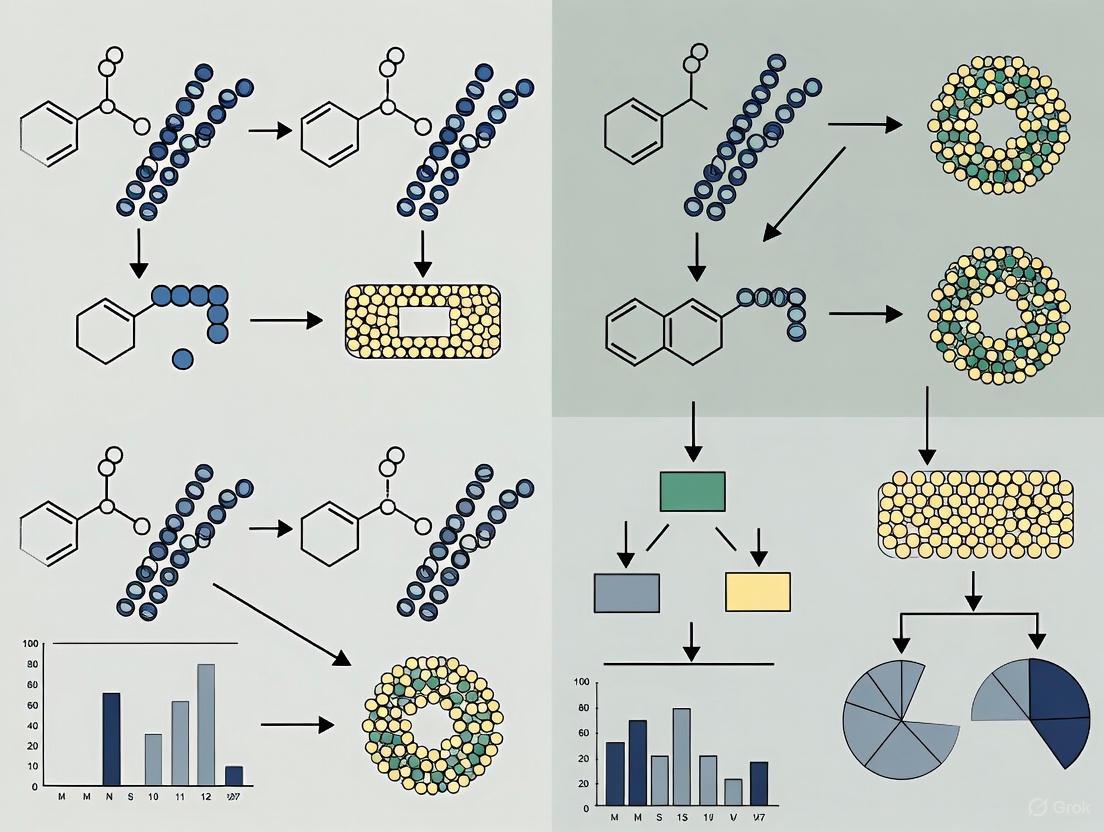
Figure 1: Fundamental mechanisms of caspase detection technologies. The fluorescent substrate method (top) reports enzymatic activity through cleavage-induced fluorescence, while the antibody-based approach (bottom) detects specific protein fragments through immunoglobulin binding.
Comparative Performance Analysis
Direct Technology Comparison
Table 1: Head-to-head comparison of cleaved caspase-3 antibodies versus fluorescent caspase substrates
| Parameter | Cleaved Caspase-3 Antibodies | Fluorescent Caspase Substrates |
|---|---|---|
| Detection Principle | Protein-epitope recognition (IgG binding) | Enzymatic activity (Peptide cleavage) |
| Molecular Target | Caspase-3 fragments (17/19 kDa) [16] | DEVD sequence in caspase-3/-7 [8] |
| Primary Applications | WB, IHC, IF/ICC, ELISA [17] | Live-cell imaging, flow cytometry, HTS [8] |
| Temporal Resolution | End-point/snapshot analysis [16] [17] | Real-time, continuous monitoring [8] |
| Specificity Profile | Highly specific for caspase-3 [16] | Broad for caspase-3/-7 [8] [20] |
| Spatial Information | Preserved in fixed tissues (IHC) [17] | Live-cell localization in 2D/3D cultures [8] |
| Throughput Capacity | Low to medium | Medium to high (HTS compatible) [8] |
| Sample Compatibility | Fixed cells, frozen/FFPE tissues [17] | Live cells, spheroids, organoids [8] |
| Key Advantage | Specific caspase-3 activation confirmation | Dynamic kinetic measurements in live cells |
Quantitative Performance Data
Table 2: Experimental performance characteristics of representative commercial and research reagents
| Reagent Type | Specificity | Sensitivity | Optimal Dilution/Concentration | Signal-to-Noise Ratio |
|---|---|---|---|---|
| Cleaved Caspase-3 Antibody #9661 [16] | Caspase-3 only (17/19 kDa) | Endogenous levels | WB: 1:1000, IHC: 1:400 [16] | High (minimal background) |
| Cleaved Caspase-3 Antibody 25128-1-AP [17] | Caspase-3 fragments | 1:500-1:2000 (WB) | IHC: 1:50-1:500 [17] | High in validated systems |
| DEVD-based ZipGFP Reporter [8] | Caspase-3 & -7 | Single-cell detection | Stable expression | ~10-fold increase post-cleavage |
| FRET-based Substrates [19] | Caspase-1 & -3 | nM enzyme concentration | Variable by construct | ~5-8 fold increase post-cleavage |
Experimental Protocols and Methodologies
Cleaved Caspase-3 Antibody Protocols
Western Blot Protocol for Cleaved Caspase-3 Detection:
- Sample Preparation: Lyse cells in RIPA buffer containing protease inhibitors. For apoptotic induction, treat cells with 1-10 µM staurosporine or other inducers for 4-16 hours [17] [20].
- Protein Separation: Load 20-30 µg protein per lane on 4-20% gradient SDS-PAGE gels and transfer to PVDF membranes.
- Blocking: Incubate membrane with 5% non-fat milk in TBST for 1 hour at room temperature.
- Primary Antibody Incubation: Incubate with cleaved caspase-3 antibody (1:1000 dilution for #9661, 1:500-1:2000 for 25128-1-AP) in blocking buffer overnight at 4°C [16] [17].
- Detection: Use appropriate HRP-conjugated secondary antibody (1:2000-1:5000) and develop with ECL reagent. Expected bands: 17 kDa and/or 19 kDa fragments.
Immunohistochemistry Protocol for Tissue Sections:
- Tissue Preparation: Fix tissues in 10% neutral buffered formalin for 24-48 hours and embed in paraffin. Cut 4-5 µm sections.
- Deparaffinization and Antigen Retrieval: Deparaffinize with xylene and ethanol series. Perform antigen retrieval with TE buffer (pH 9.0) or citrate buffer (pH 6.0) using steam or pressure cooker [17].
- Endogenous Peroxidase Blocking: Treat with 3% H₂O₂ for 10 minutes.
- Antibody Incubation: Apply cleaved caspase-3 antibody at 1:50-1:500 dilution for 60 minutes at room temperature [17].
- Detection: Use appropriate detection kit (e.g., avidin-biotin complex) and develop with DAB substrate. Counterstain with hematoxylin.
Fluorescent Caspase Substrate Protocols
Live-Cell Imaging with ZipGFP Caspase Reporter:
- Cell Line Generation: Create stable reporter cells via lentiviral transduction expressing the DEVD-based ZipGFP construct with a constitutive mCherry marker for normalization [8].
- Experimental Setup: Plate cells in glass-bottom dishes or microplates 24 hours before treatment. For 3D cultures, embed spheroids or organoids in Cultrex or Matrigel [8].
- Treatment and Imaging: Add apoptotic inducers (e.g., 1-10 µM carfilzomib, 10-100 µM oxaliplatin) and immediately begin time-lapse imaging. Include control wells with pan-caspase inhibitor zVAD-FMK (20-50 µM) to confirm specificity [8].
- Image Acquisition: Capture images every 30-60 minutes for 24-120 hours using maintained environmental control (37°C, 5% CO₂). Monitor GFP fluorescence (excitation 488 nm/emission 510 nm) and mCherry (excitation 587 nm/emission 610 nm).
- Quantification: Analyze fluorescence intensity using image analysis software (e.g., ImageJ, IncuCyte). Normalize GFP signal to mCherry to account for cell presence.
Flow Cytometry with Fluorogenic Caspase Substrates:
- Cell Staining: Harvest cells and resuspend in culture medium containing serum at 1×10⁶ cells/mL.
- Substrate Loading: Add cell-permeable fluorogenic caspase substrate (e.g., FAM-DEVD-FMK) at recommended concentration (typically 1-10 µM) and incubate for 30-60 minutes at 37°C.
- Wash and Analyze: Wash cells twice with PBS and analyze immediately by flow cytometry using appropriate laser and filter sets.
- Data Interpretation: Gate on viable cells (excluding PI-positive cells) and analyze substrate cleavage by fluorescence intensity shift compared to untreated controls.
Research Reagent Solutions
Table 3: Essential research reagents for caspase detection studies
| Reagent Category | Specific Examples | Function and Application |
|---|---|---|
| Primary Antibodies | Cleaved Caspase-3 (Asp175) #9661 (CST) [16] | Detects endogenous activated caspase-3 in WB, IHC, IF |
| Cleaved Caspase-3 25128-1-AP (Proteintech) [17] | Recognizes cleaved fragments in multiple applications | |
| Fluorescent Reporters | DEVD-based ZipGFP biosensor [8] | Real-time caspase-3/7 activity monitoring in live cells |
| CFP-YFP FRET substrates [19] | Caspase-1 or -3 specific activity measurements | |
| Apoptosis Inducers | Carfilzomib (proteasome inhibitor) [8] | Induces intrinsic apoptosis pathway in reporter validation |
| Staurosporine (kinase inhibitor) [19] [20] | Broad-spectrum apoptosis inducer for positive controls | |
| Caspase Inhibitors | zVAD-FMK (pan-caspase inhibitor) [8] | Negative control for caspase-specific signal confirmation |
| Detection Systems | HRP-conjugated secondary antibodies [16] [17] | Antibody detection in WB and IHC applications |
| Live-cell imaging systems (IncuCyte) [8] | Automated kinetic analysis of fluorescent reporter systems |
Application Workflows and Experimental Design
Integrated Pathway Analysis
Figure 2: Integrated workflow for apoptotic detection combining both fluorescent substrates and antibody-based approaches. The pathways can be used independently or in parallel for comprehensive analysis.
Technology Selection Guidelines
Select Cleaved Caspase-3 Antibodies When:
- Confirming specific caspase-3 activation (not caspase-7) is essential [16]
- Working with archived fixed tissues (FFPE) or tissue sections [17]
- Precise spatial localization within tissue architecture is required [17]
- End-point analysis with high specificity suffices for experimental goals
- Combining with other immunohistochemical markers is necessary
Select Fluorescent Caspase Substrates When:
- Real-time kinetic analysis of apoptosis is required [8]
- Monitoring live cells in 2D or 3D culture systems (spheroids, organoids) [8]
- High-content screening or high-throughput applications are planned [8]
- Detecting compensatory mechanisms like apoptosis-induced proliferation [8]
- Simultaneous tracking of multiple cellular events in live cells is needed
Both cleaved caspase-3 antibodies and fluorescent caspase substrates offer powerful, complementary approaches for apoptosis research with distinct advantages and limitations. The immunoglobulin-based detection provides exceptional specificity for caspase-3 activation and is ideal for fixed samples and histological applications, while enzymatic substrate cleavage approaches enable real-time kinetic monitoring in live cells and are superior for dynamic studies and high-throughput screening. The choice between these technologies should be guided by experimental requirements regarding specificity, temporal resolution, sample type, and throughput needs. Researchers can maximize the value of both approaches by employing them in complementary ways—using fluorescent substrates for initial kinetic studies and live-cell imaging, followed by antibody-based methods for specific caspase-3 confirmation and spatial localization in fixed samples.
Understanding the DEVD Recognition Sequence and Its Variations
Caspase-3 serves as a critical executioner protease in apoptotic pathways, with its substrate specificity largely dictated by recognition sequences surrounding the cleavage site. The canonical DEVD (Asp-Glu-Val-Asp) motif represents the optimal recognition sequence for caspase-3, though significant variations exist with important implications for research and drug development. This guide examines DEVD and its alternatives within the methodological comparison of cleaved caspase-3 antibodies versus fluorescent caspase substrates, providing researchers with quantitative data to inform experimental design.
The molecular basis for caspase-3 specificity stems from its structural architecture, which forms complementary binding sites (S4-S1) for substrate amino acids (P4-P1). The stringent requirement for aspartic acid at the P1 position is driven by a deep basic pocket formed by conserved residues Arg64, Arg207, and Gln161 [21]. This structural understanding provides the foundation for evaluating both natural and engineered substrate variations.
Structural Basis of DEVD Recognition
Caspase-3 contains specific binding pockets that accommodate the DEVD tetrapeptide sequence. The structural complementarity between enzyme and substrate explains the high specificity observed in biochemical assays:
- S4 Pocket: Accommodates the P4 aspartic acid through favorable electrostatic interactions
- S3 Pocket: A surface hydrophilic site with favorable polar interactions for P3 glutamic acid [22] [21]
- S2 Pocket: A hydrophobic groove that optimally accommodates valine [22] [21]
- S1 Pocket: The most specific binding site, with an absolute requirement for aspartic acid [21]
Recent structural analyses have revealed an additional S5 binding site in caspase-3, where side chains of Phe250 and Phe252 interact with P5 valine in substrates like Ac-VDVAD-Cho [22] [21]. This hydrophobic S5 site contributes to substrate selectivity and distinguishes caspase-3 from other caspases like caspase-7, which lacks structurally equivalent hydrophobic residues and shows less efficient hydrolysis of substrates with P5 valine or leucine [22] [21].
Figure 1: Caspase-3 substrate binding pockets and their complementary recognition sequences. The S5 pocket provides additional specificity for substrates with N-terminal extensions beyond the canonical tetrapeptide motif.
Comparative Analysis of Caspase-3 Substrate Sequences
Kinetic Performance of Common Substrate Variants
Extensive biochemical analysis has revealed how variations in the recognition sequence impact catalytic efficiency. The following table summarizes kinetic parameters for key substrate sequences:
| Substrate Sequence | P5-P1 Motif | Relative kcat/Km | Caspase Specificity | Key Structural Features |
|---|---|---|---|---|
| DEVD | Asp-Glu-Val-Asp | 100% [22] | Caspase-3, -7 [23] | Optimal fit for S4-S1 pockets |
| VDVAD | Val-Asp-Val-Ala-Asp | 37% [22] | Caspase-2, -3 [22] | P5 Val engages hydrophobic S5 site |
| DMQD | Asp-Met-Gln-Asp | 17% [22] | Caspase-3 | Suboptimal polar P3 Gln in hydrophilic S3 |
| IETD | Ile-Glu-Thr-Asp | Moderate [24] | Caspase-8, -3 | Cross-reactivity with initiator caspases |
| VEVD | Val-Glu-Val-Asp | High [18] | Caspase-6 | Group II effector caspase preference |
| LEHD | Leu-Glu-His-Asp | Moderate [18] | Caspase-9 | Initiator caspase recognition motif |
Specificity Profiling Across Caspase Family
A critical consideration in substrate selection is the concerning lack of absolute specificity among caspase family members. Degradomics analysis using N-terminal COFRADIC has demonstrated that caspases-2, -3, and -7 share "remarkably overlapping protease specificity" with the common DEVD↓G consensus cleavage sequence [23]. This overlap presents significant challenges for interpreting results when using traditional substrate-based assays.
The assumed caspase-2-specific substrate Ac-VDVAD-AMC is cleaved "almost equally well by caspase-3 and -7" [23], highlighting the promiscuity that complicates specific caspase identification. Similarly, the canonical caspase-3/7 substrate Ac-DEVD-AFC shows "high substrate activity with caspases 3 and 7 along with off-target activity with caspases 8 and 10" [25].
Experimental Comparison: Methodological Approaches
Fluorescent Caspase Substrates
Standard Experimental Protocol for Fluorogenic Assays
Reagent Preparation:
- Prepare assay buffer (e.g., 20 mM HEPES, pH 7.4, 100 mM NaCl, 5 mM DTT)
- Reconstitute lyophilized substrates (e.g., Ac-DEVD-AFC, Ac-DEVD-pNA) in DMSO
- Dilute to working concentration in assay buffer
Cell-Based Analysis:
- Induce apoptosis in cells (e.g., 48h cisplatin treatment for ovarian cancer lines) [25]
- Add fluorescent substrate (e.g., 50-100 μM final concentration)
- Incubate 1-2 hours at 37°C
- Monitor fluorescence (AFC: λ~Ex~/λ~Em~ 380/500 nm; pNA: λ~Abs~ 405 nm) [24]
- Include caspase inhibitor controls (Z-VAD-FMK, Z-DEVD-FMK)
Key Considerations:
- Cell permeability varies significantly among substrates
- Negative charges in DEVD impair cellular uptake [25]
- Modified substrates (e.g., 2MP-TbD-AFC) show improved permeability and selectivity [25]
Advanced Substrate Engineering
Recent innovations have addressed limitations of traditional DEVD-based substrates:
2MP-TbD-AFC represents a minimized caspase-3 substrate where the P2 valine is replaced with O-benzylthreonine (Tb). This modification yields a "4-fold higher cleavage by caspase-3" compared to the valine-containing analog, primarily due to increased k~cat~ [25]. Importantly, this engineered substrate demonstrates "excellent caspase-3 selectivity with minimal off-target activity" compared to the promiscuous Ac-DEVD-AFC [25].
Figure 2: Comparison of traditional versus engineered caspase-3 substrates. Minimized substrates with strategic modifications address key limitations of canonical recognition sequences.
Cleaved Caspase-3 Antibodies
Proteomic Workflow for Substrate Identification
Sample Preparation:
- SILAC labeling of cells (light: ^12^C~6~-Arg; heavy: ^13^C~6~-Arg) for 5+ population doublings [26]
- Generate mixed proteome (1:1 light:heavy)
- Treat with recombinant caspase-3 (150 nM, 1h, 37°C) [26]
- Inhibit endogenous caspase activity with cysteine alkylation
N-terminal COFRADIC Analysis:
- Mix protease-treated and control samples
- Isolate N-terminal peptides via COFRADIC sorting [26]
- Analyze by LC-MS/MS (LTQ Orbitrap XL mass spectrometer)
- Identify peptides using MASCOT algorithm
- Quantify using MASCOT Distiller software
Data Interpretation:
- Caspase-3-generated neo-N-termini show L/H ratio ≈ 1 [26]
- Background N-termini show L/H ratio ≈ 3 [26]
- Cleavage after aspartic acid confirms caspase-3 specificity [26]
Research Reagent Solutions
The following essential materials represent critical tools for investigating caspase-3 activity and specificity:
| Reagent | Function/Application | Key Features |
|---|---|---|
| Ac-DEVD-AFC [24] | Fluorogenic caspase-3 substrate | Selective cleavage, fluorescent signal upon hydrolysis (AFC release) |
| Ac-DEVD-pNA [24] | Chromogenic caspase-3 substrate | Yellow p-nitroaniline release measurable at 405 nm |
| Z-DEVD-fmk [27] | Irreversible caspase-3 inhibitor | Specific DEVDase inhibition, control for specificity |
| Recombinant Caspase-3 [26] | Enzyme source for in vitro assays | Activated form for biochemical characterization |
| SILAC Reagents [26] | Metabolic labeling for proteomics | ^12^C~6~/~13^C~6~-Arg for quantitative mass spectrometry |
| Caspase-3 Antibodies | Detection of cleaved/activated caspase-3 | Specific recognition of cleaved epitopes |
Discussion: Methodological Integration in Research
The strategic selection between cleaved caspase-3 antibodies and fluorescent substrates depends on specific research objectives. Antibodies provide snapshot activation status in specific cell populations but lack kinetic information. Fluorescent substrates enable real-time monitoring of enzymatic activity but suffer from specificity limitations due to overlapping caspase recognition patterns.
The emerging approach of genetically encoded biosensors represents a promising fusion of these methodologies. Tools like VC3AI (Venus-based Caspase-3 Activity Indicator) utilize cyclized chimeras containing caspase-3 cleavage sites that generate fluorescence only upon protease activation [27]. These systems provide single-cell resolution while maintaining genetic specificity, overcoming limitations of chemical substrates added to cell cultures.
For drug development applications, particularly in high-throughput screening, the improved specificity of engineered substrates like 2MP-TbD-AFC provides significant advantages over traditional DEVD-based compounds [25]. The development of PET-compatible caspase-3 substrates such as [^18^F]-TBD further extends these principles to in vivo imaging applications, enabling non-invasive monitoring of apoptosis in disease models [25].
The DEVD recognition sequence and its variations represent a sophisticated evolutionary adaptation in caspase-3 substrate specificity. While DEVD remains the optimal motif, understanding the structural basis for alternative sequences like VDVAD and engineered variants like 2MP-TbD provides researchers with enhanced tools for specific apoptosis detection. The integration of fluorescent substrate data with antibody-based validation and proteomic substrate identification creates a powerful multidimensional approach for studying caspase-3 biology in both physiological and pathological contexts.
Future directions will likely focus on developing even more specific recognition sequences through structural optimization and creating advanced biosensors that provide spatiotemporal resolution of caspase-3 activation in complex biological systems.
Caspase-3, a key executioner caspase, serves as a critical mediator of programmed cell death (apoptosis) through the cleavage of hundreds of cellular substrates, committing the cell to apoptotic death [25]. The detection of its activation provides a crucial indicator for studying fundamental biological processes and therapeutic interventions in diseases ranging from cancer to neurodegenerative disorders [28] [29]. Traditionally, antibody-based methods have formed the cornerstone of caspase-3 detection, providing foundational insights into its biological functions. However, the past decade has witnessed remarkable advancements with the development of genetically encoded biosensors that enable real-time, dynamic monitoring of caspase activity in living systems [29] [27]. This evolution from static, endpoint measurements to dynamic, live-cell imaging has transformed our ability to study the spatial and temporal regulation of apoptosis, opening new avenues for drug discovery and therapeutic monitoring. This guide objectively compares the performance of classical cleaved caspase-3 antibodies with emerging fluorescent caspase substrates, providing researchers with experimental data and methodologies to inform their detection strategy selection.
Technology Comparison: Mechanisms and Characteristics
The fundamental distinction between these detection methodologies lies in their operational principles: antibody-based tools detect the physical presence of the cleaved caspase-3 protein, while fluorescent substrates and biosensors detect its enzymatic activity.
Table 1: Core Characteristics of Caspase-3 Detection Methods
| Feature | Classical Antibody-Based Methods | Fluorescent Caspase Substrates | Genetically Encoded Biosensors |
|---|---|---|---|
| Detection Principle | Immunoreactivity against cleaved caspase-3 protein fragment [30] | Enzymatic cleavage of synthetic DEVD peptide sequence liberating fluorophore [25] | Caspase-dependent reconstitution of fluorescent protein structure [27] [31] |
| Primary Readout | Presence and molecular weight of protein (e.g., via Western blot) [32] | Fluorescence intensity proportional to caspase activity [25] | Fluorescence activation (switch-on signal) [27] |
| Temporal Resolution | End-point analysis (low resolution) [8] | Real-time to near real-time (minutes to hours) [25] | Real-time, continuous (high resolution) [27] [8] |
| Spatial Context | Typically requires cell lysis; lost in Western blot, preserved in IHC/IF [30] | Preserved in intact cells, but limited subcellular detail | Single-cell resolution in 2D and 3D cultures [8] |
| Throughput | Low to moderate (Western blot: ~10-15 samples/gel) [32] | High (compatible with microplate readers) [33] | High, suitable for live-cell imaging and screening [8] |
| Quantification | Semi-quantitative (Western blot) to quantitative (ELISA) [32] | Quantitative (relative fluorescence units) [25] | Quantitative (fluorescence intensity/count) [27] |
| Key Advantage | Specificity for confirmed caspase-3 cleavage; well-established | Simplicity, cell permeability, and kinetic data | Dynamic, long-term tracking in live cells without disruption |
Figure 1: Caspase-3 Detection Signaling Pathway. This diagram outlines the core pathways for detecting activated caspase-3, branching into antibody-based and substrate-based methodologies following an apoptotic stimulus.
Experimental Data and Performance Comparison
Rigorous benchmarking reveals significant performance differences between these methodologies, impacting their suitability for specific research applications. The following quantitative data summarizes key findings from direct comparisons and validation studies.
Table 2: Quantitative Performance Comparison of Detection Methods
| Method | Specific Example/Assay | Key Performance Metrics | Experimental Context |
|---|---|---|---|
| Western Blot (Antibody) | Cleaved Caspase-3 (Asp175) Antibody [30] | Working Dilution: 1:500-1:2000 [30]Observed Band: 17-25 kDa (cleaved fragments) [30] | Validation in Jurkat cells, HeLa cells, mouse brain tissue [30] |
| Fluorogenic Substrate | 2MP-TbD-AFC [25] | Caspase-3 Selectivity: >4-fold vs. caspase-8 [25]Cell Permeability: Demonstrated in OVCAR-5/8 cells [25] | Cisplatin-induced apoptosis in ovarian cancer cell lines [25] |
| FRET-Based Sensor | DEVD-linked FRET [33] | Readout: Loss of FRET efficiency upon cleavageApplication: High-throughput screening in 96-well format [33] | Intact adherent cells; identification of novel caspase inhibitors [33] |
| Switch-On Biosensor | VC3AI (Venus-based) [27] | Background Fluorescence: Undetectable in healthy cells [27]Specificity: Activated by caspase-3/7; inhibited by Z-DEVD-fmk [27] | TNF-α-induced apoptosis in MCF-7 cells (caspase-3 deficient) [27] |
| Switch-On Biosensor | mNeonGreen2-based [31] | Attribute: "Shortened response time, higher sensitivity" [31]Application: Drug effect evaluation in HeLa/MCF-7 cells [31] | Used to assess apoptosis induction by various drugs and viral infection [31] |
Detailed Experimental Protocols
To ensure methodological reproducibility, below are detailed protocols for key experiments characterizing both classical and advanced detection methods.
Protocol: Validating a Cleaved Caspase-3 Antibody via Western Blot
This protocol is adapted from the methodology for the Cleaved Caspase-3 (25128-1-AP) antibody and general Western blot principles [32] [30].
- Sample Preparation: Lyse cells (e.g., Jurkat, HeLa) in RIPA buffer supplemented with protease inhibitors. Determine protein concentration and dilute with Laemmli buffer. Denature samples by heating at 95°C for 5 minutes [30].
- Gel Electrophoresis: Load 20-30 μg of total protein per lane onto a 4-20% gradient SDS-polyacrylamide gel. Run electrophoresis at 120-150 V until the dye front reaches the bottom of the gel [32].
- Membrane Transfer: Transfer proteins from the gel to a PVDF or nitrocellulose membrane using a wet or semi-dry transfer system.
- Blocking and Antibody Incubation: Block the membrane with 5% non-fat milk in TBST for 1 hour at room temperature. Incubate with the primary cleaved caspase-3 antibody at a dilution of 1:1000 in blocking buffer overnight at 4°C. Wash the membrane 3 times for 5 minutes each with TBST. Incubate with an HRP-conjugated secondary antibody (e.g., goat anti-rabbit) for 1 hour at room temperature. Perform final washes [30].
- Detection: Develop the blot using enhanced chemiluminescence (ECL) substrate and expose to X-ray film or capture image with a digital chemiluminescence imaging system. The expected molecular weight for cleaved caspase-3 fragments is between 17 and 19 kDa (and sometimes a dimer around 30-35 kDa) [30].
Protocol: Live-Cell Imaging with a Genetically Encoded Biosensor
This protocol is based on the use of the ZipGFP-based caspase-3/-7 reporter system and similar biosensors like VC3AI [27] [8].
- Cell Line Generation and Culture: Generate a stable reporter cell line via lentiviral transduction of the biosensor construct (e.g., ZipGFP with constitutive mCherry marker). Culture these cells in appropriate medium under standard conditions (e.g., 37°C, 5% CO₂) [8].
- Experimental Setup and Imaging: Seed cells into a multi-well imaging plate. Allow cells to adhere and reach 50-70% confluence. Treat cells with the apoptotic stimulus (e.g., 1 μM carfilzomib, 10 μM oxaliplatin) and add inhibitors (e.g., 20-50 μM Z-VAD-FMK) to control wells for specificity confirmation [8]. Place the plate in a live-cell imaging system (e.g., IncuCyte) maintaining environmental control. Acquire images for both the reporter channel (e.g., GFP) and the constitutive marker channel (e.g., mCherry) every 2-4 hours for 48-120 hours [8].
- Data Analysis: Quantify the GFP fluorescence intensity normalized to the mCherry signal to account for any changes in cell number. Apoptotic cells are identified by a significant and sustained increase in the GFP/mCherry ratio. Data can be expressed as the percentage of GFP-positive cells over time or mean fluorescence intensity [8].
Figure 2: Caspase-3 Detection Method Selection Guide. A decision framework to help researchers select the most appropriate detection technology based on their specific experimental requirements.
Essential Research Reagent Solutions
Successful implementation of these detection technologies requires a suite of reliable reagents. The following table catalogs key materials cited in the research.
Table 3: Key Research Reagents for Caspase-3 Detection
| Reagent / Material | Function / Description | Example Use Case |
|---|---|---|
| Cleaved Caspase-3 Antibody (25128-1-AP) | Polyclonal antibody specific to cleaved fragments of caspase-3; does not recognize full-length protein [30] | Western Blot, IHC, and IF/ICC to confirm caspase-3 activation and localization [30] |
| Fluorogenic Substrate (e.g., Ac-DEVD-AFC, 2MP-TbD-AFC) | Cell-permeable peptide substrate containing the DEVD sequence; releases fluorescent AFC upon cleavage [25] | Quantifying caspase-3/7 activity in cell lysates or living cells using a fluorometer [25] |
| Pan-Caspase Inhibitor (Z-VAD-FMK) | Cell-permeable, irreversible broad-spectrum caspase inhibitor [8] | Control experiment to confirm caspase-dependent signal in biosensor or substrate assays [27] [8] |
| Caspase-3/7 Inhibitor (Z-DEVD-FMK) | Cell-permeable, potent and selective inhibitor of caspase-3 and caspase-7 [27] | Specific confirmation that a signal is generated by caspase-3/7 activity and not other proteases [27] |
| Lentiviral Caspase Biosensor (e.g., ZipGFP-DEVD) | Genetically encoded construct for generating stable cell lines with caspase-3/7 reporter [8] | Creating cell lines for long-term, real-time apoptosis imaging in 2D and 3D culture systems [8] |
| Annexin V / Propidium Iodide (PI) | Fluorescent dyes for detecting phosphatidylserine exposure (early apoptosis) and loss of membrane integrity (necrosis/late apoptosis) [8] | End-point validation of apoptosis by flow cytometry, correlating with caspase activation [8] |
The journey from classical antibodies to genetically encoded biosensors for caspase-3 detection represents a paradigm shift in apoptosis research. Classical antibodies remain indispensable for their high specificity in confirming protein cleavage and their utility in fixed tissues. In contrast, fluorescent substrates and biosensors provide powerful tools for dynamic, functional analysis in living systems, enabling high-resolution kinetic studies and high-throughput drug screening. The choice between these technologies is not a matter of superiority but of strategic alignment with experimental goals. Researchers requiring unambiguous confirmation of caspase-3 cleavage in endpoint analyses will find antibodies optimal, whereas those investigating the real-time dynamics of cell death in physiologically relevant models will benefit from the advanced capabilities of genetically encoded biosensors. As these technologies continue to converge and evolve, they will undoubtedly unlock deeper insights into the fundamental mechanisms of programmed cell death and its therapeutic manipulation.
Hands-On Protocols: Applying Antibody and Substrate Methods in Your Research
Immunofluorescence Protocol for Cleaved Caspase-3 Antibody in Fixed Cells
Within cell death research, detecting the activation of caspase-3, a key executioner protease in apoptosis, is a fundamental technique. Two predominant methodological strategies exist: immunofluorescence (IF) using cleaved caspase-3-specific antibodies and live-cell imaging with fluorescent caspase substrates. This guide provides a direct, objective comparison of these techniques, focusing on the detailed immunofluorescence protocol for cleaved caspase-3 and its performance metrics relative to fluorescent substrates. The broader thesis is that while immunofluorescence provides superior spatial and morphological context in fixed samples, fluorescent substrates offer unique advantages for kinetic analyses in living cells.
Critical Reagent Solutions
The success of the immunofluorescence protocol is dependent on several key reagents, whose functions and examples are detailed below.
Table 1: Essential Reagents for Cleaved Caspase-3 Immunofluorescence
| Reagent Category | Specific Example | Function in the Protocol |
|---|---|---|
| Primary Antibody | Cleaved Caspase-3 (Asp175) Antibody #9661 (Cell Signaling Technology) [34] | Specifically binds to the activated large fragment (17/19 kDa) of caspase-3, enabling detection. |
| Fixative | 4% Paraformaldehyde (PFA) in PBS [35] | Preserves cellular architecture and immobilizes antigens by cross-linking. |
| Permeabilization Agent | 0.1% Triton X-100 or 0.5% Triton X-100 [36] [35] | Dissolves membrane lipids to allow antibody access to intracellular targets. |
| Blocking Buffer | PBS with 5% serum (e.g., bovine, goat) and 0.1% Tween-20 [36] | Reduces non-specific antibody binding to minimize background signal. |
| Fluorophore-Conjugated Secondary Antibody | Alexa Fluor 594 goat anti-rabbit IgG [35] | Binds to the primary antibody and provides a detectable fluorescent signal. |
| Nuclear Counterstain | DAPI (5 µg/mL) [35] | Labels DNA, allowing for visualization of nuclear morphology and cell identification. |
| Mounting Medium | Permanent or aqueous mounting medium [36] | Preserves the sample and provides the correct refractive index for microscopy. |
Core Methodologies
Detailed Immunofluorescence Protocol for Cleaved Caspase-3
The following step-by-step protocol is optimized for detecting cleaved caspase-3 in fixed cells plated on slides or culture dishes [36] [35].
- Fixation: Aspirate culture medium and wash cells gently with phosphate-buffered saline (PBS). Fix cells with 4% Paraformaldehyde (PFA) in PBS for 15-20 minutes at room temperature [35].
- Permeabilization: Remove PFA and wash cells three times with PBS, for 5 minutes each. Incubate cells in PBS containing 0.1% Triton X-100 for 5-10 minutes at room temperature to permeabilize the membranes [36] [35].
- Blocking: Wash cells once with PBS. Drain the liquid and add an appropriate blocking buffer (e.g., PBS/0.1% Tween 20 + 5% serum from the host species of the secondary antibody) for 1-2 hours at room temperature in a humidified chamber [36].
- Primary Antibody Incubation: Prepare the cleaved caspase-3 primary antibody (e.g., #9661) at the recommended dilution (e.g., 1:400 to 1:500) in blocking buffer [34] [35]. Apply the solution to the samples and incubate overnight at 4°C in a humidified chamber [36].
- Secondary Antibody Incubation: The following day, wash the samples three times with PBS/0.1% Tween 20 for 10 minutes each. Apply the fluorescently conjugated secondary antibody (e.g., Alexa Fluor 594 goat anti-rabbit IgG at 1:500) diluted in PBS or blocking buffer. Incubate for 1-2 hours at room temperature, protected from light [36] [35].
- Nuclear Staining and Mounting: Perform a final series of washes (three times, 5 minutes each in PBS). Drain the liquid and add DAPI (5 µg/mL) for 15 minutes to stain nuclei. After a final wash, mount the slides with a suitable mounting medium and seal with a coverslip [35].
Figure 1: Immunofluorescence workflow for cleaved caspase-3 detection, showing the sequential steps from fixed cells to final imaging.
Fluorescent caspase substrates are cell-permeable compounds that become fluorescent upon cleavage by active caspases. A prominent technology uses FRET (Förster Resonance Energy Transfer)-based substrates [33]. In one approach, two fluorescent proteins (e.g., CFP and YFP) are linked by a short peptide sequence containing the caspase-3 cleavage motif DEVD. In living cells, FRET occurs between the two proteins. When caspase-3 is activated and cleaves the linker, the physical separation of the two fluorophores abolishes FRET, resulting in a quantifiable change in fluorescence emission that can be monitored in real-time [33].
Figure 2: Mechanism of FRET-based caspase substrates. Caspase-3 cleavage disrupts energy transfer, changing the fluorescence signal.
Performance Comparison: Data and Analysis
To objectively compare the performance of the antibody-based and substrate-based methods, key quantitative and qualitative data from the literature are summarized below.
Table 2: Performance Comparison of Cleaved Caspase-3 Detection Methods
| Performance Characteristic | Cleaved Caspase-3 Immunofluorescence | Fluorescent Caspase Substrates (e.g., 2MP-TbD-AFC, FRET) |
|---|---|---|
| Spatial Resolution | High (subcellular localization within fixed cell architecture) [36] | Moderate (localization to cellular compartments in live cells) [25] |
| Temporal Resolution | End-point measurement (single time point) [36] | High (real-time kinetics in living cells) [33] |
| Caspase-3 Selectivity | Excellent (antibody #9661 is specific for cleaved fragment, does not recognize full-length) [34] | Variable (substrate 2MP-TbD-AFC shows excellent selectivity [25]; Ac-DEVD-AFC has off-target activity [25]) |
| Quantitative Capability | Semi-quantitative (signal intensity correlates with abundance); requires image analysis software [36] | Highly quantitative (fluorescence intensity/FRET ratio directly proportional to activity) [33] |
| Key Advantage | Preserves cellular morphology; co-staining with other markers (e.g., DAPI) allows assessment of apoptotic nuclear fragmentation [36] [35] | Enables monitoring of caspase activation dynamics and pharmacological inhibition in intact, adherent cells [33] |
| Primary Limitation | Requires cell fixation and permeabilization, precluding live-cell analysis [36] | Does not provide detailed structural context; signal can be influenced by substrate permeability and non-specific cleavage [25] |
| Throughput Potential | Medium (suitable for multi-well formats) | High (adaptable for high-throughput screening in 96-well plates) [33] |
Integrated Experimental Workflow
The choice between these methods is not mutually exclusive and can be guided by the experimental hypothesis. The following diagram outlines a decision pathway for selecting the appropriate detection method based on research goals.
Figure 3: A decision workflow to guide researchers in selecting the optimal caspase-3 detection method based on their specific experimental requirements.
The experimental data and protocols presented herein support a clear comparative analysis. Cleaved caspase-3 immunofluorescence is the definitive method when the research question demands high-resolution spatial localization of apoptosis within the context of fixed cell or tissue morphology. Its ability to be multiplexed with other markers makes it invaluable for phenotyping apoptotic cells in a heterogeneous population, such as within the tumor microenvironment [36] [37].
In contrast, fluorescent caspase substrates are the superior tool for investigating the dynamics of cell death. They enable researchers to monitor the precise timing and rate of caspase-3 activation in individual live cells, an capability that is impossible with endpoint IF assays [33]. This makes them ideal for screening applications and for studying the real-time effects of pro-apoptotic drugs or inhibitory compounds [25] [33].
In conclusion, the choice between these two powerful techniques is not a matter of which is universally better, but which is more appropriate for the specific biological question. Immunofluorescence provides a high-resolution "snapshot" of apoptosis, while fluorescent substrates offer a "live video" of the process. For a comprehensive research strategy, they can be used as complementary approaches, with substrates identifying the timing of death and IF providing subsequent morphological validation.
Flow Cytometry with Antibody Conjugates for Quantitative Population Analysis
Within the broader thesis research on cleaved caspase-3 antibody versus fluorescent caspase substrates, understanding the capabilities and limitations of each technology is paramount for accurate quantitative population analysis in drug development. Apoptosis, or programmed cell death, is a fundamental biological process executed by caspase enzymes, with caspase-3 serving as a key effector. The detection of active caspase-3 provides a crucial marker for identifying apoptotic cells within heterogeneous populations using flow cytometry. Researchers primarily employ two methodological approaches: antibody-based conjugates that bind directly to the cleaved caspase-3 protein, and fluorescent caspase substrates that are cleaved by the enzyme's activity. Each method offers distinct advantages and limitations in sensitivity, specificity, temporal resolution, and applicability to different experimental systems. This guide objectively compares the performance of antibody conjugates and fluorescent substrates for caspase-3 detection, providing supporting experimental data and detailed methodologies to inform researchers' experimental design.
Technological Foundations: Antibody Conjugates vs. Fluorescent Substrates
Antibody-Based Conjugates for Caspase-3 Detection
Antibody conjugates for flow cytometry rely on the specific binding of fluorochrome-labeled antibodies to cleaved caspase-3. The credibility of results obtained with this method strongly depends on conjugate performance, making quality control essential. Traditional quality control is performed by spectrophotometry to measure the fluorochrome-to-protein (F/P) ratio. However, studies have shown that the F/P ratio does not necessarily express fluorescence emission in flow cytometry, as emission depends on energy excitation not present in a spectrometer. A conjugate can have a satisfactory F/P ratio but unsatisfactory emission when tested in a flow cytometer [38].
Flow cytometric methods have been developed for quality control of fluorescent conjugates, evaluating parameters such as fluorescence intensity (measured by geometric mean), homogeneity of staining (measured by coefficient of variation, CV), and percentage of positive particles. Quantitative fluorescence cytometry (QFCM) using microspheres coupled with different fluorochrome amounts can measure fluorescent intensity through molecules of equivalent soluble fluorochrome (MESF) values, expressing the number of fluorochrome molecules in solution required to produce the same fluorescence intensity as measured in the labeled particle [38].
Fluorescent Caspase Substrates
Fluorescent caspase substrates are designed as cell-permeable compounds that contain caspase cleavage motifs (typically DEVD for caspase-3) linked to fluorescent reporters. These substrates remain non-fluorescent until cleaved by active caspase enzymes within apoptotic cells, generating a fluorescent signal proportional to caspase activity [25]. Recent advances have led to the development of more sophisticated genetically-encoded fluorescent reporters.
One innovative approach utilizes a ZipGFP-based caspase-3/7 reporter, which employs a split-GFP architecture where the GFP molecule is divided into two parts tethered via a flexible linker containing a caspase-3/7-specific DEVD cleavage motif. Under basal conditions, this configuration prevents proper folding and chromophore maturation, resulting in minimal background fluorescence. Upon caspase-3/7 activation, cleavage at the DEVD site separates the β-strands, allowing spontaneous refolding into the native GFP structure with efficient chromophore formation and rapid fluorescence recovery [8].
An alternative bright-to-dark apoptosis reporter system has been developed through mutagenesis-based insertion of a caspase-3 cleavage motif directly into the green fluorescence protein. In this system, fluorescence intensity decreases upon caspase-3 activation, reportedly showing greater sensitivity for apoptosis detection compared to dark-to-bright systems [39].
Table 1: Comparison of Caspase-3 Detection Technologies
| Feature | Antibody Conjugates | Fluorescent Substrates | Genetically-Encoded Reporters |
|---|---|---|---|
| Detection Target | Cleaved caspase-3 protein | Caspase enzymatic activity | Caspase enzymatic activity |
| Cellular Resolution | Single-cell | Single-cell | Single-cell |
| Temporal Resolution | End-point measurement | Real-time (minutes to hours) | Real-time (hours to days) |
| Sample Processing | Cell fixation and permeabilization required | Live-cell compatible | Live-cell compatible |
| Signal Kinetics | Stable (protein abundance) | Dynamic (enzyme activity) | Dynamic (enzyme activity) |
| Multiplexing Potential | High (with different fluorochromes) | Moderate | High (with different FPs) |
| Applications | Fixed samples, endpoint analysis | Live-cell imaging, kinetics | Long-term tracking, 3D models |
Experimental Comparison and Performance Data
Quantitative Performance Metrics
Studies directly comparing antibody conjugates and fluorescent substrates have revealed significant differences in performance characteristics. In quality control assessments of antibody conjugates, flow cytometric analysis has demonstrated substantial variations in fluorescence intensities between different manufacturers and lots. One study evaluating anti-IgG-PE conjugates showed great differences in fluorescence intensities both between manufacturers and between lots from the same manufacturer, with coefficients of variation (CVs) providing crucial information about coupling homogeneity [38].
For fluorescent substrates, the minimized caspase-3 substrate 2MP-TbD-AFC demonstrated a 4-fold higher cleavage by caspase-3 compared to the 2MP-VD-AFC substrate, primarily due to an increase in kcat with little change in Km. When tested for caspase specificity, 2MP-TbD-AFC showed excellent caspase-3 selectivity with minimal off-target activity for caspases 1 and 8, while the canonical Ac-DEVD-AFC substrate showed high activity with both caspases 3 and 7 along with off-target activity with caspases 8 and 10 [25].
The bright-to-dark GFP mutant reporter demonstrated decreased fluorescence intensity in a time- and concentration-dependent manner upon induction of apoptosis with staurosporine and H₂O₂. Comparative studies indicated this system showed greater sensitivity for apoptosis detection compared to a dark-to-bright caspase-activatable GFP reporter [39].
Table 2: Quantitative Performance Comparison of Caspase Detection Methods
| Parameter | Antibody Conjugates | Chemical Fluorescent Substrates | Genetically-Encoded Reporters |
|---|---|---|---|
| Sensitivity | High (direct antigen binding) | Moderate to High (2MP-TbD-AFC comparable to Ac-DEVD-AFC) | High (bright-to-dark reportedly more sensitive) |
| Specificity | High (epitope-dependent) | Variable (2MP-TbD-AFC: high caspase-3 selectivity) | High (DEVD-specific) |
| Background Signal | Low with proper controls | Low with optimized substrates (e.g., 2MP-TbD-AFC) | Very low (split-GFP design) |
| Dynamic Range | Limited by antigen abundance | Proportional to enzyme activity | Proportional to enzyme activity |
| Lot-to-Lot Variability | Significant (requires quality control) | Lower with synthetic compounds | None (sequence-defined) |
| Temporal Resolution | End-point | Real-time (minutes) | Real-time (hours to days) |
Experimental Protocols for Method Comparison
Protocol 1: Quality Control of Antibody Conjugates Using Flow Cytometry
This protocol adapts the method described by de Almeida Santiago et al. for quality control of fluorescent conjugates [38]:
Coupling: Incigate microspheres with anti-caspase-3 antibody conjugates from different manufacturers and/or lots according to manufacturer's specifications.
Acquisition: Analyze coupled microspheres by flow cytometry using standardized instrument settings across all samples.
Traditional Analysis: Calculate geometric mean fluorescence intensity and coefficient of variation (CV) for the positive PE peak. The geometric mean represents conjugate brightness, while CV indicates coupling homogeneity.
Quantitative Analysis (QFCM): Evaluate fluorescence intensity using molecules of equivalent soluble fluorochrome (MESF) values with calibration beads.
Stability Testing: Monitor fluorescence intensities over time (e.g., 18 months) to assess conjugate stability.
Protocol 2: Live-Cell Apoptosis Detection with Fluorescent Reporters
This protocol is adapted from integrated reporter systems for real-time imaging of caspase dynamics [8]:
Reporter Introduction: Generate stable cell lines expressing caspase-3/7 reporter (ZipGFP with DEVD cleavage motif) alongside a constitutive mCherry marker using lentiviral delivery.
Treatment: Induce apoptosis with appropriate stimuli (e.g., carfilzomib, oxaliplatin).
Imaging: Perform time-lapse live-cell imaging over 48-120 hours to monitor GFP fluorescence recovery.
Validation: Confirm caspase dependence through co-treatment with pan-caspase inhibitor zVAD-FMK.
Analysis: Quantify GFP fluorescence intensity normalized to mCherry signal at single-cell resolution.
Protocol 3: Comparison Study Design for Caspase-3 Detection Methods
For direct comparison of antibody conjugates versus fluorescent substrates:
Cell Culture: Prepare apoptotic and control cell samples using a validated apoptosis inducer (e.g., 1μM staurosporine for 4 hours).
Parallel Processing:
- Aliquot 1: Fix and permeabilize cells for antibody staining with anti-cleaved-caspase-3-PE conjugate.
- Aliquot 2: Incubate live cells with 20μM 2MP-TbD-AFC substrate for 30-60 minutes.
- Aliquot 3: Use stable reporter cell lines expressing caspase-3/7 biosensor.
Flow Cytometry Analysis: Analyze all samples using the same flow cytometer with appropriate filter sets.
Data Comparison: Calculate signal-to-noise ratios, percentage of positive cells, and correlation with additional apoptosis markers (e.g., Annexin V).
Technical Workflows and Signaling Pathways
The following diagram illustrates the fundamental detection mechanisms for antibody conjugates versus fluorescent substrates in caspase-3 detection:
This workflow illustrates how apoptosis induction leads to caspase-3 activation, which can be detected through two distinct pathways. The antibody-based method requires cell fixation and permeabilization to allow antibodies access to intracellular cleaved caspase-3, providing a measurement of protein abundance at a single time point. In contrast, fluorescent substrates enter live cells and are cleaved by active caspase-3, enabling real-time monitoring of enzyme activity kinetics.
Essential Research Reagent Solutions
Successful implementation of flow cytometry with antibody conjugates or fluorescent substrates for quantitative population analysis requires specific research reagents and materials. The following table details key solutions for researchers in this field:
Table 3: Essential Research Reagents for Caspase-3 Detection Studies
| Reagent Category | Specific Examples | Function & Application | Considerations |
|---|---|---|---|
| Antibody Conjugates | Anti-cleaved caspase-3-PE; Anti-caspase-3-FITC | Direct detection of caspase-3 protein in fixed cells; enables multiplexing | Quality control essential [38]; significant lot-to-lot variability observed |
| Fluorescent Substrates | Ac-DEVD-AFC; 2MP-TbD-AFC; PhiPhiLux-G1D2 | Measure caspase-3 activity in live cells; cell-permeable probes | Variable specificity (2MP-TbD-AFC shows high caspase-3 selectivity) [25] |
| Genetically-Encoded Reporters | ZipGFP-DEVD; bright-to-dark GFP mutant | Long-term apoptosis tracking; stable expression in cell lines | Caspase-3 deficient cells (MCF-7) still activate via caspase-7 [8] |
| Flow Cytometry Controls | MESF calibration beads; compensation beads | Instrument calibration; quantitative fluorescence standardization | Essential for comparing results across experiments and instruments [38] |
| Apoptosis Inducers | Staurosporine; carfilzomib; oxaliplatin; cisplatin | Positive controls for apoptosis induction; mechanism studies | Different inducers activate intrinsic vs. extrinsic pathways [8] [25] |
| Caspase Inhibitors | zVAD-FMK (pan-caspase); DEVD-CHO (caspase-3 specific) | Specificity controls; mechanistic studies | zVAD-FMK abrogates GFP signal in reporter systems [8] |
| Viability Indicators | Propidium iodide; Annexin V conjugates; cisplatin viability dye | Distinguish apoptotic vs. necrotic cells; assess membrane integrity | Annexin V binding precedes complete loss of membrane integrity [8] |
The comparison between antibody conjugates and fluorescent substrates for caspase-3 detection in flow cytometry reveals complementary strengths that can be strategically leveraged based on specific research requirements. Antibody conjugates offer high specificity for the cleaved caspase-3 protein and are ideal for fixed samples and endpoint analysis, though they require rigorous quality control due to significant lot-to-lot variability. Fluorescent substrates and genetically-encoded reporters provide superior temporal resolution for monitoring caspase-3 activity dynamics in live cells, with recent advances improving specificity and sensitivity.
For drug development professionals, the choice between these technologies should be guided by specific application needs: antibody conjugates for high-throughput screening of fixed samples, chemical substrates for short-term kinetic studies in multiple cell types, and genetically-encoded reporters for long-term tracking in specialized model systems. Integrating both approaches in validation studies provides the most comprehensive assessment of apoptotic populations, combining precise protein detection with functional enzyme activity measurement to advance therapeutic development in oncology, neurodegenerative diseases, and other conditions involving dysregulated apoptosis.
Live-Cell Imaging with Fluorogenic Substrates like CellEvent for Real-Time Kinetics
The study of programmed cell death, or apoptosis, is a cornerstone of biomedical research, with implications for understanding cancer biology, neurodegenerative diseases, and drug development. Central to the apoptotic process are caspases, a family of cysteine-dependent proteases that act as crucial regulators and executioners of cell death. Among these, caspase-3 is identified as a key protease responsible for carrying out the final stages of apoptosis [29]. The activation of caspases serves as a definitive indicator of apoptotic commitment, making their detection paramount for researchers investigating cell death mechanisms. For years, the scientific community has relied on antibody-based methods for caspase detection, but these traditional approaches are now recognized as having various shortcomings, particularly for real-time kinetic studies in living cells [29].
The emergence of fluorogenic substrates like CellEvent Caspase-3/7 represents a significant technological advancement, enabling researchers to monitor caspase activation dynamically in live cells. These tools have transformed our ability to study the temporal and spatial aspects of apoptosis, providing insights that were previously inaccessible with fixed-endpoint assays. This guide objectively compares the performance of these modern fluorogenic substrates against traditional antibody-based methods, providing experimental data and protocols to inform researchers' experimental design decisions within the broader context of caspase detection methodologies.
Technical Comparison: Fluorogenic Substrates vs. Antibody-Based Detection
Fundamental Detection Principles
Fluorogenic Substrates (e.g., CellEvent): CellEvent Caspase-3/7 detection reagents are novel fluorogenic substrates that consist of a four-amino acid peptide (DEVD) containing the cleavage recognition site for caspase-3 and caspase-7, conjugated to a nucleic acid-binding dye [40] [41]. The ingenious design ensures the reagent is intrinsically non-fluorescent because the DEVD peptide inhibits the dye's ability to bind DNA. During apoptosis, activated caspase-3 and caspase-7 cleave the DEVD peptide, liberating the dye to bind DNA and produce a bright, fluorogenic response [41]. This signal is typically localized to the nucleus, providing clear visualization of apoptotic cells [41].
Antibody-Based Methods: Traditional antibody-based approaches, including immunofluorescence and Western blotting, depend on antibodies raised against specific caspase proteins or their cleaved forms [29] [36]. These methods require sample fixation and permeabilization to allow antibody access to intracellular targets [36]. Detection occurs through secondary antibodies conjugated to fluorophores or enzymes, producing a signal that indicates the presence or activation state of the caspase at the moment of cell fixation [36].
Performance Comparison Table
Table 1: Direct comparison between fluorogenic substrates and antibody-based methods for caspase detection
| Parameter | Fluorogenic Substrates (CellEvent) | Antibody-Based Methods |
|---|---|---|
| Temporal Resolution | Real-time kinetic monitoring (30 min - 72 hrs) [42] [40] | Fixed endpoint only [36] |
| Spatial Information | Live-cell dynamics with subcellular localization (nuclear) [41] | Preserved architecture in fixed samples [36] |
| Cellular State | Live cells in native state [42] [40] | Fixed, non-viable cells [36] |
| Throughput Potential | High (compatible with HCS, microplates) [40] [41] | Moderate (requires multiple processing steps) [36] |
| Multiplexing Capacity | High (compatible with viability, ROS, organelle stains) [42] [41] | Moderate (limited by antibody host species) [36] |
| Quantification Ease | Excellent for kinetic measurements [33] | Semi-quantitative (Western) to quantitative (IF with analysis) [29] [36] |
| Key Advantage | Kinetic data from live cells; no wash steps [40] [41] | Specificity; protein-level confirmation; archival tissue use [29] [36] |
| Primary Limitation | Indirect measure of enzyme activity [41] | No kinetic data; potential antigen masking [29] [36] |
Quantitative Performance Data
Table 2: Experimental performance data for CellEvent Caspase-3/7 detection reagents
| Metric | Performance Data | Experimental Context |
|---|---|---|
| Detection Window | 30 minutes - 72 hours [40] | Live-cell imaging; signal stable for kinetic measurements |
| Signal-to-Noise Ratio | High (minimal background in non-apoptotic cells) [41] | Due to DNA-binding requirement of cleaved dye |
| Compatible Systems | Fluorescence microscopy, HCS, microplate readers, flow cytometry [40] [43] [41] | Validated across multiple detection platforms |
| Fixation Compatibility | Yes (signal survives paraformaldehyde fixation) [40] [41] | Enables multiplexing with ICC post-live-cell imaging |
| Inhibitor Validation | Nearly complete signal inhibition with Caspase-3/7 Inhibitor 1 [41] | Confirms specificity for caspase-3/7 activity |
Caspase Signaling Pathways and Experimental Workflows
Caspase Activation Pathways in Apoptosis
The following diagram illustrates the fundamental pathways of caspase activation, highlighting the position where detection tools like CellEvent and cleaved caspase-3 antibodies target the process:
Experimental Workflow for Live-Cell Caspase Imaging
The standardized protocol for using fluorogenic substrates in live-cell imaging applications follows this general workflow:
Detailed Experimental Protocols
Protocol for Live-Cell Imaging with CellEvent Caspase-3/7 Reagents
Materials Required:
- CellEvent Caspase-3/7 Green Detection Reagent (e.g., Cat. No. C10423) [40]
- Appropriate cell culture vessel compatible with imaging (e.g., µ-Slide, glass-bottom dish)
- Complete cell culture medium
- Live-cell imaging system with environmental control (37°C, 5% CO₂) [42]
- FITC/GFP filter set (Excitation: ∼470/22 nm, Emission: ∼510/42 nm) [42]
Procedure:
- Cell Preparation: Seed cells at appropriate density in the imaging vessel and allow to adhere overnight. Apply experimental treatments according to study design.
Reagent Preparation: Prepare the working solution by diluting the CellEvent Caspase-3/7 stock solution 1:100 to 1:400 in complete, pre-warmed culture medium [40] [44].
Staining: Replace culture medium with the reagent-working solution. Incubate cells for 30-60 minutes at 37°C under normal culture conditions [40] [41].
Image Acquisition: Image cells directly without washing. For kinetic studies, begin imaging immediately after reagent addition and continue at regular intervals for up to 72 hours [40]. Maintain environmental control throughout imaging.
Analysis: Quantify apoptosis by counting cells with bright nuclear fluorescence or by measuring fluorescence intensity.
Critical Notes:
- The no-wash protocol is essential to preserve fragile apoptotic cells that might be lost during washing [41].
- The signal is stable after formaldehyde fixation, allowing for subsequent immunocytochemistry [40] [41].
- For multiplexing with viability markers, add compatible dyes such as SYTOX AADvanced Dead Cell Stain simultaneously [43].
Protocol for Cleaved Caspase-3 Immunofluorescence
Materials Required:
- Primary antibody against cleaved caspase-3 (e.g., anti-Caspase 3 antibody, rabbit mAb ab32351) [36]
- Fluorescently labeled secondary antibody (e.g., goat anti-rabbit Alexa Fluor 488 conjugate) [36]
- Fixative (typically 4% paraformaldehyde)
- Permeabilization solution (PBS with 0.1% Triton X-100)
- Blocking buffer (PBS with 0.1% Tween 20 and 5% serum)
Procedure:
- Cell Fixation: Aspirate culture medium and fix cells with 4% paraformaldehyde for 15 minutes at room temperature.
Permeabilization: Incubate cells in PBS/0.1% Triton X-100 for 5 minutes at room temperature [36].
Blocking: Incubate with blocking buffer for 1-2 hours at room temperature to reduce non-specific binding [36].
Primary Antibody Incubation: Apply primary antibody diluted in blocking buffer (typically 1:200) and incubate overnight at 4°C in a humidified chamber [36].
Secondary Antibody Incubation: Apply fluorescently labeled secondary antibody diluted in PBS (typically 1:500) and incubate for 1-2 hours at room temperature, protected from light [36].
Image Acquisition: After final washes, mount samples and image with appropriate fluorescence filters.
Essential Research Reagent Solutions
Table 3: Key reagents for caspase detection and apoptosis research
| Reagent/Category | Specific Examples | Primary Function | Compatibility with CellEvent |
|---|---|---|---|
| Caspase Substrates | CellEvent Caspase-3/7 Green [40], CellEvent Caspase-3/7 Red [40] | Fluorogenic detection of caspase-3/7 activation | Self |
| Viability Indicators | SYTOX AADvanced Dead Cell Stain [43], Calcein AM [42] | Distinguish apoptotic from necrotic cells | Yes [43] |
| Nuclear Stains | Hoechst 33342 [42] [41], NucBlue Live [42] | Nuclear counterstain for cell identification | Yes [41] |
| Mitochondrial Dyes | MitoTracker Deep Red FM [41], TMRM [42] | Assess mitochondrial function/position | Yes [41] |
| ROS Sensors | CellROX Deep Red Reagent [42] [41] | Detect reactive oxygen species | Yes [41] |
| Cytoskeletal Markers | Alexa Fluor phalloidin [41], Tubulin Tracker [42] | Visualize cytoskeletal rearrangements | Yes (post-fixation) [41] |
| Fixation Reagents | Image-iT Fixative [40] | Preserve cellular structure and signal | Yes [40] |
The comparison between fluorogenic substrates and antibody-based methods reveals a complementary relationship rather than a strict superiority of either approach. CellEvent Caspase-3/7 reagents provide unparalleled capabilities for real-time kinetic analysis of apoptosis in live cells, enabling researchers to capture the dynamic progression of cell death and identify heterogeneous responses within cell populations. Their ease of use, compatibility with live-cell imaging systems, and multiplexing potential make them ideal for screening applications and temporal studies.
Conversely, cleaved caspase-3 antibodies remain invaluable when protein-level confirmation is required, for archival tissue samples, or when precise subcellular localization beyond nuclear staining is needed. The strategic researcher will select the method based on their specific experimental questions, recognizing that these techniques can also be employed sequentially—using CellEvent for live-cell kinetic studies followed by fixation and immunostaining for additional markers—to extract maximum information from valuable samples.
The ongoing advancement in caspase detection technologies, including the refinement of fluorogenic substrates with improved brightness and specificity, continues to enhance our ability to unravel the complex regulatory networks of apoptosis, with direct implications for drug discovery and therapeutic development [29].
The accurate detection of programmed cell death is fundamental to biomedical research, spanning drug discovery, cancer biology, and neurobiology. Caspase-3, as a key executioner protease, serves as a primary apoptosis marker, with detection methods predominantly falling into two categories: antibody-based immunodetection of cleaved caspase-3 and substrate-based probes that report caspase activity. Within this research context, multiplexing—the simultaneous measurement of multiple apoptotic parameters from a single sample—has emerged as a powerful strategy to overcome limitations of single-parameter assays. By combining caspase-3 detection with complementary markers of cell viability and death, researchers obtain a more comprehensive, physiologically relevant understanding of apoptotic dynamics while conserving precious samples and reagents [45] [46]. This guide objectively compares the performance of cleaved caspase-3 antibodies and fluorescent caspase substrates within multiplexed experimental frameworks, providing researchers with the data and protocols necessary to implement these strategies effectively.
Comparative Performance Data: Cleaved Caspase-3 Antibodies vs. Fluorescent Caspase Substrates
The choice between immunodetection and substrate-based methods depends heavily on experimental requirements, including the need for spatial resolution, temporal dynamics, and throughput. The table below summarizes the core characteristics of these two principal approaches.
Table 1: Core Characteristics of Caspase-3 Detection Methods
| Feature | Cleaved Caspase-3 Antibodies | Fluorescent Caspase Substrates |
|---|---|---|
| Detection Target | Presence of the cleaved, activated caspase-3 fragment (17/19 kDa) [47] | Proteolytic activity of caspase-3/7 enzymes [45] [46] |
| Primary Applications | Western Blot (WB), Immunohistochemistry (IHC), Immunofluorescence (IF), Flow Cytometry (FC) [47] [48] | Microplate-based activity assays, live-cell imaging, high-throughput screening (HTS) [46] [49] |
| Key Advantage | Spatial Resolution: Confirms cellular localization and provides histological context in tissues [47] [36] | Temporal Dynamics: Enables real-time, kinetic analysis of caspase activation in live cells [33] [39] |
| Throughput | Lower (semi-quantitative with IHC/IF, quantitative with WB/FC) | High (readily adaptable to 384- and 1536-well formats) [46] [49] |
| Quantification Ease | Requires densitometry (WB) or image analysis (IHC/IF); can be semi-quantitative | Directly quantitative via fluorescence or luminescence plate readers [45] [46] |
When deployed in a multiplexed format, these methods reveal distinct performance profiles. The following table compares their quantitative output and suitability for integration with other common apoptosis assays.
Table 2: Performance in Multiplexed Assay Configurations
| Performance Parameter | Cleaved Caspase-3 Antibody (IF Multiplexing) | DEVD-Based Fluorescent Substrate | DEVD-Based Luminescent Substrate |
|---|---|---|---|
| Sensitivity | Detects endogenous levels of cleaved protein; high with high-affinity antibodies [47] [48] | Moderate (e.g., AFC, R110); signal depends on substrate efficiency [46] | High; 20-50 fold more sensitive than fluorescent versions, allowing miniaturization [46] [49] |
| Assay Linearity | N/A (end-point immunodetection) | Linear range limited by fluorophore properties and inner filter effect | Broad linear range over several orders of magnitude [49] |
| Multiplexing with Viability (Resazurin) | Possible with sequential staining and imaging | Compatible (different emission spectra); potential for spectral overlap | Excellent; no spectral conflict with fluorescent viability assays [45] |
| Multiplexing with Cytotoxicity (LDH) | Compatible with separate assay aliquots | Possible but may require sequential measurement | Ideal; homogenous "add-mix-measure" format minimizes workflow interruption [46] |
| Key Experimental Consideration | Antibody specificity is critical; must validate for each application and species [47] [36] | Susceptible to interference from auto-fluorescent compounds in small molecule libraries [46] | Less susceptible to compound interference; can be affected by luciferase inhibitors [46] |
Experimental Protocols for Key Multiplexing Strategies
Protocol 1: Multiplexing Luminescent Caspase-3/7 and Fluorescent Viability Assay
This protocol, adapted from a study on hypothalamic neurodegeneration, enables the sequential measurement of cell viability and caspase activity from the same well in a 96-well plate format [45].
Step 1: Cell Seeding and Treatment
- Seed cells (e.g., 6,000 cells/well) in a 96-well clear bottom black-walled plate in 100 µL of culture media. Incubate for 24 hours to allow attachment [45].
- Apply the apoptotic stimulus (e.g., 0.1 mM palmitic acid) in a reduced volume of 50 µL/media. Incubate for the desired period (e.g., 2 hours) [45].
Step 2: Cell Viability Measurement (Resazurin-based)
- Add 5 µL of resazurin reagent directly to the culture media. Incubate for 10 minutes at room temperature [45].
- Using a multimode microplate reader, record fluorescence at 560 nm excitation / 590 nm emission. This provides a Relative Fluorescence Unit (RFU) value proportional to the number of viable cells [45].
Step 3: Caspase-3/7 Activity Measurement (Luminogenic)
- To the same well, add 55 µL of a DEVD-based luminogenic caspase reagent (e.g., Caspase-Glo 3/7 Reagent). Incubate at room temperature for a predetermined time (e.g., 30 minutes to 2 hours) [45] [49].
- Record luminescence using a plate-reading luminometer. The signal, in Relative Luminescence Units (RLU), is directly proportional to caspase-3/7 activity [45].
Step 4: Data Normalization
- Normalize caspase activity to cell number by dividing the caspase RLU value by the cell viability RFU value for each well. This yields a ratio that accounts for variations in cell number and provides a more accurate measure of the apoptotic stimulus's effectiveness [45].
Protocol 2: Immunofluorescence Detection of Cleaved Caspase-3 with Morphological Assessment
This protocol allows for the visualization of activated caspase-3 within the cellular context, enabling co-assessment of classic apoptotic morphology [36].
Step 1: Cell Fixation and Permeabilization
- Culture and treat cells on glass coverslips. Fix with an appropriate fixative (e.g., 4% paraformaldehyde) for 15 minutes at room temperature.
- Permeabilize the fixed cells by incubating in PBS containing 0.1% Triton X-100 for 5 minutes at room temperature [36].
Step 2: Blocking and Antibody Incubation
- Incubate samples in a blocking buffer (e.g., PBS/0.1% Tween 20 + 5% serum from the secondary antibody host species) for 1-2 hours at room temperature to reduce non-specific binding [36].
- Drain the slide and apply the primary antibody (e.g., Cleaved Caspase-3 (Asp175) Antibody) diluted in blocking buffer (e.g., 1:400). Incubate in a humidified chamber overnight at 4°C [47] [36].
- The following day, wash the slides three times for 10 minutes each in PBS/0.1% Tween 20 [36].
Step 3: Fluorescent Detection and Imaging
- Apply a fluorophore-conjugated secondary antibody (e.g., Goat anti-Rabbit Alexa Fluor 488) diluted in PBS (e.g., 1:500). Incubate for 1-2 hours at room temperature, protected from light [36].
- Wash three times in PBS/0.1% Tween 20 for 5 minutes each, protected from light.
- Mount the coverslips using an anti-fade mounting medium and observe with a fluorescence microscope. Cleaved caspase-3 positive cells will display green fluorescence, which can be correlated with condensed or fragmented nuclei (visualized with a counterstain like DAPI) [36].
Visualizing Multiplexed Apoptosis Detection Pathways and Workflows
The following diagrams illustrate the core biological pathway and the corresponding experimental workflow for a multiplexed caspase activity and viability assay.
Diagram 1: Pathway for multiplexed apoptosis detection. The apoptotic stimulus triggers caspase activation, leading to substrate cleavage and a measurable signal. The viability assay runs in parallel on the same sample.
Diagram 2: Workflow for multiplexed caspase activity and viability assay. This sequential "add-mix-measure" protocol allows two parameters to be quantified from a single well.
The Scientist's Toolkit: Key Research Reagent Solutions
Successful implementation of multiplexed apoptosis assays relies on a core set of reliable reagents and tools.
Table 3: Essential Reagents for Multiplexed Apoptosis Detection
| Reagent / Tool | Function in Multiplexed Assays | Example Product / Note |
|---|---|---|
| DEVD-based Luminogenic Substrate | Provides a highly sensitive, luminescent readout of caspase-3/7 activity in a homogenous format. | Caspase-Glo 3/7 Assay System [46] [49] |
| DEVD-based Fluorogenic Substrate | Provides a fluorescent readout for caspase activity; suitable for plate readers or live-cell imaging. | Ac-DEVD-AFC, (Z-DEVD)2-R110 [46] [33] |
| High-Specificity Cleaved Caspase-3 Antibody | Enables spatial localization of activated caspase-3 via IF, IHC, or WB. | Cleaved Caspase-3 (Asp175) Antibody #9661 (CST) [47]; Cleaved Caspase 3 Antibody 25128-1-AP (PTGLab) [48] |
| Metabolic Viability Dye | Measures the metabolic activity of cells, serving as a marker for viable cell count. | Resazurin (a fluorogenic redox indicator) [45] |
| Opaque-walled Microplate | Prevents cross-talk between wells, essential for optimal luminescence signal detection. | White or black walled plates with clear bottom for optional microscopy [49] |
| Multimode Microplate Reader | Instrument capable of detecting multiple signal modalities (fluorescence, luminescence) from a single plate. | Critical for sequential multiplexing without transferring samples [45] [46] |
The strategic multiplexing of caspase-3 detection with other apoptosis markers provides a more robust and information-rich analysis than any single-parameter assay. The choice between cleaved caspase-3 antibodies and fluorescent substrates is not a matter of which is superior, but which is most appropriate for the research question. Antibodies offer unparalleled spatial resolution for confirming the presence and localization of the activated protease, making them indispensable for tissue-based studies and morphological correlation [47] [36]. In contrast, fluorescent and luminescent substrates excel in temporal resolution and throughput, providing quantitative, kinetic data on caspase activity that is ideal for live-cell imaging and high-throughput drug screening [46] [33] [39]. By understanding the performance characteristics and experimental requirements outlined in this guide, researchers can effectively design multiplexed assays that leverage the strengths of both methodologies, thereby generating more reliable and physiologically relevant insights into the mechanisms of cell death.
The detection and quantification of caspase-3 activity, a key executioner protease in apoptosis, is fundamental to biomedical research, particularly in oncology and drug development. Two primary technological approaches have emerged: cleaved caspase-3 antibodies for immunodetection and fluorescent caspase substrates for functional activity readouts. This guide provides an objective comparison of their performance, supported by experimental data, to inform researchers and drug development professionals in selecting the optimal methodology for their specific applications, from high-content phenotypic screening to in vivo molecular imaging.
Detection Principles and Technical Specifications
The fundamental difference between these technologies lies in what they detect: antibodies identify a specific structural epitope (the cleaved caspase-3 protein), while substrates report on a specific functional activity (caspase-3's enzymatic function).
Table 1: Core Detection Principles and Properties
| Feature | Cleaved Caspase-3 Antibodies | Fluorescent Caspase Substrates |
|---|---|---|
| Detection Target | Structural epitope on the cleaved (activated) caspase-3 protein [50] | Functional enzymatic activity of caspase-3 [25] [51] |
| Detection Principle | Immuno-based binding (Antigen-Antibody interaction) | Proteolytic cleavage of a designed peptide sequence (e.g., DEVD) [25] [39] |
| Primary Readout | Immunofluorescence, Immunohistochemistry, Western Blot | Fluorescence intensity (upon cleavage and fluorophore release) |
| Key Reagent | Target-specific antibody | Enzyme activity-specific substrate (e.g., Ac-DEVD-AFC) [25] |
| Cellular Context | Typically requires cell fixation and permeabilization | Can be used in live-cell assays and in vivo imaging [25] [51] |
Performance Comparison and Experimental Data
Direct comparison of these technologies reveals critical differences in sensitivity, specificity, and applicability, which are supported by quantitative experimental data.
Sensitivity and Specificity in Model Systems
Table 2: Experimental Performance in Validated Apoptosis Models
| Assay / Parameter | Cleaved Caspase-3 Antibodies | Fluorescent Caspase Substrates |
|---|---|---|
| In Vitro Model (Cisplatin-treated OVCAR-5/8 cells) | Detects increased cleaved protein via Western blot [50] | Confocal microscopy shows fluorescent signal accumulation; suppressed by Z-VAD-FMK pan-caspase inhibitor [25] |
| Cellular Specificity Control | Specificity validated by siRNA knockout or blocking peptides | Compound 2 (2MP-TbD-AFC) shows excellent caspase-3 selectivity; minimal off-target activity with caspases 1/8 [25] |
| Kinetic Profile in Live Cells | End-point measurement (fixed cells) | Real-time, dynamic readout of caspase-3 activation [39] |
| In Vivo Model (Jo2-induced hepatotoxicity) | N/A (ex vivo tissue analysis) | [18F]-TBD PET tracer: Significant signal increase in livers of Jo2-treated mice vs. controls [25] |
| In Vivo Specificity Control | N/A | Control tracer resistant to caspase-3 cleavage showed no liver accumulation change [25] |
Quantitative Biochemical Characterization
The performance of substrate-based probes is highly dependent on their molecular design. Research has focused on optimizing the peptide sequence to enhance potency and selectivity.
Table 3: Kinetic Parameters of Representative Fluorescent Substrates [25]
| Substrate | Caspase-3 Selectivity (vs. caspases 1, 8, 10) | Relative Activity | Key Structural Feature |
|---|---|---|---|
| Ac-DEVD-AFC (Canonical substrate) | High activity with caspases 3 & 7; off-target activity with caspases 8 & 10 | High (reference) | Tetrapeptide (Asp-Glu-Val-Asp) |
| 2MP-VD-AFC (1) | Modest caspase-3 selectivity; significant off-target caspase-8 activity | 4-fold lower than compound 2 | Dipeptide (Val-Asp); 2-methoxyphenyl cap |
| 2MP-TbD-AFC (2) | Excellent caspase-3 selectivity; minimal off-target activity | High (lead compound) | Dipeptide with O-benzylthreonine (Tb) at P2; improved kcat |
Detailed Experimental Protocols
To ensure reproducibility, here are the core methodologies derived from the cited literature for key applications.
This protocol is adapted from studies using ovarian cancer cell lines (OVCAR-5, OVCAR-8) to screen for chemotherapy-induced apoptosis.
- Cell Seeding and Treatment: Plate cells in multi-well HCS microplates. Treat with chemotherapeutic agents (e.g., cisplatin) for 48 hours to induce apoptosis.
- Staining and Substrate Incubation:
- Option A (Fixed Cells): Fix cells, permeabilize, and incubate with primary cleaved caspase-3 antibody, followed by a fluorescently-labeled secondary antibody.
- Option B (Live-Cell Imaging): Add the fluorescent caspase substrate (e.g., 2MP-TbD-AFC or Ac-DEVD-AFC at ~10 µM) directly to the culture medium. Incubate for 120 minutes.
- Control Setup: Include controls: untreated cells, apoptosis-induced cells, and cells pre-treated with a pan-caspase inhibitor (Z-VAD-FMK, 20 µM) to confirm caspase-dependent signal.
- Image Acquisition: Use an automated high-content imaging system (e.g., Cell Insight, ImageXpress) with a 20x objective to capture images from multiple sites per well. Use appropriate filter sets for the fluorophore (e.g., λEx/λEm ~380/500 nm for AFC).
- Image and Data Analysis:
- Substrate Assay: Quantify the mean fluorescence intensity per cell or per well, which correlates with caspase-3 activity.
- Antibody Assay: Quantify the percentage of cleaved caspase-3 positive cells or fluorescence intensity.
- Use HCS software (e.g., CellProfiler, Columbus) for automated segmentation, feature extraction, and data analysis.
This protocol describes the use of [18F]-TBD for non-invasive imaging of apoptosis in a murine hepatotoxicity model.
- Tracer Synthesis: Synthesize the caspase-3 substrate radiotracer [18F]-TBD via an automated radiosynthesis platform to achieve high molar activity (>50 GBq/µmol).
- Disease Model Induction: In a Jo2-treated mouse model of Fas-induced hepatocyte apoptosis, administer the Jo2 antibody intravenously to trigger liver apoptosis.
- Radiotracer Injection and Imaging: Intravenously inject ~5-10 MBq of [18F]-TBD into control and Jo2-treated mice. Place the animal in a PET/CT scanner under anesthesia.
- Dynamic Image Acquisition: Acquire a dynamic PET scan (e.g., 60-90 minutes) to monitor tracer uptake and clearance. Perform a CT scan for anatomical co-registration.
- Image Analysis: Reconstruct PET data and quantify the standard uptake value (SUV) or percentage injected dose per gram (%ID/g) in the liver. Compare the liver accumulation and retention of [18F]-TBD between Jo2-treated and control animals. Use a non-cleavable control tracer to validate specificity.
This biochemical and cellular protocol confirms the direct cleavage of a target protein (like CAD) by caspase-3.
- In Vitro Cleavage Assay: Incubate recombinant protein (e.g., CAD) with recombinant activated caspase-3 (at 3x Km concentration) in caspase assay buffer for 1 hour at 37°C.
- Analysis: Terminate the reaction with Laemmli buffer. Analyze the products by SDS-PAGE and Western blot, probing with an antibody against the target protein to detect cleavage fragments.
- Site-Directed Mutagenesis: To confirm the specific cleavage site (e.g., Asp1371 in CAD), mutate the critical aspartic acid residue (D1371A) and repeat the in vitro cleavage assay. The mutant protein should be resistant to cleavage.
- Cellular Validation: Express the wild-type and cleavage-resistant mutant protein in cancer cells (e.g., HGC27, HCT116). Treat the cells with a chemotherapeutic drug (e.g., 5-FU) and assess apoptosis resistance by measuring PARP cleavage via Western blot.
The Scientist's Toolkit: Key Research Reagents and Materials
Successful experimentation requires a suite of well-characterized reagents. The following table lists essential tools for studying caspase-3.
Table 4: Essential Reagents for Caspase-3 Research
| Reagent Category | Example(s) | Function and Application |
|---|---|---|
| Inducers of Apoptosis | Cisplatin, 5-Fluorouracil (5-FU), Staurosporine, Jo2 Antibody (in vivo) [25] [50] | Positive control for triggering intrinsic (chemotherapy) or extrinsic (Jo2) apoptotic pathways. |
| Caspase Inhibitors | Z-VAD-FMK (pan-caspase inhibitor) [25] | Essential control to confirm caspase-dependent signals in both antibody and substrate assays. |
| Fluorescent Substrates | Ac-DEVD-AFC (canonical), 2MP-TbD-AFC (optimized), [18F]-TBD (PET tracer) [25] | Report on caspase-3 enzymatic activity in live cells, lysates, or in vivo. |
| Validated Antibodies | Anti-cleaved Caspase-3 (Asp175) specific antibodies [50] | Detect the activated form of caspase-3 in fixed cells (IF, IHC) or lysates (Western Blot). |
| HCS Instruments | Automated Microscopes (e.g., from Thermo Fisher, PerkinElmer) [52] | Automated image acquisition and analysis for high-throughput, quantitative phenotypic screening. |
| HCS Software & Data Management | CellProfiler, OMERO [53] | Open-source image analysis and a dedicated platform for managing, sharing, and analyzing large HCS image datasets. |
The choice between cleaved caspase-3 antibodies and fluorescent substrates is not a matter of superiority but of strategic application. Cleaved caspase-3 antibodies remain the gold standard for end-point, high-resolution spatial analysis in fixed samples, providing definitive evidence of caspase-3 activation within its histological context. In contrast, fluorescent caspase substrates offer unparalleled utility for real-time kinetic assays in live cells and, most notably, enable non-invasive in vivo imaging of apoptosis, a capability beyond the scope of antibody-based methods.
The future of caspase-3 detection lies in the continued refinement of these technologies. For substrates, this includes developing next-generation radiotracers with higher tumor accumulation and improved in vivo stability for clinical PET imaging [51]. For both fields, integration with artificial intelligence and multimodal data analysis (e.g., combining HCS with transcriptomics) will extract deeper, more predictive insights from apoptotic phenotypes [54] [53]. By understanding the distinct advantages and limitations of each approach, researchers can effectively leverage these powerful tools to advance drug discovery and our understanding of cell death in health and disease.
Solving Common Problems: A Guide to Optimization and Pitfall Avoidance
Minimizing Background and Non-Specific Staining in Antibody-Based Assays
In antibody-based assays, background noise from non-specific binding remains a primary obstacle to achieving high sensitivity and reliability. This challenge is particularly acute when working with complex biological fluids, such as plasma, serum, or cell culture media, which contain a diverse mixture of molecules and vesicles that can adsorb onto assay substrates. The imperative to minimize this non-specific background is universal across applications, from immunohistochemistry (IHC) and immunofluorescence to microarray-based immunoassays and in vivo imaging. Within caspase-3 research specifically, distinguishing true apoptotic signal from background is essential for accurate quantification of cell death, whether using cleaved caspase-3 antibodies or fluorescent caspase substrates. This guide provides a comprehensive comparison of current methodologies for reducing background and non-specific staining, presenting structured experimental data and protocols to inform researchers' selection of optimal reagents and techniques for their specific applications.
Comparative Analysis of Detection Technologies
Core Technology Platforms for Apoptosis Detection
Table 1: Comparison of Cleaved Caspase-3 Antibodies and Fluorescent Caspase Substrates
| Feature | Cleaved Caspase-3 Antibodies | Fluorescent Caspase Substrates |
|---|---|---|
| Target | Endogenous cleaved caspase-3 protein | Active caspase-3/7 enzyme activity |
| Detection Mechanism | Antigen-antibody binding | Enzymatic cleavage of reporter |
| Signal Amplification | Possible via secondary antibodies | Stoichiometric (1:1 enzyme:substrate) |
| Sample Processing | Requires fixation/permeabilization | Can be used in live cells |
| Temporal Resolution | Endpoint measurement | Real-time kinetics monitoring |
| Specificity Concerns | Non-specific antibody binding | Off-target cleavage by other proteases |
| Key Advantage | Specific to caspase-3 activation | Direct measurement of enzymatic activity |
| Quantitative Potential | Semi-quantitative (IHC) | Highly quantitative (fluorescence) |
| Background Sources | Fc receptor binding, hydrophobic interactions | Cellular autofluorescence, non-specific cleavage |
Performance Characteristics in Caspase-3 Detection
Table 2: Experimental Performance Metrics of Caspase-3 Detection Methods
| Method | Specificity | Sensitivity | Background Level | Time to Result | Multiplexing Potential |
|---|---|---|---|---|---|
| Traditional IHC | High | Moderate | Variable | 1-2 days | High |
| Immunofluorescence | High | High | Moderate | 4-6 hours | Very High |
| FRET-Based Reporters | Moderate | High | Low | Real-time | Moderate |
| Dark-to-Bright Reporters | Moderate | High | Low | Real-time | Moderate |
| Genetically Encoded (VC3AI) | High | Very High | Very Low | Real-time | High |
| PET Imaging ([18F]-TBD) | High | Moderate in vivo | Variable | 60-90 minutes | Low |
Systematic Approaches to Background Reduction
Substrate and Blocking Reagent Optimization
Recent systematic investigations have revealed that blocking strategy effectiveness is highly dependent on both the assay substrate and the sample type being analyzed. A 2023 comprehensive study compared four blocking strategies across four different surface chemistries for microarray-based immunoassays [55]. The findings demonstrated that:
3-glycidoxypropyltrimethoxysilane (GPS) glass surfaces consistently produced microspots with the best morphology across all immunoassays, facilitating more accurate signal extraction with automated software [55].
Protein-based blocking strategies (BSA and non-fat milk) yielded the highest net fluorescent intensity on nitrocellulose (NC) membranes when measuring antigens in PBS, plasma, serum, and serum-free cell culture media [55].
Protein-free blocking solutions (e.g., Pierce protein-free blocker) proved superior for extracellular vesicle (EV) lysate samples on both GPS and NC surfaces [55].
The optimal blocking strategy was highly substrate-dependent, with no universal blocking reagent performing best across all surface chemistries [55].
Challenging Conventional Wisdom in Blocking Methods
Contrary to long-standing practices in immunohistochemistry, a landmark study demonstrated that traditional protein blocking steps may be unnecessary for routinely fixed cell and tissue samples [56]. The researchers found that:
Fc receptors do not retain binding capability after standard aldehyde fixation, eliminating the theoretical basis for using normal serum to block Fc-mediated non-specific binding [56].
No increased background staining was observed in collagen-rich tissues, blood cell smears, or bone marrow preparations processed without protein blocking, challenging the belief that hydrophobic interactions cause significant non-specific binding in fixed tissues [56].
Equivalent results were obtained in both human tissues and animal models (rat, mouse, cow, and swine) processed with or without blocking steps, suggesting this finding applies across species [56].
These findings indicate that for routinely fixed paraffin-embedded samples, background staining is more likely attributable to suboptimal antibody concentrations, improper fixation, or detection system issues rather than Fc receptor binding or hydrophobic interactions [56].
Experimental Protocols for Background Minimization
Protocol: Systematic Optimization of Blocking Conditions
Materials:
- Assay substrates (GPS, AAS, PLL, NC membranes)
- Blocking reagents (BSA, non-fat milk, PEG, protein-free solutions)
- Complex biological samples (plasma, serum, cell culture media, EV lysates)
- Primary and fluorescently labeled secondary antibodies
- Microarray printing and scanning equipment
Method:
- Print antibody microarrays on selected substrates using standardized printing conditions.
- Apply four different blocking strategies to identical arrays:
- 3% BSA in PBS for 1 hour
- 5% non-fat dry milk in PBS for 1 hour
- 1% PEG in PBS for 30 minutes
- Commercial protein-free blocking solution for 1 hour
- Incubate with target antigens spiked into different biological matrices.
- Detect with fluorescent secondary antibodies using standardized imaging parameters.
- Quantify background, microspot, and net signal intensities using automated software.
- Assess spot morphology and signal-to-noise ratios for each condition.
Expected Outcomes: This systematic approach will identify the optimal blocking reagent/substrate combination for specific sample types, potentially improving net signal intensity by 2-5 fold compared to non-optimized conditions [55].
Protocol: Validation of Blocking-Free IHC Staining
Materials:
- Formalin-fixed, paraffin-embedded tissue sections
- Primary antibodies against targets of interest
- Species-appropriate detection systems
- Standard IHC reagents and buffers
Method:
- Cut adjacent tissue sections from the same FFPE block.
- Deparaffinize and rehydrate sections using standard protocols.
- Perform antigen retrieval optimized for the target antigen.
- For test sections: Skip the protein blocking step entirely.
- For control sections: Apply traditional protein blocking (e.g., 5% normal serum or 3% BSA for 30 minutes).
- Apply primary antibody followed by appropriate detection systems.
- Compare staining specificity and background between blocked and non-blocked sections.
Expected Outcomes: Properly optimized assays should show equivalent specific staining with no significant increase in background for non-blocked sections, potentially simplifying and reducing costs of IHC protocols [56].
Advanced Applications in Caspase-3 Research
Novel Caspase-3 Reporter Systems
Innovative approaches to caspase-3 detection have employed protein engineering strategies to create highly specific reporters with minimal background:
Mutagenesis-Based Insertion of DEVD Motif: Researchers have developed a bright-to-dark apoptosis reporter by inserting the caspase-3 cleavage motif (DEVD) directly into green fluorescent protein (GFP). When caspase-3 is activated, it cleaves the inserted motif, disrupting fluorescence. This system demonstrated greater sensitivity than dark-to-bright systems and functions in various cellular models [39].
Genetically Encoded Switch-On Fluorescence Indicator (VC3AI): This cyclized chimera containing a caspase-3 cleavage site remains non-fluorescent until cleaved by caspase-3-like proteases. The cyclization prevents spontaneous fluorescence complementation, resulting in extremely low background signal. This system enables real-time monitoring of caspase-3-like activity in individual cells under various conditions [27].
Minimized Caspase-3 Substrates for PET Imaging: Structure-based design has yielded low-molecular-weight caspase-3 substrates such as 2MP-TbD-AFC, which shows improved caspase-3 specificity and cellular uptake compared to the canonical Ac-DEVD-AFC substrate. The optimized radiotracer [18F]-TBD enables non-invasive PET imaging of apoptosis in vivo [25].
Visualizing Caspase-3 Detection Mechanisms
Caspase-3 Detection Pathways
Visualizing Background Reduction Strategies
Background Reduction Approaches
The Scientist's Toolkit: Essential Research Reagents
Table 3: Key Reagents for Minimizing Background in Antibody-Based Assays
| Reagent Category | Specific Examples | Function & Application | Evidence Base |
|---|---|---|---|
| Surface Chemistries | GPS, PLL, AAS, Nitrocellulose | Provide optimal substrate for antibody immobilization with minimal non-specific binding | [55] |
| Protein Blockers | BSA, non-fat milk | Reduce background in immunoassays using plasma, serum, or cell culture media | [55] |
| Protein-Free Blockers | Pierce protein-free blocker | Superior for EV lysate samples and other challenging biological matrices | [55] |
| Engineered Reporters | VC3AI, DEVD-inserted GFP | Provide extremely low background caspase-3 detection via genetic encoding | [39] [27] |
| Optimized Substrates | 2MP-TbD-AFC, [18F]-TBD | Minimized caspase-3 substrates with improved specificity and cell permeability | [25] |
| Caspase Inhibitors | Z-DEVD-fmk, Z-VAD-fmk | Control compounds to verify caspase-specific signal in validation experiments | [27] |
Minimizing background and non-specific staining in antibody-based assays requires a nuanced, systematic approach tailored to specific experimental conditions. The optimal strategy depends critically on multiple factors:
- Assay substrate surface chemistry significantly influences blocking reagent effectiveness
- Sample composition dictates whether protein-based or protein-free blocking solutions will perform better
- Detection modality (antibody-based vs. substrate-based) presents different background challenges
- Fixation and processing methods can eliminate certain background sources entirely
For caspase-3 research specifically, emerging technologies like genetically encoded reporters and minimized small-molecule substrates offer promising alternatives to traditional antibody-based detection, with inherently lower background signal. By applying the systematic comparison data and experimental protocols presented in this guide, researchers can make evidence-based decisions to optimize their specific assay systems, ultimately leading to more reliable and reproducible results in both basic research and drug development applications.
Optimizing Permeabilization and Blocking for Intracellular Target Access
The precise detection of intracellular caspase-3 activation is fundamental to apoptosis research, drug development, and understanding cell death pathways. Researchers primarily rely on two powerful, yet methodologically distinct approaches: immunofluorescence using cleaved caspase-3 antibodies and live-cell imaging with fluorescent caspase substrates. Each method presents unique challenges for permeabilization and blocking, as the objective is to achieve specific signal detection while minimizing background. Antibody-based methods require optimal cell fixation and permeabilization to allow access to intracellular epitopes without destroying antigenicity [36]. In contrast, fluorescent substrates, including FRET-based probes and cyclic biosensors, must be cell-permeable and resistant to non-specific cleavage while remaining highly sensitive to caspase-3 activity [25] [27]. This guide provides a structured comparison of these techniques, supported by experimental data and detailed protocols, to help researchers optimize their conditions for robust and reliable intracellular target access and detection.
Technology Comparison: Principles and Performance Metrics
The choice between antibody-based and substrate-based detection hinges on the experimental requirements for specificity, temporal resolution, and cellular context. The table below summarizes the core principles and direct performance comparisons of the leading technologies.
Table 1: Core Technology Comparison for Caspase-3 Detection
| Feature | Cleaved Caspase-3 Antibody (e.g., #9661) | Fluorescent Caspase Substrates (e.g., Ac-DEVD-AFC) | Genetically Encoded Biosensors (e.g., VC3AI) |
|---|---|---|---|
| Detection Principle | Binds endogenous cleaved caspase-3 fragment (17/19 kDa) [57] | Proteolytic cleavage liberates fluorescent fluorophore (e.g., AFC) [25] | Caspase cleavage switches on fluorescence (e.g., Venus) via intein cyclization [27] |
| Cellular Context | Fixed and permeabilized cells or tissues [36] | Live or fixed cells (permeability-dependent) [25] | Live cells (constitutive or inducible expression) [27] |
| Temporal Resolution | End-point measurement (snapshot in time) | Real-time kinetics in live cells [33] | Real-time, continuous monitoring in live cells [27] |
| Spatial Resolution | Excellent for subcellular localization [36] | Cytosolic signal upon cleavage | Cytosolic and nuclear (size-dependent diffusion post-cleavage) |
| Key Performance Differentiator | Specific for activated caspase-3; does not recognize full-length [57] | Superior cell permeability due to minimized dipeptide design (e.g., 2MP-TbD-AFC) [25] | Virtually zero background in healthy cells; high signal-to-noise upon activation [27] |
Quantitative data further illuminates the performance differences between classic substrates and newer, optimized designs.
Table 2: Quantitative Performance Data of Caspase Detection Methods
| Method / Reagent | Catalytic Efficiency (kcat/Km, M⁻¹s⁻¹) | Caspase-3 Selectivity vs. Other Caspases | Key Experimental Observation |
|---|---|---|---|
| Canonical Substrate (Ac-DEVD-AFC) | High (Reference) [25] | Moderate (Off-target activity with caspases-7, -8, -10) [25] | Standard for in vitro assays; poor cell permeability limits live-cell use [25] |
| Optimized Substrate (2MP-TbD-AFC) | ~1000-fold lower than Ac-DEVD-AFC [25] | High (Minimal off-target activity with caspases-1 and -8) [25] | 4-fold higher cellular uptake and cleavage vs. VD-based substrate; caspase-dependent signal in ovarian cancer cells [25] |
| FRET-Based Substrate (CFP-LEVD-YFP) | N/A | Sensitive to caspases-6 and -8, less to -4, resistant to others [58] | Cleavage measured by loss of FRET via flow cytometry; enabled high-throughput analysis in living cells [58] |
| Cyclic Biosensor (VC3AI) | N/A | Specific to caspase-3-like proteases (caspase-3/7) [27] | No detectable background fluorescence in healthy MCF-7 cells; strong switch-on response upon TNF-α-induced apoptosis [27] |
| Cleaved Caspase-3 Antibody (#9661) | N/A | Highly specific for activated caspase-3 [57] | Detected endogenous protein in Western Blot (1:1000 dilution), IHC (1:400), and IF (1:400) [57] |
Experimental Protocols for Method Optimization
Protocol for Cleaved Caspase-3 Immunofluorescence
This protocol is designed for detecting caspases in fixed cell samples using fluorescently labeled antibodies, preserving spatial context for apoptosis research [36].
Materials:
- Primary Antibody: Anti-Cleaved Caspase-3 (e.g., Rabbit mAb, #9661) [57]
- Secondary Antibody: Fluorescently conjugated antibody (e.g., Goat Anti-Rabbit Alexa Fluor 488) [36]
- Permeabilization Solution: PBS with 0.1% Triton X-100 or NP-40 [36]
- Blocking Buffer: PBS/0.1% Tween 20 + 5% serum from the secondary antibody host species (e.g., Goat Serum) [36]
- Fixative: 4% Paraformaldehyde (PFA) in PBS is commonly used, though not explicitly listed in the results.
Detailed Workflow:
- Permeabilization: Incubate fixed samples in PBS/0.1% Triton X-100 for 5 minutes at room temperature [36].
- Washing: Wash the samples three times in PBS, for 5 minutes each, at room temperature [36].
- Blocking: Drain the slide and apply 200 µL of blocking buffer. Lay the slides flat in a humidified chamber and incubate for 1-2 hours at room temperature to reduce non-specific binding [36].
- Primary Antibody Incubation: Apply 100 µL of the primary antibody (e.g., diluted 1:200 in blocking buffer) [36]. Incubate the slides in a humidified chamber overnight at 4°C [36].
- Washing: The following day, wash the slides three times for 10 minutes each in PBS/0.1% Tween 20 at room temperature [36].
- Secondary Antibody Incubation: Apply 100 µL of the appropriate fluorescently conjugated secondary antibody (e.g., diluted 1:500 in PBS) [36]. Incubate in a humidified chamber, protected from light, for 1-2 hours at room temperature [36].
- Final Washing and Mounting: Wash the slides three times in PBS/0.1% Tween 20 for 5 minutes, protected from light. Drain the liquid, mount the slides with a suitable mounting medium, and image with a fluorescence microscope [36].
Protocol for Live-Cell Imaging with Fluorogenic Substrates
This protocol outlines the use of cell-permeable fluorogenic substrates to monitor caspase activity in living cells in real-time.
Materials:
- Fluorogenic Substrate: A cell-permeable substrate such as 2MP-TbD-AFC or Ac-DEVD-AFC [25].
- Caspase Inhibitor (for controls): Z-VAD-FMK (pan-caspase inhibitor) or Z-DEVD-FMK (caspase-3/7 inhibitor) [25] [27].
- Apoptosis Inducer: Such as TNF-α, etoposide, camptothecin, or cisplatin [25] [58] [27].
- Appropriate Cell Culture Media.
Detailed Workflow:
- Cell Preparation and Apoptosis Induction: Culture cells in an appropriate vessel for imaging (e.g., confocal dish or multi-well plate). Treat cells with the chosen apoptosis inducer for the required time to activate caspases [25] [27].
- Inhibitor Control (Optional): Pre-treat a control group with a caspase inhibitor (e.g., 20 µM Z-VAD-FMK) for 1-2 hours before adding the apoptosis inducer to confirm caspase-specific signal [25] [58].
- Substrate Incubation: Add the fluorogenic substrate directly to the culture media. For 2MP-TbD-AFC, incubate for approximately 120 minutes [25].
- Real-Time Imaging and Quantification: Monitor fluorescence accumulation (e.g., λEx/λEm ~380/500 nm for AFC) using live-cell fluorescence microscopy or flow cytometry over time [25] [33].
- Data Analysis: Quantify the fluorescence intensity per cell and compare between treated and untreated groups. Co-staining with Annexin-V can confirm early apoptosis [25].
Signaling Pathways and Experimental Workflows
The diagrams below illustrate the molecular mechanisms of the two primary detection methods and a generalized experimental workflow for optimizing intracellular access.
Diagram 1: Caspase-3 Detection Mechanisms. (A) Antibody-based method requires cell fixation and permeabilization for antibodies to bind cleaved caspase-3. (B) Substrate-based method uses cell-permeant probes cleaved by active caspase-3 in live cells, releasing a fluorescent signal.
Diagram 2: Immunofluorescence Workflow. Key optimization points (permeabilization, blocking, and antibody incubation) are highlighted in yellow, directly impacting intracellular target access and signal-to-noise ratio.
The Scientist's Toolkit: Key Research Reagents
Successful detection of intracellular caspase-3 relies on a suite of specific reagents. The following table details essential materials and their functions.
Table 3: Essential Research Reagents for Caspase-3 Detection
| Reagent Category | Specific Example | Function in Experiment |
|---|---|---|
| Validated Antibodies | Cleaved Caspase-3 (Asp175) Antibody #9661 [57] | Highly specific rabbit monoclonal antibody for detecting endogenous activated caspase-3 via WB, IF, IHC, and Flow Cytometry. |
| Fluorescent Substrates | 2MP-TbD-AFC [25] | Cell-permeable, caspase-3 selective minimized dipeptide substrate. Cleavage releases AFC fluorophore for detection. |
| FRET Reporters | CFP-LEVD-YFP [58] | Genetically encoded FRET probe. Caspase cleavage separates CFP and YFP, reducing FRET, measurable by flow cytometry. |
| Switch-On Biosensors | VC3AI (Venus-based C3AI) [27] | Cyclized, genetically encoded sensor. Non-fluorescent until cleaved by caspase-3-like proteases, providing ultra-low background. |
| Permeabilization Agents | Triton X-100 [36] | Non-ionic detergent that dissolves cell membranes, creating pores for antibody entry in fixed cells. |
| Blocking Agents | Normal Goat Serum [36] | Serum from the host species of the secondary antibody used to block non-specific binding sites, reducing background. |
| Caspase Inhibitors | Z-VAD-FMK (pan-caspase) [25] [58], Z-DEVD-FMK (caspase-3/7) [27] | Irreversible caspase inhibitors used as critical experimental controls to verify the caspase-dependency of the observed signal. |
Addressing Cell Permeability Challenges with Fluorescent Substrate Designs
The study of caspase activity is fundamental to apoptosis research, cancer biology, and therapeutic development. For decades, cleaved caspase-3 antibodies have served as the gold standard for detecting apoptosis in fixed cells and tissue samples. However, the emergence of fluorescent caspase substrates has introduced a powerful alternative that enables real-time monitoring of caspase dynamics in living systems. A central challenge in this field involves designing probes that effectively penetrate cell membranes while maintaining specificity and functionality. This comparison guide examines the performance characteristics of both approaches, with particular emphasis on how innovative design strategies are overcoming the persistent obstacle of cell permeability in live-cell applications.
Critical Comparison: Cleaved Caspase-3 Antibodies vs. Fluorescent Caspase Substrates
Table 1: Core Characteristics Comparison
| Feature | Cleaved Caspase-3 Antibodies | Fluorescent Caspase Substrates |
|---|---|---|
| Detection Principle | Immuno-recognition of specific caspase epitopes in fixed cells | Enzymatic cleavage of sequence-specific probes in live cells |
| Cellular Permeability | Not required (used on fixed/permeabilized cells) | Requires engineered permeability; a key design challenge |
| Temporal Resolution | End-point measurements only | Real-time, dynamic monitoring possible |
| Sample Preservation | Destructive (requires cell fixation) | Non-destructive (maintains cell viability) |
| Spatial Context | Preserved tissue architecture | Potential in 3D cultures and live tissue imaging |
| Quantification | Semi-quantitative (IHC) to quantitative (flow cytometry) | Highly quantitative with single-cell resolution |
| Throughput | Moderate (manual processing often required) | High (amenable to live-cell high-content screening) |
Experimental Performance Data
Table 2: Quantitative Performance Metrics of Selected Fluorescent Caspase Substrates
| Probe Name | Caspase Target | Recognition Sequence | Key Performance Metrics | Cell Permeability Strategy | Reference |
|---|---|---|---|---|---|
| 2MP-TbD-AFC | Caspase-3 | TbD (Minimized sequence) | 4-fold higher cleavage than VD counterpart; excellent caspase-3 selectivity | Minimized dipeptide with hydrophobic O-benzylthreonine | [25] |
| Ac-DEVD-AFC | Caspase-3/7 | DEVD | High catalytic efficiency (low Km, high kcat) but poor cell permeability | Conventional tetrapeptide with charged residues | [25] |
| FPy1 | Caspase-1 | FLTDG | Higher signal-to-background ratio; faster response rate | FRET-based design with cell-permeable fluorophores | [59] |
| VC3AI | Caspase-3/7 | DEVD | Minimal background fluorescence; high sensitivity in stable cell lines | Genetic encoding with cyclized intein structure | [27] |
| ZipGFP Reporter | Caspase-3/7 | DEVD | Irreversible fluorescence accumulation; suitable for 3D models | Split-GFP system with leucine zipper domains | [8] |
Methodological Approaches for Addressing Permeability Challenges
Molecular Design Strategies for Enhanced Permeability
Advanced fluorescent substrate designs have employed several innovative strategies to overcome the inherent permeability limitations of conventional peptide-based probes:
Chemical Minimization Approach: Research demonstrates that replacing the canonical DEVD tetrapeptide with minimized dipeptide sequences significantly enhances cell permeability. The 2MP-TbD-AFC substrate incorporates a hydrophobic O-benzylthreonine (Tb) group at the P2 position, replacing valine in traditional designs. This modification reduces formal negative charges and increases hydrophobicity, resulting in dramatically improved cellular uptake while maintaining caspase-3 specificity. The minimized structure shows a >1000-fold reduction in catalytic efficiency compared to Ac-DEVD-AFC, yet produces comparable or superior intracellular fluorescence due to enhanced permeability [25].
Cell-Penetrating Peptide (CPP) Conjugation: A general strategy for constructing cell-permeable probes utilizes the (rR)3R2 cell-penetrating peptide, which efficiently delivers membrane-impermeable cargo into the cytosol rather than trapping it in vesicles. This approach enables the use of optimized fluorophores that would otherwise be excluded from cells, expanding the available toolkit for caspase sensing applications [60].
Genetic Encoding for Intracellular Expression: Genetically encoded caspase indicators like VC3AI (Venus-based C3AI) circumvent permeability challenges entirely through intracellular expression. This system utilizes a cyclized chimera containing a caspase-3 cleavage site that prevents fluorescence until cleaved by activated caspases. The integration of Npu DnaE intein promotes efficient cyclization, virtually eliminating background fluorescence while maintaining high sensitivity to caspase activation [27].
Experimental Protocols for Probe Validation
Validation of Caspase-Specific Activity:
- Incubate purified recombinant caspase enzymes (caspase-3, -7, -8, -1) with candidate substrates at approximately 3 × Km concentration
- Monitor fluorescence development at appropriate excitation/emission wavelengths (e.g., λEx/λEm 380/500 nm for AFC-based probes)
- Calculate kinetic parameters (Km, kcat) and determine specificity ratios against off-target caspases
- Confirm caspase dependence using specific inhibitors (Z-DEVD-fmk for caspase-3/7, Z-VAD-fmk for pan-caspase inhibition) [25]
Live-Cell Apoptosis Monitoring:
- Generate stable cell lines expressing genetically encoded reporters (e.g., VC3AI, ZipGFP-based systems)
- Treat cells with apoptosis inducers (e.g., carfilzomib, oxaliplatin, TNF-α) in the presence or absence of caspase inhibitors
- Perform time-lapse live-cell imaging to monitor fluorescence activation kinetics
- Correlate reporter activation with complementary apoptosis markers (Annexin V binding, PARP cleavage, caspase processing) [27] [8]
Three-Dimensional Culture Applications:
- Embed reporter cells in appropriate 3D matrices (e.g., CultrexTM for spheroids, Matrigel for organoids)
- Induce apoptosis with therapeutic agents relevant to the model system
- Acquire z-stack images at regular intervals to capture spatial heterogeneity in caspase activation
- Quantify fluorescence intensity normalized to constitutive markers (e.g., mCherry) to account for viability changes [8]
Experimental Workflow and Signaling Pathways
Diagram 1: Comparative experimental workflows for caspase detection approaches highlighting fundamental differences in sample processing and information output.
The Scientist's Toolkit: Essential Research Reagents
Table 3: Key Research Reagent Solutions for Caspase Activity Studies
| Reagent Category | Specific Examples | Function and Application |
|---|---|---|
| Fluorescent Substrates | Ac-DEVD-AFC, 2MP-TbD-AFC, Z-DEVD-AMC | Fluorogenic caspase substrates for in vitro and cellular activity assays |
| Caspase Inhibitors | Z-VAD-FMK (pan-caspase), Z-DEVD-FMK (caspase-3/7) | Specific caspase inhibition for mechanism validation and control experiments |
| Genetic Encoded Reporters | VC3AI, ZipGFP-DEVD, SFCAI | Stably expressible biosensors for long-term apoptosis monitoring |
| Apoptosis Inducers | Carfilzomib, Oxaliplatin, TNF-α + Cycloheximide | Pharmacological agents to trigger specific apoptotic pathways |
| Validation Antibodies | Anti-cleaved caspase-3, Anti-PARP, Anti-active caspase-7 | Antibodies for orthogonal validation of apoptosis induction |
| Cell Permeabilization Tools | (rR)3R2 peptide, SLO (streptolysin O) | Methods for delivering membrane-impermeable probes when needed |
| Live-Cell Imaging Supports | Annexin V-CF488, PI, Hoechst stains | Compatible dyes for multiparametric cell death analysis |
The evolution of fluorescent substrate designs has progressively addressed the critical challenge of cell permeability, enabling researchers to monitor caspase dynamics with unprecedented temporal and spatial resolution in living systems. While cleaved caspase-3 antibodies remain invaluable for endpoint analyses in fixed samples, particularly in clinical and histopathological contexts, fluorescent substrates offer distinct advantages for real-time kinetic studies, high-content screening, and investigation of cellular heterogeneity. The continuing refinement of permeability-optimized designs—through molecular minimization, CPP conjugation, and genetic encoding strategies—promises to further bridge the gap between in vitro biochemical assays and physiological cellular environments. For drug development professionals, these advances provide increasingly sophisticated tools for evaluating therapeutic efficacy, mechanism of action, and apoptosis modulation in disease-relevant models.
In the study of programmed cell death, caspase-3 stands as a central executioner protease, responsible for the majority of proteolytic events during apoptosis. Research into its activity primarily relies on two methodological approaches: immunoassays using cleaved caspase-3 antibodies and direct activity measurement with fluorescent caspase substrates. Each method offers distinct advantages and limitations, but both share a common dependency on rigorous validation to ensure data specificity and biological relevance. The integrity of apoptosis research hinges on implementing appropriate caspase inhibitors and controls, which serve as the foundational tools for distinguishing specific caspase-3 activity from background signals and off-target enzymatic processes. Without these critical validation steps, researchers risk misinterpretation of cellular events, potentially confounding apoptotic signaling with other proteolytic activities or methodological artifacts. This guide systematically compares these core methodologies while establishing a comprehensive framework for experimental validation through pharmacological and genetic controls.
Caspase-3 in Cell Death Pathways: A Molecular Perspective
Caspase-3 functions as a crucial executioner protease in apoptotic pathways, activated downstream of both intrinsic (mitochondrial) and extrinsic (death receptor) apoptosis initiation [61]. As a member of the cysteine protease family, caspase-3 cleaves its substrates at specific aspartic acid residues, with a marked preference for the Asp-Glu-Val-Asp (DEVD) sequence [18]. Upon activation, caspase-3 is responsible for cleaving hundreds of cellular substrates, including key structural and repair proteins like poly-ADP ribose polymerase (PARP), leading to the controlled dismantling of cellular components [61] [18].
The following diagram illustrates the position of caspase-3 within the core apoptotic signaling pathways and highlights the points where inhibitors and controls exert their effects:
Figure 1: Caspase-3 Activation Pathways and Validation Points. This diagram illustrates the position of caspase-3 within intrinsic and extrinsic apoptosis pathways. Red diamonds indicate points where caspase inhibitors exert inhibitory effects, while the green diamond shows where validation controls ensure assay specificity.
Methodological Comparison: Antibodies vs. Fluorescent Substrates
The two primary techniques for detecting caspase-3 activity operate on fundamentally different principles, each with characteristic strengths and limitations that determine their appropriate application contexts.
Cleaved Caspase-3 Antibodies
This immunoassay approach utilizes antibodies specifically designed to recognize neo-epitopes exposed only after caspase-mediated cleavage of the pro-caspase-3 zymogen [62] [63]. These antibodies typically target the larger fragment of activated caspase-3 that results from cleavage at specific aspartic acid residues, enabling direct visualization of the activated enzyme rather than its catalytic activity.
Key applications: Flow cytometric analysis [62], immunohistochemistry on fixed tissues, and western blotting to confirm caspase-3 activation and monitor the cleavage status of the enzyme.
Fluorescent Caspase Substrates
These activity-based probes consist of the DEVD peptide sequence conjugated to a fluorogenic molecule (such as AFC or various fluorescent dyes) [64] [65]. While intact, the fluorescence is quenched, but cleavage by active caspase-3 releases the fluorophore, generating a measurable fluorescent signal proportional to enzymatic activity.
Key applications: Real-time monitoring of caspase activity in live cells [64] [27], high-throughput screening of apoptotic compounds, and kinetic studies of caspase activation dynamics.
Table 1: Comparative Analysis of Caspase-3 Detection Methods
| Parameter | Cleaved Caspase-3 Antibodies | Fluorescent Caspase Substrates |
|---|---|---|
| Detection Principle | Binds neo-epitope on cleaved caspase-3 [63] | Measures enzymatic cleavage of DEVD sequence [64] [65] |
| Cellular Context | Fixed cells or lysates [62] | Live cells (real-time monitoring) [64] [27] |
| Information Provided | Presence/amount of activated caspase-3 | Direct measurement of enzymatic activity |
| Temporal Resolution | End-point measurement (snapshot) [62] | Real-time kinetics possible [27] |
| Throughput Capacity | Moderate (individual samples) | High (compatible with multi-well plates) |
| Key Advantage | Specific confirmation of caspase-3 activation | Functional activity measurement in live cells |
| Primary Limitation | No information on enzymatic activity | Potential cleavage by other caspases (e.g., caspase-7) [65] |
The Specificity Toolkit: Inhibitors and Controls
Validating the specificity of caspase-3 detection requires a multi-faceted approach incorporating pharmacological inhibitors, genetic controls, and careful experimental design.
Pharmacological Caspase Inhibitors
Caspase inhibitors are indispensable tools for confirming that observed signals genuinely derive from caspase-3 activity. These compounds can be categorized by their specificity and mechanism of action:
Broad-Spectrum Caspase Inhibitors
- Z-VAD-FMK: A pan-caspase inhibitor that irreversibly binds to the active site of most caspases through its fluoromethyl ketone (FMK) group [66]. It serves as a crucial first-line control to determine whether an observed effect is caspase-dependent.
- Q-VD-OPh: An improved broad-spectrum caspase inhibitor with enhanced cell permeability and reduced cellular toxicity at high concentrations [66].
Specific Caspase Inhibitors
- Z-DEVD-FMK: A caspase-3 specific inhibitor designed around the optimal DEVD recognition sequence [66]. This inhibitor should substantially reduce signal in both antibody and substrate-based detection methods when caspase-3 is the primary protease involved.
- Ac-DEVD-CHO: A reversible aldehyde-based caspase-3 inhibitor with strong selectivity for caspase-3 over other caspases [66].
Critical Experimental Controls
Implementation of proper controls transforms caspase inhibitors from simple reagents into powerful validation tools:
For Cleaved Caspase-3 Antibodies:
- Isotype Control Antibodies: Determine non-specific antibody binding in flow cytometry and immunohistochemistry [62].
- Caspase Inhibitor Pre-treatment: Cells pre-treated with Z-DEVD-FMK should show significantly reduced cleaved caspase-3 detection by western blot or flow cytometry [62].
- Genetic Knockdown: CRISPR/Cas9 or siRNA-mediated caspase-3 knockout provides definitive confirmation of antibody specificity [27].
For Fluorescent Caspase Substrates:
- Inhibitor Control Wells: Inclusion of Z-DEVD-FMK or Q-VD-OPh in parallel wells should abolish fluorescent signal development [64] [65].
- Uncleavable Substrate Controls: Synthetic substrates with mutated or scrambled sequences (e.g., VCAIcon with GSGCG instead of DEVD) establish background fluorescence levels [27].
- Caspase-3 Deficient Cells: Using cell lines like MCF-7 (which lack functional caspase-3) helps identify caspase-7 contributions to DEVDase activity [27].
Table 2: Research Reagent Solutions for Caspase-3 Specificity Validation
| Reagent Category | Specific Examples | Mechanism of Action | Application Context |
|---|---|---|---|
| Broad-Spectrum Inhibitors | Z-VAD-FMK, Q-VD-OPh [66] | Irreversible covalent modification of catalytic cysteine | Determining caspase-dependence of cell death |
| Selective Caspase-3 Inhibitors | Z-DEVD-FMK, Ac-DEVD-CHO [66] | Competitive inhibition at caspase-3 active site | Validating caspase-3-specific signals |
| Fluorescent Substrates | Ac-DEVD-AFC, CellEvent Caspase-3/7 [64] [65] | DEVD sequence linked to fluorophore (AFC, etc.) | Live-cell imaging, kinetic studies |
| Control Substrates | VCAIcon (GSGCG sequence) [27] | Non-cleavable sequence matching physical properties | Establishing background fluorescence |
| Validation Antibodies | Neo-epitope specific antibodies [63] | Recognize caspase-cleaved fragments only | Confirm caspase-specific cleavage events |
Experimental Framework for Specificity Validation
Implementing a comprehensive validation strategy requires systematic experimental design. The following workflow provides a robust approach for confirming caspase-3 detection specificity:
Figure 2: Caspase-3 Specificity Validation Workflow. This diagram outlines a systematic approach for validating detection method specificity through pharmacological, genetic, and reagent controls.
Detailed Methodological Protocols
Flow Cytometry with Cleaved Caspase-3 Antibodies [62]
- Induce apoptosis in experimental cells using chosen stimulus (e.g., chemotherapeutic agents, death receptor ligands).
- Harvest cells and fix with paraformaldehyde to preserve intracellular epitopes.
- Permeabilize cells with mild detergent (e.g., 0.1% Triton X-100) to allow antibody access.
- Incubate with anti-cleaved caspase-3 antibody (1-2 hours, room temperature).
- Include parallel samples pre-treated with 20-50 μM Z-DEVD-FMK for 1 hour before apoptosis induction.
- Wash and incubate with fluorescent secondary antibody.
- Analyze by flow cytometry, comparing signal intensity between inhibited and non-inhibited samples.
Live-Cell Imaging with Fluorescent Substrates [64] [65]
- Seed cells in imaging-compatible plates and allow to adhere.
- Pre-treat control wells with 20 μM Z-DEVD-FMK or 50 μM Z-VAD-FMK for 1-2 hours.
- Add fluorescent caspase substrate (e.g., 5 μM CellEvent Caspase-3/7 reagent) directly to culture medium.
- Induce apoptosis while maintaining inhibitor presence in pre-treated wells.
- Monitor fluorescence development over time (30 minutes to 4 hours) using live-cell imaging systems.
- Quantify fluorescence intensity, normalizing to inhibitor-treated controls to determine specific signal.
Data Interpretation and Technical Considerations
Proper interpretation of validation experiments requires understanding key technical considerations and potential pitfalls:
Assessing Specificity Validation
- Successful Specificity Confirmation: ≥70-80% signal reduction in inhibitor-treated samples compared to non-inhibited apoptotic cells.
- Incomplete Inhibition Implications: Suggests either non-caspase-mediated cell death, technical issues (inadequate inhibitor concentration), or contribution from other proteases with DEVDase activity.
- Caspase-3 vs. Caspase-7 Specificity: The DEVD sequence is recognized by both caspase-3 and caspase-7 [65]. Genetic approaches (caspase-3 KO or caspase-7 KD) are required to distinguish their individual contributions [27].
Troubleshooting Common Issues
- High Background in Fluorescent Assays: Optimize substrate concentration, include uncleavable control substrates [27], and verify cellular permeability of inhibitors.
- Poor Antibody Specificity: Validate antibodies using caspase-3 deficient cell lines (e.g., MCF-7) [27] and include isotype controls.
- Inadequate Inhibition: Verify inhibitor solubility, stability in culture conditions, and pre-incubation time before apoptosis induction.
The critical validation of caspase-3 detection methods through appropriate inhibitors and controls represents an essential practice in cell death research. Both cleaved caspase-3 antibodies and fluorescent substrates provide valuable, complementary information when properly validated. Antibodies offer definitive confirmation of caspase-3 activation status, while fluorescent substrates enable real-time functional assessment in live cells. The integration of pharmacological inhibitors—from broad-spectrum Z-VAD-FMK to specific Z-DEVD-FMK—with genetic controls and carefully designed reference reagents creates a robust framework for distinguishing caspase-3-specific signals from experimental artifacts. As caspase research continues to evolve, with emerging roles in non-apoptotic cellular processes [66] [65], these validation principles become increasingly important for generating reliable, interpretable data. By implementing the comprehensive comparison and validation strategies outlined here, researchers can advance our understanding of apoptotic mechanisms with greater confidence and methodological rigor.
The detection of caspase activity is a cornerstone of apoptosis research, providing critical insights into programmed cell death in contexts ranging from cancer biology to drug development. Two predominant methodologies are employed: antibodies specific for the cleaved, active form of caspase-3, and fluorescent substrates that report on caspase activity. Antibodies offer a static, snapshot view of caspase activation through protein abundance, while fluorescent substrates enable dynamic, real-time monitoring of enzymatic function. This guide provides an objective comparison of these techniques, focusing on their sensitivity, specificity, and integration into experimental workflows to help researchers select the most appropriate method for their specific needs.
The table below summarizes the core characteristics and typical experimental specifications for cleaved caspase-3 antibodies and fluorescent caspase substrates.
Table 1: Core Method Comparison: Cleaved Caspase-3 Antibody vs. Fluorescent Caspase Substrates
| Feature | Cleaved Caspase-3 Antibody | Fluorescent Caspase Substrates |
|---|---|---|
| Detection Principle | Immuno-recognition of the neoeptope of cleaved caspase-3 (e.g., adjacent to Asp175) [67] | Enzymatic cleavage of a DEVD sequence (for caspase-3/7) linked to a fluorophore [8] |
| What is Detected | Presence and abundance of the activated caspase-3 protein fragment (17/19 kDa) [67] [68] | Real-time enzymatic activity of caspase-3 and the highly homologous caspase-7 [8] |
| Key Applications | Western Blot (WB), Immunohistochemistry (IHC), Immunofluorescence (IF), Flow Cytometry (FC) [67] [68] | Live-cell imaging, high-throughput screening, kinetic assays in 2D and 3D models [8] |
| Typical Dilution/Concentration | WB: 1:500 - 1:2000; IHC/IF: 1:50 - 1:500 [67] [68] | Varies by specific biosensor; used in stable, constitutively expressed cell lines [8] |
| Spatial Context | Excellent for tissue and subcellular localization (e.g., in IHC/IF) [67] [68] | Compatible with 2D monolayers and complex 3D cultures (e.g., spheroids, organoids) [8] |
| Temporal Context | End-point measurement; single timepoint snapshot [29] | Real-time, dynamic tracking of caspase activation at single-cell resolution [8] |
Performance Data: Sensitivity, Specificity, and Workflow
When selecting an assay, understanding its performance in terms of sensitivity, specificity, and impact on the experimental workflow is crucial.
Table 2: Performance and Workflow Comparison
| Performance Metric | Cleaved Caspase-3 Antibody | Fluorescent Caspase Substrates |
|---|---|---|
| Sensitivity | High; can detect endogenous levels of cleaved protein. Signal amplification possible in IHC/IF [67] | High; capable of detecting single-cell apoptosis events. ZipGFP design minimizes background [8] |
| Specificity | High for target protein; some antibodies are specific for caspase-3 only [67], while fluorescent substrates like DEVD report on both caspase-3 and the highly homologous caspase-7 [8] | |
| Multiplexing Potential | High; compatible with co-staining for other markers (e.g., cell type-specific proteins) in IHC/IF [68] | High; often co-expressed with constitutive fluorophores (e.g., mCherry) for cell viability normalization [8] |
| Throughput | Low to moderate (WB, IHC); moderate (FC) [29] | High; ideal for live-cell imaging and high-content screening over time [8] |
| Key Advantages | - Direct evidence of caspase-3 protein cleavage- Provides spatial context in tissues- Wide variety of validated commercial antibodies [67] [68] | - Kinetic data on the timing of apoptosis- Reveals cell-to-cell heterogeneity- Enables long-term tracking in physiologically relevant 3D models [8] |
| Key Limitations | - Does not inform on enzymatic activity- End-point analysis only- Potential for non-specific background in certain tissues [67] [29] | - Cannot distinguish between caspase-3 and caspase-7 activity- Requires genetic engineering for stable lines- Fluorescence can be affected by probe penetration and health [8] |
Experimental Protocols
To ensure reproducibility, detailed protocols for key applications are provided below.
Protocol 1: Western Blot Analysis Using Cleaved Caspase-3 Antibody
This protocol is adapted from common practices using antibodies from suppliers like Cell Signaling Technology and Proteintech [67] [68].
- Sample Preparation: Lyse cells or homogenize tissues in a suitable RIPA buffer supplemented with protease and phosphatase inhibitors. Keep samples on ice.
- Protein Quantification: Determine protein concentration using a standard assay (e.g., BCA assay). Normalize all samples to the same concentration.
- Gel Electrophoresis: Load 20-40 µg of total protein per lane onto a 4-20% gradient SDS-PAGE gel. Run the gel at constant voltage until the dye front reaches the bottom.
- Protein Transfer: Transfer proteins from the gel to a nitrocellulose or PVDF membrane using a wet or semi-dry transfer system.
- Blocking: Incubate the membrane in a blocking buffer (e.g., 5% non-fat milk or BSA in TBST) for 1 hour at room temperature to prevent non-specific antibody binding.
- Primary Antibody Incubation: Dilute the cleaved caspase-3 antibody (e.g., #9661 from CST at 1:1000 or 25128-1-AP from Proteintech at 1:500-1:2000) in blocking buffer [67] [68]. Incubate the membrane with the primary antibody with gentle agitation overnight at 4°C.
- Washing: Wash the membrane 3 times for 5 minutes each with TBST.
- Secondary Antibody Incubation: Incubate the membrane with an HRP-conjugated secondary antibody (e.g., anti-rabbit IgG) diluted in blocking buffer for 1 hour at room temperature.
- Washing: Wash the membrane 3 times for 5 minutes each with TBST.
- Detection: Incubate the membrane with a chemiluminescent substrate and visualize bands using a digital imager. The active cleaved caspase-3 should appear as bands at 17 and 19 kDa [67] [68]. A loading control such as β-actin should be used.
Protocol 2: Live-Cell Imaging with a Fluorescent Caspase Reporter
This protocol is based on the use of stable reporter cell lines expressing a caspase-3/7 biosensor, as described in recent literature [8].
- Reporter Cell Line Generation: Stably transduce cells of interest with a lentiviral vector encoding a caspase-3/7 biosensor (e.g., a ZipGFP-based reporter with a DEVD cleavage motif and a constitutive mCherry marker for normalization) [8].
- Cell Seeding and Treatment: Seed reporter cells into an imaging-optimized multi-well plate (e.g., 96-well black-walled plate) and allow them to adhere. Treat cells with the apoptotic stimulus of choice (e.g., chemotherapeutic agent) and appropriate controls (e.g., DMSO). Include a condition with a pan-caspase inhibitor (e.g., zVAD-FMK) to confirm caspase-specific signal [8].
- Image Acquisition: Place the plate in a live-cell imaging system (e.g., IncuCyte) maintained at 37°C and 5% CO₂. Acquire images for both GFP (caspase activity) and mCherry (cell presence/viability) channels at regular intervals (e.g., every 2-4 hours) over the course of the experiment (e.g., 48-120 hours) [8].
- Data Analysis: Use integrated software to quantify the GFP and mCherry fluorescence intensity over time. The GFP/mCherry ratio provides a normalized measure of caspase activation, correcting for changes in cell number and viability.
Apoptotic Signaling and Caspase-3 Activation
Caspase-3 acts as a key executioner protease downstream of both the intrinsic and extrinsic apoptotic pathways. The following diagram illustrates the central role of caspase-3 and the points detected by the two methods.
Experimental Workflow Comparison
The decision to use an antibody-based approach or a fluorescent reporter system significantly impacts the experimental timeline and process. The following workflow diagram contrasts the two paths.
The Scientist's Toolkit: Essential Research Reagents
Success in apoptosis detection relies on a suite of key reagents. The table below lists essential tools for both methodological approaches.
Table 3: Key Reagents for Caspase Detection Assays
| Reagent | Function | Example & Notes |
|---|---|---|
| Cleaved Caspase-3 Antibodies | To specifically detect the active, cleaved form of caspase-3 in fixed samples. | CST #9661: Rabbit polyclonal; validated for WB, IHC, IF, FC [67].PTGL 25128-1-AP: Rabbit polyclonal; validated for WB, IHC, IF/ICC [68]. |
| Fluorescent Caspase Reporter | A genetically encoded biosensor for real-time, live-cell imaging of caspase-3/7 activity. | ZipGFP-based DEVD sensor: Low background, high signal-to-noise upon activation; often paired with mCherry for normalization [8]. |
| Pan-Caspase Inhibitor | A broad-spectrum caspase inhibitor used as a critical control to confirm caspase-dependent signals. | zVAD-FMK: Irreversible inhibitor; used to abrogate caspase activity and validate reporter signal or antibody specificity [8]. |
| Apoptosis Inducers | Chemical or biological agents used to trigger apoptotic cell death in experimental models. | Carfilzomib: Proteasome inhibitor [8].Oxaliplatin: Chemotherapeutic agent [8].Staurosporine: Broad kinase inhibitor. |
| Fluorogenic Caspase Substrate | A cell-permeable peptide conjugated to a fluorophore for measuring caspase activity in cell lysates or fixed cells. | Ac-DEVD-AFC / Ac-DEVD-AMC: Cleavage releases the fluorescent AFC or AMC molecule, allowing kinetic measurement. |
| Annexin V / Propidium Iodide | Used in flow cytometry to detect phosphatidylserine externalization (early apoptosis) and loss of membrane integrity (late apoptosis/necrosis). | Common flow-based assay to validate and correlate with caspase activation data. |
The choice between cleaved caspase-3 antibodies and fluorescent caspase substrates is not a matter of which is universally superior, but which is optimal for the specific research question. Antibodies are unparalleled for confirming the presence of the active protein and providing precise spatial localization within tissues at a specific endpoint. In contrast, fluorescent substrates excel at uncovering the dynamics of cell death, capturing kinetic data and heterogeneous cellular responses in live cells, including complex 3D models. The most robust experimental strategies often employ these techniques in a complementary fashion, using antibodies to validate findings from live-cell imaging screens, thereby leveraging the unique strengths of each method to build a comprehensive understanding of apoptotic signaling.
Head-to-Head Comparison: Selecting the Right Tool for Your Experimental Goals
Within the context of cleaved caspase-3 antibody vs. fluorescent caspase substrates research, scientists are often faced with a critical choice between two fundamental detection strategies. Caspase-3, a key executioner caspase, is a critical mediator of apoptosis and serves as a primary marker for programmed cell death. Its activation involves proteolytic processing of an inactive zymogen into activated p17 and p19 fragments, an event that can be detected using specific tools. One approach utilizes antibodies that specifically recognize the cleaved, active form of the enzyme. The other employs fluorescent substrates that are cleaved by the enzyme's catalytic activity. This guide provides an objective, data-driven comparison of these two methodologies to inform selection for research and drug development applications. The choice between these reagents fundamentally shapes the type of data obtained, influencing conclusions about the spatial, temporal, and quantitative aspects of cell death.
Core Technology and Detection Mechanisms
The underlying principles of how antibodies and substrates detect active caspase-3 are fundamentally different, leading to distinct strengths and limitations.
Antibody-Based Detection
Antibodies are immunoglobulins engineered to bind with high affinity to a specific epitope on the target protein. For detecting active caspase-3, cleaved caspase-3 (Asp175) antibody is a common choice. This antibody is designed to detect the endogenous levels of the large fragment (17/19 kDa) of activated caspase-3 resulting from cleavage adjacent to Asp175. It is crucial to note that this antibody does not recognize the full-length caspase-3 or other cleaved caspases, providing specificity for the apoptosis-linked activation event [69]. The mechanism relies on the structural presence of the protein, not its enzymatic function.
Substrate-Based Detection
Fluorogenic substrates are small molecules consisting of a caspase recognition peptide sequence linked to a fluorophore. The most common recognition sequence for caspase-3 is DEVD, which is also cleaved by caspase-7 and, to a lesser extent, other caspases like -6, -8, and -10 [70]. In the intact substrate, the fluorescence is quenched. Upon cleavage by active caspase-3, the fluorophore is released, resulting in a measurable increase in fluorescence signal [71]. Newer technologies, such as the NucView substrates, link the DEVD sequence to a DNA-binding dye. Cleavage releases the dye, which then migrates to the nucleus and fluoresces upon binding DNA, providing a second layer of signal amplification and facilitating morphological analysis [72]. Advanced quenched fluorescent activity-based probes (qABPs) take this further by covalently binding to the active enzyme, which transitions the probe from a non-fluorescent state to a fluorescent enzyme-bound complex, offering high spatial and temporal resolution for imaging [73].
Comparative Workflow Diagrams
The experimental workflows for these two methods differ significantly, particularly in sample handling and data acquisition. The following diagrams illustrate the key steps for each protocol.
Diagram 1: A comparison of the core workflows for antibody-based and substrate-based detection methods, highlighting the requirement for cell fixation in the former and the compatibility with live cells in the latter.
Direct Performance Comparison Table
The following table summarizes a direct, objective comparison across key performance parameters, synthesizing data from product specifications and scientific literature.
| Parameter | Cleaved Caspase-3 Antibodies | Fluorogenic Caspase Substrates |
|---|---|---|
| Detection Principle | Binds to epitope on cleaved caspase-3 protein [69] | Enzymatic cleavage of peptide sequence (e.g., DEVD) releases fluorophore [71] [72] |
| Target Specificity | High; specific for caspase-3 p17/p19 fragment [69] | Moderate; DEVD sequence is optimal for caspase-3/-7, but can be cleaved by others (e.g., caspase-6, -8, -10) [70] |
| Cellular Context | Fixed and permeabilized cells/tissues [36] | Live cells (or cell lysates) [72] |
| Primary Applications | IHC, IF, Western Blot, Flow Cytometry [69] | Live-cell imaging, kinetic assays in microplates, flow cytometry [72] |
| Temporal Resolution | Endpoint (single time point) | Real-time kinetic monitoring [73] [72] |
| Spatial Resolution | High (subcellular localization possible) [36] | Variable; can be cytoplasmic or nuclear (e.g., NucView) [72] |
| Key Reagent Examples | Cleaved Caspase-3 (Asp175) Antibody #9661 [69] | NucView 488, Ac-DEVD-AMC, Ac-DEVD-AFC [70] [72] |
| Typical Assay Time | ~2 days (including overnight incubation) [36] | 15 minutes to 2 hours [72] |
| Quantitative Capability | Semi-quantitative (based on intensity) | Highly quantitative (fluorescence directly proportional to activity) |
| Interference with Biology | No (cells are fixed) | Minimal; NucView does not inhibit apoptosis [72] |
Table 1: A direct comparison of key parameters between cleaved caspase-3 antibodies and fluorogenic caspase substrates.
Experimental Protocols and Data Interpretation
Detailed Protocol: Immunofluorescence with Antibodies
This protocol is adapted from standard immunofluorescence procedures for detecting caspases in fixed cells [36].
- Cell Preparation and Fixation: Culture cells on glass coverslips. Induce apoptosis as required. Rinse cells with PBS and fix with 4% paraformaldehyde for 15 minutes at room temperature.
- Permeabilization: Permeabilize the fixed cells by incubating in PBS containing 0.1% Triton X-100 for 5 minutes at room temperature to allow antibody access to the intracellular space [36].
- Blocking: Wash cells three times with PBS. Incubate with a blocking buffer (e.g., PBS/0.1% Tween 20 with 5% serum from the secondary antibody host species) for 1-2 hours at room temperature to minimize non-specific antibody binding [36].
- Primary Antibody Incubation: Dilute the primary antibody (e.g., Cleaved Caspase-3 (Asp175) Antibody) in blocking buffer. A suggested starting dilution for immunohistochemistry is 1:400 [69]. Incubate the coverslips with the antibody solution in a humidified chamber overnight at 4°C.
- Secondary Antibody Incubation: The following day, wash the coverslips three times with PBS/0.1% Tween 20. Incubate with an appropriate fluorescently-labeled secondary antibody (e.g., Alexa Fluor 488 conjugate) diluted in PBS (e.g., 1:500) for 1-2 hours at room temperature, protected from light [36].
- Imaging and Analysis: Wash the coverslips thoroughly, mount on slides, and observe with a fluorescence microscope. Signal indicates the presence of the cleaved caspase-3 protein.
Detailed Protocol: Live-Cell Imaging with Fluorogenic Substrates
This protocol is based on the use of commercial substrates like NucView for real-time apoptosis detection [72].
- Cell Preparation: Seed cells in an appropriate vessel for live-cell imaging (e.g., multi-well plate). Induce apoptosis as required.
- Substrate Addition: Prepare a working solution of the fluorogenic substrate. For NucView 488, the stock solution (1 mM in DMSO or PBS) is typically diluted directly into the cell culture medium at a 1:1000 to 1:2000 final concentration [72].
- Incubation: Gently mix the plate and incubate for 15-30 minutes at 37°C in a CO₂ incubator. No washing is required.
- Real-Time Imaging and Analysis: Image the cells immediately using a fluorescence microscope or analyze by flow cytometry. For NucView, the cleaved dye binds to DNA, producing a bright nuclear fluorescence that allows for the visualization of caspase-3/7 activity alongside nuclear morphology [72].
Key Research Reagent Solutions
The following table lists essential materials and reagents used in these experimental workflows.
| Reagent | Function/Description | Example Product/Catalog Number |
|---|---|---|
| Anti-Cleaved Caspase-3 Antibody | Primary antibody that specifically binds the activated form of caspase-3; used for IHC, IF, WB. | Cleaved Caspase-3 (Asp175) Antibody #9661 [69] |
| Fluorogenic Caspase Substrate | Peptide sequence (e.g., DEVD) linked to a fluorophore; cleaved by active caspase-3/7 to generate signal. | NucView 488 Caspase-3 Substrate [72], Ac-DEVD-AMC [70] |
| Fluorescent Secondary Antibody | Antibody that binds the primary antibody, conjugated to a fluorophore for detection. | Goat anti-rabbit Alexa Fluor 488 [36] |
| Permeabilization Agent | Detergent that creates pores in the cell membrane, allowing antibodies to enter fixed cells. | Triton X-100, NP-40 [36] |
| Blocking Serum | Protein solution used to block non-specific binding sites on the sample. | Serum from the secondary antibody host species [36] |
Table 2: A list of key reagents essential for conducting experiments with caspase-3 antibodies and substrates.
Integrated Analysis and Strategic Selection
The data from the comparison table and experimental protocols reveal that the choice between antibodies and substrates is not a matter of which is superior, but which is most appropriate for the specific research question.
For studies requiring high spatial resolution and definitive confirmation that caspase-3 itself has been cleaved, antibody-based methods are unparalleled. They are the gold standard for immunohistochemistry on tissue samples and provide clear subcellular localization in fixed cells. The specificity of antibodies like #9661 for the caspase-3 p17/p19 fragment is a critical advantage when validating the specific involvement of this executioner caspase [69].
In contrast, fluorescent substrates excel in applications demanding temporal resolution and kinetic analysis of cell death. The ability to monitor caspase activity in real-time within live cells, as demonstrated by NucView and qABPs, is invaluable for tracking the dynamics of apoptosis, screening compounds for pro- or anti-apoptotic effects, and correlating activity with other real-time events [73] [72]. Their simplicity and speed make them ideal for high-throughput workflows.
The following diagram summarizes the decision-making logic for selecting the appropriate tool based on experimental goals.
Diagram 2: A decision tree to guide researchers in selecting the most appropriate detection method based on their experimental requirements.
Ultimately, these methods can be powerfully complementary. A substrate-based screen can identify compounds that induce caspase activity, which can then be validated and spatially resolved using antibody-based methods on fixed samples. Understanding the core differences outlined in this guide empowers researchers to make an informed strategic selection for their apoptosis research.
In the field of caspase research, the accurate detection of activation and localization is fundamental to understanding programmed cell death in both health and disease. Caspase-3, a critical executioner protease, serves as a key indicator of apoptosis, and the methods used to detect its active form carry significant implications for data interpretation [61] [29]. This guide objectively compares two principal methodological approaches: antibodies targeting cleaved caspase-3 and fluorescent caspase substrates. Each technique offers distinct advantages and limitations in spatial resolution and subcellular localization capabilities, influencing their application across different experimental contexts from basic research to drug development [29] [74]. As caspases regulate crucial cellular processes and represent promising therapeutic targets, particularly in oncology and neurodegenerative diseases, selecting the appropriate detection method becomes paramount for generating reliable, actionable data [61] [29]. This comparison provides researchers with the experimental evidence and technical specifications needed to make informed methodological choices based on their specific research objectives and experimental systems.
Technical Comparison of Detection Methods
Fundamental Detection Mechanisms
The core distinction between cleaved caspase-3 antibodies and fluorescent substrates lies in their detection mechanisms. Cleaved caspase-3 antibodies are designed to specifically recognize the large fragment (17/19 kDa) of activated caspase-3 resulting from cleavage adjacent to Asp175, providing direct evidence of caspase activation [75]. These antibodies do not recognize full-length caspase-3 or other cleaved caspases, offering target specificity through immunofluorescence, immunohistochemistry, and Western blotting applications [75].
In contrast, fluorescent caspase substrates operate on an activity-based detection principle. These substrates typically consist of a fluorophore connected to a quencher molecule via the caspase recognition peptide sequence DEVD [76] [71]. Upon cleavage by active caspase-3, the fluorophore is released from the quencher, generating a measurable fluorescent signal. This design allows for real-time monitoring of caspase activity in live cells, as demonstrated by bifunctional tracers like [68Ga]Ga-TC3-OGDOTA, which incorporates both fluorescent and radioactive labels for multi-modal detection [76].
Table 1: Core Detection Mechanisms
| Feature | Cleaved Caspase-3 Antibody | Fluorescent Caspase Substrates |
|---|---|---|
| Detection Principle | Epitope recognition of cleaved protein fragment | Enzymatic cleavage of synthetic peptide sequence |
| Target | Activated caspase-3 protein | Caspase-3 enzymatic activity |
| Specificity Basis | Antibody-antigen interaction | Peptide sequence recognition (DEVD) |
| Primary Applications | Fixed tissue/cells (IHC, IF), protein analysis (WB) | Live-cell imaging, kinetic studies, in vivo imaging |
Spatial Resolution and Subcellular Localization
Spatial resolution refers to the ability to precisely determine the location of caspase activation within cellular compartments, while subcellular localization defines the specific organelles or regions where active caspase is present. Cleaved caspase-3 antibodies provide high spatial resolution in fixed samples, allowing researchers to pinpoint caspase activation to specific subcellular regions [75]. However, this approach requires cell permeabilization and fixation, which eliminates the possibility of dynamic tracking.
Fluorescent substrates offer superior capability for dynamic localization studies in live cells. The cell-penetrating peptide (CPP) components in advanced substrates like [68Ga]Ga-TC3-OGDOTA facilitate tracer entry into cells, while cleavage by activated caspase-3 releases the CPP, trapping the imaging moiety inside apoptotic cells [76]. This mechanism enables precise subcellular localization of caspase activity in real-time. Nevertheless, resolution limitations persist due to potential diffusion of the cleaved fluorophore away from the actual site of caspase activation [29].
Diagram 1: Spatial and dynamic capabilities of caspase detection methods.
Performance and Experimental Data
Quantitative Performance Metrics
Direct comparison of both methods reveals significant differences in sensitivity, specificity, and temporal resolution. Cleaved caspase-3 antibodies demonstrate high specificity with minimal cross-reactivity to other caspases, detecting endogenous levels of activated caspase-3 at dilutions up to 1:1000 for Western blotting and 1:400 for immunofluorescence [75]. However, they may show non-specific labeling in specific healthy cell types, such as pancreatic alpha-cells in fixed-frozen tissues [75].
Fluorescent substrates exhibit varying sensitivity depending on their design. Bifunctional tracers demonstrate accumulation in apoptotic cells both in vitro (camptothecin-treated neurons) and in vivo (mouse models of stroke and Alzheimer's disease) [76]. The ability to detect caspase activity in live animals represents a significant advantage for therapeutic studies. Newer approaches using luciferase-based substrates offer even higher sensitivity with lower background levels, making them suitable for examining cell systems, cell lysates, and in vivo assays [71].
Table 2: Performance Comparison in Experimental Applications
| Parameter | Cleaved Caspase-3 Antibody | Fluorescent Caspase Substrates |
|---|---|---|
| Sensitivity | Detects endogenous levels; dilution-dependent | Varies by design; enhanced in newer probes |
| Temporal Resolution | Endpoint measurements only | Real-time monitoring (seconds to minutes) |
| Specificity | High for cleaved caspase-3; some noted non-specificity in healthy cells | DEVD sequence specificity; potential cleavage by other caspases |
| In Vivo Application | Limited to tissue analysis post-sacrifice | Suitable for live animal imaging (e.g., PET) |
| Multiplexing Potential | Compatible with other antibody-based detection | Compatible with multiple fluorophores |
Limitations and Technical Constraints
Both methods present distinct limitations that researchers must consider during experimental design. Antibody-based detection is constrained by fixation artifacts, epitope accessibility issues, and the inability to monitor dynamic processes [29]. Additionally, nuclear background may be observed in certain species like rat and monkey samples [75].
Fluorescent substrates face challenges including potential diffusion artifacts, where the cleaved fluorophore may migrate from the actual site of caspase activation, reducing spatial accuracy [29]. The DEVD recognition sequence, while specific for caspase-3, can also be cleaved by caspase-7 and other caspases with similar specificity, potentially reducing functional specificity [71]. Furthermore, the reliance on cell-penetrating peptides for cellular delivery varies in efficiency across different cell types and may introduce cytotoxicity at higher concentrations [76].
Advanced techniques like spatial transcriptomics and proteomics are emerging to address these limitations, enabling subcellular resolution of gene expression and protein localization through methods like ELLA (subcellular expression localization analysis), which models subcellular mRNA localization within cells [77]. These approaches provide complementary data to both antibody and substrate-based caspase detection methods.
Experimental Protocols and Methodologies
Cleaved Caspase-3 Antibody Protocol
The standard protocol for cleaved caspase-3 antibody application varies by technique. For Western blotting, use a 1:1000 dilution in blocking buffer with incubation overnight at 4°C [75]. For immunohistochemistry on paraffin-embedded sections, employ a 1:400 dilution with appropriate antigen retrieval methods [75]. Immunofluorescence protocols typically use a 1:400 dilution with careful attention to fixation and permeabilization steps to maintain subcellular structures [75].
Critical steps include:
- Fixation: 4% paraformaldehyde for 15-20 minutes at room temperature
- Permeabilization: 0.1-0.5% Triton X-100 for 10-15 minutes
- Blocking: 5% normal serum from host species of secondary antibody for 1 hour
- Primary antibody incubation: Overnight at 4°C with gentle agitation
- Detection: Species-appropriate fluorescent or enzyme-conjugated secondary antibodies
Proper controls are essential, including samples without primary antibody and known positive/negative cell or tissue sections.
Fluorescent Caspase Substrate Protocol
For fluorescent caspase substrates like those described in the search results, the general workflow involves:
- Substrate preparation: Reconstitute lyophilized substrate according to manufacturer instructions
- Cell loading: Incubate live cells with 1-10 μM substrate in culture medium for 30-60 minutes
- Wash step: Remove excess substrate with PBS or fresh medium
- Imaging: Acquire time-lapse images using appropriate fluorescence filters
For advanced bifunctional tracers like [68Ga]Ga-TC3-OGDOTA, the protocol involves solid-phase peptide synthesis using Rink Amide MBHA resin, with DOTA conjugation to the peptide sequence before purification by HPLC [76]. The tracer design incorporates an HIV TAT cell-penetrating peptide linked to imaging labels via the caspase-3 cleavable peptide DEVD [76]. When the tracer enters apoptotic cells and encounters active caspase-3, cleavage occurs, releasing the CPP and trapping the imaging moiety inside the cell [76].
Diagram 2: Experimental workflow comparison between antibody and substrate methods.
Research Reagent Solutions
Selecting appropriate reagents is crucial for successful caspase detection experiments. The following table outlines essential materials and their functions based on the cited research.
Table 3: Essential Research Reagents for Caspase Detection
| Reagent | Function | Example Specifications |
|---|---|---|
| Cleaved Caspase-3 Antibody | Specific detection of activated caspase-17/19 kDa fragments | Reactivity: Human, Mouse, Rat, Monkey; Dilution: 1:1000 (WB), 1:400 (IHC/IF) [75] |
| Fluorescent Caspase Substrate | Enzymatic activity detection in live cells | DEVD recognition sequence; Cell-penetrating peptide (e.g., TAT); Oregon Green or similar fluorophore [76] |
| Cell-Penetrating Peptide (CPP) | Facilitates intracellular delivery of substrates | HIV TAT sequence; Bidirectional transport across membranes [76] |
| DOTA Chelator | Enables radiolabeling for multimodal detection | Complexes with 68Ga for PET imaging; Used in bifunctional tracers [76] |
| HEPES Buffer | Maintains pH during experimental procedures | 10-100 mM concentration; pH adjusted to 3.50 for radiolabeling [76] |
The choice between cleaved caspase-3 antibodies and fluorescent caspase substrates fundamentally depends on research objectives and experimental requirements. Antibodies provide superior specificity for confirmed caspase-3 activation in fixed samples with high spatial resolution, making them ideal for endpoint studies in tissues and fixed cells. Fluorescent substrates offer unique capabilities for real-time kinetic measurements in live cells and in vivo applications, enabling dynamic assessment of caspase activity but with potential limitations in spatial precision due to fluorophore diffusion.
Emerging technologies such as bifunctional tracers that combine multiple detection modalities [76] and advanced spatial analysis methods like ELLA for subcellular localization [77] are pushing the boundaries of caspase detection. These innovations highlight the ongoing evolution in this field toward increasingly precise spatial and temporal resolution. Researchers should carefully consider their specific needs for spatial accuracy, temporal dynamics, and experimental context when selecting between these complementary approaches to ensure scientifically valid and impactful results in the study of apoptosis and caspase biology.
In the study of programmed cell death, or apoptosis, the activation of caspase-3 is a definitive event. Research into caspase-3 activity primarily utilizes two methodological approaches: fixed endpoint assays using cleaved caspase-3 antibodies and real-time analyses employing fluorescent caspase substrates. The choice between these strategies fundamentally shapes the temporal data a researcher can acquire. This guide provides an objective comparison of these techniques, framing them within the broader context of apoptosis research and drug development, to help scientists select the appropriate tool for their experimental needs.
How It Works: Detection Mechanisms
The two methods operate on distinct principles for detecting caspase-3 activity.
1. Fixed Endpoint Immunoassay This method involves using a cleaved caspase-3 antibody to detect the specific fragment of caspase-3 that results from its activation during apoptosis. The process requires cells to be fixed (preserved) and permeabilized (made permeable) at a single, predetermined time point, halting all biological activity. The antibody is then applied and binds to its target, and its presence is visualized typically using a colorimetric or fluorometric tag, providing a snapshot of caspase-3 activation at that specific moment [39].
2. Real-Time Fluorescent Substrates Genetically encoded or cell-permeable chemical probes are used to monitor caspase-3 activity in living cells. These probes are designed with a caspase-3 cleavage motif (DEVD) and a fluorescent protein or dye. In their uncleaved state, the fluorescence is quenched (dark) or has a specific emission profile. Upon cleavage by active caspase-3, the probe undergoes a conformational change that results in a bright fluorescent signal, allowing for continuous, non-invasive monitoring of apoptosis dynamics over time [39] [27].
The diagram below illustrates the core operational logic of the real-time fluorescent substrate method.
Head-to-Head Comparison: Key Metrics
The following table summarizes the critical performance characteristics of both methods, highlighting their inherent trade-offs.
| Feature | Fixed Endpoint Immunoassay | Real-Time Fluorescent Analysis |
|---|---|---|
| Temporal Resolution | Single time point snapshot [78] | Continuous, high-resolution kinetic data [39] [27] |
| Cell Status | Destructive; requires cell fixation and permeabilization | Non-destructive; allows long-term study of live cells [27] |
| Data Output | Quantification of caspase-3 protein levels at endpoint | Direct, real-time measurement of enzymatic activity [39] |
| Throughput | High (compatible with microplate readers) | Can be high with specialized equipment (e.g., IncuCyte) [78] |
| Key Advantage | Direct protein confirmation, standardizable | Reveals dynamics of apoptosis (onset, rate, heterogeneity) [27] |
| Primary Limitation | Misses transient events and kinetic information | Signal can be influenced by factors other than apoptosis (e.g., probe uptake, pH) |
Quantitative data from a comparative study of viability assays underscores this temporal trade-off, showing that real-time systems like the IncuCyte are highly effective at tracking drug effects over time, whereas endpoint assays provide a single, definitive measurement but lack dynamic information [78].
Experimental Protocols in Action
To effectively implement these methods, follow these core protocols.
Protocol 1: Fixed Endpoint Cleaved Caspase-3 Immunostaining This protocol is used to quantify the percentage of cells undergoing apoptosis at a specific time.
- Cell Seeding & Treatment: Seed cells onto glass coverslips in a multi-well plate and apply the apoptotic stimulus (e.g., chemotherapeutic agent).
- Fixation: At the desired endpoint, aspirate the medium and add a fixative (e.g., 4% formaldehyde) for 15 minutes at room temperature.
- Permeabilization & Blocking: Remove fixative, wash, and permeabilize cells with a buffer containing Triton X-100. Incubate with a blocking serum to prevent non-specific antibody binding.
- Antibody Incubation: Apply the primary cleaved caspase-3 antibody overnight at 4°C. The next day, wash and apply a fluorophore-conjugated secondary antibody.
- Imaging & Analysis: Mount coverslips and image using a fluorescence microscope. The percentage of cleaved caspase-3-positive cells is quantified manually or with image analysis software.
Protocol 2: Real-Time Analysis with Fluorescent Reporters This protocol is used to monitor the kinetics of caspase-3 activation in a live-cell population.
- Reporter Introduction: Stably express a genetically encoded caspase-3 indicator (e.g., VC3AI or a bright-to-dark mutant GFP [39] [27]) in your cell line. Alternatively, load cells with a cell-permeable fluorescent substrate (e.g., 2MP-TbD-AFC [25]).
- Baseline Recording: Place the cell culture plate in a live-cell imaging system (e.g., IncuCyte) and record baseline fluorescence for several hours.
- Treatment & Kinetic Imaging: Without removing the plate, add the apoptotic stimulus. The instrument continues to acquire images at regular intervals (e.g., every 30 minutes) over 24-72 hours.
- Data Processing: Software analyzes the images, quantifying the increase (or decrease) in fluorescence over time, generating kinetic curves of caspase-3 activation.
The workflow for the real-time analysis protocol is visualized below.
Successful experimentation relies on a suite of specialized reagents and tools.
| Tool Name | Function & Application |
|---|---|
| Cleaved Caspase-3 (Asp175) Antibody | Primary antibody for specific detection of the activated form of caspase-3 in fixed cells via immunofluorescence or western blot. |
| VC3AI (Venus-based C3AI) | A genetically encoded, cyclized fluorescent protein that becomes fluorescent only after cleavage by caspase-3/7, providing low background for real-time imaging [27]. |
| Mutant GFP with DEVD Insertion | A bright-to-dark reporter where fluorescence is lost upon caspase-3 cleavage; reported to offer high sensitivity [39]. |
| 2MP-TbD-AFC | A cell-permeable, minimized caspase-3 substrate. Cleavage releases the AFC fluorophore, useful for both fluorescence microscopy and plate-reader assays [25]. |
| Z-DEVD-fmk | A cell-permeable, irreversible caspase-3/7 inhibitor. Serves as a critical control to confirm that observed fluorescence or cleavage is specifically due to caspase activity [27]. |
| Live-Cell Imaging System (e.g., IncuCyte) | An automated microscope housed inside a standard cell culture incubator, enabling long-term, kinetic imaging of apoptosis in live cells without disturbing them [78]. |
The Bottom Line: Making the Right Choice
The decision between fixed endpoint and real-time analysis is not about which is universally better, but which is right for your specific research question.
Choose a Fixed Endpoint Immunoassay when you need to definitively confirm the presence of activated caspase-3 protein at a specific stage of an experiment, when working with archived samples, or when your research question only requires a single, high-throughput snapshot of apoptosis.
Choose Real-Time Live-Cell Analysis when your research aims to understand the dynamics of cell death. This is critical for determining the exact sequence of events, measuring the rate of apoptosis, observing heterogeneous responses within a cell population, or when the same cell population is too valuable to sacrifice for a single time point.
For the most comprehensive validation, particularly in complex contexts like synthetic lethal drug validation, a combined approach is often the most powerful strategy [78]. Using real-time systems to track the dynamics and endpoint assays for final confirmation leverages the strengths of both methodologies, providing a complete picture of caspase-3 activation.
Within molecular imaging and biological research, the accurate detection and quantification of biomarkers like activated caspase-3 are fundamental for understanding cellular processes such as apoptosis. This is particularly critical in therapeutic development, where a pharmacodynamic readout of cell death can directly indicate drug efficacy. The central challenge for researchers lies in selecting the most appropriate detection method, a choice primarily between semi-quantitative imaging using cleaved caspase-3 antibodies and quantitative kinetic rate measurement employing fluorescent caspase substrates. Each paradigm offers distinct advantages and limitations in sensitivity, temporal resolution, and experimental throughput. This guide provides an objective comparison of these methodologies, framing them within the broader context of caspase-3 research to equip scientists with the data necessary to inform their experimental design.
Fundamental Principles and Detection Paradigms
The core difference between these methods lies in what they measure. Antibody-based methods are typically semi-quantitative, providing a snapshot of the total amount of activated caspase-3 protein present at a specific time. In contrast, fluorescent substrate-based methods are kinetic, measuring the enzymatic activity rate of caspase-3 in real-time.
- Semi-Quantitative Imaging with Cleaved Caspase-3 Antibodies: This method relies on antibodies that specifically bind to the large fragment of caspase-3 that results from the cleavage of the inactive pro-caspase. The signal generated (e.g., via fluorescence or colorimetry) is proportional to the abundance of the cleaved protein. It is often described as providing a "snapshot" of cell death at a specific moment because it labels cells currently undergoing apoptotic death at the time of fixation [79]. This approach is endpoint in nature, requiring tissue fixation and permeabilization for antibody access.
- Quantitative Kinetic Measurement with Fluorescent Substrates: These probes are designed around the caspase-3 cleavage motif (DEVD). They act as enzyme substrates, and fluorescence is generated upon cleavage by active caspase-3. This allows for the real-time monitoring of caspase-3 activity in live cells [39] [27]. A key advantage is signal amplification, as a single enzyme can cleave multiple substrate molecules over time. Furthermore, certain advanced biosensors are "switch-on," being non-fluorescent until cleaved, which minimizes background signal [27]. This method captures the dynamic process of enzymatic activity rather than a static protein level.
The diagrams below illustrate the fundamental operational principles of each detection method.
Detection Mechanism Diagram
Direct Comparative Analysis of Methodologies
A direct comparative study in the avian cochlea highlights the practical consequences of these differing principles. The study evaluated caspase activation during gentamicin-induced hair cell death using both cleaved caspase-3 antibodies and a fluorogenic caspase substrate (CaspaTag) [79].
Table 1: Direct Comparison of Caspase-3 Detection Methods from Experimental Data
| Feature | Cleaved Caspase-3 Antibodies | Fluorescent Caspase Substrates (e.g., CaspaTag) |
|---|---|---|
| Detection Principle | Immunoaffinity binding to protein epitope [79] | Enzymatic cleavage of target sequence [79] |
| What is Measured | Protein abundance [79] | Enzymatic activity rate [79] |
| Temporal Resolution | Snapshot of a single time point [79] | Cumulative activity over a time window [79] |
| Cellular Context | Requires cell fixation and permeabilization [79] | Can be used in live, unfixed cells [79] |
| Labeled Population | Cells currently undergoing apoptosis at fixation [79] | All cells that have undergone apoptosis during the assay period [79] |
| Primary Application | Spatial mapping of apoptosis at a specific time [79] | Monitoring apoptosis dynamics and rates in real-time [79] |
This comparative data reveals a critical distinction: antibodies provide high spatial resolution of a specific moment in apoptosis, while substrates capture the temporal history of the event. The choice impacts how results are interpreted; a positive antibody signal indicates active apoptosis at fixation, whereas a substrate signal can include cells that have already completed death.
Experimental Protocols for Key Applications
Protocol for Semi-Quantitative Detection in Fixed Tissue
This protocol is adapted from studies on aminoglycoside-induced hair cell death [79].
- Induction and Fixation: Induce apoptosis in your model system (e.g., using gentamicin for hair cells). At the desired time point, sacrifice the animal and dissect the target tissue. Immediately fix the tissue using 4% paraformaldehyde (PFA) for 24 hours at 4°C.
- Tissue Processing: Wash the fixed tissue thoroughly with phosphate-buffered saline (PBS). Permeabilize the tissue to allow antibody entry by incubating in a solution containing 0.3% Triton X-100 for 2 hours.
- Antibody Staining: Incubate the tissue with a primary antibody specific for cleaved caspase-3 (e.g., Rabbit Anti-Cleaved Caspase-3) diluted in a blocking buffer (e.g., 5% normal goat serum) for 24-48 hours at 4°C. Following washes, incubate with a fluorophore-conjugated secondary antibody (e.g., Alexa Fluor 488 Goat Anti-Rabbit IgG) for 2 hours at room temperature.
- Counterstaining and Imaging: Co-label the tissue with a structural marker such as phalloidin (for F-actin) or an antibody against myosin VI (for hair cells) to provide anatomical context [79]. Mount the tissue and image using a confocal or fluorescence microscope.
Protocol for Quantitative Kinetic Measurement in Live Cells
This protocol is derived from the use of fluorogenic substrates and genetically encoded biosensors in live-cell imaging [39] [79] [27].
- Cell Preparation and Reporter Introduction: Culture the cells of interest (e.g., OVCAR-5 ovarian cancer cells). Introduce the caspase-3 activity reporter. This can be achieved by:
- Transient or Stable Transfection: With a genetically encoded biosensor like VC3AI, a cyclized chimera containing a caspase-3 cleavage site that becomes fluorescent upon cleavage [27].
- Direct Incubation: With a cell-permeable fluorogenic substrate, such as 2MP-TbD-AFC or CaspaTag, added to the culture medium [79] [25].
- Apoptosis Induction and Imaging: Treat the cells with an apoptotic stimulus (e.g., staurosporine, H2O₂, or cisplatin). For transfected biosensors, healthy cells remain non-fluorescent, while apoptotic cells become fluorescent upon caspase-3 activation [39] [27].
- Real-Time Data Acquisition: Place the culture plate in a live-cell imaging system or confocal microscope maintained at 37°C and 5% CO₂. Acquire fluorescence images at regular intervals (e.g., every 15-30 minutes) over a period of 24-48 hours.
- Kinetic Analysis: Quantify the fluorescence intensity in individual cells or regions of interest (ROIs) over time. Plot the data as fluorescence versus time to generate kinetic curves of caspase-3 activation. The rate of signal increase (slope) provides a quantitative measure of caspase-3 activity.
The following workflow diagram visualizes the parallel processes of these two core protocols.
Experimental Workflow Diagram
The Scientist's Toolkit: Key Research Reagents
Selecting the right reagents is crucial for success in caspase-3 detection. The table below catalogs essential materials used in the featured experiments.
Table 2: Essential Research Reagents for Caspase-3 Detection
| Reagent/Solution | Function/Description | Example Use Case |
|---|---|---|
| Cleaved Caspase-3 Antibody | Primary antibody that specifically binds the activated form of caspase-3; enables immunolocalization. | Detecting spatial distribution of apoptotic cells in fixed cochlear sections [79]. |
| Fluorogenic Caspase Substrate (e.g., 2MP-TbD-AFC, CaspaTag) | Cell-permeable compound containing DEVD sequence; fluorescence is activated upon cleavage by caspase-3. | Real-time monitoring of cisplatin-induced apoptosis in OVCAR-5 cancer cells [25]. |
| Genetically Encoded Biosensor (e.g., VC3AI) | Cyclized fluorescent protein containing caspase cleavage site; switches on upon cleavage for low-background live-cell imaging [27]. | Consecutive monitoring of TNF-α-induced apoptosis sensitivity in MCF-7 cells in 3D culture [27]. |
| Pan-Caspase Inhibitor (Z-VAD-FMK) | Irreversible inhibitor of caspase activity; used as a control to confirm caspase-dependent signal. | Validating that fluorescence increase from substrates/biosensors is specifically due to caspase activity [25] [27]. |
| Caspase-3/7 Selective Inhibitor (Z-DEVD-FMK) | Specific, irreversible inhibitor of caspase-3 and -7; used to confirm the role of these executioner caspases. | Demonstrating that VC3AI signal in MCF-7 cells is primarily due to caspase-7, a caspase-3-like protease [27]. |
The choice between semi-quantitative antibody-based imaging and quantitative kinetic measurement with fluorescent substrates is not a matter of which is superior, but which is optimal for the specific research question. Antibodies offer unparalleled spatial precision for mapping apoptotic events in complex tissues at a defined endpoint. In contrast, fluorescent substrates and biosensors provide a powerful window into the dynamics of cell death, enabling real-time, kinetic assessment of caspase activity in live cells, which is indispensable for pharmacodynamic studies and high-throughput drug screening. Researchers must weigh the need for spatial context against the need for temporal resolution. By understanding the inherent capabilities and limitations of each paradigm, as detailed in this guide, scientists can make an informed decision that maximizes the validity and impact of their findings in the field of apoptosis research.
Apoptosis research relies heavily on precise tools to detect and quantify the activity of key executioner caspases, primarily caspase-3. Two predominant methodologies have emerged: cleaved caspase-3 antibodies, which detect a specific proteolytic fragment of the enzyme, and fluorescent caspase substrates, which report on the enzyme's catalytic activity. While often presented in a comparative context, their true power is realized through integrated workflows that leverage their complementary strengths. This guide examines how these methods, when used together, provide a more holistic and mechanistically insightful view of apoptotic processes in research and drug development.
Methodological Foundations: Principles and Applications
To understand how these methods complement each other, it is essential first to grasp their fundamental operating principles and inherent limitations.
Table 1: Core Characteristics of Cleaved Caspase-3 Antibodies and Fluorescent Substrates
| Feature | Cleaved Caspase-3 Antibody | Fluorescent Caspase Substrates |
|---|---|---|
| Detection Target | Presence of the large fragment (17/19 kDa) resulting from cleavage adjacent to Asp175 [80] | Catalytic activity of caspase enzymes (primarily caspase-3/7) [81] |
| Key Principle | Immunological recognition of a specific neo-epitope created by proteolysis [80] | Enzymatic cleavage of a peptide linker (e.g., DEVD), releasing a fluorophore [25] [81] |
| Primary Application | Snap-shot endpoint analysis (Western Blot, IHC, IF, Flow Cytometry) [80] | Real-time kinetic analysis in live cells, lysates, or in vivo [25] [8] |
| Information Gained | Caspase-3 processing and activation state | Real-time caspase enzymatic activity |
| Key Limitation | Does not distinguish enzymatically active from inhibited caspase-3 [81] | Can exhibit off-target cleavage by related caspases (e.g., -6, -7, -8) or other proteases [81] |
The cleaved caspase-3 antibody (#9661) is a staple in apoptosis research, specifically detecting the endogenous levels of the activated large fragment of caspase-3. Its high specificity for the Asp175 cleavage site makes it a reliable marker for caspase-3 activation across various applications, from Western blotting to immunohistochemistry [80]. However, as a popular tool in Drosophila research, it was found to detect not only cleaved effector caspases but also other proteins in a DRONC (Caspase-9-like)-dependent manner, highlighting that immunoreactivity can sometimes report on upstream protease activity rather than caspase-3 alone [82].
In contrast, fluorescent substrates are designed as mechanistic tools to report on catalytic function. They typically consist of a caspase-recognition sequence (most commonly DEVD) linked to a fluorophore like AFC (7-amino-4-trifluoromethylcoumarin) or integrated into fluorescent proteins like GFP [25] [39]. Upon cleavage, the fluorophore is released, generating a quantifiable signal. A key advantage is the potential for signal amplification, as a single active caspase can cleave multiple substrate molecules [25]. Advanced applications include stable cell lines expressing genetically encoded reporters, such as the ZipGFP system, which allows for real-time tracking of caspase-3/7 dynamics at single-cell resolution in 2D and 3D cultures [8].
Complementary Data: A Multi-Dimensional View of Apoptosis
The synergy between these methods becomes apparent when they are applied to the same biological model, revealing different layers of the apoptotic process.
Table 2: Representative Experimental Data from Integrated Workflows
| Experimental Model | Cleaved Caspase-3 Antibody Readout | Fluorescent Substrate Readout | Integrated Interpretation |
|---|---|---|---|
| OVCAR-5/8 Ovarian Cancer Cells (cisplatin-treated) | N/A in cited study | Intracellular fluorescence accumulation of 2MP-TbD-AFC; inhibited by Z-VAD-FMK [25] | Fluorescent substrate confirms caspase-dependent apoptosis; antibody could independently validate caspase-3 processing. |
| Jo2-induced Hepatotoxicity (Mouse Model) | N/A in cited study | Significant increase in [¹⁸F]-TBD PET signal in livers [25] | Non-invasive imaging detects organ-level apoptosis; antibody staining on tissue sections could provide cellular validation. |
| Carfilzomib-treated Reporter Cells | Increased levels of cleaved caspase-3 and cleaved PARP on Western blot [8] | Robust increase in ZipGFP fluorescence by live-cell imaging [8] | Antibody confirms biochemical cleavage; reporter shows real-time kinetics and single-cell heterogeneity. |
| MCF-7 Cells (Caspase-3 deficient) | N/A in cited study | Significant GFP signal upon carfilzomib treatment [8] | Fluorescent reporter (DEVD-based) reveals caspase-7 activity; antibody specific for caspase-3 cleavage would be negative. |
The data in Table 2 illustrates how each method answers a different question. The antibody confirms that the caspase-3 zymogen has been proteolytically processed into its executioner form, a critical step in activation. The fluorescent substrate, however, reports that the activated enzyme is catalytically competent and actively engaging with its substrates. Discrepancies can be highly informative; for instance, the presence of cleaved caspase-3 by antibody without corresponding substrate cleavage could suggest post-translational inhibition of the enzyme, while substrate cleavage in the absence of the canonical caspase-3 cleavage fragment (e.g., in MCF-7 cells) points to the activity of other executioner caspases like caspase-7 [8].
Integrated Experimental Workflows
Combining these tools into a single experimental pipeline provides a robust framework for validating and interrogating apoptotic events. The following diagram outlines a generalized workflow for integrating these methods in a preclinical research setting, from cellular models to in vivo imaging.
Integrated Apoptosis Analysis Workflow
A typical integrated workflow begins with dynamic, functional assessment using fluorescent tools, followed by specific, biochemical confirmation with antibodies.
- Real-Time Kinetic Analysis: The workflow initiates with live-cell imaging using cell-permeable fluorescent substrates (e.g., 2MP-TbD-AFC [25]) or stable expression of caspase-activatable reporters (e.g., ZipGFP [8]). This allows for continuous, non-invasive monitoring of caspase activity, capturing the precise timing and heterogeneity of apoptotic onset in response to a stimulus.
- In Vivo Imaging: For animal models, activity-based probes can be administered systemically. For instance, the radiotracer [¹⁸F]-TBD enables the non-invasive detection of caspase activity in specific organs like the liver using PET/CT imaging [25]. Similarly, fluorescent ABPs like LE22 allow for 2D fluorescence molecular tomography (FMT) of tumors in live mice [81].
- Endpoint Biochemical Validation: Following functional imaging, tissues or cells are harvested. Cell lysates can be profiled using fluorescent activity-based probes (ABPs) like AB50 in combination with SDS-PAGE. These ABPs covalently label active caspases, providing a direct readout of the specific caspase isoforms that are active (e.g., distinguishing caspase-3 from -6 and -7) [81].
- Specific Target Confirmation: The same lysates are then probed with the cleaved caspase-3 antibody via Western blot. This step confirms the proteolytic processing of caspase-3 itself, correlating the observed catalytic activity with the definitive activation of this key executioner caspase [80].
- Spatial Localization: Finally, fixed tissue sections are subjected to immunohistochemistry (IHC) or immunofluorescence (IF) using the cleaved caspase-3 antibody. This provides high-resolution spatial context, revealing which specific cells within a heterogeneous tissue (e.g., a tumor) are undergoing apoptosis, and can be directly correlated with regions of probe accumulation in prior imaging steps [80].
The Scientist's Toolkit: Essential Reagents for Integrated Apoptosis Research
Successful implementation of these complementary workflows relies on a core set of reliable reagents and tools.
Table 3: Key Research Reagent Solutions for Apoptosis Workflows
| Reagent / Tool | Function & Utility | Example & Experimental Note |
|---|---|---|
| Cleaved Caspase-3 (Asp175) Antibody | Detects the activated p17/p19 fragment of caspase-3 in fixed samples; gold standard for validation. | Cell Signaling Technology #9661; applicable for WB, IHC, IF, and Flow Cytometry [80]. |
| Cell-Permeable Fluorescent Substrates | Report caspase activity in live cells; ideal for kinetic assays and high-throughput screening. | Substrates like 2MP-TbD-AFC offer improved cell permeability and caspase-3 selectivity over traditional Ac-DEVD-AFC [25]. |
| Activity-Based Probes (ABPs) | Covalently bind to active caspases enabling biochemical identification and in vivo imaging. | Probes like AB50 (labels caspase-3/-7) and LE22 (labels caspase-3/-6/-7) contain an acyloxymethyl ketone warhead and a fluorophore (e.g., Cy5) [81]. |
| Genetically Encoded Reporters | Enable continuous, non-destructive imaging of caspase dynamics in live cells and 3D models. | ZipGFP-based DEVD reporters provide low background and irreversible signal upon activation; often co-expressed with mCherry for normalization [8]. |
| Pan-Caspase Inhibitor | Control compound to confirm caspase-dependency of observed signals. | Z-VAD-FMK; pre-treatment should ablate both substrate cleavage and caspase-3 immunoreactivity [8]. |
The dichotomy between cleaved caspase-3 antibodies and fluorescent substrates is a false choice for the modern apoptosis researcher. The former provides a definitive, snapshot view of protease processing, while the latter delivers a dynamic, functional readout of enzymatic activity. As research continues to reveal the complex roles of caspases in diverse processes—from classical apoptosis and immunogenic cell death to non-lethal functions in cell proliferation and differentiation [83]—the need for multi-faceted analytical approaches only grows. Integrated workflows that marry the specificity of immunohistochemistry with the temporal resolution of live-cell imaging and the biochemical precision of activity-based profiling are not just complementary; they are essential for building a comprehensive and mechanistic understanding of cell death and its broader implications in biology and therapeutic development.
Conclusion
The choice between cleaved caspase-3 antibodies and fluorescent substrates is not a matter of superiority, but of context. Antibodies offer unparalleled specificity for confirming the presence of the activated protease and are ideal for endpoint analysis in fixed samples, providing excellent spatial resolution. In contrast, fluorescent substrates are the premier choice for monitoring the dynamic kinetics of caspase activation in living cells, enabling real-time assessment of apoptotic progression. The future of apoptosis research lies in leveraging the complementary strengths of both technologies—using antibodies for validation and substrates for kinetic screening—and in the adoption of cutting-edge tools like genetically encoded biosensors and in vivo imaging probes. This multi-faceted approach will continue to drive advances in understanding cell death mechanisms and in developing novel therapeutics for cancer and other diseases.
References
- 1. Emerging roles of caspase-3 in apoptosis [https://pubmed.ncbi.nlm.nih.gov/10200555/]
- 2. Caspase 3 and How it Causes Apoptosis [https://www.bio-techne.com/resources/blogs/caspase-3-the-executioner-of-apoptosis]
- 3. Death and survival from executioner caspase activation [https://www.sciencedirect.com/science/article/abs/pii/S108495212300143X]
- 4. Caspase 3 Antibody [https://www.affbiotech.com/goods-5681-DF6879-Caspase_3_Antibody.html]
- 5. Cleaved Caspase-3 (Asp175) (D3E9) Rabbit Monoclonal ... [https://www.cellsignal.com/products/primary-antibodies/cleaved-caspase-3-asp175-d3e9-rabbit-monoclonal-antibody/9579?srsltid=AfmBOoprCrwzOmKMgoKThzAKaSRWBZy6vS91bjbucYssLzWvOJVVnDmK]
- 6. Caspase-3 Antibody Comparison Table [https://www.cellsignal.com/science-resources/caspase-3-signaling/caspase-3-comparison?srsltid=AfmBOop4xTI5QAicmNcec0I4vS9EZNwhoGxScrhtY8PpHPOzHcwH6R1_]
- 7. Cleaved Caspase 3 antibody (25128-1-AP) [https://www.ptglab.com/products/cleaved-Caspase-3-Antibody-25128-1-AP.htm?srsltid=AfmBOoqDrtxa5B2sd0mQChBJraubabGdBqcToHJK8bEccAYtCehNIHHW]
- 8. Integrated real-time imaging of executioner caspase ... [https://www.nature.com/articles/s41420-025-02662-y]
- 9. Caspase-Glo® 3/7 Assay System [https://www.promega.com/products/cell-health-assays/apoptosis-assays/caspase_glo-3_7-assay-systems/]
- 10. Regulation of caspase-3 processing by cIAP2 controls the ... [https://www.nature.com/articles/cddis2014514]
- 11. Caspase Protocols in Mice - PMC - PubMed Central - NIH [https://pmc.ncbi.nlm.nih.gov/articles/PMC4084876/]
- 12. An Early and Robust Activation of Caspases Heads Cells for a ... [https://pmc.ncbi.nlm.nih.gov/articles/PMC4543646/]
- 13. Caspase-Glo® 3/7 Assay Protocol [https://www.promega.com/resources/protocols/technical-bulletins/101/caspase-glo-37-assay-protocol/]
- 14. Caspase-9 Activation by the Apoptosome Is Not Required ... [https://pmc.ncbi.nlm.nih.gov/articles/PMC2785189/]
- 15. Deorphanizing Caspase-3 and Caspase-9 Substrates In ... [https://pmc.ncbi.nlm.nih.gov/articles/PMC9116730/]
- 16. Cleaved Caspase-3 (Asp175) Antibody #9661 [https://www.cellsignal.com/products/primary-antibodies/cleaved-caspase-3-asp175-antibody/9661?srsltid=AfmBOopNqAXPE8-khiDwr_R2XQtfn_RvOMiAhnFoTPcdjvvpHvANbBwt]
- 17. Cleaved Caspase 3 antibody (25128-1-AP) [https://www.ptglab.com/products/cleaved-Caspase-3-Antibody-25128-1-AP.htm?srsltid=AfmBOorSBPAp1LdDYtGIcnFxxz5sWd4lJieYnqwZjHsF93nMVh3SDWLO]
- 18. Caspases and their substrates - PMC [https://pmc.ncbi.nlm.nih.gov/articles/PMC5520456/]
- 19. Novel mutant green fluorescent protein protease substrates ... [https://profiles.wustl.edu/en/publications/novel-mutant-green-fluorescent-protein-protease-substrates-reveal/]
- 20. Overlapping cleavage motif selectivity of caspases [https://www.nature.com/articles/4402260]
- 21. Structural and Kinetic Analysis of Caspase-3 Reveals Role ... [https://www.sciencedirect.com/science/article/abs/pii/S002228360600622X]
- 22. Structural and kinetic analysis of caspase-3 reveals role for ... [https://pubmed.ncbi.nlm.nih.gov/16781734/]
- 23. Degradomics Reveals That Cleavage Specificity Profiles of ... [https://pmc.ncbi.nlm.nih.gov/articles/PMC3464509/]
- 24. caspase-3 Substrates [https://www.scbt.com/browse/caspase-3-substrates?srsltid=AfmBOooWJbXpSyg3w9km5Ntex8h1yrr7cUx8Ss4L3aPjRRUWB3NlFPxj]
- 25. Caspase-3 Substrates for Noninvasive Pharmacodynamic ... [https://pmc.ncbi.nlm.nih.gov/articles/PMC6467260/]
- 26. A Quantitative Proteomics Design for Systematic ... [https://pmc.ncbi.nlm.nih.gov/articles/PMC2953924/]
- 27. Visualization of caspase-3-like activity in cells using a ... [https://www.nature.com/articles/ncomms3157]
- 28. Recent advances in fluorescence biosensor for caspase ... [https://www.sciencedirect.com/science/article/abs/pii/S0165993623004247]
- 29. A Comprehensive Exploration of Caspase Detection Methods [https://pmc.ncbi.nlm.nih.gov/articles/PMC11121653/]
- 30. Cleaved Caspase 3 antibody (25128-1-AP) [https://www.ptglab.com/products/cleaved-Caspase-3-Antibody-25128-1-AP.htm?srsltid=AfmBOoqO91IQ0jo4WUMxebkOLD-4d3wzZaHIhEz4RAyGmGFbmp-IO0DH]
- 31. A switch-on molecular biosensor for detection of caspase-3 ... [https://link.springer.com/content/pdf/10.1007/s11427-021-1986-7.pdf]
- 32. Western blotting vs. ELISA [https://www.abcam.com/en-us/knowledge-center/western-blot/elisa-vs-western-blot]
- 33. Quantitative analysis of fluorescent caspase substrate ... [https://www.nature.com/articles/4400769]
- 34. Cleaved Caspase-3 (Asp175) Antibody #9661 [https://www.cellsignal.com/products/primary-antibodies/cleaved-caspase-3-asp175-antibody/9661?srsltid=AfmBOorwo0rvvWJjpm6RkQVkqf98t-eZ7q6sncQnl0Sz_RL6hZSKnyCJ]
- 35. 2.11. Immunofluorescence for Cleaved Caspase-3 Detection [https://bio-protocol.org/exchange/minidetail?id=6993030&type=30]
- 36. Protocol for detection of caspases using immunofluorescence [https://www.abcam.com/en-us/technical-resources/protocols/immunofluorescence-protocol-to-detect-caspases]
- 37. Validation of an Accurate Automated Multiplex ... [https://pmc.ncbi.nlm.nih.gov/articles/PMC9160303/]
- 38. Flow Cytometry as a Tool for Quality Control of Fluorescent ... [https://journals.plos.org/plosone/article?id=10.1371/journal.pone.0167669]
- 39. Designing an apoptosis reporter by mutagenesis-based ... [https://www.sciencedirect.com/science/article/pii/S2090123225004795]
- 40. CellEvent™ Caspase-3/7 Detection Reagents 1 x 100 μL Vial | Buy Online | Invitrogen™ [https://www.thermofisher.com/order/catalog/product/C10423]
- 41. CellEvent Caspase-3 Substrate and the Multiplex Analysis ... [https://www.thermofisher.com/us/en/home/references/newsletters-and-journals/bioprobes-journal-of-cell-biology-applications/bioprobes-issues-2011/bioprobes-65-july-2011/cellevent-caspse-substrate.html]
- 42. Live-Cell Imaging | Thermo Fisher Scientific - US [https://www.thermofisher.com/us/en/home/life-science/cell-analysis/cellular-imaging/fluorescence-microscopy-and-immunofluorescence-if/microscopy-reagents-and-media/live-cell-imaging-reagents.html]
- 43. Invitrogen™ CellEvent™ Caspase 3/7 Green Ready Flow™ Reagent reviews [https://www.selectscience.net/product/invitrogen-tm-cellevent-tm-caspase-3-7-green-ready-flow-tm-reagent]
- 44. Invitrogen CellEvent Caspase-3/7 Detection Reagents 1 x 100 μL Vial | Buy Online | Invitrogen™ | Fisher Scientific [https://www.fishersci.com/shop/products/cellevent-caspase-3-7-detection-reagents-lyophilized/C10423]
- 45. Use of a Caspase Multiplexing Assay to Determine ... [https://pmc.ncbi.nlm.nih.gov/articles/PMC4172056/]
- 46. Apoptosis Marker Assays for HTS - NCBI [https://www.ncbi.nlm.nih.gov/books/NBK572437/]
- 47. Cleaved Caspase-3 (Asp175) Antibody #9661 [https://www.cellsignal.com/products/primary-antibodies/cleaved-caspase-3-asp175-antibody/9661?srsltid=AfmBOorUa88ew_C726K6V5YsPoO-BKsPb6F7oRPTlspKt2PT_9wjJOAP]
- 48. Cleaved Caspase 3 antibody (25128-1-AP) [https://www.ptglab.com/products/cleaved-Caspase-3-Antibody-25128-1-AP.htm?srsltid=AfmBOoqx_HpkcmlMOTmN9-xSpTExoU2rWpclnqHi10ncYErJLJ-gibmE]
- 49. Caspase-Glo® 3/7 Assay System [https://worldwide.promega.com/products/cell-health-assays/apoptosis-assays/caspase_glo-3_7-assay-systems/]
- 50. Cleavage of CAD by caspase-3 determines the cancer cell ... [https://www.nature.com/articles/s41467-025-60144-2]
- 51. Development of caspase-3-selective activity-based probes ... [https://pmc.ncbi.nlm.nih.gov/articles/PMC11310375/]
- 52. Market Global Forecasts High -2030 Content Screening 2025 [https://www.globenewswire.com/news-release/2025/05/07/3076232/0/en/High-Content-Screening-Market-Global-Forecasts-2025-2030-with-Danaher-Revvity-BD-Carl-Zeiss-Merck-and-Thermo-Fisher-Scientific-Dominating-the-2-19-Billion-Market.html]
- 53. High-content screening (HCS) workflows for FAIR image ... [https://www.nature.com/articles/s41598-025-00720-0]
- 54. High-content screening in drug discovery: A brief guide [https://alitheagenomics.com/blog/high-content-screening-in-drug-discovery-a-brief-guide]
- 55. Comparison of blocking reagents for antibody microarray- ... [https://pubmed.ncbi.nlm.nih.gov/36829042/]
- 56. Non-specific binding of antibodies in immunohistochemistry [https://www.nature.com/articles/srep00028]
- 57. Cleaved Caspase-3 (Asp175) Antibody #9661 [https://www.cellsignal.com/products/primary-antibodies/cleaved-caspase-3-asp175-antibody/9661?srsltid=AfmBOoqeHHUBhZdJPGPcF5kowePPQyGSdkhoC2cghofNVxU9utE1HdpI]
- 58. Monitoring Caspase Activity in Living Cells Using Fluorescent ... [https://pmc.ncbi.nlm.nih.gov/articles/PMC1615789/]
- 59. Design and optimization of caspase-1-responsive fluorescent ... [https://pmc.ncbi.nlm.nih.gov/articles/PMC12598616/]
- 60. A general strategy for developing cell-permeable photo- ... [https://www.nature.com/articles/ncomms6573]
- 61. Caspases as master regulators of programmed cell death [https://pmc.ncbi.nlm.nih.gov/articles/PMC12229594/]
- 62. Detecting Cleaved Caspase-3 in Apoptotic Cells by Flow ... [https://pubmed.ncbi.nlm.nih.gov/27803251/]
- 63. Generation and characterization of antibodies specific for ... [https://www.nature.com/articles/cddis201191]
- 64. Caspase Assays | Thermo Fisher Scientific - US [https://www.thermofisher.com/us/en/home/life-science/cell-analysis/cell-viability-and-regulation/apoptosis/caspase-assays.html]
- 65. Measuring Caspase Activity Using a Fluorometric Assay or ... [https://www.jove.com/t/64745/measuring-caspase-activity-using-fluorometric-assay-or-flow]
- 66. A long way to go: caspase inhibitors in clinical use [https://www.nature.com/articles/s41419-021-04240-3]
- 67. Cleaved Caspase-3 (Asp175) Antibody #9661 [https://www.cellsignal.com/products/primary-antibodies/cleaved-caspase-3-asp175-antibody/9661?srsltid=AfmBOopz324-etGPGa1Cbz-dHuU6MVYXtyJZw9tWnytMZbNVBnch79aF]
- 68. Cleaved Caspase 3 antibody (25128-1-AP) [https://www.ptglab.com/products/cleaved-Caspase-3-Antibody-25128-1-AP.htm?srsltid=AfmBOooZfq73o_cTwA-vqRQIOhYigS2B1RG200ZwygkZjA5WRcA9qUg2]
- 69. Cleaved Caspase-3 (Asp175) Antibody #9661 | Cell Signaling Technology [https://www.cellsignal.com/products/primary-antibodies/cleaved-caspase-3-asp175-antibody/9661]
- 70. Fluorogenic substrates for caspase activity—Table 15.5 [https://www.thermofisher.com/us/en/home/references/molecular-probes-the-handbook/tables/fluorogenic-substrates-for-caspase-activity.html]
- 71. Caspase Substrates and Inhibitors - PMC [https://pmc.ncbi.nlm.nih.gov/articles/PMC3721276/]
- 72. NucView® Caspase-3 Substrates - Biotium [https://biotium.com/technology/nucview-caspase-3-substrates/]
- 73. A novel quenched fluorescent activity-based probe reveals caspase-3 activity in the endoplasmic reticulum during apoptosis - Chemical Science (RSC Publishing) DOI:10.1039/C5SC03207E [https://pubs.rsc.org/en/content/articlehtml/2016/sc/c5sc03207e]
- 74. A Comprehensive Exploration of Caspase Detection Methods [https://www.mdpi.com/1422-0067/25/10/5460]
- 75. Cleaved Caspase-3 (Asp175) Antibody #9661 [https://www.cellsignal.com/products/primary-antibodies/cleaved-caspase-3-asp175-antibody/9661?srsltid=AfmBOorZIELkmsRPoJjcTJFmfZvzUz5bgyGSXtbfdAOnVGEIqY4J3KhT]
- 76. Detection of Active Caspase-3 in Mouse Models of Stroke and ... [https://pmc.ncbi.nlm.nih.gov/articles/PMC6348924/]
- 77. ELLA: modeling subcellular spatial variation of gene ... [https://www.nature.com/articles/s41467-025-64867-0]
- 78. A Comparison of Real-Time and Endpoint Cell Viability ... [https://pubmed.ncbi.nlm.nih.gov/26384394/]
- 79. Comparison of activated caspase detection methods in the ... [https://pmc.ncbi.nlm.nih.gov/articles/PMC2430666/]
- 80. Cleaved Caspase-3 (Asp175) Antibody #9661 [https://www.cellsignal.com/products/primary-antibodies/cleaved-caspase-3-asp175-antibody/9661?srsltid=AfmBOopUzhJGVUlC58L0MiUJ4AcIFvIWZ2POTcxVXJI83oTgERpHLSJs]
- 81. Detection of Active Caspases During Apoptosis Using ... [https://pmc.ncbi.nlm.nih.gov/articles/PMC6091214/]
- 82. The cleaved-Caspase-3 antibody is a marker of ... - PubMed [https://pubmed.ncbi.nlm.nih.gov/19960024/]
- 83. Caspase-3 Regulates YAP-Dependent Cell Proliferation ... [https://www.sciencedirect.com/science/article/pii/S1097276518303150]