Decoding Cell Death: A Western Blot Guide to Intrinsic and Extrinsic Apoptosis Markers
This article provides researchers, scientists, and drug development professionals with a comprehensive guide to using Western blotting for distinguishing between the intrinsic and extrinsic apoptotic pathways.
Decoding Cell Death: A Western Blot Guide to Intrinsic and Extrinsic Apoptosis Markers
Abstract
This article provides researchers, scientists, and drug development professionals with a comprehensive guide to using Western blotting for distinguishing between the intrinsic and extrinsic apoptotic pathways. It covers the foundational biology of key protein markers, detailed methodological protocols for their detection, and advanced strategies for troubleshooting and optimizing assays for low-abundance targets. By integrating current research and validation techniques, the content supports accurate interpretation of apoptotic signaling in diverse contexts, from cancer research to neurodegenerative disease and therapeutic screening.
Core Pathways and Key Biomarkers: Understanding the Architecture of Apoptosis
In apoptosis research, distinguishing between the intrinsic (mitochondrial) and extrinsic (death receptor) pathways is fundamental for understanding cellular responses to stress, DNA damage, or immune signaling. These pathways converge on the activation of executioner caspases but are initiated by distinct triggers and regulated by unique molecular machinery. The intrinsic pathway is primarily regulated by the BCL-2 protein family and mitochondrial outer membrane permeabilization, while the extrinsic pathway is initiated by death receptor-ligand interactions at the cell surface. This application note provides a structured framework, including key markers, experimental protocols, and data interpretation guidelines, to effectively delineate these apoptotic pathways in a research setting, with a particular focus on Western blot analysis.
Pathway Definitions and Key Molecular Markers
The Intrinsic (Mitochondrial) Pathway
The intrinsic apoptotic pathway is a cellular response to internal stressors such as DNA damage, oxidative stress, or growth factor deprivation. These signals converge on the mitochondria, leading to a decisive step known as mitochondrial outer membrane permeabilization. This process is tightly regulated by the balance between pro- and anti-apoptotic members of the BCL-2 protein family. Upon permeabilization, proteins from the mitochondrial intermembrane space, such as cytochrome c, are released into the cytoplasm. Cytochrome c then binds to Apaf-1, forming the apoptosome complex, which activates the initiator caspase-9 and subsequently the executioner caspase cascade.
The Extrinsic (Death Receptor) Pathway
The extrinsic apoptotic pathway is initiated externally by the binding of specific death ligands to their corresponding cell surface death receptors. This interaction leads to the formation of a multi-protein complex known as the Death-Inducing Signaling Complex. A key event in this complex is the activation of the initiator caspase-8, which can then directly cleave and activate executioner caspases, leading to the orderly dismantling of the cell. In some cell types, caspase-8 can amplify the death signal by cleaving the BH3-only protein Bid, linking the extrinsic pathway to the intrinsic mitochondrial pathway.
Table 1: Core Regulators of Intrinsic and Extrinsic Apoptosis
| Pathway Component | Intrinsic Pathway | Extrinsic Pathway |
|---|---|---|
| Key Initiators | Cellular stress (DNA damage, ROS), BCL-2 family proteins | Death ligands (FasL, TNF-α), Death receptors (Fas, TNFR1) |
| Upstream Regulators | Bcl-2, Bcl-xL (anti-apoptotic); Bax, Bak, Bok (pro-apoptotic) | FADD, TRADD, c-FLIP |
| Signature Initiator Caspase | Caspase-9 | Caspase-8 |
| Signature Events | Cytochrome c release, Bax/Bak oligomerization, MMP loss | DISC formation, Caspase-8 activation |
| Common Executioners | Caspase-3/7, PARP cleavage, DNA fragmentation | Caspase-3/7, PARP cleavage, DNA fragmentation |
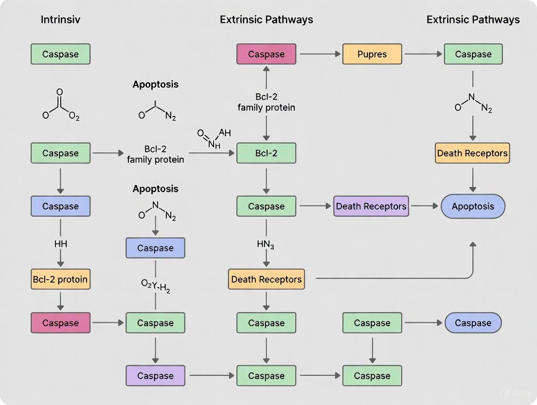
Visualizing the Apoptotic Pathways
The diagram below illustrates the sequence of events in the intrinsic and extrinsic apoptotic pathways, highlighting their unique triggers and the point where they converge on executioner caspases.
Quantitative Western Blot Markers for Pathway Differentiation
To conclusively distinguish between the intrinsic and extrinsic pathways, researchers must monitor a panel of protein markers via Western blot. The following quantitative data, derived from published studies, provides expected results for a clear pathway identification.
Table 2: Key Western Blot Markers for Apoptosis Pathway Analysis
| Target Protein | Pathway Association | Expected Change During Apoptosis | Sample Experimental Observation |
|---|---|---|---|
| Bax | Intrinsic | Upregulation / Conformational Change | Increased expression; elevated Bax/Bcl-2 ratio (from 0.51 to 1.69 over 48h) [1] |
| Bcl-2 | Intrinsic | Downregulation | Decreased expression, leading to increased Bax/Bcl-2 ratio [1] |
| Cytochrome c (Cytosol) | Intrinsic | Upregulation | Significant increase in cytosolic fraction after mitochondrial release [2] |
| Cleaved Caspase-9 | Intrinsic | Appearance of Cleaved Form | Increased activation/cleavage [3] |
| Cleaved Caspase-8 | Extrinsic | Appearance of Cleaved Form | Increased activation/cleavage; detected in DISC [4] |
| Fas (CD95) / FasL | Extrinsic | Upregulation | Upregulated protein levels [2] |
| Cleaved Caspase-3 | Convergent | Appearance of Cleaved Form | Increased activity (e.g., 2.4-fold increase with 50nM Oleandrin) [2] |
| PARP (Cleaved) | Convergent | Appearance of 89 kDa Fragment | Cleavage by executioner caspases indicates irreversible commitment to apoptosis [5] |
Detailed Experimental Protocol
This section outlines a standardized protocol for analyzing intrinsic and extrinsic apoptosis in a cell culture model, using 25-hydroxycholesterol (25OHChol) and Fas ligand as exemplary inducers.
Sample Preparation and Induction of Apoptosis
- Cell Line and Culture: Human neuroblastoma BE(2)-C cells or osteosarcoma U2OS/SaOS-2 cells are suitable models. Maintain cells in recommended medium with 10% FBS at 37°C and 5% CO₂.
- Experimental Groups:
- Control Group: Untreated cells or vehicle-treated control.
- Intrinsic Pathway Induction: Treat cells with 1-2 µg/mL 25-Hydroxycholesterol (25OHChol) for 24-48 hours [1].
- Extrinsic Pathway Induction: Treat cells with a Fas Ligand (e.g., 100 ng/mL) for 6-24 hours.
- Harvesting: Collect cells at 24h and 48h time points by gentle scraping or trypsinization. Wash cell pellets with cold PBS.
Subcellular Fractionation for Mitochondrial Markers
A critical step for confirming intrinsic apoptosis is the separation of mitochondrial and cytosolic fractions to detect cytochrome c translocation.
- Reagents: Mitochondrial Isolation Kit, Protease/Phosphatase Inhibitors.
- Procedure:
- Resuspend cell pellet in ice-cold Mitochondrial Isolation Buffer.
- Homogenize cells with a Dounce homogenizer (30-40 strokes).
- Centrifuge homogenate at 800 × g for 10 min at 4°C to remove nuclei and unbroken cells.
- Transfer supernatant to a new tube and centrifuge at 12,000 × g for 15 min at 4°C.
- Collect the supernatant as the cytosolic fraction.
- Wash the pellet (mitochondrial fraction) and lyse in RIPA buffer.
- Note: Confirm fraction purity by probing for compartment-specific markers: COX IV (mitochondria) and α-tubulin (cytosol).
Western Blot Analysis
- Protein Lysate Preparation: Lyse whole cells or subcellular fractions in RIPA buffer. Determine protein concentration using a BCA assay.
- Gel Electrophoresis and Transfer: Load 20-30 µg of protein per lane on 4-20% gradient SDS-PAGE gels. Transfer to PVDF membranes.
- Antibody Incubation:
- Primary Antibodies: Dilute according to manufacturer's instructions.
- Intrinsic Panel: Anti-Bax, Anti-Bcl-2, Anti-Cytochrome c (cytosolic fraction), Anti-Cleaved Caspase-9.
- Extrinsic Panel: Anti-Fas, Anti-Cleaved Caspase-8.
- Convergence/Execution Panel: Anti-Cleaved Caspase-3, Anti-PARP.
- Secondary Antibodies: Use HRP-conjugated anti-rabbit or anti-mouse IgG.
- Primary Antibodies: Dilute according to manufacturer's instructions.
- Detection: Develop blots using enhanced chemiluminescence substrate and image with a digital system.
- Loading Control: Probe all blots for GAPDH or β-actin to ensure equal protein loading.
The Scientist's Toolkit: Essential Research Reagents
A successful investigation into apoptotic pathways requires a carefully selected set of reagents, inhibitors, and detection kits.
Table 3: Essential Reagents for Apoptosis Research
| Reagent / Kit | Primary Function | Application Example |
|---|---|---|
| z-VAD-FMK (Pan-Caspase Inhibitor) | Irreversibly blocks activity of all caspases | Confirming caspase-dependent apoptosis; used to revert induced apoptosis [1] [2] |
| JC-1 Dye (MMP Assay) | Fluorescent probe that detects loss of mitochondrial membrane potential (ΔΨm) | Flow cytometry analysis of early intrinsic apoptosis [1] |
| Annexin V-FITC / PI Apoptosis Kit | Detects phosphatidylserine externalization (early apoptosis) and membrane integrity | Flow cytometry to quantify early (Annexin V+/PI-) and late (Annexin V+/PI+) apoptotic cells [1] [5] |
| Cytochrome c Antibody (for WB) | Detects release of cytochrome c from mitochondria | Key marker for intrinsic pathway; requires cytosolic fraction for analysis [2] |
| Cleaved Caspase-8 Antibody | Specifically detects the active form of initiator caspase for extrinsic pathway | Western blot confirmation of extrinsic pathway activation [2] |
| BCL-2 Family Antibody Sampler Kit | Contains multiple antibodies against pro- and anti-apoptotic BCL-2 members | Comprehensive analysis of the key regulatory proteins in the intrinsic pathway [5] |
| Caspase-3 Colorimetric Assay Kit | Measures the enzymatic activity of executioner caspase-3 | Quantifying the final convergence point of both pathways [2] |
Data Interpretation and Pathway Confirmation
After performing the Western blot analysis, interpret the results using this logical workflow to assign the dominant apoptotic pathway.
- Confirming Extrinsic Pathway Activation: A primary extrinsic signal is confirmed by a marked increase in cleaved caspase-8, potentially with upregulated Fas/FasL, even in the absence of strong intrinsic markers [4] [2].
- Confirming Intrinsic Pathway Activation: A primary intrinsic signal is confirmed by a combination of cytosolic cytochrome c accumulation, an increased Bax/Bcl-2 ratio, and activation of caspase-9, without significant caspase-8 cleavage [1] [2].
- Identifying Mixed Pathway Activation: Many physiological and experimental stimuli, such as STING agonist treatment, activate a coordinated "pyroptotic apoptosis" or PANoptosis, characterized by the simultaneous activation of caspase-8, caspase-9, and caspase-3/7 [6]. In these cases, a panel of markers from both pathways will be positive. The use of specific pathway inhibitors can help delineate the contribution of each.
The intrinsic apoptotic pathway, also known as the mitochondrial pathway, is a precisely regulated mechanism of programmed cell death critical for development, tissue homeostasis, and eliminating damaged cells. This pathway is characterized by mitochondrial outer membrane permeabilization (MOMP), which leads to the release of cytochrome c and other pro-apoptotic factors from the mitochondrial intermembrane space into the cytoplasm [7]. Once cytosolic, cytochrome c facilitates the formation of the apoptosome complex, which activates initiator caspase-9 and subsequently the executioner caspase cascade, ultimately leading to cellular dismantling [7] [8]. The B cell lymphoma 2 (BCL2) protein family serves as the essential regulatory network that controls the commitment to MOMP, functioning as a tripartite apoptotic switch through the balanced interactions of anti-apoptotic, pro-apoptotic multi-domain, and BH3-only proteins [7]. The detection and quantification of these key markers—cytochrome c, BCL2 family proteins, and caspase-9—via Western blotting provides crucial insights into cellular responses to intrinsic apoptotic stimuli, with significant applications in cancer research, neurodegenerative disease studies, and drug development.
Molecular Mechanisms and Key Markers
The BCL2 Protein Family: Regulators of Mitochondrial Integrity
The BCL2 protein family constitutes the fundamental regulatory circuit of the intrinsic apoptotic pathway, with members characterized by BCL2 homology (BH) domains. This family includes six anti-apoptotic proteins (BCL2, BCL-XL, BCL-w, MCL1, BCL2A1, and BCL-B), which contain four BH domains and prevent MOMP; three pro-apoptotic multi-domain proteins (BAK, BAX, and BOK), which directly execute MOMP; and multiple BH3-only proteins (BID, BIM, BAD, NOXA, PUMA, BMF, and HRK), which initiate apoptosis by sensing cellular stress and either inhibiting anti-apoptotic members or directly activating pro-apoptotic effectors [7]. Anti-apoptotic proteins such as BCL2 itself function by embedding in the outer mitochondrial membrane via a C-terminal transmembrane domain and binding to pro-apoptotic family members, thereby maintaining mitochondrial integrity and preventing cytochrome c release [7]. Genetic studies have revealed that pro-apoptotic proteins BAX and BAK have overlapping functions, with mice lacking both genes displaying profound developmental defects and perinatal lethality due to absent apoptotic activity in multiple tissues [9].
Cytochrome c: The Mitochondrial Messenger of Cell Death
Cytochrome c, a component of the mitochondrial electron transport chain, plays a pivotal role in apoptosis when released into the cytosol, where it binds to Apoptotic Protease-Activating Factor 1 (APAF-1) to form the heptameric apoptosome complex [7]. This complex serves as an activation platform for caspase-9 through induced proximity and dimerization [8]. The release of cytochrome c from mitochondria represents a critical commitment point in the intrinsic pathway, often described as a "point of no return" for cell death execution [7]. Detection of cytochrome c release from mitochondria to cytosol therefore serves as a definitive marker for intrinsic pathway activation, typically assessed through subcellular fractionation followed by Western blot analysis.
Caspase-9: The Initiator Caspase
Caspase-9 functions as the primary initiator caspase of the intrinsic pathway, activated within the apoptosome complex following cytochrome c release. Once activated, caspase-9 proteolytically cleaves and activates executioner caspases-3 and -7, which then mediate the systematic dismantling of cellular structures through cleavage of key substrates such as poly (ADP-ribose) polymerase (PARP) and cytokeratins [10] [8]. Western blot detection of caspase-9 typically focuses on identifying its cleaved, active fragments, which provide evidence of intrinsic pathway execution. Research has shown that while caspase-9 is important for neuronal apoptosis during development, it is not indispensable for apoptosis in all cell types, suggesting alternative activation mechanisms in certain contexts [11].
Quantitative Analysis of Key Apoptotic Markers
Table 1: Key Protein Markers of the Intrinsic Apoptotic Pathway
| Marker Category | Specific Protein | Molecular Weight (Full-length) | Cleaved Fragments | Function in Pathway |
|---|---|---|---|---|
| Anti-apoptotic BCL2 | BCL2 | ~26 kDa | N/A | Inhibits MOMP by binding pro-apoptotics |
| BCL-XL | ~30 kDa | N/A | Neutralizes BAX/BAK activity | |
| MCL1 | ~37 kDa | N/A | Binds and inhibits BAK | |
| Pro-apoptotic Multi-domain | BAX | ~21 kDa | N/A | Forms pores in MOM |
| BAK | ~25 kDa | N/A | Oligomerizes to permeabilize MOM | |
| BH3-only Proteins | BIM | ~23 kDa | N/A | Activates BAX/BAK, inhibits BCL2 |
| BID | ~22 kDa | ~15 kDa (tBID) | Connects extrinsic to intrinsic pathway | |
| Apoptotic Activators | Cytochrome c | ~12 kDa | N/A | Binds APAF-1 to form apoptosome |
| Initiator Caspase | Caspase-9 | ~46 kDa | ~35/37 kDa (large subunit) | Activates executioner caspases |
| Executioner Caspase | Caspase-3 | ~32 kDa | ~17/19 kDa (large subunit) | Cleaves cellular substrates |
| Caspase Substrate | PARP | ~116 kDa | ~89 kDa (cleaved) | DNA repair protein, cleavage inhibits repair |
Table 2: Serological Biomarkers for Apoptosis Detection in Clinical Applications
| Biomarker | Detection Method | Biological Significance | Advantages | Limitations |
|---|---|---|---|---|
| Caspase-cleaved CK18 (M30) | ELISA | Epithelial cell apoptosis | Specific for apoptosis, quantifiable | Limited to epithelial-derived cancers |
| Total CK18 (M65) | ELISA | Overall epithelial cell death | Detects both apoptosis and necrosis | Cannot differentiate death mechanisms |
| Circulating nucleosomes | ELISA | DNA fragmentation in apoptosis | Broad cellular applicability | Short half-life, elevated in various conditions |
| Cytokeratin fragments | ELISA (e.g., CYFRA21-1) | General tumor cell death | Correlates with tumor burden | Not specific to apoptosis |
| Phosphatidylserine exposure | Annexin V flow cytometry | Early apoptosis marker | Detects early apoptosis | Requires fresh cells, cannot use stored samples |
Experimental Protocols for Western Blot Detection
Sample Preparation for Apoptosis Detection
Proper sample preparation is critical for accurate detection of apoptotic markers. For in vitro apoptosis induction, treat cells with intrinsic pathway activators (e.g., etoposide, staurosporine, UV irradiation, or growth factor withdrawal) for appropriate timepoints. Harvest cells and lyse using RIPA buffer supplemented with protease and phosphatase inhibitors. For cytochrome c localization studies, utilize mitochondrial/cytosolic fractionation kits to separate subcellular compartments. Quantify protein concentration using BCA or Bradford assay to ensure equal loading across samples [10]. When preparing samples for BCL2 family proteins, note that some anti-apoptotic members (particularly MCL1) have short half-lives and require rapid processing to prevent degradation.
Electrophoresis and Immunoblotting
Separate 20-50 μg of total protein per sample by SDS-PAGE using 12-15% gels for optimal resolution of caspases and their cleaved fragments, and 10-12% gels for BCL2 family proteins. Transfer proteins to PVDF membranes using wet or semi-dry transfer systems. Block membranes with 5% non-fat dry milk or BSA in TBST for 1 hour at room temperature to prevent non-specific antibody binding [10]. Incubate with primary antibodies diluted in blocking buffer overnight at 4°C with gentle agitation. Essential primary antibodies for intrinsic pathway analysis include: anti-cytochrome c (for localization studies), anti-caspase-9 (for pro and cleaved forms), anti-BAX, anti-BAK, anti-BCL2, anti-BCL-XL, and anti-MCL1. Include loading controls such as β-actin, GAPDH, or COX IV for mitochondrial fractions.
Detection and Analysis
After primary antibody incubation, wash membranes and incubate with appropriate HRP-conjugated secondary antibodies for 1 hour at room temperature. Detect signals using enhanced chemiluminescence substrate and image with a digital imaging system. For quantification, use densitometry software such as ImageJ to measure band intensities [10]. Normalize target protein signals to loading controls and compare treated versus untreated samples. For caspases, calculate the ratio of cleaved to full-length protein to assess activation. When analyzing BCL2 family proteins, compare the ratios of pro-apoptotic to anti-apoptotic members, as the balance between these determines apoptotic susceptibility.
Research Reagent Solutions
Table 3: Essential Reagents for Intrinsic Apoptosis Research
| Reagent Category | Specific Examples | Application | Key Features |
|---|---|---|---|
| Apoptosis Inducers | Etoposide, Staurosporine, ABT-737 | Intrinsic pathway activation | DNA damage, kinase inhibition, BH3 mimetic |
| Western Blot Antibodies | Anti-cytochrome c, Anti-cleaved caspase-9, Anti-BAX, Anti-BCL2 | Protein detection by Western blot | Specificity to target epitopes, validated applications |
| Apoptosis Antibody Cocktails | Pro/p17-caspase-3 + cleaved PARP1 + actin mixes | Multiplex detection | Multiple targets in single assay, improved efficiency |
| Caspase Activity Assays | Fluorogenic substrates (DEVD-aminomethylcoumarin) | Caspase activity measurement | Sensitive, quantitative, kinetic measurements |
| BCL2 Family Inhibitors | Venetoclax (BCL2-specific), Navitoclax (BCL2/BCL-XL/BCL-w) | Targeted therapy, mechanistic studies | Specificity for anti-apoptotic BCL2 proteins |
Signaling Pathway Visualizations
Intrinsic Apoptosis Signaling Pathway
Western Blot Experimental Workflow
Applications in Research and Drug Development
The detection of intrinsic pathway markers has significant translational applications, particularly in cancer research and therapeutic development. Apoptosis assays have grown into a substantial market, valued at USD 6.5 billion in 2024 and projected to reach USD 14.6 billion by 2034, driven by rising cancer incidence and demand for personalized medicine [12]. Western blot analysis of intrinsic pathway components provides critical mechanistic insights for evaluating novel therapeutics, including BH3-mimetics that selectively target anti-apoptotic BCL2 proteins [7]. Venetoclax, the first FDA-approved BCL2-specific BH3-mimetic, has transformed treatment for hematologic malignancies by directly activating the intrinsic apoptosis pathway in cancer cells [7]. The development of biomarkers for apoptosis detection in clinical trials has advanced significantly, with serological assays now available for caspase-cleaved cytokeratins (M30) and circulating nucleosomes that provide minimally invasive monitoring of treatment response [8]. These applications highlight the continuing importance of precise detection and quantification of intrinsic pathway markers across both basic research and clinical translation.
The extrinsic apoptosis pathway, also known as the death receptor pathway, represents a critical mechanism for programmed cell removal that is essential for development, immune system regulation, and tissue homeostasis [13] [5]. This pathway is characterized by its initiation through extracellular signals and the involvement of specific signature proteins that distinguish it from the intrinsic (mitochondrial) apoptosis pathway. The core components of this pathway include death receptors from the tumor necrosis factor (TNF) receptor superfamily (such as Fas/CD95), adaptor proteins (primarily FADD), and initiator caspases (notably caspase-8) [14] [5]. These proteins work in a coordinated cascade to transmit death signals from the cell surface to intracellular execution machinery.
Understanding the distinct roles and detection methods for these signature proteins is particularly valuable for researchers employing Western blot analysis to differentiate between extrinsic and intrinsic apoptosis in experimental settings. The extrinsic pathway can be triggered by various stimuli, including immune cell interactions (e.g., through FasL-Fas binding) and cellular stress signals, ultimately leading to the controlled dismantling of the cell without inducing inflammation [13] [5]. Dysregulation of this pathway contributes to numerous human diseases, including cancer, autoimmune disorders, and neurodegenerative conditions, making its accurate detection and analysis a priority in both basic research and drug development [15] [14].
Molecular Mechanisms of Key Proteins
Fas (CD95): The Initiation Receptor
Fas (also known as CD95 or Apo-1) is a death receptor belonging to the TNF receptor superfamily that serves as the primary entry point for many extrinsic apoptosis signals [14]. This transmembrane receptor is characterized by an intracellular death domain (DD) that is essential for apoptosis signaling. Upon binding to its natural ligand (FasL), Fas undergoes trimerization, triggering a conformational change that enables the recruitment of intracellular adapter proteins [16] [14]. The aggregation of Fas receptors on the cell surface represents the initial commitment step to extrinsic apoptosis, making it a fundamental marker for distinguishing this pathway from intrinsic apoptosis triggers.
The critical function of Fas in apoptosis initiation has been demonstrated across multiple cell types, with its activation leading to the formation of the Death-Inducing Signaling Complex (DISC) [14]. Research has shown that Fas-mediated apoptosis plays crucial roles in immune system regulation, particularly in the elimination of autoreactive lymphocytes and the termination of immune responses [14]. In Western blot analyses, Fas can be detected as a band of approximately 45-48 kDa, though its post-translational modifications and activation state may alter its migration pattern.
FADD: The Critical Adaptor Protein
Fas-Associated protein with Death Domain (FADD) serves as an essential adaptor protein that physically bridges activated death receptors with downstream effector molecules [17] [18]. FADD contains two primary structural domains: a C-terminal death domain (DD) that facilitates interaction with trimerized death receptors like Fas, and an N-terminal death effector domain (DED) that recruits initiator caspases [14] [17]. This bipartite domain structure enables FADD to function as a molecular platform for DISC assembly, positioning it as a central hub in the extrinsic apoptosis pathway.
Recent structural studies using cryo-electron microscopy have revealed that FADD nucleates the formation of a helical filament structure through DED-mediated interactions [19]. This filament provides the structural framework for procaspase-8 oligomerization and activation. The essential nature of FADD is demonstrated by embryonic lethality in FADD-deficient mice, highlighting its non-redundant functions in development and cellular homeostasis [14] [20]. In Western blot applications, FADD typically migrates as a 28-30 kDa protein, and its recruitment to death receptors can be assessed through co-immunoprecipitation assays.
Caspase-8: The Initiator Caspase
Caspase-8 represents the most upstream protease in the extrinsic apoptosis cascade and serves as the primary initiator caspase for death receptor-mediated apoptosis [16] [14]. This cysteine-aspartic protease is synthesized as an inactive zymogen (procaspase-8) consisting of 479 amino acids with a molecular weight of 55 kDa [15]. The protein structure includes two N-terminal death effector domains (DED1 and DED2), a large protease subunit (p18) containing the catalytic cysteine residue, and a small protease subunit (p10) [15] [14]. Within the DED filaments nucleated by FADD, procaspase-8 molecules form active heterotetramers through anti-parallel dimerization of their catalytic domains [19].
The activation mechanism of caspase-8 involves sequential proteolytic cleavages that first generate partially active dimers and then fully mature enzymes capable of initiating the apoptotic cascade [14]. Once activated, caspase-8 cleaves and activates downstream executioner caspases (caspase-3, -6, and -7), which in turn mediate the proteolytic dismantling of cellular structures [14] [5]. Additionally, caspase-8 can cleave the BH3-only protein Bid to generate truncated Bid (tBid), which amplifies the apoptotic signal by engaging the mitochondrial pathway [16] [14]. Beyond its apoptotic functions, caspase-8 also plays important roles in regulating necroptosis, inflammasome activation, and NF-κB signaling, demonstrating its functional pleiotropy in cell fate decisions [15] [20].
Table 1: Key Characteristics of Extrinsic Apoptosis Signature Proteins
| Protein | Molecular Weight (kDa) | Primary Function | Domain Structure | Key Interactions |
|---|---|---|---|---|
| Fas (CD95) | 45-48 | Death Receptor | Transmembrane, Intracellular Death Domain | FasL, FADD |
| FADD | 28-30 | Adaptor Protein | Death Domain (DD), Death Effector Domain (DED) | Fas, Caspase-8 |
| Procaspase-8 | 55-57 | Initiator Caspase | Two DEDs, Large subunit (p18), Small subunit (p10) | FADD, Caspase-3, Bid |
Detection Methods and Western Blot Protocols
Western Blot Analysis for Extrinsic Pathway Proteins
Western blotting provides a powerful method for detecting and quantifying the key proteins involved in the extrinsic apoptosis pathway, allowing researchers to monitor expression levels, activation states, and cleavage events that signify pathway engagement [10]. For optimal detection of extrinsic pathway signature proteins, cell lysates should be prepared using RIPA buffer supplemented with protease and phosphatase inhibitors to preserve protein integrity and post-translational modifications. Protein concentration should be determined using a standardized assay (e.g., BCA or Bradford), and equal amounts of protein (typically 20-50 μg) should be loaded per lane on SDS-PAGE gels [10].
For Fas detection, a 10-12% gel is recommended, while FADD and caspase-8 separation may be improved on 12-15% gels due to their smaller molecular weights. Following electrophoresis, proteins should be transferred to PVDF membranes using standard wet or semi-dry transfer systems. Membrane blocking with 5% non-fat milk or BSA in TBST for 1 hour at room temperature helps reduce non-specific antibody binding. Primary antibody incubation should be performed overnight at 4°C with gentle agitation, followed by thorough washing and appropriate secondary antibody incubation [10].
Key Antibodies and Detection Strategy
The selection of specific antibodies is crucial for accurate detection of extrinsic pathway components. For Fas, antibodies targeting the extracellular domain are preferred for detecting total protein levels. FADD antibodies should be validated for specificity given its low molecular weight and potential for cross-reactivity. For caspase-8, researchers have two primary strategies: detecting the full-length zymogen (55-57 kDa) or the cleaved active fragments (p43/p41 intermediate fragments and the p18 large subunit) [10]. The appearance of these cleavage products provides definitive evidence of caspase-8 activation and extrinsic pathway engagement.
To confirm specific activation of the extrinsic pathway, it is recommended to probe for multiple components simultaneously. The combination of Fas, FADD, and caspase-8 cleavage products provides a signature profile that distinguishes extrinsic from intrinsic apoptosis. Additionally, detecting cleavage of classic caspase substrates such as PARP (89 kDa fragment) and caspase-3 (17-19 kDa fragment) can help verify downstream apoptotic execution [10]. Normalization to housekeeping proteins like β-actin, GAPDH, or tubulin is essential for accurate quantification of protein levels across experimental conditions.
Table 2: Western Blot Detection Parameters for Extrinsic Apoptosis Markers
| Protein Target | Expected Band Sizes | Recommended Gel Percentage | Key Detection Notes |
|---|---|---|---|
| Fas | 45-48 kDa | 10-12% | Confirm membrane localization via fractionation |
| FADD | 28-30 kDa | 12-15% | Low abundance may require signal amplification |
| Procaspase-8 | 55-57 kDa | 10-12% | Detects inactive zymogen |
| Cleaved Caspase-8 | 43/41 kDa, 18 kDa | 12-15% | Indicates activation; multiple fragments possible |
| Cleaved PARP | 89 kDa | 8-10% | Downstream execution marker |
Troubleshooting and Optimization
Several technical challenges may arise when detecting extrinsic pathway proteins via Western blot. For Fas detection, variable glycosylation patterns can lead to smearing or multiple bands; treatment with glycosidases or using deglycosylation buffers may improve band sharpness. FADD's low molecular weight requires careful gel percentage selection and transfer conditions to prevent transfer-through while maintaining resolution. Caspase-8 detection can be complicated by rapid processing and transient appearance of intermediate fragments; using fresh lysates with complete protease inhibition is essential [10].
To enhance detection sensitivity, researchers can employ signal amplification systems such as HRP-conjugated secondary antibodies with enhanced chemiluminescence substrates. For low-abundance proteins like FADD, increasing protein loading quantity or using more sensitive detection methods (such as fluorescent Western blotting) may be necessary. To confirm extrinsic pathway specificity, stimulation with known death receptor agonists (e.g., FasL, TRAIL) or inhibition with caspase-8-specific inhibitors (IETD-fmk) can provide functional validation of detection results [10].
Research Reagent Solutions
Table 3: Essential Research Reagents for Studying Extrinsic Apoptosis
| Reagent Category | Specific Examples | Research Application |
|---|---|---|
| Activation Ligands | Recombinant FasL, TRAIL | Induce extrinsic apoptosis through death receptor engagement |
| Caspase Inhibitors | IETD-fmk (caspase-8 inhibitor), zVAD-fmk (pan-caspase inhibitor) | Determine caspase-dependent mechanisms |
| Antibody Cocktails | Pro/p17-caspase-3, cleaved PARP, actin mixtures | Simultaneous detection of multiple apoptosis markers |
| Detection Substrates | Enhanced chemiluminescence, fluorescent Western blot substrates | Visualize protein levels and activation states |
| Necroptosis Inhibitors | Necrostatin-1 (RIPK1 inhibitor) | Distinguish apoptosis from necroptosis |
Signaling Pathway Visualization
Figure 1: Extrinsic Apoptosis Pathway Mechanism. This diagram illustrates the molecular events in the extrinsic apoptosis pathway, initiated by FasL binding to Fas receptors. The subsequent formation of the Death-Inducing Signaling Complex (DISC) through FADD-mediated recruitment and activation of caspase-8 represents the commitment step. Active caspase-8 then directly activates executioner caspases and can amplify the signal through Bid cleavage and mitochondrial involvement.
Experimental Applications and Protocol Integration
Time-Course Analysis of Extrinsic Pathway Activation
A critical application of Western blot analysis in extrinsic apoptosis research involves time-course experiments to track the sequential activation of pathway components. Following stimulation with an apoptosis-inducing ligand (e.g., FasL or TRAIL), cells should be harvested at multiple time points (e.g., 0, 15, 30, 60, 120, 240 minutes) to capture the dynamic progression of the signaling cascade [10]. Typically, Fas receptor engagement and FADD recruitment occur within minutes, followed by caspase-8 activation within 15-30 minutes, and eventual cleavage of downstream substrates like PARP within 1-2 hours.
For time-course experiments, consistent sample processing is essential. Cells should be lysed directly in Laemmli buffer or RIPA buffer with protease inhibitors to immediately halt all enzymatic activity. Loading controls should be included on each gel to normalize for potential loading inconsistencies across time points. Densitometric analysis of band intensities allows for quantification of protein expression changes and cleavage events over time. This approach enables researchers to determine the kinetics of extrinsic pathway activation in different cell types or under various experimental conditions.
Pharmacological and Genetic Manipulation Studies
Western blot analysis of extrinsic pathway proteins is invaluable for assessing the effects of pharmacological inhibitors or genetic manipulations on apoptosis signaling. To confirm the specific involvement of caspase-8, researchers can pretreat cells with the caspase-8 inhibitor IETD-fmk (20-50 μM) for 1-2 hours before apoptosis induction [20]. Effective inhibition should prevent the appearance of active caspase-8 fragments and block downstream PARP cleavage without affecting upstream events like FADD recruitment.
Genetic approaches including siRNA, CRISPR/Cas9 knockout, or dominant-negative expression can further elucidate the hierarchical relationships between pathway components. For instance, FADD-deficient Jurkat cells demonstrate complete resistance to Fas-mediated apoptosis, confirming its essential role in this pathway [18]. When using genetic models, it is important to verify protein knockdown or knockout efficiency by Western blot and to assess potential compensatory mechanisms that may develop in stable knockout lines. These manipulation studies, combined with Western blot analysis, provide powerful tools for delineating the essential components and regulatory nodes within the extrinsic apoptosis pathway.
The signature proteins of the extrinsic apoptosis pathway—Fas, FADD, and caspase-8—represent critical markers for distinguishing this programmed cell death mechanism from intrinsic apoptosis and other forms of cell death. Western blot analysis provides a robust methodology for detecting these proteins, their activation states, and their functional interactions in experimental systems. The protocols and applications outlined in this document offer researchers a framework for designing studies that accurately probe the extrinsic pathway in various biological contexts and disease models. As research continues to reveal the complex regulatory networks governing cell fate decisions, the precise detection and analysis of these core extrinsic pathway components will remain essential for advancing both basic biological knowledge and therapeutic development.
Apoptosis, or programmed cell death, is a tightly regulated process essential for cellular homeostasis, development, and the elimination of damaged cells [10]. The biochemical events of apoptosis are largely mediated by a cascade of proteolytic enzymes known as caspases, which are synthesized as inactive zymogens and become activated through proteolytic cleavage [21]. The apoptotic pathways converge on two key effector caspases, caspase-3 and caspase-7, which are responsible for the decisive cleavage of numerous cellular substrates, leading to the characteristic morphological changes of apoptosis [10] [22]. Among the most prominent and well-characterized substrates of these effector caspases is Poly (ADP-ribose) Polymerase (PARP-1), a nuclear enzyme involved in DNA repair [23] [24]. The cleavage of PARP-1 serves as a definitive biochemical marker for apoptosis, effectively halting DNA repair and facilitating cellular disassembly [24]. Within the context of apoptosis research, detecting the activation of caspase-3, caspase-7, and the cleavage of PARP-1 via Western blotting provides critical insights into the engagement and execution of cell death, and helps distinguish between the intrinsic (mitochondrial) and extrinsic (death receptor) pathways [10] [2]. This application note details the protocols and interpretive frameworks for using these convergent executioners as reliable Western blot markers in apoptosis research.
Biological Mechanisms: From Caspase Activation to PARP Cleavage
The Apoptotic Pathways: Intrinsic and Extrinsic
Apoptosis can be initiated via two principal signaling pathways that ultimately converge on the activation of effector caspases.
- The Intrinsic Pathway: This pathway is triggered by internal cellular stresses, such as DNA damage, oxidative stress, or growth factor withdrawal [21]. These signals cause mitochondrial outer membrane permeabilization (MOMP), leading to the release of cytochrome c into the cytoplasm [2]. Cytochrome c then binds to Apaf-1, forming the "apoptosome" complex, which activates caspase-9. Caspase-9, an initiator caspase, subsequently cleaves and activates the effector caspases, caspase-3 and caspase-7 [22] [21].
- The Extrinsic Pathway: This pathway is initiated by the binding of extracellular death ligands (e.g., FasL, TRAIL, TNF-α) to their corresponding cell surface death receptors [21]. Receptor activation leads to the formation of the Death-Inducing Signaling Complex (DISC), which recruits and activates caspase-8. Caspase-8 can then directly cleave and activate effector caspases like caspase-3 and caspase-7 [2].
A key connection between these two pathways is the caspase-8-mediated cleavage of the Bcl-2 family protein Bid. Truncated Bid (tBid) translocates to the mitochondria, promoting MOMP and amplifying the death signal through the intrinsic pathway [21]. The following diagram illustrates the sequence of events in both pathways, culminating in the activation of the convergent executioners.
Caspase-3 and Caspase-7: Redundant yet Distinct Executioners
Caspase-3 and caspase-7 are closely related effector caspases that share overlapping substrate specificities, including the canonical cleavage site in PARP-1 [22]. Both are activated by initiator caspases (caspase-8, -9, -10) and are responsible for the proteolytic dismantling of the cell [10] [21]. However, emerging evidence suggests they are not entirely redundant. Studies indicate that caspase-7 can directly process and activate caspase-2 and caspase-6 in the intrinsic pathway, a function previously attributed primarily to caspase-3 [22]. Furthermore, unique non-apoptotic functions and specific substrate preferences for each caspase continue to be elucidated [22]. Despite these distinctions, the activation of both enzymes is a definitive marker of apoptotic commitment, and their activity is often assessed in tandem.
PARP-1 Cleavage: A Hallmark of Apoptosis
PARP-1 is a 116 kDa nuclear enzyme that functions as a molecular sensor for DNA strand breaks, playing a key role in the DNA base excision repair pathway [24]. During apoptosis, both caspase-3 and caspase-7 cleave PARP-1 at a specific DEVD motif, separating its N-terminal DNA-binding domains (24 kDa and 46 kDa) from its C-terminal catalytic domain (89 kDa) [23] [24]. This cleavage event serves two critical purposes:
- Inactivation of DNA Repair: The 89 kDa fragment has greatly reduced DNA binding capacity, halting the energetically costly process of DNA repair and conserving cellular ATP for the execution of apoptosis [23] [24].
- Prevention of Necrotic Cell Death: Unchecked PARP-1 activation in response to extensive DNA damage can deplete cellular NAD+ and ATP pools, leading to necrotic cell death. Caspase-mediated cleavage of PARP-1 thus acts as a molecular switch ensuring the cell dies via apoptosis rather than necrosis [23].
The appearance of the 89 kDa cleaved PARP fragment and the concomitant disappearance of the 116 kDa full-length protein are therefore considered a hallmark of apoptosis and a reliable indicator of caspase activity [10] [24].
Experimental Protocols for Western Blot Detection
Sample Preparation and Protein Extraction
Proper sample preparation is critical for the accurate detection of caspases and cleaved PARP.
- Cell Lysis: Use a RIPA lysis buffer or a similar formulation containing protease inhibitors to prevent protein degradation and phosphatase inhibitors if phosphorylation status is of interest. Keep samples on ice throughout the process.
- Protein Quantification: Determine protein concentration of the whole-cell lysates using a standardized method such as the Bradford Assay to ensure equal loading across gels [25].
- Sample Buffer: Mix lysates with 3x or 5x SDS-sample buffer. For PARP detection, boiling the samples for 10 minutes at 95°C before loading is recommended [25].
Western Blot Procedure
The following workflow outlines the key steps for performing a Western blot to detect apoptosis markers.
Key Reagents and Antibodies
The table below summarizes the essential antibodies and their recommended conditions for detecting these apoptosis markers.
Table 1: Key Antibodies for Apoptosis Detection via Western Blot
| Target Protein | Antibody Clonality | Recommended Dilution | Expected Band Sizes | Key Specificity Notes |
|---|---|---|---|---|
| Caspase-3 (cleaved) | Rabbit Polyclonal [26] | 1:1000 [26] | 17 kDa / 19 kDa (large fragment) [26] | Detects endogenous activated caspase-3; does not recognize full-length caspase-3 [26]. |
| PARP (cleaved) | Mouse Monoclonal [27] | 1:250 (in cocktail) [27] | 89 kDa (apoptosis-specific fragment) [27] [24] | Specific for the cleaved fragment; does not react with full-length PARP [27]. |
| Caspase-7 (active) | Not Specified | Not Specified | ~20 kDa / ~12 kDa (subunits) | Often detected alongside caspase-3 in apoptosis studies [28] [22]. |
| β-Actin / GAPDH | Rabbit Polyclonal / Mouse Monoclonal | Varies by product | 42 kDa (Actin) / 37 kDa (GAPDH) | Used as a loading control for sample normalization [10]. |
Note on Antibody Cocktails: Pre-mixed apoptosis Western blot cocktails are available that contain multiple primary antibodies (e.g., targeting pro- and cleaved caspase-3, cleaved PARP, and a loading control like actin). These cocktails can streamline the workflow, save time and resources, and ensure consistent antibody ratios for more reproducible results [10] [27].
Data Interpretation and Analysis
Expected Band Patterns and Molecular Weights
Correct interpretation of Western blot results requires knowledge of the expected band sizes for both the full-length (inactive) and cleaved (active) forms of the proteins. The table below provides a concise reference.
Table 2: Characteristic Band Patterns for Apoptosis Markers in Western Blot
| Target Protein | Full-Length (Inactive) Form | Cleaved (Active) Form(s) | Interpretation of Cleavage |
|---|---|---|---|
| Caspase-3 | 32-35 kDa (pro-caspase-3) [27] | 17 kDa and 19 kDa fragments [26] [27] | Indicates activation of executioner caspase. Decrease in pro-form and increase in cleaved forms. |
| Caspase-7 | ~35 kDa (pro-caspase-7) | ~20 kDa and ~12 kDa subunits | Indicates activation of executioner caspase. |
| PARP-1 | 116 kDa [24] | 89 kDa (catalytic fragment) and 24 kDa (DNA-binding domain) [24] | Hallmark of apoptosis. Appearance of the 89 kDa fragment and decrease of the 116 kDa band. |
Quantification and Normalization
For robust data analysis, follow these steps:
- Normalization: Normalize the signal intensity of the cleaved protein bands (e.g., cleaved caspase-3 or cleaved PARP) to a housekeeping protein such as β-actin or GAPDH to account for variations in sample loading and transfer efficiency [10].
- Densitometry: Use software like ImageJ to measure the band intensities.
- Calculate Ratios: Present data as the ratio of cleaved to total protein (where possible) or as the relative intensity of the cleaved form compared to the control group after normalization [10]. For example, a increasing ratio of cleaved PARP to full-length PARP provides a strong indicator of active apoptosis.
Troubleshooting Common Challenges
- High Background: Ensure sufficient blocking and optimize antibody concentrations. Increase the number and duration of washes.
- Unexpected or Absent Bands: Verify antibody specificity and reactivity with your model organism. Confirm that the experimental treatment indeed induces apoptosis. Check protein degradation in lysates by confirming the integrity of housekeeping protein bands.
- Multiple Bands: Non-specific binding can occur. Use antibodies validated for Western blotting and check the supplier's information for expected band patterns [26].
The Scientist's Toolkit: Essential Research Reagents
Successful detection of apoptosis markers relies on a suite of well-validated reagents. The following table lists essential tools for your experiments.
Table 3: Essential Research Reagents for Apoptosis Detection
| Reagent / Material | Function / Application | Examples / Notes |
|---|---|---|
| Caspase 3/7 Assay Substrate | Fluorogenic substrate to measure caspase-3/7 enzyme activity in cell lysates or live cells. | Incubate lysates with substrate for 60 min at 37°C; measure cleavage via fluorescence (e.g., excitation 380 nm, emission 460 nm) [25]. |
| Anti-Cleaved Caspase-3 Antibody | Primary antibody for specific detection of activated caspase-3 by Western blot, IHC, or IF. | Rabbit polyclonal antibody detecting 17/19 kDa fragments; does not recognize full-length protein [26]. |
| Anti-Cleaved PARP Antibody | Primary antibody for specific detection of the 89 kDa apoptosis-specific PARP fragment. | Mouse monoclonal antibody that does not react with full-length PARP [27]. |
| Apoptosis Western Blot Cocktail | Pre-mixed antibody cocktail for simultaneous detection of multiple apoptosis markers. | Contains antibodies for pro/cleaved caspase-3, cleaved PARP, and muscle actin; simplifies protocol and improves reproducibility [10] [27]. |
| Caspase Inhibitors (e.g., z-VAD-fmk) | Pan-caspase inhibitor used as a control to confirm caspase-dependent apoptosis. | Pre-treatment with z-VAD-fmk should inhibit caspase activation and PARP cleavage, confirming the apoptotic mechanism [2]. |
| Chemiluminescent HRP Substrate | Detection reagent for visualizing antibody-bound targets on Western blots. | Essential for the final detection step after incubation with HRP-conjugated secondary antibodies. |
Application in Research: Intrinsic vs. Extrinsic Apoptosis
The markers described herein are pivotal for dissecting the apoptotic pathway engaged by a specific stimulus. For instance, a study on the anti-tumor compound oleandrin in osteosarcoma cells utilized these Western blot markers to demonstrate the activation of both intrinsic and extrinsic pathways. Oleandrin treatment led to:
- Intrinsic Pathway Activation: Increased expression of Bax, decreased Bcl-2, release of cytochrome c from mitochondria, and activation of caspase-9 [2].
- Extrinsic Pathway Activation: Up-regulation of Fas and FasL, and activation of caspase-8 [2].
- Convergent Execution: Ultimately, both pathways resulted in the cleavage and increased activity of caspase-3 and the subsequent cleavage of PARP, confirming the execution of apoptosis [2]. The use of caspase inhibitors (z-VAD-fmk) further validated that cell death was caspase-dependent [2].
This exemplifies how a panel of antibodies against initiator caspases, effector caspases, and their substrate PARP can provide a comprehensive map of the apoptotic signaling cascade activated in a given experimental context.
Apoptosis, or programmed cell death, is a fundamental process essential for development, immune regulation, and the maintenance of cellular homeostasis [10]. This controlled cell elimination occurs primarily through two distinct signaling routes: the extrinsic pathway, initiated by external death signals via cell surface receptors, and the intrinsic pathway, activated by internal cellular stress signals originating from within the cell [10]. While these pathways were initially characterized as separate entities, emerging research reveals sophisticated crosstalk mechanisms that integrate these signals, ultimately converging on a common execution phase of apoptosis.
Understanding the molecular integration between these pathways is crucial for both basic research and therapeutic development, particularly in diseases like cancer where apoptosis is frequently dysregulated. Western blot analysis serves as a powerful tool for dissecting these complex interactions by detecting specific protein markers and their activation states within each pathway [10]. This application note details the key nodes of pathway crosstalk and provides validated experimental protocols for researchers to investigate these interconnections within the context of intrinsic and extrinsic apoptosis research.
Molecular Mechanisms of Pathway Integration
Key Executioners and Connectors
The extrinsic apoptosis pathway is typically triggered by ligand binding to death receptors (e.g., Fas, TRAIL receptors) on the cell surface, leading to the formation of the Death-Inducing Signaling Complex (DISC) and activation of initiator caspase-8 [2]. The intrinsic pathway, in contrast, is initiated by internal cellular stresses—such as DNA damage, oxidative stress, or endoplasmic reticulum (ER) stress—that cause mitochondrial outer membrane permeabilization (MOMP) and the release of cytochrome c into the cytoplasm, triggering the formation of the apoptosome and activation of initiator caspase-9 [2] [29].
Despite their distinct origins, these pathways exhibit significant crosstalk, primarily mediated through the proteolytic cleavage of the Bcl-2 family protein Bid [30]. Active caspase-8 from the extrinsic pathway cleaves Bid to its truncated form (tBid), which then translocates to mitochondria, amplifying the apoptotic signal by engaging the intrinsic pathway through Bax/Bak activation [30]. This bidirectional communication ensures robust apoptosis induction even when one pathway is compromised, a common occurrence in cancer cells.
The following diagram illustrates the core components and their interconnections in the integrated apoptotic network:
Quantitative Analysis of Apoptotic Markers
Western blot analysis enables researchers to quantify key apoptotic markers to determine the relative contribution of each pathway. The following table summarizes critical protein targets, their molecular weights, and their significance in pathway crosstalk, with data compiled from recent studies:
Table 1: Key Apoptotic Markers for Pathway Analysis
| Protein Target | Full-Length (kDa) | Cleaved/Active Form (kDa) | Primary Pathway | Role in Crosstalk |
|---|---|---|---|---|
| Caspase-8 | 55 | 43, 41 (cleaved) | Extrinsic | Initiator; cleaves Bid to tBid to amplify intrinsic pathway |
| Caspase-9 | 45-49 | 35, 37 (cleaved) | Intrinsic | Initiator; activated by cytochrome c release |
| Caspase-3 | 35 | 17, 19 (cleaved) | Executioner | Common downstream effector of both pathways |
| PARP | 116 | 89 (cleaved) | Executioner | Cleavage indicates irreversible commitment to apoptosis |
| Bid | 22 | 15 (tBid) | Connector | Molecular link between extrinsic and intrinsic pathways |
| Bax | 21 | N/A | Intrinsic | Pro-apoptotic Bcl-2 family; activated by tBid |
| Bcl-2 | 26 | N/A | Intrinsic | Anti-apoptotic; ratio to Bax determines apoptotic susceptibility |
| Cytochrome c | 12 | N/A | Intrinsic | Released from mitochondria; activates caspase-9 |
Quantitative analysis of these markers provides insights into the dynamics of pathway activation. For example, in a study investigating the natural compound Neocarzilin A (NCA), researchers observed simultaneous activation of caspase-8, enhanced Bid processing, and cytochrome c release, demonstrating coordinated activation of both pathways [30]. Similarly, research on oleandrin in osteosarcoma cells showed regulation of both intrinsic (Bcl-2, Bax, caspase-9) and extrinsic (Fas, FasL, caspase-8) components, confirming dual pathway activation [2].
Table 2: Representative Quantitative Data from Apoptosis Studies
| Study Model | Treatment | Bax/Bcl-2 Ratio | Caspase-3 Activity | Caspase-8 Activation | Caspase-9 Activation | PARP Cleavage |
|---|---|---|---|---|---|---|
| Oleandrin in Osteosarcoma Cells [2] | 50 nM, 24h | ~3.5-fold increase | ~2.4-fold increase | ~2.2-fold increase | ~2.1-fold increase | ~3.0-fold increase |
| NCA in HeLa Cells [30] | 10 µM, 6h | Not reported | Significant activation | Significant activation | Significant activation | Complete cleavage |
| Post-COVID Elderly PBMCs [31] | Natural history | Significantly elevated | Caspase-3 activation heightened | Not specifically reported | Not specifically reported | Not reported |
| NEC in Rat Model [32] | LPS + Hypoxia | Increased in full-term | Increased activity | Variable | Variable | Not reported |
Experimental Protocols for Pathway Analysis
Western Blot Analysis of Apoptotic Markers
Sample Preparation
- Harvest cells after apoptotic induction, collecting both detached and attached populations to avoid bias [33].
- Lyse cells in ice-cold lysis buffer (e.g., Tris-HCl 10 mM pH 7.4, EDTA 5 mM, Triton X-100 1%, protease inhibitor cocktail) for 20 minutes on ice [33].
- Determine protein concentration using Bradford or BCA assay [33] [34].
- Prepare samples with loading buffer, heat at 95°C for 5 minutes, and load 20-50 µg protein per lane on 10-15% SDS-PAGE gels [32] [33].
Electrophoresis and Transfer
- Perform electrophoresis at constant voltage (100-120V) until dye front reaches bottom.
- Transfer proteins to PVDF or nitrocellulose membrane using wet or semi-dry transfer systems [34].
- Confirm transfer efficiency with Ponceau S staining if necessary [34].
Antibody Probing and Detection
- Block membranes with 5% non-fat milk or BSA in TBST for 1 hour at room temperature [33].
- Incubate with primary antibodies against apoptotic markers (see Table 3 for recommendations) diluted in blocking buffer overnight at 4°C [33].
- Wash membranes 3× with TBST for 5 minutes each.
- Incubate with appropriate HRP-conjugated secondary antibodies for 1 hour at room temperature [33].
- Detect signals using enhanced chemiluminescence (ECL) substrate and image with CCD-based system [33] [32].
Optimization Tips
- For antibody conservation, consider the sheet protector (SP) strategy, which uses only 20-150 µL of antibody solution while maintaining sensitivity and specificity [34].
- Always include loading controls (e.g., β-actin, GAPDH, α-tubulin) and apoptosis control extracts when possible [35].
- For cleaved caspase detection, use antibodies specifically recognizing the cleaved forms.
Subcellular Fractionation for Cytochrome c Release
Protocol Overview This protocol enables differentiation between mitochondrial and cytoplasmic cytochrome c, a key event in intrinsic apoptosis [29].
Procedure
- Induce apoptosis by desired method and include vehicle-treated negative controls [29].
- Collect approximately 5 × 10⁷ cells by centrifugation at 200 × g for 5 minutes at 4°C [29].
- Wash cells with 10 mL ice-cold 1× PBS, centrifuge at 600 × g for 5 minutes at 4°C [29].
- Add 1 mL ice-cold Cytosol Extraction Buffer Mix (containing DTT and protease inhibitors) to cell pellet and resuspend thoroughly [29].
- Incubate on ice for 15 minutes, then homogenize with 30-50 passes in a pre-chilled Dounce homogenizer on ice [29].
- Check homogenization efficiency microscopically; 70-80% of nuclei should lack shiny rings [29].
- Centrifuge homogenate at 700 × g for 10 minutes at 4°C to pellet nuclei and debris [29].
- Transfer supernatant to fresh tube and repeat low-speed centrifugation to remove residual nuclei [29].
- Centrifuge resulting supernatant at 10,000 × g for 30 minutes at 4°C; the resulting supernatant is the cytosolic fraction, while the pellet contains mitochondria [29].
- Resuspend mitochondrial pellet in 0.1 mL Mitochondrial Extraction Buffer [29].
- Analyze 10 µg each of cytosolic and mitochondrial fractions by Western blot using cytochrome c antibody (1 µg/mL working concentration) [29].
Validation
- Confirm fraction purity using organelle-specific markers: β-actin (cytoplasmic) and VDAC1 (mitochondrial) [29].
- In healthy cells, cytochrome c should be predominantly in mitochondrial fractions; apoptosis induction increases cytoplasmic cytochrome c [29].
The experimental workflow for analyzing apoptotic pathway integration is visualized below:
Research Reagent Solutions
Table 3: Essential Reagents for Apoptosis Pathway Research
| Reagent Category | Specific Examples | Research Application | Considerations |
|---|---|---|---|
| Control Cell Extracts | Jurkat Apoptosis Cell Extracts (etoposide) [35] | Positive control for apoptosis markers (caspases, PARP) | Validated with etoposide treatment; includes both induced and uninduced extracts |
| Caspase Control Extracts | Caspase-3 Control Cell Extracts (cytochrome c-induced) [35] | Specific control for caspase activation cascade | Cytoplasmic fraction from cytochrome c-treated Jurkat cells |
| Antibody Cocktails | Pro/p17-caspase-3, cleaved PARP1, muscle actin cocktails [10] | Simultaneous detection of multiple apoptosis markers | Streamlines workflow; ensures consistent antibody concentrations |
| Apoptosis Inducers | Etoposide, Cytochrome c, Chloroquine [35] | Positive control treatments for pathway activation | Etoposide preferentially triggers intrinsic pathway; useful for control extracts |
| Fractionation Kits | Cytochrome c Apoptosis Detection Kit [29] | Subcellular fractionation for cytochrome c localization | Includes cytosol extraction buffer with DTT and protease inhibitors |
| Pathway Inhibitors | z-VAD-fmk (pan-caspase), z-LEHD-fmk (caspase-9), Fas blocking antibody [2] | Selective inhibition to dissect pathway contributions | z-VAD-fmk blocks both intrinsic and extrinsic pathways |
Concluding Remarks
The integration between intrinsic and extrinsic apoptotic pathways represents a sophisticated biological mechanism that ensures efficient elimination of compromised cells. The Bid-tBid axis serves as the critical molecular bridge, allowing caspase-8 from the extrinsic pathway to amplify the death signal through mitochondrial engagement of the intrinsic pathway [30]. Western blot analysis, with its capacity to detect specific protein markers, cleavage events, and subcellular localization changes, remains an indispensable technique for delineating these complex interactions.
Researchers should employ a comprehensive approach that includes subcellular fractionation for cytochrome c release, analysis of both initiator and executioner caspases, and examination of key connector molecules like Bid [29]. The use of validated control extracts and pathway-specific inhibitors further strengthens experimental conclusions [35] [2]. As drug discovery efforts increasingly target apoptotic pathways, understanding these interconnections becomes paramount for developing effective therapeutics, particularly for cancer treatment where apoptosis evasion is a hallmark of the disease.
From Theory to Bench: Optimized Western Blot Protocols for Apoptosis Detection
The fidelity of Western blot analysis in apoptosis research is fundamentally dependent on the initial step of sample preparation. Preserving the delicate and often transient protein phosphorylation and cleavage events that define the intrinsic and extrinsic apoptotic pathways requires meticulously designed lysis conditions [10]. The intrinsic (mitochondrial) and extrinsic (death receptor) pathways converge on the activation of executioner caspases, which in turn cleave key substrate proteins such as Poly (ADP-ribose) polymerase (PARP) [10] [36]. These cleaved forms serve as critical markers for confirming apoptosis; however, they are highly susceptible to post-lysis degradation and dephosphorylation if not rapidly stabilized [10]. This application note details optimized protocols for the preparation of cell lysates that accurately capture these dynamic apoptotic signals, framed within the context of distinguishing between intrinsic and extrinsic apoptosis mechanisms.
Apoptotic Signaling Pathways: Key Targets for Analysis
Apoptosis proceeds via two primary signaling cascades. The extrinsic pathway is initiated by extracellular death ligands binding to cell surface receptors, leading to the formation of the death-inducing signaling complex (DISC) and activation of initiator caspase-8 [10] [36]. The intrinsic pathway is triggered by internal cellular stress, such as DNA damage or oxidative stress, causing mitochondrial outer membrane permeabilization (MOMP), cytochrome c release, and formation of the apoptosome, which activates initiator caspase-9 [2] [36]. Both pathways converge on the proteolytic activation of executioner caspases-3 and -7, which dismantle the cell by cleaving structural and repair proteins like PARP [10] [36]. A third, caspase-independent pathway can be mediated by factors such as Apoptosis-Inducing Factor (AIF), which translocates to the nucleus upon mitochondrial membrane permeabilization [37]. Accurate lysis conditions must preserve the specific markers—such as cleaved caspases, cleaved PARP, and phosphorylated Bcl-2 family proteins—that distinguish these pathways and report on their activation status.
Diagram Title: Key Signaling Pathways in Apoptosis
Lysis Buffer Composition for Apoptosis Research
The choice of lysis buffer is critical for the effective extraction and stabilization of apoptotic proteins while maintaining their native modification states. The optimal buffer must achieve complete cell disruption, inactivate endogenous proteases and phosphatases, and be compatible with downstream SDS-PAGE and Western blotting.
Table 1: Core Components of Apoptosis-Specific Lysis Buffers
| Component | Recommended Concentration | Primary Function | Considerations for Apoptosis Research |
|---|---|---|---|
| Detergent | 1% SDS, 1% Triton X-100, or 0.5% CHAPS | Solubilizes membrane proteins and disrupts lipid bilayers | Strong ionic detergents (SDS) ensure complete disruption but require dilution for some assays; milder non-ionic (Triton) preserve protein complexes [38]. |
| Salt | 150 mM NaCl | Maintains ionic strength and prevents non-specific aggregation | Mimics physiological conditions; can be adjusted to optimize protein-extraction efficiency. |
| Buffering Agent | 20-50 mM Tris-HCl or HEPES (pH 7.4-7.5) | Maintains stable physiological pH | Prevents acid/base denaturation of sensitive epitopes on cleaved caspases and PARP [10]. |
| Chaotropic Agent | 2-4 M Urea (optional) | Aids in denaturing and solubilizing difficult proteins | Can help extract tightly bound mitochondrial or nuclear proteins but may interfere with some antibodies. |
Specialized Additives for Signal Preservation
Beyond the core components, the addition of specific inhibitors is non-negotiable for preserving apoptotic signals. The following additives should be included fresh in the lysis buffer immediately before use.
Table 2: Essential Protease and Phosphatase Inhibitors
| Inhibitor Category | Specific Reagents | Target Enzymes | Rationale in Apoptosis Context |
|---|---|---|---|
| Caspase Inhibitors | Not typically added to lysis buffer | Active caspases | Generally omitted to avoid blocking the detection of caspase activity; used in control treatments to confirm pathway specificity [2]. |
| Broad-Spectrum Protease Inhibitors | 1 mM PMSF, 1-10 µg/mL Leupeptin, 1-5 µg/mL Aprotinin, 1 mM EDTA | Serine, cysteine, and metalloproteases | Prevents degradation of cleaved apoptotic markers (e.g., cleaved PARP) by non-caspase proteases released during lysis [10] [38]. |
| Phosphatase Inhibitors | 1-10 mM Sodium Fluoride, 1 mM Sodium Orthovanadate, 5 mM β-Glycerophosphate | Serine/Threonine and Tyrosine phosphatases | Preserves phosphorylation status of key regulatory proteins like Bcl-2, Bad, and other signaling molecules [10] [2]. |
| Deubiquitinase Inhibitors | 1-5 µM PR-619 (optional) | Deubiquitinating enzymes | Crucial for studying ubiquitination events in IAP regulation and NF-κB signaling pathways [39]. |
Step-by-Step Cell Lysis Protocol
Pre-Lysis Considerations
- Cell Handling: Culture and treat cells as required by the experimental design. Use positive controls, such as cells treated with 0.5-1 µM Staurosporine or 1 µM Doxorubicin for 4-24 hours, to reliably induce apoptosis [40] [2].
- Inhibitor Preparation: Prepare a 10X or 100X stock solution of the protease and phosphatase inhibitor cocktail in water or DMSO. Add this stock to the required volume of ice-cold lysis buffer immediately before use.
- Equipment Pre-cooling: Pre-cool microcentrifuge tubes, cell scrapers, and buffers on ice to minimize proteolytic activity during sample handling.
Lysis Procedure for Adherent Cells
- Post-treatment, immediately aspirate the culture medium and rinse the cell monolayer once with ice-cold phosphate-buffered saline (PBS).
- Aspirate PBS completely and add the appropriate volume of freshly prepared, ice-cold lysis buffer directly to the culture dish (e.g., 100-150 µL per 10 cm² of culture surface).
- Rock the dish gently to ensure the buffer covers the entire cell layer. Incubate on ice for 5-15 minutes with occasional gentle agitation.
- Using a cell scraper, dislodge the lysed cells from the surface and transfer the viscous lysate to a pre-cooled microcentrifuge tube.
- Pass the lysate through a 21-25 gauge needle 5-10 times to shear genomic DNA and reduce sample viscosity. Alternatively, brief sonication on ice (3-5 pulses of 5 seconds each at low intensity) can be used.
- Clarify the lysate by centrifugation at 16,000 × g for 15 minutes at 4°C.
- Immediately transfer the supernatant (the soluble protein fraction) to a new pre-cooled tube. Discard the pellet containing insoluble debris.
Post-Lysis Processing
- Protein Quantification: Determine protein concentration immediately using a compatible assay (e.g., BCA or Bradford assay). If the lysis buffer contains strong detergents like SDS, ensure the protein assay is validated for use with that detergent.
- Sample Preparation for Western Blot: Dilute the lysate with Laemmli sample buffer to the desired protein concentration. Denature the samples by heating at 95-100°C for 5-10 minutes.
- Storage: For short-term storage (up to 1 week), keep samples at -20°C. For long-term storage, aliquot and store at -80°C. Avoid repeated freeze-thaw cycles.
The Scientist's Toolkit: Research Reagent Solutions
Table 3: Essential Reagents for Apoptosis Sample Preparation and Analysis
| Reagent / Kit Name | Supplier Examples | Function in Workflow |
|---|---|---|
| RIPA Lysis Buffer | Abcam, Thermo Fisher Scientific | A widely used buffer for total protein extraction; often requires supplementation with fresh inhibitors. |
| Halt Protease & Phosphatase Inhibitor Cocktail | Thermo Fisher Scientific | A ready-to-use, concentrated cocktail that inhibits a wide range of proteases and phosphatases. |
| Caspase-3 Colorimetric Assay Kit | Abcam [2] | Used to functionally confirm caspase activation in parallel with Western blot analysis. |
| Caspase-Glo 3/7 Assay | Promega [41] | A highly sensitive luminescent assay for detecting caspase-3/7 activity in a high-throughput format. |
| Apoptosis Western Blot Cocktail | Abcam (e.g., ab136812) [10] | A pre-mixed antibody cocktail targeting multiple apoptosis markers (e.g., pro/p17-caspase-3, cleaved PARP, actin), streamlining the detection process. |
| CellEvent Caspase-3/7 Green Detection Reagent | Thermo Fisher Scientific [40] | A fluorescent reagent for live-cell imaging of caspase-3/7 activation. |
| Protease Inhibitor Cocktail (EDTA-free) | Roche, Sigma-Aldrich | Essential for preserving protein-protein interactions that might be disrupted by metal chelators. |
Troubleshooting Common Issues
- High Background or Non-Specific Bands: Ensure the lysis buffer does not contain overly harsh detergents at concentrations that cause non-specific antibody binding. Optimize blocking conditions and antibody dilutions.
- Loss of Protein Modifications (Phosphorylation/Cleavage): This is most commonly due to incomplete inhibition of phosphatases and proteases. Confirm that inhibitor stocks are fresh and added immediately before lysis. Process samples quickly on ice.
- Poor Protein Yield: Increase detergent concentration or include a brief sonication step. For difficult-to-lyse cells (e.g., primary cells, neurons), consider using a mechanical homogenization method like bead beating [38].
- Inconsistent Results Between Replicates: Standardize the lysis time and buffer volume relative to cell count. Avoid over-confluent cultures, which can alter apoptotic responses.
Experimental Workflow Visualization
The complete workflow, from experimental setup to data analysis, is summarized in the following diagram.
Diagram Title: Apoptosis Sample Preparation Workflow
Robust and reproducible detection of apoptotic markers by Western blot is contingent upon optimized sample preparation. The use of appropriately formulated lysis buffers, supplemented with potent and fresh protease and phosphatase inhibitors, is the foundational step that ensures the accurate snapshot of the cell's apoptotic status at the moment of lysis. By following the detailed protocols and guidelines outlined in this document, researchers can confidently preserve the integrity of both intrinsic and extrinsic apoptotic signals, thereby generating reliable and interpretable data for their research and drug development programs.
Apoptosis, or programmed cell death, is a tightly regulated process essential for development, tissue homeostasis, and immune function [5]. Dysregulation of apoptotic pathways contributes to various diseases, including cancer, neurodegenerative disorders, and autoimmune conditions [42]. Research into apoptosis mechanisms is therefore critical for understanding disease pathogenesis and developing targeted therapies.
The two primary apoptosis pathways—extrinsic and intrinsic—converge on a common execution phase but originate from distinct initiators [43]. The extrinsic pathway begins outside the cell when extracellular death ligands (e.g., FasL, TNF-α) bind to cell surface death receptors, leading to the formation of the Death-Inducing Signaling Complex (DISC) and activation of initiator caspase-8 [43] [5]. The intrinsic pathway, also known as the mitochondrial pathway, initiates from within the cell in response to internal stressors like DNA damage, oxidative stress, or growth factor deprivation [43]. These signals cause mitochondrial outer membrane permeabilization (MOMP) and release of cytochrome c, leading to formation of the apoptosome and activation of initiator caspase-9 [43] [5].
Both pathways ultimately activate executioner caspases (primarily caspase-3 and -7) that dismantle the cell by cleaving key structural and regulatory proteins [10] [5]. Western blot analysis enables researchers to detect these specific protein changes, providing insights into which apoptotic pathways are active in their experimental models.
Figure 1: Integrated Apoptosis Signaling Pathways. The intrinsic (mitochondrial) and extrinsic (death receptor) pathways converge on executioner caspase activation. MOMP: Mitochondrial Outer Membrane Permeabilization; DISC: Death-Inducing Signaling Complex.
Key Apoptosis Markers and Antibody Selection
Selecting appropriate antibodies is crucial for accurate detection of apoptosis via western blotting. Antibodies targeting cleaved forms of proteins provide the most specific evidence of apoptotic activity, as these cleavage events represent committed steps in the cell death cascade [10].
Caspases: The Executors of Apoptosis
Caspases are cysteine proteases that play central roles in apoptosis execution. Detecting their cleaved, activated forms provides direct evidence of apoptotic signaling [10].
Initiator Caspases:
- Caspase-8: Activated in the extrinsic pathway; cleaved form indicates death receptor engagement [43]
- Caspase-9: Activated in the intrinsic pathway; cleaved form indicates mitochondrial involvement [43]
Executioner Caspases:
- Caspase-3: The primary executioner caspase; its cleaved form (17-19 kDa) is a definitive apoptosis marker [10] [5]
- Caspase-7: Secondary executioner caspase with overlapping substrates with caspase-3 [5]
PARP: The DNA Damage Sentinel
Poly (ADP-ribose) polymerase (PARP) is a DNA repair enzyme that becomes cleaved by executioner caspases during apoptosis [10] [44]. Cleavage of the 116 kDa full-length PARP into 89 kDa and 24 kDa fragments serves as a reliable biochemical marker of apoptosis [10]. Detection of the 89 kDa cleaved fragment specifically indicates caspase-mediated apoptosis rather than other forms of cell death.
Bcl-2 Family Proteins: Regulators of Mitochondrial Apoptosis
The Bcl-2 protein family comprises both pro-apoptotic and anti-apoptotic members that regulate mitochondrial outer membrane permeabilization (MOMP), a critical event in intrinsic apoptosis [43] [5].
Anti-apoptotic Proteins:
- Bcl-2, Bcl-xL, Mcl-1: Prevent MOMP and cytochrome c release
Pro-apoptotic Proteins:
- Bax, Bak: Execute MOMP when activated
- Bad, Bid, Bim, Puma: BH3-only proteins that regulate Bax/Bak activation
The balance between these opposing factions determines cellular commitment to apoptosis. Bid deserves special attention as it connects the extrinsic and intrinsic pathways—caspase-8-mediated cleavage of Bid to tBid amplifies the apoptotic signal through mitochondrial engagement [43].
Table 1: Key Antibody Targets for Apoptosis Detection via Western Blot
| Target | Pathway | Full-length (kDa) | Cleaved/Active Form (kDa) | Biological Significance |
|---|---|---|---|---|
| Caspase-3 | Execution | 32-35 | 17, 19 | Primary executioner caspase; definitive apoptosis marker [10] |
| Caspase-8 | Extrinsic | 55 | 43, 41, 18 | Initiator caspase for death receptor pathway [43] |
| Caspase-9 | Intrinsic | 46 | 37, 35 | Initiator caspase for mitochondrial pathway [43] |
| PARP | Execution | 116 | 89 | DNA repair enzyme; cleavage confirms caspase activation [10] |
| Bid | Cross-talk | 22 | 15 (tBid) | Connects extrinsic to intrinsic pathway [43] |
| Bax | Intrinsic | 21 | - | Pro-apoptotic; translocates to mitochondria during apoptosis [5] |
| Bcl-2 | Intrinsic | 26 | - | Anti-apoptotic; expression changes indicate regulatory shifts [5] |
Antibody Validation and Specificity Considerations
Ensuring antibody specificity is paramount for generating reliable western blot data. This is particularly crucial for apoptosis research where detecting specific cleaved forms versus full-length proteins is essential for accurate interpretation [45].
Validation Strategies for Apoptosis Antibodies
Genetic Controls:
- Knockout (KO) validation: Using cells genetically deficient for the target protein provides the gold standard for specificity confirmation [45]
- Overexpression systems: Cells overexpressing the target protein can validate antibody detection capability
Orthogonal Methods:
- Complementary assays like flow cytometry or immunohistochemistry to confirm western blot findings [45]
- Pharmacological inhibitors: Caspase inhibitors (e.g., Z-VAD-FMK) should prevent appearance of cleaved forms
Multiple Cell Line Testing:
- Testing antibodies across various cell lines with different expression levels of target proteins [45]
- Apoptosis inducers: Treating cells with known inducers (e.g., staurosporine for intrinsic pathway, anti-Fas antibodies for extrinsic pathway) to validate antibody detection of cleaved forms
Special Considerations for Cleaved-form Antibodies
Antibodies targeting cleaved forms of proteins (e.g., cleaved caspase-3, cleaved PARP) require additional validation steps:
- Specificity confirmation: Demonstrate detection of only the cleaved form, not the full-length protein
- Size verification: Confirm detected bands match expected molecular weights for cleaved fragments
- Induction validation: Show increased signal in apoptosis-induced samples versus controls
- Inhibition controls: Demonstrate reduced signal with caspase inhibitor co-treatment
Table 2: Antibody Validation Checklist for Apoptosis Detection
| Validation Step | Description | Acceptance Criteria |
|---|---|---|
| Specificity | Confirm target recognition using KO controls | No band in KO lysates; single band at expected size in WT |
| Selectivity | Detect target in complex lysates with minimal background | Clear, specific band with low non-specific binding |
| Cleaved-form Specificity | distinguish cleaved vs full-length proteins | Bands at expected sizes for both forms; increased cleaved:full-length ratio after induction |
| Reproducibility | Consistent performance across experiments and batches | <20% variance in band intensity across replicates |
| Context Appropriateness | Work in intended sample types (species, cell lines, tissues) | Clean detection in relevant experimental models |
Experimental Protocol for Apoptosis Detection
Sample Preparation
Cell Culture and Treatment:
- Culture cells in appropriate medium and treat with apoptosis inducers relevant to your research question
- For intrinsic pathway: Staurosporine (0.5-1 μM, 4-6 hours), UV irradiation, or chemotherapeutic agents
- For extrinsic pathway: Anti-Fas antibodies (100-500 ng/mL, 4-8 hours) or recombinant TNF-α with cycloheximide [5]
Cell Lysis:
- Use RIPA buffer (50 mM Tris-HCl pH 8.0, 150 mM NaCl, 1% NP-40, 0.5% sodium deoxycholate, 0.1% SDS) supplemented with protease and phosphatase inhibitors [46]
- Incubate cells with cold lysis buffer for 30 minutes with constant agitation at 4°C
- Clear lysates by centrifugation at 10,400 × g for 15 minutes at 4°C [47]
Protein Quantification:
- Determine protein concentration using BCA assay [34] [47]
- Adjust samples to equal concentrations with lysis buffer
- Prepare samples with 2× Laemmli buffer containing β-mercaptoethanol
Gel Electrophoresis and Transfer
SDS-PAGE:
- Load 20-50 μg protein per lane alongside pre-stained molecular weight markers [44]
- Use 12-15% gels for optimal separation of caspases and their cleaved forms [46]
- Run gels at 80-120 V until dye front reaches bottom
Protein Transfer:
- Transfer to nitrocellulose or PVDF membranes using wet or semi-dry transfer systems [34]
- Confirm transfer efficiency with Ponceau S staining [34]
Immunoblotting
Blocking:
- Block membranes with 5% non-fat dry milk in TBST for 1 hour at room temperature [34]
Primary Antibody Incubation:
- Dilute primary antibodies in 5% BSA or milk in TBST according to manufacturer recommendations
- Incubate membranes with primary antibody with gentle agitation overnight at 4°C [34]
- Alternative: Use sheet protector method with minimal antibody volume (20-150 μL) for 15 minutes to several hours at room temperature [34]
Washing and Secondary Antibody:
- Wash membranes 3× with TBST for 5 minutes each
- Incubate with HRP-conjugated secondary antibody (1:2000-1:10000) for 1 hour at room temperature [34]
Detection:
- Develop blots using chemiluminescent substrates [34]
- Image with CCD-based imaging systems
- Ensure non-saturating exposure times for accurate quantification
Figure 2: Western Blot Workflow for Apoptosis Detection. Key steps include careful sample preparation, optimal antibody incubation, and appropriate analysis methods. SP: Sheet Protector method.
Analysis and Quantification
Normalization:
- Normalize target protein signals to loading controls (β-actin, GAPDH, α-tubulin) [10] [46]
- Use total protein staining as an alternative normalization method
Quantification:
- Measure band intensity using densitometry software (ImageJ, ImageQuant) [10]
- Calculate cleaved to full-length ratios (e.g., cleaved caspase-3:total caspase-3) [10]
- Express results as fold-change relative to control conditions
Troubleshooting:
- Multiple bands: May indicate non-specific binding; optimize antibody concentration
- High background: Increase blocking time or change blocking reagent
- Weak signal: Increase protein loading or antibody concentration
- No signal: Verify antibody specificity and target expression
Research Reagent Solutions
Table 3: Essential Research Reagents for Apoptosis Western Blotting
| Reagent Category | Specific Examples | Function/Application |
|---|---|---|
| Apoptosis Inducers | Staurosporine (intrinsic), Anti-Fas antibody (extrinsic), UV irradiation | Activate specific apoptotic pathways for positive controls [5] |
| Caspase Inhibitors | Z-VAD-FMK (pan-caspase), Z-DEVD-FMK (caspase-3 specific) | Negative controls to confirm apoptosis-specific cleavage events [5] |
| Lysis Buffers | RIPA buffer (for total protein extraction), Mitochondrial isolation buffers | Extract proteins while preserving post-translational modifications [47] |
| Protease Inhibitors | PMSF, Complete Protease Inhibitor Cocktail | Prevent protein degradation during sample preparation [47] |
| Loading Controls | β-actin, GAPDH, α-tubulin antibodies | Normalize for protein loading variations [10] [46] |
| Detection Systems | HRP-conjugated secondary antibodies, Chemiluminescent substrates | Visualize and quantify protein bands [34] |
Selecting appropriate antibodies with specificity for cleaved forms of key apoptotic proteins is fundamental for accurate pathway analysis in western blot experiments. The recommendations provided here for antibody selection, validation, and experimental protocols will enable researchers to confidently detect and distinguish between intrinsic and extrinsic apoptosis activation. Proper antibody validation and controlled experimental design are essential for generating reliable, reproducible data that advances our understanding of apoptotic mechanisms in health and disease.
Apoptosis, or programmed cell death, is a fundamental physiological process for maintaining cellular balance by eliminating damaged, unnecessary, or potentially harmful cells in a controlled manner [10]. Its detection is crucial for understanding disease mechanisms, particularly in cancer research, neurodegenerative diseases, and drug development [10]. Two primary signaling pathways initiate apoptosis: the extrinsic pathway, triggered by external signals via death receptors on the cell surface, and the intrinsic pathway, initiated by internal cellular stress signals, such as DNA damage or oxidative stress [43]. Both pathways converge on the activation of a cascade of cysteine proteases called caspases, which execute the dismantling of the cell [10] [43]. Western blotting is a powerful tool for detecting apoptosis due to its high specificity and ability to quantify protein levels and modifications [10]. The advent of apoptosis antibody cocktails for multiplex western blotting significantly streamlines this process by enabling the simultaneous detection of multiple key apoptotic markers in a single assay, saving time, resources, and precious sample material [27] [10].
Key Apoptotic Markers for Intrinsic and Extrinsic Pathways
Understanding the distinct and shared proteins of the intrinsic and extrinsic pathways is essential for interpreting western blot data. The following table summarizes the primary markers used to differentiate these pathways.
Table 1: Key Apoptosis Markers for Western Blot Analysis
| Marker | Role in Apoptosis | Molecular Weight (Full-length/Cleaved) | Associated Pathway |
|---|---|---|---|
| Caspase-3 | Executioner caspase; cleaves multiple cellular substrates [27]. | Pro-form: 32 kDa; Cleaved subunit: p17 [27]. | Convergent Point of Both [43] |
| Caspase-8 | Initiator caspase for the extrinsic pathway [10]. | ~55 kDa (inactive); ~43/41 kDa (active) [10]. | Extrinsic [10] |
| Caspase-9 | Initiator caspase for the intrinsic pathway [10]. | ~46 kDa (inactive); ~35/37 kDa (active) [10]. | Intrinsic [10] |
| PARP1 | DNA repair enzyme; cleavage inhibits repair and facilitates death [27]. | Full-length: ~116 kDa; Cleaved fragment: 89 kDa [27]. | Convergent Point of Both [27] |
| Bcl-2 Family | Regulators of mitochondrial membrane permeability (e.g., Bcl-2 anti-apoptotic, Bax pro-apoptotic) [10]. | Varies (e.g., Bcl-2 ~26 kDa, Bax ~21 kDa) [10]. | Intrinsic [43] |
| p53 | Cellular stress sensor; transcriptionally activates pro-apoptotic proteins [43]. | ~53 kDa [48]. | Intrinsic [43] |
The intrinsic pathway, initiated by internal cellular damage, is critically regulated by the Bcl-2 protein family, which controls mitochondrial outer membrane permeabilization (MOMP). This leads to the release of cytochrome c and the formation of the apoptosome, which activates caspase-9 [43]. The tumor suppressor p53 is a master regulator of this pathway, inducing the expression of pro-apoptotic genes like Bax and PUMA in response to stress [43]. In contrast, the extrinsic pathway is triggered by the binding of death ligands (e.g., FasL) to death receptors (e.g., Fas), leading to the formation of the Death-Inducing Signaling Complex (DISC) and activation of caspase-8 [43]. A key connection between the two pathways is the caspase-8-mediated cleavage of Bid, a Bcl-2 family protein, which then amplifies the death signal through the intrinsic mitochondrial pathway [43]. Both pathways ultimately activate the executioner caspases-3 and -7, which cleave proteins like PARP1, leading to the characteristic biochemical and morphological changes of apoptosis [27] [43].
Apoptosis Antibody Cocktails: A Multiplexing Solution
Apoptosis western blot cocktails are pre-mixed solutions containing multiple primary antibodies designed to detect key apoptosis-related markers simultaneously in a single assay [10]. A representative commercial product is the Apoptosis Western Blot Cocktail (ab136812), which contains antibodies against pro- and cleaved caspase-3, cleaved PARP1, and muscle actin (as a loading control) [27]. This cocktail is specifically designed to study the induction of apoptosis in response to various stimuli [27].
Table 2: Advantages of Using Antibody Cocktails for Apoptosis Detection
| Advantage | Description |
|---|---|
| Increased Efficiency | Simplifies workflow by applying multiple antibodies at once, reducing the number of steps and total experiment time [10]. |
| Enhanced Reproducibility | Ensures consistent antibody concentrations and ratios from experiment to experiment, minimizing variability [10]. |
| Conservation of Sample | Allows for the gathering of comprehensive data from a single, limited sample, which is crucial for precious samples [10]. |
| Internal Validation | The simultaneous detection of multiple markers (e.g., caspase-3 cleavage and PARP cleavage) provides a more robust and internally validated confirmation of apoptosis [27]. |
Detailed Experimental Protocol
This protocol outlines the use of an apoptosis antibody cocktail for the multiplex detection of key markers via western blot.
The Scientist's Toolkit: Essential Research Reagents
Table 3: Key Reagents for Apoptosis Multiplex Western Blot
| Reagent / Material | Function / Purpose |
|---|---|
| Apoptosis WB Cocktail (e.g., ab136812) | Pre-mixed primary antibodies for simultaneous detection of caspase-3, cleaved PARP, and a loading control [27]. |
| HRP-Conjugated Secondary Antibody Cocktail | Pre-mixed anti-mouse and anti-rabbit IgG HRP antibodies for chemiluminescent detection [27]. |
| Cell Lysis Buffer | To extract proteins from control and apoptotically-induced cells (e.g., treated with 1 µM staurosporine for 4 hours) [27]. |
| SDS-PAGE Gel & System | To separate proteins based on molecular weight (e.g., 20 µg total protein per lane) [27] [10]. |
| PVDF or Nitrocellulose Membrane | For transferring and immobilizing the separated proteins. |
| Blocking Buffer (e.g., 5% Milk) | To prevent non-specific antibody binding [27]. |
| Chemiluminescent Substrate | For visualizing the protein bands via HRP-catalyzed light emission. |
Step-by-Step Workflow
- Sample Preparation: Prepare cell lysates from both untreated control cells and cells induced to undergo apoptosis (e.g., treated with 1 µM staurosporine for 4 hours or anti-FAS antibody for 2-6 hours) [27]. Quantify protein concentration to ensure equal loading.
- Gel Electrophoresis: Load an equal amount of protein (e.g., 20 µg) for each sample onto an SDS-PAGE gel for separation [27].
- Membrane Transfer: Transfer the separated proteins from the gel to a western blot membrane (PVDF or nitrocellulose).
- Blocking: Incubate the membrane in a blocking buffer, such as 5% non-fat milk in PBS with 0.05% Tween 20, for 1 hour at room temperature to prevent non-specific binding [27].
- Primary Antibody Incubation: Dilute the apoptosis western blot cocktail (e.g., 1/250 dilution for ab136812) in the recommended buffer. Incubate the membrane with the cocktail with gentle agitation for 1 hour at room temperature or overnight at 4°C [27].
- Washing: Wash the membrane several times with wash buffer (e.g., PBS with 0.05% Tween 20) to remove unbound antibodies.
- Secondary Antibody Incubation: Incubate the membrane with the provided HRP-conjugated secondary antibody cocktail (e.g., 1/100 dilution for ab136812) for 1 hour at room temperature [27].
- Detection: After final washes, visualize the protein bands using a chemiluminescent substrate and image the membrane with a digital imager or X-ray film.
Interpreting Results and Data Analysis
Proper interpretation of western blot results is critical for drawing accurate conclusions about apoptotic pathway activation. The diagram below illustrates the expected band patterns for key markers in the intrinsic and extrinsic pathways.
When analyzing your blots, follow these steps:
- Identify Cleavage Events: Look for the appearance of cleaved fragments (e.g., the p17 subunit of caspase-3, the 89 kDa fragment of PARP) and/or the decrease of the full-length, pro- forms (e.g., pro-caspase-3 at 32 kDa) [27] [10].
- Pathway-Specific Analysis:
- Quantification and Normalization:
- Use densitometry software (e.g., ImageJ) to measure band intensities [10].
- Calculate the ratio of cleaved to total protein (where possible) to determine the proportion of activated protein [10].
- Normalize all signals to a housekeeping protein, such as β-actin or GAPDH, to account for variations in sample loading and transfer efficiency [27] [10]. The muscle actin (42 kDa) in the ab136812 cocktail serves this purpose [27].
Table 4: Expected Western Blot Results During Apoptosis
| Target Protein | Control Cells (No Apoptosis) | Cells Undergoing Apoptosis |
|---|---|---|
| Pro-Caspase-3 | Strong band at 32 kDa | Decreased band intensity [27] |
| Cleaved Caspase-3 | Undetectable or very faint | Strong band at 17 kDa [27] |
| Full-length PARP | Strong band at ~116 kDa | Decreased band intensity |
| Cleaved PARP | Undetectable | Strong band at 89 kDa [27] |
| Loading Control (e.g., Actin) | Consistent band intensity across all lanes | Consistent band intensity across all lanes [27] |
Application in Research and Drug Development
Multiplex apoptosis western blotting is indispensable in several research fields. In cancer research, it is used to understand how apoptosis pathways are altered in cancer cells and to evaluate the efficacy of novel chemotherapeutic drugs designed to reactivate these pathways [10]. For instance, the pro-apoptotic effects of compounds like trifluridine in colorectal cancer cells or novel zinc phthalocyanine-based photosensitizers in photodynamic therapy have been studied using these methods [27]. In neurodegenerative disease research, detecting apoptosis helps understand the excessive cell death contributing to diseases like Alzheimer's and Parkinson's [10]. Furthermore, in drug screening and development, apoptosis western blotting is a key method for determining whether potential therapeutic candidates induce apoptosis in target cells, thereby establishing their mechanism of action and therapeutic potential [10]. The use of antibody cocktails makes these applications more efficient and reliable, accelerating the pace of discovery.
Programmed cell death, or apoptosis, is a fundamental biological process critical for maintaining tissue homeostasis, eliminating damaged cells, and enabling proper embryonic development [5]. This highly regulated process occurs through two primary signaling pathways: the intrinsic (mitochondrial) pathway, activated by internal cellular stress signals, and the extrinsic (death receptor) pathway, initiated by external ligand-receptor interactions [10] [5]. Researchers investigating these pathways rely heavily on western blotting to detect specific protein markers that reveal the activation status and mechanism of apoptotic cell death. However, a significant technical challenge arises when these key apoptotic proteins are present in low abundance, which can occur due to low expression levels, rapid turnover, limited sample availability (e.g., rare cell populations), or inefficient protein extraction [49] [50] [51].
Detecting low-abundance apoptotic proteins requires specialized protocols that enhance sensitivity while maintaining specificity. Standard western blot procedures often prove insufficient for visualizing faint bands corresponding to critical apoptotic markers such as activated caspase fragments, cleaved PARP, or phosphorylated Bcl-2 family members [10]. This application note provides a comprehensive, optimized protocol for detecting low-abundance apoptotic proteins within the context of intrinsic versus extrinsic apoptosis research, incorporating enhanced sensitivity techniques, detailed methodologies, and practical troubleshooting guidance for research scientists and drug development professionals.
Apoptotic Pathways: Key Molecular Markers
Intrinsic (Mitochondrial) Pathway Markers
The intrinsic apoptosis pathway activates in response to internal cellular stressors including DNA damage, oxidative stress, and growth factor withdrawal. These signals trigger mitochondrial outer membrane permeabilization (MOMP), a decisive event controlled by Bcl-2 family proteins [5]. Following MOMP, cytochrome c releases from mitochondria into the cytosol, where it forms the apoptosome complex with Apaf-1, leading to caspase-9 activation [2] [5]. This initiator caspase then activates executioner caspases-3 and -7, culminating in cellular dismantling.
- Bcl-2 Family Proteins: The balance between pro-apoptotic (Bax, Bak, Bid, Bim, Bad) and anti-apoptotic (Bcl-2, Bcl-xL, Mcl-1) members determines cellular commitment to apoptosis [10] [2]. Increased Bax/Bak and decreased Bcl-2 expression indicate intrinsic pathway activation.
- Cytochrome c: Translocation from mitochondria to cytoplasm serves as a definitive marker of intrinsic pathway engagement [2].
- Caspase-9: Activated through cleavage upon apoptosome formation; cleaved caspase-9 demonstrates intrinsic pathway initiation [10].
- SMAC/DIABLO and Endonuclease G: Additional mitochondrial proteins released during intrinsic apoptosis.
Extrinsic (Death Receptor) Pathway Markers
The extrinsic pathway initiates when extracellular death ligands (FasL, TRAIL, TNF-α) bind to cognate cell surface death receptors (Fas, TNFR1, DR4, DR5), triggering formation of the Death-Inducing Signaling Complex (DISC) [10] [5]. This complex recruits and activates initiator caspase-8, which directly cleaves and activates executioner caspases-3 and -7.
- Death Receptors and Ligands: Increased expression of Fas, FasL, TRAIL receptors, or TNF receptors indicates extrinsic pathway stimulation [2].
- Caspase-8: Autoproteolytic activation within the DISC complex; cleaved caspase-8 fragments confirm extrinsic pathway activation [10].
- Caspase-10: Another initiator caspase activated in the DISC complex in response to certain death receptors.
- tBID: Caspase-8-mediated cleavage product of BID that amplifies the apoptotic signal by engaging the mitochondrial pathway.
Execution Phase Markers
Both apoptotic pathways converge on the activation of executioner caspases that proteolyze cellular substrates, leading to characteristic apoptotic morphology [5].
- Caspase-3, -6, and -7: Executioner caspases activated by both intrinsic and extrinsic pathways; their cleavage confirms apoptosis execution [10] [2].
- PARP Cleavage: A key caspase-3 substrate; cleavage from 116 kDa to 89 kDa fragments serves as a hallmark of apoptosis [10].
- Lamin A/C, ICAD/DFF45, and Gelsolin: Additional caspase substrates cleaved during apoptosis execution.
Table 1: Key Apoptotic Markers for Western Blot Analysis
| Marker | Pathway | Molecular Weight (Full-length/Cleaved) | Detection Significance |
|---|---|---|---|
| Bcl-2 | Intrinsic | ~26 kDa | Anti-apoptotic; decreased expression promotes apoptosis |
| Bax | Intrinsic | ~21 kDa | Pro-apoptotic; increased expression/translocation to mitochondria |
| Cytochrome c | Intrinsic | ~15 kDa (cytosolic fraction) | Release from mitochondria to cytosol |
| Caspase-9 | Intrinsic | ~46 kDa/~37 kDa, ~35 kDa | Initiator caspase; cleavage indicates activation |
| Fas/FasL | Extrinsic | ~48 kDa/~40 kDa | Death receptor/ligand; increased expression |
| Caspase-8 | Extrinsic | ~55 kDa/~43 kDa, ~18 kDa | Initiator caspase; cleavage indicates activation |
| Caspase-3 | Both | ~35 kDa/~17 kDa, ~19 kDa | Executioner caspase; cleavage indicates activation |
| PARP | Both | ~116 kDa/~89 kDa | DNA repair enzyme; cleavage confirms caspase activity |
Optimized Protocol for Low-Abundance Apoptotic Protein Detection
Enhanced Sample Preparation for Apoptotic Proteins
Effective sample preparation is crucial for preserving low-abundance apoptotic proteins, which are often rapidly degraded or present in limited quantities [49] [50].
Cell Culture and Apoptosis Induction:
- Culture cells in appropriate media and induce apoptosis using pathway-specific inducers (e.g., staurosporine for intrinsic pathway, Fas ligand or TRAIL for extrinsic pathway).
- Include appropriate controls: untreated cells, apoptosis-induced cells, and where possible, caspase inhibitor pre-treated cells (e.g., z-VAD-fmk) [2].
Protein Extraction with Protease Protection:
- Use pre-chilled RIPA buffer supplemented with comprehensive protease inhibitor cocktail (including caspase inhibitors to prevent post-lysis protein degradation) and phosphatase inhibitors (for phospho-protein detection) [49] [52].
- For mitochondrial proteins (e.g., cytochrome c, Smac/DIABLO), consider subcellular fractionation to enrich mitochondrial and cytosolic fractions separately [2] [51].
- Employ mechanical disruption methods: for nuclear proteins (e.g., PARP), use ultrasonication (3-second pulses, 10-second intervals, 5-15 cycles at 40 kW) to ensure complete nuclear lysis and protein release [49].
- For membrane-associated proteins (e.g., death receptors), avoid excessive heating during denaturation to prevent aggregation; instead, incubate samples at 70°C for 10-20 minutes or at room temperature for 15-20 minutes [49].
Protein Quantification and Sample Preparation:
- Determine protein concentration using Bradford or BCA assays to ensure equal loading.
- Use 5× loading buffer instead of 2× to avoid excessive sample dilution [49].
- Load 50-100 μg of total protein per lane for low-abundance targets, using 1.5 mm thick gels to increase loading capacity [49].
- Include positive controls (e.g., purified apoptotic proteins or control lysates from apoptotic cells) to confirm antibody specificity and detection efficiency.
Gel Electrophoresis and Transfer Optimization
Optimal Gel Chemistry Selection:
- For most apoptotic proteins (10-150 kDa), use Bis-Tris gels (pH 6.4-7.2) for superior band resolution and minimized protein modifications [50].
- For high molecular weight proteins (>150 kDa, e.g., full-length PARP), use Tris-Acetate gels for improved separation and transfer efficiency [50].
- For low molecular weight proteins (<15 kDa, e.g., cleaved caspase fragments, Bim, Bad), use Tricine gels for optimal resolution [50].
- Run gels at lower voltages (e.g., 60-100 V) for longer durations to enhance separation resolution, particularly for distinguishing closely sized cleaved and full-length protein forms.
Maximized Transfer Efficiency:
- Use PVDF membranes for their higher protein binding capacity and lower non-specific antibody binding compared to nitrocellulose [49] [51].
- Pre-wet PVDF membranes in 100% methanol for 1-2 minutes before equilibration in transfer buffer.
- For efficient transfer of both high and low molecular weight proteins, use wet transfer systems at 4°C for 90 minutes to 2 hours [53].
- Include post-transfer verification by brief Ponceau S staining (1-10 minutes) to confirm complete and even protein transfer before proceeding to immunodetection [49].
Enhanced Immunodetection for Low-Abundance Targets
Blocking and Antibody Incubation:
- Block membranes with 5% non-fat dry milk or BSA in TBST for 1 hour at room temperature [49] [53].
- For phospho-specific antibodies, use BSA-based blocking solutions instead of milk to reduce background.
- Incubate with primary antibodies at higher concentrations than standard protocols (check manufacturer recommendations and reduce dilution ratio accordingly) overnight at 4°C with gentle shaking [49].
- Use validated, specific antibodies with confirmed reactivity for apoptotic targets; preferentially select antibodies with knockout/knockdown validation to ensure specificity [50] [51].
- Consider using antibody cocktails (pre-mixed antibodies against multiple apoptotic markers) for comprehensive apoptosis detection while conserving sample [10].
Signal Amplification and Detection:
- Use higher concentrations of HRP-conjugated secondary antibodies (follow manufacturer recommendations) and incubate for 1 hour at room temperature [49].
- Avoid sodium azide in all detection steps as it inhibits HRP activity [49].
- Employ high-sensitivity chemiluminescent substrates (e.g., SuperSignal West Atto, SignalBright series) that can detect protein levels down to the attogram range [50] [51].
- For optimal results with high-sensitivity substrates, reduce primary and secondary antibody concentrations to minimize background while maintaining specific signal detection [51].
- Image blots using digital imaging systems with CCD cameras, which offer wider dynamic range and higher sensitivity compared to traditional X-ray film [49]. Capture multiple exposure times to ensure signals remain within the linear detection range.
Research Reagent Solutions
Table 2: Essential Reagents for Low-Abundance Apoptotic Protein Detection
| Reagent Category | Specific Products/Formulations | Function in Protocol |
|---|---|---|
| Lysis Buffers | RIPA buffer with protease/phosphatase inhibitors [49] [52] | Complete protein extraction while preserving modifications |
| Specialized Gels | Bis-Tris (6-250 kDa), Tris-Acetate (>150 kDa), Tricine (<15 kDa) [50] | Optimal separation based on protein size |
| Membranes | PVDF membranes [49] [51] | High protein binding capacity for low-abundance targets |
| Detection Substrates | High-sensitivity chemiluminescent substrates (e.g., SuperSignal West Atto, SignalBright) [50] [51] | Enhanced signal detection for low-abundance proteins |
| Antibody Validation | Knockout/Knockdown validated antibodies [50] [51] | Specific target detection with minimal background |
| Loading Controls | β-actin, GAPDH, total protein stains [10] [54] | Normalization for quantitative analysis |
Data Analysis and Normalization for Apoptotic Protein Quantification
Accurate quantification of low-abundance apoptotic proteins requires careful normalization and analysis techniques to distinguish specific signals from background noise and account for experimental variability [54] [55].
Image Acquisition and Processing:
- Capture blot images using high-resolution digital imaging systems, saving in lossless formats (TIFF or PNG) to preserve data integrity [54].
- Adjust exposure times to avoid signal saturation, particularly for strong bands, as overexposed images cannot be accurately quantified [54].
- Use multiple exposure times to ensure both faint and strong bands remain within the linear detection range of the imaging system.
Densitometry and Normalization Strategies:
- Analyze band intensity using densitometry software such as ImageJ (NIH) [54].
- Subtract local background intensity for each band to account for uneven background [54].
- Normalize target protein signals to appropriate loading controls:
- For total apoptotic proteins (e.g., Bax, Bcl-2), use traditional housekeeping proteins (β-actin, GAPDH) after verifying their stability under experimental conditions [10] [54].
- For cleaved proteins (e.g., cleaved caspases, PARP), normalize to both the total protein (e.g., total caspase-3) and a housekeeping protein [10].
- Consider total protein normalization using stains like Ponceau S or specialized total protein stains, particularly when housekeeping protein expression varies under apoptotic conditions [54].
Quantitative Analysis of Apoptotic Activation:
- Calculate cleavage ratios (cleaved/total protein) for caspases and PARP to assess apoptosis activation levels [10].
- Determine expression ratios between pro-apoptotic and anti-apoptotic Bcl-2 family members (e.g., Bax/Bcl-2 ratio) to evaluate apoptotic predisposition [2].
- Perform statistical analysis on data from at least three independent biological replicates to ensure reproducibility [54].
- Present fold-changes relative to control conditions, using log2 transformation for statistical analysis when variance is high [54].
Table 3: Troubleshooting Common Issues in Low-Abundance Apoptotic Protein Detection
| Problem | Potential Causes | Solutions |
|---|---|---|
| No signal | Insensitive detection method, inefficient transfer, low antibody affinity | Use high-sensitivity ECL substrates, verify transfer with Ponceau S, validate antibodies [50] [51] |
| High background | Non-specific antibody binding, insufficient blocking, overexposure during detection | Optimize antibody concentrations, extend blocking time, reduce exposure time [54] [51] |
| Uneven band patterns | Inconsistent sample preparation, uneven transfer, gel defects | Standardize protein extraction, ensure even transfer sandwich assembly, check gel quality [54] |
| Inconsistent results between replicates | Variable apoptosis induction, protein degradation, uneven transfer | Include positive controls, use fresh protease inhibitors, standardize transfer conditions [49] [54] |
Pathway Diagrams and Experimental Workflow
Apoptotic Signaling Pathways
Experimental Workflow for Low-Abundance Protein Detection
This detailed protocol provides researchers with a comprehensive framework for detecting low-abundance apoptotic proteins, specifically contextualized within intrinsic and extrinsic apoptosis pathway research. The method emphasizes enhanced sensitivity at each step—from specialized sample preparation through optimized transfer conditions to amplified detection strategies—while maintaining the specificity required for accurate interpretation of apoptotic signaling events. By implementing these refined techniques, researchers can reliably detect and quantify critical low-abundance apoptotic markers, advancing our understanding of programmed cell death mechanisms in both basic research and drug development contexts. The integration of optimized reagents, validated antibodies, and appropriate controls ensures reproducible detection of these challenging targets, enabling more precise investigation of apoptotic pathways in health and disease.
Programmed cell death, or apoptosis, is a fundamental process for maintaining cellular homeostasis, and its dysregulation is a hallmark of diseases such as cancer and neurodegeneration [10]. Apoptosis proceeds primarily via two signaling cascades: the intrinsic pathway (mitochondrial), initiated by internal cellular stress, and the extrinsic pathway (death receptor), triggered by external death ligands [10] [56]. Western blotting is an indispensable tool for differentiating these pathways in research, providing high specificity and sensitivity for detecting key protein markers, their activation states, and post-translational modifications during apoptotic cell death [10] [57]. This application note details how western blotting is employed in cutting-edge research to dissect these pathways across different disease contexts and drug discovery efforts.
Key Apoptotic Markers and Western Blot Detection
The cornerstone of apoptosis analysis via western blot is the detection of specific protein markers that define the pathway and phase of cell death.
Core Markers of the Intrinsic and Extrinsic Pathways
The table below summarizes the primary protein targets used to identify and distinguish between the two apoptotic pathways.
Table 1: Key Western Blot Markers for Apoptosis Pathways
| Protein Marker | Apoptosis Pathway | Role & Significance in Western Blot |
|---|---|---|
| Caspase-9 [10] | Intrinsic | Initiator caspase; activation indicates intrinsic pathway engagement. |
| Caspase-8 [10] | Extrinsic | Initiator caspase; activation indicates extrinsic pathway engagement. |
| Caspase-3 [10] | Executioner (Both) | Executioner caspase; cleaved form is a universal apoptosis marker. |
| Cytochrome c [2] | Intrinsic | Release from mitochondria to cytoplasm is a key intrinsic step. |
| Bcl-2 Family (e.g., Bcl-2, Bax) [10] [58] | Intrinsic | Ratio of pro-apoptotic (Bax) to anti-apoptotic (Bcl-2) indicates commitment to intrinsic apoptosis. |
| PARP [10] | Executioner (Both) | Cleavage of PARP is a hallmark late-stage apoptotic event. |
| Fas/FasL [2] | Extrinsic | Death receptor and ligand; upregulated during extrinsic apoptosis. |
The Scientist's Toolkit: Essential Reagents for Apoptosis Western Blotting
Successful detection of these markers relies on a suite of specialized reagents.
Table 2: Essential Research Reagent Solutions for Apoptosis Detection
| Reagent / Solution | Function | Example & Brief Protocol Note |
|---|---|---|
| RIPA Lysis Buffer [57] | Protein Extraction | Efficiently extracts total cellular protein; must be supplemented with protease and phosphatase inhibitors. |
| Laemmli Sample Buffer [57] | Protein Denaturation | Contains SDS and beta-mercaptoethanol to denature proteins and mask intrinsic charge for SDS-PAGE. |
| SDS-PAGE Gel [57] | Size-Based Separation | Polyacrylamide gel matrix separates proteins by molecular weight; critical for identifying cleaved fragments. |
| Primary Antibodies [10] | Target Protein Detection | Antibodies specific for cleaved caspases, Bcl-2 family members, and cleaved PARP are essential. |
| Apoptosis Antibody Cocktails [10] | Multiplex Detection | Pre-mixed antibodies (e.g., for caspase-3, PARP, actin) streamline workflow and ensure consistent results. |
| HRP-Conjugated Secondary Antibodies [59] | Signal Generation | Binds to primary antibody; enzyme catalyzes a chemiluminescent reaction for visualization. |
| ECL Substrate [60] | Signal Detection | Chemiluminescent substrate for HRP produces light signal captured on X-ray film or digital imager. |
Cancer Research Case Study: Targeting Apoptosis in Osteosarcoma and Melanoma
Oleandrin-Induced Apoptosis in Osteosarcoma Cells
A 2016 study investigated the anti-tumor effects of oleandrin on human osteosarcoma (OS) cells, demonstrating its unique ability to activate both intrinsic and extrinsic pathways simultaneously [2].
Experimental Protocol:
- Cell Treatment: U2OS and SaOS-2 osteosarcoma cells were treated with 0, 25, and 50 nM oleandrin for 24 and 48 hours [2].
- Protein Extraction & Western Blot: Cells were lysed, and proteins were separated by SDS-PAGE, transferred to a membrane, and probed with specific antibodies [2].
- Key Antibodies Used: Targets included cytoplasmic and mitochondrial cytochrome c, bcl-2, bax, caspase-9, Fas, FasL, caspase-8, and caspase-3 [2].
- Pathway Inhibition: Specific inhibitors (z-VAD-fmk for pan-caspase, z-LEHD-fmk for intrinsic, Fas blocking antibody for extrinsic) were used to confirm pathway involvement [2].
Quantitative Data Summary: Table 3: Oleandrin-Induced Apoptotic Effects in Osteosarcoma Cells [2]
| Parameter | U2OS Cells (50 nM Oleandrin) | SaOS-2 Cells (50 nM Oleandrin) | Detection Method |
|---|---|---|---|
| Total Apoptosis Rate | Increased to 41.7% (from 7.3% control) | Increased to 34.9% (from 7.4% control) | Flow Cytometry |
| Caspase-3 Activity | 2.4-fold increase (concentration-dependent) | 2.8-fold increase (concentration-dependent) | Colorimetric Assay |
| ROS Positive Cells | Increased to 40.33% (from 2.71% control) | Increased to 16.72% (from 2.44% control) | Flow Cytometry (DCF) |
| Mitochondrial Cytochrome c | Down-regulated | Down-regulated | Western Blot |
| Cytoplasmic Cytochrome c | Up-regulated | Up-regulated | Western Blot |
The data showed that oleandrin induced ROS production, decreased mitochondrial membrane potential, and triggered cytochrome c release, confirming intrinsic pathway activation [2]. Concurrently, it upregulated Fas and FasL expression and activated caspase-8, demonstrating extrinsic pathway engagement [2]. Critically, inhibiting one pathway did not affect oleandrin's ability to activate the other, indicating independent activation of both cascades [2].
Geranylgeranylacetone Triggers the Intrinsic Pathway in Melanoma
Another study explored the pro-apoptotic effect of Geranylgeranylacetone (GGA) on human melanoma cells, revealing a specific activation of the intrinsic pathway [58].
Experimental Protocol:
- Cell Treatment: G361, SK-MEL-2, and SK-MEL-5 melanoma cells were treated with GGA (1-100 μM) for 24 hours [58].
- Viability Assay: Cell viability was measured using a crystal violet assay [58].
- Western Blot Analysis: Lysates were probed for phospho-p38 MAPK, phospho-JNK, p53, Bax, Bcl-2, caspase-9, caspase-3, and cleaved PARP [58].
Findings: GGA significantly reduced melanoma cell viability at concentrations above 10 μM [58]. Western blot analysis revealed that GGA induced the phosphorylation of p38 MAPK and JNK, upregulated p53 and Bax expression, and did not affect Bcl-2 levels [58]. This was followed by activation of caspase-9 and caspase-3, and cleavage of PARP [58]. The absence of caspase-8 activation and the clear intrinsic pathway markers (p53, Bax, caspase-9) confirmed that GGA induces apoptosis specifically through the intrinsic, mitochondrial pathway [58].
Neurodegeneration Research: Detecting Apoptotic Biomarkers in Brain Injury
In contrast to cancer, where reduced apoptosis is often a problem, neurodegenerative diseases and acute brain injuries are characterized by excessive neuronal apoptosis [61]. Western blotting of cerebrospinal fluid (CSF) and blood samples is critical for identifying apoptotic biomarkers that serve as diagnostic and prognostic tools.
Key Apoptotic Biomarkers in Neurology:
- Caspase-3: The key executioner caspase; its activity is a central indicator of ongoing apoptosis in stroke and traumatic brain injury (TBI) [61].
- Caspase-cleaved Products: Specific cleavage products such as caspase-cleaved cytokeratin-18, caspase-cleaved tau, and a 120 kDa αII-spectrin breakdown product (SBDP120) are validated biomarkers for caspase-3-mediated cell death in human patients after stroke and TBI [61].
Research Implications: The levels of these caspase-derived biomarkers correlate with injury severity and clinical outcomes, providing a window into the molecular pathology of brain injuries and offering targets for therapeutic interventions aimed at blocking apoptotic neuronal death [61].
Drug Screening & Development: Evaluating Compound Efficacy and Mechanisms
Western blotting is a cornerstone in drug discovery for evaluating the efficacy and mechanism of action of novel therapeutic compounds.
Profiling Pro-Apoptotic Anti-Cancer Drugs
The case studies on oleandrin and GGA exemplify the use of western blotting in preclinical drug screening [2] [58]. By profiling key apoptotic markers, researchers can:
- Confirm Pro-Apoptotic Activity: Demonstrate that a candidate compound induces cell death via apoptosis (e.g., by showing PARP cleavage) rather than necrosis.
- Elucidate Mechanism of Action: Determine whether a compound acts through the intrinsic pathway (e.g., altering Bcl-2/Bax ratio, caspase-9 activation), extrinsic pathway (e.g., caspase-8 activation), or both.
- Identify Biomarkers for Clinical Development: The specific caspase cleavage products or phosphorylated proteins identified in preclinical models can be developed as biomarkers to monitor drug efficacy in future clinical trials.
Investigating Chemoresistance
Western blotting is also instrumental in understanding how cancer cells evade therapy. A 2019 study on hepatocellular carcinoma (HCC) revealed that the protein RMP/URI inhibits both intrinsic (cisplatin-induced) and extrinsic (TRAIL-induced) apoptosis, but through different mechanisms [62]. Using western blot, the study showed that RMP overexpression promoted NF-κB activation and increased Bcl-xL expression to block the intrinsic pathway, while it suppressed p53 transcription to inhibit the extrinsic pathway [62]. Such findings identify potential targets for overcoming chemoresistance.
Detailed Western Blot Protocol for Apoptosis Detection
The following standardized protocol ensures reliable detection of apoptotic markers.
Workflow Overview:
Step-by-Step Methodology:
Sample Preparation & Protein Quantification:
- Lyse cells or tissues using RIPA buffer supplemented with protease and phosphatase inhibitors [57].
- Centrifuge to remove debris and collect the supernatant.
- Quantify protein concentration using a Bradford assay to ensure equal loading across all gel lanes [57].
- Mix protein lysate with Laemmli buffer (containing SDS and beta-mercaptoethanol) and heat-denature at 95°C for 5 minutes [57].
Gel Electrophoresis (SDS-PAGE):
Protein Transfer (Blotting):
Blocking and Antibody Incubation:
- Block the membrane with 5% non-fat milk or BSA in TBST for 1 hour at room temperature to prevent non-specific antibody binding [59].
- Incubate with a primary antibody (e.g., against cleaved caspase-3, PARP, Bax) diluted in blocking buffer, overnight at 4°C [10] [59].
- Wash the membrane and incubate with an HRP-conjugated secondary antibody for 1 hour at room temperature [59].
Signal Detection and Analysis:
Western blotting remains an essential and powerful technique for dissecting the complex roles of intrinsic and extrinsic apoptosis in biomedical research. Its ability to provide specific, quantitative data on key apoptotic markers—from caspase activation to Bcl-2 family dynamics—makes it invaluable for understanding disease mechanisms in cancer and neurodegeneration, screening for novel therapeutics, and overcoming treatment resistance. The continued application of well-designed western blot protocols, as illustrated in these case studies, will undoubtedly fuel further advances in personalized medicine and targeted drug development.
Solving Common Pitfalls: Strategies for Enhanced Sensitivity and Specificity
Apoptosis, or programmed cell death, is a fundamental process critical for development, tissue homeostasis, and disease prevention in organisms. Research into apoptosis is particularly focused on understanding two primary signaling pathways: the intrinsic (mitochondrial) pathway and the extrinsic (death receptor) pathway. Detecting the key protein players in these pathways is essential for understanding disease mechanisms, such as cancer and neurodegenerative disorders, and for evaluating the efficacy of potential therapeutics. However, researchers consistently face significant technical challenges in these detection efforts, primarily concerning weak signal intensity and high background noise, which can compromise data accuracy and reliability. This application note details these central challenges and provides structured protocols and solutions to overcome them, with a specific focus on Western blotting within the context of intrinsic versus extrinsic apoptosis research.
Core Apoptotic Pathways and Key Detection Markers
The intrinsic and extrinsic apoptotic pathways converge on the activation of executioner caspases but are initiated by distinct signals and involve unique marker proteins.
- The intrinsic pathway is triggered by internal cellular stresses, such as DNA damage or oxidative stress, leading to mitochondrial outer membrane permeabilization (MOMP). Key markers include the release of cytochrome c from mitochondria into the cytoplasm, the activation of caspase-9, and shifts in the balance of Bcl-2 family proteins (e.g., increased pro-apoptotic Bax, decreased anti-apoptotic Bcl-2) [10] [2].
- The extrinsic pathway is initiated by the ligation of death receptors (e.g., Fas, TRAIL receptors) on the cell surface. This leads to the formation of the Death-Inducing Signaling Complex (DISC) and the activation of caspase-8 [10] [63].
Both pathways ultimately lead to the cleavage and activation of executioner caspases-3 and -7, and the subsequent cleavage of cellular substrates like Poly (ADP-ribose) polymerase (PARP), which serves as a hallmark of apoptosis [10].
The diagram below illustrates the core components and flow of these two pathways.
Key Challenges in Apoptotic Protein Detection
Weak Signal Intensity
The activation of caspases during apoptosis involves proteolytic processing, meaning the detectable "active" form is often a cleaved fragment present at much lower abundance than the inactive precursor. For instance, detecting cleaved caspase-3 requires an antibody specific to the novel epitope exposed after cleavage, and the signal from this small, transient population can be faint compared to the full-length protein [10]. Similarly, cytochrome c release is detected by comparing its levels in mitochondrial versus cytosolic fractions, a process that dilutes the protein across two samples, potentially leading to weak signals in the cytosolic fraction [2]. Low abundance of key regulatory proteins, such as the BH3-only proteins that initiate the intrinsic pathway, further exacerbates this challenge.
High Background Noise
A major source of high background in Western blotting is antibody non-specificity, where primary or secondary antibodies bind to off-target proteins. This is particularly problematic when detecting cleaved fragments, as cross-reactivity with other proteins of similar molecular weight can obscure results. Incomplete transfer during the blotting phase can leave proteins trapped in the gel, while inadequate blocking of the membrane allows antibodies to bind nonspecifically to the membrane itself, creating a high background that masks the target signal [10]. These issues can make it difficult to distinguish specific bands, especially when they are weak, leading to false negatives or inaccurate quantification.
Table 1: Summary of Key Challenges and Their Impact on Detection
| Challenge | Primary Cause | Impact on Detection | Affected Markers (Examples) |
|---|---|---|---|
| Weak Signal Intensity | Low abundance of active forms; protein dilution from subcellular fractionation. | False negatives; inaccurate quantification of activation level. | Cleaved caspases-3, -8, -9; cytosolic cytochrome c. |
| High Background Noise | Antibody cross-reactivity; inadequate blocking or washing. | Obscures target bands; complicates band identification and quantification. | All, but particularly problematic for cleaved fragments. |
| Transient Protein Expression | Rapid and timed induction of proteins like EGL-1 / BH3-only proteins. | Difficult to capture the precise window of activity. | Initiator proteins of the intrinsic pathway. |
Optimized Western Blot Protocol for Apoptosis Detection
This protocol is designed to maximize signal-to-noise ratio for the detection of key apoptotic proteins.
Sample Preparation
- Lysis: Use a RIPA buffer supplemented with fresh protease and phosphatase inhibitors. Keep samples on ice.
- Fractionation: For detecting cytochrome c release, use a mitochondrial/cytosolic fractionation kit to separate compartments. Confirm fraction purity with compartment-specific markers (e.g., COX IV for mitochondria) [2].
- Protein Quantification: Perform a colorimetric assay (e.g., BCA) to ensure equal loading of protein across all lanes. Load 20-30 µg of total protein per lane for whole cell lysates, adjusting for fractionated samples [10] [63].
Gel Electrophoresis and Transfer
- Gel Choice: Use precast gels with a gradient (e.g., 4-20%) for optimal separation of full-length and cleaved proteins (e.g., full-length PARP ~116 kDa, cleaved ~89 kDa).
- Transfer: For proteins of larger size (like PARP), ensure efficient transfer by using wet transfer systems at constant voltage for extended times. Include a transfer stain (e.g., Ponceau S) post-transfer to confirm uniform protein transfer and lane loading.
Blocking and Antibody Incubation
- Blocking: Block the membrane with 5% non-fat dry milk in TBST for 1 hour at room temperature to reduce nonspecific binding.
- Primary Antibody: Dilute antibodies in blocking buffer according to the manufacturer's data sheet. Incubate overnight at 4°C with gentle agitation. See Table 3 for recommended specificities.
- Washing: Wash membranes 3 times for 10 minutes each with ample TBST to remove unbound antibody.
- Secondary Antibody: Use a high-sensitivity, pre-adsorbed HRP-conjugated secondary antibody and incubate for 1 hour at room temperature. Wash again as above [10] [63].
Detection and Analysis
- Detection: Use a high-sensitivity chemiluminescent substrate. For weak signals, employ a substrate with a sustained glow-type reaction for flexible imaging times.
- Imaging: Capture images using a digital imager capable of detecting low-light signals. Take multiple exposures to ensure the signal is within the linear range and not saturated.
- Normalization: Always normalize the signal of the target protein (e.g., cleaved caspase-3) to a housekeeping protein (e.g., β-actin or GAPDH) from the same membrane to account for loading errors. Use densitometry software (e.g., ImageJ) for quantification [10].
Table 2: Key Research Reagent Solutions for Apoptosis Detection
| Reagent / Resource | Function / Application | Key Considerations |
|---|---|---|
| Antibodies for Cleaved Forms | Specifically detect activated caspases and other cleaved substrates (e.g., PARP). | Crucial for distinguishing active pathways; requires validation via knockout controls or apoptosis inducers [63]. |
| Apoptosis Antibody Cocktails | Pre-mixed antibodies for multiple markers (e.g., pro/p17-caspase-3, cleaved PARP, actin). | Increases efficiency, ensures consistent antibody ratios, and provides internal loading controls [10]. |
| Mitochondrial/Cytosolic Fractionation Kits | Isolate subcellular compartments to detect translocation events like cytochrome c release. | Essential for validating intrinsic pathway activation; requires purity verification [2]. |
| High-Sensitivity Chemiluminescent Substrate | Generates light signal for HRP-conjugated antibodies on Western blots. | Critical for visualizing low-abundance proteins; choose a low-background, high-signal substrate. |
| Positive Control Lysates | Lysates from cells treated with known apoptosis inducers (e.g., staurosporine). | Serves as essential positive control for antibody performance and experimental setup [63]. |
Advanced Visualization and Detection Techniques
To overcome the limitations of Western blotting, particularly for spatial localization and dynamic processes, researchers are increasingly turning to fluorescent protein (FP) tagging. This involves using CRISPR-Cas mediated genome editing to endogenously tag proteins like CED-9 (Bcl-2 homolog), CED-4 (Apaf1), and CED-3 (Caspase) with bright, photostable FPs such as mNeonGreen [64].
This approach allows for:
- Real-time tracking of protein localization and translocation in live cells, overcoming the static snapshot provided by Western blotting.
- Determination of endogenous expression levels and stoichiometry of core apoptotic pathway components.
- Visualization of subcellular localization, such as the mitochondrial association of CED-9, CED-4, and CED-3, and the formation of enriched CED-4 puncta upon apoptotic induction, without the need for fractionation [64].
The workflow below outlines the key steps in this advanced detection method.
Data Interpretation and Marker Specificity
Accurate interpretation of Western blot data is critical. The table below provides a guide to expected results for key apoptotic markers upon successful induction of either pathway.
Table 3: Western Blot Data Interpretation Guide for Key Apoptotic Markers
| Protein Marker | Pathway | Expected Band Sizes | Key Specificity Check | Interpretation of Activation |
|---|---|---|---|---|
| Caspase-8 | Extrinsic | ~55/54 kDa (pro-form); ~43/41 kDa, ~18 kDa (cleaved) | Loss of signal in caspase-8 knockout cells [63]. | Appearance of cleaved fragments (p43/41, p18). |
| Caspase-9 | Intrinsic | ~46 kDa (pro-form); ~35/37 kDa (cleaved) | Induction with intrinsic stimuli (e.g., etoposide). | Appearance of cleaved p35/p37 fragment. |
| Caspase-3 | Executioner | ~35 kDa (pro-form); ~17/19 kDa (cleaved) | Appearance of cleaved form upon treatment with inducers [63]. | Appearance of cleaved p17/p19 fragments. |
| PARP | Executioner Substrate | ~116 kDa (full-length); ~89 kDa (cleaved) | Induction with any apoptosis trigger. | Increase in p89 / full-length PARP ratio. |
| Cytochrome c | Intrinsic | ~12 kDa | Cytosolic fraction increase after stress [2]. | Increase in cytosolic fraction; decrease in mitochondrial fraction. |
| Bcl-2 / Bax | Intrinsic Regulator | Bcl-2: ~26 kDa; Bax: ~21 kDa | Measurement of Bcl-2/Bax ratio. | Down-regulation of Bcl-2; Up-regulation of Bax [2]. |
Optimization of Blocking Conditions and Antibody Dilutions
In the analysis of intrinsic and extrinsic apoptotic pathways via Western blotting, the optimization of blocking conditions and antibody dilutions is a foundational step for obtaining specific, reproducible, and high-fidelity data. Apoptosis research specifically involves detecting key protein markers—such as caspases, PARP, and Bcl-2 family proteins—that are often present at low levels or require specific detection of cleaved forms [10]. Non-optimized conditions can lead to excessive background, nonspecific bands, or a complete lack of signal, ultimately compromising the interpretation of which cell death pathway is activated [65]. This application note provides detailed methodologies, grounded in a thesis investigating intrinsic versus extrinsic apoptosis, to guide researchers in establishing robust and reliable Western blot protocols.
The Scientist's Toolkit: Essential Research Reagents
The following table details key reagents essential for optimizing blocking and antibody conditions in apoptosis research.
| Reagent Category | Specific Examples | Function in Optimization |
|---|---|---|
| Blocking Agents | Non-fat dry milk, Bovine Serum Albumin (BSA), Casein [65] | Blocks nonspecific binding sites on the membrane to reduce background. Choice depends on antibody and target protein compatibility. |
| Buffers | Tris-Buffered Saline with Tween (TBST), Phosphate-Buffered Saline (PBS) [66] | Serves as the base for preparing blocking and antibody solutions. TBST is preferred for most applications and for use with alkaline phosphatase (AP)-conjugates [67]. |
| Primary Antibodies | Anti-cleaved caspase-3, anti-PARP, anti-Bax, anti-Fas [2] [10] | Specifically binds to the apoptotic protein target of interest. Requires dilution optimization for a strong specific signal with minimal noise. |
| Secondary Antibodies | HRP- or Fluorophore-conjugated antibodies [67] | Binds to the primary antibody and enables detection. Conjugate choice (HRP vs. fluorescent) influences detection method and sensitivity. |
| Detection Reagents | Chemiluminescent substrates (e.g., LumiGLO), Fluorescent substrates [66] | Generates a detectable signal (light or fluorescence) that corresponds to the amount of target protein. |
Optimizing Blocking Conditions to Reduce Background
Blocking is a critical step to prevent nonspecific binding of antibodies to the membrane, which is a common source of high background and compromised data [65]. The choice of blocking agent is not universal and must be considered in the context of the specific antibodies and proteins under investigation.
Selection of Blocking Agent
The two most common blocking agents are non-fat dry milk and BSA, each with distinct advantages and limitations as summarized in the table below.
| Blocking Agent | Recommended Use | Advantages | Disadvantages & Incompatibilities |
|---|---|---|---|
| Non-fat Dry Milk | General, non-phospho protein detection; economical option [65] | Economical; effective for a wide variety of antibodies [65] | Contains biotin and phosphatases; avoid with biotin-conjugated antibodies or phospho-specific antibodies [65]. |
| Bovine Serum Albumin (BSA) | Phospho-specific protein detection; biotin-conjugated antibodies [65] | Fewer cross-reactive proteins; low in biotin and phosphatases [65] | More expensive than non-fat dry milk. |
For research focused on apoptosis, where phosphorylation events and low-abundance cleaved forms are frequently analyzed, BSA is often the superior blocking agent [65]. A standard blocking protocol involves incubating the membrane in a 5% (w/v) solution of the chosen blocking agent (prepared in TBST) for 1 hour at room temperature with gentle agitation [66].
Antibody Dilution Optimization for Enhanced Specificity
The concentration of the primary and secondary antibodies is a major determinant of Western blot quality. Suboptimal concentrations can cause weak signals, nonspecific bands, or a blotched background [68].
Dot Blot Method for Rapid Antibody Titration
Performing full Western blots for each antibody dilution is time-consuming and wasteful. A dot blot assay provides a quicker, cheaper alternative for determining the optimal antibody concentration [68].
Protocol:
- Prepare membrane strips: Cut a nitrocellulose membrane into 1 cm strips.
- Apply antigen: Dot a fixed amount of your protein sample (e.g., 1-5 µL of cell lysate) onto each strip. Allow to dry completely.
- Block: Place strips in blocking buffer (e.g., 5% BSA in TBST) for 1-2 hours at room temperature.
- Incubate with primary antibody: Prepare a series of primary antibody dilutions (e.g., 1:500, 1:1000, 1:2000, 1:5000) in antibody dilution buffer. Incubate separate membrane strips in each dilution for 1 hour.
- Wash: Wash strips thoroughly with TBST.
- Incubate with secondary antibody: Prepare a series of secondary antibody dilutions. Incubate strips with the corresponding secondary antibody for 1 hour.
- Wash and detect: Perform final washes, then incubate with your detection substrate. The optimal dilution will yield a strong, clear dot with minimal background [68].
Standard Western Blot Antibody Incubation
Once a dilution range is identified, it can be confirmed in a full Western blot.
- Primary Antibody Incubation: Dilute the primary antibody in the appropriate buffer (e.g., 5% BSA in TBST). Incubate the membrane with gentle agitation overnight at 4°C [66]. The datasheet for the apoptosis marker antibody (e.g., cleaved caspase-3) should be consulted for a recommended starting dilution.
- Secondary Antibody Incubation: Dilute the HRP- or fluorophore-conjugated secondary antibody in blocking buffer (typically 1:2000 for HRP-conjugates). Incubate the membrane for 1 hour at room temperature with gentle agitation [66].
Integrated Protocol: Detecting Apoptotic Markers with Optimized Conditions
The following workflow integrates optimized blocking and antibody conditions for the detection of key markers in intrinsic and extrinsic apoptosis.
Detailed Procedure:
- Sample Preparation: Prepare cell lysates from treated and control samples using a RIPA buffer supplemented with protease and phosphatase inhibitors (e.g., PMSF, sodium orthovanadate) to preserve apoptotic protein modifications [69]. Determine protein concentration using a compatible assay (e.g., BCA assay).
- Gel Electrophoresis & Transfer: Separate equal amounts of protein (20-30 µg) via SDS-PAGE. Electrophoretically transfer proteins to a nitrocellulose or PVDF membrane [66].
- Blocking: Incubate the membrane in 25 mL of 5% BSA in TBST for 1 hour at room temperature with gentle shaking [66] [65].
- Primary Antibody Incubation: Incubate the membrane with the primary antibody (e.g., anti-cleaved caspase-3, anti-Fas) diluted in 5% BSA/TBST at the optimized concentration (determined via dot blot) overnight at 4°C with gentle agitation [66].
- Washing and Secondary Antibody Incubation: Wash the membrane three times for 5 minutes each with TBST. Incubate with HRP-conjugated secondary antibody diluted 1:2000 in blocking buffer for 1 hour at room temperature [66].
- Detection and Analysis: After final washes, detect signals using a chemiluminescent substrate. Capture images digitally. For accurate quantification, normalize the signal of the apoptotic marker (e.g., cleaved PARP) to the total protein loaded in each lane using total protein normalization (TPN), which is now considered the gold standard over housekeeping proteins [70].
Apoptotic Signaling Pathways: Intrinsic and Extrinsic
Understanding the pathways under investigation is crucial for selecting the appropriate markers. The following diagram illustrates the key proteins in the intrinsic and extrinsic apoptotic pathways that can be detected by Western blot.
Key Markers and Interpretation:
- Extrinsic Pathway Initiation: Look for increased levels of death receptors (e.g., Fas) and the cleaved, active form of initiator caspase-8 [2] [10].
- Intrinsic Pathway Initiation: Monitor the Bax/Bcl-2 ratio, release of cytochrome c from mitochondria into the cytoplasm, and the cleaved, active form of initiator caspase-9 [2] [10].
- Execution Phase: Both pathways converge to activate executioner caspases-3 and -7. A definitive marker of apoptosis is the cleavage of PARP from its full-length form (116 kDa) to a cleaved fragment (89 kDa) [10].
Apoptosis, or programmed cell death, is a fundamental biological process essential for maintaining cellular homeostasis, eliminating damaged cells, and supporting proper development. Research into apoptosis is particularly critical for understanding disease mechanisms and developing novel therapeutics, especially in cancer and neurodegenerative diseases [10]. The process occurs through two primary signaling pathways: the extrinsic pathway, initiated by external death signals through cell surface receptors, and the intrinsic pathway, triggered by internal cellular stress signals that cause mitochondrial outer membrane permeabilization [43]. Both pathways converge on the activation of executioner caspases that systematically dismantle cellular components in a controlled manner.
A significant challenge in apoptosis research involves the detection of low-abundance protein targets that serve as critical markers for these pathways. Key apoptotic regulators, including initiator caspases, cleaved protein fragments, and phosphorylated Bcl-2 family members, often exist at transient low concentrations that fall below the detection limit of standard western blot protocols [10]. This technical limitation can obscure important biological insights, particularly when studying early apoptotic events or analyzing limited sample types such as primary cell cultures or tissue biopsies. The following diagram illustrates the key components of intrinsic and extrinsic apoptosis pathways, highlighting several low-abundance targets that present detection challenges:
Key Low-Abundance Targets in Apoptosis Research
Critical Low-Abundance Markers in Intrinsic and Extrinsic Pathways
The detection of specific low-abundance protein targets provides crucial information for distinguishing between apoptotic pathways and determining the stage of cell death. The intrinsic pathway, activated by internal cellular damage, features several challenging targets including cleaved caspase-9, cytochrome c release, and phosphorylated Bcl-2 family proteins. Meanwhile, the extrinsic pathway, triggered by external death ligands, presents detection difficulties with proteins such as cleaved caspase-8 and the truncated form of BID (tBID) [43]. Downstream convergence points of both pathways include particularly important low-abundance targets like cleaved caspase-3 and cleaved PARP, which serve as definitive markers of committed apoptosis [10].
Recent studies have highlighted the significance of these targets in various disease contexts. For instance, research on photodynamic therapy for lung cancer demonstrated that detection of reduced BCL-2 protein levels, upregulated BAX expression, and activated caspase-3 were essential for understanding treatment efficacy, despite the challenges presented by their low abundance or transient activation states [71]. Similarly, investigations into multiple organ dysfunction syndrome (MODS) have identified critical roles for low-abundance apoptosis-related genes including S100A9, S100A8, and BCL2A1, requiring sophisticated enrichment strategies for their detection and quantification [72].
Technical Challenges in Low-Abundance Target Detection
The reliable detection of low-abundance apoptotic markers faces several technical obstacles that necessitate specialized enrichment strategies. These challenges include limited protein quantity in small sample sizes, transient expression of activated forms (e.g., cleaved caspases), low stoichiometry of modified proteins relative to their unmodified counterparts, and antibody sensitivity limitations [10]. Furthermore, the rapid progression of apoptotic signaling means that key molecular events may occur within narrow timeframes, making temporal capture difficult. Traditional western blotting methods often lack the sensitivity to overcome these limitations without implementing targeted enrichment approaches, potentially leading to false negative results and incomplete understanding of apoptotic mechanisms.
Enrichment Strategies for Low-Abundance Apoptosis Targets
Sample Preparation and Prefractionation Techniques
Optimal sample preparation forms the foundation for successful detection of low-abundance apoptotic targets. The following protocol outlines a comprehensive approach designed to maximize target preservation while minimizing degradation:
Protocol 1: Optimized Sample Preparation for Apoptotic Protein Detection
Cell Harvesting and Lysis:
- Harvest cells at appropriate apoptotic induction timepoints (typically 4-48 hours post-stimulation).
- Use pre-chilled PBS for washing to prevent premature protein degradation.
- Lyse cells using RIPA buffer supplemented with fresh protease and phosphatase inhibitors.
- For mitochondrial protein extraction (e.g., cytochrome c, SMAC/Diablo), employ specialized mitochondrial isolation kits prior to lysis.
Protein Concentration Normalization:
- Quantify protein concentration using bicinchoninic acid (BCA) assay [71].
- Normalize samples to equal concentrations using lysis buffer.
- Prepare aliquots to avoid repeated freeze-thaw cycles that degrade sensitive epitopes.
Prefractionation Methods:
- Implement subcellular fractionation to enrich for organelle-specific apoptotic proteins:
- Mitochondrial fraction for cytochrome c, SMAC/Diablo, AIF
- Nuclear fraction for EndoG, AIF, PARP
- Cytosolic fraction for caspases, IAPs
- Use commercial fractionation kits or differential centrifugation protocols.
- For cleaved caspase detection, consider immunoprecipitation prior to western blotting to enrich low-abundance active fragments.
- Implement subcellular fractionation to enrich for organelle-specific apoptotic proteins:
Protocol 2: Subcellular Fractionation for Mitochondrial Apoptotic Proteins
Mitochondrial Isolation:
- Resuspend cell pellet in isotonic mitochondrial buffer (e.g., 210mM mannitol, 70mM sucrose, 10mM HEPES, 1mM EDTA).
- Homogenize with 20-30 strokes in a Dounce homogenizer on ice.
- Centrifuge at 800×g for 10 minutes at 4°C to remove nuclei and unbroken cells.
- Transfer supernatant and centrifuge at 10,000×g for 15 minutes at 4°C.
- Collect mitochondrial pellet and supernatant (cytosolic fraction).
Mitochondrial Protein Extraction:
- Solubilize mitochondrial pellet in RIPA buffer with sonication.
- Centrifuge at 12,000×g for 15 minutes to remove insoluble material.
- Use supernatant for western blot analysis of mitochondrial proteins.
Electrophoresis and Transfer Optimization
Enhanced separation and transfer techniques significantly improve detection of low-abundance apoptotic markers:
Protocol 3: Modified Electrophoresis for Low-Abundance Proteins
Gel Selection and Loading:
- Use 12-15% Tris-Glycine gels for optimal separation of caspase fragments (e.g., cleaved caspase-3 at 17/19 kDa).
- Employ mini-gel formats (8×10 cm) for improved protein band sharpness.
- Load 30-50μg of total protein per lane; increase to 80-100μg for low-abundance targets.
- Include precast protein standards for accurate molecular weight determination.
Electrophoresis Conditions:
- Run gels at constant voltage (100-120V) for 2-3 hours at 4°C to prevent protein degradation.
- Use freshly prepared electrophoresis buffer.
- Include positive controls (apoptosis-induced cell lysates) and negative controls.
Enhanced Transfer Methods:
- Utilize PVDF membranes (0.2μm pore size) for superior protein retention, especially for low molecular weight targets.
- Implement wet transfer systems at 4°C for 2 hours at 100V or overnight at 30V.
- Add 0.01% SDS to transfer buffer to improve protein elution, but reduce concentration for targets <20kDa.
- Verify transfer efficiency with reversible protein stains (e.g., Ponceau S) before blocking.
Signal Amplification and Detection Approaches
Advanced detection strategies enable visualization of low-abundance apoptotic targets:
Protocol 4: Signal Amplification for Low-Abundance Targets
Antibody Selection and Validation:
- Select antibodies specifically validated for apoptosis detection in western blotting [10].
- Prioritize antibodies targeting cleaved forms (e.g., cleaved caspase-3, cleaved PARP) for specific apoptotic detection.
- Verify antibody specificity using knockout controls or peptide competition.
Enhanced Blocking and Incubation:
- Block membranes with 5% BSA in TBST for 1-2 hours at room temperature.
- Incubate with primary antibodies in blocking buffer overnight at 4°C with gentle agitation.
- Use higher primary antibody concentrations (e.g., 1:500-1:1000) for low-abundance targets.
- Employ extended washing times (4×10 minutes) with TBST to reduce background.
Signal Amplification Methods:
- Implement tyramide-based signal amplification (TSA) for dramatically enhanced sensitivity.
- Use fluorescent secondary antibodies with near-infrared detection for improved dynamic range.
- Consider enzyme-linked detection with extended substrate incubation times (5-30 minutes).
- Employ multiple detection methods for targets with varying abundances on the same membrane.
The following table summarizes key reagent solutions for enhancing detection of low-abundance apoptotic targets:
Table 1: Research Reagent Solutions for Low-Abundance Apoptosis Target Detection
| Reagent Category | Specific Examples | Function in Enrichment | Application Notes |
|---|---|---|---|
| Cell Lysis Buffers | RIPA buffer, CHAPS buffer, Mitochondrial lysis buffers | Protein solubilization while maintaining epitope integrity | Include fresh protease inhibitors; match buffer to target localization |
| Protease Inhibitors | PMSF, Complete Mini tablets, Phosphatase inhibitor cocktails | Preserve phosphorylation states and prevent degradation | Use broad-spectrum cocktails; add immediately before use |
| Primary Antibodies | Cleaved caspase-3 (Asp175), Cleaved PARP (Asp214), Phospho-Bcl-2 (Ser70) | Specific detection of activated apoptotic forms | Validate for western blot; check species reactivity |
| Signal Amplification Reagents | Tyramide signal amplification kits, High-sensitivity ECL substrates, IRDye fluorescent conjugates | Enhance detection sensitivity for low-abundance targets | Optimize concentration to avoid background; use within linear range |
| Membrane Substrates | PVDF (0.2μm), Nitrocellulose (0.45μm) | Optimal protein retention based on molecular weight | PVDF for proteins <20kDa; activate with methanol before use |
Integrated Workflow for Comprehensive Apoptosis Analysis
The following integrated approach combines multiple enrichment strategies to maximize detection of low-abundance apoptotic targets across both intrinsic and extrinsic pathways:
Protocol 5: Integrated Workflow for Low-Abundance Apoptosis Marker Detection
Experimental Design and Apoptosis Induction:
- Include appropriate controls (untreated, apoptosis-induced, apoptosis-inhibited).
- Use established apoptosis inducers specific to intrinsic (e.g., staurosporine, UV irradiation) or extrinsic pathways (e.g., Fas ligand, TRAIL).
- Collect time-course samples to capture transient activation events.
Comprehensive Sample Processing:
- Process samples according to Protocol 1 for whole cell extracts.
- Perform subcellular fractionation following Protocol 2 for pathway-specific targets.
- Quantify protein concentrations and normalize across samples.
Optimized Western Blotting:
- Separate proteins using conditions outlined in Protocol 3.
- Transfer to PVDF membranes using optimized conditions.
- Implement signal detection as described in Protocol 4.
Data Analysis and Validation:
- Acquire images at multiple exposure times to ensure linear detection range.
- Perform densitometric analysis using software such as ImageJ [10].
- Calculate ratios of cleaved to full-length proteins (e.g., cleaved PARP:total PARP).
- Normalize to loading controls (β-actin, GAPDH, or total protein).
- Validate findings with orthogonal methods when possible (e.g., flow cytometry for caspase activation).
Troubleshooting and Quality Control
Common Detection Issues and Solutions
Detection of low-abundance apoptotic targets presents several common challenges that require specific troubleshooting approaches:
Table 2: Troubleshooting Guide for Low-Abundance Apoptosis Target Detection
| Problem | Potential Causes | Solutions | Preventive Measures |
|---|---|---|---|
| Weak or Absent Signal | Insufficient protein loading, inefficient transfer, antibody sensitivity | Increase protein load to 50-80μg, optimize transfer conditions, try signal amplification | Perform pilot experiments to determine optimal conditions |
| High Background | Incomplete blocking, antibody concentration too high, insufficient washing | Extend blocking time, titrate antibodies, increase wash frequency and duration | Use fresh blocking solutions, validate antibody concentrations |
| Non-Specific Bands | Antibody cross-reactivity, protein degradation, overexposure | Include knockout controls, use fresh protease inhibitors, reduce exposure time | Validate antibodies with specific controls, use aliquoted inhibitors |
| Inconsistent Results | Variable sample preparation, membrane drying, detection reagent instability | Standardize protocols, ensure membrane remains wet, use fresh detection reagents | Establish SOPs, prepare fresh solutions, track reagent lots |
Validation and Quality Control Measures
Rigorous validation ensures reliable detection of low-abundance apoptotic targets:
Specificity Controls:
- Include lysates from cells treated with caspase inhibitors (e.g., Z-VAD-FMK) to confirm apoptosis-specific signals.
- Use genetic approaches (siRNA, CRISPR) to verify target specificity.
- Test antibody specificity with peptide competition experiments.
Quantitative Accuracy:
- Establish standard curves using recombinant proteins when available.
- Ensure detection remains within linear range through dilution series.
- Use multiple loading controls to account for potential changes during apoptosis.
Reproducibility Assurance:
- Perform independent experimental replicates (minimum n=3).
- Include reference samples across multiple blots for normalization.
- Document all protocol modifications thoroughly.
The detection of low-abundance targets in apoptosis research requires sophisticated enrichment strategies that span sample preparation, separation, transfer, and detection methodologies. By implementing the integrated approaches outlined in these application notes—including subcellular fractionation, signal amplification, and rigorous validation—researchers can significantly enhance their ability to study key apoptotic regulators that would otherwise remain undetectable with standard protocols. These advanced techniques provide crucial insights into the complex regulation of both intrinsic and extrinsic apoptosis pathways, supporting drug development efforts and advancing our understanding of cell death mechanisms in health and disease. The continued refinement of these enrichment strategies will further enable researchers to unravel the subtleties of apoptotic signaling networks and their therapeutic implications.
Validating Antibody Specificity and Avoiding Cross-Reactivity
In Western blot analysis of intrinsic and extrinsic apoptosis, antibody specificity is the cornerstone of reliable data. Cross-reactivity occurs when an antibody binds to off-target antigens that share structural or sequence similarities with the intended target, leading to false positives and compromised experimental integrity [73]. This is a particular concern in apoptosis research due to the high sequence homology within protein families, such as caspases and Bcl-2 proteins [73] [10]. Validating antibodies for these pathways is therefore not optional but a fundamental requirement for producing reproducible and scientifically sound results.
Key Apoptosis Markers and Cross-Reactivity Risks
The intrinsic (mitochondrial) and extrinsic (death receptor) apoptotic pathways are characterized by distinct protein markers. The table below summarizes the primary targets and their associated risks for cross-reactivity.
Table 1: Key Apoptosis Markers for Western Blot Analysis
| Apoptosis Pathway | Key Protein Markers | Function | Cross-Reactivity Risks |
|---|---|---|---|
| Extrinsic | Caspase-8 (initiator), Caspase-3 (executioner) | Initiates and executes cell death via death receptors [10]. | High homology among initiator caspases [73]. |
| Extrinsic | Fas, FasL | Death receptor and its ligand [2]. | Shared motifs within the tumor necrosis factor (TNF) receptor family. |
| Intrinsic | Caspase-9 (initiator), Caspase-3 (executioner) | Initiates and executes cell death via mitochondrial signals [10]. | High homology among initiator caspases [73]. |
| Intrinsic | Bcl-2 Family (e.g., Bcl-2, Bax) | Regulates mitochondrial membrane permeability [10] [2]. | High sequence similarity within the multi-protein Bcl-2 family [73]. |
| Intrinsic | Cytochrome c | Released from mitochondria; activates caspase-9 [2]. | Generally low risk. |
| Execution Phase | Cleaved PARP | A hallmark substrate of executioner caspases [10]. | Cleaved fragments may share epitopes with other nuclear proteins. |
Experimental Protocols for Antibody Validation
A multi-faceted approach is essential for rigorous antibody validation. The following protocols outline key strategies to confirm specificity and identify cross-reactivity.
Knockout/Knockdown Validation
This genetic strategy is considered a gold standard for establishing antibody specificity [74] [75].
- Methodology:
- Generate Knockout/Knockdown Cells: Use CRISPR, siRNA, or shRNA to deplete the target protein in the cell line of interest [74] [75].
- Prepare Lysates: Prepare protein lysates from both knockout/kno`ckdown and wild-type control cells.
- Perform Western Blot: Analyze the lysates via standard Western blotting using the antibody under validation.
- Interpretation: A specific antibody will show a loss of signal in the knockout/kno`ckdown sample compared to the wild-type control. A persistent signal indicates off-target binding and cross-reactivity [74] [75].
Recombinant Protein Expression & Heterologous Strategy
This method uses non-native expression to test antibody binding in a controlled context [76].
- Methodology:
- Express Target Protein: Transfect a cell line (e.g., 293T) that does not endogenously express your target with a plasmid encoding the protein of interest, often with a tag (e.g., Myc/DDK) [76].
- Analyze Specificity: Perform Western blot on lysates from transfected and mock-transfected cells. The tag antibody serves as a transfection control.
- Test Cross-Reactivity: To test for isoform cross-reactivity, transfert cells with plasmids expressing different, highly similar protein isoforms (e.g., various MAGE-A isoforms or PKC isoforms). Probe the blot with your antibody [76].
- Interpretation: Specificity is confirmed by a signal only in the target-transfected lane. Cross-reactivity is identified if the antibody binds to multiple related isoforms [76].
Immunoprecipitation Followed by Mass Spectrometry (IP/MS)
IP-MS directly identifies all proteins that an antibody pulls down from a complex lysate, providing a comprehensive view of its targets [74].
- Methodology:
- Perform Immunoprecipitation: Incubate the antibody with a cell lysate to pull down the target antigen and any associated proteins or off-target binders.
- Elute and Digest Proteins: Separate the immunoprecipitated complexes by SDS-PAGE, digest the proteins in-gel with trypsin.
- Mass Spectrometry Analysis: Analyze the resulting peptides by liquid chromatography-tandem mass spectrometry (LC-MS/MS) to identify all proteins in the sample [74].
- Interpretation: The data confirms specificity if the only protein identified is the intended target. The identification of additional proteins indicates potential cross-reactivity or non-specific binding [74].
BLASTp Sequence Analysis
A proactive, computational method to predict cross-reactivity risk before an antibody is even used [73].
- Methodology:
- Obtain Immunogen Sequence: Acquire the amino acid sequence of the immunogen used to generate the antibody from the manufacturer's datasheet.
- Run BLASTp Analysis: Input the immunogen sequence into the NCBI BLASTp tool, specifying the organism (e.g., Homo sapiens) you are studying.
- Analyze Homology: Review the results for proteins with high sequence similarity to the immunogen.
- Interpretation: Proteins with >75% sequence identity to the immunogen are considered a high risk for cross-reactivity. A result of <60% identity indicates low risk [73].
The Scientist's Toolkit: Research Reagent Solutions
Table 2: Essential Reagents for Validating Apoptosis Antibodies
| Reagent / Solution | Function in Validation | Key Considerations |
|---|---|---|
| Validated Primary Antibodies | To detect specific apoptosis markers (e.g., cleaved caspases, PARP, Bcl-2) [10]. | Prefer monoclonal antibodies for single-epitope specificity [73]. Select antibodies validated for Western blot in your species of interest. |
| Knockout Cell Lines | To serve as a negative control for antibody specificity using genetic strategies [74] [75]. | Can be generated in-house via CRISPR or purchased from commercial repositories. |
| Recombinant Proteins / Expression Plasmids | To test antibody specificity and cross-reactivity against specific isoforms or family members [74] [76]. | Ideal for validating antibodies against low-abundance targets or specific phosphorylation sites [76]. |
| Cross-Adsorbed Secondary Antibodies | To minimize non-specific signal from secondary antibodies in multiplex assays [73]. | Reduces background and false positives by being adsorbed against serum proteins from other species. |
| Blocking Buffers (e.g., BSA, Milk) | To reduce non-specific binding to the membrane, improving signal-to-noise ratio [73]. | Optimize blocking agent and concentration for your specific antibody-antigen pair. |
| Peptide Arrays / Competitive ELISA | To rigorously validate the specificity of antibodies against post-translational modifications (PTMs) like phosphorylation [77]. | Directly tests if antibody binding is blocked only by the modified peptide, not the unmodified form [77]. |
Data Interpretation and Troubleshooting
Accurate interpretation of validation data is critical. When analyzing Western blots for apoptosis markers, compare the cleaved forms of proteins (e.g., cleaved caspase-3) to their full-length counterparts. The ratio of cleaved to total protein provides information on the level of apoptotic activation [10]. Always normalize band intensity to a housekeeping protein (e.g., β-actin, GAPDH) to account for loading variations [10].
Common challenges and solutions include:
- Non-specific bands: Optimize antibody concentration, blocking conditions, and buffer composition (pH, salt concentration) [73] [74].
- High background: Increase the number and duration of washes, titrate secondary antibody, and use a different blocking agent [73].
- Missing band: Verify the antibody is validated for Western blot and that your sample expresses the target. Use a positive control, such as lysate from cells treated with a known apoptosis inducer [2].
Visualizing the Validation Strategy
The following diagram illustrates the logical workflow for selecting and implementing antibody validation strategies based on the specific apoptosis research context.
Western blot analysis of apoptosis is a fundamental technique in biological research and drug development, enabling scientists to detect specific protein markers associated with programmed cell death. However, researchers often encounter challenges such as sample degradation and unclear band patterns that can compromise data interpretation. This guide provides comprehensive troubleshooting strategies within the context of intrinsic and extrinsic apoptosis research, offering practical solutions to overcome common obstacles in detecting key apoptotic markers. By addressing these technical challenges, researchers can generate more reliable data to distinguish between apoptotic pathways and advance their understanding of cell death mechanisms in various disease contexts.
Understanding Apoptotic Pathways and Key Markers
Apoptosis proceeds through two main signaling pathways: the intrinsic (mitochondrial) pathway and the extrinsic (death receptor) pathway. Both pathways ultimately activate a cascade of proteolytic enzymes called caspases that execute the apoptotic program [10] [78]. The intrinsic pathway is triggered by internal cellular stress signals such as DNA damage, oxidative stress, or growth factor deprivation, leading to mitochondrial outer membrane permeabilization (MOMP) and release of cytochrome c into the cytoplasm [78]. In contrast, the extrinsic pathway is initiated by extracellular death ligands binding to cell surface death receptors, which directly activate initiator caspases [79].
The table below summarizes the key markers that differentiate these pathways in Western blot experiments:
| Pathway | Initiator Caspases | Regulatory Proteins | Executioner Caspases | Specific Substrates |
|---|---|---|---|---|
| Extrinsic | Caspase-8, Caspase-10 | FADD, TRADD | Caspase-3, Caspase-7 | Cleaved PARP, Cleaved Caspase-3 |
| Intrinsic | Caspase-9 | Bcl-2 family (BAX, BAK, Bid), Cytochrome c | Caspase-3, Caspase-7 | Cleaved PARP, Cleaved Caspase-3 |
Common Problems and Troubleshooting Solutions
Sample Degradation and Preparation Issues
Sample degradation presents a significant challenge in apoptosis research due to the rapid activation of proteolytic enzymes during cell death. Proper sample handling is crucial for preserving protein integrity and obtaining accurate Western blot results.
| Problem | Possible Causes | Solutions | Preventive Measures |
|---|---|---|---|
| Protein Degradation | Delayed processing, inadequate protease inhibitors, improper storage | Add fresh caspase inhibitors, work quickly on ice, use specialized apoptosis lysis buffer | Process samples immediately, aliquot lysates, store at -80°C |
| Poor Protein Quantification | Presence of apoptotic debris, inconsistent lysis | Centrifuge samples to remove debris, use compatible protein assays | Normalize to housekeeping proteins, verify quantification method |
| Inconsistent Results | Variable apoptotic induction, uneven sample loading | Include positive controls, optimize apoptosis induction time | Use loading controls (β-actin, GAPDH), replicate experiments |
Apoptotic cells contain activated caspases and nucleases that can degrade target proteins if not properly controlled. During sample preparation, include broad-spectrum caspase inhibitors and custom apoptosis lysis buffers to preserve protein integrity [10]. Always process samples immediately after collection and maintain them on ice throughout preparation. For apoptotic tissues, homogenize quickly in chilled buffer with fresh protease inhibitors. After preparation, aliquot lysates to avoid repeated freeze-thaw cycles and store at -80°C for long-term preservation [10].
Unclear Band Patterns and Interpretation Challenges
Interpreting Western blot results for apoptosis requires understanding the characteristic band patterns of key markers. Unclear results can stem from various technical issues that need systematic addressing.
| Band Pattern Issue | Biological Meaning | Technical Causes | Solutions |
|---|---|---|---|
| Multiple bands for caspases | Presence of both pro-form and cleaved forms | Incomplete electrophoresis, antibody cross-reactivity | Optimize gel percentage, run markers longer, validate antibodies |
| Weak or absent cleaved caspase bands | Low level of apoptosis, early time point | Insensitive detection, inadequate transfer | Increase protein load, use high-sensitivity substrates, optimize transfer conditions |
| Non-specific bands | Antibody cross-reactivity, protein degradation | Poor antibody specificity, overexposure | Optimize antibody dilution, include controls, try different antibodies |
When analyzing caspase activation, expect to see both the pro-form (inactive) and cleaved forms (active). For caspase-3, the pro-form appears at approximately 35 kDa, while the cleaved active fragments are observed at 17 and 12 kDa [10]. Similarly, PARP cleavage produces an 89 kDa fragment from the full-length 116 kDa protein. These characteristic cleavage patterns serve as definitive markers of apoptosis execution [10]. To improve band clarity, optimize gel percentage (10-12% for most caspases), extend electrophoresis time to ensure proper separation, and validate antibodies using positive controls from apoptosis-induced cells.
Optimization of Detection and Visualization
Enhancing signal detection is crucial for identifying low-abundance apoptotic markers, particularly cleaved caspase fragments that may be present in limited quantities during early apoptosis.
- Antibody Selection: Use antibodies specifically recognizing cleaved forms of caspases and PARP for increased specificity. Validate antibodies using cells treated with known apoptosis inducers (e.g., staurosporine) [78].
- Enhanced Sensitivity: Employ high-sensitivity chemiluminescent substrates or fluorescent detection systems to visualize weak bands. Consider using antibody cocktails that simultaneously detect multiple apoptosis markers for comprehensive pathway analysis [10].
- Signal Normalization: Always normalize signals for cleaved proteins to both total protein levels and housekeeping proteins. Calculate the ratio of cleaved to total protein to determine activation levels [10].
Detailed Experimental Protocols
Western Blot Protocol for Apoptosis Detection
This optimized protocol ensures reliable detection of key apoptotic markers while minimizing technical artifacts that can compromise data interpretation.
Sample Preparation
- Induce Apoptosis: Treat cells with appropriate apoptosis inducers (e.g., staurosporine 0.5-1 μM for 2-6 hours for intrinsic pathway activation) [78].
- Harvest Cells: Collect cells at specific time points post-induction. Include both untreated and induced samples for comparison.
- Lysate Preparation: Lyse cells in RIPA buffer supplemented with fresh protease inhibitors and caspase inhibitors. Place on ice for 30 minutes with occasional vortexing.
- Clear Lysates: Centrifuge at 14,000 × g for 15 minutes at 4°C to remove insoluble material.
- Quantify Protein: Determine protein concentration using a compatible assay (BCA or Bradford). Adjust concentrations to ensure equal loading.
Electrophoresis and Transfer
- Gel Preparation: Prepare 10-12% SDS-PAGE gels suitable for resolving proteins in the 10-120 kDa range to cover both full-length and cleaved apoptotic markers.
- Sample Loading: Load 20-50 μg of total protein per lane. Include pre-stained molecular weight markers and appropriate positive controls.
- Electrophoresis: Run gels at constant voltage (100-120V) until the dye front reaches the bottom.
- Protein Transfer: Transfer proteins to PVDF membranes using wet transfer system at 100V for 60-90 minutes at 4°C.
Immunodetection
- Blocking: Incubate membrane in 5% non-fat dry milk in TBST for 1 hour at room temperature.
- Primary Antibody Incubation: Dilute primary antibodies in blocking solution according to manufacturer's recommendations. Incubate membrane overnight at 4°C with gentle agitation.
- Caspase-3: 1:1000 dilution
- Cleaved PARP: 1:1000 dilution
- β-actin: 1:5000 dilution
- Washing: Wash membrane 3 times for 10 minutes each with TBST.
- Secondary Antibody Incubation: Incubate with HRP-conjugated secondary antibody (1:2000-1:5000) in blocking solution for 1 hour at room temperature.
- Detection: Develop using enhanced chemiluminescence substrate according to manufacturer's instructions. Image using a digital imaging system with multiple exposure times.
Validation Protocol for Antibody Specificity
Confirming antibody specificity is essential for accurate interpretation of apoptotic markers, particularly when distinguishing between intrinsic and extrinsic pathways.
- Positive Control Preparation: Treat Jurkat or HeLa cells with 1 μM staurosporine for 4-6 hours to induce robust apoptosis [78].
- Knockdown Validation: Use siRNA targeting specific caspases to confirm band specificity in Western blot.
- Inhibitor Controls: Pre-treat cells with pan-caspase inhibitors (Z-VAD-FMK, 20 μM) to block apoptosis and subsequent cleavage events.
- Multi-Method Correlation: Validate Western blot results with complementary apoptosis detection methods such as caspase activity assays or annexin V staining [41].
The Scientist's Toolkit: Research Reagent Solutions
| Reagent Category | Specific Examples | Function in Apoptosis Research |
|---|---|---|
| Caspase Antibodies | Anti-Caspase-3, Anti-Cleaved Caspase-3, Anti-Caspase-8, Anti-Caspase-9 | Detect initiator and executioner caspases; cleaved forms indicate activation |
| Pathway-Specific Markers | Anti-Bax, Anti-Bcl-2, Anti-Cytochrome c, Anti-FADD | Differentiate between intrinsic and extrinsic apoptotic pathways |
| Apoptosis Substrates | Anti-PARP, Anti-Cleaved PARP | Confirm apoptosis execution through characteristic cleavage patterns |
| Detection Systems | HRP-conjugated secondary antibodies, ECL substrates, fluorescent secondaries | Visualize and quantify apoptotic protein bands with high sensitivity |
| Loading Controls | Anti-β-actin, Anti-GAPDH, Anti-tubulin | Normalize protein loading and account for variations between samples |
| Specialized Kits | Apoptosis antibody cocktails, Caspase activity assay kits | Streamline detection of multiple markers simultaneously |
Data Analysis and Interpretation Framework
Proper analysis of Western blot data is essential for accurate assessment of apoptosis activation and pathway identification. Follow this structured approach to ensure reliable interpretation of your results.
Quantitative Densitometry
- Band Intensity Measurement: Use densitometry software (ImageJ or instrument-specific software) to quantify band intensities for both target proteins and loading controls [10].
- Normalization: Calculate normalized values by dividing target protein intensity by loading control intensity for each sample.
- Activation Ratios: For caspase activation, determine the ratio of cleaved form to total protein to assess the extent of activation [10].
- Statistical Analysis: Perform appropriate statistical tests on data from at least three independent experiments to ensure reproducibility.
Pathway-Specific Interpretation
- Extrinsic Pathway Activation: Characterized by early caspase-8 cleavage, followed by caspase-3 activation and PARP cleavage. May show Bid cleavage connecting to mitochondrial amplification [79].
- Intrinsic Pathway Activation: Features cytochrome c release, caspase-9 activation, and subsequent caspase-3/7 execution. Often accompanied by Bax/Bak activation and Bcl-2 phosphorylation [78].
- Cross-Talk Evidence: Look for tBid formation (caspase-8-mediated Bid cleavage) as an indicator of cross-talk between extrinsic and intrinsic pathways.
Effective troubleshooting of Western blot experiments for apoptosis research requires a systematic approach addressing sample preparation, detection optimization, and data interpretation. By understanding the characteristic patterns of key apoptotic markers and implementing the protocols outlined in this guide, researchers can overcome common challenges such as sample degradation and unclear band patterns. The provided framework for distinguishing between intrinsic and extrinsic pathways enables more accurate interpretation of apoptotic mechanisms in various experimental contexts. Through careful attention to technical details and validation strategies, scientists can generate reliable, reproducible data that advances our understanding of cell death processes in health and disease.
Ensuring Accuracy: Data Interpretation, Cross-Validation, and Assay Comparison
Apoptosis, or programmed cell death, is a fundamental physiological process that occurs in a controlled and organized manner, eliminating damaged, unnecessary, or potentially harmful cells without causing harm to surrounding tissue. This process is crucial for maintaining cellular balance, embryonic development, immune system regulation, and cancer prevention [10]. Dysregulation of apoptosis is implicated in various diseases, including neurodegenerative disorders and cancer [10]. Western blotting has emerged as a powerful and widely used technique for detecting apoptosis by assessing changes in the expression and activation of key protein markers. Unlike simple presence/absence detection, modern quantitative western blotting allows researchers to measure relative changes in protein expression, providing critical insights into apoptotic signaling pathways [70]. This application note focuses specifically on the quantitative analysis of cleaved to full-length protein ratios, a key methodological approach for distinguishing between intrinsic and extrinsic apoptosis pathways in research and drug development contexts.
Key Apoptosis Markers and Their Significance
Caspase Family Proteases
Caspases are cysteine proteases that act as central executors in the apoptotic cascade. Caspase-8 serves as a primary initiator in the extrinsic pathway activated by death receptors, while caspase-9 functions as an initiator in the intrinsic pathway, linking mitochondrial signals to the apoptotic cascade. Caspases-3 and -7 act as executioner caspases that carry out the final stages of apoptosis by cleaving various cellular substrates [10]. Detection of the cleaved, activated forms of these caspases provides critical information about pathway engagement.
PARP as an Apoptosis Marker
Poly (ADP-ribose) polymerase-1 (PARP-1) is a nuclear enzyme involved in DNA repair that becomes cleaved during apoptosis. The presence of cleaved PARP fragments serves as a reliable marker for programmed cell death, with the cleavage resulting in the inactivation of its DNA repair function and facilitating cellular dismantling [10].
Bcl-2 Family Proteins
The Bcl-2 family includes both pro-apoptotic (e.g., Bax, Bak) and anti-apoptotic (e.g., Bcl-2, Bcl-xL) proteins that regulate mitochondrial outer membrane permeabilization, a key event in the intrinsic pathway. The balance between these opposing family members determines cellular commitment to apoptosis, making their expression levels valuable indicators of apoptotic predisposition [10].
Table 1: Key Apoptosis Markers for Western Blot Analysis
| Marker Category | Specific Proteins | Pathway Association | Detection Form |
|---|---|---|---|
| Initiator Caspases | Caspase-8 | Extrinsic | Cleaved (activated) fragments |
| Initiator Caspases | Caspase-9 | Intrinsic | Cleaved (activated) fragments |
| Executioner Caspases | Caspase-3, -7 | Both Pathways | Cleaved (activated) fragments |
| DNA Repair Enzyme | PARP-1 | Both Pathways | Cleaved fragments (89 kDa, 24 kDa) |
| Regulatory Proteins | Bcl-2, Bax, Bad, Bid | Intrinsic (Regulatory) | Total protein, Phospho-forms |
Quantitative Analysis: Cleaved to Total Protein Ratios
Fundamental Principles of Ratio Analysis
The quantitative analysis of cleaved to full-length protein ratios represents a sophisticated approach to measuring apoptosis activation. This method involves comparing the signal intensity of the cleaved form of apoptotic proteins (e.g., cleaved caspase-3) to the uncleaved form within the same sample. This ratio indicates the proportion of activated forms associated with apoptosis relative to the overall protein pool, providing information about the level of activation of apoptosis-related proteins [10]. The cleaved-to-total ratio offers significant advantages over simple presence/absence detection by normalizing for variations in total protein expression between samples and providing a quantitative measure of apoptotic activation extent.
Normalization Strategies
Proper normalization is essential for accurate quantitative western blot analysis. While traditional housekeeping proteins (HKPs) like GAPDH, β-actin, and β-tubulin have been widely used, they are increasingly falling out of favor with scientific journals due to documented variability in their expression under different experimental conditions, cell types, and developmental stages [70]. Total Protein Normalization (TPN) has emerged as the new gold standard, where the target protein is normalized to the total amount of protein in each lane rather than a single loading control. TPN is not affected by experimental manipulations, provides a larger dynamic range for detection, and offers information about electrophoresis and blotting quality [70]. TPN can be achieved with total protein stains or fluorogenic labeling technologies such as the Invit No-Stain Protein Labeling Reagent, which enables streamlined, rapid fluorescent labeling of total protein within a gel or on a membrane [70].
Densitometric Analysis Workflow
The technical workflow for ratio quantification involves several critical steps. First, high-resolution imaging of western blots is performed using systems such as the iBright Imaging System. Subsequently, band intensity measurements are conducted using densitometry software such as ImageJ or the Li-COR Odyssey system. The calculated ratio of cleaved to total protein is then normalized to loading controls (HKPs or total protein) to account for variations in sample loading or transfer efficiency. Finally, results are presented as relative intensity levels or ratios to demonstrate activation patterns across experimental conditions [10].
Table 2: Comparison of Normalization Methods for Quantitative Western Blot
| Normalization Method | Principle | Advantages | Limitations |
|---|---|---|---|
| Housekeeping Protein (HKP) | Normalization to constitutively expressed proteins (GAPDH, β-actin) | Familiar methodology, widely used | Variable expression under different conditions, narrow linear dynamic range |
| Total Protein Normalization (TPN) | Normalization to total protein in each lane | Not affected by experimental manipulations, larger dynamic range | Requires additional staining/labeling steps |
| Fluorogenic Labeling | Fluorescent labeling of total protein prior to immunodetection | High sensitivity, low background, no destaining | Requires fluorescent-compatible imaging systems |
Experimental Protocols
Standard Western Blot Protocol for Apoptosis Detection
The standard western blot protocol for apoptosis detection begins with preparation of cell lysates from samples of interest, typically using RIPA buffer supplemented with protease and phosphatase inhibitors. Protein quantification is then performed using assays such as BCA or Bradford to ensure equal loading across samples. Proteins are separated by SDS-PAGE electrophoresis based on molecular weight, with gel concentrations typically between 10-15% to optimally resolve apoptosis markers. Following separation, proteins are transferred to PVDF or nitrocellulose membranes using wet or semi-dry transfer systems. Membranes are blocked with 5% non-fat dry milk or BSA in TBST to prevent non-specific antibody binding. Primary antibody incubation is performed overnight at 4°C with antibodies targeting specific apoptotic markers. After thorough washing, membranes are incubated with species-appropriate HRP-conjugated or fluorescently-labeled secondary antibodies. Finally, protein detection is performed using chemiluminescent, fluorescent, or colorimetric detection methods appropriate for the application [10].
Protocol for Cleaved-to-Total Protein Ratio Analysis
For precise cleaved-to-total protein ratio analysis, several methodological adaptations enhance accuracy. Simultaneous detection of both cleaved and full-length forms on the same blot is preferred, with careful optimization to ensure both forms fall within the linear dynamic range of detection. When using fluorescent detection, multiplexing with different fluorophores enables simultaneous detection of multiple targets. Validation of antibody specificity is critical, particularly ensuring that antibodies against cleaved forms do not cross-react with full-length proteins and vice versa. Including appropriate controls such as apoptotic inducers (e.g., staurosporine) and caspase inhibitors confirms the specificity of observed cleavage events. For publication-quality data, follow journal-specific guidelines which increasingly require total protein normalization and prohibit inappropriate image manipulation [70].
Using Apoptosis Antibody Cocktails
Apoptosis western blot cocktails are pre-mixed solutions containing multiple antibodies designed to detect various apoptosis-related markers in a single assay. These cocktails typically target key proteins such as caspases, Bcl-2 family members, and PARP. Their use offers significant advantages including increased efficiency by reducing the need for multiple separate antibodies and steps, enhanced detection capability across multiple markers, improved reproducibility through consistent antibody concentrations, and cost-effectiveness by minimizing the number of individual antibodies required [10]. These cocktails are particularly valuable when studying complex apoptosis pathways, comparing apoptotic activity across different conditions, or working with limited sample quantities.
Distinguishing Intrinsic versus Extrinsic Apoptosis Pathways
Pathway-Specific Molecular Signatures
The intrinsic (mitochondrial) and extrinsic (death receptor) apoptosis pathways display characteristic molecular signatures detectable by western blot analysis. The extrinsic pathway typically shows early activation of caspase-8, often accompanied by cleavage of Bid to tBid, which connects to the intrinsic pathway. The intrinsic pathway features cytochrome c release, indicated by increased cytosolic fractions, activation of caspase-9, and prominent involvement of Bcl-2 family proteins with shifts in the balance between pro- and anti-apoptotic members. Both pathways converge on the activation of executioner caspases-3 and -7 and subsequent cleavage of PARP [10]. Careful analysis of the temporal sequence of these events and their relative activation levels enables researchers to determine the predominant pathway engaged in specific experimental conditions.
Experimental Approaches for Pathway Differentiation
Several experimental strategies facilitate distinction between intrinsic and extrinsic pathways. Pathway-specific inhibitors, such as caspase-8 inhibitors for extrinsic pathway or Bcl-2 family inhibitors for intrinsic pathway, can selectively block activation. Time-course experiments revealing the sequence of caspase activation provide temporal evidence of pathway engagement, with early caspase-8 activation suggesting extrinsic initiation versus early caspase-9 indicating intrinsic initiation. Analysis of upstream regulators, including death receptor expression for extrinsic pathway or mitochondrial proteins and stress responses for intrinsic pathway, offers additional discriminatory evidence. Combined assessment of multiple markers across different pathway stages creates a comprehensive activation profile that distinguishes the primary apoptotic mechanism [10].
Table 3: Western Blot Markers for Differentiating Apoptosis Pathways
| Analysis Target | Extrinsic Pathway Indicators | Intrinsic Pathway Indicators | Convergence Point Markers |
|---|---|---|---|
| Initiator Caspases | Early caspase-8 cleavage/activation | Early caspase-9 cleavage/activation | - |
| Adaptor Proteins | FADD recruitment, TRADD activation | Apaf-1 oligomerization | - |
| Mitochondrial Events | Bid cleavage to tBid (connection) | Cytochrome c release, Smac/DIABLO release | - |
| Regulatory Proteins | Death receptor upregulation | Bcl-2 phosphorylation, Bax/Bak activation, Bad dephosphorylation | - |
| Effector Caspases | Secondary caspase-3/7 activation | Secondary caspase-3/7 activation | Caspase-3/7 cleavage |
| Substrates | Context-dependent PARP cleavage | Context-dependent PARP cleavage | PARP cleavage (89/24 kDa fragments) |
Research Reagent Solutions
Table 4: Essential Reagents for Apoptosis Western Blot Analysis
| Reagent Category | Specific Examples | Function & Application |
|---|---|---|
| Primary Antibodies | Anti-cleaved caspase-3, Anti-PARP, Anti-Bcl-2, Anti-Bax | Detection of specific apoptosis markers and their activated forms |
| Apoptosis Cocktails | Pro/p17-caspase-3, cleaved PARP1, muscle actin cocktails | Simultaneous detection of multiple apoptosis markers in a single assay |
| Detection Systems | HRP-conjugated secondary antibodies, fluorescent secondaries, ECL substrates | Visualization of target proteins with various sensitivity requirements |
| Normalization Tools | No-Stain Protein Labeling Reagents, anti-GAPDH, anti-β-actin | Accurate quantification through loading control normalization |
| Apoptosis Inducers/Inhibitors | Staurosporine, caspase inhibitors, Bcl-2 family modulators | Experimental controls for pathway validation |
| Membrane & Detection | PVDF/nitrocellulose membranes, chemiluminescent substrates | Protein immobilization and signal generation |
Data Interpretation Guidelines
Analytical Framework for Ratio Interpretation
Interpreting cleaved to full-length protein ratios requires a systematic analytical approach. The activation threshold must be established by determining the baseline ratio in untreated/control cells and setting a statistically significant increase threshold (typically 1.5-2 fold) for biological significance. Pathway inference should consider elevated cleaved caspase-8 to total caspase-8 ratios as indicative of extrinsic pathway engagement, while increased cleaved caspase-9 to total caspase-9 ratios suggest intrinsic pathway activation. Simultaneous elevation of both initiator caspases may indicate cross-talk between pathways. The magnitude of the cleaved executioner caspase-3/7 to total ratios generally correlates with the extent of apoptotic commitment, while high cleaved PARP to full-length PARP ratios confirm downstream execution phase activation. Contextual integration with complementary assays such as annexin V staining, DNA fragmentation analysis, or mitochondrial membrane potential measurements provides additional validation of apoptotic progression [10].
Common Challenges and Troubleshooting
Apoptosis protein detection presents several technical challenges that can impact ratio quantification. Sample preparation issues including protein degradation during extraction can artificially elevate cleaved protein levels, while incomplete lysis may miss important subcellular fractions. Antibody-related problems such as non-specific binding, cross-reactivity, or lot-to-lot variability can compromise results, requiring careful validation and appropriate controls. Detection limitations including signal saturation outside the linear range invalidate quantitative comparisons, while weak signals may fail to detect biologically relevant cleavage events. Normalization errors from variable housekeeping protein expression or uneven transfer can distort ratio calculations, emphasizing the advantage of total protein normalization approaches [10] [70].
Applications in Research and Drug Development
The analysis of cleaved to full-length protein ratios in apoptosis research has significant applications across multiple domains. In cancer research, this approach enables the evaluation of how chemotherapeutic agents induce apoptosis through specific pathways and the assessment of apoptotic resistance mechanisms in treatment-resistant cells. In neurodegenerative disease research, it facilitates the quantification of excessive apoptosis contributing to disease pathology and the screening of neuroprotective compounds that reduce apoptotic activation. In drug discovery and development, this methodology allows high-throughput screening of pro-apoptotic compounds for oncology applications, assessment of drug-induced hepatotoxicity through apoptotic pathway activation, and evaluation of pathway-specific therapeutics that target either intrinsic or extrinsic apoptosis regulation [10]. These applications highlight the utility of quantitative cleaved-to-total protein analysis in both basic research and translational drug development contexts.
The Critical Role of Loading Controls and Densitometry for Quantification
In the molecular analysis of programmed cell death, Western blotting remains a cornerstone technique for dissecting the complex protein signatures of the intrinsic and extrinsic apoptotic pathways. The transition from qualitative protein detection to robust quantification is paramount for drawing meaningful biological conclusions, particularly when evaluating the efficacy of novel chemotherapeutic agents designed to modulate these pathways. Accurate quantification hinges on two fundamental pillars: the use of appropriate loading controls to account for technical variability, and the application of rigorous densitometry analysis to precisely measure protein abundance. Without these controls, interpretations of crucial apoptotic markers—such as the cleavage of caspases or PARP—can be misleading, potentially obscuring the effects of experimental treatments on mitochondrial (intrinsic) or death receptor-mediated (extrinsic) cell death signaling.
This application note provides detailed protocols and frameworks for incorporating these critical quantification practices into apoptosis research. By focusing on the specific challenges of detecting dynamic protein changes during cell death, we aim to empower researchers in cancer biology and drug development to generate reliable, reproducible, and publication-ready data.
The Necessity of Loading Controls in Apoptosis Studies
The Concept and Importance of Normalization
In Western blotting, normalization is the process of correcting for variations in sample preparation and handling to ensure that observed differences in band intensity reflect true biological changes rather than technical artifacts. Technical variability can arise from multiple sources, including inconsistent protein quantification, unequal sample loading, uneven transfer efficiency from gel to membrane, and fluctuations in antibody incubation times. These inconsistencies are especially problematic in apoptosis research, where treatments can themselves affect total protein content or the expression of commonly used housekeeping proteins.
Normalization distinguishes experimental variability from true biological changes in protein expression, which is crucial for accuracy and reproducibility [70]. By using a loading control, researchers can calculate a normalized target protein level (the ratio of the target protein signal to the loading control signal), enabling valid comparisons across different samples and experimental conditions.
Choosing the Right Loading Control
The choice of loading control is a critical decision that can significantly impact the outcome and interpretation of an apoptosis experiment. The two primary strategies are Housekeeping Protein (HKP) normalization and Total Protein Normalization (TPN).
- Housekeeping Protein (HKP) Normalization: This traditional method relies on measuring the level of a constitutively and stably expressed protein, such as GAPDH, β-actin, or β-tubulin. The target protein level is then expressed as a ratio to this HKP.
- Total Protein Normalization (TPN): This method normalizes the target protein signal to the total amount of protein present in each sample lane, typically measured by staining the membrane or gel with a total protein stain (e.g., Coomassie) or a fluorescent protein label.
The table below compares these two methods, highlighting the particular advantages of TPN for apoptosis studies.
Table 1: Comparison of Loading Control Strategies for Apoptosis Research
| Feature | Housekeeping Protein (HKP) Normalization | Total Protein Normalization (TPN) |
|---|---|---|
| Principle | Normalizes to a single, constitutively expressed protein. | Normalizes to the total protein load in each lane. |
| Common Examples | GAPDH, β-actin, β-tubulin, Cyclophilin B. | No-Stain Protein Labeling Reagents, Coomassie staining. |
| Major Advantage | Well-established and familiar to many researchers. | Not affected by changes in single protein expression; superior for apoptosis studies. |
| Key Limitations | HKP expression can vary with cell type, tissue pathology, and experimental conditions, including apoptotic stimuli [70]. HKPs are highly abundant, leading to signal saturation [70]. | May require specific imaging systems (for fluorescent labels). |
| Recommended For | Preliminary experiments where HKP stability has been rigorously validated under the exact experimental conditions. | Gold standard for most apoptosis studies, and increasingly required by top scientific journals [70]. |
For apoptosis research, TPN is often the superior choice. The process of cell death itself can profoundly alter cellular architecture and protein expression, leading to the degradation or regulation of common HKPs. TPN avoids this pitfall by using the aggregate protein signal as a more stable and reliable reference point.
Densitometry: From Image to Quantitative Data
Acquiring a Quantifiable Western Blot Image
The foundation of accurate densitometry is a high-quality, non-saturated blot image. Proper image acquisition is crucial, as overexposed or underexposed bands will not accurately represent protein abundance.
- Image Capture: Use a digital imaging system capable of detecting your chosen signal (chemiluminescence or fluorescence). Adjust the exposure time to ensure that all bands of interest are within the linear dynamic range of the detector—where the signal intensity is directly proportional to the amount of protein.
- Avoiding Saturation: An overexposed, saturated band appears as a uniformly bright signal with no internal variation, and its intensity does not accurately reflect protein levels. Acquiring multiple exposures can help ensure you have one with no saturated bands [54] [70].
- File Format: Always save the original image in a lossless file format (e.g., TIFF or PNG) to prevent compression artifacts from distorting band details [54].
A Step-by-Step Protocol for Densitometry with ImageJ
ImageJ, an open-source image analysis software from the NIH, is a widely used tool for performing densitometry [54]. The following protocol outlines the core steps for quantifying band intensity.
- Step 1: Image Preprocessing. Open your blot image in ImageJ. If the bands are dark on a light background, invert the image so bands appear as peaks on a dark background. Adjust the brightness and contrast linearly across the entire image to improve clarity, but avoid non-linear adjustments that alter the underlying data.
- Step 2: Define Lanes and Bands. Use the rectangular selection tool to outline the first band of interest. The rectangle should be just wide enough to cover the width of the band and tall enough to cover its entire vertical extent.
- Step 3: Measure Band Intensity. With the band selected, use the "Analyze > Measure" (or Ctrl+M) function. This will report the integrated density (the sum of all pixel values within the selection). Record this value. Move the rectangle to the next band and repeat, ensuring the selection size remains consistent for all bands.
- Step 4: Measure Background Subtraction. Move the rectangle to an adjacent area of the membrane with no band and measure the intensity. This background value should be subtracted from each band's integrated density to yield the final, background-corrected intensity.
- Step 5: Normalize and Analyze. For each sample lane, calculate the normalized target protein level by dividing the background-corrected intensity of the target protein band by the background-corrected intensity of the loading control (HKP or total protein stain) from the same lane. These normalized values can then be used for statistical analysis and plotting, often expressed as a fold-change relative to a control sample.
Diagram 1: Densitometry analysis workflow for Western blot quantification.
An Integrated Protocol: Quantifying Apoptotic Markers
This section provides a detailed protocol for applying the principles of loading controls and densitometry to the specific context of detecting key apoptotic markers via the intrinsic and extrinsic pathways.
Experimental Workflow for Apoptosis Detection
A typical experiment involves treating cells with an agent that induces apoptosis (e.g., a chemotherapeutic drug known to trigger the intrinsic pathway or an antibody that activates death receptors for the extrinsic pathway) and then preparing samples for Western blotting at various time points to capture the dynamic process of cell death.
Table 2: Key Apoptosis Markers for Western Blot Analysis
| Apoptotic Pathway | Key Marker | Function & Detection | Molecular Weight (Approx.) |
|---|---|---|---|
| Extrinsic | Caspase-8 | Initiator caspase. Look for cleavage from ~55 kDa pro-form to ~43/18 kDa active fragments [10]. | 55 kDa (inactive) |
| Intrinsic | Caspase-9 | Initiator caspase. Look for cleavage from ~46 kDa pro-form to ~35/37 kDa active forms [10]. | 46 kDa (inactive) |
| Executioner | Caspase-3/-7 | Executioner caspases for both pathways. Caspase-3 cleaves from ~35 kDa to ~17/19 kDa active fragments [10] [80]. | 35 kDa (inactive) |
| Downstream Substrate | PARP | DNA repair enzyme cleaved by executioner caspases. Full-length (116 kDa) vs. cleaved (89 kDa) is a classic apoptosis marker [10] [8]. | 116 kDa (full-length) |
| Regulator (Intrinsic) | Bcl-2 Family | Balance of pro- (e.g., Bax) and anti-apoptotic (e.g., Bcl-2) members determines commitment to apoptosis [10] [2]. | Varies (e.g., Bcl-2 ~26 kDa) |
Detailed Protocol
Sample Preparation and Protein Quantification:
- Lyse control and treated cells in an appropriate RIPA buffer containing protease and phosphatase inhibitors.
- Perform protein quantification using a colorimetric assay (e.g., BCA or Bradford assay) to ensure equal loading.
- Prepare samples with Laemmli buffer and denature at 95°C for 5 minutes.
Gel Electrophoresis and Transfer:
- Load an equal amount of total protein (e.g., 20-30 µg) for each sample onto an SDS-PAGE gel. Include a molecular weight ladder.
- Run the gel at a constant voltage until the dye front nears the bottom.
- Transfer proteins from the gel to a nitrocellulose or PVDF membrane using a wet or semi-dry transfer system.
Total Protein Normalization (Recommended):
- If using a fluorescent total protein stain: Following transfer, incubate the membrane with the total protein stain according to the manufacturer's instructions (e.g., No-Stain Protein Labeling Reagent) [70].
- Image the membrane at the appropriate channel to capture the total protein pattern in each lane. This image is for normalization and should be taken before blocking.
- If using HKP: Proceed directly to blocking.
Immunoblotting:
- Block the membrane with 5% non-fat milk or BSA in TBST for 1 hour at room temperature.
- Incubate with primary antibody (e.g., Cleaved Caspase-3, Cleaved PARP, etc.) diluted in blocking buffer overnight at 4°C.
- Wash the membrane 3 x 5 minutes with TBST.
- Incubate with an HRP- or fluorochrome-conjugated secondary antibody for 1 hour at room temperature.
- Wash again 3 x 5 minutes with TBST.
Detection and Image Acquisition:
- Develop the blot using chemiluminescent or fluorescent substrate.
- Image the blot using a digital imager. For chemiluminescence, take multiple exposures to ensure you capture at least one image where the bands of interest are not saturated.
Stripping and Re-probing (if necessary):
- If probing for multiple proteins of similar molecular weights, the membrane may need to be stripped and re-probed for the HKP (if TPN was not used).
- Note: TPN eliminates the need for stripping and re-probing for a separate loading control.
Densitometry and Data Analysis:
- Follow the ImageJ protocol in Section 3.2 to measure the intensity of your target apoptotic markers and your loading control (either the HKP from a re-probed blot or the total protein signal from the TPN image).
- For each sample, calculate the normalized density (Target Protein / Loading Control).
- Graph the normalized densities, typically expressing the treatment groups as a fold-change relative to the untreated control.
Table 3: Key Research Reagent Solutions for Apoptosis Western Blotting
| Item | Function/Description | Example Application in Apoptosis Research |
|---|---|---|
| Total Protein Stain | A fluorescent dye that labels all proteins on a blot, enabling Total Protein Normalization (TPN). | Provides a more reliable loading control than housekeeping proteins, which can degrade during apoptosis [70]. |
| Caspase Antibody Cocktails | Pre-mixed solutions of antibodies targeting multiple apoptosis-related proteins (e.g., caspase-3, PARP) [10]. | Streamlines the detection of multiple key apoptotic markers in a single assay, saving time and sample. |
| Phospho-Specific Antibodies | Antibodies that detect proteins only when phosphorylated at a specific amino acid residue. | Useful for detecting activation of signaling pathways upstream of apoptosis (e.g., JNK, p38 MAPK). |
| Apoptosis Inducers/Inhibitors | Chemical compounds used to trigger (e.g., Staurosporine) or inhibit (e.g., z-VAD-fmk) apoptotic pathways [2] [80]. | Essential positive and negative controls for validating the specificity of apoptotic signals in your experimental system. |
| Fluorescent Western Blotting Systems | Detection systems using fluorescently-labeled secondary antibodies instead of chemiluminescence. | Allows for multiplexing—simultaneous detection of multiple proteins on a single blot—and facilitates TPN. |
Troubleshooting and Best Practices
Even with careful execution, challenges can arise in quantitative Western blotting. The table below addresses common issues specific to apoptosis research.
Table 4: Troubleshooting Common Challenges in Apoptosis Western Blot Quantification
| Challenge | Potential Cause | Solution |
|---|---|---|
| High Background | Inadequate blocking, insufficient washing, or antibody concentration too high. | Optimize blocking conditions (e.g., use BSA instead of milk for phospho-antibodies); increase wash stringency; titrate antibodies. |
| No Signal or Weak Signal | Insufficient protein loading, inefficient transfer, or inactive antibodies. | Confirm protein concentration; check transfer efficiency with Ponceau S or TPN stain; validate antibodies with a positive control lysate. |
| Inconsistent Band Patterns | Protein degradation during sample preparation or uneven transfer. | Always keep samples on ice; use fresh protease inhibitors; ensure consistent transfer conditions across the gel. |
| Unreliable HKP Signal | The housekeeping protein itself is degraded or regulated by the apoptotic stimulus. | Switch to Total Protein Normalization (TPN), as it is less susceptible to changes in individual protein expression [70]. |
| Saturated Bands | Image overexposure, rendering densitometry measurements inaccurate. | Always acquire multiple exposure times and use the one where the bands of interest are not saturated for quantification [54]. |
Diagram 2: Experimental workflow for apoptosis marker detection and quantification.
Apoptosis, or programmed cell death, is a highly regulated process crucial for maintaining cellular homeostasis, eliminating damaged cells, and shaping tissues during development. Disruptions in apoptotic pathways are implicated in numerous diseases, including cancer and neurodegenerative disorders, making accurate detection and quantification essential for both basic research and drug development [10]. Apoptosis proceeds via two primary signaling pathways: the extrinsic pathway, initiated by external death signals through cell surface receptors, and the intrinsic pathway, triggered by internal cellular stress leading to mitochondrial involvement [10]. Western blotting serves as a powerful technique for detecting specific protein markers associated with these pathways, providing information about the molecular mechanisms of cell death. However, to fully understand the functional consequences of these molecular events, Western blot data must be correlated with functional assays that quantify phenotypic outcomes such as membrane alterations, DNA fragmentation, and changes in cell morphology.
This application note provides a structured framework for connecting protein-level data obtained from Western blotting with functional cell death phenotypes, focusing specifically on differentiating between intrinsic and extrinsic apoptosis. We present detailed protocols for parallel assessment, quantitative data analysis strategies, and visual tools to help researchers establish robust correlations between molecular markers and cellular outcomes.
Key Apoptosis Markers and Their Detection by Western Blot
Pathway-Specific Protein Markers
The intrinsic and extrinsic apoptosis pathways activate distinct molecular cascades, primarily involving different initiator caspases that then activate common executioner caspases. Western blot analysis allows for the specific detection of both full-length and cleaved forms of these key proteins, providing insight into which pathway has been engaged. The table below summarizes the primary markers used to differentiate between apoptosis pathways.
Table 1: Key Apoptosis Markers for Western Blot Analysis
| Marker Category | Specific Protein | Molecular Weight (Full-length/Cleaved) | Pathway Association | Significance of Detection |
|---|---|---|---|---|
| Initiator Caspases | Caspase-8 | ~55/43, 41, 18 kDa | Extrinsic | Activated by death receptor engagement [10] |
| Caspase-9 | ~45/35, 37 kDa | Intrinsic | Activated by mitochondrial cytochrome c release [10] | |
| Executioner Caspases | Caspase-3 | ~35/17, 19 kDa | Common | Cleaves multiple cellular substrates, final cell dismantling [10] |
| Caspase-7 | ~35/20 kDa | Common | Works with caspase-3 to execute apoptosis [10] | |
| Caspase Substrate | PARP-1 | ~116/89 kDa | Common | Cleavage inhibits DNA repair, hallmark of apoptosis [10] |
| Regulatory Proteins | Bcl-2 Family | Variable (e.g., Bcl-2 ~26 kDa) | Intrinsic | Balance of pro- (e.g., Bax) and anti-apoptotic (e.g., Bcl-2) members regulates commitment [10] |
Apoptosis Antibody Cocktails for Multiplexed Detection
To streamline the detection of multiple apoptosis markers simultaneously, researchers can employ pre-mixed apoptosis antibody cocktails. These cocktails typically contain antibodies against key proteins such as caspases, PARP, and Bcl-2 family members. Using these cocktails offers several advantages:
- Efficiency: Reduces the number of separate blots and incubations needed, simplifying complex workflows [10].
- Enhanced Detection: Increases the likelihood of detecting apoptotic activity by monitoring multiple markers concurrently [10].
- Reproducibility: Ensures consistent antibody concentrations and ratios, leading to more reliable results across experiments [10].
- Sample Conservation: Allows for comprehensive data collection from a single, limited sample [10].
These cocktails are particularly useful for initial screening experiments, studying complex apoptosis pathways, or comparing apoptotic activity across multiple treatment conditions [10].
Quantitative Western Blotting for Apoptosis Analysis
Critical Steps for Quantitative Data
Generating quantitative data from Western blots is essential for accurately correlating protein expression with functional outcomes. A systematic approach is required to minimize variability and ensure reproducibility [55] [81] [82].
- Sample Preparation: Use ice-cold RIPA buffer supplemented with protease inhibitors. For tissues, snap-freeze in liquid nitrogen and homogenize using a Dounce homogenizer followed by sonication. Clear lysates by high-speed centrifugation [81]. Always perform protein quantification using a detergent-compatible assay (e.g., RC DC assay) [81].
- Determining Linear Dynamic Range: A critical but often overlooked step. Load a serial dilution (e.g., 1:2 series over 12 dilutions) of a pooled sample to determine the concentration range where band intensity decreases proportionally with load. Perform this for each antibody to avoid membrane saturation, which leads to non-linear data [81] [82]. The optimal load is the midpoint of this linear range.
- Normalization Strategy: Avoid over-reliance on single housekeeping proteins (e.g., GAPDH, β-actin), as their expression can vary under experimental conditions and they are often overloaded [81] [82]. Total protein normalization (using stains like Spyro Ruby or Coomassie on the blot membrane) is a more reliable method for quantitative studies [81] [82].
- Detection Method Selection: Both chemiluminescence and fluorescence are viable, with trade-offs.
- Chemiluminescence: Traditionally used, but has a narrower linear range and the signal is transient. Multiplexing requires stripping and reprobing, which can damage antigens and introduce variability [82].
- Fluorescence: Allows for simultaneous multiplexing of targets from different species without stripping. Offers a wider linear dynamic range and better precision, though may require more expensive instrumentation and reagents [82].
Interpretation and Quantification of Results
When analyzing apoptosis Western blots, focus on specific band patterns:
- Caspase Activation: Look for the disappearance of the pro-caspase band (inactive form) and the appearance of lower molecular weight cleaved fragments [10].
- PARP Cleavage: A clear indicator of apoptosis is the shift from the full-length 116 kDa band to the cleaved 89 kDa fragment [10].
- Quantitative Analysis: Use densitometry software (e.g., ImageJ, Image Lab) to measure band intensities. Calculate two key ratios:
Presenting this quantitative data as relative intensity levels or fold-changes compared to a control group provides a solid numerical basis for correlation with functional assays.
Functional Assays for Correlating Cell Death Phenotypes
Flow Cytometry-Based Assays
Flow cytometry is a powerful tool for quantifying apoptosis in individual cells based on phenotypic changes.
Annexin V/Propidium Iodide (PI) Staining: This is the gold standard for detecting early and late apoptosis.
- Principle: In early apoptosis, phosphatidylserine (PS) translocates from the inner to the outer leaflet of the plasma membrane. Annexin V binds to exposed PS in a calcium-dependent manner. PI is a membrane-impermeant dye that stains DNA in late apoptotic and necrotic cells with compromised membrane integrity [83] [84].
- Protocol Summary:
- Harvest cells (~1x10^6), including floating cells.
- Wash with PBS and resuspend in 100 µL of Annexin V incubation reagent (containing Annexin V conjugate and PI in binding buffer) [84].
- Incubate in the dark for 15 minutes at room temperature.
- Add 400 µL of binding buffer and analyze by flow cytometry within 1 hour [84].
- Gating Strategy:
- Annexin V-/PI-: Viable cells.
- Annexin V+/PI-: Early apoptotic cells.
- Annexin V+/PI+: Late apoptotic cells.
- Annexin V-/PI+: Necrotic cells.
TUNEL Assay: Detects DNA fragmentation, a hallmark of late apoptosis.
- Principle: The enzyme terminal deoxynucleotidyl transferase (TdT) labels the 3'-ends of DNA fragments with fluorescent nucleotides [84].
- Protocol Summary:
- Fix cells with 4% paraformaldehyde.
- Permeabilize cells with 0.1% Triton X-100.
- Incubate with TdT enzyme and labeled nucleotide mixture.
- Analyze by flow cytometry. A positive signal indicates cells undergoing DNA fragmentation [84].
Table 2: Comparison of Functional Apoptosis Assays
| Assay | Measured Parameter | Apoptosis Stage Detected | Key Reagents | Complementary Western Blot Marker |
|---|---|---|---|---|
| Annexin V/PI | PS externalization & membrane integrity | Early & Late | Annexin V conjugate, PI, Binding Buffer [84] | Cleaved Caspase-3, Caspase-8 Activation |
| TUNEL | DNA fragmentation | Late | TdT enzyme, Labeled nucleotides [84] | Cleaved Caspase-3, Cleaved PARP |
| Cell Cycle Analysis | DNA content (sub-G1 peak) | Late | Propidium Iodide, RNase [83] | Cleaved PARP |
| High-Content Morphology (e.g., NeuroPainting) | Multiparametric cellular morphology (mitochondria, ER, cytoskeleton) | Early to Late | Cell-permeant fluorescent dyes [85] | Bcl-2 family proteins, Cleaved Caspases |
Advanced Morphological Profiling
Techniques like Cell Painting or NeuroPainting (adapted for neural cells) provide a high-dimensional, unbiased approach to assessing apoptosis-induced morphological changes [85]. These assays use fluorescent dyes to label multiple organelles (nuclei, cytoplasm, mitochondria, etc.) and automated microscopy to extract thousands of morphological features. This can reveal subtle, cell-type-specific phenotypes, such as mitochondrial disruption and cytoskeletal changes, which can be powerfully correlated with molecular marker data from Western blots [85].
Integrated Protocol: Correlating Western Blot with Flow Cytometry
This protocol outlines a parallel experiment to connect the activation of apoptosis markers (via Western blot) with the resulting cell death phenotype (via flow cytometry).
Experimental Workflow
Detailed Methodologies
A. Sample Preparation (Day 1)
- Plate cells in multiple culture vessels (e.g., 6-well plates) at a consistent density and allow to adhere.
- Apply your experimental treatments (e.g., intrinsic apoptosis inducer: Staurosporine; extrinsic apoptosis inducer: Anti-Fas antibody). Include untreated controls.
B. Parallel Analysis (Day 2 or predetermined time point)
- Harvesting: For each treatment group, collect the culture supernatant (contains floating dead/dying cells) and then trypsinize and pool with the adherent cells. Centrifuge to pellet.
- Splitting: Resuspend the cell pellet in PBS. Split the suspension into two equal aliquots by cell count. Pellet one for Western blot and keep the other in PBS for flow cytometry.
- Western Blot Protocol: a. Lysis: Lyse the cell pellet in ice-cold RIPA buffer with protease/phosphatase inhibitors. Incubate on ice for 30 min, then centrifuge at 14,000 x g for 15 min at 4°C. Collect the supernatant [81]. b. Quantification and Electrophoresis: Determine protein concentration. Dilute samples in Laemmli buffer. Load an amount within the predetermined linear range (e.g., 20-30 µg) onto an SDS-PAGE gel. Include molecular weight markers. c. Transfer and Blocking: Transfer proteins to a PVDF or nitrocellulose membrane. Block the membrane with 5% non-fat milk or BSA in TBST for 1 hour. d. Antibody Incubation: Incubate with primary antibodies (e.g., anti-Caspase-3, anti-Caspase-8, anti-Caspase-9, anti-PARP, anti-Bcl-2) diluted in blocking buffer overnight at 4°C. Wash and incubate with appropriate HRP-conjugated or fluorescent secondary antibodies. e. Detection and Analysis: Detect using chemiluminescent or fluorescent imaging systems. Perform densitometry on bands of interest and normalize to total protein or a validated housekeeping protein.
- Annexin V/PI Flow Cytometry Protocol: a. Staining: Resuspend the reserved cell pellet (~1x10^6 cells) in 100 µL of Annexin V binding buffer containing Annexin V-FITC (e.g., 1:100 dilution) and PI (e.g., 1 µg/mL) [84]. b. Incubation: Incubate for 15 minutes at room temperature in the dark. c. Acquisition: Add 400 µL of binding buffer and analyze immediately on a flow cytometer. Use unstained, Annexin V-only, and PI-only controls to set up compensation and gating [84]. d. Analysis: Quantify the percentage of cells in each quadrant: viable (Annexin V-/PI-), early apoptotic (Annexin V+/PI-), late apoptotic (Annexin V+/PI+), and necrotic (Annexin V-/PI+).
Data Correlation and Interpretation
Correlate the quantitative data from both techniques:
- A strong positive correlation between the percentage of Annexin V+ cells and the levels of cleaved Caspase-3 or cleaved PARP validates that the molecular execution of apoptosis is manifesting in the expected phenotypic change.
- If the extrinsic pathway is activated, expect early and strong activation of Caspase-8, followed by Caspase-3 and PARP cleavage.
- If the intrinsic pathway is activated, look for changes in Bcl-2 family proteins and activation of Caspase-9, followed by Caspase-3 and PARP cleavage.
- Discrepancies (e.g., high cleaved Caspase-3 but low Annexin V binding) could indicate assay timing issues, alternative cell death pathways, or off-target effects, warranting further investigation.
Table 3: Key Research Reagent Solutions for Apoptosis Detection
| Reagent / Resource | Function / Application | Example Specifics |
|---|---|---|
| Apoptosis Antibody Cocktails | Simultaneous detection of multiple key apoptosis markers (e.g., caspases, PARP) in a single Western blot, saving time and sample [10]. | Pro/p17-caspase-3, Cleaved PARP1, Muscle Actin Cocktail (ab136812) [10] |
| Phospho-Specific Antibodies | Detect post-translational modifications that regulate protein activity, such as phosphorylated Bcl-2, which can inhibit its anti-apoptotic function [10]. | Anti-phospho-Bcl-2 (multiple sites available) |
| Annexin V Conjugates | Label early apoptotic cells by binding to externalized phosphatidylserine for flow cytometry or microscopy. Available in multiple fluorophores [84]. | Annexin V-FITC, Annexin V-APC |
| Cell Viability Dyes | Distinguish live cells from dead cells in flow cytometry. Membrane-impermeant dyes like PI or 7-AAD stain DNA in dead/dying cells [84] [86]. | Propidium Iodide (PI), 7-AAD |
| Fluorophore-Conjugated Secondary Antibodies | Enable multiplexed Western blot detection or flow cytometry. Crucial for panel design. | Anti-mouse-IgG-Alexa Fluor 647, Anti-rabbit-IgG-DyLight 680 |
| Flow Cytometry Panel Builder Tools | Online free tools to design multicolor flow panels, helping to select fluorophores with minimal spectral overlap [86]. | Thermo Fisher's Panel Builder, Molecular Probes' SpectraViewer [86] |
| Fluorescence Spectra Viewer | Online tool to visualize excitation/emission spectra of fluorophores to check for overlap and compatibility with instrument lasers/filters [86]. | Molecular Probes Fluorescence SpectraViewer [86] |
Visualizing Apoptosis Signaling Pathways
Understanding the molecular connectivity between the pathways helps in selecting the right markers for correlation.
By integrating the molecular specificity of Western blotting with the functional and quantitative power of phenotypic assays like flow cytometry, researchers can build a comprehensive and convincing picture of cell death mechanisms, accelerating the validation of therapeutic targets and the development of novel drugs.
This application note provides a detailed comparative analysis of Western blot, flow cytometry, and ELISA in the context of apoptosis research, with specific focus on distinguishing intrinsic versus extrinsic pathways. These techniques offer complementary approaches for detecting and validating apoptotic markers, each providing unique advantages in specificity, quantification, and multiparametric analysis. We present structured experimental protocols, quantitative comparisons, and pathway visualizations to guide researchers in selecting appropriate methodological combinations for comprehensive apoptosis analysis in drug development and basic research.
Technical Comparison of Protein Analysis Methods
The selection of appropriate protein analysis methods is critical for accurate apoptosis research. Western blot, flow cytometry, and ELISA each occupy distinct niches in the experimental workflow, providing complementary data on apoptotic processes.
Table 1: Comparative Analysis of Key Protein Detection Techniques
| Parameter | Western Blot | Flow Cytometry | ELISA |
|---|---|---|---|
| Primary Strength | Confirms protein identity & detects post-translational modifications [87] | Single-cell, multiparametric analysis of cell populations [87] | High-throughput, precise quantification of soluble proteins [88] [60] |
| Sensitivity | High specificity for protein size [87] | Very high (single-cell level) [87] | High (pg–ng/mL range) [87] |
| Throughput | Low to moderate [89] [60] | Moderate to high (thousands of cells/sec) [90] [87] | High (96-384 well plates) [88] [60] [87] |
| Sample Type | Lysates from tissue or cells [87] | Live or fixed cell suspensions [87] | Serum, plasma, cell culture supernatants [87] |
| Quantification | Semi-quantitative [60] [91] | Quantitative for cell populations [90] | Fully quantitative [88] [60] |
| Key Apoptosis Applications | Detecting caspase cleavage, PARP cleavage, Bcl-2 family modifications [10] | Annexin V binding, caspase activation (FLICA), mitochondrial potential (ΔΨm), DNA fragmentation [90] | Quantifying cytoplasmic nucleosomes or specific apoptotic markers [92] |
Apoptosis Signaling Pathways: Intrinsic vs. Extrinsic
Apoptosis proceeds via two major pathways that converge on a common execution phase. Western blotting is indispensable for distinguishing the pathway involved by detecting pathway-specific protein markers and cleaved fragments.
Diagram 1: Apoptosis Signaling Pathways. Western blot detects key markers like cleaved caspases (pathway-specific initiators and common effectors) and PARP to distinguish the route of cell death induction.
Key Apoptosis Markers and Detection Methods
Table 2: Essential Apoptosis Markers and Complementary Detection Techniques
| Marker | Role in Apoptosis | Western Blot Detection | Flow Cytometry Detection | ELISA Detection |
|---|---|---|---|---|
| Caspases | Key executioner proteases [10] | Pro-form vs. cleaved fragments (e.g., Caspase-3: 32 kDa → 17/12 kDa) [10] | FLICA assay (fluorochrome-labeled inhibitors) [90] | Possible for specific caspases, less common |
| PARP | DNA repair enzyme, early cleavage target [10] | Full-length (116 kDa) vs. cleaved (89 kDa) fragment [10] | Not typically used | Cell Death Detection ELISA (cytoplasmic nucleosomes) [92] |
| Bcl-2 Family | Regulates mitochondrial pathway (pro/anti-apoptotic) [10] | Expression level changes, phosphorylation status [10] | Not typically used for intracellular levels | Not typically used |
| Phosphatidylserine Externalization | "Eat-me" signal on outer membrane leaflet | Not applicable | Gold Standard: Annexin V binding (often with PI for viability) [90] | Not applicable |
| Mitochondrial ΔΨm | Early intrinsic pathway event [90] | Not applicable | TMRM, JC-1 dyes [90] | Not applicable |
| DNA Fragmentation | Late-stage apoptosis marker | DNA laddering gel | Sub-G1 fraction analysis [90] | Cell Death Detection ELISA [92] |
Experimental Protocols for Apoptosis Detection
Western Blot Protocol for Apoptosis Markers
Sample Preparation:
- Lyse cells in RIPA buffer supplemented with protease and phosphatase inhibitors.
- Quantify protein concentration using a standard assay (e.g., BCA).
- Denature samples in Laemmli buffer at 95-100°C for 5 minutes [10].
Gel Electrophoresis and Transfer:
- Load 20-30 μg of protein per lane on an SDS-polyacrylamide gel (SDS-PAGE).
- Include a pre-stained protein molecular weight marker.
- Separate proteins by electrophoresis (100-150 V for 1-2 hours).
- Transfer proteins from gel to PVDF or nitrocellulose membrane using wet or semi-dry transfer systems [10] [91].
Antibody Detection:
- Block membrane with 5% non-fat milk or BSA in TBST for 1 hour.
- Incubate with primary antibody (e.g., anti-cleaved caspase-3, anti-PARP) diluted in blocking buffer overnight at 4°C.
- Wash membrane 3× with TBST, 10 minutes each.
- Incubate with HRP-conjugated secondary antibody for 1 hour at room temperature.
- Wash membrane 3× with TBST, 10 minutes each.
- Develop with enhanced chemiluminescence (ECL) substrate and image [10].
Analysis:
- Use densitometry software (e.g., ImageJ) to quantify band intensities.
- Normalize target protein signals to housekeeping proteins (e.g., β-actin, GAPDH).
- Calculate cleaved-to-total protein ratios (e.g., cleaved PARP/full-length PARP) [10].
Flow Cytometry Protocol for Apoptosis
Annexin V/Propidium Iodide (PI) Staining:
- Harvest cells and wash with cold PBS.
- Resuspend 1×10⁶ cells in 100 μL of Annexin V Binding Buffer.
- Add Annexin V-FITC and PI (per manufacturer's instructions).
- Incubate for 15 minutes at room temperature in the dark.
- Add 400 μL of Annexin V Binding Buffer and analyze by flow cytometry within 1 hour [90].
FLICA Caspase Assay:
- Harvest cells and wash with PBS.
- Resuspend cell pellet in 100 μL of PBS.
- Add 3 μL of FLICA working solution.
- Incubate 60 minutes at 37°C, protected from light, agitating every 20 minutes.
- Wash with 2 mL PBS and centrifuge.
- Resuspend in PI staining mix, incubate 3-5 minutes.
- Add 500 μL PBS and analyze immediately by flow cytometry [90].
ELISA Protocol for Apoptosis Detection
Cell Death Detection ELISA:
- Centrifuge cell culture supernatant at 200×g for 10 minutes to obtain cytoplasmic fraction.
- Add diluted sample to streptavidin-coated microplate.
- Add anti-histone-biotin and anti-DNA-POD antibodies.
- Incubate for 2 hours at room temperature with shaking.
- Wash plate 3× with incubation buffer.
- Add ABTS substrate solution, incubate for 10-20 minutes.
- Measure absorbance at 405 nm with reference wavelength at 490 nm [92].
Integrated Workflow for Apoptosis Research
A complementary approach using all three techniques provides the most comprehensive analysis of apoptotic mechanisms.
Diagram 2: Integrated Apoptosis Analysis Workflow. Flow cytometry provides initial quantification, Western blot confirms specific pathway activation, and ELISA enables high-throughput quantification, together delivering validated mechanistic insights.
Research Reagent Solutions for Apoptosis Detection
Table 3: Essential Reagents for Apoptosis Research
| Reagent/Category | Specific Examples | Function & Application |
|---|---|---|
| Antibodies for Western Blot | Anti-cleaved caspase-3, anti-PARP, anti-Bax, anti-Bcl-2 [10] | Detect specific protein targets, cleavage events, and post-translational modifications to confirm apoptosis and identify pathways. |
| Flow Cytometry Kits | Annexin V-FITC/PI kits, FLICA caspase kits, TMRM mitochondrial dyes [90] | Enable multiparameter analysis of early/late apoptosis, caspase activation, and mitochondrial membrane potential at single-cell level. |
| ELISA Kits | Cell Death Detection ELISA, specific caspase activity ELISA [92] | Provide quantitative, high-throughput measurement of soluble apoptotic markers in cell culture supernatants or serum. |
| Apoptosis Antibody Cocktails | Pro/p17-caspase-3 + cleaved PARP1 + muscle actin cocktails [10] | Simultaneously detect multiple apoptosis markers in a single Western blot, improving efficiency and reproducibility. |
| Detection Systems | HRP-conjugated secondary antibodies, ECL substrates, fluorescent secondaries [10] [91] | Generate measurable signals from antibody-antigen interactions for visualization and quantification in Western blot and ELISA. |
Western blot, flow cytometry, and ELISA form a powerful complementary triad for comprehensive apoptosis research. While flow cytometry excels in rapid quantification of apoptotic populations and ELISA in high-throughput soluble marker detection, Western blot provides critical pathway-specific information through detection of protein cleavage and modifications. The integration of these methods enables researchers to distinguish between intrinsic and extrinsic apoptosis pathways, validate findings across platforms, and generate robust data for both basic research and drug development applications.
Application Notes
Integration of Computational and Laboratory Workflows for Apoptosis Validation
The validation of apoptosis biomarkers, particularly within the context of distinguishing intrinsic from extrinsic pathways, now critically depends on the integration of high-fidelity laboratory data with advanced computational analysis. Traditional methods like western blotting provide specific protein expression data but generate complex, quantitative information that requires sophisticated normalization and analysis. Machine learning algorithms are increasingly deployed to process these large datasets, identifying patterns and correlations that may be imperceptible through manual analysis. This is especially valuable in apoptosis research due to the dynamic nature of protein expression and cleavage events, such as the activation of caspases and the cleavage of PARP. The application of bioinformatics tools allows for the systematic comparison of these events across different experimental conditions, such as drug treatments, enabling a more robust classification of cell death pathways and the identification of novel, predictive biomarker signatures [10] [2].
Key Apoptosis Biomarkers and Their Computational Signatures
A core application of bioinformatics in apoptosis research is the curation and modeling of key biomarker behavior. The following table summarizes the primary protein targets for western blot analysis and their significance in pathway characterization.
Table 1: Key Apoptosis Biomarkers for Western Blot Validation
| Biomarker | Role in Apoptosis | Pathway | Key Detectable Forms | Bioinformatics Signature |
|---|---|---|---|---|
| Caspase-3 | Executioner caspase; cleaves multiple cellular substrates [10] | Intrinsic & Extrinsic | Pro-caspase-3 (inactive), Cleaved caspase-3 (active) [10] | Ratio of cleaved to total protein indicates activation level [10] |
| Caspase-8 | Initiator caspase; death receptor-mediated pathway [10] | Extrinsic | Pro-caspase-8, Cleaved caspase-8 [10] | Early activation signal for extrinsic pathway commitment |
| Caspase-9 | Initiator caspase; mitochondrial pathway [10] | Intrinsic | Pro-caspase-9, Cleaved caspase-9 [10] | Early activation signal for intrinsic pathway commitment |
| PARP | DNA repair enzyme; cleavage inactivates it [10] | Intrinsic & Extrinsic | Full-length (116 kDa), Cleaved fragment (89 kDa) [10] | Cleavage product is a late-stage apoptosis marker |
| Bcl-2 | Inhibits mitochondrial pore formation [10] | Intrinsic (Regulator) | Anti-apoptotic (Bcl-2), Pro-apoptotic (Bax) [10] [2] | Protein expression ratio (e.g., Bax/Bcl-2) predicts susceptibility [2] |
| Cytochrome c | Released from mitochondria; triggers apoptosome [2] | Intrinsic | Cytosolic increase, mitochondrial decrease [2] | Subcellular fractionation data is key for pathway confirmation |
Machine learning models can be trained on the densitometry data from these western blot signals. For instance, a random forest classifier could use the normalized ratios of cleaved caspase-8, cleaved caspase-9, and Bax/Bcl-2 to accurately predict whether a novel chemotherapeutic agent triggers cell death primarily via the intrinsic or extrinsic pathway.
Experimental Design and Data Standardization for Computational Analysis
The reliability of any subsequent computational analysis is contingent upon rigorous experimental design and data standardization. A major shift in the field is the move from housekeeping protein (HKP) normalization (e.g., GAPDH, β-actin) to total protein normalization (TPN) as a gold standard. HKPs can vary significantly with experimental conditions, cell type, and pathology, introducing bias [70]. TPN, which normalizes the target protein signal to the total protein loaded in each lane, provides a more accurate and reliable quantification, which is essential for building high-quality training datasets for machine learning models [70]. Furthermore, adherence to journal publication standards for western blots—such as providing original, uncropped images, including molecular weight markers, and avoiding inappropriate image manipulations—ensures data integrity and facilitates the reproducibility required for computational validation [93] [94].
Advanced Analytical Techniques for Multi-Parameter Apoptosis Assessment
Beyond standard western blot quantification, machine learning enables more complex, multi-parameter analyses. Techniques such as single-cell western blotting are emerging, which can profile cell-to-cell heterogeneity in protein expression, a critical factor in understanding why some cells in a population undergo apoptosis while others survive [95]. Analyzing such high-dimensional data requires unsupervised learning algorithms like t-distributed stochastic neighbor embedding (t-SNE) or uniform manifold approximation and projection (UMAP) to visualize and identify distinct cellular subpopulations based on their apoptosis marker profiles. Furthermore, network analysis can model the complex interactions and cross-talk between the intrinsic and extrinsic pathways, such as the role of caspase-8 in cleaving Bid, a Bcl-2 family protein, thereby linking the extrinsic pathway to mitochondrial amplification [96]. These computational models can predict the system-level response to perturbations, guiding targeted therapeutic interventions.
Experimental Protocols
Protocol 1: Western Blot Analysis for Intrinsic and Extrinsic Apoptosis Markers
This protocol details a multiplexed western blot procedure optimized for the simultaneous detection of key biomarkers from the intrinsic and extrinsic apoptosis pathways, yielding data suitable for quantitative computational analysis.
I. Sample Preparation and Protein Quantification
- Cell Lysis: Lyse control and treated cells (e.g., with 50 nM oleandrin for 24-48 hours as a model inducer [2]) using RIPA buffer supplemented with protease and phosphatase inhibitors.
- Protein Quantification: Determine protein concentration of each lysate using a colorimetric assay (e.g., BCA or Bradford). This step is critical for loading equal amounts of protein.
- Sample Preparation: Dilute lysates in Laemmli buffer, boil at 95°C for 5 minutes, and centrifuge briefly.
II. Gel Electrophoresis and Transfer
- SDS-PAGE: Load an equal amount of total protein (e.g., 20-30 µg) per lane onto a 4-12% Bis-Tris polyacrylamide gel. Include a pre-stained protein molecular weight marker. Run the gel at constant voltage until the dye front reaches the bottom.
- Protein Transfer: Transfer proteins from the gel to a nitrocellulose or PVDF membrane using a wet or semi-dry transfer system.
III. Total Protein Normalization and Immunoblotting
- Total Protein Stain (TPN): Following transfer, stain the membrane with a total protein stain (e.g., No-Stain Protein Labeling Reagent or Coomassie-based fluorescent stain [70]). Image the membrane to capture the total protein in each lane. This image is crucial for normalization and must be saved as the original, unprocessed file.
- Blocking: Block the membrane in 5% non-fat dry milk or BSA in TBST for 1 hour at room temperature.
- Primary Antibody Incubation: Incubate the membrane with primary antibodies against target proteins. A recommended cocktail for apoptosis includes:
- Cleaved Caspase-3
- Cleaved Caspase-8
- Cleaved Caspase-9
- Cleaved PARP
- Bcl-2 / Bax
- Cytochrome c (for fractionated samples) Dilute antibodies according to the manufacturer's instructions in blocking buffer and incubate overnight at 4°C with gentle agitation [10].
- Washing and Secondary Antibody Incubation: Wash the membrane 3 times for 5 minutes each with TBST. Incubate with appropriate fluorophore- or HRP-conjugated secondary antibodies for 1 hour at room temperature. Wash again as before.
IV. Visualization and Data Acquisition
- Image Acquisition: For fluorescent secondary antibodies, image the membrane using a digital imager (e.g., Azure Sapphire Imager or iBright Imaging System [93]). For chemiluminescence, expose the membrane to X-ray film or use a digital imager with a CCD camera.
- Data Archiving: Save the original, unmodified image files for both the total protein stain and the immunoblots. Record all imaging parameters (exposure time, laser power, etc.).
Protocol 2: Computational Analysis and Model Training for Biomarker Validation
This protocol outlines the steps for transforming raw western blot image data into a validated computational model for apoptosis pathway classification.
I. Data Pre-processing and Normalization
- Band Densitometry: Use image analysis software (e.g., ImageJ) to measure the signal intensity of each target band and its corresponding total protein lane signal.
- Normalization: For each lane, normalize the target band intensity to the total protein signal for that same lane. This corrects for any minor variations in loading and transfer efficiency [70].
Normalized Target = (Target Band Intensity) / (Total Protein Lane Intensity)
- Data Structuring: Compile the normalized values for all biomarkers (Caspase-3, -8, -9, PARP, Bcl-2, Bax, etc.) and all experimental replicates into a single table (e.g., CSV format). Each row should represent a single biological sample, and each column a normalized biomarker value.
II. Feature Engineering and Model Training
- Feature Calculation: Create additional computational features from the raw normalized data. Key ratios include:
Cleaved/Total Caspase-3 RatioBax/Bcl-2 RatioCytochrome c (Cytosol/Mitochondria)
- Dataset Splitting: Divide the compiled dataset into a training set (e.g., 70-80%) and a hold-out test set (e.g., 20-30%).
- Model Training: Using a programming environment like Python/R, train a supervised machine learning model on the training set. A suitable starting model is a Random Forest Classifier, which can handle non-linear relationships and provides feature importance.
- Input Features: Normalized biomarker values and calculated ratios.
- Output Label: The known pathway of apoptosis (e.g., "Intrinsic", "Extrinsic", "None"), as determined by experimental conditions.
III. Model Validation and Interpretation
- Performance Assessment: Use the hold-out test set to evaluate the model's performance. Report metrics such as accuracy, precision, recall, and F1-score.
- Feature Importance Analysis: Extract and plot the feature importance scores from the trained Random Forest model. This identifies which biomarkers (e.g., high Cleaved Caspase-9, high Bax/Bcl-2 ratio) were most decisive in classifying the apoptosis pathway, providing biological validation for the model.
The Scientist's Toolkit: Research Reagent Solutions
Table 2: Essential Materials for Apoptosis Biomarker Validation
| Item | Function/Application | Specific Example/Note |
|---|---|---|
| Apoptosis Antibody Cocktails | Simultaneous detection of multiple key proteins (e.g., caspases, PARP) in a single assay, saving time and sample [10]. | Pre-mixed cocktails (e.g., ab136812) enhance reproducibility and detection efficiency [10]. |
| Phospho-Specific Antibodies | Detection of post-translational modifications that regulate protein activity, such as phosphorylated Bcl-2 family members [10]. | Critical for understanding regulatory mechanisms beyond total protein expression. |
| Fluorescent Total Protein Stains | Accurate and sensitive method for Total Protein Normalization (TPN), superior to traditional housekeeping proteins [70]. | No-Stain Protein Labeling Reagent provides a uniform signal with low background [70]. |
| Digital Western Blot Imagers | High-resolution imaging for both chemiluminescent and fluorescent blots, enabling precise quantitation [93]. | Systems like the Azure Sapphire or iBright ensure data meets publication standards for resolution (300+ dpi) [93]. |
| Caspase Activity Assay Kits | Colorimetric or fluorometric measurement of caspase enzyme activity, providing functional validation of western blot results [2]. | Complements western blot data by confirming the activity of cleaved/activated caspases. |
| Machine Learning Software Libraries | Open-source libraries for data analysis, model training, and validation (e.g., scikit-learn in Python). | Essential for performing the classification and feature importance analysis described in Protocol 2. |
Conclusion
Western blotting remains an indispensable technique for precisely dissecting the complex protein signatures of intrinsic and extrinsic apoptosis. Mastering the identification of pathway-specific markers, from cytochrome c release to caspase-8 activation, provides critical insights into disease mechanisms and therapeutic responses. The future of apoptosis research lies in developing even more sensitive multiplex assays, integrating Western blot data with other functional readouts, and leveraging computational tools to identify novel biomarkers. For researchers in drug development and disease biology, a robust understanding of these detection and validation strategies is paramount for advancing targeted therapies that modulate cell death in cancer, neurodegeneration, and beyond.
References
- 1. 25-Hydroxycholesterol Induces Intrinsic Apoptosis via ... [https://pmc.ncbi.nlm.nih.gov/articles/PMC12386542/]
- 2. Regulation of Intrinsic and Extrinsic Apoptotic Pathways in ... [https://pmc.ncbi.nlm.nih.gov/articles/PMC5133944/]
- 3. Apoptosis: A Comprehensive Overview of Signaling ... [https://www.mdpi.com/2073-4409/13/22/1838]
- 4. Apoptosis Regulators Bcl-2 and Caspase-3 [https://www.mdpi.com/2673-8392/2/4/111]
- 5. Mechanisms of Cell Death: Apoptosis - CST Blog [https://blog.cellsignal.com/mechanisms-of-cell-death-apoptosis]
- 6. STING agonists trigger monocyte death via apoptosis ... [https://www.nature.com/articles/s41420-025-02786-1]
- 7. The BCL2 family: from apoptosis mechanisms to new ... [https://www.nature.com/articles/s41392-025-02176-0]
- 8. Biomarkers of apoptosis | British Journal of Cancer [https://www.nature.com/articles/6604519]
- 9. The Combined Functions of Proapoptotic Bcl-2 Family ... [https://www.sciencedirect.com/science/article/pii/S1097276500001362]
- 10. Apoptosis western blot guide [https://www.abcam.com/en-us/knowledge-center/western-blot/western-blot-analysis-of-apoptosis]
- 11. Bcl-2–regulated apoptosis and cytochrome c release can ... [https://pmc.ncbi.nlm.nih.gov/articles/PMC2172407/]
- 12. Apoptosis Assay Market Size Report, 2025 – 2034 [https://www.gminsights.com/industry-analysis/apoptosis-assay-market]
- 13. Apoptosis: A Review of Programmed Cell Death - PMC [https://pmc.ncbi.nlm.nih.gov/articles/PMC2117903/]
- 14. Caspase-8; regulating life and death - PMC [https://pmc.ncbi.nlm.nih.gov/articles/PMC5417704/]
- 15. Caspase-8 in inflammatory diseases: a potential therapeutic target | Cellular & Molecular Biology Letters [https://link.springer.com/article/10.1186/s11658-024-00646-x]
- 16. Caspase 8 - an overview | ScienceDirect Topics [https://www.sciencedirect.com/topics/neuroscience/caspase-8]
- 17. DED Interaction of FADD and Caspase-8 in the Induction of Apoptotic Cell Death - PMC [https://pmc.ncbi.nlm.nih.gov/articles/PMC9628938/]
- 18. The adaptor protein FADD and the initiator caspase-8 mediate activation of NF-κB by TRAIL | Cell Death & Disease [https://www.nature.com/articles/cddis2012154]
- 19. Cryo-EM structural analysis of FADD:Caspase-8 complexes ... [https://pmc.ncbi.nlm.nih.gov/articles/PMC7864959/]
- 20. FADD and caspase-8 mediate priming and activation of the ... [https://pmc.ncbi.nlm.nih.gov/articles/PMC3933570/]
- 21. Apoptosis Overview | Thermo Fisher Scientific - US [https://www.thermofisher.com/us/en/home/life-science/antibodies/antibodies-learning-center/antibodies-resource-library/antibody-methods/apoptosis-overview.html]
- 22. Ordering of caspases in cells undergoing apoptosis by the ... [https://www.nature.com/articles/cdd200929]
- 23. Activation and Caspase-mediated Inhibition of PARP - PMC [https://pmc.ncbi.nlm.nih.gov/articles/PMC99613/]
- 24. PARP-1 cleavage fragments: signatures of cell-death ... [https://biosignaling.biomedcentral.com/articles/10.1186/1478-811X-8-31]
- 25. 4.7. Caspase 3/7 Assay and PARP Cleavage [https://bio-protocol.org/exchange/minidetail?id=7033620&type=30]
- 26. Cleaved Caspase-3 (Asp175) Antibody #9661 [https://www.cellsignal.com/products/primary-antibodies/cleaved-caspase-3-asp175-antibody/9661?srsltid=AfmBOooj5cdP2dPuoq9i6gO3gNjNoAO6MMFy1r8AMGeWJkqCxELVy6dX]
- 27. Apoptosis Western Blot Cocktail (pro/cleaved Caspase-3 ... [https://www.abcam.com/en-us/products/panels/apoptosis-western-blot-cocktail-pro-p17-caspase-3-cleaved-parp1-muscle-actin-ab136812]
- 28. Assessment of apoptosis by immunohistochemistry to ... [https://pubmed.ncbi.nlm.nih.gov/19029405/]
- 29. PROTOCOL: Western Blot Detection of Cytoplasmic Cytochr [https://www.novusbio.com/support/apoptosis-western-blot-cytochrome-c-protocol?srsltid=AfmBOopSZ0GWRkhRxDSDl8bC33QMozkwMeFr4LXv1JjWzfo-eaEep8wm]
- 30. Neocarzilin A induces apoptosis and mitochondrial ... [https://www.nature.com/articles/s41420-025-02560-3]
- 31. Investigating apoptosis in peripheral blood mononuclear cells ... [https://pmc.ncbi.nlm.nih.gov/articles/PMC12577001/]
- 32. related apoptosis factors between preterm and full-term rats [https://www.nature.com/articles/s41598-025-86506-w]
- 33. Apoptosis detection, western blot [https://bio-protocol.org/exchange/minidetail?id=613108&type=30]
- 34. Sheet Protector Strategy for Western Blot to Reduce ... [https://pmc.ncbi.nlm.nih.gov/articles/PMC12462392/]
- 35. How to Control Your Apoptosis, Autophagy & Mitophagy ... [https://blog.cellsignal.com/control-extracts-for-western-blot]
- 36. Caspases as master regulators of programmed cell death [https://www.nature.com/articles/s12276-025-01470-9]
- 37. HIV protease inhibitors provide neuroprotection through ... [https://pmc.ncbi.nlm.nih.gov/articles/PMC2391064/]
- 38. Introduction to cell lysis for cellular analysis [https://www.abcam.com/en-us/knowledge-center/cell-biology/cell-lysis]
- 39. Targeting the inhibitors of apoptosis proteins (IAPs) to ... [https://www.frontiersin.org/journals/pharmacology/articles/10.3389/fphar.2025.1562167/full]
- 40. The Quantitative-Phase Dynamics of Apoptosis and Lytic ... [https://pmc.ncbi.nlm.nih.gov/articles/PMC6994697/]
- 41. Apoptosis Marker Assays for HTS - NCBI [https://www.ncbi.nlm.nih.gov/books/NBK572437/]
- 42. Apoptosis: A Comprehensive Overview of Signaling Pathways ... [https://pmc.ncbi.nlm.nih.gov/articles/PMC11592877/]
- 43. Intrinsic and Extrinsic Pathways of Apoptosis [https://www.thermofisher.com/us/en/home/life-science/antibodies/antibodies-learning-center/antibodies-resource-library/cell-signaling-pathways/cellular-apoptosis-pathway.html]
- 44. How to Use Western for Apoptosis and Cell Death Studies Blot [https://www.linkedin.com/pulse/how-use-western-blot-apoptosis-cell-death-studies-huang-md-phd-y59ye]
- 45. validation for Antibody : By the user, for the user - PMC Western blot [https://pmc.ncbi.nlm.nih.gov/articles/PMC6983856/]
- 46. Analysis of Western blot to detect apoptosis-related proteins [https://bio-protocol.org/exchange/minidetail?id=3592019&type=30]
- 47. of a commercial Validation to detect... | F1000Research antibody [https://f1000research.com/articles/8-1211]
- 48. PathScan ® Multiplex Western Cocktail II: Phospho- ... [https://www.cellsignal.com/products/primary-antibodies/multiplex-western-cocktail-ii-phospho-p90rsk-phospho-p53-phospho-p38-mapk-and-phospho-s6-ribosomal-protein-detection-kit/7110?srsltid=AfmBOopJLvLNSmU-p8eCA-4yDgi3sHSknuSOe4tKnFkVTv0p1VckU4Ae]
- 49. Western blot protocol for low abundance proteins [https://www.abcam.com/en-us/technical-resources/protocols/western-blot-protocol-for-low-abundance-proteins]
- 50. Detecting Low Abundance Proteins in Western Blotting [https://www.thermofisher.com/us/en/home/life-science/protein-biology/protein-biology-learning-center/protein-biology-resource-library/pierce-protein-methods/detecting-low-abundance-proteins-in-western-blotting.html]
- 51. Detecting low abundance proteins via Western Blot [https://www.ptglab.com/news/blog/detecting-low-abundance-proteins-via-western-blot/?srsltid=AfmBOoqStnofplDTnJ1C6nTGKN3K2o6Gun480mpruYyzV0rhZMExguna]
- 52. Protocol for high-sensitivity western blot on murine ... [https://pmc.ncbi.nlm.nih.gov/articles/PMC10519847/]
- 53. Western Blot: Technique, Theory, and Trouble Shooting [https://pmc.ncbi.nlm.nih.gov/articles/PMC3456489/]
- 54. Guide to western blot quantification [https://www.abcam.com/en-us/knowledge-center/western-blot/western-blot-quantification]
- 55. A systematic approach to quantitative Western blot analysis [https://www.sciencedirect.com/science/article/pii/S000326971930750X]
- 56. Intrinsic and extrinsic pathway signaling during neuronal ... [https://pmc.ncbi.nlm.nih.gov/articles/PMC2173286/]
- 57. Western Blot - StatPearls - NCBI Bookshelf - NIH [https://www.ncbi.nlm.nih.gov/books/NBK542290/]
- 58. Geranylgeranylacetone induces apoptosis via the intrinsic ... [https://www.sciencedirect.com/science/article/abs/pii/S0753332216302360]
- 59. Western blot [https://en.wikipedia.org/wiki/Western_blot]
- 60. Western blotting vs. ELISA [https://www.abcam.com/en-us/knowledge-center/western-blot/elisa-vs-western-blot]
- 61. Prospective clinical biomarkers of caspase-mediated ... [https://pmc.ncbi.nlm.nih.gov/articles/PMC6126261/]
- 62. RMP/URI inhibits both intrinsic and extrinsic apoptosis through ... [https://pmc.ncbi.nlm.nih.gov/articles/PMC6854365/]
- 63. Apoptosis Antibodies | Thermo Fisher Scientific - US [https://www.thermofisher.com/us/en/home/life-science/antibodies/primary-antibodies/research-area-antibodies/apoptosis-antibodies.html]
- 64. Fluorescent protein tagging of C. elegans core apoptosis ... [https://www.nature.com/articles/s41418-025-01567-8]
- 65. Blocking in Western Blot: Best Practices & Optimization Tips [https://www.bosterbio.com/blog/post/western-blot-tips-and-tricks-blocking-optimization?srsltid=AfmBOopDVLyW455Pp0A3-Bm3_-2Ecmb6mlsmDBhjCxPK8rK2GH5XVPfh]
- 66. Western Blotting Protocol [https://www.cellsignal.com/learn-and-support/protocols/protocol-western?srsltid=AfmBOooBMeKDD_D3arpt8MuC-wWTQd2aYhrlcqhSx1-RgqbFUNFm1WAQ]
- 67. Western Blotting: Products, Protocols, & Applications [https://www.thermofisher.com/us/en/home/life-science/protein-biology/protein-biology-learning-center/protein-biology-resource-library/pierce-protein-methods/overview-western-blotting.html]
- 68. Western Blotting Antibody Concentration Optimization [https://www.bosterbio.com/protocol-and-troubleshooting/western-blotting-optimization/antibody-concentration?srsltid=AfmBOopisjfnSkeY2fv56YBBbV2LwHqU7Qfx5Y9LjHTE_JgQ9l1uwvqW]
- 69. Western Blot: The Complete Guide [https://www.antibodies.com/applications/western-blotting]
- 70. 2024 Guide to Quantitative Western Blot Publication [https://www.thermofisher.com/blog/life-in-the-lab/2023-guide-to-quantitative-western-blot-publication/]
- 71. A comparison of cell death pathways in three different kinds of ... [https://pmc.ncbi.nlm.nih.gov/articles/PMC12320871/]
- 72. Identification and validation of key genes related to apoptosis ... [https://pmc.ncbi.nlm.nih.gov/articles/PMC12595649/]
- 73. - Cross In Reactivity Testing: What It Is & How... | AAA Biotech Antibody [https://www.aaabiotech.com/blogs/mitigating-cross-reactivity-in-antibody-testing]
- 74. Guide to Antibody Validation Techniques [https://www.neobiotechnologies.com/resources/antibody-validation-methods/?srsltid=AfmBOoofOFWPpCRA80s3_mAWIZVmCyh82ot724BuQL9d6iOQCNuzN5Wy]
- 75. How are Antibodies ? Validated [https://www.news-medical.net/life-sciences/How-are-Antibodies-Validated.aspx]
- 76. Heterologous Antibody : Validation - Cross & Reactivity Specificity [https://blog.cellsignal.com/hallmarks-of-validation-heterologous-strategy]
- 77. Hallmarks of Antibody Validation: Complementary Strategies [https://www.cellsignal.com/about-us/approach-validation-principles/complementary-assay?srsltid=AfmBOor6KqDRjumbGGswh-w1HFsRoqMKignUlT4zH7sfODUeGobwfQJ_]
- 78. Apoptotic Cell Death [https://www.ptgcn.com/products/autophagy-and-cell-death/apoptotic-cell-death/]
- 79. What is the difference between necrosis and apoptosis? [https://www.ptgcn.com/news/blog/what-is-the-difference-between-necrosis-and-apoptosis/]
- 80. Integrated real-time imaging of executioner caspase ... [https://www.nature.com/articles/s41420-025-02662-y]
- 81. The Design of a Quantitative Western Blot Experiment [https://pmc.ncbi.nlm.nih.gov/articles/PMC3971489/]
- 82. A critical path to producing high quality, reproducible data ... [https://www.nature.com/articles/s41598-022-22294-x]
- 83. Detection of cell cycle and apoptosis by flow cytometry [https://bio-protocol.org/exchange/minidetail?id=2492825&type=30]
- 84. Apoptosis Protocols [https://health.usf.edu/medicine/corefacilities/flowcytometry/methods-apoptosis]
- 85. Combining phenomics with transcriptomics reveals cell- ... [https://www.nature.com/articles/s41467-025-61547-x]
- 86. Flow Cytometry Panel Design: The Basics [https://www.thermofisher.com/us/en/home/references/newsletters-and-journals/bioprobes-journal-of-cell-biology-applications/bioprobes-71/bioprobes-71-flow-cytometry-panel-design.html]
- 87. ELISA, Flow, Western: Choosing the Right Assay for Your ... [https://precisionantibody.com/elisa-flow-western-choosing-the-right-assay-for-your-antibody/]
- 88. ELISA vs Western Blot: When to Use Each Immunoassay ... [https://www.thermofisher.com/blog/life-in-the-lab/western-blot-or-elisa/]
- 89. Traditional protein analysis methods - Western blotting ... [https://www.nautilus.bio/blog/traditional-approaches-to-study-proteins-antibodies-and-other-affinity-reagents/]
- 90. Flow cytometry-based apoptosis detection - PMC [https://pmc.ncbi.nlm.nih.gov/articles/PMC3863590/]
- 91. ELISA vs western blot | INTEGRA [https://www.integra-biosciences.com/united-states/en/blog/article/elisa-vs-western-blot]
- 92. An ELISA for detection of apoptosis - PMC [https://pmc.ncbi.nlm.nih.gov/articles/PMC146463/]
- 93. 2024 Scientific Publication Requirements for Western Blots ... [https://azurebiosystems.com/blog/publication-requirements-for-western-blots-and-gels/]
- 94. Figures | PLOS One - Research journals [https://journals.plos.org/plosone/s/figures]
- 95. Western Blot - an overview [https://www.sciencedirect.com/topics/biochemistry-genetics-and-molecular-biology/western-blot]
- 96. Programmed cell death detection methods - PubMed Central [https://pmc.ncbi.nlm.nih.gov/articles/PMC9308588/]