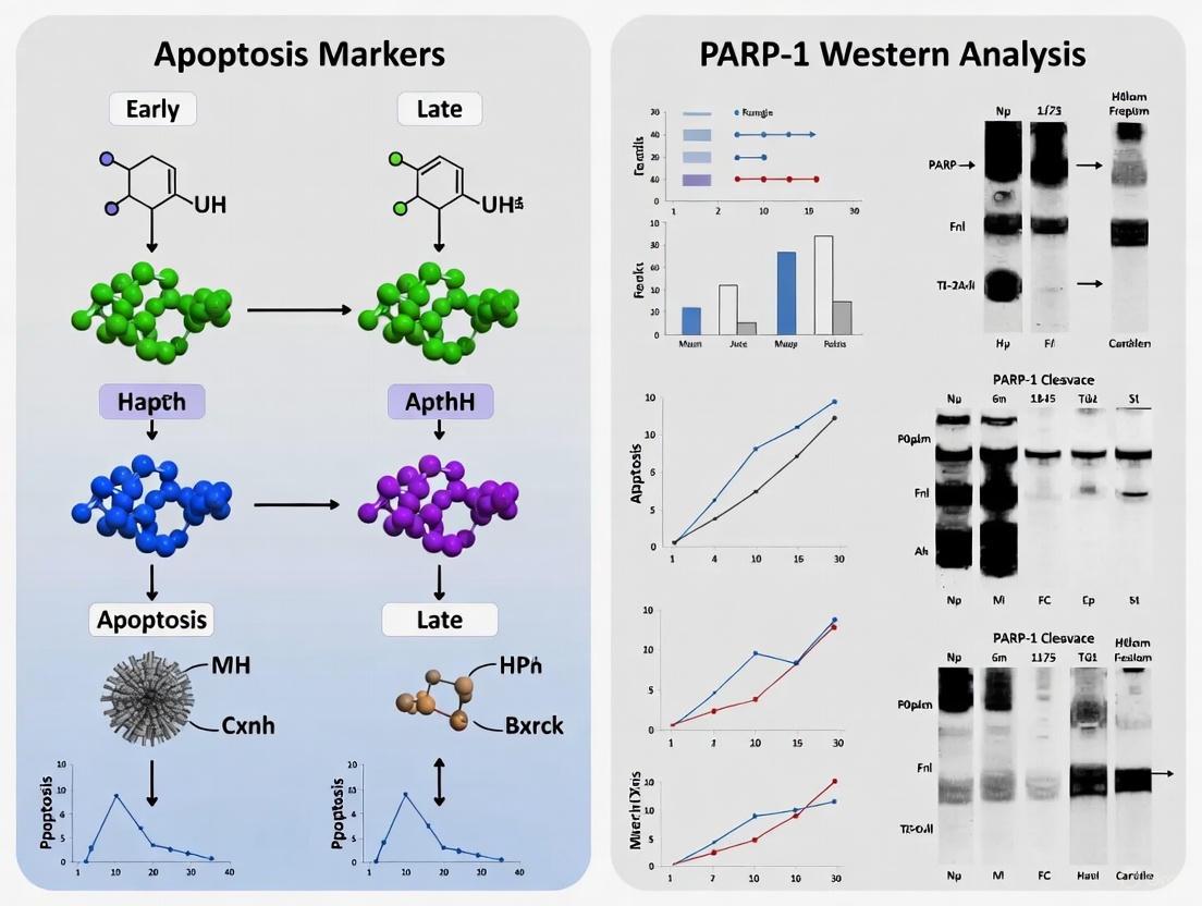
Detecting Apoptosis: A Comprehensive Guide to PARP-1 Cleavage Analysis by Western Blot
This article provides researchers, scientists, and drug development professionals with a definitive guide to using PARP-1 western blotting for the specific detection of early and late apoptosis.
Detecting Apoptosis: A Comprehensive Guide to PARP-1 Cleavage Analysis by Western Blot
Abstract
This article provides researchers, scientists, and drug development professionals with a definitive guide to using PARP-1 western blotting for the specific detection of early and late apoptosis. It covers the foundational role of PARP-1 cleavage as a central apoptotic biomarker, detailed methodological protocols for detection and quantification, common troubleshooting strategies to overcome challenges, and advanced validation techniques. The content also explores the cutting-edge context of PARP-1 in novel cell death pathways like ferroptosis and its relevance in evaluating cancer therapeutics, providing a complete resource for accurate apoptosis assessment in biomedical research.
PARP-1 as a Central Apoptotic Executor: From Molecular Function to Biomarker Selection
The Role of PARP-1 in DNA Repair and its Cleavage as an Apoptotic Point of No Return
Poly (ADP-ribose) polymerase 1 (PARP1) is a multifunctional nuclear enzyme that serves as a critical DNA damage sensor and facilitator of DNA repair processes. This 113 kDa protein accounts for approximately 90% of cellular PARP activity and plays essential roles in maintaining genomic integrity [1]. Beyond its DNA repair functions, PARP1 regulates transcription, chromatin remodeling, and inflammatory responses [2] [3] [4]. The cleavage of PARP1 by caspases during apoptosis represents a definitive biochemical marker of programmed cell death and is considered a point of no return in the apoptotic pathway [5] [6]. This application note examines the dual roles of PARP1 in DNA repair and apoptosis, with specific protocols for detecting its cleavage as a definitive marker of irreversible cell death commitment.
PARP1 Structure and Functional Domains
PARP1 contains three primary functional domains that dictate its cellular functions:
- DNA-binding domain (DBD): Located at the N-terminus, this domain contains two zinc finger motifs (Zn1 and Zn2) that recognize DNA strand breaks, plus a third zinc finger (Zn3) that regulates catalytic activity [3].
- Automodification domain (AMD): This central domain contains glutamate and lysine residues that serve as acceptors for ADP-ribose moieties, plus a BRCT domain that facilitates protein-protein interactions with DNA damage response proteins [5] [3].
- Catalytic domain (CD): Positioned at the C-terminus, this domain contains the conserved "PARP signature" sequence required for poly(ADP-ribose) (PAR) synthesis [3].
Table 1: PARP1 Domains and Their Functions
| Domain | Location | Key Features | Primary Functions |
|---|---|---|---|
| DNA-binding domain (DBD) | N-terminus (aa 1-372) | Two zinc fingers for DNA break recognition, nuclear localization signal | DNA damage sensor, strand break binding |
| Automodification domain (AMD) | Central region (aa 373-524) | BRCT domain, acceptor residues for ADP-ribose | Protein-protein interactions, auto-regulation |
| Catalytic domain (CD) | C-terminus (aa 525-1014) | WGR motif, PARP signature sequence | NAD+ binding, PAR synthesis |
PARP1's Role in DNA Repair Mechanisms
PARP1 functions as a first responder to DNA damage through multiple repair pathways [2] [1] [4]:
DNA Damage Sensing and Repair Initiation
Upon binding to DNA single-strand breaks (SSBs) or double-strand breaks (DSBs), PARP1 undergoes conformational changes that significantly enhance its catalytic activity [2]. This activation leads to auto-ribosylation and the synthesis of poly(ADP-ribose) (PAR) chains, which serve as recruitment signals for DNA repair proteins including XRCC1, which is crucial for base excision repair (BER) [2].
Chromatin Remodeling for Repair Accessibility
Through ADP-ribosylation of histones H1 and H2B, PARP1 promotes chromatin decompaction, enabling repair machinery to access damaged sites [2]. This function is particularly important for facilitating the recruitment of large protein complexes during DNA repair and transcription.
Specific DNA Repair Pathways
PARP1 contributes to several distinct DNA repair mechanisms:
- Base excision repair (BER): Primary pathway for single-strand break repair
- Nucleotide excision repair (NER): Removal of helix-distorting DNA lesions
- Double-strand break repair: Both non-homologous end joining (NHEJ) and microhomology-mediated end joining (MMEJ)
- Homologous recombination (HR): Error-free repair of double-strand breaks
- DNA mismatch repair: Correction of replication errors [1] [4]
Figure 1: PARP1-Mediated DNA Damage Response Pathway. PARP1 activation by DNA damage triggers auto-PARylation, chromatin remodeling, and recruitment of repair proteins to facilitate multiple DNA repair pathways.
PARP1 Cleavage as an Apoptotic Marker
Caspase-Mediated Cleavage: The Apoptotic Point of No Return
During apoptosis, PARP1 is cleaved by caspase-3 and caspase-7 at the DEVD214↓G215 motif located between the second and third zinc-binding domains [5] [6]. This proteolytic event generates two characteristic fragments:
- 24 kDa N-terminal fragment: Contains the DNA-binding domain and remains nuclear-bound
- 89 kDa C-terminal fragment: Contains the automodification and catalytic domains [5] [6]
This cleavage event serves as a biochemical hallmark of apoptosis and represents a commitment point in cell death pathways for several reasons:
- Irreversible inhibition of DNA repair: The 24 kDa fragment acts as a trans-dominant inhibitor of PARP1 by irreversibly binding to DNA strand breaks, preventing DNA repair enzymes from accessing damage sites [5].
- Conservation of cellular ATP: By inactivating PARP1's catalytic function, cells prevent NAD+ and ATP depletion, ensuring sufficient energy for the orderly execution of apoptosis [7].
- Disruption of PARP1's survival functions: Cleavage terminates PARP1's roles in DNA repair and cellular homeostasis, redirecting the cell toward death [6].
Alternative Proteolytic Processing in Cell Death
Beyond caspase-mediated cleavage, PARP1 serves as a substrate for other "suicidal proteases" in different cell death contexts:
- Calpains: Calcium-activated proteases involved in excitotoxicity and neuronal death
- Granzymes: Proteases released by cytotoxic lymphocytes in immune-mediated cell killing
- Cathepsins: Lysosomal proteases involved in autophagic cell death
- Matrix metalloproteinases (MMPs): Extracellular proteases with specific intracellular functions [5]
Each protease generates distinctive PARP1 cleavage fragments that serve as signature biomarkers for specific cell death pathways [5].
Table 2: PARP1 Cleavage Fragments in Different Cell Death Pathways
| Protease | Cleavage Sites | Fragment Sizes | Cell Death Context | Functional Consequences |
|---|---|---|---|---|
| Caspase-3/7 | DEVD214↓G215 | 24 kDa + 89 kDa | Apoptosis | Inactivation of DNA repair, energy conservation |
| Calpain | Multiple sites | 50 kDa + 62 kDa variants | Excitotoxicity, necrosis | Alternative regulation patterns |
| Granzyme A | Unknown | Unique fragments | Immune-mediated killing | Distinct from apoptotic cleavage |
| Cathepsins | Unknown | Unique fragments | Autophagic cell death | Lysosomal protease involvement |
| MMPs | Unknown | Unique fragments | Specific pathological contexts | Extracellular protease function |
Experimental Protocols for PARP1 Cleavage Detection
Western Blot Protocol for PARP1 Cleavage Analysis
Sample Preparation:
- Cell lysis: Use RIPA buffer (50 mM Tris-HCl pH 7.4, 150 mM NaCl, 1% NP-40, 0.5% sodium deoxycholate, 0.1% SDS) supplemented with protease inhibitor cocktail and 1 mM PMSF.
- Protein quantification: Perform BCA assay to normalize protein concentrations (20-40 μg per lane recommended).
- Sample denaturation: Heat samples at 95°C for 5 minutes in Laemmli buffer containing 5% β-mercaptoethanol.
Electrophoresis and Transfer:
- Gel electrophoresis: Use 8-12% SDS-PAGE gels for optimal separation of full-length (113 kDa) and cleaved (89 kDa) PARP1.
- Protein transfer: Transfer to PVDF membrane at 100V for 1 hour or 30V overnight at 4°C.
Immunoblotting:
- Blocking: Incubate membrane with 5% non-fat dry milk in TBST for 1 hour at room temperature.
- Primary antibody incubation: Use anti-PARP1 antibody (recognizing C-terminal epitope for detecting 89 kDa fragment) at manufacturer's recommended dilution in blocking buffer, overnight at 4°C.
- Washing: 3 × 10 minutes with TBST.
- Secondary antibody incubation: HRP-conjugated anti-rabbit or anti-mouse IgG (1:2000-1:5000) for 1 hour at room temperature.
- Detection: Use enhanced chemiluminescence (ECL) substrate and image with chemiluminescence detection system.
Controls and Validation:
- Include apoptosis-positive controls (e.g., staurosporine-treated cells at 1 μM for 4-6 hours)
- Use loading controls (β-actin, GAPDH, or histone H3 for nuclear proteins)
- Confirm specificity with PARP1 knockout cells or caspase inhibitors (e.g., Z-VAD-FMK at 20-50 μM)
Flow Cytometry Protocol for cPARP Detection
Cell Staining:
- Cell fixation: Use 4% paraformaldehyde for 15 minutes at room temperature.
- Permeabilization: Treat with 90% ice-cold methanol for 30 minutes on ice or 0.1% Triton X-100 for 10 minutes.
- Antibody staining: Incubate with anti-cleaved PARP1 (Asp214) antibody for 1 hour at room temperature.
- Secondary detection: Use FITC-conjugated secondary antibody for 30 minutes in the dark.
- Analysis: Analyze using flow cytometry with appropriate fluorescence channels.
Experimental Considerations:
- Include unstained and isotype controls for gating
- Use apoptosis inducers as positive controls (e.g., 10 μM staurosporine or 200 μM betulinic acid for 4 hours) [8]
- Consider density gradient separation for spermatozoa or specific cell types [8]
Figure 2: Western Blot Workflow for PARP1 Cleavage Detection. This protocol enables specific identification of full-length PARP1 (113 kDa) and the apoptotic cleavage fragment (89 kDa).
The Scientist's Toolkit: Essential Research Reagents
Table 3: Key Research Reagents for PARP1 Cleavage Studies
| Reagent Category | Specific Examples | Applications | Considerations |
|---|---|---|---|
| PARP1 Antibodies | Anti-PARP1 (C-terminal specific), Anti-cleaved PARP1 (Asp214) | Western blot, immunofluorescence, flow cytometry | Epitope recognition critical for detecting cleavage fragments |
| Apoptosis Inducers | Staurosporine (10 μM), Betulinic acid (200 μM), Etoposide (VP-16) | Positive controls for PARP1 cleavage | Concentration and exposure time optimization required |
| Caspase Inhibitors | Z-VAD-FMK (pan-caspase inhibitor, 20-50 μM) | Specificity controls for caspase-dependent cleavage | Pre-treatment (1-2 hours) before apoptosis induction |
| PARP Activity Assays | NAD+ consumption assays, PAR polymer detection | Functional assessment of PARP1 activation | Correlate with cleavage status |
| Cell Death Detection Kits | Annexin V/propidium iodide, caspase-3 activity assays | Multiparameter apoptosis analysis | Combine with PARP1 cleavage for comprehensive assessment |
| Positive Control Cells | Staurosporine-treated cells (4-6 hours, 1 μM) | Assay validation | Include in every experiment |
Interpretation Guidelines and Technical Considerations
Quantitative Analysis of PARP1 Cleavage
When interpreting PARP1 cleavage results, consider these key aspects:
- Cleavage ratio: Calculate the ratio of cleaved (89 kDa) to full-length (113 kDa) PARP1 for quantitative comparisons
- Temporal progression: Cleavage typically precedes other late apoptotic markers
- Cell-type variability: Baseline PARP1 expression varies significantly between cell types
Common Technical Challenges and Solutions
- Non-specific bands: Optimize antibody concentration and include appropriate controls
- Incomplete transfer: Verify transfer efficiency with reversible protein stains
- Signal saturation: Use multiple exposure times for ECL detection
- Nuclear localization: Consider subcellular fractionation for specific localization studies
PARP1 cleavage at aspartate 214 represents a definitive commitment point in apoptotic pathways, serving as both a functional regulator and reliable biomarker for programmed cell death. The detection of the characteristic 89 kDa fragment provides researchers with a specific tool for identifying apoptosis in experimental systems, with applications ranging from basic research to drug development. The protocols outlined herein enable robust detection and quantification of this critical apoptotic event, facilitating research into cell death mechanisms and therapeutic interventions targeting apoptotic pathways.
Biological Significance of PARP-1 Cleavage Fragments
Poly(ADP-ribose) polymerase-1 (PARP-1) is a nuclear enzyme that plays a central role in the cellular response to DNA damage. Upon activation by DNA strand breaks, it catalyzes the synthesis of poly(ADP-ribose) (PAR) chains on target proteins, facilitating DNA repair [9] [10]. Caspase-dependent cleavage of PARP-1 is a well-established hallmark of apoptosis and serves as a critical biochemical switch that shuts down DNA repair efforts and facilitates the dismantling of the cell [11] [10]. The cleavage occurs at a specific aspartic acid residue (Asp214) within a conserved nuclear localization signal sequence, mediated primarily by the effector caspases-3 and -7 [11] [12]. This proteolytic event generates two distinct fragments with different molecular weights and biological fates: a 24-kDa fragment and an 89-kDa fragment [9] [10].
The following diagram illustrates the caspase-3 mediated cleavage of PARP-1 and the divergent roles of the resulting fragments:
The table below summarizes the core characteristics and functions of these two key fragments:
| Feature | 24-kDa Fragment | 89-kDa Fragment |
|---|---|---|
| Domains Contained | DNA-binding domain (DBD) with two zinc fingers [10] | Auto-modification domain (AMD) and catalytic domain (CD) [10] |
| Cellular Localization | Retained in the nucleus [9] [13] | Translocates to the cytoplasm [9] [12] |
| Primary Function | Acts as a trans-dominant inhibitor of DNA repair by irreversibly binding to DNA strand breaks [10] | Serves as a cytoplasmic PAR carrier; induces AIF-mediated death (parthanatos) [9] [13] |
| Regulatory Role | Suppresses PARP-1 activity and conserves cellular ATP [11] [10] | Can mediate mono-ADP-ribosylation of cytoplasmic targets (e.g., RNA Polymerase III) [12] |
Detection and Analysis of PARP-1 Cleavage
Western Blot Protocol for Apoptosis Detection
The detection of PARP-1 cleavage by western blot is a fundamental technique for confirming apoptosis in experimental models. The following workflow provides a robust method for researchers.
Key Markers and Antibodies for Apoptosis Detection
Beyond PARP-1, a comprehensive analysis of apoptosis should include other key markers. The table below lists essential reagents for detecting PARP-1 cleavage and related apoptotic events.
| Research Reagent | Function/Application in Apoptosis Detection |
|---|---|
| Anti-PARP-1 Antibody | Detects both full-length (116 kDa) and the 89-kDa cleavage fragment; some antibodies are specific to the cleaved form [14] |
| Anti-Cleaved Caspase-3 Antibody | Detects activated caspase-3, the primary enzyme executing PARP-1 cleavage; confirms upstream apoptotic signal [14] |
| Anti-AIF Antibody | Detects apoptosis-inducing factor, which is released in PAR-mediated parthanatos cell death [9] [15] |
| Caspase Inhibitor (e.g., zVAD-fmk) | Pan-caspase inhibitor used as a control to confirm caspase-dependent apoptosis and PARP-1 cleavage [11] |
| Apoptosis Antibody Cocktails | Pre-mixed solutions containing multiple antibodies (e.g., against caspase-3, PARP, Bcl-2) for efficient and simultaneous detection of several apoptotic markers [14] |
| Chemiluminescent Substrate | Used with HRP-conjugated secondary antibodies for visualization of protein bands on western blots [14] |
Data Interpretation and Quantification
Accurate interpretation of western blot data is crucial for validating apoptosis.
- Band Pattern Analysis: A definitive sign of apoptosis is the disappearance of the 116 kDa band (full-length PARP-1) and the concomitant appearance of the 89 kDa band (cleavage fragment) [14] [10]. The 24-kDa fragment is less commonly detected in standard western blots.
- Quantification: Use densitometry software (e.g., ImageJ) to measure band intensities. Calculate the ratio of cleaved PARP-1 (89 kDa) to total PARP-1 (full-length + cleaved) or to a loading control. This ratio provides a quantitative measure of the extent of apoptosis in the sample [14].
- Normalization: Always normalize the signals for PARP-1 and its cleavage products to a housekeeping protein such as β-actin or GAPDH to account for variations in protein loading and transfer efficiency [14].
Advanced Research Applications and Protocols
Investigating the 89-kDa Fragment in Parthanatos
The 89-kDa fragment is not merely an inert byproduct of cleavage. Recent research reveals its active role in coordinating other forms of programmed cell death, particularly parthanatos, a caspase-independent pathway [9] [13].
Experimental Protocol to Study AIF Translocation:
- Induce Apoptosis: Treat cells (e.g., neuronal cell lines) with a DNA-damaging agent such as staurosporine (1 μM) or actinomycin D (0.5 μM) for 4-16 hours to trigger caspase activation and PARP-1 cleavage [9].
- Fractionate Cells: At the end of the treatment, harvest cells and separate cytoplasmic and nuclear fractions using a commercial cell fractionation kit.
- Western Blot Analysis: Probe the cytoplasmic fractions for the 89-kDa PARP-1 fragment and AIF. The co-presence of both proteins in the cytoplasm indicates the activation of the parthanatos pathway [9] [13].
- Immunofluorescence Validation: Fix treated cells and perform double-label immunofluorescence using antibodies against the 89-kDa PARP-1 fragment and AIF. The co-localization of these signals in the cytoplasm provides visual confirmation of the pathway [9].
A Novel Protocol: Assessing tPARP1 in Innate Immune Activation
A groundbreaking study revealed that the 89-kDa truncated PARP-1 (tPARP1) can regulate the innate immune response during apoptosis induced by cytoplasmic DNA [12].
Detailed Methodology:
- Cell Transfection and Apoptosis Induction:
- Co-Immunoprecipitation (Co-IP):
- Lyse cells and incubate the supernatant with an antibody against tPARP1.
- Pull down the immune complexes with protein A/G beads.
- Wash the beads and elute the bound proteins.
- Analyze the eluates by western blotting for subunits of the Pol III complex (e.g., POLR3A, POLR3B) to confirm interaction [12].
- Functional Assays:
- IFN-β Production: Measure IFN-β mRNA levels via RT-PCR or protein levels via ELISA in cell supernatants after poly(dA-dT) transfection.
- Apoptosis Quantification: Use flow cytometry with Annexin V-FITC and propidium iodide staining to quantify the percentage of apoptotic cells [12].
The table below summarizes key quantitative findings from recent studies on PARP-1 fragment functions:
| Experimental Context | Key Finding | Significance |
|---|---|---|
| Staurosporine/Acitnomycin D treatment [9] | The 89-kDa fragment, with covalently attached PAR, translocates to the cytoplasm. | Links caspase-mediated apoptosis to AIF-dependent parthanatos. |
| Poly(dA-dT)-induced apoptosis [12] | tPARP1 interacts with and mono-ADP-ribosylates the Pol III complex via its BRCT domain. | Reveals a novel pro-apoptotic role for tPARP1 in innate immune activation. |
| Oxygen/Glucose Deprivation (OGD) [16] | Expression of the 89-kDa fragment was cytotoxic, while the 24-kDa fragment was protective. | Highlights the opposing biological activities of the two fragments in ischemia models. |
| TNF-induced necrosis [11] | Prevention of PARP-1 cleavage by caspase inhibition (zVAD) promotes necrotic cell death. | Establishes PARP-1 cleavage as a molecular switch between apoptosis and necrosis. |
The Scientist's Toolkit
| Essential Material | Function | Specific Example/Application |
|---|---|---|
| Anti-PARP-1 Antibody | Primary antibody for western blot to detect full-length and cleaved PARP-1. | Antibodies recognizing both the 116 kDa and 89 kDa bands are essential for assessing the cleavage ratio [14]. |
| Caspase-3 Antibody | Detects the executor caspase responsible for PARP-1 cleavage; confirms apoptosis initiation. | Using antibodies against both the pro-form and cleaved, active form of caspase-3 strengthens the evidence for apoptosis [14]. |
| PARP Inhibitor (e.g., DPQ) | Chemical inhibitor of PARP-1 catalytic activity; used as a control to dissect PAR-dependent and independent functions. | Helps differentiate between PARP-1's role in DNA repair (inhibited by DPQ) and its function as a cleavage substrate [15]. |
| Caspase Inhibitor (zVAD-fmk) | Pan-caspase inhibitor; used to confirm that PARP-1 cleavage is caspase-dependent. | Pre-treatment with zVAD-fmk should abolish the appearance of the 89 kDa fragment in apoptotic samples [11]. |
| Apoptosis Inducers | Positive control agents to trigger apoptosis and PARP-1 cleavage in experimental systems. | Staurosporine and Actinomycin D are well-characterized inducers of caspase-dependent apoptosis and PARP-1 cleavage [9]. |
Poly(ADP-ribose) polymerase-1 (PARP-1) cleavage serves as a critical biochemical hallmark of apoptosis, representing a key downstream event in the caspase activation cascade. This application note details the molecular relationship between caspase-3/7 activation and PARP-1 proteolysis, providing optimized methodologies for detecting these events in apoptotic research. We present comprehensive data on the specific cleavage fragments generated, their cellular functions, and detailed Western blot protocols for simultaneous detection of caspase activity and PARP-1 processing. The information presented herein enables researchers to accurately interpret PARP-1 cleavage patterns as indicators of both early and late apoptotic stages, with particular utility for drug development screening and mechanistic studies of cell death pathways.
PARP-1 is a 116-kDa nuclear enzyme primarily involved in DNA repair and genomic maintenance, utilizing NAD+ to catalyze poly(ADP-ribosyl)ation of target proteins in response to DNA damage [11]. During apoptosis, PARP-1 undergoes specific proteolytic cleavage that serves as a reliable biomarker for programmed cell death. This cleavage event represents a crucial point of convergence in the apoptotic cascade, effectively halting DNA repair processes while facilitating the cell's dismantling [10].
The proteolysis of PARP-1 is predominantly executed by the effector caspases-3 and -7, which recognize and cleave a conserved DEVD214-Gly215 motif within the PARP-1 DNA-binding domain [16]. This cleavage event generates two characteristic fragments: a 24-kDa fragment containing the DNA-binding domain and an 89-kDa fragment comprising the automodification and catalytic domains [10]. The separation of these functional domains represents a molecular switch that contributes to the irreversibility of the apoptotic process by simultaneously inactivating DNA repair capacity and conserving cellular ATP pools that would otherwise be depleted by PARP-1 activation [11].
Beyond its role as a caspase substrate, emerging evidence indicates that PARP-1 cleavage products may actively participate in signaling pathways. The 89-kDa fragment translocates to the cytoplasm where it can function as a poly(ADP-ribose) carrier and participate in alternative cell death pathways, including parthanatos [9]. Recent research has also identified novel roles for truncated PARP-1 in mediating ADP-ribosylation of RNA polymerase III during innate immune responses [12]. These findings underscore the functional significance of PARP-1 cleavage beyond its established role as a mere apoptotic marker.
Molecular Relationship: Caspase-3/7 and PARP-1 Cleavage
Specificity and Kinetics of Cleavage
Caspase-3 and caspase-7, as executioner caspases, demonstrate distinct yet complementary roles in PARP-1 proteolysis during apoptosis. Both caspases recognize the DEVD214↓Gly215 cleavage site in PARP-1, but exhibit differential affinities influenced by PARP-1's modification state. Caspase-7 shows enhanced cleavage efficiency toward automodified PARP-1, while caspase-3 activity appears less affected by the PARP-1 modification state [17]. This specificity is mediated through caspase-7's affinity for poly(ADP-ribose) polymers, which facilitates its interaction with automodified PARP-1 [17].
The temporal sequence of caspase activation and PARP-1 cleavage positions PARP-1 proteolysis as a mid-to-late apoptotic event. Following caspase-3/7 activation, PARP-1 cleavage occurs rapidly, with the 89-kDa fragment appearing concurrently with maximal caspase activity [17]. The nuclear accumulation of caspase-7 during apoptosis further ensures efficient PARP-1 processing [17].
Table 1: PARP-1 Cleavage Fragments Generated by Caspase-3/7
| Fragment Size | Domains Contained | Cellular Localization | Functional Consequences |
|---|---|---|---|
| 24 kDa | Zinc finger DNA-binding domains (N-terminal) | Nuclear retention | Acts as trans-dominant inhibitor of DNA repair; occupies DNA strand breaks |
| 89 kDa | BRCT, WGR, Catalytic domain (C-terminal) | Cytoplasmic translocation | May retain catalytic activity; functions as PAR carrier; mediates AIF release |
Functional Consequences of PARP-1 Cleavage
The proteolytic cleavage of PARP-1 serves multiple critical functions in the apoptotic cascade:
Energy Conservation: Prevents NAD+ and ATP depletion that would occur from PARP-1 activation in response to apoptotic DNA fragmentation, thereby maintaining energy-dependent apoptotic processes [11].
DNA Repair Inactivation: The 24-kDa fragment binds irreversibly to DNA strand breaks, acting as a trans-dominant inhibitor that blocks access for DNA repair enzymes [10].
Apoptotic Progression Facilitation: Cleavage ensures the irreversibility of cell death by preventing DNA repair attempts that could otherwise subvert the apoptotic program [11].
Recent studies have revealed additional signaling functions of the cleavage fragments. The 89-kDa fragment can translocate to the cytoplasm where it facilitates apoptosis-inducing factor (AIF) release from mitochondria, bridging caspase-dependent apoptosis and parthanatos [9]. Additionally, truncated PARP-1 mediates ADP-ribosylation of RNA polymerase III, potentially linking apoptosis to innate immune responses [12].
Experimental Detection and Analysis
Western Blot Protocol for PARP-1 Cleavage Detection
Sample Preparation
- Harvest cells and lyse in RIPA buffer (Thermo Fisher Scientific, #89900) supplemented with protease inhibitors [18].
- Determine protein concentration using BCA assay (Thermo Fisher Scientific, #23225) [18].
- Prepare samples with 2X Laemmli buffer, heat at 95°C for 5 minutes, and load 10-30 μg protein per lane [18].
Gel Electrophoresis and Transfer
- Use 8-12% SDS-PAGE gels for optimal separation of full-length and cleaved PARP-1 fragments [18].
- Transfer to nitrocellulose membrane (0.2 μm pore size, Cytiva #10600001) using standard wet or semi-dry transfer systems [18].
- Confirm transfer efficiency with Ponceau S staining (Merck, #P7170) [18].
Antibody Probing and Detection
- Block membranes with 5% non-fat milk in TBST for 1 hour at room temperature [18].
- Incubate with primary antibodies in one of the following configurations:
- Conventional Method: 10 mL antibody solution in TBST with 5% milk, overnight at 4°C with agitation [18].
- Sheet Protector Strategy (Antibody Conservation): Place membrane between sheet protector leaflets with 20-150 μL primary antibody solution, incubate at room temperature (15 minutes to 2 hours) without agitation [18].
- Recommended primary antibodies:
- PARP (Cell Signaling Technology, #9542) - detects full-length and 89-kDa fragment
- Cleaved PARP (Asp214) (Cell Signaling Technology, #5625) - specific to cleaved form
- Caspase-3 (Cell Signaling Technology, #14220)
- Cleaved Caspase-3 (Asp175) (Cell Signaling Technology, #9664)
- Wash membranes 3× with TBST, 5 minutes per wash
- Incubate with HRP-conjugated secondary antibodies (1:2000-1:5000) for 1 hour at room temperature
- Detect using chemiluminescent substrates (e.g., WesternBright Quantum, Advansta #K-12045-D50) and imaging system [18]
Controls and Validation
- Include apoptosis-positive controls such as:
- Normalize signals to housekeeping proteins (GAPDH, β-actin, or α-tubulin)
- Use densitometry software (ImageJ) for quantification of cleavage ratios [14]
Data Interpretation Guidelines
Band Pattern Analysis
- Full-length PARP-1 (116 kDa): Indicates minimal caspase activity; predominant in healthy cells
- 89-kDa fragment: Confirms caspase-mediated cleavage; appears during early-mid apoptosis
- 24-kDa fragment: Often difficult to detect by standard Western due to small size and potential degradation
Quantitative Assessment
- Calculate cleavage ratio: (intensity of 89-kDa fragment) / (intensity of full-length + 89-kDa fragment)
- Normalize to loading controls to account for protein loading variations
- Relate PARP-1 cleavage to caspase activation by simultaneous detection of:
- Caspase-3/7 proforms (inactive zymogens)
- Cleaved caspase fragments (activated forms)
Table 2: Key Antibodies for Apoptosis Detection via Western Blot
| Target | Antibody Example | Detection Purpose | Appearance in Apoptosis |
|---|---|---|---|
| Full-length PARP-1 | PARP Antibody (CST #9542) | Baseline PARP-1 expression | Decreases with progression |
| Cleaved PARP-1 (Asp214) | Cleaved PARP (Asp214) (D64E10) XP Rabbit mAb (CST #5625) | Specific detection of apoptosis-related cleavage | Increases with progression |
| Caspase-3 | Caspase-3 (D3R6Y) Rabbit mAb (CST #14220) | Pro-caspase-3 levels | Proform decreases |
| Cleaved Caspase-3 | Cleaved Caspase-3 (Asp175) (D3E9) Rabbit mAb (CST #9664) | Activated caspase-3 | Appears and increases |
| Caspase-7 | Caspase-7 Antibody (CST #9492) | Pro-caspase-7 levels | Proform decreases |
| Loading Control | GAPDH, β-actin, or α-tubulin antibodies | Normalization reference | Should remain constant |
Research Reagent Solutions
The following essential reagents facilitate reliable detection of PARP-1 cleavage and caspase activation in apoptosis research:
Table 3: Essential Research Reagents for PARP-1 Cleavage Studies
| Reagent | Supplier/Example | Application | Key Features |
|---|---|---|---|
| Jurkat Apoptosis Cell Extracts (etoposide) | Cell Signaling Technology (#2043) | Positive control for apoptosis markers | Contains full-length and cleaved PARP-1, caspases |
| Caspase-3 Control Cell Extracts | Cell Signaling Technology (#9663) | Caspase activation control | Cytochrome c-treated; contains cleaved caspase-3, -9 |
| PARP Antibody | Cell Signaling Technology (#9542) | Detects full-length and 89-kDa fragment | Rabbit polyclonal; works in WB, IP |
| Cleaved PARP (Asp214) Antibody | Cell Signaling Technology (#5625) | Specific for cleaved form | Rabbit monoclonal; specific to apoptosis-related cleavage |
| Caspase-3 (D3R6Y) Rabbit mAb | Cell Signaling Technology (#14220) | Detects pro and cleaved forms | Rabbit monoclonal; works in WB, IF, FC |
| HRP-conjugated Secondary Antibodies | Various suppliers | Signal detection | Anti-rabbit and anti-mouse options |
| Chemiluminescent Substrate | WesternBright Quantum (Advansta #K-12045-D50) | Western blot detection | High sensitivity, prolonged signal |
Technical Considerations and Troubleshooting
Optimal Sample Collection Timing PARP-1 cleavage is a transient event that requires careful timing of sample collection. For drug-induced apoptosis studies, conduct time-course experiments with sampling at 2-24 hours post-treatment, as cleavage kinetics vary by cell type and apoptotic stimulus [19]. Simultaneously monitor caspase activation to establish the temporal relationship.
Fragment Detection Challenges The 24-kDa PARP-1 fragment is frequently undetectable in standard Western blots due to:
- Rapid degradation following cleavage
- Poor transfer or retention on membranes
- Limited antibody epitopes in small fragment Focus instead on the disappearance of full-length PARP-1 (116 kDa) and appearance of the 89-kDa fragment as primary indicators of cleavage [10].
Alternative Cleavage Contexts While caspase-mediated PARP-1 cleavage typically indicates apoptosis, note that other proteases (calpains, cathepsins, granzymes, MMPs) can generate different PARP-1 fragments under specific pathological conditions [10]. These alternative fragments (50-60 kDa, 40-55 kDa, 35-40 kDa) may indicate non-apoptotic cell death pathways and should be distinguished from canonical caspase-generated fragments.
Visualizing the Caspase-PARP-1 Axis
The following diagrams illustrate the key molecular relationships and experimental workflow for detecting PARP-1 cleavage in apoptosis research.
Caspase-Mediated PARP-1 Cleavage Pathway
Experimental Workflow for Detection
The integration of PARP-1 cleavage analysis into apoptosis research provides a critical window into the activation status of executioner caspases and the commitment to cell death. The detailed protocols and analytical frameworks presented herein enable researchers to accurately detect and interpret PARP-1 cleavage patterns in conjunction with caspase-3/7 activation. As research continues to reveal novel functions for PARP-1 cleavage fragments in cell death signaling pathways, the methodologies outlined in this application note will support further investigation into the complex regulatory networks governing apoptotic progression. The combination of optimized detection protocols, appropriate controls, and careful data interpretation ensures reliable assessment of this key apoptotic event in both basic research and drug development contexts.
Poly(ADP-ribose) polymerase-1 (PARP1) is a multifunctional nuclear protein with well-established roles in DNA damage repair and the regulation of apoptotic cell death. Traditionally, detection of PARP1 cleavage via Western blot has served as a definitive marker for caspase-dependent apoptosis in research settings [14]. However, emerging evidence reveals that PARP1's functions extend far beyond apoptosis, encompassing novel roles in regulating ferroptosis and immunogenic cell death (ICD) [20] [21] [22]. This expansion of PARP1's biological significance necessitates updated experimental frameworks for researchers investigating cell death mechanisms. This Application Note details the latest methodologies and mechanistic insights for studying PARP1 in these non-apoptotic cell death pathways, providing essential context for interpreting Western blot results within a broader cell death signaling network.
PARP-1 at the Crossroads of Cell Death Signaling
The traditional view of PARP1 in cell death was relatively straightforward: in response to severe DNA damage, PARP1 activation could lead to energy depletion and necrotic cell death, while its cleavage by caspases (resulting in 24 kDa and 89 kDa fragments) was a hallmark of apoptosis [14] [22]. Recent research has uncovered a more complex picture, positioning PARP1 as a critical node in a network of interconnected cell death pathways. The diagram below illustrates PARP1's central role in coordinating these diverse cellular responses.
PARP-1 in Ferroptosis Regulation
Mechanistic Insights
Ferroptosis is an iron-dependent form of regulated cell death characterized by uncontrolled lipid peroxidation, distinct from apoptosis in both morphology and biochemistry [23] [24]. Recent studies have established a compelling mechanistic link between PARP1 activity and ferroptosis induction. The primary connection points are:
- SLC7A11 Repression: PARP inhibition downregulates the expression of SLC7A11, the core component of the system Xc- cystine/glutamate antiporter, in a p53-dependent manner. This repression limits cystine uptake, depleting glutathione (GSH) and disabling the glutathione peroxidase 4 (GPX4) antioxidant defense, ultimately leading to lethal lipid peroxidation [21].
- Alternative Pathways: Beyond SLC7A11 regulation, niraparib can induce ferroptosis by upregulating the fatty acid transporter CD36, promoting dysregulated fatty acid uptake and lipid peroxidation in ovarian cancer cells independently of p53 and BRCA status [24].
- Bidirectional Regulation: Interestingly, the ferroptosis inducer RSL3 can promote PARP1's apoptotic functions through distinct mechanisms, including caspase-dependent PARP1 cleavage and METTL3-mediated reduction of PARP1 translation, demonstrating complex bidirectional crosstalk between these pathways [22].
Quantitative Evidence Base
The table below summarizes key experimental findings that establish the relationship between PARP1 activity and ferroptosis regulation.
Table 1: Key Experimental Evidence for PARP1's Role in Ferroptosis
| Experimental Context | Key Finding | Proposed Mechanism | Citation |
|---|---|---|---|
| BRCA-proficient ovarian cancer cells | Olaparib sensitizes to ferroptosis inducers | p53-dependent SLC7A11 repression and GSH depletion | [21] |
| Ovarian cancer (in vitro/vivo) | Niraparib triggers ferroptosis and suppresses metastasis | Transcriptional upregulation of fatty acid transporter CD36 | [24] |
| Multiple cancer cell lines | RSL3 induces PARP1 cleavage and reduces full-length PARP1 | Caspase-3 activation and METTL3-mediated translational suppression | [22] |
| PARP inhibitor-resistant tumors | RSL3 maintains pro-apoptotic function in resistant cells | ROS-mediated PARP1 regulation bypasses traditional resistance | [22] |
Essential Research Reagents
The table below outlines critical reagents for investigating PARP1-ferroptosis crosstalk, with specific examples from recent literature.
Table 2: Essential Research Reagents for Studying PARP1-Ferroptosis Crosstalk
| Reagent Category | Specific Examples | Research Application | Key Function | |
|---|---|---|---|---|
| PARP Inhibitors | Olaparib, PJ34, AZD9574 | Inhibit PARP1 catalytic activity; induce ferroptosis | Study PARP1's role in SLC7A11 regulation and lipid metabolism | [25] [21] [26] |
| Ferroptosis Inducers | RSL3, Erastin | Trigger ferroptosis through distinct mechanisms | Investigate ferroptosis-PARP1 feedback loops | [24] [22] |
| Ferroptosis Inhibitors | Ferrostatin-1, Liproxstatin-1 | Suppress lipid peroxidation | Confirm ferroptosis-specific phenotypes | [22] |
| Apoptosis Inhibitors | Z-VAD-FMK (pan-caspase inhibitor) | Block apoptotic signaling | Differentiate apoptosis from ferroptosis | [25] [20] |
| Antibodies for Detection | Anti-PARP1 (cleaved/full length), Anti-SLC7A11, Anti-GPX4 | Western blot analysis | Monitor PARP1 processing and ferroptosis markers | [21] [14] [22] |
PARP-1 in Immunogenic Cell Death
Mechanistic Framework
Immunogenic cell death represents a functionally unique form of cell death that activates adaptive immune responses against dead cell-associated antigens, particularly relevant to cancer therapy [23] [20]. The emerging role of PARP1 in ICD regulation involves:
- DAMP Regulation: Natural compounds like Macrocarpal I, which directly targets PARP1, induce robust ICD characterized by the exposure of calreticulin on the cell surface and release of ATP and HMGB1 - key damage-associated molecular patterns that recruit and activate antigen-presenting cells [20].
- ER Stress Involvement: PARP1 inhibition contributes to ICD through activation of the PERK/eIF2α/ATF4/CHOP signaling pathway, a key endoplasmic reticulum stress response that links cellular damage to immune recognition [20].
- Synergy with Checkpoint Inhibition: PARP1-targeting agents can overcome resistance to immune checkpoint inhibitors (e.g., anti-PD-1) in immunologically "cold" tumors, suggesting their utility in combinatorial immunotherapy approaches [20].
The following diagram illustrates the experimental workflow for detecting PARP1's role in ICD, integrating Western blot analysis with functional immune assays.
Integrated Protocols for PARP-1 Analysis in Cell Death
Comprehensive Western Blot Protocol for PARP-1 and Cell Death Markers
Sample Preparation:
- Lyse cells in RIPA buffer supplemented with PARP inhibitor (to prevent auto-PARylation during processing) and protease/phosphatase inhibitors [25] [14].
- Quantify protein concentration using BCA assay; load 20-30 μg per lane for SDS-PAGE [14] [22].
Gel Electrophoresis and Transfer:
- Use 4-12% Bis-Tris gradient gels for optimal resolution of full-length PARP1 (116 kDa) and its 89 kDa apoptotic fragment [14].
- Transfer to PVDF membranes using standard wet transfer systems.
Antibody Detection:
- Block membranes with 5% non-fat milk in TBST for 1 hour at room temperature [25] [14].
- Incubate with primary antibodies in blocking solution overnight at 4°C:
- Wash and incubate with appropriate HRP-conjugated secondary antibodies (1:5000) for 1 hour at room temperature [25].
- Visualize using enhanced chemiluminescence and quantify band intensity using densitometry software (e.g., ImageJ) [14].
Critical Controls:
- Include apoptosis inducers (e.g., staurosporine) as positive controls for PARP1 cleavage.
- Use ferroptosis inducers (e.g., RSL3, erastin) with and without ferroptosis inhibitors as controls for SLC7A11 detection [21] [22].
Complementary Functional Assays
Ferroptosis-Specific Assessment:
- Measure lipid peroxidation using C11-BODIPY 581/591 probe via flow cytometry [24] [21].
- Quantify intracellular GSH levels using commercial GSH/GSSG assay kits [21].
- Assess cell viability in the presence of ferroptosis inhibitors (e.g., ferrostatin-1) to confirm ferroptosis contribution [22].
ICD-Specific Assessment:
- Detect surface calreticulin by flow cytometry or immunofluorescence 16-24 hours post-treatment [20].
- Measure extracellular ATP using luciferase-based assays in cell culture supernatants [20].
- Quantify HMGB1 release in supernatants via ELISA or Western blot [20].
The evolving understanding of PARP1's functions in ferroptosis and immunogenic cell death significantly expands its utility as a biomarker and therapeutic target beyond traditional apoptosis contexts. Researchers interpreting PARP1 Western blot data must now consider this broader regulatory landscape, where PARP1 cleavage may represent just one facet of a complex cell death response. The integrated methodologies presented here provide a framework for dissecting PARP1's multifaceted roles in cell death signaling, enabling more comprehensive mechanistic studies and therapeutic development in cancer and other diseases characterized by dysregulated cell death.
Poly (ADP-ribose) polymerase-1 (PARP-1) is a 113-116 kDa nuclear enzyme that plays a fundamental role in DNA repair and maintenance of genomic integrity [16] [10]. During the early stages of apoptosis, PARP-1 becomes a key substrate for executioner caspases (primarily caspase-3 and -7), which cleave the full-length protein at the conserved aspartate residue 214 into characteristic fragments of approximately 89 kDa and 24 kDa [10] [27] [11]. This proteolytic event is widely recognized as a biochemical hallmark of apoptosis, serving as a critical marker for researchers distinguishing between cell death pathways. The detection of these specific cleavage fragments, particularly the 89 kDa C-terminal fragment, provides invaluable evidence of caspase activation and commitment to apoptotic cell death. The selection of antibodies with precise specificity for either the full-length protein or its cleavage products is therefore paramount for accurate interpretation of Western blot data in experimental models of cell death, drug efficacy, and neurodegenerative diseases [28] [10].
PARP-1 Biology and Cleavage Significance in Cell Death
Domain Architecture and Cleavage Sites
PARP-1 is organized into three primary functional domains: a DNA-binding domain (DBD) containing two zinc fingers at the N-terminus, a central auto-modification domain (AMD), and a C-terminal catalytic domain (CD) responsible for poly(ADP-ribose) polymerization [10]. During apoptosis, caspase-3 and -7 cleave the protein within the nuclear localization signal of the DBD at the DEVD214↓G motif [16] [13]. This cleavage event separates the N-terminal 24 kDa fragment (containing the DBD) from the C-terminal 89 kDa fragment (containing the AMD and CD), effectively inactivating the enzyme's DNA repair capacity and facilitating the apoptotic process [10].
Functional Consequences of Cleavage
The cleavage of PARP-1 serves as a critical molecular switch in cell fate determination:
- Inactivation of DNA Repair: The separation of the DNA-binding domain from the catalytic domain halts PARP-1's DNA repair functions, conserving cellular ATP and NAD+ pools that would otherwise be depleted by excessive PARP-1 activation [11].
- Modulation of Cell Death Pathways: The 24 kDa fragment remains bound to DNA breaks, potentially acting as a trans-dominant inhibitor of DNA repair [10]. Recent research indicates the 89 kDa fragment may translocate to the cytoplasm, potentially functioning as a carrier of poly(ADP-ribose) (PAR) polymers and contributing to parthanatos, a caspase-independent programmed cell death pathway [13].
- Differential Effects on Viability: Studies demonstrate that while the full-length PARP-1 and the 89 kDa fragment can promote cell death under certain conditions, an uncleavable PARP-1 mutant (PARP-1UNCL) and the 24 kDa fragment can confer protection from ischemic damage in neuronal models [16].
The following diagram illustrates the PARP-1 cleavage process and its role in cell death pathways:
Antibody Specificity Guide for Apoptosis Detection
The critical application of PARP-1 antibodies in apoptosis research necessitates understanding their distinct target epitopes and resulting specificity. The table below summarizes the characteristics of representative antibodies based on commercial and research reagents.
Table 1: Characteristics of PARP-1 Antibodies for Apoptosis Detection
| Antibody Clone/ Name | Specificity | Recognized Bands | Epitope Location | Key Applications |
|---|---|---|---|---|
| HLNC4 [27] | Cleaved PARP-1 only | 85-89 kDa fragment only | Around Asp214 cleavage site | Specific detection of apoptosis |
| Y34 [28] | Cleaved PARP-1 only | ~85 kDa fragment | Proprietary (cleavage-specific) | WB, IF, IP, Flow Cytometry |
| Polyclonal 13371-1-AP [29] | Full-length & cleaved forms | 113-116 kDa & 89 kDa fragments | C-terminal region (667-1014 aa) | Total PARP-1 detection |
| Polyclonal 200-401-x51 [30] | Full-length (C-Term) | 113 kDa full-length | C-terminal region | DNA damage research |
Antibodies targeting the C-terminal region of PARP-1 (e.g., 13371-1-AP, 200-401-x51) typically recognize both the full-length protein and the 89 kDa cleavage fragment, providing a view of total PARP-1 expression but requiring careful interpretation to distinguish intact versus cleaved protein [29] [30]. In contrast, cleavage-specific antibodies (e.g., HLNC4, Y34) are engineered to recognize the neo-epitope created only after caspase cleavage at Asp214, providing definitive evidence of apoptosis without cross-reactivity with the full-length protein [28] [27].
Experimental Protocols for PARP-1 Detection
Western Blot Protocol for Apoptosis Detection
Cell Treatment and Lysis:
- Induction of Apoptosis: Treat cells (e.g., Jurkat, HeLa) with apoptosis inducers: 1-4 μM camptothecin for 5 hours [28], 1 μM staurosporine for 4 hours [28], or 1 μM etoposide for 16 hours [27]. Include untreated controls.
- Cell Lysis: Harvest cells and lyse in RIPA buffer supplemented with protease and phosphatase inhibitors. Maintain samples on ice throughout.
- Protein Quantification: Determine protein concentration using a Bradford or BCA assay. Prepare aliquots with Laemmli buffer containing β-mercaptoethanol.
Gel Electrophoresis and Transfer:
- Gel Preparation: Cast 8-12% SDS-polyacrylamide gels to optimally resolve proteins in the 25-250 kDa range.
- Sample Preparation: Denature 20-40 μg of total protein per sample at 95°C for 5 minutes [28] [27].
- Electrophoresis: Load samples and molecular weight markers. Run at constant voltage (100-120V) until the dye front reaches the bottom.
- Protein Transfer: Transfer to PVDF membrane using wet or semi-dry transfer systems at 100V for 60-90 minutes.
Immunoblotting:
- Blocking: Incubate membrane in 5% non-fat dry milk or BSA in TBST for 1 hour at room temperature.
- Primary Antibody Incubation: Dilute antibodies in blocking buffer as follows:
- Washing: Wash membrane 3×10 minutes with TBST.
- Secondary Antibody Incubation: Incubate with appropriate HRP-conjugated secondary antibody (1:2000-1:5000) for 1 hour at room temperature.
- Detection: Develop blots using ECL reagent and image with chemiluminescence detection system.
Experimental Design Considerations
Controls are critical for proper interpretation:
- Positive Control: Include lysates from cells treated with known apoptosis inducers (e.g., camptothecin-treated Jurkat cells) [28].
- Negative Control: Use untreated cells or cells where apoptosis has been inhibited with caspase inhibitors (e.g., zVAD) [11].
- Loading Control: Probe for housekeeping proteins (GAPDH, β-actin, or tubulin) to normalize protein loading.
- Specificity Control: When using cleavage-specific antibodies, include PARP-1 knockout cell lysates if available [28].
Troubleshooting Tips:
- If signal is weak, optimize antibody concentration and increase exposure time.
- If background is high, increase number of washes and optimize blocking conditions.
- If cleaved band is not detected, confirm apoptosis induction with additional markers (e.g., caspase-3 cleavage).
Data Interpretation Guidelines
Quantitative Analysis of Apoptosis
Western blot data for PARP-1 cleavage should be analyzed both qualitatively and quantitatively. Densitometric analysis of band intensities allows for calculation of the cleavage ratio, a quantitative measure of apoptosis extent.
Table 2: Expected Molecular Weights and Biological Significance of PARP-1 Forms
| PARP-1 Form | Molecular Weight | Biological Significance | Detection Antibody Type |
|---|---|---|---|
| Full-length | 113-116 kDa | DNA repair active, cell viability | Total PARP-1 antibodies |
| Cleaved (89 kDa) | 85-89 kDa | Caspase activation, apoptosis execution | Cleaved & total PARP-1 antibodies |
| Cleaved (24 kDa) | 24 kDa | Caspase activation, DNA binding | Specialized N-terminal antibodies |
The cleavage ratio can be calculated as: Cleaved PARP-1 / (Cleaved PARP-1 + Full-length PARP-1). This ratio provides a quantitative measure of apoptosis extent in the population. A ratio >0.5 typically indicates significant commitment to apoptotic cell death.
Pathway Integration
PARP-1 cleavage should not be interpreted in isolation but as part of a comprehensive apoptotic signaling cascade. The following diagram illustrates the integration of PARP-1 cleavage within broader cell death pathways:
Research Reagent Solutions
Table 3: Essential Research Reagents for PARP-1 Apoptosis Studies
| Reagent Category | Specific Examples | Research Application |
|---|---|---|
| Cleavage-specific Antibodies | Anti-PARP1 (cleaved Asp214) HLNC4 [27], Anti-Cleaved PARP1 [Y34] [28] | Specific detection of apoptotic cells via Western blot, flow cytometry |
| Total PARP-1 Antibodies | PARP1 Polyclonal (13371-1-AP) [29], Anti-PARP1 (C-Term) [30] | Detection of both full-length and cleaved PARP-1 |
| Apoptosis Inducers | Camptothecin [28], Staurosporine [28], Etoposide [27] | Positive controls for PARP-1 cleavage experiments |
| PARP Inhibitors | Olaparib [31], PJ34, Veliparib | Investigate PARP-1 function in DNA repair and cell death |
| Caspase Inhibitors | zVAD-fmk [11] | Negative control to suppress PARP-1 cleavage |
| Model Cell Lines | Jurkat (human T-cell leukemia) [28] [27], HeLa (cervical adenocarcinoma) [28] [27] | Well-characterized models for apoptosis studies |
The precise detection of PARP-1 cleavage fragments requires antibodies with well-characterized specificity for either the full-length protein or the caspase-generated neo-epitopes. Cleavage-specific antibodies provide definitive evidence of apoptosis, while antibodies recognizing total PARP-1 offer a comprehensive view of protein expression and processing. The experimental protocols outlined herein, when implemented with appropriate controls and interpretation guidelines, enable robust detection of this critical apoptotic marker. As research continues to elucidate the complex roles of PARP-1 fragments in different cell death pathways [13] [10], the strategic selection of antibodies remains fundamental to advancing our understanding of cell death mechanisms in health and disease.
Step-by-Step Protocol: Optimizing Western Blot for PARP-1 Cleavage Detection
Sample Preparation Strategies for Preserving PARP-1 Cleavage Fragments
Poly (ADP-ribose) polymerase-1 (PARP-1) is a 116 kDa nuclear enzyme that plays a fundamental role in the cellular response to DNA damage, primarily through its involvement in DNA repair pathways [5]. During the early stages of apoptosis, PARP-1 becomes a primary substrate for executioner caspases (caspase-3 and -7), which cleave the protein at a specific aspartic acid residue (Asp214) to generate two characteristic fragments: a 24-kDa DNA-binding domain fragment and an 89-kDa catalytic domain fragment [32] [5]. This proteolytic cleavage event serves as a critical biochemical marker for distinguishing apoptosis from other forms of cell death, as it inactivates PARP-1's DNA repair function and facilitates the systematic dismantling of the cell [14] [5]. The 89-kDa fragment has recently been found to translocate to the cytoplasm, where it can participate in additional signaling events, including facilitating apoptosis-inducing factor (AIF) release and interacting with the RNA polymerase III complex to potentiate immune responses [33] [12]. Preserving these specific cleavage fragments during sample preparation is therefore paramount for accurate interpretation of apoptotic signaling in research and drug development contexts.
PARP-1 Cleavage Fragments: Significance and Detection
Characteristics of Major PARP-1 Cleavage Fragments
The cleavage of PARP-1 by caspases represents a definitive commitment to apoptotic cell death. The generated fragments possess distinct cellular localizations and functions that extend beyond the mere inactivation of DNA repair.
Table 1: Key PARP-1 Cleavage Fragments and Their Biological Significance
| Fragment Size | Domains Contained | Cellular Localization Post-Cleavage | Primary Functions |
|---|---|---|---|
| 24 kDa | Two zinc-finger DNA-binding motifs, Nuclear Localization Signal (NLS) [33] | Retained in nucleus [5] | Irreversibly binds DNA strand breaks; acts as a trans-dominant inhibitor of full-length PARP-1, preventing DNA repair and conserving cellular ATP [5] [34] |
| 89 kDa | BRCT domain, WGR domain, Catalytic domain (lacks the first two zinc fingers) [33] [12] | Translocates from nucleus to cytoplasm [33] | Can be auto-poly(ADP-ribosyl)ated; acts as a carrier for PAR polymers to the cytoplasm, facilitating AIF-mediated DNA fragmentation [33]; can mono-ADP-ribosylate RNA Pol III to promote innate immune signaling [12] |
Proteases Beyond Caspases
While caspase-3 and -7 are the primary proteases responsible for generating the classic 24-kDa and 89-kDa fragments, other "suicidal" proteases can cleave PARP-1 under specific pathological conditions, yielding fragments of different sizes. These include calpains, cathepsins, granzymes, and matrix metalloproteinases (MMPs), which can produce PARP-1 fragments ranging from 42-89 kDa [5] [35]. The presence of these alternative fragments can indicate the activation of unique cell death programs, such as those involving calcium dysregulation (calpains) or lysosomal permeabilization (cathepsins). Therefore, a well-preserved sample that captures the full spectrum of potential fragments is crucial for accurate mechanistic insight.
Critical Considerations for Sample Preparation
The lability of proteolytic fragments and the rapid, dynamic nature of apoptotic signaling necessitate a sample preparation strategy that prioritizes speed, low temperatures, and comprehensive inhibition of post-lysis proteolysis.
Fundamental Principles for Preserving Fragments
- Pre-cool and Work Rapidly: All procedures must be performed on ice or at 4°C using pre-chilled buffers to instantly quench enzymatic activity. The time from cell disruption to complete lysis should be minimized.
- Inhibit Multiple Protease Classes: Apoptotic samples may contain active caspases, calpains, and other proteases. Broad-spectrum protease inhibitors are essential, but including specific caspase inhibitors (e.g., Z-VAD-FMK) can prevent continued cleavage of PARP-1 and other substrates after lysis, which could otherwise lead to overestimation of apoptotic activity.
- Prevent Phosphatase Activity: While the focus is on cleavage, preserving phosphorylation status is often important for parallel signaling analysis. Include phosphatase inhibitors in your lysis buffer.
- Ensure Efficient Lysis and Homogenization: Use a lysis buffer with sufficient detergent (e.g., 1% NP-40 or SDS) to fully disrupt the nucleus and solubilize chromatin-bound proteins, ensuring quantitative recovery of the 24-kDa fragment that remains tightly bound to damaged DNA.
Quantitative Data on Apoptotic Inducers and Detection
Researchers can utilize various chemical and physical inducers to trigger apoptosis and study PARP-1 cleavage. The table below summarizes common agents and the expected experimental outcomes based on published research.
Table 2: Experimental Inducers of Apoptosis and PARP-1 Cleavage Outcomes
| Inducer / Context | Mechanism of Action | Key Caspase Activated | Expected PARP-1 Cleavage Outcome | Supporting Evidence |
|---|---|---|---|---|
| Staurosporine [33] | Broad-spectrum protein kinase inhibitor; intrinsic apoptosis | Caspase-3 | Generation of 89-kDa and 24-kDa fragments; PAR synthesis; AIF translocation | Cell Death & Differentiation, 2020 [33] |
| RSL3 (Ferroptosis Inducer) [34] | Inhibits GPX4; triggers ROS-dependent apoptosis | Caspase-3 | Caspase-dependent PARP1 cleavage; also reduces full-length PARP1 via translational suppression | Cell Death and Differentiation, 2025 [34] |
| Ionizing Radiation [36] | Causes severe DNA damage; promotes STING-PAR interaction | Caspase-3 | Increased PARP1 cleavage; enhanced by STING presence | Cell Death & Differentiation, 2025 [36] |
| Poly(dA-dT) Transfection [12] | Mimics pathogenic DNA; triggers innate immune apoptosis | Caspase-3 | Cleavage of PARP1; tPARP1 (89-kDa) interacts with Pol III in cytosol | Cell Research, 2021 [12] |
| Actinomycin D [33] | Inhibits transcription; induces intrinsic apoptosis | Caspase-3 | PARP1 autopoly(ADP-ribosyl)ation and fragmentation | Cell Death & Differentiation, 2020 [33] |
Detailed Experimental Protocols
Protocol 1: Rapid Lysis of Adherent Cells for PARP-1 Immunoblotting
This protocol is optimized for the simultaneous preservation of full-length PARP-1 (116 kDa) and its cleavage fragments (89 kDa being the most prominent).
Reagents and Solutions
- Lysis Buffer: 50 mM Tris-HCl (pH 7.5), 150 mM NaCl, 1% NP-40 (or 1% SDS for more stringent lysis).
- Protease Inhibitor Cocktail: Commercially available EDTA-free cocktail.
- Specific Caspase Inhibitor: Z-VAD-FMK (e.g., 20 µM final concentration).
- Phosphatase Inhibitor Cocktail.
- Lysis Buffer Working Solution: Prepare fresh by adding protease inhibitor cocktail, Z-VAD-FMK, and phosphatase inhibitor to the chilled lysis buffer.
Procedure
- Induce Apoptosis and at the desired time point, immediately remove culture media.
- Rinse Cells once with ice-cold 1X PBS.
- Place Culture Dish directly on ice.
- Add Lysis Buffer directly to the cells (e.g., 100-200 µL per 10 cm² dish). Scrape cells thoroughly and transfer the lysate to a pre-cooled microcentrifuge tube.
- Vortex briefly and incubate on ice for 15-30 minutes with occasional vortexing.
- Clarify Lysate by centrifugation at >12,000 × g for 15 minutes at 4°C.
- Transfer Supernatant to a new pre-cooled tube. Determine protein concentration immediately using a BCA or Bradford assay.
- Prepare Samples for Western Blotting by adding Laemmli sample buffer and heating at 95-100°C for 5-10 minutes. Avoid longer heating times to prevent protein degradation.
Protocol 2: Preparation of Samples from Tissues or 3D Models
Tissues and spheroids present additional challenges due to their structural complexity.
Procedure
- Rapid Dissection/Collection: Immediately after isolation, snap-freeze the tissue sample in liquid nitrogen. Store at -80°C until processing.
- Pulverization: Using a pre-cooled mortar and pestle (or a dedicated homogenizer), crush the frozen tissue to a fine powder under liquid nitrogen. Do not allow the tissue to thaw.
- Homogenization: Transfer the powdered tissue to a tube containing ice-cold lysis buffer (as in Protocol 1, but potentially with higher detergent concentration) and homogenize using a mechanical homogenizer (e.g., Dounce, Polytron) for 30-60 seconds on ice.
- Follow Steps 6-8 from Protocol 1 to complete the sample preparation.
The Scientist's Toolkit: Essential Research Reagents
Table 3: Key Reagent Solutions for PARP-1 Cleavage Analysis
| Reagent / Resource | Specific Example(s) | Function in Experiment | Experimental Note |
|---|---|---|---|
| PARP-1 Antibodies | CST #9542 [32]; PTGLab 13371-1-AP [35] | Detects full-length (116 kDa) and cleaved (89 kDa) PARP1 by Western Blot | #9542 is raised against the caspase cleavage site; 13371-1-AP targets the C-terminal region [32] [35] |
| Caspase Inhibitor | Z-VAD-FMK (pan-caspase inhibitor) [33] [36] | Added during lysis to prevent post-lysis cleavage and artifact generation | Critical for obtaining an accurate "snapshot" of cleavage at the moment of lysis |
| PARP Inhibitors | PJ34, ABT-888 (Olaparib) [33] [36] | Tool compounds to inhibit PARP1 catalytic activity; used to probe mechanism | PJ34 used to confirm PARP1-dependent cell death in staurosporine model [33] |
| Apoptosis Inducers | Staurosporine, Actinomycin D [33] | Positive control stimuli to trigger caspase-3 activation and PARP-1 cleavage | Staurosporine induces PAR synthesis and AIF translocation downstream of caspase [33] |
| Ferroptosis Inducers | RSL3 [34] | Induces apoptosis via ROS-dependent pathways, leading to PARP1 cleavage | Useful for studying crosstalk between ferroptosis and apoptosis [34] |
Visualizing Signaling Pathways and Workflows
Figure 1: PARP-1 Cleavage in Apoptotic Signaling. This diagram illustrates the central role of caspase-mediated PARP-1 cleavage in apoptosis, highlighting the distinct nuclear and cytoplasmic functions of the resulting 24-kDa and 89-kDa fragments that collectively facilitate cell death.
Figure 2: Sample Preparation Workflow for PARP-1 Fragment Preservation. This workflow emphasizes the critical steps and reagents, particularly the rapid operation and comprehensive inhibition of proteases, required to obtain an accurate snapshot of PARP-1 cleavage status.
Gel Electrophoresis Conditions for Resolving Full-Length (116-kDa) and Cleaved (89-kDa) PARP-1
Poly (ADP-ribose) polymerase-1 (PARP-1), a 116 kDa nuclear enzyme, plays a multifaceted role in cellular homeostasis, including DNA repair and the regulation of gene transcription [16] [10]. During the early stages of apoptosis, PARP-1 becomes a primary target for cleavage by executioner caspases-3 and -7. This proteolytic event occurs at a specific aspartic acid residue (Asp214) within a conserved caspase recognition sequence, generating a 24 kDa DNA-binding fragment and an 89 kDa catalytic fragment [37] [10]. The detection of the 89 kDa fragment, resulting from the separation of the PARP-1 catalytic domain from its DNA-binding domain, serves as a definitive biochemical hallmark for the onset of apoptosis [37] [33]. Consequently, Western blot analysis resolving the full-length (116 kDa) PARP-1 from its cleaved (89 kDa) product has become an indispensable technique for identifying and quantifying apoptotic events in diverse research contexts, from basic molecular biology to drug development in oncology and neurodegeneration. This application note provides a detailed protocol optimized for the clear resolution and specific detection of these key apoptotic markers.
PARP-1 Cleavage in Apoptosis Signaling Pathways
The cleavage of PARP-1 is a critical event in the execution of apoptosis. As illustrated in the pathway below, it connects caspase activation to the dismantling of the cellular machinery.
The 89-kDa fragment is not merely an inert byproduct of cleavage. Recent research has revealed its active role in amplifying cell death signals. This fragment, often with poly(ADP-ribose) (PAR) polymers still attached, can translocate from the nucleus to the cytoplasm [33] [13]. In the cytoplasm, it acts as a PAR carrier, facilitating the release of Apoptosis-Inducing Factor (AIF) from mitochondria. AIF then translocates to the nucleus, triggering large-scale DNA fragmentation in a caspase-independent cell death pathway known as parthanatos [33]. Therefore, detecting the 89-kDa fragment not only confirms apoptosis but may also indicate the engagement of this specific cell death subroutine.
Electrophoresis and Immunoblotting Conditions
Optimal resolution of the 116-kDa full-length PARP-1 and the 89-kDa cleavage fragment is critical for accurate interpretation. The table below summarizes the key parameters for a successful Western blot.
Table 1: Standardized Western Blot Conditions for PARP-1 Detection
| Parameter | Recommended Condition | Purpose & Rationale |
|---|---|---|
| Gel Type | SDS-PAGE (Tris-Glycine or Bis-Tris) | Denaturing protein separation based on molecular weight. |
| Gel Concentration | 8-12% resolving gel | Ideal range for resolving proteins between 50-150 kDa. A 10% gel provides excellent separation of 116-kDa and 89-kDa fragments. |
| Sample Preparation | Laemmli buffer, boiling for 5-10 minutes | Ensures complete denaturation and reduction of proteins. |
| Electrophoresis Buffer | Tris-Glycine-SDS (or compatible MOPS/MES) | Standard buffer for SDS-PAGE. |
| Protein Transfer | PVDF or Nitrocellulose membrane | PVDF is preferred for its high binding capacity and mechanical strength. |
| Primary Antibody | PARP Antibody (#9542, Cell Signaling Technology) [37] | A well-characterized rabbit monoclonal antibody that detects endogenous levels of full-length PARP-1 (116 kDa) and the large cleavage fragment (89 kDa). |
| Antibody Dilution | 1:1000 in 5% BSA/TBST [37] | Provides specific signal with low background. |
| Secondary Antibody | Anti-Rabbit IgG, HRP-linked | For chemiluminescent detection. |
| Detection Method | Chemiluminescent substrate (e.g., SuperSignal West Pico PLUS) [38] | High-sensitivity detection for endogenous protein levels. |
Critical Notes for Resolution
- Positive Control: Always include a lysate from apoptotic cells (e.g., cells treated with 1µM Staurosporine for 4-6 hours) to serve as a positive control for the 89-kDa fragment [33].
- Molecular Weight Marker: Use a pre-stained protein standard to accurately monitor electrophoresis progress and confirm the molecular weights of the detected bands [38].
- Gel Percentage: While a 10% gel is standard, a 4-20% gradient gel can also provide superb resolution across a broader molecular weight range, ensuring sharp, distinct bands for both fragments.
Step-by-Step Experimental Protocol
Sample Preparation from Cultured Cells
- Lyse Cells: Wash cells with ice-cold PBS and lyse directly in an appropriate lysis buffer (e.g., RIPA buffer) supplemented with protease and phosphatase inhibitors. Keep samples on ice.
- Quantify Protein: Determine protein concentration of the clarified supernatants using a standard assay like BCA or Bradford.
- Prepare Samples: Dilute an equal amount of total protein (20-40 µg) with Laemmli sample buffer to a 1X final concentration.
- Denature Samples: Heat samples at 95-100°C for 5-10 minutes to fully denature proteins, then briefly centrifuge.
Gel Electrophoresis
- Set Up Gel Apparatus: Assemble the gel electrophoresis unit and fill the tank with 1X Tris-Glycine-SDS running buffer.
- Load Samples: Carefully load equal amounts of protein (including molecular weight markers and positive controls) into the wells of the pre-cast or hand-cast gel.
- Run Gel: Apply a constant voltage: 80 V through the stacking gel, then 120 V through the resolving gel until the dye front reaches the bottom.
Western Blotting
- Activate PVDF Membrane: Briefly immerse PVDF membrane in 100% methanol for 15-30 seconds, then transfer to transfer buffer.
- Transfer Proteins: Using a wet transfer system, transfer proteins at 100 V (constant voltage) for 60-90 minutes (or 30 V overnight) at 4°C.
- Block Membrane: After transfer, incubate the membrane in 5% w/v non-fat dry milk or BSA in TBST for 1 hour at room temperature with gentle agitation.
- Incubate with Primary Antibody: Dilute the PARP Antibody (#9542) to 1:1000 in 5% BSA/TBST. Incubate the membrane with the primary antibody with gentle shaking overnight at 4°C.
- Wash Membrane: Wash the membrane 3 times for 5-10 minutes each with TBST.
- Incubate with Secondary Antibody: Incubate with an HRP-linked anti-rabbit secondary antibody (typically 1:2000-1:10000) in 5% milk/TBST for 1 hour at room temperature.
- Wash Membrane: Repeat the washing step as after the primary antibody.
- Detect Signal: Incubate the membrane with a chemiluminescent substrate according to the manufacturer's instructions and visualize using a digital imager or X-ray film.
Troubleshooting and Data Interpretation
The workflow below outlines the key steps for performing the experiment and addressing potential issues.
Expected Results and Interpretation
- Healthy Cells: A dominant band at ~116 kDa, with little to no band at 89 kDa.
- Apoptotic Cells: A clear and distinct band at ~89 kDa. The intensity of this band relative to the 116 kDa band typically increases with the extent of apoptosis.
- The 24-kDa Fragment: Note that the 24-kDa fragment is often not detected in standard Western blots. Its small size may run off the gel, or the epitope recognized by the antibody may be lost depending on the antibody used.
The Scientist's Toolkit: Key Research Reagents
Table 2: Essential Reagents for PARP-1 Western Blotting
| Reagent / Resource | Function / Role | Example & Specification |
|---|---|---|
| PARP-1 Primary Antibody | Specifically binds to full-length and cleaved PARP-1. | PARP Antibody #9542 (Cell Signaling Technology). Rabbit mAb, reacts with Human, Mouse, Rat, Monkey [37]. |
| Caspase Inhibitors | To experimentally suppress PARP-1 cleavage; used for negative controls. | zVAD-fmk (pan-caspase inhibitor). Validates caspase-dependence of cleavage [33]. |
| PARP Inhibitors | To inhibit PARP-1 enzymatic activity; used to study functional consequences. | PJ34, ABT-888 (Veliparib), Olaparib. Useful for investigating parthanatos [39] [33]. |
| Apoptosis Inducers | To generate positive control lysates for the 89-kDa fragment. | Staurosporine, Actinomycin D. Well-characterized inducers of caspase-dependent apoptosis and PARP-1 cleavage [33] [13]. |
| Chemiluminescent Substrate | For sensitive detection of HRP-conjugated secondary antibodies. | SuperSignal West Pico PLUS or similar high-sensitivity substrates [38]. |
Optimal Transfer and Blocking Conditions for PARP-1 Immunodetection
Poly(ADP-ribose) polymerase-1 (PARP-1) is a nuclear enzyme with essential functions in DNA repair and maintenance of genomic integrity. During apoptosis, PARP-1 serves as a primary substrate for caspases, with its cleavage representing a definitive biochemical marker for programmed cell death. The proteolytic cleavage of PARP-1 by executioner caspases generates characteristic fragments of 89 kDa and 24 kDa, which serve as signatures for distinguishing apoptosis from other forms of cell death [5]. Detection of these specific cleavage fragments via Western blotting provides critical information about the timing and extent of apoptotic signaling in experimental systems, making optimal immunodetection protocols essential for researchers investigating cell death mechanisms, cancer biology, and therapeutic responses.
This application note details standardized methodologies for the reliable detection of both full-length PARP-1 and its apoptosis-specific cleavage fragments, with particular emphasis on transfer efficiency, antibody selection, and detection parameters tailored to the context of a broader thesis on early and late apoptosis markers.
PARP-1 Biology and Apoptotic Significance
Structural Domains and Cleavage Patterns
PARP-1 is a modular protein comprising three primary functional domains:
- A DNA-binding domain (DBD) containing two zinc finger motifs that facilitate recognition of DNA strand breaks
- An automodification domain (AMD) that serves as a target for poly(ADP-ribosyl)ation
- A catalytic domain (CD) that mediates poly(ADP-ribose) polymerization using NAD+ as a substrate [5]
During apoptosis, caspase-3 and caspase-7 cleave PARP-1 at the conserved sequence DEVD²¹⁴G, located within the AMD, producing an 89 kDa fragment containing the catalytic domain and a 24 kDa fragment containing the DNA-binding domain [5]. This cleavage event separates the DNA-binding and catalytic functions of PARP-1, resulting in inactivation of its DNA repair capacity and conservation of cellular energy pools for the apoptotic process.
Biological Consequences of PARP-1 Cleavage
The 24 kDa fragment retains the zinc finger motifs and remains tightly bound to DNA strand breaks, where it functions as a trans-dominant inhibitor of DNA repair by blocking access of additional DNA repair enzymes to damaged sites [5]. The 89 kDa catalytic fragment, while largely inactive, may translocate to the cytosol. This cleavage event represents an irreversible commitment to apoptotic cell death and serves as a reliable indicator of caspase activation in experimental systems.
Diagram 1: PARP-1 cleavage pathway during apoptosis. During apoptosis, activated caspase-3 and caspase-7 cleave full-length PARP-1 (113 kDa) at aspartate residue 214, generating characteristic 89 kDa and 24 kDa fragments. The 24 kDa DNA-binding fragment remains tightly associated with DNA damage sites, inhibiting DNA repair and committing the cell to apoptotic death.
Experimental Protocols for PARP-1 Immunodetection
Sample Preparation and Electrophoresis
Cell Lysis and Protein Extraction
- Harvest cells and wash with ice-cold PBS
- Lyse cells in RIPA buffer (50 mM Tris-HCl pH 7.4, 150 mM NaCl, 1% NP-40, 0.5% sodium deoxycholate, 0.1% SDS) supplemented with protease inhibitors (1 mM PMSF, 10 μg/mL aprotinin, 10 μg/mL leupeptin) and phosphatase inhibitors (1 mM Na₃VO₄, 1 mM NaF)
- Incubate on ice for 30 minutes with occasional vortexing
- Centrifuge at 14,000 × g for 15 minutes at 4°C
- Collect supernatant and determine protein concentration using BCA assay
- Prepare samples with 2× Laemmli buffer (125 mM Tris-HCl pH 6.8, 4% SDS, 20% glycerol, 0.02% bromophenol blue) containing 5% β-mercaptoethanol
- Denature samples at 95°C for 5 minutes
Electrophoresis Conditions
- Load 20-50 μg of protein per well on 4-12% Bis-Tris polyacrylamide gels
- Run in 1× MES-SDS or MOPS-SDS running buffer at constant voltage (150-200V) until dye front reaches bottom of gel
- Include pre-stained protein molecular weight markers for accurate size determination
Optimal Transfer Conditions for PARP-1 Detection
Western Blot Transfer Protocol
- Following electrophoresis, equilibrate gel in transfer buffer for 15 minutes
- Prepare PVDF membrane by activating in 100% methanol for 1 minute, then equilibrate in transfer buffer
- Assemble transfer stack in the following order: cathode, filter paper, gel, membrane, filter paper, anode
- Transfer using wet tank system at constant current (300 mA) for 90 minutes at 4°C
- Alternatively, use semi-dry transfer system at constant voltage (15-25V) for 60 minutes
Transfer Buffer Formulations
- Towbin Buffer: 25 mM Tris, 192 mM glycine, 20% methanol, pH 8.3
- Bjerrum Buffer: 48 mM Tris, 39 mM glycine, 20% methanol, 0.0375% SDS
- CAPS Buffer: 10 mM CAPS, 10% methanol, pH 11.0 (optimal for high molecular weight proteins)
Blocking and Immunodetection
Blocking Conditions
- Following transfer, block membrane in 5% non-fat dry milk in TBST (Tris-buffered saline with 0.1% Tween-20) for 1 hour at room temperature with gentle agitation
- Alternative blocking buffers: 3-5% BSA in TBST for phospho-specific antibodies
Antibody Incubation
- Incubate membrane with primary antibody diluted in blocking buffer or TBST overnight at 4°C with gentle agitation
- Recommended primary antibody dilutions:
- Anti-PARP-1 (full length and cleaved): 1:1000 dilution
- Anti-cleaved PARP-1 (Asp214): 1:1000 dilution
- Anti-β-actin (loading control): 1:5000 dilution
- Wash membrane 3× for 10 minutes each with TBST
- Incubate with species-appropriate HRP-conjugated secondary antibody diluted 1:5000 in blocking buffer for 1 hour at room temperature
- Wash membrane 3× for 10 minutes each with TBST
Detection and Imaging
- Develop blots using enhanced chemiluminescence (ECL) substrate
- Expose to X-ray film or capture using digital imaging system
- Optimize exposure times to avoid saturation and ensure linear detection range
Critical Parameters for Quantitative PARP-1 Western Blotting
Validation of Quantitative Western Blot Conditions
To ensure accurate quantification of PARP-1 cleavage fragments, several critical validation steps must be implemented as part of a systematic approach to quantitative Western blot analysis [40].
Linearity and Dynamic Range Assessment
- Perform serial dilutions of protein samples to establish the linear range of detection for both full-length PARP-1 (113 kDa) and the 89 kDa cleavage fragment
- Ensure that band intensities fall within the linear range of the detection system for accurate quantification
- Use appropriate loading controls (β-actin, GAPDH, or tubulin) to normalize for protein loading variations
Antibody Validation
- Validate antibody specificity using PARP-1 knockout cell lines or siRNA-mediated PARP-1 knockdown
- Confirm detection of appropriate molecular weight species: 113 kDa (full-length), 89 kDa (caspase-cleaved fragment)
- Test multiple antibody clones from different commercial sources for consistency
Membrane Re-probing Protocol
- Strip membranes using mild stripping buffer (15 g glycine, 1 g SDS, 10 mL Tween-20 in 1L, pH 2.2) for 10-15 minutes at room temperature
- Wash extensively with TBST before re-blocking and re-probing
- Confirm complete antibody removal before re-probing by incubating with ECL substrate
Troubleshooting Common Issues
Poor Transfer Efficiency
- Optimize methanol concentration in transfer buffer (15-20% for proteins >80 kDa)
- Extend transfer time for high molecular weight proteins
- Confirm complete transfer using reversible protein stains (Ponceau S)
Non-specific Bands
- Titrate primary antibody to optimal concentration
- Increase stringency of washes (increase salt concentration to 500 mM NaCl or add 0.5% SDS)
- Use more specific blocking agents (casein, fish skin gelatin)
High Background
- Ensure sufficient blocking time (minimum 1 hour)
- Reduce primary antibody concentration or incubation time
- Increase number and duration of washes
Research Reagent Solutions for PARP-1 Detection
Table 1: Essential reagents for PARP-1 immunodetection in apoptosis research
| Reagent Category | Specific Product/Composition | Function in PARP-1 Detection | Optimization Notes |
|---|---|---|---|
| Primary Antibodies | Anti-PARP-1 (full length), Anti-cleaved PARP-1 (Asp214) | Detection of full-length and apoptotic fragments | Validate using PARP-1 knockout controls; optimal dilution typically 1:1000 |
| Cell Lysis Buffer | RIPA buffer (50 mM Tris-HCl pH 7.4, 150 mM NaCl, 1% NP-40, 0.5% sodium deoxycholate, 0.1% SDS) | Efficient extraction of nuclear and cleaved PARP-1 | Supplement with fresh protease inhibitors to prevent degradation |
| Blocking Solution | 5% non-fat dry milk in TBST | Reduction of non-specific antibody binding | BSA (3-5%) may be preferable for phospho-specific antibodies |
| Transfer Buffer | Towbin buffer (25 mM Tris, 192 mM glycine, 20% methanol) | Efficient transfer of PARP-1 (113 kDa) and fragments (89 kDa) | Methanol concentration critical for high molecular weight proteins |
| Detection System | Enhanced chemiluminescence (ECL) substrate | Sensitive detection of PARP-1 bands | Use high-sensitivity ECL for low-abundance cleavage fragments |
| Loading Control | β-actin, GAPDH, or tubulin antibodies | Normalization of protein loading | Select based on molecular weight separation from PARP-1 fragments |
Data Interpretation and Analysis
Quantification of PARP-1 Cleavage
Densitometric Analysis
- Capture digital images of Western blot membranes at multiple exposure times
- Use image analysis software to measure band intensities for full-length PARP-1 (113 kDa), cleaved PARP-1 (89 kDa), and loading control
- Calculate the ratio of cleaved PARP-1 to total PARP-1 for quantitative assessment of apoptosis:
- % PARP-1 Cleavage = [Intensity89kDa / (Intensity113kDa + Intensity89kDa)] × 100
- Normalize values to loading control to account for protein loading variations
Temporal Analysis of Apoptosis
- Analyze PARP-1 cleavage at multiple time points following apoptotic stimuli
- Early apoptosis: detection of initial 89 kDa fragment with minimal reduction in full-length PARP-1
- Late apoptosis: predominant 89 kDa fragment with minimal full-length PARP-1 remaining
Table 2: Interpretation of PARP-1 cleavage patterns in apoptosis
| PARP-1 Band Pattern | Full-length (113 kDa) | Cleaved (89 kDa) | Biological Interpretation | Additional Markers to Assess |
|---|---|---|---|---|
| Healthy Cells | Strong | Absent | Basal conditions without apoptotic activation | Normal caspase-3 activity, intact mitochondrial membrane potential |
| Early Apoptosis | Strong | Present | Initial caspase activation, reversible phase | Activated caspase-3, phosphatidylserine externalization |
| Mid Apoptosis | Moderate | Strong | Committed apoptotic signaling | Cytochrome c release, disrupted mitochondrial membrane potential |
| Late Apoptosis | Weak/Faint | Strong | Irreversible apoptotic execution | DNA fragmentation, loss of membrane integrity |
| Necrosis | Strong | Absent | Alternative cell death pathway | Plasma membrane rupture, no caspase activation |
Integration with Other Apoptosis Markers
For comprehensive analysis of apoptotic signaling, PARP-1 cleavage should be evaluated in conjunction with additional apoptosis markers:
Early Apoptosis Markers
- Caspase-3 activation (cleavage of procaspase-3 to active fragments)
- Phosphatidylserine externalization (Annexin V staining)
- Mitochondrial membrane potential disruption (JC-1 or TMRM staining)
Late Apoptosis Markers
- DNA fragmentation (TUNEL assay)
- Nuclear condensation (Hoechst or DAPI staining)
- Loss of plasma membrane integrity (propidium iodide inclusion)
Diagram 2: PARP-1 Western blot workflow. The complete experimental workflow for optimal detection of PARP-1 and its cleavage fragments includes sample preparation, electrophoresis, transfer, blocking, antibody incubations, detection, and quantitative analysis. Each step requires optimization for reliable detection of apoptosis-specific cleavage fragments.
Reliable detection of PARP-1 cleavage fragments requires meticulous optimization of transfer conditions, antibody selection, and detection parameters. The protocols detailed in this application note provide a standardized approach for quantifying PARP-1 cleavage as a definitive marker of apoptotic progression. Implementation of these methodologies within the broader context of apoptosis marker analysis will enable researchers to accurately stage apoptotic signaling and assess therapeutic responses in experimental systems. The critical importance of validating transfer efficiency and antibody specificity cannot be overstated, as these parameters directly impact the sensitivity and reproducibility of PARP-1 cleavage detection in apoptosis research.
Apoptosis, a form of programmed cell death, occurs in a controlled manner essential for maintaining cellular balance, eliminating damaged or unnecessary cells without causing harm to surrounding tissue [14]. This process is characterized by specific morphological changes including cell shrinkage, chromatin condensation, DNA fragmentation, and membrane blebbing [14]. Apoptosis proceeds through two primary signaling pathways: the extrinsic pathway (initiated by extracellular death signals) and the intrinsic pathway (triggered by internal cellular stress) [14]. Both pathways converge to activate executioner caspases, particularly caspase-3 and caspase-7, which systematically dismantle cellular components through proteolytic cleavage of key structural and regulatory proteins [14] [41].
Among the critical substrates of executioner caspases is poly(ADP-ribose) polymerase 1 (PARP1), a nuclear enzyme involved in DNA damage repair [34] [14]. During apoptosis, caspase-3 cleaves the 116-kDa full-length PARP1 into characteristic 24-kDa and 89-kDa fragments [34]. The 89-kDa fragment, in particular, is translocated from the nucleus to the cytoplasm where it directly induces caspase-mediated DNA fragmentation and apoptosis [34]. This cleavage event serves as a definitive biochemical marker of apoptotic commitment, making PARP1 cleavage detection a gold standard for apoptosis confirmation in research and drug development contexts [14]. Validating antibody specificity for these apoptotic forms is therefore essential for accurate interpretation of cell death mechanisms in experimental systems.
Principles of Antibody Validation for Apoptotic Forms
The Critical Importance of Validation
Antibody validation for apoptotic forms ensures that detected signals genuinely represent specific cleavage events rather than non-specific binding or cross-reactivity with unrelated proteins. This is particularly crucial when studying cell death mechanisms because multiple signaling pathways can coexist and intersect within the same cellular context [34] [20] [41]. For instance, recent research has revealed crosstalk between ferroptosis and apoptosis, where the ferroptosis activator RSL3 was found to trigger PARP1 cleavage through caspase-3 activation while simultaneously reducing full-length PARP1 through epitranscriptomic regulation [34]. Without proper antibody validation, researchers might misinterpret these complex regulatory mechanisms.
The specificity of antibodies targeting cleaved forms of apoptotic markers must be rigorously established because commercial antibodies can vary considerably in their recognition profiles. Antibodies against cleaved PARP1 should detect only the 89-kDa fragment and not the full-length protein, while caspase-3 antibodies should distinguish between the pro-form (inactive) and cleaved form (active) [14]. Proper validation becomes especially important when investigating novel cell death inducers or combination therapies, such as natural compounds like Macrocarpal I which simultaneously targets both tubulin and PARP1 to induce immunogenic cell death [20].
Key Validation Parameters
Comprehensive antibody validation for apoptotic forms should address several critical parameters, with specificity being paramount. Researchers must confirm that antibodies specifically recognize their intended targets without cross-reacting with other proteins of similar molecular weights or related epitopes. Sensitivity is another crucial factor, determining the lowest detectable concentration of the apoptotic marker, which is particularly important for detecting early apoptosis when cleavage fragments are present in low abundance [14].
Additional validation parameters include reproducibility across experimental replicates, linearity of detection across a range of protein concentrations, and determination of the optimal antibody dilution that maximizes signal-to-noise ratio [14]. For apoptosis markers, it is also essential to confirm that antibody recognition depends on the specific cleavage event, such as the caspase-mediated cleavage of PARP1 after aspartic acid residues [34] [14]. This often requires testing antibodies against both positive controls (apoptosis-induced samples) and negative controls (samples treated with caspase inhibitors) to establish the specificity of the detection [41].
Experimental Protocols for Antibody Validation
Sample Preparation for Apoptosis Induction
Proper sample preparation begins with inducing apoptosis using well-characterized agents. Staurosporine (0.5-1 μM for 4-6 hours) and carfilzomib (0.1-0.5 μM for 8-16 hours) serve as effective apoptosis inducers for most cell lines [41]. For positive controls targeting PARP1 cleavage specifically, treat cells with these inducers and include a set pre-treated with pan-caspase inhibitor Z-VAD-FMK (20-50 μM for 1-hour pre-incubation) to confirm caspase dependence [34] [41].
Prepare cell lysates using RIPA buffer supplemented with protease and phosphatase inhibitors. For apoptosis studies, include 1 mM PMSF and caspase inhibitor cocktails in negative control samples. Maintain consistent protein concentrations (1-2 mg/mL) across all samples, and quantify using BCA assay [14]. Aliquot and store lysates at -80°C to prevent protein degradation and preserve cleavage fragments. Include molecular weight markers specifically covering the range of 15-250 kDa to properly resolve both full-length and cleaved forms of apoptotic proteins [42].
Western Blot Setup and Electrophoresis
Load 20-30 μg of total protein per lane for optimal detection of both abundant and rare cleavage products. For apoptosis markers, use 4-20% gradient gels to ensure proper resolution of size differences between full-length and cleaved forms [14]. Include both induced and non-induced samples on the same gel to enable direct comparison, and always run replicates to assess variability.
After electrophoresis, transfer proteins to PVDF membranes using standard wet transfer systems (100 V for 60-90 minutes) [14]. Confirm transfer efficiency and equal loading through total protein staining using methods like No-Stain Protein Labeling Reagent or similar fluorescent total protein stains [43] [42]. This total protein normalization (TPN) approach is increasingly preferred over housekeeping proteins like GAPDH or β-actin, as their expression can vary with experimental conditions including apoptosis induction [43].
Antibody Incubation and Specificity Testing
The antibody incubation process requires careful optimization to ensure specific detection of apoptotic forms. The following protocol outlines a standardized approach:
Blocking: Incubate membrane with 5% non-fat dry milk or BSA in TBST for 1 hour at room temperature with gentle agitation [14].
Primary Antibody Incubation: Dilute primary antibodies in blocking solution according to manufacturer's recommended concentrations. For cleaved PARP1, typical dilutions range from 1:1000 to 1:2000; for cleaved caspase-3, 1:500 to 1:1000 [14]. Incubate overnight at 4°C with gentle agitation.
Washing: Wash membrane 3-5 times for 5 minutes each with TBST to remove unbound antibodies [14].
Secondary Antibody Incubation: Incubate with species-appropriate HRP-conjugated secondary antibodies diluted 1:2000 to 1:5000 in blocking solution for 1 hour at room temperature [14].
Washing: Repeat washing step as above [14].
Detection: Develop using enhanced chemiluminescence or near-infrared fluorescence according to imaging system specifications [43] [14].
For specificity testing, include the following controls on each blot: (1) Apoptosis-induced samples, (2) Non-induced samples, (3) Caspase-inhibited samples, (4) Lysates from caspase-3 deficient cell lines (e.g., MCF-7) when testing caspase-3 antibodies [41], and (5) Peptide competition controls where available.
Data Analysis and Normalization
Acquire blot images using digital imaging systems capable of capturing linear signal ranges, such as the iBright Imaging System or similar platforms [43] [42]. Analyze band intensities using densitometry software (ImageJ, Empiria Studio, or instrument-native software). For quantitative comparisons, normalize signals for cleaved apoptotic forms to total protein load using total protein normalization rather than housekeeping proteins [43].
Calculate cleavage ratios by comparing the intensity of cleaved fragments to full-length proteins or to total protein load. For example, the PARP1 cleavage ratio can be expressed as: Cleaved PARP1 (89 kDa) / (Full-length PARP1 (116 kDa) + Cleaved PARP1 (89 kDa)) [14]. Report data from at least three independent experiments with statistical analysis of significance.
Table 1: Optimal Conditions for Key Apoptosis Marker Antibodies
| Target Protein | Recommended Dilution | Expected Band Sizes | Optimal Loading Amount | Key Validation Controls |
|---|---|---|---|---|
| Cleaved PARP1 | 1:1000 - 1:2000 | Full-length: 116 kDa; Cleaved: 89 kDa | 20-30 μg | Caspase inhibitor treatment; Peptide blocking |
| Cleaved Caspase-3 | 1:500 - 1:1000 | Full-length: 35 kDa; Cleaved: 17/19 kDa | 25-30 μg | Caspase-3 deficient cell lines (MCF-7) |
| Caspase-9 | 1:1000 | Full-length: 46 kDa; Cleaved: 35/37 kDa | 20-25 μg | Staurosporine-induced apoptosis |
| Bcl-2 | 1:500 - 1:1000 | 26 kDa | 25-30 μg | Multiple tissue lysates with known expression |
Quantitative Data Presentation and Analysis
Normalization Strategies for Apoptotic Markers
Accurate quantification of apoptotic forms requires appropriate normalization strategies to account for technical variations. Total Protein Normalization (TPN) has emerged as the gold standard, as it normalizes target protein signals to the total protein content in each lane, overcoming the limitations of traditional housekeeping proteins which often exhibit expression variability during apoptosis [43]. TPN can be achieved through total protein stains (e.g., No-Stain Protein Labeling Reagents, Coomassie-based stains) or fluorescent labeling methods applied to the membrane after transfer [43] [42].
For apoptosis studies specifically, researchers can employ dual normalization approaches: (1) Normalize cleaved forms to total protein load to assess the extent of apoptosis across different samples, and (2) Calculate cleavage ratios (cleaved protein:full-length protein) to determine the activation state of apoptotic pathways [14]. This dual approach provides comprehensive information about both the absolute amount of apoptotic markers and the relative activation of cell death processes.
Data Interpretation Guidelines
When interpreting western blot data for apoptotic forms, several key patterns indicate specific biological processes. A strong cleaved PARP1 band (89 kDa) with corresponding decrease in full-length PARP1 (116 kDa) indicates active apoptosis execution [34] [14]. The presence of cleaved caspase-3 (17/19 kDa) fragments confirms caspase activation, while their absence in caspase-3 deficient cell lines (e.g., MCF-7) despite cell death induction suggests alternative execution mechanisms [41].
Time-dependent increases in cleavage fragments indicate progressive apoptosis induction, while cleavage that is inhibited by Z-VAD-FMK confirms caspase dependence [41]. For drug development applications, calculate IC50 values for apoptosis induction by quantifying cleavage fragments across a range of drug concentrations. When reporting data, include molecular weight markers, uncropped blots, and normalization methods to ensure reproducibility and transparency [43].
Table 2: Troubleshooting Common Issues in Apoptotic Marker Detection
| Problem | Potential Causes | Solutions | Preventive Measures |
|---|---|---|---|
| Weak or absent cleaved band | Insufficient apoptosis induction; Antibody specificity issues | Optimize induction time/concentration; Validate antibody with positive controls | Include robust positive controls on every blot |
| Non-specific bands | Antibody cross-reactivity; Incomplete blocking | Optimize antibody dilution; Use BSA instead of milk blocking | Pre-absorb antibodies; Include secondary-only controls |
| High background | Overexposure; Inadequate washing | Shorten detection time; Increase wash stringency | Optimize antibody concentrations; Use fresh buffers |
| Inconsistent replicates | Variable protein loading; Transfer inconsistencies | Implement total protein normalization; Standardize transfer protocols | Use automated electrophoresis systems; Validate transfer efficiency |
Signaling Pathways and Experimental Workflows
The detection of PARP1 cleavage exists within a broader network of apoptotic signaling pathways. The following diagram illustrates the key apoptotic pathways culminating in PARP1 cleavage and the subsequent experimental workflow for validation:
Research Reagent Solutions
Selecting appropriate reagents is fundamental for successful detection and validation of apoptotic forms. The following table outlines essential materials and their specific applications in apoptosis research:
Table 3: Essential Research Reagents for Apoptosis Detection
| Reagent Category | Specific Examples | Application in Apoptosis Research | Technical Notes |
|---|---|---|---|
| Apoptosis Inducers | Staurosporine, Carfilzomib, Oxaliplatin | Positive controls for caspase activation and PARP1 cleavage [41] | Use multiple inducers with different mechanisms to confirm specificity |
| Caspase Inhibitors | Z-VAD-FMK (pan-caspase) | Negative controls to confirm caspase-dependent cleavage events [34] [41] | Pre-treat cells 1-2 hours before apoptosis induction |
| Primary Antibodies | Anti-cleaved PARP1, Anti-cleaved Caspase-3 | Specific detection of apoptotic forms rather than full-length proteins [14] | Validate each new lot with positive and negative controls |
| Total Protein Stains | No-Stain Protein Labeling Reagent, Reversible protein stains | Normalization method superior to housekeeping proteins for apoptosis studies [43] [42] | Apply before or after transfer depending on stain type |
| Detection Systems | Chemiluminescent substrates, Near-infrared fluorescent secondaries | Quantification of cleavage fragments with linear dynamic range [43] | Choose based on abundance of target protein |
| Apoptosis Antibody Cocktails | Pro/p17-caspase-3 + cleaved PARP1 + actin | Simultaneous detection of multiple apoptosis markers in single assay [14] | Optimize dilution for each component in cocktail |
Validating antibody specificity for apoptotic forms, particularly cleaved PARP1, is a critical component of apoptosis research that requires meticulous experimental design and appropriate controls. Through implementation of the protocols outlined in this document—including proper sample preparation, optimized antibody incubation conditions, rigorous specificity testing, and appropriate normalization strategies—researchers can generate reliable, reproducible data on apoptotic processes. The growing recognition of cell death pathway crosstalk, such as the recently elucidated ferroptosis-apoptosis-PARP1 axis [34], further underscores the importance of antibody validation in deciphering complex cell death mechanisms. As drug development increasingly targets apoptotic pathways, these validation approaches will remain essential for accurate assessment of therapeutic efficacy and mechanism of action.
Poly(ADP-ribose) polymerase-1 (PARP-1) is a 116 kDa nuclear enzyme that plays a critical role in DNA repair and maintenance of genomic integrity [44]. During the execution phase of apoptosis, caspases-3 and -7 cleave PARP-1 at the conserved DEVD214 site, generating two characteristic fragments: a 24 kDa DNA-binding domain fragment and an 89 kDa catalytic domain fragment [16] [44]. This proteolytic cleavage event serves as a definitive biochemical marker for programmed cell death, as it inactivates PARP-1's DNA repair function and facilitates cellular disassembly. The detection and quantification of the cleaved to full-length PARP-1 ratio provides researchers with a reliable metric for assessing apoptotic activity in response to various experimental treatments, making it invaluable for cancer research, neurodegenerative disease studies, and drug development screening [14].
The significance of PARP-1 cleavage extends beyond merely serving as an apoptosis indicator. Research has demonstrated that the cleavage fragments themselves may regulate cellular viability and inflammatory responses in opposing ways. The 24 kDa fragment appears to confer protection from oxygen/glucose deprivation damage, while the 89 kDa fragment exhibits cytotoxic properties [16]. Furthermore, PARP-1 cleavage influences NF-κB transcriptional activity, thereby modulating the expression of inflammatory mediators such as iNOS and COX-2 [16]. These findings underscore the importance of accurate quantification of PARP-1 cleavage fragments, as their relative abundance may provide insights into both cell death mechanisms and inflammatory signaling pathways.
PARP-1 Biology and Cleavage Significance
Structural Domains and Cleavage Sites
PARP-1 consists of three primary functional domains: an N-terminal DNA-binding domain (DBD) containing two zinc fingers, a central automodification domain, and a C-terminal catalytic domain that mediates poly(ADP-ribose) formation [16]. The caspase cleavage site DEVD214 is situated within the DBD, specifically within the nuclear localization signal (NLS) [16]. Cleavage at this site separates the 24 kDa N-terminal fragment (containing the DBD) from the 89 kDa C-terminal fragment (containing the catalytic domain), effectively disrupting both the DNA-binding and catalytic functions of the enzyme.
The following diagram illustrates the domain structure of PARP-1 and the caspase cleavage event:
Functional Consequences of Cleavage
The biological significance of PARP-1 cleavage extends beyond its role as a mere apoptosis marker. Research indicates that the cleavage fragments themselves exert distinct biological effects:
- Cytoprotective vs. Cytotoxic Effects: Expression of the 24 kDa fragment or an uncleavable PARP-1 mutant (PARP-1UNCL) confers protection from oxygen/glucose deprivation damage, while expression of the 89 kDa fragment promotes cytotoxicity [16].
- Regulation of Inflammatory Response: PARP-1 cleavage influences NF-κB transcriptional activity. The 89 kDa fragment induces significantly higher NF-κB and iNOS promoter activity compared to wild-type PARP-1, while uncleavable PARP-1 decreases expression of inflammatory mediators like iNOS and COX-2 [16].
- Impact on Cell Survival Pathways: Cells expressing PARP-1UNCL or the 24 kDa fragment show increased protein levels of the anti-apoptotic factor Bcl-xL, whereas expression of the 89 kDa fragment reduces Bcl-xL expression [16].
These findings suggest that PARP-1 cleavage fragments may actively participate in regulating cell viability and inflammatory responses during ischemic stress and other pathological conditions.
Quantitative Western Blot Methodology
Sample Preparation and Protein Extraction
Proper sample preparation is critical for accurate detection and quantification of PARP-1 cleavage fragments. The following protocol ensures preservation of both full-length and cleaved PARP-1:
- Cell Lysis: Use ice-cold RIPA lysis buffer supplemented with protease inhibitors (including caspase inhibitors to prevent post-lysis cleavage) and PARP inhibitors to prevent artifactual poly(ADP-ribosyl)ation.
- Protein Quantification: Determine protein concentration using a compatible assay (e.g., BCA assay). Adjust sample concentrations to ensure equal loading across all lanes [45].
- Sample Preparation: Dilute protein lysates in Laemmli buffer containing β-mercaptoethanol or DTT. Heat samples at 95°C for 5 minutes to denature proteins while avoiding excessive heating that may degrade PARP-1 fragments.
Electrophoresis and Transfer
- Gel Selection: Use 4-12% Bis-Tris gradient gels for optimal separation of full-length PARP-1 (116 kDa) and the 89 kDa cleavage fragment. Higher percentage gels (12-15%) may improve resolution of the 24 kDa fragment.
- Electrophoresis Conditions: Run gels at constant voltage (120-150V) until the dye front reaches the bottom. Use prestained protein standards to monitor separation efficiency.
- Membrane Transfer: Transfer proteins to PVDF membranes using wet or semi-dry transfer systems. PVDF membranes provide superior binding capacity for PARP-1 fragments compared to nitrocellulose.
Immunoblotting and Detection
- Blocking: Incubate membranes with 5% non-fat dry milk or BSA in TBST for 1 hour at room temperature to prevent non-specific antibody binding.
- Primary Antibody Incubation: Use validated PARP-1 antibodies that recognize both full-length and cleaved fragments. Incubate membranes with primary antibodies diluted in blocking buffer overnight at 4°C with gentle agitation [44].
- Secondary Antibody Incubation: Apply appropriate HRP-conjugated secondary antibodies for 1 hour at room temperature.
- Signal Detection: Use enhanced chemiluminescence (ECL) substrates with a wide dynamic range. Capture images using a digital imaging system capable of detecting both strong and weak signals without saturation.
Table 1: Key Antibodies for PARP-1 Cleavage Detection
| Antibody Specificity | Clone/Catalog # | Dilution | Detection | Supplier |
|---|---|---|---|---|
| Total PARP-1 (full-length + cleaved) | #9542 | 1:1000 | 116 kDa, 89 kDa | Cell Signaling Technology |
| PARP-1 p24 (cleaved) | Custom | 1:1000 | 24 kDa | Various |
| Caspase-3 (cleaved) | - | 1:1000 | 17 kDa, 19 kDa | Various |
| β-actin | AC-15 | 1:5000 | 42 kDa | Various |
Quantification and Normalization Strategies
Image Acquisition and Band Detection
Accurate quantification of PARP-1 cleavage requires careful attention to image acquisition parameters:
- Avoid Signal Saturation: Ensure that neither the full-length nor cleaved PARP-1 signals are saturated, as this compromises accurate quantification [45]. Use multiple exposure times to capture signals within the linear range of detection.
- Background Subtraction: Apply uniform background subtraction across all lanes to correct for non-specific membrane staining.
- Band Detection and Integration: Use densitometry software (e.g., ImageJ, Li-COR Odyssey software) to define regions of interest around each band and integrate pixel intensity values.
Normalization Methods
Normalization corrects for technical variations in sample loading, transfer efficiency, and detection. The following strategies are recommended for PARP-1 cleavage quantification:
- Housekeeping Protein (HKP) Normalization: Divide the intensity of each PARP-1 band by the intensity of a loading control protein (e.g., β-actin, GAPDH, tubulin). This method requires validation that the HKP expression is unaffected by experimental conditions [45].
- Total Protein Normalization: Use total protein staining (e.g., Revert 700 Total Protein Stain) to normalize PARP-1 signals. This approach avoids potential variations in single HKP expression [45].
- Signaling Protein Strategy: Normalize cleaved PARP-1 to total PARP-1 signal, which provides a direct measure of the cleavage proportion independent of PARP-1 expression levels.
The following workflow illustrates the complete process from sample preparation to data analysis:
Calculation of Cleaved to Full-Length Ratios
The cleaved to full-length PARP-1 ratio provides a sensitive indicator of apoptotic activity. Calculate this ratio using the following formula:
Cleaved/Full-length PARP-1 Ratio = (Intensity of 89 kDa band ÷ Intensity of 116 kDa band)
For more comprehensive analysis, include the 24 kDa fragment in calculations:
Total Cleaved/Full-length Ratio = [(Intensity of 89 kDa + Intensity of 24 kDa) ÷ Intensity of 116 kDa]
Normalize these ratios to loading controls:
Normalized Ratio = (Cleaved/Full-length PARP-1 Ratio) ÷ (HKP Intensity)
Table 2: Quantitative Expectations for PARP-1 Cleavage in Apoptosis Models
| Experimental Condition | Expected 89 kDa/116 kDa Ratio | Expected 24 kDa Detection | Notes |
|---|---|---|---|
| Healthy Cells (Basal) | 0.05 - 0.15 | Minimal to undetectable | Background cleavage during sample preparation |
| Early Apoptosis | 0.2 - 0.5 | Detectable | Caspase-3 activation evident |
| Mid Apoptosis | 0.5 - 1.5 | Clearly detectable | Significant PARP-1 cleavage |
| Late Apoptosis | 1.5 - 5.0+ | May degrade | Full-length PARP-1 largely depleted |
Research Reagent Solutions
Table 3: Essential Materials for PARP-1 Cleavage Analysis
| Reagent/Category | Specific Examples | Function/Application | Technical Notes |
|---|---|---|---|
| PARP-1 Antibodies | PARP Antibody #9542 (Cell Signaling) | Detects endogenous levels of full-length PARP1 (116 kDa) and large fragment (89 kDa) | Does not cross-react with related proteins or other PARP isoforms [44] |
| Apoptosis Inducers | Staurosporine, Etoposide, TRAIL | Positive controls for inducing PARP-1 cleavage | Titrate for appropriate cleavage levels in specific cell types |
| Caspase Inhibitors | Z-VAD-FMK | Negative control to prevent PARP-1 cleavage | Confirm caspase-dependence of cleavage observed |
| Protein Ladders | Prestained protein standards | Molecular weight determination | Ensure clear separation between 116 kDa and 89 kDa bands |
| Detection Systems | ECL substrates, fluorescent secondaries | Signal detection and visualization | Choose based on sensitivity requirements and equipment availability |
| Loading Controls | β-actin, GAPDH, tubulin, total protein stains | Normalization of Western blot data | Validate stability under experimental conditions [45] |
| Cell Lines | SH-SY5Y, HeLa, primary neurons | Apoptosis model systems | SH-SY5Y human neuroblastoma commonly used in PARP-1 studies [16] |
Troubleshooting and Technical Considerations
Common Challenges and Solutions
- Weak or No Signal: Ensure adequate protein loading (20-50 μg per lane for total cell lysates). Validate antibody specificity using PARP-1 knockout cells or siRNA knockdown.
- High Background: Optimize blocking conditions (5% BSA may yield lower background than milk for phospho-specific antibodies). Increase wash stringency and duration.
- Poor Separation Between Bands: Use fresh electrophoresis buffers and appropriate gel percentages. Extend electrophoresis time to improve resolution.
- Inconsistent Results: Include reference samples across blots to control for inter-experimental variability. Use fresh protease inhibitors to prevent protein degradation.
Validation of Normalization Controls
Before finalizing experimental data, validate the chosen normalization method:
- Housekeeping Protein Stability: Confirm that expression of the loading control protein (e.g., β-actin, GAPDH) is unaffected by experimental treatments through preliminary experiments [45].
- Linear Range Detection: Ensure that both the PARP-1 signals and loading control signals are within the linear detection range of your imaging system by analyzing a dilution series of sample lysates.
- Total Protein Normalization: When using total protein staining, verify uniform staining across all molecular weights and minimal background.
Applications in Apoptosis Research
The quantification of PARP-1 cleavage has broad applications across biomedical research:
- Cancer Therapeutics: Evaluate efficacy of chemotherapeutic agents and targeted therapies in inducing apoptosis in tumor cells. PARP inhibitors themselves induce specific PARP-1 trapping on chromatin, leading to different cleavage patterns [46] [47].
- Neurodegenerative Disease: Assess neuronal cell death in models of Alzheimer's, Parkinson's, and ischemic stroke. PARP-1 cleavage fragments regulate cell viability during oxygen/glucose deprivation [16].
- Drug Development Screening: Use PARP-1 cleavage ratios as a quantitative endpoint for high-throughput screening of pro-apoptotic compounds.
- Toxicology Studies: Monitor apoptotic responses to environmental toxins or pharmaceutical side effects.
The accurate quantification and normalization of cleaved to full-length PARP-1 ratios provides researchers with a robust, reproducible method for assessing apoptotic activity across diverse experimental systems. By following the detailed protocols and considerations outlined above, researchers can confidently incorporate this powerful apoptosis marker into their investigative workflows, generating reliable data that advances our understanding of cell death mechanisms in health and disease.
Solving Common Challenges in Apoptotic Protein Detection and Data Interpretation
Troubleshooting Weak or Absent Cleaved PARP-1 Signal
Within the context of apoptosis research, detecting cleaved PARP-1 is a critical method for distinguishing early and late apoptotic events. The 89 kDa cleavage fragment, generated by caspases, serves as a definitive late-stage marker. However, obtaining a strong, specific signal for cleaved PARP-1 can be technically challenging. This application note details a systematic troubleshooting approach and provides optimized protocols to help researchers overcome common obstacles, ensuring reliable detection in western blot experiments.
Core Concepts and Significance
Poly(ADP-ribose) polymerase-1 (PARP-1) is a nuclear enzyme involved in DNA repair and maintenance of genomic integrity [48]. Upon induction of apoptosis, executioner caspases (primarily caspase-3) cleave PARP-1 at aspartic acid 214, separating its 24 kDa DNA-binding domain from its 89 kDa catalytic domain [49]. This cleavage event inactivates DNA repair activity and is considered a hallmark of committed apoptosis, making it a crucial readout in drug development and cell death studies.
Troubleshooting Guide: Systematic Analysis
A weak or absent cleaved PARP-1 signal can result from problems at multiple stages of the western blotting workflow. The following table provides a structured approach to identify and resolve these issues.
Table 1: Troubleshooting Weak or Absent Cleaved PARP-1 Signal
| Problem Category | Potential Cause | Recommended Solution | Supporting Experimental Evidence |
|---|---|---|---|
| Sample Preparation | Protein degradation due to protease activity. | Use fresh protease inhibitors in lysis buffer; keep samples on ice; avoid freeze-thaw cycles [50] [51]. | PARP-1 is a known target of caspases and other proteases; degradation can obscure the cleaved fragment [49]. |
| Insufficient apoptotic induction. | Include a positive control (e.g., cells treated with a known apoptosis inducer like staurosporine); optimize treatment concentration and duration. | Essential for validating the entire assay system [52] [53]. | |
| Low abundance of the cleaved fragment. | Load more total protein (e.g., 20-50 μg per lane); enrich for nuclear fractions; use immunoprecipitation to concentrate the target [50] [52]. | The cleaved 89 kDa fragment may be transient and less abundant than full-length PARP-1. | |
| Gel Electrophoresis & Transfer | Inefficient transfer of the 89 kDa fragment. | Verify transfer efficiency using reversible protein stains like Ponceau S [50] [53]; for low MW targets, reduce transfer time to prevent pass-through [52]. | Confirms protein presence on membrane post-transfer [50]. |
| Incorrect membrane choice or handling. | For low MW proteins like the 89 kDa fragment, use a 0.22 μm pore size PVDF membrane; activate PVDF in methanol before use [50] [52]. | Ensures optimal protein binding to the membrane matrix. | |
| Antibody & Detection | Primary antibody specificity or affinity. | Use antibodies validated for western blotting that specifically recognize the cleaved 89 kDa fragment, not full-length PARP-1 [51]. | Antibodies against the caspase-cleaved neo-epitope are required for specific detection. |
| Sub-optimal antibody concentration. | Titrate both primary and secondary antibodies to find the optimal dilution; increase incubation time (e.g., overnight at 4°C) for low-abundance targets [52] [53]. | Prevents weak signal (under-concentration) or high background (over-concentration) [53]. | |
| Incompatible antibody pairs. | Confirm secondary antibody is raised against the host species of the primary antibody (e.g., anti-rabbit secondary for rabbit primary) [50] [51]. | Ensures effective detection. | |
| Inactive detection reagents. | Check expiration dates of chemiluminescent substrates; ensure sufficient development time; use a fresh, high-sensitivity substrate [50] [52]. | Inactive HRP-conjugated substrates are a common cause of failure [50]. | |
| Buffer & Reagents | Presence of sodium azide. | Avoid sodium azide in any buffers used with HRP-conjugated antibodies, as it is an irreversible inhibitor of HRP [50] [53]. | Critical for preserving enzyme activity. |
| Over-washing or harsh blocking. | Reduce number/duration of washes; re-optimize blocking conditions (e.g., switch from milk to BSA) if signal is masked [50] [53]. | Prevents elution of weakly bound protein or antibody. |
Optimized Experimental Protocol for Cleaved PARP-1 Detection
Sample Preparation and Gel Electrophoresis
- Induce Apoptosis: Treat cells with a validated apoptotic agent. Include an untreated control and a positive control (e.g., 1 μM Staurosporine for 4-6 hours).
- Lysate Preparation: Lyse cells in RIPA buffer supplemented with a fresh protease inhibitor cocktail and 1 mM PMSF. Keep samples on ice throughout. Centrifuge at 12,000 × g for 15 minutes at 4°C to remove insoluble material [53] [51].
- Protein Quantification: Determine protein concentration using a BCA or Bradford assay.
- Gel Loading: Load 20-50 μg of total protein per lane on a 8-12% Bis-Tris gel. Use a pre-stained protein ladder. For resolving the 89 kDa fragment, a standard Tris-Glycine SDS-PAGE gel is sufficient [53].
Western Blotting
- Transfer: Perform wet tank transfer at 100V for 1 hour or 30V overnight at 4°C onto a 0.22 μm PVDF membrane pre-activated in 100% methanol [38].
- Transfer Efficiency Check: After transfer, stain the membrane with Ponceau S to visualize total protein and confirm successful and even transfer [50] [53].
- Blocking: Block the membrane in 5% Bovine Serum Albumin (BSA) in TBST (Tris-Buffered Saline with 0.1% Tween-20) for 1 hour at room temperature. BSA is preferred over milk for potential phospho-epitopes and some antibodies [53].
- Antibody Incubation:
- Primary Antibody: Incubate with anti-cleaved PARP-1 (Asp214) antibody diluted in blocking buffer overnight at 4°C with gentle agitation.
- Washing: Wash the membrane 3 times for 5-10 minutes each with TBST.
- Secondary Antibody: Incubate with an HRP-conjugated secondary antibody (e.g., anti-rabbit HRP) diluted in blocking buffer for 1 hour at room temperature. Ensure no sodium azide is present in the buffer. [50] [53]
- Detection: Develop the blot using a high-sensitivity chemiluminescent substrate (e.g., SuperSignal West Pico PLUS or Femto). Expose to an imager or film for varying durations to capture the optimal signal without saturation [38] [53].
This workflow is summarized in the following diagram:
The Scientist's Toolkit: Key Research Reagents
Table 2: Essential Reagents for Cleaved PARP-1 Western Blotting
| Reagent / Tool | Function / Role | Specific Example / Note |
|---|---|---|
| Anti-Cleaved PARP-1 (Asp214) Ab | Primary antibody specifically recognizing the caspase-generated neo-epitope. | Critical for specificity; must be validated for WB. Clone F21-852 is used in flow cytometry [49]. |
| HRP-Conjugated Secondary Ab | Enzyme-linked antibody for chemiluminescent detection. | Must be host-specific and used in sodium azide-free buffers [50] [53]. |
| Protease Inhibitor Cocktail | Prevents non-apoptotic proteolysis of PARP-1 in lysates. | Essential for preserving the cleaved fragment pattern; add fresh to lysis buffer [50] [51]. |
| PVDF Membrane (0.22 μm) | Matrix for immobilizing proteins after transfer. | Provides better binding for the 89 kDa fragment than nitrocellulose; requires methanol activation [50] [38]. |
| High-Sensitivity ECL Substrate | Chemiluminescent reagent for signal generation. | Necessary for detecting low-abundance cleaved fragments (e.g., SuperSignal West Femto) [53]. |
| PARP Inhibitor (e.g., PJ34) | Pharmacological tool to study PARP-1 function. | PJ34 inhibits PARP-1 activity and can affect its role in inflammation and transcription [48]. |
| Positive Control Lysate | Lysate from apoptotic cells to validate the assay. | Commercial lysates or self-prepared from treated cells are indispensable for troubleshooting [52] [53]. |
Reliable detection of cleaved PARP-1 is fundamental to accurate interpretation of apoptotic signaling in research and drug development. By systematically addressing common pitfalls in sample preparation, transfer efficiency, and antibody optimization, and by implementing the validated protocols detailed herein, researchers can significantly enhance the robustness and reproducibility of their apoptosis assays.
Addressing Non-Specific Bands and High Background in PARP-1 Blots
In the study of apoptosis, the detection of Poly (ADP-ribose) polymerase-1 (PARP-1) cleavage serves as a critical benchmark, distinguishing early from late stages of programmed cell death. The full-length PARP-1 ( approximately 116 kDa) is cleaved by caspases, primarily caspase-3, into characteristic fragments of 89 kDa and 24 kDa, a hallmark of apoptosis [10]. However, Western blot analysis for PARP-1 is frequently confounded by non-specific bands and high background, which can obscure these key apoptotic signatures and lead to erroneous interpretation. This application note provides a detailed, systematic guide to troubleshooting these issues, ensuring reliable detection of PARP-1 cleavage in apoptosis research.
PARP-1 in Apoptosis: Biological Context and Detection Challenges
PARP-1 is a nuclear enzyme involved in DNA repair and other nuclear processes. During the execution phase of apoptosis, caspase-3 cleaves PARP-1 after aspartic acid at position 214, separating the DNA-binding domain (24 kDa fragment) from the catalytic domain (89 kDa fragment) [10]. This cleavage event inactivates PARP-1, preventing futile DNA repair and facilitating cellular dismantling.
The 89 kDa fragment, which contains the auto-modification and catalytic domains, often translocates from the nucleus to the cytoplasm [10] [12]. This fragment can be a source of confusion on Western blots, as it may be mistaken for non-specific bands if not properly identified. Furthermore, the high abundance of PARP-1 in the nucleus (approximately 1-2 million copies per cell) means that even minor antibody non-specificity or suboptimal blotting conditions can result in a high background or multiple extraneous bands, masking the true apoptotic signal [10].
Troubleshooting Non-Specific Bands and High Background
The following sections outline the primary causes and solutions for common issues in PARP-1 Western blotting. The strategies are summarized in the table below for quick reference.
Table 1: Summary of Troubleshooting Strategies for PARP-1 Western Blots
| Problem | Potential Cause | Recommended Solution |
|---|---|---|
| Non-specific Bands | High antibody concentration [54] [53] | Titrate primary and secondary antibodies to find the optimal dilution. |
| Incomplete blocking [54] | Use a fresh, appropriate blocking buffer (e.g., 1-5% BSA or an engineered buffer) for at least 1 hour at room temperature [55]. | |
| Poor antibody specificity [53] | Use antibodies validated for Western blotting; pre-adsorbed antibodies can reduce cross-reactivity. | |
| Sample degradation or aggregation [53] | Ensure proper sample preparation; avoid repeated freeze-thaw cycles; shear genomic DNA if sample is viscous. | |
| High Background | Excessive antibody concentration [54] [55] | Reduce concentration of primary and/or secondary antibody. |
| Incompatible or insufficient blocking [53] | Switch blocking buffers (e.g., use BSA for phospho-specific antibodies instead of milk) [53] [55]; extend blocking time. | |
| Inadequate washing [55] | Increase wash number, duration, and volume; include 0.05% Tween-20 in wash buffer [53]. | |
| Membrane handling issues [53] | Prevent membrane from drying out; handle with clean gloves or forceps; ensure proper activation of PVDF membrane [55]. |
Antibody Optimization
The most common cause of non-specific bands and high background is using an incorrect antibody concentration. Too high an antibody concentration can cause off-target binding to proteins with similar epitopes and general adherence to the membrane itself [54] [53].
- Primary Antibody: Begin by testing a range of dilutions recommended by the manufacturer. Incubating the primary antibody at 4°C overnight can often decrease non-specific binding compared to shorter incubations at room temperature [54] [55].
- Secondary Antibody: Perform a control blot with no primary antibody to determine if the secondary antibody is contributing to background signal [55].
Blocking and Buffers
Incomplete blocking leaves "sticky" sites on the membrane available for antibodies to bind to, creating a high background [54].
- Buffer Selection: While non-fat dry milk is a common and effective blocking agent, it contains biotin and phosphoproteins (casein) which can interfere with avidin-biotin systems or phospho-specific antibodies. For such applications, BSA is a superior choice [53] [55]. Engineered commercial blocking buffers are also designed to enhance specific interactions and reduce background [54].
- Blocking Practice: Always prepare blocking solution fresh to prevent bacterial growth, which can increase background. Ensure the membrane is fully submerged and agitated during blocking.
Sample Integrity and Preparation
The quality of your protein sample is paramount. Protein degradation due to protease activity or improper handling can create a smear or multiple bands, making it difficult to distinguish the specific 89 kDa cleavage product [53]. Furthermore, contamination with genomic DNA can cause sample viscosity, leading to aberrant migration and streaking on the gel [53].
- Preparation: Use fresh protease inhibitors during lysis and avoid overheating samples.
- DNA Contamination: If the sample is viscous, briefly sonicate or use a nuclease to shear genomic DNA before loading [53].
Detection and Washing
Inefficient washing can leave unbound antibodies on the membrane, contributing to high background. After antibody incubations, wash the membrane thoroughly with a buffer containing a mild detergent like Tween-20 (e.g., TBST or PBST) [53] [55].
- Wash Protocol: Perform at least three washes for 5-10 minutes each with ample buffer volume. If background persists, consider a high-salt wash [55].
- Detection: When using chemiluminescent substrates, ensure they are mixed well and applied evenly. Remove excess substrate by gently wicking the membrane with a lab wipe before imaging to prevent pooling and uneven signal [55].
The Scientist's Toolkit: Essential Reagents for PARP-1 Blotting
Table 2: Key Research Reagent Solutions for PARP-1 Western Blotting
| Reagent/Material | Function/Application | Key Considerations |
|---|---|---|
| BSA (Bovine Serum Albumin) | Blocking agent; diluent for antibodies. | Preferred over milk for phospho-specific antibodies or with avidin-biotin systems [53] [55]. |
| Engineered Blocking Buffers | Commercial buffers designed to minimize non-specific binding. | Can provide superior blocking for difficult antibodies, reducing background and non-specific bands [54]. |
| Tween-20 | Detergent added to wash buffers. | Helps remove non-specifically bound antibodies; typical concentration is 0.05% [53] [55]. |
| Nitrocellulose Membrane | Matrix for protein immobilization after transfer. | Generally produces lower background than PVDF; suitable for most applications without need for re-probing [55]. |
| Validated PARP-1 Antibodies | Primary antibodies specifically tested for Western blot. | Crucial for detecting full-length (116 kDa) and cleaved (89 kDa) PARP-1 with high specificity [53]. |
| Protease Inhibitor Cocktails | Added to lysis buffer to prevent protein degradation. | Essential for preserving intact PARP-1 and preventing artifactual cleavage fragments [53]. |
Experimental Protocol: A Reliable PARP-1 Western Blot Workflow
The following protocol is designed to minimize non-specific signals and produce clear, interpretable results for apoptosis detection.
Sample Preparation:
- Lysis: Harvest cells and lyse in RIPA buffer supplemented with a comprehensive protease inhibitor cocktail. Keep samples on ice.
- Shearing: If samples are viscous due to DNA, pass the lysate through a small-gauge needle (e.g., 27G) 10-15 times or briefly sonicate on ice.
- Quantification: Determine protein concentration using a compatible assay (e.g., BCA).
- Denaturation: Mix protein sample with Laemmli buffer. Heat at 70°C for 10 minutes instead of boiling to avoid proteolysis and protein aggregation [53].
Gel Electrophoresis and Transfer:
- Load an appropriate amount of protein (typically 10-50 μg) per lane. Avoid overloading.
- Run the gel at constant voltage until the dye front reaches the bottom.
- Perform a standard wet or semi-dry transfer to a nitrocellulose or PVDF membrane. If using PVDF, pre-activate it in 100% methanol.
Blocking and Incubation:
- Block: Incubate the membrane in a freshly prepared 3-5% BSA in TBST blocking solution with agitation for 1 hour at room temperature.
- Primary Antibody: Dilute the validated PARP-1 antibody in the same blocking solution. Incubate the membrane with the antibody overnight at 4°C with gentle agitation.
- Wash: Wash the membrane 3 times for 10 minutes each with a large volume of TBST.
- Secondary Antibody: Dilute the HRP-conjugated secondary antibody in blocking solution. Incubate for 1 hour at room temperature with agitation.
- Wash: Repeat the washing step as after the primary antibody.
Detection:
- Mix chemiluminescent substrate reagents according to the manufacturer's instructions.
- Incubate the membrane with substrate for 1-5 minutes.
- Remove excess substrate and image using a digital imager or X-ray film. Use multiple exposure times to ensure the signal is within the linear range.
Visual Workflow for PARP-1 Cleavage Detection
The following diagram illustrates the core workflow and the critical decision points for troubleshooting a PARP-1 Western blot.
Successfully detecting PARP-1 cleavage as a marker for apoptosis requires a meticulous approach to Western blotting. The challenges of non-specific bands and high background can be systematically addressed by optimizing antibody concentrations, ensuring complete blocking and thorough washing, and maintaining impeccable sample integrity. By adhering to the protocols and troubleshooting guidelines outlined in this note, researchers can obtain clean, reliable, and interpretable data, thereby strengthening conclusions drawn in the context of cell death research and drug development.
Optimizing Detection for Different Cell Lines and Apoptotic Inducers
Apoptosis, or programmed cell death, is a fundamental cellular process crucial for maintaining tissue homeostasis and eliminating damaged or unwanted cells. Detecting apoptosis accurately is paramount in biomedical research, particularly in cancer biology and neurodegenerative diseases. Western blot analysis remains a cornerstone technique for identifying specific protein markers of apoptosis, offering high specificity and the ability to discern between different apoptotic pathways. Among these markers, Poly(ADP-ribose) polymerase-1 (PARP-1) and its cleavage products serve as critical indicators that help distinguish between early and late apoptotic stages, as well as between different cell death modalities.
This application note provides a detailed framework for optimizing PARP-1 detection across various experimental conditions. We present standardized protocols, quantitative data summaries, and visual workflows to assist researchers in obtaining reliable, reproducible results when studying apoptosis induced by diverse stimuli in different cellular contexts.
PARP-1 in Apoptosis: Mechanisms and Markers
PARP-1 is a 113 kDa nuclear enzyme involved in DNA repair and maintenance of genomic integrity. During apoptosis, PARP-1 is cleaved by executioner caspases-3 and -7 at the DEVD214 site, generating two characteristic fragments: a 24 kDa DNA-binding fragment and an 89 kDa catalytic fragment [16] [22]. This cleavage event serves as a definitive biochemical marker of apoptosis, effectively halting DNA repair and facilitating cellular dismantling.
The detection of these cleavage products via western blotting provides valuable insights into the apoptotic status of cells. However, the expression and cleavage patterns of PARP-1 can vary significantly depending on the cell type and the specific apoptotic inducer used. Furthermore, emerging research reveals that PARP-1 can participate in cell death through caspase-independent pathways, such as those involving apoptosis-inducing factor (AIF), adding complexity to its role in cellular fate decisions [15].
Table 1: Key Apoptotic Markers for Western Blot Analysis
| Marker | Molecular Weight | Role in Apoptosis | Detection Significance |
|---|---|---|---|
| Full-length PARP-1 | 113 kDa | DNA repair enzyme | Decreased levels indicate translational suppression or pre-cleavage degradation |
| Cleaved PARP-1 (89 kDa fragment) | 89 kDa | Caspase-generated fragment | Definitive marker of caspase-dependent apoptosis execution |
| Cleaved PARP-1 (24 kDa fragment) | 24 kDa | Caspase-generated fragment; binds DNA breaks | Correlates with irreversible commitment to apoptosis |
| Cleaved Caspase-3 | 17/19 kDa (active fragments) | Executioner caspase | Confirms activation of the apoptotic caspase cascade |
| AIF | 67 kDa (mitochondrial) | Caspase-independent death effector | Translocation indicates alternative death pathways |
The following tables consolidate key quantitative findings from published research on PARP-1 expression and cleavage patterns under different apoptotic stimuli and across various cell lines.
Table 2: PARP-1 Cleavage and Cell Viability in Neuronal Models Under Ischemic Challenge [16]
| PARP-1 Construct Expressed | Cell Viability Post-OGD/ROG | NF-κB Activation Level | Downstream Effector Expression |
|---|---|---|---|
| PARP-1WT (Wild-type) | Baseline viability (~40%) | Baseline activation | Reference levels of iNOS/COX-2 |
| PARP-1UNCL (Uncleavable mutant) | Increased viability (~70%) | Unchanged nuclear translocation | ↓ iNOS, ↓ COX-2, ↑ Bcl-xL |
| PARP-124 (24 kDa fragment) | Increased viability (~65%) | Unchanged nuclear translocation | ↓ iNOS, ↓ COX-2, ↑ Bcl-xL |
| PARP-189 (89 kDa fragment) | Decreased viability (~20%) | Significantly increased activity | ↑ iNOS, ↑ COX-2, ↓ Bcl-xL |
Table 3: PARP-1-Independent Apoptosis Induced by α-Eleostearic Acid (α-ESA) [15]
| Experimental Parameter | Finding in α-ESA-Induced Apoptosis | Key Experimental Evidence |
|---|---|---|
| PARP-1 Activation | Not detected | No PARP-1 cleavage; not inhibited by PARP inhibitor DPQ |
| Caspase-3 Activation | Not detected | No cleaved caspase-3 observed |
| AIF Translocation | Yes | AIF movement to nucleus confirmed |
| ERK Phosphorylation | Prolonged (>16 hours) | Inhibited by MEK inhibitor U0126 |
| Mitochondrial Involvement | Superoxide production, reduced membrane potential | Inhibited by mitochondrial α-tocopherol |
| Bcl-2 Overexpression | No protective effect | Cell death proceeded normally |
| Key Inhibitors | U0126 (MEK inhibitor), α-tocopherol (antioxidant) | Significantly reduced cell death |
Optimized Western Blot Protocol for Apoptosis Detection
Sample Preparation
Cell Lysis Buffer Composition:
- RIPA Lysis Buffer: 50 mM Tris-HCl (pH 7.5), 150 mM NaCl, 1% NP-40, 0.5% sodium deoxycholate, 0.1% SDS
- Supplement with: Protease inhibitor cocktail, Phosphatase inhibitor cocktail, 1 mM PMSF, 10 μM Z-VAD-fmk (optional caspase inhibitor for baseline assessment)
Procedure:
- Culture and treat cells according to experimental design. For time-course studies of PARP-1 cleavage, collect samples at 0, 2, 4, 8, 12, and 24 hours post-treatment.
- Wash cells twice with ice-cold PBS.
- Add appropriate volume of lysis buffer (typically 100-200 μL for a 6-well plate).
- Incubate on ice for 15-20 minutes with occasional gentle agitation.
- Scrape cells and transfer lysate to microcentrifuge tubes.
- Centrifuge at 14,000 × g for 15 minutes at 4°C.
- Transfer supernatant to new tubes and perform protein quantification using BCA assay.
Protein Separation and Transfer
Gel Electrophoresis:
- Use 4-12% Bis-Tris gradient gels for optimal separation of full-length PARP-1 (113 kDa) and its cleavage fragments (89 kDa and 24 kDa).
- Load 20-30 μg of total protein per lane.
- Include molecular weight markers and appropriate controls (untreated, apoptosis-induced positive control).
- Run at constant voltage (120-150V) until dye front reaches bottom.
Membrane Transfer:
- Use PVDF membranes for better protein retention.
- Transfer at constant current (300 mA) for 90 minutes or overnight at 30 mA at 4°C.
Immunoblotting
Blocking and Antibody Incubation:
- Block membrane with 5% non-fat dry milk in TBST for 1 hour at room temperature.
- Incubate with primary antibodies diluted in blocking buffer overnight at 4°C with gentle agitation.
Table 4: Recommended Primary Antibodies for Apoptosis Detection
| Antibody Target | Recommended Dilution | Incubation Conditions | Expected Band Pattern |
|---|---|---|---|
| PARP-1 (full length + cleavage) | 1:1000 | Overnight, 4°C | 113 kDa (full-length), 89 kDa and 24 kDa (cleaved) |
| Cleaved Caspase-3 | 1:1000 | Overnight, 4°C | 17 kDa and 19 kDa (cleaved fragments) |
| AIF | 1:1000 | Overnight, 4°C | 67 kDa (mitochondrial), 57 kDa (processed) |
| Phospho-ERK | 1:2000 | Overnight, 4°C | 42/44 kDa (dual bands) |
| β-Actin (loading control) | 1:5000 | 1 hour, RT | 42 kDa |
- Wash membrane 3× with TBST for 10 minutes each.
- Incubate with appropriate HRP-conjugated secondary antibody (1:5000) for 1 hour at room temperature.
- Wash 3× with TBST for 10 minutes each.
- Develop with enhanced chemiluminescence (ECL) substrate and image.
Troubleshooting Common Issues
- High Background: Increase TBST washes, optimize blocking conditions (try 3% BSA instead of milk).
- No Signal: Verify antibody specificity, check expiration dates, ensure sufficient protein loading.
- Non-specific Bands: Include peptide competition controls, optimize antibody dilution.
- Uneven Blotting: Ensure proper gel polymerization, check transfer apparatus for bubbles.
Cell Line-Specific Optimization Strategies
Different cell lines exhibit varying sensitivities to apoptotic inducers and may demonstrate distinct PARP-1 processing patterns. Below are optimized conditions for commonly used cell lines in apoptosis research.
Neuronal Cell Lines
PC12 Cells (Rat Pheochromocytoma):
- Differentiation: Differentiate with 50 ng/ml NGF for 48 hours prior to treatment [15].
- Apoptotic Inducers: α-ESA (10-50 μM), staurosporine (0.5-1 μM).
- Key Considerations: PC12 cells exhibit PARP-1-independent AIF-mediated apoptosis with α-ESA treatment [15]. Monitor prolonged ERK phosphorylation as secondary marker.
SH-SY5Y Cells (Human Neuroblastoma):
- Differentiation: Treat with 10 μM all-trans-retinoic acid for neuronal differentiation [15].
- Apoptotic Inducers: OGD (oxygen-glucose deprivation) for in vitro ischemia models [16].
- Key Considerations: SH-SY5Y cells show clear PARP-1 cleavage fragments during caspase-dependent apoptosis. Ideal for studying PARP-1 cleavage products' functions [16].
Cancer Cell Lines
BRCA-Deficient Cells (e.g., HCC1937, MDA-MB-436):
- Apoptotic Inducers: PARP inhibitors (olaparib, 10-50 μM), RSL3 (0.5-5 μM) [22].
- Key Considerations: These cells are highly sensitive to PARP inhibition due to defective HR repair. Monitor both PARP-1 cleavage and γH2AX as DNA damage markers.
Other Cancer Cells (MCF7, LoVo, SW480):
- Apoptotic Inducers: RSL3 (1-10 μM), conventional chemotherapeutics.
- Key Considerations: RSL3 induces both caspase-dependent PARP-1 cleavage and reduction of full-length PARP-1 via translational suppression [22].
Visualizing Apoptotic Pathways and Experimental Workflows
Diagram 1: Apoptotic Signaling Pathways and PARP-1 Cleavage. This diagram illustrates the major apoptotic pathways, highlighting both caspase-dependent PARP-1 cleavage and alternative caspase-independent pathways involving AIF.
Diagram 2: Western Blot Experimental Workflow. This simplified workflow outlines the key steps in apoptosis detection via western blotting, from cell culture to data analysis.
The Scientist's Toolkit: Essential Research Reagents
Table 5: Key Research Reagent Solutions for Apoptosis Detection
| Reagent/Category | Specific Examples | Function/Application | Considerations |
|---|---|---|---|
| PARP-1 Antibodies | Anti-PARP-1 (H-250), Cleaved PARP-1 (Asp214) | Detection of full-length and cleaved PARP-1 | Select antibodies that recognize both full-length and cleavage fragments |
| Apoptosis Inducers | Staurosporine, α-ESA, RSL3, PARP inhibitors (olaparib) | Induce apoptosis through various mechanisms | Mechanism of action varies; include multiple inducers for comprehensive studies |
| Caspase Inhibitors | Z-VAD-FMK (pan-caspase) | Inhibit caspase activity to confirm caspase-dependent pathways | Use to distinguish caspase-dependent and independent death |
| Pathway Inhibitors | U0126 (MEK inhibitor), α-Tocopherol (antioxidant) | Inhibit specific signaling pathways | Essential for mechanistic studies |
| Cell Line Models | PC12, SH-SY5Y, BRCA-deficient lines | Provide diverse cellular contexts for apoptosis studies | Select based on research question and apoptotic pathway of interest |
| Detection Systems | ECL substrates, fluorescent secondaries | Visualize protein bands | ECL offers high sensitivity; fluorescent detection enables multiplexing |
Optimizing PARP-1 detection for apoptosis research requires careful consideration of cell line characteristics, apoptotic inducers, and appropriate detection methodologies. This application note provides comprehensive protocols and reference data to facilitate robust experimental design and interpretation. The varying patterns of PARP-1 expression and cleavage across different experimental systems highlight the complexity of apoptotic regulation and underscore the importance of context-specific optimization.
By implementing these standardized approaches and considering the cell-type-specific recommendations, researchers can enhance the reliability and reproducibility of their apoptosis studies, ultimately advancing our understanding of cell death mechanisms in health and disease.
Apoptosis, or programmed cell death, is a fundamental biological process essential for maintaining cellular homeostasis, eliminating damaged or infected cells, and ensuring proper development [14]. This highly regulated process occurs through two primary signaling pathways: the extrinsic (death receptor) pathway and the intrinsic (mitochondrial) pathway [14]. The extrinsic pathway initiates when extracellular ligands bind to death receptors on the cell surface, leading to the activation of caspase-8. In contrast, the intrinsic pathway triggers in response to internal cellular stress, such as DNA damage, resulting in mitochondrial outer membrane permeabilization and caspase-9 activation [56]. Both pathways converge on the activation of executioner caspases (caspase-3 and -7), which dismantle the cell through proteolytic cleavage of key structural and functional proteins [14].
Among the most prominent substrates of executioner caspases is Poly(ADP-ribose) polymerase-1 (PARP-1), a nuclear enzyme involved in DNA repair [10] [11]. During apoptosis, caspase-3 cleaves PARP-1 at the aspartic acid residue 214 (Asp214), separating its 116 kDa full-length form into two characteristic fragments: a 24 kDa DNA-binding fragment and an 89 kDa catalytic fragment [57] [12]. This cleavage event serves as a reliable biochemical marker of apoptosis, as it inactivates PARP-1's DNA repair function and facilitates cellular disassembly [11] [57]. This application note provides a comprehensive framework for validating apoptosis by correlating PARP-1 cleavage with other established markers, particularly caspase activation, within the context of western blot research.
The Central Role of PARP-1 Cleavage in Apoptosis
PARP-1 cleavage is not merely a consequence but a functionally significant event in the apoptotic cascade. The 24 kDa fragment retains the DNA-binding domain and remains bound to damaged DNA, acting as a trans-dominant inhibitor that blocks further DNA repair by intact PARP-1 molecules [10]. This conservation of cellular ATP pools supports the energy-dependent apoptotic process [11]. Recent research has revealed that the 89 kDa truncated PARP-1 (tPARP-1) translocates to the cytoplasm, where it can mono-ADP-ribosylate the RNA Polymerase III (Pol III) complex, potentially amplifying innate immune responses during pathogen-induced apoptosis [12].
The detection of the 89 kDa cleaved PARP-1 fragment via western blot using antibodies specific to the cleavage site (e.g., Asp214) provides a definitive signature of caspase-mediated apoptosis [57]. However, given the complexity of cell death pathways and potential cross-talk between different forms of death, relying solely on PARP-1 cleavage for apoptosis validation presents limitations. Therefore, correlative analysis with additional markers, particularly upstream caspases, provides a more robust validation strategy that can differentiate between apoptotic and non-apoptotic cell death mechanisms [11].
Key Apoptosis Markers for Western Blot Analysis
The following table summarizes the primary and secondary markers used for comprehensive apoptosis validation in western blot assays.
Table 1: Key Apoptosis Markers for Western Blot Analysis
| Marker Category | Target Protein | Full-Length Size (kDa) | Cleaved/Active Form (kDa) | Biological Significance in Apoptosis |
|---|---|---|---|---|
| Primary Execution Marker | PARP-1 | 116 | 89 (catalytic fragment), 24 (DNA-binding fragment) | Caspase-3 substrate; cleavage inactivates DNA repair and facilitates cell death [14] [57] |
| Initiator Caspases | Caspase-8 | 55 | 43, 41, 18 (active subunits) | Extrinsic pathway initiator; activates executioner caspases directly or via Bid cleavage [56] |
| Caspase-9 | 46 | 35, 37 (active subunits) | Intrinsic pathway initiator; activated by apoptosome complex [56] | |
| Executioner Caspases | Caspase-3 | 35 | 17, 19 (active subunits) | Key executioner caspase; cleaves PARP-1 and other downstream substrates [14] [56] |
| Caspase-7 | 35 | 20, 11 (active subunits) | Executioner caspase with overlapping substrates with caspase-3 [14] | |
| BCL-2 Family Regulators | Bcl-2 | 26 | - | Anti-apoptotic; prevents mitochondrial membrane permeabilization [14] [56] |
| Bax | 21 | - | Pro-apoptotic; promotes cytochrome c release from mitochondria [56] | |
| Bid | 22 | 15 (truncated tBid) | Connects extrinsic to intrinsic pathway; cleaved by caspase-8 [56] |
Experimental Protocol for Multi-Marker Apoptosis Validation
Sample Preparation and Protein Extraction
Materials Required:
- Cell culture or tissue samples under apoptotic induction
- Appropriate lysis buffer (e.g., RIPA buffer) supplemented with protease and phosphatase inhibitors
- Bicinchoninic acid (BCA) or Bradford assay reagents for protein quantification
- Key Reagent: Protease/phosphatase inhibitor cocktail to prevent post-lysis protein degradation
Methodology:
- Induce Apoptosis: Treat cells with your chosen apoptotic stimulus (e.g., staurosporine, etoposide, TNF-α, or other compounds relevant to your research) for various time points (e.g., 0, 2, 4, 8, 12, 24 hours) to capture early and late apoptotic events [14].
- Harvest Cells: Collect both adherent and floating cells, as apoptotic cells often detach from the culture surface.
- Lyse Cells: Resuspend cell pellets in ice-cold lysis buffer (150-200 µL per 10⁶ cells) and incubate on ice for 30 minutes with occasional vortexing.
- Clarify Lysates: Centrifuge at 14,000 × g for 15 minutes at 4°C and transfer the supernatant to a fresh tube.
- Quantify Protein: Determine protein concentration using the BCA assay according to manufacturer instructions. Normalize all samples to the same concentration (1-2 µg/µL) using lysis buffer [14].
- Prepare Samples: Mix normalized lysates with 4× Laemmli sample buffer, boil at 95-100°C for 5 minutes, and store at -80°C until use.
Western Blot Analysis for Apoptosis Markers
Materials Required:
- SDS-PAGE gel system (4-20% gradient gels recommended for optimal separation of proteins from 15-120 kDa)
- PVDF or nitrocellulose membranes
- Transfer apparatus for wet or semi-dry transfer
- Blocking buffer (5% non-fat dry milk or BSA in TBST)
- Primary and secondary antibodies
- Chemiluminescent or fluorescent detection system
Table 2: Essential Research Reagent Solutions for Apoptosis Western Blotting
| Reagent Category | Specific Examples | Function in Protocol |
|---|---|---|
| Primary Antibodies | Anti-cleaved PARP-1 (Asp214) [57] | Specifically detects the 89 kDa apoptotic fragment of PARP-1 |
| Anti-caspase-3 (cleaved) | Detects active subunits of executioner caspase-3 | |
| Anti-caspase-8 (cleaved) | Identifies activated initiator caspase of extrinsic pathway | |
| Anti-caspase-9 (cleaved) | Identifies activated initiator caspase of intrinsic pathway | |
| Anti-Bax, Anti-Bcl-2 | Assess balance of pro- and anti-apoptotic regulators | |
| Secondary Antibodies | HRP-conjugated anti-rabbit/anti-mouse IgG | Enables chemiluminescent detection of target proteins |
| Detection Reagents | Enhanced chemiluminescent (ECL) substrate | Generates light signal for band visualization and quantification |
| Loading Controls | Anti-β-actin, Anti-GAPDH | Normalizes for protein loading variations between samples [14] |
Methodology:
- Protein Separation: Load 20-40 µg of total protein per well on SDS-PAGE gels. Include pre-stained protein molecular weight markers. Run gels at 100-120 V until the dye front reaches the bottom.
- Protein Transfer: Transfer proteins to PVDF membranes using wet transfer at 100 V for 1 hour or semi-dry transfer at 15-25 V for 30 minutes.
- Membrane Blocking: Block membranes with 5% non-fat dry milk or BSA in TBST for 1 hour at room temperature to prevent non-specific antibody binding.
- Primary Antibody Incubation: Incubate membranes with primary antibodies diluted in blocking buffer or TBST overnight at 4°C with gentle agitation. Recommended dilutions:
- Cleaved PARP-1 (Asp214): 1:1000 [57]
- Cleaved caspase-3: 1:1000
- Cleaved caspase-8: 1:1000
- Cleaved caspase-9: 1:1000
- β-actin: 1:5000
- Membrane Washing: Wash membranes 3 times for 5-10 minutes each with TBST.
- Secondary Antibody Incubation: Incubate membranes with appropriate HRP-conjugated secondary antibodies (1:2000-1:5000) for 1 hour at room temperature.
- Detection: Develop blots using enhanced chemiluminescence substrate according to manufacturer instructions. Image using a digital imaging system with multiple exposure times to ensure linear signal detection.
Data Analysis and Interpretation
Densitometric Analysis:
- Use image analysis software (e.g., ImageJ, Image Studio Lite) to quantify band intensities.
- Normalize the intensity of each target band (cleaved PARP-1, cleaved caspases) to the corresponding loading control (β-actin) to account for loading variations.
- For pro-caspases and their cleaved forms, calculate the ratio of cleaved to total protein to assess activation status.
- Present data as fold-change relative to untreated control samples.
Interpretation Guidelines:
- Early Apoptosis: Activation of initiator caspases (caspase-8 or -9) precedes PARP-1 cleavage.
- Mid-stage Apoptosis: Concurrent activation of executioner caspases (caspase-3/7) and initial PARP-1 cleavage.
- Late Apoptosis: Prominent PARP-1 cleavage (strong 89 kDa signal) with decreasing caspase activation as the cell disassembles.
- Pathway Differentiation: Predominant caspase-8 activation suggests extrinsic pathway; caspase-9 activation indicates intrinsic pathway involvement.
Apoptosis Signaling Pathway and Experimental Workflow
The following diagrams illustrate the key apoptotic signaling pathways and the experimental workflow for their validation.
Apoptosis Signaling Pathways
Diagram 1: Apoptosis Signaling Pathways. The diagram illustrates the convergence of extrinsic and intrinsic pathways on executioner caspases, which cleave PARP-1 to execute apoptosis.
Experimental Workflow for Apoptosis Validation
Diagram 2: Experimental Workflow for Apoptosis Validation. The diagram outlines the sequential steps for western blot-based detection and correlation of apoptosis markers.
Troubleshooting and Technical Considerations
Common Challenges and Solutions:
- Weak or No Signal for Cleaved PARP-1: Optimize apoptosis induction time course; ensure use of antibodies specific for cleaved forms (not total protein); check membrane transfer efficiency using Ponceau S staining.
- High Background: Increase blocking time; optimize antibody concentrations; increase TBST wash stringency (add 0.1% Tween-20); use BSA instead of milk for phospho-specific antibodies.
- Multiple Non-specific Bands: Validate antibody specificity using positive and negative controls; check for antibody cross-reactivity with related proteins; optimize antibody dilution.
- Inconsistent Loading Controls: Run preliminary gels to confirm equal loading; use multiple housekeeping proteins (β-actin, GAPDH, tubulin) for verification; ensure consistent sample preparation across all conditions.
Technical Considerations for Correlation Analysis:
- Temporal Dynamics: Apoptosis markers appear sequentially. Sample at multiple time points to capture the progression from caspase activation to PARP-1 cleavage.
- Signal Quantification: Use densitometry software to quantify band intensities and calculate cleaved-to-total protein ratios for statistical analysis of correlation.
- Pathway Specificity: Include markers for both intrinsic (caspase-9, Bax) and extrinsic (caspase-8, Bid) pathways to determine the route of apoptosis initiation in your experimental system.
- Positive and Negative Controls: Always include:
- Apoptotic positive control (e.g., staurosporine-treated cells)
- Negative control (untreated healthy cells)
- Caspase inhibitor control (e.g., zVAD-fmk treated) to confirm caspase-dependent PARP-1 cleavage
The correlative analysis of PARP-1 cleavage with caspase activation provides a robust framework for validating apoptosis in research and drug development contexts. This multi-marker approach not only confirms apoptotic cell death but also offers insights into the specific signaling pathways involved, potentially revealing mechanisms of action for therapeutic compounds. The protocols and guidelines presented here enable researchers to implement this validated approach in their western blot-based apoptosis studies, contributing to more reliable and interpretable data in cell death research.
Pitfalls in Quantification and How to Avoid Them
Quantifying apoptosis through the detection of Poly(ADP-ribose) polymerase-1 (PARP-1) cleavage by western blot is a cornerstone technique in cell death research. The cleavage of full-length PARP-1 (113 kDa) into its signature 89 kDa fragment by activated caspases serves as a definitive marker for late-stage apoptosis. However, this seemingly straightforward measurement is fraught with technical and biological challenges that can compromise data integrity. This application note details common pitfalls in the quantification of PARP-1 cleavage and provides robust protocols to ensure reliable and reproducible results, which is paramount for researchers and drug development professionals validating therapeutic efficacy.
Key Apoptosis Markers and Quantitative Challenges
A critical understanding of the markers involved is the first step toward accurate quantification. The table below summarizes the key proteins, their roles in apoptosis, and the specific challenges associated with their detection via western blot.
Table 1: Key Apoptosis Markers and Associated Quantification Challenges
| Marker | Role in Apoptosis | Molecular Weight | Key Quantification Challenges |
|---|---|---|---|
| PARP-1 (Full-length) | DNA repair; cellular homeostasis | 113 kDa | Variable basal expression; cleavage can be partial [58]. |
| PARP-1 (Cleaved) | Hallmark of caspase-3/7 activation; late apoptosis marker | 89 kDa | Co-migration with other proteins; rapid degradation post-cleavage [14]. |
| Caspase-3 (Cleaved) | Executioner caspase; cleaves PARP-1 | 17/19 kDa (subunits) | Distinguishing from full-length (32 kDa); multiple active forms [14]. |
| Caspase-resistant PARP-1 Mutant | Experimental control; cannot be cleaved | 113 kDa | Altered kinetics in cell death models; not a true loading control [58]. |
Common Pitfalls and Recommended Avoidance Strategies
The journey from sample preparation to data analysis is riddled with potential sources of error. The following section outlines major pitfalls and provides actionable protocols to mitigate them.
Pitfall 1: Inadequate Sample Preparation Leading to Protein Degradation
Improper handling of cell lysates is a primary cause of unreliable PARP-1 detection. The enzymatic activity of caspases and other proteases can persist post-lysis, artificially altering the ratio of full-length to cleaved PARP-1.
Avoidance Protocol:
- Lysis with Fresh, Cold Inhibitors: Use ice-cold RIPA or similar lysis buffer supplemented with a broad-spectrum protease inhibitor cocktail immediately before use. Ensure the cocktail contains potent caspase inhibitors (e.g., Z-VAD-FMK) to halt all apoptotic protease activity upon cell disruption.
- Rapid Processing: After adding lysis buffer to cells, incubate on ice for no more than 30 minutes with brief vortexing every 10 minutes.
- Immediate Aliquoting and Storage: Centrifuge lysates at 12,000 × g for 10 minutes at 4°C to remove insoluble debris. Aliquot the supernatant to avoid repeated freeze-thaw cycles and store at -80°C.
Pitfall 2: Non-Linear and Saturated Signal Detection
A fundamental error in quantification is assuming that the chemiluminescent or fluorescent signal from your western blot bands is within a linear range. Saturated signals do not accurately reflect the amount of protein present, rendering densitometry useless.
Avoidance Protocol:
- Pilot Experiment with Dilution Series: Prepare a series of lysate dilutions (e.g., 5 µg, 10 µg, 20 µg, 40 µg of total protein) from a positive control sample (e.g., apoptotic cells induced with 0.5-2 mM H₂O₂ for 1 hour) [59].
- Generate a Standard Curve: Process these dilutions on the same blot and perform densitometry. The ideal loading amount is one where the signal intensity for both full-length and cleaved PARP-1 increases linearly with the amount of protein loaded.
- Use Multiple Exposure Times: Always capture multiple exposures of your blot, from very short to longer durations. For quantification, use an exposure where no bands are saturated (completely white).
Pitfall 3: Poor Normalization and Use of Inappropriate Controls
Normalizing solely to a housekeeping protein like β-actin or GAPDH is insufficient for apoptosis studies, as their expression can fluctuate under stress conditions. Furthermore, failing to include proper controls makes interpretation difficult.
Avoidance Protocol:
- Normalize to Total Protein: The most robust way to quantify PARP-1 cleavage is to normalize the intensity of the cleaved fragment (89 kDa) to the total PARP-1 pool (sum of the 113 kDa and 89 kDa bands) in the same sample. This ratio (Cleaved/Total PARP-1) directly reflects the extent of apoptosis [14].
- Utilize a Multi-Tiered Control Strategy:
- Loading Control: Use a housekeeping protein (e.g., β-actin) to confirm equal protein loading across all wells.
- Positive Control: Include a lysate from cells treated with a known apoptosis inducer (e.g., Staurosporine). This validates your antibody and detection system.
- Negative Control: Include a lysate from healthy, untreated cells.
Pitfall 4: Overlooking Biological Complexity in PARP-1 Regulation
Quantification can be misinterpreted if the biological context of PARP-1 is ignored. PARP-1 is involved in pathways beyond apoptosis, such as DNA repair and transcription, and its expression can be regulated independently of cell death [59] [60] [61].
Avoidance Protocol:
- Correlate with Other Apoptosis Markers: Never rely on PARP-1 cleavage alone. Always probe the same membrane or a replicate blot for other markers, such as cleaved caspase-3, to confirm the apoptotic pathway is engaged [14].
- Account for Cell Type-Specific Effects: Be aware that basal PARP-1 expression and activity can vary significantly between different cell lines, independent of DNA damage [60]. Characterize this baseline for your model system.
- Consider Caspase-Independent Pathways: In some death paradigms, PARP-1 can be activated without leading to classic cleavage. Use PAR antibodies to detect poly(ADP-ribose) formation as a complementary readout [58].
Experimental Workflow for Robust PARP-1 Quantification
The following diagram summarizes the integrated workflow, from experimental setup to data analysis, designed to circumvent the pitfalls discussed above.
The Scientist's Toolkit: Essential Research Reagents
A successful experiment depends on using the right tools. The table below lists key reagents and their critical functions in the PARP-1 apoptosis detection workflow.
Table 2: Research Reagent Solutions for Apoptosis Detection via Western Blot
| Reagent / Kit | Function / Application | Key Considerations |
|---|---|---|
| Caspase Inhibitor (e.g., Z-VAD-FMK) | Pan-caspase inhibitor; added to lysis buffer to prevent post-lysis PARP-1 cleavage. | Essential for preserving the in vivo cleavage state; ensures quantification accuracy. |
| PARP-1 Antibody Cocktail | Pre-mixed antibodies for detecting full-length and cleaved PARP-1, caspases, and a loading control. | Improves workflow efficiency and reproducibility; ideal for screening [14]. |
| Phospho-Histone H2AX (γH2AX) Antibody | Detects DNA double-strand breaks; helps differentiate apoptotic from DNA-damaged cells. | Useful for ruling out PARP-1 activation due to genotoxic stress without apoptosis [60]. |
| Chemiluminescent Substrate | Enzyme substrate for HRP-conjugated secondary antibodies; generates light signal for detection. | Choose a substrate with a wide linear dynamic range for accurate densitometry. |
| Apoptosis Inducer (e.g., H₂O₂) | Positive control treatment to induce oxidative stress and trigger apoptosis in cell cultures. | Validates the entire experimental protocol; use a standardized concentration and time [59]. |
| PARP-1 Inhibitor (e.g., ABT-888) | Small molecule inhibitor of PARP-1 enzymatic activity. | Useful for investigating the role of PARP-1 activity (vs. cleavage) in cell death pathways [60]. |
Pathway Context: PARP-1 in Apoptosis Signaling
To properly interpret western blot data, it is crucial to understand where PARP-1 cleavage fits within the broader apoptotic signaling cascade. The following diagram illustrates the key pathways.
By adhering to these detailed protocols and maintaining a critical awareness of both technical and biological variables, researchers can significantly enhance the reliability of their apoptosis quantification, thereby producing data that is robust, reproducible, and scientifically defensible.
Beyond the Basics: Validating PARP-1 Cleavage and Correlating with Functional Assays
Correlating PARP-1 Western Blot Data with Flow Cytometry and Viability Assays
This application note provides a comprehensive methodological framework for integrating PARP-1 Western blot analysis with flow cytometry and cell viability assays to detect and quantify apoptosis. We present optimized protocols for identifying both full-length PARP-1 (116 kDa) and its characteristic 89 kDa and 24 kDa cleavage fragments, which serve as definitive biochemical markers of apoptosis. By correlating these specific proteolytic events with flow cytometric assessment of membrane alterations (Annexin V/propidium iodide staining) and viability metrics, researchers can obtain a multi-dimensional perspective on cell death mechanisms. This integrated approach is particularly valuable for evaluating drug efficacy, mechanisms of action in cancer research, and toxicological assessments, providing robust data stratification for researchers and drug development professionals.
Poly(ADP-ribose) polymerase-1 (PARP-1) is a nuclear enzyme that plays a critical role in DNA repair and maintenance of genomic stability. During apoptosis, PARP-1 is cleaved by caspases-3 and -7 at the DEVD214 site, generating characteristic 24 kDa and 89 kDa fragments [16] [62]. This cleavage event serves as a definitive biochemical marker for apoptosis, effectively distinguishing it from other forms of cell death such as necrosis. The detection of these cleavage fragments via Western blotting provides crucial information about the initiation and progression of apoptotic pathways, making PARP-1 proteolysis a valuable indicator in cell death research and drug development studies.
PARP-1 Cleavage Fragments in Apoptosis Signaling
The cleavage of PARP-1 represents a significant event in the apoptotic cascade. Caspase-mediated cleavage separates the N-terminal DNA-binding domain (24 kDa fragment) from the C-terminal catalytic domain (89 kDa fragment), effectively inactivating the enzyme's DNA repair function and facilitating cellular disassembly [16]. Research indicates that these cleavage products may differentially influence cell survival and inflammatory responses, with the 89 kDa fragment exhibiting cytotoxic properties while the 24 kDa fragment appears cytoprotective in some experimental models [16]. Understanding this signaling pathway is essential for proper interpretation of Western blot data in the context of experimental treatments.
The following diagram illustrates the PARP-1 cleavage pathway during apoptosis:
Quantitative Data Correlation Table
The following table summarizes the expected correlations between PARP-1 Western blot findings, flow cytometry results, and viability assay data across different experimental conditions:
Table 1: Correlation of PARP-1 Western Blot Data with Flow Cytometry and Viability Assays
| Experimental Condition | PARP-1 Western Blot Profile | Flow Cytometry (Annexin V/PI) | Viability Assay (e.g., Alamar Blue) | Biological Interpretation |
|---|---|---|---|---|
| Healthy Cells | Predominant 116 kDa band; minimal cleavage fragments | High Annexin V-/PI- population (>90%) | Normal metabolic activity (100% viability) | Baseline viability without significant apoptosis |
| Early Apoptosis | Detectable 89 kDa fragment; reduced 116 kDa intensity | Increased Annexin V+/PI- population (10-30%) | Reduced metabolic activity (70-90% viability) | Initiation of apoptotic program with caspase activation |
| Late Apoptosis | Prominent 89 kDa and 24 kDa fragments; minimal 116 kDa | Increased Annexin V+/PI+ population (30-60%) | Significantly reduced metabolic activity (30-70% viability) | Advanced apoptosis with loss of membrane integrity |
| Necrosis | Full-length 116 kDa predominates; minimal cleavage | High Annexin V-/PI+ population | Dramatically reduced metabolic activity (<30% viability) | Caspase-independent cell death with different mechanism |
| PARP Inhibitor Treatment | Altered cleavage pattern; potential band shifts | Variable based on compound mechanism | Context-dependent viability changes | Specific pathway inhibition requiring mechanistic validation |
Detailed Experimental Protocols
PARP-1 Western Blot Analysis
Sample Preparation
- Cell Lysis: Use RIPA buffer supplemented with protease inhibitors (including caspase inhibitors to prevent post-lysis PARP-1 cleavage) and PARP activity inhibitors such as 3-AB to stabilize PAR modifications.
- Protein Quantification: Perform precise protein quantification using Bradford or BCA assay. Load equal amounts (20-30 μg) of protein per lane for accurate comparison.
Electrophoresis and Transfer
- Gel Selection: Use 8-10% SDS-PAGE gels to optimally resolve PARP-1 fragments (116 kDa, 89 kDa, and 24 kDa).
- Transfer Conditions: Wet transfer at 100V for 60-90 minutes to PVDF membrane for optimal high-molecular-weight protein transfer.
Normalization and Detection
- Normalization Method: Implement Total Protein Normalization (TPN) using No-Stain Protein Labeling Reagent or similar fluorescent total protein stains. TPN provides superior accuracy over housekeeping proteins (e.g., GAPDH, β-actin) which often show variable expression under apoptotic conditions [43].
- Antibody Incubation:
- Primary Antibodies: Mouse anti-PARP-1 antibody (specific for full-length and cleavage fragments), 1:1000 dilution, overnight at 4°C
- Secondary Antibodies: HRP-conjugated anti-mouse IgG, 1:5000 dilution, 1 hour at room temperature
- Detection: Enhanced chemiluminescence (ECL) with CCD camera capture for quantitative analysis across a broad dynamic range [63].
Flow Cytometry for Apoptosis Detection
Cell Staining Protocol
- Annexin V/Propidium Iodide Staining:
- Harvest approximately 2×10^5 cells per condition and wash with cold PBS
- Resuspend in 100 μL of 1× Annexin V Binding Buffer
- Add 5 μL of Annexin V FITC conjugate and incubate for 15 minutes in the dark at room temperature
- Add 5 μL of propidium iodide (PI) or 7-AAD prior to analysis [64]
- PAR Detection for Early DNA Damage Response:
- Fix cells with ethanol prefixation followed by formaldehyde post-fixation to stabilize poly(ADP-ribose) polymers
- Permeabilize with 0.2% Triton X-100 for 10 minutes
- Incubate with mouse anti-PAR antibody (1:200) for 45 minutes at 4°C
- Use AlexaFluor 488-conjugated secondary antibody (1:500) for detection [49] [65]
Data Acquisition and Analysis
- Instrument Settings: Use BD FACSymphony or similar flow cytometer with appropriate filter sets for FITC (Annexin V) and PE/PerCP (PI) channels
- Gating Strategy:
- Gate on intact cells using FSC-A/SSC-A
- Exclude doublets using FSC-H/FSC-A
- Analyze minimum of 10,000 events per sample
- Population Quantification:
- Viable cells: Annexin V-/PI-
- Early apoptotic: Annexin V+/PI-
- Late apoptotic: Annexin V+/PI+
- Necrotic: Annexin V-/PI+
Cell Viability Assessment
Alamar Blue Cytotoxicity Assay
- Cell Plating: Plate 1.5-2×10^4 cells/well in 96-well plates and incubate for 24 hours
- Treatment: Add experimental compounds in serial dilutions and incubate for 24-72 hours
- Detection: Add 0.02% Alamar Blue solution and incubate for 3-5 hours
- Measurement: Read fluorescence at excitation 560 nm/emission 590 nm [64]
- Data Analysis: Calculate IC50 values using non-linear regression of inhibition percentage versus concentration log
Experimental Integration Timeline
The following workflow diagram illustrates the temporal relationship between these complementary techniques:
The Scientist's Toolkit: Essential Research Reagents
Table 2: Key Research Reagent Solutions for PARP-1 Apoptosis Studies
| Reagent/Category | Specific Examples | Function/Application | Technical Notes |
|---|---|---|---|
| PARP-1 Antibodies | Mouse anti-PARP-1 (clone 10H), Rabbit anti-cleaved PARP-1 (Asp214) | Detection of full-length and cleaved PARP-1 in Western blot | Validate for specific fragment recognition; optimal for flow cytometry |
| Apoptosis Detection Kits | Annexin V FITC/PI kits, Caspase-3 activity assays | Flow cytometric quantification of apoptotic stages | Include calcium in binding buffer for Annexin V specificity |
| Viability Assay Reagents | Alamar Blue, MTT, CellTiter-Glo | Metabolic activity and viability assessment | Alamar Blue preferred for longitudinal studies without cell lysis |
| PARP Inhibitors | ABT-888 (Veliparib), AZD2461, 3-aminobenzamide | Experimental controls and therapeutic studies | Consider selectivity profiles (PARP1 vs pan-PARP) [49] [64] |
| Flow Cytometry Reagents | Cytofix/Cytoperm buffer, 7-AAD, PAR antibodies | Intracellular staining for PAR and cell cycle analysis | Include fixation/permeabilization controls [49] [65] |
| Western Blot Detection | HRP conjugates, ECL substrates, Total protein stains | Signal detection and normalization | Total protein normalization preferred over housekeeping proteins [43] |
Data Interpretation Guidelines
Correlation of Multi-Method Data
When correlating PARP-1 Western blot data with flow cytometry and viability results:
- Consistent Apoptotic Signature: Clear 89 kDa fragment coupled with increased Annexin V+/PI- population and reduced metabolic activity confirms classical apoptosis
- Discordant Results: Presence of PARP-1 cleavage fragments without corresponding Annexin V positivity may indicate atypical cell death pathways or technical artifacts
- PARP Inhibitor Effects: Altered cleavage patterns may occur with PARP inhibitors; combine with PAR detection to confirm target engagement [64]
Troubleshooting Common Discrepancies
- High Viability with PARP-1 Cleavage: May indicate subpopulations undergoing apoptosis or incomplete execution—analyze by single-cell methods (flow cytometry)
- Viability Loss Without PARP-1 Cleavage: Suggests non-apoptotic cell death (necrosis, ferroptosis)—assess additional markers [20]
- Variable Cleavage Fragment Ratios: May reflect cell-type specific caspase activities or differential degradation—optimize lysis conditions with broader caspase inhibitors
The integrated application of PARP-1 Western blotting, flow cytometry, and viability assays provides a powerful multidimensional approach for apoptosis assessment in research and drug development. The detection of PARP-1 cleavage fragments serves as a definitive molecular marker that, when correlated with flow cytometric quantification of membrane alterations and metabolic viability measures, delivers robust confirmation of apoptotic mechanisms. This methodological framework enables researchers to distinguish between apoptotic and non-apoptotic cell death pathways, assess therapeutic efficacy of PARP inhibitors and other agents, and generate comprehensive datasets for mechanistic studies. Proper implementation of these correlated techniques, with attention to the detailed protocols and interpretation guidelines provided, will yield highly reliable apoptosis data for research publications and drug development applications.
Using PARP-1 Cleavage to Assess Efficacy of Novel Therapeutics and PARP Inhibitors
The detection of apoptosis is a critical endpoint in evaluating the efficacy of novel anticancer therapeutics and understanding drug mechanisms. Poly (ADP-ribose) polymerase-1 (PARP-1), a nuclear enzyme central to DNA repair, becomes a definitive marker for apoptosis when cleaved by caspases. This application note details the methodologies for using PARP-1 cleavage as a key metric in drug development, providing structured protocols, essential reagent information, and visual guides to equip researchers with the tools for robust apoptosis detection.
PARP-1 in Apoptosis and Drug Development
PARP-1 Cleavage as an Apoptotic Hallmark
PARP-1 is a primary substrate for executioner caspases, notably caspase-3 and -7, during the onset of apoptosis [10] [12]. The cleavage occurs at a specific aspartic acid residue (Asp214), located within its DNA-binding domain, producing two characteristic fragments: a 24 kDa DNA-binding fragment and an 89 kDa catalytic fragment [66] [10] [16]. This proteolytic event serves as a widely recognized biomarker for programmed cell death and is instrumental in distinguishing apoptosis from other forms of cell death [8] [10]. The 24 kDa fragment, which retains the ability to bind DNA, is thought to act as a trans-dominant inhibitor of intact PARP-1, potentially conserving cellular ATP and aiding the apoptotic process [10] [16].
PARP Inhibitors and Their Mechanisms
PARP inhibitors (PARPi) are a class of cancer drugs that exploit synthetic lethality in homologous recombination-deficient cells, such as those with BRCA mutations [67] [68] [69]. Their efficacy stems from two primary mechanisms:
- Enzymatic Inhibition: Blocking PARP's catalytic activity, preventing the repair of DNA single-strand breaks [68] [69].
- PARP Trapping: The formation of stable, cytotoxic PARP-DNA complexes that stall replication forks [68] [69]. The potency of different PARPi (e.g., olaparib, niraparib, talazoparib) varies, with trapping ability being a key differentiator [68] [69]. The subsequent induction of apoptosis in response to these lethal lesions can be monitored via PARP-1 cleavage.
The following diagram illustrates the pathway from PARP inhibition and DNA damage to apoptosis and the detectable cleaved PARP-1 fragment.
Detection Methods and Protocols
Workflow for Detecting PARP-1 Cleavage
A generalized workflow for preparing and analyzing samples to detect PARP-1 cleavage encompasses cell treatment, protein extraction, and immunodetection, as outlined below.
Detailed Experimental Protocols
This section provides specific methodologies for detecting PARP-1 cleavage via immunofluorescence, flow cytometry, and ELISA.
Protocol: Immunofluorescence Detection of Cleaved PARP-1
This protocol is adapted from a study investigating rotavirus infection, demonstrating the specific detection of cleaved PARP-1 in fixed cells [70].
Cell Preparation and Fixation
- Culture and treat cells on coverslips. Include non-treated and H₂O₂-treated (1 mM) cells as negative and positive controls, respectively [70].
- Harvest cells and fix in cold absolute ethanol for 30 minutes.
- Centrifuge at 400 × g for 5 minutes and wash twice with PBS-T (PBS with Tween 20) containing 1% BSA [70].
Immunostaining
- Incubate cells with a rabbit anti-cleaved PARP-1 primary antibody (e.g., Santa Cruz Biotechnology, SC-194C1439) diluted in a blocking buffer (e.g., 50 mM Tris-HCl, pH 8, 150 mM NaCl, 0.3% Tween 20, 5% skimmed milk) for 1 hour at room temperature [70].
- Wash twice with PBS-T.
- Incubate with a FITC-conjugated secondary antibody (e.g., donkey anti-rabbit, 0.88 mg/mL) in PBS containing 1% BSA for 40 minutes at 37°C in the dark [70].
- Wash twice with PBS before proceeding with other staining or mounting.
Imaging and Analysis
Protocol: Flow Cytometric Analysis of Cleaved PARP-1
This protocol, based on research in ram spermatozoa, can be adapted for mammalian cell lines to quantify the proportion of apoptotic cells [8].
Cell Staining
Data Acquisition and Analysis
- Analyze stained cells using a flow cytometer.
- A preliminary study reported that apoptosis inducers caused a significant increase in cPARP-positive spermatozoa (from 21.4% to 44.3% at 0 hours, and up to 53.3% after 4 hours of incubation) [8]. Density gradient separation can also yield fractions with different proportions of cPARP-positive cells (e.g., 28.5% in pellet vs. 36.2% in interface) [8].
Protocol: Sandwich ELISA for Quantifying Cleaved PARP-1
Commercial ELISA kits provide a highly sensitive and quantitative method for detecting cleaved PARP-1 in cell lysates.
Kit Principle and Specifications
Procedure
- Add standards and samples to wells pre-coated with a capture antibody specific for PARP-1 cleaved at Asp214.
- After incubation and washing, add a detection antibody specific for the target protein.
- Complete the sandwich by adding an HRP-conjugated secondary antibody.
- After a final wash, add TMB substrate. The color intensity is directly proportional to the concentration of cleaved PARP-1 [66].
Research Applications and Data Analysis
PARP-1 Cleavage in Evaluating PARP Inhibitor Efficacy
PARP-1 cleavage is a critical downstream marker for confirming the on-target activity and apoptotic induction of PARP inhibitors (PARPi). Research shows that combining PARPi with other therapies, such as photothermal therapy (PTT), can synergistically enhance DNA damage and apoptosis. In a study on triple-negative breast cancer (TNBC), an Olaparib-loaded nanoplatform (PDMN-AZD) combined with PTT resulted in increased DNA double-strand breaks (DSBs) and enhanced homologous recombination defects, leading to a strong antitumor effect [67]. This synergy can be validated by increased PARP-1 cleavage.
Combining PARPi with inhibitors of other DNA damage response (DDR) kinases, such as ATR and ATM, can overcome resistance. A study in ovarian cancer cells (including cisplatin-resistant lines) found that adding an ATR inhibitor (elimusertib) or an ATM inhibitor (AZD1390) to niraparib or olaparib treatment synergistically increased PARPi activity and induced profound G2/M cell cycle arrest [69]. PARP-1 cleavage analysis would be a key method to confirm the enhanced apoptosis from these combinations.
Table 1: Quantifying PARP-1 Cleavage in Therapeutic Studies
| Therapeutic Agent / Combination | Cell Line / Model | Key Finding Related to Apoptosis/PARP-1 | Detection Method |
|---|---|---|---|
| Olaparib + Photothermal Therapy [67] | BRCA-deficient TNBC model | Synergistic increase in DNA damage and antitumor effect. | Western Blot, Flow Cytometry |
| Niraparib/Olaparib + ATR/ATM inhibitor [69] | Ovarian cancer & cisplatin-resistant sublines | Synergistic activity; overcame collagen I-mediated resistance. | Cell Viability, Synergy Analysis |
| CRLX101 + Olaparib (Gapped Schedule) [71] | Phase I Trial (Advanced Solid Tumors) | Enabled higher olaparib dosing with mechanistic efficacy (elevated γH2AX). | Immunofluorescence, IHC |
| Poly(dA-dT) Transfection [12] | 293T Cells | Induced caspase-mediated PARP-1 cleavage and innate immune apoptosis. | Western Blot, Flow Cytometry |
Quantitative Data from Apoptosis Induction Studies
The following table consolidates quantitative findings on apoptosis induction and PARP-1 cleavage from relevant experimental models.
Table 2: Experimental Data on Apoptosis and PARP-1 Cleavage
| Experimental Model | Treatment / Condition | Effect on Apoptosis / PARP-1 Cleavage | Reference |
|---|---|---|---|
| Ram Spermatozoa [8] | Staurosporine (10 µM) / Betulinic Acid (200 µM) | Increased cPARP+ cells from 21.4% to 44.3% (0h); further increase after 4h incubation. | [8] |
| Ram Spermatozoa (Density Gradient) [8] | Pellet vs. Interface fraction | Lower cPARP+ cells in pellet (28.5%) vs. interface (36.2%), indicating better quality. | [8] |
| SH-SY5Y Neuroblastoma Cells [16] | Expression of PARP-189 fragment | Fragment was cytotoxic during OGD/ROG, increasing pro-inflammatory proteins (iNOS, COX-2). | [16] |
| SH-SY5Y Neuroblastoma Cells [16] | Expression of PARP-1UNCL (uncleavable) or PARP-124 | Both conferred protection from OGD/ROG damage, decreasing iNOS/COX-2 and increasing Bcl-xL. | [16] |
The Scientist's Toolkit: Essential Research Reagents
The following table lists key reagents and tools crucial for conducting research on PARP-1 cleavage and apoptosis.
Table 3: Key Research Reagent Solutions
| Reagent / Assay | Specific Example / Catalog Number | Function and Application in PARP-1 Research |
|---|---|---|
| Anti-Cleaved PARP-1 Antibody | Rabbit cleaved PARP-1 antibody (e.g., Santa Cruz SC-194C1439) [70] | Primary antibody for specific detection of the caspase-cleaved fragment in IF, WB, and FC. |
| Human PARP (Cleaved) ELISA Kit | Invitrogen Human PARP (Cleaved) [214/215] ELISA Kit (KHO0741) [66] | Sensitive and quantitative measurement of cleaved PARP-1 (Asp214) in human cell lysates. |
| Secondary Antibody (FITC) | Donkey anti-rabbit FITC-conjugated (e.g., Santa Cruz SC-2024) [70] | Fluorescently-labeled secondary antibody for immunofluorescence and flow cytometry applications. |
| PARP Inhibitors (Clinical) | Olaparib, Niraparib, Rucaparib, Talazoparib [67] [68] [69] | Small molecule inhibitors used to induce PARP trapping and synthetic lethality; investigational compounds. |
| Caspase Inhibitors | Pan-caspase or caspase-3 specific inhibitors (e.g., Z-VAD-FMK) | Used as negative controls to confirm caspase-dependent PARP-1 cleavage is abrogated. |
| Positive Control Inducers | H₂O₂ (1 mM), Staurosporine (10 µM), Betulinic Acid (200 µM) [70] [8] | Chemical inducers of apoptosis to generate a positive control signal for cleaved PARP-1. |
Leveraging PARP-1 Analysis in Drug Screening and Mechanism of Action Studies
Poly (ADP-ribose) polymerase 1 (PARP-1) is a ubiquitous nuclear enzyme that plays a critical dual role in cellular homeostasis and cell death pathways, making it an invaluable target and biomarker in drug discovery research. As a first responder to DNA damage, PARP-1's primary function involves detecting and facilitating repair of single-strand breaks through the base excision repair pathway [72] [73]. However, under conditions of severe genotoxic stress, PARP-1 becomes overactivated, triggering energy depletion and programmed cell death through multiple mechanisms, including apoptosis and parthanatos [72]. This functional duality positions PARP-1 at the crossroads of cell survival and death decisions, particularly relevant in oncology drug development and neurodegenerative disease research.
The analysis of PARP-1 cleavage provides critical insights into apoptotic pathways, as it serves as a well-established substrate for executioner caspases. During apoptosis, caspase-3 and caspase-7 cleave the 116-kDa PARP-1 protein into characteristic 24-kDa and 89-kDa fragments [34]. The 89-kDa fragment, in particular, translocates from the nucleus to the cytoplasm and directly induces caspase-mediated DNA fragmentation, serving as a definitive marker of committed apoptotic progression [34]. This proteolytic processing of PARP-1 represents a point of no return in the apoptotic cascade, making its detection through Western blot analysis an essential technique for evaluating drug efficacy and mechanism of action in preclinical studies.
Table 1: PARP-1 Forms and Their Significance in Cell Death Pathways
| PARP-1 Form | Molecular Weight | Cellular Localization | Biological Significance |
|---|---|---|---|
| Full-length PARP-1 | 116 kDa | Nucleus | DNA repair function, activated by mild DNA damage |
| Cleaved PARP-1 (Apoptotic Fragment) | 89 kDa | Nucleus to Cytoplasm | Caspase-mediated apoptosis marker, induces DNA fragmentation |
| Alternative Cleavage Fragment | 24 kDa | Nucleus | Binds irreversibly to DNA breaks, prevents repair |
| PAR Polymers | Variable | Nucleus | Signals parthanatos when overaccumulated, triggers AIF translocation |
PARP-1 in Cell Death Pathways and Drug Mechanisms
PARP-1 occupies a central position in multiple cell death pathways, each with distinct implications for drug screening and therapeutic development. In apoptosis, PARP-1 cleavage serves as a definitive commitment step, with the 89-kDa fragment directly participating in DNA fragmentation [34]. Beyond this classical programmed cell death pathway, PARP-1 overactivation drives parthanatos, a caspase-independent cell death process characterized by poly(ADP-ribose) (PAR) polymer accumulation and apoptosis-inducing factor (AIF) translocation from mitochondria to the nucleus [72]. The centrality of PARP-1 in these disparate cell death mechanisms underscores its value as a pharmacological target and analytical biomarker.
Emerging research has revealed sophisticated cross-talk between PARP-1 and other cell death modalities, particularly ferroptosis. Recent investigations demonstrate that the ferroptosis activator RSL3 orchestrates dual pathways impacting PARP1: it triggers caspase-dependent PARP1 cleavage while simultaneously reducing full-length PARP1 through inhibition of METTL3-mediated m6A modification and subsequent suppression of PARP1 translation [34]. This epitranscriptomic regulation represents a novel layer of PARP-1 control that expands its utility in comprehensive drug mechanism studies. Furthermore, natural compounds like Macrocarpal I have been shown to directly target PARP1 while simultaneously inducing both apoptosis and ferroptosis, highlighting PARP-1's position as a convergence point for multiple cell death pathways [74].
The diagram below illustrates PARP-1's central role in integrating diverse cell death signals:
Experimental Approaches and Applications
PARP-1 Western Blot Analysis for Apoptosis Detection
Western blot analysis of PARP-1 cleavage remains the gold standard for detecting and quantifying apoptosis in drug screening applications. This protocol details the methodology for reliable detection of both full-length and cleaved PARP-1 forms in cell culture models, enabling accurate assessment of apoptotic induction by candidate therapeutics.
Protocol: PARP-1 Western Blot for Apoptosis Assessment
- Cell Culture and Treatment: Plate appropriate cancer cell lines (e.g., breast cancer Bcap37 cells, SW620 colorectal cancer cells) in complete growth medium and allow to adhere for 24 hours. Treat cells with experimental compounds: for PARP inhibitors like olaparib, use concentration ranges of 100-400 µM; for combination therapies with chemotherapeutic agents like paclitaxel, use established IC50 values as baseline [31]. Include control groups with DMSO vehicle only.
- Protein Extraction and Quantification: Harvest cells after treatment period (typically 24-48 hours) by scraping. Lyse cells in RIPA buffer supplemented with protease and phosphatase inhibitors. Centrifuge at 14,000 × g for 15 minutes at 4°C. Transfer supernatant to fresh tubes and quantify protein concentration using BCA assay [31].
- Gel Electrophoresis and Transfer: Load 20-40 µg of total protein per lane on 8-12% SDS-PAGE gels. Separate proteins at 100-120V for 1-2 hours. Transfer to PVDF membranes using wet or semi-dry transfer systems [31].
- Antibody Incubation and Detection: Block membranes with 5% non-fat milk in TBST for 1 hour. Incubate with primary antibodies against PARP-1 (specific for both full-length and cleaved forms) overnight at 4°C with gentle shaking. Recommended dilutions: anti-PARP1 (1:1000), anti-cleaved PARP (1:1000). Wash membranes and incubate with HRP-conjugated secondary antibodies (1:5000 dilution) for 1 hour at room temperature [31]. Detect using ECL or other chemiluminescent substrates and image with appropriate documentation system.
- Data Analysis: Quantify band intensities using image analysis software. Calculate the ratio of cleaved PARP-1 (89 kDa) to full-length PARP-1 (116 kDa) to determine apoptosis induction level. Normalize to loading controls such as β-actin [31].
Table 2: Research Reagent Solutions for PARP-1 Analysis
| Reagent/Catalog | Application | Experimental Function | Example Usage |
|---|---|---|---|
| PARP-1 Primary Antibodies | Western Blot, Immunofluorescence | Detection of full-length and cleaved PARP-1 forms | Differentiate apoptotic (cleaved) vs. healthy (full-length) PARP-1 [31] |
| Caspase-3 Antibodies | Western Blot | Detection of caspase activation and cleavage | Confirm apoptotic pathway activation upstream of PARP-1 cleavage [31] |
| Olaparib, Rucaparib, Talazoparib | PARP Inhibition Controls | Positive controls for PARP inhibition and synthetic lethality | Establish baseline PARP-1 response in BRCA-deficient models [72] [31] |
| Paclitaxel, Camptothecin | DNA Damage Inducers | Chemotherapeutic agents that induce DNA damage and PARP-1 activation | Combination studies with PARP inhibitors [75] [31] |
| Z-VAD-FMK | Pan-Caspase Inhibitor | Apoptosis inhibition control | Confirm caspase-dependent nature of PARP-1 cleavage [34] [74] |
| Ferrostatin-1 | Ferroptosis Inhibitor | Selective inhibition of ferroptotic cell death | Differentiate apoptosis from ferroptosis in cell death studies [34] [74] |
Advanced Integration Methods for Comprehensive MOA Studies
Beyond standard Western blot analysis, advanced integrated approaches provide deeper mechanistic insights into compound actions:
Multiplex Cell Death Assessment: Combine PARP-1 Western blot analysis with additional techniques to discriminate between cell death modalities. As demonstrated in studies of Macrocarpal I, annexin V/PI staining and flow cytometry can quantify early and late apoptosis, while lipid peroxidation assays can concurrently assess ferroptosis contribution [74]. This multidimensional approach precisely characterizes complex cell death mechanisms.
DNA Damage Response Integration: Correlate PARP-1 cleavage with DNA damage markers for comprehensive mechanism of action studies. Monitor γH2AX foci formation (a marker of double-strand breaks) alongside PARP-1 processing, as implemented in clinical trials combining CRLX101 with olaparib [75]. This integration is particularly valuable for profiling DNA-damaging agents and repair inhibitors.
Machine Learning-Enhanced Screening: Implement computational approaches for initial PARP-1 inhibitor identification. Recent studies have successfully utilized random forest models and molecular docking simulations to screen phytochemical libraries for PARP-1 inhibitors, followed by experimental validation of top candidates [76]. This integrated computational-experimental workflow accelerates drug discovery pipelines.
The following workflow outlines the integrated experimental approach for PARP-1 targeted drug screening:
Research Applications and Case Studies
Oncology Drug Development
PARP-1 analysis has proven particularly valuable in oncology therapeutic development, both for direct PARP inhibitors and for combination strategies:
PARP Inhibitor Efficacy Assessment: In breast cancer models, olaparib demonstrated dose-dependent enhancement of paclitaxel efficacy, with combination treatment showing significantly greater apoptosis induction (evidenced by increased PARP-1 cleavage and pro-caspase-3 degradation) compared to paclitaxel alone [31]. The 400 mg olaparib combination yielded the most pronounced effects, establishing optimal dosing for subsequent studies.
Overcoming PARP Inhibitor Resistance: Research into ferroptosis inducers like RSL3 has revealed their potential to bypass PARP inhibitor resistance mechanisms. RSL3 maintains pro-apoptotic function in PARP inhibitor-resistant cells by simultaneously triggering caspase-dependent PARP1 cleavage and reducing full-length PARP1 through epitranscriptomic mechanisms [34]. This dual approach effectively induces apoptosis regardless of resistance status.
Novel Compound Validation: Natural compounds like Macrocarpal I have been shown to directly target PARP1 while inducing immunogenic cell death, as validated through Drug Affinity Responsive Target Stability (DARTS) assays and isothermal titration calorimetry [74]. This demonstrates PARP-1 analysis in characterizing novel multi-target agents.
Table 3: Quantitative Analysis of PARP-1 Targeted Therapies in Experimental Models
| Therapeutic Approach | Experimental Model | PARP-1 Cleavage/Effect | Apoptosis Rate | Reference |
|---|---|---|---|---|
| Paclitaxel + Olaparib (100 µM) | Bcap37 Breast Cancer Cells | Moderate Increase | ~1.5-fold vs paclitaxel alone | [31] |
| Paclitaxel + Olaparib (400 µM) | Bcap37 Breast Cancer Cells | Maximal Increase | ~2.5-fold vs paclitaxel alone | [31] |
| RSL3 Treatment | Multiple Cancer Cell Lines | Caspase-dependent cleavage + full-length reduction | Significant induction in PARPi-resistant cells | [34] |
| Macrocarpal I | SW620, DLD1 Colorectal Cancer Cells | Direct PARP1 targeting + activity inhibition | Induction of apoptosis and ferroptosis | [74] |
| CRLX101 + Olaparib | Advanced Solid Tumor Patients | Increased γH2AX kinetics | 2 partial responses, 6 stable disease (19 evaluable patients) | [75] |
Protocol for Combination Therapy Assessment
The following protocol outlines a standardized approach for evaluating PARP inhibitor combinations with chemotherapeutic agents:
Protocol: PARP Inhibitor Combination Screening
- Experimental Design: Establish treatment groups including vehicle control, PARP inhibitor alone, chemotherapeutic agent alone, and combination therapy. Use a minimum of three biologically independent replicates per group.
- Dose Optimization: Conduct initial MTT cytotoxicity assays to determine IC50 values for individual agents. For combination studies, use fixed-ratio designs based on IC50 values (e.g., 1/4x, 1/2x, 1x IC50) [31].
- Time-Course Analysis: Collect samples at multiple time points (e.g., 24, 48, 72 hours) to capture dynamic PARP-1 cleavage patterns and distinguish early versus late apoptotic responses.
- Multiparameter Assessment:
- Perform MTT assays to measure cell viability and calculate combination indices
- Conduct annexin V/PI staining with flow cytometry to quantify apoptosis stages
- Analyze PARP-1 cleavage by Western blot as described in Section 3.1
- Assess additional relevant markers based on combination agents (e.g., γH2AX for DNA damage, LC3 for autophagy) [31]
- Data Interpretation: Calculate combination indices to determine synergistic, additive, or antagonistic effects. Correlate PARP-1 cleavage patterns with other apoptotic markers to establish mechanism consistency.
Technical Considerations and Troubleshooting
Critical Optimization Parameters: Several technical factors require careful optimization for reliable PARP-1 analysis. Protein extraction methods must efficiently recover both nuclear and cytoplasmic fractions to capture full-length and cleaved PARP-1 fragments. Antibody validation is essential, with preference for antibodies that specifically recognize the caspase-cleaved 89-kDa fragment without cross-reactivity with other PARP family members. Sample processing timing should be standardized, as prolonged processing can artificially increase cleavage through ex vivo apoptosis.
Interpretation Challenges: Researchers must distinguish between caspase-dependent PARP-1 cleavage (apoptosis) and PARP-1 overactivation (parthanatos). Complementary techniques including caspase activity assays, PAR polymer detection, and AIF localization studies may be necessary for definitive classification [72] [34]. Additionally, cell-type specific variations in PARP-1 expression and cleavage kinetics require establishment of baseline parameters for each experimental model.
Troubleshooting Common Issues: Inadequate PARP-1 cleavage detection may result from suboptimal caspase activation - verification with caspase-3 cleavage analysis is recommended. Excessive background on Western blots often stems from insufficient blocking; extending blocking time or using different blocking agents can improve signal-to-noise ratio. Inconsistent results across replicates may reflect variations in cell confluence at treatment initiation, requiring strict standardization of cell culture conditions.
PARP-1 analysis represents a powerful, versatile approach in modern drug screening and mechanism of action studies. The methodologies outlined in this application note provide researchers with robust protocols for evaluating apoptotic induction, characterizing novel therapeutic agents, and identifying combination strategies across multiple disease contexts. As research continues to reveal new dimensions of PARP-1 biology—from its role in non-apoptotic cell death pathways to its epitranscriptomic regulation—these core techniques will remain essential tools for advancing therapeutic development in oncology and beyond.
PARP inhibitors (PARPi) have revolutionized cancer treatment, particularly for tumors with homologous recombination repair (HRR) deficiencies, through the mechanism of synthetic lethality [77] [78]. However, the emergence of therapy resistance remains a significant clinical challenge, with resistance mechanisms including HRR restoration, replication fork stabilization, and altered PARP-1 activity [79] [77] [78]. A critical cellular event in the response to PARPi and other DNA-damaging agents is the caspase-mediated cleavage of PARP-1, which serves as a well-established biomarker for apoptosis [16]. This application note details methodologies for tracking PARP-1 cleavage in PARPi-resistant cancer models, providing a framework for investigating resistance mechanisms and potential combinatorial strategies.
PARP-1 cleavage occurs primarily at the DEVD214 site by activated caspases-3 and -7 during apoptosis, generating a 24 kDa N-terminal fragment (PARP-1({24})) containing the DNA-binding domain and an 89 kDa C-terminal fragment (PARP-1({89})) harboring the catalytic domain [16]. While the appearance of these fragments is a hallmark of apoptosis, evidence suggests the fragments themselves may have distinct biological functions, with PARP-1({24}) potentially exerting cytoprotective effects and PARP-1({89}) displaying cytotoxic properties in certain models [16]. In PARPi-resistant cells, the balance between these fragments may be altered, providing insights into the mechanisms of resistance and potential therapeutic vulnerabilities.
Key Mechanisms of PARP Inhibitor Resistance
Understanding PARPi resistance is essential for contextualizing PARP-1 cleavage studies. The table below summarizes the primary resistance mechanisms identified in current literature.
Table 1: Key Mechanisms of PARP Inhibitor Resistance
| Resistance Mechanism | Molecular Basis | Functional Consequence |
|---|---|---|
| HRR Restoration [79] [77] [78] | Reversion mutations in BRCA1/2; Demethylation of epigenetically silenced promoters; Hypomorphic BRCA isoforms. | Restores error-free DNA double-strand break repair, bypassing synthetic lethality. |
| Reduced PARP-1 Trapping [78] | Downregulation of PARP-1 protein; Mutations in PARP-1 DNA-binding domain (e.g., R591C). | Diminishes the formation of cytotoxic PARP-1-DNA complexes. |
| Replication Fork Protection [78] [80] | Loss of fork destabilizers (MRE11, PTIP, CHD4); Suppression of EZH2/MUS81 axis; Enhanced ATR/CHK1 signaling. | Prevents degradation of stalled replication forks, promoting cell survival. |
| Altered DNA Repair Pathways [79] [81] | Upregulation of alternative repair pathways like Microhomology-Mediated End Joining (MMEJ) or Non-Homologous End Joining (NHEJ). | Compensates for HRR deficiency independently of BRCA1/2. |
| Drug Efflux and Metabolism [77] | Upregulation of efflux transporters (e.g., P-glycoprotein). | Reduces intracellular concentration of PARPi. |
Experimental Protocol: Tracking PARP-1 Cleavage
This section provides a detailed methodology for detecting and quantifying PARP-1 cleavage in PARPi-treated cancer cell models via western blotting.
Cell Culture and Treatment
- Cell Lines: Use appropriate PARPi-sensitive (e.g., BRCA1-mutant) and resistant isogenic pairs. Resistant lines can be generated by chronic, low-dose exposure to PARPi (e.g., Olaparib, Talazoparib) over 6-9 months [82] [78].
- Culture Conditions: Maintain cells in recommended media (e.g., RPMI 1640, DMEM) supplemented with 10% FBS and 1% penicillin/streptomycin at 37°C with 5% CO(_2) [82].
- Treatment for Apoptosis Induction: Seed cells at 60-70% confluence. The following day, treat with a dose-response curve of your chosen PARPi (e.g., Olaparib: 1-50 µM; Talazoparib: 1-100 nM) for 24-72 hours [82] [78]. A known apoptosis inducer (e.g., 1 µM Staurosporine for 4-6 hours) should be included as a positive control.
Protein Extraction and Quantification
- Lysis: Place cells on ice, wash with cold PBS, and lyse using RIPA Lysis Buffer supplemented with 1 mM PMSF and a protease/phosphatase inhibitor cocktail [82].
- Clarification: Centrifuge lysates at 14,000-16,000 × g for 15 minutes at 4°C. Transfer the supernatant to a new tube.
- Quantification: Determine protein concentration using a BCA Protein Assay Kit according to the manufacturer's instructions [82].
Western Blot Analysis
- Gel Electrophoresis: Load 20-40 µg of total protein per well onto a 4-12% Bis-Tris polyacrylamide gel. Include a pre-stained protein ladder. Run at 120-150 V until the dye front reaches the bottom.
- Protein Transfer: Transfer proteins to a 0.22 µm PVDF membrane using a wet or semi-dry transfer system.
- Blocking: Incubate the membrane in 5% non-fat milk in TBST for 1 hour at room temperature.
- Antibody Probing:
- Primary Antibodies: Incubate membrane overnight at 4°C with gentle shaking.
- Secondary Antibodies: Incubate with appropriate HRP-conjugated secondary antibodies (1:5000 dilution) for 1 hour at room temperature [82].
- Detection: Develop the membrane using an enhanced chemiluminescence (ECL) detection kit and image with a chemiluminescence-compatible imager [82].
Table 2: Key Antibodies for Detecting Apoptosis Markers via Western Blot
| Target | Recommended Clones/References | Dilution | Function & Cleavage Products |
|---|---|---|---|
| PARP-1 (Full-length) | Rabbit monoclonal [82] | 1:1000 | DNA repair enzyme (113 kDa). Cleavage indicates apoptosis. |
| PARP-1 (Cleaved) | Mouse monoclonal specific for 24 kDa or 89 kDa fragments [16] | 1:1000 | 89 kDa (catalytic) and 24 kDa (DNA-binding) fragments. |
| Caspase-3 | Rabbit polyclonal [16] | 1:1000 | Executioner caspase. Cleaved to active p17/p19 fragments. |
| γH2AX | Rabbit monoclonal [82] | 1:1000 | Phosphorylated histone H2AX (Ser139), marker of DNA double-strand breaks. |
| β-Actin | Mouse monoclonal [82] | 1:5000 | Loading control. |
Data Interpretation and Analysis
- Sensitive vs. Resistant Models: PARPi-sensitive cells will typically show a strong, dose-dependent increase in PARP-1 cleavage fragments (89 kDa and 24 kDa) alongside activation of caspase-3. Resistant models will demonstrate significantly attenuated cleavage, indicating an evasion of apoptosis [16] [78].
- Quantification: Densitometric analysis of western blot bands should be performed using software like ImageJ. Calculate the ratio of cleaved PARP-1 (89 kDa fragment) to full-length PARP-1, or the ratio of cleaved to total PARP-1, normalized to the loading control.
- Correlation with Viability: Correlate PARP-1 cleavage data with cell viability assays (e.g., MTT, CellTiter-Glo) to establish a functional link between apoptosis and cell death.
Table 3: Expected Western Blot Results in PARPi-Treated Models
| Experimental Condition | Full-length PARP-1 (113 kDa) | Cleaved PARP-1 (89 kDa) | Cleaved Caspase-3 | Interpretation |
|---|---|---|---|---|
| Untreated Control | Strong | Absent/Very Weak | Absent | Baseline, healthy cells. |
| PARPi-Sensitive Cells + PARPi | Weak/Depleted | Strong | Strong | Robust induction of apoptosis. |
| PARPi-Resistant Cells + PARPi | Strong | Weak/Absent | Weak/Absent | Functional resistance to PARPi-induced apoptosis. |
| Positive Control (e.g., Staurosporine) | Weak/Depleted | Strong | Strong | Validates experimental apoptosis induction. |
The Scientist's Toolkit: Research Reagent Solutions
Table 4: Essential Reagents for PARP-1 Cleavage and Apoptosis Studies
| Item | Function/Application | Example |
|---|---|---|
| PARP Inhibitors | Induce synthetic lethality in HRD models and trigger apoptosis. | Olaparib, Talazoparib, Niraparib [83] [78]. |
| PARP-1 & Cleavage Fragment Antibodies | Detect full-length and cleaved PARP-1 by western blot, immunofluorescence. | Anti-PARP1 (Proteintech 13371-1-AP), cleavage-specific antibodies [82] [16]. |
| Caspase-3 Antibody | Detect initiator and executioner caspase activation. | Anti-Caspase-3 (Proteintech 19677-1-AP) [82]. |
| DNA Damage Marker Antibodies | Assess DNA damage response activation alongside apoptosis. | Anti-γH2AX (Abcam ab22551) [82]. |
| Cell Viability/Cytotoxicity Assays | Quantify cell death and correlate with biochemical apoptosis markers. | MTT, CellTiter-Glo Assay. |
| Protease & Phosphatase Inhibitors | Preserve protein integrity and phosphorylation status during lysis. | PMSF, commercial inhibitor cocktails. |
| Chemiluminescent Substrate | Detect HRP-conjugated antibodies for western blot visualization. | Enhanced Chemiluminescence (ECL) detection kits [82]. |
Signaling Pathway and Experimental Workflow
The following diagram illustrates the core signaling pathway of PARP-1 in DNA damage response and apoptosis, and its relation to PARP inhibitor mechanisms.
Diagram 1: PARP-1 Role in DNA Damage and Apoptosis. This pathway shows how PARP-1 detects DNA damage and facilitates repair. PARP inhibitors block this process, leading to PARP trapping and persistent DNA damage. In sensitive cells, this triggers caspase activation, resulting in PARP-1 cleavage and apoptosis. In resistant cells, alternative repair pathways or failed caspase activation prevent this outcome.
Tracking PARP-1 cleavage provides a direct and reliable method for monitoring the apoptosis endpoint in studies investigating PARP inhibitor resistance. The protocols outlined herein allow researchers to quantitatively assess this key biomarker, facilitating the mechanistic dissection of resistance and the evaluation of strategies to overcome it. Integrating this approach with functional viability assays and other DNA damage markers creates a comprehensive framework for advancing our understanding of PARPi resistance in cancer models.
Apoptosis, a form of programmed cell death, occurs in a controlled manner to eliminate damaged, unnecessary, or potentially harmful cells without causing harm to surrounding tissue. This physiological process is essential for maintaining cellular balance, embryonic development, immune system regulation, and cancer prevention [14]. Apoptosis proceeds through distinct phases: early phase (cell shrinkage, reduced water content), middle phase (chromatin condensation, nuclear fragmentation), and late phase (membrane blebbing, formation of apoptotic bodies) [14].
A hallmark biochemical event in apoptosis is the cleavage of poly(ADP-ribose) polymerase-1 (PARP-1), a nuclear enzyme involved in DNA repair. During apoptosis, PARP-1 is cleaved by caspases at the conserved DEVD214 site, generating characteristic 89-kDa and 24-kDa fragments [58] [10]. This cleavage event serves as a critical regulatory mechanism – the 24-kDa fragment containing the DNA-binding domain irreversibly binds to damaged DNA, while the 89-kDa catalytic fragment translocates from the nucleus to the cytoplasm, collectively promoting apoptotic dismantling of the cell [34] [10]. The detection of cleaved PARP-1 fragments has become a gold standard biomarker for confirming apoptosis in experimental systems.
Multiplexing apoptosis marker cocktails represents an advanced approach that enables researchers to simultaneously detect multiple key apoptotic markers in a single assay, providing a comprehensive view of cell death pathways and their activation status.
Key Apoptosis Markers for Multiplexed Analysis
A comprehensive apoptosis analysis requires monitoring multiple markers across different stages of cell death. The table below summarizes the primary markers detectable through multiplexed western blot approaches.
Table 1: Key Apoptosis Markers for Comprehensive Analysis
| Marker | Type | Function in Apoptosis | Detection Method | Molecular Weight |
|---|---|---|---|---|
| PARP-1 | Nuclear DNA repair enzyme | Caspase substrate; cleavage (89-kDa & 24-kDa fragments) indicates apoptosis execution | Western blot, Immunofluorescence | Full-length: 113-kDa; Cleaved: 89-kDa & 24-kDa [14] [10] |
| Caspase-3 | Executioner caspase | Primary protease cleaving PARP-1; activated by both intrinsic and extrinsic pathways | Western blot (cleaved forms), Activity assays | Full-length: 32-35-kDa; Cleaved: 17-kDa & 12-kDa [14] |
| Caspase-7 | Executioner caspase | Cooperates with caspase-3 in cleaving cellular substrates including PARP-1 | Western blot (cleaved forms) | Full-length: 35-kDa; Cleaved: 20-kDa & 11-kDa [10] |
| Caspase-9 | Initiator caspase | Mitochondrial pathway activator; forms apoptosome complex | Western blot (cleaved forms) | Full-length: 46-kDa; Cleaved: 35-kDa & 37-kDa [14] |
| Caspase-8 | Initiator caspase | Death receptor pathway initiator | Western blot (cleaved forms) | Full-length: 55-kDa; Cleaved: 43-kDa & 41-kDa [14] |
| Bcl-2 Family | Regulatory proteins | Balance pro-apoptotic (Bax, Bak) and anti-apoptotic (Bcl-2, Bcl-xL) signals | Western blot | Varies by protein (e.g., Bcl-2: 26-kDa) [14] |
| Annexin V | Phospholipid-binding protein | Binds phosphatidylserine externalized in early apoptosis | Flow cytometry, fluorescence microscopy | 35-kDa [14] |
The presence of cleaved PARP-1 fragments provides particularly robust evidence of apoptosis execution. As noted in recent research, "cleavage of PARP-1 by caspase-3 has been implicated in several neurological diseases e.g. cerebral ischemia, Alzheimer's disease, multiple sclerosis, Parkinson's disease, traumatic brain injury, NMDA-mediated excitotoxicity and brain tumors" [10]. Beyond caspase-3, other proteases including caspase-7 can also cleave PARP-1 in vivo, producing the characteristic 89-kDa and 24-kDa fragments [10].
Apoptosis Antibody Cocktails: Advantages and Applications
Antibody cocktails are pre-mixed solutions containing multiple antibodies designed to detect various apoptosis-related markers simultaneously. These cocktails typically target key proteins in apoptosis pathways, such as caspases, Bcl-2 family members, and PARP [14].
Major advantages of apoptosis antibody cocktails include:
- Workflow Efficiency: Reduces the need for multiple separate antibodies and incubation steps, significantly simplifying experimental workflows
- Enhanced Detection Capability: Increases the likelihood of detecting apoptotic activity across various markers in a single assay
- Improved Reproducibility: Ensures consistent antibody concentrations across experiments, generating more reliable and reproducible results
- Resource Conservation: Minimizes consumption of precious samples and reduces overall experimental costs
- Comprehensive Pathway Analysis: Enables simultaneous assessment of multiple nodes in apoptotic signaling networks
These cocktails are particularly valuable when studying complex apoptosis pathways, comparing apoptotic activity across different experimental conditions, or working with limited sample quantities [14]. They are ideal for comprehensive apoptosis screening in drug efficacy studies, disease modeling, and mechanistic investigations of cell death.
Recent applications of multiplexed apoptosis detection include cancer research investigating therapeutic responses, neurodegenerative disease studies examining neuronal death pathways, and drug development screening for pro-apoptotic compounds [14]. For example, research on RSL3-induced ferroptosis-apoptosis crosstalk demonstrated the importance of monitoring PARP-1 cleavage alongside other markers to understand cell death mechanisms [34].
Experimental Protocol: Multiplex Western Blot for Apoptosis Detection
Sample Preparation and Protein Extraction
- Cell Culture and Treatment: Culture appropriate cell lines (e.g., SW620, DLD1 for colorectal cancer models) in complete medium [34] [20]. Treat cells with apoptosis inducers (e.g., 50-100 μM Macrocarpal I, various RSL3 concentrations) for specified time periods (typically 6-48 hours) based on experimental design [34] [20].
- Apoptosis Inhibition Controls: Include control groups with specific inhibitors:
- Protein Extraction: Lyse cells in RIPA buffer or TNN buffer (50 mM Tris-HCl pH 7.5, 150 mM NaCl, 1% NP-40) supplemented with protease and phosphatase inhibitors [34] [84]. Incubate on ice for 30 minutes with periodic mixing.
- Protein Quantification: Determine protein concentration using BCA assay according to manufacturer's instructions [34] [84]. Normalize samples to equal concentrations using lysis buffer.
Electrophoresis and Immunoblotting
- SDS-PAGE: Load 20-30 μg of protein per lane on 4-20% gradient gels for optimal resolution of both high and low molecular weight proteins [14]. Include pre-stained molecular weight markers.
- Protein Transfer: Transfer proteins to PVDF membranes using standard wet or semi-dry transfer systems.
- Blocking: Block membranes with 5% non-fat dry milk or BSA in TBST for 1 hour at room temperature.
- Cocktail Antibody Incubation: Incubate membranes with apoptosis antibody cocktail (e.g., ab136812 containing anti-pro/p17-caspase-3, cleaved PARP1, and muscle actin antibodies) according to manufacturer's instructions, typically overnight at 4°C with gentle agitation [14].
- Secondary Antibody Detection: Incubate with appropriate HRP-conjugated secondary antibodies for 1 hour at room temperature.
- Signal Development: Detect using enhanced chemiluminescence (ECL) substrates and image with digital imaging systems.
Data Analysis and Interpretation
- Band Quantification: Use densitometry software (ImageJ or commercial alternatives) to quantify band intensities [14].
- Normalization: Normalize target protein signals to loading controls (β-actin, GAPDH, or muscle actin) [14].
- Cleavage Ratio Calculation: Calculate cleaved to full-length ratios (e.g., cleaved PARP-1:full-length PARP-1 ratio) to assess apoptosis activation [14].
- Data Presentation: Express results as fold-change relative to control conditions.
Table 2: Troubleshooting Common Challenges in Apoptotic Protein Detection
| Challenge | Potential Cause | Solution |
|---|---|---|
| Weak or No Signal | Insufficient protein loading, inefficient transfer, inappropriate antibody dilution | Optimize protein loading (25-50 μg), verify transfer efficiency with Ponceau S staining, perform antibody titration |
| High Background | Incomplete blocking, excessive antibody concentration, insufficient washing | Extend blocking time to 2 hours, optimize antibody concentrations, increase wash frequency and duration |
| Non-Specific Bands | Antibody cross-reactivity, protein degradation | Include peptide competition controls, verify protein integrity, use fresh protease inhibitors |
| Inconsistent Results | Variable sample preparation, membrane drying, detection reagent expiration | Standardize sample processing protocols, ensure membranes remain hydrated, use fresh detection reagents |
| Poor Cleavage Detection | Suboptimal apoptosis induction, early time points | Extend treatment duration, include positive controls (staurosporine), use more sensitive detection methods |
Research Reagent Solutions
Table 3: Essential Research Reagents for Apoptosis Detection
| Reagent | Function/Application | Examples/Specifications |
|---|---|---|
| Apoptosis Antibody Cocktails | Simultaneous detection of multiple apoptosis markers | Pro/p17-caspase-3, cleaved PARP1, muscle actin (ab136812) [14] |
| PARP-1 Antibodies | Specific detection of full-length and cleaved PARP-1 | Anti-PARP1 (Abcam) for Western blot, immunofluorescence [84] |
| Caspase Inhibitors | Mechanism determination; caspase-dependency confirmation | Z-VAD-FMK (pan-caspase inhibitor) [34] [20] |
| Ferroptosis Inhibitors | Discrimination from ferroptotic pathways | Ferrostatin-1 [34] |
| Apoptosis Inducers | Positive controls; mechanism studies | Staurosporine (10 μM), Betulinic acid (200 μM) [8] |
| Detection Systems | Signal visualization and quantification | HRP-conjugated secondary antibodies, ECL substrates, digital imaging systems [14] |
| Loading Controls | Normalization for protein loading | β-actin, GAPDH, muscle actin [14] |
Signaling Pathways and Experimental Workflow
The following diagrams illustrate key apoptotic signaling pathways and the experimental workflow for multiplexed apoptosis analysis using DOT language visualization.
Apoptotic Signaling Pathways
Experimental Workflow for Multiplexed Apoptosis Analysis
Advanced Applications and Recent Findings
Recent research has revealed sophisticated mechanisms of PARP-1 regulation beyond classical caspase cleavage. The ferroptosis inducer RSL3 orchestrates ferroptosis-apoptosis crosstalk through PARP-1 via two parallel pathways: (1) caspase-dependent PARP-1 cleavage, and (2) DNA damage-dependent apoptosis resulting from reduced full-length PARP-1 through inhibition of METTL3-mediated m6A modification and subsequent suppression of PARP-1 translation [34].
Natural compounds like Macrocarpal I have demonstrated the ability to induce immunogenic cell death by targeting both tubulin and PARP-1, representing a promising approach for overcoming resistance to immune checkpoint inhibitors in colorectal cancer models [20]. Similarly, novel synthetic compounds such as spirooxindole-triazole hybrids show potent dual EGFR/PARP-1 inhibition, with compound 4a inducing a 6.6-fold increase in apoptosis in HepG2 cells [85].
These advanced findings highlight the importance of comprehensive apoptosis analysis using multiplexed approaches to fully understand complex cell death mechanisms and their therapeutic implications, particularly in treatment-resistant malignancies.
Multiplexing apoptosis marker cocktails represents a powerful methodological advancement for comprehensive cell death analysis. By enabling simultaneous detection of PARP-1 cleavage alongside other key apoptotic markers, this approach provides researchers with a more complete understanding of cell death pathways activation and regulation. The integration of antibody cocktails with standardized western blot protocols offers enhanced efficiency, reproducibility, and analytical depth, making it particularly valuable for drug discovery, toxicology studies, and mechanistic investigations of cell death in various disease contexts. As research continues to reveal new dimensions of apoptotic regulation, particularly in the context of interconnected cell death pathways, these multiplexed approaches will become increasingly essential for deciphering complex cellular responses to therapeutic interventions.
Conclusion
Western blot analysis of PARP-1 cleavage remains a cornerstone technique for the specific and reliable detection of apoptosis in biomedical research. Its value is amplified when correctly integrated into a broader methodological framework that includes understanding its caspase-dependent mechanism, optimizing detection protocols, rigorously troubleshooting data, and correlating findings with functional cellular assays. The ongoing discovery of PARP-1's roles in non-apoptotic cell death pathways, such as its regulation via METTL3-mediated m6A modification in ferroptosis-apoptosis crosstalk, opens new frontiers for its use in evaluating novel cancer therapeutics, especially in treatment-resistant malignancies. As drug discovery advances, robust detection of this key biomarker will continue to be vital for validating drug efficacy, understanding mechanisms of action, and developing next-generation combination therapies.
References
- 1. PARP1 - Wikipedia [https://en.wikipedia.org/wiki/PARP1]
- 2. Functional Aspects of PARP1 in DNA Repair and Transcription - PMC [https://pmc.ncbi.nlm.nih.gov/articles/PMC4030864/]
- 3. Transcriptional regulation mechanism of PARP1 and its ... [https://pmc.ncbi.nlm.nih.gov/articles/PMC11308664/]
- 4. Multifaceted Role of PARP-1 in DNA Repair and Inflammation: Pathological and Therapeutic Implications in Cancer and Non-Cancer Diseases - PMC [https://pmc.ncbi.nlm.nih.gov/articles/PMC7017201/]
- 5. PARP-1 cleavage fragments: signatures of cell-death ... [https://pmc.ncbi.nlm.nih.gov/articles/PMC3022541/]
- 6. Poly(ADP-ribose) polymerase-1 cleavage during apoptosis [https://pubmed.ncbi.nlm.nih.gov/12101391/]
- 7. Significance of Caspase-Mediated Cleavage of PARP-1 in ... [https://grantome.com/grant/NIH/R36-AG031572-01]
- 8. Cleaved PARP-1, an Apoptotic Marker, can be Detected in ... [https://pubmed.ncbi.nlm.nih.gov/26031316/]
- 9. The 89 - kDa fragment serves as a cytoplasmic PAR... PARP 1 cleavage [https://pubmed.ncbi.nlm.nih.gov/33168626/]
- 10. PARP-1 cleavage fragments: signatures of cell-death ... [https://biosignaling.biomedcentral.com/articles/10.1186/1478-811X-8-31]
- 11. Activation and Caspase-mediated Inhibition of PARP [https://pmc.ncbi.nlm.nih.gov/articles/PMC99613/]
- 12. Truncated PARP1 mediates ADP-ribosylation of RNA ... [https://www.nature.com/articles/s41421-021-00355-1]
- 13. The 89-kDa PARP1 cleavage fragment serves as a ... [https://www.sciencedirect.com/science/article/pii/S0021925820000320]
- 14. Apoptosis western blot guide [https://www.abcam.com/en-us/knowledge-center/western-blot/western-blot-analysis-of-apoptosis]
- 15. 1-independent Apoptosis-inducing Factor (AIF) Release ... [https://pmc.ncbi.nlm.nih.gov/articles/PMC2857103/]
- 16. Poly(ADP-ribose) polymerase-1 and its cleavage products ... [https://pmc.ncbi.nlm.nih.gov/articles/PMC4013233/]
- 17. Cleavage of Automodified Poly(ADP-ribose) Polymerase ... [https://www.sciencedirect.com/science/article/pii/S0021925819520610]
- 18. Sheet Protector Strategy for Western Blot to Reduce ... [https://pmc.ncbi.nlm.nih.gov/articles/PMC12462392/]
- 19. How to Control Your Apoptosis, Autophagy & Mitophagy ... [https://blog.cellsignal.com/control-extracts-for-western-blot]
- 20. Macrocarpal I induces immunogenic cell death ... - Nature [https://www.nature.com/articles/s41420-025-02360-9]
- 21. PARP inhibition promotes ferroptosis via repressing ... [https://www.sciencedirect.com/science/article/pii/S2213231721000768]
- 22. RSL3 promotes PARP1 apoptotic functions by distinct ... [https://cmbl.biomedcentral.com/articles/10.1186/s11658-025-00785-9]
- 23. Research progress on the cross-regulation between ... [https://pmc.ncbi.nlm.nih.gov/articles/PMC12174911/]
- 24. Research progress on ferroptosis and PARP inhibitors in ... [https://pmc.ncbi.nlm.nih.gov/articles/PMC12058875/]
- 25. A Dual Role for Poly(ADP-Ribose) Polymerase-1 During ... [https://pmc.ncbi.nlm.nih.gov/articles/PMC3491962/]
- 26. Imaging poly(ADP-ribose) polymerase-1 (PARP1) in vivo ... [https://www.sciencedirect.com/science/article/pii/S2211383525003260]
- 27. PARP1 (cleaved Asp214) Monoclonal Antibody (HLNC4) ... [https://www.thermofisher.com/antibody/product/PARP1-cleaved-Asp214-Antibody-clone-HLNC4-Monoclonal/14-6668-82]
- 28. Anti-Cleaved PARP1 antibody [Y34] (ab32561) [https://www.abcam.com/en-us/products/primary-antibodies/cleaved-parp1-antibody-y34-ab32561]
- 29. PARP1 antibody (13371-1-AP) [https://www.ptglab.com/products/PARP1-Antibody-13371-1-AP.htm?srsltid=AfmBOoooFHsBQCJW9pJD2O2g-E_hoihxzibCBTu1obD7MfCEydnDoaT6]
- 30. anti-PARP1 Antibody [200-401-x51] - Human, WB, 2D-PAGE [https://www.antibodies-online.com/antibody/5596914/anti-Poly+ADP-Ribose+Polymerase+1+PARP1+C-Term+antibody/?srsltid=AfmBOoq5jFmoDq37iob6haaQLrrTPpBddxvxWDrNL7xvj1fubzkXnOAb]
- 31. PARP inhibitor reduces proliferation and increases apoptosis ... [https://www.cjcrcn.org/article/html_3691.html]
- 32. PARP Antibody #9542 [https://www.cellsignal.com/products/primary-antibodies/parp-antibody/9542?srsltid=AfmBOorS3yFIbgStFxFvIBhiJPwAJWwdAdS5HEodZJG4h9dYk1CYGAyZ]
- 33. The 89-kDa PARP1 cleavage fragment serves as a ... [https://pmc.ncbi.nlm.nih.gov/articles/PMC7948984/]
- 34. RSL3 promotes PARP1 apoptotic functions by distinct ... [https://pmc.ncbi.nlm.nih.gov/articles/PMC12492750/]
- 35. PARP1 antibody (13371-1-AP) [https://www.ptglab.com/products/PARP1-Antibody-13371-1-AP.htm?srsltid=AfmBOoqisYFybDvm4e8gnClTdSh0r74DHyCSpCKsjwjwF9l31VlrXyQZ]
- 36. STING directly interacts with PAR to promote apoptosis ... [https://www.nature.com/articles/s41418-025-01457-z]
- 37. PARP Antibody #9542 [https://www.cellsignal.com/products/primary-antibodies/parp-antibody/9542?srsltid=AfmBOoo0bWnEaGxg8tBJDnitHERzQPF5ig9Sf_1IOwNGpf6jXPcFEXUz]
- 38. Western blotting analysis of PARP1 and PARP10 [https://bio-protocol.org/exchange/minidetail?id=10340569&type=30]
- 39. The deubiquitination-PARylation positive feedback loop of ... [https://www.nature.com/articles/s41388-025-03428-7]
- 40. A systematic approach to quantitative Western blot analysis [https://www.sciencedirect.com/science/article/pii/S000326971930750X]
- 41. Integrated real-time imaging of executioner caspase ... [https://www.nature.com/articles/s41420-025-02662-y]
- 42. Quantitative Western Blot: TPS + Targets Experiment [https://www.licorbio.com/support/contents/software/empiria-studio/quantitative-western-blot/analysis-with-total-protein-stain.html]
- 43. 2024 Guide to Quantitative Western Blot Publication [https://www.thermofisher.com/blog/life-in-the-lab/2023-guide-to-quantitative-western-blot-publication/]
- 44. PARP Antibody #9542 [https://www.cellsignal.com/products/primary-antibodies/parp-antibody/9542?srsltid=AfmBOopmr3-O08EZvka3KxC4uKjxNXXFEWlUAGwlouf8UDCeSvmOyE6k]
- 45. Normalization Handbook [https://www.licorbio.com/support/contents/applications/western-blots/normalization-handbook.html]
- 46. Targeting PARP - 1 and DNA Damage Response Defects in Colorectal... [https://pmc.ncbi.nlm.nih.gov/articles/PMC11506018/]
- 47. The ubiquitin-dependent ATPase p97 removes cytotoxic ... [https://pmc.ncbi.nlm.nih.gov/articles/PMC8760077/]
- 48. Regulation of poly(ADP-ribose) polymerase-1 (PARP-1) gene ... [https://pmc.ncbi.nlm.nih.gov/articles/PMC2175517/]
- 49. New Insights into the Significance of PARP-1 Activation [https://pmc.ncbi.nlm.nih.gov/articles/PMC8001672/]
- 50. Western blot troubleshooting: no signal - Blog [https://www.arp1.com/blog/post/western-blot-troubleshooting-no-signal.html]
- 51. Western Blotting Troubleshooting [https://www.southernbiotech.com/troubleshooting-tips-for-western-blotting/]
- 52. Western Blot Troubleshooting: Weak/No Signal & Other [https://www.ptglab.com/support/western-blot-protocol/western-blot-troubleshooting-weak-no-signal-other/?srsltid=AfmBOopsGf0f1KmVtHPReYH1_arLF_Ezrzc2Q4rLjmGCXKkguwy-GaIP]
- 53. Western Blot Troubleshooting [https://www.thermofisher.com/us/en/home/life-science/protein-biology/protein-biology-learning-center/protein-gel-electrophoresis-information/western-blot-troubleshooting.html]
- 54. Western Blot Troubleshooting: Non-specific bands [https://azurebiosystems.com/blog/western-blot-troubleshooting-what-to-do-about-non-specific-bands/]
- 55. 5 Tips for Reducing Non-specific Signal on Western Blots [https://www.biossusa.com/blogs/news/5-tips-for-reducing-non-specific-signal-on-western-blots?srsltid=AfmBOopTR8VG85f1Xr_DWPxW-Cgyhjf0EepVKizTwY3Mw69Kg8ADNE2x]
- 56. Early Markers of Apoptosis [https://www.creative-diagnostics.com/blog/index.php/early-markers-of-apoptosis/]
- 57. Cleaved PARP (Asp214) Antibody #9541 [https://www.cellsignal.com/products/primary-antibodies/cleaved-parp-asp214-antibody/9541?srsltid=AfmBOoq1EkKD6JdYVNvKkffKBqHYAF--kz-Rq7NUFZEMIaxQvQVFvdcj]
- 58. Noncleavable poly(ADP-ribose) polymerase-1 regulates ... [https://pmc.ncbi.nlm.nih.gov/articles/PMC522248/]
- 59. PARP-1 negatively regulates nucleolar protein pool and ... [https://pmc.ncbi.nlm.nih.gov/articles/PMC11409631/]
- 60. Basal activity of a PARP1-NuA4 complex varies ... [https://pmc.ncbi.nlm.nih.gov/articles/PMC4177365/]
- 61. The Enzymatic and DNA Binding Activity of PARP-1 Are ... [https://www.sciencedirect.com/science/article/pii/S0021925819373417]
- 62. PARP and the Release of Apoptosis-Inducing Factor ... - NCBI [https://www.ncbi.nlm.nih.gov/books/NBK6179/]
- 63. Western blotting guide: Part 8, Visualization [https://www.jacksonimmuno.com/secondary-antibody-resource/immuno-techniques/western-blotting-guide-part-8/]
- 64. PARP1 Characterization as a Potential Biomarker for BCR [https://pmc.ncbi.nlm.nih.gov/articles/PMC10705790/]
- 65. Flow-cytometric assessment of cellular poly(ADP-ribosyl)ation ... [https://pmc.ncbi.nlm.nih.gov/articles/PMC1564410/]
- 66. Human PARP (Cleaved) [214/215] ELISA Kit (KHO0741) [https://www.thermofisher.com/elisa/product/Human-PARP-Cleaved-214-215-ELISA-Kit/KHO0741]
- 67. PARP inhibitor boost the efficacy of photothermal therapy ... [https://pubmed.ncbi.nlm.nih.gov/38954244/]
- 68. PARP trapping is governed by the PARP inhibitor ... [https://pmc.ncbi.nlm.nih.gov/articles/PMC11259578/]
- 69. resistant ovarian cancer cells mediated via ATR and ATM ... [https://www.nature.com/articles/s41420-025-02740-1]
- 70. 2.20. Poly (ADP-Ribose) Polymerase-1 (PARP- ... [https://bio-protocol.org/exchange/minidetail?id=8382907&type=30]
- 71. Tumor-targeted top1 inhibitor delivery with optimized parp ... [https://www.nature.com/articles/s41467-025-64509-5]
- 72. Roles and therapeutic potential of PARP-1 in ... [https://www.sciencedirect.com/science/article/abs/pii/S0006295225006380]
- 73. The role of Poly-ADP ribose polymerase (PARP) enzymes in ... [https://pmc.ncbi.nlm.nih.gov/articles/PMC12183237/]
- 74. Macrocarpal I induces immunogenic cell death and ... [https://pmc.ncbi.nlm.nih.gov/articles/PMC11846858/]
- 75. Tumor-targeted top1 inhibitor delivery with optimized parp ... [https://pmc.ncbi.nlm.nih.gov/articles/PMC12559310/]
- 76. Integrating machine learning driven virtual screening and ... [https://www.nature.com/articles/s41598-025-97208-8]
- 77. Clinical approaches to overcome PARP inhibitor resistance [https://molecular-cancer.biomedcentral.com/articles/10.1186/s12943-025-02355-1]
- 78. PARP inhibitors: enhancing efficacy through rational ... [https://www.nature.com/articles/s41416-023-02326-7]
- 79. PARP Inhibitors Resistance: Mechanisms and Perspectives [https://pmc.ncbi.nlm.nih.gov/articles/PMC8945953/]
- 80. PARP inhibitor resistance: the underlying mechanisms and ... [https://molecular-cancer.biomedcentral.com/articles/10.1186/s12943-020-01227-0]
- 81. Targeting fatty acid synthase to overcome PARP inhibitor ... [https://www.sciencedirect.com/science/article/pii/S235230422500306X]
- 82. HMGB3 promotes PARP inhibitor resistance through ... [https://pmc.ncbi.nlm.nih.gov/articles/PMC8948190/]
- 83. Poly(ADP-Ribose) Polymerase (PARP) Inhibitors for ... [https://pmc.ncbi.nlm.nih.gov/articles/PMC11506039/]
- 84. - PARP negatively regulates nucleolar protein pool and mitochondrial... 1 [https://genesenvironment.biomedcentral.com/articles/10.1186/s41021-024-00312-w]
- 85. Potent EGFR/ PARP - 1 inhibition by spirooxindole-triazole hybrids for... [https://pubs.rsc.org/en/content/articlelanding/2025/ra/d4ra05966b]