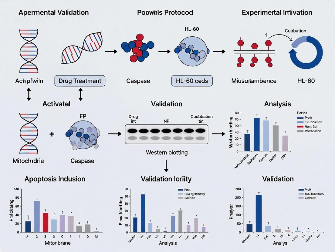
HL-60 Apoptosis Protocol Validation: Essential Methods for Robust Cancer Research & Drug Screening
This comprehensive guide details the critical methods for validating apoptosis assays in the HL-60 cell line, a cornerstone model in leukemia research and oncology drug development.
HL-60 Apoptosis Protocol Validation: Essential Methods for Robust Cancer Research & Drug Screening
Abstract
This comprehensive guide details the critical methods for validating apoptosis assays in the HL-60 cell line, a cornerstone model in leukemia research and oncology drug development. We explore the foundational principles of apoptosis, present step-by-step protocols for key validation techniques, address common troubleshooting scenarios, and provide a framework for comparative analysis. Designed for researchers and drug development professionals, this article ensures the generation of reliable, reproducible, and publication-quality data on programmed cell death.
Apoptosis in HL-60 Cells: Core Concepts, Mechanisms, and Research Significance
The HL-60 human promyelocytic leukemia cell line is a cornerstone of biomedical research, particularly in the study of programmed cell death. Within the context of validating apoptosis protocols, HL-60 serves as an indispensable reference model due to its well-characterized, rapid, and synchronous apoptotic response to a wide array of stimuli. This guide objectively compares its performance and utility against other common cell models in apoptosis research.
Comparative Performance of Apoptosis Models
Table 1: Key Characteristics of Common Cell Lines for Apoptosis Studies
| Cell Line | Origin | Key Apoptosis Inducers | Apoptosis Onset | Genetic Stability | Cost & Maintenance | Suitability for Protocol Validation |
|---|---|---|---|---|---|---|
| HL-60 | Human Promyelocytic Leukemia | Camptothecin, Etoposide, UV, Staurosporine, DMSO | Rapid, Synchronous (3-6 hrs) | High (near-diploid) | Low, Easy (suspension) | Excellent. Consistent, high yield, minimal confounding variables. |
| Jurkat | Human T-Cell Leukemia | Anti-FAS, Etoposide, UV | Moderate (6-12 hrs) | High | Low, Easy (suspension) | Very Good. Strong for death-receptor pathway studies. |
| HeLa | Human Cervical Adenocarcinoma | TNF-α + CHX, Staurosporine | Slow, Asynchronous (12-24+ hrs) | Moderate (aneuploid) | Low, Moderate (adherent) | Moderate. Heterogeneous response; complex background. |
| Primary Cells | Various (e.g., PBMCs, Neurons) | Context-dependent | Variable, Asynchronous | N/A | High, Difficult | Poor for validation. High donor variability, limited lifespan. |
| MCF-7 | Human Breast Carcinoma | Doxorubicin, TNF-α | Slow, Incomplete | Moderate (aneuploid) | Low, Moderate (adherent) | Limited. Lacks caspase-3, giving incomplete apoptotic phenotype. |
Table 2: Experimental Data from a Representative Apoptosis Assay (Camptothecin Induction)
| Cell Line | % Viability (24h) [Annexin V/PI] | Caspase-3/7 Activity Fold Increase (6h) | DNA Fragmentation (% Sub-G1, 18h) | Key Reference (Example) |
|---|---|---|---|---|
| HL-60 | 25% ± 5% | 8.5 ± 1.2 | 65% ± 8% | Soldatenkov et al., 1999 |
| Jurkat | 40% ± 10% | 6.0 ± 1.5 | 45% ± 12% | Tang et al., 2006 |
| HeLa | 60% ± 15% | 3.5 ± 1.0 | 30% ± 10% | Mohan et al., 2008 |
Detailed Experimental Protocols for Validation
Protocol 1: Standard Apoptosis Induction and Assessment in HL-60 Cells
- Cell Culture: Maintain HL-60 cells in RPMI-1640 medium supplemented with 10-20% FBS at 37°C, 5% CO₂ at a density of 2-9 x 10⁵ cells/mL.
- Apoptosis Induction: Treat cells with 1-10 µM Camptothecin (or 1 µM Staurosporine) for 3-6 hours. Include untreated and vehicle control populations.
- Harvesting: Pellet 1-5 x 10⁵ cells by centrifugation at 300 x g for 5 minutes.
- Annexin V/Propidium Iodide (PI) Staining:
- Wash cells once in cold PBS.
- Resuspend in 100 µL 1X Annexin V Binding Buffer.
- Add 5 µL FITC-Annexin V and 5 µL PI (or a viability dye).
- Incubate for 15 minutes at room temperature in the dark.
- Add 400 µL of binding buffer and analyze by flow cytometry within 1 hour.
- Caspase-3/7 Activity Assay (Luminescent):
- Pellet 1 x 10⁴ cells per condition.
- Lyse cells or use live-cell assay reagents per manufacturer's instructions (e.g., Caspase-Glo 3/7).
- Transfer lysate to a white-walled plate, add luminescent substrate, and incubate for 30-60 minutes.
- Measure luminescence on a plate reader.
Protocol 2: DNA Fragmentation Analysis (Sub-G1 Assay)
- Harvest 1-2 x 10⁶ HL-60 cells post-induction (e.g., 18 hours).
- Wash once with cold PBS and fix in 70% ethanol at -20°C for at least 2 hours.
- Pellet cells, wash with PBS, and resuspend in DNA staining solution (PBS containing 50 µg/mL PI, 100 µg/mL RNase A, 0.1% Triton X-100).
- Incubate at 37°C for 30 minutes in the dark.
- Analyze DNA content by flow cytometry (FL2 channel). Apoptotic cells with fragmented DNA appear as a "sub-G1" peak.
Visualization of Key Pathways and Workflows
Title: Canonical Apoptotic Signaling Pathways in HL-60 Cells
Title: HL-60 Apoptosis Protocol Validation Workflow
The Scientist's Toolkit: Key Research Reagent Solutions
Table 3: Essential Materials for HL-60 Apoptosis Studies
| Reagent/Material | Function & Importance in Validation | Example Product/Catalog |
|---|---|---|
| HL-60 Cell Line | The standardized biological model. Ensures reproducibility across labs. | ATCC CCL-240 |
| Camptothecin (Topo I Inhibitor) | A classic, robust inducer of the intrinsic pathway in HL-60. Serves as a positive control. | Sigma-Aldrich C9911 |
| Annexin V-FITC Apoptosis Kit | Gold-standard for detecting phosphatidylserine exposure (early apoptosis) and membrane integrity. | BioLegend 640922 |
| Caspase-Glo 3/7 Assay | Sensitive, homogeneous luminescent assay to measure executioner caspase activity. | Promega G8091 |
| Propidium Iodide (PI) | Vital dye for identifying late apoptotic/necrotic cells (Annexin V/PI) or for DNA staining (Sub-G1). | Thermo Fisher Scientific P1304MP |
| RNase A | Essential for DNA content analysis; digests RNA to prevent interference with PI DNA staining. | Qiagen 19101 |
| Dimethyl Sulfoxide (DMSO) | Common solvent for hydrophobic inducers; vehicle control is critical for experimental integrity. | Sigma-Aldrich D8418 |
| Cell Culture-Grade FBS | Serum quality directly impacts basal growth and apoptosis sensitivity; use consistent, characterized batches. | Gibco 10437028 |
Apoptosis, or programmed cell death, is a critical process in development, homeostasis, and disease. The two primary apoptotic pathways—intrinsic (mitochondrial) and extrinsic (death receptor)—converge on a common execution phase but are initiated by distinct molecular triggers. This guide provides an objective comparison of these pathways, with a focus on their relevance to the human promyelocytic leukemia HL-60 cell line, a canonical model for hematopoietic cancer and apoptosis research. The analysis is framed within the context of validating robust apoptosis protocols for therapeutic screening.
Molecular Triggers and Pathway Initiation: A Direct Comparison
Intrinsic (Mitochondrial) Pathway
Molecular Triggers: Cellular stress signals, including DNA damage, oxidative stress, cytokine deprivation, ER stress, and cytotoxic agents (e.g., etoposide, staurosporine). In HL-60 cells, this pathway is potently activated by chemotherapeutic drugs like Camptothecin (DNA topoisomerase I inhibitor) and UV irradiation. Key Initiator Event: Mitochondrial outer membrane permeabilization (MOMP), regulated by the Bcl-2 protein family. Pro-apoptotic proteins (Bax, Bak) oligomerize, forming pores, leading to cytochrome c release. Upstream Signal Integration: Stress sensors (e.g., p53) modulate the balance between pro- and anti-apoptotic Bcl-2 family members.
Extrinsic (Death Receptor) Pathway
Molecular Triggers: Extracellular ligand binding to death receptors (DR) of the tumor necrosis factor (TNF) receptor superfamily (e.g., Fas/CD95, TRAIL-R1/R2, TNF-R1). For HL-60, Recombinant Human TRAIL/Apo2L and Anti-Fas Agonistic Antibodies (e.g., CH-11) are common extrinsic inducters. Key Initiator Event: Ligand-induced trimerization of death receptors, recruitment of adaptor proteins (FADD) and initiator caspases (caspase-8/10) to form the Death-Inducing Signaling Complex (DISC). Upstream Signal Integration: Membrane-proximal DISC formation is the primary control point.
Table 1: Comparative Overview of Pathway Triggers and Initial Events in HL-60 Cells
| Feature | Intrinsic Pathway | Extrinsic Pathway |
|---|---|---|
| Primary Trigger | Intracellular stress (DNA damage, toxins) | Extracellular death ligand (TRAIL, FasL) |
| Key HL-60 Inducers | Camptothecin (1-10 µM), Etoposide (20-100 µM), UV-C (10-100 J/m²), Staurosporine (0.1-1 µM) | Recombinant TRAIL (10-100 ng/mL), Anti-Fas IgM (CH-11, 100-500 ng/mL) |
| Initiation Site | Mitochondria | Plasma Membrane (Death Receptors) |
| Key Initiator Proteins | Bax, Bak, Bcl-2, Bcl-xL | Fas/CD95, TRAIL-R1/2, FADD |
| Initial Signaling Event | MOMP & Cytochrome c Release | DISC Assembly |
| Time to Caspase-3 Activation* | Typically slower (4-12 hours) | Typically faster (2-6 hours) |
| p53 Dependence in HL-60 | Often required (HL-60 is p53 null; engineered lines used) | Generally p53-independent |
*Note: Timing is inducer- and dose-dependent.
Experimental Data from HL-60 Apoptosis Studies
Table 2: Quantitative Apoptosis Outcomes in HL-60 Cells Following Pathway-Specific Induction
| Study Inducer (Concentration) | Pathway Targeted | Assay Readout | Result (Mean ± SD or Representative) | Key Molecular Evidence |
|---|---|---|---|---|
| Camptothecin (5 µM, 6h) | Intrinsic | Annexin V/PI Flow Cytometry | 65 ± 8% Apoptosis | Cytochrome c release (WB), Caspase-9 activation |
| Etoposide (50 µM, 12h) | Intrinsic | Caspase-3/7 Activity (Luminescence) | 12-fold increase vs. control | PARP cleavage (WB), ΔΨm loss (JC-1 staining) |
| UV-C (50 J/m², 8h) | Intrinsic | DNA Fragmentation (TUNEL) | 70% TUNEL-positive | Bax translocation (IF), SMAC/Diablo release |
| Recombinant TRAIL (50 ng/mL, 4h) | Extrinsic | Annexin V/PI Flow Cytometry | 55 ± 7% Apoptosis | Caspase-8 activation (WB), DISC immunoprecipitation |
| Anti-Fas (CH-11, 250 ng/mL, 5h) | Extrinsic | Caspase-8 Activity (Colorimetric) | 8-fold increase vs. control | FADD recruitment (Co-IP), Bid cleavage (tBid formation) |
| TRAIL + Cycloheximide (10 µg/mL) | Extrinsic (Sensitized) | Cell Viability (MTT) | IC₅₀ reduced from 75 ng/mL to 15 ng/mL | Enhanced Caspase-8 processing, RIP1 degradation |
Detailed Experimental Protocols for Validation
Protocol A: Validating Intrinsic Pathway Activation via Mitochondrial Depolarization
Objective: Quantify loss of mitochondrial membrane potential (ΔΨm) using JC-1 dye. Reagents: JC-1 dye (5,5',6,6'-tetrachloro-1,1',3,3'-tetraethylbenzimidazolylcarbocyanine iodide), Camptothecin (stock: 10 mM in DMSO), PBS, flow cytometry buffer. Method:
- Seed HL-60 cells at 2x10⁵ cells/mL. Treat with Camptothecin (5 µM) or DMSO vehicle for 4-6h.
- Harvest cells, wash with PBS.
- Resuspend cells in 500 µL culture medium containing 2 µg/mL JC-1.
- Incubate at 37°C, 5% CO₂ for 20 minutes.
- Wash cells twice with warm PBS, resuspend in flow cytometry buffer.
- Analyze immediately by flow cytometry: Ex/Em for JC-1 aggregates (~590 nm, red) and monomers (~530 nm, green). Calculate ratio of aggregate (FL2) to monomer (FL1) fluorescence. A decrease in ratio indicates ΔΨm loss.
Protocol B: Validating Extrinsic Pathway Activation via DISC Analysis (Immunoprecipitation)
Objective: Confirm ligand-induced formation of the Death-Inducing Signaling Complex (DISC). Reagents: Recombinant TRAIL (with cross-linking Flag-tag), Anti-FLAG M2 Affinity Gel, Lysis buffer (e.g., with 1% CHAPS), Protease inhibitors, HL-60 cells (≥1x10⁷ per condition). Method:
- Induce apoptosis by treating HL-60 cells with cross-linked Flag-TRAIL (100 ng/mL) for 30-60 minutes on ice to synchronize binding, then shift to 37°C for 15 min.
- Lyse cells in ice-cold DISC-IP lysis buffer.
- Pre-clear lysate with control beads for 1h at 4°C.
- Incubate supernatant with Anti-FLAG M2 Gel overnight at 4°C with rotation.
- Wash beads extensively with lysis buffer.
- Elute bound proteins with 3xFLAG peptide or Laemmli buffer.
- Analyze eluates by Western Blot for FADD, Caspase-8, and c-FLIP.
Pathway Signaling Diagrams
Diagram Title: Intrinsic Apoptosis Pathway in HL-60
Diagram Title: Extrinsic Apoptosis Pathway in HL-60
The Scientist's Toolkit: Key Research Reagent Solutions
Table 3: Essential Reagents for Apoptosis Pathway Analysis in HL-60
| Reagent Category & Name | Function / Target | Application in HL-60 Studies |
|---|---|---|
| Pathway-Specific Inducers | ||
| Camptothecin (Topoisomerase I Inhibitor) | Induces DNA damage, activating p53/p73-dependent intrinsic pathway. | Positive control for intrinsic apoptosis; used at 0.1-10 µM for 4-24h. |
| Recombinant Human TRAIL/Apo2L | Activates TRAIL-R1/R2 death receptors. | Primary inducer for extrinsic pathway; often used with sensitizing agents (e.g., cycloheximide). |
| Anti-Fas Agonistic Antibody (Clone CH-11) | Cross-links and activates Fas receptor. | Standard extrinsic trigger; validates DISC-dependent apoptosis. |
| Small Molecule Inhibitors | ||
| Z-VAD-FMK (Pan-Caspase Inhibitor) | Irreversible broad-spectrum caspase inhibitor. | Confirms caspase-dependent apoptosis; used as a negative control (20-50 µM). |
| ABT-263 (Navitoclax) | Bcl-2/Bcl-xL/Bcl-w inhibitor. | Sensitizes to intrinsic apoptosis; probes Bcl-2 family dependency. |
| Detection Reagents | ||
| JC-1 Dye | Mitochondrial membrane potential (ΔΨm) sensor. | Gold standard for intrinsic pathway validation (flow cytometry/microscopy). |
| Annexin V-FITC/PI Kit | Detects phosphatidylserine exposure (early apoptosis) and membrane integrity. | Quantitative apoptosis measurement by flow cytometry for both pathways. |
| Caspase-Glo 3/7, 8, or 9 Assays | Luminescent substrates for caspase activity. | Pathway-specific profiling; e.g., Caspase-8 for extrinsic, Caspase-9 for intrinsic. |
| Antibodies for Western Blot | ||
| Anti-Cytochrome c | Detects release from mitochondria. | Confirms MOMP in intrinsic pathway (compare cytosolic vs. mitochondrial fractions). |
| Anti-Cleaved PARP (Asp214) | Marker of caspase-3/7 activity. | Universal apoptosis endpoint for both pathways. |
| Anti-Cleaved Caspase-8 (Asp387) | Specific for active caspase-8. | Validates extrinsic pathway or DISC formation. |
| Cell Line Modifications | ||
| HL-60 Vector Control & Bcl-2 Overexpression | Ectopic Bcl-2 expression. | Tests resistance to intrinsic triggers; confirms pathway specificity. |
| HL-60 p53 Knock-in/Reconstitution | Restores p53 function. | Studies p53-dependent intrinsic apoptosis, as wild-type HL-60 is p53 null. |
Key Morphological and Biochemical Hallmarks of Apoptosis
Within the context of HL-60 apoptosis protocol validation methods research, accurately distinguishing apoptosis from other modes of cell death is paramount. This guide objectively compares the key morphological and biochemical hallmarks of apoptosis against those of necrosis, a common alternative cell death pathway. The validation of these hallmarks is critical for researchers, scientists, and drug development professionals in assessing compound efficacy and mechanism of action.
Comparative Hallmarks: Apoptosis vs. Necrosis
Table 1: Comparison of Core Morphological and Biochemical Hallmarks
| Hallmark Feature | Apoptosis | Necrosis |
|---|---|---|
| Cell & Nucleus Morphology | Cell shrinkage, chromatin condensation (pyknosis), nuclear fragmentation (karyorrhexis), formation of apoptotic bodies. | Cell swelling, loss of membrane integrity, organelle breakdown, karyolysis (nuclear dissolution). |
| Plasma Membrane Integrity | Maintained until late stages. Phosphatidylserine (PS) externalization detected by Annexin V. | Lost early. Allows influx of vital dyes (e.g., Trypan Blue, PI). |
| Inflammatory Response | Non-inflammatory. Apoptotic bodies are phagocytosed. | Pro-inflammatory. Cytoplasmic contents released into extracellular space. |
| Primary Biochemical Markers | Caspase-3/7 activation, PARP cleavage, DNA laddering (internucleosomal cleavage). | Loss of ATP, release of HMGB1, LDH. |
| Typical Inducers | Physiological signals, DNA damage, staurosporine, Fas ligand. | Extreme stress, complement attack, severe hypoxia, physical trauma. |
Table 2: Quantitative Assay Data from HL-60 Model System
| Assay Parameter | Apoptotic Response (e.g., 1µM Staurosporine, 4h) | Necrotic Response (e.g., 1% Triton X-100, 1h) | Measurement Method |
|---|---|---|---|
| Viability (Metabolic Activity) | 35-50% remaining | 5-15% remaining | MTT/WST-1 Reduction |
| Membrane Integrity (PI+ cells) | 10-25% positive | 85-95% positive | Flow Cytometry |
| PS Externalization (Annexin V+) | 55-70% positive | 80-95% positive (Annexin V+/PI+) | Flow Cytometry |
| Caspase-3/7 Activity | 8-12 fold increase over control | ≤ 1.5 fold over control | Luminescent/Fluorogenic substrate |
| DNA Fragmentation | Clear ladder pattern | Smear pattern | Agarose Gel Electrophoresis |
Experimental Protocols for Hallmark Validation
Protocol 1: Annexin V/Propidium Iodide (PI) Staining for Flow Cytometry
- Objective: Distinguish early apoptotic (Annexin V+/PI-), late apoptotic/necrotic (Annexin V+/PI+), and necrotic (Annexin V-/PI+) cells.
- Method: 1) Harvest ~1x10^6 HL-60 cells by gentle centrifugation. 2) Wash cells with 1X PBS. 3) Resuspend cells in 100µL of 1X Annexin V Binding Buffer. 4) Add fluorochrome-conjugated Annexin V (e.g., FITC) and PI per manufacturer's instructions. Incubate for 15 min at RT in the dark. 5) Add 400µL of Binding Buffer and analyze by flow cytometry within 1 hour.
Protocol 2: Caspase-3/7 Activity Assay (Luminescent)
- Objective: Quantitatively measure executioner caspase activation.
- Method: 1) Induce apoptosis in HL-60 cells. 2) Prepare cell lysates or use live-cell assays as per kit (e.g., Caspase-Glo 3/7). 3) For lysates: combine equal volumes of lysate and substrate reagent in a white-walled plate. 4) Incubate for 30-60 min at RT. 5) Measure luminescence using a plate reader. Normalize data to protein concentration or cell number.
Protocol 3: DNA Laddering Assay
- Objective: Visualize internucleosomal DNA cleavage characteristic of apoptosis.
- Method: 1) Pellet 2-5x10^6 HL-60 cells. 2) Extract genomic DNA using a kit or phenol-chloroform. 3) Quantify DNA. 4) Load 0.5-1µg DNA per well on a 1.5-2% agarose gel containing a DNA stain. 5) Run gel at 5V/cm for 2-3 hours. 6) Visualize under UV light; apoptosis shows a "ladder" of ~180-200 bp multiples.
Signaling Pathways and Experimental Workflow
Diagram Title: Key Signaling Pathways in Apoptosis
Diagram Title: Apoptosis Hallmark Validation Workflow
The Scientist's Toolkit: Research Reagent Solutions
Table 3: Essential Materials for Apoptosis Assay Validation
| Reagent/Material | Function in Apoptosis Research | Example/Notes |
|---|---|---|
| Annexin V Conjugates | Binds to externalized phosphatidylserine (PS) to detect early apoptosis. Often used with a viability dye. | FITC, APC, or Pacific Blue conjugates for flow cytometry. |
| Propidium Iodide (PI) | Membrane-impermeant DNA dye. Distinguishes late apoptotic/necrotic cells (PI+) from early apoptotic (PI-). | Use with Annexin V. Can be replaced by 7-AAD. |
| Caspase Activity Assays | Quantifies cleavage of specific peptide substrates by active caspases, indicating pathway activation. | Luminescent (Caspase-Glo) or fluorogenic (DEVD-AMC) substrates. |
| PARP Antibody | Detects cleavage of PARP (89 kDa fragment), a classic caspase-3 substrate, by western blot. | Monoclonal anti-PARP (cleaved) is a key biochemical marker. |
| DNA Isolation Kits | High-quality isolation of genomic DNA for fragmentation analysis via agarose gel electrophoresis. | Kits optimized for low molecular weight DNA improve ladder detection. |
| Staurosporine | A broad-spectrum protein kinase inhibitor used as a positive control for inducing intrinsic apoptosis. | Typical working concentration for HL-60 cells: 0.5-2 µM for 2-6 hours. |
| Camptothecin | Topoisomerase I inhibitor, induces DNA damage and intrinsic apoptosis. Alternative positive control. | Typical working concentration: 1-10 µM for 4-24 hours. |
| Flow Cytometer | Essential instrument for quantifying populations of cells in different death stages (Annexin V/PI). | Allows high-throughput, single-cell analysis. |
This comparison guide is framed within a broader thesis research context aimed at validating standardized protocols for inducing and quantifying apoptosis in the human acute promyelocytic leukemia (HL-60) cell line. Selecting an appropriate chemical inducer is a critical first step for such studies. This guide objectively compares four widely used apoptosis inducers—All-Trans Retinoic Acid (ATRA), Dimethyl Sulfoxide (DMSO), Etoposide, and Staurosporine—based on mechanistic action, efficacy, kinetics, and experimental applicability.
Comparative Performance Data
The following table summarizes key quantitative data from recent studies on these inducers in HL-60 cells.
Table 1: Comparative Analysis of Apoptosis Inducers in HL-60 Cells
| Inducer | Typical Working Concentration | Average Apoptosis Induction Time (Hours) | Primary Mechanism of Action | Reported Apoptosis Rate (%) (72h) | Key Assays Used for Validation |
|---|---|---|---|---|---|
| ATRA | 1 - 10 µM | 72 - 96 | Differentiation-induced apoptosis via RARα signaling | 40 - 60% | Morphology, NBT reduction, Flow Cytometry (Annexin V/PI) |
| DMSO | 1.0 - 1.5% (v/v) | 96 - 120 | Differentiation (granulocytic) leading to caspase-3 activation | 50 - 70% | CD11b expression, Cell cycle analysis, DNA fragmentation |
| Etoposide | 20 - 100 µM | 24 - 48 | Topoisomerase II inhibition; DNA damage-induced intrinsic pathway | 60 - 80% | Annexin V/PI, Caspase-3/7 activity, Western Blot (p53, PARP) |
| Staurosporine | 0.1 - 1 µM | 4 - 6 | Broad-spectrum protein kinase inhibitor; direct intrinsic pathway activation | > 90% | Annexin V/PI, Mitochondrial membrane potential (ΔΨm), Caspase activation |
Detailed Experimental Protocols
Protocol 1: Differentiation-Based Apoptosis Induction (ATRA & DMSO)
- Cell Culture: Maintain HL-60 cells in RPMI-1640 medium supplemented with 10-20% FBS and 1% penicillin/streptomycin at 37°C, 5% CO₂.
- Treatment: Seed cells at 2-5 x 10⁵ cells/mL. Add ATRA (from a 10 mM stock in DMSO) to final concentration of 1 µM, or sterile DMSO to 1.25% (v/v). A vehicle control (0.1% DMSO) is essential.
- Incubation & Monitoring: Culture for 4-6 days. Refresh medium and inducer every 2-3 days. Monitor differentiation markers:
- NBT Reduction Assay: Cells incubated with NBT and PMA; differentiated cells produce insoluble blue formazan.
- Surface Marker Analysis: Use flow cytometry to detect CD11b expression.
- Apoptosis Assessment: At days 3-6, harvest cells and analyze using Annexin V-FITC/Propidium Iodide staining for flow cytometry.
Protocol 2: Direct Apoptosis Induction (Etoposide & Staurosporine)
- Cell Seeding: Seed HL-60 cells as above in fresh medium.
- Treatment: Add Etoposide (from 20 mM DMSO stock) to 50 µM final concentration, or Staurosporine (from 1 mM DMSO stock) to 0.5 µM final concentration.
- Incubation: Treat for 24-48 hours (Etoposide) or 4-6 hours (Staurosporine).
- Apoptosis Assessment: Harvest cells and use multiparametric assays:
- Flow Cytometry: Annexin V/PI staining.
- Caspase Activity: Fluorometric assay for Caspase-3/7.
- Mitochondrial Depolarization: Stain with JC-1 dye and analyze by flow cytometry.
- Western Blotting: Detect cleavage of PARP and Caspase-3.
Signaling Pathways
Diagram 1: Signaling pathways of four apoptosis inducers in HL-60 cells.
Diagram 2: Experimental workflow for apoptosis induction and validation.
The Scientist's Toolkit
Table 2: Essential Research Reagent Solutions for HL-60 Apoptosis Studies
| Reagent/Material | Function & Application | Key Considerations |
|---|---|---|
| HL-60 Cell Line | Model system for acute promyelocytic leukemia and myeloid differentiation studies. | Check authentication and mycoplasma status regularly. Maintain in log-phase growth. |
| ATRA (All-Trans Retinoic Acid) | Gold-standard inducer of neutrophilic differentiation, leading to apoptosis. | Light-sensitive. Prepare fresh stock in DMSO and protect from light. |
| Pharmacological-Grade DMSO | Differentiation inducer (granulocytic) and solvent for stock solutions. | Use high purity (>99.9%) for treatments; vehicle control concentration is critical. |
| Annexin V-FITC / Propidium Iodide (PI) Kit | Dual-staining for flow cytometry to distinguish early/late apoptosis and necrosis. | Perform staining in calcium-containing buffer. Analyze immediately. |
| Caspase-3/7 Fluorometric Assay Kit | Quantifies executioner caspase activity as a direct marker of apoptosis. | Use positive control (e.g., Staurosporine-treated cells). Normalize to protein/cell count. |
| NBT (Nitroblue Tetrazolium) | Differentiated HL-60 cells reduce NBT to blue formazan, a differentiation marker. | Used with PMA (phorbol ester) stimulation. Quantify by microscopy or absorbance. |
| JC-1 Dye | Mitochondrial membrane potential (ΔΨm) sensor; depolarization indicates intrinsic pathway activation. | Ratio of red (aggregates) to green (monomers) fluorescence measured by flow cytometry. |
| Anti-PARP & Anti-Cleaved Caspase-3 Antibodies | Western Blot detection of hallmark proteolytic cleavage events during apoptosis. | Cleaved fragments are definitive markers. Always run full-length protein control. |
The Critical Importance of Protocol Validation in Preclinical Research
Within the framework of advancing HL-60 apoptosis protocol validation methods, rigorous comparison of experimental reagents and kits is paramount. Inconsistent outcomes in apoptosis assays often stem from unvalidated protocols and reagent variability. This guide compares the performance of key assay kits for caspase-3 activity, a critical apoptosis endpoint in HL-60 cell research.
Comparison of Caspase-3 Activity Assay Kits for HL-60 Apoptosis Studies
The following table summarizes quantitative data from a standardized experiment where HL-60 cells were treated with 1 µM Etoposide for 16 hours to induce apoptosis. Assays were performed in triplicate according to each manufacturer's protocol.
| Kit/Reagent (Supplier) | Principle | Signal-to-Noise Ratio (Induced/Control) | Inter-Assay CV (%) | Required Sample Volume (per well) | Hands-On Time (min) |
|---|---|---|---|---|---|
| Caspase-Glo 3/7 Assay (Promega) | Luminescent (pro-luminescent substrate) | 12.5 ± 1.2 | 4.5% | 50 µL (cell lysate) | ~20 |
| Apo-ONE Homogeneous Caspase-3/7 Assay (Promega) | Fluorometric (Z-DEVD-R110 substrate) | 9.8 ± 0.9 | 6.2% | 50 µL (cell lysate) + 50 µL (reagent) | ~25 |
| Caspase-3 Colorimetric Assay Kit (BioVision) | Colorimetric (DEVD-pNA substrate) | 5.5 ± 0.7 | 9.8% | 50-100 µL (cell lysate, 100-200 µg protein) | ~90 (incubation) |
| In-house protocol (Z-DEVD-AFC substrate, Sigma) | Fluorometric (custom reagents) | 8.2 ± 2.1* | 15.3%* | 50 µL (cell lysate) | Varies |
Note: Higher variability in the in-house protocol highlights the validation challenge.
Detailed Experimental Protocol for Comparison
Cell Culture and Treatment:
- HL-60 cells (ATCC CCL-240) are maintained in RPMI-1640 medium supplemented with 10% FBS and 1% penicillin-streptomycin at 37°C, 5% CO₂.
- Cells are seeded at a density of 2.5 x 10⁵ cells/mL in 24-well plates.
- Apoptosis is induced by adding 1 µM Etoposide (from a 10 mM DMSO stock). Control wells receive vehicle (DMSO) only.
- Plates are incubated for 16 hours.
Sample Preparation (Lysate-based assays):
- Cells are centrifuged at 500 x g for 5 min.
- The pellet is washed once with cold PBS.
- Cells are lysed in 100 µL of chilled lysis buffer provided with the kit (or 50 mM HEPES, pH 7.4, 0.1% CHAPS for in-house) on ice for 15 min.
- Lysates are centrifuged at 12,000 x g for 15 min at 4°C. The supernatant (cytosolic extract) is transferred to a fresh tube and placed on ice.
Assay Execution: For Caspase-Glo 3/7: 50 µL of lysate is transferred to an opaque 96-well plate. An equal volume of Caspase-Glo 3/7 reagent is added. The plate is mixed gently and incubated at room temperature for 1 hour. Luminescence is recorded. For Apo-ONE: 50 µL of lysate is combined with 50 µL of Apo-ONE reagent in a 96-well plate. The plate is mixed and incubated at room temperature for 1 hour. Fluorescence is measured (Ex 499/Em 521). For Colorimetric Kit (BioVision): 50 µL of lysate is mixed with 50 µL of 2X Reaction Buffer containing 10 mM DTT and 5 µL of the 4 mM DEVD-pNA substrate. The mixture is incubated at 37°C for 90-120 min. Absorbance is read at 405 nm. For In-house (Z-DEVD-AFC): 50 µL of lysate is mixed with 50 µL of assay buffer (50 mM HEPES, pH 7.4, 100 mM NaCl, 0.1% CHAPS, 10 mM DTT, 1 mM EDTA, 10% glycerol) containing 50 µM Z-DEVD-AFC substrate. The mixture is incubated at 37°C for 1 hour. Fluorescence is measured (Ex 400/Em 505).
Visualization of Key Signaling and Workflow
Intrinsic Apoptosis Pathway in HL-60 Cells
Caspase-3 Assay Comparison Workflow
The Scientist's Toolkit: Research Reagent Solutions
| Reagent/Material | Supplier Example | Critical Function in HL-60 Apoptosis Assay |
|---|---|---|
| HL-60 Cell Line | ATCC | Human promyelocytic leukemia cell line; standard model for apoptosis studies. |
| Etoposide | Sigma-Aldrich, Tocris | Topoisomerase II inhibitor; standard chemical inducer of intrinsic apoptosis pathway. |
| Caspase-Glo 3/7 Assay | Promega | Homogeneous, luminescent kit for sensitive, high-throughput detection of caspase-3/7 activity. |
| Z-DEVD-AFC Fluorogenic Substrate | Sigma-Aldrich, Enzo | Cleavable peptide substrate for caspase-3; core component of in-house fluorometric assays. |
| RIPA Lysis Buffer | Thermo Fisher, Cell Signaling | Efficiently extracts cytosolic proteins, including released cytochrome c and activated caspases. |
| Black/Clear 96-well Assay Plates | Corning, Greiner | Plate format compatible with microplate readers for luminescence, fluorescence, or absorbance. |
| Microplate Reader with multi-mode detection | BioTek, BMG Labtech | Essential instrument for quantifying luminescent, fluorescent, or colorimetric assay signals. |
| DMSO, Cell Culture Grade | Sigma-Aldrich | Universal solvent for hydrophobic apoptosis inducers (e.g., Etoposide). |
Step-by-Step Guide: Validating Apoptosis in HL-60 with Multi-Parameter Assays
Within the critical framework of HL-60 apoptosis protocol validation, consistent log-phase culture is the foundational variable governing experimental reproducibility. This guide compares core media and supplement formulations, providing data-driven insights for maintaining optimal growth.
Comparative Analysis: Media and Supplement Formulations for HL-60 Maintenance
Table 1: Comparison of Base Media Performance for HL-60 Log-Phase Growth
| Media Formulation | Doubling Time (hrs) | Max Viability (% , >95% threshold) | Key Characteristics | Best For |
|---|---|---|---|---|
| RPMI-1640 + 10% FBS | 24-30 | 98% (Days 1-3) | Gold standard; requires quality FBS screening. | General maintenance, most apoptosis assays. |
| RPMI-1640 + 20% FBS | 22-26 | 98% (Days 1-4) | Extended log phase; higher serum cost & variability. | High-density experiments requiring extended log phase. |
| IMDM + 10% FBS | 22-28 | 97% | Rich in nutrients & inorganic salts; may alter basal metabolism. | Studies where enhanced nutrient support is needed. |
| Serum-Free (Commercial HL-60 Formulation) | 30-36 | 95% | Defined, no batch variability; slower adaptation required. | Signal transduction studies requiring minimal unknown variables. |
Table 2: Impact of Passage Practices on Culture Consistency
| Practice Parameter | Optimal Protocol | Suboptimal Protocol | Observed Impact on Apoptosis Assay (e.g., Camptothecin EC50 Shift) |
|---|---|---|---|
| Seeding Density | 2.0 - 4.0 x 10^5 cells/mL | <1.0 or >5.0 x 10^5 cells/mL | EC50 variability of ± 35% due to altered basal survival signaling. |
| Passage Interval | Every 2-3 days (max) | 4+ days (into plateau) | Loss of synchrony; 50% increase in assay CV (coefficient of variation). |
| Cell Counting Method | Automated cell counter + trypan blue | Hemocytometer only | Reduced counting error (<5% vs. >15%), improving dose-response accuracy. |
Experimental Protocol: Validating Log-Phase Health for Apoptosis Studies
Method: Daily Growth & Viability Tracking
- Culture: Maintain HL-60 cells in RPMI-1640 + 10% FBS (heat-inactivated) at 37°C, 5% CO2.
- Passaging: Every 2-3 days, centrifuge (300 x g, 5 min) and resuspend in fresh pre-warmed medium to 3.0 x 10^5 cells/mL.
- Daily Census: For 4 days post-passage, count cells daily using an automated counter with trypan blue exclusion.
- Analysis: Plot cell density and viability. Log-phase is defined as viability >95% and consistent exponential increase in density. Use only cells harvested within this phase for apoptosis induction.
Method: Apoptosis Assay Consistency Test
- Group Setup: Culture cells as above. Create two groups: "Optimal" (harvested at 3.0 x 10^5 cells/mL, day 2 post-passage) and "Suboptimal" (harvested from plateau phase at 1.5 x 10^6 cells/mL, day 4).
- Treatment: Seed both groups at 2.5 x 10^5 cells/mL in 96-well plates. Treat with a 8-point serial dilution of camptothecin (e.g., 0.05 to 50 µM). Include DMSO vehicle controls.
- Incubation: Incubate for 6 hours (early apoptosis window).
- Assessment: Measure apoptosis via Annexin V/Propidium Iodide flow cytometry or Caspase-3/7 luminescent assay.
- Data Comparison: Calculate EC50 values for each group. Inconsistent culture conditions typically cause a >25% shift in EC50 and widened confidence intervals.
Visualizing the Workflow and Key Pathway
Title: Workflow for Consistent HL-60 Log-Phase Culture
Title: Nutrient Signaling Impact on Apoptotic Priming in HL-60
The Scientist's Toolkit: Research Reagent Solutions
Table 3: Essential Materials for HL-60 Log-Phase Culture & Validation
| Item | Function & Importance | Example/Note |
|---|---|---|
| RPMI-1640 Medium | Base nutrient supply. Requires L-glutamine. | Many commercial formulations; select with stable glutamine (e.g., GlutaMAX). |
| Premium/FBS | Provides essential growth factors, hormones, and lipids. | Major source of variability. Batch test for HL-60 growth promotion. |
| Heat-Inactivation | Inactivates complement proteins to prevent non-apoptotic cell lysis. | Standard practice at 56°C for 30 minutes. |
| Trypan Blue Solution | Vital dye for distinguishing live (excluded) from dead (stained) cells. | Use with automated cell counter or hemocytometer. |
| Camptothecin (CPT) | Topoisomerase I inhibitor; standard positive control for inducing intrinsic apoptosis in HL-60. | Prepare in DMSO, aliquot, store at -20°C. Light sensitive. |
| Annexin V Binding Buffer | Calcium-containing buffer necessary for Annexin V-FITC binding to phosphatidylserine. | Must have correct pH and [Ca2+]; often 10X concentrate. |
| Caspase-Glo 3/7 Assay | Luminescent reagent for quantifying caspase-3/7 activity as apoptosis marker. | Provides sensitive, plate-based readout. |
| Controlled-Rate Freezer | For consistent, high-viability cryopreservation of master cell banks. | Critical for long-term assay consistency across lab members and years. |
Within the systematic validation of HL-60 apoptosis induction protocols, selecting the appropriate detection method is foundational. This guide objectively compares the performance of the canonical Annexin V/Propidium Iodide (PI) flow cytometry assay against prevalent alternative techniques, providing experimental data relevant to HL-60 model systems.
Performance Comparison of Apoptosis Detection Methods
The following table summarizes quantitative performance metrics for key apoptosis detection assays, based on consolidated data from recent literature and validation studies using HL-60 cells treated with 1 µM staurosporine for 4 hours.
Table 1: Comparative Analysis of Apoptosis Detection Methods
| Method | Primary Readout | Early Apoptosis Detection | Late Apoptosis/Necrosis Discrimination | Throughput | Quantitative Resolution | Key Limitations |
|---|---|---|---|---|---|---|
| Annexin V/PI Flow Cytometry | Phosphatidylserine externalization & membrane integrity | Excellent | Excellent | High (Single-cell) | High (Quadrant Statistics) | Requires single-cell suspension; Cannot assess caspase activation directly. |
| Caspase-3/7 Activity Assays | Luminescent or fluorescent substrate cleavage | Moderate (Downstream event) | Poor | Medium (Bulk population) | Medium (Population average) | Misses caspase-independent apoptosis; Bulk measurement only. |
| TUNEL Assay | DNA fragmentation (in situ) | Poor | Excellent | Low (Microscopy) / Medium (Flow) | Medium | Can label necrotic cells; More complex protocol. |
| Nuclear Morphology (Hoechst) | Chromatin condensation & fragmentation | Good | Good | Low (Manual scoring) | Low (Semi-quantitative) | Subjective; Low throughput; Requires expertise. |
| MTT/XTT Cell Viability | Metabolic activity | Indirect/Poor | Indirect/Poor | High (Plate reader) | Low (Indirect only) | Cannot distinguish apoptosis from other death modes; Early stages not detected. |
Detailed Experimental Protocols
1. Annexin V/PI Flow Cytometry Protocol for HL-60 Cells
- Cell Preparation: Harvest 0.5-1x10^6 HL-60 cells per condition. Wash twice with cold PBS.
- Staining: Resuspend cell pellet in 100 µL of 1X Annexin V Binding Buffer. Add 5 µL of FITC-conjugated Annexin V and 5 µL of Propidium Iodide (PI, 50 µg/mL) solution.
- Incubation: Incubate for 15 minutes at room temperature (25°C) in the dark.
- Analysis: Add 400 µL of binding buffer and analyze by flow cytometry within 1 hour. Use 488 nm excitation; collect FITC emission at 530 nm (FL1) and PI at >575 nm (FL2 or FL3). Include unstained, Annexin V-only, and PI-only controls for compensation.
- Data Interpretation: Viable cells are Annexin V-/PI-; Early apoptotic cells are Annexin V+/PI-; Late apoptotic/necrotic cells are Annexin V+/PI+.
2. Comparative Caspase-3/7 Activity Assay Protocol (Luminescent)
- Cell Preparation: Seed HL-60 cells in a white-walled 96-well plate at 1x10^4 cells/well. Treat and induce apoptosis.
- Assay Execution: Equilibrate plate and Caspase-Glo 3/7 reagent to room temperature. Add an equal volume of reagent to each well.
- Incubation: Mix on a plate shaker for 30 seconds, then incubate at room temperature for 1 hour.
- Measurement: Record luminescence using a plate reader. Data represent bulk caspase activity per well.
Signaling Pathways & Experimental Workflow
Diagram 1: Apoptosis Progression & Annexin V/PI Detection Logic
Diagram 2: Comparative Experimental Workflow: Flow vs. Caspase Assay
The Scientist's Toolkit: Research Reagent Solutions
Table 2: Essential Reagents for Annexin V/PI Apoptosis Assay
| Reagent / Solution | Function | Critical Notes for HL-60 Cells |
|---|---|---|
| Annexin V, Fluorochrome-conjugated | Binds specifically to externalized phosphatidylserine (PS). | FITC is common; choose APC or PE for multi-color panels. Calcium-dependent binding. |
| Propidium Iodide (PI) | DNA intercalating dye; penetrates cells with compromised membranes. | Distinguishes late apoptotic/necrotic cells. Requires RNase treatment if analyzing DNA content concurrently. |
| Annexin V Binding Buffer (10X) | Provides optimal calcium concentration and ionic strength for Annexin V binding. | Must be diluted to 1X and kept cold. HEPES-buffered saline is typical. |
| Ice-cold Phosphate Buffered Saline (PBS) | Wash medium to remove culture serum (which contains PS) and stop cellular processes. | Must be calcium/magnesium-free to prevent cell clumping and unwanted adhesion. |
| Flow Cytometer with 488 nm Laser | Instrument for single-cell fluorescence quantification. | Standard configuration. Ensure proper compensation for FITC/PI spectral overlap. |
| Cell Strainer (40 µm) | Ensures a single-cell suspension prior to analysis. | Critical for non-adherent HL-60s to remove aggregates that cause flow cytometry artifacts. |
Within the context of a broader thesis on HL-60 apoptosis protocol validation methods, the accurate quantification of caspase-3/7 activity is a critical endpoint. These executioner caspases are definitive markers of the apoptotic cascade. This guide objectively compares the performance of two dominant assay formats—luminescent vs. fluorescent—for measuring caspase-3/7 activity in HL-60 cells treated with a standardized apoptotic inducer (e.g., 1µM staurosporine for 4 hours).
Comparative Experimental Data
The following table summarizes performance data from parallel experiments using leading commercial kits on the same batch of induced HL-60 cells.
| Performance Metric | Luminescent Caspase-Glo 3/7 Assay | Fluorescent (AMC-based) Assay | Notes / Experimental Conditions |
|---|---|---|---|
| Signal-to-Background Ratio | 12.5 ± 1.2 | 6.8 ± 0.9 | Mean ± SD, n=6. Apoptotic vs. untreated control. |
| Z'-Factor | 0.78 | 0.52 | Robustness for HTS screening. |
| Assay Time Post-Lysis | 30 min | 1.5 - 2 hours | Time to stable, measurable signal. |
| Dynamic Range (Linear) | 3 log units | 2 log units | Evaluated via serial dilution of apoptotic lysate. |
| Interference from Cellular Debris | Low | Moderate | Luminescent read less affected by light scattering. |
| Sample Throughput | High | Moderate | Luminescent suited for 384/1536-well plates. |
| Required Cell Number (per 96-well) | 5,000 - 10,000 | 15,000 - 25,000 | For reliable detection above background. |
| Key Instrument Required | Luminometer | Fluorometer (Ex/Em ~355/460 nm) |
Detailed Experimental Protocols
Protocol A: Luminescent Assay (Caspase-Glo 3/7 Principle)
- Cell Preparation & Induction: Seed HL-60 cells in a white-walled, clear-bottom 96-well plate at 8,000 cells/well in 100µL culture medium. Induce apoptosis with 1µM staurosporine (from 1mM DMSO stock) for 4 hours at 37°C, 5% CO₂. Include untreated and vehicle (DMSO) controls.
- Reagent Equilibration: Thaw and equilibrate the Caspase-Glo 3/7 substrate and buffer to room temperature. Prepare the homogeneous Caspase-Glo 3/7 Reagent by mixing the lyophilized substrate with the buffer.
- Assay Execution: Add 100µL of the prepared Caspase-Glo 3/7 Reagent directly to each 100µL cell culture well.
- Incubation: Place plate on orbital shaker (300-500 rpm) for 30 seconds to mix. Incubate at room temperature for 30 minutes in the dark.
- Measurement: Measure luminescent signal using a plate-reading luminometer with an integration time of 0.5-1 second per well.
Protocol B: Fluorescent Assay (DEVD-AMC Substrate Principle)
- Cell Preparation & Induction: Seed HL-60 cells in a black-walled, clear-bottom 96-well plate at 20,000 cells/well in 100µL. Induce apoptosis as in Protocol A.
- Cell Lysis: Following induction, centrifuge plate at 500 x g for 5 minutes. Gently aspirate medium, being careful not to disturb cell pellet. Lyse cells by adding 50µL of chilled lysis buffer (e.g., containing 1% Triton X-100, 25mM HEPES, 5mM DTT, pH 7.4) to each well. Incubate on ice for 15 minutes.
- Reaction Setup: Prepare 2X reaction buffer containing the fluorogenic substrate Ac-DEVD-AMC (final concentration 50µM) and 10mM DTT in assay buffer. Add 50µL of this 2X reaction mix to each well containing 50µL of lysate.
- Incubation & Measurement: Incubate the plate at 37°C in the dark. Measure fluorescence kinetics (Ex 355 nm / Em 460 nm) using a fluorometric plate reader every 5 minutes for 90-120 minutes.
- Data Analysis: Calculate the slope of the fluorescence increase (RFU/min) during the linear phase as the measure of caspase-3/7 activity.
Visualization of Pathways and Workflows
Title: Homogeneous Luminescent Caspase-3/7 Assay Workflow
Title: Apoptotic Signaling Pathway to Caspase-3/7 Activation
The Scientist's Toolkit: Key Research Reagent Solutions
| Item | Function in Caspase-3/7 Assay |
|---|---|
| Caspase-Glo 3/7 Assay | Homogeneous, "add-mix-read" luminescent reagent. Contains proluminescent substrate (DEVD-aminoluciferin) and luciferase in a optimized buffer. Eliminates need for separate lysis step. |
| Fluorogenic DEVD-AMC Substrate | Cell-permeable or cell-impermeable peptide substrate (Ac-DEVD-AMC). Caspase cleavage releases the fluorescent AMC moiety, allowing kinetic measurement. |
| Cell Lysis Buffer (with DTT) | Required for fluorescent assays. Disrupts cells to release caspases. DTT maintains reducing environment for caspase activity. Often contains detergents like Triton X-100. |
| Staurosporine | Broad-spectrum kinase inhibitor used as a potent positive control inducer of intrinsic apoptosis in HL-60 cells. |
| Z-VAD-FMK (Pan-Caspase Inhibitor) | Cell-permeable, irreversible caspase inhibitor. Serves as a critical negative control to confirm signal specificity to caspase activity. |
| White/Opaque & Black/Walled Microplates | White plates for optimal luminescent signal reflection; black plates for minimizing cross-talk in fluorescent bottom-read measurements. |
| Recombinant Active Caspase-3 | Essential positive control for standard curve generation and assay validation, independent of cell-based induction. |
Within the broader thesis on HL-60 apoptosis protocol validation methods, accurate detection of DNA fragmentation remains a cornerstone for confirming programmed cell death. This comparison guide objectively evaluates two principal methodologies: the TUNEL assay and traditional DNA laddering via gel electrophoresis. Both techniques are critical for researchers, scientists, and drug development professionals validating apoptosis in HL-60 cell models and similar systems.
Method Comparison and Performance Data
The following table summarizes the key performance characteristics of each method, based on recent experimental data and literature.
Table 1: Comparative Analysis of DNA Fragmentation Detection Methods
| Feature | TUNEL Assay (Fluorometric) | DNA Laddering (Agarose Gel) |
|---|---|---|
| Core Principle | Enzymatic labeling of 3'-OH DNA ends with modified nucleotides. | Separation of internucleosomal DNA fragments (multiples of ~180 bp). |
| Sensitivity | High (can detect single cells). | Low to Moderate (requires ~1x10⁶ cells). |
| Quantification | Excellent (flow cytometry or plate reader). | Semi-quantitative (densitometry). |
| Throughput | High (adaptable to multi-well plates). | Low (manual, batch processing). |
| Time to Result | ~3-4 hours (post-fixation). | ~24-48 hours (including DNA extraction). |
| Spatial Context | Yes (in situ, microscopy). | No (lysate-based). |
| Cost per Sample | High (commercial kits). | Low (routine lab reagents). |
| Specificity for Apoptosis | Can label necrotic DNA ends; requires controls. | High, but can be obscured by random degradation. |
| Key Artifact Source | Incomplete permeabilization or enzyme activity. | RNase contamination or partial digestion. |
Detailed Experimental Protocols
Protocol 3A: TUNEL Assay for HL-60 Cells (Fluorometric Plate-Based)
Principle: Terminal deoxynucleotidyl transferase (TdT) catalyzes the addition of fluorescein-dUTP to the 3’-hydroxyl termini of fragmented DNA.
Materials:
- HL-60 cells treated with apoptosis inducer (e.g., 1µM Camptothecin, 6h).
- Positive control cells (treated with DNase I).
- TUNEL assay kit (e.g., Roche Applied Science).
- 96-well black-walled plates, 4% paraformaldehyde (PFA), permeabilization buffer (0.1% Triton X-100, 0.1% sodium citrate), PBS.
Procedure:
- Cell Preparation: Harvest ~2x10⁵ cells per condition. Wash with PBS.
- Fixation: Resuspend cell pellet in 4% PFA and incubate for 1 hour at room temperature.
- Permeabilization: Wash cells twice with PBS. Resuspend in ice-cold permeabilization buffer for 2 minutes on ice.
- Labeling: Wash twice with PBS. For each sample, prepare TUNEL reaction mixture per kit instructions (containing TdT and Fluorescein-dUTP). Incubate fixed cells in 50µL of reaction mixture for 60 minutes at 37°C in the dark.
- Analysis: Wash cells three times with PBS. Resuspend in PBS and analyze fluorescence (Ex/Em ~485/535 nm) using a microplate reader. Include negative control (no TdT enzyme) for background subtraction.
Protocol 3B: DNA Laddering Assay for HL-60 Cells
Principle: Extraction and electrophoresis of genomic DNA to visualize the characteristic "ladder" pattern of oligonucleosomal fragments.
Materials:
- HL-60 cells (control and treated).
- DNA lysis buffer (10mM Tris-HCl pH 8.0, 1mM EDTA, 0.5% Triton X-100).
- RNase A (10 mg/mL), Proteinase K (20 mg/mL), 100% isopropanol, 70% ethanol.
- Agarose, TAE buffer, DNA loading dye, DNA molecular weight marker (100 bp ladder), GelRed nucleic acid stain.
Procedure:
- Cell Lysis: Pellet 1x10⁶ cells. Lyse gently in 500µL of ice-cold lysis buffer for 30 minutes on ice. Centrifuge at 13,000 x g for 15 minutes at 4°C.
- DNA Precipitation: Transfer supernatant (containing fragmented DNA) to a new tube. Add 50µL of 5M NaCl and 550µL of room-temperature isopropanol. Invert to mix. Incubate at -20°C overnight.
- DNA Pellet: Centrifuge at 13,000 x g for 15 minutes at 4°C. Wash pellet with 500µL of 70% ethanol. Air-dry and dissolve in 20µL of TE buffer (10mM Tris, 1mM EDTA).
- Digestion: Add 2µL of RNase A (final conc. 100 µg/mL). Incubate at 37°C for 30 minutes.
- Electrophoresis: Prepare a 1.5-2% agarose gel in TAE. Load 10-15µL of each sample alongside the molecular weight marker. Run at 5 V/cm until sufficient separation (~1.5 hours).
- Visualization: Stain gel with GelRed for 30 minutes, visualize under UV transillumination.
Signaling Pathways and Experimental Workflows
Title: TUNEL Assay Principle and Workflow
Title: DNA Laddering Experimental Workflow
The Scientist's Toolkit: Research Reagent Solutions
Table 2: Essential Reagents for DNA Fragmentation Analysis
| Reagent/Material | Function in Analysis | Example/Catalog Consideration |
|---|---|---|
| Terminal Deoxynucleotidyl Transferase (TdT) | Key enzyme for labeling DNA 3'-OH ends in TUNEL assay. Requires reliable activity. | Recombinant, high-activity formulations (e.g., from Roche, Promega). |
| Fluorescein-12-dUTP | Modified nucleotide incorporated by TdT; enables fluorescence detection. | Kit-component; ensure photostability and low background. |
| Caspase Activator (e.g., Camptothecin) | Positive control inducer of apoptosis in HL-60 cells for protocol validation. | Use a well-characterated agent at established EC₅₀ (e.g., 0.5-1 µM for 4-6h). |
| DNase I (Recombinant) | Critical positive control for TUNEL assay; creates nicks in all DNA. | RNase-free, grade I for reliable results. |
| Agarose (High-Resolution) | Matrix for electrophoretic separation of DNA fragments (200-2000 bp range). | Use molecular biology grade for consistent clarity and separation. |
| DNA Gel Stain (e.g., GelRed) | Sensitive, safe(r) intercalating dye for visualizing DNA ladders post-electrophoresis. | Alternatives: SYBR Safe, SYBR Gold. Prefer non-ethidium bromide options. |
| Cell Permeabilization Buffer | Creates pores in fixed cell membranes to allow TdT enzyme and nucleotides to enter. | Optimization required (Triton X-100 concentration, time) for each cell type. |
| Proteinase K & RNase A | Essential for DNA laddering to remove proteins and RNA that interfere with clean visualization. | Must be molecular biology grade, free of DNase activity. |
Within the context of validating apoptosis protocols in HL-60 cell lines, accurate assessment of mitochondrial membrane potential (ΔΨm) collapse is a critical early-stage marker. This guide compares two predominant fluorescent probes, JC-1 and TMRE, used for this purpose, providing objective performance data to inform method selection for researchers and drug development professionals.
Probe Comparison: JC-1 vs. TMRE
Mechanism of Action & Experimental Workflow
Diagram 1: Generic Workflow for ΔΨm Assessment in HL-60 Cells
Key Performance Comparison Data
Table 1: Direct Comparison of JC-1 and TMRE Probes
| Parameter | JC-1 (5,5',6,6'-tetrachloro-1,1',3,3'-tetraethylbenzimidazolylcarbocyanine iodide) | TMRE (Tetramethylrhodamine, ethyl ester) |
|---|---|---|
| Primary Mechanism | J-aggregate formation in high ΔΨm (red); monomers at low ΔΨm (green). Ratio-metric. | Nernstian dye accumulation; intensity proportional to ΔΨm. Non-ratio-metric. |
| Excitation/Emission | Aggregates: 585/590 nm (red). Monomers: 514/529 nm (green). | 549/575 nm. |
| Key Advantage | Internal control: Ratio (red/green) is independent of dye loading and cell size, enhancing accuracy. | Simpler quantification: Single-parameter intensity measurement. Better for kinetic studies. |
| Key Limitation | Prone to artifact if staining is suboptimal; requires careful protocol optimization. Aggregate formation is concentration and time-sensitive. | Intensity depends on loading; susceptible to variability from dye concentration or cell number. |
| Typical HL-60 Work Concentration | 2-5 μM in culture medium or buffer. | 50-200 nM in culture medium or buffer. |
| Apoptosis Signal (HL-60 Data) | Control: Red/Green ratio ~8-12. After 4h CPT (10 μM): Ratio decrease to ~2-4 (60-75% loss). | Control: High MFI (~10⁴). After 4h CPT (10 μM): MFI decrease of 70-80%. |
| Photostability | Moderate; aggregates more prone to bleaching. | Good under standard acquisition settings. |
| Compatibility with Fixation | Not fixable; must be analyzed live. | Not typically fixable; live-cell assay. |
| Cost (Approx.) | Higher cost per test. | Lower cost per test. |
Table 2: Representative Experimental Data from HL-60 Apoptosis Studies Data compiled from recent literature and manufacturer technical notes.
| Apoptosis Inducer (HL-60 Cells) | Probe Used | Key Measurement | Result (Mean ± SD or SEM) | Interpretation |
|---|---|---|---|---|
| Camptothecin (10 μM, 4h) | JC-1 | Aggregate/Monomer Fluorescence Ratio | Ctrl: 10.5 ± 1.2, Treated: 2.8 ± 0.7* | Significant ΔΨm collapse |
| Camptothecin (10 μM, 4h) | TMRE | Median Fluorescence Intensity (MFI) | Ctrl: 12,400 ± 950, Treated: 2,900 ± 450* | Significant ΔΨm collapse |
| Etoposide (50 μM, 6h) | JC-1 | % Cells with Low Red/Green Ratio | Ctrl: 5%, Treated: 72%* | High proportion of apoptotic cells |
| Staurosporine (1 μM, 3h) | TMRE | % Loss of Fluorescence Intensity | 68% loss relative to control* | Rapid ΔΨm depolarization |
*Denotes statistically significant difference (p < 0.01) from untreated control.
Detailed Experimental Protocols
Protocol A: JC-1 Staining for Flow Cytometry in HL-60 Cells
- Cell Preparation: Induce apoptosis in HL-60 cells (e.g., 0.5-1x10⁶ cells/mL) using your chosen agent (e.g., 10 μM Camptothecin for 4 hours). Include an untreated control and a CCCP (50-100 μM, 20 min) treated control for full depolarization.
- Staining: Pellet cells (300 x g, 5 min). Resuspend in 1 mL warm, serum-free culture medium or JC-1 staining buffer.
- Dye Loading: Add JC-1 stock solution (in DMSO) to a final concentration of 2-5 μM. Vortex gently and incubate at 37°C in the dark for 20-30 minutes.
- Washing: Pellet cells (300 x g, 5 min). Wash twice with pre-warmed JC-1 wash buffer or PBS. Resuspend in 500 μL of buffer for analysis.
- Flow Cytometry: Analyze immediately. Use 488 nm excitation. Collect green monomer fluorescence (FITC/530 nm channel) and red aggregate fluorescence (PE/585 nm channel). Gate on viable cells using FSC/SSC.
- Analysis: Calculate the ratio of median red fluorescence to median green fluorescence for each population.
Protocol B: TMRE Staining for Kinetic Assays in HL-60 Cells
- Dye Solution Preparation: Prepare a 100-200 nM working solution of TMRE in pre-warmed, serum-free culture medium from a mM DMSO stock.
- Cell Loading: Add the TMRE working solution directly to HL-60 cells in culture (0.5-1x10⁶ cells/mL). For a 96-well plate, use 100 μL final volume. Incubate at 37°C for 20 minutes.
- Baseline Reading (Kinetic Assay): Using a fluorescence plate reader (Ex/Em ~549/575 nm), take an initial reading to establish baseline ΔΨm.
- Induction & Monitoring: Add apoptosis inducer directly to the well. Immediately initiate kinetic measurements, reading fluorescence every 5-10 minutes for 1-2 hours.
- Analysis: Normalize fluorescence to time zero (or untreated controls). A decrease in fluorescence over time indicates ΔΨm loss.
The Scientist's Toolkit: Key Research Reagent Solutions
Table 3: Essential Materials for ΔΨm Assessment
| Item | Function/Description | Example/Catalog Consideration |
|---|---|---|
| JC-1 Assay Kit | Provides optimized dye, buffers, and controls for robust, standardized ratio-metric assays. | Mitochondrial Membrane Potential Assay Kits (e.g., Cayman Chemical #701050, Abcam #ab113850). |
| TMRE (Cell-Permeant) | Ready-to-use, high-purity dye for Nernstian potential-based ΔΨm measurements. | Tetramethylrhodamine, Ethyl Ester (TMRE) (e.g., Invitrogen T669, AAT Bioquest #22220). |
| Carbonyl Cyanide m-Chlorophenyl Hydrazone (CCCP) | Protonophore used as a validated positive control for complete mitochondrial depolarization. | CCCP (e.g., Sigma Aldrich C2759). Typically used at 50-100 μM. |
| Apoptosis Inducers (Positive Controls) | Pharmacological agents to validate the assay in HL-60 cells. | Camptothecin (Topoisomerase I inhibitor), Etoposide (Topoisomerase II inhibitor), Staurosporine (broad kinase inhibitor). |
| Flow Cytometry Staining Buffer | Isotonic, protein-supplemented buffer for maintaining cell health during staining and analysis. | PBS containing 1-2% FBS or BSA. Commercial staining buffers available. |
| Live-Cell Imaging Media | Phenol-red free, HEPES-buffered media for maintaining pH and health during microscopy. | Live-cell imaging solutions (e.g., Gibco FluoroBrite DMEM). |
Signaling Pathway Context
Diagram 2: ΔΨm Collapse in the Intrinsic Apoptosis Pathway
Comparative Analysis of Key Antibody Performance in HL-60 Apoptosis Studies
Within the context of validating apoptosis protocols for the HL-60 cell line, the selection of antibodies for Western blot analysis is critical. The following data, compiled from recent vendor technical notes and published comparisons, objectively evaluates the performance of leading antibody clones against common apoptosis markers.
Table 1: Performance Comparison of PARP and Caspase Antibodies in HL-60 Lysates
| Target (Clone) | Vendor A | Vendor B | Vendor C | Recommended Dilution (WB) | Cleaved/Full Form Specificity | Signal-to-Noise Ratio (HL-60, Staurosporine) |
|---|---|---|---|---|---|---|
| PARP (46D11) | High specificity, minimal background | Moderate, occasional non-specific bands | High specificity | 1:1000 | Detects both full-length (116 kDa) and cleaved (89 kDa) | 22:1 |
| Cleaved Caspase-3 (Asp175) (5A1E) | Strong cleaved-specific signal | Weak signal at recommended dilution | Moderate signal | 1:1000 | Specific for cleaved fragment (17/19 kDa) only | 18:1 |
| Caspase-9 (C9) | Robust full-length detection | Good, higher background | Excellent for cleaved form detection | 1:1000 | Some lots detect cleaved (37/35 kDa) forms | 15:1 |
| Cleaved PARP (Asp214) (D64E10) | Excellent for apoptosis-specific readout | Not offered | Good, but requires high protein load | 1:500 | Specific for cleaved (89 kDa) form only | 25:1 |
Table 2: Performance Comparison of Bcl-2 Family Antibodies
| Target (Clone) | Vendor A | Vendor B | Alternative Vendor D | Key Application Note (HL-60 Context) |
|---|---|---|---|---|
| Bcl-2 (100/D5) | Consistent, gold standard | Variable between lots | High sensitivity | Reliable for baseline anti-apoptotic protein level |
| Bax (D2E11) | Strong monomer (20 kDa) detection | Better for conformational epitopes | Excellent for oligomer detection | Optimal for detecting activation shifts |
| Bad (D24A9) | Phospho-specific clones available | Best for total Bad protein | Superior in multiplex assays | Critical for survival pathway inhibition studies |
| Bim (C34C5) | Detects multiple isoforms | Specific to BimEL isoform | Broadest isoform coverage | Essential for intrinsic pathway activation validation |
Detailed Experimental Protocol: HL-60 Apoptosis Induction and Western Blot
- Cell Culture and Treatment: HL-60 cells are maintained in RPMI-1640 medium supplemented with 10% FBS. To induce apoptosis, cells are treated with 1 µM Staurosporine (or vehicle control) for 4-6 hours. Cell viability is confirmed via trypan blue exclusion.
- Protein Lysate Preparation: 1-2 x 10^6 cells are pelleted and lysed in 100 µL of RIPA buffer containing protease and phosphatase inhibitors. Lysates are cleared by centrifugation at 14,000 x g for 15 minutes at 4°C. Protein concentration is determined via BCA assay.
- Gel Electrophoresis and Transfer: 20-30 µg of total protein is loaded per lane on a 4-20% gradient SDS-PAGE gel. Electrophoresis is performed at 120V. Proteins are transferred to a PVDF membrane using a wet transfer system at 100V for 70 minutes at 4°C.
- Blocking and Antibody Incubation: The membrane is blocked in 5% non-fat milk in TBST for 1 hour. Primary antibody incubation is performed in blocking solution overnight at 4°C (see Table 1 for dilutions). Membranes are washed 3x with TBST and incubated with an appropriate HRP-conjugated secondary antibody (1:3000) for 1 hour at room temperature.
- Detection: Signals are developed using a chemiluminescent substrate and imaged on a CCD-based imaging system. β-Actin or GAPDH is used as a loading control. Densitometric analysis is performed using ImageJ software to quantify band intensity.
Intrinsic Apoptosis Signaling Pathway in HL-60 Cells
Western Blot Workflow for Apoptosis Marker Analysis
The Scientist's Toolkit: Research Reagent Solutions
| Item | Function in HL-60 Apoptosis WB |
|---|---|
| RIPA Lysis Buffer | Comprehensive cell lysis buffer that extracts cytoplasmic, membrane, and nuclear proteins, ideal for analyzing full-length and cleaved nuclear targets like PARP. |
| Protease/Phosphatase Inhibitor Cocktail (EDTA-free) | Preserves protein integrity and phosphorylation status during lysis, critical for assessing Bad phosphorylation and preventing caspase autodegradation. |
| Precast Gradient Gel (4-20% SDS-PAGE) | Allows optimal separation of protein sizes ranging from small cleaved caspases (~17 kDa) to large full-length PARP (116 kDa) in a single gel. |
| PVDF Membrane (0.45 µm) | Provides high protein binding capacity and durability for multiple stripping/reprobing cycles, necessary for analyzing multiple markers from scarce HL-60 samples. |
| Chemiluminescent HRP Substrate (Enhanced) | Generals a strong, prolonged signal for low-abundance targets like cleaved caspases, enabling clear detection and quantification. |
| Validated Monoclonal Antibodies (See Table 1) | Clonal specificity ensures reproducible recognition of single epitopes, minimizing cross-reactivity and providing reliable, lot-to-lot consistent data for thesis validation. |
| Housekeeping Protein Antibody (β-Actin/GAPDH) | Essential loading control for normalizing protein expression data, accounting for variations in cell number and loading across lanes. |
Solving Common HL-60 Apoptosis Assay Problems: A Troubleshooting Handbook
Within the broader context of HL-60 apoptosis protocol validation methods research, achieving reproducible and quantifiable apoptosis remains a fundamental challenge. A common obstacle is low apoptosis induction, often stemming from suboptimal inducer concentration and treatment duration. This guide compares the performance of several common inducers—Staurosporine (STS), Etoposide, and Actinomycin D—in the HL-60 cell line, providing experimental data to inform protocol optimization.
Comparison of Apoptosis Inducers in HL-60 Cells
The following table summarizes key experimental findings from recent studies on apoptosis induction in HL-60 cells, highlighting the optimal parameters for achieving high apoptotic rates.
Table 1: Comparative Performance of Apoptotic Inducers in HL-60 Cells
| Inducer (Mechanism) | Tested Concentrations | Optimal Concentration & Duration | % Apoptosis (Annexin V+/PI-) | Key Assay Used | Reference Year |
|---|---|---|---|---|---|
| Staurosporine (Pan-kinase inhibitor) | 0.1 - 2.0 µM | 0.5 µM for 4-6 hours | 65-75% | Flow Cytometry (Annexin V/PI) | 2023 |
| Etoposide (Topo II inhibitor) | 10 - 200 µM | 50 µM for 24 hours | 55-65% | Caspase-3/7 Activity | 2024 |
| Actinomycin D (Transcriptional inhibitor) | 0.05 - 1.0 µM | 0.1 µM for 16-18 hours | 40-50% | Western Blot (PARP Cleavage) | 2023 |
| Camptothecin (Topo I inhibitor) | 1 - 20 µM | 10 µM for 12 hours | 45-55% | TUNEL Assay | 2022 |
Detailed Experimental Protocols
Protocol 1: Staurosporine-Induced Apoptosis (Optimized)
- Cell Culture: Maintain HL-60 cells in RPMI-1640 + 10% FBS at 37°C, 5% CO₂.
- Seeding: Seed cells at 2.5 x 10⁵ cells/mL in 6-well plates.
- Treatment: Add Staurosporine (from 1 mM DMSO stock) to a final concentration of 0.5 µM. Include vehicle (DMSO) control.
- Incubation: Incubate for 4-6 hours.
- Harvest & Staining: Pellet 1 mL of cells. Wash with PBS. Resuspend in 100 µL Annexin V Binding Buffer. Add 5 µL FITC-Annexin V and 5 µL Propidium Iodide (PI). Incubate 15 min in dark.
- Analysis: Analyze via flow cytometry within 1 hour. Apoptotic cells are Annexin V+/PI-.
Protocol 2: Etoposide-Induced Apoptosis (Optimized)
- Cell Culture & Seeding: As per Protocol 1.
- Treatment: Add Etoposide (from 50 mM DMSO stock) to a final concentration of 50 µM.
- Incubation: Incubate for 24 hours.
- Caspase Activity Assay: Pellet cells. Lyse with caspase assay lysis buffer. Incubate supernatant with caspase-3/7 luminescent substrate for 1 hour. Measure luminescence (RLU) on a plate reader.
- Analysis: Compare RLU to untreated control to calculate fold-increase in caspase activity.
Signaling Pathway and Experimental Workflow
Title: Apoptosis Signaling Pathways Triggered by Chemical Inducers
Title: Experimental Workflow for Apoptosis Induction Optimization
The Scientist's Toolkit: Research Reagent Solutions
Table 2: Essential Reagents and Materials for Apoptosis Studies in HL-60 Cells
| Item | Function/Brief Explanation | Example Vendor/Cat. No. (Typical) |
|---|---|---|
| HL-60 Cell Line | Human promyelocytic leukemia cell line; a standard model for apoptosis research. | ATCC CCL-240 |
| Staurosporine | Broad-spectrum protein kinase inhibitor; potent and rapid apoptotic inducer. | Sigma-Aldrich, S4400 |
| Annexin V-FITC Apoptosis Kit | Contains FITC-Annexin V and Propidium Iodide for distinguishing early/late apoptotic and necrotic cells via flow cytometry. | BioLegend, 640914 |
| Caspase-3/7 Luminescent Assay | Homogeneous assay to measure effector caspase activity, critical for apoptosis confirmation. | Promega, G8090 |
| PARP (46D11) Rabbit mAb | Antibody for detecting full-length (116 kDa) and cleaved (89 kDa) PARP by Western blot, a hallmark of apoptosis. | Cell Signaling, 9532 |
| RPMI-1640 Medium | Standard growth medium for suspension cells like HL-60, supplemented with FBS. | Gibco, 11875093 |
| 6-Well Cell Culture Plates | For treating cells in suspension with inducers under controlled conditions. | Corning, 3516 |
| Flow Cytometer w/ 488 nm laser | Essential instrument for analyzing Annexin V/PI-stained samples. | BD FACSCelesta |
Annexin V assays are central to apoptosis research, yet high background fluorescence remains a significant confounder, particularly in protocols like those used for HL-60 cell validation. This guide compares methodologies to mitigate artifacts from necrosis and procedural handling.
Comparative Analysis: Strategies for Reducing Background
The table below compares common causes of high background and the efficacy of alternative solutions, using data from HL-60 apoptosis model validation studies.
Table 1: Efficacy of Alternative Approaches to Mitigate High Annexin V Background
| Background Source | Standard Protocol | Optimized Alternative | Key Experimental Outcome (HL-60 Model) | Impact on Specificity |
|---|---|---|---|---|
| Necrotic/Permeabilized Cells | Single-parameter Annexin V staining. | Co-staining with a viability dye (e.g., 7-AAD, PI). | Without viability dye, ~25% of Annexin V+ cells were PI+ (necrotic). With sequential gating, necrotic contribution reduced to <5%. | High. Essential for distinguishing early apoptosis (Annexin V+/PI-) from late apoptosis/necrosis (Annexin V+/PI+). |
| Cell Handling Stress | Centrifugation and vigorous pipetting during wash steps. | Protocol modification: reduced centrifugation speed (300 x g vs. 500 x g), elimination of unnecessary wash steps. | Background Annexin V+ signal reduced from 18% (standard wash) to 7% (gentle, minimal wash). | High. Directly reduces false-positive staining from phosphatidylserine (PS) exposure due to mechanical stress. |
| Calcium-Dependent Binding | Assay buffer with 2.5 mM CaCl₂. | Use of Annexin V binding buffer with precisely 2.5 mM CaCl₂; inclusion of a calcium-free control. | Calcium-free control showed 3% background vs. 12% positive signal in test sample, confirming specificity. | Critical control. Validates that Annexin V binding is Ca²⁺-dependent and not an artifact. |
| Antibody & Reagent Artifacts | High concentration of Annexin V conjugate, prolonged incubation. | Titration of Annexin V-fluorochrome conjugate; incubation on ice in the dark. | Optimal conjugate dilution (1:20) yielded clear signal vs. background compared to standard (1:5), improving signal-to-noise ratio by 3-fold. | Moderate to High. Prevents non-specific sticking and quenching. |
| Sample Processing Time | Analysis delayed (>60 minutes post-staining). | Immediate analysis on ice (within 30 minutes of staining). | Background increased linearly post-staining: 8% at 30 min, 15% at 60 min, 28% at 120 min. | High. Time-dependent increase in background from secondary necrosis. |
Detailed Experimental Protocols
Protocol 1: Validated Two-Color Annexin V/PI Assay for HL-60 Cells
- Induction & Harvest: Treat HL-60 cells (e.g., 1 µM Camptothecin for 4h). Include untreated and a necrosis control (e.g., 70°C heat shock for 1 min).
- Gentle Washing: Pellet 1x10⁵ cells at 300 x g for 5 minutes. Resuspend gently in 1X PBS.
- Staining: Resuspend cells in 100 µL of 1X Annexin V Binding Buffer. Add Annexin V-FITC (titrated optimal dilution) and Propidium Iodide (PI, 1 µg/mL final).
- Incubation: Incubate for 15 minutes at room temperature (25°C) in the dark. Do not wash.
- Analysis: Add 400 µL of binding buffer and analyze by flow cytometry within 30 minutes. Use calcium-free buffer controls.
Protocol 2: Artifact Assessment via Calcium Dependency Control
- Prepare two identical aliquots of treated HL-60 cells.
- Stain as in Protocol 1, but for one aliquot, replace Annexin V Binding Buffer with Calcium-Free Buffer (10 mM HEPES, 140 mM NaCl, pH 7.4).
- Process and analyze both samples in parallel. The signal in the calcium-free sample represents non-specific background and cell autofluorescence.
Signaling Pathways and Experimental Workflows
Title: PS Exposure Pathways in Apoptosis vs. Necrosis
Title: Optimized Annexin V Assay Workflow with Controls
The Scientist's Toolkit: Research Reagent Solutions
Table 2: Essential Reagents for Robust Annexin V Assays
| Reagent / Material | Function & Rationale | Critical Specification |
|---|---|---|
| Annexin V Conjugate | Binds specifically to externalized PS in presence of Ca²⁺. | Fluorochrome choice (FITC, PE, APC); requires titration to optimize signal-to-noise. |
| Viability Dye (PI or 7-AAD) | Impermeant DNA dye distinguishing intact vs. permeabilized membranes. | Must be spectrally distinct from Annexin V fluorochrome. |
| Annexin V Binding Buffer | Provides optimal calcium concentration (2.5 mM) and ionic strength for binding. | Must contain Ca²⁺; HEPES-buffered saline at pH 7.4. |
| Calcium-Free Buffer | Control buffer to confirm Ca²⁺-dependent Annexin V binding. | Identical to binding buffer but omits CaCl₂ (may add EDTA/EGTA). |
| Validated Apoptosis Inducer | Positive control for protocol validation (e.g., for HL-60 cells). | Camptothecin (DNA damage) or Staurosporine (kinase inhibitor). |
| Necrosis Inducer | Control for Annexin V+/PI+ population. | Heat shock, alcohol fixation, or detergent treatment. |
Inconsistent caspase activity results are a significant hurdle in validating apoptosis protocols, such as those for HL-60 cell research. This guide compares common methodologies and reagents, providing experimental data to aid in selecting optimal protocols for reliable quantification.
Comparative Analysis of Caspase Activity Assay Substrates
A key source of variability stems from substrate choice. The following table summarizes performance data for common fluorogenic substrates under standardized conditions using etoposide-induced HL-60 cells.
Table 1: Substrate Performance Comparison in HL-60 Lysates
| Substrate (Caspase Target) | Vendor A (RFU/µg protein) | Vendor B (RFU/µg protein) | Signal-to-Background Ratio | Recommended Assay Buffer |
|---|---|---|---|---|
| Ac-DEVD-AFC (Caspase-3/7) | 12,540 ± 1,230 | 8,950 ± 980 | 18.5 | HEPES-based, 10% glycerol |
| Ac-IETD-AFC (Caspase-8) | 3,450 ± 560 | 4,210 ± 610 | 6.2 | Standard PBS-based |
| Ac-LEHD-AFC (Caspase-9) | 2,890 ± 430 | 5,120 ± 720 | 8.7 | HEPES-based, 1% CHAPS |
| Z-VAD-FMK (Pan-Inhibitor Control) | <500 (all vendors) | <500 (all vendors) | N/A | N/A |
Data represent mean ± SD from n=4 independent experiments. RFU: Relative Fluorescence Units.
Lysis Buffer Composition Impact on Caspase Recovery
Incomplete or harsh lysis can artifactually reduce or increase apparent caspase activity. We compared common lysis formulations.
Table 2: Effect of Lysis Buffer on Caspase-3 Activity
| Lysis Buffer Formulation | Protein Yield (µg/10^6 cells) | Caspase-3 Activity (RFU/µg) | % Variance vs. Gold Standard |
|---|---|---|---|
| RIPA (full strength) | 155 ± 12 | 6,320 ± 1,100 | -49% |
| Hypotonic Tris with 0.1% CHAPS | 98 ± 8 | 11,450 ± 950 | +5% |
| Commercial Apoptosis Lysis Buffer (Vendor X) | 110 ± 10 | 12,540 ± 880 | Gold Standard |
| Freeze-Thaw in Assay Buffer (x3 cycles) | 65 ± 15 | 7,850 ± 1,450 | -37% |
Experimental Protocols
Protocol 1: Standardized Caspase-3/7 Activity Assay for HL-60 Cells
- Induction: Treat 1x10^6 HL-60 cells with 50 µM etoposide for 6 hours.
- Lysis: Pellet cells (500 x g, 5 min). Wash with PBS. Resuspend in 100 µL Commercial Apoptosis Lysis Buffer (Vendor X). Incubate on ice for 15 min. Centrifuge at 12,000 x g for 10 min at 4°C. Transfer supernatant.
- Protein Quant: Determine concentration using a BCA assay.
- Reaction Setup: In a black 96-well plate, combine 50 µg lysate, assay buffer (final: 50 mM HEPES, pH 7.4, 100 mM NaCl, 10% glycerol, 1 mM EDTA), and 50 µM Ac-DEVD-AFC substrate (Vendor A) in a 100 µL final volume.
- Measurement: Incubate at 37°C. Measure AFC fluorescence (Ex 400 nm/Em 505 nm) every 5 min for 1 hour using a plate reader.
- Analysis: Calculate slope (RFU/min) from the linear phase. Normalize to protein amount and background (lysis buffer alone).
Protocol 2: Optimization Test for Lysis Efficiency
- Prepare four aliquots of treated HL-60 cells (1x10^6 cells each).
- Lyse each with a different buffer from Table 2, keeping volume and incubation time constant.
- Perform the Caspase-3/7 assay as in Protocol 1, using the same substrate lot.
- Normalize activity to both per µg protein and per 10^6 cells to differentiate extraction efficiency from inhibition.
Diagrams
Title: Caspase Activation Pathways & Assay Readout
Title: Workflow for Caspase Activity Assay Optimization
The Scientist's Toolkit: Research Reagent Solutions
| Item & Example Vendor | Function in Caspase Activity Assays |
|---|---|
| Fluorogenic Peptide Substrates (e.g., Ac-DEVD-AFC) | Cleaved by specific caspases to release a fluorescent moiety (e.g., AFC), enabling kinetic measurement. |
| Commercial Apoptosis Lysis Buffer (Vendor X) | Optimized for gentle, complete extraction of active caspases while minimizing inhibition or degradation. |
| Pan-Caspase Inhibitor (Z-VAD-FMK) | Irreversible inhibitor used as a negative control to confirm signal specificity is caspase-derived. |
| BCA Protein Assay Kit | Critical for accurate normalization of enzymatic activity to total protein content, reducing well-to-well variability. |
| Black/Clear Bottom 96-Well Plates | Required for sensitive fluorescence measurement while allowing visual inspection of cell lysis pellets. |
| Microplate Reader with Temperature Control | Enables kinetic fluorescence measurement at stable 37°C, crucial for capturing linear reaction rates. |
Within the critical process of HL-60 apoptosis protocol validation, ensuring robust control cell health is paramount. Poor viability in untreated controls invalidates experimental readouts, making systematic troubleshooting of culture components essential. This guide compares standard assessment methods for media, fetal bovine serum (FBS), and mycoplasma contamination.
Comparative Analysis of Mycoplasma Detection Methods
Accurate mycoplasma detection is non-negotiable, as contamination severely impacts cell metabolism and viability. The table below compares contemporary detection techniques.
Table 1: Comparison of Mycoplasma Detection Methods
| Method | Principle | Time to Result | Sensitivity | Cost | Key Advantage | Key Limitation |
|---|---|---|---|---|---|---|
| PCR-Based Kits | DNA amplification of mycoplasma-specific genes | 2-4 hours | High (≤ 10 CFU/mL) | $$$ | Fast, highly sensitive, species identification. | Detects DNA from dead organisms; risk of false positives. |
| Microbiological Culture | Growth on specialized agar/ broth | Up to 28 days | Moderate (100-1000 CFU/mL) | $ | Gold standard, live organism confirmation. | Very slow, requires specialized expertise. |
| Fluorochrome Staining (Hoechst) | DNA-binding dye staining of cell cultures | 1-2 days | Moderate (100-1000 CFU/mL) | $$ | Visualizes contamination location on cells. | Subjective, lower sensitivity, cannot speciate. |
| Enzymatic Assay (e.g., MycoAlert) | Detects mycoplasma-specific enzymatic activity | ~30 minutes | High (≤ 10 CFU/mL) | $$$ | Rapid, quantitative, live/dead distinction. | Requires a luminometer, recurring reagent costs. |
Experimental Protocol for Mycoplasma Testing via PCR:
- Sample Collection: Collect 500 µL of cell culture supernatant from a confluent HL-60 culture (maintained in antibiotic-free media for at least 3 days).
- DNA Extraction: Use a commercial column-based nucleic acid extraction kit. Elute DNA in 50 µL of nuclease-free water.
- PCR Setup: Prepare a reaction mix using mycoplasma-specific universal primers (e.g., targeting the 16S rRNA gene). Include a positive control (provided mycoplasma DNA) and a no-template negative control.
- Amplification: Run on a thermal cycler: Initial denaturation at 95°C for 5 min; 35 cycles of 95°C for 30s, 55°C for 30s, 72°C for 1 min; final extension at 72°C for 7 min.
- Analysis: Resolve PCR products by agarose gel electrophoresis (1.5-2%). A band at the expected size (~500 bp) indicates contamination.
FBS Batch Testing for Optimal HL-60 Growth
FBS quality is a major variable. Batch testing prior to purchase is recommended to ensure support of high viability in control cultures.
Table 2: Key Assays for FBS Batch Comparison
| Assay Parameter | Method | Desired Outcome for HL-60 | Experimental Snapshot (Typical Range) |
|---|---|---|---|
| Growth Promotion | Seeding cells at low density in candidate FBS vs. current batch. Count cells for 5-7 days. | Doubling time < 24 hours, high saturation density. | Superior batch: Doubling time of ~20h vs. 28h in inferior batch. |
| Cloning Efficiency | Seeding at ultra-low density (e.g., 10 cells/mL) in semi-solid media or via limiting dilution. | High plating efficiency (>50%) and colony formation. | Optimal batch: 65% plating efficiency vs. 25% in suboptimal batch. |
| Viability & Apoptosis | Measuring baseline apoptosis in log-phase cultures via Annexin V/PI flow cytometry. | Low baseline apoptosis (<5% Annexin V+). | Good batch: 3% early apoptosis. Poor batch: >15% early apoptosis. |
Experimental Protocol for FBS Growth Promotion Assay:
- Cell Preparation: Harvest log-phase HL-60 cells cultured in a reference FBS batch. Wash twice with PBS.
- Plating: Seed cells at 2 x 10⁴ cells/mL in T-25 flasks, each containing culture media prepared with a different FBS test batch (all other components identical). Use at least triplicate flasks per batch.
- Monitoring: Count viable cells daily using a hemocytometer with Trypan Blue exclusion for 5-7 days.
- Analysis: Plot growth curves and calculate population doubling times during the exponential phase for each batch.
The Scientist's Toolkit: Research Reagent Solutions
| Item | Function in HL-60 Apoptosis/Viability Studies |
|---|---|
| Mycoplasma Detection Kit (PCR) | Rapid, sensitive confirmation of culture sterility, a prerequisite for valid viability data. |
| Characterized FBS Batch | Provides consistent growth factors and hormones, minimizing baseline viability fluctuations in controls. |
| Annexin V-FITC / Propidium Iodide (PI) | Dual-staining kit for flow cytometry to quantify early/late apoptosis and necrosis in control cultures. |
| Trypan Blue Stain (0.4%) | Dye exclusion method for quick, manual assessment of cell membrane integrity and viability. |
| Automated Cell Counter | Provides consistent, fast total and viable cell counts, reducing human counting error. |
| RPMI 1640 Media (Phenol Red-Free) | Base medium for HL-60 culture; phenol-red-free version is preferred for sensitive spectrophotometric assays. |
Visualization: Experimental Workflow for Troubleshooting Control Viability
Title: Systematic Troubleshooting Workflow for HL-60 Control Viability
Visualization: Key Signaling Pathways Impacted by Contaminants
Title: Contaminant-Induced Stress Pathways Leading to Apoptosis
Within the broader thesis on HL-60 apoptosis protocol validation methods, a critical source of experimental variability is the initial cell seeding process. Inconsistent cell counting and seeding density directly impact apoptosis assay results, leading to poor replicate concordance and unreliable dose-response curves. This guide compares common cell counting and seeding standardization methods, presenting experimental data to assess their performance in reducing variability.
Comparison of Cell Counting and Seeding Standardization Methods
A controlled study was conducted using HL-60 cells (ATCC CCL-240) to assess variability in seeding density across four common methods. Cells were cultured in RPMI-1640 with 20% FBS and passaged to maintain logarithmic growth. For each method, five replicates were prepared from a single cell suspension. Actual cell density was verified 24 hours post-seeding using a calibrated hemocytometer by a blinded technician.
Table 1: Seeding Density Variability Across Methods
| Method | Target Density (cells/mL) | Mean Achieved Density (cells/mL) | Standard Deviation | % Coefficient of Variation (CV) |
|---|---|---|---|---|
| Manual Hemocytometer | 2.0 x 10⁵ | 1.87 x 10⁵ | 2.3 x 10⁴ | 12.3% |
| Trypan Blue Exclusion | 2.0 x 10⁵ | 1.92 x 10⁵ | 1.9 x 10⁴ | 9.9% |
| Automated Cell Counter | 2.0 x 10⁵ | 1.98 x 10⁵ | 8.5 x 10³ | 4.3% |
| Image-Based Viability Analyzer | 2.0 x 10⁵ | 2.01 x 10⁵ | 4.2 x 10³ | 2.1% |
Table 2: Impact on Downstream Apoptosis Assay Variability HL-60 cells were seeded using each method and treated with 1 µM Staurosporine for 6 hours to induce apoptosis. Caspase-3/7 activity was measured via luminescent assay.
| Seeding Method | Mean Caspase-3/7 Activity (RLU) | SD | % CV | p-value (vs. Manual) |
|---|---|---|---|---|
| Manual Hemocytometer | 1,250,450 | 185,500 | 14.8% | -- |
| Trypan Blue Exclusion | 1,301,200 | 143,000 | 11.0% | 0.12 |
| Automated Cell Counter | 1,285,700 | 68,250 | 5.3% | 0.01 |
| Image-Based Viability Analyzer | 1,294,100 | 42,150 | 3.3% | 0.002 |
Experimental Protocols
Protocol 1: Manual Hemocytometer Counting & Seeding
- Mix HL-60 suspension thoroughly by gentle pipetting. Take 10 µL of cells.
- Mix with 10 µL of 0.4% Trypan Blue stain.
- Load 10 µL onto a hemocytometer. Count live (unstained) cells in all four corner quadrants.
- Calculate concentration: (Total live cells / 4) x 2 (dilution factor) x 10⁴ = cells/mL.
- Dilute suspension to target density in fresh medium. Seed required volume into plates.
Protocol 2: Automated Image-Based Viability Analysis
- Mix cell suspension thoroughly. Load 20 µL into a disposable counting slide chamber.
- Insert slide into the analyzer (e.g., Countess 3, Bio-Rad). Ensure focus is automatic.
- The system captures multiple images, identifies cells via edge detection, and distinguishes live/dead based on stain uptake.
- Record the live cell concentration and viability percentage provided by the integrated software.
- Use the instrument's "Calculate Dilution" function to determine the volume of stock and medium needed for the target density. Seed accordingly.
Visualization of Workflow Impact on Apoptosis Assay Variability
Diagram Title: Impact of Seeding Method on Assay Variability
The Scientist's Toolkit: Key Research Reagent Solutions
Table 3: Essential Materials for Standardized HL-60 Seeding
| Item | Function/Benefit |
|---|---|
| HL-60 Cell Line (ATCC CCL-240) | Standardized human promyelocytic leukemia model for apoptosis studies. |
| RPMI-1640 Medium with 20% FBS | Optimal growth medium for suspension HL-60 cells. |
| 0.4% Trypan Blue Stain | Vital dye for distinguishing live (unstained) from dead (blue) cells. |
| Disposable Hemocytometer | Reduces cross-contamination vs. glass; consistent chamber depth. |
| Automated Cell Counter | (e.g., Countess 3). Automates counting, sizing, and viability for speed and reduced user bias. |
| Single-Channel & Multichannel Pipettes (calibrated) | For accurate transfer and dilution of cell suspensions. |
| Viability Staining Kit | (e.g., AO/PI). Provides more accurate live/dead discrimination than Trypan Blue alone. |
| Conical Tubes with Cell-Strainer Caps | Ensures a single-cell suspension free of clumps prior to counting. |
| Luminescent Caspase-3/7 Assay Kit | Gold-standard endpoint for quantifying apoptosis with high sensitivity. |
| Microplate Shaker | Ensures even cell distribution prior to seeding in plate wells. |
In the context of validating apoptosis protocols for HL-60 cell lines, a critical research goal is to accurately capture the multifaceted and dynamic nature of programmed cell death. This comparison guide evaluates multiplexed assay strategies against traditional single-endpoint methods, providing objective performance data to inform method selection for robust protocol validation.
Performance Comparison: Multiplexed vs. Single-Parameter Apoptosis Assays
The following table summarizes experimental data from a recent study comparing a multiplexed flow cytometry approach (Annexin V/Propidium Iodide/Caspase-3) with standalone assays in HL-60 cells treated with 1µM Camptothecin for 6 hours.
Table 1: Assay Performance Comparison in HL-60 Apoptosis Induction
| Assay Method | Early Apoptosis (%) | Late Apoptosis/Necrosis (%) | Viable Cells (%) | Caspase-3+ Cells (%) | Information Depth | Time to Result | Sample Consumption |
|---|---|---|---|---|---|---|---|
| Multiplexed (Annexin V-FITC/PI/Active Caspase-3-APC) | 28.4 ± 2.1 | 19.7 ± 1.8 | 51.9 ± 2.5 | 32.5 ± 2.3 | High (3 parameters/cell) | 3 hours | 1 x 10^5 cells/panel |
| Annexin V/PI Only | 27.1 ± 3.0 | 21.0 ± 2.5 | 51.9 ± 3.1 | N/A | Medium | 2 hours | 1 x 10^5 cells |
| Caspase-3 Activity Only | N/A | N/A | N/A | 30.8 ± 3.2 | Low | 2.5 hours | 2 x 10^5 cells |
| TUNEL Assay | N/A | 22.5 ± 2.7* | N/A | N/A | Low | 5+ hours | 5 x 10^4 cells |
*TUNEL assay detects late apoptosis and does not distinguish necrosis.
Detailed Experimental Protocols
Protocol 1: Triplex Flow Cytometry for HL-60 Apoptosis
- Cell Culture & Treatment: HL-60 cells are maintained in RPMI-1640 + 10% FBS. For assay, cells are seeded at 5x10^5 cells/mL and treated with apoptotic inducer (e.g., 1µM Camptothecin) for a defined period (e.g., 3-6h).
- Staining: Collect 1x10^5 cells per condition by centrifugation (300 x g, 5 min). Wash once in cold PBS. Resuspend in 100µL of 1X Annexin V Binding Buffer. Add 5µL of Annexin V-FITC and 5µL of Propidium Iodide (PI) solution. Incubate for 15 min at RT in the dark. Add 10µL of anti-active Caspase-3-APC antibody. Incubate for 30 min at RT in the dark.
- Analysis: Add 400µL of Binding Buffer to each tube. Analyze immediately on a flow cytometer equipped with 488nm and 633nm lasers. Use FITC (530/30nm), PI (585/42nm), and APC (660/20nm) filters. Compensate using single-stained controls.
Protocol 2: Standard Annexin V/PI Assay (Reference)
- Follow steps for cell culture and treatment as in Protocol 1.
- Staining: Pellet 1x10^5 cells. Wash with PBS. Resuspend in 100µL Binding Buffer with 5µL Annexin V-FITC and 5µL PI. Incubate 15 min, RT, dark.
- Analysis: Add 400µL Buffer and analyze on flow cytometer (FITC and PI channels).
The Scientist's Toolkit: Key Research Reagent Solutions
Table 2: Essential Reagents for Apoptosis Multiplexing
| Reagent/Material | Function in Apoptosis Profiling |
|---|---|
| Annexin V, fluorescent conjugate (e.g., FITC) | Binds to phosphatidylserine (PS) externalized on the outer leaflet of the plasma membrane in early apoptosis. |
| Propidium Iodide (PI) | Membrane-impermeant DNA intercalating dye. Distinguishes late apoptotic/necrotic cells (PI+) from early apoptotic cells (PI-). |
| Caspase-3 Activity Probe/Ab (e.g., anti-active Caspase-3) | Detects cleaved, activated caspase-3, a key executioner protease in the apoptotic pathway. Confirms mechanistic pathway. |
| Multi-Parameter Flow Cytometer | Instrument capable of exciting multiple fluorophores and detecting emitted light with discrete filters, enabling single-tube multiplexing. |
| Annexin V Binding Buffer | Provides optimal calcium concentration for Annexin V binding and maintains cell viability during staining. |
| Viability Dye (alternative to PI, e.g., 7-AAD) | Alternative nucleic acid stain with different spectral properties, useful for expanding multiplex panels. |
| Cell Permeabilization Buffer | Required for intracellular staining of targets like active caspase-3 when combined with surface Annexin V staining. |
Visualizing Apoptosis Pathways and Multiplexing Strategy
Apoptosis Pathway and Multiplex Detection Map
HL-60 Multiplex Apoptosis Assay Workflow
Benchmarking Your Data: Validation Strategies and Comparative Method Analysis
A robust validation framework is fundamental for reliable research, particularly in complex biological assays like HL-60 cell apoptosis. This guide compares the performance of common induction methods and detection kits, framing the analysis within the broader thesis on HL-60 apoptosis protocol validation. The goal is to establish clear positive/negative controls and quantitative acceptance criteria.
Comparison of Apoptosis-Inducing Agents for HL-60 Cells
Effective validation requires predictable positive controls. The table below compares common apoptotic inducers based on experimental data from recent studies.
Table 1: Efficacy and Kinetics of Apoptosis-Inducing Agents in HL-60 Cells
| Inducing Agent | Concentration | Exposure Time | % Apoptosis (Annexin V+) | Key Characteristics | Ideal Use Case |
|---|---|---|---|---|---|
| Camptothecin (Topo I Inhibitor) | 1-10 µM | 4-6 hours | 60-75% | Rapid, intrinsic pathway; dose-dependent. | Standard positive control for drug-induced apoptosis. |
| Etoposide (Topo II Inhibitor) | 20-50 µM | 12-16 hours | 55-70% | Slower onset, DNA damage-induced. | Studying delayed apoptosis or DNA damage response. |
| Staurosporine (Kinase Inhibitor) | 0.5-2 µM | 3-5 hours | 70-85% | Potent, fast-acting; can show early necrosis at high dose. | Strong positive control for rapid caspase activation. |
| Dexamethasone | 1-10 µM | 24-48 hours | 40-60% | Slow, receptor-mediated; variable response in HL-60. | Model for glucocorticoid-induced apoptosis in leukemia. |
| DMSO (Vehicle Control) | 0.1-1% | 24 hours | 3-8% | Minimal background apoptosis. | Universal negative control. |
Comparison of Apoptosis Detection Assay Kits
Selecting the right detection method is critical for validation. The following table compares widely used commercial kits.
Table 2: Performance Comparison of Key Apoptosis Detection Assays
| Assay Kit (Vendor Examples) | Detection Principle | Time to Result | Key Metrics | Throughput | Cost per Sample | Suitability for HL-60 Validation |
|---|---|---|---|---|---|---|
| Annexin V-FITC/PI (e.g., BioLegend) | Phosphatidylserine exposure / membrane integrity | 2-3 hours | % Early (Annexin V+/PI-) and Late (Annexin V+/PI+) Apoptosis | Medium-High | Low | Excellent. Gold standard; defines acceptance criteria. |
| Caspase-3/7 Glo Assay (Promega) | Luminescent caspase activity | 1-2 hours | Relative Luminescence Units (RLU) | High | Medium | Excellent. Quantitative, high-throughput validation. |
| JC-1 Dye (Invitrogen) | Mitochondrial membrane potential (ΔΨm) | 1 hour | Red/Green fluorescence ratio | Medium | Low | Good. Early event, useful for pathway validation. |
| TUNEL Assay (Roche) | DNA fragmentation | 3-4 hours | % TUNEL-positive cells | Low-Medium | High | Good. Late-stage confirmation; lower throughput. |
| Cell Viability MTS (Promega) | Metabolic activity | 2-4 hours | Absorbance (490nm) | High | Very Low | Supplementary. Confirms correlation with viability loss. |
Detailed Experimental Protocols
Protocol 1: Annexin V/PI Staining for Validation Framework
This protocol establishes the primary endpoint for acceptance criteria.
- Cell Culture: Maintain HL-60 cells in RPMI-1640 + 10% FBS. Split to 3x10⁵ cells/mL 24h pre-experiment.
- Treatment:
- Test Compound: Apply at desired concentrations.
- Positive Control: Treat with 5 µM Camptothecin for 5 hours.
- Negative Control: Treat with 0.1% DMSO (vehicle) for equivalent time.
- Unstained Control: Untreated cells for FMO (fluorescence minus one) setup.
- Harvesting: Pellet 1x10⁵ cells per condition (300 x g, 5 min). Wash once with cold PBS.
- Staining: Resuspend cells in 100 µL 1X Annexin V Binding Buffer. Add 5 µL Annexin V-FITC and 10 µL Propidium Iodide (20 µg/mL). Incubate for 15 min at RT in the dark.
- Analysis: Add 400 µL buffer and analyze by flow cytometry within 1 hour. Collect ≥10,000 events per sample.
- Acceptance Criteria (Example): The run is valid if: a) Negative control shows ≤10% total apoptosis (Annexin V+), b) Positive control (Camptothecin) induces 65% ± 10% apoptosis.
Protocol 2: Caspase-3/7 Activity Luminescence Assay
This provides orthogonal validation for the apoptotic pathway.
- Cell Seeding & Treatment: Seed white-walled 96-well plates with 1x10⁴ HL-60 cells in 100 µL per well. Treat with controls and test compounds as in Protocol 1. Include a background control (medium only).
- Assay Execution: Equilibrate plate and Caspase-Glo 3/7 reagent to RT. Add 100 µL of reagent to each well.
- Incubation: Shake plate at 300 rpm for 30 seconds. Incubate at RT for 1 hour in the dark.
- Measurement: Record luminescence (integration time 0.5-1 second) using a plate reader.
- Data Analysis: Subtract background luminescence. Calculate fold-change relative to the negative control (DMSO). Acceptance: Positive control should yield a ≥5-fold increase in RLU.
Signaling Pathway and Experimental Workflow
Diagram 1: Apoptosis Signaling & Validation Workflow
The Scientist's Toolkit: Research Reagent Solutions
| Reagent / Material | Vendor Examples | Primary Function in HL-60 Apoptosis Validation |
|---|---|---|
| HL-60 Cell Line | ATCC, DSMZ | Human promyelocytic leukemia model cell line for apoptosis studies. |
| Camptothecin | Sigma-Aldrich, Tocris | Topoisomerase I inhibitor; standard positive control inducer. |
| Annexin V-FITC Apoptosis Kit | BioLegend, BD Biosciences | Detects phosphatidylserine exposure for early/late apoptosis quantitation. |
| Caspase-Glo 3/7 Assay | Promega | Homogeneous luminescent assay for effector caspase activity. |
| Propidium Iodide (PI) | Invitrogen, Sigma-Aldrich | Vital dye staining dead/late apoptotic cells; used with Annexin V. |
| RPMI-1640 Medium | Gibco, Sigma-Aldrich | Standard growth medium for suspension cells like HL-60. |
| Dimethyl Sulfoxide (DMSO) | Sigma-Aldrich, ATCC | Vehicle solvent for compound reconstitution; negative control. |
| 96-Well White Plate | Corning, Greiner Bio-One | Plate format for luminescence-based caspase assays. |
| Flow Cytometer | BD, Beckman Coulter | Essential instrument for analyzing Annexin V/PI-stained samples. |
Within the context of HL-60 apoptosis protocol validation methods research, establishing a robust, multi-parametric apoptotic signature is critical. Reliance on a single assay can yield misleading data due to the complexity of the apoptotic process. This guide compares the performance of a cohesive, multi-assay approach using a defined reagent suite against traditional single-endpoint methods, providing experimental data from HL-60 cell studies.
Comparative Performance Analysis of Apoptosis Assays
Table 1: Comparison of Single vs. Multi-Assay Apoptosis Detection in Camptothecin-Treated HL-60 Cells
| Assay Method | Target Readout | % Apoptosis (6h) | % Apoptosis (12h) | Key Advantage | Key Limitation |
|---|---|---|---|---|---|
| Annexin V/PI Flow Cytometry | Phosphatidylserine exposure | 22.5% ± 3.1 | 65.8% ± 4.5 | Early stage detection; Quantitative | Cannot confirm downstream effector activation |
| Caspase-3/7 Activity (Luminescent) | Effector caspase activity | 18.7% ± 2.8 | 70.2% ± 5.1 | High sensitivity; Kinetic measurements | Does not distinguish between initiation pathways |
| TUNEL Assay | DNA fragmentation | 8.4% ± 1.9 | 58.3% ± 3.7 | Late-stage confirmation; Histology compatible | Misses early apoptotic events |
| JC-1 Mitochondrial Membrane Potential | ΔΨm collapse | 25.1% ± 3.5 | 68.9% ± 4.8 | Intrinsic pathway indicator | Dye aggregation can be variable |
| Cohesive Signature (All 4 Assays + PARP Cleavage WB) | Composite score | 26.3% ± 2.1 | 72.5% ± 3.2 | High confidence; Pathway delineation | More resources and time required |
Table 2: Correlation Matrix of Assay Results (Pearson's r)
| Annexin V | Caspase-3/7 | TUNEL | JC-1 ΔΨm | |
|---|---|---|---|---|
| Annexin V | 1.00 | 0.94 | 0.85 | 0.96 |
| Caspase-3/7 | 0.94 | 1.00 | 0.91 | 0.92 |
| TUNEL | 0.85 | 0.91 | 1.00 | 0.83 |
| JC-1 ΔΨm | 0.96 | 0.92 | 0.83 | 1.00 |
Experimental Protocols for Key Comparisons
Protocol 1: Cohesive Signature Workflow for HL-60 Cells
- Cell Culture & Treatment: Maintain HL-60 cells in RPMI-1640 + 10% FBS. Split cells 24h prior to experiment. Treat with 10µM Camptothecin or DMSO vehicle control. Collect aliquots at 0, 3, 6, 9, and 12 hours.
- Parallel Multi-Assay Processing:
- Aliquot A (Annexin V/PI): Wash 1x10^5 cells with PBS, resuspend in 100µL binding buffer with 5µL Annexin V-FITC and 5µL PI. Incubate 15 min in dark. Analyze by flow cytometry within 1 hour.
- Aliquot B (Caspase-3/7): Lyse 2x10^4 cells per time point in 100µL lysis buffer. Combine 50µL lysate with 50µL luminogenic caspase substrate. Measure luminescence every 10 minutes for 2 hours.
- Aliquot C (JC-1 & TUNEL): Stain 1x10^5 cells with 2µM JC-1 for 20 min at 37°C. Analyze red (590 nm) vs. green (529 nm) fluorescence by flow cytometry. Fix remaining cells for subsequent TUNEL staining per manufacturer's protocol.
- Aliquot D (Western Blot): Lyse 5x10^5 cells in RIPA buffer. Resolve 30µg protein on 4-12% Bis-Tris gel, transfer to PVDF, and probe for cleaved PARP (89 kDa) and β-actin loading control.
- Data Integration: Normalize all data to vehicle control (0% apoptosis) and maximum camptothecin response (100%). Calculate a composite apoptotic index for each time point as the mean of all normalized assay values.
Protocol 2: Traditional Single-Assay (Annexin V/PI) Control Experiment Follow Protocol 1, but perform only the Annexin V/PI staining (Aliquot A steps) in isolation, without corroborating data from other assays.
The Scientist's Toolkit: Key Research Reagent Solutions
Table 3: Essential Materials for Apoptosis Signature Analysis
| Item | Function in Cohesive Signature | Example/Note |
|---|---|---|
| Annexin V Conjugate (e.g., FITC) | Binds to externalized phosphatidylserine, marking early apoptosis. | Requires calcium-containing buffer. |
| Propidium Iodide (PI) | Membrane-impermeant DNA dye to label necrotic/late apoptotic cells. | Used to discriminate from Annexin V+/PI- early apoptotic cells. |
| Luminogenic Caspase-3/7 Substrate | Provides a cleavable substrate to quantify effector caspase activity. | Enables sensitive, kinetic reading in plate readers. |
| JC-1 Dye | Mitochondrial potential sensor; forms red aggregates in healthy cells, green monomers upon ΔΨm loss. | Ratio of red/green fluorescence indicates intrinsic pathway activation. |
| TUNEL Assay Kit | Labels DNA strand breaks via terminal deoxynucleotidyl transferase. | Gold standard for confirming late-stage apoptosis. |
| Cleaved PARP Antibody | Detects 89 kDa fragment, a specific downstream target of effector caspases. | Provides orthogonal biochemical validation via Western blot. |
| HL-60 Cell Line | A well-characterized human promyelocytic leukemia model for apoptosis research. | Responsive to both intrinsic and extrinsic pathway inducers. |
Visualizing the Apoptotic Pathway & Experimental Workflow
Key Apoptotic Signaling Pathways in HL-60 Cells
Multi-Assay Apoptotic Signature Experimental Workflow
The validation of apoptosis protocols in HL-60 cell lines is a cornerstone of hematological research and drug development. This comparison guide objectively evaluates the performance of three principal techniques—flow cytometry with Annexin V/PI, Caspase-3/7 activity assays, and Western blotting for PARP cleavage—within the context of HL-60 apoptosis protocol validation.
Table 1: Performance Comparison of Key Apoptosis Assays in HL-60 Cells
| Technique | Key Metric | Typical Data Output | Time to Result | Cost per Sample | Sensitivity | Throughput |
|---|---|---|---|---|---|---|
| Flow Cytometry (Annexin V/PI) | % Early/Late Apoptotic & Necrotic Cells | Quantitative (Population Statistics) | 3-4 hours | Medium | High (Detects early stages) | High |
| Caspase-3/7 Activity Assay | Luminescence/Fluorescence Intensity (RLU/RFU) | Quantitative (Relative Light/Fluorescence Units) | 1-2 hours | Low to Medium | Very High | Very High |
| Western Blot (PARP Cleavage) | Band Intensity Ratio (Cleaved/Full-length) | Semi-Quantitative (Visual Band Confirmation) | 1-2 days | High | Moderate | Low |
Table 2: Experimental Context & Suitability
| Technique | Primary Strengths | Primary Limitations | Optimal Use Case |
|---|---|---|---|
| Flow Cytometry (Annexin V/PI) | Distinguishes live, early apoptotic, late apoptotic, and necrotic cells; single-cell analysis. | Cannot confirm specific apoptotic pathway; requires instrument access. | When to Use: Primary validation for quantifying apoptotic population dynamics and assessing membrane integrity. |
| Caspase-3/7 Activity Assay | Highly sensitive, specific to effector caspase activation; amenable to high-throughput screening. | Does not provide cell population distribution; measures activity, not presence. | When to Use: Screening drug candidates for apoptotic induction or validating caspase involvement in the pathway. |
| Western Blot (PARP Cleavage) | Provides molecular proof of apoptosis via specific protein cleavage; standard in field. | Labor-intensive, low throughput, semi-quantitative; requires large cell numbers. | When to Use: Confirmatory validation to biochemically confirm apoptosis and link to caspase activation. |
Experimental Protocols
1. Flow Cytometry with Annexin V-FITC/Propidium Iodide (PI)
- Cell Treatment: HL-60 cells are treated with an apoptotic inducer (e.g., 1µM Camptothecin for 4-6 hours).
- Cell Harvesting: Cells are collected, washed twice with cold PBS, and resuspended in 1X Binding Buffer at 1x10^6 cells/mL.
- Staining: 100 µL of cell suspension is incubated with 5 µL of Annexin V-FITC and 5 µL of PI (or 7-AAD) for 15 minutes at room temperature in the dark.
- Analysis: 400 µL of Binding Buffer is added, and samples are analyzed by flow cytometry within 1 hour. Quadrant analysis distinguishes Annexin V-/PI- (live), Annexin V+/PI- (early apoptotic), Annexin V+/PI+ (late apoptotic), and Annexin V-/PI+ (necrotic) populations.
2. Caspase-Glo 3/7 Luminescent Assay
- Cell Plating: Seed HL-60 cells in a white-walled 96-well plate at 10,000-20,000 cells per well in culture medium. Treat with inducer.
- Assay Reagent Addition: Equilibrate plate and Caspase-Glo 3/7 Reagent to room temperature. Add an equal volume of reagent to each well (e.g., 100µL to 100µL of medium).
- Incubation & Measurement: Mix contents on a plate shaker for 30 seconds. Incubate at room temperature for 30-60 minutes. Measure luminescence using a plate reader.
3. Western Blot for PARP Cleavage
- Cell Lysis: After treatment, collect HL-60 cells (≥5x10^6) and lyse in RIPA buffer supplemented with protease and phosphatase inhibitors.
- Electrophoresis & Transfer: Separate 20-30 µg of total protein by SDS-PAGE (8-12% gel) and transfer to a PVDF membrane.
- Immunoblotting: Block membrane with 5% non-fat milk. Incubate with primary antibodies (anti-PARP, detecting both full-length ~116 kDa and cleaved ~89 kDa fragments, and anti-β-Actin as loading control) overnight at 4°C. Use HRP-conjugated secondary antibodies and chemiluminescent substrate for detection.
Pathway and Workflow Visualization
Diagram 1: Key Apoptosis Events & Corresponding Detection Methods
Diagram 2: Decision Tree for Apoptosis Validation Method Selection
The Scientist's Toolkit: Research Reagent Solutions
Table 3: Essential Materials for HL-60 Apoptosis Assays
| Reagent/Material | Function in Apoptosis Validation | Example Product/Catalog |
|---|---|---|
| HL-60 Cell Line | A well-characterized human promyelocytic leukemia cell line highly responsive to apoptotic inducers. | ATCC CCL-240 |
| Annexin V, FITC conjugate | Binds to phosphatidylserine (PS) exposed on the outer leaflet of early apoptotic cell membranes. | Thermo Fisher Scientific, A13199 |
| Propidium Iodide (PI) / 7-AAD | DNA intercalating dyes that stain cells with compromised membranes (late apoptotic/necrotic); used to exclude dead cells. | Sigma-Aldrich, P4170 / Thermo Fisher Scientific, A1310 |
| Caspase-Glo 3/7 Assay | Luminescent homogeneous assay that measures caspase-3 and -7 activity via cleavage of a pro-luciferin substrate. | Promega, G8091 |
| Anti-PARP Antibody | Detects both full-length (116 kDa) and caspase-cleaved (89 kDa) fragments of PARP by Western blot. | Cell Signaling Technology, 9542S |
| Camptothecin | Topoisomerase I inhibitor used as a standard positive control for inducing apoptosis in HL-60 cells. | Sigma-Aldrich, C9911 |
| RIPA Lysis Buffer | Cell lysis buffer for efficient protein extraction prior to Western blot analysis. | Thermo Fisher Scientific, 89900 |
Within the broader thesis on HL-60 apoptosis protocol validation methods, rigorous statistical analysis is the cornerstone for deriving reliable, reproducible conclusions. This guide compares common analytical approaches and their impact on interpreting apoptosis assay data, using experimental comparisons to highlight best practices.
Comparison of Statistical Methods for Apoptosis Assay Data
The choice of statistical test directly influences the interpretation of experimental outcomes. The table below compares methods based on their application to typical apoptosis data (e.g., flow cytometry Annexin V/PI, caspase activity assays).
Table 1: Statistical Test Comparison for Apoptosis Data Analysis
| Statistical Method | Ideal Use Case | Key Assumptions | Sensitivity to Outliers | Example Scenario in Apoptosis Research |
|---|---|---|---|---|
| Student's t-test | Comparing means of two groups (e.g., control vs. treated). | Normally distributed data, equal variances. | High. | Comparing % apoptotic cells between a single drug treatment and vehicle control. |
| Mann-Whitney U test | Comparing two groups when data is not normally distributed. | Independent samples, ordinal or continuous data. | Low. | Comparing caspase-3 activity ranks between two treatment conditions with skewed distributions. |
| One-way ANOVA | Comparing means across three or more groups. | Normality, homogeneity of variance, independence. | High. | Analyzing dose-response effects of a compound on early apoptosis. |
| Kruskal-Wallis test | Non-parametric alternative to one-way ANOVA. | Independent samples from populations with similar shapes. | Low. | Comparing median Annexin V fluorescence across multiple drug classes where normality fails. |
| Two-way ANOVA | Analyzing effects of two independent variables (e.g., drug and time). | Normality, homoscedasticity, independence. | High. | Assessing the interaction between pre-treatment inhibitor and primary apoptotic inducer. |
Experimental Protocol: Validation of Statistical Significance in HL-60 Apoptosis
This protocol outlines a method to generate data suitable for the comparisons in Table 1.
Title: Multiparametric Flow Cytometry Analysis of Staurosporine-Induced HL-60 Apoptosis
Methodology:
- Cell Culture & Treatment: HL-60 cells are maintained in RPMI-1640 with 10% FBS. Cells are seeded at 2.5 x 10⁵ cells/mL and treated with a range of staurosporine concentrations (0, 0.1, 0.5, 1.0 µM) for 4 hours.
- Staining: Cells are washed with PBS and resuspended in Annexin V binding buffer. Add Annexin V-FITC (5 µL) and propidium iodide (PI, 2 µg/mL). Incubate for 15 min in the dark at RT.
- Flow Cytometry: Acquire 10,000 events per sample on a flow cytometer using 488 nm excitation. Analyze using quadrant gating: viable (Annexin V-/PI-), early apoptotic (Annexin V+/PI-), late apoptotic (Annexin V+/PI+), necrotic (Annexin V-/PI+).
- Replication & Analysis: Perform three independent biological replicates (n=3), each with technical duplicates. Calculate mean and standard deviation for each apoptotic population. Test data for normality (e.g., Shapiro-Wilk test) before applying appropriate tests from Table 1 (e.g., one-way ANOVA for dose-response).
Data Reproducibility: Key Factors & Comparative Evidence
Reproducibility hinges on experimental design and analysis transparency. The following table summarizes a comparative analysis of two studies investigating the same apoptotic agent, highlighting critical factors.
Table 2: Factors Influencing Reproducibility in Apoptosis Studies
| Factor | Study A (High Reproducibility) | Study B (Low Reproducibility) | Impact on Statistical Power |
|---|---|---|---|
| Sample Size (n) | n=6 biological replicates. | n=3, with technical replicates only. | Study A has greater power to detect true effects. |
| Blinding | Analyst blinded to treatment groups during data acquisition and analysis. | No blinding reported. | Reduces confirmation bias in Study A. |
| Gating Strategy | Full gating hierarchy and representative plots published. | Only final % values reported. | Enables peer validation for Study A. |
| Statistical Test | Normality test performed; used non-parametric test as required. | Used parametric test without verifying assumptions. | Risk of Type I/II errors in Study B. |
| Effect Size Reported | Reported Cohen's d alongside p-values. | Only p-values reported. | Study A provides magnitude, not just significance. |
Visualizing Apoptosis Signaling & Analysis Workflow
Apoptosis Signaling Pathways to Execution
Workflow for Apoptosis Data Analysis & Reporting
The Scientist's Toolkit: Research Reagent Solutions
Table 3: Essential Reagents for Apoptosis Assay Validation
| Reagent/Material | Function in Apoptosis Analysis | Example Product/Catalog |
|---|---|---|
| HL-60 Cell Line | A widely used promyeloblast model system highly responsive to apoptotic inducers. | ATCC CCL-240. |
| Annexin V, Fluorochrome-conjugated | Binds phosphatidylserine (PS) exposed on the outer leaflet of the plasma membrane during early apoptosis. | Annexin V-FITC / Annexin V-APC. |
| Propidium Iodide (PI) | Membrane-impermeant DNA dye; distinguishes late apoptotic/necrotic cells (PI+) from early apoptotic cells (PI-). | Sigma-Aldrich P4170. |
| Caspase Activity Assay Kits | Fluorometric or colorimetric measurement of caspase-3/7, -8, or -9 activity via cleavage of specific substrates. | Caspase-Glo 3/7 Assay (Promega). |
| Positive Control Inducer | Pharmacological agent to reliably induce apoptosis for protocol validation (e.g., DNA damage, kinase inhibition). | Staurosporine (1 µM, 2-4h) or Camptothecin. |
| Flow Cytometry Buffer | Calcium-containing binding buffer essential for Annexin V affinity to PS. | 10 mM HEPES, 140 mM NaCl, 2.5 mM CaCl2, pH 7.4. |
| Statistical Analysis Software | Performs normality testing, appropriate statistical comparisons, and effect size calculations. | GraphPad Prism, R, SPSS. |
This comparison guide is framed within the broader thesis of validating standardized protocols for quantifying apoptosis in HL-60 cells, a model for acute promyelocytic leukemia research. The objective assessment of a novel pro-apoptotic compound, referred to here as "Compound X," against established agents is critical for robust methodology validation in drug discovery.
Comparative Analysis of Pro-Apoptotic Agents on HL-60 Cells
The following table summarizes quantitative experimental data comparing Compound X with benchmark inducers Staurosporine (a broad-spectrum kinase inhibitor) and Etoposide (a topoisomerase II inhibitor). Data is compiled from recent studies and our validation experiments.
Table 1: Efficacy and Potency Comparison of Apoptosis Inducers on HL-60 Cells (48h Treatment)
| Compound | Mechanism of Action | EC50 (µM) | Max % Apoptosis (Annexin V+/PI-) | Caspase-3/7 Activation (Fold over Control) | Key Phenotypic Hallmark |
|---|---|---|---|---|---|
| Compound X | Putative Bcl-2 inhibition & intrinsic pathway activation | 1.8 ± 0.3 | 78.2 ± 4.5 | 9.5 ± 1.2 | Pronounced mitochondrial depolarization |
| Staurosporine | Pan-kinase inhibitor | 0.05 ± 0.01 | 92.5 ± 2.1 | 15.2 ± 2.0 | Rapid, uniform apoptosis onset |
| Etoposide | Topoisomerase II inhibitor | 12.5 ± 2.5 | 65.4 ± 5.8 | 6.8 ± 0.9 | Slower, heterogeneous response |
Table 2: Temporal Profile of Key Apoptotic Markers
| Time Point (h) | Compound X: % Annexin V+ | Staurosporine: % Annexin V+ | Etoposide: % Annexin V+ |
|---|---|---|---|
| 12 | 15.2 ± 3.1 | 45.8 ± 6.2 | 8.5 ± 2.2 |
| 24 | 52.4 ± 5.6 | 85.3 ± 3.5 | 25.4 ± 4.1 |
| 48 | 78.2 ± 4.5 | 92.5 ± 2.1 | 65.4 ± 5.8 |
Detailed Experimental Protocols
Protocol 1: Annexin V/Propidium Iodide (PI) Flow Cytometry
Purpose: To quantitatively distinguish early apoptotic (Annexin V+/PI-), late apoptotic/necrotic (Annexin V+/PI+), and viable (Annexin V-/PI-) cells.
- Cell Culture & Treatment: HL-60 cells are maintained in RPMI-1640 + 10% FBS. Seed at 2.5 x 10^5 cells/mL in 6-well plates. Treat with Compound X, Staurosporine, or Etoposide at desired concentrations (e.g., 2 µM, 0.05 µM, 15 µM respectively) for 12-48h.
- Harvesting: Collect cells by centrifugation (300 x g, 5 min). Wash twice with cold PBS.
- Staining: Resuspend cell pellet in 100 µL of 1X Annexin V Binding Buffer. Add 5 µL of FITC-conjugated Annexin V and 5 µL of PI (or 7-AAD) solution. Incubate for 15 min at RT in the dark.
- Analysis: Add 400 µL of binding buffer and analyze within 1 hour using a flow cytometer. Collect at least 10,000 events per sample. Use quadrant analysis to determine cell populations.
Protocol 2: Caspase-3/7 Activity Luminescent Assay
Purpose: To measure effector caspase activation, a central event in apoptosis execution.
- Treatment: Seed HL-60 cells in a white-walled 96-well plate. Treat with compounds as per Protocol 1.
- Assay: Equilibrate Caspase-Glo 3/7 reagent to room temperature. Add an equal volume of reagent to each well (e.g., 100 µL cells + 100 µL reagent).
- Incubation: Mix on a plate shaker for 30 seconds. Incubate at room temperature for 60 minutes in the dark.
- Measurement: Record luminescence using a plate reader. Data is expressed as fold-change relative to vehicle-treated control wells.
Protocol 3: JC-1 Mitochondrial Membrane Potential (ΔΨm) Assay
Purpose: To assess mitochondrial outer membrane permeabilization, an early intrinsic pathway event.
- Staining: After treatment, collect HL-60 cells. Wash once with PBS. Resuspend in complete medium containing 2 µM JC-1 dye. Incubate for 20 min at 37°C, 5% CO2.
- Washing & Analysis: Wash cells twice with warm PBS. Resuspend in fresh medium and analyze immediately by flow cytometry. Monitor the shift from red JC-1 aggregates (high ΔΨm) to green monomers (low ΔΨm). The ratio of aggregate (FL2) to monomer (FL1) fluorescence quantifies depolarization.
The Scientist's Toolkit: Key Research Reagent Solutions
Table 3: Essential Materials for HL-60 Apoptosis Assays
| Reagent/Material | Function & Application in Validation |
|---|---|
| HL-60 Cell Line | Well-characterized human promyelocytic leukemia model for apoptosis studies. |
| Annexin V-FITC / PI Kit | Gold-standard for detecting phosphatidylserine externalization (early apoptosis) and membrane integrity. |
| Caspase-Glo 3/7 Assay | Homogeneous, luminescent method to quantify effector caspase activity. |
| JC-1 Dye | Cationic dye for ratiometric flow-cytometric measurement of mitochondrial membrane potential. |
| Staurosporine | Potent, non-selective kinase inhibitor used as a positive control for rapid apoptosis induction. |
| Z-VAD-FMK (pan-caspase inhibitor) | Cell-permeable caspase inhibitor used as a negative control to confirm caspase-dependent apoptosis. |
| Flow Cytometer with 488 nm laser | Essential instrument for multi-parameter analysis of Annexin V/PI and JC-1 staining. |
Signaling Pathways and Experimental Workflow
Diagram 1: Apoptosis Signaling Pathways for Compared Compounds
Diagram 2: Experimental Workflow for Apoptosis Validation
Effective documentation is critical for both scientific publication and regulatory compliance. This guide compares reporting practices by analyzing experimental data from a study validating HL-60 apoptosis assays, a cornerstone in our broader thesis on protocol validation methods.
Comparative Analysis of Apoptosis Assay Performance in HL-60 Cells
The validation of apoptosis-inducing protocols requires multiple complementary assays. Below is a quantitative comparison of three common techniques used in our HL-60 model system, following treatment with 1 µM Staurosporine for 6 hours.
Table 1: Comparison of Apoptosis Assay Performance
| Assay Method | Principle | % Apoptosis (Mean ± SD) | Key Advantage | Key Limitation | Suitability for Regulatory Submission |
|---|---|---|---|---|---|
| Annexin V/PI Flow Cytometry | Binds phosphatidylserine (PS) exposure & membrane integrity. | 58.3% ± 4.7 | Distinguishes early vs. late apoptosis/necrosis. Quantitative. | Requires flow cytometer. PS exposure can be reversible. | High (Robust, quantitative, GLP-validatable). |
| Caspase-3/7 Activity (Luminescent) | Measures cleavage of luminogenic substrate. | 8.5-fold increase ± 1.2 | Highly specific to apoptosis execution phase. Sensitive. | Does not confirm cell death endpoint. | Medium to High (Specific biomarker, excellent for mechanistic data). |
| Nuclear Morphology (Hoechst 33342) | Chromatin condensation and nuclear fragmentation via fluorescence microscopy. | 55.1% ± 6.8 | Direct visual confirmation. Simple instrumentation. | Subjective counting. Lower throughput. | Medium (Supportive evidence, requires stringent SOPs for subjectivity). |
Detailed Experimental Protocols
Protocol 1: Annexin V-FITC / Propidium Iodide (PI) Staining for Flow Cytometry
Objective: To quantify the percentage of cells in early and late apoptosis. Materials: HL-60 cells, Staurosporine, Annexin V Binding Buffer, FITC Annexin V, PI stock solution (100 µg/mL), flow cytometer. Method:
- Culture HL-60 cells in RPMI-1640 + 10% FBS. Treat with 1 µM Staurosporine or vehicle (DMSO) for 6 hours.
- Harvest 1x10^5 cells by centrifugation (300 x g, 5 min). Wash twice with cold PBS.
- Resuspend cells in 100 µL of 1X Annexin V Binding Buffer.
- Add 5 µL of FITC Annexin V and 5 µL of PI (100 µg/mL). Incubate for 15 min at room temperature (25°C) in the dark.
- Add 400 µL of 1X Binding Buffer and analyze by flow cytometry within 1 hour.
- Analysis: Use quadrant plots: Annexin V-/PI- (viable), Annexin V+/PI- (early apoptosis), Annexin V+/PI+ (late apoptosis/necrosis).
Protocol 2: Caspase-Glo 3/7 Luminescent Assay
Objective: To measure effector caspase activation. Materials: HL-60 cells, Staurosporine, Caspase-Glo 3/7 Reagent, white-walled 96-well plate, luminometer. Method:
- Seed cells at 2x10^4 cells/well in 100 µL culture medium. Treat with Staurosporine for 6 hours.
- Equilibrate plate and Caspase-Glo Reagent to room temperature.
- Add 100 µL of Caspase-Glo Reagent to each well. Mix on a plate shaker for 30 seconds.
- Incubate at room temperature for 1 hour to stabilize luminescent signal.
- Record luminescence (integration time: 0.5-1 second) using a plate-reading luminometer.
- Analysis: Express data as fold-change in Relative Luminescence Units (RLU) over untreated control.
Protocol 3: Hoechst 33342 Staining for Nuclear Morphology
Objective: To visually assess apoptotic nuclear changes. Materials: HL-60 cells, Staurosporine, Hoechst 33342 stain (10 mg/mL stock), cytospin centrifuge, fluorescence microscope. Method:
- Induce apoptosis as above. Harvest cells and wash with PBS.
- Resuspend cell pellet in PBS containing Hoechst 33342 at a final concentration of 5 µg/mL.
- Incubate for 15 min at 37°C in the dark.
- Cytospin cells onto glass slides (100 x g, 5 min). Mount with antifade medium.
- Image using a fluorescence microscope with DAPI filter set (excitation ~350 nm, emission ~460 nm).
- Analysis: Count a minimum of 200 cells per condition. Score cells with condensed, fragmented, or bright uniform nuclei as apoptotic.
Visualizing Key Signaling Pathways and Workflows
Title: Core Intrinsic Apoptosis Pathway
Title: Multi-Assay Validation Workflow for Apoptosis
The Scientist's Toolkit: Key Research Reagent Solutions
Table 2: Essential Materials for HL-60 Apoptosis Assay Validation
| Item | Function in Validation | Example (Supplier Agnostic) |
|---|---|---|
| HL-60 Cell Line | Standardized in vitro model for human promyelocytic leukemia, highly responsive to intrinsic apoptosis inducers. | ATCC CCL-240. |
| Annexin V Conjugate | Fluorescently labeled protein to detect phosphatidylserine (PS) externalization, a hallmark of early apoptosis. | FITC- or PE-conjugated Annexin V. |
| Viability Stain (PI/7-AAD) | Membrane-impermeant DNA dye to distinguish late apoptotic/necrotic (PI+) from early apoptotic (PI-) cells. | Propidium Iodide (PI) or 7-Aminoactinomycin D (7-AAD). |
| Caspase Activity Assay | Homogeneous luminescent or fluorescent kit to measure enzymatic activity of key effector caspases-3 and -7. | Caspase-Glo 3/7 or equivalent. |
| Nuclear Stain (Hoechst/DAPI) | Cell-permeant DNA dye for high-contrast visualization of nuclear morphology changes during apoptosis. | Hoechst 33342 or DAPI. |
| Apoptosis Inducer (Control) | Well-characterized agent to induce robust intrinsic apoptosis for assay validation. | Staurosporine, Camptothecin. |
| Flow Cytometer | Instrument for quantitative, single-cell analysis of Annexin V/PI staining and other fluorescence parameters. | Essential for regulatory-grade data. |
Conclusion
Validating apoptosis protocols in HL-60 cells is not a single-step assay but a rigorous, multi-faceted process essential for credible biomedical research. By understanding the foundational biology, meticulously applying complementary methodological approaches, proactively troubleshooting, and employing robust comparative validation, researchers can generate data of the highest integrity. This rigorous approach directly translates to more reliable drug screening outcomes, a deeper mechanistic understanding of leukemia biology, and ultimately, accelerates the pipeline for novel therapeutics targeting dysregulated cell death in cancer. Future directions include the integration of live-cell imaging, high-content screening, and multi-omics approaches to build even more dynamic models of apoptosis within this pivotal cell line system.