Intrinsic vs Extrinsic Apoptosis: Molecular Pathways, Research Methods, and Therapeutic Targeting
This comprehensive review delineates the molecular mechanisms of the intrinsic (mitochondrial) and extrinsic (death receptor) apoptosis pathways, crucial for researchers and drug development professionals.
Intrinsic vs Extrinsic Apoptosis: Molecular Pathways, Research Methods, and Therapeutic Targeting
Abstract
This comprehensive review delineates the molecular mechanisms of the intrinsic (mitochondrial) and extrinsic (death receptor) apoptosis pathways, crucial for researchers and drug development professionals. It explores the fundamental biology, including key regulators like the BCL-2 family and caspases, and details advanced methodological approaches for pathway analysis. The article addresses common experimental challenges and provides optimization strategies, alongside a direct comparative analysis of the pathways' initiation, regulation, and cellular outcomes. Finally, it synthesizes the current landscape and future directions of apoptosis-targeting therapeutics, such as BH3 mimetics and DR5 agonists, highlighting their clinical implications in oncology and beyond.
Core Mechanisms: Deconstructing the Intrinsic and Extrinsic Apoptosis Pathways
Programmed cell death is an essential, genetically controlled process that eliminates unwanted or damaged cells in multicellular organisms. Among its various forms, apoptosis is the most characterized and plays a fundamental role in embryonic development and the maintenance of tissue homeostasis in adults [1] [2]. This highly conserved process is critical for shaping future adult structures during embryogenesis, such as limbs and fingers, and for suppressing vestigial embryonic structures [3]. After birth, apoptosis continues to maintain cellular balance by removing old, damaged, or unnecessary cells, thereby preventing diseases that may arise from disrupted cell death regulation [4]. Dysregulation of apoptotic pathways can lead to severe pathological consequences: excessive apoptosis is associated with neurodegenerative diseases and developmental abnormalities, while insufficient apoptosis may promote cancer progression, autoimmune diseases, and chronic viral infections [1]. This technical guide provides an in-depth examination of the intrinsic and extrinsic apoptotic pathways, their molecular mechanisms, roles in development and homeostasis, detection methodologies, and therapeutic targeting strategies relevant to researchers, scientists, and drug development professionals.
Core Mechanisms of Apoptosis
Apoptosis is characterized by distinct morphological changes that differentiate it from other forms of cell death like necrosis. These changes include cell shrinkage, chromatin condensation, DNA fragmentation, membrane blebbing, and the formation of apoptotic bodies that are rapidly phagocytosed by neighboring cells or professional phagocytes without inducing an inflammatory response [1] [2]. The process is mediated by a family of cysteine proteases known as caspases, which are initially synthesized as inactive zymogens (procaspases) and become activated through proteolytic cleavage during the apoptotic process [3].
Caspase Classification and Function
Caspases can be functionally categorized based on their position and role in the apoptotic cascade:
- Initiator caspases (caspase-2, -8, -9, -10, -12) are activated in response to pro-apoptotic signals and cleave downstream executioner caspases [1].
- Executioner caspases (caspase-3, -6, -7) are responsible for dismantling the cell by cleaving key structural and regulatory proteins, including those involved in DNA repair, cell cycle control, and nuclear and cytoskeletal assembly [1] [3].
The following table summarizes the key morphological and biochemical features distinguishing apoptosis from necrosis:
Table 1: Characteristic Features of Apoptosis versus Necrosis
| Feature | Apoptosis | Necrosis |
|---|---|---|
| Cell Morphology | Cell shrinkage, membrane blebbing | Cell swelling, membrane rupture |
| Nuclear Changes | Chromatin condensation, nuclear fragmentation (pyknosis) | Karyolysis (nuclear dissolution) |
| DNA Fragmentation | Ordered fragmentation into nucleosomal units (DNA laddering) | Random, disorganized digestion |
| Membrane Integrity | Maintained until late stages (apoptotic body formation) | Lost early in the process |
| Inflammatory Response | None; contents not released | Significant; cellular contents released |
| Physiological Role | Programmed, physiological process | Pathological, accidental process |
Intrinsic and Extrinsic Apoptotic Pathways
Apoptosis proceeds through two principal signaling pathways—the intrinsic and extrinsic pathways—that converge on the activation of executioner caspases. While distinct in their initiation, these pathways exhibit significant crosstalk and can amplify each other's signals.
The Intrinsic (Mitochondrial) Pathway
The intrinsic pathway, also known as the mitochondrial or BCL-2-regulated pathway, is initiated in response to internal cellular stressors including DNA damage, oxidative stress, growth factor deprivation, and developmental cues [1] [5]. This pathway is primarily regulated by the B-cell lymphoma 2 (BCL-2) protein family, whose members control mitochondrial outer membrane permeabilization (MOMP)—the "point of no return" in the intrinsic pathway [6].
The BCL-2 protein family comprises three functional subgroups:
- Anti-apoptotic proteins (BCL-2, BCL-xL, BCL-w, MCL-1, A1/BFL-1) that preserve mitochondrial integrity by binding and sequestering pro-apoptotic members [1] [5].
- Multi-domain pro-apoptotic effectors (BAX, BAK, BOK) that, when activated, form pores in the mitochondrial outer membrane [1] [6].
- BH3-only proteins (BIM, PUMA, BID, BAD, NOXA, BMF, HRK) that sense cellular stress and initiate the apoptotic cascade by either neutralizing anti-apoptotic proteins or directly activating BAX/BAK [5].
Upon activation, BAX and BAK oligomerize to form pores in the mitochondrial outer membrane, leading to MOMP and the release of several apoptogenic factors into the cytosol, including cytochrome c, SMAC/DIABLO, and AIF [1] [6]. Cytochrome c then binds to APAF-1, forming the "apoptosome" complex which recruits and activates procaspase-9. Active caspase-9 subsequently cleaves and activates executioner caspases-3, -6, and -7, initiating the demolition phase of apoptosis [3].
The Extrinsic (Death Receptor) Pathway
The extrinsic pathway is initiated by the binding of extracellular death ligands to their corresponding cell surface death receptors belonging to the tumor necrosis factor (TNF) receptor superfamily [1] [6]. Key death receptors include Fas (CD95), TNFR1, DR3, DR4 (TRAIL-R1), and DR5 (TRAIL-R2), which typically exist as homotrimeric transmembrane proteins characterized by cysteine-rich extracellular domains and an intracellular death domain (DD) [6].
The extrinsic pathway activation mechanism involves:
- Death receptor ligation by specific ligands such as FasL (binds Fas), TNF-α (binds TNFR1), and TRAIL (binds DR4/DR5) [6].
- Death-Inducing Signaling Complex (DISC) formation through intracellular recruitment of adaptor proteins like FADD (Fas-associated death domain) via death domain interactions [6].
- Caspase-8 activation occurs when FADD recruits procaspase-8 to the DISC, promoting its dimerization and autoproteolytic activation [6].
- Downstream signaling from active caspase-8 diverges based on cell type:
- In Type I cells, caspase-8 directly cleaves and activates executioner caspase-3.
- In Type II cells, the apoptotic signal requires amplification through the mitochondrial pathway via caspase-8-mediated cleavage of the BH3-only protein BID to its active truncated form (tBID). tBID then translocates to mitochondria, activating BAX/BAK and engaging the intrinsic pathway [6].
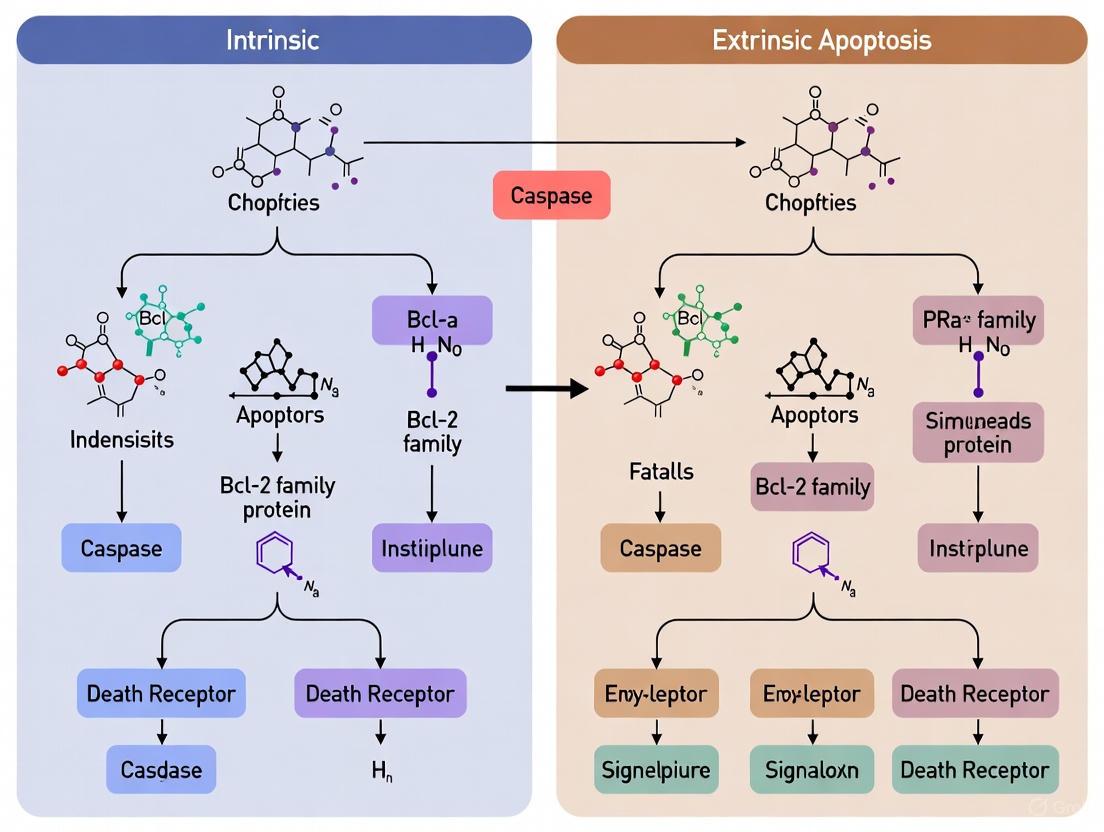
The following diagram illustrates the key components and interactions of both apoptotic pathways:
Diagram 1: Intrinsic and Extrinsic Apoptotic Pathways. The intrinsic pathway (left) is triggered by internal cellular stressors and regulated by BCL-2 family proteins, leading to mitochondrial outer membrane permeabilization (MOMP) and caspase-9 activation. The extrinsic pathway (right) is initiated by extracellular death ligands binding to cell surface receptors, resulting in caspase-8 activation. Both pathways converge on the activation of executioner caspases. Crosstalk occurs via caspase-8-mediated BID cleavage.
Pathway Integration and Crosstalk
While the intrinsic and extrinsic pathways can operate independently, significant crosstalk exists between them, primarily mediated by the BH3-only protein BID. Caspase-8-mediated cleavage of BID to tBID provides a critical amplification loop, particularly in Type II cells where the initial death receptor signal is insufficient to directly activate executioner caspases [6]. Additionally, recent research has revealed that certain forms of regulated necrosis, termed necroptosis, can be initiated when caspase-8 is inhibited or absent, highlighting the complex interplay between different cell death modalities [7].
Apoptosis in Development and Homeostasis
Developmental Apoptosis
Apoptosis plays a crucial role in embryonic and fetal development, serving to eliminate unnecessary cells and tissues at specific developmental stages. The predictable spatiotemporal pattern of developmental cell death was first observed in the 1920s, with the term "apoptosis" being formally introduced in 1972 by Kerr, Wyllie, and Currie [5] [8]. Key developmental processes dependent on apoptosis include:
- Digit individualization: Apoptosis eliminates the interdigital tissue to separate fingers and toes, with species exhibiting webbed limbs (e.g., ducks, bats) showing scarce cell death in these regions [2].
- Neural development: Approximately 80% of programmed cell deaths in C. elegans occur in neural cells, with similar extensive apoptosis observed in mammalian nervous system development to eliminate superfluous neurons and refine connections [5] [8].
- Vestigial structure removal: Regression of primitive structures such as the pronephros, notochord, and Müllerian/Wolffian ducts in a sex-specific manner [5].
- Tissue fusion events: Apoptosis facilitates the fusion of epithelial sheets during neural tube closure, midline body wall formation, and palate fusion [5] [2].
- Oocyte elimination: The human fetal ovary contains 7-8 million oocytes, which are reduced to approximately 100,000 at birth and further decline to a few hundred by menopause through apoptotic mechanisms [8].
Genetic studies in mice lacking key apoptotic regulators have refined our understanding of developmental apoptosis. While early histological observations suggested apoptosis was required for numerous developmental processes, functional assessments using gene-targeted mice revealed a more restricted set of essential functions. Current evidence indicates apoptosis is predominantly required to balance cell proliferation, ensure appropriate tissue size, facilitate fusion events in the body midline, and maintain the size of cavities once formed [5].
Homeostatic Apoptosis
In adult organisms, apoptosis maintains tissue homeostasis by balancing cell proliferation with cell death, thus ensuring the stability of tissue size and architecture. Key homeostatic functions include:
- Immune system regulation: Apoptosis eliminates self-reactive lymphocytes during differentiation (central tolerance) and removes antigen-specific lymphocytes at the termination of an immune response (peripheral tolerance) through activation-induced cell death (AICD) [3].
- Tissue turnover: Apoptosis facilitates the continuous replacement of old cells with new ones in tissues such as the intestinal epithelium and skin [4].
- Damage response: Cells with irreparable DNA damage or other significant injuries are eliminated through apoptosis to prevent the propagation of damaged cells [4].
Recent research has revealed that apoptotic cells can actively influence their microenvironment through the release of signaling molecules. For instance, during stress-induced apoptosis, dying cells can produce mitogenic signals (e.g., Wg, Dpp) that promote compensatory proliferation in surrounding cells, highlighting the integrated nature of cell death and tissue homeostasis [2].
Detection Methods and Experimental Approaches
The accurate detection and quantification of apoptosis are essential for both basic research and drug development. Multiple complementary approaches are typically employed to verify the type and stage of cell death.
Key Apoptosis Assays
Table 2: Essential Methods for Apoptosis Detection
| Method | Target/Principle | Stage Detected | Key Reagents | Applications |
|---|---|---|---|---|
| TUNEL Assay | Labels 3'OH ends of fragmented DNA with modified dUTP | Late apoptosis | TUNEL assay kits, fluorophore-conjugated dUTP | IF, IHC, flow cytometry; detects DNA fragmentation |
| Annexin V/PI Staining | Annexin V binds phosphatidylserine exposed on cell surface; PI stains DNA when membrane integrity is lost | Early apoptosis (Annexin V+/PI-), late apoptosis/necrosis (Annexin V+/PI+) | Annexin V-FITC, propidium iodide | Flow cytometry, distinguishes early/late apoptosis and necrosis |
| Caspase Activity Assays | Measures cleavage of caspase-specific substrates | Mid-stage apoptosis | Fluorogenic caspase substrates, caspase inhibitors | Enzyme activity measurement, high-throughput screening |
| Mitochondrial Membrane Potential (ΔΨm) | Detects loss of mitochondrial membrane potential using potential-sensitive dyes | Early apoptosis (intrinsic pathway) | TMRE, JC-1, MitoTracker dyes | Flow cytometry, fluorescence microscopy |
| Western Blotting | Detects cleavage of apoptotic markers (PARP, caspases) | Mid to late apoptosis | Antibodies against cleaved caspases, PARP, Bcl-2 family proteins | Protein expression and activation analysis |
| CyTOF (Mass Cytometry) | Simultaneous measurement of multiple protein markers at single-cell resolution | Multiple stages | Metal-conjugated antibodies, cisplatin viability stain | High-dimensional analysis of cell death in complex populations |
Detailed Experimental Protocols
Annexin V/Propidium Iodide (PI) Staining for Flow Cytometry
This widely used method distinguishes between early apoptotic (Annexin V+/PI-), late apoptotic (Annexin V+/PI+), and necrotic (Annexin V-/PI+) cells based on phosphatidylserine exposure and membrane integrity [1] [9].
Protocol:
- Cell Preparation: Harvest cells (approximately 1×10^6) by gentle trypsinization or collection from suspension cultures.
- Washing: Wash cells twice with cold PBS and resuspend in 1× binding buffer at a concentration of 1×10^6 cells/mL.
- Staining: Add Annexin V-FITC (e.g., 5 μL per test) and propidium iodide (e.g., 5 μL per test of a 50 μg/mL solution) to 100 μL of cell suspension.
- Incubation: Incubate for 15 minutes at room temperature in the dark.
- Dilution: Add 400 μL of 1× binding buffer to each tube.
- Analysis: Analyze by flow cytometry within 1 hour, using FITC (Ex=488 nm, Em=530 nm) and PI (Ex=488 nm, Em=617 nm) channels.
- Controls: Include unstained cells, single-stained controls for compensation, and cells treated with apoptosis inducers (e.g., camptothecin) as positive controls.
TUNEL Assay for DNA Fragmentation
The TUNEL (TdT-mediated dUTP Nick-End Labeling) assay detects DNA fragmentation, a hallmark of late-stage apoptosis, by labeling the 3'-OH ends of fragmented DNA [1].
Protocol for Cultured Cells:
- Fixation: Fix cells with 4% formaldehyde in PBS for 15 minutes at room temperature.
- Permeabilization: Permeabilize cells with 0.1% Triton X-100 in PBS for 5 minutes on ice.
- Labeling: Prepare TUNEL reaction mixture according to manufacturer's instructions (typically containing TdT enzyme and fluorochrome-labeled dUTP) and incubate with cells for 60 minutes at 37°C in the dark.
- Washing: Wash cells three times with PBS.
- Counterstaining (optional): Stain with DAPI (300 nM for 5 minutes) to visualize all nuclei.
- Analysis: Analyze by fluorescence microscopy or flow cytometry. Apoptotic nuclei will show bright fluorescent labeling.
Mitochondrial Membrane Potential Assessment Using TMRE
Tetramethylrhodamine ethyl ester (TMRE) is a cell-permeable, positively charged dye that accumulates in active mitochondria based on membrane potential. Apoptotic cells show decreased TMRE fluorescence due to loss of mitochondrial membrane potential (ΔΨm) [1].
Protocol:
- Loading: Incubate cells with 50-200 nM TMRE in culture medium for 15-30 minutes at 37°C.
- Washing: Wash cells with PBS to remove excess dye.
- Analysis: Analyze by flow cytometry or fluorescence microscopy (Ex=549 nm, Em=574 nm).
- Controls: Include untreated cells (high TMRE fluorescence) and cells treated with mitochondrial uncouplers like CCCP (50 μM for 15 minutes, low TMRE fluorescence) as controls.
The following diagram illustrates a comprehensive experimental workflow for apoptosis detection:
Diagram 2: Comprehensive Workflow for Apoptosis Detection. The experimental process begins with sample preparation, followed by selection of appropriate detection assays based on the apoptotic stage or pathway of interest. Multiple complementary methods are typically employed for verification. Data analysis and interpretation integrate results from various assays to determine the stage and mechanism of cell death.
Research Reagent Solutions
Table 3: Essential Research Reagents for Apoptosis Studies
| Reagent Category | Specific Examples | Function/Application |
|---|---|---|
| Caspase Inhibitors | Z-VAD-FMK (pan-caspase), Z-DEVD-FMK (caspase-3), Z-IETD-FMK (caspase-8) | Mechanism studies, determining caspase-dependence |
| Apoptosis Inducers | Staurosporine, Camptothecin, Etoposide, ABT-737 (BH3 mimetic), TRAIL | Positive controls, therapeutic mechanism studies |
| Antibodies | Anti-cleaved caspase-3, anti-PARP (cleaved), anti-Bax, anti-Bcl-2, anti-cytochrome c | Western blotting, immunohistochemistry, flow cytometry |
| Mitochondrial Dyes | TMRE, JC-1, MitoTracker Red CMXRos | Assessment of mitochondrial membrane potential and mass |
| Viability Indicators | Propidium iodide, 7-AAD, DAPI, Cisplatin-based viability dyes | Membrane integrity assessment, dead cell exclusion |
| Commercial Kits | Annexin V-FITC/PI kits, TUNEL assay kits, caspase activity assay kits | Standardized protocols for specific apoptosis detection |
| BH3 Mimetics | Venetoclax (ABT-199), Navitoclax (ABT-263) | Therapeutic research, BCL-2 family protein studies |
Therapeutic Targeting and Research Applications
The strategic manipulation of apoptotic pathways holds significant promise for therapeutic intervention, particularly in oncology where defective apoptosis is a cancer hallmark.
Targeting the Intrinsic Pathway
The development of BH3 mimetics represents a major advancement in targeting the intrinsic apoptotic pathway. These small molecules mimic the function of BH3-only proteins by binding to and inhibiting anti-apoptotic BCL-2 family proteins:
- Venetoclax (ABT-199): Specifically inhibits BCL-2, leading to BIM release and subsequent BAX/BAK activation [10]. Approved by the FDA for treatment of chronic lymphocytic leukemia (CLL) and acute myeloid leukemia (AML).
- Navitoclax (ABT-263): Inhibits BCL-2, BCL-xL, and BCL-w, but associated with thrombocytopenia due to BCL-xL inhibition [10].
- MCL-1 inhibitors (e.g., S63845): Currently in clinical development, showing promise in MCL-1-dependent cancers [10].
Targeting the Extrinsic Pathway
Therapeutic approaches targeting the extrinsic pathway have focused on recombinant TRAIL and death receptor agonists:
- Recombinant human TRAIL (dulanermin): Binds to DR4/DR5, selectively inducing apoptosis in cancer cells, though limited by short half-life [10].
- Agonistic DR4/DR5 antibodies (e.g., mapatumumab, lexatumumab): Designed to activate death receptors, though clinical efficacy has been limited by insufficient receptor clustering [10].
- Second-generation TRAIL variants (e.g., TLY012): PEGylated form with extended half-life (12-18 hours) showing enhanced antitumor activity in preclinical models [10].
- Eftozanermin alfa (ABBV-621): TRAIL receptor agonist fused to Fc domain, currently in clinical trials [10].
Combination Strategies
Overcoming resistance to single-agent apoptosis inducers often requires combination approaches:
- Venetoclax + Obinutuzumab: Superior to chemotherapy in CLL, providing a chemotherapy-free regimen [10].
- TLY012 + ONC201: Synergistic apoptosis induction in TRAIL-resistant pancreatic cancer models [10].
- TLY012 + PD-1 inhibition: Enhanced antitumor immunity and CD8+ T cell infiltration in pancreatic cancer models [10].
Apoptosis represents a critically important biological process that extends from embryonic development to adult tissue homeostasis. The intricate balance between pro-apoptotic and anti-apoptotic signals, particularly within the intrinsic and extrinsic pathways, ensures proper organismal development and maintains cellular equilibrium throughout life. Understanding the molecular mechanisms governing these pathways has enabled the development of targeted therapies, especially for cancer, where apoptotic evasion is a fundamental hallmark. Continued research into the nuanced regulation of apoptotic pathways, their interconnections with other cell death mechanisms, and the development of more effective strategies to modulate these pathways therapeutically remains a vital pursuit with significant implications for human health and disease treatment.
The intrinsic apoptotic pathway, also known as the mitochondrial pathway, is a precisely regulated cell death program essential for tissue homeostasis, development, and the elimination of damaged cells [11] [12]. This pathway is primarily controlled by the BCL-2 protein family, which integrates diverse intracellular stress signals to determine cellular fate [13] [14]. The critical event regulated by this protein family is mitochondrial outer membrane permeabilization (MOMP), which represents a point of no return in the commitment to cell death [12] [15]. Upon MOMP, cytochrome c is released into the cytosol, leading to the formation of the apoptosome and activation of caspase proteases that execute the controlled demolition of the cell [11] [12]. Dysregulation of this pathway is a hallmark of cancer, with many malignancies overexpressing anti-apoptotic BCL-2 family members to ensure survival [11] [13]. This technical guide provides an in-depth examination of the molecular mechanisms, experimental methodologies, and therapeutic targeting of the BCL-2-regulated intrinsic apoptosis pathway.
The BCL-2 Protein Family: Core Components and Classification
The BCL-2 protein family constitutes a critical regulatory network that governs MOMP. These proteins are categorized into three functional subgroups based on their structure and apoptotic function, each characterized by the presence of BCL-2 homology (BH) domains [11] [13].
Table 1: Classification of Core BCL-2 Family Proteins
| Subgroup | Representative Members | BH Domains | Primary Function |
|---|---|---|---|
| Anti-apoptotic | BCL-2, BCL-XL, MCL-1, BCL-w, A1/Bfl-1 [11] [13] [12] | BH1, BH2, BH3, BH4 [13] | Promote cell survival by inhibiting pro-apoptotic members and preventing MOMP [12] [14]. |
| Multi-domain Pro-apoptotic | BAX, BAK, BOK [11] [13] [16] | BH1, BH2, BH3 [12] | Direct executors of MOMP; form pores in the mitochondrial outer membrane [12] [15]. |
| BH3-only Pro-apoptotic | BID, BIM, PUMA, BAD, NOXA, BIK, BMF, HRK [11] [13] [14] | BH3-only [11] | Sense cellular stress and transmit death signals by engaging other BCL-2 family members [11] [14]. |
The anti-apoptotic proteins, such as BCL-2 and BCL-XL, are globular proteins featuring a surface hydrophobic groove formed by their BH1-3 domains [13]. This groove serves as the primary binding site for the BH3 domains of pro-apoptotic members [13] [14]. The multi-domain pro-apoptotic effectors BAX and BAK are essential for MOMP, as cells deficient in both proteins are highly resistant to a wide array of intrinsic apoptotic stimuli [12]. The BH3-only proteins act as specialized sentinels that respond to specific death signals, such as DNA damage (PUMA, NOXA), growth factor withdrawal (BAD), or death receptor signaling (tBID) [11].
Figure 1. BCL-2 Family Regulation of the Intrinsic Apoptotic Pathway. Cellular stress activates BH3-only proteins, which neutralize anti-apoptotic members. This derepresses the effectors BAX/BAK, leading to their oligomerization, MOMP, and cytochrome c release.
Molecular Mechanism of the BCL-2 Regulated Apoptotic Switch
The core mechanism of intrinsic apoptosis activation involves a delicate balance of interactions between the three BCL-2 subfamilies, ultimately controlling the activation of BAX and BAK.
The Indirect Activation Model
Prevailing evidence supports the indirect activation model, where apoptosis is the default state and pro-survival proteins function as the primary brakes by constraining BAX and BAK [14]. In healthy cells, anti-apoptotic proteins like BCL-XL and MCL-1 bind and sequester either the activated forms of BAX/BAK or the BH3-only activator proteins (e.g., BIM, tBID) that would otherwise trigger BAX/BAK activation [14]. Upon cellular stress, the activated BH3-only proteins bind to the hydrophobic groove of anti-apoptotic proteins with distinct affinity profiles [11] [14]. This binding neutralizes their protective function, thereby "derepressing" BAX and BAK and allowing them to undergo conformational activation and oligomerization [14].
Hierarchical Control by BH3-only Proteins
BH3-only proteins exhibit a hierarchy in their ability to engage anti-apoptotic family members [11]. "Promiscuous" binders like BIM, PUMA, and tBID can bind with high affinity to all anti-apoptotic BCL-2 proteins, making them particularly potent inducers of apoptosis [14] [17]. In contrast, "selective" binders such as BAD (binds BCL-2, BCL-XL, BCL-w) and NOXA (binds MCL-1, A1) have narrower binding profiles [11] [14]. Efficient apoptosis often requires the combined action of multiple BH3-only proteins to neutralize the full complement of anti-apoptotic guards present in a cell [14].
Membrane-dependent Complex Formation
The lipid environment of the mitochondrial membrane profoundly influences BCL-2 protein interactions. BAX is largely cytosolic and monomeric in healthy cells but translocates to the mitochondria and undergoes conformational change upon an apoptotic stimulus [12] [15]. BAK is already integrated into the mitochondrial membrane in an inactive state [12]. Fluorescence cross-correlation spectroscopy (FCCS) studies reveal that BCL-XL forms homodimers in solution and can heterodimerize with cBID both in solution and in membranes [15]. In contrast, BCL-XL binding to BAX occurs predominantly in membranes and with lower affinity than its binding to cBID [15]. Furthermore, membrane-inserted BAX can recruit soluble BAX in a feed-forward mechanism, while BCL-XL can retrotranslocate BAX from the membrane back to the cytosol, thereby preserving membrane integrity [15].
Table 2: Affinity and Specificity of Select BH3-only Proteins for Anti-apoptotic BCL-2 Members
| BH3-only Protein | Primary Inducing Signal | Anti-apoptotic Binding Partners | Relative Potency |
|---|---|---|---|
| BIM | Cytoskeletal disruption, ER stress [11] | BCL-2, BCL-XL, MCL-1, BCL-w, A1 [11] [14] | High (Promiscuous) |
| PUMA | DNA damage, p53 activation [11] [17] | BCL-2, BCL-XL, MCL-1, BCL-w [17] | High (Promiscuous) |
| tBID | Death receptor signaling [11] | BCL-2, BCL-XL, MCL-1, BCL-w, A1 [14] | High (Promiscuous) |
| BAD | Growth factor withdrawal [11] | BCL-2, BCL-XL, BCL-w [11] [14] | Moderate (Selective) |
| NOXA | DNA damage, p53 activation [11] | MCL-1, A1 [11] [14] | Moderate (Selective) |
Key Experimental Methods for Probing the BCL-2 Network
BH3 Profiling
BH3 profiling is a functional assay that measures the mitochondrial commitment to apoptosis, or "primed" state, of a cell [12].
- Principle: The technique uses synthetic peptides corresponding to the BH3 domains of different BH3-only proteins to deliver a standardized death signal to isolated mitochondria or permeabilized cells. The resulting pattern of MOMP reveals the dependence on specific anti-apoptotic proteins for survival and the functional status of BAX/BAK [12].
- Workflow:
- Sample Preparation: Isolate mitochondria from cell lines or patient-derived samples.
- Peptide Incubation: Incubate mitochondria with individual BH3 domain peptides (e.g., BIM, BAD, NOXA, HRK).
- MOMP Measurement: Quantify cytochrome c release or changes in mitochondrial membrane potential.
- Interpretation: The specific peptides that induce MOMP create a signature that identifies which anti-apoptotic proteins are maintaining survival (e.g., BAD-sensitivity indicates BCL-2/BCL-XL dependence; NOXA-sensitivity indicates MCL-1 dependence) [12].
- Application: BH3 profiling can classify apoptotic blocks into categories, such as a block due to high anti-apoptotic signaling (Class C) or a block due to deficient BAX/BAK function (Class B) [12]. It is used to predict sensitivity to BH3-mimetic drugs and to study mechanisms of resistance.
Figure 2. BH3 Profiling Experimental Workflow. A functional assay to determine mitochondrial priming by applying standardized BH3 death signals.
Quantitative Analysis of Protein Interactions
Understanding the precise affinities and stoichiometries of BCL-2 family interactions is crucial. Fluorescence Cross-Correlation Spectroscopy (FCCS) is a powerful solution-based technique used for this purpose [15].
- Principle: FCCS uses confocal microscopy to measure intensity fluctuations of fluorescently labeled molecules in a very small observation volume. When two differently colored molecules interact and diffuse together, their fluorescence signals fluctuate in synchrony, generating a cross-correlation signal [15].
- Protocol for BCL-2 Studies:
- Protein Labeling: Label full-length, purified BCL-2 family proteins (e.g., cBid, Bax, Bcl-xL) with distinct fluorophores (e.g., Alexa 488 and Cy5).
- Data Acquisition: Perform scanning FCCS measurements on proteins in solution or in the presence of membrane lipids (e.g., liposomes, mitochondrial membranes).
- Data Analysis: Calculate auto-correlation and cross-correlation curves. A significant positive cross-correlation amplitude indicates molecular interaction. The diffusion coefficients and interaction amplitudes provide information on complex stoichiometry and affinity [15].
- Key Findings: Using FCCS, researchers demonstrated that BAX is monomeric in solution, while BCL-XL forms homodimers. Crucially, the pattern of BCL-2 complex formation is drastically altered by membrane insertion, with key inhibitory interactions (e.g., BCL-XL/Bax) occurring exclusively in the membrane environment [15].
Additional Key Methodologies
- Co-immunoprecipitation & Western Blotting: Used to validate protein-protein interactions and study conformational changes in BAX/BAK using conformation-specific antibodies (e.g., Bax 6A7 antibody for active Bax) [12] [14].
- Cytochrome c Release Assays: Isolated mitochondria are treated with recombinant BH3-only proteins or peptides, and cytochrome c in the supernatant is quantified via ELISA or Western blot to directly measure MOMP [12].
- Genetic Knockout Models: Cells lacking specific BCL-2 family members (e.g., Bax/Bak DKO, Bid/Bim DKO) are used to establish the essential non-redundant functions of these proteins in the apoptotic pathway [14].
Table 3: Essential Research Reagent Solutions for Studying the BCL-2 Network
| Reagent / Tool | Primary Function / Assay | Key Characteristics and Examples |
|---|---|---|
| BH3 Domain Peptides | BH3 Profiling [12] | Synthetic peptides (~20 aa) derived from BH3 domains of BIM, BAD, NOXA, etc.; used to classify apoptotic dependence. |
| Recombinant Full-length Proteins | In vitro interaction & MOMP assays [15] | Fluorescently labeled full-length cBid, Bax, Bcl-xL for quantitative biophysical studies (e.g., FCCS). |
| Conformation-specific Antibodies | Detecting protein activation (e.g., IHC, WB) [12] | Antibody 6A7 detects active, conformation-changed Bax; anti-Bak Ab-1 for active Bak. |
| BH3-mimetic Compounds | Functional inhibition of anti-apoptotic proteins [13] [12] | ABT-737 (Bcl-2/Bcl-xL/Bcl-w inhibitor), Venetoclax/ABT-199 (Bcl-2 selective), Obatoclax (pan-inhibitor). |
| Genetic Models (KO/KD) | Establishing protein function [14] | Bax/Bak double-knockout cells; CRISPR/Cas9 or siRNA-mediated knockdown of specific BCL-2 members. |
Dysregulation in Cancer and Therapeutic Targeting
Pathological Deregulation
Cancer cells frequently hijack the intrinsic apoptotic pathway to ensure their survival. Common mechanisms include: overexpression of anti-apoptotic proteins like BCL-2 (e.g., in follicular lymphoma due to t(14;18) translocation) [11] [13]; loss of tumor suppressor p53, leading to impaired transcriptional activation of PUMA and NOXA [11]; and upregulation of pro-survival transcription factors like NF-κB, which enhances the expression of BCL-XL and BFL-1 [11]. This deregulation creates a state where malignant cells are "primed for death" but remain dependent on one or more anti-apoptotic BCL-2 family members for survival, a vulnerability known as "oncogenic addiction" [12].
BH3-mimetics: From Concept to Clinic
BH3-mimetics are a class of small molecule drugs designed to mimic the function of native BH3-only proteins by binding into the hydrophobic groove of anti-apoptotic BCL-2 proteins, thereby displacing pro-apoptotic partners and triggering apoptosis [11] [13].
- First-generation (Navitoclax/ABT-263): Orally available inhibitor of BCL-2, BCL-XL, and BCL-w. Its clinical development was limited by dose-limiting thrombocytopenia caused by on-target BCL-XL inhibition, which is essential for platelet survival [13].
- Second-generation (Venetoclax/ABT-199): A highly selective BCL-2 inhibitor that avoids BCL-XL-related thrombocytopenia. Venetoclax received FDA and EMA approval for treating certain hematologic malignancies and has transformed the therapeutic landscape for diseases like chronic lymphocytic leukemia (CLL) [13].
- Challenges and Next-generation Agents: Targeting other anti-apoptotic members like MCL-1 has proven challenging due to cardiac toxicity concerns [13]. Novel approaches such as PROTACs (Proteolysis Targeting Chimeras) and antibody-drug conjugates are being explored to achieve tumor-specific inhibition of BCL-XL or MCL-1, which could broaden the applicability of BH3-mimetics to solid tumors [13].
The BCL-2 protein family functions as a sophisticated and tightly regulated switch controlling the intrinsic apoptotic pathway. The quantitative and mechanistic understanding of the hierarchical interactions within this family has not only clarified a fundamental biological process but has also enabled the rational design of novel cancer therapeutics. Despite significant progress, challenges remain, including understanding the precise structural dynamics of BAX/BAK pore formation, the influence of mitochondrial lipid composition, and overcoming resistance to BH3-mimetics in the clinic. Future research integrating mitochondrial bioenergetics, non-canonical BCL-2 functions, and novel drug delivery platforms holds the promise of expanding the therapeutic potential of targeting the BCL-2 network in cancer and other diseases.
Apoptosis, or programmed cell death, is an energy-dependent and biochemically mediated process essential for eliminating infected or transformed cells, maintaining a properly functioning immune system, and ensuring normal development and homeostasis [18]. The extrinsic pathway of apoptosis, the focus of this whitepaper, is characterized by its initiation from outside the cell, most often through signals delivered by immune cells such as Natural Killer (NK) cells or CD8-positive Cytotoxic T lymphocytes (CTLs) [18]. This pathway is activated when extracellular death ligands bind to their cognate cell surface death receptors, leading to the assembly of a multi-protein complex known as the Death-Inducing Signaling Complex (DISC), which triggers a proteolytic cascade that dismantles the cell [18] [19]. Understanding the precise molecular mechanisms governing death receptor activation and DISC formation is not only fundamental to cell biology but also critical for drug development, particularly in oncology, where modulating this pathway can induce tumor regression [20].
Core Components of the Extrinsic Pathway
Death Receptors and Their Ligands
Death Receptors (DRs) are a subgroup of the Tumor Necrosis Factor Receptor (TNFR) superfamily characterized by a conserved intracellular protein-protein interaction domain known as the Death Domain (DD) [19] [21]. These receptors are transmembrane proteins that transmit apoptotic signals upon binding to their specific, trimeric death ligands [18] [19].
Table 1: Principal Death Receptors and Their Ligands
| Death Receptor | Main Ligand(s) | Key Features |
|---|---|---|
| CD95 (Fas/APO-1) | CD95 Ligand (FasL) | One of the best-studied prototypic death receptors; forms the DISC with FADD and caspase-8 [21]. |
| TRAIL-R1 (DR4) | TRAIL (Apo2L) | Along with TRAIL-R2, can selectively induce apoptosis in tumor cells, making it a therapeutic target [22]. |
| TRAIL-R2 (DR5) | TRAIL (Apo2L) | Shares high homology with TRAIL-R1; both receptors initiate apoptosis via DISC formation [22]. |
| TNFR1 | TNF-α | Can initiate both survival (via NF-κB) and apoptotic pathways; apoptosis involves a complex internalization process [19]. |
The Death-Inducing Signaling Complex (DISC)
The DISC is the foundational signaling platform of the extrinsic apoptotic pathway. Its assembly begins when a death receptor binds its trimeric ligand, inducing receptor oligomerization and recruitment of adapter proteins [18] [22].
The core components of the canonical DISC include:
- Receptors: Oligomerized death receptors (e.g., CD95, TRAIL-R1/R2) [22].
- Adapter Protein: FADD (Fas-Associated protein with Death Domain), which is recruited to the clustered receptor DDs via its own DD [18] [22].
- Initiator Caspases: Primarily procaspase-8 and/or procaspase-10, which are recruited to FADD via homotypic interactions between their Death Effector Domains (DEDs) [22] [21].
- Regulatory Proteins: c-FLIP (cellular FLICE-inhibitory protein), which exists in long (c-FLIP~L~) and short (c-FLIP~S~/c-FLIP~R~) isoforms and can either inhibit or potentiate caspase-8 activation depending on its concentration and isoform [21].
The assembly of these components brings multiple procaspase-8 molecules into close proximity, enabling their activation through dimerization and autoproteolytic cleavage [22] [21].
Figure 1: DISC Assembly and Activation. A death ligand binds and trimerizes its receptor, recruiting FADD via Death Domain (DD) interactions. FADD then recruits procaspase-8 and its regulator c-FLIP via Death Effector Domain (DED) interactions, forming the DISC. Procaspase-8 dimerization within the complex leads to its activation.
Molecular Mechanism and Stoichiometry of the DISC
A Paradigm Shift: DED Chain Model
The traditional model proposed a 1:1:1 stoichiometry for receptor:FADD:caspase-8 within the DISC. However, quantitative mass spectrometry analyses have revealed a more complex architecture. In the native TRAIL DISC, the adaptor protein FADD is substoichiometric, with up to a 9-fold greater quantity of caspase-8 than FADD [22]. This finding challenged the conventional model and led to the proposal of the DED chain model.
This model posits that the DED of FADD nucleates the formation of a filamentous chain composed of multiple procaspase-8 molecules (and/or c-FLIP), each interacting via their DEDs in a head-to-tail fashion [22] [21]. This linear assembly, rather than a simple trimeric complex, brings numerous procaspase-8 molecules into close proximity, facilitating dimerization and activation at specific points along the chain [22].
Table 2: Key Quantitative Findings from Native DISC Analysis
| Parameter | Traditional Model | Quantitative MS-Based Finding | Experimental System |
|---|---|---|---|
| FADD : Caspase-8 Ratio | ~1:1 | FADD is substoichiometric; up to 9x more caspase-8 than FADD [22]. | TRAIL DISC in hematopoietic cell lines (Jurkat, BJAB, Z138) [22]. |
| Overall DISC Stoichiometry | 1:1:1 (Receptor:FADD:Caspase-8) | Does not conform to a 1:1:1 model; supports a sequential DED chain [22]. | Affinity-purified native TRAIL DISC analyzed by LC-MS/MS [22]. |
| Functional Implication | Limited caspase-8 activation sites | DED chain assembly provides a mechanism for recruiting and activating multiple caspases, amplifying the death signal [22]. | Mutating key residues in procaspase-8 DED2 disrupts chain formation and cell death [22]. |
Caspase-8 Activation and Signal Propagation
Within the DED chain, procaspase-8 molecules form homodimers, which is the critical step for their activation. Dimerization induces autoproteolysis, cleaving procaspase-8 into its active form, which consists of the heterotetramer p10~2~-p18~2~ [21]. Once active caspase-8 is liberated from the DISC, it cleaves and activates downstream executioner caspases (caspase-3, -6, and -7), which then systematically dismantle the cell by cleaving hundreds of cellular substrates [18] [1].
The activated caspase-8 can propagate the death signal through two pathways:
- Direct Pathway: In so-called Type I cells, high levels of active caspase-8 from the DISC directly and robustly cleave and activate executioner caspase-3 [21].
- Amplification Loop (Type II cells): In Type II cells, where DISC formation is less robust, the apoptotic signal is amplified through the mitochondrial (intrinsic) pathway. Caspase-8 cleaves the BH3-only protein Bid into its active truncated form (tBid). tBid then translocates to mitochondria, promoting BAX/BAK oligomerization, Mitochondrial Outer Membrane Permeabilization (MOMP), and the release of cytochrome c, leading to apoptosome formation and caspase-9 activation [19] [21]. This represents a critical point of crosstalk between the extrinsic and intrinsic pathways.
Figure 2: Signal Propagation from the DISC. Active caspase-8 can trigger apoptosis via two primary routes. In Type I cells, it directly activates executioner caspase-3. In Type II cells, it cleaves Bid to tBid, which triggers mitochondrial amplification via MOMP, leading to caspase-9 activation and subsequent caspase-3 activation.
Regulation of the Pathway
The extrinsic pathway is subject to stringent and multi-layered regulation to prevent inappropriate cell death.
- c-FLIP Regulation: The c-FLIP protein is a critical regulator of DISC activity. At high concentrations, all c-FLIP isoforms (L, S, R) potently inhibit caspase-8 activation by occupying binding sites in the DED chain [21]. Intriguingly, at lower concentrations, c-FLIP~L~ can form heterodimers with procaspase-8, which paradoxically enhances caspase-8 activation, acting as a molecular switch that determines life/death decisions [21].
- Bcl-2 Family Proteins: In Type II cells, the anti-apoptotic members of the Bcl-2 family (e.g., Bcl-2, Bcl-X~L~, Mcl-1) can bind and sequester tBid, thereby blocking mitochondrial amplification and inhibiting apoptosis. This is the basis for the development of BH3-mimetics like venetoclax, which displace pro-apoptotic proteins from these anti-apoptotic guards, promoting cell death [13] [23].
Experimental Analysis of the DISC
Detailed Protocol: DISC Immunoprecipitation and Analysis
The following methodology, adapted from a key study, outlines the procedure for isolating and analyzing the native TRAIL DISC [22].
Objective: To affinity purify and characterize the composition and stoichiometry of the native TRAIL-induced DISC.
Materials and Reagents:
- Cells: Target cell line (e.g., Jurkat, BJAB, or Z138 hematopoietic tumor cells).
- Stimulus: Biotin-labeled, Strep-tag-modified recombinant TRAIL ligand.
- Lysis Buffer: Non-ionic detergent-based buffer (e.g., 1% Triton X-100 or CHAPS) supplemented with protease inhibitors.
- Isolation Matrix: Streptactin- or streptavidin-conjugated beads.
- Wash Buffer: Lysis buffer with adjusted salt concentration (e.g., 150-500 mM NaCl) to reduce non-specific binding.
- Elution Buffer: Suitable for mass spectrometry (e.g., 2x Laemmli buffer without bromophenol blue) or for western blotting (with bromophenol blue).
- Analysis Tools: SDS-PAGE equipment, western blot apparatus, and mass spectrometry instrumentation.
Procedure:
- Stimulation: Wash cells and resuspend in cold serum-free medium. Treat with biotinylated TRAIL (e.g., 1 µg/mL) for a defined period (typically 5-30 minutes) at 37°C. Include an untreated control.
- Termination and Lysis: Stop stimulation by placing cells on ice. Pellet cells by centrifugation and wash once with cold PBS. Lyse the cell pellet in a sufficient volume of ice-cold lysis buffer for 30-60 minutes with gentle agitation.
- Clarification: Centrifuge the lysate at high speed (e.g., 15,000 x g) for 15 minutes at 4°C to remove insoluble material and nuclei.
- Affinity Purification (DISC Pull-down): Incubate the clarified lysate with streptactin/streptavidin beads for several hours or overnight at 4°C with gentle rotation.
- Washing: Pellet the beads and wash thoroughly 3-5 times with cold wash buffer to remove non-specifically bound proteins.
- Elution: Elute the bound protein complex from the beads. This can be achieved by:
- Competitive Elution: Using biotin or desthiobiotin.
- Denaturing Elution: Boiling the beads in Laemmli sample buffer for western blot analysis.
- On-bead Digestion: For mass spectrometry, proteins can be digested with trypsin directly on the beads.
- Analysis:
- Western Blotting: Resolve eluted proteins by SDS-PAGE. Transfer to a membrane and probe with antibodies against core DISC components (TRAIL-R1/R2, FADD, caspase-8, c-FLIP).
- Quantitative Mass Spectrometry: Analyze tryptic peptides by LC-MS/MS. Use label-free quantification methods, such as measuring the Spectral Abundance Factor (SAF), to determine the relative stoichiometry of the proteins in the complex [22].
The Scientist's Toolkit: Key Research Reagents
Table 3: Essential Reagents for Studying the Extrinsic Pathway
| Research Reagent | Function / Target | Key Application |
|---|---|---|
| Recombinant Death Ligands (e.g., FasL, TRAIL) | Activate specific death receptors. | Inducing extrinsic apoptosis; studying receptor activation kinetics and DISC assembly [22]. |
| Agonistic Anti-DR Antibodies (e.g., anti-APO-1 for CD95) | Cluster and activate death receptors independently of native ligands. | A tool for specific receptor activation, useful in vitro and in vivo [21]. |
| c-FLIP Inhibitors (e.g., siRNA, small molecules) | Knock down or inhibit c-FLIP expression/function. | Studying the role of c-FLIP as a critical DISC regulator; sensitizing cells to death receptor-mediated apoptosis [21]. |
| Caspase Inhibitors (e.g., z-VAD-fmk) | Pan-caspase inhibitor, blocks catalytic activity of caspases. | Determining if cell death is caspase-dependent; distinguishing apoptosis from other death mechanisms like necroptosis [24]. |
| BH3-mimetics (e.g., Venetoclax/ABT-199) | Inhibit anti-apoptotic Bcl-2 proteins (e.g., Bcl-2). | Studying pathway crosstalk in Type II cells; cancer therapeutic to promote MOMP [13] [23]. |
| Annexin V Conjugates | Binds phosphatidylserine exposed on the outer leaflet of the plasma membrane. | Flow cytometry or microscopy detection of early-stage apoptosis [1]. |
| TUNEL Assay Kits | Labels fragmented DNA (3'-OH ends). | Detecting late-stage apoptosis; visualizing DNA cleavage in situ [1]. |
| Antibodies to Cleaved Caspases (e.g., Cleaved Caspase-3, -8) | Detect activated, cleaved fragments of caspases. | Specific immunohistochemical or western blot confirmation of apoptotic pathway execution [1]. |
The extrinsic apoptotic pathway, initiated by death receptors and executed through the DISC, represents a vital mechanism for controlled cell elimination. Recent quantitative studies have fundamentally refined our understanding of the DISC, moving from a simple stoichiometric complex to a dynamic, filamentous DED chain that serves as a potent activation platform for caspase-8 [22]. The precise regulation of this complex by molecules like c-FLIP, and its interconnection with the intrinsic pathway via Bid, creates a sophisticated network that integrates multiple death and survival signals [21]. For researchers and drug development professionals, continued elucidation of these mechanisms, including the non-linear dynamics and systems-level properties of the network, is essential for developing novel targeted therapies, such as specific TRAIL receptor agonists or next-generation BH3-mimetics, to harness the power of programmed cell death in treating cancer and other diseases [13] [20].
Apoptosis, or programmed cell death, is a fundamental biological process essential for maintaining tissue homeostasis, eliminating damaged cells, and ensuring proper embryonic development. This highly regulated form of cell death occurs through two principal signaling pathways: the extrinsic (death receptor) pathway and the intrinsic (mitochondrial) pathway [25]. While these pathways initiate through distinct mechanisms, they converge on a common execution phase mediated by proteolytic enzymes called caspases. The delicate balance between cell survival and death is critically controlled by three key molecular families: caspases as the primary executioners, B-cell lymphoma 2 (BCL-2) proteins as the major regulators of mitochondrial integrity, and inhibitor of apoptosis proteins (IAPs) as the central modulators of caspase activity [26] [27] [28]. Understanding the intricate interactions between these components provides crucial insights into normal physiology and disease pathogenesis, particularly in cancer and neurodegenerative disorders, and informs the development of targeted therapeutic strategies.
The Caspase Family: Executors of Cell Death
Classification and Activation Mechanisms
Caspases are evolutionarily conserved cysteine proteases that cleave their substrates at specific aspartic acid residues, serving as central mediators of programmed cell death [26]. These enzymes are synthesized as inactive zymogens (procaspases) that require proteolytic processing for activation. Caspases are categorized based on their structural features and position in apoptotic signaling cascades:
- Initiator Caspases (caspase-2, -8, -9, -10): Characterized by long prodomains that facilitate recruitment to specific activation complexes. They function apically in cell death pathways and are activated through "induced proximity" dimerization [29].
- Effector Caspases (caspase-3, -6, -7): Contain short prodomains and exist as preformed, inactive homodimers. They are cleaved and activated by initiator caspases and directly mediate the proteolytic dismantling of the cell [29].
Table 1: Caspase Classification and Functions in Programmed Cell Death
| Caspase | Type | Activation Complex | Primary Pathways | Key Substrates/Functions |
|---|---|---|---|---|
| Caspase-8 | Initiator | DISC, FADDosome | Extrinsic Apoptosis, Necroptosis, Pyroptosis | Activates caspase-3, cleaves BID, cleaves GSDMC [26] |
| Caspase-9 | Initiator | Apoptosome | Intrinsic Apoptosis | Activates caspase-3/7, inhibits necroptosis via RIPK1 cleavage [26] [29] |
| Caspase-10 | Initiator | DISC | Extrinsic Apoptosis | Regulates caspase-8-mediated cell death [26] |
| Caspase-2 | Initiator | PIDDosome | Intrinsic Apoptosis | Cleaves BID, DNA damage response [29] |
| Caspase-3 | Effector | - | Apoptosis, Pyroptosis | Cleaves PARP, lamin, cytoskeletal proteins; activates GSDME [26] |
| Caspase-6 | Effector | - | Apoptosis | Activates caspase-8; regulates GSDMB-mediated pyroptosis [26] |
| Caspase-7 | Effector | - | Apoptosis | Cleaves PARP; suppresses pyroptosis via GSDMD cleavage [26] |
| Caspase-1 | Inflammatory | Inflammasome | Pyroptosis | Cleaves GSDMD, pro-IL-1β, pro-IL-18 [26] |
| Caspase-4/5/11 | Inflammatory | - | Pyroptosis | Cleaves GSDMD, mediates non-canonical inflammasome activation [26] |
Caspase Activation Complexes
The activation of initiator caspases occurs within large multiprotein complexes that serve as molecular platforms for proximity-induced dimerization and autoactivation:
- Death-Inducing Signaling Complex (DISC): Formed following engagement of death receptors (e.g., Fas, TRAIL receptors), the DISC recruits and activates caspase-8 and caspase-10 through adapter proteins like FADD (Fas-associated death domain) [29].
- Apoptosome: Formed when cytochrome c released from mitochondria binds to Apaf-1 (apoptotic protease activating factor-1), creating a heptameric platform that recruits and activates caspase-9 [29]. Each apoptosome backbone recruits and activates two caspase-9 molecules, establishing a 7:2 ratio between Apaf-1 and caspase-9 [29].
- PIDDosome: Activates caspase-2 in response to DNA damage and consists of five PIDDs, seven RAIDDs, and seven caspase-2 molecules [29].
- Inflammasome: Activates caspase-1 in response to inflammatory signals, leading to maturation of IL-1β and IL-18 and cleavage of gasdermin D to induce pyroptosis [26].
Diagram 1: Caspase Activation Complexes. Caspases are activated in large multiprotein complexes: the DISC (extrinsic pathway), apoptosome (intrinsic pathway), and PIDDosome (DNA damage response).
BCL-2 Protein Family: Regulators of Mitochondrial Apoptosis
Structural Classification and Functions
The BCL-2 protein family constitutes the critical regulatory checkpoint for the intrinsic apoptotic pathway, functioning primarily at the mitochondrial outer membrane [13]. This family is defined by the presence of BCL-2 homology (BH) domains and can be divided into three functional subgroups:
- Anti-apoptotic proteins (BCL-2, BCL-XL, MCL-1, BCL-W, BFL-1, BCL-B): Characterized by the presence of four BH domains (BH1-BH4), these proteins promote cell survival by sequestering pro-apoptotic family members and maintaining mitochondrial integrity [13] [28].
- Multi-domain pro-apoptotic proteins (BAX, BAK, BOK): Contain BH1-3 domains and directly mediate mitochondrial outer membrane permeabilization (MOMP), enabling the release of cytochrome c and other apoptogenic factors [13].
- BH3-only proteins (BID, BIM, BAD, PUMA, NOXA, BMF, HRK): Function as sentinels of cellular stress and initiate apoptosis by either neutralizing anti-apoptotic proteins or directly activating BAX/BAK [13].
Table 2: BCL-2 Family Protein Classification and Characteristics
| Subfamily | Protein | BH Domains | Molecular Weight | Primary Function | Regulatory Mechanisms |
|---|---|---|---|---|---|
| Anti-apoptotic | BCL-2 | BH1-4 | 26 kDa | Inhibits MOMP, binds pro-apoptotic members | Overexpressed in follicular lymphoma [13] |
| BCL-XL | BH1-4 | 30 kDa | Inhibits MOMP, regulates platelet survival | Critical for embryonic development [13] | |
| MCL-1 | BH1-3 | 37 kDa | Rapid turnover, inhibits apoptosis | Essential for lymphocyte development [13] | |
| BCL-W | BH1-4 | 18 kDa | Inhibits apoptosis in germ cells | Supports neuronal survival [28] | |
| BFL-1 | BH1,3 | 21 kDa | Inhibits apoptosis in hematopoietic cells | Regulated by NF-κB [28] | |
| Pro-apoptotic Multi-domain | BAX | BH1-3 | 21 kDa | Mediates MOMP | Activated by BH3-only proteins [13] |
| BAK | BH1-3 | 23 kDa | Mediates MOMP | Constitutively mitochondrial [13] | |
| BOK | BH1-3 | 25 kDa | Mediates MOMP | Regulates ER stress-induced apoptosis [28] | |
| BH3-only | BIM | BH3 | 25 kDa | Activates BAX/BAK, neutralizes anti-apoptotics | Connects cytoskeletal integrity to apoptosis [13] |
| BID | BH3 | 22 kDa | Links extrinsic to intrinsic pathway | Cleaved by caspase-8 to active tBID [26] | |
| BAD | BH3 | 24 kDa | Neutralizes BCL-2, BCL-XL | Regulated by phosphorylation [28] | |
| PUMA | BH3 | 26 kDa | Neutralizes all anti-apoptotic proteins | p53 target gene [13] |
Mechanism of Mitochondrial Outer Membrane Permeabilization
The BCL-2 protein family critically controls apoptosis by regulating the release of cytochrome c from mitochondria [13]. In response to cellular stress signals (e.g., DNA damage, growth factor withdrawal), activated BH3-only proteins engage with anti-apoptotic proteins and multi-domain pro-apoptotic effectors:
- Sensitizer BH3-only proteins (BAD, NOXA, BMF) bind to and neutralize specific anti-apoptotic proteins, displacing previously sequestered activator BH3-only proteins (BIM, tBID, PUMA) [13].
- Freed activator BH3-only proteins directly activate BAX and BAK, inducing conformational changes that promote their oligomerization and integration into the mitochondrial outer membrane [13].
- BAX/BAK oligomers form macropores that facilitate mitochondrial outer membrane permeabilization (MOMP), resulting in the release of cytochrome c, SMAC/DIABLO, and other intermembrane space proteins into the cytosol [25].
- Cytochrome c promotes apoptosome formation and caspase-9 activation, while SMAC/DIABLO neutralizes IAP-mediated caspase inhibition [27].
Diagram 2: BCL-2 Protein Regulation of Mitochondrial Apoptosis. Cellular stress activates BH3-only proteins that neutralize anti-apoptotic proteins and directly activate BAX/BAK, leading to mitochondrial outer membrane permeabilization and cytochrome c release.
Inhibitor of Apoptosis Proteins (IAPs): Caspase Regulators
Structural Domains and Family Members
The Inhibitor of Apoptosis (IAP) protein family comprises crucial negative regulators of caspase activity and cell death signaling pathways. IAPs are characterized by the presence of one to three baculoviral IAP repeat (BIR) domains, which are zinc-binding motifs that mediate protein-protein interactions [27]. Most IAPs also contain a C-terminal RING domain that confers E3-ubiquitin ligase activity, enabling them to target bound proteins for ubiquitination and degradation [27].
Key members of the IAP family include:
- XIAP (X-linked IAP): Directly binds to and inhibits caspase-3, -7, and -9 through its BIR2 and BIR3 domains [27].
- cIAP1 and cIAP2 (cellular IAPs): Regulate NF-κB signaling and modulate death receptor pathways through their E3 ubiquitin ligase activities [27].
- ML-IAP (melanoma IAP): Expressed primarily in melanoma cells, inhibits caspase activation [27].
- Survivin: Functions in both apoptosis inhibition and regulation of cell division [27].
- ILP-2 (IAP-like protein 2): Testis-specific IAP that inhibits caspase-9 [27].
Mechanisms of Caspase Inhibition and Regulation
IAPs employ multiple strategies to suppress apoptotic signaling:
- Direct caspase inhibition: XIAP directly binds to and inhibits active caspase-3, -7, and -9 through its BIR domains. The BIR2 domain of XIAP interacts with the substrate-binding cleft of caspase-3 and -7, while BIR3 binds to the dimerization interface of caspase-9, preventing its activation [27].
- Ubiquitin-mediated regulation: The RING domain of IAPs facilitates ubiquitination of bound caspases and IAPs themselves, leading to proteasomal degradation or altered activity [27].
- Modulation of death receptor signaling: cIAP1 and cIAP2 regulate TNF receptor signaling by ubiquitinating key components of the signaling complex, potentially diverting signals away from apoptosis [27].
- Neutralization by mitochondrial proteins: During apoptosis, proteins released from mitochondria (SMAC/DIABLO, HtrA2/Omi) bind to IAPs through an IAP-binding motif (IBM), displacing them from caspases and relieving inhibition [27].
Table 3: IAP Family Members and Their Functions
| IAP Protein | BIR Domains | RING Domain | Primary Functions | Regulatory Mechanisms |
|---|---|---|---|---|
| XIAP | 3 | Yes | Direct caspase inhibition; BIR2 binds caspase-3/7; BIR3 binds caspase-9 | Neutralized by SMAC/DIABLO [27] |
| cIAP1 | 3 | Yes | Regulates TNF signaling, NF-κB activation | Auto-ubiquitination and degradation [27] |
| cIAP2 | 3 | Yes | Regulates TNF signaling, NF-κB activation | Gene amplification in cancers [27] |
| ML-IAP | 1 | Yes | Inhibits caspase activation, binds SMAC | Overexpressed in melanoma [27] |
| Survivin | 1 | No | Inhibits apoptosis, regulates mitosis | Cell cycle-dependent expression [27] |
| ILP-2 | 1 | Yes | Inhibits caspase-9 | Testis-specific expression [27] |
| NAIP | 3 | No | Inhibits caspase-9, bacterial infection response | Mutated in spinal muscular atrophy [27] |
| Bruce | 1 | Yes | Regulates TNF signaling, large protein | May inhibit caspase-9 [27] |
Molecular Interplay in Apoptotic Pathways
Integrated Apoptotic Signaling Network
The extrinsic and intrinsic apoptotic pathways converge through molecular interactions between caspases, BCL-2 proteins, and IAPs, creating a finely tuned regulatory network:
- Extrinsic Pathway Initiation: Ligation of death receptors (e.g., Fas, TRAIL-R) leads to DISC formation, caspase-8 activation, and subsequent direct activation of effector caspases (caspase-3, -7) [26] [29]. In some cell types (Type II), caspase-8 cleaves BID to truncated tBID, which translocates to mitochondria and engages the intrinsic pathway through BAX/BAK activation [26].
- Intrinsic Pathway Amplification: Cellular stresses (DNA damage, ER stress) activate BH3-only proteins that neutralize anti-apoptotic BCL-2 proteins and directly activate BAX/BAK, leading to MOMP, cytochrome c release, apoptosome formation, and caspase-9 activation [13] [29].
- IAP Regulation: XIAP directly inhibits active caspase-3, -7, and -9, while cIAP1/2 modulate death receptor signaling. Mitochondrial release of SMAC/DIABLO and HtrA2/Omi during MOMP neutralizes IAP-mediated caspase inhibition [27].
Diagram 3: Integrated Apoptotic Signaling Network. The extrinsic and intrinsic pathways converge through molecular interactions between caspases, BCL-2 proteins, and IAPs, with key cross-talk at the level of BID cleavage and SMAC-mediated IAP neutralization.
Experimental Approaches for Apoptosis Research
Core Methodologies for Studying Apoptotic Players
Table 4: Key Experimental Methods for Apoptosis Research
| Methodology | Application | Key Reagents | Technical Considerations |
|---|---|---|---|
| BH3 Profiling | Measures mitochondrial priming to assess dependence on anti-apoptotic proteins | Synthetic BH3 peptides (BIM, BAD, NOXA), JC-1 or TMRE dyes | Requires fresh mitochondria, quantitative flow cytometry [13] |
| Co-immunoprecipitation | Detects protein-protein interactions between BCL-2 family members | Antibodies to BCL-2 proteins, protein A/G beads, crosslinkers | Maintain weak interactions during lysis, include controls [28] |
| Caspase Activity Assays | Quantifies caspase activation using fluorogenic substrates | DEVD-AFC (caspase-3/7), LEHD-AFC (caspase-9), IETD-AFC (caspase-8) | Measure kinetics, use specific inhibitors for validation [29] |
| Mitochondrial Isolation and Cytochrome c Release | Assesses MOMP in response to apoptotic stimuli | Differential centrifugation, cytochrome c ELISA, Western blot | Purity mitochondria, prevent mechanical rupture [13] |
| Surface Plasmon Resonance | Measures binding kinetics of BH3 mimetics to BCL-2 proteins | Recombinant BCL-2 proteins, BH3 peptides, Biacore system | Control for DMSO solvent, regenerate chips properly [13] |
Research Reagent Solutions
Table 5: Essential Research Reagents for Apoptosis Studies
| Reagent Category | Specific Examples | Function/Application | Key Features |
|---|---|---|---|
| BH3 Mimetics | Venetoclax (ABT-199), Navitoclax (ABT-263), A-1331852 (BCL-XL specific) | Selectively inhibit anti-apoptotic BCL-2 proteins | Venetoclax is BCL-2 selective; Navitoclax targets BCL-2/BCL-XL/BCL-w [13] |
| SMAC Mimetics | Birinapant, LCL161, AT-406 | Antagonize IAP proteins to promote caspase activation | Mimic SMAC/DIABLO IBM motif, induce cIAP1/2 degradation [30] |
| Caspase Inhibitors | Z-VAD-FMK (pan-caspase), Q-VD-OPh, Emricasan | Broad-spectrum caspase inhibition for mechanistic studies | Z-VAD-FMK is irreversible; Q-VD-OPh has better cell permeability [29] |
| Death Receptor Agonists | Recombinant TRAIL, Agonistic anti-Fas antibodies | Activate extrinsic apoptotic pathway | TRAIL preferentially kills transformed cells [30] |
| IAP Antibodies | Anti-XIAP, Anti-cIAP1, Anti-Survivin | Detect IAP expression and localization by Western blot, IHC | Many commercial antibodies validated for specific applications [27] |
| BCL-2 Family Antibodies | Anti-BCL-2, Anti-BAX, Anti-BIM, Anti-BID | Protein detection, conformation-specific antibodies available | Conformation-specific antibodies detect active BAX/BAK [28] |
Therapeutic Targeting and Clinical Applications
Current Clinical Agents
The understanding of caspase regulation, BCL-2 family function, and IAP biology has enabled the development of targeted therapeutic agents:
- Venetoclax (ABT-199): First selective BCL-2 inhibitor approved by FDA in 2016 for treatment of chronic lymphocytic leukemia (CLL) and acute myeloid leukemia (AML) [13] [28]. It disrupts BCL-2 interaction with pro-apoptotic proteins, promoting mitochondrial apoptosis in cancer cells.
- Navitoclax (ABT-263): Oral inhibitor of BCL-2, BCL-XL, and BCL-w that demonstrated efficacy in clinical trials but limited by BCL-XL inhibition-mediated thrombocytopenia [13].
- SMAC Mimetics (birinapant, LCL161): Antagonize IAP proteins and promote caspase activation, currently in clinical trials as single agents and in combination therapies [30].
- MCL-1 Inhibitors (S63845, AMG-176): Selective MCL-1 inhibitors that have shown preclinical efficacy in multiple cancer types, with several agents in clinical development [13].
Emerging Therapeutic Approaches
Novel strategies are being developed to overcome limitations of current agents:
- PROTACs (Proteolysis Targeting Chimeras): Bifunctional molecules that recruit E3 ubiquitin ligases to target proteins, enabling degradation of specific BCL-2 family members [13].
- Antibody-Drug Conjugates (ADCs): Enable selective delivery of apoptotic agents to tumor cells expressing specific surface markers [13].
- BH4 Domain Targeting: Emerging approach targeting the BH4 domain of BCL-2, which is critical for its anti-apoptotic function and implicated in non-apoptotic signaling [13].
- Combination Therapies: Rational combinations of BH3 mimetics with conventional chemotherapy, targeted agents, or immunotherapy to overcome resistance and enhance efficacy [30].
The intricate interplay between caspases, BCL-2 proteins, and IAPs constitutes the core regulatory machinery of apoptotic cell death. The BCL-2 family serves as the decisive checkpoint for mitochondrial integrity, caspases function as the executioners of cell dismantling, and IAPs provide a critical control layer that modulates caspase activity. Continued research into the structural biology, regulatory mechanisms, and pathophysiological roles of these key molecular players continues to yield insights into fundamental cell biology and provides innovative approaches for therapeutic intervention in cancer and other diseases characterized by dysregulated cell death. The ongoing clinical development of novel agents targeting these pathways holds promise for improving outcomes for patients with malignancies resistant to conventional therapies.
In the comparative analysis of intrinsic and extrinsic apoptosis, the pathways converge unequivocally at the activation of executioner caspases, which serve as the ultimate effectors of cellular dismantling. While the intrinsic (mitochondrial) and extrinsic (death receptor) pathways initiate apoptosis through distinct molecular mechanisms and signaling complexes, they ultimately both activate caspase-3, -6, and -7, which execute the ordered demolition of cellular structures [31] [26]. This point of convergence represents a critical commitment to cell death, where diverse upstream signals become channeled into a common destructive cascade. Executioner caspases function as the central processing units that coordinate the systematic deconstruction of the cell through limited proteolysis of key structural and regulatory proteins, ultimately producing the characteristic morphological hallmarks of apoptosis while minimizing inflammatory consequences [1]. Understanding this terminal phase provides crucial insights for therapeutic interventions in cancer, neurodegenerative disorders, and other conditions characterized by dysregulated cell death.
Molecular Mechanisms of Executioner Caspase Activation
Executioner caspases-3, -6, and -7 exist in healthy cells as inactive dimeric zymogens (pro-caspases) that require proteolytic cleavage for activation [31]. Their activation represents the definitive commitment to apoptotic cell death and is mediated by initiator caspases from both major pathways.
Activation by the Intrinsic Pathway
The intrinsic apoptotic pathway activates executioner caspases through mitochondrial outer membrane permeabilization (MOMP) and formation of the apoptosome complex [26]. Cellular stresses (e.g., DNA damage, oxidative stress) trigger the release of cytochrome c from mitochondria, which binds to Apaf-1 in the cytosol. This binding, in the presence of dATP/ATP, induces conformational changes in Apaf-1 that expose its CARD domain and promote oligomerization into a heptameric wheel-like structure called the apoptosome [31]. The apoptosome then recruits and activates procaspase-9 through CARD-CARD interactions, forming a catalytic platform where caspase-9 dimers gain proteolytic activity. Once activated, caspase-9 cleaves and activates executioner caspases-3 and -7, initiating the demolition phase of apoptosis [26] [32].
Activation by the Extrinsic Pathway
The extrinsic apoptotic pathway initiates at the plasma membrane through ligand binding to death receptors (e.g., Fas, TRAIL receptors) [31]. Receptor activation leads to the formation of the Death-Inducing Signaling Complex (DISC), where adapter proteins (FADD/TRADD) recruit and activate procaspase-8 through dimerization [26] [32]. In type I cells, active caspase-8 directly cleaves and activates executioner caspases-3 and -7. In type II cells, caspase-8 cleaves the BH3-only protein Bid to generate tBid, which translocates to mitochondria and amplifies the death signal through the intrinsic pathway, resulting in a more robust executioner caspase activation [31].
The following diagram illustrates how both pathways converge on executioner caspase activation:
Structural Transformation Upon Activation
The transition from inactive zymogen to active executioner caspase involves precise structural rearrangements. Procaspase-3 exists as a dimer where the catalytic cysteine residues are improperly positioned for substrate binding [31]. Cleavage by initiator caspases at specific aspartic acid residues separates the prodomain and generates large (p17) and small (p12) subunits that reassemble into an active heterotetramer with two opposing active sites [31] [32]. This conformational change creates a functional mature protease capable of recognizing and cleaving target substrates after specific aspartate residues, with executioner caspases exhibiting preferences for different tetra-peptide motifs (e.g., DEVD for caspase-3) [32].
The Demolition Cascade: Substrate Cleavage and Cellular Deconstruction
Once activated, executioner caspases orchestrate cellular demolition through limited proteolysis of several hundred cellular proteins, resulting in the characteristic morphological and biochemical changes of apoptosis.
Key Substrates and Their Functional Consequences
Table 1: Major Substrates of Executioner Caspases and Their Roles in Cellular Dismantling
| Substrate Category | Representative Substrates | Cleavage Consequence | Functional Outcome |
|---|---|---|---|
| DNA Repair Enzymes | PARP, DNA-PK | Inactivation | Prevents DNA repair, promotes genomic disintegration [26] [1] |
| Structural Proteins | Lamin A/C, Nuclear Mitotic Apparatus (NuMA) | Disassembly | Nuclear envelope breakdown, chromatin condensation [26] [1] |
| Cytoskeletal Components | Actin, Gelsolin, Keratins | Fragmentation | Membrane blebbing, loss of cell shape, apoptotic body formation [1] |
| Cell Cycle Regulators | p21, RB1 | Altered function | Cell cycle arrest [26] |
| Signaling Molecules | PKC, AKT | Inactivation | Termination of survival signals [26] |
| Other Caspases | Caspase-6 | Activation | Amplification cascade, additional substrate cleavage [26] |
Morphological Stages of Execution
The proteolytic activity of executioner caspases produces a stereotypic sequence of morphological changes that define apoptosis:
- Early Phase: Externalization of phosphatidylserine to the outer leaflet of the plasma membrane serves as an "eat me" signal for phagocytes [1].
- Mid Phase: Cell shrinkage, chromatin condensation, and disruption of the nuclear envelope through lamin cleavage [26] [1].
- Late Phase: DNA fragmentation into nucleosomal fragments, membrane blebbing, and formation of apoptotic bodies containing cellular debris [1].
This controlled dismantling ensures the cell is removed without triggering inflammation, distinguishing apoptosis from necrotic cell death.
Experimental Methods for Detection and Quantification
Research into executioner caspase activity relies on well-established methodologies that detect activation, localization, and functional consequences. The following workflow outlines key experimental approaches:
The Scientist's Toolkit: Essential Research Reagents
Table 2: Key Research Reagents for Studying Executioner Caspases
| Reagent Category | Specific Examples | Research Application | Experimental Notes |
|---|---|---|---|
| Activity Assays | Fluorogenic substrates (DEVD-AFC, DEVD-AMC) | Quantifying caspase-3/7 activity in cell lysates | Provides kinetic data; highly sensitive to inhibitor effects [33] |
| Antibodies | Anti-cleaved caspase-3, Anti-PARP (cleaved) | Western blot, IHC, and immunofluorescence detection | Distinguishes active from inactive forms; crucial for tissue staining [1] |
| Detection Kits | Annexin V-FITC/PI apoptosis detection kits | Flow cytometry analysis of early apoptosis | Distinguishes early apoptotic (Annexin V+/PI-) from late apoptotic/necrotic cells [1] |
| Live-Cell Probes | Cell-permeable FLICA reagents | Real-time caspase activity in living cells | Enables kinetic studies but can inhibit caspase activity [34] |
| DNA Fragmentation Kits | TUNEL assay kits | Detecting late-stage apoptotic DNA cleavage | Specific for apoptosis when combined with morphological analysis [1] |
| Caspase Inhibitors | Z-VAD-FMK (pan-caspase), DEVD-CHO (caspase-3) | Determining caspase-dependence of cell death | Used as negative controls and mechanistic tools [26] |
Advanced Technological Platforms
The apoptosis assays market is experiencing significant technological advancement, with the North American market projected to grow from USD 2.7 billion in 2024 to USD 6.1 billion by 2034, reflecting increased research activity [33]. Key technological platforms include:
- Flow Cytometry: Enables multiparameter analysis of caspase activation combined with cell surface markers and viability dyes [33].
- High-Content Screening Systems: Automated microscopy platforms that quantify caspase activation and morphological changes in large compound screens [34].
- Luminescence/Fluorescence Plate Readers: High-throughput capability for kinetic analysis of caspase activity in multi-well formats [33].
- Multiplex Assays: Simultaneous measurement of multiple caspases and cell death parameters in single samples [34].
Research Applications and Therapeutic Implications
Executioner caspases represent not only fundamental biological mediators but also valuable therapeutic targets and biomarkers in disease research and drug development.
Cancer Research and Therapeutics
Dysregulation of executioner caspase activation is a hallmark of cancer, enabling tumor cells to evade programmed cell death [31] [35]. Research applications include:
- Therapeutic Resistance Assessment: Evaluating how cancer cells develop resistance to chemo- and radiotherapy through defective executioner caspase activation [35].
- Targeted Therapy Development: Screening for compounds that restore executioner caspase activation in apoptosis-resistant cancers [36].
- BH3-mimetic Drugs: Developing and testing small molecules (e.g., Venetoclax) that bypass upstream defects to directly activate the intrinsic apoptotic pathway [1].
The global apoptosis market, valued at USD 4.04 billion in 2025, reflects substantial investment in these research areas, particularly in oncology which dominates 40.5% of the application share [35].
Neurodegenerative Disease Research
Excessive executioner caspase activity contributes to neuronal loss in neurodegenerative conditions [31] [35]. Research focuses include:
- Biomarker Development: Detecting activated caspase-3 in cerebrospinal fluid or via imaging as an early disease marker [35].
- Neuroprotective Strategies: Screening caspase inhibitors to prevent excessive neuronal loss while maintaining physiological apoptosis [31].
- Pathway Elucidation: Determining specific executioner caspase substrates responsible for neuronal dysfunction in Alzheimer's and Parkinson's diseases [26].
Drug Discovery and Development
Executioner caspase assays are integral to pharmaceutical development, with applications in:
- Toxicology Studies: Assessing drug-induced apoptosis in liver, kidney, and cardiac tissues [33] [34].
- Efficacy Screening: Evaluating candidate compounds' ability to induce caspase-mediated apoptosis in target cells [34].
- Personalized Medicine: Using caspase activation profiles to predict individual patient responses to therapies [33] [35].
The growing emphasis on personalized medicine is driving demand for more precise apoptosis assays, with the pharmaceutical and biotechnology companies segment constituting 42.8% of end-users in the apoptosis market [35].
The point of convergence at executioner caspase activation represents both a fundamental biological principle and a strategic research focus. While the intrinsic and extrinsic pathways initiate through distinct mechanisms, their ultimate reliance on caspase-3, -6, and -7 highlights the efficiency of evolution in creating diverse sensors that feed into common effectors. Current research continues to reveal unexpected complexities in this convergence, including newly discovered non-apoptotic functions of executioner caspases in cellular differentiation and inflammation [26]. Technological advances in detection methods, particularly multiparameter assays and live-cell imaging, are providing unprecedented resolution of the spatiotemporal dynamics of executioner caspase activation. Furthermore, the therapeutic targeting of this convergence point continues to yield novel approaches for modulating cell death in human disease, solidifying the central importance of executioner caspases in both basic biology and translational medicine.
From Bench to Bedside: Research Techniques and Therapeutic Applications
Apoptosis, a form of programmed cell death (PCD), is characterized by distinct morphological changes including cell shrinkage, chromatin condensation, membrane blebbing, and nuclear fragmentation [1]. This process is critical for tissue homeostasis, embryonic development, and the elimination of damaged or unwanted cells [1]. The detection of apoptosis is fundamental in diverse research areas, from basic cell biology to the development of novel therapeutics, particularly in oncology where the goal is often to induce apoptosis in cancer cells [37] [38].
The two main apoptotic pathways—intrinsic (mitochondrial) and extrinsic (death receptor)—converge on the activation of executioner caspases, leading to the systematic dismantling of the cell [1] [39]. This technical guide focuses on three cornerstone methodologies for detecting apoptosis: the TUNEL assay, which identifies DNA fragmentation; Annexin V staining, which detects phosphatidylserine externalization; and caspase activity measurement, which probes the core enzymatic machinery of apoptosis. Understanding the principles, applications, and limitations of these techniques is essential for accurately interpreting cell death in the context of intrinsic versus extrinsic pathway research.
Core Signaling Pathways in Apoptosis
Intrinsic and Extrinsic Apoptosis Pathways
The intrinsic and extrinsic apoptotic pathways are initiated by different stimuli but converge on the activation of executioner caspases. The diagram below illustrates the key components and sequence of events in these pathways.
The Central Role of Caspases
Caspases are cysteine-aspartic proteases that are synthesized as inactive zymogens (pro-caspases) and become activated through proteolytic cleavage [39]. They are the primary executioners of apoptosis and can be categorized based on their function and substrate specificity.
- Initiator Caspases (e.g., Caspase-8, -9, -10): These contain long pro-domains and are activated early in the apoptotic cascade. Caspase-8 is associated with the extrinsic pathway, while caspase-9 is associated with the intrinsic pathway [1] [39].
- Executioner Caspases (e.g., Caspase-3, -6, -7): These are activated by initiator caspases and are responsible for the cleavage of key cellular proteins, such as PARP and nuclear lamins, leading to the characteristic morphological changes of apoptosis [1] [40] [39].
The substrate specificity of different caspases is a key consideration for detection assays, as illustrated in the table below.
Table 1: Caspase Substrate Specificity and Primary Functions [41] [39] [37]
| Caspase | Primary Role/Pathway | Preferred Tetrapeptide Motif | Cleaves DEVD? |
|---|---|---|---|
| Caspase-8 | Initiator / Extrinsic | LETD | Weakly (++) |
| Caspase-9 | Initiator / Intrinsic | LEHD | Weakly (+) |
| Caspase-2 | Initiator / Stress | VDVAD | Yes (+) |
| Caspase-10 | Initiator / Extrinsic | LEHD | Weakly (+) |
| Caspase-3 | Executioner | DEVD | Strongly (+++) |
| Caspase-7 | Executioner | DEVD | Strongly (+++) |
| Caspase-6 | Executioner | VEID | Weakly (++) |
| Caspase-1 | Inflammatory / Pyroptosis | WEHD | No (-) |
| Caspase-4/5/11 | Inflammatory / Pyroptosis | LEVD/WEHD | No (-) |
Key Methods for Detecting Apoptosis
TUNEL Assay
The TUNEL (Terminal deoxynucleotidyl transferase dUTP Nick End Labeling) assay detects DNA fragmentation, a hallmark of late-stage apoptosis [1] [37]. The enzyme terminal deoxynucleotidyl transferase (TdT) catalyzes the addition of fluorescently-labeled dUTP to the 3'-hydroxyl termini of double-stranded DNA breaks, allowing visualization of apoptotic cells via microscopy or flow cytometry [1].
Experimental Protocol
- Sample Preparation: Fix cells or tissue sections using paraformaldehyde. For paraffin-embedded tissues, deparaffinization and rehydration are required [1].
- Permeabilization: Treat samples with a permeabilization buffer (e.g., containing Triton X-100 or proteinase K) to allow reagent access to the nucleus.
- Labeling: Incubate samples with the TUNEL reaction mixture containing TdT enzyme and fluorescently-tagged dUTP (e.g., fluorescein-dUTP).
- Detection and Analysis: Visualize labeled DNA fragments using fluorescence microscopy, or quantify the signal using flow cytometry. Counterstaining with DAPI or PI is recommended to visualize total nuclei [1].
Data Interpretation and Considerations
A key advantage of the TUNEL assay is its sensitivity in detecting late-stage apoptosis. However, it is crucial to note that DNA fragmentation is not exclusive to apoptosis; it also occurs during necrotic cell death [1]. Therefore, results should be corroborated with morphological analysis. Apoptotic cells display small, round, evenly distributed apoptotic bodies, whereas necrotic DNA fragmentation is less organized and associated with cell lysis [1].
Annexin V Staining
The externalization of phosphatidylserine (PS) from the inner to the outer leaflet of the plasma membrane is a hallmark of early apoptosis [1]. Annexin V, a calcium-dependent phospholipid-binding protein, has a high affinity for PS and can be used as a sensitive probe for detecting this event [1] [37].
Experimental Protocol
- Cell Harvesting: Gently harvest cells to avoid mechanical induction of apoptosis or necrosis. Wash cells with cold PBS.
- Staining: Resuspend cells in a binding buffer containing calcium and a fluorescent conjugate of Annexin V (e.g., Annexin V-FITC). To distinguish early apoptosis from late apoptosis/necrosis, include a vital dye such as propidium iodide (PI) [1] [38].
- Incubation: Incubate the cell suspension in the dark for 15-20 minutes.
- Analysis: Analyze by flow cytometry within 1 hour. Cells are categorized as follows:
- Annexin V⁻ / PI⁻: Viable, non-apoptotic cells.
- Annexin V⁺ / PI⁻: Early apoptotic cells.
- Annexin V⁺ / PI⁺: Late apoptotic or necrotic cells.
Data Interpretation and Considerations
Annexin V staining is a powerful tool for identifying early apoptosis before loss of membrane integrity. However, PS exposure can also occur in other contexts, such as in activated lymphocytes or during necrosis if the membrane is damaged [42]. The combination with PI is therefore essential for accurate interpretation. Furthermore, fixation can permeabilize cells, leading to artifactual Annexin V binding; thus, analysis is best performed on live, unfixed cells [1].
Caspase Activity Detection
Measuring the activity of executioner caspases, particularly caspase-3 and -7, is one of the most popular and definitive methods for confirming apoptosis, as their activation is often considered a "point of no return" [41] [37]. This is typically done using synthetic substrates containing the caspase-specific cleavage sequence DEVD.
Experimental Protocol (Luminescent Assay)
The following protocol is adapted for a homogeneous, "add-mix-measure" format suitable for high-throughput screening in plate readers [37].
- Cell Plating: Plate cells in an opaque-walled, white microplate for optimal luminescence signal detection. A clear bottom can be used for concurrent microscopic observation.
- Treatment: Treat cells with the apoptotic inducer of choice for a predetermined time.
- Assay Reagent Addition: Add an equal volume of Caspase-Glo 3/7 Reagent to each well. The reagent contains a proluminescent substrate (DEVD-aminoluciferin), luciferase, and other necessary components.
- Incubation: Mix contents gently on a plate shaker and incubate at room temperature for 30-120 minutes to allow caspase cleavage to release aminoluciferin, which is converted to light by luciferase.
- Measurement: Measure the generated luminescence (Relative Luminescence Units, RLU) using a plate-reading luminometer. The signal is proportional to caspase activity [37].
Data Interpretation and Considerations
Luminescent caspase assays are highly sensitive and can be miniaturized for high-density plate formats [37]. It is important to validate the assay specificity using pan-caspase inhibitors (e.g., Z-VAD-FMK) or caspase-specific inhibitors, which should abrogate the signal [41]. Furthermore, in caspase-3 deficient cell lines (e.g., MCF-7), a significant signal may still be detected due to caspase-7 activity, which also cleaves the DEVD motif [41].
Comparative Analysis of Apoptosis Detection Methods
The table below provides a consolidated comparison of the three core apoptosis detection methods, highlighting their primary applications, advantages, and limitations.
Table 2: Comparative Overview of Key Apoptosis Detection Methods [1] [37] [38]
| Method | Detects | Stage of Apoptosis | Key Advantages | Key Limitations |
|---|---|---|---|---|
| TUNEL Assay | DNA fragmentation | Late | High sensitivity; applicable to tissue sections and fixed cells. | Not specific to apoptosis (also labels necrotic cells); requires careful morphological confirmation. |
| Annexin V Staining | Phosphatidylserine (PS) externalization | Early | Identifies apoptosis before loss of membrane integrity; can be combined with vital dyes. | PS exposure can occur in non-apoptotic cells; requires analysis of live, unfixed cells. |
| Caspase Activity | Executioner caspase (3/7) activity | Mid | Highly specific and definitive for apoptosis; amenable to HTS and multiplexing. | Does not distinguish between intrinsic/extrinsic pathways; may miss caspase-independent cell death. |
Advanced Techniques and Integrated Workflows
Real-Time Imaging and Multiparameter Analysis
Advanced techniques now allow for real-time, dynamic tracking of apoptosis in live cells. Genetically encoded biosensors, such as FRET-based caspase probes or fluorescent protein-based reporters, enable the visualization of caspase activation at single-cell resolution [41] [38]. For instance, a reporter with a DEVD sequence linking two halves of a fluorescent protein (e.g., ZipGFP) will fluoresce only upon caspase-mediated cleavage, providing a real-time readout of apoptosis [41].
Integrating multiple parameters is often necessary to unambiguously characterize cell death. A powerful approach involves combining:
- A FRET-based caspase sensor to detect caspase activation.
- A constitutively expressed fluorescent marker targeted to an organelle (e.g., Mito-DsRed) to monitor cell presence and membrane integrity [38]. This allows simultaneous discrimination of live cells (no FRET loss, intact fluorescence), apoptotic cells (FRET loss, intact organellar fluorescence), and necrotic cells (loss of FRET probe without cleavage, loss of organellar fluorescence) [38].
The Scientist's Toolkit: Key Research Reagents
The following table catalogues essential reagents and tools used in apoptosis detection, as featured in the cited research.
Table 3: Research Reagent Solutions for Apoptosis Detection [1] [41] [37]
| Reagent / Tool | Function / Target | Example Application |
|---|---|---|
| TUNEL Assay Kit | Labels 3'-OH ends of fragmented DNA | In situ detection of late apoptosis in fixed cells or tissues [1]. |
| Annexin V-FITC/PI | Binds externalized PS / labels DNA in permeabilized cells | Flow cytometric discrimination of early apoptotic (AnnV+/PI-) and late apoptotic/necrotic (AnnV+/PI+) cells [1]. |
| Caspase-Glo 3/7 Assay | Luminescent substrate for caspase-3/7 | High-throughput screening of caspase activity in live cells; measures DEVDase activity [37]. |
| FRET-based Caspase Sensor (e.g., CFP-DEVD-YFP) | Genetically encoded reporter for live-cell caspase imaging | Real-time, single-cell visualization of caspase activation via loss of FRET signal [38]. |
| Caspase Inhibitors (e.g., Z-VAD-FMK (pan), Z-IETD-FMK (Casp-8)) | Irreversibly inhibit caspase activity | Validation of caspase-dependent apoptosis; pathway dissection (e.g., extrinsic vs. intrinsic) [41] [43]. |
| MitoTracker Red | Fluorescent dye that accumulates in active mitochondria | Staining mitochondria to assess health and localization of Bcl-2 family proteins during apoptosis [1]. |
| Anti-Cleaved Caspase-3 Antibody | Detects activated caspase-3 by IHC/IF | Specific immunohistochemical or immunofluorescent detection of cells undergoing apoptosis [1]. |
The accurate detection of apoptosis is a cornerstone of cell death research. The TUNEL assay, Annexin V staining, and caspase activity measurements each provide unique and complementary information about different stages of the process. The choice of assay depends on the specific research question, the need for multiparametric data, and the context within the intrinsic or extrinsic apoptotic pathways. By understanding the principles and caveats of these core techniques, and by leveraging advanced tools for real-time, integrated analysis, researchers can generate robust and interpretable data to advance our understanding of cell death in health and disease.
Apoptosis, a form of programmed cell death, occurs through two primary pathways: the intrinsic (mitochondrial) pathway and the extrinsic (death receptor) pathway. These pathways converge on the activation of caspases that execute the cell death program but are initiated by distinct signals and involve unique molecular events. The intrinsic pathway responds to internal cellular stressors such as DNA damage, oxidative stress, or growth factor withdrawal, culminating in mitochondrial outer membrane permeabilization (MOMP) and the release of cytochrome c. In contrast, the extrinsic pathway is triggered by the binding of extracellular death ligands to cell surface receptors, leading to the formation of the death-inducing signaling complex (DISC). Monitoring the key events—MOMP, cytochrome c release, and DISC formation—is therefore crucial for elucidating the specific apoptotic route activated and for screening potential therapeutic compounds that modulate these pathways [44] [20].
This guide provides an in-depth technical framework for researchers and drug development professionals to monitor these pivotal events. We present detailed methodologies, quantitative parameters, and visualization tools to facilitate precise detection and interpretation within the context of comparative apoptosis research.
Pathway-Specific Molecular Events
The Intrinsic Pathway: MOMP and Cytochrome c Release
The intrinsic apoptosis pathway is regulated by the B-cell lymphoma 2 (BCL-2) protein family, which includes both anti-apoptotic (e.g., BCL-2, BCL-xL) and pro-apoptotic (e.g., BAX, BAK) members. In response to cellular stress, the activation of pro-apoptotic BH3-only proteins (e.g., BIM, PUMA) counteracts the function of anti-apoptotic proteins, enabling the effectors BAX and BAK to oligomerize and form pores in the mitochondrial outer membrane [44] [20]. This process is known as mitochondrial outer membrane permeabilization (MOMP).
MOMP is a pivotal event that leads to the release of several proteins from the mitochondrial intermembrane space into the cytosol. Among these, cytochrome c is of critical importance. Once in the cytosol, cytochrome c binds to Apoptotic Protease-Activating Factor 1 (APAF-1), leading to the formation of the apoptosome complex. This complex recruits and activates caspase-9, which then cleaves and activates downstream executioner caspases (e.g., caspase-3 and -7), ultimately leading to the controlled demolition of the cell [30] [20]. MOMP also results in the release of other factors, such as SMAC/DIABLO, which promotes apoptosis by inhibiting Inhibitor of Apoptosis Proteins (IAPs) [10] [30].
The Extrinsic Pathway: DISC Formation
The extrinsic apoptosis pathway is initiated by the binding of specific death ligands—such as Fas Ligand (FasL), Tumor Necrosis Factor (TNF)-α, and TNF-Related Apoptosis-Inducing Ligand (TRAIL)—to their corresponding death receptors (e.g., Fas, TNFR1, DR4/DR5) on the cell surface [24] [20].
Ligand-receptor binding triggers the recruitment of adapter proteins, such as Fas-Associated Death Domain (FADD) and, in some cases, TNF Receptor-Associated Death Domain (TRADD). These adapters then recruit initiator procaspase-8 (and in some cases, procaspase-10) molecules. The resulting multi-protein complex is known as the Death-Inducing Signaling Complex (DISC). Within the DISC, procaspase-8 undergoes autocatalytic activation [44] [24]. The activated caspase-8 then directly cleaves and activates downstream executioner caspases (caspase-3, -6, -7), initiating the apoptotic cascade. In some cell types (known as Type II cells), the apoptotic signal is amplified through the intrinsic pathway via caspase-8-mediated cleavage of the BCL-2 family protein BID to its active form, tBID, which subsequently promotes MOMP [30] [20].
Figure 1: Apoptotic Signaling Pathways. The diagram illustrates the key events in the extrinsic (death receptor) and intrinsic (mitochondrial) apoptosis pathways, culminating in the activation of executioner caspases and cell death.
Experimental Monitoring Methodologies
Monitoring Mitochondrial Outer Membrane Permeabilization (MOMP)
MOMP represents a commitment point in the intrinsic apoptotic pathway. The following protocols outline robust methods for its detection.
Protocol 3.1.1: Immunofluorescence Microscopy for BAX/BAK Oligomerization
- Principle: Detect the activation and oligomerization of pro-apoptotic proteins BAX and BAK on the mitochondrial membrane using conformation-specific antibodies.
- Procedure:
- Cell Seeding and Treatment: Seed cells on glass-bottom culture dishes. Treat with an intrinsic apoptotic inducer (e.g., 1-10 µM Staurosporine, 50 µM Etoposide, or UV irradiation).
- Fixation and Permeabilization: At designated time points (e.g., 0, 1, 2, 4, 6 hours), wash cells with PBS and fix with 4% paraformaldehyde for 15 minutes at room temperature. Permeabilize with 0.1% Triton X-100 in PBS for 10 minutes.
- Blocking: Incubate with blocking buffer (5% BSA in PBS) for 1 hour.
- Immunostaining: Incubate with primary antibodies against active conformation BAX (e.g., clone 6A7) and/or BAK, diluted in blocking buffer, overnight at 4°C. Include a mitochondrial marker (e.g., Anti-TOM20) for co-localization.
- Secondary Antibody Incubation: Wash and incubate with appropriate fluorescently-labeled secondary antibodies (e.g., Alexa Fluor 488, 555) for 1 hour at room temperature in the dark.
- Image Acquisition: Acquire high-resolution images using a confocal microscope. BAX/BAK oligomers appear as bright, punctate clusters on mitochondria.
- Data Analysis: Quantify the percentage of cells displaying punctate BAX/BAK staining or measure the co-localization coefficient between BAX/BAK and the mitochondrial marker using image analysis software (e.g., ImageJ, CellProfiler).
Protocol 3.1.2: Cytochrome c Release Assay by Subcellular Fractionation
- Principle: Biochemically separate cytosolic and mitochondrial fractions to track the translocation of cytochrome c.
- Procedure:
- Cell Harvest and Washing: Harvest approximately 1-2 x 10^7 treated and control cells by trypsinization and wash with ice-cold PBS.
- Fractionation: Resuspend the cell pellet in isotonic mitochondrial isolation buffer (e.g., containing mannitol, sucrose, EDTA, HEPES, and protease inhibitors). Homogenize cells using a Dounce homogenizer (30-50 strokes on ice). Confirm cell breakage (>90%) by trypan blue staining.
- Centrifugation: Centrifuge the homogenate at 800 x g for 10 minutes at 4°C to remove nuclei and unbroken cells. Transfer the supernatant to a new tube and centrifuge at 12,000 x g for 15 minutes at 4°C. The resulting pellet (mitochondrial fraction) and supernatant (cytosolic fraction) are collected separately.
- Western Blotting: Resuspend the mitochondrial pellet in lysis buffer. Determine protein concentration for both fractions. Load equal amounts of protein (20-30 µg) from cytosolic and mitochondrial fractions onto an SDS-PAGE gel.
- Detection: Perform Western blotting using antibodies against cytochrome c. Use antibodies for compartment-specific markers to assess fractionation purity: COX IV (mitochondrial) and α-tubulin or LDH (cytosolic).
- Data Analysis: A decrease in mitochondrial cytochrome c coupled with an increase in cytosolic cytochrome c confirms MOMP. Densitometric analysis of blots allows for semi-quantification.
Monitoring DISC Formation
The DISC is a transient complex whose formation can be captured using immunoprecipitation techniques.
Protocol 3.2.1: DISC Immunoprecipitation and Analysis
- Principle: Isolate the native DISC complex from the cell membrane using an antibody against a death receptor and analyze its components.
- Procedure:
- Stimulation and Lysis: Stimulate cells (e.g., 5-10 x 10^6) expressing a target death receptor (e.g., Fas, DR5) with the corresponding ligand (e.g., 100-500 ng/mL recombinant FasL or TRAIL) for a short duration (e.g., 0-30 minutes). Use unstimulated cells as a control.
- Rapid Lysis: Immediately after stimulation, place cells on ice and lyse using a mild, non-denaturing lysis buffer (e.g., 1% CHAPS or Digitonin in Tris-buffered saline with protease inhibitors) to preserve protein complexes. Avoid SDS or strong ionic detergents.
- Immunoprecipitation (IP): Pre-clear the lysate by incubating with protein A/G beads for 30 minutes. Incubate the pre-cleared lysate with an antibody specific to the intracellular death domain of the death receptor (e.g., anti-Fas C-terminal antibody) or a control IgG overnight at 4°C with gentle rotation.
- Bead Capture: Add protein A/G sepharose beads and incubate for 2-4 hours. Pellet the beads and wash extensively with lysis buffer.
- Elution and Analysis: Elute the immunoprecipitated proteins by boiling in 2X Laemmli SDS sample buffer. Resolve the proteins by SDS-PAGE and perform Western blotting to detect key DISC components: FADD, Caspase-8 (look for cleavage products), and c-FLIP.
- Data Analysis: Successful DISC formation is indicated by the co-precipitation of FADD and procaspase-8/cleaved caspase-8 with the death receptor in stimulated, but not unstimulated, samples.
Figure 2: Experimental Workflow for Key Event Monitoring. The chart outlines the primary methodologies for detecting MOMP and DISC formation.
Quantitative and High-Throughput Methods
Protocol 3.3.1: Flow Cytometry for Mitochondrial Membrane Potential (ΔΨm)
- Principle: MOMP disrupts the mitochondrial inner membrane potential. Fluorescent dyes like JC-1 or Tetramethylrhodamine (TMRM) can detect this loss.
- JC-1 Protocol: Stain ~1x10^6 cells with 2 µM JC-1 for 20-30 minutes at 37°C. Wash and analyze by flow cytometry. Healthy mitochondria with high ΔΨm cause JC-1 to form aggregates (red fluorescence, ~590 nm), while depolarized mitochondria contain JC-1 monomers (green fluorescence, ~529 nm). The ratio of red/green fluorescence is a quantitative measure of ΔΨm.
- Data Analysis: A decrease in the red/green fluorescence ratio indicates mitochondrial depolarization, a downstream consequence of MOMP.
Protocol 3.3.2: Caspase Activity Assays
- Principle: Measure the activity of initiator caspases as a functional readout of upstream events.
- Caspase-9 Activity: A luminescent or colorimetric assay using the specific substrate LEHD-pNA or LEHD-AFC. Increased activity confirms successful apoptosome formation following cytochrome c release.
- Caspase-8 Activity: A similar assay using the substrate IETD-pNA or IETD-AFC. Increased activity confirms successful DISC formation.
- Procedure: Lyse treated cells. Incubate cell lysates with the caspase-specific substrate according to the manufacturer's protocol (e.g., Caspase-Glo, Promega). Measure the resulting luminescence or fluorescence over time.
Table 1: Quantitative Parameters for Apoptotic Event Monitoring
| Event | Assay Method | Key Readout | Quantitative Metric | Typical Timeline Post-Induction |
|---|---|---|---|---|
| MOMP Initiation | BAX/BAK IF Microscopy | Punctate mitochondrial staining | % cells with oligomers / Co-localization coefficient | 1-4 hours |
| Cytochrome c Release | Subcellular Fractionation + WB | Cytochrome c in cytosol vs. mitochondria | Densitometry ratio (Cyt Ccytosol / Cyt Cmito) | 2-6 hours |
| Mitochondrial Depolarization | JC-1 Flow Cytometry | Red/Green fluorescence ratio | Fold decrease in Red/Green ratio | 30 min - 4 hours |
| DISC Formation | Receptor IP + WB | FADD & Caspase-8 in IP eluate | Presence/Absence in Western Blot | 5-30 minutes |
| Caspase-8 Activation | IETDase Activity Assay | Cleavage of IETD substrate | Fold increase in luminescence/fluorescence | 30 min - 2 hours |
| Caspase-9 Activation | LEHDase Activity Assay | Cleavage of LEHD substrate | Fold increase in luminescence/fluorescence | 2-6 hours |
The Scientist's Toolkit: Key Research Reagents
Table 2: Essential Reagents for Monitoring Apoptotic Events
| Reagent Category | Specific Examples | Function / Application | Key Considerations |
|---|---|---|---|
| Apoptosis Inducers (Intrinsic) | Staurosporine (1-10 µM), Etoposide (10-100 µM), UV-C Irradiation (10-100 J/m²) | Induce DNA damage or general stress to trigger the intrinsic pathway. | Titrate for cell type-specific response; use positive controls. |
| Apoptosis Inducers (Extrinsic) | Recombinant TRAIL (50-500 ng/mL), Anti-Fas Agonist Antibody (e.g., CH11, 100-500 ng/mL) | Activate death receptors to trigger the extrinsic pathway and DISC formation. | Sensitivity varies by cell line (Type I/II); check receptor expression. |
| Key Antibodies | Anti-BAX (6A7, conformation-specific), Anti-Cytochrome c, Anti-FADD, Anti-Caspase-8, Anti-Fas (for IP) | Detect protein localization, activation, and complex formation via IF, WB, and IP. | Validate for specific applications (e.g., IP, IF); check species reactivity. |
| Fluorescent Probes & Dyes | JC-1, TMRM (for ΔΨm); MitoTracker (for mitochondrial mass); fluorescent secondary antibodies (e.g., Alexa Fluor series) | Visualize and quantify mitochondrial health and protein localization. | Optimize staining concentration and time; control for photobleaching. |
| Caspase Activity Assays | Caspase-Glo 8, Caspase-Glo 9, Colorimetric Caspase-8/9 Kits (e.g., with IETD-pNA/LEHD-pNA) | Quantitatively measure initiator caspase activation as a downstream readout. | Use in a plate reader format for high-throughput screening. |
| Immunoprecipitation Kits | Magnetic or Agarose-based Protein A/G IP Kits | Isolate and purify specific protein complexes like the DISC from cell lysates. | Use mild, non-denaturing lysis buffers (e.g., CHAPS) to preserve complexes. |
| BCL-2 Family Inhibitors | Venetoclax (ABT-199, BCL-2 inhibitor), ABT-737 (BCL-2/BCL-xL inhibitor), BH3 mimetics | Tool compounds to directly probe the regulation of the intrinsic pathway and MOMP. | Can be used to induce apoptosis or sensitize cells to other stimuli. |
| IAP Antagonists | SMAC Mimetics (e.g., Birinapant, LCL161) | Antagonize IAPs to promote caspase activity; useful in combination studies. | Can synergize with death receptor agonists to overcome resistance. |
The precise monitoring of MOMP, cytochrome c release, and DISC formation is fundamental to dissecting the complex regulation of apoptotic pathways. The methodologies detailed herein—from high-resolution imaging and biochemical fractionation to immunoprecipitation and functional enzymatic assays—provide a comprehensive toolkit for researchers. By applying these techniques, scientists can not only classify cell death stimuli as intrinsic or extrinsic but also identify specific nodes of dysregulation in pathological states such as cancer. This capability is paramount for the rational development and screening of novel therapeutics, such as BH3 mimetics and SMAC mimetics, which are designed to reactivate apoptotic pathways in resistant cells. The continuous refinement of these monitoring strategies will undoubtedly accelerate progress in both basic apoptosis research and translational drug discovery.
Apoptosis, or programmed cell death, is a fundamental process critical for maintaining tissue homeostasis and eliminating damaged or unwanted cells. Its dysregulation is a hallmark of cancer, enabling malignant cells to survive and proliferate uncontrollably [10] [1]. Therapeutically restoring apoptosis in cancer cells represents a promising strategy for anticancer therapy. Apoptosis proceeds primarily through two distinct yet interconnected signaling pathways: the extrinsic (death receptor) pathway and the intrinsic (mitochondrial) pathway [10] [24] [1]. The extrinsic pathway is triggered by extracellular ligands binding to death receptors on the cell surface, leading to the formation of the death-inducing signaling complex (DISC) and activation of initiator caspases (e.g., caspase-8 and -10) [24] [1]. In contrast, the intrinsic pathway is initiated by intracellular stress signals such as DNA damage or oxidative stress, leading to mitochondrial outer membrane permeabilization (MOMP) and release of cytochrome c, which activates caspase-9 via the apoptosome [24] [1]. Both pathways converge on the activation of executioner caspases (caspase-3, -6, and -7) that orchestrate the dismantling of the cell [1].
The B-cell lymphoma 2 (BCL-2) family of proteins serves as the crucial regulator of the intrinsic apoptotic pathway. This family includes pro-survival members (e.g., BCL-2, BCL-xL, MCL-1), pro-apoptotic effector proteins (BAX and BAK), and BH3-only proteins that initiate apoptosis signaling [10] [1]. The delicate balance between these opposing factions determines cellular fate. Cancer cells often overexpress anti-apoptotic BCL-2 family proteins to evade cell death, making these proteins attractive therapeutic targets [10]. BH3 mimetics are a novel class of small molecule inhibitors that selectively target and inhibit anti-apoptotic BCL-2 family proteins, thereby promoting apoptosis in malignant cells [45] [10]. Venetoclax, the first approved BH3 mimetic, specifically inhibits BCL-2 and has revolutionized treatment for certain hematological malignancies [45] [10].
Molecular Mechanisms of BH3 Mimetics
Venetoclax: The Pioneer BCL-2-Selective Inhibitor
Venetoclax (ABT-199) is a highly selective BCL-2 inhibitor that functions as a BH3 mimetic, designed to mimic the natural BH3-only proteins that bind and neutralize pro-survival BCL-2 family members [45] [10]. By competitively binding to the BH3-binding groove of BCL-2, venetoclax displaces pro-apoptotic proteins such as BIM, which are then free to directly activate BAX and BAK [10]. Activation of BAX and BAK induces mitochondrial outer membrane permeabilization (MOMP), leading to cytochrome c release, caspase activation, and ultimately apoptotic cell death [10] [1]. This mechanism effectively restores the apoptosis machinery in cancer cells that depend on BCL-2 for survival.
The selectivity of venetoclax for BCL-2 over other pro-survival family members like BCL-xL and MCL-1 was engineered to minimize platelet toxicity associated with broader BCL-2 family inhibition [45] [10]. This selectivity profile is particularly advantageous for treating hematological malignancies where BCL-2 dependency is common. Once MOMP occurs, the cell commits to apoptosis through the caspase cascade, which cleaves numerous cellular substrates and leads to the characteristic morphological changes of apoptosis, including cell shrinkage, chromatin condensation, DNA fragmentation, and formation of apoptotic bodies [1].
Beyond Venetoclax: Targeting Other BCL-2 Family Proteins
While venetoclax specifically targets BCL-2, ongoing drug development efforts are focused on targeting other pro-survival BCL-2 family members to overcome resistance and expand therapeutic applications. MCL-1 and BCL-xL represent particularly important targets, as their upregulation can confer resistance to venetoclax [10] [46]. MCL-1 inhibitors such as S63845 and BCL-xL inhibitors like A-1331852 are currently under investigation and show promise in preclinical models [46]. These agents employ a similar mechanism as venetoclax but target different pro-survival proteins, making them potentially effective against tumors dependent on these specific anti-apoptotic factors.
The complex interplay among BCL-2 family proteins presents both challenges and opportunities for BH3 mimetic therapies. Most cells express multiple pro-survival BCL-2 proteins that collectively restrain BAX and BAK, creating redundancy in apoptosis regulation [46]. Inhibition of a single pro-survival protein may be insufficient to induce apoptosis if other family members can compensate. This underscores the importance of understanding specific dependencies in different cancer types and the potential need for combination approaches targeting multiple pro-survival proteins simultaneously [46]. Furthermore, selective inhibitors induce redistribution of BH3-only proteins to ancillary pro-survival proteins not directly engaged by the inhibitor, creating complex feedback mechanisms that influence therapeutic efficacy [46].
Table 1: BH3 Mimetics in Clinical Development
| BH3 Mimetic | Primary Target | Development Stage | Key Indications | Notable Characteristics |
|---|---|---|---|---|
| Venetoclax | BCL-2 | FDA-approved | CLL, AML | First selective BCL-2 inhibitor; low platelet toxicity |
| S63845 | MCL-1 | Preclinical/Clinical trials | Multiple myeloma, AML | Potent MCL-1 inhibitor; shows activity in venetoclax-resistant models |
| A-1331852 | BCL-xL | Preclinical/Clinical trials | Solid tumors, lymphomas | Selective BCL-xL inhibition; potential for thrombocytopenia |
| ABT-737 | BCL-2/BCL-xL/BCL-w | Preclinical | Various cancer models | Pan-BCL-2 inhibitor; tool compound for research |
Clinical Applications and Therapeutic Efficacy
Approved Indications and Efficacy Data
Venetoclax has received regulatory approval for several hematological malignancies based on impressive clinical efficacy. In chronic lymphocytic leukemia (CLL), particularly in patients with 17p deletion, venetoclax monotherapy induces high rates of remission [45] [46]. The combination of venetoclax with anti-CD20 monoclonal antibodies like obinutuzumab has become a standard chemotherapy-free regimen for both frontline and relapsed CLL, demonstrating superior efficacy compared to chlorambucil plus obinutuzumab [45] [10]. In acute myeloid leukemia (AML), venetoclax received FDA approval in 2020 for elderly patients with newly diagnosed AML who are ineligible for intensive induction chemotherapy [10]. Venetoclax combinations with hypomethylating agents or low-dose cytarabine have significantly improved outcomes in this challenging patient population.
Beyond these approved indications, venetoclax shows significant activity across various lymphoid malignancies. The highest intrinsic sensitivity is observed in mantle cell lymphoma and Waldenström macroglobulinemia [45]. Venetoclax combination regimens in follicular lymphoma, multiple myeloma, and aggressive B-cell neoplasms have shown promise, though optimization of dosing and scheduling is needed to mitigate increased myelosuppression and infection risk [45]. Response rates vary across cancer types, reflecting differential dependencies on BCL-2 for survival among various malignancies.
Management of Toxicities and Resistance Mechanisms
The clinical use of venetoclax requires careful management of specific toxicities, with tumour lysis syndrome (TLS) and myelosuppression being the most commonly encountered [45]. TLS risk is particularly relevant in CLL patients with high tumor burden and necessitates established risk mitigation protocols including hydration, uric acid-lowering agents, and stepwise dose escalation [45]. Myelosuppression, especially neutropenia, is frequently observed with venetoclax, particularly when combined with other myelosuppressive agents, and may require dose modifications or growth factor support [45].
Resistance to venetoclax presents a significant clinical challenge and can emerge through various mechanisms. These include upregulation of alternative pro-survival BCL-2 family members such as MCL-1 or BCL-xL, mutations in BCL-2 that reduce drug binding, and genetic alterations in the apoptotic machinery [45] [46]. The complex interplay among BCL-2 family proteins means that inhibition of BCL-2 alone may be insufficient if other pro-survival members compensate. Understanding these resistance mechanisms informs rational combination strategies and the development of next-generation BH3 mimetics targeting other BCL-2 family members [45] [46].
Table 2: Clinical Efficacy of Venetoclax in Key Indications
| Indication | Regimen | Patient Population | Key Efficacy Outcomes | Common Toxicities |
|---|---|---|---|---|
| CLL | Venetoclax + Obinutuzumab | Frontline | Superior PFS vs chlorambucil + obinutuzumab | Neutropenia, thrombocytopenia, TLS risk |
| CLL | Venetoclax monotherapy | Relapsed/refractory, 17p deletion | High response rates in high-risk genetics | Neutropenia, infection, anemia |
| AML | Venetoclax + Azacitidine/LDAC | Newly diagnosed, elderly, unfit for intensive chemo | Improved response rates and survival vs conventional care | Febrile neutropenia, thrombocytopenia |
| Multiple Myeloma | Venetoclax combinations | t(11;14) positive | Promising activity in biomarker-selected population | Infections, gastrointestinal toxicity |
Research Tools and Methodological Approaches
Apoptosis Assay Technologies and Market Landscape
The growing importance of apoptosis-targeting therapies has driven parallel advancements in apoptosis detection technologies. The apoptosis assay market represents a rapidly growing sector, with the North American market valued at USD 2.7 billion in 2024 and projected to reach USD 6.1 billion by 2034, expanding at a compound annual growth rate of 8.4% [33]. This growth is fueled by increasing prevalence of chronic diseases, demand for personalized medicine, and technological innovations in cell analysis instrumentation [33] [47]. Consumables including assay kits, reagents, and microplates dominate the product segment, holding 62.8% market share in 2024, reflecting the recurring nature of these supplies in research workflows [33].
Flow cytometry represents the leading technology platform for apoptosis detection, holding 41.6% market share due to its precision in quantifying apoptotic cell populations and ability to perform multiparametric analysis [33] [48]. This technology enables researchers to detect early and late apoptosis markers simultaneously through multicolor analysis, providing comprehensive insights into apoptotic dynamics [33] [48]. Key applications driving apoptosis assay utilization include cancer research (46.2% share), degenerative disorders, chronic inflammation, and cardiovascular diseases [48]. Among specific assay types, caspase assays dominate with 39.3% market share, reflecting researchers' focus on caspase-mediated pathways in apoptosis regulation [48].
Table 3: Key Apoptosis Detection Assays and Their Applications
| Assay Type | Detection Principle | Stage of Apoptosis Detected | Common Readouts | Advantages/Limitations |
|---|---|---|---|---|
| Annexin V staining | Phosphatidylserine externalization | Early apoptosis | Flow cytometry, microscopy | Early detection; requires viability dye to distinguish from necrosis |
| Caspase activity assays | Caspase cleavage of substrates | Mid apoptosis | Luminescence, fluorescence | Specific to apoptotic pathway; does not detect caspase-independent death |
| TUNEL assay | DNA fragmentation | Late apoptosis | Microscopy, flow cytometry | Robust marker of late apoptosis; can also detect necrotic DNA damage |
| Mitochondrial membrane potential (e.g., TMRE) | Mitochondrial depolarization | Early apoptosis (intrinsic pathway) | Fluorescence intensity | Early intrinsic pathway indicator; not specific to apoptosis |
| BH3 profiling | Mitochondrial depolarization after BH3 stimulation | Priming for apoptosis | Fluorescence intensity | Functional assessment of dependence on specific pro-survival proteins |
Experimental Protocols for Apoptosis Assessment
Annexin V/Propidium Iodide Staining for Flow Cytometry
The Annexin V/propidium iodide (PI) assay represents a widely used method for detecting early and late apoptosis. The protocol begins with harvesting cells and washing them with cold phosphate-buffered saline (PBS). Cells are then resuspended in Annexin V binding buffer at a concentration of 1-5×10^6 cells/mL. Annexin V conjugate (typically FITC-labeled) is added to the cell suspension, which is incubated for 15 minutes in the dark at room temperature. Following incubation, PI is added shortly before analysis by flow cytometry. Cells are analyzed using flow cytometry with FITC detection channel (typically 488 nm excitation/530 nm emission) for Annexin V and PI channel (usually 488 nm excitation/617 nm emission) for propidium iodide. Viable cells are Annexin V-negative/PI-negative; early apoptotic cells are Annexin V-positive/PI-negative; late apoptotic/necrotic cells are Annexin V-positive/PI-positive [1]. This method is particularly valuable for assessing response to BH3 mimetics, as it can detect the early phosphatidylserine externalization that occurs following MOMP.
BH3 Profiling Assay
BH3 profiling represents a functional assay designed to measure mitochondrial primed for apoptosis, providing insights into dependence on specific pro-survival BCL-2 family proteins. The assay involves permeabilizing cells with digitonin to allow controlled access of BH3 peptides or BH3 mimetics to mitochondria. Cells are incubated with specific BH3 peptides or mimetics (e.g., BAD-like peptides that target BCL-2/BCL-xL/BCL-w, or NOXA-like peptides targeting MCL-1) or with specific BH3 mimetic compounds like venetoclax. Mitochondrial membrane depolarization is measured using fluorescent dyes such as TMRE or JC-1, typically over a time course of 1-4 hours. The pattern of response to different BH3 peptides or mimetics indicates which pro-survival proteins the cells depend on for survival [46]. However, recent studies have highlighted important limitations of this methodology, particularly when using permeabilized cells, as the altered biophysical conditions and limited time frame may not accurately recapitulate the complex redistribution of BH3-only proteins that occurs in intact cells [46].
Emerging Research and Future Directions
Current Challenges in Apoptosis Assay Methodologies
Recent investigations have revealed significant limitations in commonly used apoptosis assay methodologies, particularly BH3 profiling on permeabilized cells. Studies demonstrate that permeabilized cell-based assays cannot reliably distinguish the specific effects of targeting BCL-2 with venetoclax from those of BH3 mimetics targeting other pro-survival relatives like MCL-1 or BCL-xL [46]. In permeabilized chronic lymphocytic leukemia (CLL) cells, surprisingly high concentrations (IC50 > 1 μM) of venetoclax were required to induce mitochondrial depolarization, and the dose-response could not be readily distinguished from responses to drugs inhibiting MCL-1 or BCL-xL, despite clear differential activity in intact cells [46]. This limitation appears to stem from the fact that permeabilized cell assays may not accurately recapitulate the complex redistribution of BH3-only proteins that occurs in response to selective inhibitors in intact cells over longer time frames [46].
Furthermore, the induction of MOMP observed when high micromolar concentrations of BH3 mimetics are added to permeabilized cells often appears to be mediated non-specifically, as these activities were only minimally attenuated in isogenic cells lacking BAX/BAK, the essential mediators of MOMP [46]. These findings emphasize the need for careful validation of apoptosis assays and caution in interpreting results from permeabilized cell systems. They also highlight the importance of using intact cell-based assays complemented by multiple methodological approaches to accurately assess BH3 mimetic activity and mechanisms of action.
Novel Therapeutic Approaches and Combination Strategies
Future research directions focus on overcoming venetoclax resistance, targeting other BCL-2 family members, and designing rational combination therapies [45]. The most promising applications involve using apoptosis modulators in combination with immune checkpoint inhibitors, radiotherapy, and personalized medicine approaches [49]. Co-targeting apoptotic and immune pathways represents an emerging strategy to circumvent drug resistance and extend treatment durability [49]. Additionally, biomarker-guided treatment and tumor-specific targeting are driving innovation in the field [49].
The oncology apoptosis modulators market is projected to grow at a CAGR of 10.9% from 2025 to 2035, reflecting increasing investment and development in this therapeutic area [49]. BCL-2 inhibitors currently dominate drug development, capturing 61.5% market share among apoptosis-targeting therapies [49]. Future trends include expansion into solid tumors, pediatric oncology, and rare cancers, facilitated by AI-enabled drug discovery, real-world evidence generation, and multi-omics profiling [49]. The exploration of novel cell death mechanisms and their crosstalk with apoptosis, including ferroptosis, necroptosis, and pyroptosis, may provide additional opportunities for therapeutic intervention [24].
Diagram 1: Intrinsic and Extrinsic Apoptosis Pathways and Convergence Points. The intrinsic pathway (yellow) initiates from cellular stress and proceeds through mitochondrial outer membrane permeabilization (MOMP). The extrinsic pathway (red) begins with death receptor activation. Both pathways converge on executioner caspase activation (green). BH3 mimetics like venetoclax target BCL-2 in the intrinsic pathway.
The Scientist's Toolkit: Essential Research Reagents
Table 4: Key Research Reagent Solutions for Apoptosis Research
| Reagent/Category | Specific Examples | Primary Function | Application Notes |
|---|---|---|---|
| BH3 mimetic compounds | Venetoclax, S63845 (MCL1i), A-1331852 (BCLxLi) | Selective inhibition of pro-survival BCL-2 proteins | Tool compounds for mechanistic studies; dose-response essential |
| Antibody-based detection | Cleaved Caspase-3 antibodies, Annexin V conjugates, BCL-2 family antibodies | Detection of apoptotic markers and protein localization | Validate specificity with appropriate controls |
| Fluorescent probes and dyes | TMRE/JC-1 (mitochondrial potential), Annexin V conjugates, viability dyes | Assessment of mitochondrial function and membrane changes | Optimize concentrations to avoid artifactual results |
| Apoptosis assay kits | TUNEL assay kits, caspase activity kits, Annexin V apoptosis detection kits | Standardized protocols for apoptosis quantification | Follow manufacturer protocols for optimal performance |
| Cell line models | CLL primary cells, AML cell lines (MV4-11), multiple myeloma lines (KMS-12-PE) | Model systems for evaluating BH3 mimetic sensitivity | Characterize BCL-2 family expression profiles |
| BH3 peptides | BIM BH3 peptide, BAD BH3 peptide, NOXA BH3 peptide | Functional assessment of mitochondrial priming | Use in BH3 profiling; quality and purity critical |
Venetoclax and other BH3 mimetics represent a transformative class of targeted cancer therapeutics that directly engage the apoptotic machinery in malignant cells. Their development stems from decades of fundamental research into BCL-2 family biology and apoptosis regulation. The clinical success of venetoclax in CLL and AML validates the strategy of targeting anti-apoptotic proteins to restore programmed cell death in cancer. Ongoing research focuses on expanding their utility to other malignancies, overcoming resistance mechanisms, and developing rational combination strategies. Methodological advances in apoptosis detection, particularly multiparametric approaches, continue to enhance our understanding of these agents' mechanisms of action and support biomarker development. As the field progresses, apoptosis-targeting therapies are poised to play an increasingly prominent role in the oncotherapeutic landscape.
The tumor necrosis factor (TNF)-related apoptosis-inducing ligand (TRAIL) pathway represents a promising target for cancer therapy due to its unique ability to selectively induce apoptosis in cancer cells while sparing most normal cells [50] [51]. This pathway is a key component of the extrinsic apoptosis mechanism, which is initiated through extracellular death signals binding to cell surface receptors, notably death receptor 4 (DR4/TRAIL-R1) and death receptor 5 (DR5/TRAIL-R2) [10] [52]. Unlike the intrinsic apoptosis pathway, which is mitochondrial-controlled and regulated by the BCL-2 protein family, the extrinsic pathway is directly activated by ligand-receptor interactions at the plasma membrane [10] [13]. The therapeutic potential of targeting the TRAIL-DR4/5 axis has driven the development of various agonists, including TRAIL analogs and DR4/5 antibodies, with the goal of harnessing the body's innate cancer surveillance mechanisms for therapeutic benefit [50] [51].
Molecular Mechanisms of TRAIL-Induced Apoptosis
Core Signaling Pathway
TRAIL-induced apoptosis begins when the homotrimeric TRAIL ligand binds to DR4 or DR5, leading to receptor trimerization and recruitment of intracellular adaptor proteins [52] [51]. This binding induces a conformational change in the intracellular death domains (DD) of the receptors, facilitating the recruitment of Fas-associated death domain (FADD) protein through DD interactions [52]. FADD then recruits procaspase-8 and procaspase-10 via death effector domain (DED) interactions, forming the death-inducing signaling complex (DISC) [52] [51]. Within the DISC, procaspase-8 undergoes proximity-induced autoactivation, generating active caspase-8, which then activates downstream effector caspases (caspase-3, -6, and -7) that execute the apoptotic program through cleavage of cellular substrates [10] [52].
Pathway Cross-Talk and Amplification
In certain cell types (designated Type II cells), the initial death receptor signal requires amplification through the intrinsic mitochondrial pathway [52] [51]. Activated caspase-8 cleaves the BH3-only protein Bid to generate truncated tBid, which translocates to mitochondria and promotes oligomerization of BAX/BAK proteins [52]. This leads to mitochondrial outer membrane permeabilization (MOMP), releasing cytochrome c and other pro-apoptotic factors [52] [13]. Cytochrome c forms the apoptosome with Apaf-1, activating caspase-9 and further amplifying the caspase cascade [52].
Current Landscape of TRAIL Pathway Agonists
First-Generation Therapeutics
First-generation TRAIL pathway agonists included recombinant human TRAIL (dulanermin) and DR4/5 agonist antibodies (mapatumumab, lexatumumab, conatumumab) [10] [50]. While demonstrating promising preclinical activity and cancer selectivity, these initial agents showed limited efficacy in clinical trials due to several factors: short half-life of recombinant TRAIL (0.56-1.02 hours), inadequate receptor clustering, and inherent resistance mechanisms in many tumors [10] [51].
Next-Generation Agonists in Development
Table 1: Emerging TRAIL Agonists in Development
| Therapeutic Agent | Type | Key Features | Development Status | Key Findings |
|---|---|---|---|---|
| TLY012 [10] | PEGylated recombinant TRAIL | Extended half-life (12-18 hours); improved pharmacokinetics | Preclinical/Orphan Drug Designation (2019) | Synergistic activity with ONC201 in pancreatic cancer models; enhances anti-PD-1 therapy |
| Eftozanermin alfa (ABBV-621) [10] | TRAIL receptor agonist | Second-generation fusion protein; enhanced receptor clustering | Clinical trials | Designed to overcome limitations of first-generation agonists |
| INBRX-109 [53] | DR5 agonist antibody | Optimized valency and clustering | Phase I clinical trials | Encouraging antitumor activity in chondrosarcoma; favorable safety profile |
Experimental Approaches for Evaluating TRAIL Agonists
Standardized Cytotoxicity Assay Protocol
Purpose: To evaluate the potency and efficacy of TRAIL agonists in inducing apoptosis in cancer cell lines [50].
Materials and Reagents:
- Cancer cell lines of interest (e.g., pancreatic, colorectal, lung carcinoma)
- TRAIL agonist (recombinant TRAIL or agonist antibody)
- Cell culture medium and supplements
- Apoptosis detection reagents (Annexin V-FITC, propidium iodide)
- Caspase activity assay kit
- Western blotting equipment and antibodies
Procedure:
- Cell Plating: Plate cells in 96-well plates at optimal density (typically 5,000-10,000 cells/well) and incubate for 24 hours [50].
- Treatment: Apply serial dilutions of TRAIL agonist alone or in combination with sensitizing agents. Include controls (vehicle-only and positive apoptosis control) [50].
- Incubation: Incubate for 16-48 hours depending on cell type and readout.
- Viability Assessment: Measure cell viability using MTT, MTS, or resazurin assays.
- Apoptosis Confirmation:
- Data Analysis: Calculate IC50 values and combination indices (for combination studies).
Mechanism of Action Studies
DR5 Expression Analysis: Treat cells with TRAIL agonists with or without pathway inhibitors (e.g., JNK, ERK, or p53 modulators) and measure DR5 mRNA and protein expression over time using qRT-PCR and Western blotting [53] [52].
DISC Immunoprecipitation: Immunoprecipitate the DISC complex using anti-DR5 or anti-FADD antibodies and analyze components (caspase-8, FADD, c-FLIP) by Western blotting to evaluate DISC assembly efficiency [51].
Mitochondrial Amplification Assessment: Measure Bid cleavage, cytochrome c release, and BAX/BAK activation in Type II cells to evaluate cross-talk with the intrinsic pathway [52] [51].
Research Reagent Solutions Toolkit
Table 2: Essential Reagents for TRAIL-DR5 Signaling Research
| Reagent/Category | Specific Examples | Research Application |
|---|---|---|
| Recombinant TRAIL Agonists | TLY012 [10], Eftozanermin alfa (ABBV-621) [10] | Apoptosis induction studies; combination therapy screening |
| DR5 Agonist Antibodies | INBRX-109 [53], Drozitumab [53] | Receptor-specific activation; mechanism of action studies |
| Pathway Modulators | JNK inhibitors (SP600125) [52], ERK inhibitors (PD98059) [52] | Study regulation of DR5 expression; overcome resistance |
| Apoptosis Detection | Annexin V kits, caspase activity assays, PARP cleavage antibodies [50] | Quantify apoptotic response; confirm mechanism |
| Cell Line Models | Pancreatic cancer lines [10], chondrosarcoma models [53], TRAIL-sensitive and resistant pairs | Evaluate tissue-specific responses; study resistance mechanisms |
Combination Strategies to Overcome Resistance
Rationale for Combination Therapies
Most cancers exhibit intrinsic or acquired resistance to TRAIL-induced apoptosis through multiple mechanisms, including: downregulation of DR4/5 expression, overexpression of decoy receptors (DcR1, DcR2), elevated cellular FLICE-inhibitory protein (c-FLIP) levels, and imbalances in BCL-2 family proteins [10] [50] [51]. Therefore, combination strategies that sensitize cells to TRAIL-induced apoptosis are essential for clinical success.
Promising Combination Approaches
PROTACs and Protein Degradation: Proteolysis-targeting chimeras (PROTACs) that target E3 ligases (cereblon, VHL, MDM2, IAP) can degrade anti-apoptotic proteins and sensitize cells to TRAIL-induced apoptosis [54].
BCL-2 Family Inhibitors: Venetoclax (BCL-2 inhibitor) and other BH3-mimetics can overcome mitochondrial resistance mechanisms in Type II cells by promoting MOMP [10] [13].
Kinase Pathway Inhibitors: ERK and JNK inhibitors can upregulate DR5 expression, while IAP antagonists can enhance caspase activation [52].
Conventional Chemotherapeutics: Several chemotherapeutic agents (e.g., aclarubicin, casticin, ibuprofen) have been shown to upregulate DR5 expression through CHOP, p53, JNK, and NF-κB pathways [53] [50].
Immunotherapy Combinations: TRAIL agonists combined with immune checkpoint inhibitors (anti-PD-1) promote tumor infiltration of CD8+ T cells and enhance antitumor immunity [10].
The development of TRAIL analogs and DR4/5 antibodies continues to evolve with second-generation agents showing improved pharmacokinetics and target engagement. The future of this field lies in identifying predictive biomarkers for patient selection, optimizing combination regimens that overcome resistance mechanisms, and developing novel agents with enhanced tumor specificity. As our understanding of the complex interplay between intrinsic and extrinsic apoptosis pathways deepens, rationally designed combination therapies incorporating TRAIL pathway agonists hold significant promise for advancing cancer treatment.
Abstract Targeted protein degradation via Proteolysis-Targeting Chimeras (PROTACs) represents a paradigm shift in overcoming resistance to cancer therapies, particularly those targeting apoptotic pathways. This technical review examines how PROTACs counter resistance mechanisms by degrading key proteins in intrinsic and extrinsic apoptosis pathways. We analyze clinical-stage degraders targeting BCL2 family proteins, androgen/estrogen receptors, and kinase signaling components, supplemented by experimental protocols for evaluating efficacy. The integration of PROTACs with conventional apoptosis-targeting agents demonstrates synergistic potential, supported by pathway visualizations and reagent toolkits for researchers.
1. Apoptosis Pathways and Therapeutic Resistance The intrinsic and extrinsic apoptosis pathways constitute fundamental regulatory mechanisms for programmed cell death, with frequent dysregulation in cancer driving therapeutic resistance [44] [10].
1.1 Intrinsic Apoptosis Pathway
- Activation: Triggered by intracellular stress signals including DNA damage, oxidative stress, and oncogene activation [10] [19]
- Regulation: Governed by BCL2 protein family interactions at mitochondrial membranes [13]
- Key Components: Anti-apoptotic (BCL2, BCL-XL, MCL1) and pro-apoptotic proteins (BAX, BAK, BIM) balance mitochondrial outer membrane permeabilization (MOMP) [55] [13]
- Resistance Mechanisms: Cancer cells overexpress anti-apoptotic BCL2 family members, acquire BAX/BAK mutations, or exhibit impaired cytochrome c release [10] [30]
1.2 Extrinsic Apoptosis Pathway
- Activation: Initiated by extracellular ligand binding to death receptors (Fas, TNFR, TRAIL-R) [10] [19]
- Signaling Cascade: Death receptor engagement recruits FADD/TRADD adaptors and caspase-8, forming the Death-Inducing Signaling Complex (DISC) [55] [19]
- Resistance Mechanisms: Downregulation of death receptors, overexpression of decoy receptors (DcR1/2), or elevated FLIP levels inhibiting caspase-8 activation [10] [30]
1.3 Apoptosis Pathway Interconnectivity Significant cross-talk occurs between pathways via caspase-8-mediated BID cleavage, linking extrinsic activation to mitochondrial amplification [10] [19]. Cancer cells exploit this plasticity through dynamic adaptation of death machinery components under therapeutic pressure [30].
Table 1: Core Apoptosis Pathway Components and Resistance Mechanisms
| Pathway | Key Initiators | Regulatory Nodes | Common Resistance Alterations |
|---|---|---|---|
| Intrinsic | DNA damage, cellular stress | BCL2 family balance, cytochrome c release | BCL2/BCL-XL overexpression, BAX/BAK mutation |
| Extrinsic | TRAIL, FasL | DISC formation, caspase-8 activation | Death receptor downregulation, FLIP elevation |
2. PROTAC Mechanism and Advantages PROTACs are heterobifunctional molecules comprising three elements: a target protein-binding ligand, an E3 ubiquitin ligase recruiter, and a connecting linker [56] [57].
2.1 Catalytic Degradation Mechanism
- Ternary Complex Formation: PROTAC simultaneously engages target protein and E3 ligase, inducing proximity [56] [58]
- Ubiquitination: E2 ubiquitin-conjugating enzyme transfers ubiquitin chains to target protein lysine residues [56]
- Proteasomal Degradation: Polyubiquitinated target is recognized and degraded by 26S proteasome [57] [58]
- Catalytic Recycling: PROTAC releases unchanged, enabling multiple degradation cycles [56]
2.2 Therapeutic Advantages over Inhibition
- Target Scope Expansion: Degrades scaffolding proteins and transcription factors lacking enzymatic activity [56] [58]
- Overcoming Resistance: Effective against mutant targets resistant to small-molecule inhibition [58]
- Sub-stoichiometric Activity: Sustained effects beyond pharmacokinetic exposure [56]
- Increased Selectivity: Enhanced specificity through cooperative ternary complex formation [56]
Figure 1: PROTAC Mechanism of Action - Catalytic Protein Degradation
3. PROTACs Targeting Apoptosis Regulation Nodes 3.1 BCL2 Family-Targeting PROTACs BCL2 family proteins constitute critical resistance nodes in intrinsic apoptosis, with PROTACs overcoming limitations of BH3 mimetics [58] [13]. DT-2216 targets BCL-XL, mitigating thrombocytopenia risks associated with BCL-XL inhibition through platelet-sparing degradation [59]. Preclinical models demonstrate synergistic activity with venetoclax in overcoming MCL1-mediated resistance [13].
3.2 Kinase and Receptor-Targeting PROTACs
- BTK Degraders: BGB-16673, NX-2127, and NX-5948 effectively degrade wild-type and mutant BTK, including C481S variants resistant to ibrutinib [58] [59]
- AR Degraders: ARV-110 and ARV-766 degrade androgen receptor and clinically relevant mutants, overcoming resistance in metastatic castration-resistant prostate cancer [56] [59]
- ER Degraders: ARV-471 (vepdegestrant) demonstrates robust estrogen receptor degradation in advanced breast cancer, showing clinical efficacy in ESR1-mutant tumors [56] [59]
Table 2: Clinical-Stage PROTACs Targeting Apoptosis-Related Proteins
| PROTAC | Target | E3 Ligase | Indication | Clinical Stage | Key Resistance Overcome |
|---|---|---|---|---|---|
| DT-2216 | BCL-XL | VHL | Liquid/solid tumors | Phase I | BCL-XL dependence, platelet toxicity |
| BGB-16673 | BTK | CRBN | B-cell malignancies | Phase III | C481S and other BTK mutations |
| ARV-110 | AR | CRBN | mCRPC | Phase II | AR mutations, overexpression |
| ARV-471 | ER | CRBN | Breast cancer | Phase III | ESR1 mutations, endocrine resistance |
| KT-333 | STAT3 | VHL | Liquid/solid tumors | Phase I | STAT3 activation, survival signaling |
4. Experimental Protocols for PROTAC Evaluation 4.1 In Vitro Degradation Assay Protocol
- Cell Culture: Maintain appropriate cancer cell lines (e.g., K562, MCF-7, LNCaP) in recommended media with 10% FBS [58]
- PROTAC Treatment: Prepare serial dilutions (1 nM - 10 µM) in DMSO, treating cells for 6-24 hours [58]
- Western Blot Analysis:
- Lyse cells in RIPA buffer with protease/phosphatase inhibitors
- Separate proteins via SDS-PAGE (8-12% gels)
- Transfer to PVDF membranes, block with 5% BSA
- Incubate with primary antibodies (target protein, loading control) overnight at 4°C
- Detect with HRP-conjugated secondary antibodies and chemiluminescence [58]
- EC50 Calculation: Quantify band intensity, plot concentration-response curves, calculate half-maximal degradation concentrations [58]
4.2 Ternary Complex Formation Assessment
- Surface Plasmon Resonance (SPR): Immobilize E3 ligase on chip surface, measure binding kinetics with PROTAC and target protein [56]
- Cellular Thermal Shift Assay (CETSA): Monitor target protein thermal stability shifts upon PROTAC treatment indicating engagement [56]
- Co-immunoprecipitation: Immunoprecipitate E3 ligase complex, detect co-precipitated target protein via Western blot [58]
4.3 Anti-proliferative and Apoptosis assays
- Cell Viability: MTT or CellTiter-Glo assays after 72-hour PROTAC treatment [58]
- Annexin V/PI Staining: Flow cytometry analysis of early/late apoptotic populations [55] [58]
- Caspase Activation: Caspase-Glo 3/7 assays to measure effector caspase activity [55]
5. Research Reagent Solutions Table 3: Essential Research Tools for PROTAC Development and Evaluation
| Reagent/Category | Specific Examples | Research Application | Key Function |
|---|---|---|---|
| E3 Ligase Ligands | VHL ligand VH-032, CRBN ligand pomalidomide | PROTAC design | E3 ubiquitin ligase recruitment |
| Target Binders | Dasatinib (kinases), GNF-5 (BCR-ABL allosteric) | Warhead selection | Target protein engagement |
| Linker Libraries | PEG-based, alkyl chain, triazole-containing | PROTAC optimization | Spatial orientation for ternary complex |
| Detection Antibodies | Anti-ubiquitin, anti-target protein, anti-actin | Degradation confirmation | Western blot, immunofluorescence detection |
| Apoptosis Assays | Annexin V kits, caspase substrates, JC-1 dye | Functional validation | Apoptosis pathway activation measurement |
| Proteasome Inhibitors | MG-132, bortezomib, carfilzomib | Mechanism confirmation | Block degradation to verify UPS dependence |
6. Combination Strategies with Apoptosis-Targeting Agents 6.1 PROTACs with BH3 Mimetics Venetoclax (BCL2 inhibitor) combined with BCL-XL or MCL1 degraders addresses compensatory anti-apoptotic protein upregulation [10] [13]. Preclinical data demonstrate synergistic apoptosis induction in double-hit lymphoma and AML models resistant to single agents [13].
6.2 PROTACs with TRAIL Pathway Agonists PROTAC-mediated degradation of c-FLIP or IAP proteins sensitizes to TRAIL-induced apoptosis [10] [30]. Second-generation TRAIL agonists (TLY012) combined with SMAC-mimetic PROTACs show enhanced caspase-8 activation in pancreatic cancer models [10].
6.3 Sequential Dosing Strategies
- PROTAC Priming: Initial target protein degradation removes resistance mechanisms
- Follow-on Inhibition: Subsequent treatment with conventional inhibitors targets adaptive survival pathways [58]
- Biomarker Monitoring: Dynamic assessment of target levels and apoptotic signaling guides timing [30]
Figure 2: Combination Therapy Strategy to Overcome Apoptosis Resistance
7. Conclusion PROTAC technology represents a transformative approach for overcoming resistance in apoptosis-targeted cancer therapy. By catalytically degrading key regulatory proteins in both intrinsic and extrinsic pathways, PROTACs address limitations of conventional inhibitors against mutated, overexpressed, or scaffolding proteins. The expanding clinical landscape, with multiple degraders in Phase II/III trials targeting AR, ER, BTK, and BCL2 family members, demonstrates the translational potential of this paradigm. Strategic combinations with BH3 mimetics, TRAIL agonists, and other apoptosis-modulating agents create synergistic opportunities to counter adaptive resistance mechanisms. Continued optimization of PROTAC design, delivery systems, and biomarker-driven patient selection will further establish targeted protein degradation as a cornerstone of precision oncology.
Navigating Experimental Challenges and Therapeutic Resistance
Common Pitfalls in Differentiating Apoptosis from Other Cell Death Forms
Within the context of a broader thesis comparing intrinsic and extrinsic apoptosis pathways, the precise differentiation of apoptosis from other forms of cell death is a fundamental challenge in cell biology and drug development research. While apoptosis is characterized by a tightly regulated, caspase-dependent process involving specific morphological changes, other mechanisms such as autophagy, necroptosis, pyroptosis, and ferroptosis present overlapping yet distinct features [24] [60]. The common pitfalls in distinguishing these pathways often stem from the misinterpretation of molecular markers, the overlooking of interconnected signaling cascades, and the reliance on single-method validation. Furthermore, the crosstalk between different cell death modalities, where one pathway can positively or negatively influence another, adds a significant layer of complexity [24] [61]. This guide details these pitfalls, provides comparative data, and outlines robust experimental protocols to ensure accurate identification and interpretation of cell death mechanisms, which is critical for advancing therapeutic strategies in diseases like cancer and neurodegeneration.
Core Mechanisms: Intrinsic vs. Extrinsic Apoptosis
A clear understanding of the two primary apoptotic pathways is essential for distinguishing apoptosis from other death forms.
- Extrinsic Apoptosis (Death Receptor Pathway): This pathway is initiated outside the cell through the binding of death ligands (e.g., TNF-α, FasL) to their respective cell surface death receptors [24] [19] [60]. This ligand-receptor interaction leads to the formation of the Death-Inducing Signaling Complex (DISC), which recruits and activates initiator caspase-8 and caspase-10 [24] [62]. Once activated, these caspases can directly cleave and activate executioner caspases-3, -6, and -7, leading to cellular dismantling [19].
- Intrinsic Apoptosis (Mitochondrial Pathway): This pathway is activated in response to internal cellular stresses, including DNA damage, oxidative stress, or growth factor deprivation [19] [60]. These stresses trigger a shift in the balance of Bcl-2 family proteins, favoring pro-apoptotic members like Bax and Bak. This leads to Mitochondrial Outer Membrane Permeabilization (MOMP), resulting in the release of cytochrome c and other apoptogenic factors into the cytosol [60] [63]. Cytochrome c, along with Apaf-1, forms the apoptosome, which activates caspase-9, subsequently activating the executioner caspases [24] [19].
Critical Crosstalk Point: The pathways are not isolated. Activated caspase-8 from the extrinsic pathway can cleave the protein Bid to truncated Bid (tBid), which translocates to mitochondria and amplifies the death signal via the intrinsic pathway [19] [62].
The following diagram illustrates the key steps and crucial crosstalk between these two pathways:
Comparative Analysis of Cell Death Mechanisms
A primary pitfall is the over-reliance on a single marker to identify cell death type. Accurate differentiation requires a multi-parametric approach that assesses morphology, key molecular players, and biochemical hallmarks. The table below provides a structured comparison of the defining features of major cell death forms.
Table 1: Key Characteristics of Major Cell Death Types
| Feature | Apoptosis | Autophagic Cell Death | Necroptosis | Pyroptosis | Ferroptosis |
|---|---|---|---|---|---|
| Morphology | Cell shrinkage, membrane blebbing, chromatin condensation, apoptotic bodies [60] | Cytoplasmic vacuolization (autophagosomes), minimal chromatin condensation [24] | Cell and organelle swelling, plasma membrane rupture (similar to necrosis) [60] | Cell swelling, plasma membrane perforation, chromatin condensation [24] | Loss of plasma membrane integrity, mitochondrial shrinkage [24] |
| Key Molecular Regulators | Caspases, Bcl-2 family, p53 [19] [60] | ATG proteins, Beclin-1, LC3-I/II [24] | RIPK1, RIPK3, MLKL [24] [61] | Caspase-1, -4, -5, Gasdermin D [24] | GPX4, Glutathione, ACSL4, Lipid ROS [24] |
| Biochemical Hallmarks | Phosphatidylserine externalization, caspase activation, DNA fragmentation (laddering) [19] [60] | LC3-I to LC3-II conversion, autolysosome formation [24] | Phosphorylation of RIPK3 and MLKL [61] | Cleavage of Gasdermin D, release of IL-1β/IL-18 [24] | Iron dependence, lipid peroxidation, GSH depletion [24] |
| Inflammatory Response | Anti-inflammatory (no release of cellular content) [24] [60] | Generally anti-inflammatory [24] | Pro-inflammatory [60] | Pro-inflammatory [24] | Immunogenic/Pro-inflammatory [24] |
| Crosstalk with Apoptosis | N/A | Autophagy can inhibit or promote apoptosis [24] [61] | Activated when apoptosis is blocked [60] | Inflammasome can activate caspases that bridge to apoptosis [24] | Distinct pathway, but can be influenced by apoptotic regulators [24] |
Common Pitfalls and How to Avoid Them
Overreliance on Single-Parameter Assays
Using a single method, such as Annexin V staining alone, is a critical error. Annexin V binds to phosphatidylserine (PS), which is externalized during apoptosis but can also become accessible in late-stage necroptosis, pyroptosis, or ferroptosis upon loss of membrane integrity [60]. To confirm apoptosis, Annexin V staining should be combined with a viability dye (e.g., Propidium Iodide, PI) and a caspase activity assay [64].
Misinterpreting Morphological Data
Morphology assessed by microscopy is crucial, but can be misleading. For instance, cytoplasmic vacuoles may suggest autophagic cell death, but can also be present in other stress responses. Similarly, distinguishing late apoptosis from secondary necrosis based on morphology alone is challenging. Mitigation Strategy: Always correlate morphology with biochemical data. The presence of autophagic vesicles (LC3-positive puncta) should be confirmed with LC3-I/II immunoblotting, while apoptotic bodies should be paired with evidence of caspase activation [24] [60].
Ignoring Pathway Crosstalk and Redundancy
A major pitfall is viewing cell death pathways as isolated, linear routes. In reality, extensive crosstalk exists. For example, cellular stress can simultaneously initiate intrinsic apoptosis and autophagy, with the outcome determined by the cellular context and the magnitude of the stress [24] [61]. Furthermore, inhibition of caspase-8, a key extrinsic apoptosis mediator, can shift cell fate towards necroptosis [60] [62]. Mitigation Strategy: Use genetic (e.g., siRNA) or pharmacological inhibitors to dissect pathway contributions. If a caspase inhibitor fails to block cell death, investigate alternative pathways like necroptosis or ferroptosis.
Confusing Apoptosis with Pyroptosis
Both apoptosis and pyroptosis involve caspase activation and can exhibit TUNEL-positive DNA fragmentation. However, pyroptosis is primarily pro-inflammatory and executed by Gasdermin D, forming pores in the plasma membrane. Mitigation Strategy: Differentiate them by measuring the release of mature IL-1β (a hallmark of pyroptosis) and by detecting cleaved Gasdermin D via immunoblotting, which is specific to pyroptosis [24].
Recommended Experimental Workflows for Differentiation
To conclusively identify the type of cell death, a multi-step experimental approach is required. The following workflow provides a robust methodology.
Detailed Experimental Protocols
Differentiating Apoptosis from Necroptosis/Pyroptosis
This protocol is essential when caspase activity is detected, to distinguish between apoptotic and pyroptotic execution.
- Induce Cell Death: Treat cells with a known apoptosis inducer (e.g., Staurosporine) or a pyroptosis inducer (e.g., NLRP3 inflammasome activator like Nigericin + LPS priming) [24].
- Caspase Activity Assay: Use a fluorometric or luminescent caspase assay kit. Measure activity of caspase-8 (extrinsic apoptosis), caspase-9 (intrinsic apoptosis), and caspase-1 (pyroptosis) [64].
- Membrane Integrity and PS Exposure: Perform Annexin V-FITC/propidium iodide (PI) staining followed by flow cytometry.
- Western Blot Analysis:
- Cytokine Measurement: Use ELISA to measure the release of IL-1β from the cell supernatant, a key indicator of pyroptosis [24].
Differentiating Apoptosis from Autophagy
This protocol addresses the crosstalk between apoptosis and autophagy, where autophagy can either promote cell survival or death.
- Morphological Analysis: Image cells using transmission electron microscopy (TEM) to identify double-membraned autophagosomes (autophagy) versus apoptotic bodies (apoptosis) [24] [60].
- Western Blot for Autophagy Markers:
- Detect LC3-I to LC3-II conversion: An increase in the lipidated LC3-II form indicates autophagosome formation.
- Monitor p62/SQSTM1 degradation: Successful autophagy leads to a decrease in p62 levels [24].
- Immunofluorescence: Stain for LC3 protein. An increase in punctate LC3 staining indicates autophagy induction.
- Genetic/Pharmacological Inhibition: Use autophagy inhibitors such as chloroquine (blocks autolysosome degradation) or siRNA against key autophagy genes (e.g., ATG5, ATG7). If cell death is reduced, it suggests autophagy-dependent cell death. If cell death increases, it suggests a pro-survival role of autophagy [24] [61].
The Scientist's Toolkit: Key Reagent Solutions
The following table lists essential reagents and tools for the accurate study of cell death, as utilized in current research and commercial markets.
Table 2: Essential Research Reagents for Cell Death Differentiation
| Reagent/Tool Name | Primary Function | Utility in Differentiation |
|---|---|---|
| Annexin V Assay Kits (e.g., Annexin V-FITC/PI) [64] [65] | Detects phosphatidylserine (PS) exposure on the outer leaflet of the cell membrane. | Distinguishes early apoptosis (Annexin V+/PI-) from late-stage death and necrosis (Annexin V+/PI+ or Annexin V-/PI+). A cornerstone of flow cytometry-based death assays. |
| Caspase Activity Assays (Fluorometric/Luminescent) [64] [60] | Measures the proteolytic activity of specific caspases. | Identifying initiator caspases (-8, -9, -1) helps pinpoint the activating pathway (extrinsic, intrinsic, pyroptosis). Executioner caspase (-3/7) activity confirms apoptotic execution. |
| LC3 Antibodies [24] | Detects LC3 protein isoforms (LC3-I and LC3-II) via Western Blot or immunofluorescence. | The conversion of LC3-I to LC3-II and the formation of LC3-positive puncta are definitive markers for autophagosome formation, key for identifying autophagy. |
| Cell Death Pathway Inhibitors (e.g., Z-VAD-FMK, Necrostatin-1, Ferrostatin-1) [24] [60] | Pharmacologically inhibits specific death pathways (pan-caspase, necroptosis, ferroptosis, respectively). | Critical for functional validation. If Z-VAD fails to inhibit death, non-apoptotic pathways are implicated. Used to dissect crosstalk and redundancy. |
| Antibodies for Key Markers (e.g., cleaved PARP, cleaved Caspase-3, pMLKL, cleaved Gasdermin D) [24] [64] [61] | Specific detection of activated/cleaved forms of proteins via Western Blot or immunofluorescence. | Provides molecular evidence for pathway activation: cleaved PARP/Casp3 (apoptosis), pMLKL (necroptosis), cleaved Gasdermin D (pyroptosis). |
| High-Throughput Flow Cytometers [64] [65] | Multi-parameter analysis of single cells for surface and intracellular markers. | Enables simultaneous analysis of Annexin V, PI, caspase activity, and other markers in a heterogeneous population, providing a powerful quantitative differentiation tool. |
The accurate differentiation of apoptosis from other cell death forms is a non-trivial challenge that requires a multifaceted approach. By moving beyond single-parameter assays, acknowledging and testing for pathway crosstalk, and implementing the robust experimental workflows and reagent tools outlined in this guide, researchers can avoid common pitfalls. This rigorous approach is fundamental for advancing our understanding of cell death in health and disease and for the successful development of targeted therapies that modulate these critical pathways.
Addressing Tumor Cell Resistance to Apoptotic Induction
Apoptosis, or programmed cell death, is a critical process for maintaining tissue homeostasis and eliminating damaged cells. In cancer, the evasion of apoptosis is a hallmark of the disease, enabling tumor cells to survive, proliferate, and develop resistance to conventional therapies. This resistance is mediated through complex alterations in both the intrinsic (mitochondrial) and extrinsic (death receptor) apoptotic pathways. This whitepaper provides an in-depth technical analysis of the molecular mechanisms underlying tumor cell resistance to apoptotic induction, explores current and emerging therapeutic strategies designed to overcome this resistance and summarizes key experimental methodologies. The content is framed within a broader comparative analysis of intrinsic versus extrinsic apoptosis pathways, offering insights for researchers, scientists, and drug development professionals working in oncology and cell death biology.
Apoptosis is a highly regulated form of programmed cell death essential for normal development and tissue homeostasis. It occurs primarily through two distinct yet interconnected signaling cascades: the intrinsic and extrinsic pathways. The intrinsic pathway, also known as the mitochondrial pathway, is activated by internal cellular stressors such as DNA damage, oxidative stress, and growth factor deprivation [55] [19]. This pathway is critically regulated by the B-cell lymphoma 2 (BCL-2) protein family and culminates in mitochondrial outer membrane permeabilization (MOMP), leading to the release of cytochrome c and other pro-apoptotic factors [13]. The extrinsic pathway, in contrast, is initiated by extracellular ligands binding to death receptors on the cell surface, such as Fas, TNFR1, and TRAIL receptors DR4/DR5 [10] [19]. This binding triggers the formation of the death-inducing signaling complex (DISC) and activation of initiator caspases.
In cancer, both pathways are frequently dysregulated, enabling tumor cells to evade cell death and survive despite genomic instability and therapeutic insults. Understanding the distinct and overlapping components of these pathways is fundamental to developing effective strategies to reactivate apoptosis in malignant cells.
Molecular Mechanisms of Apoptosis Resistance
Resistance in the Intrinsic Pathway
The intrinsic apoptosis pathway is regulated by the delicate balance between pro-apoptotic and anti-apoptotic members of the BCL-2 protein family. Resistance mechanisms in this pathway often involve shifts that favor cell survival [10] [30].
Key Resistance Mechanisms:
- Overexpression of Anti-apoptotic BCL-2 Proteins: Tumors frequently overexpress anti-apoptotic proteins such as BCL-2, BCL-XL, and MCL-1. These proteins sequester pro-apoptotic activators (like BIM and BID) and effectors (BAX and BAK), preventing MOMP and cytochrome c release [10] [13].
- Inactivation of Pro-apoptotic Proteins: Inactivating mutations or decreased expression of pro-apoptotic proteins like BAX, BAK, and BH3-only proteins (e.g., PUMA, NOXA) reduces apoptotic priming and raises the threshold for cell death initiation [10].
- Impaired Caspase Function: Mutations in caspase genes, particularly executioner caspases-3, -6, and -7, can inhibit the final stages of apoptosis [10].
- Dysregulation of p53 Signaling: The tumor suppressor p53 is a critical activator of the intrinsic pathway in response to DNA damage. Mutations in the TP53 gene, found in over 50% of human cancers, abrogate its ability to transactivate pro-apoptotic targets like PUMA, NOXA, and BAX [24] [66].
- Overexpression of Inhibitor of Apoptosis Proteins (IAPs): Proteins like XIAP, cIAP1, and cIAP2 directly bind to and inhibit active caspases, thereby blocking the execution of apoptosis [10].
Resistance in the Extrinsic Pathway
Resistance to extrinsic apoptosis is common in carcinomas and is mediated through alterations in death receptor expression, function, and downstream signaling [10].
Key Resistance Mechanisms:
- Downregulation of Death Receptors: Reduced surface expression of DR4 and DR5 limits the ability of ligands like TRAIL to initiate apoptosis [10] [66].
- Overexpression of Decoy Receptors: Tumor cells may overexpress decoy receptors (DcR1, DcR2, DcR3) that bind death ligands but are incapable of transmitting a death signal, thus acting as molecular sinks [10].
- DISC Inhibition by c-FLIP: The cellular FLICE-inhibitory protein (c-FLIP) is a key regulator that exists in multiple isoforms. c-FLIP competes with caspase-8 for binding to FADD at the DISC, effectively inhibiting the activation of the caspase cascade [10] [67].
- Epigenetic Silencing: Epigenetic modifications can silence the genes encoding key components of the extrinsic pathway, including death receptors and caspases [10].
The following diagram illustrates the core components and key resistance nodes of the intrinsic and extrinsic apoptotic pathways.
Figure 1: Intrinsic and Extrinsic Apoptotic Pathways with Key Resistance Mechanisms. The intrinsic pathway (left) is initiated by cellular stress and regulated by BCL-2 family proteins. The extrinsic pathway (right) is triggered by extracellular death ligands. Key resistance nodes include overexpression of anti-apoptotic BCL-2 proteins and c-FLIP. The pathways converge on the activation of executioner caspases. MOMP: Mitochondrial Outer Membrane Permeabilization.
Therapeutic Strategies to Overcome Apoptosis Resistance
Targeting the Intrinsic Pathway
BH3 Mimetics: BH3 mimetics are small molecule inhibitors designed to mimic the function of native BH3-only proteins by binding to and neutralizing anti-apoptotic BCL-2 family proteins [10] [13].
Table 1: BH3 Mimetics in Cancer Therapy
| Therapeutic Agent | Molecular Target | Indication(s) | Development Stage | Key Clinical Findings |
|---|---|---|---|---|
| Venetoclax (ABT-199) | BCL-2 | CLL, AML | FDA-approved | Superior efficacy in CLL with 17p deletion; combined with obinutuzumab for frontline CLL [10] |
| Navitoclax (ABT-263) | BCL-2, BCL-XL, BCL-w | Lymphoid malignancies | Clinical trials | Dose-limiting thrombocytopenia due to BCL-XL inhibition [13] |
| Sonrotoclax | BCL-2 | Hematologic malignancies | Clinical evaluation | Next-generation BCL-2 inhibitor; under evaluation alone and in combination [13] |
| Lisaftoclax | BCL-2 | Hematologic malignancies | Clinical evaluation | Similar to sonrotoclax; part of newer BH3 mimetics [13] |
Other Intrinsic Pathway Targets:
- p53 Reactivation: Strategies include MDM2 inhibitors (e.g., idasanutlin) that disrupt the p53-MDM2 interaction, leading to p53 stabilization and activation [30].
- SMAC Mimetics: These compounds antagonize IAPs, promoting caspase activation. They can sensitize cancer cells to apoptosis induced by other agents [30].
Targeting the Extrinsic Pathway
TRAIL Receptor Agonists: The TRAIL pathway remains a compelling therapeutic target due to its potential to selectively induce apoptosis in cancer cells [10] [66].
Table 2: Extrinsic Pathway-Targeting Therapeutics
| Therapeutic Class | Agent Examples | Mechanism of Action | Clinical Challenges | Engineering Solutions |
|---|---|---|---|---|
| Recombinant TRAIL | Dulanermin (rhTRAIL) | Binds DR4/DR5 to trigger apoptosis | Short half-life (<1 hour); limited efficacy [10] [66] | PEGylation (TLY012); half-life extended to 12-18 hours [10] |
| DR4/DR5 Agonistic Antibodies | Mapatumumab (DR4), Lexatumumab, Conatumumab (DR5) | Receptor clustering and DISC formation | Bivalent binding limits higher-order clustering; weak apoptotic signal [10] [66] | Nanoparticle delivery; cell-based carriers for multivalent presentation [66] |
| TRAIL-Inducing Compounds | ONC201 | Induces DR5 and TRAIL expression | Resistance in pancreatic cancer | Synergistic effect with TLY012 in pancreatic models [10] |
Advanced Delivery Platforms for TRAIL Therapy: To overcome the pharmacokinetic limitations of TRAIL-based therapeutics, innovative delivery systems are under development:
- Biomaterial-Mediated Carriers: Nanoparticles (liposomes, polymeric NPs) can be functionalized with TRAIL and targeting ligands to improve tumor-specific delivery and prolong half-life [66].
- Cell-Based Carriers: Immune cells (e.g., T cells, NK cells) engineered to express membrane-bound TRAIL on their surface can act as living delivery vehicles, leveraging their inherent tumor-homing capabilities [66].
Emerging and Combinatorial Approaches
- Targeting MCL-1 and BCL-XL: While selective MCL-1 and BCL-XL inhibitors have been developed, their clinical application has been challenged by on-target toxicities (cardiotoxicity for MCL-1 inhibitors and thrombocytopenia for BCL-XL inhibitors). Novel approaches like Proteolysis Targeting Chimeras (PROTACs) and antibody-drug conjugates (ADCs) are being explored to achieve tumor-specific inhibition [13].
- Leveraging Natural Compounds: Phytochemicals like curcumin and resveratrol can modulate apoptotic pathways. Their clinical application is often limited by poor bioavailability, which is being addressed using nanoparticle-based delivery systems [55] [67].
- Combination Therapies: The plasticity of cell death pathways often leads to resistance when single agents are used. Combining BH3 mimetics with conventional chemotherapy, targeted therapy, or immunotherapy can help overcome resistance by simultaneously targeting multiple nodes in the apoptotic network [30].
The following diagram outlines the mechanism of action for key therapeutic classes and their place in combinatorial strategies.
Figure 2: Therapeutic Strategies to Overcome Apoptotic Resistance. Key therapeutic classes (top) act on specific molecular targets (middle) to promote tumor cell apoptosis. Approaches like nanoparticle delivery can enhance the efficacy of other therapeutics (dashed line). Combining these strategies can simultaneously target multiple resistance mechanisms.
Experimental Protocols for Apoptosis Research
Assessing Apoptotic Sensitivity and Resistance
BH3 Profiling: This technique measures mitochondrial priming, i.e., how close a cell is to the apoptotic threshold, to predict functional dependence on anti-apoptotic proteins and sensitivity to BH3 mimetics [13].
Protocol Outline:
- Permeabilize Cells: Isolate tumor cells and permeabilize with digitonin to allow controlled access of peptides to mitochondria.
- Incubate with BH3 Peptides: Expose permeabilized cells to a panel of synthetic BH3 peptides (e.g., BIM, BAD, PUMA, NOXA) that target specific anti-apoptotic proteins.
- Measure MOMP: Quantify mitochondrial outer membrane permeabilization (MOMP) by flow cytometry using cytochrome c antibody staining or by measuring the loss of mitochondrial membrane potential (ΔΨm) with fluorescent dyes like JC-1 or TMRE.
- Data Interpretation: A loss of cytochrome c retention after exposure to a specific BH3 peptide indicates dependence on the corresponding anti-apoptotic protein (e.g., sensitivity to BAD peptide suggests BCL-2/BCL-XL dependence).
Analysis of Death Receptor Signaling:
- DISC Immunoprecipitation: Stimulate cells with a death ligand (e.g., TRAIL) or an agonistic antibody. Lyse cells and immunoprecipitate the DISC complex using an antibody against the death receptor (e.g., DR5) or FADD.
- Western Blot Analysis: Analyze the immunoprecipitate by Western blotting for key DISC components, including FADD, caspase-8, c-FLIP, and cleaved caspase-8. The ratio of caspase-8 to c-FLIP in the DISC is a critical determinant of apoptosis initiation.
In Vitro and In Vivo Efficacy Models
In Vitro Combination Screening:
- Cell Viability Assay: Plate cancer cell lines (including those with known resistance mutations) in 96-well plates.
- Drug Treatment: Treat cells with single agents or combinations (e.g., venetoclax + TRAIL receptor agonist) across a range of concentrations in a matrix format.
- Viability Quantification: After 72-96 hours, measure cell viability using assays like CellTiter-Glo (ATP quantification). Analyze synergy using software such as CalcuSyn.
- Mechanistic Follow-up: In synergistic combinations, perform Western blotting to analyze PARP and caspase-3 cleavage, and siRNA knockdown to validate key targets.
In Vivo Metastasis Model with Apoptotic Cells: This protocol is based on research investigating the role of apoptotic cells in promoting metastasis [68].
- Induction of Apoptosis: Induce apoptosis in donor cells (e.g., fibroblasts or tumor cells) using UV irradiation or a chemical inducer. Confirm apoptosis by Annexin V/propidium iodide staining via flow cytometry.
- Coinjection Model: Intravenously co-inject viable tumor cells (e.g., B16-F10 melanoma or Met-1 breast cancer cells) with apoptotic cells at a defined ratio (e.g., 1:1) into syngeneic mice. A control group receives viable tumor cells alone.
- Metastasis Quantification: After 14-21 days, euthanize mice and harvest lungs. Count surface metastatic nodules and/or quantify tumor burden histologically.
- Inhibition Studies: To dissect mechanisms, include groups where mice are treated with an anticoagulant (e.g., heparin) or a phosphatidylserine-blocking antibody to interfere with the pro-metastatic effect of apoptotic cells.
The Scientist's Toolkit: Key Research Reagents
Table 3: Essential Reagents for Apoptosis Resistance Research
| Reagent Category | Specific Examples | Key Application / Function | Technical Notes |
|---|---|---|---|
| BH3 Mimetics | Venetoclax (BCL-2 inhibitor), A-1331852 (BCL-XL inhibitor), S63845 (MCL-1 inhibitor) | Tool compounds to dissect anti-apoptotic protein dependence and model therapeutic response in vitro and in vivo. | Use in combination with genetic knockdown (siRNA) to confirm on-target effects. |
| Recombinant Death Ligands | Recombinant human TRAIL/Apo2L, Fas Ligand | Activate the extrinsic pathway in cell-based assays to probe sensitivity and resistance mechanisms. | Check species specificity; efficacy can vary. Use cross-linking enhancers for optimal activity. |
| IAP Antagonists | BV6, LCL161 (SMAC Mimetics) | Investigate the role of IAPs (XIAP, cIAP1/2) in conferring resistance and potential for combination therapy. | Can induce NF-κB signaling and cytokine production as an off-target effect; include proper controls. |
| Caspase Activity Assays | Fluorogenic substrates (e.g., DEVD-AFC for caspase-3), Western blot antibodies for cleaved caspases and PARP | Directly measure the activation of the apoptotic execution phase. | Distinguish between initiator (caspase-8, -9) and executioner (caspase-3/7) caspase cleavage. |
| Mitochondrial Assays | JC-1, TMRE (ΔΨm), MitoTracker dyes, cytochrome c release assays (imaging/flow cytometry) | Assess the integrity of mitochondria and the occurrence of MOMP, a key commitment step in intrinsic apoptosis. | Use in conjunction with BH3 profiling for a comprehensive view of mitochondrial priming. |
| Flow Cytometry Antibodies | Annexin V (PS exposure), active caspase-3 antibodies, death receptor surface staining (e.g., anti-DR5) | Multiparametric analysis of apoptotic markers and death receptor expression in heterogeneous cell populations. | Vital dye (PI or 7-AAD) is essential with Annexin V to distinguish early apoptosis from necrosis. |
| c-FLIP Inhibitors/Detection | c-FLIP-specific siRNA, antibodies for Western blot (detect long/short isoforms) | Probe the critical inhibitory role of c-FLIP in the extrinsic pathway, particularly at the DISC. | Isoform-specific analysis is crucial due to their opposing functions in some contexts. |
Overcoming tumor cell resistance to apoptotic induction remains a central challenge in oncology. The comparative analysis of the intrinsic and extrinsic pathways reveals a complex network of regulatory proteins and compensatory mechanisms that cancer cells exploit to survive. While the development of targeted agents like venetoclax represents a major breakthrough, particularly in hematologic malignancies, resistance inevitably occurs. The future of apoptosis-based cancer therapy lies in the rational design of combination regimens that simultaneously target multiple vulnerabilities within and across cell death pathways. This includes leveraging advances in drug delivery, such as nanoparticles and cell-based carriers, to improve the pharmacokinetics and tumor specificity of existing agents like TRAIL receptor agonists. Furthermore, a deep understanding of the tumor microenvironment and the plasticity of cell death signaling will be essential to preempt and counteract resistance mechanisms. Continued research into the fundamental biology of apoptosis, coupled with innovative translational approaches, holds the promise of restoring the innate cell death program as a powerful weapon against cancer.
Strategies for Overcoming Limitations of TRAIL and Death Receptor Agonists
Tumor Necrosis Factor (TNF)-Related Apoptosis-Inducing Ligand (TRAIL) and its death receptors (DR4 and DR5) once represented a promising cancer therapeutic avenue due to their ability to selectively induce apoptosis in malignant cells while sparing normal cells. However, the transition from preclinical promise to clinical efficacy has been hampered by challenges including intrinsic resistance in many primary tumors, inadequate receptor clustering, and combination therapy-related toxicities. This whitepaper delineates the current landscape of TRAIL-based therapeutics, detailing the molecular underpinnings of resistance and presenting innovative strategies—from protein engineering to rational combination therapies—that are revitalizing this field. By framing these advances within the broader context of intrinsic versus extrinsic apoptotic signaling, we provide a comprehensive technical guide for researchers and drug development professionals seeking to harness regulated cell death for cancer treatment.
The extrinsic apoptosis pathway, initiated by ligand binding to cell surface death receptors, represents a key mechanism for eliminating damaged or malignant cells. TRAIL (Apo2L), a member of the TNF superfamily, binds to functional death receptors DR4 (TRAIL-R1) and DR5 (TRAIL-R2), triggering receptor trimerization and formation of the Death-Inducing Signaling Complex (DISC) [69] [51]. The DISC recruits FADD and procaspase-8, leading to caspase-8 activation which then directly (in Type I cells) or indirectly through mitochondrial amplification (in Type II cells) activates executioner caspases-3, -6, and -7, culminating in apoptotic cell death [69] [70].
This pathway intersects with the intrinsic (mitochondrial) apoptosis pathway primarily through caspase-8-mediated cleavage of the BH3-only protein Bid to generate tBid, which promotes mitochondrial outer membrane permeabilization (MOMP) and cytochrome c release [69] [7]. The convergence of these pathways creates vulnerabilities that can be exploited therapeutically, particularly in resistant malignancies.
Despite TRAIL's theoretical selectivity for transformed cells, first-generation TRAIL-receptor agonists (TRAs)—including recombinant soluble TRAIL and agonistic DR4/DR5 antibodies—demonstrated limited efficacy in clinical trials despite favorable safety profiles [69] [10] [51]. The scientific community has since identified key limitations and developed sophisticated strategies to overcome them, breathing new life into this therapeutic approach.
Limitations of First-Generation TRAIL Therapeutics
Molecular Mechanisms of Resistance
Cellular Resistance Mechanisms
- Receptor Expression Imbalances: Many tumors exhibit downregulation of DR4/DR5 or overexpression of decoy receptors (DcR1, DcR2) that compete for ligand binding without transmitting death signals [70] [10].
- DISC Dysregulation: Elevated cellular FLICE-inhibitory protein (c-FLIP) competes with caspase-8 for FADD binding, effectively blocking initiation of the caspase cascade [70] [10].
- Inhibitor of Apoptosis Proteins (IAPs): Overexpression of XIAP and other IAPs directly inhibits effector caspases, creating a formidable barrier to apoptosis completion [70] [10].
- Intrinsic Pathway Defects: In Type II cells, which require mitochondrial amplification, overexpression of anti-apoptotic Bcl-2 family proteins (Bcl-2, Bcl-xL) confers resistance by preventing MOMP [70] [10].
Pharmacokinetic and Signaling Deficiencies
- Short Half-Life: Recombinant soluble TRAIL exhibits an extremely short plasma half-life (0.56-1.02 hours), severely limiting tumor exposure [10].
- Suboptimal Receptor Aggregation: Native soluble TRAIL and bivalent antibodies inefficiently induce the higher-order receptor clustering required for robust DISC formation and signal amplification [69] [10].
Toxicity Concerns in Combination Therapies
While monotherapy with TRAs has generally proven safe, combination strategies with conventional DNA-damaging chemotherapeutics have revealed unexpected synergistic toxicities. Studies in mouse models demonstrated that combining DR5 agonists with 5-FU or CPT-11 triggered severe gastrointestinal toxicity characterized by apoptosis of Lgr5+ intestinal stem cells in a p53-dependent manner [71]. Similar hepatotoxicity concerns have emerged with certain TRAIL formulations and agonistic antibodies [71] [51]. These findings highlight the critical importance of understanding the complex interplay between DNA damage response pathways and death receptor signaling in normal tissues when designing combination regimens.
Table 1: Key Resistance Mechanisms to TRAIL-Induced Apoptosis
| Resistance Category | Specific Mechanism | Molecular Consequence |
|---|---|---|
| Receptor-Level | Downregulation of DR4/DR5 | Reduced death receptor availability |
| Overexpression of decoy receptors (DcR1/DcR2) | Competitive ligand binding without signal transduction | |
| Intracellular Regulators | Upregulation of c-FLIP | Inhibition of caspase-8 activation at the DISC |
| Overexpression of IAPs (XIAP, survivin) | Direct inhibition of executioner caspases | |
| Increased anti-apoptotic Bcl-2 family proteins | Blockade of mitochondrial amplification (Type II cells) | |
| Pharmacokinetic | Short plasma half-life of soluble TRAIL | Insufficient tumor exposure |
| Inefficient receptor clustering | Weak DISC formation and signal initiation |
Engineering Strategies to Enhance TRAIL Agonists
Fusion Protein Technologies
Genetic fusion of TRAIL to various protein domains has yielded several classes of optimized agonists with improved biophysical and pharmacological properties:
Stability-Enhanced Constructs
- Single-chain TRAIL (scTRAIL): Three TRAIL monomers connected by flexible linkers ensure stable trimerization independent of zinc coordination [69].
- Leucine zipper fusions: Fusion with oligomerization domains (e.g., LZ) promotes sustained trimeric structure and bioactivity [69].
Targeted TRAIL Variants
- Antibody-based fusions: Fusion with antibody fragments (scFv) against tumor-associated antigens (e.g., EGFR, HER2) increases tumor-specific accumulation [69].
- Peptide-guided TRAIL: Incorporation of tumor-homing peptides enhances localization to tumor microenvironments [69].
Half-Life Extended Constructs
- Albumin-binding domains: Fusion with ABD exploits the long circulatory half-life of albumin [69].
- PEGylated TRAIL: Covalent attachment of polyethylene glycol (e.g., TLY012) reduces renal clearance, extending half-life to 12-18 hours [10].
Agonistic Antibody Optimization
Second-generation DR5 agonistic antibodies have been engineered to overcome the clustering limitations of native immunoglobulins:
- Tetravalent designs: Antibodies with increased valency (e.g., eftozanermin alfa/ABBV-621) demonstrate enhanced capacity to induce higher-order receptor oligomerization [10].
- Fc domain engineering: Modifications to enhance antibody-dependent cellular cytotoxicity (ADCC) can engage immune effector cells to contribute to tumor cell killing [69].
Table 2: Engineered TRAIL Agonists in Development
| Therapeutic Agent | Engineering Strategy | Key Pharmacological Improvement |
|---|---|---|
| TLY012 | PEGylated recombinant TRAIL | Extended half-life (12-18 hours); enhanced tumor suppression in CRC models [10] |
| Eftozanermin alfa (ABBV-621) | Tetravalent DR5 agonist | Potent receptor clustering; currently in clinical trials [10] |
| scFv:TRAIL fusion proteins | Antibody fragment fusion | Tumor-selective targeting via antigen recognition [69] |
| LZ-TRAIL | Leucine zipper fusion | Stabilized trimeric structure; enhanced bioactivity [69] |
| ABD-TRAIL | Albumin-binding domain fusion | Prolonged circulatory half-life [69] |
Overcoming Resistance Through Rational Combination Therapies
Sensitization via Co-administration
Strategic combination with sensitizing agents can overcome intrinsic resistance by modulating apoptotic regulators:
IAP Antagonists (SMAC Mimetics)
- Mechanism: Counteract caspase inhibition by XIAP and promote cIAP degradation, indirectly enhancing TRAIL-R-mediated caspase activation [10].
- Evidence: Synergistic apoptosis induction in pancreatic cancer models resistant to single-agent therapy [10].
BCL-2 Family Inhibitors
- Mechanism: Venetoclax and other BH3 mimetics neutralize anti-apoptotic Bcl-2 proteins, facilitating mitochondrial apoptosis in Type II cells [10].
- Evidence: Particularly effective in hematological malignancies; venetoclax approved for CLL and AML [10].
CDK9 Inhibitors
- Mechanism: Transcriptional repression of short-lived anti-apoptotic proteins (c-FLIP, Mcl-1) through inhibition of RNA polymerase II phosphorylation [51].
- Evidence: Promising preclinical data across multiple cancer types, including therapy-resistant models [51].
DNA Damage Response Modulators
- Mechanism: Chemotherapeutics and radiation induce p53-dependent DR5 upregulation, priming cells for TRAIL-induced apoptosis [71] [53].
- Consideration: Requires careful dosing and scheduling to mitigate toxicity to normal tissues [71].
Experimental Protocol for Combination Screening
Objective: To identify synergistic combinations of TRAIL/DR5 agonists with sensitizing agents in resistant cancer cell lines.
Materials:
- Recombinant TRAIL agonists (e.g., TLY012, stability-enhanced variants)
- Sensitizing agents (SMAC mimetics [e.g., birinapant], CDK9 inhibitors, BCL-2 inhibitors)
- Cancer cell lines with documented TRAIL resistance
- Annexin V-FITC/PI apoptosis detection kit
- Caspase-3/7 activity assay
- Western blot reagents for detecting PARP cleavage, caspase-8 activation, c-FLIP, Bcl-2 family proteins
Methodology:
- Cell Plating: Seed cells in 96-well plates at optimized density (e.g., 5×10³ cells/well) and incubate for 24 hours.
- Dose-Response Matrix: Treat cells with serial dilutions of TRAIL agonist alone and in combination with sensitizing agents using a checkerboard design.
- Viability Assessment: After 24-48 hours, measure cell viability using MTT or ATP-based assays.
- Synergy Analysis: Calculate combination indices using Chou-Talalay method with CompuSyn software; indices <0.9 indicate synergy.
- Mechanistic Studies: For synergistic combinations, perform time-course experiments analyzing:
- Phosphatidylserine externalization (Annexin V staining)
- Caspase-3/7 activation
- Mitochondrial membrane potential (ΔΨm) using JC-1 dye
- Protein cleavage events by Western blot (procaspase-8, PARP, Bid)
- Statistical Analysis: Conduct three independent experiments; express data as mean ± SEM; use two-way ANOVA with post-hoc tests.
The Scientist's Toolkit: Essential Research Reagents
Table 3: Key Reagents for TRAIL Death Receptor Research
| Reagent Category | Specific Examples | Research Application |
|---|---|---|
| Recombinant TRAIL Agonists | Soluble HIS-TRAIL, FLAG-TRAIL, LZ-TRAIL, scTRAIL | Apoptosis induction studies; structure-function analysis |
| Agonistic Antibodies | Anti-DR4 (mapatumumab), anti-DR5 (conatumumab, lexatumumab) | Receptor-specific activation; mechanism studies |
| Sensitizing Agents | SMAC mimetics (birinapant), CDK9 inhibitors, BCL-2 inhibitors (venetoclax) | Combination studies to overcome resistance |
| Apoptosis Detection | Annexin V/PI staining kits, caspase activity assays, mitochondrial dyes (JC-1, TMRM) | Quantification and mechanistic analysis of cell death |
| Pathway Inhibitors | z-VAD-fmk (pan-caspase inhibitor), necrostatin-1 (necroptosis inhibitor) | Control experiments; death mechanism discrimination |
| Cell Models | TRAIL-sensitive (e.g., HCT116, Jurkat) and resistant lines (e.g., pancreatic cancer lines) | Screening therapeutic efficacy; resistance mechanism studies |
Signaling Pathway Visualization
Diagram 1: TRAIL Signaling and Resistance Mechanisms
The development of effective TRAIL-based therapeutics has evolved from simple recombinant cytokine approaches to sophisticated engineering strategies that address the multifaceted nature of apoptotic resistance. Second-generation TRAIL agonists with enhanced stability, tumor targeting, and pharmacokinetic properties are showing renewed promise in preclinical models. When strategically combined with agents that counter specific resistance mechanisms—IAP antagonists, BCL-2 inhibitors, or CDK9 inhibitors—these optimized TRAs can overcome both intrinsic and acquired resistance.
Future progress will depend on several key factors: First, the identification of predictive biomarkers—such as GALNT14 polymorphisms, Six1 expression, or specific caspase-8 mutations—will enable patient stratification and personalized application of TRAIL-based therapies [70]. Second, continued engineering innovations to fine-tune receptor clustering and tumor specificity may further widen the therapeutic window. Finally, a deeper understanding of the complex crosstalk between extrinsic and intrinsic apoptosis pathways will inform more rational and effective combination strategies.
As these advances mature, TRAIL receptor agonists may yet fulfill their initial promise as cancer-selective therapeutics, potentially offering new options for patients with resistant disease. The continued exploration of death receptor biology within the broader context of apoptotic signaling represents a fertile frontier for both basic research and therapeutic development.
Managing On-Target Toxicities of BCL-XL and MCL1 Inhibitors
The intrinsic (mitochondrial) apoptosis pathway is critically regulated by the B-cell lymphoma 2 (BCL-2) protein family, which controls mitochondrial outer membrane permeabilization (MOMP), cytochrome c release, and subsequent caspase activation [13] [72]. This pathway stands in contrast to the extrinsic apoptosis pathway, which is initiated by extracellular death ligands binding to cell surface death receptors [10]. The BCL-2 family comprises both pro-apoptotic and anti-apoptotic proteins that maintain cellular homeostasis. Among the anti-apoptotic members, BCL-XL and MCL1 have emerged as crucial therapeutic targets in oncology due to their frequent overexpression in malignancies and their role in therapeutic resistance [13] [73]. However, the development of inhibitors targeting these proteins has been challenged by significant on-target toxicities, as these proteins perform essential physiological functions in normal tissues [13] [74]. This whitepaper examines the mechanistic basis for these toxicities and outlines emerging strategies to manage them within the context of intrinsic apoptosis pathway research.
The fundamental challenge in targeting BCL-XL and MCL1 lies in their differential tissue expression and non-redundant physiological functions. While cancer cells often become addicted to specific anti-apoptotic BCL-2 family members for survival, normal tissues likewise depend on these proteins for homeostasis. BCL-XL is essential for platelet survival, with inhibition resulting in dose-dependent thrombocytopenia, whereas MCL1 is critical for maintaining mitochondrial function in cardiomyocytes, with its inhibition leading to cardiac toxicity [13] [74]. Understanding these tissue-specific dependencies is paramount for developing therapeutic windows for BCL-XL and MCL1 inhibitors.
Toxicity Profiles and Mechanistic Bases
BCL-XL Inhibition and Thrombocytopenia
Thrombocytopenia represents the dose-limiting toxicity for BCL-XL inhibitors, stemming from the essential role of BCL-XL in platelet biology. Mature platelets rely on BCL-XL to maintain mitochondrial integrity and prevent apoptosis throughout their circulatory lifespan [13]. The hydrophobic groove of BCL-XL interacts with pro-apoptotic proteins including BIM, BAD, BAX, and BAK, thereby preserving platelet viability [72]. Inhibition of this interaction precipitates rapid platelet apoptosis and clearance.
Table 1: BCL-XL Inhibitor Toxicity Profile
| Toxicity | Mechanistic Basis | Onset | Recovery | Dose-Limiting |
|---|---|---|---|---|
| Thrombocytopenia | BCL-XL dependency in platelet mitochondria | Rapid (24-48 hours) | Upon discontinuation | Yes |
| Other hematological effects | Progenitor cell effects in bone marrow | Delayed | Variable | No |
The structural similarity between BCL-XL and BCL-2 presents an additional challenge for selective inhibition. These proteins share 45% sequence identity and a conserved hydrophobic groove structure, complicating the development of specific inhibitors [75]. Recent structural analyses have revealed that conformational dynamics in the α2-α3 helical domains and differential binding modes mediated by non-conserved residues constitute the structural basis for binding specificity, informing more selective drug design [75] [76].
MCL1 Inhibition and Cardiotoxicity
Cardiotoxicity emerges as the primary safety concern for MCL1 inhibitors, observed consistently across multiple clinical candidates [73] [77] [74]. This class-effect toxicity stems from the essential role of MCL1 in maintaining mitochondrial integrity and function in cardiomyocytes, which exhibit high energy demands and continuous contractile activity [73]. MCL1 helps regulate cristae structure, calcium homeostasis, and oxidative phosphorylation within cardiac mitochondria, and its inhibition disrupts these critical processes, leading to loss of mitochondrial membrane potential and cardiomyocyte apoptosis [73].
Table 2: MCL1 Inhibitor Toxicity Profile
| Toxicity | Mechanistic Basis | Clinical Manifestations | Monitoring Parameters |
|---|---|---|---|
| Cardiotoxicity | Mitochondrial dysfunction in cardiomyocytes | Elevated cardiac biomarkers, reduced ejection fraction, histologic damage | Troponin, BNP, echocardiography |
| Other tissue toxicities | Differential tissue expression patterns | Variable, context-dependent | Comprehensive metabolic panel |
The dual roles of MCL1 in both apoptosis regulation and mitochondrial metabolism create particular challenges for therapeutic targeting. Beyond its canonical anti-apoptotic function through sequestering pro-apoptotic effectors like BIM and BAK, MCL1 participates in essential metabolic processes through interactions with voltage-dependent anion channels (VDAC) to modulate calcium homeostasis and enhance oxidative phosphorylation [73]. This multifunctionality means that MCL1 inhibition disrupts both survival pathways and energy metabolism in dependent tissues.
Strategic Approaches for Toxicity Mitigation
Pharmacokinetic Optimization
Short-half-life inhibitors represent a promising approach to minimize target-mediated toxicities while maintaining antitumor efficacy. This strategy aims to achieve transient but potent target inhibition sufficient to trigger apoptosis in cancer cells, while allowing recovery of normal tissues during drug-free intervals [77]. For MCL1 inhibitors, reducing systemic exposure through optimized pharmacokinetic profiles may specifically address cardiotoxicity concerns by limiting continuous exposure of cardiomyocytes to the drug [77].
Pulsed dosing regimens are being investigated to exploit differential recovery kinetics between malignant and normal cells. This approach administers inhibitors in discrete pulses rather than continuous dosing, providing windows for recovery of platelets (for BCL-XL inhibitors) or cardiomyocytes (for MCL1 inhibitors) [77]. Mathematical modeling and simulation platforms are being applied to optimize these dosing schedules, balancing efficacy with acceptable toxicity profiles [77].
Novel Therapeutic Modalities
PROTACs (Proteolysis-Targeting Chimeras) represent an innovative approach to achieve tissue-specific targeting through preferential degradation in malignant versus normal tissues. These bifunctional molecules recruit E3 ubiquitin ligases to target proteins, leading to their ubiquitination and proteasomal degradation [73] [13]. The theoretical advantage of PROTACs includes their catalytic nature and the potential for enhanced selectivity, as they require simultaneous engagement with both the target protein and specific E3 ligases that may exhibit differential expression across tissues [13].
Drug delivery technologies including antibody-drug conjugates (ADCs) and nanoparticle-based systems offer promising strategies to minimize on-target toxicities by selectively delivering inhibitors to tumor cells. These approaches leverage tumor-specific antigens or the enhanced permeability and retention effect to achieve higher drug concentrations in malignant tissues while sparing normal cells [13]. For BCL-XL inhibitors, targeted delivery systems could potentially mitigate dose-limiting thrombocytopenia by reducing platelet exposure.
Biomarker-Driven Patient Selection
BH3 profiling has emerged as a functional biomarker platform to identify tumors with true dependence on specific anti-apoptotic proteins, potentially enabling patient stratification for targeted therapies [73] [77]. This technique measures mitochondrial sensitivity to synthetic BH3 peptides that mimic specific pro-apoptotic proteins, quantifying apoptotic priming and dependencies on BCL-XL or MCL1 [77]. By identifying patients whose tumors are particularly dependent on BCL-XL or MCL1, this approach may enrich for populations most likely to respond, potentially allowing for lower doses and reduced toxicity.
Transcriptomic signatures of MCL1 or BCL-XL dependency are being developed to complement functional assays. Analysis of The Cancer Genome Atlas (TCGA) data has revealed that high MCL1 expression levels associate with worse overall survival in specific cancer types (including ACC, CESC, ESCA, HNSC, LGG, and UVM), suggesting these malignancies may be particularly amenable to MCL1-targeted therapies [73]. Similar analyses for BCL-XL are underway to identify predictive biomarkers for patient selection.
Experimental Protocols for Toxicity Assessment
BH3 Profiling for Dependency Assessment
Functional BH3 profiling provides a direct measurement of a cell's reliance on specific anti-apoptotic proteins, serving as both a predictive biomarker for therapy response and a tool for understanding toxicity mechanisms [73] [77]. The core protocol involves isolating mitochondria from fresh tumor samples or relevant normal tissues (e.g., cardiomyocytes for MCL1 inhibitor toxicity assessment) and exposing them to synthetic BH3 peptides that specifically target individual anti-apoptotic family members.
Procedure:
- Mitochondrial Isolation: Homogenize tissue samples in mitochondrial isolation buffer (225 mM mannitol, 75 mM sucrose, 0.1% BSA, 30 mM Tris-HCl, pH 7.4) using a Dounce homogenizer. Centrifuge at 600 × g for 5 minutes to remove nuclei and unbroken cells. Collect supernatant and centrifuge at 10,000 × g for 10 minutes to pellet mitochondria.
- Peptide Incubation: Resuspend mitochondrial pellets in experimental buffer (125 mM KCl, 10 mM HEPES, 5 mM succinate, 1 mM KH₂PO₄, 0.1% BSA, pH 7.4) and aliquot into 96-well plates. Add BH3 peptides (1-100 μM final concentration) specific for MCL1 (e.g., MS1 peptide), BCL-XL (e.g., HRK-derived peptides), or BCL-2 (e.g., BAD peptide).
- Cytochrome c Release Measurement: Incubate at 28°C for 60-90 minutes. Centrifuge plates at 10,000 × g for 5 minutes. Transfer supernatants to ELISA plates and quantify cytochrome c release by immunoassay.
- Data Analysis: Calculate percentage cytochrome c release normalized to positive control (100% release with alamethicin). Compare peptide-induced release to identify specific anti-apoptotic dependencies.
This protocol can be adapted for primary cell cultures from patient samples or for co-culture systems that model tumor-microenvironment interactions, providing insights into how stromal cells might influence therapeutic responses and toxicities.
Cardiovascular Safety Pharmacology Assessment
Comprehensive cardiovascular assessment is essential for MCL1 inhibitors due to their cardiotoxicity risk. The following multi-tiered approach integrates in vitro and in vivo assessments:
In Vitro Cardiotoxicity Screening:
- Human Stem Cell-Derived Cardiomyocytes (hSC-CMs): Culture hSC-CMs in 96-well plates and treat with MCL1 inhibitors across a concentration range (0.1-10 × Cmax). Assess mitochondrial membrane potential using JC-1 or TMRM dyes, measure ATP levels via luminescence assays, and quantify apoptosis via caspase-3/7 activation.
- Multielectrode Array (MEA) Analysis: Record extracellular field potentials from hSC-CMs following inhibitor treatment to detect changes in beat rate, field potential duration, and arrhythmogenic potential.
- Mechanistic Studies: Evaluate effects on mitochondrial respiration using Seahorse XF Analyzer, measuring oxygen consumption rate (OCR) under basal conditions and in response to stress stimuli.
In Vivo Safety Assessment:
- Telemetric Monitoring: Instrument animals with implantable telemetry devices to continuously monitor electrocardiogram (ECG), heart rate, and blood pressure during inhibitor administration.
- Biomarker Analysis: Measure plasma cardiac troponin I/T, B-type natriuretic peptide (BNP), and other cardiac damage biomarkers at multiple timepoints.
- Echocardiography: Perform serial echocardiograms to assess left ventricular ejection fraction, fractional shortening, and chamber dimensions.
- Histopathological Examination: Conduct comprehensive histological analysis of cardiac tissues following terminal sacrifice, with special attention to mitochondrial ultrastructure via electron microscopy.
Visualization of Toxicity Mechanisms and Mitigation Strategies
BCL-XL and MCL1 Toxicity Mechanisms
Diagram 1: BCL-XL and MCL1 inhibitor toxicity mechanisms. BCL-XL inhibition triggers platelet apoptosis and thrombocytopenia, while MCL1 inhibition causes cardiomyocyte mitochondrial dysfunction and cardiotoxicity.
Toxicity Mitigation Strategies
Diagram 2: Strategic approaches for mitigating BCL-XL and MCL1 inhibitor toxicities, including pharmacokinetic optimization, novel therapeutic modalities, and biomarker-driven patient selection.
Research Reagent Solutions
Table 3: Essential Research Reagents for BCL-XL and MCL1 Toxicity Studies
| Reagent/Category | Specific Examples | Research Application | Toxicity Assessment Utility |
|---|---|---|---|
| Selective Inhibitors | A-1331852 (BCL-XL), S63845 (MCL1), AZD5991 (MCL1), AMG 176 (MCL1) | Target validation, efficacy studies, toxicity profiling | Define therapeutic windows, identify tissue-specific toxicities |
| BH3 Profiling Peptides | HRK-derived peptides (BCL-XL selective), MS1 peptide (MCL1 selective), BAD peptide (BCL-2/BCL-XL) | Functional dependency assessment, predictive biomarker development | Identify susceptible normal tissues, model on-target toxicities |
| Cell Line Models | MCL1-dependent cancer lines (multiple myeloma, AML), BCL-XL-dependent lines (solid tumors), hSC-CMs | Mechanism of action studies, combination therapy screening | Cardiotoxicity assessment (hSC-CMs), platelet toxicity models |
| Antibodies & Detection Reagents | Anti-cytochrome c, anti-cleaved caspase-3, anti-troponin, mitochondrial dyes (JC-1, TMRM) | Apoptosis quantification, mitochondrial function assessment | Measure cardiomyocyte damage, platelet apoptosis |
| PROTAC Molecules | MCL1-directed PROTACs, BCL-XL-directed PROTACs | Targeted protein degradation studies, catalytic inhibition models | Evaluate tissue-specific degradation, reduced exposure strategies |
The development of BCL-XL and MCL1 inhibitors represents a promising frontier in targeted cancer therapy, particularly for tumors dependent on these specific anti-apoptotic proteins. However, their therapeutic potential has been limited by on-target toxicities—thrombocytopenia for BCL-XL inhibitors and cardiotoxicity for MCL1 inhibitors—that reflect the essential physiological functions of these proteins in normal tissues. Managing these toxicities requires sophisticated approaches including pharmacokinetic optimization, novel therapeutic modalities like PROTACs and targeted delivery systems, and biomarker-driven patient selection using functional assays such as BH3 profiling. As research advances, the integration of these strategies holds promise for unlocking the full therapeutic potential of BCL-XL and MCL1 inhibition while maintaining acceptable safety profiles, ultimately expanding treatment options for patients with resistant malignancies.
Programmed cell death, or apoptosis, is a fundamental biological process critical for maintaining tissue homeostasis, eliminating damaged cells, and ensuring proper development [1]. The precise detection of apoptosis is paramount in biomedical research, particularly in cancer biology and therapeutic development, where understanding cell death mechanisms can inform treatment strategies and drug efficacy assessments [78] [10]. Apoptosis proceeds primarily through two well-defined signaling cascades: the intrinsic pathway (mitochondrial), triggered by internal cellular stress such as DNA damage or oxidative stress, and the extrinsic pathway (death receptor), initiated by extracellular ligands binding to cell surface death receptors [78] [79]. Both pathways converge on the activation of executioner caspases that dismantle the cell in an orderly fashion [1].
A significant challenge in apoptosis research lies in the accurate and specific identification of this form of cell death, distinguishing it from other cell death modalities such as necrosis, necroptosis, and ferroptosis [78]. These distinct pathways exhibit unique morphological characteristics, biochemical events, and molecular markers. Relying on a single detection method often leads to misinterpretation due to overlapping features between different cell death types and the dynamic, multi-phase nature of apoptosis itself [80] [79]. For instance, DNA fragmentation, often assessed via TUNEL assay, can occur in both apoptosis and necrosis, while loss of mitochondrial membrane potential is not exclusive to apoptosis [1]. Furthermore, the temporal sequence of apoptotic events—from early phosphatidylserine externalization to mid-stage caspase activation and late-stage DNA fragmentation—necessitates the monitoring of multiple markers to correctly identify the stage and pathway of cell death [1].
This technical guide articulates the critical need for a multi-marker approach to enhance the specificity and reliability of apoptosis detection. By strategically combining markers specific to different apoptotic pathways and stages, researchers can overcome the limitations of single-parameter assays, obtain a comprehensive view of cell death dynamics, and generate more robust and interpretable data, particularly within the context of distinguishing intrinsic versus extrinsic apoptotic pathways in research and drug screening.
Apoptosis Pathway Mechanisms: Intrinsic vs. Extrinsic
A fundamental understanding of the distinct yet interconnected apoptotic pathways is essential for selecting appropriate detection markers. The following diagram illustrates the key components and sequence of events in both the intrinsic and extrinsic pathways.
The extrinsic pathway is initiated outside the cell through the binding of specific death ligands (e.g., FasL, TRAIL) to their corresponding transmembrane death receptors (e.g., Fas, DR4/5) [78] [10]. This ligand-receptor interaction triggers the assembly of a multi-protein complex known as the Death-Inducing Signaling Complex (DISC), which recruits and activates the initiator caspase, caspase-8 [78] [1]. In some cell types (designated Type I cells), active caspase-8 is sufficient to directly cleave and activate the executioner caspases-3 and -7. In other cells (Type II), the signal is amplified through the intrinsic pathway via caspase-8-mediated cleavage of the BH3-only protein Bid to its active form, tBid [10] [1].
In contrast, the intrinsic pathway is activated by internal cellular insults, including DNA damage, oxidative stress, growth factor withdrawal, and chemotherapeutic agents [78] [79]. These stressors tip the balance of the Bcl-2 protein family in favor of pro-apoptotic members like Bax and Bak, which oligomerize and cause Mitochondrial Outer Membrane Permeabilization (MOMP) [1]. This pivotal event leads to the release of mitochondrial intermembrane space proteins, most notably cytochrome c, into the cytosol. Cytochrome c then binds to Apaf-1, forming a complex called the apoptosome, which activates the initiator caspase-9 [78] [79].
Both pathways converge on the execution phase, characterized by the activation of effector caspases-3, -6, and -7 [79]. These enzymes systematically cleave hundreds of cellular substrates, including structural proteins like nuclear lamins and the DNA repair enzyme PARP, leading to the characteristic morphological hallmarks of apoptosis: cell shrinkage, chromatin condensation, DNA fragmentation, and formation of apoptotic bodies [78] [1] [81].
Strategic Combination of Apoptosis Markers
To achieve high specificity and pathway discrimination, a panel of markers spanning different stages and pathways must be employed. The table below summarizes key apoptotic markers, their biological significance, and the primary detection techniques used to assess them.
Table 1: Key Apoptosis Markers for Pathway-Specific Detection
| Marker Category | Specific Marker | Pathway Association | Stage of Apoptosis | Detection Methods |
|---|---|---|---|---|
| Initiator Caspases | Cleaved Caspase-8 | Extrinsic | Early | WB, FC, IHC [78] [81] |
| Cleaved Caspase-9 | Intrinsic | Early | WB, FC, IHC [78] [81] | |
| Effector Caspases | Cleaved Caspase-3 | Convergent Point | Mid | WB, FC, IHC [78] [79] [81] |
| Bcl-2 Family | Bax / Bak Oligomerization | Intrinsic | Early | WB, IF [78] [1] |
| Bcl-2 / Bcl-xL Phosphorylation | Intrinsic (Regulatory) | Early | WB [78] [81] | |
| Mitochondrial | Cytochrome c Release | Intrinsic | Early | IF, WB (subcellular fractionation) [79] |
| Loss of ΔΨm | Intrinsic | Early | Fluorescent dyes (e.g., TMRE, JC-1) [1] | |
| Membrane Alterations | Phosphatidylserine Exposure | General (Early) | Early | Annexin V staining + FC/FM [80] [1] [79] |
| Proteolytic Substrates | Cleaved PARP | Execution Phase | Mid-Late | WB [78] [81] |
| DNA Damage | DNA Fragmentation | Execution Phase | Late | TUNEL assay, DNA laddering [1] [79] |
Experimental Design for Pathway Differentiation
The following workflow provides a recommended experimental strategy for differentiating intrinsic and extrinsic apoptosis using a multi-marker, multi-technique approach.
1. Early-Stage Pathway Initiation (Flow Cytometry): Begin by analyzing early apoptotic cells, typically gated as Annexin V-positive and propidium iodide (PI)-negative, to assess initial pathway commitment [80] [1]. Within this population, probe for the activation of initiator caspases:
- Strong caspase-8 activation with minimal caspase-9 activity suggests a dominant extrinsic pathway.
- Strong caspase-9 activation with minimal caspase-8 activity indicates a dominant intrinsic pathway.
- Simultaneous activation of both suggests cross-talk, often via caspase-8-mediated Bid cleavage in Type II cells [78] [1].
2. Mid- to Late-Stage Execution (Western Blot): Use western blotting on whole cell lysates to confirm the downstream execution phase and provide semi-quantitative data on protein cleavage events [81]. Key analyses include:
- Detection of cleaved, active fragments of caspases-8, -9, and -3.
- Assessment of PARP cleavage, a classic substrate of executioner caspases, which appears as a shift from the full-length (116 kDa) to a cleaved (89 kDa) fragment [78] [81].
- Evaluation of Bcl-2 family dynamics, such as the phosphorylation of anti-apoptotic Bcl-2 or the upregulation of pro-apoptotic Bax [78] [81].
3. Morphological and Spatial Confirmation (Imaging): Corroborate biochemical data with morphological assessment using imaging flow cytometry or fluorescence microscopy [82]. This allows for:
- Visualization of phosphatidylserine externalization (Annexin V) in the context of cell morphology.
- Observation of chromatin condensation and nuclear fragmentation using DNA-binding dyes like DAPI or Hoechst [79].
- Immunofluorescence staining to detect the subcellular redistribution of proteins like cytochrome c (from punctate mitochondrial to diffuse cytosolic pattern) or the clustering of Bax/Bak on mitochondria [1] [79].
Detailed Experimental Protocols
Multiparametric Flow Cytometry for Early Apoptosis
This protocol details a four-color flow cytometry panel to distinguish early apoptotic cells and hint at the initiating pathway.
Key Reagents:
- Annexin V conjugated to Fluorochrome A (e.g., FITC): Binds to phosphatidylserine exposed on the outer leaflet of the plasma membrane, a hallmark of early apoptosis [80] [1].
- Propidium Iodide (PI): A DNA dye that is excluded from live and early apoptotic cells but stains late apoptotic and necrotic cells with compromised membranes. It is used to gate out dead cells [78] [80].
- Antibody against Active Caspase-8 conjugated to Fluorochrome B: Specifically detects the cleaved, active form of caspase-8 [78].
- Antibody against Active Caspase-9 conjugated to Fluorochrome C: Specifically detects the cleaved, active form of caspase-9 [78].
Procedure:
- Cell Preparation: Harvest treated and control cells, wash with cold PBS, and resuspend in a binding buffer at a density of 1x10^6 cells/mL.
- Staining: Aliquot cell suspensions into staining tubes.
- Add Annexin V-FITC, anti-active caspase-8 antibody, and anti-active caspase-9 antibody to the cell suspension.
- Incubate for 15-20 minutes in the dark at room temperature.
- Propidium Iodide Addition: Just before analysis, add PI to a final concentration of 1 µg/mL.
- Flow Cytometric Analysis: Analyze the cells on a flow cytometer within 1 hour.
- Use forward and side scatter to gate on the intact cell population.
- Create a dot plot of Annexin V vs. PI. Gate the Annexin V+/PI- population as early apoptotic.
- Within the early apoptotic gate, analyze the fluorescence for active caspase-8 and active caspase-9.
Interpretation: A predominance of active caspase-8 positive cells suggests extrinsic apoptosis, while active caspase-9 positive cells indicate intrinsic apoptosis. Co-expression may signal cross-talk.
Western Blot Analysis for Apoptotic Execution
Western blotting provides definitive evidence of caspase activation and substrate cleavage.
Key Reagents:
- Primary Antibodies: Specific for key markers are essential. Using pre-validated antibody cocktails can increase efficiency and reproducibility [81].
- Table: Essential Reagents for Apoptosis Western Blotting
Reagent Function Example Targets Caspase-3 Antibody Detects full-length (inactive) and cleaved (active) forms Pro-caspase-3 (35 kDa), Cleaved fragments (17/19 kDa) [81] Cleaved Caspase-8 Antibody Specific for activated caspase-8 fragment ~18 kDa fragment [78] Cleaved Caspase-9 Antibody Specific for activated caspase-9 fragment ~37/35 kDa fragment [78] PARP Antibody Detects full-length and cleaved PARP Full-length (116 kDa), Cleaved (89 kDa) [78] [81] Bax / Bcl-2 Antibodies Assess balance of pro-/anti-apoptotic proteins Bax (20 kDa), Bcl-2 (26 kDa) [78] [81] β-Actin / GAPDH Antibody Loading control for data normalization β-Actin (42 kDa), GAPDH (36 kDa) [81] HRP-conjugated Secondary Antibody Enables chemiluminescent detection -
- Table: Essential Reagents for Apoptosis Western Blotting
- Lysis Buffer: RIPA buffer supplemented with protease and phosphatase inhibitors.
- SDS-PAGE Gels: Pre-cast gradient gels (4-20%) are recommended for optimal separation of proteins of different sizes.
Procedure:
- Protein Extraction and Quantification: Lyse cells in RIPA buffer on ice. Centrifuge to remove debris and quantify protein concentration using a Bradford or BCA assay.
- Gel Electrophoresis and Transfer: Load equal amounts of protein (20-30 µg) onto an SDS-PAGE gel. Separate proteins by electrophoresis and transfer to a PVDF or nitrocellulose membrane.
- Blocking and Antibody Incubation:
- Block the membrane with 5% non-fat milk or BSA in TBST for 1 hour.
- Incubate with primary antibodies diluted in blocking buffer overnight at 4°C.
- Wash the membrane and incubate with an HRP-conjugated secondary antibody for 1 hour.
- Detection and Analysis:
- Develop the blot using a chemiluminescent substrate and image with a digital imager.
- Use densitometry software (e.g., ImageJ) to quantify band intensities [81].
- Normalize the intensity of the target protein (e.g., cleaved caspase-3) to a loading control (e.g., β-actin). Calculate the cleaved-to-full-length ratio for markers like PARP and caspase-3 to assess the extent of activation [81].
Advanced Techniques and Future Directions
Imaging Flow Cytometry
Imaging flow cytometry (IFC) represents a powerful fusion of conventional flow cytometry and digital microscopy [82]. It combines the high-throughput, multi-parametric capability of flow cytometry with the morphological detail of imaging, making it ideally suited for apoptosis studies. IFC can simultaneously quantify fluorescence intensity and provide high-resolution images of cellular events, such as mitochondrial membrane potential loss, cytochrome c release, and nuclear fragmentation, all on a cell-by-cell basis within a large population [82]. This allows researchers to directly correlate the biochemical markers of apoptosis (e.g., Annexin V binding) with definitive morphological changes in thousands of individual cells, drastically improving the confidence of apoptosis identification and pathway assignment.
High-Throughput Fluorescence Lifetime Imaging
Emerging technologies like high-throughput fluorescence lifetime imaging (FLIM) flow cytometry are pushing the boundaries of sensitivity. FLIM measures the average time a fluorophore spends in the excited state, which is independent of fluorophore concentration and laser intensity, factors that often plague traditional fluorescence intensity-based measurements [83]. This makes FLIM exceptionally robust for detecting environmental changes within cells, such as those occurring during apoptosis. It has been successfully used to distinguish subpopulations in heterogeneous samples, like rat glioma, and to capture dynamic changes in the cell nucleus induced by anti-cancer drugs, providing a new dimension for precise apoptosis detection in complex biological systems [83].
Optimizing assay specificity in apoptosis research is not merely a technical exercise but a fundamental requirement for generating accurate and biologically relevant data. The complex interplay between intrinsic and extrinsic pathways, coupled with the existence of alternative cell death mechanisms, makes reliance on a single marker or assay fraught with potential for error. By adopting a strategic, multi-marker approach that leverages the complementary strengths of techniques like flow cytometry, western blotting, and advanced imaging, researchers can deconvolute the apoptotic process with high confidence. This integrated methodology enables precise differentiation between apoptotic pathways, accurate staging of cell death, and clear distinction from other forms of cellular demise. As drug development increasingly focuses on targeted therapies that modulate specific apoptotic components, such as BH3 mimetics like venetoclax [10] [1], the implementation of these robust detection strategies becomes ever more critical for validating drug mechanisms and advancing novel cancer therapeutics.
Direct Comparison: Intrinsic vs. Extrinsic Apoptosis Signaling
Apoptosis, or programmed cell death, is a fundamental biological process essential for maintaining tissue homeostasis by eliminating unnecessary or damaged cells [20]. This genetically regulated form of cell death occurs through two principal signaling pathways: the intrinsic (mitochondrial) pathway, initiated by internal cellular stresses, and the extrinsic (death receptor) pathway, triggered by external death signals [84] [85]. The precise regulation of these pathways is critical for embryonic development, immune function, and tissue maintenance, while dysregulation contributes to various diseases including cancer, neurodegenerative disorders, and autoimmune conditions [85] [20]. This technical guide provides an in-depth examination of the initiating stimuli, molecular mechanisms, and experimental methodologies for studying these apoptotic pathways, with particular relevance for researchers and drug development professionals working in the field of cell death biology.
Molecular Mechanisms of Apoptotic Initiation
The Intrinsic Apoptosis Pathway: Response to Internal Stress
The intrinsic apoptosis pathway, also known as the mitochondrial pathway, represents the cell's primary response to severe internal damage or stress. This pathway is activated when the cell experiences irreparable genomic damage, hypoxia, oxidative stress, high concentrations of cytosolic Ca²⁺, or survival factor deprivation [84] [86]. These diverse stress signals converge on a central regulatory system mediated by the BCL-2 protein family, which ultimately decides cellular fate [13].
The tumor suppressor protein p53 serves as a critical sensor and activator of the intrinsic pathway [84]. Following DNA damage, checkpoint proteins ATM and Chk2 phosphorylate and stabilize p53, inhibiting its degradation by MDM2 [84]. Stabilized p53 then functions as a transcription factor that initiates apoptosis by activating pro-apoptotic BCL-2 family members (such as Bax, Noxa, and PUMA) while repressing anti-apoptotic proteins (including BCL-2 itself and CIAPs) [84]. Other p53 targets include PTEN, APAF1, and genes that increase reactive oxygen species (ROS), leading to oxidative damage to mitochondrial components [84].
The BCL-2 protein family comprises approximately 20 proteins that regulate mitochondrial outer membrane permeabilization (MOMP), the pivotal event in intrinsic apoptosis [13]. This protein family can be divided into three functional groups: multi-domain anti-apoptotic proteins (BCL-2, BCL-XL, BCL-w, MCL1, BCL2A1, BCLB), multi-domain pro-apoptotic proteins (BAK, BAX, and BOK), and BH3-only pro-apoptotic proteins (BID, BIM, BAD, BIK, NOXA, PUMA, BMF, and HRK) [13]. Cellular stress causes the upregulation or activation of pro-apoptotic BH3-only proteins, which inhibit anti-apoptotic proteins and activate the multi-domain pro-apoptotic proteins BAK and BAX [13].
Once activated, BAX and BAK form oligomeric pores in the mitochondrial outer membrane, leading to MOMP and the release of several apoptogenic factors into the cytosol [84] [13]. These include cytochrome c, SMAC/DIABLO, AIF, EndoG, and Omi/HTRA2 [84]. Cytochrome c binds to APAF-1, forming the apoptosome complex which activates caspase-9, initiating the caspase cascade [84] [20]. SMAC/DIABLO and Omi/HTRA2 counteract inhibitor of apoptosis proteins (IAPs), thereby promoting caspase activation [84]. AIF and EndoG translocate to the nucleus and contribute to caspase-independent chromatin condensation and DNA fragmentation [84].
Table 1: Key Components of the Intrinsic Apoptosis Pathway
| Component Category | Key Elements | Primary Function |
|---|---|---|
| Stress Sensors | p53, ATM, Chk2 | Detect DNA damage and cellular stress |
| BCL-2 Family Anti-apoptotic | BCL-2, BCL-XL, MCL1 | Maintain mitochondrial integrity, prevent MOMP |
| BCL-2 Family Pro-apoptotic | BAX, BAK, BID, BIM, PUMA | Promote MOMP through pore formation |
| Mitochondrial Proteins Released | Cytochrome c, SMAC, AIF, EndoG | Activate caspases and mediate DNA fragmentation |
| Apoptotic Complex | APAF-1, Caspase-9 | Form apoptosome, initiate caspase cascade |
The Extrinsic Apoptosis Pathway: Response to External Signals
The extrinsic apoptosis pathway begins when extracellular death ligands bind to specific death receptors on the cell surface, transmitting signals that the cell must die [84]. This pathway represents a critical mechanism for immune-mediated cell elimination and tissue homeostasis. Death receptors belong to the tumor necrosis factor receptor (TNFR) superfamily and are characterized by cysteine-rich extracellular domains and intracellular death domains (DD) [84] [20]. The best-characterized death receptors include Fas (CD95), TNFR1, and TRAIL receptors (DR4/DR5) [84].
When death ligands such as FasL (for Fas) or TNF-α (for TNFR1) bind to their cognate receptors, they induce receptor trimerization and clustering of intracellular death domains [84]. This clustering enables the recruitment of adapter proteins including FADD (Fas-associated via death domain) and TRADD (TNFR1-associated death domain) [84]. The complex formed by the death receptor, adapter proteins, and initiator caspase (procaspase-8) is known as the death-inducing signaling complex (DISC) [84] [20].
Within the DISC, procaspase-8 undergoes proximity-induced dimerization and autoactivation [84]. The activated caspase-8 then initiates apoptosis through two primary mechanisms. In Type I cells, caspase-8 directly cleaves and activates executioner caspases (caspase-3 and -7) [84]. In Type II cells, caspase-8 cleaves the BCL-2 family protein BID to generate truncated BID (tBID), which translocates to mitochondria and amplifies the death signal through the intrinsic pathway [84]. This cross-talk between extrinsic and intrinsic pathways ensures robust apoptosis induction in cells where direct caspase activation is insufficient.
The extrinsic pathway is subject to precise regulation at multiple levels. FLIP (FLICE inhibitory protein) can bind to FADD and caspase-8, preventing full activation of the latter [84]. For TNFR1 signaling, the cellular response is determined by the balance between two sequentially formed complexes [84]. Complex I, formed rapidly after receptor activation, contains TRADD, TRAF2, RIPK1, and CIAP1/2, and primarily activates NF-κB signaling leading to survival and inflammatory responses [84]. When Complex I signaling is insufficient, a secondary Complex II forms, which contains FADD and caspase-8 and initiates apoptosis [84].
Table 2: Key Components of the Extrinsic Apoptosis Pathway
| Component Category | Key Elements | Primary Function |
|---|---|---|
| Death Receptors | Fas, TNFR1, TRAIL-R2/DR5 | Transmit external death signals into cell |
| Death Ligands | FasL, TNF-α, TRAIL | Activate death receptors through binding |
| Adapter Proteins | FADD, TRADD | Bridge death receptors to initiator caspases |
| Signaling Complex | DISC | Platform for caspase-8 activation |
| Regulatory Proteins | FLIP, cFLIP | Modulate caspase-8 activation threshold |
| Cross-talk Mediators | BID, tBID | Amplify signal through mitochondrial pathway |
Cross-Talk and Integration Between Pathways
While the intrinsic and extrinsic pathways are often presented as distinct linear cascades, they exhibit significant cross-talk and integration that enhances the precision and robustness of cell fate decisions [87]. The BID protein represents the most characterized molecular link between these pathways [84]. When cleaved by caspase-8 in the extrinsic pathway, tBID translocates to mitochondria where it activates BAX and BAK, thereby engaging the intrinsic pathway [84]. This amplification mechanism is particularly important in Type II cells where direct caspase activation is insufficient for full apoptosis commitment.
Recent research has revealed additional integration points between these pathways. The tumor suppressor p53, traditionally associated with the intrinsic pathway, can transcriptionally upregulate death receptors such as Fas and DR5, thereby sensitizing cells to extrinsic apoptosis [84]. Conversely, survival signals activated by death receptors, particularly through NF-κB activation by TNFR1 Complex I, can induce expression of anti-apoptotic BCL-2 family members like BCL-XL, increasing the threshold for intrinsic apoptosis [84].
Emerging evidence also indicates cross-regulation between apoptotic pathways and other forms of regulated cell death. Caspase-8, the initiator of extrinsic apoptosis, also functions to suppress necroptosis by cleaving RIPK1 and RIPK3 [7] [87]. When caspase-8 activity is inhibited, TNFR1 signaling can shift from apoptosis to necroptosis through the formation of the necrosome complex containing RIPK1, RIPK3, and MLKL [87]. This delicate balance ensures that cells unable to die by apoptosis can still be eliminated through alternative mechanisms.
Experimental Approaches and Methodologies
Genetic Manipulation Strategies
Genetic approaches provide powerful tools for dissecting apoptotic pathways and identifying critical regulatory components. Gene knockout models, particularly in mice, have been instrumental in establishing the non-redundant functions of specific apoptotic regulators [7]. For example, studies in RIPK3/Caspase-8 double knockout (DKO) mice revealed a 12.6% increase in total telencephalon cell count compared to wild-type controls, demonstrating the significant contribution of extrinsic apoptosis to developmental cell elimination [7].
RNA interference techniques enable transient gene silencing to assess the functional consequences of specific protein depletion. siRNA or shRNA targeting key apoptotic regulators such as BCL-2 family members, caspases, or death receptors can identify essential pathway components in specific cellular contexts [13]. CRISPR-Cas9 genome editing allows for precise genetic modifications including gene knockouts, point mutations, and endogenous tagging, facilitating structure-function studies of apoptotic proteins [13].
Table 3: Key Genetic Models in Apoptosis Research
| Genetic Model | Key Phenotypes | Experimental Applications |
|---|---|---|
| Caspase-8 KO | Embryonic lethality (E11.5) due to necroptosis; rescued by RIPK3 co-deletion | Study extrinsic apoptosis and its cross-talk with necroptosis |
| Bax/Bak DKO | Resistance to intrinsic apoptotic stimuli; persistent interdigital webbing | Define requirement for mitochondrial pathway in development |
| RIPK3/Casp8 DKO | Increased telencephalic cell count (12.6%); enrichment of Tbr2+ progenitors | Investigate combined role of extrinsic apoptosis and necroptosis |
| p53 KO | Tumor-prone; defective DNA damage-induced apoptosis | Establish p53's role in stress-induced intrinsic apoptosis |
Biochemical and Cell Biological Assays
Multiple biochemical techniques are employed to monitor specific events in apoptotic pathways. Western blotting detects protein cleavage events (e.g., caspase activation, BID cleavage, PARP cleavage) and changes in protein expression levels (e.g., BCL-2 family members) [84]. Immunoprecipitation approaches identify protein complexes such as the DISC or BCL-2 family interactions [84]. Cytochrome c release from mitochondria, a key event in intrinsic apoptosis, can be assessed by comparing mitochondrial and cytosolic fractions or by immunofluorescence microscopy [84] [13].
Flow cytometry and mass cytometry (CyTOF) enable multiparameter analysis of apoptotic markers at single-cell resolution [7]. Key apoptotic parameters measurable by cytometry include:
- Caspase activation using fluorogenic substrates or cleavage-specific antibodies
- Mitochondrial membrane potential with dyes like JC-1 or TMRM
- Phosphatidylserine externalization using Annexin V binding
- Plasma membrane integrity with viability dyes like propidium iodide or Cisplatin
- DNA fragmentation via TUNEL staining [7]
For dynamic live-cell imaging, fluorescent biosensors can track apoptosis progression in real time. FRET-based caspase sensors detect caspase activation, while biosensors for BAX/BAK activation, mitochondrial membrane potential, and cytochrome c release provide spatial and temporal information about intrinsic pathway engagement [88] [7].
Pharmacological Modulation
Small molecule inhibitors and activators represent valuable tools for acute manipulation of apoptotic pathways. BH3-mimetics such as venetoclax (ABT-199) specifically inhibit BCL-2, promoting intrinsic apoptosis [13]. Pan-caspase inhibitors (e.g., Z-VAD-FMK) or specific caspase inhibitors can distinguish caspase-dependent and independent death mechanisms [87]. Necroptosis inhibitors targeting RIPK1 (necrostatin-1) or MLKL establish the contribution of this alternative death pathway [87].
Recombinant death ligands including FasL, TNF-α, and TRAIL directly activate extrinsic apoptosis and are used to study receptor-mediated death signaling [84]. Cellular sensitivity to these ligands can be modulated by pre-treatment with chemotherapeutic agents, UV irradiation, or ER stress inducers, revealing cross-talk between stress pathways and death receptor signaling [89].
Research Reagent Solutions
Table 4: Essential Research Reagents for Apoptosis Studies
| Reagent Category | Specific Examples | Research Applications |
|---|---|---|
| BH3-Mimetics | Venetoclax (BCL-2 inhibitor), A-1331852 (BCL-XL inhibitor), S63845 (MCL1 inhibitor) | Selective targeting of anti-apoptotic BCL-2 family proteins |
| Caspase Inhibitors | Z-VAD-FMK (pan-caspase), Z-IETD-FMK (caspase-8), Z-LEHD-FMK (caspase-9) | Determine caspase dependence; identify specific caspase involvement |
| Recombinant Death Ligands | FasL, TNF-α, TRAIL | Activate extrinsic apoptosis pathway; study receptor-specific signaling |
| Apoptosis Inducers | Staurosporine, Actinomycin D, Etoposide, UV irradiation | Activate intrinsic apoptosis through DNA damage or cellular stress |
| Antibodies for Detection | Anti-cleaved caspase-3, anti-cleaved PARP, anti-cytochrome c, Annexin V | Detect and quantify apoptotic cells by WB, flow cytometry, IF |
| Viability Assays | MTT, ATP-lite, Colony formation | Measure cellular proliferation and survival endpoints |
| Genetic Tools | siRNA libraries, CRISPR Cas9, overexpression vectors | Manipulate expression of specific apoptotic regulators |
Therapeutic Targeting of Apoptotic Pathways
The strategic manipulation of apoptotic pathways holds immense promise for cancer therapy, particularly through the targeted inhibition of anti-apoptotic proteins that are frequently overexpressed in malignancies [13]. Venetoclax, the first FDA-approved BCL-2-selective BH3-mimetic, has demonstrated remarkable efficacy in treating hematologic malignancies including chronic lymphocytic leukemia (CLL) and acute myeloid leukemia (AML) [13]. Its success has prompted the development of next-generation BCL-2 inhibitors such as sonrotoclax and lisaftoclax, currently under clinical evaluation [13].
The therapeutic targeting of other anti-apoptotic BCL-2 family members, particularly BCL-XL and MCL1, has proven more challenging due to on-target toxicities. BCL-XL inhibition causes dose-limiting thrombocytopenia, while MCL1 inhibitors exhibit cardiac toxicities, limiting their clinical development [13]. Emerging strategies to overcome these limitations include proteolysis targeting chimeras (PROTACs) that achieve tissue-selective protein degradation, and antibody-drug conjugates (ADCs) that enable targeted delivery to malignant cells [13].
The extrinsic pathway also presents attractive therapeutic targets. Agonistic antibodies against TRAIL-R2/DR5 can directly activate extrinsic apoptosis in tumor cells [89]. Recent research has revealed that the Hippo pathway effectors YAP/TAZ regulate ER stress-induced apoptosis by controlling TRAIL-R2/DR5 signaling through a dual mechanism: preventing intracellular receptor clustering and inhibiting cFLIP downregulation [89]. This discovery suggests that manipulating YAP/TAZ activity could sensitize tumor cells to ER stress-induced death, particularly in the context of matrix rigidity and mechanical signaling [89].
The interconnected nature of cell death pathways also presents therapeutic opportunities. Many conventional chemotherapeutic agents activate intrinsic apoptosis through DNA damage or cellular stress pathways [85] [20]. Combination therapies that simultaneously target multiple apoptotic regulators or engage complementary death pathways may overcome the resistance mechanisms that frequently limit single-agent efficacy [13] [87].
Visualizing Apoptotic Signaling Pathways
Apoptotic Signaling Pathways Overview
The initiation of apoptosis through intrinsic and extrinsic pathways represents a sophisticated cellular response system that maintains tissue homeostasis by eliminating damaged or superfluous cells. The intrinsic pathway responds to diverse internal stresses including DNA damage, oxidative stress, and growth factor withdrawal, culminating in mitochondrial outer membrane permeabilization and caspase activation through the apoptosome. The extrinsic pathway transduces extracellular death signals via death receptors, forming the DISC complex to directly activate caspase cascades. These pathways exhibit significant cross-talk and integration, particularly through BID cleavage, which amplifies the death signal from extrinsic to intrinsic pathways.
Advanced research methodologies including genetic manipulation, biochemical assays, and pharmacological approaches continue to refine our understanding of these complex regulatory networks. The therapeutic targeting of apoptotic regulators, exemplified by the clinical success of venetoclax in hematologic malignancies, demonstrates the translational potential of fundamental apoptosis research. Future directions include developing more selective BH3-mimetics, overcoming resistance mechanisms through rational combination therapies, and exploiting the cross-regulation between apoptosis and other forms of regulated cell death. For researchers and drug development professionals, continued elucidation of the nuanced control mechanisms governing apoptotic initiation promises to yield novel therapeutic strategies for cancer and other diseases characterized by apoptotic dysregulation.
Within the fundamental biological process of apoptosis, or programmed cell death, two principal signaling pathways exist: the intrinsic (mitochondrial) pathway and the extrinsic (death receptor) pathway [85]. The regulation of these pathways is critical for tissue homeostasis, and its dysregulation is a hallmark of diseases like cancer. Two primary, yet mechanistically distinct, regulatory strategies have evolved: the BCL-2 family's intracellular control over the intrinsic pathway and the extracellular interference mediated by decoy receptors in the extrinsic pathway. The BCL-2 family operates as a complex, intracellular protein network that governs mitochondrial integrity, functioning as a tripartite apoptotic switch within the cell [13]. In stark contrast, decoy receptors are soluble or membrane-bound proteins that act as molecular traps or baits for extracellular death ligands, effectively preventing them from initiating signaling through their cognate death receptors [90] [91]. This whitepaper provides a comparative analysis of these two systems, detailing their mechanisms, presenting key experimental data, and outlining methodologies for their study, framed within the context of intrinsic versus extrinsic apoptosis.
Core Mechanisms and Key Components
BCL-2 Family: Intracellular Gatekeepers of Mitochondrial Apoptosis
The BCL-2 protein family critically controls intrinsic apoptosis by regulating the release of cytochrome c from mitochondria [13]. This family is defined by BCL-2 homology (BH) domains and is divided into three functional groups that interact to determine cellular fate.
- Anti-apoptotic Proteins: Members like BCL-2, BCL-XL, and MCL1 possess four BH domains and preserve mitochondrial outer membrane integrity by sequestering pro-apoptotic activators [13] [28].
- Pro-apoptotic Effectors: Proteins such as BAX and BAK contain multiple BH domains and are the direct mediators of mitochondrial outer membrane permeabilization (MOMP), the event that leads to cytochrome c release and caspase activation [92].
- BH3-only Proteins: A group including BIM, BID, and PUMA that share only the BH3 domain. They act as cellular sentinels, activated by stress signals to inhibit anti-apoptotic members and/or directly activate BAX/BAK [93].
The balance and interactions between these three groups form a rheostat that controls the intrinsic apoptosis pathway [13] [93].
Decoy Receptors: Extracellular Molecular Traps
Decoy receptors (DcRs) function as extracellular inhibitors of the extrinsic apoptosis pathway. They are soluble proteins that bind to death ligands like FasL/CD95L, LIGHT, and TL1A with high affinity but are structurally incapable of transmitting an intracellular death signal [90] [91]. The primary mechanism is ligand sequestration, where the decoy competitively binds the ligand, preventing it from engaging with its functional death receptors [94] [91]. For example, DcR3, frequently overexpressed in malignant cells, binds to FasL and neutralizes its ability to trigger apoptosis and cell migration [94] [90]. Some decoy receptors, such as the Arabidopsis immune receptor RRS1-R, can integrate into signaling complexes and convert pathogen virulence activities into a robust defense response, demonstrating a more complex role beyond simple ligand trapping [95].
Table 1: Comparative Overview of BCL-2 Family and Decoy Receptor Mechanisms
| Feature | BCL-2 Family Control | Decoy Receptor Interference |
|---|---|---|
| Primary Location | Intracellular; Mitochondrial Outer Membrane, ER [13] | Extracellular; Circulating or Membrane-bound [90] |
| Pathway Regulated | Intrinsic (Mitochondrial) Apoptosis [13] | Extrinsic (Death Receptor) Apoptosis [91] |
| Core Mechanism | Protein-protein interactions regulating MOMP [92] | Ligand sequestration and receptor complex disruption [90] |
| Key Functional Domains | BCL-2 Homology (BH) Domains (BH1-4) [93] | Ligand-binding domain (lacking death domain) [91] |
| Representative Members | BCL-2 (anti-apoptotic), BAX (pro-apoptotic), BIM (BH3-only) [13] [93] | DcR3, IL-1R2, VEGFR-1 [90] [91] |
Quantitative Data and Functional Profiles
BCL-2 Family Protein Specifications
The function of BCL-2 family proteins is determined by their structural domains and molecular interactions.
Table 2: BCL-2 Family Protein Classification and Characteristics
| Subfamily Group | Protein Name | Structural Domains | Molecular Weight | Primary Function |
|---|---|---|---|---|
| Anti-apoptotic | BCL-2 | BH1, BH2, BH3, BH4 [28] | 26 kDa [28] | Inhibits MOMP, sequesters pro-apoptotic proteins [13] |
| BCL-XL | BH1, BH2, BH3, BH4 [28] | 30 kDa [28] | Binds and inhibits BH3-only proteins and effectors [13] | |
| MCL-1 | BH1, BH2, BH3 [28] | 37 kDa [28] | Rapid-response apoptotic inhibitor [13] | |
| Pro-apoptotic Effectors | BAX | BH1, BH2, BH3 [28] | 21 kDa [28] | Forms pores in MOM, releases cytochrome c [92] |
| BAK | BH1, BH2, BH3 [28] | 23 kDa [28] | Oligomerizes at MOM to permeabilize it [13] | |
| BH3-only Proteins | BID | BH3 [28] | 22 kDa [28] | Activated by cleavage, links extrinsic to intrinsic pathway [13] |
| BIM | BH3 [28] | 25 kDa [28] | Direct activator of BAX/BAK [93] | |
| BAD | BH3 [28] | 24 kDa [28] | Sensitizer that displaces activators from anti-apoptotic proteins [13] |
Decoy Receptor Pathophysiology and Therapeutic Potential
Decoy receptors are implicated in a range of pathophysiological conditions, particularly cancer and inflammatory diseases, making them biomarkers and therapeutic targets.
Table 3: Profile of Key Decoy Receptors in Human Pathophysiology
| Decoy Receptor | Ligands Bound | Pathophysiological Role | Therapeutic Potential & Status |
|---|---|---|---|
| DcR3 | FasL, LIGHT, TL1A [90] | Overexpressed in glioma, RCC; inhibits apoptosis and immune responses [94] [90] | Investigated as a therapeutic target; implicated in autoimmune diseases [90] |
| IL-1R2 | IL-1α, IL-1β, IL-1Ra [91] | Regulates inflammation in cardiovascular diseases [90] | Anakinra (IL-1R antagonist) used for autoimmune diseases [90] |
| sTNFRs | TNF-α [90] | Predicts chronic kidney disease (CKD) progression and cardiovascular events [90] | Etanercept (sTNFR-Fc fusion) approved for rheumatoid arthritis [90] |
| VEGFR-1 | VEGF [91] | Modulates angiogenesis by sequestering VEGF from VEGFR-2 [91] | Investigated as a potential target in cancer and retinal diseases [90] |
Experimental Methodologies and Protocols
Protocol 1: Analyzing BCL-2 Dependencies via BH3 Profiling
Objective: To determine the dependence of cancer cells on specific anti-apoptotic BCL-2 proteins for survival, a technique essential for predicting response to BH3-mimetic drugs [13].
Workflow:
- Cell Preparation: Isolate mitochondria from primary tumor cells or culture a cell line of interest.
- BH3 Peptide Exposure: Incubate mitochondria with synthetic peptides corresponding to the BH3 domains of different pro-apoptotic proteins (e.g., BIM, BAD, HRK). Each peptide has a defined specificity profile.
- MOMP Measurement:
- Load mitochondria with a fluorescent dye sensitive to mitochondrial membrane potential (e.g., JC-1 or Tetramethylrhodamine, Ethyl Ester - TMRE).
- Use a fluorescence plate reader to measure the loss of fluorescence over time, which indicates cytochrome c release and MOMP.
- Data Analysis: Cells dependent on BCL-2 will show high cytochrome c release in response to BAD-like BH3 peptides (which selectively target BCL-2, BCL-XL, BCL-w), while MCL-1-dependent cells will be sensitive to NOXA-like peptides.
BH3 Profiling Workflow: A method to determine reliance on specific anti-apoptotic BCL-2 family proteins.
Protocol 2: Boyden Chamber Assay for Decoy Receptor-Mediated Chemotaxis
Objective: To quantitatively measure the effect of a decoy receptor (e.g., DcR3) on ligand-induced cell migration, a key process in cancer metastasis and immune cell recruitment [94].
Workflow:
- Chamber Setup: Use a Boyden chamber, consisting of an upper and lower well separated by a porous membrane filter.
- Gradient Formation:
- Prepare a chemo-attractant solution (e.g., CD95L) and place it in the lower well.
- Suspend cells in a serum-free medium and place them in the upper well.
- For test conditions, add recombinant DcR3 protein to the upper well or pre-incubate the ligand with DcR3.
- Cell Migration: Incubate the chamber for a defined period (e.g., 4-24 hours) at 37°C to allow cells to migrate through the pores towards the lower chamber.
- Quantification:
- Remove non-migrated cells from the upper side of the filter.
- Fix and stain the migrated cells on the lower side of the filter.
- Count the stained cells under a microscope or use a colorimetric/fluorometric assay for quantification.
- Mathematical Modeling: Model the cell density ( n(x,t) ) and flux ( Jn(x,t) ) using a modified Keller-Segel equation: ( Jn(x,t) = -Dn \frac{\partial n}{\partial x} + \chin(c) n \frac{\partial c}{\partial x} ), where ( Dn ) is random diffusivity and ( \chin(c) ) is chemotactic sensitivity, adjusted for decoy-ligand binding kinetics [94].
Boyden Chamber Assay: Measuring decoy receptor effects on cell migration.
Therapeutic Targeting and Clinical Applications
BCL-2 Family: BH3-Mimetics and Beyond
The development of BH3-mimetic drugs represents a success story in translating basic apoptosis research into cancer therapy. These small molecules are designed to occupy the hydrophobic groove of anti-apoptotic BCL-2 proteins, displacing pro-apoptotic proteins to initiate apoptosis [13].
- Venetoclax (ABT-199): A first-in-class, selective BCL-2 inhibitor approved for the treatment of certain hematologic malignancies like chronic lymphocytic leukemia (CLL) and acute myeloid leukemia (AML). It mimics the action of BH3-only proteins like BAD [13] [28].
- Challenges and Novel Approaches: Targeting other anti-apoptotic members like BCL-XL and MCL1 has been complicated by on-target toxicities (e.g., thrombocytopenia for BCL-XL inhibitors). New strategies such as Proteolysis Targeting Chimeras (PROTACs) and antibody-drug conjugates (ADCs) are being developed to achieve tumor-specific inhibition [13].
Decoy Receptors: From Biomarkers to Engineered Therapeutics
Soluble decoy receptors are not only valuable biomarkers for disease prognosis but are also being harnessed directly as therapeutic agents.
- Biomarker Applications: Circulating levels of decoy receptors like soluble TNFRs (sTNFRs) are direct predictors of progression in chronic kidney disease and cardiovascular risk, providing insight into disease activity [90].
- Engineered Decoy Receptors:
- Etanercept: A fusion protein of the TNF receptor and Fc portion of IgG, acting as a decoy for TNF-α and used to treat autoimmune diseases like rheumatoid arthritis [90].
- ACE-031: An engineered receptor that binds to myostatin, acting as a decoy to prevent myostatin from limiting muscle growth. It has been investigated for treating Duchenne muscular dystrophy [91].
The Scientist's Toolkit: Essential Research Reagents
Table 4: Key Reagents for Investigating Apoptotic Regulatory Mechanisms
| Reagent / Tool | Function / Application | Example Use-Case |
|---|---|---|
| BH3 Mimetics (e.g., Venetoclax) | Small molecule inhibitors that selectively bind and inhibit anti-apoptotic BCL-2 proteins [13]. | Inducing apoptosis in BCL-2-dependent cancer cell lines; modeling therapeutic response [13]. |
| Recombinant Decoy Receptors (e.g., DcR3-Fc) | Soluble, purified decoy receptor proteins used to neutralize specific death ligands in vitro or in vivo [94]. | Inhibiting FasL-induced apoptosis and chemotaxis in Boyden chamber assays [94]. |
| BH3 Peptides | Synthetic peptides corresponding to the BH3 domains of pro-apoptotic proteins (BAD-like, NOXA-like) [13]. | Profiling mitochondrial priming and identifying dependencies on BCL-2, BCL-XL, or MCL1 [13]. |
| Cytochrome c Release Assay Kit | Fluorescent or ELISA-based kits to quantitatively measure cytochrome c release from isolated mitochondria [13]. | Determining the point of MOMP in intrinsic apoptosis signaling studies. |
| Phospho-Specific Antibodies (e.g., p-BAD) | Antibodies that detect post-translationally modified (e.g., phosphorylated) forms of BCL-2 family proteins. | Studying survival signaling pathways that inactivate pro-apoptotic BH3-only proteins [28]. |
| Boyden Chamber / Transwell Assay | A chamber with a porous membrane used to study chemotaxis and cell migration towards a ligand gradient [94]. | Quantifying the effect of DcR3 on FasL-induced glioma cell migration [94]. |
The intricate cross-talk between the extrinsic (death receptor) and intrinsic (mitochondrial) apoptotic pathways represents a crucial amplification mechanism in programmed cell death. This whitepaper examines the central role of Bid (BH3-interacting domain death agonist), a pro-apoptotic Bcl-2 family protein, in mediating this inter-pathway communication. Through its function as a molecular sentinel, Bid translates death receptor signals into mitochondrial membrane permeabilization, significantly amplifying the apoptotic cascade. This technical analysis synthesizes current understanding of Bid's molecular mechanisms, structural characteristics, and experimental evidence from neurological and hepatic models, providing researchers with comprehensive methodological frameworks and reagent solutions for investigating Bid-mediated pathway cross-talk.
Apoptosis, a form of programmed cell death, proceeds through two principal signaling routes: the extrinsic and intrinsic pathways. The extrinsic pathway initiates when extracellular death ligands (e.g., FasL, TNF-α, TRAIL) bind to cell surface death receptors, leading to the formation of the death-inducing signaling complex (DISC) and activation of initiator caspase-8 and caspase-10 [24] [1]. Conversely, the intrinsic pathway activates in response to intracellular stress signals (e.g., DNA damage, oxidative stress), triggering mitochondrial outer membrane permeabilization (MOMP) and cytochrome c release, which activates caspase-9 through apoptosome formation [10] [24].
The molecular cross-talk between these pathways represents a critical amplification mechanism, primarily mediated by Bid, a BH3-only Bcl-2 family protein [96] [97]. Bid functions as a crucial link that translates limited death receptor signaling into robust mitochondrial engagement, ensuring efficient apoptosis execution. This bidirectional communication enables cells to integrate multiple death signals and overcome anti-apoptotic defenses, particularly important in cancer cells and neurodegenerative conditions where apoptotic resistance develops [10] [24].
Molecular Mechanisms of Bid Activation and Function
Structural Characteristics of Bid
Bid contains eight α helices in which two central hydrophobic helices are surrounded by six amphipathic ones [98]. This structural fold resembles pore-forming bacterial toxins and shows similarity to BCL-XL, though sequence homology is limited to the 16-residue BH3 domain [98]. The solution structure of Bid reveals it functions as an intracellular cross-talk agent that amplifies FAS/TNF apoptotic signaling through the mitochondrial death pathway after caspase-8 cleavage [98].
Proteolytic Activation of Bid
The activation mechanism of Bid involves precise proteolytic processing:
- Extrinsic Pathway Activation: Upon death receptor engagement, activated caspase-8 cleaves full-length Bid (22 kDa) at a specific site, generating truncated Bid (tBid, 15 kDa) [96] [97].
- Subcellular Translocation: Following cleavage, tBid translocates to mitochondrial membranes, where it undergoes conformational changes that expose its BH3 domain [99] [98].
- Mitochondrial Targeting: The N-terminal myristoylation of tBid enhances its mitochondrial binding capacity, facilitating direct interaction with mitochondrial membrane components [99].
Table 1: Bid Activation and Functional Characteristics
| Characteristic | Full-length Bid | Truncated Bid (tBid) |
|---|---|---|
| Molecular Weight | 22 kDa | 15 kDa |
| Subcellular Localization | Cytosolic | Mitochondrial membrane |
| Activation State | Inactive | Active |
| Primary Function | Signal integration | Mitochondrial amplification |
| Structural Features | Eight α-helices, BH3 domain buried | Exposed BH3 domain, N-terminal myristoylation |
Mitochondrial Amplification Mechanisms
Bid activates multiple mitochondrial apoptotic mechanisms through distinct but complementary pathways:
- Bax/Bak Oligomerization: tBid directly activates the pro-apoptotic proteins Bax and Bak, inducing their conformational change and oligomerization to form pores in the mitochondrial outer membrane [99] [1].
- Cytochrome c Release: Mitochondrial membrane permeabilization facilitates the release of cytochrome c and other pro-apoptotic factors from the intermembrane space into the cytosol [96] [97].
- Permeability Transition Regulation: Bid induces mitochondrial permeability transition pore (MPTP) opening and membrane depolarization in hepatocytes, contributing to caspase activation cascades [99].
The diagram below illustrates the core Bid-mediated cross-talk mechanism:
Experimental Evidence and Functional Validation
Neurological Models of Cerebral Ischemia
Research utilizing Bid-deficient (Bid⁻/⁻) mice has demonstrated Bid's critical role in neuronal apoptosis after ischemic insult:
- Infarct Volume Reduction: Bid⁻/⁻ mice exhibited significantly smaller ischemic infarct volumes (-67%) compared to wild-type controls following middle cerebral artery occlusion [96] [97].
- Cytochrome c Release Impairment: Bid-deficient brains showed markedly reduced cytochrome c release (-41%) after mild focal ischemia, with significant delays in its activation kinetics [96] [97].
- Caspase Activation Patterns: While caspase-3 activation was severely impaired in Bid⁻/⁻ brains, caspase-8 cleavage remained comparable to wild-type, indicating Bid's position downstream of caspase-8 but upstream of effector caspases [96] [97].
- Temporal Activation Profile: Following 2 hours of oxygen/glucose deprivation, Bid cleavage occurred concurrently with caspase-8 activation but preceding caspase-3 cleavage, confirming its early position in the amplification cascade [96].
Table 2: Quantitative Experimental Findings from Bid⁻/⁻ Models
| Experimental Parameter | Wild-type Response | Bid⁻/⁻ Response | Change (%) | Experimental Model |
|---|---|---|---|---|
| Infarct Volume | 45.2 ± 3.1 mm³ | 14.9 ± 2.8 mm³ | -67% | Transient MCAO |
| Cytochrome c Release | 18.3 ± 2.1 ng/mg | 10.8 ± 1.7 ng/mg | -41% | Mild focal ischemia |
| Neuronal Viability | 23.4 ± 4.2% survival | 68.7 ± 5.1% survival | +194% | Oxygen/glucose deprivation |
| Caspase-3 Activation | 15.2-fold increase | 3.1-fold increase | -80% | Primary neuronal cultures |
Hepatic Models of Death Receptor Engagement
Studies in primary hepatocytes further elucidate Bid's mitochondrial mechanisms:
- Dual Mechanism Activation: Bid activates mitochondria through two primary mechanisms: permeability transition-related and Bak oligomerization-related pathways [99].
- Caspase Feedback Regulation: Mitochondrial depolarization can be induced by caspases whose activation is predominantly Bid-dependent, establishing an amplification feedback loop [99].
- Resistance to Apoptosis: Bid-deficient hepatocytes demonstrated significantly increased resistance to Fas- and TNF-α-induced apoptosis compared to wild-type cells [99].
- Pharmacological Inhibition: Permeability transition inhibitors (cyclosporin A, aristolochic acid) partially inhibited mitochondrial activation but were less effective than Bid gene deletion [99].
Research Methodology and Experimental Protocols
In Vitro BID Cleavage Assay
This protocol detects Bid cleavage by caspase-8 in cellular homogenates:
- Tissue Homogenization: Homogenize tissues from C57BL/6 mice (20-25 g) in five volumes of homogenization buffer [96].
- Caspase Incubation: Incubate homogenate (100 μL) with active recombinant caspase-8 (10 μL of 100 units/μL) for 60 minutes at 37°C [96].
- Reaction Termination: Transfer samples to -80°C to terminate the enzymatic reaction [96].
- Detection Method: Analyze Bid cleavage via Western blot using anti-BID antibodies (1:1,000 dilution) to detect full-length (22 kDa) and truncated (15 kDa) forms [96].
Neuronal Oxygen/Glucose Deprivation (OGD)
A well-established model for studying ischemic neuronal death in vitro:
- Cell Culture Preparation: Prepare primary mouse neurons from E14-E16 embryos and plate at 200,000 cells per cm² in serum-free neurobasal medium with B27 supplement [96].
- OGD Induction: Replace culture medium with glucose-free Earle's balanced salt solution purged with nitrogen gas (pO₂ ≈ 5-6%). Place cells in a chamber with 5% CO₂ and 95% N₂ for 2 hours [96].
- Assessment Methods: After 24 hours, stain cells with Hoechst 33342 (33 μg/mL) and analyze nuclear morphology (condensation/fragmentation) by fluorescence microscopy [96].
- Validation: Over 95% of cells showing nuclear condensation/fragmentation should be TUNEL-positive to confirm apoptosis specificity [96].
Subcellular Fractionation and Cytochrome c Measurement
A critical technique for evaluating mitochondrial events in Bid-mediated apoptosis:
- Cytosolic Protein Isolation: Homogenize tissues in hypertonic buffer and centrifuge at 20,000 × g to isolate cytosolic fractions [96].
- Mitochondrial Isolation: Separate mitochondrial fractions using differential centrifugation and sonicate for 2 × 30 seconds [96].
- Cytochrome c Quantification: Measure cytochrome c in cytosolic fractions using mouse-specific ELISA. Subtract values from non-ischemic hemispheres to determine ischemia-specific release [96].
- Purity Validation: Confirm cytosolic origin by testing for absence of cytochrome oxidase (mitochondrial marker) in cytosolic fractions [96].
Research Reagent Solutions
Table 3: Essential Research Reagents for Investigating Bid-Mediated Cross-Talk
| Reagent/Category | Specific Examples | Research Application | Experimental Notes |
|---|---|---|---|
| Antibodies | Anti-BID/tBID (polyclonal, 1:1,000) | Detection of Bid cleavage and translocation | Immunoprecipitation for tBID enrichment [96] |
| Anti-cytochrome c (clone 7H8.2C12, 1:200) | Detection of cytochrome c release | Confirm cytosolic localization [96] | |
| Anti-cleaved caspase-3 (D175, 1:1,000) | Apoptosis verification | Specific for activated caspase-3 [96] | |
| Anti-caspase-8 (proform SK441, cleaved SK439) | Extrinsic pathway activation | Distinguish proform vs. activated [96] | |
| Cell Culture Reagents | Primary neuronal culture system | OGD models of ischemia | >90% neuronal purity (MAP-2 positive) [96] |
| B27 supplement in neurobasal medium | Neuronal maintenance | Serum-free conditions reduce glial growth [96] | |
| Animal Models | Bid-deficient mice (Bid⁻/⁻) | Genetic validation studies | Backcrossed 7-8 generations into C57BL/6 [96] |
| Detection Kits | TUNEL Assay Kit (#48513) | DNA fragmentation detection | Combine with morphological analysis [1] |
| Annexin V-FITC Early Apoptosis Detection Kit (#6592) | Phosphatidylserine externalization | Use with propidium iodide viability dye [1] | |
| Mitochondrial Membrane Potential Assay Kit (#13296) | MOMP detection | TMRE fluorescence indicates membrane potential [1] |
Therapeutic Implications and Research Perspectives
The central role of Bid in pathway cross-talk presents significant therapeutic implications:
- Neuroprotection Strategy: Bid inhibition may offer neuroprotective benefits in cerebral ischemia, as demonstrated by reduced infarct volumes in Bid⁻/⁻ models [96] [97].
- Cancer Therapy Resistance: Tumor cells may exploit Bid regulation to evade apoptosis, contributing to treatment resistance [10].
- Hepatocyte Protection: Modulating Bid activation could protect against death receptor-mediated liver damage in inflammatory conditions [99].
- Therapeutic Targeting: Developing specific Bid inhibitors represents a promising approach for conditions involving excessive apoptosis, while Bid mimetics could overcome apoptotic resistance in cancer cells [10] [1].
The following diagram illustrates the experimental workflow for investigating Bid-mediated cross-talk:
Future research directions should focus on elucidating tissue-specific differences in Bid regulation, developing clinically applicable Bid modulators, and exploring Bid's potential interactions with other cell death modalities such as necroptosis and ferroptosis [24] [100]. Understanding the nuanced regulation of Bid activation and function will enable more precise therapeutic interventions for conditions ranging from neurodegenerative diseases to cancer.
Morphological and Biochemical Hallmarks of Pathway Activation
Abstract Apoptosis, or programmed cell death, is a fundamental process critical for development and tissue homeostasis, executed primarily through the intrinsic (mitochondrial) and extrinsic (death receptor) pathways. This in-depth technical guide delineates the morphological and biochemical hallmarks that signify the activation of these pathways. We provide a detailed comparison of the initiating signals, key molecular regulators, and characteristic cellular changes, supported by structured quantitative data. Furthermore, the guide includes detailed experimental protocols for hallmark assessment, pathway visualizations, and a curated list of essential research reagents. This resource is designed to equip researchers and drug development professionals with the methodologies necessary to investigate apoptotic signaling and evaluate novel therapeutic agents that modulate these critical cell death pathways.
Apoptosis is a genetically regulated form of cell death, distinct from necrosis, characterized by specific morphological and biochemical features [101] [20]. These features include cell shrinkage, membrane blebbing, chromatin condensation, nuclear fragmentation, and the formation of apoptotic bodies [102] [101]. Biochemically, apoptosis is driven by a cascade of proteolytic enzymes called caspases [102] [19]. The process is vital for eliminating superfluous, damaged, or potentially harmful cells, and its dysregulation is a hallmark of diseases like cancer and neurodegenerative disorders [85] [10].
The two principal apoptotic routes are the intrinsic and extrinsic pathways. The extrinsic pathway is initiated by the binding of extracellular death ligands (e.g., FasL, TRAIL) to cell surface death receptors, leading to the rapid assembly of a signaling complex that activates initiator caspases [102] [19]. In contrast, the intrinsic pathway is activated by internal cellular stresses, such as DNA damage, oxidative stress, or growth factor deprivation. This pathway is critically regulated by the B-cell lymphoma 2 (Bcl-2) family of proteins at the mitochondrial level [102] [10]. While distinct in their initiation, both pathways converge on the activation of executioner caspases, which orchestrate the systematic dismantling of the cell [19] [20].
Core Pathway Mechanisms: A Comparative Analysis
The following tables summarize the key characteristics, molecular components, and regulatory elements of the intrinsic and extrinsic apoptotic pathways.
Table 1: Key Characteristics of Intrinsic and Extrinsic Apoptosis
| Feature | Intrinsic Pathway | Extrinsic Pathway |
|---|---|---|
| Initiating Stimulus | Internal cellular stress (DNA damage, hypoxia, ER stress) [19] | Extracellular death ligands (FasL, TRAIL, TNF-α) [19] |
| Regulatory Core | Bcl-2 protein family balance [102] [10] | Death-Inducing Signaling Complex (DISC) [102] [19] |
| Key Initiator Caspase | Caspase-9 [102] [20] | Caspase-8 (or -10) [102] [19] |
| Molecular Pore Formation | Mitochondrial Outer Membrane Permeabilization (MOMP) by Bax/Bak [102] [103] | Not applicable; initiated at plasma membrane |
| Critical Apoptogenic Factor | Cytochrome c [102] [19] | N/A (direct caspase activation) |
| Signal Amplification | Formation of the Apaf-1 apoptosome [102] | Type II cells: Cleavage of Bid (tBid) to engage mitochondria [102] [19] |
Table 2: Key Molecular Regulators of Apoptosis
| Molecule | Pathway | Function | Role in Cancer/Therapeutics |
|---|---|---|---|
| Bax / Bak | Intrinsic | Pro-apoptotic; form pores in the mitochondrial membrane (MOMP) [102] | Often inactivated; targeted for reactivation [10] |
| Bcl-2 / Bcl-xL | Intrinsic | Anti-apoptotic; prevent Bax/Bak activation and MOMP [102] [10] | Frequently overexpressed; target of BH3 mimetics (e.g., Venetoclax) [10] |
| p53 | Intrinsic | Tumor suppressor; transcriptionally upregulates pro-apoptotic proteins like Puma, Noxa, and Bax [102] [19] | Most frequently mutated gene in cancer [102] |
| Caspase-8 | Extrinsic | Initiator caspase; activated at the DISC [102] [19] | Target for therapeutic activation [10] |
| FADD | Extrinsic | Adaptor protein; critical for DISC assembly and caspase-8 recruitment [102] [19] | - |
| c-FLIP | Extrinsic | Caspase-8 homolog that inhibits DISC function [10] [19] | Overexpressed in tumors; confers resistance [10] |
| SMAC / DIABLO | Both | Mitochondrial protein released during MOMP; inhibits IAPs [10] [19] | Target for SMAC mimetic drugs [10] |
| XIAP | Both | Inhibitor of Apoptosis Protein; directly binds and inhibits caspases [10] | Overexpressed in many tumors [10] |
Hallmarks of Pathway Activation
Morphological Hallmarks
The execution of apoptosis, irrespective of the initiating pathway, results in a series of conserved morphological changes that can be visualized using various microscopic techniques [101].
- Cell Shrinkage and Rounding: One of the earliest events, driven by water loss following ion efflux (e.g., K+, Na+) and the inactivation of the Na+/K+-ATPase [101].
- Plasma Membrane Blebbing: Characterized by the formation of dynamic, outward protrusions. This results from caspase-mediated activation of ROCK I, which leads to hyperphosphorylation of myosin light chains and contraction of the actin-myosin cytoskeleton [101].
- Chromatin Condensation (Pyknosis) and Nuclear Fragmentation (Karyorrhexis): The nucleus undergoes dramatic compaction of chromatin into dense, crescent-shaped masses beneath the nuclear envelope, followed by fragmentation into discrete apoptotic bodies. This is mediated by the activation of endonucleases such as CAD (Caspase-Activated DNase) and involves the degradation of nuclear structural proteins like lamins [101] [20].
- Formation of Apoptotic Bodies: The cell packages its contents into small, membrane-bound vesicles. These bodies are swiftly phagocytosed by neighboring cells or macrophages, preventing an inflammatory response—a key feature distinguishing apoptosis from necrosis [101].
Biochemical Hallmarks
- Caspase Activation: A proteolytic cascade is the biochemical engine of apoptosis. Initiator caspases (e.g., caspase-8, -9) auto-activate upon proximity-induced dimerization, and then cleave and activate executioner caspases (e.g., caspase-3, -7). Active executioner caspases then systematically degrade hundreds of cellular proteins [102] [100].
- Phosphatidylserine (PS) Externalization: In viable cells, PS is restricted to the inner leaflet of the plasma membrane. During apoptosis, it is rapidly translocated to the outer leaflet, serving as an "eat-me" signal for phagocytes. This is the basis for the widely used Annexin V binding assay [64] [20].
- DNA Fragmentation: Executioner caspases activate the endonuclease CAD by cleaving its inhibitor, ICAD. CAD then cleaves DNA at internucleosomal sites, producing a characteristic "DNA ladder" upon gel electrophoresis [19] [101].
- Mitochondrial Outer Membrane Permeabilization (MOMP): A decisive event in the intrinsic pathway, MOMP is regulated by the Bcl-2 family. Pore formation by Bax/Bak leads to the release of cytochrome c, SMAC, AIF, and other proteins into the cytosol [102] [103]. Cytochrome c, together with Apaf-1 and dATP, forms the apoptosome, which activates caspase-9 [102] [19].
Experimental Protocols for Hallmark Assessment
Real-Time Discrimination of Apoptosis and Necrosis using FRET
This protocol uses a genetically encoded biosensor to simultaneously track caspase activation (apoptosis) and loss of membrane integrity (necrosis/secondary necrosis) in live cells [38].
Methodology:
- Cell Line Engineering: Generate a stable cell line (e.g., Neuro-2a, HEK293T) expressing two constructs:
- FRET-based Caspase Sensor: A fusion protein of ECFP and EYFP linked by a caspase-3/7 cleavage sequence (DEVD). Upon cleavage, FRET is lost, increasing the ECFP/EYFP emission ratio [38].
- Mitochondrial Marker: A constitutively expressed DsRed or similar fluorescent protein targeted to the mitochondria via a localization sequence (e.g., Mito-DsRed). This protein remains associated with the cell even if the soluble FRET probe is lost [38].
- Live-Cell Imaging and Analysis:
- Plate the dual-expressing cells in a multi-well imaging plate and treat with the compound of interest.
- Acquire time-lapse images using a fluorescence microscope (wide-field, confocal, or high-throughput imager) with appropriate filter sets for ECFP, EYFP, and DsRed.
- Quantification:
- Viable Cells: Display homogeneous ECFP/EYFP fluorescence (intact FRET) and retain Mito-DsRed signal.
- Apoptotic Cells: Exhibit a increase in the ECFP/EYFP emission ratio (indicating caspase activation) while retaining the Mito-DsRed signal.
- Necrotic Cells: Lose the soluble ECFP/EYFP fluorescence completely (no FRET signal) but continue to display the Mito-DsRed fluorescence, indicating membrane rupture without prior caspase activation [38].
This method provides single-cell resolution and temporal data on the mode of cell death, adaptable for high-throughput drug screening [38].
Light and Fluorescence Microscopy for Morphological Assessment
This protocol details the staining and visualization of key apoptotic morphological features [101].
Methodology:
- Sample Preparation: Culture cells on glass coverslips. After treatment, fix cells with 4% paraformaldehyde or neutral buffered formalin. Permeabilize with 0.1% Triton X-100 if intracellular staining is required.
- Staining:
- Light Microscopy: Stain with Hematoxylin and Eosin (H&E). Hematoxylin stains nuclei blue, and eosin stains cytoplasm pink. Apoptotic cells appear shrunken with condensed, fragmented blue-black nuclei [101].
- Fluorescence Microscopy for Nuclear Morphology: Stain with DNA-binding dyes like Hoechst 33342 (5 µg/mL) or DAPI. Apoptotic nuclei are identified by intensely stained, condensed, and fragmented chromatin, often localized to the nuclear periphery, compared to the diffuse staining of viable nuclei [101].
- Imaging and Analysis: Visualize using a light or fluorescence microscope. A minimum of 200-300 cells per condition should be counted to quantify the percentage of cells with apoptotic morphology.
Pathway and Experimental Visualization
Diagram 1: Intrinsic and Extrinsic Apoptotic Signaling Pathways. The extrinsic pathway is initiated at the plasma membrane, while the intrinsic pathway is initiated at the mitochondria. Both converge on the activation of executioner caspases. The cleavage of Bid by caspase-8 provides a key cross-talk mechanism.
Diagram 2: Experimental Workflow for Real-Time Apoptosis/Necrosis Discrimination. The flowchart outlines the key steps for using a dual-fluorescent biosensor system to dynamically track cell death modes.
The Scientist's Toolkit: Key Research Reagents
Table 3: Essential Reagents for Apoptosis Research
| Research Reagent / Assay | Primary Function / Target | Experimental Application |
|---|---|---|
| Annexin V-FITC/PI Apoptosis Detection Kit [64] | Binds externalized Phosphatidylserine (PS). PI stains DNA in membrane-compromised cells. | Flow Cytometry / Microscopy: Differentiates viable (Annexin V-/PI-), early apoptotic (Annexin V+/PI-), and late apoptotic/necrotic (Annexin V+/PI+) cells. |
| FRET-based Caspase Sensor (e.g., ECFP-DEVD-EYFP) [38] | Substrate for effector caspases (Caspase-3/7). Cleavage disrupts FRET. | Live-cell imaging: Real-time, ratiometric detection of caspase activation in single cells. |
| Mito-DsRed / MitoTracker [38] | Fluorescently labels mitochondria. | Live-cell imaging: Serves as a stable marker for cell integrity; loss of cytosolic FRET probe while retaining mitochondrial fluorescence indicates necrosis. |
| Hoechst 33342 / DAPI [101] | Cell-permeable DNA dyes. | Fluorescence Microscopy: Visualizes nuclear morphology changes (condensation, fragmentation) characteristic of apoptosis. |
| Anti-Cleaved Caspase-3 Antibody | Binds the active form of caspase-3. | Immunocytochemistry / Western Blot: Specific detection of cells undergoing apoptosis. |
| BCL-2 Inhibitor (Venetoclax/ABT-199) [10] | BH3-mimetic; inhibits anti-apoptotic BCL-2. | Functional Studies / Therapeutics: Induces intrinsic apoptosis, particularly in hematological cancers. |
| Recombinant Human TRAIL (rhTRAIL) [10] | Death receptor ligand; activates extrinsic pathway. | Functional Studies: Selectively induces apoptosis in cancer cells. |
| Z-VAD-FMK | Pan-caspase inhibitor. | Control Experiments: Confirms caspase-dependent nature of cell death. |
| JC-1 Dye | Fluorescent probe that accumulates in mitochondria. | Flow Cytometry / Fluorescence Assays: Measures mitochondrial membrane potential (ΔΨm) collapse, an early event in intrinsic apoptosis. |
Apoptosis, or programmed cell death, is a fundamental physiological process for eliminating damaged or unwanted cells, playing a critical role in maintaining tissue homeostasis and genome integrity [30]. The evasion of apoptosis is a recognized hallmark of cancer, enabling tumor cells to survive, proliferate, and develop resistance to conventional therapies [10] [30]. This evasion occurs through various mechanisms, including the downregulation of pro-apoptotic factors, upregulation of anti-apoptotic proteins, and mutations in key apoptotic regulators [30]. Consequently, reactivating apoptotic pathways in malignant cells has emerged as a promising strategic approach in oncology drug development.
The two principal apoptosis pathways—intrinsic (mitochondrial) and extrinsic (death receptor)—converge on the activation of executioner caspases that dismantle the cell in a controlled manner [10] [19]. The intrinsic pathway is regulated by the B-cell lymphoma 2 (BCL-2) protein family and responds to internal cellular damage, while the extrinsic pathway is triggered by external death ligands binding to cell surface receptors [19]. This review provides a comparative analysis of clinical candidates designed to target these pathways, framing the discussion within the broader context of intrinsic versus extrinsic apoptosis pathway research. It summarizes quantitative data on drug candidates, details essential experimental methodologies, and outlines critical research tools for investigating these therapeutic agents.
Core Apoptosis Pathway Mechanisms
The Intrinsic Apoptosis Pathway
The intrinsic apoptosis pathway, also known as the mitochondrial pathway, initiates within the cell in response to internal stressors such as DNA damage, oncogene activation, hypoxia, or survival factor deprivation [19]. These stresses are often sensed by the tumor suppressor protein p53, which acts as a critical activator of this pathway [19]. Upon activation, p53 transcriptionally upregulates pro-apoptotic BCL-2 family members, including Bax, Noxa, and PUMA, while repressing anti-apoptotic BCL-2 proteins and cellular inhibitors of apoptosis (cIAPs) [19].
The BCL-2 protein family constitutes the primary regulatory network of the intrinsic pathway and is categorized by function [10]:
- Pro-apoptotic Effectors (e.g., BAX, BAK): These proteins, when activated, oligomerize to form pores in the mitochondrial outer membrane (MOM), a process known as mitochondrial outer membrane permeabilization (MOMP).
- Anti-apoptotic Guardians (e.g., BCL-2, BCL-XL, MCL-1): They preserve mitochondrial integrity by binding and neutralizing the pro-apoptotic effectors.
- BH3-Only Sensitizers/Activators (e.g., BIM, BID, PUMA): These proteins act as upstream sentinels that initiate the apoptotic cascade by antagonizing the anti-apoptotic members or directly activating the effectors.
MOMP leads to the release of several mitochondrial intermembrane proteins into the cytosol, including cytochrome c and Second Mitochondria-derived Activator of Caspases (SMAC/Diablo) [19]. Cytochrome c binds to Apaf-1, forming the apoptosome complex, which activates the initiator caspase, caspase-9. Active caspase-9 then cleaves and activates the executioner caspases, caspase-3 and -7 [19]. Simultaneously, SMAC neutralizes XIAP (X-linked Inhibitor of Apoptosis Protein), a potent cellular blocker of caspase activity, thereby facilitating the apoptotic process [10] [30].
The Extrinsic Apoptosis Pathway
The extrinsic apoptosis pathway begins outside the cell when specific death ligands from the tumor necrosis factor (TNF) family bind to their corresponding death receptors (DRs) on the cell surface [10] [19]. Key death ligand/receptor pairs include FasL/Fas, TNF-α/TNFR1, and TRAIL (TNF-related apoptosis-inducing ligand)/DR4 or DR5 [19].
Upon ligand binding, the receptors trimerize and recruit adapter proteins such as FADD (Fas-Associated via Death Domain) via shared death domains [19]. FADD then recruits the initiator caspase-8 (and in humans, caspase-10) through death effector domain (DED) interactions, forming a multi-protein complex known as the Death-Inducing Signaling Complex (DISC) [19]. Within the DISC, caspase-8 undergoes autocatalytic activation. The pathway then diverges into two types of cells [19]:
- In Type I cells, active caspase-8 directly cleaves and activates the executioner caspases-3 and -7.
- In Type II cells (e.g., pancreatic cancer cells), the apoptotic signal is amplified through the intrinsic pathway. Here, caspase-8 cleaves the BH3-only protein BID to its active truncated form (tBID), which translocates to the mitochondria, promoting MOMP and engaging the intrinsic amplification loop [10] [104].
A critical regulatory checkpoint of the extrinsic pathway is the cellular FLICE-inhibitory protein (c-FLIP), which can bind to FADD and caspase-8 at the DISC, thereby inhibiting caspase-8 activation [10].
Pathway Convergence and Execution
Both the intrinsic and extrinsic pathways converge on the activation of the executioner caspases-3, -6, and -7 [10]. These proteases systematically cleave hundreds of cellular substrates, leading to the characteristic morphological hallmarks of apoptosis, including cell shrinkage, chromatin condensation, DNA fragmentation, membrane blebbing, and the formation of apoptotic bodies that are neatly phagocytosed by neighboring cells without inducing inflammation [10] [19] [105]. The caspase-activated DNase (CAD), which is freed from its inhibitor (ICAD) by caspase cleavage, is responsible for the internucleosomal DNA fragmentation that is a biochemical hallmark of apoptosis [19].
Clinical Candidates Targeting Apoptosis
BCL-2 Inhibitors (Targeting the Intrinsic Pathway)
BCL-2 inhibitors, known as BH3 mimetics, are small molecules designed to mimic the function of native BH3-only proteins. They bind to the hydrophobic groove of anti-apoptotic BCL-2 family proteins, displacing pro-apoptotic proteins like BIM and thereby promoting MOMP and apoptosis [10].
Venetoclax (ABT-199) is the first-in-class, FDA-approved BCL-2 selective inhibitor. It binds with high affinity to BCL-2, freeing BIM to activate BAX and BAK, which triggers cytochrome c release and caspase activation [10]. Approved in 2016 for patients with chronic lymphocytic leukemia (CLL) with 17p deletion, it subsequently gained approvals for frontline CLL (2019) and newly diagnosed acute myeloid leukemia (AML) in elderly patients unfit for intensive chemotherapy (2020) [10]. Resistance to venetoclax can emerge through upregulation of other anti-apoptotic proteins, particularly MCL-1 [10] [30].
TRAIL Receptor Agonists (Targeting the Extrinsic Pathway)
Agents designed to activate the extrinsic pathway include recombinant forms of the TRAIL ligand and agonist antibodies against DR4 and DR5. These aim to selectively induce apoptosis in cancer cells by directly activating the DISC, with theoretically minimal toxicity to normal cells [10].
First-generation agents, such as rhTRAIL (dulanermin) and agonist antibodies (lexatumumab and conatumumab for DR5; mapatumumab for DR4), showed limited clinical success due to short half-life (0.56-1.02 hours for rhTRAIL) and an inability to induce higher-order clustering of death receptors necessary for a strong apoptotic signal [10].
Second-generation candidates have been engineered to overcome these limitations:
- TLY012: A PEGylated recombinant human TRAIL with an extended half-life of 12-18 hours, showing enhanced antitumor activity in colorectal cancer models and synergy with other agents like ONC201 in pancreatic cancer [10].
- Eftozanermin alfa (ABBV-621): A TRAIL receptor agonist fused to an Fc domain to promote receptor clustering, currently in clinical trials [10].
SMAC Mimetics
SMAC mimetics are small molecules that antagonize IAP proteins, particularly XIAP, cIAP1, and cIAP2 [30]. By mimicking the natural IAP antagonist SMAC/Diablo, these compounds promote caspase activation and apoptosis. They can single-agent induce cell death in some cancer types or sensitize tumor cells to TRAIL receptor agonists and conventional chemotherapy by relieving caspase inhibition [30].
p53-Targeted Therapies
The p53 pathway is a critical activator of the intrinsic apoptosis pathway and is mutated in over half of all human cancers [19]. Therapeutic strategies include:
- MDM2 Inhibitors: Compounds that disrupt the interaction between p53 and its negative regulator MDM2, thereby stabilizing and activating p53 in tumors with wild-type p53.
- Compounds targeting mutant p53: Agents designed to reactivate the function of specific p53 mutants [10].
Table 1: Clinical Candidates Targeting Apoptosis Pathways
| Therapeutic Class | Representative Agent | Molecular Target | Primary Pathway | Key Indications (Approved or in Trials) | Major Limitations/Resistance Mechanisms |
|---|---|---|---|---|---|
| BCL-2 Inhibitor | Venetoclax (ABT-199) | BCL-2 | Intrinsic | CLL, AML | Upregulation of MCL-1, BCL-XL |
| TRAIL Receptor Agonist | TLY012 (PEGylated TRAIL) | DR4/DR5 | Extrinsic | CRC, Systemic Sclerosis (Orphan Designation) | Requires combination therapy in resistant cancers (e.g., pancreatic) |
| TRAIL Receptor Agonist | Eftozanermin alfa (ABBV-621) | DR4/DR5 | Extrinsic | In Clinical Trials | Limited efficacy of first-gen agents due to poor receptor clustering |
| SMAC Mimetic | Various in development | XIAP, cIAP1/2 | Both (Primarily Intrinsic) | In Clinical Trials | As single agent, efficacy may be limited to specific cancer subtypes |
| MDM2 Inhibitor | Various in development | MDM2-p53 interaction | Intrinsic | Cancers with wild-type p53 | Potential for on-target toxicity in normal tissues |
Experimental Analysis of Apoptosis-Targeting Therapies
Live-Cell Imaging of Caspase Dynamics
Advanced live-cell imaging techniques enable real-time, single-cell analysis of caspase activation and the commitment to apoptosis, providing insights into the kinetics and heterogeneity of drug response [104].
Key Methodological Steps:
- Reporter Construction: Generate stable cell lines expressing fluorescent reporter proteins.
- Effector Caspase Reporter (EC-RP): A CFP-YFP FRET pair linked by a sequence (e.g., DEVDR) highly specific for caspases-3/7. Cleavage disrupts FRET, increasing CFP fluorescence.
- Initiator Caspase Reporter (IC-RP): A FRET pair linked by a sequence (e.g., IETD) cleaved preferentially by initiator caspases-8/10.
- MOMP Reporter (IMS-RP): A fluorescent protein (e.g., RFP) fused to a mitochondrial import signal (e.g., from Smac). MOMP causes a shift from punctate mitochondrial to diffuse cytosolic fluorescence [104].
- Treatment and Imaging: Treat reporter cells with the therapeutic agent (e.g., TRAIL, venetoclax) alone or in combination. Use time-lapse microscopy to track fluorescence changes every 3-5 minutes over 8-24 hours.
- Data Analysis: Quantify the timing, sequence, and rate of initiator caspase activity, MOMP, and effector caspase activation for individual cells. This reveals the variable delay between stimulus and commitment to death and the ordering of apoptotic events [104].
This approach was instrumental in identifying a pre-MOMP state where initiator caspases are active but effector caspases are restrained by XIAP and proteasomal degradation. Perturbing this restraint can lead to an "indeterminate" state of partial cell death, potentially causing genomic instability [104].
Assessing Synergy in Resistant Models
Given that monotherapies often face resistance, combination strategies are crucial. A standard protocol involves:
- Cell Line Selection: Use cancer cell lines known to be resistant to the primary agent (e.g., pancreatic cancer cells resistant to TRAIL due to IAP overexpression) [10].
- Combination Treatment: Apply the primary agent (e.g., TLY012) with a sensitizing agent (e.g., ONC201, a TRAIL-inducing compound; or SMAC mimetics) across a range of doses.
- Viability and Apoptosis Assay: After 24-48 hours, measure cell viability (using MTT or ATP-based assays) and apoptosis-specific death (using caspase-3/7 activity assays or Annexin V/propidium iodide staining by flow cytometry).
- Synergy Calculation: Analyze the data using models like the Bliss Independence or Chou-Talalay method to quantify synergistic, additive, or antagonistic effects [10]. A study demonstrating that ONC201 and TLY012 synergistically induced apoptosis in multiple pancreatic cancer lines used such a methodology [10].
The Scientist's Toolkit: Essential Research Reagents
Table 2: Key Reagent Solutions for Apoptosis Research
| Reagent Category | Specific Example | Primary Function/Application |
|---|---|---|
| Live-Cell Caspase Reporters | EC-RP (DEVDR substrate), IC-RP (IETD substrate) | Real-time, specific monitoring of effector and initiator caspase activity in live cells [104]. |
| MOMP Reporters | IMS-RP (Smac-RFP fusion) | Visualizing mitochondrial outer membrane permeabilization as a point-of-no-return in apoptosis [104]. |
| Caspase Activity Assays | Fluorogenic substrates (DEVD-afc for caspases-3/7), TUNEL Assay | Quantitative measurement of caspase enzyme activity and DNA fragmentation (gold standard for apoptosis detection) [105]. |
| Flow Cytometry Reagents | Annexin V-FITC / Propidium Iodide (PI) | Distinguishing early apoptotic (Annexin V+/PI-), late apoptotic (Annexin V+/PI+), and necrotic (Annexin V-/PI+) cells [105]. |
| Key Antibodies | Anti-BCL-2, Anti-BAX, Anti-caspase-3 (cleaved), Anti-PARP (cleaved) | Detecting protein expression, cleavage, and conformational changes by Western blot or immunofluorescence [10] [19]. |
| BH3 Profiling Peptides | Synthetic BIM, BAD, MS-1 peptides | Functional assessment of mitochondrial priming and dependencies on anti-apoptotic proteins (BCL-2, MCL-1, BCL-XL) [10]. |
| Recombinant Death Ligands | rhTRAIL, FasL | Directly activating the extrinsic apoptosis pathway in experimental models [10]. |
| Small Molecule Inhibitors/Agonists | Venetoclax (BCL-2i), SMAC Mimetics, MDM2 Inhibitors | Tool compounds for perturbing specific nodes of the apoptotic machinery to study function and therapeutic potential [10] [30]. |
The strategic reactivation of apoptosis represents a cornerstone of modern targeted cancer therapy. The distinct yet interconnected intrinsic and extrinsic pathways offer multiple validated targets, as evidenced by the clinical success of BCL-2 inhibition with venetoclax and the ongoing development of advanced TRAIL receptor agonists and SMAC mimetics. The future of this field lies in leveraging a deep understanding of apoptotic regulatory networks and death pathway plasticity to design rational combination therapies. Overcoming resistance—whether through MCL-1 co-targeting with venetoclax or using SMAC mimetics to sensitize tumors to TRAIL—requires the sophisticated experimental tools and detailed mechanistic knowledge outlined in this review. As our comprehension of the crosstalk between apoptosis and non-apoptotic cell death pathways deepens, so too will our ability to create adaptive, effective, and personalized cancer treatment regimens.
Conclusion
The intricate interplay between the intrinsic and extrinsic apoptosis pathways represents a cornerstone of cellular homeostasis and a promising frontier for therapeutic intervention. A thorough comparative understanding of their unique and shared mechanisms is paramount for advancing biomedical research. The successful clinical translation of BH3 mimetics like venetoclax validates the potential of apoptosis-targeting strategies, yet challenges such as tumor resistance and on-target toxicities remain. Future directions will likely focus on developing next-generation, highly selective agents such as PROTACs, optimizing combination regimens to overcome resistance, and exploiting pathway cross-talk for enhanced efficacy. Continued research into the nuanced regulation of these pathways will undoubtedly yield transformative therapies for cancer and other diseases characterized by apoptotic dysregulation.
References
- 1. Mechanisms of Cell Death: Apoptosis - CST Blog [https://blog.cellsignal.com/mechanisms-of-cell-death-apoptosis]
- 2. Shaping organisms with apoptosis | Cell Death & ... [https://www.nature.com/articles/cdd201311]
- 3. Apoptosis and the Balance of Homeostatic and Pathologic ... [https://pmc.ncbi.nlm.nih.gov/articles/PMC219574/]
- 4. Apoptosis (Programmed Cell Death) [https://my.clevelandclinic.org/health/articles/apoptosis]
- 5. The essentials of developmental apoptosis - PMC [https://pmc.ncbi.nlm.nih.gov/articles/PMC7047912/]
- 6. Your Complete Guide to Intrinsic & Extrinsic Pathways [https://www.assaygenie.com/blog/apoptosis-unveiled-your-complete-guide-to-intrinsic-extrinsic-pathways/?srsltid=AfmBOopZ6-jNLPTmnaQX9BVEWk37TLypA417XAARwKzl970GLAAGtHh_]
- 7. Extrinsic apoptosis and necroptosis in telencephalic ... [https://www.nature.com/articles/s41418-025-01594-5]
- 8. Apoptosis in Embryonic Development [https://embryo.asu.edu/pages/apoptosis-embryonic-development]
- 9. Investigating apoptosis in peripheral blood mononuclear cells ... [https://pmc.ncbi.nlm.nih.gov/articles/PMC12577001/]
- 10. Targeting apoptotic pathways for cancer therapy - PMC [https://pmc.ncbi.nlm.nih.gov/articles/PMC11245162/]
- 11. Mitochondrial signaling in cell death via the Bcl-2 family - PMC [https://pmc.ncbi.nlm.nih.gov/articles/PMC2874116/]
- 12. Control of mitochondrial apoptosis by the Bcl-2 family - PMC [https://pmc.ncbi.nlm.nih.gov/articles/PMC2714431/]
- 13. The BCL2 family: from apoptosis mechanisms to new ... [https://www.nature.com/articles/s41392-025-02176-0]
- 14. The Bcl-2-regulated apoptosis switch: mechanism and ... [https://pmc.ncbi.nlm.nih.gov/articles/PMC2754308/]
- 15. Quantitative interactome of a membrane Bcl-2 network ... [https://www.nature.com/articles/s41467-017-00086-6]
- 16. Multifaceted Roles of Bcl-2 Family Proteins [https://pubmed.ncbi.nlm.nih.gov/41182644/]
- 17. Harnessing PUMA's lethal potential: BCL-2 family ... [https://www.sciencedirect.com/science/article/pii/S246829422500111X]
- 18. Biochemistry, Extrinsic Pathway of Apoptosis - StatPearls - NCBI [https://www.ncbi.nlm.nih.gov/books/NBK560811/]
- 19. Intrinsic and Extrinsic Pathways of Apoptosis [https://www.thermofisher.com/us/en/home/life-science/antibodies/antibodies-learning-center/antibodies-resource-library/cell-signaling-pathways/cellular-apoptosis-pathway.html]
- 20. Apoptosis: A Comprehensive Overview of Signaling ... [https://www.mdpi.com/2073-4409/13/22/1838]
- 21. Systems biology of death receptor networks: live and let die [https://www.nature.com/articles/cddis2014160]
- 22. A Death Effector Domain Chain DISC Model Reveals ... [https://pmc.ncbi.nlm.nih.gov/articles/PMC3477315/]
- 23. The BCL-2 family: key mediators of the apoptotic response to ... [https://pmc.ncbi.nlm.nih.gov/articles/PMC4727530/]
- 24. Insights on the crosstalk among different cell death ... [https://www.nature.com/articles/s41420-025-02328-9]
- 25. Apoptosis Regulators Bcl-2 and Caspase-3 [https://www.mdpi.com/2673-8392/2/4/111]
- 26. Caspases as master regulators of programmed cell death [https://www.nature.com/articles/s12276-025-01470-9]
- 27. Regulation of Apoptosis by Inhibitors of Apoptosis (IAPs) - PMC [https://pmc.ncbi.nlm.nih.gov/articles/PMC3972657/]
- 28. The role of BCL-2 family proteins in regulating apoptosis and ... [https://pmc.ncbi.nlm.nih.gov/articles/PMC9597512/]
- 29. Cellular Mechanisms Controlling Caspase Activation and ... [https://pmc.ncbi.nlm.nih.gov/articles/PMC3660825/]
- 30. Reactivating apoptotic pathways in cancer: A review of ... [https://www.sciencedirect.com/science/article/abs/pii/S0014299925007198]
- 31. Caspase Functions in Cell Death and Disease - PMC [https://pmc.ncbi.nlm.nih.gov/articles/PMC3683896/]
- 32. Caspase - Wikipedia [https://en.wikipedia.org/wiki/Caspase]
- 33. North America Apoptosis Assay Market Trends 2025–2034 [https://www.gminsights.com/industry-analysis/north-america-apoptosis-assay-market]
- 34. Apoptosis Assays Market Outlook [ 9.3% CAGR ... [https://www.linkedin.com/pulse/apoptosis-assays-market-outlook-93-cagr-expanding-usa-europe-xkyee/]
- 35. Apoptosis Market Size, Share and Opportunities, 2025-2032 [https://www.coherentmarketinsights.com/industry-reports/apoptosis-market]
- 36. Targeting apoptotic pathways for cancer therapy [https://www.jci.org/articles/view/179570]
- 37. Apoptosis Marker Assays for HTS - NCBI [https://www.ncbi.nlm.nih.gov/books/NBK572437/]
- 38. A quantitative real-time approach for discriminating ... [https://www.nature.com/articles/cddiscovery2016101]
- 39. structural and molecular mechanisms and functions in cell ... [https://www.nature.com/articles/s41421-025-00791-3]
- 40. Caspases as master regulators of programmed cell death [https://pmc.ncbi.nlm.nih.gov/articles/PMC12229594/]
- 41. Integrated real-time imaging of executioner caspase dynamics ... [https://pmc.ncbi.nlm.nih.gov/articles/PMC12328661/]
- 42. A HSP60-targeting peptide for cell apoptosis imaging [https://www.nature.com/articles/oncsis201614]
- 43. Extrinsic apoptosis participates to tail regression during the ... [https://www.nature.com/articles/s41598-023-48411-y]
- 44. Comprehensive landscape of cell death mechanisms [https://www.frontiersin.org/journals/cell-and-developmental-biology/articles/10.3389/fcell.2025.1611055/full]
- 45. experiences with Clinical and other pro- venetoclax agents... apoptotic [https://pubmed.ncbi.nlm.nih.gov/35659041/]
- 46. Re-appraising assays on permeabilized blood cancer cells testing... [https://www.nature.com/articles/s41418-025-01487-7?error=cookies_not_supported&code=e0acfd4e-49c6-4655-8dc0-b20dffb331ca]
- 47. Apoptosis Assays Market Outlook [ 9.3% CAGR ... [https://www.linkedin.com/pulse/apoptosis-assays-market-outlook-93-cagr-expanding-usa-europe-xkyee]
- 48. Apoptosis Testing Market Size, Share, Growth [https://market.us/report/apoptosis-testing-market/]
- 49. Oncology Apoptosis Modulators Market 2025-2035 [https://www.futuremarketinsights.com/reports/oncology-apoptosis-modulators-market]
- 50. Review TRIAL-based combination therapies in cancers [https://www.sciencedirect.com/science/article/abs/pii/S1567576924010919]
- 51. Harnessing TRAIL-induced cell death for cancer therapy [https://www.nature.com/articles/s41418-022-01059-z]
- 52. The Dual Role of the TRAIL-DR5 Signaling Axis in ... [https://pmc.ncbi.nlm.nih.gov/articles/PMC12570989/]
- 53. Advances in the study of death receptor 5 [https://www.frontiersin.org/journals/pharmacology/articles/10.3389/fphar.2025.1549808/full]
- 54. The latest trends in drug discovery [https://www.cas.org/resources/cas-insights/2025-drug-discovery-trends]
- 55. Overcoming cancer drug resistance: insights into Apoptotic ... [https://pmc.ncbi.nlm.nih.gov/articles/PMC12583045/]
- 56. Proteolysis‐Targeting Chimera (PROTAC) - PubMed Central [https://pmc.ncbi.nlm.nih.gov/articles/PMC12495453/]
- 57. Design and development of PROTACs: A new paradigm in ... [https://www.sciencedirect.com/science/article/pii/S2590098625000181]
- 58. Overcoming Cancer Drug Resistance Utilizing PROTAC ... [https://www.frontiersin.org/journals/cell-and-developmental-biology/articles/10.3389/fcell.2022.872729/full]
- 59. PROTAC Degraders in Clinical Trails: 2025 Update [https://www.biochempeg.com/article/434.html]
- 60. Apoptosis: A Comprehensive Overview of Signaling Pathways ... [https://pmc.ncbi.nlm.nih.gov/articles/PMC11592877/]
- 61. Crosstalk between autophagy and other forms of ... [https://www.sciencedirect.com/science/article/pii/S0014299925001682]
- 62. Apoptosis signaling pathways and lymphocyte homeostasis [https://www.nature.com/articles/cr200752]
- 63. The concept of intrinsic versus extrinsic apoptosis [https://pubmed.ncbi.nlm.nih.gov/35147165/]
- 64. Apoptosis Assay Market Size Report, 2025 – 2034 [https://www.gminsights.com/industry-analysis/apoptosis-assay-market]
- 65. Apoptosis Testing Market Size, Demand & Forecast 2025- ... [https://www.futuremarketinsights.com/reports/apoptosis-testing-market]
- 66. Recent Tumor Necrosis Factor-Related Apoptosis-Inducing ... [https://pmc.ncbi.nlm.nih.gov/articles/PMC11922527/]
- 67. Targeting anti-apoptotic mechanisms in tumour cells [https://www.sciencedirect.com/science/article/abs/pii/S0045206825002688]
- 68. Apoptotic cells promote circulating tumor cell survival and ... [https://www.nature.com/articles/s42003-025-08541-7]
- 69. Turn TRAIL Into Better Anticancer Therapeutic Through ... [https://pmc.ncbi.nlm.nih.gov/articles/PMC11683677/]
- 70. TRAIL Receptor-targeted therapeutics: Resistance ... [https://pmc.ncbi.nlm.nih.gov/articles/PMC2435079/]
- 71. Agonists of the TRAIL death receptor DR5 sensitize ... [https://pmc.ncbi.nlm.nih.gov/articles/PMC5001853/]
- 72. Advances and Challenges in BH3 Mimetic-Based Therapies [https://www.mdpi.com/1422-0067/26/20/9859]
- 73. Targeting MCL‐1 to Overcome Therapeutic Resistance ... [https://pmc.ncbi.nlm.nih.gov/articles/PMC12540191/]
- 74. Targeting MCL-1 to Overcome Therapeutic Resistance and ... [https://pubmed.ncbi.nlm.nih.gov/41132188/]
- 75. Molecular mechanisms underlying HRK interaction with BCL ... [https://pmc.ncbi.nlm.nih.gov/articles/PMC12396273/]
- 76. Article Molecular mechanisms underlying HRK interaction ... [https://www.sciencedirect.com/science/article/pii/S2589004225015706]
- 77. MCL1 Inhibitor Drugs Clinical Trials & Market Opportunity ... [https://finance.yahoo.com/news/mcl1-inhibitor-drugs-clinical-trials-082800109.html]
- 78. Apoptosis antibodies: Tools for cell death detection | Abcam [https://www.abcam.com/en-us/knowledge-center/antibodies/apoptosis-antibodies]
- 79. Apoptosis - StatPearls - NCBI Bookshelf [https://www.ncbi.nlm.nih.gov/books/NBK499821/]
- 80. Evaluating cell viability assessment techniques [https://pmc.ncbi.nlm.nih.gov/articles/PMC12492793/]
- 81. Apoptosis western blot guide | Abcam [https://www.abcam.com/en-us/knowledge-center/western-blot/western-blot-analysis-of-apoptosis]
- 82. Imaging flow cytometry: from high - resolution ... [https://www.frontiersin.org/journals/bioengineering-and-biotechnology/articles/10.3389/fbioe.2025.1580749/full]
- 83. High-throughput fluorescence lifetime imaging flow cytometry [https://www.nature.com/articles/s41467-024-51125-y]
- 84. and Intrinsic Pathways of Extrinsic Apoptosis [https://www.thermofisher.com/pk/en/home/life-science/antibodies/antibodies-learning-center/antibodies-resource-library/cell-signaling-pathways/cellular-apoptosis-pathway.html]
- 85. A Comprehensive Insight into Apoptosis [https://pubmed.ncbi.nlm.nih.gov/39760426/]
- 86. , biological changes, pathways Apoptosis , extrinsic ... intrinsic [https://www.slideshare.net/slideshow/apotosisbiological-changespathways-extrinsic-intrinsic-carcinogenecitynecrosis-and-apotosis-comparison/75189949]
- 87. Different types of cell death and their shift in shaping disease [https://www.nature.com/articles/s41420-023-01581-0]
- 88. Dynamic regulation of cell death signaling [https://www.sciencedirect.com/science/article/pii/S0955067425001218]
- 89. Regulation of ER stress-induced apoptotic and ... [https://pubmed.ncbi.nlm.nih.gov/39904986/]
- 90. Decoy receptors as biomarkers for exploring aetiology and ... [https://pmc.ncbi.nlm.nih.gov/articles/PMC11341986/]
- 91. Decoy receptors [https://en.wikipedia.org/wiki/Decoy_receptors]
- 92. Role of Bcl-2 family proteins in apoptosis: apoptosomes or ... [https://pubmed.ncbi.nlm.nih.gov/9990505/]
- 93. Bcl-2 family [https://en.wikipedia.org/wiki/Bcl-2_family]
- 94. A mathematical modeling technique to understand the role ... [https://www.nature.com/articles/s41598-023-33596-z]
- 95. Article A Receptor Pair with an Integrated Decoy Converts ... [https://www.sciencedirect.com/science/article/pii/S0092867415004420]
- 96. BID mediates neuronal cell death after oxygen - PubMed Central [https://pmc.ncbi.nlm.nih.gov/articles/PMC65027/]
- 97. Bid-mediated Mitochondrial Pathway Is Critical to Ischemic ... [https://www.sciencedirect.com/science/article/pii/S0021925819720596]
- 98. Solution Structure of BID, an Intracellular Amplifier ... [https://www.sciencedirect.com/science/article/pii/S0092867400805723]
- 99. Bid activates multiple mitochondrial apoptotic mechanisms in ... [https://pubmed.ncbi.nlm.nih.gov/12949730/]
- 100. Molecular mechanisms of cell death: recommendations ... [https://www.nature.com/articles/s41418-017-0012-4]
- 101. Morphological assessment of apoptosis [https://www.sciencedirect.com/science/article/abs/pii/S1046202307002150]
- 102. Your Complete Guide to Intrinsic & Extrinsic Pathways [https://www.assaygenie.com/blog/apoptosis-unveiled-your-complete-guide-to-intrinsic-extrinsic-pathways/?srsltid=AfmBOooxIjRDlPHyj9Y3wpid4Sd86fFtpiLbu00V0C-8eEpuI7CeC45e]
- 103. Imaging of morphological and biochemical hallmarks ... [https://pmc.ncbi.nlm.nih.gov/articles/PMC6851291/]
- 104. Quantitative analysis of pathways controlling extrinsic ... [https://pmc.ncbi.nlm.nih.gov/articles/PMC2858979/]
- 105. Differential Analysis and Experimental Strategies of ... [https://www.linkedin.com/pulse/differential-analysis-experimental-strategies-apoptosis-pyroptosis-jdrde]