Mastering the TUNEL Assay: A Complete Protocol and Troubleshooting Guide for Tissue Sections
This article provides a comprehensive guide to the Terminal deoxynucleotidyl transferase dUTP nick end labeling (TUNEL) assay, a cornerstone technique for detecting DNA fragmentation associated with cell death in tissue...
Mastering the TUNEL Assay: A Complete Protocol and Troubleshooting Guide for Tissue Sections
Abstract
This article provides a comprehensive guide to the Terminal deoxynucleotidyl transferase dUTP nick end labeling (TUNEL) assay, a cornerstone technique for detecting DNA fragmentation associated with cell death in tissue sections. Tailored for researchers and drug development professionals, the content spans from foundational principles and step-by-step protocols for fluorescence and colorimetric detection to advanced troubleshooting and optimization strategies. It further addresses the critical validation of results against common pitfalls and explores the assay's evolving applications in modern spatial biology and clinical research, empowering scientists to generate robust, reproducible, and interpretable data.
Understanding TUNEL Assay Principles: From DNA Fragmentation to Detection
The TUNEL assay is a cornerstone method in molecular biology and cell death research for the in situ detection of DNA fragmentation [1]. The core principle of this assay hinges on the unique enzymatic activity of Terminal Deoxynucleotidyl Transferase (TdT), which is employed to specifically label the 3'-hydroxyl (3'-OH) termini of DNA strand breaks [2]. This specific labeling allows researchers to visualize and quantify cells undergoing irreversible cell death, a process characterized by extensive DNA cleavage [3]. Initially celebrated as a specific assay for apoptosis, subsequent research has clarified that TUNEL detects DNA fragmentation resulting from a wide spectrum of cell death mechanisms, making it a universal tool for identifying irreversible cell injury, particularly in organs with high endonuclease activity like the kidney [3]. This application note details the biochemical principle of 3'-OH terminus detection and provides a standardized protocol for tissue sections, contextualized within a broader thesis on TUNEL methodology.
Core Biochemical Principle
The TUNEL (Terminal deoxynucleotidyl transferase dUTP nick end labeling) assay is fundamentally based on the enzymatic activity of TdT [4] [2]. Unlike DNA polymerases, TdT is a template-independent enzyme that catalyzes the addition of deoxynucleotides to the 3'-hydroxyl ends of DNA molecules without requiring a complementary template strand [5] [4]. This property is crucial for its application in detecting DNA fragmentation.
In the context of cell death, endonucleases are activated and cleave chromosomal DNA, generating a multitude of DNA fragments with exposed 3'-OH ends [6] [7]. During the TUNEL assay, the TdT enzyme is used to incorporate exogenously supplied, labeled nucleotides directly onto these 3'-OH termini [4]. The reaction mixture includes TdT, a reaction buffer often containing cobalt chloride as a cofactor, and a modified deoxyuridine triphosphate (dUTP), which serves as the substrate for the enzyme [7] [8].
The detection strategies for the incorporated nucleotides can be broadly classified as follows:
- Direct Labeling: The dUTP is directly conjugated to a fluorophore (e.g., fluorescein-dUTP), allowing for immediate visualization after incorporation [2].
- Indirect Labeling: The dUTP is tagged with a hapten, such as biotin or bromodeoxyuridine (BrdU), which is subsequently detected using a secondary reporter system (e.g., streptavidin-enzyme conjugates or anti-BrdU antibodies) [2] [7].
- Click Chemistry: A more recent approach uses an alkyne-modified dUTP (EdUTP). After incorporation by TdT, a copper-catalyzed "click" reaction couples an azide-bearing dye to the alkyne moiety, enabling highly sensitive detection [1] [8]. This method is noted for its efficiency and the small size of the labeling moiety, which improves penetration [8].
Applications and Quantitative Analysis in Tissue Research
The TUNEL assay is a versatile tool with broad applications in basic research, toxicology, and drug development. Its ability to be used on fixed tissue sections makes it particularly valuable for evaluating tissue injury in a pathological context [3]. In clinical and preclinical settings, TUNEL is widely used to assess kidney injury resulting from medical treatments, environmental exposures, or industrial toxins [3]. Furthermore, it is extensively applied in cancer research to evaluate the effectiveness of chemotherapeutic agents by quantifying apoptosis within tumor tissues [4].
The quantification of TUNEL staining is typically presented as the percentage of TUNEL-positive cells within a total cell population. The table below summarizes typical quantitative findings from various experimental models, demonstrating the assay's application in measuring cellular response to injuries or treatments.
Table 1: Representative Quantitative Data from TUNEL Assay Applications
| Experimental Model | Treatment / Condition | Quantitative Readout | Key Finding | Source |
|---|---|---|---|---|
| HeLa Cells (in vitro) | 0.5 µM Staurosporine (4 hrs) | % Apoptotic Cells | Demonstrated rapid induction of apoptosis for assay validation [8]. | [8] |
| Fetal Brain Tissue | Prenatal Alcohol Exposure | Apoptosis Rate | Significantly increased apoptosis in treated groups versus controls [4]. | [4] |
| Mouse Lung Tissue | Pseudomonas aeruginosa infection | Apoptosis Rate | Marked increase in apoptotic cells in infected lungs [4]. | [4] |
| Human Tumor Samples (ex vivo) | HSP90 Inhibitor Drug | Apoptosis Rate | Drug treatment significantly increased apoptosis in tumor tissue [4]. | [4] |
Essential Reagents and Research Toolkit
Performing a reliable TUNEL assay requires a specific set of reagents and instruments. The selection of labeling method (direct, indirect, or click chemistry) will dictate the exact components needed.
Table 2: Essential Research Reagent Solutions for TUNEL Assay
| Item Name | Function / Description | Critical Notes |
|---|---|---|
| Terminal Deoxynucleotidyl Transferase (TdT) | Template-independent enzyme that catalyzes the addition of labeled dUTPs to 3'-OH DNA ends. | The core enzyme of the assay; requires careful storage [7] [8]. |
| Labeled dUTP | The nucleotide substrate incorporated by TdT. Common labels: Fluorescein, Biotin, BrdU, or EdUTP (for click chemistry). | BrdUTP-based methods offer high sensitivity [7]. EdUTP allows efficient click chemistry detection [1]. |
| TdT Reaction Buffer | Provides optimal pH and ionic conditions for TdT activity. | Often contains cobalt chloride (CoCl₂) as an essential cofactor [7] [8]. |
| Fixative (e.g., 4% PFA) | Crosslinks cellular components, preserves morphology, and prevents loss of fragmented DNA. | Methanol-free formaldehyde is recommended to avoid DNA damage [9] [8]. |
| Permeabilization Reagent (e.g., Triton X-100) | Disrupts cell membranes to allow TdT and labeled nucleotides to access the nuclear DNA. | Concentration and incubation time require optimization for different tissues [9] [8]. |
| Proteinase K / DNase I | Proteinase K aids in antigen retrieval; DNase I is used to intentionally create DNA breaks for a positive control. | DNase I treatment validates assay performance [8] [3]. |
| Detection Reagents | Varies by method: Streptavidin-HRP, anti-BrdU antibody, or Alexa Fluor azides for click chemistry. | Indirect methods may offer signal amplification [2]. Click chemistry is compatible with many fluorescent dyes [8]. |
| Counterstains (e.g., DAPI, Hoechst) | Stains all nuclear DNA, enabling visualization of total cell numbers and tissue architecture. | Essential for calculating the percentage of TUNEL-positive cells [1] [8]. |
Detailed Protocol for Tissue Sections
The following protocol is optimized for the detection of apoptotic cells in formalin-fixed, paraffin-embedded (FFPE) tissue sections using a fluorescence-based TUNEL assay. The workflow can be adapted for frozen sections or cultured cells with adjustments to fixation and permeabilization steps.
Sample Preparation, Fixation, and Permeabilization
- Dewaxing and Rehydration: For FFPE tissue sections, sequentially incubate slides in xylene (or a substitute) and a graded series of ethanol (100%, 95%, 70%) to remove paraffin and hydrate the tissue. Conclude with a rinse in phosphate-buffered saline (PBS) [1].
- Fixation: Fixation is a critical step that preserves cellular structure and cross-links low molecular weight DNA fragments to prevent their loss during subsequent washes [7]. While FFPE tissues are already fixed, a post-fixation step is often recommended.
- Procedure: Immerse tissue sections in 4% paraformaldehyde (PFA) in PBS for 15 minutes at room temperature [8].
- Permeabilization: Permeabilization is essential to allow the TUNEL reagents to access the nuclear DNA. This step must be optimized based on tissue type and thickness.
- Procedure: Treat tissue sections with a permeabilization solution, such as 0.25% Triton X-100 in PBS, for 20 minutes at room temperature [8]. For tissues with highly compact chromatin, such as sperm, a additional step with a reducing agent like dithiothreitol (DTT) may be necessary to enhance accessibility [5].
- Positive Control Preparation (Optional but Recommended): To confirm the assay is functioning correctly, treat a control section with DNase I to introduce intentional DNA strand breaks.
- Procedure: After permeabilization, incubate a designated section with DNase I solution (e.g., 1-3 µg/mL in DNase buffer) for 30 minutes at room temperature. Rinse thoroughly with deionized water before proceeding [8].
TUNEL Reaction and Detection
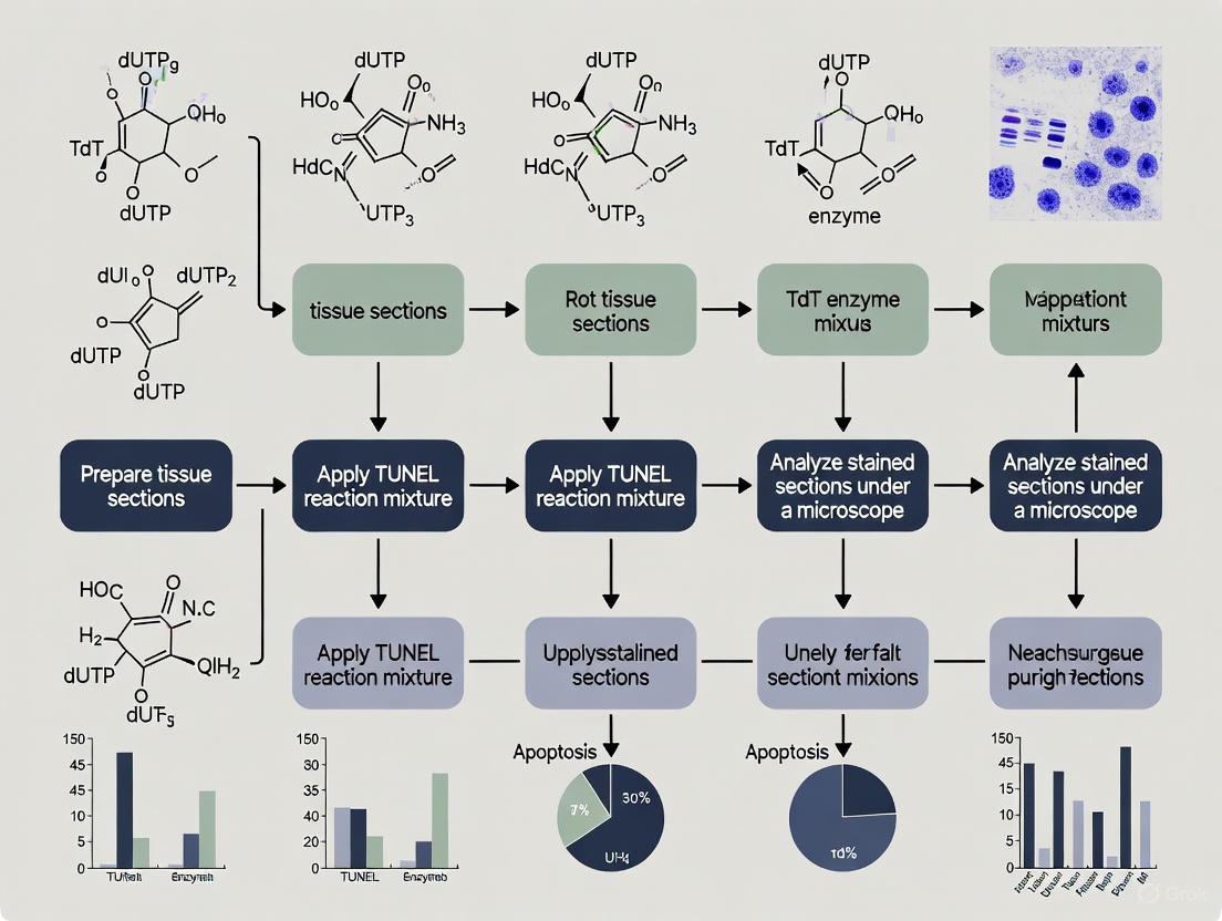
- Prepare TUNEL Reaction Mixture: On ice, prepare the labeling mixture sufficient for all samples. A typical 50 µL reaction may contain:
- Apply Mixture and Incubate: Apply the TUNEL reaction mixture to the tissue sections, ensuring complete coverage. Incubate the slides in a dark, humidified chamber for 1 hour at 37°C [9] [8].
- Terminate Reaction and Wash: After incubation, remove the reaction mixture and immerse the slides in a stop/wash buffer (often provided in kits) or 2X saline-sodium citrate (SSC) buffer for 15 minutes. This is followed by two 5-minute washes with PBS [4] [8].
- Detect Incorporated Nucleotide:
- For Direct Fluorescence: If using fluorescein-dUTP, proceed directly to counterstaining and mounting [2].
- For Indirect or Click Chemistry Detection:
- BrdU Method: Incubate sections with a FITC-conjugated anti-BrdU monoclonal antibody for 30 minutes at 37°C in the dark. Wash with PBS [7].
- Click-iT Method: Prepare the click reaction mixture according to the manufacturer's instructions (containing the Alexa Fluor azide, reaction buffer, and additive). Apply to the tissue and incubate for 30 minutes at room temperature, protected from light. Wash with PBS [8].
Visualization and Analysis
- Counterstaining and Mounting: Apply a nuclear counterstain, such as DAPI (1 µg/mL) or Hoechst 33342, for 5-10 minutes at room temperature. Wash with PBS and mount the coverslip with an anti-fade mounting medium [1] [8].
- Imaging: Visualize the stained sections using a fluorescence microscope equipped with appropriate filter sets for the fluorophores used (e.g., FITC for green, DAPI for blue) [9]. Capture images from multiple random fields to ensure representative sampling.
- Quantification: The percentage of apoptotic cells is quantified by counting the number of TUNEL-positive nuclei (e.g., green fluorescence) and the total number of nuclei (from the counterstain, e.g., blue) in each field. This can be done manually or by using image analysis software [4] [9]. For flow cytometry-based TUNEL, cells are analyzed for fluorescence intensity, and apoptotic cells are gated based on their labeling [5] [7].
Critical Technical Considerations
- Specificity and Interpretation: A positive TUNEL signal indicates the presence of DNA fragmentation but is not exclusive to apoptosis. It can also occur in necrosis, necroptosis, and other forms of cell death [5] [3]. Therefore, results should be interpreted in conjunction with morphological analysis (e.g., chromatin condensation, nuclear fragmentation) and other apoptotic markers.
- Optimization: Factors such as fixation time, permeabilization agent concentration, and TdT incubation time can significantly impact the assay's sensitivity and background. These parameters must be optimized for each tissue type and experimental system [5] [7].
- Controls: Including appropriate controls is mandatory for a valid interpretation. Essential controls are:
- Limitations: The TUNEL assay does not quantify the magnitude of DNA damage within a single cell but rather estimates the number of cells with DNA damage in a population [5]. The fluorescence labeling protocol requires sophisticated and expensive instrumentation, and a lack of standardized protocols can sometimes make comparisons between laboratories challenging [5].
The TUNEL (Terminal deoxynucleotidyl transferase dUTP nick end labeling) assay has long been established as a fundamental method for detecting apoptotic cell death through the identification of DNA fragmentation. However, emerging research and a reevaluation of existing evidence position TUNEL as a broader marker for irreversible cell death, encompassing both apoptotic and necrotic pathways. This application note delineates detailed protocols for performing TUNEL on tissue sections, provides a critical analysis of its expanded utility, and presents quantitative data on its performance across different cell death contexts. Designed for researchers, scientists, and drug development professionals, this resource integrates foundational principles with advanced methodological adaptations, including compatibility with cutting-edge spatial proteomic techniques, to empower a more nuanced investigation of cell death in physiological and pathological states.
Initially developed to label the DNA strand breaks characteristic of apoptotic cell death, the TUNEL assay exploits the enzyme terminal deoxynucleotidyl transferase (TdT) to catalyze the addition of modified deoxynucleotides to the 3'-hydroxyl termini of fragmented DNA [1] [10]. These incorporated nucleotides are then detected via fluorescence or colorimetric methods, allowing for the in situ visualization of dying cells.
While its specificity for apoptosis is well-documented, it is crucial to recognize that DNA fragmentation is not an exclusive feature of apoptosis. Necrotic cell death also results in DNA breaks, albeit through different mechanisms [11] [12]. Consequently, a growing body of evidence supports the use of TUNEL as a universal marker for committed cell death, where a positive signal indicates an irreversible commitment to cellular demise, regardless of the initiating pathway. This shift in interpretation requires careful morphological validation but significantly expands the assay's application in research and drug discovery, particularly in contexts where multiple forms of cell death coexist, such as in tumor response to therapy or ischemic tissue injury.
Principles and Mechanisms
The foundational principle of the TUNEL assay is the enzymatic labeling of DNA breaks, a process that is agnostic to the upstream signaling events that created those breaks.
Core Biochemical Reaction
The assay hinges on the activity of Terminal Deoxynucleotidyl Transferase (TdT), a template-independent DNA polymerase. In the presence of TdT and labeled nucleotides (e.g., dUTP conjugated to fluorescein, biotin, or EdUTP), the enzyme repetitively adds these nucleotides to the 3’-OH ends of DNA fragments [1] [13]. This reaction labels both single and double-strand DNA breaks, providing high sensitivity for detecting widespread DNA damage [5].
Detecting Diverse Cell Death Pathways
The versatility of TUNEL stems from its ability to detect DNA damage from various sources. The diagram below illustrates the primary cell death pathways leading to a TUNEL-positive signal.
Research Reagent Solutions: A Toolkit for Cell Death Analysis
The following table summarizes key reagents and their critical functions in a standard TUNEL assay workflow, providing a foundation for experimental setup and troubleshooting.
Table 1: Essential Reagents for TUNEL Assay Protocols
| Reagent / Component | Function / Role in the Assay | Examples & Notes |
|---|---|---|
| Terminal Deoxynucleotidyl Transferase (TdT) | Catalyzes the addition of labeled nucleotides to 3'-OH ends of fragmented DNA [1]. | The core enzyme; concentration and activity are critical [12]. |
| Labeled Nucleotide (dUTP) | Provides the detectable label incorporated at DNA break sites [1]. | Direct labels: Fluorescein-dUTP, Tunnelyte Red [14] [13]. Indirect labels: EdUTP, BrdUTP, biotin-dUTP [1]. |
| Reaction Buffer | Provides optimal ionic and pH conditions for TdT enzyme activity. | May contain cofactors like cobalt ions [10]. Kits without cacodylate are safer [14]. |
| Proteinase K / Antigen Retrieval Reagents | Unmasks DNA breaks by digesting proteins and increasing accessibility [11] [12]. | Proteinase K concentration must be optimized [12]. Pressure cooker retrieval is superior for multiplexing [11]. |
| Detection Reagents | Visualizes the incorporated nucleotide. | Direct: No secondary step needed [13]. Indirect: Anti-BrdU, streptavidin-HRP, or Click-iT chemistry with azide-dyes [1] [13]. |
| Counterstain | Provides contextual nuclear or cellular staining. | DAPI (fluorescence), Hoechst, Methyl Green (colorimetric) [1] [12]. |
Quantitative Performance Data Across Applications
The performance and quantitation of TUNEL assays can vary based on the sample type, detection method, and cell death inducer. The following table consolidates quantitative data from various studies to facilitate experimental planning and comparison.
Table 2: Quantitative TUNEL Assay Performance Across Experimental Conditions
| Sample Type / Model | Inducer of Cell Death | TUNEL Assay Type | Key Quantitative Outcome / Detection Rate | Reference / Context |
|---|---|---|---|---|
| HeLa Cells (in vitro) | 0.5 μM Staurosporine (4 hr) | Click-iT TUNEL (EdUTP) | Higher percentage of apoptotic cells detected vs. BrdUTP & fluorescein-dUTP methods [1]. | [1] |
| Formalin-Fixed, Paraffin-Embedded (FFPE) Tissue | Acetaminophen (APAP) hepatotoxicity (6 hr) | Click-iT Plus TUNEL | Reliable detection of spatially restricted necrosis around central veins [11]. | [11] |
| Spermatozoa | Infertility analysis | Flow Cytometry TUNEL | Measures "actual" DNA fragmentation; correlates well with SCSA; reference ranges established [5]. | [5] |
| Colorectal Adenocarcinoma (FFPE) | Apoptag Plus Peroxidase Kit | Optimized TUNEL | Low standard deviation, demonstrating high reproducibility after protocol optimization [12]. | [12] |
| Published Literature Survey (2017) | Various | Multiple Kits | 50% of studies used dUTP directly conjugated to FITC; over 90% used commercial kits [13]. | [13] |
Detailed Experimental Protocols
This section provides a core protocol for tissue sections and an advanced integrated protocol for multiplexed spatial analysis.
Core Protocol: TUNEL Assay for Formalin-Fixed Paraffin-Embedded (FFPE) Tissue Sections
The following workflow outlines the critical steps for performing a TUNEL assay on FFPE tissue sections, from deparaffinization to imaging.
Step-by-Step Procedure:
Sample Preparation and Deparaffinization:
- Cut 4-10 µm thick sections from FFPE tissue blocks and mount on slides.
- Deparaffinize by immersing slides in xylene (2 changes, 5-10 minutes each).
- Rehydrate through a graded ethanol series (100%, 95%, 70%) and finish with a rinse in deionized water [12].
Antigen Retrieval and Permeabilization (Critical Step):
- Proteinase K Method: Treat slides with Proteinase K (e.g., 25 µg/mL in PBS) for 20 minutes at 37°C. Note: Concentration and time must be optimized for each tissue type to avoid over-digestion (high background) or under-digestion (low signal) [12].
- Superior Alternative for Multiplexing: Pressure Cooker Method: For harmonization with subsequent immunofluorescence, heat-induced epitope retrieval in citrate buffer using a pressure cooker is recommended, as it preserves protein antigenicity better than Proteinase K [11].
- Rinse slides thoroughly with PBS.
TUNEL Reaction:
- Prepare the TUNEL reaction mixture according to kit instructions, containing TdT enzyme and labeled dUTP (e.g., fluorescein-dUTP, EdUTP, or biotin-dUTP) in an appropriate reaction buffer [1] [9].
- Apply the mixture to the tissue sections and incubate in a humidified, dark chamber for 1 hour at 37°C. Incubation time may be extended to 3 hours for low levels of cell death.
Stopping the Reaction and Detection:
- Terminate the reaction by washing slides with a stop/wash buffer or PBS.
- For Direct Detection (e.g., fluorescein-dUTP): Proceed to mounting and counterstaining.
- For Indirect Detection (e.g., BrdUTP, EdUTP):
- If using BrdUTP, incubate with an Alexa Fluor-conjugated anti-BrdU antibody [1].
- If using EdUTP, perform a "click" chemistry reaction with a fluorescent azide dye [1].
- If using biotin-dUTP, incubate with streptavidin-HRP, followed by a chromogenic substrate like DAB to generate a brown precipitate [13].
Counterstaining and Mounting:
Imaging and Quantification:
- Visualize using a fluorescence or brightfield microscope.
- Quantify TUNEL-positive cells manually by counting or by using digital image analysis software (e.g., Bacus Laboratories Incorporated Slide Scanner) [12]. For flow cytometry, analyze thousands of cells to determine the percentage of TUNEL-positive events [5] [9].
Advanced Protocol: Harmonizing TUNEL with Multiplexed Iterative Immunofluorescence (MILAN)
Recent research demonstrates that TUNEL can be seamlessly integrated with advanced spatial proteomics methods like Multiple Iterative Labeling by Antibody Neodeposition (MILAN), enabling the rich contextualization of cell death within the tissue microenvironment [11].
Key Modification: Replace the standard Proteinase K retrieval step with pressure cooker-based antigen retrieval (e.g., in citrate buffer). This step is crucial as Proteinase K extensively degrades protein antigens, preventing subsequent iterative antibody staining, while pressure cooking preserves protein antigenicity without compromising TUNEL sensitivity [11].
Integrated Workflow:
- Perform TUNEL assay on an FFPE section using the pressure cooker retrieval method and direct fluorescent detection.
- Image the TUNEL signal.
- Erase the primary and secondary antibodies (if used) by incubating the slide in a solution of 2-mercaptoethanol and SDS (2-ME/SDS) at 66°C. Note: The TdT-mediated incorporation of nucleotides is not reversed by this step.
- Proceed with multiple cycles of standard immunofluorescence using the MILAN protocol, staining for various protein markers (e.g., cell lineage, signaling, or structural markers).
- Co-register the TUNEL images with the multiplexed IF images to precisely determine the phenotype and spatial context of dying cells [11].
Troubleshooting and Technical Pitfalls
Successful application and interpretation of the TUNEL assay require awareness of its potential technical challenges.
- False Positives: Can arise from extensive fixation delay, incomplete fixation, or excessive proteolytic digestion during antigen retrieval [12]. Necrosis also produces DNA strand breaks and will yield a true positive signal for cell death, which may be misinterpreted as a "false positive" for apoptosis if not validated morphologically [5] [12].
- False Negatives: Often due to inadequate permeabilization or tissue pretreatment, which prevents the TUNEL reagents from accessing the fragmented DNA. This is a particular concern in cell types with highly compact chromatin, such as spermatozoa, which may require a reducing agent like dithiothreitol (DTT) for chromatin relaxation [5].
- Background Staining: High background can be caused by over-fixation, inappropriate Proteinase K concentration, or inadequate blocking (especially when using biotin-streptavidin systems) [13] [12].
- Standardization: A lack of universally standardized protocols and threshold values has hindered the clinical translation of TUNEL, particularly in andrology. Consistent fixation, permeabilization, and analysis protocols are essential for reproducible results [5].
The TUNEL assay is a powerful and versatile tool that transcends its traditional role as a simple apoptosis detector. When performed with optimized and validated protocols, it serves as a robust universal marker for irreversible cell death. Its compatibility with advanced spatial biology techniques, as demonstrated by its integration with MILAN, opens new frontiers for understanding the tissue microenvironment and the spatial dynamics of cell death in disease and treatment. By adhering to detailed protocols, acknowledging its broader specificity, and employing careful morphological correlation, researchers can leverage the full potential of TUNEL to advance fundamental research and therapeutic development.
The TUNEL (Terminal deoxynucleotidyl transferase dUTP nick end labeling) assay is a fundamental method for detecting DNA fragmentation, a hallmark of apoptotic cell death, in tissue sections and cultured cells [15]. During the later stages of apoptosis, endogenous endonucleases cleave DNA into fragments, generating exposed 3'-OH ends that serve as substrates for the enzyme Terminal Deoxynucleotidyl Transferase (TdT) [15]. This enzyme catalyzes the incorporation of modified deoxyuridine triphosphate (dUTP) molecules into these DNA strand breaks. The choice of dUTP modification and corresponding detection system significantly impacts assay sensitivity, specificity, multiplexing capability, and procedural workflow.
This application note provides a comprehensive comparison of four key dUTP labels—BrdU, EdU, fluorescein, and biotin—within the context of TUNEL assay optimization for tissue section research. We include detailed protocols, analytical comparisons, and visualization tools to guide researchers in selecting appropriate reagents for their specific experimental needs in basic research and drug development.
Comparative Analysis of dUTP Labels
The table below summarizes the key characteristics of the four primary dUTP labels used in TUNEL assays, providing a quick reference for researchers to compare their properties and applications.
Table 1: Comprehensive Comparison of dUTP Labels for TUNEL Assays
| Label | Chemical Nature | Detection Method | Detection Time | Key Advantages | Key Limitations | Compatibility with Tissue Sections | Multiplexing Potential |
|---|---|---|---|---|---|---|---|
| BrdU | Thymidine analog | Anti-BrdU antibody (immunodetection) | ~3-4 hours (post-incorporation) | Extensive validation; compatible with archival samples; gold standard [16] | Requires DNA denaturation (harsh HCl treatment); epitope masking [16] | Excellent for FFPE tissues [16] | High (with other antibodies) [16] |
| EdU | Alkyne-modified nucleoside | Click chemistry (Cu-catalyzed cycloaddition) | ~1.5-2 hours (post-incorporation) [15] | No DNA denaturation; small label preserves morphology; fast detection [15] [17] | Copper catalyst can damage some fluorescent proteins and epitopes [15] | Excellent (Click-iT Plus optimized for tissue) [15] | Good (Click-iT Plus reduces copper issues) [15] |
| Fluorescein-dUTP | Fluorescently-conjugated dUTP | Direct fluorescence | Immediate (post-wash) | Most direct and simple protocol; no secondary reagents [18] | Lower sensitivity due to limited signal amplification [18] | Good (requires careful optimization) | Moderate (limited by direct fluorescence) |
| Biotin-dUTP | Biotin-conjugated dUTP | Streptavidin-enzyme/fluorophore conjugate | ~2-3 hours (post-incorporation) | High sensitivity via signal amplification; flexible detection (colorimetric/fluorescent) [15] [18] | Additional incubation step required; potential endogenous biotin interference | Excellent for colorimetric IHC [15] | High (multiple streptavidin conjugates available) |
Detailed Experimental Protocols
Click-iT EdU TUNEL Assay for Tissue Sections
The Click-iT TUNEL assay utilizes an alkyne-modified EdUTP, which is incorporated into DNA breaks by TdT and subsequently detected via a copper-catalyzed "click" reaction with a fluorescent azide [15]. This protocol has been optimized for formalin-fixed, paraffin-embedded (FFPE) tissue sections.
Table 2: Key Reagent Solutions for Click-iT EdU TUNEL Assay
| Reagent | Function | Example Product/Catalog Number |
|---|---|---|
| Click-iT Plus TUNEL Assay | Provides EdUTP, TdT enzyme, and reaction buffers | Invitrogen Click-iT Plus TUNEL Assay (e.g., C10625) [15] |
| Alexa Fluor Azide | Fluorescent detection via click chemistry | Click-iT Plus TUNEL Assay with Alexa Fluor 488, 594, or 647 azide [15] |
| Proteinase K | Antigen retrieval for FFPE tissues | Supplied in kit or separately [19] |
| DNase I | Positive control treatment to induce DNA breaks | Available from various molecular biology suppliers |
| Hoechst 33342 | Nuclear counterstain | Available from various fluorescent dye suppliers |
Workflow Steps:
- Sample Preparation and Fixation: Deparaffinize and rehydrate FFPE tissue sections following standard histological protocols. Fix cells or tissues using mild fixatives such as 4% formaldehyde for 15 minutes at room temperature [15].
- Permeabilization: Treat tissues with Proteinase K (e.g., 30 minutes at room temperature) to expose DNA breaks [19]. Wash slides twice in DNase-free water for 5 minutes each [19].
- TdT-Mediated EdUTP Incorporation: Prepare the TdT reaction mixture according to the kit instructions. Apply to tissue sections and incubate in a humidity chamber for 60 minutes at 37°C [15] [19].
- Click Chemistry Detection: Prepare the click reaction mixture containing the fluorescent azide and copper protectant. Apply to sections and incubate for 30 minutes at room temperature, protected from light [15].
- Counterstaining and Mounting: Wash sections and apply a nuclear counterstain such as Hoechst 33342. Mount slides with an anti-fade mounting medium [15].
- Microscopy and Analysis: Visualize using a fluorescence microscope with appropriate filter sets. TUNEL-positive nuclei will display specific fluorescence corresponding to the azide dye used.
Diagram 1: Click-iT EdU TUNEL assay workflow for tissue sections.
BrdU TUNEL Assay with Immunodetection
The BrdU TUNEL assay incorporates BrdUTP into DNA breaks, which is subsequently detected using specific anti-BrdU antibodies. This protocol includes the critical DNA denaturation step required for antibody access to the incorporated BrdU [16].
Workflow Steps:
- BrdU Incorporation: Follow steps 1-4 of the EdU protocol, replacing the EdUTP reaction mix with a solution containing TdT and BrdUTP. Incubate for 60 minutes at 37°C [16] [18].
- DNA Denaturation: Incubate tissue sections in 1-2.5 M HCl for 30-60 minutes at room temperature or 37°C. This critical step exposes the BrdU epitope by denaturing the DNA [16].
- Neutralization: Optional: Remove HCl and neutralize with 0.1 M sodium borate buffer (pH 8.5) for 10 minutes at room temperature [16].
- Immunological Detection: Wash sections in PBS. Apply anti-BrdU primary antibody (e.g., Abcam ab6326) diluted in PBS for 60 minutes at room temperature [16].
- Signal Amplification and Visualization: Wash and apply an enzyme-conjugated (e.g., HRP) secondary antibody. Develop signal using an appropriate chromogenic substrate such as DAB, resulting in a brown precipitate [16] [19].
- Counterstaining and Analysis: Counterstain with hematoxylin or methyl green, dehydrate, and mount. Analyze using bright-field microscopy [15].
Diagram 2: BrdU TUNEL assay workflow requiring DNA denaturation.
Direct and Indirect Detection Protocols
Fluorescein-dUTP Protocol: Following standard TdT-mediated incorporation of fluorescein-dUTP, simply wash the samples and analyze via fluorescence microscopy with a FITC filter set [18]. This method is the most straightforward but offers no signal amplification.
Biotin-dUTP Protocol: After TdT-mediated incorporation of biotin-dUTP [18], detect the incorporated label using streptavidin-HRP (for colorimetric detection with DAB) or streptavidin conjugated to a fluorophore (e.g., Alexa Fluor dyes) for fluorescent detection [15] [19]. Incubate with the streptavidin conjugate for 30 minutes at room temperature after the incorporation step, then proceed with signal development or mounting [19].
Advanced Applications and Multiplexing Strategies
Double-Labeling TUNEL with Active Caspase-3
Combining TUNEL with immunohistochemical labeling of active caspase-3 provides more specific confirmation of apoptosis by detecting two distinct hallmarks of the process [19]. The following protocol adapts this strategy for tissue sections:
Integrated Workflow:
- Complete TUNEL Staining: Perform the TUNEL assay (using BrdU-, EdU-, or biotin-dUTP) through the signal development step (e.g., with DAB, yielding a brown nuclear signal) [19].
- Blocking: After TUNEL development, block sections with avidin-biotin blocking reagents and serum to reduce non-specific binding [19].
- Caspase-3 Immunostaining: Incubate tissues with anti-active caspase-3 primary antibody (e.g., R&D Systems AF835 at 5-15 µg/mL) overnight at 2-8°C [19].
- Signal Detection: Apply appropriate secondary antibodies and develop using a contrasting chromogen such as AEC, which produces a red cytoplasmic signal [19].
- Analysis: Apoptotic cells are identified by double-labeling: dark brown TUNEL-positive nuclei with red caspase-3-positive cytoplasm [19].
Diagram 3: Multiplexing TUNEL with active caspase-3 detection.
Troubleshooting and Optimization Guide
Table 3: Troubleshooting Common Issues in TUNEL Assays
| Problem | Potential Causes | Recommended Solutions |
|---|---|---|
| High Background | Inadequate permeabilization; over-fixation; insufficient washing | Optimize Proteinase K concentration and incubation time [19]; reduce fixation time; increase wash stringency |
| Weak or No Signal | Under-permeabilization; low apoptosis level; enzyme inactivation | Include positive control (DNase I treated tissue) [15]; check TdT enzyme activity; optimize permeabilization step |
| Specific to BrdU: Incomplete DNA denaturation | Optimize HCl concentration and incubation time; try heat-induced epitope retrieval [16] | |
| Specific to EdU: Fluorescence quenching | Copper concentration too high; excessive light exposure | Use Click-iT Plus kits with optimized copper [15]; protect samples from light during and after click reaction |
| Tissue Damage | Over-permeabilization; harsh denaturation (BrdU) | Titrate Proteinase K; for BrdU, try milder denaturation conditions or switch to EdU [15] [16] |
| Inconsistent Staining | Irregular reagent application; drying of sections | Use humidity chamber for all incubations; ensure even coverage of reaction mixtures |
Selection of appropriate dUTP labels and detection systems is critical for successful TUNEL assay implementation in tissue section research. BrdU remains a well-validated choice despite its requirement for DNA denaturation, while EdU offers a faster, gentler alternative through click chemistry. Fluorescein-dUTP provides simplicity for direct detection, whereas biotin-dUTP delivers high sensitivity through signal amplification. For definitive apoptosis confirmation in thesis research, multiplexing TUNEL with active caspase-3 immunohistochemistry is highly recommended. Understanding the strengths and limitations of each system enables researchers to optimize their TUNEL assays for specific applications in drug development and mechanistic studies of cell death.
The Terminal deoxynucleotidyl Transferase dUTP Nick End Labeling (TUNEL) assay is a cornerstone method for detecting apoptotic cell death in situ. Initially developed in 1992, this technique identifies the DNA fragmentation that is a hallmark of the final stages of apoptosis [3] [20]. The assay operates on the principle that the enzyme terminal deoxynucleotidyl transferase (TdT) catalyzes the addition of labeled deoxynucleotides to the 3'-hydroxyl termini of DNA fragments [13] [21]. These labels can then be visualized using fluorescence or colorimetric detection, allowing researchers to pinpoint apoptotic cells within the context of intact tissue architecture or cell cultures.
While initially celebrated as a specific assay for apoptosis, subsequent research has clarified that TUNEL is a universal assay for irreversible cell death associated with DNA fragmentation, detectable across various modes of cell death including necrosis, pyroptosis, and ferroptosis [3]. This characteristic, coupled with its unique technical advantages, solidifies its role as a powerful tool in basic research, toxicology, and drug development, particularly for organs with high endonuclease activity like the kidney [3].
Comparative Advantages of the TUNEL Assay
The TUNEL assay offers a distinct combination of sensitivity, quantification capability, and in situ application that sets it apart from other classical methods for analyzing cell death.
Superior Sensitivity
The TUNEL assay demonstrates exceptional sensitivity in detecting the earliest stages of DNA fragmentation, often identifying apoptotic cells before morphological changes become apparent [20].
- Early Detection: TUNEL can detect DNA strand breaks that occur early in the apoptotic process, enabling researchers to identify committed cells before they display characteristic morphological features like membrane blebbing or chromatin condensation [20].
- Direct Terminal Labeling: Theoretically, TUNEL is more sensitive than methods like DNA laddering because it identifies DNA termini rather than waiting for the accumulation of small, high-mobility fragments [3]. This direct labeling approach provides a more linear and sensitive response to initial DNA fragmentation.
- Enhanced Detection Chemistry: Modern TUNEL kits, such as those utilizing Click-iT chemistry with an alkyne-modified dUTP (EdUTP), offer improved efficiency. The small size of the alkyne moiety allows for easier incorporation by the TdT enzyme compared to larger nucleotide conjugates, and the subsequent detection step using click chemistry is highly specific and efficient [22] [8]. Studies have shown that this two-step method can detect a higher percentage of apoptotic cells under identical conditions compared to assays using one-step incorporation of dye-modified nucleotides [22].
Table 1: Sensitivity Comparison Between TUNEL and Alternative Apoptosis Assays
| Assay Method | Target | Detection Stage | Key Advantage |
|---|---|---|---|
| TUNEL Assay [20] [3] | DNA fragmentation (3'-OH ends) | Mid to late apoptosis | Detects DNA breaks early in the death process; highly sensitive. |
| Annexin V Staining [9] | Phosphatidylserine externalization | Early apoptosis | Identifies cells before loss of membrane integrity. |
| Caspase Activation Assays [9] | Caspase enzyme activity | Early to mid apoptosis | Provides insight into specific apoptotic signaling pathways. |
| DNA Laddering [3] | Oligonucleosomal DNA fragments | Late apoptosis | A classic biochemical method, but less sensitive and not quantitative at single-cell level. |
Robust Quantification Capabilities
A significant strength of the TUNEL assay is its capacity for robust quantitative analysis, moving beyond mere qualitative detection to provide meaningful statistical data on cell death.
- Single-Cell Resolution: Unlike DNA laddering, which provides a population-level assessment, TUNEL allows for the quantification of apoptosis at the single-cell level [21]. This is crucial for understanding the distribution and frequency of cell death within a heterogeneous sample, such as a tissue section.
- Flexible Readouts: TUNEL staining can be quantified using various platforms. Researchers can manually or automatically count TUNEL-positive cells in tissue sections via fluorescence microscopy [23] [9]. Furthermore, the assay is compatible with flow cytometry for suspended cells, enabling high-throughput, quantitative analysis of large cell populations [13] [9].
- Multiplexing for Context: A key advantage for quantification is the ability to combine TUNEL with immunohistochemical staining for specific protein markers. This allows researchers not only to count dead cells but also to identify the cell type undergoing death, providing rich contextual data [23] [3]. For instance, one study used TUNEL with an anti-desmin antibody to quantitatively assess apoptosis specifically in cardiac myocytes [23].
Powerful In Situ Application
The ability to perform the assay in situ is arguably the most defining advantage of TUNEL, preserving the spatial context of cell death within a tissue.
- Spatial Context Preservation: TUNEL can be applied to formalin-fixed, paraffin-embedded (FFPE) tissue sections, which are standard in clinical pathology [11] [22]. This allows for the direct visualization of apoptotic cells in their native tissue microenvironment, enabling the correlation of cell death with specific anatomical structures, injury zones, or disease pathologies [11].
- Compatibility with Multiplexed Spatial Proteomics: Recent advancements have successfully harmonized TUNEL with modern spatial proteomic methods like Multiple Iterative Labeling by Antibody Neodeposition (MILAN) and Cyclic Immunofluorescence (CycIF) [11]. A critical finding was that replacing the traditional proteinase K (ProK) antigen retrieval with pressure cooking (PC) quantitatively preserves the TUNEL signal without compromising subsequent protein antigenicity. This integration allows for the rich spatial contextualization of cell death alongside the expression of dozens of other protein targets on the same tissue specimen [11].
- Universal Applicability: The TUNEL assay is versatile and can be used on a wide array of sample types, including frozen sections, FFPE tissues, cell suspensions, and adherent cells from diverse species, ensuring methodological consistency across in vitro and in vivo studies [3] [9].
Table 2: Comparison of DNA Fragmentation Detection Methods
| Feature | TUNEL Assay | DNA Laddering | Comet Assay |
|---|---|---|---|
| Sensitivity | High [3] | Low to Moderate [3] | High [3] |
| Quantification | Quantitative (single-cell) [21] | Qualitative / Semi-Quantitative [3] | Quantitative [3] |
| Spatial Context | Yes (In Situ) [3] | No | No |
| Throughput | Medium to High [9] | Low | Low (labor-intensive) [3] |
| Primary Application | Tissue sections & cultured cells [3] | Cell populations (lysates) [3] | Primarily cultured cells [3] |
Detailed TUNEL Protocol for Tissue Sections
The following protocol is optimized for formalin-fixed, paraffin-embedded (FFPE) tissue sections, incorporating best practices for sensitivity and compatibility with multiplexed immunofluorescence.
Research Reagent Solutions
Table 3: Essential Reagents and Materials for TUNEL Assay
| Reagent/Material | Function | Notes & Examples |
|---|---|---|
| FFPE Tissue Sections | Sample substrate | Standard 5 μm sections on charged slides. |
| Terminal Deoxynucleotidyl Transferase (TdT) [8] | Core enzyme that adds labeled nucleotides to 3'-OH DNA ends. | Recombinant enzyme is highly active. |
| Labeled Nucleotide (e.g., EdUTP, BrdUTP) [22] [13] | Substrate for TdT; provides detectable signal. | EdUTP allows flexible click chemistry detection; BrdUTP is detected with antibodies. |
| Click-iT Reaction Mixture [22] or Antibody Conjugate [13] | Detects the incorporated nucleotide. | Contains fluorescent azide for Click-iT; Fluorophore-conjugated anti-BrdU for antibody-based detection. |
| Antigen Retrieval Reagents | Unmasks cross-linked epitopes and DNA ends. | Critical: Pressure cooker with citrate/EDTA buffer is preferred over proteinase K for multiplexing [11]. |
| Blocking Buffer (e.g., 3% BSA) [8] | Reduces non-specific background staining. | Essential for antibody-based detection methods. |
| Nuclear Counterstain (e.g., DAPI, Hoechst) [8] [21] | Labels all nuclei for morphological context. | Allows visualization of total cell population. |
| Mounting Medium | Preserves fluorescence and enables microscopy. | Use antifade medium for fluorescence imaging. |
Step-by-Step Workflow
Diagram 1: TUNEL assay workflow for tissue sections
Step 1: Sample Preparation
- Dewaxing and Rehydration: Deparaffinize FFPE sections using xylene (or substitute) and rehydrate through a graded ethanol series (100%, 95%, 70%) to water.
- Antigen Retrieval: Perform heat-induced epitope retrieval using a pressure cooker and appropriate buffer (e.g., citrate buffer, pH 6.0). This step is superior to proteinase K digestion, which can degrade protein antigens and hinder subsequent multiplexed immunofluorescence [11].
- Permeabilization (Optional): To further enhance accessibility, treat sections with a permeabilization reagent (e.g., 0.25% Triton X-100 in PBS) for 20 minutes at room temperature. Wash with PBS [8].
Step 2: TUNEL Reaction
- Prepare Reaction Mixture: Combine the TdT enzyme with the reaction buffer and the labeled nucleotide (e.g., EdUTP or BrdUTP) according to kit instructions. For a positive control, treat a separate section with DNase I (e.g., 1-3 µg/mL for 30 minutes) to intentionally create DNA breaks before this step [8]. For a negative control, omit the TdT enzyme from the reaction mix.
- Incubation: Apply the reaction mixture to the tissue section and incubate in a humidified chamber at 37°C for 60 minutes. This allows the TdT enzyme to incorporate the labeled nucleotides at the sites of DNA breaks.
- Washing: Rinse the slides thoroughly with PBS to stop the reaction and remove any unincorporated nucleotides.
Step 3: Detection and Visualization
- Signal Development:
- For Click-iT-based assays (using EdUTP): Incubate the section with the Click-iT reaction mixture containing a fluorescent azide (e.g., Alexa Fluor 488 azide) [22].
- For Antibody-based assays (using BrdUTP): Block the section with 3% BSA, then incubate with a fluorophore-conjugated anti-BrdU antibody [13].
- Counterstaining and Mounting: Wash the slides and apply a nuclear counterstain such as DAPI or Hoechst to visualize all nuclei. Apply an antifade mounting medium and a coverslip.
- Imaging and Analysis: Image the slides using a fluorescence or brightfield microscope. TUNEL-positive nuclei will display specific fluorescence or colorimetric staining. Quantify the results by counting TUNEL-positive cells manually or using image analysis software across multiple fields of view [9].
Integration with Multiplexed Spatial Proteomics
A groundbreaking application of the TUNEL assay is its integration with advanced spatial proteomics techniques. This harmonization allows for the unprecedented contextualization of cell death within the complex cellular ecosystem of a tissue.
The core incompatibility between traditional TUNEL and methods like MILAN was the use of proteinase K (ProK) for antigen retrieval. ProK treatment consistently reduces or abrogates protein antigenicity, preventing subsequent rounds of antibody staining [11]. The key innovation is the substitution of ProK with pressure cooker (PC)-based antigen retrieval. This method not only preserves but can enhance protein antigenicity for the targets tested, all without compromising the sensitivity or specificity of the TUNEL signal [11].
This compatible protocol allows TUNEL to be performed as one cycle within a MILAN or CycIF staining series. After TUNEL imaging, the fluorescent signal can be erased using a 2-ME/SDS treatment, and the slide can be restained with antibodies for protein markers. This process can be iterated dozens of times, generating rich multiparameter data from a single precious tissue specimen [11].
Diagram 2: Integrated TUNEL and multiplexed imaging workflow
The TUNEL assay remains an indispensable tool in cell death research due to its unique and powerful combination of high sensitivity, robust quantitative capabilities, and unparalleled in situ application. Its ability to detect DNA fragmentation at the single-cell level within the native tissue architecture provides insights that population-based biochemical methods cannot offer. The recent harmonization of TUNEL with multiplexed spatial proteomics platforms, facilitated by the critical replacement of proteinase K with pressure cooker antigen retrieval, has further elevated its utility. This integration allows researchers to not only identify dying cells but also to deeply characterize their phenotypic state, spatial relationships, and tissue microenvironment simultaneously. For researchers and drug development professionals investigating mechanisms of tissue injury, toxicology, and therapeutic efficacy, the TUNEL assay, especially when combined with these advanced multiplexing techniques, offers a comprehensive and powerful approach for the spatial contextualization of cell death.
A Step-by-Step TUNEL Protocol for Fluorescence and Colorimetric Detection
Within the context of apoptosis research using the TUNEL (Terminal deoxynucleotidyl transferase dUTP Nick End Labeling) assay on tissue sections, sample preparation is the critical foundation upon which reliable data is built. Improperly prepared tissues can lead to significant artifacts, including both false-positive and false-negative results, ultimately compromising the validity of the scientific conclusions [24]. This application note details a standardized, optimized protocol for the fixation and permeabilization of tissue sections, specifically tailored for subsequent TUNEL staining. The goal is to provide researchers with a robust methodology that preserves tissue morphology while simultaneously ensuring the accessibility of fragmented DNA to the assay reagents, thereby enhancing the sensitivity and specificity of apoptosis detection [1] [24].
Materials and Reagents
Research Reagent Solutions
The following table lists essential materials and reagents required for the fixation and permeabilization of tissue sections for TUNEL assays.
Table 1: Essential Reagents for Fixation and Permeabilization in TUNEL Assays
| Item | Function/Description | Example / Notes |
|---|---|---|
| Fixative | Preserves cellular architecture and cross-links biomolecules to maintain tissue morphology during analysis. | 4% Paraformaldehyde (PFA) in PBS [8] [9]. |
| Permeabilization Reagent | Disrupts cell membranes to allow TUNEL reaction reagents to access the nuclear DNA. | 0.1-0.25% Triton X-100 in PBS [8] [9]. |
| Proteolytic Enzyme (Optional) | Enhances sensitivity by breaking cross-links and improving reagent access to DNA breaks; requires careful optimization. | Proteinase K [24]. |
| Wash Buffer | Used for rinsing steps to remove excess fixative and permeabilization reagents. | 1X Phosphate Buffered Saline (PBS) [8]. |
| Blocking Solution | Reduces non-specific background binding in some detection methods. | 3% Bovine Serum Albumin (BSA) in PBS [8]. |
| Positive Control Reagent | Generates DNA strand breaks to validate assay performance. | DNase I (Deoxyribonuclease I) [8]. |
Methods
Detailed Experimental Protocol
The following workflow outlines the key steps for preparing tissue sections for TUNEL assay, from fixation to the point of the TUNEL reaction.
Fixation Protocol
The objective of fixation is to preserve tissue morphology and prevent the loss of cellular components, including fragmented DNA, during subsequent processing steps [9].
For Formalin-Fixed, Paraffin-Embedded (FFPE) Tissues:
- Dewaxing and Rehydration: Cut tissue sections to 5-8 μm thickness. Dewax in xylene (or a safe substitute) and rehydrate through a graded series of ethanol (e.g., 100%, 95%, 70%) to water [1] [5].
- Fixation: Immerse the rehydrated sections in a sufficient volume of 4% paraformaldehyde (PFA) in PBS.
- Incubation: Fix for 15 minutes at room temperature [8].
- Note: Prolonged fixation can lead to excessive protein-DNA cross-linking, which may mask DNA breaks and reduce TUNEL signal intensity. If tissues have been over-fixed, antigen retrieval methods (e.g., microwave heating in citrate buffer) may be necessary to reverse cross-links and restore sensitivity [24].
For Frozen Tissues:
- Initial Preparation: Collect fresh frozen tissue sections and air-dry.
- Fixation: Fix the sections by immersing in 4% PFA in PBS for 15 minutes at room temperature [1].
Permeabilization Protocol
Permeabilization is crucial for rendering the cell and nuclear membranes permeable, allowing the TdT enzyme and labeled nucleotides to access the fragmented DNA within the nucleus [9] [24].
Standard Permeabilization:
Enhanced Permeabilization (for challenging tissues):
- For tissues with highly compact chromatin or to maximize assay sensitivity, a proteolytic pretreatment can be employed.
- After standard permeabilization and washing, treat sections with Proteinase K (concentration and time require empirical optimization for each tissue type) [24].
- Critical Note: Over-digestion with proteases can damage tissue morphology and even lead to the loss of DNA, resulting in false-positive signals or tissue degradation. Always include a no-enzyme control.
Preparation of Positive Control
To confirm the efficacy of the entire TUNEL procedure, a positive control is essential.
- Following permeabilization and washing, select one section for positive control treatment.
- Prepare a solution of DNase I in the supplied buffer according to the manufacturer's instructions. Do not vortex, as vigorous mixing can denature the enzyme [8].
- Apply 100 μL of the DNase I solution to the control section and incubate for 30 minutes at room temperature to introduce deliberate DNA strand breaks.
- Wash the section once with deionized water before proceeding to the TUNEL reaction [8].
Key Experimental Parameters
Optimal fixation and permeabilization require careful attention to reagent concentrations and incubation times. The following table summarizes the key quantitative parameters for this protocol.
Table 2: Key Parameters for Fixation and Permeabilization
| Parameter | Optimal Condition | Purpose & Rationale |
|---|---|---|
| Fixative | 4% Paraformaldehyde | Provides adequate cross-linking without excessively masking DNA ends [8] [9]. |
| Fixation Time | 15 minutes at Room Temperature | Balances morphology preservation with reagent accessibility; longer times can reduce sensitivity [8] [24]. |
| Permeabilization Agent | 0.1 - 0.25% Triton X-100 | Effectively dissolves lipid membranes without causing excessive damage to nuclear structure [8] [9]. |
| Permeabilization Time | 20 minutes at Room Temperature | Sufficient for reagent penetration; may require optimization for different tissue densities [8]. |
| Proteolytic Pretreatment | Proteinase K (e.g., 20 μg/mL), variable time | Breaks protein-DNA cross-links, dramatically increasing TUNEL sensitivity, especially in over-fixed tissues [24]. |
Troubleshooting
A well-optimized protocol prevents common issues. The table below outlines potential problems and their solutions related to sample preparation.
Table 3: Troubleshooting Guide for Sample Preparation
| Problem | Potential Cause | Recommended Solution |
|---|---|---|
| High Background / Nonspecific Staining | Inadequate washing after permeabilization; endogenous biotin (if using biotin-streptavidin detection). | Increase number and duration of PBS washes; use a blocking step with 3% BSA or an endogenous biotin blocking kit [8] [13]. |
| Weak or No TUNEL Signal | Over-fixation causing excessive cross-linking; insufficient permeabilization; inactive reagents. | Incorporate a proteolytic pretreatment (Proteinase K) or microwave antigen retrieval; optimize Triton X-100 concentration/time; include a DNase I positive control to validate reagents [24]. |
| Poor Tissue Morphology | Over-digestion with protease; harsh permeabilization. | Titrate Proteinase K concentration and incubation time; reduce the concentration of Triton X-100 [24]. |
| Loss of Tissue from Slide | Inadequate slide coating; aggressive washing. | Use positively charged or poly-lysine coated slides; ensure sections are completely dry before starting; gentle pipetting during wash steps [8]. |
Meticulous execution of the fixation and permeabilization steps described in this application note is paramount for generating accurate, reproducible, and interpretable data from TUNEL assays on tissue sections. By standardizing these pre-analytical procedures, researchers can minimize technical variability and artifacts, thereby ensuring that the observed TUNEL signal truly reflects the biological process of apoptosis. This robust foundation in sample preparation empowers scientists and drug development professionals to draw confident conclusions in their research on cell death mechanisms.
Within the framework of TUNEL assay research for tissue sections, the analysis of Formalin-Fixed Paraffin-Embedded (FFPE) tissues is a cornerstone. While formalin fixation excellently preserves tissue morphology, it forms methylene cross-links that mask antigenic sites, a process that can severely impair antibody binding and detection in subsequent assays [25] [26] [27]. Antigen retrieval (AR) is, therefore, a critical and mandatory step to reverse this masking. The two principal methods to achieve this are Heat-Induced Epitope Retrieval (HIER) and Proteolysis-Induced Epitope Retrieval (PIER), which includes the use of enzymes like Proteinase K [25]. Selecting the appropriate method is paramount for the success of downstream applications, including the sensitive detection of DNA fragmentation in TUNEL assays. This application note provides a detailed comparison of these two techniques and offers optimized protocols to guide researchers and drug development professionals in their experimental workflows.
Mechanisms and Comparative Analysis
Fundamental Principles of Antigen Retrieval
Formalin fixation creates cross-links between proteins and nucleic acids, which although beneficial for morphology, obscures epitopes and hinders antibody access [25] [26]. AR methods aim to break these cross-links and restore antigenicity.
Heat-Induced Epitope Retrieval (HIER): This method employs high temperatures (typically 95-100°C) in a specific buffer solution. The mechanism is not fully elucidated but is hypothesized to involve the hydrolytic cleavage of formaldehyde-induced cross-links, the unfolding of epitopes, and the extraction of calcium ions from coordination complexes with proteins [25] [26]. The process is believed to restore the original conformation of antigenic sites, allowing antibodies to bind effectively [26].
Proteolysis-Induced Epitope Retrieval (PIER): This technique utilizes proteolytic enzymes such as Proteinase K, trypsin, or pepsin. These enzymes function by digesting the proteins surrounding the epitopes, thereby physically unmasking the hidden antigenic sites [25] [27]. Unlike the broader, physical reversal hypothesized for HIER, PIER is a targeted biochemical digestion.
Direct Comparison: HIER vs. Proteinase K Retrieval
The choice between HIER and Proteinase K retrieval depends on the target antigen, tissue type, and the specific requirements of the downstream assay, such as the TUNEL assay. The table below summarizes the key characteristics of each method.
Table 1: Comparative Analysis of Heat-Induced vs. Proteinase K Antigen Retrieval Methods
| Feature | Heat-Induced Epitope Retrieval (HIER) | Proteinase K Retrieval (PIER) |
|---|---|---|
| Primary Mechanism | High-temperature reversal of cross-links [25] [26] | Enzymatic digestion of masking proteins [25] |
| Key Advantage | Broader range of antigens, especially nuclear; superior for most IHC applications; less morphological damage when optimized [25] | Effective for difficult-to-recover epitopes; gentler on delicate tissues; no specialized heating equipment needed [25] |
| Main Disadvantage | Risk of tissue damage from overheating; potential for uneven heating with some devices [25] [27] | Risk of destroying antigen and tissue morphology; requires precise calibration of concentration and time [25] |
| Typical Incubation | 10-20 minutes at 95-100°C [25] [27] | 10-30 minutes at 37°C [25] |
| Compatibility with TUNEL | Often required as a first step in TUNEL protocols for FFPE tissues [1] [28] | Used in specific TUNEL workflow steps for enzyme retrieval [28] |
The effectiveness of HIER is significantly influenced by the pH and composition of the retrieval buffer. The response of different antigens to pH varies, which necessitates optimization.
Table 2: Impact of HIER Buffer pH on Antigen Staining Results
| Staining Pattern | Description | Example Antigens |
|---|---|---|
| Stable Type | pH has minimal effect on staining results [25] | PCNA, AE1, EMA, CD20 [25] |
| V Type | Good staining at high and low pH, poorer around pH 4-5 [25] | ER, Ki-67 [25] |
| Increasing Type | Staining improves with increasing pH [25] | HMB45 [25] |
| Decreasing Type | Staining weakens as pH increases (rare) [25] | MOC31 [25] |
For most antibodies, particularly those targeting nuclear antigens, EDTA-based buffers (pH 8.0-9.0) are more effective than citrate buffer (pH 6.0) [25] [26].
Experimental Protocols
Heat-Induced Epitope Retrieval (HIER) Protocol
The following protocol for HIER using a microwave is a robust starting point for most antigens [25] [27].
Research Reagent Solutions & Materials
- Antigen Retrieval Buffer: Tris-EDTA (10 mM Tris, 1 mM EDTA, 0.05% Tween 20, pH 9.0) or Sodium Citrate (10 mM, 0.05% Tween 20, pH 6.0) [25] [27].
- Deparaffinization Reagents: Xylene or substitute, and a graded series of ethanol (100%, 95%, 70%, 50%) [29] [30].
- Equipment: Microwave (scientific grade recommended for uniformity), microwave-safe staining dish, slide rack, and cold tap water source [27].
Step-by-Step Methodology
- Deparaffinization and Rehydration: Immerse slides sequentially through the following series:
- Antigen Retrieval: Immerse the slides in a staining dish filled with pre-heated antigen retrieval buffer. Microwave the dish at 95°C for 8 minutes. Allow the slides to cool for 5 minutes, then microwave again at 95°C for 4 minutes.
- Cooling: Cool the slides to room temperature in the buffer (approximately 20-30 minutes). This cooling step is crucial as it allows the antigenic sites to re-form after heat exposure [25] [27].
- Washing: Rinse the slides with deionized water before proceeding to the immunohistochemical or TUNEL staining protocol [29].
Note: Alternative heating sources like pressure cookers, steamers, or water baths can be used. For a pressure cooker, heating at full pressure for 3 minutes is often sufficient [27].
Proteinase K Retrieval (PIER) Protocol
This protocol provides a gentle alternative for specific antigens or fragile tissues [25].
Research Reagent Solutions & Materials
- Enzyme Solution: 0.1% Proteinase K in Tris-EDTA buffer, pre-warmed to 37°C. Concentration and time require optimization [25] [28].
- Equipment: 37°C incubator, humidified chamber, pipette, and container for washing.
Step-by-Step Methodology
- Deparaffinization and Rehydration: Perform as described in the HIER protocol (Step 1).
- Enzymatic Digestion: Pipette the pre-heated Proteinase K solution onto the tissue section, ensuring complete coverage. Place the slides in a humidified container and incubate at 37°C for 10-30 minutes. The incubation time must be carefully optimized to avoid under- or over-digestion [25].
- Stop Reaction and Rinse: After incubation, transfer the slides to a rack in a container of tap water to stop the enzymatic reaction. Rinse under running water for 3 minutes to remove residual enzyme [25].
- Proceed with Staining: Continue with the immunohistochemical or TUNEL staining protocol.
Integration with TUNEL Assay Workflow
The TUNEL (Terminal deoxynucleotidyl transferase dUTP Nick End Labeling) assay is a cornerstone technique for detecting DNA fragmentation, a hallmark of late-stage apoptosis [1] [9] [14]. For FFPE tissues, robust antigen retrieval is a prerequisite for a successful TUNEL assay, as it ensures both antibody access for compartment labeling and, critically, access for the TdT enzyme to the fragmented DNA.
Advanced TUNEL assays, such as the Click-iT Plus TUNEL assay, have been optimized for compatibility with FFPE tissues. The published protocol explicitly incorporates a dual retrieval step: a primary HIER using Tris-EDTA pH9 buffer, followed by a secondary enzyme retrieval step using Proteinase K [28]. This powerful combination leverages the strengths of both methods—HIER to broadly reverse formalin cross-links and Proteinase K to finely unmask specific DNA breaks—ensuring high sensitivity and low background.
Table 3: Key Reagents for a TUNEL Assay Incorporating Antigen Retrieval
| Reagent / Kit | Function in the Assay |
|---|---|
| Click-iT Plus TUNEL Assay | Provides optimized reagents (TdT enzyme, EdUTP, detection azides) for in-situ apoptosis detection in tissues, compatible with fluorescent proteins [1]. |
| Tris-EDTA Buffer (pH 9.0) | A high-pH retrieval buffer used in the initial HIER step to unmask antigens and DNA breaks [28]. |
| Proteinase K | An enzyme used after HIER for further epitope retrieval, crucial for exposing DNA nicks for TdT enzyme labeling [28]. |
| Protein Blocking Serum | Reduces non-specific antibody binding, lowering background signal [29]. |
| Pan-Cytokeratin (CK) & CD45 Antibodies | Visualization antibodies for defining tumor and immune cell compartments for spatially resolved analysis [28]. |
| SYTO 13 Nuclear Stain | Fluorescent counterstain to visualize all cell nuclei [28]. |
The choice between HIER and Proteinase K retrieval is not one-size-fits-all. HIER is generally the preferred first-line method due to its broad applicability, particularly for nuclear antigens and its lower risk of damaging tissue morphology when standardized [25] [26]. However, Proteinase K retrieval remains a vital tool for recovering epitopes that are resistant to heat or for working with delicate tissues where intense heat could be detrimental [25].
For the most critical applications, such as quantitative spatial profiling in TUNEL assays, a sequential combination of HIER followed by a brief Proteinase K treatment has been demonstrated as a superior strategy [28]. This approach maximizes epitope exposure while mitigating the individual limitations of each method.
In conclusion, mastering antigen retrieval techniques is non-negotiable for reliable biomarker detection in FFPE tissues. Researchers must empirically optimize the retrieval method, buffer, and conditions for their specific antigen-antibody pair and tissue type. The protocols and data provided here serve as a foundational guide for scientists and drug development professionals to achieve consistent, high-quality results in their TUNEL assay research and broader histological analyses.
Within the framework of a comprehensive TUNEL assay protocol for tissue sections research, the labeling reaction constitutes the core biochemical step that enables specific detection of apoptotic cells. This reaction harnesses the unique activity of Terminal Deoxynucleotidyl Transferase (TdT) to incorporate modified nucleotides at the sites of DNA fragmentation [20]. The specificity and sensitivity of the entire assay hinge on the precise setup of this enzymatic reaction, making optimization critical for accurate quantification of apoptosis in tissue sections [31]. This application note provides detailed methodologies for establishing robust and reproducible labeling conditions suited for various detection modalities.
Principles of the TdT-Mediated Labeling Reaction
Terminal deoxynucleotidyl transferase (TdT) is a specialized DNA polymerase that catalyzes the template-independent addition of deoxynucleotides to the 3'-hydroxyl termini of DNA molecules [32] [33]. In the context of apoptosis, this enzyme efficiently labels the multitude of DNA double-strand breaks generated during the cell death process by adding modified nucleotides to the exposed 3'-OH groups [31]. Unlike other DNA polymerases, TdT does not require a template strand, enabling it to add nucleotides sequentially to any available 3'-OH terminus without base-pairing constraints [34]. This property is exploited in the TUNEL assay to create densely labeled DNA fragments that can be visualized through appropriate detection systems.
The enzyme demonstrates distinct structural preferences, exhibiting highest activity toward the 3' ends of single-stranded DNA but can also modify the 3' overhang of double-stranded DNA with lower efficiency [32]. TdT has poor activity towards double-stranded DNA with blunt ends or 5' overhangs, which makes it particularly suitable for detecting the specific types of DNA ends generated during apoptotic DNA fragmentation [32]. The labeling reaction requires a divalent cation cofactor, typically cobalt, provided in specialized reaction buffers to maximize enzymatic activity [8].
Research Reagent Solutions
The following table details essential reagents required for establishing the TdT-mediated labeling reaction in TUNEL assays.
Table 1: Essential Reagents for TdT-Mediated Labeling Reaction
| Reagent | Function | Specifications & Notes |
|---|---|---|
| Terminal Deoxynucleotidyl Transferase (TdT) | Catalyzes the addition of modified nucleotides to 3'-OH ends of fragmented DNA [9] [31]. | Recombinant enzyme (15-20 U/μL); store at -20°C [8]. |
| TdT Reaction Buffer | Provides optimal pH and cofactors for enzymatic activity [8]. | Typically contains potassium cacodylate and cobalt chloride; harmful if swallowed [8]. |
| Modified Nucleotides (X-dUTP) | Substrate for TdT; provides detectable label incorporated into DNA [32] [31]. | Choice affects sensitivity (e.g., EdUTP, BrdUTP for high efficiency) [8] [31]. |
| Positive Control (DNase I) | Generates DNA strand breaks in control samples to validate assay performance [8]. | Treat fixed/permeabilized samples before TdT reaction; do not vortex to prevent denaturation [8]. |
| Fixative (4% Paraformaldehyde) | Preserves tissue architecture and nuclear DNA integrity [8] [9]. | Cross-links proteins; standard fixation time is 15 minutes at room temperature [8]. |
| Permeabilization Reagent (0.25% Triton X-100) | Disrupts cell membranes to allow TdT and nucleotides to access nuclear DNA [8] [9]. | Incubate for 20 minutes at room temperature after fixation [8]. |
Workflow and Mechanism
The following diagram illustrates the sequential steps and mechanism of the TdT-mediated labeling reaction within the broader context of tissue sample processing for TUNEL assay.
Quantitative Optimization Parameters
Successful labeling requires careful optimization of reaction components. The following table summarizes key quantitative parameters for setting up the TdT labeling reaction.
Table 2: Optimization Parameters for TdT Labeling Reaction
| Parameter | Recommended Condition | Effect on Labeling |
|---|---|---|
| TdT Enzyme Concentration | 10-15 units per reaction (~30-45 U/μL final) [8] [34] | Insufficient enzyme reduces signal; excess increases background. |
| Reaction Temperature | 37°C [9] | Standard for optimal enzyme activity. |
| Reaction Time | 60 minutes [9] (1-4 hours possible) [32] [8] | Shorter times may yield low signal; longer times can increase non-specific labeling. |
| Nucleotide Concentration | Equimolar mix with native dNTPs; 50X stock solution [8] | Concentration and ratio critical for efficient incorporation. |
| Sample DNA Integrity | High molecular weight DNA for negative control; DNase I-treated for positive control [8] | Validates specificity of labeling for fragmented DNA. |
Selection of Modified Nucleotides
The choice of modified nucleotide significantly impacts labeling efficiency due to steric effects. The following diagram categorizes common modifications and their detection strategies.
Step-by-Step Protocol
Reagent Preparation
- Thaw Components: Thaw TdT reaction buffer (Component A) and EdUTP nucleotide mixture (Component B) on ice. Briefly vortex the reaction buffer and gently pipet-mix the nucleotide mixture [8].
Prepare TdT Reaction Mixture: Prepare the labeling mixture in a nuclease-free microcentrifuge tube according to the table below. Adjust volumes for the number of samples, preparing excess to account for pipetting error. Table 3: TdT Reaction Mixture Formulation
Component Volume per Reaction Final Concentration/Amount TdT Reaction Buffer (Component A) 45.5 µL 1X EdUTP Nucleotide Mixture (Component B) 1.0 µL 1X TdT Enzyme (Component C) 3.3 µL ~50 units Total Volume ~49.8 µL Mix Thoroughly: Gently pipet the entire mixture up and down 5-10 times to ensure homogeneity. Avoid vortexing after adding the enzyme to prevent denaturation. Centrifuge briefly to collect the mixture at the bottom of the tube.
Labeling Reaction Procedure
- Apply Reaction Mixture: For tissue sections on slides, carefully remove excess water from the samples after the final permeabilization wash. Apply 50 µL of the TdT reaction mixture directly onto each tissue section, ensuring complete coverage.
- Incubate: Place the slides in a humidified chamber to prevent evaporation. Incubate for 60 minutes at 37°C [9]. Protect the slides from light during incubation if using light-sensitive detection systems.
- Terminate Reaction and Wash: Following incubation, carefully tap off the reaction mixture and rinse the slides by immersing them in 1X PBS. Perform three washes with 1X PBS for 5 minutes each with gentle agitation.
- Proceed to Detection: After the final wash, the samples are ready for the detection step, which will vary depending on the modified nucleotide used (e.g., click reaction for EdUTP, antibody detection for BrdUTP, or direct fluorescence for fluorophore-conjugated dUTP) [8] [31].
Troubleshooting Guide
Table 4: Common Issues and Solutions in TdT Labeling Reaction
| Problem | Potential Cause | Solution |
|---|---|---|
| Weak or No Signal | Inactive TdT enzyme | Use fresh, positive control (DNase I-treated tissue) to validate TdT activity [8]. |
| Incomplete permeabilization | Optimize permeabilization time and detergent concentration; validate nuclear access. | |
| Insufficient reaction time or enzyme | Increase incubation time up to 4 hours or increase TdT concentration [32]. | |
| High Background | Over-fixation | Reduce fixation time; avoid cross-linking fixatives other than PFA where possible. |
| Non-specific incorporation | Titrate TdT enzyme concentration; include negative control without TdT. | |
| Inadequate washing post-reaction | Increase wash volume, duration, or number of washes after labeling. |
The Terminal deoxynucleotidyl transferase dUTP Nick End Labeling (TUNEL) assay is a fundamental method for detecting programmed cell death (apoptosis) in tissue sections and cell cultures. This technique identifies the extensive DNA fragmentation that occurs during the late stages of apoptosis, where endonucleases cleave chromosomal DNA, generating a multitude of DNA strand breaks. The core principle of the TUNEL assay relies on the enzymatic activity of Terminal Deoxynucleotidyl Transferase (TdT), a template-independent DNA polymerase that catalyzes the addition of deoxynucleotides to the 3'-hydroxyl termini of DNA fragments. By incorporating labeled nucleotides into these DNA breaks, researchers can visually identify and quantify apoptotic cells within complex tissue architectures [5] [20].
The accurate detection of apoptosis is crucial for researchers and drug development professionals studying diseases characterized by dysregulated cell death, including neurodegenerative disorders, cancer, and ischemic injuries. The TUNEL assay has become a cornerstone technique in this field due to its ability to provide spatial context of cell death within tissue morphology, a significant advantage over solution-based methods. For tissue section research, the choice between direct fluorescence and indirect chromogenic detection workflows represents a critical methodological decision, impacting factors such as multiplexing capability, sensitivity, equipment requirements, and compatibility with downstream analyses [11] [20].
Comparative Analysis of Detection Strategies
The fundamental difference between direct and indirect TUNEL detection strategies lies in the method used to visualize the incorporated nucleotides. Direct methods utilize nucleotides that are pre-conjugated to a reporter molecule (typically a fluorophore), allowing for single-step detection after the TdT reaction. In contrast, indirect methods employ hapten-labeled nucleotides (e.g., biotin-, BrdU-, or digoxigenin-dUTP) that require a subsequent secondary detection step, such as an enzyme-conjugated antibody or streptavidin complex, to produce a visible signal [13] [1].
Table 1: Core Characteristics of Direct Fluorescence vs. Indirect Chromogenic (HRP-DAB) Detection
| Characteristic | Direct Fluorescence | Indirect Chromogenic (HRP-DAB) |
|---|---|---|
| Basic Principle | Fluorophore-conjugated dUTP (e.g., FITC-dUTP) is directly incorporated by TdT and visualized [13]. | Hapten-labeled dUTP (e.g., biotin-dUTP) is incorporated, then detected via enzyme-streptavidin/antibody and a chromogen [13] [35]. |
| Key Steps | Fixation, Permeabilization, TUNEL reaction (TdT + labeled nucleotide), Wash, Mount, Image [9]. | Fixation, Permeabilization, TUNEL reaction, Blocking, Secondary Incubation, Chromogen Development, Counterstain, Mount, Image [13] [35]. |
| Typical Assay Time | Faster (fewer steps, ~1.5-2 hours post-fixation) [1] [8]. | Slower (additional incubation and development steps) [13]. |
| Sensitivity | Good. Limited by fluorophore brightness and microscope sensitivity. | High. Signal amplification occurs via the secondary detection system (e.g., streptavidin-biotin or antibody-enzyme complexes) [35]. |
| Spatial Resolution | Excellent for co-localization within subcellular compartments [35]. | Good, but chromogen precipitate can diffuse slightly, potentially obscuring fine nuclear details. |
| Signal Permanence | Subject to photobleaching over time; requires careful storage in the dark [35]. | Highly permanent. DAB chromogen forms a stable, insoluble brown precipitate that is resistant to fading [35]. |
| Compatibility with Multiplexing | Excellent. Multiple fluorescent dyes with distinct emission spectra can be used simultaneously [35]. | Limited. Difficult to distinguish mixed colors from co-localized targets; best for antigens in distinct cellular locations [35]. |
| Compatibility with Brightfield Microscopy | No | Yes, this is its primary application. |
| Compatibility with Fluorescence Microscopy | Yes, this is its primary application. | Possible with fluorescent chromogens (e.g., AEC), but DAB is for brightfield. |
| Compatibility with Flow Cytometry | Yes [5] [9]. | Not typically used. |
| Background/Non-Specific Signal | Autofluorescence from tissue components can be an issue, requiring optimized filters and controls. | Endogenous enzyme activity (e.g., peroxidases) must be quenched; endogenous biotin may need blocking [13]. |
| Relative Popularity in Published Imaging Studies | ~50% (using directly conjugated FITC-dUTP) [13]. | ~15% (using biotin-dUTP with streptavidin-HRP) [13]. |
Table 2: Suitability Assessment for Different Research Scenarios
| Research Scenario | Recommended Workflow | Rationale |
|---|---|---|
| Multiplexing with multiple protein markers | Direct Fluorescence | The narrow emission spectra of fluorescent dyes allow for simultaneous detection of several targets [35]. |
| High-throughput screening / Quantification | Direct Fluorescence | Compatibility with flow cytometry and high-content analysis systems enables rapid quantification of thousands of cells [5] [1]. |
| Archival of samples for long-term review | Indirect Chromogenic (HRP-DAB) | The DAB signal is highly stable and does not fade, making it ideal for clinical samples or biobanks [35]. |
| Integration with brightfield histopathology | Indirect Chromogenic (HRP-DAB) | Provides a familiar, permanent stain that can be easily evaluated alongside H&E or other histology stains by pathologists. |
| Situations with low-level DNA fragmentation | Indirect Chromogenic (HRP-DAB) | The signal amplification from the secondary system enhances detection sensitivity for low-abundance targets [35]. |
| Labs with limited budget but access to standard microscopes | Indirect Chromogenic (HRP-DAB) | Avoids the need for expensive fluorescence microscopy equipment. |
| Co-localization studies with fluorescent proteins (e.g., GFP) | Direct Fluorescence (with Click chemistry) | Modern Click-iT Plus TUNEL assays are optimized to preserve the signal of fluorescent proteins during the detection reaction [1]. |
Detailed Experimental Protocols
Protocol for Direct Fluorescence Detection on Tissue Sections
This protocol is optimized for formalin-fixed, paraffin-embedded (FFPE) tissue sections using a click chemistry-based detection kit, such as the Click-iT Plus TUNEL assay, which offers enhanced sensitivity and compatibility with fluorescent proteins [1] [11].
Workflow Overview:
Step-by-Step Methodology:
Sample Preparation and Fixation:
- Begin with 4-5 µm thick FFPE tissue sections mounted on glass slides.
- Dewaxing: Bake slides at 60°C for 20 minutes, then deparaffinize by immersing in xylene (or xylene substitute) for 10 minutes, twice.
- Rehydration: Hydrate through a graded ethanol series: 100% ethanol (twice), 95% ethanol, 70% ethanol, for 2 minutes each. Rinse briefly in deionized water.
Antigen Retrieval and Permeabilization:
- Antigen Retrieval: Perform heat-induced epitope retrieval by incubating slides in a preheated citrate-based buffer (pH 6.0) or Tris-EDTA buffer (pH 9.0) using a pressure cooker or decloaking chamber for 15-20 minutes. Note: Recent evidence suggests pressure cooker retrieval is superior to proteinase K for preserving protein antigenicity for multiplexing with spatial proteomics [11].
- Cool slides to room temperature in the retrieval buffer for 30 minutes.
- Permeabilization: Immerse slides in 0.25% Triton X-100 in PBS for 20 minutes at room temperature to allow reagent penetration.
- Wash slides with PBS (2 x 5 minutes).
TUNEL Reaction Mixture:
- Prepare the TdT reaction buffer according to the manufacturer's instructions. A typical reaction mixture for one sample (100 µL) contains:
- 1X TdT Reaction Buffer
- 50 µM EdUTP (alkyne-modified dUTP)
- 1 mM CoCl₂ (Cobalt cofactor)
- 10 U Terminal Deoxynucleotidyl Transferase (TdT)
- Apply the reaction mixture to the tissue section, ensuring complete coverage.
- Incubate in a humidified chamber at 37°C for 60 minutes. Tip: Include a negative control (TdT omitted from the reaction mix) and a positive control (sample pre-treated with DNase I to induce DNA breaks).
- Prepare the TdT reaction buffer according to the manufacturer's instructions. A typical reaction mixture for one sample (100 µL) contains:
Click Chemistry Detection:
- Prepare the Click-iT reaction cocktail. For a 100 µL reaction:
- 1X Click-iT Reaction Buffer
- Fluorescent Azide (e.g., Alexa Fluor 488, 594, or 647 azide) at the manufacturer's recommended dilution.
- CuSO₄ and the reaction buffer additive (ascorbate) to catalyze the reaction.
- Rinse the slides briefly with PBS after the TUNEL reaction.
- Apply the Click-iT reaction cocktail and incubate for 30 minutes at room temperature, protected from light.
- Wash with PBS (3 x 5 minutes).
- Prepare the Click-iT reaction cocktail. For a 100 µL reaction:
Counterstaining and Mounting:
- Apply a nuclear counterstain such as DAPI (1 µg/mL) or Hoechst 33342 for 5-10 minutes.
- Perform a final wash with PBS (2 x 5 minutes).
- Coverslip using a compatible antifade mounting medium.
Imaging and Analysis:
- Visualize using a fluorescence or confocal microscope with appropriate filter sets for the fluorophore and counterstain.
- For the positive control (DNase I treated), nearly 100% of nuclei should be TUNEL-positive.
- The negative control (no TdT) should show no specific nuclear signal.
- Quantify TUNEL-positive cells manually or using image analysis software by counting fluorescent nuclei across multiple random fields of view.
Protocol for Indirect Chromogenic (HRP-DAB) Detection on Tissue Sections
This protocol is adapted for brightfield microscopy and utilizes biotin-streptavidin amplification for high sensitivity, ideal for samples with low levels of DNA fragmentation or for archival purposes [13] [35].
Workflow Overview:
Step-by-Step Methodology:
Sample Preparation, Deparaffinization, and Rehydration:
- Follow the same steps as the direct fluorescence protocol (Step 3.1.1).
Blocking and Permeabilization:
- Block Endogenous Peroxidases: Quench endogenous peroxidase activity by incubating slides in 3% hydrogen peroxide (H₂O₂) in methanol for 15 minutes at room temperature.
- Wash with PBS (2 x 5 minutes).
- Antigen Retrieval and Permeabilization: Perform as described in the direct fluorescence protocol (Step 3.1.2).
TUNEL Reaction Mixture:
- Prepare the TdT reaction buffer. A typical reaction mixture for one sample (100 µL) contains:
- 1X TdT Reaction Buffer
- 2-5 µM Biotin-16-dUTP
- 1 mM CoCl₂
- 10 U TdT
- Apply the mixture to the tissue section and incubate in a humidified chamber at 37°C for 60-90 minutes.
- Wash with PBS (2 x 5 minutes).
- Prepare the TdT reaction buffer. A typical reaction mixture for one sample (100 µL) contains:
Signal Detection and Amplification:
- Blocking (Optional but Recommended): If the kit does not include a blocking step, apply a commercial biotin-blocking system or 3% BSA in PBS for 30 minutes to block endogenous biotin, which is prevalent in some tissues like liver and kidney.
- Apply Streptavidin conjugated to Horseradish Peroxidase (Streptavidin-HRP) at the manufacturer's recommended dilution in PBS. Incubate for 30-60 minutes at room temperature.
- Wash with PBS (3 x 5 minutes).
Chromogen Development:
- Prepare the DAB substrate solution immediately before use according to the manufacturer's instructions.
- Apply the DAB solution to the tissue section and monitor development under a brightfield microscope. The positive signal will appear as a brown nuclear precipitate.
- Typically, development takes 2-10 minutes. Stop the reaction by immersing slides in deionized water as soon as optimal staining intensity is achieved.
Counterstaining, Dehydration, and Mounting:
- Counterstain with Hematoxylin for 30-60 seconds to visualize all cell nuclei (blue).
- "Blue" the slides in running tap water or a weak ammonia solution for 1 minute.
- Dehydrate the tissue sections through a graded ethanol series (70%, 95%, 100%), clear in xylene, and mount with a permanent mounting medium.
Imaging and Analysis:
- Image using a standard brightfield microscope.
- Apoptotic cells (TUNEL-positive) will display brown nuclear staining. Viable cells will show only the blue hematoxylin counterstain.
- Quantification is performed by counting the percentage of brown-stained nuclei among the total cells in multiple representative fields of view.
The Scientist's Toolkit: Essential Reagents and Materials
Table 3: Key Research Reagent Solutions for TUNEL Assays
| Reagent / Material | Function / Role in the Workflow | Examples / Notes |
|---|---|---|
| Terminal Deoxynucleotidyl Transferase (TdT) | The core enzyme that catalyzes the template-independent addition of labeled nucleotides to 3'-OH ends of fragmented DNA [5]. | Recombinant enzyme is standard. Supplied in kits with optimized reaction buffers. |
| Labeled Nucleotides | The substrate incorporated into DNA breaks, providing the detectable signal. | Direct: Fluorescein-dUTP (FITC-dUTP). Indirect: Biotin-dUTP, BrdUTP, Digoxigenin-dUTP. Advanced: EdUTP for click chemistry [13] [1]. |
| TdT Reaction Buffer | Provides optimal ionic and pH conditions for TdT enzyme activity. | Typically contains potassium cacodylate, Tris-HCl, and CoCl₂ as a necessary cofactor [8] [7]. |
| Click-iT Reaction Components | Enables the bio-orthogonal conjugation of the fluorescent label to the incorporated alkyne-modified nucleotide (EdUTP) [1]. | Includes a reaction buffer, copper protectant, and fluorescent azide (e.g., Alexa Fluor azides). |
| Streptavidin-HRP Conjugate | For indirect chromogenic detection; binds to biotin-labeled nucleotides and provides the enzymatic activity for signal generation [13] [35]. | Part of the Labeled Streptavidin-Biotin (LSAB) detection method. |
| DAB (3,3'-Diaminobenzidine) Chromogen | HRP substrate that yields a brown, insoluble precipitate at the site of enzymatic activity, allowing for brightfield visualization [13] [35]. | Caution: DAB is a suspected carcinogen; handle with appropriate PPE and waste disposal. |
| Protease or Heat-Based Antigen Retrieval Reagents | Unmasks hidden epitopes and DNA breaks in FFPE tissue sections that were cross-linked during formalin fixation. | Proteinase K: Traditional for TUNEL but can degrade protein antigens [11]. Citrate Buffer (pH 6.0): Used for heat-induced retrieval (pressure cooker/microwave), preferred for multiplexing [11]. |
| Permeabilization Agent | Disrupts cell membranes to allow large enzyme complexes (TdT, antibodies) to enter the nucleus. | 0.1–0.25% Triton X-100 or Saponin in PBS [8] [9]. |
| Nuclear Counterstains | Provides contrast by staining all cell nuclei, enabling morphological assessment and quantification. | For Fluorescence: DAPI (blue), Hoechst 33342 (blue). For Brightfield: Hematoxylin (blue) [1] [8]. |
| Antifade Mounting Medium | Preserves fluorescence by reducing photobleaching during microscopy and storage. | Commercial aqueous mounting media (e.g., ProLong Gold, Vectashield). |
Troubleshooting and Technical Considerations
Successful implementation of TUNEL assays requires careful attention to potential pitfalls. A primary challenge is the distinction between apoptosis and necrosis, as both processes can result in DNA fragmentation. Apoptotic cells typically display TUNEL staining in nuclei that are also shrunken, condensed, or fragmented (pyknosis and karyorrhexis), whereas necrotic cells may show a more diffuse, weaker staining pattern. Therefore, morphological correlation is essential for accurate interpretation [5] [20].
For indirect chromogenic methods, a common issue is high background staining. This can be mitigated by:
- Quenching Endogenous Peroxidases: Crucial for HRP-based systems to prevent false-positive signals from red blood cells or myeloid cells [35].
- Blocking Endogenous Biotin: Especially important in tissues like liver and kidney, using a commercial biotin-blocking kit [13].
- Optimizing Antibody Concentrations: Titrating the streptavidin-HRP conjugate to find the dilution that provides maximal signal with minimal background.
In direct fluorescence workflows, high background or nonspecific signal can arise from:
- Tissue Autofluorescence: Fixed tissues, particularly from older specimens, can autofluoresce. Using spectral imaging or control slides to establish baseline autofluorescence can help distinguish true signal.
- Inadequate Washing: Ensure thorough washing after the TdT and Click-iT reactions to remove unincorporated reagents.
A critical advancement for tissue-based research, particularly when integrating TUNEL with multiplexed spatial proteomics, is the replacement of proteinase K with heat-induced antigen retrieval. A 2025 study demonstrated that pressure cooker treatment quantitatively preserves the TUNEL signal while maintaining full protein antigenicity, whereas proteinase K digestion vastly diminishes it. This simple modification enables the harmonization of TUNEL with powerful iterative immunofluorescence methods like MILAN (Multiple Iterative Labeling by Antibody Neodeposition), allowing for rich spatial contextualization of cell death alongside dozens of protein markers [11].
In the analysis of tissue sections using the TUNEL (Terminal deoxynucleotidyl transferase dUTP nick end labeling) assay, counterstaining is not merely an optional enhancement but a fundamental component for accurate morphological interpretation. The TUNEL assay specifically labels the 3'-hydroxyl termini of fragmented DNA, a hallmark of apoptotic cells, with modified nucleotides (e.g., BrdUTP, EdUTP) catalyzed by the enzyme Terminal Deoxynucleotidyl Transferase (TdT) [1] [10] [13]. However, without a proper counterstain, the contextual tissue architecture and the nuclei of non-apoptotic cells remain invisible, making it difficult to localize the TUNEL-positive signals and assess the overall cellularity of the sample.
A counterstain is a second stain with a contrasting color that highlights specific cellular compartments, most commonly nuclei, thereby providing a reference framework for the principal stain [36]. The selection of an appropriate counterstain is dictated by the detection method (fluorescent or colorimetric), the specific TUNEL label used, and the research question at hand. This application note provides a detailed comparison of three prevalent counterstains—DAPI, Methyl Green, and Hematoxylin—within the context of a TUNEL assay protocol for tissue sections, equipping researchers and drug development professionals with the knowledge to optimize their experimental outcomes.
Counterstain Profiles and Comparison
The following section delineates the properties, mechanisms, and applications of DAPI, Methyl Green, and Hematoxylin, summarized in Table 1 for direct comparison.
Table 1: Comparative Analysis of Counterstains for TUNEL Assays
| Feature | DAPI | Methyl Green | Hematoxylin |
|---|---|---|---|
| Primary Application | Fluorescent immunostaining [36] | Colorimetric IHC (Enzyme/Chromogen) [36] [13] | Colorimetric IHC (Enzyme/Chromogen) [36] |
| Staining Target | DNA (A-T rich regions) [36] | DNA [36] | Nucleic acids (with mordant) [36] |
| Staining Mechanism | Intercalates into DNA minor groove [36] | Binds to DNA [36] | Metal complex (e.g., Al³⁺) binds anions [36] |
| Resulting Color | Blue fluorescence [36] | Blue/Green [36] | Deep Blue/Purple [36] |
| Compatibility with Common TUNEL Colors | Green (FITC), Red (Alexa Fluor 594, PI) [1] [13] | Brown (DAB) [13] | Brown (DAB) [1] |
| Photostability | Moderate; can photobleach [36] | High (color permanent) [36] | High (color permanent) [36] |
| Sample Permeability | Requires permeabilized/fixed cells [36] | N/A for tissue sections | N/A for tissue sections |
| Protocol Flexibility | Can be included in mounting medium [36] | Requires separate staining step [13] | Requires separate staining step with differentiation & bluing [36] |
DAPI (4′,6-diamidino-2-phenylindole): This fluorescent dye is a staple in multiplex fluorescent TUNEL assays. It intercalates into the minor groove of DNA, preferentially binding A-T rich regions, and produces a strong blue fluorescence upon exposure to UV light [36]. Its key advantage is the clear spectral separation from common TUNEL labels like FITC (green) and Alexa Fluor red dyes (e.g., 594, 647) [1] [13]. DAPI is typically used on fixed and permeabilized cells and tissues, and it is less membrane-permeable than Hoechst dyes, making it ideal for fixed samples [36]. A significant practical benefit is that it is available in mounting media, streamlining the staining process [36].
Methyl Green: This is a colorimetric nuclear dye that stains DNA a distinctive blue-green color [36]. It is often used as a counterstain for chromogenic TUNEL assays where the apoptotic signal is developed with a brown precipitate, such as with Horseradish Peroxidase (HRP) and Diaminobenzidine (DAB) [13]. Methyl Green provides excellent contrast to the brown DAB signal, allowing for easy differentiation of TUNEL-positive nuclei under brightfield microscopy. Unlike Hematoxylin, its staining procedure is relatively straightforward and does not require a differentiation or "bluing" step, making it a simpler alternative [36] [13].
Hematoxylin: The most widely used nuclear counterstain in histology, Hematoxylin (when combined with a mordant like aluminum salts) forms a positively charged lake that binds to negatively charged molecules in the nucleus, resulting in a deep blue-purple color after a "bluing" step in alkaline conditions [36]. It is the counterstain of choice for H&E (Hematoxylin and Eosin) staining and pairs effectively with DAB in colorimetric TUNEL assays [1]. Its staining can be performed in regressive (over-stain and differentiate) or progressive (stain to endpoint) modes [36]. However, its protocol is more complex, requiring careful control of staining time and subsequent differentiation and bluing to achieve optimal nuclear detail with minimal background.
Diagram 1: Counterstain selection workflow for TUNEL assays.
Detailed Experimental Protocols
Protocol A: TUNEL Assay with DAPI Counterstaining for Fluorescence Microscopy
This protocol is optimized for detecting apoptotic cells in formalin-fixed, paraffin-embedded (FFPE) or frozen tissue sections using a fluorescent TUNEL assay and DAPI counterstain [1] [10] [9].
Workflow Overview:
- Sample Preparation: Deparaffinize and rehydrate FFPE sections. For frozen sections, fix directly.
- Antigen Retrieval & Permeabilization: Unmask epitopes and allow reagent penetration.
- TUNEL Reaction: Incorporate labeled nucleotides into fragmented DNA.
- DAPI Counterstaining & Mounting: Stain all nuclei and preserve the sample.
Diagram 2: Protocol for fluorescent TUNEL assay with DAPI.
Step-by-Step Methodology:
Sample Preparation and Fixation:
- FFPE Tissues: Cut 4-8 µm sections and mount on charged slides. Deparaffinize by immersing slides in xylene (or xylene-substitute) twice for 5-10 minutes each. Rehydrate through a graded ethanol series (100%, 95%, 70%) and finally rinse in deionized water.
- Frozen Tissues: Fix cryosections (5-10 µm) in 4% Paraformaldehyde (PFA) in PBS for 15-30 minutes at room temperature (RT). Wash thoroughly with PBS [10].
Permeabilization:
- Treat slides with Proteinase K (e.g., 15-20 µg/mL in PBS or Tris-EDTA buffer) for 5-15 minutes at 37°C. The optimal concentration and time must be determined empirically to avoid over- or under-digestion [10].
- Alternatively, permeabilize by incubating with 0.1-0.5% Triton X-100 in PBS for 5-10 minutes at RT [9].
- Wash slides 2-3 times with PBS.
TUNEL Reaction:
- Prepare the TUNEL reaction mixture according to the manufacturer's instructions. This typically contains TdT enzyme and fluorescently-labeled dUTP (e.g., FITC-dUTP, or a bioorthogonal nucleotide like EdUTP for click chemistry assays) [1] [13] [9].
- Apply the mixture to the tissue sections, ensuring complete coverage. Incubate in a humidified chamber at 37°C for 1 to 3 hours, protected from light.
- For Click-iT TUNEL Assays: After TdT-mediated EdUTP incorporation, a second incubation with a fluorescent azide (in a copper-catalyzed click reaction) is required for detection [1].
- Wash slides several times with PBS to stop the reaction and remove unincorporated nucleotides.
DAPI Counterstaining and Mounting:
- Apply a DAPI solution (e.g., 1-5 µg/mL in PBS or water) to the sections and incubate for 5-15 minutes at RT, protected from light [36].
- Alternatively, use a commercial antifade mounting medium that already contains DAPI. Apply a few drops and carefully lower a coverslip, avoiding air bubbles [36].
- Gently blot excess liquid and seal the coverslip with clear nail polish or a proprietary sealant if required for long-term storage.
- Store slides at 4°C in the dark until imaging.
Protocol B: TUNEL Assay with Methyl Green or Hematoxylin for Brightfield Microscopy
This protocol is designed for chromogenic detection of apoptosis, where TUNEL-positive cells are labeled with a brown DAB precipitate and counterstained for context [1] [13].
Workflow Overview:
- Sample Preparation: Identical to Protocol A.
- TUNEL Reaction: Incorporate biotin- or digoxigenin-dUTP, followed by enzyme conjugate (HRP).
- Chromogenic Development: Apply DAB substrate to generate brown signal.
- Counterstaining & Dehydration: Stain nuclei with Methyl Green or Hematoxylin, then dehydrate and mount.
Diagram 3: Protocol for colorimetric TUNEL assay.
Step-by-Step Methodology (Steps 1-3 are similar to Protocol A, with key differences):
Sample Preparation, Fixation, and Permeabilization: Perform as described in Protocol A, Steps 1 and 2.
TUNEL Reaction and Signal Development:
- Incubate tissue sections with the TUNEL reaction mixture containing TdT and a hapten-labeled dUTP (e.g., biotin-dUTP or digoxigenin-dUTP) [13].
- After incubation and washing, apply the corresponding detection conjugate (e.g., Streptavidin-HRP for biotin or anti-digoxigenin-HRP).
- Wash thoroughly to remove unbound conjugate.
- Develop the signal by applying DAB substrate solution. Monitor the development of the brown precipitate under a microscope (typically 1-10 minutes). Immerse slides in deionized water to stop the reaction.
Counterstaining (Choose one):
- With Methyl Green:
- Apply a working solution of Methyl Green to the tissue sections for a predetermined time (e.g., 3-10 minutes at RT) [13].
- Rinse briefly in deionized water. Blot excess water and air dry or proceed through a rapid dehydration series.
- With Hematoxylin:
- Immerse slides in Hematoxylin solution (e.g., Mayer's) for 1-5 minutes.
- Rinse in running tap water until clear.
- Differentiate (if using regressive hematoxylin): Dip slides briefly in 1% acid alcohol (1% HCl in 70% ethanol) to remove excess stain, then rinse again in tap water.
- Bluing: Immerse slides in an alkaline solution (e.g., ammonia water, lithium carbonate, or Scott's tap water substitute) for ~1 minute until the nuclei turn a distinct blue-purple color. Rinse in tap water [36].
- With Methyl Green:
Dehydration, Clearing, and Mounting:
- Dehydrate the sections by passing through a graded series of alcohols (e.g., 70%, 95%, 100% ethanol).
- Clear the sections by immersing in xylene or a xylene-substitute.
- Mount with a permanent, non-aqueous mounting medium (e.g., synthetic resin) and apply a coverslip [36].
The Scientist's Toolkit: Essential Reagents for TUNEL Assay and Counterstaining
Table 2: Key Research Reagent Solutions
| Item | Function/Application in Protocol |
|---|---|
| Terminal Deoxynucleotidyl Transferase (TdT) | The core enzyme of the assay; catalyzes the addition of labeled dUTPs to the 3'-OH ends of fragmented DNA [1] [13]. |
| Labeled dUTP (e.g., Fluorescent-dUTP, Biotin-dUTP, EdUTP) | The label incorporated into apoptotic DNA; the type dictates the detection method (fluorescence vs. colorimetric) [1] [13]. |
| DAPI | A fluorescent nuclear counterstain for use in multiplex fluorescent detection; provides blue signal to identify all nuclei [36]. |
| Methyl Green | A colorimetric nuclear counterstain providing a blue-green stain; used as a simpler alternative to hematoxylin in colorimetric TUNEL [36] [13]. |
| Hematoxylin (with Mordant) | The standard colorimetric nuclear counterstain in histology; provides a blue-purple stain for morphological context [36]. |
| Proteinase K | A proteolytic enzyme used for antigen retrieval and permeabilization of tissue sections prior to the TUNEL reaction [10]. |
| DAB (3,3'-Diaminobenzidine) Substrate | A chromogen for HRP; yields a brown, insoluble precipitate at the site of TUNEL signal, visible under brightfield microscopy [13]. |
| Mounting Medium (Aqueous or Non-aqueous) | Preserves the stained sample under a coverslip. Choice is critical: aqueous for fluorescence (with/without DAPI), non-aqueous resin for colorimetric sections after dehydration [36]. |
The judicious selection of a counterstain—DAPI for fluorescent multiplexing, or Methyl Green or Hematoxylin for colorimetric brightfield analysis—is integral to the success and interpretability of a TUNEL assay on tissue sections. DAPI offers simplicity and clear contrast for fluorescence microscopy, while Methyl Green provides a straightforward protocol for colorimetric assays. Hematoxylin remains the gold standard for superior morphological detail in diagnostic and pathological contexts. By adhering to the detailed protocols and principles outlined in this application note, researchers can confidently prepare high-quality samples, ensuring accurate localization and quantification of apoptotic cells within the complex architecture of tissue, thereby advancing research in cell death mechanisms and drug development.
The terminal deoxynucleotidyl transferase dUTP nick-end labeling (TUNEL) assay is a cornerstone method for detecting cell death in situ, providing critical spatial context in fixed tissue specimens [11]. However, a significant limitation has been its inability to be colocalized with more than 2–3 additional protein targets by conventional immunofluorescence (IF), restricting comprehensive elaboration of spatial and mechanistic relationships between cell death and tissue context [11]. The advent of modern spatial proteomic methods has revolutionized our capacity to map 20–80 protein targets within a single tissue specimen, yet the compatibility of TUNEL with these multiplexed approaches has remained largely unexplored [11] [37].
This application note addresses this technological gap by presenting validated protocols for harmonizing TUNEL with cutting-edge spatial proteomic methods, specifically multiple iterative labeling by antibody neodeposition (MILAN) and sequential immunofluorescence (seqIF). We demonstrate that the key incompatibility lies in the proteinase K (ProK) treatment traditionally used in TUNEL protocols, which vastly diminishes protein antigenicity required for subsequent multiplexed protein detection [11]. By replacing ProK with pressure cooker-based antigen retrieval, TUNEL signal is quantitatively preserved without compromising protein antigenicity, enabling rich spatial contextualization of cell death within complex tissue environments [11].
Key Innovation: Resolving the Antigen Retrieval Incompatibility
The Proteinase K Problem
Traditional TUNEL assays rely on ProK digestion for antigen retrieval, a step that consistently reduces or abrogates protein antigenicity for multiplexed spatial proteomics [11]. Experimental evidence demonstrates that ProK treatment permanently degrades protein epitopes, rendering them undetectable by antibody-based methods in subsequent staining cycles [11].
Pressure Cooker Solution
Replacing ProK with heat-induced epitope retrieval using a pressure cooker completely resolves this incompatibility. This method enhances protein antigenicity for the targets tested while maintaining TUNEL sensitivity across both apoptotic and necrotic cell death models [11]. The table below summarizes the comparative effects of these antigen retrieval methods:
Table 1: Comparative Analysis of Antigen Retrieval Methods for TUNEL and Spatial Proteomics Integration
| Antigen Retrieval Method | TUNEL Signal Quality | Protein Antigenicity Preservation | Compatibility with Multiplexed Proteomics | Recommended Applications |
|---|---|---|---|---|
| Proteinase K (ProK) | Reliable signal production | Severely reduced or abrogated | Incompatible | Traditional TUNEL only |
| Pressure Cooker | Preserved with tissue-specific minor differences in signal-to-noise | Enhanced for targets tested | Fully compatible | Integrated TUNEL with MILAN, CycIF, or seqIF |
| Trypsin | Limited data available | Variable based on digestion intensity | Limited compatibility | Alternative when heat-induced retrieval is unsuitable |
Integrated Experimental Protocols
Harmonized TUNEL-MILAN Protocol for FFPE Tissue Sections
This protocol enables sequential TUNEL detection followed by multiplexed protein imaging using the MILAN method on the same formalin-fixed paraffin-embedded (FFPE) tissue section.
Materials and Reagents
Table 2: Essential Research Reagent Solutions for TUNEL-Spatial Proteomics Integration
| Item | Function | Specific Examples | Compatibility Notes |
|---|---|---|---|
| Terminal Deoxynucleotidyl Transferase (TdT) | Catalyzes addition of modified nucleotides to 3'-OH DNA ends | Recombinant TdT enzyme | Critical for TUNEL reaction; stable across antigen retrieval methods |
| Modified dUTP | Substrate for DNA end-labeling | EdUTP, BrdUTP, FITC-dUTP | EdUTP enables click chemistry detection; BrdUTP allows antibody-based detection |
| Click-iT Chemistry Components | Copper-catalyzed azide-alkyne cycloaddition | Alexa Fluor azides, Biotin azide | Copper concentration affects fluorescent protein preservation |
| Pressure Cooker | Heat-mediated antigen retrieval | Commercial antigen retrieval systems | Alternative to proteinase K; preserves protein epitopes |
| 2-Mercaptoethanol/SDS (2-ME/SDS) | Antibody erasure solution | 2-ME/SDS in buffer, 66°C | Enables iterative staining in MILAN; compatible with TUNEL erasure |
| Primary Antibodies | Protein target detection | Off-the-shelf antibodies validated for FFPE | Wide range compatible post-pressure cooker retrieval |
| Fluorophore-conjugated Secondary Antibodies | Signal detection for protein targets | Alexa Fluor series | Multiple species/reactivities for multiplexing |
Step-by-Step Procedure
Tissue Preparation and Antigen Retrieval
- Cut 5μm FFPE tissue sections and mount on charged slides
- Deparaffinize and rehydrate through graded ethanol series
- Perform antigen retrieval using pressure cooker (120°C, 15 minutes) with appropriate buffer (e.g., citrate, pH 6.0 or EDTA, pH 8.0)
- Cool slides to room temperature and wash with PBS
TUNEL Reaction
- Prepare TUNEL reaction mixture according to manufacturer's instructions
- For antibody-based detection: Use BrdUTP with TdT enzyme in reaction buffer
- For click chemistry-based detection: Use EdUTP with TdT enzyme in reaction buffer
- Apply reaction mixture to tissue sections and incubate at 37°C for 60 minutes in humidified chamber
- Wash slides with PBS to terminate reaction
TUNEL Signal Detection
- For antibody-based detection: Incubate with anti-BrdU primary antibody (60 minutes, room temperature), wash, then apply fluorophore-conjugated secondary antibody (30 minutes, room temperature)
- For click chemistry detection: Prepare Click-iT reaction cocktail with appropriate azide (fluorophore or biotin) and incubate (30 minutes, room temperature), protected from light
- Wash thoroughly with PBS
Image Acquisition and TUNEL Signal Erasure
- Acquire TUNEL signal using appropriate fluorescence filters
- Prepare 2-ME/SDS erasure buffer (2% 2-mercaptoethanol, 0.1% SDS in PBS)
- Incubate slides in 2-ME/SDS buffer at 66°C for 1-2 hours to remove TUNEL detection antibodies
- Wash extensively with PBS
MILAN Staining Cycles
- Begin iterative MILAN staining with protein targets of interest:
- Incubate with primary antibody (60 minutes, room temperature)
- Wash with PBS
- Incubate with fluorophore-conjugated secondary antibody (30 minutes, room temperature)
- Wash with PBS
- Acquire image using appropriate fluorescence channels
- Erase antibodies with 2-ME/SDS buffer (66°C, 1 hour)
- Repeat cycle for subsequent protein targets
- After final cycle, counterstain with DAPI and acquire final composite image
- Begin iterative MILAN staining with protein targets of interest:
Image Processing and Analysis
- Register and align all imaging cycles using appropriate software (e.g., ASHLAR)
- Perform cell segmentation based on DAPI and membrane markers
- Quantify TUNEL-positive cells and colocalize with protein expression patterns
Figure 1: TUNEL-MILAN Integrated Workflow. This diagram illustrates the sequential process combining TUNEL assay with multiple iterative labeling by antibody neodeposition (MILAN) on the same FFPE tissue section.
Automated TUNEL-seqIF Protocol
This protocol leverages fully automated sequential immunofluorescence (seqIF) platforms for high-throughput integration of TUNEL with hyperplex spatial proteomics.
Materials and Specialized Equipment
- COMET platform (Lunaphore) or equivalent automated seqIF system
- Microfluidic chips and associated reagents
- Validated primary antibody panel for targets of interest
- Fluorophore-conjugated secondary antibodies (TRITC and Cy5 compatible)
- DAPI counterstain
Automated Workflow Procedure
Platform Setup and Initialization
- Load FFPE tissue sections onto COMET slides
- Program staining protocol with integrated TUNEL cycle
- Prime fluidic system with appropriate buffers
Autofluorescence Acquisition
- Acquire baseline tissue autofluorescence in all channels
- This will be used for background subtraction during image processing
TUNEL Cycle Integration
- The automated system performs:
- Pressure cooker-based antigen retrieval
- TUNEL reaction with EdUTP incorporation
- Click-iT reaction with Alexa Fluor azide
- Image acquisition of TUNEL signal
- Gentle elution of TUNEL detection complex
- The automated system performs:
Iterative Protein Staining Cycles
- For each staining cycle (up to 20 cycles for 40-plex):
- Automated application of two primary antibodies
- Incubation under optimized microfluidic conditions
- Application of species-specific secondary antibodies
- Image acquisition in TRITC and Cy5 channels
- Elution of antibody complexes
- Parallel processing of multiple slides increases throughput
- For each staining cycle (up to 20 cycles for 40-plex):
Final Processing and Data Output
- Apply DAPI counterstain
- Acquire final reference image
- Software performs image stitching, alignment, and stacking
- Generate OME-TIFF file with all channels for downstream analysis
Figure 2: Automated TUNEL-seqIF Workflow. This diagram illustrates the fully automated process for integrating TUNEL with sequential immunofluorescence on the COMET platform.
Applications and Biological Validation
Model Systems for Protocol Validation
The integrated TUNEL-spatial proteomics approach has been validated in multiple biological contexts:
Acetaminophen (APAP)-induced hepatocyte necrosis: Provides robust spatially restricted necrosis in the first 1–2 cell layers around central veins, maximal at 6 hours after APAP exposure in male mice [11]
Dexamethasone-induced adrenocortical apoptosis: Offers a well-characterized model of programmed cell death for evaluating apoptotic detection [11]
Human tonsil and lung cancer tissues: Demonstrates applicability to clinical specimens and tumor microenvironment analysis [37] [38]
Quantitative Performance Metrics
Table 3: Performance Characteristics of Integrated TUNEL-Spatial Proteomics Methods
| Parameter | TUNEL-MILAN | TUNEL-seqIF | Traditional TUNEL Only |
|---|---|---|---|
| Multiplexing Capacity | 10-20 protein targets | Up to 40 protein targets | 2-3 protein targets |
| Protocol Duration | 2-3 days | <24 hours | 1 day |
| TUNEL Signal Preservation | Quantitative preservation post-erasure | Maintained through elution cycles | N/A |
| Protein Antigenicity | Retained after pressure cooker retrieval | Preserved through multiple elutions | Compromised by ProK treatment |
| Spatial Resolution | Subcellular (0.45μm) | Subcellular (0.23μm pixel size) | Tissue and cellular |
| Automation Level | Semi-automated | Fully automated | Manual |
| Tissue Preservation | Compatible with long-term storage | No tissue damage | Single-use specimens |
Troubleshooting and Optimization Guidelines
Antigen Retrieval Optimization
- Pressure cooker conditions may require optimization for different tissue types
- Buffer pH selection (citrate pH 6.0 vs. EDTA pH 8.0) affects different epitopes
- Cooling rate post-retrieval can impact epitope accessibility
TUNEL Signal Quality Control
- Include DNase-treated positive controls with each experiment [11]
- Use TdT omission negative controls to validate specific signal [11]
- Tissue-specific differences in signal-to-noise may require adjustment of TUNEL reaction time [11]
Multiplexed Protein Detection
- Antibody validation is critical for each specific integrated protocol
- Fluorophore selection should consider tissue autofluorescence and spectral overlap
- Elution efficiency should be verified by imaging after each erasure step
The integration of TUNEL with spatial proteomic methods represents a significant advancement in cell death research, enabling unprecedented contextualization of apoptotic and necrotic processes within complex tissue environments. The key innovation of replacing proteinase K with pressure cooker-based antigen retrieval resolves the fundamental incompatibility between these techniques, opening new possibilities for understanding cell death in physiological and pathological contexts.
These protocols provide researchers with robust, validated methods for combining TUNEL with both semi-automated (MILAN) and fully automated (seqIF) spatial proteomics platforms. The ability to detect cell death while simultaneously mapping dozens of protein markers in the same tissue section will accelerate research in cancer biology, drug development, and fundamental mechanisms of tissue homeostasis and disease.
Solving Common TUNEL Assay Problems: A Troubleshooting Guide
Within the broader context of TUNEL assay development for tissue section research, a recurring challenge in both academic and drug development settings is the complete failure of an experiment due to an absence of detectable signal. This "no signal" problem often stems from two fundamental protocol failures: the inactivation of the critical enzyme, Terminal Deoxynucleotidyl Transferase (TT), and inadequate tissue permeabilization that prevents reagent access to fragmented DNA. This application note details the underlying causes of these issues and provides optimized, validated protocols to overcome them, ensuring reliable and reproducible detection of apoptosis in tissue sections.
The Core Challenge: Enzyme and Access
The TUNEL assay functions by utilizing TdT to catalyze the addition of labeled nucleotides to the 3'-OH ends of fragmented DNA, a hallmark of apoptotic cells [22]. When this reaction fails, the root causes typically lie in the following areas:
- Enzyme Inactivation: TdT is a delicate recombinant enzyme that can be easily inactivated by improper handling, storage, or the presence of contaminants [8]. The activity of TdT is also critically dependent on divalent cations, such as cobalt, provided in the reaction buffer [8].
- Inadequate Permeabilization: The compact nature of tissue architecture and cross-linking from fixation create a significant barrier. Incomplete permeabilization physically blocks TdT and its nucleotide substrates from reaching their intracellular DNA targets [11] [5]. This is particularly pronounced in certain sample types, such as sperm chromatin, which has a highly compact structure [5].
Table 1: Primary Causes of "No Signal" in TUNEL Assays
| Problem Category | Specific Cause | Impact on Assay |
|---|---|---|
| Enzyme Inactivation | Improper storage (repeated freeze-thaw cycles) | Loss of TdT enzymatic activity |
| Contaminated buffers or labware | Denaturation or inhibition of TdT | |
| Incorrect reaction buffer composition | Lack of co-factors (e.g., Co²⁺) for catalysis | |
| Inadequate Permeabilization | Insufficient concentration of permeabilization agent | Failure to create pores for reagent entry |
| Inadequate duration of permeabilization step | Reagents cannot access nuclear DNA | |
| Over-fixation of tissue | Excessive cross-linking creates a denser barrier |
Optimized Protocols for Robust Signal Detection
Validated Permeabilization and Antigen Retrieval Methods
A critical step for success in tissue sections is effective antigen retrieval and permeabilization. Recent research has demonstrated that the classical proteinase K (ProK) method can be detrimental to subsequent protein antigenicity for multiplexing, while alternative methods preserve TUNEL signal quality [11]. The table below summarizes quantitative findings from a comparative study.
Table 2: Comparison of Antigen Retrieval Methods for TUNEL on FFPE Tissues
| Method | TUNEL Signal Quality | Compatibility with Protein Antigens | Key Findings |
|---|---|---|---|
| Proteinase K (ProK) | Reliable | Poor (massively diminishes antigenicity) | Traditional method; not suitable for multiplexed spatial proteomics [11]. |
| Pressure Cooking (PC) | Preserved (no significant difference from ProK) | Excellent (enhances antigenicity) | Enables harmonization with iterative immunofluorescence (e.g., MILAN) [11]. |
| Trypsin | Variable (tissue-dependent) | Moderate | An alternative protease; less consistent than pressure cooking [11]. |
The following workflow integrates these findings into a robust procedure designed to prevent both enzyme inactivation and permeabilization problems.
Detailed Step-by-Step Protocol for Tissue Sections
This protocol is optimized for formalin-fixed, paraffin-embedded (FFPE) tissue sections and incorporates best practices to mitigate "no signal" issues.
I. Deparaffinization and Antigen Retrieval
- Deparaffinize and rehydrate FFPE sections using standard xylenes and graded ethanol series [19].
- Perform antigen retrieval using a pressure cooker with an appropriate buffer (e.g., citrate-based), as this method preserves both DNA accessibility for TUNEL and protein antigenicity for multiplexing [11].
- Cool slides and wash with DNase-free water [19].
II. Permeabilization
- Treat sections with a sufficient volume of permeabilization reagent (0.25% Triton X-100 in PBS) to completely cover the sample.
- Incubate for 20 minutes at room temperature [8].
- Wash slides twice with deionized water. Note: For particularly dense tissues or sperm cells, a pre-treatment with a reducing agent like dithiothreitol (DTT) may be necessary to relax compact chromatin structure [5].
III. TdT Reaction (Critical Steps for Enzyme Integrity)
- Prepare Positive Control (Optional but Recommended): Apply a DNase I solution (~100 µL per section) to a control slide and incubate for 30 minutes at room temperature to intentionally create DNA strand breaks. Wash with deionized water. Note: Do not vortex the DNase I solution, as vigorous mixing can denature the enzyme [8].
- TdT Labeling:
- Prepare the TdT reaction mixture according to kit instructions. Thaw components on ice and avoid repeated freeze-thaw cycles.
- Apply the TdT labeling mixture to the tissue sections, ensuring complete coverage.
- Incubate slides in a humidity chamber for 1 hour at 37°C to ensure optimal enzyme activity [19]. Avoid higher temperatures that can inactivate TdT.
- Wash slides to remove unincorporated nucleotides.
IV. Detection and Analysis
- For Click-iT-based assays, perform the copper-catalyzed click reaction with the appropriate Alexa Fluor azide according to the manufacturer's instructions [8] [22].
- For antibody-based detection (e.g., using BrdUTP), apply the conjugated antibody and incubate as required.
- Apply a nuclear counterstain, such as Hoechst 33342 or DAPI [8] [9].
- Mount slides and image using fluorescence or brightfield microscopy.
The Scientist's Toolkit: Essential Reagents and Materials
Table 3: Key Research Reagent Solutions for TUNEL Assay
| Reagent / Material | Function / Rationale | Example / Note |
|---|---|---|
| Terminal Deoxynucleotidyl Transferase (TdT) | Key enzyme that adds labeled dUTP to 3'-OH DNA ends. | Recombinant enzyme; store at ≤ -20°C in a desiccated state; avoid repeated freeze-thaws [8]. |
| TdT Reaction Buffer | Provides optimal pH and essential co-factors (e.g., cobalt chloride) for TdT activity. | Contains cacodylate and CoCl₂; harmful if swallowed [8]. |
| Modified Nucleotide (EdUTP/BrdUTP) | The substrate incorporated into fragmented DNA. | EdUTP is smaller and often more efficiently incorporated than fluorescein-dUTP [8] [22]. |
| Click-iT Reaction Buffer | Catalyzes the bio-orthogonal reaction between EdUTP and the detection azide. | Contains Alexa Fluor azide and a copper catalyst; concentration is critical for multiplexing compatibility [22]. |
| Permeabilization Reagent | Disrupts cell membranes to allow reagent entry. | 0.25% Triton X-100 in PBS is standard [8]. Proteinase K can be used but harms protein antigens [11]. |
| DNase I (Component G) | Essential positive control reagent to validate the entire assay workflow. | Generates DNA strand breaks; confirm activity by a robust positive signal [8]. |
The Terminal deoxynucleotidyl transferase dUTP nick end labeling (TUNEL) assay is a cornerstone technique for detecting DNA fragmentation that occurs during apoptotic cell death [39]. While this method provides exceptional sensitivity for identifying programmed cell death in tissue sections, technical challenges related to high background fluorescence can compromise data interpretation and experimental validity. Non-specific staining obscures genuine apoptotic signals, potentially leading to false positives or inaccurate quantification of cell death [40]. For researchers investigating tissue sections, particularly in the context of drug development and toxicology studies, optimizing signal-to-noise ratio is not merely a technical exercise but a fundamental requirement for generating reliable, reproducible data [3].
The challenge of background fluorescence stems from multiple potential sources throughout the experimental workflow, ranging from sample preparation to final detection. Tissue sections present unique complications due to their complex architecture, endogenous biomolecules, and fixation-induced alterations [40]. Understanding these sources is paramount for implementing effective corrective strategies. This application note provides a systematic framework for identifying, troubleshooting, and resolving non-specific staining in TUNEL assays, with specific protocols tailored for tissue section research.
Fundamental Principles of TUNEL Assay and Detection Methods
The TUNEL assay operates on the principle of enzymatically labeling the 3'-hydroxyl termini of DNA fragments using terminal deoxynucleotidyl transferase (TdT) [1] [13]. The detection strategies for these labeled ends vary, each with distinct advantages and potential pitfalls concerning background:
- Direct Methods: Utilize nucleotides directly conjugated to fluorophores (e.g., fluorescein-dUTP) [13] [2]. These protocols are typically faster with fewer steps but may sometimes yield less amplified signals.
- Indirect Methods: Employ hapten-tagged nucleotides (e.g., biotin-dUTP, EdUTP, or BrdUTP) that require secondary detection reagents, such as enzyme-streptavidin conjugates or antibodies [1] [41]. While these methods can provide signal amplification, they introduce more processing steps and potential sources of non-specific binding.
- Click Chemistry Methods: A more recent innovation using alkyne-modified dUTP (EdUTP) incorporated by TdT, followed by detection via copper-catalyzed azide-alkyne cycloaddition with fluorescent azides [1]. The Click-iT Plus TUNEL assay was specifically developed to overcome compatibility issues with fluorescent proteins and phalloidin by optimizing copper concentration [1].
Table 1: Prevalence of Different TUNEL Detection Methods in Published Research (Based on a 2017 Survey)
| Detection Method | Prevalence in Publications | Primary Application in Imaging |
|---|---|---|
| dUTP directly conjugated to FITC | 50% | 100% |
| Biotin-dUTP + Streptavidin-HRP | 15% | 100% |
| FITC-dUTP + anti-FITC-HRP | 15% | 100% |
| Digoxygenin-dUTP + anti-digoxygenin antibody | 12% | 100% |
| Br-dUTP + anti-BrdU antibody | 8% | 100% |
Source: Adapted from Abcam survey data [13] [2]
Common Causes of High Background Fluorescence
Non-specific staining in TUNEL assays arises from multiple technical factors. The diagram below outlines the primary sources and their relationships.
Sample Preparation Issues: Tissue fixation represents a critical balancing act. Under-fixation fails to adequately preserve cellular architecture, leading to DNA degradation and subsequent non-specific probe binding [40]. Conversely, over-fixation, particularly with formalin, creates excessive protein-nucleic acid cross-linking that masks target sequences while simultaneously promoting non-specific binding, both of which elevate background [40]. Inadequate permeabilization prevents proper reagent access to the nucleus, while endogenous enzymes (like peroxidases) or biotin present in certain tissues can react with detection systems, generating false-positive signals [13].
Enzyme and Reaction Conditions: Excessive TdT enzyme concentration or prolonged incubation times can promote non-specific incorporation of labeled nucleotides into intact DNA or non-apoptotic DNA gaps [3]. The type of nucleotide used also influences background; for instance, BrdU-based methods may incorporate more efficiently but require additional antibody steps that can increase noise [13] [2].
Detection System Problems: In indirect methods, antibody cross-reactivity with cellular components is a frequent culprit [13]. Fluorophore aggregation can cause non-specific precipitation, while degraded or contaminated reagents contribute significantly to background haze [40]. Often overlooked, damaged or outdated optical filters in fluorescence microscopes can scatter light and dramatically increase background noise [40].
Systematic Troubleshooting and Protocol Optimization
Sample Preparation and Fixation
Proper sample preparation establishes the foundation for a clean TUNEL assay. For tissue sections, consistent and optimal fixation is paramount.
- Optimal Fixation Protocol: For formalin-fixed paraffin-embedded (FFPE) tissues, use freshly prepared 4% paraformaldehyde for fixation [40] [41]. Adhere strictly to recommended fixation times (typically 24-48 hours for most tissues at 4°C) to avoid both under- and over-fixation artifacts. For frozen sections, post-fixation in 4% paraformaldehyde for 15-30 minutes immediately after sectioning is recommended [41].
- Section Thickness: Cut FFPE tissue sections to 3-4μm thickness [40]. Thicker sections promote reagent trapping and increase autofluorescence, while thinner sections may compromise tissue integrity.
- Permeabilization Optimization: Treat fixed tissues with proteinase K (10-20 μg/mL in 10mM Tris/HCl, pH 7.4-8.0) for 15-30 minutes at room temperature or with 0.1-0.5% Triton X-100 in PBS for 5-10 minutes on ice [41]. The optimal concentration and time should be determined empirically for each tissue type, as over-digestion damages morphology and increases background.
- Blocking Endogenous Activity: For peroxidase-based detection systems, quench endogenous peroxidase activity by incubating sections with 0.3-3% H₂O₂ in methanol for 10-30 minutes before the TUNEL reaction [41]. For assays using biotin-streptavidin detection, block endogenous biotin using an avidin/biotin blocking kit according to manufacturer protocols [13].
TUNEL Reaction Optimization
The enzymatic labeling step requires precise optimization to maximize specific signal while minimizing non-specific incorporation.
- Enzyme Titration: Perform a TdT enzyme titration series (e.g., 0, 25, 50, 100% of recommended concentration) using both positive controls (DNase I-treated sections) and negative controls (sections omitting TdT) to identify the minimum concentration that provides robust specific labeling [3].
- Appropriate Controls: Always include the following controls in every experiment:
- Negative Control: Omit TdT enzyme from the reaction mixture to identify non-enzymatic labeling [13] [42].
- Positive Control: Treat a separate section with DNase I (1-3 U/mL in 50mM Tris-HCl, pH 7.5, 1mg/mL BSA) for 10-30 minutes at room temperature to induce DNA breaks and verify assay functionality [13] [41].
- Tissue Autofluorescence Control: Process a section without any labeling reagents to assess inherent tissue fluorescence.
- Reaction Time Optimization: While many commercial kits recommend 60-minute incubations at 37°C, testing shorter times (30-45 minutes) can reduce background without significantly compromising signal intensity, particularly for highly apoptotic tissues [3].
Detection and Washing Strategies
Stringent washing and optimized detection are crucial final steps for reducing background.
- Wash Stringency Optimization: Increase wash stringency by adding 0.1% Triton X-100 to PBS wash buffers and/or raising wash temperature (to 42-45°C) [40]. Perform multiple washes (3-5 times for 5 minutes each) with gentle agitation after the TUNEL reaction and detection steps. Always use freshly prepared wash buffers to prevent contamination [40].
- Detection Reagent Titration: For indirect detection methods, titrate secondary antibodies or streptavidin conjugates to determine the lowest concentration that provides clear specific signal. High concentrations often contribute significantly to background.
- Photobleaching Reduction: Include an anti-fading agent in mounting media, and minimize light exposure during processing and storage to prevent fluorophore degradation and background accumulation [40].
Table 2: TROUBLESHOOTING: Solutions for Common Background Problems
| Problem | Possible Causes | Recommended Solutions |
|---|---|---|
| High general background | Inadequate washing, over-fixation, excessive TdT concentration, reagent degradation | Optimize wash stringency and volume; titrate TdT; use fresh reagents; ensure proper fixation time [40] |
| Specific background on certain tissues | Endogenous biotin (kidney, liver) or enzymes (peroxidases in erythrocytes) | Apply appropriate blocking steps; use alternative detection methods (e.g., direct fluorescence) for problematic tissues [13] |
| Punctate or speckled background | Fluorophore aggregation, particulate contamination, damaged microscope filters | Centrifuge fluorescent reagents before use; filter solutions through 0.2μm filter; inspect and replace microscope filters as needed [40] |
| High nuclear background in non-apoptotic cells | Non-specific DNA end labeling, insufficient permeabilization | Optimize permeabilization; include proper negative controls; ensure correct pH of reaction buffers [3] |
| Weak specific signal with high background | Probe degradation, under-fixation, damaged tissue sections | Use fresh fixation solutions; validate reagents with positive control; check tissue quality and section thickness [40] [41] |
Recommended Protocols for Low-Background TUNEL Staining
Optimized Protocol for Fluorescence TUNEL on FFPE Tissue Sections
The following workflow diagram illustrates the optimized protocol for achieving low-background TUNEL staining on FFPE tissue sections, incorporating key troubleshooting steps.
Step-by-Step Protocol:
Deparaffinization and Rehydration:
- Dewax sections in xylene (3 changes, 5 minutes each).
- Rehydrate through graded ethanol series (100%, 95%, 70% - 2 minutes each).
- Rinse in distilled water and PBS (pH 7.4) for 5 minutes.
Antigen Retrieval:
- Perform heat-induced epitope retrieval using citrate buffer (pH 6.0) or EDTA buffer (pH 8.0) at 95-100°C for 20-30 minutes in a water bath [40].
- Cool slides to room temperature for 20-30 minutes.
- Rinse with PBS.
Permeabilization:
- Treat sections with 0.1% Triton X-100 in PBS for 10 minutes on ice.
- Wash with PBS (2 x 5 minutes).
Endogenous Enzyme Block:
- For peroxidase-based detection, incubate with 3% H₂O₂ in methanol for 15 minutes at room temperature [41].
- Wash with PBS (2 x 5 minutes).
TUNEL Reaction:
- Prepare TUNEL reaction mixture according to manufacturer instructions, using the pre-optimized TdT concentration.
- Apply 50-100μL per tissue section and cover with parafilm to ensure even distribution.
- Incubate in a humidified dark chamber at 37°C for 45-60 minutes.
- For negative controls, omit TdT enzyme; for positive controls, pre-treat with DNase I.
Stringent Washes:
- Wash slides with 0.1% Triton X-100 in PBS (3 x 5 minutes with gentle agitation) [40].
- Perform a final wash with PBS alone.
Detection and Mounting:
- For direct fluorescence assays, proceed to mounting.
- For indirect detection, apply appropriate diluted detection reagent (e.g., streptavidin-HRP or anti-fluorescein antibody) for 30-60 minutes at room temperature, followed by additional stringent washes.
- Apply counterstain (e.g., DAPI or Hoechst) if desired.
- Mount sections with anti-fade mounting medium.
Imaging:
- Image sections using a fluorescence microscope with verified, undamaged optical filters [40].
- Capture images using identical exposure settings between experimental and control sections.
The Scientist's Toolkit: Essential Reagents and Materials
Table 3: Research Reagent Solutions for Low-Background TUNEL Assays
| Reagent/Category | Specific Examples | Function & Optimization Purpose |
|---|---|---|
| Fixatives | Freshly prepared 4% Paraformaldehyde [41] | Preserves tissue architecture while maintaining DNA integrity; prevents under/over-fixation artifacts |
| Permeabilization Agents | Triton X-100 (0.1-0.5%), Proteinase K (10-20 μg/mL) [41] | Enables reagent access to nuclear DNA; concentration must be optimized for each tissue type |
| TUNEL Kits (Fluorescence) | Click-iT Plus TUNEL Assay [1], CF Dye TUNEL Assay Kits [43] | Provides optimized TdT and labeling reagents; Click-iT chemistry offers reduced background with fluorescent proteins |
| TUNEL Kits (Colorimetric) | Click-iT TUNEL Colorimetric IHC Detection Kit [1], HRP-DAB TUNEL Assay Kits [13] [41] | For brightfield microscopy; uses enzyme-substrate (e.g., HRP-DAB) detection |
| Blocking Reagents | Avidin/Biotin Blocking Kit, Serum from secondary antibody host species | Reduces non-specific binding of detection reagents; critical for indirect detection methods |
| Wash Buffers | PBS with 0.1% Triton X-100, Freshly prepared SSC buffers [40] | Removes unbound reagents; additives like Triton X-100 increase stringency and reduce background |
| Mounting Media | Anti-fade mounting media (e.g., with propyl gallate or commercial formulations) | Preserves fluorescence and reduces photobleaching during imaging and storage |
High background fluorescence in TUNEL assays is a multifactorial problem requiring systematic investigation and optimization. By understanding the core principles of the assay and implementing the targeted troubleshooting strategies outlined in this application note, researchers can significantly improve their signal-to-noise ratio. The consistent use of appropriate controls remains the most critical element for distinguishing true apoptotic signaling from technical artifacts. Through meticulous attention to sample preparation, reaction optimization, and detection conditions, scientists can generate reliable, publication-quality data that accurately reflects the underlying biology of cell death in their tissue models, thereby supporting robust conclusions in both basic research and drug development contexts.
Within the context of tissue-based research, accurately identifying programmed cell death (apoptosis) and distinguishing it from accidental cell death (necrosis) or artifacts from tissue autolysis is a fundamental challenge. The TUNEL (Terminal deoxynucleotidyl transferase dUTP Nick End Labeling) assay is a cornerstone technique for detecting DNA fragmentation, a hallmark of apoptotic cell death [1] [44]. However, its utility is critically dependent on the researcher's ability to interpret results correctly and avoid the pitfall of non-specific staining, which can lead to false-positive identification of apoptosis [45]. This application note provides a detailed framework for differentiating these modes of cell death in tissue sections, integrating morphological assessment with specific biochemical markers to ensure reliable data for drug development and basic research.
The pathological processes of apoptosis, necrosis, and autolysis involve distinct biochemical events, yet they can manifest with overlapping features in tissue samples. Necrosis is characterized by cellular swelling and membrane rupture, while apoptosis involves cell shrinkage, chromatin condensation, and the formation of membrane-bound apoptotic bodies [46] [47]. Tissue autolysis, a post-mortem degenerative process, can further confound analysis by introducing generalized DNA degradation [48]. Therefore, relying on a single detection method, such as TUNEL, without morphological correlation is insufficient for a definitive diagnosis.
Key Differences in Cell Death Pathways
Understanding the underlying mechanisms of different cell death types is crucial for their identification. The following diagram illustrates the key signaling pathways for apoptosis and the cellular events in necrosis.
Biochemical and Morphological Hallmarks
The differentiation of apoptosis from necrosis and autolysis relies on a combination of specific biochemical markers and distinct cellular morphology, as summarized in the table below.
Table 1: Key Characteristics of Apoptosis, Necrosis, and Tissue Autolysis
| Feature | Apoptosis | Necrosis | Tissue Autolysis |
|---|---|---|---|
| Induction | Physiological or pathological signals [47] | Pathological, extreme stress [49] | Post-mortem, enzymatic degradation |
| Primary Biochemical Markers | DNA fragmentation (TUNEL), caspase activation [46] [47], cleaved cytokeratin-18 [50] | Loss of membrane integrity, random DNA degradation [45] | Generalized protein and DNA degradation |
| Cellular Morphology | Cell shrinkage, membrane blebbing, chromatin condensation, apoptotic bodies [46] [49] | Cell swelling, organelle disruption, membrane rupture [47] [49] | Loss of tissue architecture, homogeneous appearance |
| Tissue Response | No inflammation; phagocytosis by adjacent cells [49] | Significant inflammation [47] [49] | No vital response |
| TUNEL Staining | Positive (specific internucleosomal cleavage) [1] [44] | Can be positive (non-specific DNA breaks) [45] | Can be positive (random DNA degradation) |
Comparative Analysis of Apoptosis Detection Methods
No single assay is perfect, and the choice of method depends on the required sensitivity, specificity, and the need to differentiate between death pathways. The following table compares common techniques.
Table 2: Sensitivity and Specificity of Common Apoptosis Detection Methods
| Method | Target | Reported Sensitivity | Reported Specificity | Ability to Discriminate Apoptosis from Necrosis |
|---|---|---|---|---|
| ssDNA Staining | Single-stranded DNA in apoptotic cells [45] | High (detects early apoptosis) [45] | High (100% in some models) [45] | Excellent (complete differentiation) [45] |
| TUNEL Assay | DNA strand breaks [1] [44] | Lower for early apoptosis; high for late stages [45] | Variable; can label necrotic cells [45] | Poor without morphological confirmation [45] |
| Annexin V | Phosphatidylserine externalization [46] | Lower for early apoptosis; high for late stages [45] | Low; also labels necrotic cells [45] | Poor (cannot differentiate primary necrosis) [45] |
| Caspase-3 (active) | Activated executioner caspase [50] [49] | High for early apoptosis [49] | High (specific for apoptotic pathway) [50] | Good |
| Cleaved Cytokeratin-18 | Caspase-cleaved CK18 [50] | High (comparable to morphology) [50] | High (specific for epithelial apoptosis) [50] | Good |
| Apo2.7 | Mitochondrial membrane antigen [45] | Lower for early apoptosis; high for late stages [45] | Low; also labels necrotic cells [45] | Poor [45] |
Experimental Protocol: A Multimodal Approach for Tissue Sections
This integrated protocol combines the TUNEL assay with morphological and immunohistochemical validation to ensure specific apoptosis detection in formalin-fixed, paraffin-embedded (FFPE) tissue sections.
Reagent Solutions
Table 3: Essential Research Reagents for Apoptosis Detection in Tissues
| Reagent / Kit | Function / Target | Application Notes |
|---|---|---|
| Click-iT Plus TUNEL Assay [1] | Detects DNA breaks via EdUTP incorporation & click chemistry | Optimized for tissue; compatible with fluorescent proteins & phalloidin [1]. |
| Anti-cleaved Caspase-3 Antibody [50] [49] | Binds activated caspase-3, an executioner caspase | Specific marker of early apoptosis; reliable indicator of apoptotic rate [50] [49]. |
| Anti-cleaved Cytokeratin-18 (M30) Antibody [50] [49] | Detects caspase-cleaved CK18 neo-epitope | Highly specific for apoptotic epithelial cells; as useful as morphology [50]. |
| Propidium Iodide (PI) [46] [49] | DNA intercalator; labels cells with compromised membranes | Used to identify late apoptotic/necrotic cells; counterstain for TUNEL [46]. |
| Proteinase K [44] | Digests proteins for tissue permeabilization | Enables reagent penetration; critical for FFPE sections [44]. |
| Paraformaldehyde (4%) [44] | Cross-linking fixative | Preserves tissue morphology and antigen integrity [44]. |
Workflow Diagram
The following flowchart outlines the key decision points and steps in the multimodal protocol for differentiating apoptosis from necrosis in tissue sections.
Step-by-Step Procedure
Tissue Preparation and Fixation:
- Cut 5-8 µm sections from FFPE tissue blocks and mount on charged slides.
- Deparaffinize and rehydrate the sections using xylene and a graded ethanol series.
- Fix tissues in 4% phosphate-buffered paraformaldehyde for 15 minutes at room temperature to preserve morphology [44].
- Rinse slides gently with phosphate-buffered saline (PBS).
Permeabilization and Blocking:
- Treat sections with Proteinase K (e.g., 15-30 µg/mL) for 5-15 minutes at 37°C to allow reagent penetration [44]. Optimize concentration and time to prevent over-digestion.
- Rinse thoroughly with PBS.
- If using a chromogenic TUNEL or IHC detection system, block endogenous peroxidase activity by incubating with 3% H₂O₂ in methanol for 10 minutes. For biotin-based systems, also block endogenous biotin.
TUNEL Reaction:
- Prepare the TUNEL reaction mixture according to the manufacturer's instructions (e.g., Click-iT TUNEL assay kits [1]). This typically contains Terminal deoxynucleotidyl transferase (TdT), a modified nucleotide (EdUTP or BrdUTP), and reaction buffer.
- Apply the mixture to the tissue sections and incubate in a humidified, dark chamber for 60 minutes at 37°C [44].
- For negative controls, omit the TdT enzyme. For positive controls, pre-treat a separate section with DNase I to induce DNA strand breaks.
Detection and Multiplexing:
- Fluorescent Detection: If using a directly labeled nucleotide or a click chemistry-based detection (e.g., with an Alexa Fluor azide), perform this step as per the kit protocol [1].
- Chromogenic Detection: If using an antibody-conjugate system (e.g., anti-FITC-HRP or streptavidin-HRP), apply the conjugate and develop with a substrate like DAB, which produces a brown precipitate [13].
- Multiplexing with IHC: After the TUNEL reaction but before the final detection step, perform immunohistochemistry for a validation marker like cleaved caspase-3 or cleaved cytokeratin-18 using a standard IHC protocol with a different chromogen (e.g., Fast Red) or a compatible fluorescent label [50].
Counterstaining and Mounting:
Microscopy and Analysis:
- Image the sections using brightfield or fluorescence microscopy.
- Systematically analyze the tissue, correlating TUNEL staining with the validation marker signal and, most critically, with cellular and nuclear morphology as described in Table 1.
Troubleshooting and Data Interpretation
The most critical step is the integrated analysis of staining patterns. A cell should only be classified as apoptotic if it is TUNEL-positive, positive for a validation marker (like cleaved caspase-3), and displays characteristic apoptotic morphology (condensed chromatin, cell shrinkage) [50] [45]. Isolated TUNEL positivity in areas of tissue damage or in cells with swollen, ruptured membranes is indicative of necrosis or autolysis. Be aware that autolyzed tissue may show widespread, weak TUNEL staining without the specific morphological features of apoptosis. In such cases, the analysis should be confined to well-preserved tissue regions.
The Terminal deoxynucleotidyl transferase dUTP nick-end labeling (TUNEL) assay is an essential methodology for spatially localizing cell death in fixed tissue sections, enabling researchers to investigate apoptotic and necrotic processes within their native tissue context [11] [3]. However, a significant technical challenge persists: standard TUNEL protocols utilizing proteinase K (ProK) for antigen retrieval frequently compromise tissue integrity, leading to sample detachment, loss of protein antigenicity, and ultimately unreliable results [11] [51]. This application note addresses this critical issue by presenting a validated, optimized protocol that replaces proteinase K digestion with heat-induced antigen retrieval (HIER) using a pressure cooker, thereby preserving both tissue morphology and protein antigenicity for multiplexed imaging applications [11] [51].
The fundamental incompatibility between conventional TUNEL and modern spatial proteomics stems from proteinase K's enzymatic action, which digests proteins and can lead to over-digestion signs such as tissue tearing, hole formation, and complete sample detachment from slides [11]. This is particularly problematic for precious or limited clinical samples where every section is invaluable. Evidence demonstrates that proteinase K treatment consistently reduces or even abrogates protein antigenicity, thereby preventing effective co-localization of cell death with cell-type-specific markers [11]. In contrast, pressure cooker treatment not only preserves tissue integrity but enhances protein antigenicity for the targets tested, enabling rich spatial contextualization of cell death in complex tissues [11].
Comparative Analysis: Quantitative Assessment of Antigen Retrieval Methods
Side-by-Side Evaluation of Tissue Integrity and Antigenicity
The table below summarizes a comprehensive comparative analysis between conventional proteinase K and the optimized pressure cooker method for TUNEL assay antigen retrieval, highlighting their differential effects on tissue preservation and experimental capabilities.
Table 1: Comparative Analysis of Antigen Retrieval Methods for TUNEL Assays
| Parameter | Proteinase K (Standard Protocol) | Pressure Cooker (Optimized Protocol) |
|---|---|---|
| TUNEL Signal Quality | Reliable signal production [11] | Reliable signal production, independent of antigen retrieval method [11] |
| Protein Antigenicity | Consistently reduced or abrogated [11] | Preserved or enhanced for targets tested [11] |
| Tissue Integrity | Compromised, leading to potential detachment [11] [51] | Maintained, preventing tissue loss [51] |
| Compatibility with Multiplexed IF | Incompatible with iterative methods (MILAN, CycIF) [11] | Fully compatible with MILAN, CycIF, and other spatial proteomics [11] [51] |
| Downstream Applications | Limited to simple TUNEL with few co-stains [11] | Enables rich spatial contextualization and iterative staining [11] |
Mechanisms of Tissue Damage from Proteolytic Digestion
Proteinase K, a broad-spectrum serine protease, acts by hydrolyzing peptide bonds at the carboxylic sides of aliphatic and aromatic amino acids. In the context of tissue sections, this non-specific proteolytic activity leads to:
- Degradation of adhesive proteins that anchor the tissue to the slide surface.
- Disruption of extracellular matrix components that provide structural integrity.
- Cleavage of epitopes recognized by antibodies in subsequent immunofluorescence rounds.
- Progressive loss of morphological details critical for accurate cellular identification and spatial analysis.
The transition to heat-induced antigen retrieval works by reversing formaldehyde cross-links through thermal energy, thereby exposing nucleic acid ends for TUNEL labeling while simultaneously preserving protein epitopes for antibody recognition without the destructive proteolytic activity [11] [51].
Optimized TUNEL Protocol with Preserved Antigenicity
Reagents and Equipment Requirements
The following comprehensive list details essential materials and reagents required for implementing the optimized TUNEL protocol while maintaining tissue integrity.
Table 2: Essential Research Reagents and Equipment for Protein-Sparing TUNEL
| Item | Function/Application | Examples/Specifications |
|---|---|---|
| Terminal Deoxynucleotidyl Transferase (TdT) | Catalyzes BrdUTP addition to 3'-OH DNA ends | Recombinant enzyme (e.g., New England Biolabs #M0315S) [51] |
| BrdUTP | Modified nucleotide for TUNEL detection | 5-Bromo-2´-Deoxyuridine 5´-Triphosphate, 10 mM in TE buffer [51] |
| Anti-BrdU Antibody | Detects incorporated BrdUTP | Multiple clones validated (e.g., Abcam #ab152095; Thermo Fisher #B35128) [51] |
| Pressure Cooker | Heat-induced antigen retrieval | Standard laboratory pressure cooker [11] [51] |
| TE Buffer (pH=9) | Antigen retrieval solution | Alkaline buffer for epitope unmasking [51] |
| Hydrophobic Barrier Pen | Creates liquid barrier on slides | Creates defined reaction areas on slides (e.g., Vector Laboratories #H-4000) [51] |
| MILAN Wash Buffer | Permeabilization and wash solution | Alternative to Triton X-100-based buffers [51] |
| 2-Mercaptoethanol/SDS Solution | Antibody stripping for iterative IF | Enables TUNEL signal erasure and restaining (for MILAN) [11] |
Step-by-Step Workflow for Tissue Preservation
The following diagram illustrates the complete optimized TUNEL workflow, highlighting critical steps for preventing tissue loss and maintaining compatibility with downstream applications.
Detailed Procedural Guidelines
Deparaffinization and Rehydration (Initial Processing)
- Immerse slides in xylene (2 × 10 minutes)
- Transfer through graded ethanol series: 100% ethanol (2 × 10 minutes), 95% ethanol (5 minutes), 70% ethanol (5 minutes), 50% ethanol (5 minutes)
- Rehydrate in 1X PBS (10 minutes)
- Critical Note: Avoid allowing tissue sections to dry completely at any stage, as this will cause irreversible tissue damage and compromise adherence [51]
Pressure Cooker Antigen Retrieval (Proteinase K Alternative)
- Immerse slides in TE buffer (pH=9) within a staining container placed in pressure cooker basin
- Add 500mL water to the pressure cooker outside the staining container
- Process at full pressure for 20 minutes (approximately 45 minutes total processing time)
- Depressurize naturally and allow slides to cool to approximately 50°C (12-15 minutes) before handling
- Wash slides sequentially in water (5 minutes) and PBS (5 minutes) [51]
Permeabilization and Background Staining
- Permeabilize tissue in MILAN wash buffer (10 minutes)
- Wash slides in PBS (2 × 5 minutes)
- Outline tissue sections with a hydrophobic barrier pen to minimize reagent usage
- Optional but Recommended: For iterative methods, perform background imaging with DAPI to establish reference points for subsequent imaging rounds [51]
TdT-Mediated BrdUTP Incorporation (TUNEL Reaction)
- Prepare TdT reaction mastermix on ice (add enzyme last):
- TdT reaction buffer: 5μL per slide
- CoCl₂: 5μL per slide
- BrdUTP (10mM): 0.5μL per slide
- Terminal transferase: 0.5μL per slide
- ddH₂O: 39μL per slide
- Apply 50μL reaction mixture per tissue section, cover with paraffin film to prevent evaporation
- Incubate in dark, humidified chamber at 37°C for 90 minutes [51]
BrdU Detection and Imaging
- Remove paraffin film and wash slides gently in PBS
- Apply appropriate anti-BrdU antibody diluted in antibody buffer (e.g., 1:500)
- Incubate for 1 hour at room temperature, then wash (3 × 5 minutes in PBS)
- Coverslip using DAPI-containing mounting medium for nuclear counterstaining
- Image TUNEL signal using appropriate fluorescence filters [51]
Optional Iterative Staining for Spatial Proteomics (MILAN/CycIF)
- For multiplexed protein detection, decoverslip and erase antibodies by incubating in 2-ME/SDS at 66°C
- Wash thoroughly in PBS (3 × 3 minutes)
- Proceed with standard MILAN or CycIF protocols for iterative antibody staining
- Validation Note: This protocol preserves protein antigenicity for at least 4 rounds of MILAN staining following TUNEL detection [11] [51]
Troubleshooting Guide for Tissue Preservation
Addressing Common Implementation Challenges
The following decision pathway provides systematic guidance for identifying and resolving tissue integrity issues during protocol implementation.
Additional Technical Considerations for Protocol Success
- Tissue-specific Optimization: While this protocol has been validated in liver, adrenal, breast cancer, and mammary tissues [51], some tissues may require slight adjustments in pressure cooker time or permeabilization duration.
- Antibody Validation: Always validate anti-BrdU antibodies for TUNEL applications specifically, as performance characteristics may differ from cell proliferation assays.
- Reagent Quality: Use fresh preparation of TdT reaction components, as nucleotide integrity is critical for efficient labeling.
- Experimental Controls: Include essential controls with each experiment: DNase-treated tissue (positive), omission of TdT enzyme (negative), and known TUNEL-positive/negative tissue sections [11] [51].
The optimized TUNEL protocol presented herein directly addresses the critical challenge of tissue loss from over-digestion and handling by replacing proteinase K with pressure cooker-mediated antigen retrieval. This methodological advancement enables researchers to:
- Preserve precious tissue samples from degradation and detachment
- Enable multiplexed spatial analysis of cell death within tissue microenvironment context
- Maximize data yield from limited clinical specimens through iterative staining approaches
- Maintain compatibility with both conventional immunofluorescence and advanced spatial proteomics platforms
This protocol harmonization of TUNEL with spatial proteomics represents a significant advancement for cell death research in complex tissues, particularly in drug development contexts where understanding the spatial distribution of treatment effects is paramount. The ability to simultaneously map cell death events alongside dozens of protein markers in the same tissue section will enhance our understanding of mechanistic relationships between therapeutic interventions, cellular responses, and tissue microenvironment contexts [11].
For researchers implementing this protocol, the key transition involves replacing a single step (proteinase K digestion) with an equally accessible alternative (pressure cooker retrieval) while maintaining all subsequent TUNEL labeling steps. This straightforward modification yields substantial benefits in tissue preservation and experimental flexibility, making it particularly valuable for long-term studies involving precious biobank samples or complex phenotypic characterization in preclinical drug development.}
The TUNEL (Terminal deoxynucleotidyl transferase dUTP nick end labeling) assay is a cornerstone technique for detecting DNA fragmentation, a hallmark of apoptotic cell death, in tissue sections and cultured cells [39]. Since its introduction in 1992, it has become the most widely used in situ test for apoptosis studies [8]. However, the accuracy and reproducibility of the TUNEL assay are highly dependent on meticulous optimization of several key variables. Incorrect protocol execution can lead to false positives or negatives, misrepresenting the true extent of cell death [39] [11]. This application note provides a detailed optimization checklist and supporting protocols to ensure robust and reproducible TUNEL assay results for researchers, scientists, and drug development professionals.
TUNEL Assay Principle and Detection Strategies
The TUNEL assay identifies programmed cell death by leveraging the enzyme terminal deoxynucleotidyl transferase (TdT), which catalyzes the attachment of modified deoxynucleotides to the 3'-hydroxyl termini of DNA double-strand breaks [13] [39]. These incorporated nucleotides are tagged with labels that enable detection through various methods.
Table 1: Common TUNEL Detection Methodologies
| Detection Method | Mechanism | Relative Popularity* | Key Advantages | Key Considerations |
|---|---|---|---|---|
| Direct FITC-dUTP [13] | Nucleotide directly conjugated to a fluorophore (e.g., FITC). | 50% | Fastest protocol; fewer staining steps. | Less signal amplification. |
| Biotin-dUTP & Streptavidin-HRP [13] | Biotinylated nucleotide detected by streptavidin-HRP, followed by chromogenic substrate (e.g., DAB). | 15% | Signal amplification; suitable for brightfield microscopy. | Requires endogenous biotin blocking. |
| Click-iT Chemistry (EdUTP) [8] [1] | Alkyne-modified EdUTP incorporated by TdT, detected via copper-catalyzed "click" reaction with a fluorescent or biotin azide. | N/A | Small label size improves penetration; high sensitivity and specificity; flexible detection. | Copper catalyst can interfere with some fluorescent proteins [8]. |
| BrdUTP & Antibody Detection [13] [1] | BrdUTP incorporated by TdT, detected by an anti-BrdU antibody conjugated to a fluorophore or enzyme. | 8% | Bright signal due to efficient TdT incorporation and antibody-based detection. | Requires an additional antibody incubation step. |
Based on a survey of 50 research papers published in 2017 [13].
Optimization Checklist: Key Variables
A robust TUNEL assay requires careful attention to sample preparation, reagent quality, and detection conditions. The following checklist outlines the critical variables requiring optimization.
Table 2: TUNEL Assay Optimization Checklist
| Category | Key Variable | Optimization Goal | Recommended Protocols & Parameters |
|---|---|---|---|
| Sample Preparation | Fixation [8] [52] | Preserve morphology and antigenicity while allowing reagent access. | • Use 4% paraformaldehyde (PFA) in PBS for 15 minutes at room temperature [8].• Over-fixation can mask DNA breaks. |
| Permeabilization [8] [52] [11] | Enable TdT enzyme and detection reagents to access nuclear DNA. | • Triton X-100 (0.25%) for 20 minutes at room temperature [8].• Proteinase K treatment is common but can degrade protein antigens, hindering multiplexing [11]. | |
| Antigen Retrieval [11] | Unmask DNA breaks and protein epitopes, especially in FFPE tissues. | • Pressure cooker-based retrieval is superior to proteinase K for multiplexing with immunofluorescence, as it preserves protein antigenicity [11]. | |
| Assay Execution | TdT Enzyme Reaction [8] | Ensure efficient labeling of DNA breaks with modified nucleotides. | • Use manufacturer-recommended buffers and enzyme concentrations.• Include a no-TdT negative control and a DNase I-treated positive control [8]. |
| Detection Method Selection [8] [13] [1] | Choose a method balancing sensitivity, speed, and multiplexing needs. | • Click-iT chemistry offers high sensitivity and a small label size [8].• See Table 1 for method comparisons. | |
| Controls & Validation | Experimental Controls [8] [13] | Verify assay specificity and correctly interpret results. | • Negative Control: Omit TdT enzyme.• Positive Control: Treat sample with DNase I to introduce DNA breaks [8].• Background Control: Use untreated/healthy tissue. |
| Cell Type Identification [23] [53] | Identify the specific cell types undergoing apoptosis. | • Use cell-specific markers (e.g., anti-desmin for cardiac myocytes [23]) and nuclear counterstains (e.g., Hoechst, DAPI, PI) for contextual analysis. | |
| Quantification & Analysis | Imaging & Quantification [53] | Achieve accurate, reproducible, and unbiased counting of TUNEL+ cells. | • For manual counting, establish clear, consistent criteria between observers to reduce inter-observer variation (which can exceed 50% [53]).• Use automated image analysis tools (e.g., ImageJ macros [53] or AI-based models [54]) for improved objectivity and throughput. |
Visual Guide to TUNEL Assay Optimization
The diagram below illustrates the core workflow and key decision points for optimizing a TUNEL assay.
Figure 1: TUNEL Assay Workflow and Key Optimization Points. Each step contains critical variables (red ovals) that must be optimized for a robust assay, alongside the primary goal for each step (green notes).
Advanced Applications and Integration
Multiplexing with Spatial Proteomics
A significant advancement is the harmonization of TUNEL with modern multiplexed spatial proteomic methods like MILAN (Multiple Iterative Labeling by Antibody Neodeposition) and Cyclic Immunofluorescence (CycIF) [11]. This allows for the rich spatial contextualization of cell death within tissues alongside dozens of protein markers. The key to this integration is replacing the traditional proteinase K antigen retrieval with pressure cooker-based retrieval, which preserves protein antigenicity for subsequent iterative staining rounds without compromising TUNEL sensitivity [11].
Automated Quantification and AI
Manual quantification of TUNEL-positive cells is time-consuming and prone to inter-observer variability, with coefficients of variation between observers potentially exceeding 50% [53]. Employing automated image analysis tools, such as customized ImageJ macros [53], provides a more repeatable, unbiased, and faster alternative. Furthermore, novel artificial intelligence (AI) tools are being developed to predict DNA fragmentation in sperm using phase-contrast images, demonstrating the potential for non-destructive, real-time analysis [54].
The Scientist's Toolkit: Essential Reagents and Materials
Table 3: Essential Research Reagent Solutions for TUNEL Assay
| Reagent / Material | Function / Purpose | Example Products / Notes |
|---|---|---|
| Terminal Deoxynucleotidyl Transferase (TdT) | Core enzyme that incorporates modified nucleotides at DNA break sites. | Recombinant TdT is standard [8] [52]. |
| Modified Nucleotides (dUTP) | Substrate for TdT; the tag enables detection of labeled breaks. | EdUTP (for click chemistry) [8], BrdUTP [1], FITC-dUTP [13], Biotin-dUTP [13]. |
| Click-iT Reaction Components | For click chemistry-based detection: catalyzes the reaction between EdUTP (alkyne) and the detection azide. | Click-iT reaction buffer and additive (contains Alexa Fluor azide) [8]. |
| Detection Antibodies | For indirect methods: detects the incorporated hapten (e.g., BrdU, Digoxigenin). | Anti-BrdU [1], Anti-FITC [13]. Use validated antibodies for IF. |
| DNase I (Deoxyribonuclease I) | Critical positive control: introduces DNA strand breaks to validate the assay's labeling efficiency [8]. | Included in many commercial kits. |
| Nuclear Counterstain | Labels all nuclei, allowing for visualization of tissue architecture and calculation of TUNEL+ cell percentages. | Hoechst 33342 [8], DAPI [52], Propidium Iodide (PI) [23]. |
| Blocking Solution | Reduces non-specific background signal in antibody-based or biotin-based detection. | Bovine Serum Albumin (BSA) in PBS [8] [52]. |
| Mounting Medium | Preserves fluorescence and allows for long-term storage of slides. | ProLong Gold Antifade mountant [52]. |
A robust and reproducible TUNEL assay is achievable through systematic optimization of key variables spanning sample preparation, assay execution, and data analysis. Critical steps include appropriate fixation and permeabilization, careful selection and validation of the detection method, and the mandatory inclusion of controls. Furthermore, embracing modern techniques—such as pressure cooker antigen retrieval for multiplexing with spatial proteomics and automated image analysis for quantification—significantly enhances the depth, reliability, and translational value of TUNEL-based cell death research.
Ensuring Accuracy: Validation, Standardization, and Comparative Analysis
Within the framework of TUNEL (Terminal deoxynucleotidyl transferase dUTP nick end labeling) assay methodology for tissue sections, the inclusion of appropriate experimental controls is a fundamental requirement for data validity and accurate interpretation. The assay detects DNA fragmentation, a hallmark of irreversible cell death, by enzymatically labeling 3'-OH DNA ends [3]. However, the signal can be influenced by multiple factors beyond apoptosis, including tissue processing, enzyme efficiency, and non-specific labeling [2] [3]. Therefore, running the TUNEL assay without verifying its proper functioning can lead to significant misinterpretation. The DNase I-treated positive control and the No-TdT (terminal deoxynucleotidyl transferase) negative control together form the critical backbone for any rigorous TUNEL experiment, allowing researchers to confidently distinguish specific labeling from experimental artifacts [55] [2].
The Scientist's Toolkit: Essential Research Reagents
The following table details key reagents required for implementing the essential controls in a TUNEL assay.
Table 1: Key Research Reagents for TUNEL Assay Controls
| Reagent | Function in Control Experiments |
|---|---|
| DNase I (Deoxyribonuclease I) | Used to intentionally introduce DNA strand breaks in the positive control sample, ensuring the TUNEL reaction reagents are working correctly [56] [2]. |
| Terminal Deoxynucleotidyl Transferase (TdT) | The core enzyme that catalyzes the addition of labeled nucleotides to 3'-OH DNA ends. Omitting it creates the negative control [56] [55]. |
| TdT Reaction Buffer | Provides the optimal biochemical environment (including cobalt chloride) for TdT enzyme activity [56]. |
| Labeled dUTP (e.g., EdUTP, FITC-dUTP) | The modified nucleotide that is incorporated at DNA break sites. Its label (e.g., fluorescent dye) enables detection [56] [13]. |
| Fixative (e.g., 4% Paraformaldehyde) | Preserves tissue architecture and stabilizes the cellular contents before permeabilization and labeling [56]. |
| Permeabilization Reagent (e.g., Triton X-100) | Creates pores in the cell membrane to allow large enzyme molecules like TdT to enter the cell and access nuclear DNA [56]. |
Experimental Design and Quantitative Outcomes
A well-designed control experiment anticipates the expected outcomes, providing a benchmark for interpreting the results of the experimental samples. The quantitative data from published methods surveys further underscore the prevalence and importance of these controls.
Table 2: Anticipated Results and Methodological Context for TUNEL Assay Controls
| Control Type | Experimental Manipulation | Expected Outcome | Methodological Context |
|---|---|---|---|
| DNase I (Positive Control) | Treat fixed and permeabilized tissue sections with DNase I prior to the TUNEL labeling step [56]. | Strong positive signal: Nearly all cell nuclei should exhibit clear TUNEL labeling [2]. | A survey of 50 TUNEL-related papers found that over 90% used commercial kits, which typically include protocols for these essential controls [13]. |
| No-TdT (Negative Control) | Perform the TUNEL labeling step in the absence of the TdT enzyme [55] [2]. | No signal: A complete absence of specific TUNEL labeling in nuclei [2]. | The most common TUNEL methods use directly conjugated nucleotides (50%) or indirect immuno-detection (50%), both of which require these controls for validation [2] [13]. |
Detailed Experimental Protocols
DNase I Positive Control Protocol
The following procedure ensures the TUNEL assay reagents are functioning correctly by creating abundant DNA breaks.
- Preparation: Following standard protocol, deparaffinize, rehydrate, and perform fixation and permeabilization on your tissue sections [56].
- DNase I Solution Preparation: Prepare a working solution of DNase I. A representative formulation is 1 µg/mL of DNase I in a buffer containing 50 mM Tris-HCl (pH 7.5) and 10 mM MgCl₂ [56]. Note: Do not vortex the DNase I solution, as vigorous mixing can denature the enzyme [56].
- Application and Incubation: Apply a sufficient volume of the DNase I solution to completely cover the tissue section on the slide.
- Incubation: Incubate the slides for 30 minutes at room temperature in a humidified chamber to prevent evaporation [56].
- Washing: Carefully rinse the slides once with deionized or molecular biology-grade water to terminate the DNase I reaction [56].
- Proceed to Labeling: Immediately proceed with the standard TUNEL labeling reaction steps as outlined for your specific kit.
No-TdT Negative Control Protocol
This control is crucial for identifying non-specific labeling or background fluorescence.
- Sample Preparation: Prepare a duplicate tissue section alongside the experimental and positive control sections. Subject it to the same fixation and permeabilization steps [56].
- Preparation of TdT-Omitted Reaction Mix: Prepare the TUNEL reaction mixture according to the kit's standard protocol, but critically omit the Terminal Deoxynucleotidyl Transferase (TdT) enzyme [55] [2]. All other components (reaction buffer, labeled nucleotide) should be included.
- Application and Incubation: Apply this "No-TdT" mixture to the tissue section.
- Incubation and Washing: Carry out the incubation and subsequent washing steps exactly as performed for the experimental samples.
- Detection: Proceed with detection (microscopy or flow cytometry). The absence of a specific signal confirms that the observed labeling in experimental samples is due to the specific activity of TdT.
Workflow and Logical Relationships
The following diagram illustrates the logical sequence and decision-making process involved in implementing and interpreting the essential TUNEL assay controls.
Diagram: TUNEL Control Workflow Logic
Within the framework of a broader thesis on TUNEL assay protocol for tissue sections, this document details the critical role of Hematoxylin and Eosin (H&E) staining in providing morphological corroboration of apoptosis. The TUNEL (Terminal deoxynucleotidyl transferase dUTP nick end labeling) assay is a cornerstone technique for identifying DNA fragmentation, a hallmark of programmed cell death [1] [44]. However, while TUNEL is highly sensitive, it can sometimes label cells undergoing other forms of cell death, such as necrosis, or even DNA repair processes, potentially leading to false-positive interpretations [57] [47]. Therefore, independent morphological validation is essential for accurate cell death classification.
H&E staining, the principal stain in histology, provides this indispensable morphological context [58]. It allows pathologists and researchers to visualize the distinct structural changes characteristic of apoptotic cells within their tissue microenvironment [57]. This application note provides a detailed protocol for using H&E staining to confirm the apoptotic features initially identified by TUNEL, ensuring a more robust and reliable assessment of cell death in research and drug development.
The Scientific Basis: Apoptotic Morphology on H&E
Apoptosis is a genetically controlled, energy-dependent process that exhibits a conserved sequence of morphological alterations, which are readily observable under a light microscope in H&E-stained sections [57]. Unlike necrosis, which often affects contiguous cells and provokes an inflammatory response, apoptotic cells typically occur as single, non-contiguous cells or small clusters scattered within a tissue [57].
The key morphological features of apoptosis that can be identified with H&E staining are summarized in the table below.
Table 1: Key Morphological Features of Apoptosis in H&E-Stained Sections
| Feature | Description | Biological Basis |
|---|---|---|
| Nuclear Condensation | Shrinkage and increased basophilia (blue-purple staining) of the nucleus due to chromatin condensation [57]. | Activation of endonucleases and cleavage of nuclear proteins [44]. |
| Nuclear Fragmentation | The condensed nucleus breaks into multiple discrete, round fragments [57]. | Irreversible collapse of the nuclear structure. |
| Cytoplasmic Condensation | The cell body shrinks, becoming smaller and more eosinophilic (pink) [58]. | Proteolytic cleavage of cytoskeletal proteins and compaction of cellular organelles. |
| Membrane Blebbing | Formation of bulges or "blebs" on the cell surface [47]. | Breakdown of the cytoskeletal architecture. |
| Formation of Apoptotic Bodies | The cell fragments into small, membrane-bound vesicles containing condensed cytoplasm and nuclear fragments [57]. | The final step of cellular disintegration. |
| Rapid Phagocytosis | Apoptotic bodies are swiftly engulfed by neighboring cells or macrophages, typically without inflammation [57]. | Exposure of "eat-me" signals like phosphatidylserine on the outer membrane. |
These features form a definitive picture that distinguishes apoptosis from other cell death modalities. The following diagram illustrates the sequence of these key morphological events.
Integrated H&E and TUNEL Assessment Protocol
Sample Preparation and H&E Staining
This protocol is optimized for formalin-fixed, paraffin-embedded (FFPE) tissue sections, the most common specimen type in pathology.
Table 2: Detailed H&E Staining Protocol for FFPE Tissue Sections
| Step | Procedure | Purpose | Critical Parameters |
|---|---|---|---|
| 1. Deparaffinization & Rehydration | Sequentially incubate slides in xylene (or substitute), graded ethanol series (100%, 95%, 70%), and finally distilled water. | Removes embedding paraffin and prepares tissue for aqueous-based stains. | Ensure complete paraffin removal for uniform staining. |
| 2. Hematoxylin Application | Immerse slides in Mayer's or Harris's hematoxylin for 5-10 minutes. | Stains nucleic acids (DNA/RNA) in the nucleus blue-purple [58]. | Over-staining can be corrected in the differentiation step. |
| 3. Rinsing & Differentiation | Rinse in running tap water. Briefly dip in acid alcohol (1% HCl in 70% EtOH). | Removes excess hematoxylin and non-specific nuclear staining. | Over-differentiation can lead to faint nuclear staining. |
| 4. Bluing | Immerse slides in a mild alkaline solution (e.g., Scott's tap water substitute or ammonia water). | Converts the initial red hue of hematoxylin to a stable, blue-purple color [58]. | Enhances nuclear contrast. |
| 5. Eosin Application | Immerse slides in Eosin Y solution for 1-3 minutes. | Stains cytoplasmic proteins and extracellular matrix pink [58]. | Timing affects the intensity of cytoplasmic staining. |
| 6. Dehydration & Clearing | Rapidly dehydrate through graded alcohols (95%, 100%) and clear in xylene. | Removes water and prepares the section for mounting with a resinous medium. | Incomplete dehydration will cause clouding under the coverslip. |
| 7. Mounting | Apply a drop of synthetic resin mounting medium and cover with a coverslip. | Preserves the stained sample for long-term storage and microscopy. | Avoid air bubbles during application. |
Morphological Analysis and Correlation with TUNEL
After staining, analyze the H&E slides under a light microscope. The primary goal is to examine TUNEL-positive cells for the classic morphological features of apoptosis listed in Table 1.
- Low-power assessment (4x-10x objective): Identify the general architecture and locate any TUNEL-positive areas or single cells of interest.
- High-power examination (40x-63x objective): Critically evaluate the morphology of the TUNEL-positive cells.
- Confirmatory Evidence: A TUNEL-positive cell showing cytoplasmic and nuclear condensation and nuclear fragmentation is confidently classified as apoptotic [57].
- Discordant Findings: A TUNEL-positive cell with a swollen nucleus, loss of membrane integrity, and the presence of inflammation in the surrounding area may indicate necrosis. Such a finding suggests the TUNEL signal requires careful re-interpretation [57] [47].
- Documentation: Capture high-resolution images of correlative fields, clearly showing TUNEL-positive cells with apoptotic morphology.
The workflow below outlines the logical process for integrating H&E and TUNEL data to reach a conclusive diagnosis.
The Scientist's Toolkit: Essential Research Reagents
Table 3: Key Research Reagent Solutions for H&E and TUNEL Assays
| Reagent / Kit | Function / Principle | Application Note |
|---|---|---|
| Formalin (10% Neutral Buffered) | Fixative that cross-links proteins, preserving tissue morphology and antigenicity. | Standard fixation for FFPE tissues; over-fixation can mask epitopes for IHC. |
| Hematoxylin (with Mordant) | Natural dye that complexes with metals (e.g., aluminum) to stain nuclei [58]. | The active colorant is hematein; different formulations (e.g., Mayer's, Harris's) offer varying intensities. |
| Eosin Y | Synthetic, acidic dye that stains proteins in the cytoplasm and extracellular matrix pink [58]. | The most common counterstain for hematoxylin, providing structural context. |
| Click-iT Plus TUNEL Assay | Detects DNA breaks by incorporating EdUTP via TdT, followed by a click chemistry reaction for detection [1]. | Optimized for multiplexing with fluorescent proteins and phalloidin, reducing background [1] [11]. |
| Proteinase K | Proteolytic enzyme used for antigen retrieval in some TUNEL protocols by digesting proteins and unmasking DNA nicks. | Can degrade protein antigens, limiting multiplexing with immunofluorescence [11]. |
| Pressure Cooker (with Citrate Buffer) | Heat-induced epitope retrieval method. | An effective alternative to Proteinase K that better preserves protein antigenicity for multiplexing studies [11]. |
The synergy between TUNEL and H&E staining provides a powerful, multi-faceted approach for the accurate identification of apoptosis in tissue sections. While the TUNEL assay offers high sensitivity for detecting a key biochemical event—DNA fragmentation—it lacks the specific morphological context required for definitive diagnosis [57] [47]. H&E staining fills this critical gap by revealing the structural hallmarks of apoptosis, such as nuclear condensation and fragmentation, allowing researchers to distinguish it from necrosis and other TUNEL-positive states.
For researchers in drug development, this correlative approach is indispensable. When evaluating the efficacy of a novel chemotherapeutic agent designed to induce apoptosis in tumor cells, confirming that TUNEL-positive cells indeed display apoptotic morphology ensures that the observed cell death is on-target and not an artifact of toxicity or necrosis. The protocols and guidelines provided here establish a standardized method for this morphological corroboration, enhancing the reliability and interpretability of cell death data in preclinical studies. By integrating the molecular specificity of TUNEL with the morphological gold standard of H&E, scientists can advance with greater confidence in their understanding of cellular responses to disease and therapeutic intervention.
The Terminal deoxynucleotidyl transferase dUTP nick end labeling (TUNEL) assay is a cornerstone technique for detecting DNA fragmentation associated with programmed cell death in tissue sections [1] [59]. Despite its widespread use, the technique has been plagued by inter-laboratory inconsistencies that compromise data comparability and reproducibility [60]. These inconsistencies often stem from variations in tissue processing, fixation times, permeabilization methods, antigen retrieval techniques, and analytical approaches [61] [11]. Successful standardization, as demonstrated by a multicenter study, enables TUNEL to serve as a robust, reproducible assay capable of generating reliable, quantifiable data across different research settings [60]. This application note details the protocols and methodological considerations essential for achieving such standardization, with particular emphasis on tissue section analysis.
Standardized TUNEL Protocol for Tissue Sections
The following protocol is optimized for formalin-fixed, paraffin-embedded (FFPE) tissue sections and incorporates steps validated for inter-laboratory consistency.
Tissue Preparation and Sectioning
- Fixation: Immersion-fix tissue samples in 4% paraformaldehyde (PFA) in phosphate-buffered saline (PBS) for 4-24 hours at 4°C [61]. Avoid fixation periods exceeding 24 hours, as over-fixation can reduce TUNEL assay efficiency.
- Cryoprotection: For frozen sections, cryoprotect fixed tissue by sequential immersion in 10% sucrose in PBS overnight at 4°C, followed by 30% sucrose in PBS until the tissue sinks [61].
- Sectioning: Cut sections at a thickness of 10 μm or less using a cryostat. Thicker sections may not be fully penetrated by TUNEL reagents [61]. Collect serial sections onto gelatin- or vectabond-coated glass slides.
TUNEL Staining Procedure
The following procedure is adapted for a fluorescence-based readout, which allows for subsequent multiplexing and signal quantification [61] [11].
- Deparaffinization and Rehydration (for FFPE sections): Dewax sections in xylene and dehydrate through a graded ethanol series [59].
- Antigen Retrieval: This critical step requires standardization. Two validated methods are:
- Pressure Cooker Method: Place slides in preheated antigen retrieval buffer (e.g., citrate buffer, pH 6.0) and process in a pressure cooker according to manufacturer's instructions. This method preserves protein antigenicity for multiplexing [11].
- Proteinase K Digestion: Incubate slides with Proteinase K (e.g., 15-20 μg/mL) for 15-30 minutes at room temperature. Note: Proteinase K can severely diminish protein antigenicity, limiting options for subsequent immunofluorescence [11].
- Permeabilization: Incubate sections with a permeabilization reagent (e.g., 0.1% Triton X-100 in 0.1% sodium citrate buffer or a saponin-based commercial reagent) for 30-60 minutes at room temperature in a humidity chamber [61] [59].
- Washing: Wash slides in PBS (2 x 5 minutes) to stop the permeabilization reaction [59].
- TUNEL Reaction Mixture Incubation: Prepare the TUNEL reaction mixture according to kit instructions. For a standardized in-house protocol, this contains Terminal Deoxynucleotidyl Transferase (TdT) enzyme and a modified nucleotide (e.g., BrdUTP or EdUTP). Pipette enough mixture to cover the section and incubate for 1 hour at 37°C in a humidified, light-proof container [1] [59].
- Detection:
- For Click-iT Chemistry (EdUTP): Detect the incorporated alkyne-modified EdUTP using a copper-catalyzed azide-alkyne cycloaddition reaction with a fluorescent azide (e.g., Alexa Fluor 488, 594, or 647 azide) [1].
- For Antibody-Based Detection (BrdUTP): Incubate sections with an Alexa Fluor-labeled anti-BrdU antibody for detection [11].
- Washing and Mounting: Wash slides in PBS (3 x 10 minutes), then mount with a fluorescence-compatible mounting medium. Include a nuclear counterstain such as Hoechst 33342 or DAPI [59].
Essential Controls for Reproducibility
Including the following controls in every experiment is mandatory for validating assay performance and enabling cross-laboratory data comparison.
- Positive Control: Treat a representative tissue section with DNase I (e.g., 1-3 μg/mL for 10-20 minutes) after permeabilization to introduce DNA nicks, ensuring a strong pan-nuclear TUNEL signal [11].
- Negative Control: Omit the TdT enzyme from the TUNEL reaction mixture on a parallel section. This identifies any non-specific labeling or background fluorescence [11].
- No-Label Control: Omit the modified nucleotide (BrdUTP/EdUTP) to control for the specificity of the detection system [11].
Diagram Title: Standardized TUNEL Workflow for Tissue Sections
Quantitative Analysis and Automated Quantitation
Automated Image Analysis for Objectivity
Manual counting of TUNEL-positive (TUNEL+) cells is time-consuming and prone to observer bias and variability [53]. Automated image analysis using software like ImageJ/Fiji provides a faster, more accurate, and reproducible alternative [53]. The key steps for a standardized automated analysis include:
- Nuclear Segmentation: Identify and count all cell nuclei based on the counterstain channel (e.g., DAPI) [53].
- TUNEL+ Signal Detection: Apply a consistent intensity threshold to the TUNEL channel to identify positive signals [53].
- Colocalization Analysis: Determine true TUNEL+ nuclei by requiring colocalization of the TUNEL signal with a segmented nucleus, which prevents false positives from non-nuclear artifacts [53].
Data Presentation and Metrics
Report quantitative data using standardized metrics to facilitate comparison. The table below summarizes key outcome variables that can be generated by automated macros [53].
Table 1: Key Quantitative Outputs for TUNEL Assay Analysis
| Output Variable | Description | Unit |
|---|---|---|
| Layer Area | Area of the specific tissue layer analyzed (e.g., ONL, INL in retina). | mm² |
| Total Cells | Total number of nuclei within the defined area. | Count |
| TUNEL+ Cells | Number of TUNEL-positive nuclei within the defined area. | Count |
| TUNEL+ Cell Density | Ratio of TUNEL+ cells to area. | Count/mm² |
| % TUNEL+ Cells | Percentage of TUNEL+ cells relative to total cells. | % |
Inter-Laboratory Standardization Data
A pivotal inter- and intra-laboratory study established that the TUNEL assay can yield highly reproducible data when a strict protocol is followed [60]. The study implemented a standardized staining protocol and used identical flow cytometers with matched acquisition settings across two reference laboratories.
Table 2: Key Findings from an Inter-Laboratory Standardization Study
| Standardization Parameter | Implementation | Outcome |
|---|---|---|
| Sample Processing | Inclusion of an additional washing step after paraformaldehyde fixation. | High correlation between laboratories (r = 0.94). |
| Instrumentation | Use of identical flow cytometers (BD Accuri C6) with identical acquisition settings. | No significant differences between duplicates; similar mean SDF rates measured in each lab. |
| Statistical Correlation | Comparison of sperm DNA fragmentation (SDF) rates measured at two sites. | Strong positive correlation between average SDF rates (r = 0.719). |
Advanced Applications: Harmonization with Spatial Proteomics
Modern research increasingly requires multiplexing TUNEL with other markers to contextualize cell death. A key advancement is the replacement of Proteinase K with heat-induced antigen retrieval (e.g., pressure cooking) [11]. This protocol modification preserves protein antigenicity, making TUNEL fully compatible with advanced multiplexed spatial proteomic methods like Multiple Iterative Labeling by Antibody Neodeposition (MILAN) and Cyclic Immunofluorescence (CycIF) [11]. This allows for the rich spatial contextualization of cell death within complex tissues alongside dozens of other protein targets.
Diagram Title: TUNEL Harmonized with Spatial Proteomics (MILAN)
The Scientist's Toolkit: Essential Research Reagent Solutions
Table 3: Key Reagents for Standardized TUNEL Assays
| Reagent / Kit | Function / Principle | Key Consideration |
|---|---|---|
| Click-iT TUNEL Assays [1] | Uses EdUTP and click chemistry for detection. Flexible (colorimetric/fluorescent). | Copper concentration in reaction can affect fluorescent proteins; Click-iT Plus version optimizes copper. |
| APO-BrdU TUNEL Assay [1] | Uses BrdUTP incorporation detected by Alexa Fluor-anti-BrdU antibody. | Classic, well-established method. Compatible with flow cytometry and imaging. |
| In Situ Cell Death Detection Kits [59] | Fluorescein-labeled dUTP mixture for direct detection. | Fast, one-step detection. May require optimization for tissue penetration. |
| Terminal Deoxynucleotidyl Transferase (TdT) | Core enzyme that catalyzes the addition of modified dUTPs to 3'-OH DNA ends. | Enzyme activity and concentration are critical for reproducible labeling efficiency. |
| Proteinase K | Proteolytic enzyme for antigen retrieval in FFPE tissues. | Can massively degrade protein antigenicity, hindering multiplexing [11]. |
| DNase I | Enzyme used to introduce controlled DNA breaks for positive control slides. | Essential validation step for confirming assay worked correctly on every run. |
The accurate detection of DNA fragmentation is a critical component of research in cell death, toxicology, and drug development. Among the most established techniques for this purpose are the Terminal deoxynucleotidyl transferase dUTP nick end labeling (TUNEL) assay, the Comet assay, and DNA laddering assay. Each method operates on distinct principles and offers unique advantages and limitations. For scientists working with tissue sections, a deep understanding of these differences is essential for selecting the most appropriate assay for their specific research context. This application note provides a detailed, evidence-based comparison of these three key methodologies, focusing on their performance characteristics, optimal protocols, and applicability in preclinical and clinical research.
Performance Comparison & Quantitative Data
A comprehensive understanding of each assay's capabilities allows for informed experimental design. The table below summarizes the key characteristics and performance metrics of the TUNEL, Comet, and DNA laddering assays, synthesizing data from comparative studies.
Table 1: Comparative Analysis of DNA Fragmentation Assays
| Feature | TUNEL Assay | Comet Assay (Alkaline) | DNA Laddering Assay |
|---|---|---|---|
| Core Principle | Enzymatic labeling of 3'-OH DNA ends with modified nucleotides (e.g., EdUTP, BrdUTP) [1] [3] | Single-cell gel electrophoresis to measure DNA migration under electric current [62] [63] | Agarose gel electrophoresis to separate internucleosomal DNA fragments [64] |
| Measured Parameter | DNA strand breaks (nicks, gaps, overhangs) [3] | Single and double-stranded DNA breaks [62] [63] | Oligonucleosomal DNA fragmentation pattern (≈180 bp ladder) [64] |
| Sample Type | Fixed cells and tissues (FFPE, frozen), cultured cells [3] [1] | Isolated single-cell suspensions (fresh or frozen) [3] | Isolated DNA from cell lysates or tissues [3] |
| Spatial Context | Yes, allows for precise localization of DNA damage within tissue architecture and specific cell types [3] [11] | No, analyzes individual cells but loses tissue context [62] | No, provides a bulk population average [64] |
| Throughput & Speed | Medium throughput; protocol can take several hours to 2 days [1] | Low throughput; labor-intensive quantification [3] [65] | Low throughput; requires overnight electrophoresis [64] |
| Sensitivity & Quantification | Highly sensitive and quantitative (via fluorescence intensity or cell counting) [3] | Highly sensitive; quantitative with image analysis software (parameters: tail intensity, Olive tail moment) [65] | Low sensitivity; semi-quantitative; requires significant DNA fragmentation for detection [3] [64] |
| Predictive Power in Male Infertility | Distinguishes fertile and infertile men (threshold: ~20.05%) [63] | Best predictor among common assays (threshold: ~45.37%) [63] | Not typically used for infertility assessment |
| Correlation with Epigenetics | Shows weak association with sperm DNA methylation patterns (23 DMRs) [62] | Shows strong association with sperm DNA methylation disruption (3,387 DMRs) [62] | Data not available from search results |
Detailed Experimental Protocols
TUNEL Assay for Tissue Sections (Click-iT Plus Platform)
The following protocol is optimized for formalin-fixed, paraffin-embedded (FFPE) tissue sections, leveraging modern click chemistry for high specificity and compatibility with multiplexing.
Table 2: Key Reagent Solutions for TUNEL Assay
| Reagent | Function | Example/Note |
|---|---|---|
| Terminal Deoxynucleotidyl Transferase (TdT) | Enzyme that catalyzes the addition of modified nucleotides to 3'-OH ends of fragmented DNA [1] | Core component of the TUNEL reaction. |
| EdUTP (Alkyne-modified dUTP) | A modified nucleotide incorporated into DNA breaks; serves as a handle for detection [1] | Preferable over BrdUTP or direct fluorescent-dUTP for its small size and efficient detection. |
| Azide-Labeled Dye (e.g., Alexa Fluor Azide) | Binds to the alkyne group on EdUTP via copper-catalyzed "click" chemistry for visualization [1] | Allows for flexible fluorescent or colorimetric detection. |
| Proteinase K or Antigen Retrieval Reagents | Unmasks hidden DNA breaks by digesting proteins or using heat-induced epitope retrieval [11] [66] | Critical Note: Proteinase K can degrade protein antigens, hindering multiplexing. Pressure cooker retrieval is recommended for multi-target studies [11]. |
| Hoechst 33342 / Propidium Iodide | Nuclear counterstains for visualizing all nuclei in the sample [1] | Essential for identifying TUNEL-positive cells within tissue morphology. |
Workflow:
- Sample Preparation: Deparaffinize and rehydrate FFPE tissue sections using standard xylene and ethanol series.
- Antigen Retrieval: Perform heat-induced antigen retrieval using a pressure cooker in a suitable buffer (e.g., citrate, pH 6.0 or Tris-EDTA, pH 9.0). This step is superior to proteinase K for preserving protein antigenicity for multiplex immunofluorescence [11].
- Permeabilization: Incubate sections with a permeabilization buffer (e.g., containing 0.25% Triton X-100) to allow reagent access to the nucleus.
- TUNEL Reaction:
- Prepare the TdT reaction mix containing EdUTP.
- Apply the mix to the tissue sections and incubate in a humidified chamber at 37°C for 60-90 minutes.
- Click Chemistry Detection:
- Prepare the click reaction cocktail containing the Alexa Fluor azide, copper protectant, and buffer.
- Apply the cocktail to the sections and incubate for 30 minutes at room temperature, protected from light.
- Counterstaining and Mounting: Stain nuclei with Hoechst 33342 (or similar) and mount with an anti-fade mounting medium.
- Imaging & Analysis: Analyze slides using fluorescence microscopy. TUNEL-positive nuclei will display specific fluorescence, distinguishable from the counterstain.
Diagram 1: TUNEL assay workflow for tissue sections.
Alkaline Comet Assay Protocol
The Comet assay is a highly sensitive technique for detecting DNA damage at the single-cell level but requires a cell suspension.
Workflow:
- Sample Preparation: Prepare a single-cell suspension from the tissue of interest using gentle mechanical or enzymatic dissociation. Ensure cell viability and concentration.
- Embedding: Mix cells with low-melting-point agarose and pipette onto a pre-coated comet slide. Allow the agarose to solidify.
- Lysis: Immerse slides in a cold, high-salt lysis solution (containing NaCl, Triton X-100, and DMSO) for at least 1 hour to remove cellular membranes and proteins, leaving the "nucleoid" (supercoiled DNA attached to the nuclear matrix).
- Alkaline Unwinding: Incubate slides in a fresh alkaline electrophoresis solution (pH >13) to unwind DNA and express alkali-labile sites.
- Electrophoresis: Perform electrophoresis under alkaline conditions (e.g., 25V, 300mA, 20-30 minutes). DNA fragments migrate out of the nucleoid, forming a "comet tail."
- Neutralization & Staining: Neutralize slides in a neutral buffer and stain with a DNA-binding dye such as SYBR Gold, propidium iodide, or DAPI.
- Imaging & Analysis: Analyze slides using fluorescence microscopy. Quantify DNA damage using parameters like Tail Intensity (%) (most direct), Tail Moment, or Olive Tail Moment, which are considered more reliable than simple tail length [65].
Diagram 2: Comet assay workflow for single-cell suspensions.
DNA Laddering Assay Protocol
This classic assay detects the internucleosomal DNA cleavage pattern characteristic of late-stage apoptosis.
Workflow:
- DNA Extraction: Isolate genomic DNA from tissue homogenates or cell pellets using a standard phenol-chloroform extraction kit or a commercial DNA extraction kit.
- Quantification: Precisely quantify the DNA concentration using a spectrophotometer or fluorometer to ensure equal loading.
- Gel Electrophoresis: Load 1-2 µg of DNA per well on a 1.5-2% agarose gel containing a fluorescent DNA stain (e.g., ethidium bromide or SYBR Safe). Include a DNA molecular weight marker.
- Electrophoresis: Run the gel at a constant voltage (e.g., 5 V/cm) until sufficient separation of DNA fragments is achieved.
- Visualization: Image the gel under UV light. A positive apoptotic result is indicated by a DNA "ladder" consisting of bands at ~180-200 base pairs and their multiples [64].
The choice between TUNEL, Comet, and DNA laddering assays is not a matter of identifying the "best" assay, but rather the most fit-for-purpose tool. The key differentiator for tissue-based research is the preservation of spatial context. TUNEL is unparalleled in its ability to pinpoint exactly which cells within a complex tissue architecture are undergoing DNA fragmentation, and when combined with immunofluorescence, it can identify the cell type and physiological state [11] [3]. This is invaluable in toxicology and drug development for understanding specific target cell populations.
However, if the research question demands the highest possible sensitivity to detect low levels of DNA damage, particularly double-stranded breaks, the Comet assay is superior. Evidence from a large-scale study on sperm DNA damage showed that while TUNEL and Comet scores were correlated, the Comet assay identified a vastly greater number of genomic regions with associated aberrant DNA methylation (3,387 DMRs vs. 23 for TUNEL), suggesting it is a more robust indicator of underlying genomic and epigenetic disruption [62]. Furthermore, in a direct comparison, the alkaline Comet assay was the best predictor of male infertility, followed by TUNEL [63].
The DNA laddering assay serves as a cost-effective tool for confirming late-stage, committed apoptosis in bulk cell populations, but its low sensitivity and lack of spatial information limit its utility in advanced research settings [3] [64].
Conclusion for Researchers: For spatial contextualization of cell death in tissue sections, the TUNEL assay is the definitive method, especially with protocols optimized for multiplexing. For the most sensitive quantification of DNA strand breaks in single cells, the Comet assay is optimal. For a simple, low-cost confirmation of apoptotic DNA fragmentation in a population, DNA laddering remains viable. Ultimately, the complementary use of these assays can provide a more comprehensive picture of cellular health and death in response to experimental compounds and disease states.
The TUNEL (Terminal deoxynucleotidyl transferase dUTP Nick-End Labeling) assay is a widely established method for detecting DNA fragmentation, a hallmark of irreversible cell death [3]. Initially marketed and commonly perceived as a specific assay for apoptosis, it is now recognized as a universal technique for identifying DNA strand breaks associated with multiple cell death pathways, including apoptosis, necrosis, pyroptosis, and ferroptosis [3] [24]. This application note provides a detailed framework for interpreting TUNEL staining patterns within tissue sections, moving beyond a simple positive/negative classification to extract meaningful biological information about the underlying mechanisms of cell death. The content is situated within a broader thesis on optimizing TUNEL assay protocols for tissue section research, aiming to equip scientists with the knowledge to enhance the accuracy and informational yield of their cell death analyses in fields ranging from toxicology to drug discovery.
The fundamental principle of the TUNEL assay relies on the enzyme Terminal Deoxynucleotidyl Transferase (TdT), which catalyzes the addition of labeled deoxyuridine triphosphate (dUTP) to the 3'-hydroxyl (3'-OH) termini of fragmented DNA [1] [13] [10]. These incorporated labels are then detected via fluorescence or colorimetry, allowing visualization of cells with severe DNA damage [1] [13]. It is critical to understand that the 3'-OH ends detected by TdT are a common product of various DNA-degrading enzymes and are not exclusive to any single cell death pathway [3]. Consequently, the specific pattern, intensity, and spatial distribution of the TUNEL signal within a tissue and its individual nuclei become critical discriminators for interpreting the mode of cell death.
TUNEL Assay Methodologies and Detection Platforms
The TUNEL assay can be implemented using several detection strategies, each with distinct advantages and compatibility considerations. The choice of methodology influences not only the workflow but also the potential for multiplexing with other biomarkers.
Table 1: Comparison of TUNEL Assay Detection Methodologies
| Methodology | Key Feature | Label Example | Detection Mode | Best For | Multiplexing Compatibility |
|---|---|---|---|---|---|
| Click-iT TUNEL [1] | Two-step "click" chemistry using EdUTP | Alexa Fluor dyes, Biotin | Fluorescent, Colorimetric | Cultured cells; Bright, photostable signal | Not recommended with fluorescent proteins or phalloidin. |
| Click-iT Plus TUNEL [1] | Optimized copper concentration for click chemistry | Alexa Fluor 488, 594, 647 | Fluorescent | Tissue sections; preserving fluorescent proteins (e.g., GFP) | Compatible with fluorescent proteins and phalloidin. |
| Direct Labeling [13] | Single-step incorporation of dye-conjugated dUTP | FITC-dUTP | Fluorescent | Fastest protocols; Flow cytometry | Standard fluorescent dyes. |
| BrdU-Based TUNEL [1] [13] | Incorporation of BrdUTP with antibody detection | Anti-BrdU-Alexa Fluor 488 | Fluorescent | Potentially brighter signal | Dependent on antibody protocol. |
| Biotin-Streptavidin [13] [67] | Indirect detection using biotin-dUTP | Streptavidin-HRP + DAB | Colorimetric | Light microscopy; Permanent slide records | Chromogenic counterstains (e.g., hematoxylin, methyl green). |
A survey of recent literature indicates that approximately 50% of published TUNEL assays use FITC-dUTP for direct fluorescent detection, making it the most prevalent method [13]. Colorimetric detection via biotin-streptavidin-HRP and DAB substrate is also common, producing a brown precipitate at the site of DNA fragmentation that is visible with a standard light microscope and suitable for permanent record-keeping [13] [67].
Essential Reagents and Materials
A successful TUNEL assay requires careful selection of reagents. The following table details key components of a standard TUNEL workflow.
Table 2: Research Reagent Solutions for TUNEL Assay
| Reagent / Material | Function | Examples & Notes |
|---|---|---|
| Terminal Deoxynucleotidyl Transferase (TdT) | Enzyme that catalyzes the addition of labeled nucleotides to 3'-OH DNA ends. | Core enzyme of the assay; provided in commercial kits. |
| Labeled Nucleotide (dUTP) | Substrate incorporated into fragmented DNA for detection. | EdUTP, BrdUTP, FITC-dUTP, Biotin-dUTP [1] [13]. |
| Fixative | Preserves tissue architecture and cross-links biomolecules. | 4% Paraformaldehyde; fixation time is critical to avoid over-cross-linking [10] [24]. |
| Permeabilization Agent | Disrupts membranes to allow TdT enzyme access to nuclear DNA. | Proteinase K, Triton X-100; requires optimization for each tissue type [10] [24]. |
| Detection Reagent | Binds to the incorporated label to generate a signal. | HRP-conjugated streptavidin (for biotin), anti-BrdU antibody, or azide dyes for click chemistry [1] [13] [67]. |
| Counterstain | Provides contextual tissue and nuclear morphology. | DAPI (fluorescent), Hematoxylin (colorimetric), Methyl Green (colorimetric) [1] [67]. |
Relating TUNEL Patterns to Cell Death Mechanisms
The distribution and appearance of the TUNEL signal within a cell's nucleus provide critical clues about the activity of different DNases and, by extension, the likely cell death pathway.
Diagram 1: From stimulus to TUNEL pattern, illustrating the relationship between cell death pathways, nuclease activity, and the resulting nuclear staining patterns used for interpretation.
The diagram above outlines the logical relationship from cell death initiation to the final TUNEL pattern. The following section details the interpretation of these patterns.
Classic Apoptotic Pattern (Peripheral/Nuclear Margin): This pattern is characterized by strong TUNEL labeling concentrated at the periphery of the nucleus or in distinct, rounded masses adjacent to the nuclear envelope [3]. It results from the systematic activity of caspase-activated DNase (CAD) or DNase I, which cleave DNA at internucleosomal regions, leading to chromatin condensation and margination. This pattern is highly suggestive of classical apoptosis and is often accompanied by classic morphological features like cell shrinkage and nuclear fragmentation.
Necrotic Pattern (Diffuse/Pan-Nuclear): In necrosis, the TUNEL signal appears as a weaker, more diffuse, and homogeneous staining throughout the entire nucleus [3]. This pattern arises from the random and uncontrolled cleavage of DNA by lysosomal DNases (e.g., DNase II) and other factors released during cellular disintegration. The diffuse pattern reflects widespread and non-specific DNA degradation. It is crucial to correlate this finding with tissue morphology, such as loss of membrane integrity and inflammatory infiltrate.
Focal/Speckled Pattern (Indicative of Genotoxic Stress or Other Death Pathways): The presence of multiple, discrete, and intense TUNEL-positive foci within the nucleus, against a background of minimal staining, can indicate localized DNA damage [3]. This pattern is not typically associated with end-stage apoptosis or necrosis but may be seen in early stages of genotoxic stress, certain types of pyroptosis, or during DNA repair processes. Its presence necessitates caution in interpretation and underscores the need for complementary assays.
Table 3: Quantitative and Morphological Guide to TUNEL Pattern Interpretation
| TUNEL Pattern | Associated Cell Death Mechanism | Signal Intensity | Nuclear Morphology | Key DNases Involved |
|---|---|---|---|---|
| Peripheral / Marginal | Apoptosis | Strong, focused | Chromatin condensation, nuclear shrinkage, karyorrhexis | CAD, DNase I [3] |
| Diffuse / Pan-Nuclear | Necrosis | Weaker, homogeneous | Nuclear swelling, loss of membrane integrity | DNase II, random cleavage [3] |
| Focal / Speckled | Genotoxic stress, early-stage death, some non-apoptotic pathways | Variable, punctate | Relatively intact | Various, often non-caspase dependent |
| Mixed / Atypical | Complex or overlapping death pathways | Heterogeneous | Variable | Multiple |
Detailed Protocol for TUNEL Assay on Tissue Sections
This protocol is optimized for formalin-fixed, paraffin-embedded (FFPE) tissue sections and can be adapted for frozen sections or cultured cells with adjustments to fixation and permeabilization steps.
Diagram 2: A step-by-step workflow for performing TUNEL assay on tissue sections, highlighting critical steps and necessary controls.
Step-by-Step Procedure
Sample Preparation and Fixation: For FFPE tissues, ensure fixation in 10% neutral buffered formalin for 24-48 hours. Over-fixation can lead to excessive cross-linking, masking DNA breaks and resulting in false negatives [24]. Section tissues at 4-5 μm thickness and mount on charged slides.
Deparaffinization and Rehydration: Follow standard histology protocols: immerse slides in xylene (2-3 changes, 5 minutes each) to remove paraffin, then rehydrate through a graded ethanol series (100%, 95%, 70%) to water.
Antigen Retrieval and Permeabilization: This is a critical step for FFPE tissues. Heat-induced epitope retrieval (HIER) using citrate buffer (pH 6.0) in a microwave or pressure cooker is highly effective at breaking protein-DNA cross-links and exposing DNA breaks [24]. Subsequently, treat sections with Proteinase K (e.g., 15-30 minutes at 37°C) to further permeabilize the tissue and allow TdT enzyme access. The concentration and duration of Proteinase K treatment must be optimized for each tissue type to prevent over-digestion, which can damage morphology, or under-digestion, which reduces sensitivity [5] [24].
TUNEL Reaction:
- Prepare the TUNEL reaction mixture according to the manufacturer's instructions, containing TdT enzyme and the chosen labeled dUTP (e.g., EdUTP, FITC-dUTP).
- Apply the mixture to the tissue sections and incubate in a humidified, dark chamber at 37°C for 1 to 3 hours. The incubation time may require optimization.
- Critical Controls: Include a positive control (e.g., a section pre-treated with DNase I to induce DNA breaks) and a negative control (where the TdT enzyme is omitted from the reaction mixture) in every run [3] [24].
Detection and Visualization:
- For fluorescent detection: If using directly labeled dUTP (e.g., FITC), wash slides thoroughly to remove unincorporated nucleotides, apply an appropriate nuclear counterstain (e.g., DAPI), and mount with an anti-fade medium.
- For colorimetric detection: If using biotin-dUTP, after the TUNEL reaction, incubate sections with Streptavidin-Horseradish Peroxidase (HRP) conjugate, followed by incubation with DAB substrate which produces a brown precipitate. Counterstain with hematoxylin [13] [67].
Imaging and Analysis: Image slides using a fluorescence or brightfield microscope. For quantification, analyze multiple fields of view to ensure representative sampling. The extent of apoptosis or cell death can be quantified by calculating the percentage of TUNEL-positive cells per total number of cells (determined by counterstain) [9].
Application in Research and Drug Development
Interpreting TUNEL patterns significantly enhances the utility of the assay in preclinical and clinical research. In kidney injury evaluation, where DNase I is highly active, TUNEL is a sensitive marker for toxic or hypoxic injury. Distinguishing between apoptotic and necrotic patterns helps elucidate the primary mechanism of nephrotoxicity, informing drug safety profiles [3]. In cancer research, evaluating tumor biopsies ex vivo after treatment with investigational drugs can reveal the therapy's efficacy in inducing apoptosis. A shift from minimal TUNEL signal to a strong peripheral pattern post-treatment indicates successful induction of apoptotic cell death [10] [9]. Furthermore, in neurodegeneration studies, TUNEL staining on brain sections can help map and quantify neuronal loss, with pattern analysis providing clues on whether neurons are dying via apoptosis or other pathways, thereby validating therapeutic targets aimed at modulating specific death mechanisms [67].
A key best practice is to never rely solely on TUNEL data. The assay should be combined with morphological assessment (e.g., cell shrinkage, pyknosis for apoptosis; swelling for necrosis) and validation with other biomarkers [3] [24]. For instance, co-staining for activated caspase-3 can confirm an apoptotic pathway, while markers of membrane integrity can help rule out necrosis. This multi-parametric approach ensures accurate and meaningful interpretation of cell death mechanisms in complex tissue environments.
Conclusion
The TUNEL assay remains an indispensable, sensitive, and versatile tool for quantifying cell death in tissue sections. Its proper application, however, demands a nuanced understanding that it marks DNA fragmentation from various modes of cell death, not just apoptosis. By adhering to standardized protocols, implementing rigorous controls, and integrating morphological assessment, researchers can avoid common misinterpretations. Future directions point toward deeper integration with multiplexed spatial proteomics, enhancing the rich contextualization of cell death within the tissue microenvironment. This evolution will further solidify TUNEL's role in both fundamental research and the development of novel therapeutics for diseases involving dysregulated cell death, such as in kidney injury, cancer, and neurodegeneration.
References
- 1. TUNEL Assays | Thermo Fisher Scientific - US [https://www.thermofisher.com/us/en/home/life-science/cell-analysis/cell-viability-and-regulation/apoptosis/tunel-assays.html]
- 2. Staining: An Overview TUNEL [https://www.news-medical.net/whitepaper/20190225/TUNEL-Staining-an-Overview.aspx]
- 3. TUNEL Assay: A Powerful Tool for Kidney Injury Evaluation - PMC [https://pmc.ncbi.nlm.nih.gov/articles/PMC7795088/]
- 4. Video: The TUNEL Assay [https://www.jove.com/v/5651/the-tunel-assay]
- 5. sciencedirect.com/topics/medicine-and-dentistry/ tunel - assay [https://www.sciencedirect.com/topics/medicine-and-dentistry/tunel-assay]
- 6. Apoptosis and Measurement of Cell Death | PPTX [https://www.slideshare.net/slideshow/apoptosis-and-measurement-of-cell-death/250693641]
- 7. Analysis of apoptosis by cytometry using TUNEL assay - PMC [https://pmc.ncbi.nlm.nih.gov/articles/PMC2295206/]
- 8. Click-iT TUNEL Alexa Fluor Imaging Assay Protocol | Thermo Fisher Scientific - US [https://www.thermofisher.com/us/en/home/references/protocols/cell-and-tissue-analysis/apoptosis-protocol/tunel-protocols/click-it-tunel-alexa-fluor-imaging-assay-protocol.html]
- 9. for Analyzing Apoptosis & Cell Death TUNEL Assay [https://opentrons.com/applications/flow-cytometry/tunel-assay]
- 10. The TUNEL – JoVE Assay [https://www.jove.com/fr/science-education/5651/the-tunel-assay]
- 11. Harmonizing TUNEL with multiplexed iterative immunofluorescence enriches spatial contextualization of cell death - PMC [https://pmc.ncbi.nlm.nih.gov/articles/PMC12146639/]
- 12. Identifying and Quantifying Apoptosis: Navigating Technical Pitfalls | Modern Pathology [https://www.nature.com/articles/3880776]
- 13. TUNEL staining [https://www.abcam.com/en-us/technical-resources/guides/cell-health-guide/tunel-staining-or-tunel-assay]
- 14. A Superior Direct Method for the Quantitative Analysis of DNA Fragments in Late-Stage Apoptosis | AAT Bioquest [https://www.aatbio.com/resources/assaywise/2018-7-1/a-superior-direct-method-for-the-quantitative-analysis-of-dna-fragments-in-late-stage-apoptosis]
- 15. TUNEL Assays | Thermo Fisher Scientific - ES [https://www.thermofisher.com/es/es/home/life-science/cell-analysis/cell-viability-and-regulation/apoptosis/tunel-assays.html]
- 16. staining and BrdU assay BrdU | Abcam protocol [https://www.abcam.com/en-us/technical-resources/protocols/brdu-staining]
- 17. Click-iT EdU Assays—A Superior BrdU ... | Thermo Fisher Scientific - RU [https://www.thermofisher.com/ru/ru/home/life-science/cell-analysis/cell-viability-and-regulation/cell-proliferation/click-it-edu-assays.html]
- 18. Application of biotin , digoxigenin or fluorescein conjugated... [https://pubmed.ncbi.nlm.nih.gov/8580207/]
- 19. Technical Notes: Detection of Apoptosis with Double Labeling: TUNEL and Active Caspase-3: R&D Systems [https://www.rndsystems.com/resources/protocols/technical-notes-detection-apoptosis-double-labeling-tunel-and-active-caspase-3]
- 20. What is TUNEL Staining? | News-Medical [https://www.news-medical.net/life-sciences/What-is-TUNEL-Staining.aspx]
- 21. Use TUNEL to Assess DNA Fragmentation | AAT Bioquest Assays [https://www.aatbio.com/resources/application-notes/use-tunel-assays-to-assess-dna-fragmentation]
- 22. | Thermo Fisher Scientific - RU TUNEL Assays [https://www.thermofisher.com/ru/ru/home/life-science/cell-analysis/cell-viability-and-regulation/apoptosis/tunel-assays.html]
- 23. Quantitative assessment of cardiac myocyte apoptosis in tissue sections using the fluorescence-based tunel technique enhanced with counterstains - PubMed [https://pubmed.ncbi.nlm.nih.gov/10556539/]
- 24. TUNEL Assay - an overview | ScienceDirect Topics [https://www.sciencedirect.com/topics/biochemistry-genetics-and-molecular-biology/tunel-assay]
- 25. Immunohistochemistry Antigen | Boster Bio Retrieval Methods [https://www.bosterbio.com/blog/post/antigen-retrieval-in-immunohistochemistry]
- 26. - Heat for Immunohistochemical Reactions... Induced Antigen Retrieval [https://pmc.ncbi.nlm.nih.gov/articles/PMC7604828/]
- 27. IHC antigen protocol | Abcam retrieval [https://www.abcam.com/en-us/technical-resources/protocols/ihc-antigen-retrieval]
- 28. Quantitative RNA assessment and long-term stability in the FFPE tumor samples using Digital Spatial Profiler - PMC [https://pmc.ncbi.nlm.nih.gov/articles/PMC9216648/]
- 29. FFPE Immunofluorescence Protocol | IHC Staining of FFPE Tissue | Bio-Techne [https://www.bio-techne.com/resources/protocols-troubleshooting/fluorescent-ihc-staining-of-ffpe-tissue]
- 30. BestProtocols: IHC FFPE Tissue Low pH Antigen Retrieval—Direct Method | Thermo Fisher Scientific - US [https://www.thermofisher.com/us/en/home/references/protocols/cell-and-tissue-analysis/protocols/ihc-ffpe-tissue-low-ph-antigen-retrieval-direct-method.html]
- 31. for Application in Apoptosis ( Nucleotides ) TUNEL assay [https://www.jenabioscience.com/nucleotides-nucleosides/nucleotides-by-application/in-cell-biology/apoptosis-tunel-assay]
- 32. Methods for Labeling Nucleic Acids | Thermo Fisher Scientific - RU [https://www.thermofisher.com/ru/ru/home/life-science/protein-biology/protein-biology-learning-center/protein-biology-resource-library/pierce-protein-methods/methods-labeling-nucleic-acids.html]
- 33. DNA oligonucleotide labeling using 4-thiouridine [https://www.biosyn.com/tew/DNA-oligonucleotide-labeling-using-4-thiouridine.aspx]
- 34. Terminal Deoxynucleotidyl Transferase Mediated Production of... [https://bio-protocol.org/en/bpdetail?id=2750&type=0]
- 35. IHC detection systems: Advantages and Disadvantages [https://www.bio-techne.com/applications/imaging/immunohistochemistry/ihc-detection]
- 36. IHC Counterstains for Enzyme/Chromogen and Fluorescent... - Enzo [https://www.enzo.com/note/ihc-counterstains-for-enzyme-chromogen-and-fluorescent-immunostaining/]
- 37. Fully automated sequential immunofluorescence (seqIF) for... [https://www.nature.com/articles/s41598-023-43435-w?error=cookies_not_supported&code=ec1c9b76-579a-4b83-9b1e-6ad99ae220be]
- 38. Frontiers | An integrated approach for analyzing spatially resolved... [https://www.frontiersin.org/journals/molecular-biosciences/articles/10.3389/fmolb.2025.1614288/full]
- 39. TUNEL assay - Wikipedia [https://en.wikipedia.org/wiki/TUNEL_assay]
- 40. How do I reduce high in my FISH background ? assay [https://www.ogt.com/resources/fish-resources-and-support/fish-resources/optimise-your-fish-assay-simple-fixes-for-reducing-high-background-signal/]
- 41. In Situ Apoptosis Kit (HRP-DAB TUNEL ) - Elabscience Method [https://www.elabscience.com/p/tunel-in-situ-apoptosis-kit-hrp-dab-method--e-ck-a331]
- 42. Quantitative analysis of apoptotic cell death by TUNEL assay. [https://plos.figshare.com/articles/figure/Quantitative_analysis_of_apoptotic_cell_death_by_TUNEL_assay_/3964335]
- 43. Apoptosis Detection Kit - Biotium TUNEL Assay [https://biotium.com/product/cf-dye-tunel-assay-apoptosis-detection-kit/]
- 44. Video: The TUNEL Assay [https://app.jove.com/v/5651/the-tunel-assay]
- 45. Sensitivity and specificity of different staining to monitor... methods [https://pubmed.ncbi.nlm.nih.gov/14687485/]
- 46. Flow cytometry-based apoptosis detection - PMC [https://pmc.ncbi.nlm.nih.gov/articles/PMC3863590/]
- 47. Detection of apoptosis : A review of conventional and novel... [https://pubs.rsc.org/en/content/articlehtml/2010/ay/c0ay00247j]
- 48. When Is Immunohistochemistry Useful in Assessing Tumor Necrotic Tissue? - PubMed [https://pubmed.ncbi.nlm.nih.gov/33419813/]
- 49. Apoptosis (Programmed Cell Death) Markers - IHC WORLD [https://ihcworld.com/2024/03/06/apoptosis-markers/]
- 50. Assessment of apoptosis by immunohistochemical markers compared to cellular morphology in ex vivo-stressed colonic mucosa - PubMed [https://pubmed.ncbi.nlm.nih.gov/15684335/]
- 51. TUNEL protocol that preserves protein antigenicity for co [https://www.protocols.io/view/tunel-protocol-that-preserves-protein-antigenicity-ddus26we.html]
- 52. TUNEL Assay to Assess Extent of DNA Fragmentation and Programmed Cell Death in Root Cells under Various Stress Conditions - PMC [https://pmc.ncbi.nlm.nih.gov/articles/PMC8413497/]
- 53. A Novel ImageJ Macro for Automated Cell Death Quantitation in the Retina - PMC [https://www.ncbi.nlm.nih.gov/pmc/articles/PMC4611955/]
- 54. [2510.11142] Validation of an Artificial Intelligence Tool for the Detection of Sperm DNA Fragmentation Using the TUNEL In Situ Hybridization Assay [https://arxiv.org/abs/2510.11142]
- 55. – Encyclopedia of Biological Methods TUNEL assay [https://rwu.pressbooks.pub/encyclopediaofbiologicalmethods/chapter/tunel-assay/]
- 56. Click-iT TUNEL Alexa Fluor Imaging Assay Protocol [https://www.thermofisher.com/eg/en/home/references/protocols/cell-and-tissue-analysis/apoptosis-protocol/tunel-protocols/click-it-tunel-alexa-fluor-imaging-assay-protocol.html]
- 57. Recommendations from the INHAND Apoptosis /Necrosis Working... [https://pmc.ncbi.nlm.nih.gov/articles/PMC4785073/]
- 58. H&E stain - Wikipedia [https://en.wikipedia.org/wiki/H&E_stain]
- 59. sciencedirect.com/topics/neuroscience/ tunel - assay [https://www.sciencedirect.com/topics/neuroscience/tunel-assay]
- 60. - and intra- Inter of laboratory for... standardization TUNEL assay [https://pubmed.ncbi.nlm.nih.gov/28245344/]
- 61. Detection and Analysis of DNA Damage in Mouse Skeletal Muscle In... [https://www.jove.com/t/52211/detection-analysis-dna-damage-mouse-skeletal-muscle-situ-using-tunel]
- 62. Frontiers | The correlation between sperm DNA methylation and DNA ... [https://www.frontiersin.org/journals/reproductive-health/articles/10.3389/frph.2025.1523386/full]
- 63. Comprehensive analysis of sperm DNA fragmentation by five different assays: TUNEL assay, SCSA, SCD test and alkaline and neutral Comet assay - PubMed [https://pubmed.ncbi.nlm.nih.gov/23843251/]
- 64. Quantitative spectrofluorometric assay detecting nuclear condensation... [https://www.nature.com/articles/s41598-021-91380-3?error=cookies_not_supported&code=e8059c91-838a-4b1e-97af-4102c6d7826b]
- 65. Comparison of comet assay parameters for estimation of genotoxicity by sum of ranking differences - PubMed [https://pubmed.ncbi.nlm.nih.gov/23525541/]
- 66. TUNEL apoptotic cell detection in tissue sections: critical evaluation and improvement - PubMed [https://pubmed.ncbi.nlm.nih.gov/9487114/]
- 67. ihisto.io/molecular-pathology/ tunel - staining [https://www.ihisto.io/molecular-pathology/tunel-staining]