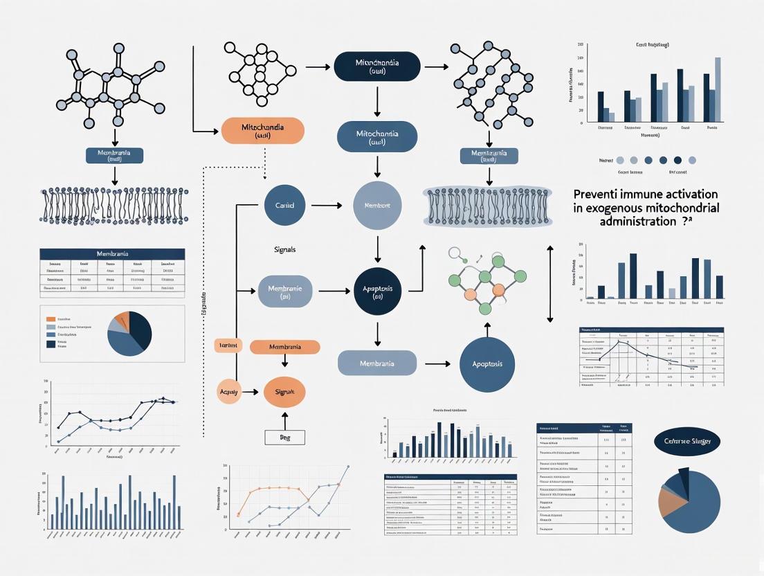
Mitigating Immune Activation in Exogenous Mitochondrial Administration: Strategies for Therapeutic Applications
Exogenous mitochondrial administration represents a groundbreaking therapeutic paradigm for diseases involving mitochondrial dysfunction.
Mitigating Immune Activation in Exogenous Mitochondrial Administration: Strategies for Therapeutic Applications
Abstract
Exogenous mitochondrial administration represents a groundbreaking therapeutic paradigm for diseases involving mitochondrial dysfunction. However, the clinical translation of this approach is significantly hampered by the potential for unintended immune activation, as mitochondrial components can be recognized as damage-associated molecular patterns (DAMPs) by the host's immune system. This article provides a comprehensive analysis for researchers and drug development professionals, exploring the foundational immunology of mitochondrial DAMPs, current methodological approaches for administration, advanced strategies to suppress immune responses, and comparative validation of different techniques. By synthesizing the latest research, we aim to outline a path toward safer and more effective mitochondrial therapies by addressing the critical challenge of immune compatibility.
The Double-Edged Sword: Understanding Mitochondrial Immunology and Innate Immune Recognition
Mitochondrial Damage-Associated Molecular Patterns (mitoDAMPs) are molecules normally contained within healthy mitochondria that, when released into the cytosol or extracellular space during cellular stress or damage, trigger potent immune responses by mimicking pathogenic threats [1] [2]. This occurs because of the bacterial origin of mitochondria, which results in mitochondrial components sharing structural similarities with bacterial pathogen-associated molecular patterns (PAMPs) [2] [3]. When cells experience stress from infection, mechanical damage, or other insults, these mitochondrial molecules escape their compartments and are recognized by the immune system as "non-self" or "alarm" signals, initiating inflammatory pathways [1] [4].
For researchers investigating exogenous mitochondrial administration, this phenomenon presents a significant challenge: the very therapeutic mitochondria intended to restore cellular function may trigger unwanted immune activation through DAMP release. Understanding these mechanisms is crucial for developing strategies to mitigate immune responses while preserving mitochondrial function.
Key Mitochondrial DAMPs and Their Immune Recognition
The table below summarizes the primary mitochondrial DAMPs, their release mechanisms, and the immune pathways they activate.
Table 1: Key Mitochondrial DAMPs and Their Immune Recognition Pathways
| Mitochondrial DAMP | Release Mechanisms | Pattern Recognition Receptors | Downstream Immune Effects |
|---|---|---|---|
| mtDNA | VDAC oligomerization, mitochondrial permeability transitions, BAK/BAX activation [5] [6] | cGAS, TLR9, AIM2, NLRP3 [1] [7] [5] | Type I IFN production, NF-κB activation, NLRP3 inflammasome activation, pro-inflammatory cytokine secretion [1] [7] |
| ATP | Mechanical stress, necrotic cell death, connexin hemichannels [1] | P2X7, P2Y2 purinergic receptors [1] | NLRP3 inflammasome activation, potassium efflux, chemotaxis of immune cells [1] |
| Cardiolipin | Mitochondrial outer membrane permeabilization, mitochondrial stress [3] | NLRP3, caspase-1, caspase-11 [3] | Inflammasome assembly, IL-1β and IL-18 processing [3] |
| N-formyl peptides (NFPs) | Mitochondrial disruption, cellular necrosis [1] | Formyl peptide receptors (FPRs) [1] | Neutrophil and platelet chemotaxis, inflammatory responses [1] |
| TFAM | Mitochondrial membrane damage, hemorrhagic shock [1] | Unknown receptor(s) | Proinflammatory cytokine secretion (TNF, IL-6) in macrophages [1] |
Table 2: Experimental Models Demonstrating Mitochondrial DAMP Release
| Experimental Model | Inducing Stimulus | DAMP Released | Observed Effect |
|---|---|---|---|
| TLR-primed macrophages | ATP treatment [1] | mtDNA to cytosol | AIM2/NLRP3 inflammasome activation [1] |
| Eosinophils | LPS and IL-5 or IL-5 and C5a [1] | mtDNA to extracellular space | Intestinal eosinophil infiltration in sepsis [1] |
| Irradiated mice | Radiation exposure [1] | ATP, mtDNA | GVHD development, donor T-cell expansion [1] |
| Acetaminophen-induced liver injury | Acetaminophen overdose [1] | mtDNA to circulation | Systemic inflammation and lung injury (TLR9-dependent) [1] |
| Cardiac hypertrophy model | DNase IIa deletion [1] | mtDNA accumulation | Severe myocarditis, mortality [1] |
Troubleshooting Guide: Common Experimental Challenges
Why is my mitochondrial preparation triggering immune responses in recipient cells?
This is likely due to the release of mitochondrial DAMPs during isolation or administration. mtDNA is particularly potent due to its hypomethylated CpG motifs that resemble bacterial DNA [7] [4]. Additionally, cardiolipin exposure during mitochondrial handling can activate NLRP3 inflammasomes [3].
Solutions:
- Implement rapid isolation protocols to minimize mitochondrial deterioration
- Add nuclease inhibitors to prevent mtDNA release and degradation
- Consider DNase pretreatment of mitochondrial preparations (with appropriate controls)
- Verify mitochondrial integrity using membrane potential dyes (JC-1, TMRE) before administration
- Include purity assessments to rule out contamination with other cellular components
How can I distinguish mtDNA-mediated effects from nuclear DNA effects?
mtDNA has several unique properties that differentiate it from nuclear DNA:
- Lacks histone packaging and is organized by TFAM instead [7]
- Contains hypomethylated CpG motifs similar to bacterial DNA [7] [4]
- Has a circular structure and is present in high copy number [7]
- Vulnerable to oxidative damage due to proximity to ROS production [7]
Experimental approaches:
- Use qPCR with specific primers for mitochondrial vs. nuclear genes
- Employ TLR9 inhibitors to test involvement of this pathway
- Utilize cells lacking cGAS or STING to determine pathway specificity [5]
- Measure oxidative modifications to DNA to confirm mitochondrial origin
Why do I observe different immune responses to mitochondrial transfer in different cell types?
Immune cell populations have distinct metabolic requirements and mitochondrial functions that shape their responses to mitochondrial DAMPs [4]. For example:
- Pro-inflammatory cells (activated monocytes, T cells) rely on glycolysis and may respond differently to mitochondrial signals [4]
- Regulatory cells (Tregs, M2 macrophages) depend on mitochondrial function and β-oxidation [4]
- Non-immune cells vary in their expression of PRRs and downstream signaling components
Solutions:
- Characterize PRR expression patterns in your target cell types
- Test mitochondrial transfer in multiple relevant cell types
- Consider co-culture systems to examine paracrine effects
Key Signaling Pathways Activated by Mitochondrial DAMPs
The following diagram illustrates the primary immune signaling pathways activated by mitochondrial DAMPs, particularly focusing on mtDNA:
Diagram 1: Immune signaling pathways activated by mitochondrial DAMPs
Detailed Methodologies for Key Experiments
Protocol 1: Detecting mtDNA Release in vitro
Principle: This protocol utilizes quantitative PCR to detect and quantify mtDNA that has been released into the cytosol or cell culture supernatant.
Reagents Required:
- Cell lysis buffer (0.025% digitonin in PBS)
- DNA extraction kit (specific for cell culture supernatants)
- qPCR reagents (SYBR Green master mix)
- Primers for mitochondrial genes (e.g., ND1, ND6) and nuclear genes (e.g., 18S rRNA)
- Standard curves for absolute quantification
Procedure:
- Fractionate cells using digitonin lysis (0.025% in PBS) to isolate cytosolic fraction without disrupting organelles
- Centrifuge at 800×g for 5 minutes to pellet nuclei and unlysed cells
- Collect supernatant and centrifuge at 16,000×g for 15 minutes to remove mitochondria
- Extract DNA from the resulting supernatant (cytosolic fraction) using a commercial kit
- Perform qPCR using mitochondrial-specific and nuclear-specific primers
- Calculate mtDNA release as the ratio of mitochondrial to nuclear DNA in the cytosolic fraction compared to total cellular DNA
Troubleshooting Tips:
- Avoid freeze-thaw cycles which can damage mitochondrial membranes
- Include DNase control treatments to confirm intracellular location of detected DNA
- Normalize to cell number rather than total protein for more accurate quantification
Protocol 2: Assessing cGAS-STING Pathway Activation
Principle: This method evaluates activation of the cGAS-STING pathway by measuring phosphorylation of key signaling components and downstream gene expression.
Reagents Required:
- Phospho-specific antibodies (p-TBK1, p-IRF3)
- STING agonists (e.g., cGAMP) and inhibitors (e.g., H-151)
- Type I IFN reporter cell lines or ELISA kits
- RT-PCR reagents for IFN-β and ISG expression
Procedure:
- Treat cells with mitochondrial preparations or isolated mtDNA
- Lyse cells at various time points (15 min to 6 hours post-treatment)
- Perform Western blotting for p-TBK1 (Ser172) and p-IRF3 (Ser396)
- Measure IFN-β production using ELISA or reporter assays
- Quantify ISG expression (e.g., MX1, ISG15) by RT-qPCR
- Confirm pathway specificity using STING-knockout cells or pharmacological inhibitors
Validation Methods:
- Immunofluorescence for IRF3 nuclear translocation
- cGAMP measurement by LC-MS to confirm cGAS activation
- Genetic approaches (siRNA, CRISPR) targeting cGAS or STING
The Scientist's Toolkit: Essential Research Reagents
Table 3: Key Research Reagents for Mitochondrial DAMP Studies
| Reagent Category | Specific Examples | Research Application | Key Considerations |
|---|---|---|---|
| PRR Inhibitors | H-151 (STING), ODN TTAGGG (TLR9), MCC950 (NLRP3) | Pathway validation, mechanism determination | Test multiple inhibitors to confirm specificity; monitor off-target effects |
| Detection Antibodies | Anti-p-TBK1, anti-p-IRF3, anti-cleaved caspase-1 | Pathway activation assessment | Validate phospho-specificity with appropriate controls |
| Mitochondrial Damps | Isolated mtDNA, recombinant TFAM, synthetic cardiolipin | Positive controls, direct stimulation | Use endotoxin-free preparations to avoid confounding effects |
| Metabolic Probes | JC-1, TMRM, MitoSOX | Mitochondrial function and integrity assessment | Correlate membrane potential with DAMP release |
| Genetic Tools | cGAS/STING KO cells, MAVS mutants, DNase II-deficient models | Mechanistic studies | Use appropriate wild-type controls from same genetic background |
FAQs: Addressing Common Research Questions
Q1: What are the key differences between mitochondrial and nuclear DNA in immune activation?
mtDNA possesses several unique features that enhance its immunostimulatory potential compared to nuclear DNA:
- Evolutionary origin: mtDNA retains bacterial molecular signatures due to its prokaryotic origin [7] [3]
- Methylation pattern: mtDNA contains hypomethylated CpG motifs that strongly activate TLR9 [7] [4]
- Packaging: mtDNA is not protected by histones but organized by TFAM, making it more accessible to DNA sensors [7]
- Damage susceptibility: mtDNA is more vulnerable to oxidative damage, which can enhance its immunogenicity [7]
Q2: How can I minimize immune activation in mitochondrial transplantation studies?
Several strategies can reduce immune responses to administered mitochondria:
- Isolation optimization: Use gentle isolation methods that preserve mitochondrial integrity
- DNase pretreatment: Briefly treat mitochondria with DNase to remove surface-associated mtDNA (with appropriate validation)
- Immunomodulatory co-treatments: Consider transient anti-inflammatory treatments during transplantation
- Source matching: Use autologous or immunologically compatible mitochondrial sources when possible
- Quality validation: Implement rigorous quality control measures including membrane potential assessment and purity checks
Q3: What are the most reliable markers for mitochondrial DAMP-mediated inflammation?
The optimal markers depend on the specific DAMP and pathway being activated:
- For mtDNA/cGAS-STING: Phospho-TBK1, phospho-IRF3, IFN-β, CXCL10 [5] [6]
- For mtDNA/TLR9: NF-κB activation, TNF-α, IL-6 [7]
- For ATP/NLRP3: Caspase-1 cleavage, IL-1β secretion [1]
- General inflammation markers: IL-6, IL-8, TNF-α across multiple pathways
Q4: How does mitochondrial transfer occur naturally between cells, and can this be exploited therapeutically?
Natural mitochondrial transfer occurs through several mechanisms:
- Tunneling nanotubes (TNTs): Long, thin membrane connections between cells [8]
- Extracellular vesicles: Mitochondria packaged in membrane-bound structures [8]
- Cell fusion: Direct merging of cell membranes [8]
- Gap junctions: Direct connections between adjacent cells [8]
Therapeutic approaches can exploit these mechanisms by enhancing natural transfer processes or developing engineered delivery systems that mimic these natural pathways while minimizing immune recognition.
In the pioneering field of exogenous mitochondrial administration, a central challenge is the unintended activation of the recipient's innate immune system. The very mitochondrial components meant to provide therapeutic benefit—particularly mitochondrial DNA (mtDNA)—can be recognized as foreign "danger" signals by the host's cellular sentinels. This technical support center is designed to help researchers navigate the complexities of three key DNA-sensing pathways—cGAS-STING, NLRP3 inflammasome, and TLR9—that are critical gatekeepers in this immune response. The following troubleshooting guides, FAQs, and detailed protocols provide a structured framework to identify, prevent, and resolve immune activation issues in your experiments, thereby enhancing the efficacy and safety of mitochondrial therapies.
Pathway Diagrams and Mechanisms
Integrated Signaling Pathway Activation by mtDNA
The following diagram illustrates the core pathways through which released mitochondrial DNA activates innate immune signaling, connecting the key molecules involved in the cGAS-STING, NLRP3, and TLR9 pathways.
Mitochondrial DNA Release Mechanisms
This diagram details the specific molecular mechanisms by which mitochondrial DNA is released from mitochondria during cellular stress, serving as the initial trigger for immune pathway activation.
Troubleshooting Guides
cGAS-STING Pathway Activation
Problem: Unwanted type I interferon response following mitochondrial administration.
Background: The cGAS-STING pathway is triggered when cytosolic DNA sensors detect mitochondrial DNA that has been released during administration [9] [10] [6]. cGAS binds to mtDNA and synthesizes the second messenger 2'3'-cGAMP, which activates STING, leading to TBK1 and IRF3 phosphorylation and subsequent type I interferon production [10] [6].
| Issue | Possible Causes | Recommended Solutions | Validation Experiments |
|---|---|---|---|
| High IFN-β production | mtDNA contamination from damaged mitochondria; Improper purification; cGAS activation by cytosolic mtDNA | Use DNase I treatment during preparation; Optimize mitochondrial quality control assays; Implement density gradient purification | Measure IFN-β mRNA via qPCR; Monitor phospho-IRF3 via Western blot |
| Constitutive STING activation | mtDNA fragments in final preparation; Contaminating nucleic acids; Oxidized mtDNA | Implement mtDNA quantification in final product; Use antioxidants during isolation; Employ ultrafiltration steps | cGAMP measurement via LC-MS; STING trafficking assays by immunofluorescence |
| Variable immune response | Inconsistent mitochondrial quality between preparations; Differences in donor-recipient species mismatch | Standardize quality control metrics (membrane potential, cytochrome c release); Use syngeneic systems where possible | Establish minimum ΔΨm threshold for administration; Batch testing for mtDNA contamination |
Critical Controls:
- Include DNase-treated mitochondrial preparations
- Use mtDNA-depleted (ρ0) cells as negative control
- Test with cGAS or STING knockout cells to confirm pathway specificity
NLRP3 Inflammasome Activation
Problem: IL-1β and IL-18 secretion leading to pyroptotic cell death.
Background: The NLRP3 inflammasome can be activated by oxidized mtDNA, particularly under conditions of mitochondrial dysfunction and reactive oxygen species (ROS) production [8] [11] [6]. This leads to caspase-1 activation, which processes pro-IL-1β and pro-IL-18 into their mature forms and cleaves gasdermin D to induce pyroptosis [11].
| Issue | Possible Causes | Recommended Solutions | Validation Experiments |
|---|---|---|---|
| Caspase-1 activation | ROS generation from damaged mitochondria; Oxidized mtDNA release; Potassium efflux | Add mitochondrial antioxidants (MitoTEMPO); Use NLRP3 inhibitors (MCC950); Control osmolarity | Caspase-1 activity assay; IL-1β ELISA; LDH release for pyroptosis |
| Inconsistent inflammasome activation | Variable mitochondrial membrane potential between batches; Differences in ROS production | Standardize ΔΨm measurements; Include ROS scavengers in buffer; Control incubation temperature | MitoSOX Red staining for mtROS; JC-1 assay for ΔΨm; ASC speck formation assay |
| Secondary inflammation | Amplification through cGAS-STING crosstalk; Gasdermin D pore formation | Combine with STING inhibitors; Use disulfiram to inhibit GSDMD | Measure extracellular HMGB1 and ATP; Perform live-cell imaging with propidium iodide |
Experimental Protocol: NLRP3 Activation Assessment
- Priming Step: Pre-treat recipient cells with LPS (100 ng/mL, 3 hours)
- Mitochondrial Challenge: Apply purified mitochondria (10-50 μg protein/mL, 6 hours)
- Inhibition Controls: Include MCC950 (10 μM) or MitoTEMPO (100 μM) in parallel
- Readouts:
- Collect supernatant for IL-1β ELISA
- Lyse cells for caspase-1 p20 Western blot
- Measure LDH release as pyroptosis indicator
- Stain with MitoSOX Red for mitochondrial ROS
TLR9-Mediated Immune Recognition
Problem: Endosomal DNA sensing causing pro-inflammatory cytokine production.
Background: TLR9 is activated by unmethylated CpG DNA motifs present in mtDNA, which resembles bacterial DNA due to its evolutionary origin [12] [13] [14]. This occurs when mtDNA is internalized into endolysosomal compartments where TLR9 is localized, leading to MyD88-dependent NF-κB and IRF7 activation [12].
| Issue | Possible Causes | Recommended Solutions | Validation Experiments |
|---|---|---|---|
| NF-κB activation | Endosomal uptake of mtDNA; Unmethylated CpG motifs in mtDNA; MyD88 recruitment | Use chloroquine to inhibit endosomal acidification; Test TLR9 inhibitory ODNs; Employ mtDNA methylation approaches | NF-κB luciferase reporter assay; Phospho-IκBα Western blot; TLR9 translocation imaging |
| Cell-type specific responses | Differential TLR9 expression; Variations in endosomal trafficking | Characterize TLR9 expression in target cells; Modulate endosomal uptake with inhibitors | Flow cytometry for TLR9 surface expression; qPCR for TLR9 mRNA levels |
| Synergistic activation | Cooperation with cGAS-STING pathway; Enhanced cytokine production | Combine TLR9 and STING inhibition; Block downstream signaling nodes | Multiplex cytokine array; RNA-seq for interferon-stimulated genes |
Frequently Asked Questions (FAQs)
Q1: Why is exogenously administered mitochondrial DNA so immunogenic compared to nuclear DNA?
A: Mitochondrial DNA possesses several intrinsic features that make it highly immunogenic [13] [14]:
- Evolutionary Origin: mtDNA resembles bacterial DNA due to its evolutionary origin from α-proteobacteria
- Unmethylated CpG Motifs: mtDNA contains abundant unmethylated cytosine-phosphate-guanine (CpG) islands, similar to bacterial DNA, which are potent TLR9 agonists [12] [14]
- Lack of Chromatinization: Unlike nuclear DNA, mtDNA is not packaged with histones, making it more accessible to DNA sensors
- High Copy Number: Cells contain hundreds to thousands of mtDNA copies, increasing the likelihood of immune recognition
- Oxidative Damage: mtDNA is particularly susceptible to oxidative damage, and oxidized mtDNA is a potent NLRP3 inflammasome activator [6]
Q2: What are the key differences between BAK/BAX-mediated and VDAC-mediated mtDNA release?
A: These two primary release mechanisms differ in their regulation and consequences [9] [13] [6]:
| Feature | BAK/BAX Macropores | VDAC Oligomerization |
|---|---|---|
| Cellular Context | Apoptotic stress | Living cells, oxidative stress |
| Pore Size | Large macropores | Smaller pores |
| mtDNA Form Released | Entire nucleoids | mtDNA fragments |
| Caspase Dependence | Caspase-independent | Caspase-independent |
| Primary Regulators | BAK, BAX oligomerization | VDAC oligomerization, mPTP opening |
| Therapeutic Targeting | BAK/BAX inhibitors difficult due to apoptotic role | VDAC inhibitors (VBIT-4) available |
Q3: How can I determine which immune pathway is primarily responsible for the inflammatory response in my experimental system?
A: Employ a systematic pharmacological and genetic approach:
- Pathway-Specific Inhibitors:
- cGAS-STING: H-151 (STING inhibitor), RU.521 (cGAS inhibitor)
- NLRP3: MCC950 (NLRP3 inhibitor), VX-765 (caspase-1 inhibitor)
- TLR9: Chloroquine (endosomal acidification inhibitor), ODN INH-18 (TLR9 antagonist)
Genetic Validation:
- CRISPR/Cas9 knockout of cGAS, STING, or NLRP3
- siRNA knockdown of TLR9 or MyD88
- Use of immortalized bone marrow-derived macrophages from gene-deficient mice
Readout Specificity:
- cGAS-STING: Type I interferon (IFN-β), CXCL10
- NLRP3: Caspase-1 cleavage, IL-1β maturation
- TLR9: Early NF-κB activation, particular ISG subset
Q4: What quality control measures are most critical for preventing immune activation in mitochondrial preparations?
A: Implement a multi-tiered QC strategy:
- Membrane Integrity: ≥85% of mitochondria should maintain membrane potential (ΔΨm) as measured by JC-1 or TMRM staining
- mtDNA Contamination: <5 ng mtDNA per mg mitochondrial protein by qPCR
- Cytochrome c Retention: >90% cytochrome c retention via Western blot of supernatant vs pellet
- Functional Assessment: Oxygen consumption rate (OCR) >50% of theoretical maximum
- Sterility: Negative endotoxin testing (<0.1 EU/mL)
Q5: Are there circumstances where controlled immune activation following mitochondrial transfer might be beneficial?
A: Yes, in certain therapeutic contexts:
- Cancer Immunotherapy: Mitochondrial transfer could potentially enhance antitumor immunity through controlled cGAS-STING activation [15] [10]
- Vaccine Adjuvants: mtDNA components could serve as natural adjuvants to boost immune responses [11]
- Wound Healing: Transient, controlled inflammation can promote tissue repair mechanisms
- Metabolic Reprogramming: Mild STING activation can enhance immune cell function in certain scenarios [8]
The key is achieving spatiotemporal control over the immune activation, potentially through mitochondrial engineering or targeted delivery systems.
The Scientist's Toolkit: Research Reagent Solutions
| Reagent/Category | Specific Examples | Primary Function | Application Notes |
|---|---|---|---|
| Pathway Inhibitors | H-151 (STING), MCC950 (NLRP3), Chloroquine (TLR9) | Specific pathway blockade | Use multiple inhibitors to confirm pathway specificity; titrate carefully |
| mtDNA Quantification | qPCR (ND1, CYTB genes), Picogreen assay, Anti-dsDNA ELISA | Measure mtDNA contamination | Establish baseline in your system; different assays may give varying results |
| Mitochondrial Quality Probes | JC-1 (ΔΨm), MitoSOX Red (mtROS), MitoTracker Deep Red | Assess mitochondrial integrity | Always include fresh vs. aged mitochondrial controls |
| Cytokine Detection | IFN-β ELISA, IL-1β ELISA, Luminex multiplex arrays | Quantify immune activation | Time-course experiments recommended; different cytokines have different kinetics |
| Genetic Tools | cGAS/STING/NLRP3/TLR9 KO cells, siRNA/shRNA knockdown | Confirm pathway requirement | Use rescue experiments to validate specificity |
| VDAC Inhibitors | VBIT-4, DIDS | Block VDAC oligomerization | Particularly useful for non-apoptotic mtDNA release models [6] |
| Antioxidants | MitoTEMPO, N-acetylcysteine (NAC) | Reduce oxidative stress | Can specifically prevent oxidized mtDNA formation |
| Caspase Assays | Caspase-1 FLICA, Western for p20 subunit, VAD-FMK inhibitors | Detect inflammasome activation | Combine with LDH release for pyroptosis confirmation |
Advanced Experimental Design
Comprehensive Pathway Analysis Workflow
For thorough characterization of immune pathway activation in mitochondrial transfer experiments, implement this multi-step workflow:
Phase 1: Initial Screening (24-48 hours)
- Mitochondrial Administration: Dose range (1-100 μg protein/mL) in relevant cell types
- Early Time Points: Collect supernatant and lysates at 6, 12, and 24 hours
- Multiplex Cytokine Array: Simultaneous measurement of IFN-β, IL-1β, IL-6, IL-18, TNF-α, CXCL10
- Cell Viability Assessment: MTT/LDH parallel assays to distinguish apoptosis from pyroptosis
Phase 2: Mechanistic Studies (Pathway Specific)
- Inhibitor Panel Testing: Apply specific inhibitors 1 hour prior to mitochondrial administration
- Kinetic Analysis: High-time resolution sampling (1, 3, 6, 12, 24 hours) for pathway activation
- Imaging Correlates: Live-cell imaging of mitochondrial membrane potential, ROS production, and cell death morphology
- Biochemical Confirmation: Western blot for phospho-proteins (p-TBK1, p-IRF3, p-IκBα), caspase cleavage, and gasdermin D processing
Phase 3: Genetic Validation
- CRISPR/Cas9 Knockouts: Generate or utilize cGAS, STING, NLRP3, and TLR9 deficient cells
- Rescue Experiments: Re-expression of wild-type and mutant constructs
- In Vivo Correlates: Use of gene-deficient mice for translational models
Data Interpretation Framework
When analyzing results, consider these key aspects:
Temporal Patterns:
- Early Response (2-6 hours): Typically TLR9-mediated (NF-κB activation)
- Intermediate (6-12 hours): cGAS-STING activation (IFN-β production)
- Late Phase (12-24 hours): NLRP3 inflammasome activation and pyroptosis
Amplification Loops:
- STING activation can prime NLRP3 inflammasome
- Inflammasome-derived cytokines can enhance DNA sensor expression
- Type I interferon signaling upregulates additional PRRs
Threshold Considerations:
- Immune activation typically follows a threshold effect rather than linear response
- Multiple pathways may activate simultaneously at higher mitochondrial doses
- Cell-type specific differences in pathway dominance exist
By implementing this comprehensive troubleshooting framework, researchers can systematically identify and mitigate unwanted immune activation, advancing the therapeutic potential of mitochondrial administration while maintaining scientific rigor.
Core Concepts & Frequently Asked Questions (FAQs)
FAQ 1: Why do exogenously administered mitochondria trigger an innate immune response?
The innate immune system uses Pattern Recognition Receptors (PRRs) to identify conserved microbial molecules. Due to their bacterial origin, mitochondria share molecular patterns with bacteria. Mitochondrial DNA (mtDNA) is a primary trigger because it contains unmethylated cytosine-phosphate-guanine (CpG) islands, a hallmark of bacterial DNA that is recognized by Toll-like Receptor 9 (TLR9) [16] [13]. Furthermore, when released into the cytosol or extracellular space, mtDNA is sensed by other receptors like cGAS (cyclic GMP-AMP synthase), which activates the STING pathway, leading to type I interferon production [17] [18] [13]. Other mitochondrial components, such as N-formyl peptides, can also activate immune cells by binding to formyl peptide receptors, mimicking the response to bacterial infection [17].
FAQ 2: What are the primary signaling pathways activated by misplaced mtDNA?
The following table summarizes the key DNA-sensing receptors and the consequences of their activation by mtDNA.
| DNA Sensor | Localization | Downstream Pathway | Key Immune Effectors | References |
|---|---|---|---|---|
| TLR9 | Endosome | MyD88/NF-κB | Pro-inflammatory cytokines (TNF, IL-6) | [16] [13] |
| cGAS | Cytosol | cGAMP/STING/TBK1/IRF3 | Type I Interferons (IFN-β) | [17] [18] [13] |
| NLRP3 | Cytosol | Inflammasome assembly / Caspase-1 | IL-1β, IL-18, Pyroptosis | [17] [18] |
| AIM2 | Cytosol | Inflammasome assembly / Caspase-1 | IL-1β, IL-18, Pyroptosis | [13] |
FAQ 3: What specific features of mtDNA make it so immunogenic?
mtDNA possesses several intrinsic properties that contribute to its high immunogenicity [16] [13]:
- Unmethylated CpG Motifs: Unlike mammalian nuclear DNA, mtDNA is rich in unmethylated CpG sequences, making it resemble bacterial DNA.
- Lack of Histones: mtDNA is not packaged with histones, which increases its accessibility to DNA sensors.
- High Copy Number: Each cell contains hundreds to thousands of mtDNA copies, increasing the probability of detection upon release.
- Susceptibility to Damage: The mitochondrial matrix is a site of reactive oxygen species (ROS) generation, making mtDNA more prone to oxidation and fragmentation, which can enhance its immunogenicity.
FAQ 4: Under what conditions is mtDNA released from mitochondria?
MtDNA release is not a passive process but occurs through specific mechanisms, often during cellular stress or mitochondrial dysfunction [18] [13]:
- Mitochondrial Permeability Transition Pore (mPTP) Opening
- Oligomerization of BAX/BAK pores in the outer mitochondrial membrane.
- Mitochondrial membrane destabilization caused by oxidative stress or calcium overload.
- Defective mitophagy, the process that normally clears damaged mitochondria.
Troubleshooting Guide: Preventing Immune Activation
This guide addresses common experimental challenges in exogenous mitochondrial administration.
Problem: Inflammatory Response to Transplanted Mitochondria
| Symptom | Potential Cause | Solution & Experimental Considerations |
|---|---|---|
| Elevated pro-inflammatory cytokines (e.g., IL-6, TNF-α) in culture/media. | Activation of the TLR9 pathway by mtDNA contaminants or damaged mitochondria. | Pre-treatment: Isolate mitochondria using density gradient centrifugation in sterile, nuclease-free buffers to remove contaminating nuclear DNA. Inhibition: Use TLR9 inhibitory oligonucleotides (e.g., ODN INH-18) in your experimental system. Validation: Perform qPCR to quantify mtDNA in your mitochondrial prep and use electron microscopy to confirm structural integrity. |
| Increased expression of interferon-stimulated genes (ISGs). | Activation of the cGAS-STING pathway by cytosolic mtDNA. | Quality Control: Ensure mitochondrial preparations are highly purified and free of lysed organelle debris. Pharmacological Inhibition: Treat recipient cells with a STING inhibitor (e.g., H-151). Genetic Knockdown: Use siRNA to knock down cGAS or STING in recipient cells prior to mitochondrial transfer. |
| Activation of the NLRP3 inflammasome and Caspase-1. | mtDNA and/or ROS released from dysfunctional transplanted mitochondria. | Mitochondrial Fitness: Use functional assays (e.g., Seahorse Analyzer) to confirm the respiratory capacity of isolated mitochondria before transfer. Antioxidant Treatment: Include antioxidants (e.g., MitoTEMPO) in the culture medium to scavenge mitochondrial ROS. |
| Rapid clearance of transplanted mitochondria and failure of functional integration. | Innate immune recognition by recipient phagocytes. | Surface Modification: Chemically modify the mitochondrial surface with biocompatible polymers (e.g., polyethylene glycol) or use cell-penetrating peptides (CPPs) like Pep-1 to "shield" mitochondria and enhance uptake via non-immune pathways [19]. Use of Vesicles: Encapsulate mitochondria in extracellular vesicles or liposomes to mask their immunogenic surface patterns [19]. |
Problem: Low Mitochondrial Transfer/Transplantation Efficiency
| Symptom | Potential Cause | Solution & Experimental Considerations |
|---|---|---|
| Poor uptake of isolated mitochondria by recipient cells. | Inefficient delivery method; mitochondrial aggregation; negative surface charge repelling the cell membrane. | Optimized Delivery: Utilize direct injection, pressure-enhanced delivery, or magnetically-guided delivery for in vitro and in vivo models. Surface Functionalization: Conjugate mitochondria with cell-penetrating peptides (CPPs) like TAT or Pep-1 to facilitate membrane crossing and improve uptake efficiency by 4-5 fold [19]. Use of Hydrogels: Employ hydrogel-based delivery systems to protect mitochondria and provide a controlled release. |
| Loss of mitochondrial function after isolation. | Extended isolation time; inappropriate isolation buffer; mechanical stress. | Rapid Processing: Isolate and transplant mitochondria within a short timeframe (ideally <2 hours) to preserve respiratory function [19]. Optimized Buffer: Use ice-cold, isotonic isolation buffers with energy substrates (e.g., pyruvate, malate) and ATP. Viability Assay: Routinely assess mitochondrial membrane potential (using JC-1 or TMRM dyes) and oxygen consumption rate (OCR) pre- and post-isolation. |
Key Experimental Protocols
Protocol 1: Assessing Immune Activation by Isolated Mitochondria In Vitro
Title: Co-culture of isolated mitochondria with reporter macrophages to quantify innate immune activation.
Background: This protocol is used to test the immunogenicity of a mitochondrial preparation before proceeding to more complex transplantation experiments.
Reagents & Materials:
- Primary macrophages (e.g., bone marrow-derived macrophages) or macrophage cell line (e.g., RAW 264.7)
- Isolation buffer (e.g., Mannitol-Sucrose-HEPES-EGTA buffer)
- Cell culture plates
- ELISA kits for TNF-α, IL-6, IFN-β
- TLR9 inhibitor (e.g., ODN 2088)
- STING inhibitor (e.g., H-151)
Procedure:
- Isolate Mitochondria: Isolate mitochondria from the desired tissue (e.g., liver) using standard differential centrifugation. Keep the preparation on ice and use within 60-90 minutes.
- Plate Cells: Seed macrophages in a 24-well plate and allow them to adhere overnight.
- Pre-treatment (Optional): To identify the involved pathway, pre-treat some wells with a TLR9 inhibitor (1-5 µM) or STING inhibitor (1 µM) for 1 hour.
- Stimulation: Add the isolated mitochondria (e.g., 10-50 µg protein per well) to the macrophages. Include controls with:
- Media only (negative control).
- Lipopolysaccharide (LPS) (positive control for TLR4).
- CpG ODN (positive control for TLR9).
- Incubate: Incubate the plate for 6-24 hours at 37°C.
- Analysis:
- Collect culture supernatant and analyze for cytokine secretion by ELISA.
- Extract total RNA from cells and analyze expression of interferon-stimulated genes (e.g.,
MX1,ISG15) by qRT-PCR.
Protocol 2: Mitochondrial Surface Modification with Pep-1
Title: Conjugation of cell-penetrating peptide Pep-1 to isolated mitochondria to enhance uptake and reduce immunogenicity.
Background: This protocol, based on the work of Chang et al., details the conjugation of the Pep-1 peptide to the mitochondrial surface to improve delivery efficiency [19].
Reagents & Materials:
- Isolated mitochondria
- Pep-1 peptide
- Sterile PBS or mitochondrial respiration buffer
- Water bath or incubator set to 37°C
Procedure:
- Prepare Mitochondria: Isolate mitochondria as per standard protocol and resuspend in a suitable buffer at a known protein concentration (e.g., 1-2 mg/mL).
- Prepare Pep-1 Solution: Dissolve Pep-1 peptide in sterile buffer.
- Form Complex: Combine the mitochondrial suspension and the Pep-1 solution at a weight ratio of 1:1750 (mitochondria:Pep-1) [19]. For example, mix 10 µg of mitochondria with 17.5 µg of Pep-1.
- Incubate: Incubate the mixture at 37°C for 30 minutes to allow the complex to form.
- Wash (Optional): Centrifuge the mixture to remove unbound peptide and resuspend the modified mitochondria in fresh buffer for immediate use.
- Validation: The efficiency of modification and uptake can be validated by flow cytometry if mitochondria are pre-labeled with a dye (e.g., MitoTracker).
Signaling Pathways & Experimental Workflows
Mitochondrial Immune Signaling Pathways
The diagram below illustrates the primary innate immune pathways activated by mitochondrial damage and mtDNA release.
Experimental Workflow for Testing Mitochondrial Immunogenicity
The following diagram outlines a logical workflow for systematically testing and mitigating the immune response to transplanted mitochondria.
The Scientist's Toolkit: Research Reagent Solutions
The table below lists key reagents used in mitochondrial transplantation and immunogenicity research.
| Research Reagent | Function / Application | Specific Examples & Notes |
|---|---|---|
| TLR9 Inhibitors | To block immune activation via the endosomal mtDNA-sensing pathway. | ODN 2088: A competitive antagonist that prevents TLR9 binding and activation. Use at 1-5 µM for pre-treatment in cell culture. |
| STING Inhibitors | To block the cytosolic mtDNA-sensing cGAS-STING pathway. | H-151: A potent and selective STING antagonist. Typically used at 1 µM concentration in cell-based assays. |
| Cell-Penetrating Peptides (CPPs) | To enhance mitochondrial uptake by recipient cells and potentially shield immunogenic epitopes. | Pep-1 & TAT: Conjugated to isolated mitochondria at specific weight ratios (e.g., 1750:1 Pep-1:mitochondria) to form a complex that facilitates delivery [19]. |
| MitoTracker Probes | To label and track isolated, living mitochondria via fluorescence microscopy or flow cytometry. | MitoTracker Green FM: Labels mitochondria regardless of membrane potential. MitoTracker Red CMXRos: Accumulates in active mitochondria based on membrane potential. |
| Extracellular Vesicles (EVs) | A biotechnological delivery vehicle to encapsulate mitochondria, protecting them from immune recognition. | Mitochondria can be encapsulated inside engineered EVs or liposomes. This masks their bacterial-like surface and utilizes natural vesicle uptake mechanisms [19]. |
| MitoTEMPO | A mitochondrial-targeted antioxidant. | Scavenges mitochondrial reactive oxygen species (mtROS), reducing one of the triggers for NLRP3 inflammasome activation and mtDNA damage. |
The administration of exogenous mitochondria represents a promising therapeutic strategy for restoring cellular function in damaged tissues, particularly in cardiac repair and regenerative medicine. However, a significant barrier to its clinical translation is the initiation of undesirable immune activation. When mitochondrial administration is performed without adequate precaution, the introduced organelles can be recognized by the host's immune system as "damage-associated molecular patterns" (DAMPs), triggering a cascade of inflammatory events. This response is primarily mediated by the innate immune system through pattern recognition receptors that detect mitochondrial components shared with their bacterial ancestors, notably mitochondrial DNA (mtDNA). This immune recognition can lead to local inflammation, reduced therapeutic efficacy of the administered mitochondria, and potential tissue damage through the very mechanisms this therapy aims to repair. Understanding these pathways and implementing strategies to mitigate immune activation is therefore crucial for advancing mitochondrial transplantation from experimental models to clinical applications.
Key Signaling Pathways in Mitochondria-Induced Immune Activation
The STING-NF-κB Signaling Axis
The stimulator of interferon genes (STING) pathway serves as a central hub for immune activation in response to exogenous mitochondria. Research has demonstrated that mitochondria released from injured cells robustly activate endothelial cells (ECs), fostering inflammatory processes that can contribute to complications such as allograft rejection [20].
The mechanism involves exogenous mitochondria being internalized by cells, where their DNA is sensed not by the canonical cytosolic sensor cGAS, but rather by the nuclear factor interferon gamma–inducible factor 16 (IFI16) [20]. This IFI16-mediated sensing triggers STING activation, which in turn promotes the phosphorylation and activation of the NF-κB transcriptional complex [20]. Activated NF-κB then translocates to the nucleus, driving the expression of pro-inflammatory cytokines, chemokines, and adhesion molecules, including:
- Adhesion molecules: Upregulation of CD54 (ICAM-1) and CD106 (VCAM-1) on endothelial cells
- Cytokines and chemokines: Increased production of IL-6, IL-8, and MCP-1 [20]
This phenotypic change in endothelial cells fosters increased adhesion with effector memory T-cells, creating a pro-inflammatory microenvironment that can compromise therapeutic outcomes [20].
Parallel Immune Sensing Pathways
Beyond the STING pathway, mitochondrial components can activate other pattern recognition receptors, creating a network of potential immune challenges:
- TLR Activation: Mitochondrial DNA can be recognized by endosomal Toll-like receptor 9 (TLR9), while other mitochondrial components may activate other TLRs [21]
- NLRP3 Inflammasome Activation: Mitochondrial dysfunction and reactive oxygen species (ROS) production can trigger NLRP3 inflammasome assembly, leading to caspase-1 activation and maturation of pro-inflammatory cytokines IL-1β and IL-18 [22] [21]
Table 1: Major Immune Sensing Pathways for Exogenous Mitochondria
| Pathway | Mitochondrial Component Sensed | Key Signaling Molecules | Immune Output |
|---|---|---|---|
| IFI16-STING-NF-κB | Mitochondrial DNA | IFI16, STING, NF-κB | Type I interferons, pro-inflammatory cytokines, adhesion molecules |
| TLR9 | Mitochondrial DNA | MyD88, NF-κB | Pro-inflammatory cytokines and chemokines |
| NLRP3 Inflammasome | Mitochondrial ROS, cardiolipin | NLRP3, ASC, caspase-1 | Active IL-1β, IL-18, pyroptosis |
These pathways often function in concert, creating a synergistic inflammatory response that can significantly reduce the efficacy of mitochondrial therapies and potentially cause tissue damage.
Experimental Protocols for Assessing Immune Activation
Protocol 1: Evaluating Mitochondrion-Induced Endothelial Cell Activation
Purpose: To assess the inflammatory potential of mitochondrial preparations on endothelial cells, which form the first barrier between administered mitochondria and host tissues [20].
Methodology:
- Isplate and purify mitochondria from relevant tissue sources using differential centrifugation
- Culture human endothelial cells (e.g., HUVECs) to confluence in appropriate media
- Add purified mitochondria (typically 50-100 μg/mL) to endothelial cell cultures
- Incubate for 12-24 hours with GolgiPlug added for the final 12 hours to detect intracellular cytokines
- Analyze cells by flow cytometry for:
- Surface activation markers: CD54 and CD106
- Intracellular cytokines: IL-6, IL-8, and MCP-1
- Collect culture supernatants without GolgiPlug for verification of secreted cytokines by ELISA
Validation Experiments:
- Include transwell controls (0.4 μm pore) to distinguish contact-dependent from soluble factor-mediated effects
- Assess cell viability and apoptosis using viability dyes and anti-active-caspase-3 staining
- Confirm mitochondrial uptake using fluorescently-labeled (e.g., dsRed-expressing) mitochondria
Protocol 2: Testing Inhibitors of Mitochondrion-Induced Immune Activation
Purpose: To evaluate potential inhibitors of mitochondria-induced immune pathways and develop mitigation strategies [20].
Methodology:
- Pre-treat endothelial cells with specific inhibitors for 1-2 hours before mitochondrial addition:
- NF-κB inhibitor: Bay11-7082 (10 μM)
- STING inhibitor: H151
- IFI16 inhibitor: ODN TTAGGG (A151)
- cGAS inhibitor: RU.521
- Add purified mitochondria (50 μg/mL) and continue incubation for 12-24 hours
- Assess activation markers by flow cytometry as in Protocol 1
- Measure phosphorylation of signaling intermediates:
- STING phosphorylation at Ser366
- NF-κB-p65 phosphorylation at Ser529
Additional Assessments:
- Quantify 2'3'cGAMP production by ELISA to rule out cGAS involvement
- Evaluate mitophagy and mitochondrial fate using ImageStreamX imaging flow cytometry
The Scientist's Toolkit: Key Research Reagents
Table 2: Essential Reagents for Investigating Mitochondrial Immune Activation
| Reagent Category | Specific Examples | Research Application | Key Findings from Literature |
|---|---|---|---|
| Pathway Inhibitors | Bay11-7082 (NF-κB), H151 (STING), RU.521 (cGAS), ODN TTAGGG (IFI16) | Pathway dissection and therapeutic mitigation | H151 and ODN TTAGGG effectively reduce mitochondrion-induced EC activation [20] |
| Mitochondrial Labels | MitoTracker dyes, dsRed-mitochondria | Tracking uptake and intracellular fate | Internalized mitochondria undergo mitofusion and STING-dependent mitophagy [20] |
| Antioxidants | N-acetyl cysteine (NAC), MitoQ, mitochondrial catalase (mCAT) | Countering mitochondrial ROS | NAC and mCAT transgenic mice show reduced oxidative stress and inflammation [22] [23] [24] |
| Detection Antibodies | Anti-CD54, anti-CD106, anti-phospho-STING(Ser366), anti-phospho-NF-κB-p65(Ser529) | Quantifying immune activation | Phospho-STING and adhesion molecule upregulation confirmed in mitochondrion-activated ECs [20] |
| Mitophagy/Autophagy Tools | Autophagy Green dye, 3-methyladenine (3-MA), LysoTracker | Assessing mitochondrial clearance | Mitochondria undergo STING-dependent mitophagy; 3-MA blocks autophagic flux [20] |
Signaling Pathway Diagrams
Mitochondrial Immune Sensing and Activation Pathway
Experimental Workflow for Immune Safety Assessment
FAQs: Addressing Common Experimental Challenges
Q1: Our mitochondrial preparations consistently trigger strong immune activation in recipient cells. What strategies can we implement to mitigate this response?
A: Several approaches have demonstrated efficacy in preclinical models:
- Pre-treatment with inhibitors: Targeting the IFI16-STING axis with H151 or ODN TTAGGG (A151) significantly reduces endothelial cell activation [20]
- Antioxidant therapy: N-acetyl cysteine (NAC) and mitochondrial-targeted antioxidants like MitoQ ameliorate ROS-driven inflammation and have shown benefit in reducing mitochondrial-induced immune activation [22] [23]
- Mitochondrial quality control: Ensuring isolated mitochondria have intact membrane potential and minimal DAMPs release can reduce immune recognition
- Utilizing engineered extracellular vesicles: Consider using mitochondrial components delivered via EVs rather than intact organelles, as EVs may have inherent immunomodulatory properties [25]
Q2: How can we distinguish between specific mitochondrial immune activation versus general cellular stress responses in our experiments?
A: Implement these specific controls and assays:
- Transwell experiments: Use transwell inserts (0.4 μm pore) to distinguish contact-dependent effects from soluble factor-mediated activation [20]
- Pathway-specific inhibitors: Test selective inhibitors against different pathways (NF-κB, STING, TLR9) to identify the dominant activation mechanism
- Mitochondrial specificity controls: Compare responses to mitochondrial preparations versus other DAMPs or PAMPs
- Comprehensive signaling assessment: Measure phosphorylation of specific signaling intermediates (STING-Ser366, NF-κB-p65-Ser529) rather than just cytokine output [20]
Q3: What are the key quality control metrics we should implement for mitochondrial preparations to minimize immune activation?
A: Critical quality metrics include:
- Membrane integrity: Assess ΔΨm with JC-1 or TMRM staining; depolarized mitochondria are more likely to trigger immune responses
- DAMP contamination: Screen preparations for mtDNA, TFAM, and cardiolipin release, as these are potent immunostimulators [21]
- Functional assessment: Verify oxygen consumption rate (OCR) and ATP production capacity
- Purity evaluation: Ensure minimal contamination with other cellular components that could trigger immune responses
Q4: We observe different immune responses depending on the mitochondrial source (allogeneic vs. xenogeneic). What factors contribute to these differences?
A: The immunogenicity of mitochondrial preparations is influenced by:
- Species differences: Sequence variations in mtDNA-encoded proteins can create immunogenic epitopes in xenogeneic transfers
- Heteroplasmy levels: The ratio of mutant to wild-type mtDNA affects mitochondrial function and DAMP release
- Tissue source: Mitochondria from different tissues have distinct lipid and protein compositions that may influence immune recognition
- Recipient immune status: Pre-existing inflammation or immune sensitization can amplify responses to administered mitochondria
Q5: What in vivo models best recapitulate the immune challenges of mitochondrial administration in humans?
A: Several models have provided valuable insights:
- Cardiac transplant models: Exposure of donor hearts to exogenous mitochondria activates murine heart endothelial cells in vivo and promotes memory T-cell-mediated rejection [20]
- Leigh syndrome models: Ndufs4(KO) models demonstrate that immune cells causally drive CNS lesions, highlighting the role of immunity in mitochondrial disease pathogenesis [26]
- Sterile inflammation models: Systems that induce mitochondrial release (e.g., ischemia-reperfusion injury) allow study of endogenous mitochondrial DAMP effects [21]
Table 3: Quantitative Effects of Interventions on Mitochondria-Induced Immune Activation
| Intervention | Experimental Model | Effect on Immune Activation | Key Metrics Measured |
|---|---|---|---|
| STING Inhibitor (H151) | Human endothelial cells + mitochondria | ~70% reduction in activation | CD54 and CD106 expression; Phospho-STING (Ser366) [20] |
| IFI16 Inhibitor (ODN TTAGGG) | Human endothelial cells + mitochondria | Significant reduction (similar to STING inhibition) | CD54 and CD106 expression [20] |
| N-acetyl cysteine (NAC) | Microgravity simulation (rodents) | Ameliorated redox imbalance and inflammation | Neutrophil-to-lymphocyte ratio; myeloperoxidase expression [22] |
| Mitochondrial Catalase (mCAT) | Transgenic mice | Reduced oxidative stress response vs. wild type | Oxidative stress markers; inflammatory cytokines [22] |
| NF-κB Inhibitor (Bay11-7082) | Human endothelial cells + mitochondria | Abrogated mitochondrion-induced activation | CD54 and CD106 expression [20] |
Delivery Paradigms and Technical Platforms for Mitochondrial Transplantation
The administration of exogenous mitochondria represents a promising frontier in treating diverse conditions, from neurodegenerative diseases to inflammatory disorders. However, a significant challenge researchers face is the potent immune activation triggered by mitochondrial components, particularly mitochondrial DNA (mtDNA). This technical support guide addresses specific troubleshooting issues and provides detailed protocols to help researchers navigate the complexities of mitochondrial delivery while minimizing unwanted immune responses.
Understanding the Immune Response to Exogenous Mitochondria
Mechanisms of mtDNA-Driven Immune Activation
Mitochondrial DNA (mtDNA) possesses intrinsic features that make it highly immunogenic. When mitochondria are damaged during isolation or after administration, mtDNA can be released and recognized by the host's immune system as a danger signal [13].
The table below summarizes the key immunogenic features of mtDNA and the corresponding DNA-sensing receptors that initiate immune responses.
| Immunogenic Feature of mtDNA | DNA-Sensing Receptor(s) | Downstream Signaling Pathway | Primary Immune Output |
|---|---|---|---|
| Unmethylated CpG motifs [13] | Toll-like Receptor 9 (TLR9) [13] | MyD88/NF-κB | Pro-inflammatory cytokine production |
| Double-stranded structure [14] | cyclic GMP-AMP Synthase (cGAS) [27] [13] | cGAS-STING-IRF3 | Type I Interferon (IFN) response |
| Aberrant cytosolic localization [14] | Absent in Melanoma 2 (AIM2) [13] | AIM2-ASC Inflammasome | IL-1β and IL-18 maturation |
| Oxidized or damaged forms [13] | NOD-, LRR-, and PYD- domain-containing protein 3 (NLRP3) [18] [13] | NLRP3-ASC Inflammasome | IL-1β and IL-18 maturation |
The following diagram illustrates the core signaling pathways activated by mtDNA release.
Key Pathways for Intranasal Delivery to the Brain
The intranasal route offers a direct pathway to the brain, bypassing the blood-brain barrier. Understanding these pathways is crucial for designing effective delivery protocols and troubleshooting inefficient targeting [28].
Troubleshooting Guide: FAQs on Mitochondrial Delivery and Immune Activation
FAQ 1: How can I minimize mtDNA release and immune activation during mitochondrial isolation?
- Problem: The isolation process itself can damage mitochondria, leading to mtDNA leakage and pre-activation of immune pathways before administration.
- Solution:
- Use Gentle Isolation Buffers: Include protease-free DNase inhibitors in all isolation buffers to degrade any free mtDNA released during the procedure.
- Optimize Centrifugation: Avoid high-speed centrifugation that can rupture mitochondrial membranes. Use slower, density-gradient centrifugation protocols instead.
- Validate Integrity: Assess mitochondrial membrane potential (using JC-1 or TMRE dyes) and the presence of outer membrane proteins (like TOM20) via Western blot immediately after isolation. Discard preparations with >15% damaged mitochondria.
- Functional Test: Use a cell-based reporter assay (e.g., HEK-293T STING Reporter Cells) to check if the mitochondrial preparation activates cGAS-STING signaling before in vivo use.
FAQ 2: My intranasally delivered mitochondria show poor brain uptake. What formulation strategies can improve delivery efficiency?
- Problem: Low bioavailability in the target brain regions due to rapid mucociliary clearance and inefficient transport along neural pathways.
- Solution:
- Use Mucoadhesive Agents: Formulate mitochondria with chitosan (0.1-0.5% w/v), a cationic polymer that prolongs nasal residence time by interacting with negatively charged mucin [29]. It also transiently opens tight junctions, potentially enhancing paracellular transport.
- Employ Cell-Penetrating Peptides (CPPs): Conjugate isolated mitochondria with Pep-1, a CPP that enhances cellular uptake. A proven protocol involves incubating mitochondria with 20 μM Pep-1 for 30 minutes at 4°C before administration [30].
- Consider Nano-encapsulation: Encapsulate mitochondria within lipid nanoparticles (LNPs) of 50-200 nm size, which are optimal for transport via olfactory and trigeminal pathways [28].
FAQ 3: After successful intranasal delivery, I observe an inflammatory response in the host. How can I mitigate this?
- Problem: The administered mitochondria, while functional, are triggering an innate immune response as detailed in Section 2.1.
- Solution:
- Pre-treatment with Immunomodulators: Consider a low-dose, short-term pre-treatment with a STING pathway inhibitor (e.g., H-151) or an IL-1 receptor antagonist (Anakinra) 24 hours prior to mitochondrial administration.
- Use of "Stealth" Mitochondria: Modify the surface of isolated mitochondria with polymers like polyethylene glycol (PEGylation) to shield immunogenic surface markers and reduce phagocytosis by immune cells.
- Co-administer Anti-inflammatory Agents: Include a low concentration of an anti-inflammatory agent like resolvin D1 (RvD1, 1-10 nM) in the final formulation to promote inflammation resolution without compromising mitochondrial function.
FAQ 4: How do I confirm that the administered mitochondria are responsible for observed therapeutic effects versus host immune effects?
- Problem: Difficulty in distinguishing between the metabolic benefits of the transplanted mitochondria and the confounding effects of the inflammatory response they may elicit.
- Solution:
- Use a Dual Tracking System: Label mitochondria with two distinct tags: one for tracking location (e.g., MitoTracker Deep Red) and one for confirming functional integration (e.g., a DAMP like BrdU). This allows you to correlate the presence of donor mitochondria with functional recovery [30].
- Conduct "Parabiosis" Experiments: In your animal model, deplete host immune cells (e.g., using clodronate liposomes for macrophages) and observe if the therapeutic effect of mitochondrial administration is abolished or diminished.
- Measure Specific Functional Markers: Beyond behavioral tests, directly assess restoration of host mitochondrial function. Analyze proteins of the electron transport chain (Complex I-IV) via Western blot in target tissues (e.g., substantia nigra for PD models) to confirm bioenergetic recovery [30].
Detailed Experimental Protocol: Intranasal Delivery of Pep-1-Conjugated Mitochondria
This protocol is adapted from a study demonstrating the efficacy of intranasal mitochondrial delivery in a rat model of Parkinson's disease [30].
Materials and Reagent Solutions
| Research Reagent / Material | Function / Role in Experiment | Example / Specifics |
|---|---|---|
| Pep-1 Cell-Penetrating Peptide | Enhances cellular uptake of isolated mitochondria [30] | 20 μM working solution in isotonic buffer |
| Density Gradient Medium | Purifies intact mitochondria from cell debris | Percoll or OptiPrep density gradients |
| MitoTracker Deep Red FM | Fluorescently labels mitochondria for tracking | 100-500 nM working solution |
| BrdU (5-Bromo-2'-deoxyuridine) | Labels mitochondrial nucleoids for definitive identification of donor mtDNA [30] | 10 μg/mL added to mitochondrial culture pre-isolation |
| JC-1 Dye | Assesses mitochondrial membrane potential (validation of integrity) | 2-5 μg/mL in DMSO |
| Chitosan Solution | Mucoadhesive agent to prolong nasal residence time [29] | 0.2% (w/v) in mild acetic acid, pH adjusted to 5.5-6.0 |
Step-by-Step Methodology
Mitochondrial Isolation:
- Isolate mitochondria from the desired donor tissue (e.g., liver) of syngeneic or immunocompromised rodents using standard differential centrifugation.
- Further purify the mitochondrial fraction using a discontinuous Percoll density gradient centrifugation (e.g., 18%, 30%, 60% layers) at 40,000 x g for 30 minutes. Collect the fraction at the 30%/60% interface.
- Resuspend the intact mitochondria in mitochondrial respiration buffer (e.g., MiR05). Keep on ice.
Quality Control and Labeling:
- Determine protein concentration via BCA assay.
- Validate integrity and membrane potential using JC-1 dye. A high red/green fluorescence ratio indicates healthy mitochondria. Only use preparations with >85% viability.
- For tracking, label a small aliquot with MitoTracker Deep Red FM (100 nM, 30 minutes at 4°C) and wash twice.
Pep-1 Conjugation:
- Incubate the mitochondrial suspension (0.5-1 mg/mL protein concentration) with 20 μM Pep-1 peptide for 30 minutes on a rotator at 4°C [30].
- Pellet the mitochondria (7,000 x g, 10 minutes) and gently resuspend in cold, preservative-free saline or the chosen formulation buffer (e.g., with chitosan).
Intranasal Administration:
- Anesthetize the animal (e.g., rat or mouse) and position it on its back.
- Using a fine pipette tip or a specialized micro-applicator, slowly administer the mitochondrial suspension (typical dose: 0.5-1 mg mitochondrial protein per animal for rats) drop by drop into the nostril. For unilateral targeting, administer only to the nostril ipsilateral to the lesion.
- Allow the animal to remain in a supine position for 1-2 minutes after administration to ensure the formulation is absorbed via the nasal epithelium.
Validation and Analysis:
- Tissue Processing: Perfuse and harvest brain tissues at desired time points (e.g., 24 hours to 7 days post-administration).
- Tracking: Process tissues for cryosectioning. Visualize MitoTracker-labeled mitochondria or perform IHC for BrdU to confirm the presence and distribution of donor mitochondria in regions like the olfactory bulb, rostral migratory stream, and target areas like the striatum or substantia nigra [30].
- Functional Assessment: Perform behavioral tests and biochemical analyses (e.g., Western blot for tyrosine hydroxylase in PD models, or OXPHOS complex assays) to confirm therapeutic efficacy.
Successfully administering exogenous mitochondria requires a delicate balance between achieving therapeutic delivery and managing the inherent immunogenicity of mitochondrial components. By understanding the underlying immune mechanisms, carefully designing formulations, and implementing rigorous quality control during isolation, researchers can significantly improve outcomes. The protocols and troubleshooting guides provided here offer a practical framework for advancing this promising therapeutic strategy.
FAQs: Mechanisms and Immune Implications
Q1: What are the key mechanisms for intercellular mitochondrial transfer, and how do they differ? Three primary mechanisms facilitate the direct transfer of cellular components, including mitochondria, between cells. Their distinct characteristics are summarized in the table below.
Table 1: Key Characteristics of Natural Transfer Mechanisms
| Feature | Tunneling Nanotubes (TNTs) | Extracellular Vesicles (EVs) | Gap Junctions (GJs) |
|---|---|---|---|
| Structure | Long, actin-based membranous tubes [31] [32] | Lipid bilayer-enclosed particles (e.g., exosomes, microvesicles) [33] | Clusters of channels (connexons) directly connecting cell cytoplasms [34] [35] |
| Primary Function | Long-range, directed transfer of organelles, vesicles, and signals [31] [32] | Paracrine/endocrine delivery of proteins, nucleic acids, and lipids [33] | Direct exchange of ions, small metabolites, and second messengers [34] |
| Role in Immunity | Potentiates immune response; can spread pathogens or signals [31] [32] | Ubiquitous roles in innate/adaptive immunity; antigen presentation [33] | Facilitates cross-presentation; immunological synaps [34] [35] |
| Immune Activation Risk in Mitochondrial Transfer | High (can directly expose mtDNA to cytosol of recipient cell) | Moderate (EV membrane can protect cargo; contents vary) [33] | Low (only allows transfer of small molecules, not intact mitochondria) |
Q2: How can transferred mitochondrial components trigger an unwanted immune response? Mitochondrial DNA (mtDNA) is a potent damage-associated molecular pattern (DAMP). When mtDNA is mislocalized into the cytoplasm or extracellular space, it can be sensed by pattern recognition receptors (PRRs) like cGAS, activating the cGAS-STING pathway [7] [5]. This leads to the production of type I interferons and pro-inflammatory cytokines, initiating an innate immune response [7] [5]. The unique features of mtDNA, such as its bacterial-like, hypomethylated CpG motifs, contribute to its high immunostimulatory potential [7].
Q3: Which intercellular communication structures can incorporate gap junction channels, and why is this significant? Tunneling Nanotubes (TNTs) have been shown to contain functional gap junction channels composed of connexin 43 (Cx43) [32] [36]. This combination enables a powerful hybrid communication system: TNTs bridge long distances between cells, while the embedded gap junctions allow for the direct, electrical, and metabolic coupling of their cytoplasms [32]. This is significant for coordinating signaling and metabolic activities over long ranges, which is observed in processes from immune coordination to cancer progression [32] [36].
Troubleshooting Guides
Q4: Our experiments suggest mitochondrial transfer is causing immune activation. How can we identify the primary mechanism responsible? Follow the diagnostic workflow below to identify the transfer mechanism.
Table 2: Inhibitors to Block Specific Transfer Pathways
| Target Mechanism | Research Reagent / Inhibitor | Recommended Concentration | Key Considerations & Experimental Controls |
|---|---|---|---|
| TNTs (General Formation) | Latrunculin B (Actin polymerization inhibitor) [32] | 1-10 µM (cell-type dependent) | Confirms actin-dependence but is highly cytotoxic; monitor cell viability closely. |
| TNTs (Specific Signaling) | Wnt Pathway Inhibitors (e.g., IWP-2) [37] | 1-5 µM | Targets Wnt-driven actin remodeling; specificity depends on cell context. |
| Gap Junctions | Carbenoxolone (General GJ blocker) [34] [32] | 10-100 µM | Broad-spectrum inhibitor; may have off-target effects. Use Mimetic peptides (e.g., Gap26/27) for higher Cx43 specificity. |
| EV Biogenesis/Release | GW4869 (Neutral sphingomyelinase inhibitor) [33] | 1-10 µM | Inhibits ESCRT-independent exosome biogenesis; does not block microvesicle release. |
| cGAS-STING Pathway | H-151 (STING inhibitor) [5] | 1 µM | Confirms immune activation is STING-dependent. Use after transfer occurs. |
Q5: How can we modulate Wnt signaling to control TNT formation in our cellular model? The Wnt signaling pathway is a central regulator of TNT dynamics through cytoskeletal remodeling [37]. The diagram below illustrates the pathway and key intervention points.
Protocol: Modulating Wnt to Study TNTs
- Activation: Treat cells with recombinant Wnt protein (e.g., Wnt3a, 50-100 ng/mL) for 24-48 hours to potentially enhance TNT formation [37].
- Inhibition: Pre-treat cells with a Wnt pathway inhibitor like IWP-2 (1-5 µM) for 2 hours before assaying for TNTs.
- Validation: Quantify TNT number and length using confocal microscopy (e.g., after phalloidin staining for F-actin). Correlate with Wnt pathway activity using a TOPFlash reporter assay or β-catenin nuclear localization.
The Scientist's Toolkit: Research Reagent Solutions
Table 3: Essential Reagents for Studying Natural Transfer Mechanisms
| Reagent / Assay | Primary Function | Key Application in Mitochondrial Transfer Research |
|---|---|---|
| MitoTracker Probes | Fluorescent labeling of live mitochondria | Tracking organelle movement via TNTs or uptake via EVs. |
| Actin Polymerization Inhibitors (e.g., Latrunculin B, Cytochalasin D) | Disrupts the actin cytoskeleton | Confirming actin-dependent TNT formation and function [32]. |
| GW4869 | Inhibits neutral sphingomyelinase (nSMase) | Blocking ESCRT-independent exosome biogenesis to determine EV involvement [33]. |
| cGAS/STING Inhibitors (e.g., H-151) | Potent and selective STING antagonists | Mitigating mtDNA-triggered innate immune responses post-transfer [5]. |
| Connexin Mimetic Peptides (e.g., Gap26, Gap27) | Specific blockers of gap junction channels and hemichannels | Isolating the role of GJ-mediated communication in co-culture systems [34]. |
| Nanoparticle Tracking Analysis (NTA) | Characterizes EV particle size and concentration | Quantifying and sizing EVs released from donor cells. |
FAQs & Troubleshooting Guides
Frequently Asked Questions
Q1: What is the most critical factor for maintaining mitochondrial activity after cryopreservation? A: The most critical factor is the speed of the thawing process. To maintain high mitochondrial activity, thawing must be completed in under 1.5 minutes. Slow thawing leads to significant loss of function, with the proportion of mitochondria with polarized inner membranes decreasing by only about 10% when this rapid thawing guideline is followed [38].
Q2: How can I isolate mitochondria with minimal damage to the outer membrane? A: Use gentle isolation methods like the iMIT (intact mitochondria isolation technique). This method avoids harsh homogenization and surfactants by first incorporating a low concentration of digitonin to selectively weaken the plasma membrane without increasing its permeability, followed by gentle mechanical disruption. This approach preserves outer membrane integrity and retains more intermembrane space proteins compared to conventional homogenization [38].
Q3: Why is it crucial to use intact, functional mitochondria for transplantation research? A: Damaged or non-viable mitochondria can trigger unwanted immune activation by releasing damage-associated molecular patterns (DAMPs). In contrast, only intact, highly functional mitochondria have been shown to effectively reduce injury in models like cardiac ischemia-reperfusion, underscoring their importance for both research and therapeutic applications [38] [19].
Q4: What is a simple method to enhance mitochondrial delivery and uptake in recipient cells? A: Surface modification with cell-penetrating peptides (CPPs), such as Pep-1, enhances the precision of delivery and cellular internalization. The Pep-1/mitochondria complex is prepared by incubation at a specific weight ratio and has been shown to improve transfer efficiency in various disease models [19].
Troubleshooting Common Experimental Issues
| Problem | Possible Cause | Solution |
|---|---|---|
| Low mitochondrial activity after thawing | Slow or inconsistent thawing process | Standardize protocol to ensure thawing is completed in under 1.5 minutes using a pre-warmed water bath [38]. |
| High levels of immune activation in recipient cells | Administration of damaged or swollen mitochondria | Use iMIT or other gentle isolation; confirm membrane integrity and function prior to use. Avoid mitochondrial aggregation [38] [19]. |
| Poor uptake of isolated mitochondria into target cells | Lack of targeting; negative surface charge repelling cell membranes | Employ biotechnological enhancements like lipid encapsulation or surface conjugation with CPPs (e.g., Pep-1) to improve cellular interaction and uptake [39] [19]. |
| Rapid loss of function in isolated mitochondria | Hostile extracellular environment (e.g., high Ca²⁺, ROS) | Shield mitochondria by using protective coatings (e.g., lipids, polymers) or hydrogels to create a controlled-release system and improve stability [19]. |
Experimental Protocols & Data
Detailed Methodology: iMIT for Cultured Cells
This protocol is designed to isolate structurally intact mitochondria from cultured cells [38].
- Preparation: Culture cells until approximately 80% confluence in 150-mm dishes. Wash twice with 10 mL of ice-cold Tris-isolation buffer (10 mM Tris-HCl, 250 mM sucrose, 0.5 mM EGTA, pH 7.4).
- Plasma Membrane Weakening: Incubate cells with 9 mL of Tris-isolation buffer containing 30 µM digitonin at 4°C for 3 minutes.
- Washing and Incubation: Wash cells twice with Tris-isolation buffer to remove digitonin. Further incubate in buffer at 4°C for 10 minutes.
- Cell Disruption: Detach cells by gentle pipetting and agitate the suspension several times through pipetting.
- Differential Centrifugation:
- Centrifuge the suspension at 500 × g for 10 minutes at 4°C to remove nuclei and cell debris.
- Transfer the supernatant to a new tube and centrifuge at 3,000 × g for 10 minutes at 4°C to pellet the mitochondrial fraction.
- Resuspension: Resuspend the final pellet in a suitable volume of Tris-isolation buffer for immediate use or cryopreservation.
Cryopreservation and Thawing Protocol
- Freezing: Dispense mitochondrial suspension (approx. 500 µg protein/mL) into cryovials. Snap-freeze in liquid nitrogen or a -80°C freezer.
- Thawing (Critical Step): Rapidly thaw the mitochondrial suspension by placing the vial in a 37°C water bath with gentle agitation. Ensure thawing is complete in under 1.5 minutes [38].
- Immediate Use: Use the mitochondria immediately after thawing for optimal activity. Do not re-freeze.
Quantitative Data on Protocol Efficacy
The table below summarizes key performance metrics for the iMIT isolation and cryopreservation protocol compared to conventional methods, based on data from the search results [38].
| Parameter | iMIT Protocol | Conventional Homogenization |
|---|---|---|
| Mitochondria with polarized inner membranes | ~90% of population | Lower (specific value not provided) |
| Outer membrane integrity | Greater | Reduced |
| Intermembrane space protein retention | Higher | Lower |
| Polarized mitochondria post-thaw (if thawed in <1.5 min) | Decrease of ~10% | Significant activity loss |
Workflow and Signaling Pathways
Mitochondrial Isolation & Preservation Workflow
The following diagram illustrates the key decision points and steps in a workflow designed to isolate and preserve functional mitochondria for transplantation, incorporating strategies to minimize immune activation.
Pathway: mtDNA Release and Immune Activation
This diagram outlines the signaling pathway by which damaged mitochondria can trigger an innate immune response—a key risk to mitigate in mitochondrial transplantation research.
The Scientist's Toolkit
Key Research Reagent Solutions
| Reagent / Material | Function in Protocol |
|---|---|
| Digitonin | A mild detergent used at low concentrations (e.g., 30 µM) to selectively weaken the plasma membrane during the iMIT isolation process, facilitating the release of mitochondria with minimal organelle damage [38]. |
| Tris-isolation Buffer (with Sucrose/EGTA) | Standard suspension and isolation medium. Sucrose provides an osmotic buffer, while EGTA chelates calcium, helping to preserve mitochondrial integrity and prevent permeability transition [38]. |
| Tetramethylrhodamine ethyl ester (TMRE) | A cell-permeant, cationic fluorescent dye used to assess mitochondrial membrane potential (ΔΨm), a key indicator of functional health and integrity [38]. |
| Cell-Penetrating Peptides (CPPs) | Short peptides (e.g., Pep-1, TAT) conjugated to isolated mitochondria to enhance their cellular uptake and precision of delivery in transplantation experiments [19]. |
| Lipids (e.g., DOPE, DOTAP) | Used for encapsulating or coating mitochondria to create a protective barrier, enhancing stability, biocompatibility, and potentially reducing immune recognition [39]. |
| Pore Preserving Agents (PPAs) | While primarily used for polymer membranes, the concept illustrates the use of additives to prevent structural collapse during drying processes, a principle that can inform mitochondrial preservation strategy development [40]. |
FAQs: Mitigating Immune Responses to Exogenous Mitochondria
Q1: Why does administered mitochondrial material trigger an innate immune response?
Administered mitochondrial components are potent inducers of innate immunity because they share molecular patterns with their bacterial ancestors. Key immunostimulatory features include:
- Mitochondrial DNA (mtDNA): mtDNA contains hypomethylated cytidine-phosphate-guanosine (CpG) motifs, is double-stranded and circular, and can form aberrant RNA-DNA hybrids, closely resembling bacterial DNA [13]. When mislocalized to the cytosol or extracellular space, it is recognized by DNA-sensing receptors (DSRs) like cGAS, TLR9, and NLRP3 [8] [13].
- Other Mitochondrial Components: Mitochondrial transcription factor A (TFAM), which packages mtDNA, belongs to the high-mobility group (HMG) protein family and can have alarmin functions. Furthermore, the release of mitochondrial ROS (mtROS) and metabolites can act as damage-associated molecular patterns (DAMPs) [8] [24].
Q2: What are the primary host DNA sensors activated by mislocalized mtDNA?
The major DNA-sensing receptors (DSRs) known to bind mtDNA and their downstream signaling cascades are summarized below [8] [13]:
| DNA Sensor | Location | Key Downstream Signaling | Resultant Immune Output |
|---|---|---|---|
| cGAS | Cytosol | cGAS → cGAMP → STING → TBK1 → IRF3 | Type I Interferon (IFN-I) production [13] |
| TLR9 | Endosome | TLR9 → MyD88 → NF-κB / IRF7 | Pro-inflammatory cytokines / IFN-I [13] |
| AIM2 | Cytosol | AIM2 → ASC → Caspase-1 | Cleavage and release of IL-1β and IL-18 (inflammasome) [13] |
| NLRP3 | Cytosol | NLRP3 → ASC → Caspase-1 | Cleavage and release of IL-1β and IL-18 (inflammasome) [8] [13] |
| ZBP1 | Cytosol | ZBP1 → RIPK3 → MLKL / IRF3 | Necroptosis / IFN-I production [13] |
Q3: What mechanisms can cause mtDNA release into the cytosol during mitochondrial isolation or administration?
The process of isolating or transplanting mitochondria can induce stress that activates pathways for mtDNA release [13]:
- Mitochondrial Permeability Transition Pore (mPTP) Opening: Driven by calcium overload, oxidative stress, or membrane depolarization.
- Membrane Permeabilization: Oligomerization of BAX/BAK or VDAC proteins on the outer mitochondrial membrane can form pores.
- Nucleoid Instability: Compromised mtDNA packaging, for example by reduced TFAM, makes nucleoids prone to release.
- Dysfunctional Mitophagy: Failure to clear damaged mitochondria increases the pool of organelles susceptible to leakage.
Q4: What biotechnological strategies can minimize the immunogenicity of transplanted mitochondria?
Several advanced strategies are being developed to cloak mitochondria and enhance their compatibility [19]:
- Surface Modification: Conjugating cell-penetrating peptides (CPPs like Pep-1 or TAT) to the mitochondrial surface improves cellular uptake efficiency and may shield immunogenic epitopes.
- Encapsulation: Packaging isolated mitochondria within artificial liposomes or using native extracellular vesicles (EVs) creates a physical barrier, protecting them from immune surveillance and extracellular degradation.
- Hydrogel Embedding: Incorporating mitochondria into biocompatible hydrogels can provide a protective microenvironment for controlled release, shielding them from the inhospitable extracellular milieu.
- Polymer Functionalization: Coating mitochondrial surfaces with hydrophilic, biocompatible polymers (e.g., polyethylene glycol) can reduce immune recognition and prevent aggregation.
Q5: How can researchers rigorously detect and quantify intercellular mitochondrial transfer?
Studying mitochondrial transfer requires multiple complementary methods to avoid artifacts [41]:
- Mitochondrial Reporter Systems (Gold Standard): Using cells stably expressing fluorescent proteins (e.g., GFP) targeted to the mitochondrial matrix allows for confident tracking via microscopy or flow cytometry.
- mtDNA Tracking: Exploiting naturally occurring or engineered sequence differences in mtDNA between donor and recipient cells, detectable by qPCR or sequencing. DNase pre-treatment ensures only mtDNA within intact mitochondria is quantified.
- Mitochondrial Dyes (With Caution): Membrane-potential-dependent dyes (e.g., MitoTracker) can indicate functional transfer but are prone to dye leakage and transfer, leading to false positives. They should only be used for confirmatory experiments.
Troubleshooting Guides
Issue 1: High Inflammatory Cytokine Response in Recipient Cells
Problem: After administering exogenous mitochondria, recipient cells exhibit a strong inflammatory response, characterized by elevated IFN-β, IL-6, or IL-1β.
| Possible Cause | Diagnostic Experiments | Potential Solution |
|---|---|---|
| Contaminated mtDNA | - Extract mtDNA from prep and treat with DNase I. Check if immunogenicity is abolished [13].- Quantify mtDNA in cytosol of recipient cells (qPCR) [13]. | Implement stricter quality control: use endotoxin-free reagents; assess mtDNA release (see Protocol 1). |
| Damaged Mitochondria | Measure mitochondrial membrane potential (ΔΨm) with JC-1 or TMRM dye pre-transplantation [19]. | Optimize isolation protocol to be gentler; use a density gradient purification; use mitochondria quickly (within 2 hours) [19]. |
| Uptake by Immune Sensors | Use receptor-specific inhibitors (e.g., C646 for cGAS) or knockout cells. Check which pathway is activated (see FAQ Q2 table). | Employ stealth coating: encapsulate mitochondria in liposomes or functionalize with polymers (e.g., PEG) [19]. |
Issue 2: Low Efficiency of Mitochondrial Transfer/Uptake
Problem: Few transplanted mitochondria are successfully internalized by the target recipient cells, limiting functional rescue.
| Possible Cause | Diagnostic Experiments | Potential Solution |
|---|---|---|
| Rapid Clearance | Co-treat recipient cells with lysosomal inhibitors (e.g., chloroquine) and assess for increased mitochondrial signal [41]. | Modify delivery vehicle to evade lysosomes: use pH-sensitive liposomes or surface-modify with TAT peptide to promote direct cytosolic entry [19]. |
| Poor Cell Targeting | In an animal model, track fluorescently labelled mitochondria to see if they localize to non-target tissues [19]. | Develop targeted delivery: conjugate mitochondria or their vesicles with antibodies or ligands specific to recipient cell surface markers. |
| Non-functional Mitochondria | Perform respirometry (Seahorse Analyzer) on isolated mitochondria to confirm OXPHOS function prior to transplantation [41]. | Ensure mitochondrial quality: use a functional assay (see Protocol 2) as a release criterion for transplantation. |
Detailed Experimental Protocols
Protocol 1: Assessing mtDNA Release and Cytosolic Localization
Purpose: To determine if your mitochondrial preparation or transplantation procedure causes mtDNA leakage, a key trigger of immune activation [13].
Methodology:
- Cell Fractionation: After mitochondrial administration, lyse recipient cells with a gentle, digitonin-based buffer that selectively permeabilizes the plasma membrane but not organelle membranes.
- Centrifugation: Centrifuge the lysate to separate the heavy membrane fraction (containing nuclei and intact mitochondria) from the cytosol.
- DNase Treatment: Split the cytosolic fraction into two. Treat one aliquot with DNase I to degrade any free nucleic acids not protected by a membrane.
- mtDNA Quantification: Isolve total DNA from both DNase-treated and untreated cytosolic fractions. Perform qPCR using primers specific for a mitochondrial gene (e.g., MT-ND1) and a nuclear gene (e.g., 18S rDNA) as a control for contamination. The presence of DNase-protected mtDNA in the cytosol indicates transfer of intact mitochondria or mitochondrial vesicles. The presence of DNase-sensitive mtDNA indicates damaging of mitochondria and release of naked mtDNA [41] [13].
Protocol 2: Functional Validation of Isolated Mitochondria via Respirometry
Purpose: To ensure that mitochondria intended for transplantation are metabolically competent and can improve bioenergetics in recipient cells [41] [19].
Methodology:
- Mitochondrial Isolation: Isolate mitochondria from donor cells (e.g., mesenchymal stem cells) using standard differential centrifugation or a commercial kit.
- Oxygen Consumption Rate (OCR) Measurement: Using a Clark-type electrode or a Seahorse XF Analyzer, measure the OCR of the isolated mitochondria in a defined respiration buffer.
- Substrate-Uncoupler-Inhibitor Titration (SUIT) Protocol: Sequentially inject compounds to dissect different states of respiration:
- Basal Respiration: Measure OCR with only substrates (e.g., pyruvate, malate).
- State 3 Respiration (ATP-linked): Add ADP. A sharp increase indicates coupled respiration.
- State 4 Respiration (Proton Leak): Add oligomycin (ATP synthase inhibitor). The remaining OCR is due to proton leak.
- Maximal Respiration: Add an uncoupler like FCCP, which collapses the proton gradient and reveals the maximum electron transport system capacity.
- Non-Mitochondrial Respiration: Add inhibitors of complex I (rotenone) and III (antimycin A). The remaining OCR is non-mitochondrial.
- Quality Threshold: Calculate the Respiratory Control Ratio (RCR = State 3 / State 4). An RCR > 4-5 is typically indicative of high-quality, functionally intact mitochondria suitable for transplantation.
Signaling Pathway Visualizations
mtDNA Innate Immune Activation
Mitochondrial Transfer Experimental Workflow
The Scientist's Toolkit: Research Reagent Solutions
| Reagent / Tool | Function / Application | Key Consideration |
|---|---|---|
| Mito-GFP Reporter | Genetically encoded fluorescent label for specific, reliable tracking of mitochondrial transfer via microscopy/flow cytometry [41]. | Prefer cell type-specific, inducible systems. Confirm label does not disrupt function. |
| Cell-Penetrating Peptides (CPPs) | Peptides (e.g., TAT, Pep-1) conjugated to mitochondria to enhance uptake efficiency and potentially reduce immune recognition [19]. | Optimize peptide-to-mitochondria ratio to avoid aggregation or toxicity. |
| Liposomes / Extracellular Vesicles | Artificial or natural lipid bilayers used to encapsulate mitochondria, protecting them from immune detection and extracellular degradation [19]. | Engineer vesicles with targeting ligands (e.g., antibodies) for specific cell delivery. |
| Membrane Potential Dyes | Dyes (e.g., MitoTracker Red CMXRos, TMRM) to assess mitochondrial health and track transfer [41]. | Use as a secondary method only due to risk of dye transfer artifacts [41]. |
| cGAS/STING Inhibitors | Small molecules (e.g., C646, H-151) to pharmacologically inhibit the cGAS-STING pathway and confirm its role in immune activation [13]. | Useful for mechanistic studies but not a therapeutic solution. |
| Density Gradient Medium | (e.g., Percoll, Nycodenz) for purifying intact mitochondria from damaged ones and cellular debris during isolation [19]. | Critical step to improve the quality and functionality of the mitochondrial prep. |
Advanced Strategies to Suppress and Circumvent Immune Responses
F1: What is the core objective of surface-modifying mitochondria with CPPs? The primary goal is to create a protective, "stealth" layer around isolated mitochondria. This shield serves two key functions in the context of exogenous mitochondrial administration: it enhances uptake efficiency into target recipient cells and reduces immune recognition, thereby preventing undesirable immune activation [19].
F2: Which CPPs are most commonly used for mitochondrial modification and why? TAT (derived from the HIV transactivating regulatory protein) and Pep-1 are two of the most prominent CPPs used. TAT is a cationic peptide known for its robust membrane-translocating ability [42] [43]. Pep-1 is a synthetic chimeric peptide designed to translocate cargo via an endocytosis-independent mechanism, which is highly efficient for intracellular delivery [19].
F3: What are the critical challenges in mitochondrial transfer that this technique aims to overcome? The procedure faces several key hurdles:
- Immune Activation: Injected mitochondria can be recognized as foreign by the host immune system, triggering an inflammatory response [19] [15].
- Low Transfer Efficiency: A very small proportion (e.g., around 10%) of injected mitochondria typically reach target cells [19].
- Instability: Isolated mitochondria have a short lifespan and can quickly lose function outside a cellular environment [19].
- Lack of Specificity: Without modification, transferred mitochondria lack targeting precision, leading to non-specific distribution [19].
F4: Is there a risk that the CPPs themselves will trigger an immune response? While generally considered to have low immunogenicity compared to viral vectors, some CPPs with multiple positive charges and aromatic side chains can potentially induce a humoral immune response. Careful design and selection of CPP sequences are crucial to minimize this risk [43].
Troubleshooting Guide: Common Experimental Issues
| Problem Phenotype | Potential Root Cause | Recommended Solution |
|---|---|---|
| Poor cellular uptake of modified mitochondria | Incorrect CPP-to-mitochondria ratio; suboptimal conjugation incubation conditions. | Titrate the CPP ratio (e.g., test a weight ratio of 1750:1 for Pep-1) and ensure incubation at 37°C for a sufficient duration (e.g., 30 min) [19]. |
| Activation of immune response in recipient cells | Damage to mitochondria during isolation; CPP layer is insufficient to prevent immune recognition. | Isolate mitochondria using gentle, optimized protocols to maximize viability. Validate the completeness of the surface coating and consider using different CPPs or combination strategies [19]. |
| Rapid loss of mitochondrial function post-isolation | Extended processing time; harsh isolation buffers. | Minimize the time from isolation to transplantation. Use respiratory buffer systems that maintain structural integrity and function. The entire process should ideally be completed within 2 hours [19]. |
| Aggregation of modified mitochondria | Excessive CPP leading to cross-linking; improper buffer ionic strength. | Optimize the CPP concentration. Ensure the conjugation buffer has sufficient ionic strength (at least 10-20 mM) to provide electrostatic shielding and prevent non-specific aggregation [19] [44]. |
Core Experimental Protocols
P1: Protocol for Conjugating Pep-1 to Isolated Mitochondria
This protocol is adapted from research by Chang et al. [19].
Objective: To efficiently form a stable Pep-1/Mitochondria complex (PMD) for enhanced cellular delivery.
Key Research Reagent Solutions
| Reagent / Material | Function in the Protocol |
|---|---|
| Functional Isolated Mitochondria | The therapeutic cargo to be delivered. Source (e.g., from mesenchymal stem cells) should be consistent. |
| Pep-1 Peptide | The synthetic cell-penetrating peptide that forms the protective shield and facilitates uptake. |
| Respiration Buffer | A specialized buffer (e.g., Mannitol-Sucrose based) to maintain mitochondrial viability and function. |
| Sterile Microcentrifuge Tubes | For the conjugation reaction. |
| Water Bath or Thermal Cycler | To maintain a stable 37°C incubation temperature. |
Methodology:
- Mitochondria Isolation: Isolate intact, functional mitochondria from donor cells using a standardized differential centrifugation protocol. Confirm protein concentration and functionality (e.g., via respirometry) before proceeding.
- Complex Preparation: Prepare the Pep-1/mitochondria complex at a weight ratio of 1750:1 (Pep-1:Mitochondria) by combining the two components in respiration buffer.
- Conjugation Incubation: Incubate the mixture at 37°C for 30 minutes to allow for the stable complex to form [19].
- Quality Control: Post-incubation, assess the complex for aggregation visually or via dynamic light scattering. Validate the integrity of the conjugated mitochondria using mitochondrial membrane potential dyes (e.g., JC-1 or TMRE).
P2: Workflow for Validating Immune Evasion
Objective: To confirm that CPP-shielded mitochondria do not trigger a significant immune response in vitro.
Methodology:
- Treatment Groups: Set up co-cultures of recipient cells (e.g., macrophages or other immune-relevant cells) with the following:
- Group A: Untreated cells (control)
- Group B: Cells + Unmodified mitochondria
- Group C: Cells + CPP-modified mitochondria (PMD)
- Incubation and Analysis: Incubate for a predetermined time (e.g., 6-24 hours) and collect supernatant and cells.
- Immune Marker Assay:
- Cytokine Profiling: Use ELISA or a multiplex cytokine array to measure the levels of pro-inflammatory cytokines (e.g., IL-1β, IL-6, TNF-α) in the supernatant. Successful shielding should show significantly reduced cytokine levels in Group C compared to Group B [19] [45].
- Cell Surface Marker Analysis: Use flow cytometry to analyze the expression of immune activation markers (e.g., CD80, CD86, MHC-II) on the surface of recipient immune cells.
Workflow and Pathway Diagrams
Mitochondrial Shielding and Administration Workflow
CPP-Mediated Uptake and Immune Shielding Pathway
FAQs: Core Concepts and Troubleshooting
Q1: What is the primary advantage of using extracellular vesicles (EVs) over synthetic liposomes for mitochondrial delivery to evade immune detection?
EVs, particularly exosomes, are naturally equipped with surface proteins derived from their parent cells, which provide inherent biocompatibility and reduce immunogenicity. This allows them to bypass immune surveillance more effectively than synthetic liposomes, which require extensive engineering to achieve "stealth" properties [46] [47]. Furthermore, EVs can participate in targeted intercellular communication, enhancing the specificity of delivery [48] [49].
Q2: Our team has observed rapid clearance of administered liposomes by the mononuclear phagocyte system. What surface modifications can mitigate this?
Rapid clearance is often due to opsonization and subsequent phagocytic uptake. The following modifications have proven effective:
- PEGylation: Covalently grafting polyethylene glycol (PEG) onto the liposome surface creates a hydrophilic layer that sterically hinders opsonin binding, prolonging circulation time [50].
- Biomimetic Coating: Using cell membranes, such as from neutrophils or stem cells, to cloak liposomes creates a "self" camouflage. Neutrophil membrane-coated nanovesicles, for instance, exhibit excellent biocompatibility and possess inherent immune-evasive properties [51].
Q3: We are preparing mitochondrial-loaded nanocarriers. What are the critical quality attributes (CQAs) to monitor for batch consistency and in vivo efficacy?
Ensuring batch-to-batch consistency is crucial for translational success. Key CQAs to monitor include:
- Particle Size and Zeta Potential: Use dynamic light scattering (DLS) to ensure consistent nano-size distribution (typically 30-200 nm for EVs) and surface charge, which influences stability and cellular uptake [50] [47].
- Mitochondrial Integrity and Function: Post-loading, assess mitochondrial membrane potential and oxygen consumption rate to confirm functional integrity [19].
- Surface Marker Profile: For EVs, characterize specific protein markers (e.g., CD63, CD81 for exosomes) via western blot or flow cytometry to confirm vesicle identity and purity [46] [47].
- Endotoxin and Sterility: These are critical safety parameters, as contaminants can trigger potent immune responses, negating the evasion strategy [47].
Q4: In an in vivo model, our extracellular vesicle preparation triggered an inflammatory response. What could be the cause?
Unexpected immunogenicity can arise from several factors:
- Source of Parent Cells: EVs reflect the state of their parent cells. Inflammatory stimuli during cell culture can yield EVs with pro-inflammatory cargo [46].
- Contaminants: Impurities like bovine serum albumin from culture media or apoptotic bodies can co-isolate with EVs and activate immune patterns [47].
- Cargo-Dependent Effects: Specific molecular cargo within the EVs (e.g., mitochondrial DNA) can activate innate immune pathways like STING in recipient antigen-presenting cells [46] [52].
Troubleshooting Guides
Guide 1: Diagnosing and Resolving Poor Mitochondrial Loading Efficiency
Problem: Low efficiency of functional mitochondria encapsulation into liposomes or EVs.
| Step | Diagnosis Action | Potential Solution |
|---|---|---|
| 1 | Measure mitochondrial size and vesicle size. | If mitochondria are too large, use extrusion or filter to reduce vesicle size. Isolate mitochondria with a size below 200 nm [19]. |
| 2 | Assess mitochondrial membrane integrity post-isolation. | Optimize isolation buffer; use density gradient ultracentrifugation to purify intact mitochondria [19]. |
| 3 | Evaluate encapsulation method. | Switch from passive to active loading. For liposomes, use techniques like freeze-thaw or electroporation. For EVs, consider co-incubation with saponin-permeabilized vesicles [50] [47]. |
Guide 2: Addressing Rapid Clearance and Low Target Tissue Accumulation
Problem: Nanocarriers are quickly cleared from circulation and fail to reach the target tissue.
| Step | Diagnosis Action | Potential Solution |
|---|---|---|
| 1 | Analyze size and surface charge. | Ensure diameter is between 50-150 nm and zeta potential is near neutral for optimal circulation [50]. |
| 2 | Check for lack of "self" markers. | Functionalize surface with CD47 "don't eat me" signal or use biomimetic coatings from stem cells or white blood cells [51] [47]. |
| 3 | Confirm absence of active targeting. | Engineer the surface with targeting ligands (e.g., peptides, antibodies) specific to receptors on your target cells [50] [49]. |
Experimental Protocols
Protocol 1: Engineering "Stealth" Liposomes for Mitochondrial Encapsulation
This protocol details the creation of PEGylated, mitochondria-loaded liposomes designed for extended circulation.
Materials:
- Lipids: Egg yolk lecithin, Cholesterol, MPEG2000-DSPE [51].
- Mitochondria: Isolated from desired cell source (e.g., mesenchymal stem cells).
- Equipment: Extruder, Ultracentrifuge, Dynamic Light Scatter (DLS).
Method:
- Lipid Film Formation: Dissolve lecithin, cholesterol, and MPEG2000-DSPE in chloroform at a molar ratio of 55:40:5. Evaporate under nitrogen to form a thin film and desiccate overnight [51].
- Hydration: Hydrate the lipid film with a suspension of isolated mitochondria in isotonic buffer (e.g., sucrose-based). Gently agitate for 30-60 minutes [51] [19].
- Size Reduction: Sequentially extrude the hydrated suspension through polycarbonate membranes (e.g., 400 nm, then 200 nm) to form uniformly sized liposomes [51].
- Purification: Use ultracentrifugation (e.g., 100,000 × g for 90 minutes) to remove unencapsulated mitochondria and free lipids. Resuspend the pellet in sterile PBS [51].
- Quality Control: Characterize particle size and polydispersity index (PDI) via DLS. Measure mitochondrial function (e.g., JC-1 assay for membrane potential) post-encapsulation [19].
Protocol 2: Functionalizing Extracellular Vesicles with Targeting Ligands
This protocol describes surface modification of EVs to enhance their targeting to specific cell types.
Materials:
- EVs: Isolated from cell culture supernatant.
- Linker: DSPE-PEG-Maleimide [50].
- Targeting Ligand: Peptide or antibody fragment with a free thiol group (-SH).
Method:
- EV Isolation: Isolate EVs from cell culture supernatant using ultracentrifugation or size-exclusion chromatography [50] [47].
- Linker Preparation: Incubate DSPE-PEG-Maleimide with the targeting ligand to form a ligand-PEG-lipid conjugate via thiol-maleimide chemistry [50].
- Surface Functionalization: Co-incubate the purified EVs with the ligand-PEG-lipid conjugate. The hydrophobic DSPE moiety inserts into the EV lipid bilayer, displaying the targeting ligand on the surface [50] [49].
- Purification: Use ultrafiltration or size-exclusion chromatography to remove unincorporated conjugates.
- Validation: Confirm ligand presence on the EV surface using flow cytometry or immunogold electron microscopy. Perform a functional uptake assay with target cells [50].
Data Presentation
Table 1: Comparison of Nano-Encapsulation Platforms for Mitochondrial Delivery and Immune Evasion
| Platform | Key Immune Evasion Features | Typical Size Range | Mitochondrial Loading Efficiency | Key Challenges |
|---|---|---|---|---|
| Synthetic Liposomes | High customizability (PEGylation, surface charge); Good batch consistency [51]. | 80-200 nm [51] | Moderate to High (via active loading) [51] | Recognized as foreign; requires extensive engineering; potential for accelerated blood clearance with PEG [50]. |
| Extracellular Vesicles (EVs) | Innate biocompatibility; "Self" markers (e.g., CD47); natural homing capabilities [46] [47]. | 30-150 nm (Exosomes) [47] | Low to Moderate (passive loading) [47] | Heterogeneous population; complex and scalable isolation; risk of immunogenic cargo [49] [47]. |
| Hybrid Biomimetic Vesicles | Combines synthetic controllability with innate cell membrane camouflage (e.g., neutrophil membrane) [51]. | 100-180 nm [51] | High (loaded into core liposome) [51] | Complex manufacturing process; ensuring stable membrane fusion [51]. |
Table 2: Quantitative Data on Immune Evasion Performance of Coated Nanocarriers
| Nanocarrier Type | Surface Coating/Modification | Circulation Half-Life (in vivo) | Reduction in Phagocytic Uptake (in vitro) | Reference Model |
|---|---|---|---|---|
| Liposome | PEG (5% molar ratio) | ~12 hours | 40-50% reduction | Mouse [50] |
| Liposome | Neutrophil Membrane | >18 hours | ~70% reduction | Mouse [51] |
| Extracellular Vesicle | Native (MSC-derived) | ~6 hours | ~60% reduction (vs. synthetic particle) | Mouse [47] |
| Hybrid Nanovesicle | Neutrophil Membrane + TPP (mitochondria-targeting) | Significantly prolonged vs. uncoated | Data not specified | Colorectal Cancer Mouse Model [51] |
Signaling Pathways and Workflows
Immune Evasion Pathway
Hybrid Nanovesicle Synthesis
The Scientist's Toolkit
Table 3: Essential Research Reagent Solutions for Nano-Encapsulation
| Reagent / Material | Function in Experimental Context | Key Consideration |
|---|---|---|
| DSPE-PEG2000 | Creates a hydrophilic "stealth" layer on liposomes, reducing protein adsorption and phagocytic uptake [50] [51]. | High purity is critical. The PEG chain length (e.g., 2000 Da) impacts the thickness of the stealth corona. |
| Triphenylphosphonium (TPP) | A cationic mitochondriotropic agent used to functionalize nanocarriers for specific mitochondrial targeting [51]. | The positive charge drives accumulation in the negatively charged mitochondrial matrix. |
| Cell-Penetrating Peptides (e.g., TAT, Pep-1) | Enhances cellular uptake and can facilitate the internalization of nano-encapsulated mitochondria into recipient cells [19]. | Must be tested for cell-type specificity and potential for inducing unintended signaling or toxicity. |
| Sucrose-based Isotonic Buffer | The optimal medium for mitochondrial isolation and storage, preserving osmotic balance and function prior to encapsulation [19]. | Prevents mitochondrial swelling and rupture, which can release immunostimulatory DAMPs. |
| Size-Exclusion Chromatography (SEC) Columns | For purifying EVs or functionalized nanocarriers from unincorporated dyes, proteins, or ligands with minimal damage [50] [47]. | Provides higher purity and preserves vesicle integrity compared to ultracentrifugation alone. |
Mitochondrial transplantation (MT) has emerged as a promising therapeutic strategy for restoring cellular function in damaged tissues. However, a significant challenge in its clinical translation is the potential for damaged mitochondria to activate the immune system. When mitochondria become dysfunctional, they release damage-associated molecular patterns (DAMPs), including mitochondrial DNA (mtDNA), formyl peptides, and reactive oxygen species (ROS). These molecules are recognized by pattern recognition receptors (PRRs) on immune cells, triggering inflammatory responses that can compromise therapeutic efficacy and safety [8] [53].
This technical support guide addresses the critical quality control measures necessary to ensure mitochondrial viability during isolation, storage, and administration. By implementing robust protocols to maintain mitochondrial integrity, researchers can minimize DAMP release and prevent unintended immune activation, thereby enhancing the therapeutic potential of mitochondrial transplantation approaches.
Core Concepts: Mitochondrial Viability and DAMP Signaling
What are Mitochondrial DAMPs and How Do They Trigger Immune Responses?
Mitochondrial DAMPs (mtDAMPs) are molecules released from damaged or dysfunctional mitochondria that act as endogenous "alarm signals" recognized by the innate immune system [8]. Key mtDAMPs include:
- Mitochondrial DNA (mtDNA): Contains unmethylated CpG motifs similar to bacterial DNA, recognized by Toll-like receptor 9 (TLR9) [8]
- Formyl peptides: Share sequence homology with bacterial N-formyl peptides, activating formyl peptide receptors [54]
- Cytochrome c: Released during mitochondrial membrane permeabilization [53]
- Reactive oxygen species (ROS): Generated by damaged electron transport chains [53] [55]
These mtDAMPs trigger immune activation through multiple pathways:
Figure 1: mtDAMP-Mediated Immune Activation Pathway
Key Metrics for Assessing Mitochondrial Viability
The following table summarizes critical parameters for evaluating mitochondrial viability and function, along with their implications for DAMP release:
| Parameter | Optimal Range | Assessment Method | Association with DAMP Release |
|---|---|---|---|
| Membrane Potential | ΔΨm > 180 mV | JC-1, TMRM staining | Low ΔΨm correlates with cytochrome c release [53] |
| Oxygen Consumption Rate (OCR) | Basal: 50-100 pmol/minMax: 200-400 pmol/min | Seahorse XF Analyzer | Reduced OCR indicates ETC dysfunction & ROS generation [56] |
| ATP Production Rate | >100 nmol/min/mg protein | Luciferase-based assays | ATP depletion triggers permeability transition [53] |
| Structural Integrity | Intact outer & inner membranes | Electron microscopy | Membrane rupture releases mtDNA & formyl peptides [19] |
| ROS Levels | <5% increase over baseline | MitoSOX, H2DCFDA | Elevated ROS promotes inflammasome activation [55] |
Troubleshooting Guide: Common Mitochondrial Viability Issues
FAQ: Addressing Frequent Challenges in Mitochondrial Preparation
Q1: Our isolated mitochondria show rapid decline in membrane potential within 30 minutes of isolation. What factors might be contributing to this?
A: Rapid loss of membrane potential typically indicates several potential issues:
- Isolation buffer composition: Ensure adequate concentration of protective agents (e.g., 5-10 mM ATP, 2-5 mM succinate, 0.5-1% BSA) [19]
- Temperature management: Maintain consistent 4°C during isolation procedures [57]
- Centrifugation force: Excessive g-forces (>8,000-10,000 ×g) can damage cristae structure; optimize for your tissue type [19]
- Isolation time: Complete the entire process within 60-90 minutes to maintain function [19]
Q2: We're detecting elevated mtDNA in supernatants after mitochondrial transplantation. Does this indicate significant DAMP release?
A: Yes, extracellular mtDNA is a potent DAMP that activates TLR9 and NLRP3 inflammasomes [8]. To minimize this:
- Implement quality control checkpoints using mtDNA quantification assays before administration
- Consider adding nuclease inhibitors to isolation buffers if working with fragile tissues
- Validate membrane integrity using cytochrome c release assays [53]
Q3: What are the optimal storage conditions to maintain mitochondrial viability while minimizing DAMP generation?
A: Mitochondria rapidly lose respiratory function, with significant decline after approximately 2 hours post-isolation [19]. For short-term storage:
- Use mitochondrial preservation media with 5% trehalose, 1% BSA, and 10 mM creatine phosphate
- Maintain at 4°C with gentle agitation if storing longer than 30 minutes
- Avoid freeze-thaw cycles without specialized cryopreservation protocols
- Consider biotechnological approaches like surface modification for extended stability [19]
Experimental Protocols: Assessing Mitochondrial Viability and DAMP Release
Protocol 1: Comprehensive Mitochondrial Viability Assessment
Objective: Evaluate multiple parameters of mitochondrial function to predict DAMP release potential.
Materials:
- Isolation buffer: 250 mM sucrose, 10 mM HEPES, 1 mM EGTA, 0.5% BSA, pH 7.4
- JC-1 dye (5,5',6,6'-tetrachloro-1,1',3,3'-tetraethylbenzimidazolylcarbocyanine iodide)
- Seahorse XF Cell Mito Stress Test Kit [56]
- ATP luminescence assay kit
- mtDNA quantification primers (targeting ND1, CYTB)
Procedure:
- Mitochondrial Isolation:
Membrane Potential Assessment:
- Incubate mitochondria with 2 μM JC-1 for 15 minutes at 37°C
- Measure fluorescence ratio (590 nm/530 nm) - ratio >3 indicates healthy polarization [53]
Respiratory Function:
DAMP Release Markers:
- Collect supernatant after 1-hour incubation at 37°C
- Quantify mtDNA release using qPCR with mitochondrial-specific primers
- Measure cytochrome c release by Western blot [53]
Protocol 2: Functional Integration Assay
Objective: Validate that transplanted mitochondria functionally integrate into recipient cells without triggering immune responses.
Materials:
- Recipient cells (primary or cell line)
- MitoTracker Deep Red and MitoTracker Green
- IL-1β and IL-6 ELISA kits
- Extracellular flux analyzer
Procedure:
- Mitochondrial Labeling and Transfer:
- Label isolated mitochondria with MitoTracker Deep Red (50 nM, 30 minutes)
- Co-culture with recipient cells at 100:1 ratio (mitochondria:cells) for 4-6 hours
- Confirm transfer using flow cytometry and confocal microscopy [58]
Functional Assessment:
- Measure cellular OCR 24 hours post-transfer
- Compare ATP production rates between treated and control cells
- Assess maximal respiratory capacity using FCCP challenge [56]
Immune Activation Monitoring:
- Collect culture supernatants at 6, 24, and 48 hours
- Quantify IL-1β, IL-6, TNF-α, and type I interferons using ELISA/multiplex assays
- Assess NLRP3 inflammasome activation in recipient immune cells [8]
The Scientist's Toolkit: Essential Reagents and Technologies
Research Reagent Solutions for Mitochondrial Quality Control
| Reagent/Category | Specific Examples | Function/Application |
|---|---|---|
| Membrane Potential Probes | JC-1, TMRM, TMRE | Quantitative assessment of mitochondrial polarization state [53] |
| ROS Detection | MitoSOX Red, H2DCFDA | Detection of superoxide and general oxidative stress [55] |
| Viability Assays | ATP luminescence, MTT, resazurin reduction | Metabolic activity and viability quantification [53] |
| Structural Integrity | Cytochrome c release ELISA, EM fixatives | Assessment of membrane integrity and ultrastructure [19] |
| DAMP Detection | mtDNA qPCR, formyl peptide ELISA | Quantification of specific DAMPs released [8] |
| Functional Assessment | Seahorse XF Kits, O2k Oxygraph | Comprehensive respiratory analysis [56] |
Advanced Strategies: Biotechnological Approaches to Enhance Viability
Surface Modification Techniques to Improve Mitochondrial Stability
Recent biotechnological advances offer promising approaches to enhance mitochondrial viability and reduce immunogenicity:
- Cell-penetrating peptides (CPPs): Conjugation with Pep-1 or TAT peptides enhances cellular uptake efficiency and reduces extracellular exposure time, thereby limiting DAMP recognition [19]
- Polymer encapsulation: Coating mitochondria with hydrophilic, biocompatible polymers (e.g., polyethylene glycol) creates a protective barrier that minimizes immune recognition [19]
- Extracellular vesicle encapsulation: Packaging mitochondria within native lipid bilayers provides natural protection and enhances targeting specificity [19]
- Hydrogel delivery systems: Embedding mitochondria in supportive matrices allows controlled release and maintains viability during transplantation [19]
The following diagram illustrates how these advanced approaches integrate into a comprehensive quality control workflow:
Figure 2: Integrated Quality Control Workflow with Advanced Modification
Mitochondrial Quality Control Pathways as Therapeutic Targets
Understanding endogenous mitochondrial quality control mechanisms provides insights for enhancing transplanted mitochondrial viability:
- Mitophagic clearance: Transplanted mitochondria with pre-existing damage are more likely to release DAMPs; enhancing mitophagy in recipient cells can help clear dysfunctional organelles [55]
- Mitochondrial dynamics: Promoting fusion with host mitochondrial networks enhances functional complementation [59]
- Unfolded protein response: Activating mitochondrial stress responses (mitoUPR) in recipient cells improves tolerance to transplanted organelles [54]
- Antioxidant systems: Pre-treatment with mitochondrial-targeted antioxidants (e.g., MitoTEMPO) reduces ROS-mediated DAMP signaling [55]
By integrating these advanced biotechnological approaches with rigorous quality control measures, researchers can significantly improve mitochondrial viability and minimize immune activation, thereby advancing the therapeutic potential of mitochondrial transplantation.
Troubleshooting Guide: Mitigating Immune Activation in Mitochondrial Administration
This guide addresses common challenges researchers face when administering exogenous mitochondria, focusing on practical solutions to prevent undesirable immune activation.
Table 1: Troubleshooting Immune Activation in Mitochondrial Administration
| Problem | Possible Cause | Solution | Key References/Mechanism |
|---|---|---|---|
| Innate immune activation & inflammation | Release of Mitochondrial DAMPs (mtDNA, formyl peptides) triggering NLRP3 inflammasome [60] [61]. | Pre-treat mitochondria with inhibitors of TLR signaling or caspase-1 [60]. Co-administer low-dose IL-1 receptor antagonist (IL-1Ra) [60]. | Inflammasome activation leads to cleavage of pro-IL-1β/pro-IL-18 and pyroptosis [60]. |
| Rejection of transplanted mitochondria | Recognition of allogeneic or heterologous mtDNA by the host immune system. | Utilize autologous or syngeneic mitochondrial sources where possible. Consider transient immunosuppression with regulatory T cell (Treg) promoters [62]. | Tregs suppress effector T cell function and maintain immune tolerance [62] [63]. |
| Suppressive Tumor Microenvironment (in oncology models) | Mitochondrial transfer inadvertently supports immunosuppressive stromal cells or Tregs [64] [65]. | Co-therapy with metabolic modulators that disrupt Treg function (e.g., FLCN deletion) [62]. | FLCN deletion in Tregs impairs their immunosuppressive function and enhances anti-tumor CD8+ T cell activity [62]. |
| Failure of mitochondrial engraftment/function | Host complement activation or antibody-mediated clearance. | Shield mitochondria with biocompatible nanocarriers [63]. Administer after blocking complement component C5. | Nanomaterials can mask immunogenic surfaces and enable targeted delivery [63]. |
Frequently Asked Questions (FAQs)
Q1: What are the primary immune triggers in exogenous mitochondrial administration? The primary triggers are Mitochondrial Damage-Associated Molecular Patterns (mtDAMPs), such as mitochondrial DNA (mtDNA) and formyl peptides [60] [61]. When released from damaged cells or administered mitochondria, these molecules are recognized by pattern recognition receptors (PRRs) like TLRs on innate immune cells. This can activate the NLRP3 inflammasome, leading to the production of potent pro-inflammatory cytokines like IL-1β and IL-18, and drive inflammatory cell death (pyroptosis) [60] [61].
Q2: Which immunomodulatory agents are most promising for co-therapy? Agents that target key inflammatory pathways initiated by mtDAMPs show great promise:
- IL-1β Blockers: Drugs like canakinumab directly inhibit a major cytokine downstream of NLRP3 activation [60].
- Treg Promoters: Compounds that enhance the function or proliferation of regulatory T cells (Tregs) can help re-establish immune tolerance. Research shows the metabolic state of Tregs, controlled by mitochondria and lysosomes, is critical for their immunosuppressive function [62].
- Metabolic Modulators: Drugs that target specific metabolic pathways in immune cells can skew the immune response. For example, deleting the FLCN gene in Tregs disrupts their metabolic programming and reduces their immunosuppressive capacity, which could be beneficial in cancer models to allow for a stronger attack on tumors [62].
Q3: Are there non-pharmacological strategies to reduce immunogenicity? Yes. Biomaterial-based encapsulation is a key strategy. Loading mitochondria or their components into nanoparticles or liposomes can shield them from immediate immune recognition, provide controlled release, and allow for surface functionalization to target specific cell types, thereby reducing systemic immune exposure [63].
Detailed Experimental Protocols
Protocol: Evaluating the Role of the NLRP3 Inflammasome
Objective: To determine if the immune response to administered mitochondria is mediated by the NLRP3 inflammasome.
Materials:
- Wild-type (WT) and NLRP3 knockout (NLRP3⁻/⁻) mice.
- Purified mitochondrial preparation.
- NLRP3 inhibitor (e.g., MCC950).
- ELISA kits for IL-1β and IL-18.
- Materials for Western blot (Caspase-1 p20 subunit).
Methodology:
- Animal Groups: Divide WT and NLRP3⁻/⁻ mice into the following groups (n≥5):
- Group 1 (WT + Vehicle)
- Group 2 (WT + Mitochondria)
- Group 3 (NLRP3⁻/⁻ + Mitochondria)
- Group 4 (WT + MCC950 pre-treatment + Mitochondria)
- Pre-treatment: Administer NLRP3 inhibitor or vehicle to respective groups 1 hour prior to mitochondrial administration.
- Mitochondrial Challenge: Administer mitochondria intravenously or to the target tissue.
- Sample Collection: At 6 and 24 hours post-injection, collect blood serum and target tissue homogenates.
- Analysis:
- Cytokine Measurement: Use ELISA to quantify levels of IL-1β and IL-18 in serum and tissue homogenates. A significant reduction in Groups 3 and 4 compared to Group 2 confirms NLRP3 involvement [60].
- Inflammasome Activation: Analyze tissue lysates by Western blot for activated caspase-1 (p20 fragment) [60].
Protocol: Testing Treg-Mediated Tolerance
Objective: To assess if expanding regulatory T cells (Tregs) can mitigate immune rejection of exogenous mitochondria.
Materials:
- Mouse model of interest.
- Low-dose IL-2 complex (IL-2 plus anti-IL-2 antibody JES6-1) to expand Tregs.
- Flow cytometry antibodies: CD4, CD25, FoxP3.
- Purified mitochondrial preparation.
Methodology:
- Treg Expansion: Treat mice with low-dose IL-2 complexes for 5 consecutive days to expand the Treg population [63].
- Validation: On day 6, analyze peripheral blood by flow cytometry (staining for CD4+CD25+FoxP3+) to confirm Treg expansion.
- Mitochondrial Administration: Administer mitochondria on day 7.
- Assessment:
- Functional Assay: Co-culture splenocytes from treated mice with target cells containing the administered mitochondria. Measure T cell proliferation and cytokine release.
- In vivo Persistence: Use imaging or PCR-based methods to track the persistence of the administered mitochondria over time. Enhanced persistence in Treg-expanded mice indicates successful tolerance induction [62].
Signaling Pathways and Metabolic Regulation
The following diagrams illustrate key immune pathways and metabolic states relevant to mitochondrial therapy.
Mitochondrial DAMP Inflammasome Activation
Metabolic Reprogramming of Key Immune Cells
The Scientist's Toolkit: Research Reagent Solutions
Table 2: Essential Reagents for Investigating Immunomodulation in Mitochondrial Therapy
| Reagent / Tool | Function / Mechanism | Example Application |
|---|---|---|
| MCC950 | A potent and selective small-molecule inhibitor of the NLRP3 inflammasome. | To experimentally dissect the contribution of the NLRP3 pathway to inflammation post-mitochondrial administration [60]. |
| Recombinant IL-2/JES6-1 Complex | Antibody-bound IL-2 with an extended half-life that preferentially expands regulatory T cells (Tregs). | To boost Treg populations in vivo and test if tolerance to administered mitochondria can be induced [62] [63]. |
| Anti-IL-1β Neutralizing Antibody | Binds and neutralizes the biological activity of IL-1β (e.g., Canakinumab). | To block the downstream inflammatory effects of NLRP3 inflammasome activation without affecting upstream events [60]. |
| FLCN siRNA/shRNA | Knocks down Folliculin (Flcn) expression, a protein that restrains TFEB and lysosomal activity in Tregs. | To metabolically reprogram and impair the immunosuppressive function of Tregs, particularly useful in cancer models [62]. |
| Mitotracker Probes | Cell-permeant fluorescent dyes that accumulate in active mitochondria. | To visualize and track the localization and persistence of administered mitochondria in host cells and tissues. |
| Nanocarriers (e.g., LNPs, PLGA NPs) | Biodegradable particles that encapsulate therapeutic cargo, protecting it from immune detection and enabling targeted delivery. | To shield mitochondria or immunomodulatory drugs, reducing systemic toxicity and improving delivery to the target site [63]. |
Assessing Efficacy and Safety: Preclinical Models and Analytical Frameworks
In Vitro and In Vivo Models for Evaluating Immunogenicity
The administration of exogenous mitochondria represents a promising therapeutic strategy for rescuing dysfunctional cells in various disease models. However, its clinical application is significantly challenged by the potential for immune activation, as mitochondria share evolutionary similarities with bacteria and contain highly immunogenic components such as mitochondrial DNA (mtDNA). This technical support center provides comprehensive guidance for researchers navigating the complexities of immunogenicity evaluation, offering troubleshooting solutions and established methodologies to de-risk experimental outcomes in mitochondrial transfer and transplantation (MTT) studies [19] [13].
Frequently Asked Questions (FAQs)
1. What makes exogenous mitochondrial administration immunogenic? Mitochondria contain mtDNA, which is a double-stranded, circular molecule that lacks histones and is rich in unmethylated cytidine-phosphate-guanosine (CpG) motifs. These features closely resemble bacterial DNA, making it highly immunogenic. When mtDNA is mislocalized to the cytosol or extracellular space, it can be recognized by DNA-sensing receptors (e.g., cGAS, TLR9), potentially triggering innate immune responses such as type I interferon production [13].
2. What are the primary in vitro models for initial immunogenicity screening? For initial screening, 2D in vitro models such as human peripheral blood mononuclear cell (PBMC) cultures are widely used due to their simplicity and practicality. These systems allow for the assessment of innate and adaptive immune cell activation, cytokine release, and T cell epitope mapping using primary human cells, thereby accounting for human-specific immune responses [66] [67].
3. When should I consider using more complex 3D in vitro models? 3D in vitro models are recommended when your research requires greater physiological relevance, particularly for studying tissue-specific immunity or cell-cell interactions within a structured microenvironment. Examples include 3D co-cultures of lung epithelial cells (e.g., Calu-3) with PBMCs to model the alveolar interface, or lymphoid organoids that recapitulate lymph node architecture for studying germinal center responses [66] [67].
4. How do I bridge findings between in vitro assays and in vivo relevance? Bridging requires a correlation strategy. Generate samples with varying potencies (e.g., by subjecting them to controlled stress like mild heat). Then, test these samples in parallel in your in vitro potency assay (e.g., antigen expression in transfected cells) and in a relevant in vivo immunogenicity model (e.g., antigen-specific antibody induction in mice). Establishing a correlation between the two datasets validates your in vitro assay as a predictive tool for in vivo outcomes [68].
5. What in vivo models are suitable for assessing the immunogenicity of mitochondrial transfer? Immune-deficient mouse models, such as RAG-KO mice, have been used in proof-of-concept studies. These models allow for the adoptive transfer of human or treated murine T cells to evaluate their protective capacity against challenges like influenza A or Mycobacterium tuberculosis, providing a direct functional readout of the immunomodulatory effects of mitochondrial transfer [69].
Troubleshooting Common Experimental Issues
Problem 1: Unwanted Immune Activation in Recipient Cells
- Issue: After mitochondrial co-culture or injection, you observe elevated levels of pro-inflammatory cytokines (e.g., IFN-γ, IL-12p70) or activation of immune signaling pathways (e.g., cGAS-STING).
- Potential Causes & Solutions:
- Cause: Immunogenic mtDNA release from damaged mitochondria during isolation or transfer.
- Solution: Optimize mitochondrial isolation protocols to minimize structural damage. Use quick, gentle methods and confirm mitochondrial integrity and membrane potential (ΔΨm) prior to administration [19].
- Cause: Activation of DNA-sensing receptors by misplaced mtDNA.
- Solution: Consider using inhibitors (e.g., cGAS or TLR9 inhibitors) to confirm the mechanism. Explore biotechnological mitigation strategies, such as shielding mitochondria with extracellular vesicles or surface modifications with cell-penetrating peptides (e.g., Pep-1) to improve uptake and reduce cytosolic exposure [19].
- Cause: Immunogenic mtDNA release from damaged mitochondria during isolation or transfer.
Problem 2: Low Mitochondrial Transfer Efficiency
- Issue: Only a small fraction of recipient cells successfully incorporate the transferred mitochondria, limiting the therapeutic effect.
- Potential Causes & Solutions:
- Cause: Inefficient delivery method.
- Solution: Standardize and optimize the transfer technique. For in vitro mito-centrifugation, titrate the volume and quantity of mitochondria (measured by cardiolipin content) delivered to the target cells. A protocol using 5 nmol of cardiolipin equivalent has been shown to achieve high transfer efficiency into CD4+ T cells [69].
- Cause: Poor viability or functionality of isolated mitochondria.
- Cause: Inefficient delivery method.
Problem 3: High Variability in In Vivo Immunogenicity Data
- Issue: Inconsistent antibody or T cell responses in animal models following vaccination or mitochondrial administration.
- Potential Causes & Solutions:
- Cause: Interindividual variability of the animal model not accounting for age, sex, or genetic background.
- Solution: Use inbred strains and control for age and sex. Alternatively, incorporate in vitro models that capture human demographic variability, such as PBMCs from multiple donors of different ages, to de-risk the translation to a heterogeneous human population [67].
- Cause: Instability of the therapeutic agent (e.g., mRNA-LNP vaccines, isolated mitochondria).
- Cause: Interindividual variability of the animal model not accounting for age, sex, or genetic background.
Detailed Experimental Protocols
Protocol 1: In Vitro Potency and Immunogenicity Assessment using a 2D PBMC Model
This protocol is adapted from studies evaluating adenovirus-based vaccine platforms and can be adapted for assessing mitochondrial immunogenicity [66].
- PBMC Isolation: Isolate PBMCs from fresh human blood or buffy coats using density gradient centrifugation (e.g., Ficoll-Paque).
- Cell Culture: Culture PBMCs in complete medium (e.g., RPMI-1640 with 10% FBS). For antigen-specific responses, use serum-free media or media with autologous plasma to avoid interference from foreign proteins [67].
- Treatment:
- Experimental: Co-culture PBMCs with the test article (e.g., isolated mitochondria, vaccine candidate).
- Controls: Include untreated cells, a positive control (e.g., TLR agonist like LPS), and a negative control (vehicle alone).
- Incubation: Incubate for a timeframe relevant to your endpoint (e.g., 24 hours for early activation markers, 7-14 days for memory responses).
- Endpoint Analysis:
- Flow Cytometry: Analyze immune cell populations. Key panels should include:
- T cells: CD3+/CD4+/CD8+ for overall populations; CD45RA/CCR7 to identify Naïve, Central Memory (TCM), Effector Memory (TEM), and TEMRA subsets; CD25/CD69 for activation.
- B cells: CD19+ for B cell frequency [66].
- Cytokine Profiling: Use multiplex ELISA or MSD assays to quantify cytokines (e.g., IL-2, IL-6, IL-10, IL-12p70, IFN-γ, TNF-α) in the supernatant [66].
- RNA-Seq: For deep mechanistic insight, perform transcriptomic analysis on treated vs. untreated cells to identify differentially expressed genes and signaling pathways [66].
- Flow Cytometry: Analyze immune cell populations. Key panels should include:
Protocol 2: Mito-Transfer into CD4+ T Cells and Functional Assay
This protocol is based on a proof-of-concept study that successfully improved aged CD4+ T cell function [69].
- Mitochondria Isolation:
- Source mitochondria from a donor cell line (e.g., Mouse Embryonic Fibroblasts - MEFs).
- Use differential centrifugation to isolate the mitochondrial fraction. Confirm purity by assessing the presence of cardiolipin and the absence of nuclear contaminants (e.g., PCNA) via Western blot [69].
- Mito-Transfer via Centrifugation:
- Isolate naive CD4+ T cells from young and old mice.
- Resuspend the T cell pellet in a defined volume of mitochondria-containing supernatant (e.g., equivalent to 5 nmol of cardiolipin per sample).
- Centrifuge the cell-mitochondria mixture (e.g., at 12,500 x g for 5-10 minutes).
- Carefully resuspend the pellet in fresh culture medium and incubate for 30-60 minutes to allow mitochondrial integration [69].
- Validation of Transfer:
- Pre-stain isolated mitochondria with a fluorescent dye (e.g., MitoTracker Deep Red) before transfer.
- Analyze recipient T cells by flow cytometry to determine the frequency of MitoTracker+ cells, indicating successful transfer.
- Use additional dyes like MitoSOX Red to measure mitochondrial superoxide and Mitoview Green to assess mitochondrial mass in the transferred population [69].
- Functional T Cell Assay:
- Activate the mito-transferred and control T cells ex vivo using anti-CD3/anti-CD28 beads.
- Assess functional improvements by measuring:
- Activation Markers: Surface expression of CD25 via flow cytometry.
- Proliferation: Using dye dilution assays (e.g., CFSE).
- Cytokine Production: Intracellular staining for IL-2 or quantification of IL-2 in the supernatant [69].
Research Reagent Solutions
Table: Essential Reagents for Mitochondrial Transfer and Immunogenicity Studies
| Reagent / Material | Function / Application | Key Considerations |
|---|---|---|
| Ficoll-Paque | Density gradient medium for PBMC isolation from whole blood or buffy coats [66]. | Use fresh blood samples for optimal cell viability. |
| MitoTracker Dyes (e.g., Deep Red, Red CMXRos) | Fluorescent probes for labeling and tracking mitochondria in live cells [69]. | Confirm staining does not impair mitochondrial function. |
| MitoSOX Red | Fluorogenic probe for selective detection of mitochondrial superoxide in live cells [69]. | Indicator of mitochondrial redox state and health. |
| Cell Penetrating Peptides (CPPs) (e.g., Pep-1, TAT) | Enhances mitochondrial uptake by recipient cells and can reduce cytosolic immune activation [19]. | Requires optimization of the peptide-to-mitochondria ratio for complex formation. |
| Antibodies for Flow Cytometry | Phenotyping immune cells (CD3, CD4, CD8, CD19, CD25) and memory subsets (CD45RA, CCR7) [66]. | Titrate antibodies for optimal signal-to-noise ratio. |
| cGAS/STING Pathway Inhibitors | Tool compounds to investigate the role of the mtDNA-sensing pathway in observed immunogenicity [13]. | Use to confirm mechanistic involvement of this pathway. |
Signaling Pathways and Experimental Workflows
Diagram 1: mtDNA Release and Immune Activation
Diagram 2: Experimental Workflow for Mito-Transfer
Comparative Analysis of Immune Activation Across Different Delivery Methods
FAQs: Understanding Immune Activation in Mitochondrial Research
Q1: What are the primary immune pathways activated by exogenous mitochondrial administration?
The administration of exogenous mitochondria can trigger innate immune responses primarily through two key pathways. The cGAS-STING pathway is activated when cytosolic mitochondrial DNA (mtDNA) is sensed by the cyclic GMP-AMP synthase (cGAS) protein [70] [71]. mtDNA is a potent activator of this pathway, partly due to the geometry imposed on it by binding to the mitochondrial transcription factor TFAM [70]. This leads to the production of type I interferons [70]. Additionally, the NLRP3 inflammasome can be activated by mitochondrial components such as cardiolipin, a phospholipid found in the inner mitochondrial membrane that binds directly to NLRP3 upon exposure to the cytosol, driving caspase-1 activation and inflammation [70] [71].
Q2: How can researchers detect and quantify immune activation in their mitochondrial transfer experiments?
Immune activation can be quantified by measuring specific cytokines and chemokines released by immune cells. A whole blood assay (WBA) is a cost-effective and straightforward method for this purpose [72]. In this setup, diluted whole blood is incubated with the test material (e.g., isolated mitochondria), and the supernatant is later analyzed for secreted molecules [72]. Key markers to monitor include interferon-α (IFN-α), the release of which can be influenced by nucleic acid content, and a panel of other inflammatory cytokines and chemokines [73]. Furthermore, flow cytometry (FACS) and quantitative PCR (qPCR) can be used to quantify mitochondrial uptake and the resulting inflammatory gene expression in recipient cells [74].
Q3: What strategies can minimize unwanted immune responses to administered mitochondria?
Several strategies can help mitigate immune activation. Optimizing mitochondrial quality by ensuring high purity and membrane potential can reduce the release of immune-stimulatory molecules [74]. Modulating the recipient cell's membrane permeability prior to transfer, for instance with Pluronic F-68, can enhance uptake efficiency and potentially reduce extracellular immune triggers [74]. Furthermore, since mitochondrial damage can release immunostimulatory molecules, coupling mitochondrial transfer with the enhancement of mitophagy (the process that clears damaged mitochondria) could be a promising strategy to attenuate downstream immune signaling [70].
Troubleshooting Guides
Problem: High Level of Inflammatory Cytokines Post-Mitochondrial Transfer
Potential Cause: Activation of cytosolic nucleic acid sensors (cGAS) or inflammasomes by mitochondrial components like mtDNA or cardiolipin released from damaged or low-quality mitochondrial preparations [70] [71].
Solutions:
- Improve Mitochondrial Isolation: Use high-purity, respiration-competent mitochondria. Validate isolation quality by assessing markers like COX IV and cytochrome C, and ensure the absence of nuclear contaminants (e.g., β-actin) via western blot [74].
- Characterize mtDNA Content: Quantify mtDNA in your preparation. Consider using mtDNA depletion protocols or DNase treatments to assess the role of mtDNA in the observed immune activation.
- Assess Inflammasome Activation: Measure levels of active caspase-1 and IL-1β in the supernatant to determine if the NLRP3 inflammasome is involved [70].
Problem: Low Efficiency of Mitochondrial Transfer into Target Cells
Potential Cause: Inefficient delivery method or suboptimal conditions for mitochondrial uptake by recipient cells.
Solutions:
- Optimize Centrifugation Protocol: Utilize a simple yet efficient centrifugation method (e.g., 1,500 × g for 5 minutes). Pre-treating recipient cells with membrane-fluidizing agents like Pluronic F-68 (20 mg/mL for 2 hours) can significantly enhance transfer efficiency [74].
- Quantify Transfer Rigorously: Use a combination of flow cytometry (FACS) for cells that have taken up stained mitochondria and quantitative PCR (qPCR) with species-specific primers to quantify the copy number of exogenous mtDNA in recipient cells [74].
- Validate Functional Integration: Confirm successful transfer by measuring the recovery of metabolic function in recipient cells, including ATP production, oxygen consumption rate (OCR), and mitochondrial membrane potential [74].
Problem: Inconsistent Experimental Results Across Donor Cell Lines or Batches
Potential Cause: Biological variability in mitochondrial donors and differences in mitochondrial health, which can affect function and immunogenicity.
Solutions:
- Standardize Donor Cells: Use a consistent source and passage number for donor cells, such as human umbilical cord-derived mesenchymal stem cells (UC-MSCs) or adipose-derived stem cells (ADSCs) [75] [74].
- Functional Potency Testing: Implement a standardized assay to check mitochondrial function before transfer. A BCA assay for protein concentration should be paired with a functional assay like ATP content measurement to ensure batch-to-batch consistency [74].
- Control for Immune Cell Variability: When using primary immune cells in co-culture assays, use cryopreserved PBMCs from characterized donors and account for donor-to-donor variability in baseline immune responses by using multiple donors [72].
Data Presentation: Quantitative Immune Response Profiles
Table 1: Summary of Key Experimental Readouts for Immune Activation
| Assay Type | Measured Parameters | Key Findings from Literature | Experimental Reference |
|---|---|---|---|
| Whole Blood Assay (WBA) | Complement activation; Cytokine/Chemokine release (e.g., IFN-α) | AAV2/8 capsids activated complement via classical pathway; IFN-α release was CpG-dependent. [73] | Xicluna et al. 2025 [73] |
| Flow Cytometry (FACS) | Mitochondrial uptake efficiency; Surface marker phenotyping | Centrifugation (1,500 × g, 5 min) achieved 33-93% uptake efficiency, enhanced by Pluronic F-68. [74] | Kim et al. 2018 [74] |
| Quantitative PCR (qPCR) | Copy number of exogenous mtDNA; Expression of interferon-stimulated genes (ISGs) | h-mtDNA copy number in rat L6 cells increased proportionally to the amount of mitochondria transferred. [74] | Kim et al. 2018 [74] |
| Metabolic Assays | ATP production; Oxygen Consumption Rate (OCR); Mitochondrial membrane potential | Transfer of intact mitochondria normalized ATP production, OCR, and membrane potential in dysfunctional cells. [74] | Kim et al. 2018 [74] |
Table 2: Research Reagent Solutions for Mitochondrial Studies
| Reagent / Material | Function / Application | Example Usage in Protocol |
|---|---|---|
| Pluronic F-68 | Non-ionic surfactant that increases cell membrane fluidity. | Pretreat recipient cells (20 mg/mL for 2h) before centrifugation to significantly enhance mitochondrial transfer efficiency. [74] |
| MitoTracker Probes (e.g., CMXRos, MTG) | Fluorescent dyes for labeling and visualizing viable mitochondria. | Stain isolated mitochondria (CMXRos) to track their transfer and co-localization with endogenous networks (MTG) via confocal microscopy. [74] |
| Differential Centrifugation Buffers (Lysis, Wash, Store) | For isolating high-purity, functional mitochondria from cells. | Isolate mitochondria from UC-MSCs or ADSCs for transfer experiments. Validate purity by Western blot (COX IV, Cytochrome C). [75] [74] |
| Dyngo-4a | A dynamin inhibitor that blocks clathrin-mediated endocytosis. | Use as an experimental control to investigate the mechanism of mitochondrial uptake (e.g., in fat grafting studies). [75] |
| Species-Specific qPCR Primers | To quantitatively distinguish and measure transferred exogenous mtDNA from endogenous mtDNA. | Validate and quantify the success of mitochondrial transfer into recipient cells from a different species. [74] |
Experimental Protocols
Detailed Protocol: Mitochondrial Transfer via Centrifugation
This protocol is adapted from the efficient method described by Kim et al. (2018) [74].
Isolation of Mitochondria:
- Source: Use donor cells such as Human Umbilical Cord Mesenchymal Stem Cells (UC-MSCs) or Adipose-Derived Stem Cells (ADSCs) at 70-90% confluency.
- Lysis: Wash cells with PBS, trypsinize, and centrifuge at 800 g for 5 min. Resuspend the pellet (from 2-5×10^7 cells) in 1 mL of ice-cold Lysis Buffer.
- Homogenization: Transfer the suspension to a small-volume glass homogenizer. Grind on ice for 30-40 passes.
- Purification: Centrifuge the homogenate at 1,000 g for 5 min at 4°C to remove debris and nuclei. Transfer the supernatant to a new tube and repeat this step. Then, centrifuge the supernatant at 12,000 g for 10 min at 4°C to pellet the mitochondria.
- Washing: Resuspend the mitochondrial pellet in 0.5 mL of Wash Buffer. Centrifuge at 1,000 g for 5 min. Discard the supernatant and perform a final centrifugation at 12,000 g for 10 min.
- Resuspension & Quality Control: Resuspend the final mitochondrial pellet in 50-100 µL of Store Buffer or PBS. Determine mitochondrial protein concentration using a BCA assay. Validate purity (via Western Blot for COX IV/cytochrome c, absence of β-actin) and function (via ATP assay) [74].
Mitochondrial Transfer:
- Prepare Recipient Cells: Seed recipient cells (e.g., L6 myoblasts, UC-MSCs) in a culture plate.
- Optional Pretreatment: To enhance uptake, pretreat recipient cells with 20 mg/mL Pluronic F-68 for 2 hours [74].
- Apply Mitochondria: Add the isolated mitochondria (e.g., 0.05 - 5 µg protein) directly onto the recipient cells in culture medium.
- Centrifugation: Immediately centrifuge the culture plate at 1,500 g for 5 minutes at room temperature.
- Post-Transfer Incubation: After centrifugation, carefully return the plate to the 37°C CO₂ incubator for further culture without removing the medium.
Validation and Immune Monitoring:
- Uptake Efficiency: Confirm transfer using confocal microscopy (if mitochondria are pre-stained) or by flow cytometry (FACS) [74].
- Quantification: Use species-specific qPCR to quantify the copy number of exogenous mitochondrial DNA in recipient cells [74].
- Immune Response: Co-culture the mitochondria-treated cells in a Whole Blood Assay (WBA) or with PBMCs. Collect supernatant after 24-48 hours and analyze for cytokines (e.g., IFN-α, IL-6, TNF-α) using a multiplex immunoassay [73] [72].
Signaling Pathways and Experimental Workflows
Mitochondrial Immune Activation Pathways
Experimental Workflow for Mitochondrial Transfer
Troubleshooting Guide: Common Challenges in Mitochondrial Administration
This guide addresses frequent experimental hurdles in exogenous mitochondrial administration, providing solutions to ensure accurate measurement of engraftment, bioenergetic rescue, and minimal immune activation.
Table 1: Troubleshooting Common Experimental Issues
| Problem Area | Specific Challenge | Potential Causes | Recommended Solutions |
|---|---|---|---|
| Engraftment Efficiency | Low uptake of exogenous mitochondria in recipient cells. | Incorrect recipient cell metabolic state; suboptimal mitochondrial quantity or quality; improper co-culture conditions. | Pre-condition recipient cells to a metabolically active state; optimize mitochondrial concentration; use validated isolation protocols; confirm mitochondrial membrane potential pre-transfer [76]. |
| Bioenergetic Rescue | Failure to improve cellular ATP levels or OXPHOS after administration. | Non-functional mitochondria transferred; mitochondrial rejection by recipient cell; insufficient engraftment. | Perform functional assays (e.g., Seahorse) on isolated mitochondria pre-transfer; use fluorescent tags (e.g., MitoTracker) to confirm functional uptake; ensure mitochondrial purity [77] [76]. |
| Inflammatory Biomarkers | Unwanted immune activation post-administration. | mtDNA release during isolation/transfer; contamination with pathogen-associated molecular patterns (PAMPs). | Use high-purity, rapid isolation kits to minimize mtDNA leakage; employ endotoxin-free reagents; assay for DAMPs (e.g., extracellular mtDNA, ROS) post-transfer [7] [78]. |
| Mitochondrial Transfer | Inefficient intercellular delivery. | Reliance on a single transfer method; low TNT formation in culture. | Combine direct co-culture (for TNT-mediated transfer) with EV-based approaches; confirm presence of mitochondrial proteins (e.g., cytochrome c) in isolated EVs [76]. |
| Data Reproducibility | High variability in metric measurements between experiments. | Inconsistent mitochondrial isolation; variable recipient cell health; non-standardized assay protocols. | Implement strict QC for mitochondrial preparations (e.g., standardized protein content, functional validation); use controlled passage number for cells; establish detailed SOPs for all assays [79]. |
Frequently Asked Questions (FAQs)
FAQ 1: What are the most critical metrics to confirm successful mitochondrial engraftment without provoking an immune response?
Confirmation requires a multi-parametric approach focusing on both functional integration and immune quiescence.
- Key Engraftment Metrics: Direct visualization of transferred mitochondria using confocal microscopy with mito-specific dyes (e.g., MitoTracker) in recipient cells. Quantitative PCR (qPCR) to detect donor-specific mtDNA sequences in recipient cells can provide further confirmation [76].
- Key Immune Safety Metrics: Measure the release of damage-associated molecular patterns (DAMPs), particularly mitochondrial DNA (mtDNA) in the extracellular medium, as mtDNA is a potent agonist for innate immune receptors like cGAS and TLR9. Additionally, profile culture supernatants for pro-inflammatory cytokines (e.g., IL-1β, TNF-α, type I interferons) using multiplex ELISA or flow cytometry [7] [78]. The absence of a significant increase in these markers indicates low immunogenicity.
FAQ 2: Which specific bioenergetic parameters best indicate a successful "rescue" of mitochondrial function in deficient cells?
A successful bioenergetic rescue is indicated by a restoration of oxidative phosphorylation (OXPHOS) capacity.
- Seahorse XF Analyzer Metrics: The gold-standard assay is the Mitochondrial Stress Test. Key parameters to monitor are the Basal Respiration, ATP-linked Respiration, Maximal Respiration, and Spare Respiratory Capacity. A significant increase in these values post-administration compared to deficient controls indicates functional rescue of electron transport chain activity [77] [80].
- Associated Molecular Markers: Confirm rescue by tracking an increase in the cellular phosphorylation potential (ATP/ADP ratio) via luciferase-based assays and a reduction in mitochondrial superoxide production, measured by dyes like MitoSOX [80].
FAQ 3: Our experiments show good engraftment but also elevated inflammatory markers. What is the most likely source of this activation?
The most probable source is the release of mitochondrial damage-associated molecular patterns (DAMPs) during your isolation or transfer procedure.
- Mechanism: Mitochondria share bacterial ancestry, and their components, like mtDNA and formyl peptides, are recognized by the innate immune system as "non-self" or "damaged-self." [7] [78] Disruption of the mitochondrial outer membrane during isolation releases these molecules.
- Solutions: Optimize your mitochondrial isolation protocol to be gentler and faster, minimizing mechanical stress. Use density gradient centrifugation for higher purity. Always include a functional assessment (e.g., membrane potential dye) to ensure you are transferring intact, healthy organelles rather than mitochondrial debris [79].
FAQ 4: What are the primary methods for delivering exogenous mitochondria to recipient cells, and how does the choice impact the metrics of success?
The two primary methods are direct co-culture and extracellular vesicle (EV)-mediated transfer, each with distinct implications.
- Direct Co-culture: Facilitates transfer via tunneling nanotubes (TNTs). This method often results in high-efficiency, direct organelle transfer, which can lead to strong bioenergetic rescue. However, it requires careful control of cell ratios and can be difficult to quantify precisely [76].
- EV-Mediated Transfer: Involves isolating mitochondria and packaging them into synthetic liposomes or harnessing naturally occurring extracellular vesicles. This method is more scalable and controllable for dosing. It may reduce immediate immune activation by shielding mitochondrial DAMPs, but the encapsulation efficiency and functional release into the cytosome must be rigorously confirmed [79] [81] [76]. The choice of method directly impacts engraftment efficiency (often higher in co-culture) and inflammatory biomarkers (often better controlled in EV-mediated approaches).
Experimental Protocols for Key Metrics
Protocol 1: Assessing Engraftment Efficiency via Flow Cytometry
Objective: To quantitatively measure the percentage of recipient cells that have taken up exogenous mitochondria. Background: This protocol uses mitochondria pre-labeled with a fluorescent dye that covalently binds to mitochondrial proteins, enabling tracking after co-culture [76].
Materials:
- Recipient cells (e.g., primary T cells or cell line)
- Isolated mitochondria labeled with MitoTracker Deep Red (or similar)
- Flow cytometry buffer (PBS with 1-5% FCS)
- Fixative (e.g., 1-4% PFA)
- FcR blocking buffer
- Viability dye (e.g., 7-AAD)
- Centrifuge and 96-well U-bottom plates
Step-by-Step Method:
- Staining and Co-culture: Label isolated mitochondria with MitoTracker Deep Red according to the manufacturer's protocol. Co-culture labeled mitochondria with recipient cells for 4-24 hours.
- Harvesting and Washing: Harvest the recipient cells into a 96-well U-bottom plate. Wash cells twice with flow cytometry buffer by centrifugation (200 x g for 5 minutes at 4°C) to remove non-engrafted mitochondria [82].
- Viability Staining: Resuspend the cell pellet in viability dye and incubate in the dark for 10-20 minutes on ice. Wash twice with buffer to remove excess dye [82].
- Fixation: Fix cells with 1-4% PFA for 15-20 minutes on ice to preserve the fluorescent signal. Wash twice with buffer [82].
- Fc Receptor Blocking: Resuspend cells in FcR blocking buffer (e.g., 2% goat serum) and incubate for 30 minutes on ice to prevent non-specific antibody binding. Wash once [82].
- Data Acquisition: Resuspend cells in flow buffer and acquire data on a flow cytometer. Gate on single, live cells and analyze the fluorescence in the channel corresponding to MitoTracker Deep Red. The percentage of positive cells indicates engraftment efficiency.
Protocol 2: Evaluating Bioenergetic Rescue via Seahorse Assay
Objective: To functionally profile the oxidative phosphorylation (OXPHOS) of recipient cells after mitochondrial administration. Background: The Seahorse XF Analyzer measures the oxygen consumption rate (OCR) of live cells in real-time, providing direct readouts of mitochondrial function [77] [80].
Materials:
- Seahorse XF Analyzer and XF96 Cell Culture Microplates
- XF Assay Medium (Agilent)
- Substrates: Glucose, Pyruvate, Glutamine
- Mitochondrial Stress Test Injectables: Oligomycin, FCCP, Rotenone/Antimycin A
Step-by-Step Method:
- Cell Seeding: Seed recipient cells (e.g., 20,000-80,000 per well) in a Seahorse microplate 24 hours before the assay. Include untreated and positive control groups.
- Mitochondrial Administration: On the day of the assay, administer exogenous mitochondria to the treatment group. Include a negative control group that does not receive mitochondria.
- Assay Preparation: 1 hour before the assay, replace the culture medium with Seahorse XF Assay Medium (pH 7.4) supplemented with glucose, pyruvate, and glutamine. Incubate cells in a non-CO2 incubator for 45-60 minutes.
- Sensor Cartridge Loading: Load the Seahorse sensor cartridge with the mitochondrial stress test compounds: Port A: Oligomycin (ATP synthase inhibitor), Port B: FCCP (uncoupler), Port C: Rotenone & Antimycin A (Complex I and III inhibitors).
- Program and Run: Set up the assay program in the Seahorse Wave software (typically: 3x Baseline measurement, 3x after Oligomycin, 3x after FCCP, 3x after Rotenone/Antimycin A).
- Data Analysis: Normalize OCR data to total protein content per well (via BCA assay). Calculate key parameters: Basal Respiration, ATP Production, Maximal Respiration, and Spare Respiratory Capacity. Compare these values between treated and untreated cells to quantify bioenergetic rescue.
Protocol 3: Profiling Inflammatory Biomarkers via Cytokine Array
Objective: To screen for a broad panel of pro-inflammatory cytokines and chemokines released in response to mitochondrial administration. Background: A multiplex bead-based immunoassay (Luminex) allows for the simultaneous quantification of multiple inflammatory mediators from a small sample volume, providing a comprehensive immune profile [83] [84].
Materials:
- Cell culture supernatant from mitochondrial transfer experiment
- Multiplex cytokine/chemokine panel kit (e.g., for IL-1β, IL-6, TNF-α, IFN-α, IFN-γ, IL-8)
- Luminex compatible instrument
- Low-protein binding microcentrifuge tubes
Step-by-Step Method:
- Sample Collection: Centrifuge cell cultures at 300 x g for 5 minutes to pellet cells. Carefully transfer the supernatant to a new, low-protein binding tube. Store at -80°C if not used immediately.
- Assay Setup: Following the manufacturer's instructions for your chosen multiplex kit, prepare standards, controls, and samples in a 96-well plate.
- Incubation: Add the mixed antibody-coated bead mixture to each well. After washing, add the biotinylated detection antibody mixture, followed by Streptavidin-PE.
- Data Acquisition: Run the plate on the Luminex analyzer. The instrument will measure the fluorescence of each bead and the associated PE signal.
- Data Analysis: Use the standard curve from the kit to interpolate the concentration of each analyte in your samples. Compare the cytokine profile of the mitochondrial-treated group to that of untreated controls and cells treated with a known immune activator (e.g., LPS) as a positive control.
Signaling Pathways and Experimental Workflows
Mitochondrial Immune Signaling Pathways
Mitochondrial Transfer & Analysis Workflow
The Scientist's Toolkit: Research Reagent Solutions
Table 2: Essential Reagents for Mitochondrial Transfer and Analysis
| Reagent / Kit | Primary Function | Example Application in Research |
|---|---|---|
| MitoTracker Probes (e.g., Deep Red, Green) | Covalently labels active mitochondria, allowing tracking post-transfer. | Visualizing and quantifying mitochondrial engraftment into recipient cells via flow cytometry or confocal microscopy [76]. |
| Seahorse XF Cell Mito Stress Test Kit | Measures key parameters of mitochondrial function in live cells. | Quantifying bioenergetic rescue by assessing OCR changes after mitochondrial administration [77] [80]. |
| Luminex Multiplex Cytokine Panels | Simultaneously quantifies multiple cytokines/chemokines from a single sample. | Comprehensive profiling of inflammatory responses to identify immune activation by mitochondrial DAMPs [83] [84]. |
| Extracellular Vesicle Isolation Kits | Isulates EVs for mitochondrial packaging and transfer. | Studying EV-mediated mitochondrial transfer as a method to shield mitochondrial DAMPs and reduce immunogenicity [79] [81]. |
| Mitochondrial Isolation Kits | Rapidly purifies intact mitochondria from tissue or cells. | Providing a high-quality, functional mitochondrial fraction for transfer experiments, minimizing DAMP release [79]. |
| Fixable Viability Dyes (e.g., 7-AAD) | Distinguishes live from dead cells in fixed samples. | Gating on live cells during flow cytometry analysis to ensure accurate measurement of engraftment efficiency [82]. |
FAQs: Core Concepts and Strategic Choices
Q1: What are the fundamental mechanistic differences between gene editing and small molecule approaches for managing immune responses in mitochondrial therapies?
Gene editing and small molecule therapies represent two distinct strategic paradigms. Gene editing (e.g., using CRISPR-Cas systems, ZFNs, or TALENs) aims to make permanent, precise modifications to the host's nuclear or mitochondrial DNA to correct disease-causing mutations at their source [85] [86]. In the context of immune activation, strategies include editing nuclear genes involved in the immune recognition of transplanted mitochondria or modifying the transplanted organelles themselves ex vivo. In contrast, small molecule approaches rely on chemical compounds that transiently modulate biological pathways, such as administering immunosuppressive drugs (e.g., Cyclosporine A) or inhibitors of specific immune signaling pathways to temporarily dampen the host's immune response to the administered mitochondria [87] [88]. The choice hinges on the desired durability (permanent vs. transient) and the specific immune mechanism being targeted.
Q2: What are the primary immune risks associated with exogenous mitochondrial administration, and which technologies are best suited to mitigate them?
The administration of exogenous mitochondria can trigger several immune risks. Key among them is the potential for allogeneic immune recognition, where the host's immune system recognizes the transplanted mtDNA or mitochondrial peptides as foreign, potentially leading to inflammatory responses [53] [89]. MtDNA itself can act as a damage-associated molecular pattern (DAMP), potentially triggering innate immune pathways and sterile inflammation [53]. Technologies for mitigation include:
- Pre-treatment with Small Molecules: Using small molecule drugs to pre-emptively inhibit key inflammatory signaling pathways (e.g., NF-κB) or to modulate the recipient's immune cell activity before mitochondrial transfer [87].
- Ex Vivo Gene Editing: Engineering donor cells or the mitochondria themselves to knock down or silence surface proteins that are highly immunogenic before transplantation [90] [86].
- Vector Selection: Choosing appropriate delivery vehicles (e.g., AAVs, LNPs) that have lower inherent immunogenicity can also reduce overall immune activation [89].
Q3: How do I decide whether to target the nuclear or mitochondrial genome for editing in mitochondrial disease therapy?
The choice depends on the genetic origin of the disease and the technical feasibility of the approach.
- Target the Nuclear Genome (nDNA): This is the standard and more feasible approach for mutations in nuclear DNA-encoded mitochondrial proteins. It is also used for strategic "allotropic expression," where a wild-type copy of a gene normally encoded by mtDNA is synthesized and inserted into the nDNA, bypassing the need to edit mtDNA directly [90] [89]. This leverages well-established gene editing tools like CRISPR-Cas9.
- Target the Mitochondrial Genome (mtDNA): This is necessary for diseases caused by primary mtDNA mutations. Until recently, this was highly challenging, but new tools like DdCBE (double-stranded DNA cytosine base editor) and TALEDs (TALE-linked deaminases) now enable precise base editing of mtDNA to correct point mutations [90] [85]. However, delivery of these editors into the mitochondrial matrix remains a significant technical hurdle.
Troubleshooting Guides: Common Experimental Issues
Problem: Low Efficiency of Mitochondrial Engraftment Post-Transplantation
| Potential Cause | Diagnostic Steps | Solution Strategies |
|---|---|---|
| Immune Clearance | Measure inflammatory cytokine levels (e.g., TNF-α, IL-6) in the recipient environment post-transplantation [53]. | Pre-treat recipient with small molecule immunomodulators (e.g., Cyclosporine A) [87]. Use mitochondria isolated from an immunologically compatible source. |
| Poor Cell Viability | Check viability of recipient cells post-transplantation using assays for apoptosis (e.g., TUNEL, caspase activation) [53]. | Optimize the mitochondrial isolation protocol to preserve membrane potential (ΔΨm). Co-deliver pro-survival small molecules or factors. |
| Inefficient Transfer Mechanism | Visualize transfer using fluorescently labeled mitochondria and live-cell imaging to confirm TNT or extracellular vesicle formation [58]. | Use a different delivery method (e.g., co-culture with MSCs as donors, which are efficient at forming TNTs). Utilize a physical method like photothermal nanoblade. |
Problem: Significant Inflammatory Response Following Mitochondrial Delivery
| Potential Cause | Diagnostic Steps | Solution Strategies |
|---|---|---|
| mtDNA Release Acting as DAMP | Detect extracellular mtDNA in the supernatant and assess activation of cGAS-STING or TLR9 pathways [53]. | Ensure mitochondrial preparations are free of rupture; use density gradient purification. Adminire small molecule inhibitors of TLR9 or STING. |
| Allogeneic Recognition | Perform mixed lymphocyte reaction assays with donor mitochondria and recipient immune cells. | Switch to an autologous mitochondrial source if possible. Implement an ex vivo gene editing step on donor cells to reduce expression of immunogenic proteins. |
| Contaminants in Prep | Test for endotoxin/LPS contamination in the final mitochondrial isolate. | Implement stricter sterile techniques and use endotoxin-free reagents throughout the isolation process. |
Problem: Off-Target Effects in Gene Editing for Immunomodulation
| Potential Cause | Diagnostic Steps | Solution Strategies |
|---|---|---|
| gRNA with Low Specificity | Perform genome-wide off-target analysis (e.g., GUIDE-seq, CIRCLE-seq) [91]. | Redesign gRNA using AI-powered prediction tools (e.g., DeepCRISPR, CRISPRon) to maximize on-target and minimize off-target activity [91]. |
| High Editor Dosage | Conduct a dose-response curve to correlate editor concentration with both on-target and off-target effects. | Titrate the editor to the lowest effective dose. Use lipid nanoparticles (LNPs) for delivery, which allow for dose control and can be re-dosed if needed [92]. |
| Unspecific Base Editor Activity | Use targeted deep sequencing of genomic regions with high sequence similarity to the target site. | Switch to a high-fidelity Cas variant or a more precise editor like a prime editor. Validate findings using orthogonal editing methods. |
Table 1: Comparison of Gene Editing and Small Molecule Platforms for Mitigating Immune Activation
| Platform | Typical Immune Risk | Onset/Duration of Action | Key Efficacy Metrics (from Preclinical Studies) | Re-dosing Potential |
|---|---|---|---|---|
| CRISPR-Cas9 (nDNA) | Immune reaction to Cas protein; off-target edits [89]. | Slow onset (days); Permanent effect. | >80% gene knockout efficiency in hematopoietic stem cells; ~90% reduction in target protein (e.g., TTR) [92]. | Low (viral vector); Moderate (LNP) [92]. |
| mtDNA Base Editors (DdCBE) | Unknown long-term effects of mtDNA edits; potential heteroplasmy shifts [90]. | Slow onset; Permanent effect. | Successful in vivo heteroplasmy shift; precise point mutation correction in mouse models [90]. | Low (delivery is a major challenge). |
| Immunosuppressive Small Molecules (e.g., Cyclosporine A) | Non-specific immunosuppression; drug toxicity [87]. | Rapid onset (hours); Transient effect (requires continuous dosing). | Significant reduction in inflammatory cytokines (e.g., >60% reduction in IL-6); improved graft survival in transplant models [87]. | High (standard pharmacokinetics). |
| Pathway-Specific Small Molecules (e.g., TLR9 inhibitors) | Potential for pathway-specific side effects. | Rapid onset (hours); Transient effect. | Blockade of mtDNA-induced IFN-α/β production; reduced neutrophil infiltration in injury models [53]. | High. |
Experimental Protocols
Protocol 1: Ex Vivo Gene Editing of Donor Cells to Reduce Immunogenicity
Objective: To use CRISPR-Cas9 to knock out a highly immunogenic gene in mesenchymal stem cells (MSCs) prior to using them as mitochondrial donors.
Materials:
- Research Reagents:
- Primary Human MSCs: Donor cells.
- CRISPR-Cas9 RNP: Complex of purified Cas9 protein and synthetic gRNA targeting the immunogenic gene.
- Electroporation System: e.g., Neon Transfection System.
- Flow Cytometry Antibodies: For assessing target protein knockout.
- Cell Culture Media: Standard MSC growth media.
Methodology:
- gRNA Design: Design and validate gRNAs using an AI-powered tool like DeepCRISPR to maximize on-target efficiency and predict off-target sites [91].
- RNP Complex Formation: Complex the Cas9 protein with the gRNA at a predetermined molar ratio and incubate at room temperature for 10-20 minutes.
- Electroporation: Harvest and resuspend MSCs in the provided resuspension buffer. Mix the cell suspension with the RNP complex and electroporate using optimized parameters for MSCs.
- Recovery and Expansion: Immediately transfer the electroporated cells to pre-warmed culture media and incubate. Allow cells to recover and expand for 3-5 days.
- Validation: Analyze a sample of the edited cells by flow cytometry or Western blot to confirm the knockout of the target protein. Sequence the target locus to verify editing.
- Mitochondrial Transfer: Co-culture the edited MSCs with recipient cells in need of mitochondrial rescue (e.g., stressed chondrocytes) and assess functional improvement and inflammatory response compared to controls [58].
Protocol 2: Evaluating Immune Response to Transplanted Mitochondria In Vivo
Objective: To quantify the innate immune response following systemic administration of isolated mitochondria and test the efficacy of a small molecule immunomodulator.
Materials:
- Research Reagents:
- Isolated Mitochondria: Purified from mouse liver or cell lines using differential centrifugation.
- Small Molecule Inhibitor: e.g., a TLR9 antagonist.
- ELISA Kits: For mouse TNF-α, IL-6, and IL-1β.
- qPCR Reagents: For detecting interferon-stimulated genes (ISGs).
- Flow Cytometer: For immune cell phenotyping.
Methodology:
- Mitochondrial Isolation: Isolate mitochondria from donor tissue using a standard differential centrifugation protocol, ensuring high purity and membrane integrity (confirmed by JC-1 assay for membrane potential).
- Pre-treatment: Administer the TLR9 antagonist or vehicle control to experimental mice via intraperitoneal injection 1 hour before mitochondrial transplantation.
- Mitochondrial Administration: Inject the isolated mitochondria intravenously into the mice.
- Sample Collection: At predetermined time points (e.g., 2, 6, 24 hours) post-injection, collect blood serum and relevant tissues (e.g., liver, spleen).
- Immune Analysis:
- Cytokine Measurement: Use serum to quantify levels of TNF-α, IL-6, and IL-1β by ELISA [53].
- Gene Expression: Isolate RNA from tissue samples and perform qPCR to measure expression of ISGs (e.g., MX1, ISG15).
- Immune Cell Infiltration: Analyze immune cell populations in tissues by flow cytometry (e.g., neutrophil and macrophage counts).
- Data Interpretation: Compare cytokine levels, ISG expression, and immune cell infiltration between the pre-treated and control groups to determine the efficacy of the immunomodulator.
Signaling Pathway and Experimental Workflow Visualizations
Diagram Title: Immune Activation by Exogenous Mitochondria and Therapeutic Interventions
Diagram Title: Decision Workflow for Therapy Selection and Immune Risk Mitigation
The Scientist's Toolkit: Research Reagent Solutions
Table 2: Essential Reagents for Mitochondrial Therapy and Immune Modulation Research
| Reagent / Tool | Function / Application | Example in Context |
|---|---|---|
| CRISPR-Cas9 RNP Complex | Enables precise knockout of immunogenic genes in donor cells or host immune pathways without viral transduction, reducing off-target risks compared to plasmid DNA [85]. | Knocking out MHC class I/II in donor MSCs to create a "universal" mitochondrial donor cell line. |
| mtDNA Base Editors (DdCBE) | Allows for precise C-to-T base conversion directly in the mitochondrial genome to correct pathogenic mutations, reducing the need for wild-type mtDNA transplantation [90]. | Correcting a pathogenic m.3243A>G mutation in patient-derived cells, eliminating the root cause of the disease. |
| Lipid Nanoparticles (LNPs) | A delivery vehicle for mRNA or CRISPR components, particularly effective for liver targets. Advantages include dose control and potential for re-dosing due to low immunogenicity [92]. | Systemically delivering mRNA encoding a therapeutic protein to hepatocytes to support mitochondrial function while minimizing anti-vector immunity. |
| TLR9/STING Pathway Inhibitors | Small molecule compounds that specifically block the innate immune sensing of cytosolic or extracellular mtDNA, preventing DAMP-induced inflammation [53] [87]. | Co-administering with mitochondrial transplantation to reduce acute cytokine storm and improve engraftment efficiency. |
| Cyclosporine A | A classic calcineurin inhibitor that broadly suppresses T-cell activation, useful for managing adaptive immune responses against allogeneic transplants [87]. | Used in preclinical models as a positive control for immunosuppression to distinguish specific from non-specific immune effects. |
| JC-1 Dye | A fluorescent cationic dye used to quantify mitochondrial membrane potential (ΔΨm), a key indicator of mitochondrial health and viability after isolation [53]. | Quality control step to ensure only functional, healthy mitochondria with intact membranes are used for transplantation experiments. |
Conclusion
The successful clinical translation of exogenous mitochondrial administration hinges on proactively managing its inherent immunogenicity. Key takeaways indicate that a multi-pronged strategy is essential: combining rigorous mitochondrial quality control to minimize DAMP release with advanced biotechnological delivery systems that shield mitochondria from immune recognition. Future research must prioritize the development of standardized, scalable isolation protocols that ensure mitochondrial viability and the engineering of 'stealth' mitochondria with surface modifications that promote immune tolerance. As these challenges are addressed, mitochondrial transplantation holds immense promise to transition from an experimental concept to a mainstream therapeutic modality for a wide spectrum of diseases rooted in mitochondrial dysfunction, ultimately fulfilling its potential as a powerful form of organelle-based medicine.
References
- 1. The Roles of Mitochondrial Damage-Associated Molecular ... [https://pmc.ncbi.nlm.nih.gov/articles/PMC4685486/]
- 2. Mitochondrial Dysfunction and Damage Associated Molecular ... [https://pmc.ncbi.nlm.nih.gov/articles/PMC5988941/]
- 3. control of innate Mitochondrial responses - PMC immune [https://pmc.ncbi.nlm.nih.gov/articles/PMC10267745/]
- 4. function in Mitochondrial cells in health and disease immune [https://research.rug.nl/en/publications/mitochondrial-function-in-immune-cells-in-health-and-disease]
- 5. Mitochondrial DNA-triggered innate immune response [https://www.nature.com/articles/s41423-023-01086-x]
- 6. Molecular mechanisms of mitochondrial DNA release and ... [https://www.nature.com/articles/s12276-023-00965-7]
- 7. Mitochondrial DNA in innate immune responses and ... [https://pmc.ncbi.nlm.nih.gov/articles/PMC7289178/]
- 8. From powerhouse to modulator: regulating immune system ... [https://pmc.ncbi.nlm.nih.gov/articles/PMC12090700/]
- 9. unraveling the role of the mtDNA-cGAS-STING signal pathway ... [https://pmc.ncbi.nlm.nih.gov/articles/PMC11877805/]
- 10. Advances in cGAS-STING Signaling Pathway and Diseases [https://pmc.ncbi.nlm.nih.gov/articles/PMC8851069/]
- 11. cGAS-STING, inflammasomes and pyroptosis: an overview of ... [https://biosignaling.biomedcentral.com/articles/10.1186/s12964-023-01466-w]
- 12. Advances in crosstalk among innate immune pathways ... [https://www.sciencedirect.com/science/article/pii/S2405844024000604]
- 13. Mitochondrial DNA signals driving immune responses [https://biosignaling.biomedcentral.com/articles/10.1186/s12964-025-02042-0]
- 14. Mitochondrial nucleic acids in innate immunity and beyond [https://www.nature.com/articles/s12276-023-01121-x]
- 15. Mitochondrial transfer in immunity, inflammation, and cancer [https://www.sciencedirect.com/science/article/pii/S0165247825000240]
- 16. Mitochondrial DNA: A Key Regulator of Anti-Microbial Innate ... [https://pmc.ncbi.nlm.nih.gov/articles/PMC7017290/]
- 17. Mitochondria Drive Immune Responses in Critical Disease [https://pmc.ncbi.nlm.nih.gov/articles/PMC9777392/]
- 18. Mitochondrial dysfunction acts as a modulator of the ... [https://pmc.ncbi.nlm.nih.gov/articles/PMC12628636/]
- 19. Biotechnological approaches and therapeutic potential of ... [https://www.nature.com/articles/s41467-025-61239-6]
- 20. IFI16-STING-NF-κB Signaling Controls Exogenous ... [https://pmc.ncbi.nlm.nih.gov/articles/PMC9177674/]
- 21. at the Crossroads of Mitochondria and Immunity ... Inflammatory [https://www.frontiersin.org/research-topics/12527/mitochondria-at-the-crossroads-of-immunity-and-inflammatory-tissue-damage/impact]
- 22. Editorial: Mitochondria at the Crossroads of Immunity and... [https://pmc.ncbi.nlm.nih.gov/articles/PMC8669588/]
- 23. Advancements in Mitochondrial-Targeted Antioxidants [https://www.sciencedirect.com/science/article/pii/S2667137925000232]
- 24. Mitochondrial dysfunction: mechanisms and advances in ... [https://www.nature.com/articles/s41392-024-01839-8]
- 25. Targeting Mitochondrial Dynamics via EV Delivery in ... [https://pmc.ncbi.nlm.nih.gov/articles/PMC12609785/]
- 26. The immune system as a driver of mitochondrial disease... [https://ojrd.biomedcentral.com/articles/10.1186/s13023-022-02495-3]
- 27. Mitochondrial DNA-triggered innate immune response [https://pmc.ncbi.nlm.nih.gov/articles/PMC10687031/]
- 28. Intranasal Drug Delivery Technology in the Treatment of ... [https://pmc.ncbi.nlm.nih.gov/articles/PMC12197310/]
- 29. Strategies for intranasal delivery of vaccines - PMC [https://pmc.ncbi.nlm.nih.gov/articles/PMC3539070/]
- 30. Intranasal delivery of mitochondria for treatment ... [https://www.nature.com/articles/s41598-021-90094-w]
- 31. Immune Cell Connection by Tunneling Nanotubes [https://pubmed.ncbi.nlm.nih.gov/33529022/]
- 32. Tunneling Nanotubes and Gap Junctions–Their Role in Long ... [https://pmc.ncbi.nlm.nih.gov/articles/PMC5651011/]
- 33. The roles of extracellular vesicles in the immune system [https://pmc.ncbi.nlm.nih.gov/articles/PMC9361922/]
- 34. Cross-Activation of Hemichannels/Gap Junctions and ... [https://pmc.ncbi.nlm.nih.gov/articles/PMC9334851/]
- 35. Gap junction-mediated intercellular communication in the ... [https://www.sciencedirect.com/science/article/pii/S007961070700017X]
- 36. Tunneling Nanotubes and Gap Junctions–Their Role in ... [https://www.frontiersin.org/journals/molecular-neuroscience/articles/10.3389/fnmol.2017.00333/full]
- 37. Interplay between tunneling nanotubes and Wnt Signaling [https://www.sciencedirect.com/science/article/pii/S2405580825001529]
- 38. A method for isolating and cryopreserving intact ... [https://pmc.ncbi.nlm.nih.gov/articles/PMC12226260/]
- 39. Protocol for mitochondria lipid encapsulation using a dual ... [https://pmc.ncbi.nlm.nih.gov/articles/PMC12615739/]
- 40. Drying polymer membranes for preservation: a review [https://www.sciencedirect.com/science/article/abs/pii/S0376738825005034]
- 41. A Guide for Studying Mitochondria Transfer - PMC [https://pmc.ncbi.nlm.nih.gov/articles/PMC11610514/]
- 42. Cell penetrating peptide: A potent delivery system in vaccine ... [https://pmc.ncbi.nlm.nih.gov/articles/PMC9679422/]
- 43. Viral and Other Cell-Penetrating Peptides as Vectors of ... [https://www.sciencedirect.com/science/article/abs/pii/S0022356524190544]
- 44. MW-AUC Sample Preparation Guide [https://www.huck.psu.edu/core-facilities/automated-biological-calorimetry-facility/technical-guides/mw-auc-sample-preparation-guide]
- 45. The Role of Mitochondria in Immune-Cell-Mediated Tissue ... [https://www.mdpi.com/1422-0067/22/5/2668]
- 46. Immune activation and regulation mediated by immune cell ... [https://pmc.ncbi.nlm.nih.gov/articles/PMC12224892/]
- 47. Extracellular vesicle-based drug overview [https://www.nature.com/articles/s41392-025-02312-w]
- 48. Extracellular vesicles as biomarkers and drug delivery ... [https://www.sciencedirect.com/science/article/pii/S2211383525003041]
- 49. Prospects and challenges of targeted extracellular vesicles ... [https://www.sciencedirect.com/science/article/pii/S277304172400026X]
- 50. Harnessing engineered extracellular vesicles for enhanced ... [https://pmc.ncbi.nlm.nih.gov/articles/PMC12048990/]
- 51. Mitochondria-targeted hybrid nanovesicles trigger immunogenic cell death and remodel tumor neutrophils for enhanced colorectal cancer immunotherapy - ScienceDirect [https://www.sciencedirect.com/science/article/abs/pii/S0168365925008545]
- 52. Current advancements in the investigation of mitochondria ... [https://pubs.rsc.org/en/content/articlehtml/2025/bm/d5bm01193k]
- 53. Mitochondrial Transplantation: A Novel Therapeutic Approach ... [https://pmc.ncbi.nlm.nih.gov/articles/PMC12152381/]
- 54. Mitochondrial unfolded protein response in regulatory T ... [https://www.frontiersin.org/journals/immunology/articles/10.3389/fimmu.2025.1621759/full]
- 55. Mitochondrial Quality Control in Health and Disease - PMC [https://pmc.ncbi.nlm.nih.gov/articles/PMC12356995/]
- 56. Protocol to assess changes in mitochondrial respiration in ... [https://pmc.ncbi.nlm.nih.gov/articles/PMC12410553/]
- 57. Exploring mitochondrial health and transplantation strategies ... [https://translational-medicine.biomedcentral.com/articles/10.1186/s12967-025-06805-8]
- 58. The potential of mitochondrial transfer as the modifying ... [https://www.frontiersin.org/journals/cell-and-developmental-biology/articles/10.3389/fcell.2025.1643141/full]
- 59. Molecular mechanisms of mitochondrial quality control [https://translationalneurodegeneration.biomedcentral.com/articles/10.1186/s40035-025-00505-5]
- 60. Anti-inflammatory and Immunomodulatory Therapies in ... - NCBI [https://www.ncbi.nlm.nih.gov/books/NBK584290/]
- 61. Clinical Trial Highlights: Anti-Inflammatory and ... [https://pmc.ncbi.nlm.nih.gov/articles/PMC11492043/]
- 62. Mitochondria and lysosomes reprogram immune cells that ... [https://www.stjude.org/media-resources/news-releases/2025-medicine-science-news/mitochondria-and-lysosomes-reprogram-immune-cells-that-dampen-inflammation.html]
- 63. Evolving understanding of autoimmune mechanisms and ... [https://www.nature.com/articles/s41392-024-01952-8]
- 64. Mitochondria-targeted strategies in tumor immunity [https://pubmed.ncbi.nlm.nih.gov/41246345/]
- 65. Mitochondria-targeted strategies in tumor immunity [https://www.frontiersin.org/journals/immunology/articles/10.3389/fimmu.2025.1646138/full]
- 66. Development of an in vitro method to assess the ... [https://pmc.ncbi.nlm.nih.gov/articles/PMC12408726/]
- 67. Modeling human immune responses to vaccination in vitro [https://www.sciencedirect.com/science/article/abs/pii/S1471490623002569]
- 68. Establishing correlation between in vitro potency and in ... [https://www.nature.com/articles/s41541-025-01181-2]
- 69. Extracellular Delivery of Functional Mitochondria Rescues the ... [https://pmc.ncbi.nlm.nih.gov/articles/PMC10837346/]
- 70. Regulation of Mitochondria-Derived Immune Activation ... - PMC [https://pmc.ncbi.nlm.nih.gov/articles/PMC11281404/]
- 71. Mitochondrial control of innate immune responses [https://www.frontiersin.org/journals/immunology/articles/10.3389/fimmu.2023.1166214/full]
- 72. Methods integrating innate and adaptive immune ... [https://www.frontiersin.org/journals/immunology/articles/10.3389/fimmu.2025.1584852/full]
- 73. Innate immune response to AAV-based gene therapy vectors [https://pubmed.ncbi.nlm.nih.gov/40917687/]
- 74. Delivery of exogenous mitochondria via centrifugation ... [https://www.nature.com/articles/s41598-018-21539-y]
- 75. The effect of exogenous mitochondria in enhancing the ... [https://stemcellres.biomedcentral.com/articles/10.1186/s13287-024-03938-3]
- 76. Immune evasion through mitochondrial transfer in the ... [https://www.nature.com/articles/s41586-024-08439-0]
- 77. Circulating Immune Bioenergetic, Metabolic, and Genetic ... [https://pubmed.ncbi.nlm.nih.gov/35284940/]
- 78. Multifaceted mitochondria in innate immunity [https://www.nature.com/articles/s44324-024-00008-3]
- 79. Current challenges surrounding exosome treatments - PMC [https://pmc.ncbi.nlm.nih.gov/articles/PMC11870656/]
- 80. Diverse Roles of Mitochondria in Immune Responses [https://pmc.ncbi.nlm.nih.gov/articles/PMC6052888/]
- 81. Exosomes in Immune Regulation [https://www.mdpi.com/2311-553X/7/1/4]
- 82. Flow Cytometry Protocol | Abcam [https://www.abcam.com/en-us/technical-resources/protocols/flow-cytometry-for-intracellular-and-extracellular-targets]
- 83. Immune monitoring using mass cytometry and related high ... [https://pmc.ncbi.nlm.nih.gov/articles/PMC7232872/]
- 84. Immune Biomarkers for Checkpoint Blockade in Solid Tumors [https://www.mdpi.com/2072-6694/17/16/2639]
- 85. Gene and RNA Editing: Revolutionary Approaches to Treating ... [https://pmc.ncbi.nlm.nih.gov/articles/PMC12516091/]
- 86. Current advances in gene therapy of mitochondrial diseases | Journal of Translational Medicine | Full Text [https://translational-medicine.biomedcentral.com/articles/10.1186/s12967-022-03685-0]
- 87. Frontiers | Decoding immune -metabolic crosstalk in ARDS... [https://www.frontiersin.org/journals/immunology/articles/10.3389/fimmu.2025.1615748/full]
- 88. Integrating artificial intelligence into small molecule ... [https://www.nature.com/articles/s44386-025-00029-y]
- 89. Gene Therapy for Mitochondrial Diseases: Current Status and Future Perspective - PMC [https://pmc.ncbi.nlm.nih.gov/articles/PMC9231068/]
- 90. Engineered mitochondria in diseases: mechanisms, strategies, and applications | Signal Transduction and Targeted Therapy [https://www.nature.com/articles/s41392-024-02081-y]
- 91. Revolutionizing CRISPR technology with artificial intelligence [https://www.nature.com/articles/s12276-025-01462-9]
- 92. CRISPR Clinical Trials: A 2025 Update [https://innovativegenomics.org/news/crispr-clinical-trials-2025/]