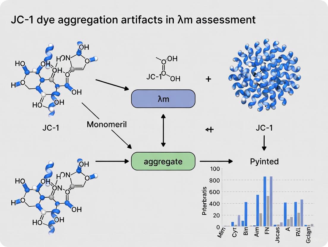
Mitigating JC-1 Dye Aggregation Artifacts for Accurate Assessment of Mitochondrial Membrane Potential (ΔΨm)
This article provides a comprehensive guide for researchers and drug development professionals on addressing JC-1 dye aggregation artifacts in the assessment of mitochondrial membrane potential (ΔΨm).
Mitigating JC-1 Dye Aggregation Artifacts for Accurate Assessment of Mitochondrial Membrane Potential (ΔΨm)
Abstract
This article provides a comprehensive guide for researchers and drug development professionals on addressing JC-1 dye aggregation artifacts in the assessment of mitochondrial membrane potential (ΔΨm). JC-1 is a widely used ratiometric probe whose potential-dependent formation of J-aggregates (red fluorescence) and monomers (green fluorescence) is crucial for measuring ΔΨm. However, its application is prone to technical artifacts related to dye concentration, loading conditions, and cellular context, which can lead to data misinterpretation. We explore the foundational principles of JC-1 fluorescence and ΔΨm, detail optimized methodological protocols for flow cytometry and imaging, present a systematic troubleshooting framework for common pitfalls, and validate the approach through comparative analysis with other mitochondrial parameters. The goal is to empower scientists with the knowledge to generate robust, reproducible data on mitochondrial health in fields ranging from cancer research to apoptosis studies.
Understanding JC-1: From Ratiometric Principles to Critical Artifacts in ΔΨm Measurement
FAQ: Core Principles and Common Artifacts
Q1: What is the fundamental principle behind how JC-1 reports mitochondrial membrane potential (ΔΨm)?
JC-1 is a lipophilic, cationic fluorescent dye that accumulates in mitochondria in a potential-dependent manner. The core principle is its concentration-dependent spectral shift:
- In mitochondria with low ΔΨm, the dye enters but does not concentrate highly, existing as monomers that emit green fluorescence (emission maximum ~529 nm).
- In mitochondria with high ΔΨm, the dye is actively concentrated in the mitochondrial matrix. When its local concentration exceeds a threshold, it forms J-aggregates that emit red fluorescence (emission maximum ~590 nm) [1] [2] [3]. The ratio of red (aggregates) to green (monomers) fluorescence is therefore a relative measure of ΔΨm, where a higher ratio indicates a more polarized (energized) mitochondrial membrane [3].
Q2: What is the most critical artifact associated with JC-1 use, and how can it be identified?
The most significant artifact is the misinterpretation of spatial heterogeneity in ΔΨm due to dye-specific properties. A key study challenged the long-held belief that mitochondria in the oocyte cortex have a preferentially higher ΔΨm. This phenomenon was consistently reported in studies using JC-1 but was not observed in studies using other potentiometric dyes like TMRM. The authors concluded that the apparent cortical polarization might be an artifact of JC-1, potentially related to its lipophilicity and complex spectral properties, rather than a true biological signal [4]. This underscores the importance of validating critical findings with an alternative method.
Q3: Why does my JC-1 working solution sometimes form red crystals, and how can I prevent this?
The formation of insoluble particulate crystals is typically due to:
- Incorrect preparation order: JC-1 must be dissolved in anhydrous DMSO first to create a stock solution before further dilution in aqueous buffers [2].
- Limited aqueous solubility: JC-1 has inherently low solubility in water [2].
- Solution: Ensure the JC-1 stock solution is fully dissolved in DMSO. For the working solution, you can promote dissolution by briefly placing it in a 37°C water bath or using ultrasonication [2].
Q4: Can I fix my cells after staining with JC-1 for later analysis?
No. JC-1 is designed for use with live cells. Fixation kills cells, disrupts mitochondrial membranes, and leads to the loss of ΔΨm, which invalidates the assay. Furthermore, fluorescence quenching can occur over time. It is recommended to perform detection immediately, ideally within 30 minutes of staining [2].
Troubleshooting Guide: Common JC-1 Experimental Issues
| Problem Phenomenon | Possible Causes | Recommended Solutions |
|---|---|---|
| Red particulate crystals in working solution | Incorrect preparation order; poor aqueous solubility [2] | Dissolve JC-1 in DMSO first; use 37°C water bath or ultrasonication [2]. |
| Weak or absent red fluorescence in healthy cells | Low ∆Ψm in cells; JC-1 concentration too low; excitation wavelength suboptimal [5] | Include a positive control (healthy, untreated cells); optimize JC-1 loading concentration; try 405 nm excitation to improve J-aggregate detection [5]. |
| High green background fluorescence | Excessive JC-1 concentration leading to cytosolic monomer buildup; mitochondrial depolarization [3] | Titrate JC-1 concentration; include CCCP control to confirm depolarization signature [1] [3]. |
| Poor separation between red and green signals in flow cytometry | Significant spectral spillover (monomer emission detected in red channel) with 488 nm excitation [5] | Use flow cytometer with 405 nm laser for cleaner J-aggregate excitation and less spillover; apply correct electronic compensation [5]. |
| Inconsistent staining of adherent cells | Uneven dye contact due to high cell density or confluency [2] | For flow cytometry, detach cells and stain in suspension after trypsinization. For imaging, ensure sub-confluent culture and uniform dye coverage [2]. |
Experimental Protocols for Validated ΔΨm Assessment
Protocol 1: Ratiometric JC-1 Imaging to Mitigate Aggregation Artifacts
This protocol is adapted from a study that successfully used ratiometric imaging to challenge the artifact of cortical mitochondrial polarization in oocytes [4].
Key Reagents and Materials
- JC-1 dye (e.g., Thermo Fisher Scientific, T3168) [3]
- Live cells (e.g., mouse oocytes, astrocytes) cultured on imaging-appropriate dishes
- An appropriate physiological buffer (e.g., M2 medium for oocytes, ACSF for neural cells) [4] [6]
- Control reagents: FCCP or CCCP (e.g., 1-5 µM) to depolarize mitochondria and confirm signal specificity [4]
- Confocal or high-resolution fluorescence microscope with temperature control (37°C) [4]
Methodology
- Dye Loading: Incubate live cells with 2.5 - 5 µM JC-1 in buffer for 20-30 minutes at 37°C [4] [6].
- Washing: Gently wash cells 2-3 times with fresh, pre-warmed buffer to remove excess extracellular dye.
- Image Acquisition: Transfer cells to the microscope stage maintained at 37°C. Acquire images using a confocal microscope.
- Ratiometric Analysis: For each pixel or region of interest (e.g., individual mitochondria, cellular sub-regions), calculate the ratio of the fluorescence intensity in the red channel to the intensity in the green channel (Red/Green Ratio). This ratio is proportional to ΔΨm and helps control for artifacts related to mitochondrial density, shape, and dye concentration [4] [6].
- Validation Control: Treat a separate group of cells with 5 µM FCCP/CCCP for 15-30 minutes prior to or during imaging. A collapse of the Red/Green Ratio confirms the signal is ΔΨm-dependent [4].
This protocol leverages 405 nm excitation to reduce spectral spillover, a common source of artifact in flow cytometry [5].
Key Reagents and Materials
- MitoProbe JC-1 Assay Kit (e.g., Thermo Fisher Scientific, M34152) or equivalent [1] [3]
- Cells in suspension (e.g., Jurkat cells, L1210 lymphoblasts)
- Flow cytometer equipped with both 488 nm and 405 nm lasers
- Bandpass filters: ~525/50 nm (for monomers) and ~585/42 nm (for J-aggregates)
Methodology
- Cell Preparation: Harvest and wash cells. Resuspend in pre-warmed buffer at a density of ~1x10⁶ cells/mL.
- Staining: Add 2 µM JC-1 (from kit) to the cell suspension and incubate for 15-30 minutes at 37°C, protected from light.
- Positive Control: Pre-treat a separate sample with 50 µM CCCP for 5-10 minutes at 37°C before staining to depolarize mitochondria [1].
- Data Acquisition:
- Standard 488 nm excitation: Run the sample and note the significant spectral overlap, which requires electronic compensation.
- Optimized 405 nm excitation: Switch to the violet laser for excitation. J-aggregates are efficiently excited at 405 nm, while monomer excitation is minimal. This results in a much cleaner red signal with drastically reduced spillover from the green channel, often eliminating the need for compensation [5].
- Analysis: Create a density plot of Red (585/42 nm) vs. Green (525/50 nm) fluorescence. A population with high red and low green fluorescence indicates healthy, polarized mitochondria. A shift towards high green and low red indicates depolarization.
Diagram 1: Experimental workflow for JC-1 use, highlighting the critical step of artifact checking.
Research Reagent Solutions
Essential materials and reagents for conducting robust JC-1 experiments.
| Item | Function/Benefit | Example Source / Catalog Number |
|---|---|---|
| JC-1 Dye (bulk) | Flexible dye source for imaging and protocol development. | Thermo Fisher Scientific (T3168) [3] |
| MitoProbe JC-1 Assay Kit | Optimized for flow cytometry; includes JC-1, DMSO, CCCP, and buffer. | Thermo Fisher Scientific (M34152) [1] [3] |
| Mitochondrial Depolarization Control (CCCP/FCCP) | Uncouples oxidative phosphorylation to collapse ΔΨm; essential for validating signal specificity. | Supplied in kit or available separately (e.g., Sigma-Aldrich) [4] [1] |
| Tetramethylrhodamine Methyl Ester (TMRM) | A single-wavelength, potentiometric dye used as an orthogonal method to confirm JC-1 findings and rule out dye-specific artifacts. | Available from multiple suppliers (e.g., Thermo Fisher Scientific) [4] |
Diagram 2: The mechanism of JC-1 fluorescence response to mitochondrial membrane potential.
Accurate assessment of mitochondrial membrane potential (ΔΨm) is fundamental to understanding cellular health, apoptosis, and metabolic function. The fluorescent cationic dye JC-1 is a widely used tool for this purpose, valued for its ratiometric properties that theoretically compensate for variables like mitochondrial density and dye loading. However, a significant challenge in its application is the propensity for dye aggregation, which can introduce substantial artifacts into experimental data. This technical guide explores the root causes of these artifacts and provides validated methodologies to identify, troubleshoot, and prevent them, ensuring the reliable measurement of ΔΨm in your research.
Frequently Asked Questions (FAQs) on JC-1 Aggregation
Q1: What are the visible signs of JC-1 aggregation in my experiment? You may observe red particulate crystals in your JC-1 working solution or notice uneven, speckled staining under the microscope instead of a uniform, punctate mitochondrial pattern. In flow cytometry, this can manifest as high background signal and poor separation between healthy and depolarized cell populations [7].
Q2: Why does JC-1 aggregate, and how does this lead to artifacts? JC-1 is a lipophilic dye with limited solubility in aqueous solutions [7]. When the dye is not properly dissolved or the working solution is not correctly prepared, it can form aggregates outside the mitochondria. These non-specific aggregates can fluoresce, leading to false-positive red signals that are misinterpreted as a high ΔΨm, thereby obscuring genuine mitochondrial depolarization events [7] [3].
Q3: I am working with adherent cells. What is the recommended staining protocol to avoid aggregation artifacts? It is not recommended to stain adherent cells in a well plate and then trypsinize them for flow cytometry, as cell-to-cell contact can cause uneven dye uptake [7]. For flow cytometric analysis, the optimal protocol is to first gently digest and harvest the cells to create a single-cell suspension, and then incubate the suspended cells with the JC-1 dye. This ensures each cell has equal access to the dye, promoting uniform staining [1] [7].
Q4: Can I fix my cells after JC-1 staining and analyze them later? No. JC-1 is a probe for live-cell analysis. Cell fixation kills the cells and disrupts the mitochondrial membrane potential, which is the very parameter you are trying to measure. Furthermore, JC-1 fluorescence can quench over time. You should analyze your stained samples immediately, ideally within 30 minutes of completing the staining procedure [7].
Q5: Can tissue samples be used for JC-1 analysis? Yes, but not directly. You must first prepare a single-cell suspension from the tissue. Be aware that the mechanical or enzymatic process of creating this suspension can itself stress the cells and affect ΔΨm, so the process must be carefully optimized. As an alternative, you can extract intact mitochondria from the tissue and then incubate the mitochondrial fraction with JC-1 for analysis with a fluorescence plate reader [7].
Troubleshooting Guide: Common JC-1 Aggregation Issues and Solutions
| Problem Observed | Potential Cause | Recommended Solution |
|---|---|---|
| Red crystals in working solution | Incorrect preparation order; JC-1's low water solubility [7]. | Always prepare the working solution in the correct order: first dilute the JC-1 stock with distilled water, then add assay buffer. Gently warm the solution in a 37°C water bath or use brief sonication to promote dissolution [7]. |
| High background noise in flow cytometry | Non-specific aggregation of dye; spectral spillover from monomers [5]. | Ensure proper dye dissolution. Use a flow cytometer with a 405 nm excitation laser, which produces less spillover from JC-1 monomer fluorescence into the J-aggregate (red) channel compared to standard 488 nm excitation [5]. |
| Uneven staining in adherent cells | Staining cells while densely packed and adherent [7]. | For microscopy, stain cells directly on the chamber slide. For flow cytometry, harvest cells first to create a suspension before staining [1] [7]. |
| Poor separation between control & CCCP-treated cells | Inadequate compensation for spectral overlap; true signal masked by aggregation [5]. | Always include a CCCP-treated positive control. Use this control to set the correct fluorescence compensation on your flow cytometer to subtract monomer spillover from the aggregate channel [1] [5]. |
Standardized Experimental Protocol for Reliable JC-1 Staining
The following step-by-step protocol is designed to minimize aggregation and ensure consistent results.
Materials and Equipment
- JC-1 Dye: MitoProbe JC-1 Assay Kit (Thermo Fisher, M34152) or equivalent [1] [3].
- Carbonyl cyanide m-chlorophenyl hydrazone (CCCP): For a positive control to depolarize mitochondria [1].
- Dimethyl sulfoxide (DMSO): High-quality, anhydrous solvent for reconstituting JC-1 [1].
- Phosphate-buffered saline (PBS)
- Cell culture medium
- Equipment: Flow cytometer with 488 nm laser or fluorescence microscope with FITC/TRITC filters; incubator; centrifuge [1].
Step-by-Step Procedure for Cells in Suspension
- Preparation: Allow JC-1 and DMSO to warm to 25°C. Prepare a fresh 200 µM JC-1 stock solution by reconstituting the lyophilized dye in DMSO. Mix thoroughly until the solution is clear [1].
- Cell Harvesting: Harvest your cells and wash them with warm PBS (~37°C). Centrifuge at 400 × g for 5 minutes and aspirate the supernatant [1].
- Staining: Resuspend the cell pellet in 1 mL of warm culture medium or PBS at a density not exceeding 1 x 10^6 cells/mL. Add 10 µL of the 200 µM JC-1 stock solution to achieve a final concentration of 2 µM. Incubate the cells at 37°C with 5% CO₂ for 15-30 minutes [1] [3].
- Positive Control Preparation: Prepare a separate tube of cells. Add 1 µL of 50 mM CCCP to 1 mL of cell suspension (final concentration 50 µM). Incubate at 37°C for 5 minutes before proceeding with JC-1 staining as above [1].
- Washing: After incubation, add 2 mL of warm PBS to each tube and centrifuge at 400 × g for 5 minutes. Aspirate the supernatant carefully [1].
- Resuspension and Analysis: Resuspend the cell pellet in a small volume of fresh PBS. Analyze the samples immediately by flow cytometry or fluorescence microscopy [1].
This workflow is summarized in the following diagram:
The Scientist's Toolkit: Essential Reagents and Their Functions
| Item | Function / Role in Preventing Aggregation | Example / Specification |
|---|---|---|
| JC-1 Assay Kit | Provides optimized dye, buffer, and control reagents for a standardized protocol, reducing variability [3]. | MitoProbe JC-1 Assay Kit (M34152, Thermo Fisher) [1] [3]. |
| High-Quality DMSO | Essential for properly dissolving the lyophilized JC-1 dye to create a monodisperse stock solution [1]. | Anhydrous, cell culture tested DMSO [1]. |
| Dispersant / Surfactant | Prevents agglomeration by enhancing the dispersibility of dye particles in aqueous solution [8] [9]. | Use included assay buffer or agents like phenol/naphthol sulfonate condensates [8] [9]. |
| CCCP | A mitochondrial uncoupler used as a positive control to collapse ΔΨm, validating the assay's performance and compensation [1] [3]. | Carbonyl cyanide m-chlorophenyl hydrazone, typically 50 mM stock in DMSO [1]. |
| Imaging Buffer | Maintains optimal cell health and pH during live-cell imaging, preserving the true ΔΨm for the duration of data capture [10]. | HEPES-buffered saline solution [10]. |
Understanding the Core Principle: How Membrane Potential Drives JC-1 Behavior
The reliability of JC-1 hinges on its potential-dependent accumulation within mitochondria. The diagram below illustrates the fundamental principle that underpins both its utility and its vulnerability to aggregation artifacts.
A common source of artifact in flow cytometry is the spectral spillover between the green (monomer) and red (J-aggregate) channels when using 488 nm excitation. The monomers, which are abundant in the cytosol, have significant emission at 585 nm, which can be mistaken for a genuine J-aggregate signal [5].
Solution: If your flow cytometer is equipped with a 405 nm violet laser, use it to excite JC-1. Research shows that 405 nm excitation produces strong signals from J-aggregates with considerably less spillover from monomer fluorescence. This results in more accurate data and can eliminate the need for complex fluorescence compensation [5].
The artifacts arising from JC-1 dye aggregation are a significant, yet manageable, challenge in mitochondrial research. By understanding the delicate interplay between dye concentration, membrane potential, and the physicochemical properties that drive aggregation, researchers can implement robust protocols. Adherence to the detailed troubleshooting guides, standardized protocols, and advanced techniques outlined in this support center will empower you to generate reliable, high-quality data, thereby strengthening the conclusions drawn from your vital research into cellular health and disease.
The Critical Role of ΔΨm in Cellular Health, Apoptosis, and Drug Screening
Mitochondrial membrane potential (ΔΨm) is the electrical potential difference across the inner mitochondrial membrane, generated by the electron transport chain during oxidative phosphorylation [11]. As the main component of the proton motive force, ΔΨm is essential for driving ATP production and serves as a key indicator of mitochondrial health and cellular viability [11] [12].
Disruption of ΔΨm is a hallmark early event in apoptosis, making it a critical parameter for assessing cell health and screening potential therapeutic compounds [3]. The fluorescent dye JC-1 has become a widely used tool for monitoring ΔΨm due to its unique ratiometric properties that allow researchers to distinguish between polarized and depolarized mitochondrial states [13] [6].
Technical Challenges: JC-1 Aggregation Artifacts and Solutions
Understanding JC-1 Fluorescence Mechanics
JC-1 is a lipophilic, cationic dye that accumulates in mitochondria in a membrane potential-dependent manner [3]. In healthy cells with high ΔΨm, JC-1 forms J-aggregates within mitochondria, emitting red fluorescence at ~590 nm [13] [6]. When ΔΨm collapses, as occurs during early apoptosis, JC-1 remains in its monomeric form, emitting green fluorescence at ~529 nm [14] [3]. The red/green fluorescence ratio provides a quantitative measure of mitochondrial polarization that is independent of mitochondrial size, shape, and density [3].
Common Artifacts and Their Resolution
Despite its utility, JC-1 is prone to several artifacts that can compromise data interpretation. The table below summarizes key challenges and recommended solutions:
Table 1: Troubleshooting Common JC-1 Artifacts
| Challenge | Root Cause | Impact on Data | Recommended Solution |
|---|---|---|---|
| Drug-Induced Fluorescence [13] | Test compounds (e.g., SB216763) with intrinsic fluorescence in JC-1's emission spectrum. | False depolarization signals; inaccurate red/green ratios. | Implement spectral deconvolution algorithms to isolate true JC-1 signal [13]. |
| Excitation Spillover [5] | Standard 488 nm excitation causes significant emission spillover from monomers into the J-aggregate detection channel. | Overestimation of red fluorescence in depolarized cells; requires compensation. | Use 405 nm excitation to minimize spillover or apply precise fluorescence compensation during flow cytometry [5]. |
| Aqueous Precipitation [14] | JC-1 has limited solubility in aqueous buffers, leading to crystal formation. | Uneven staining; high background noise; flow cytometer clogging. | Prepare working solution by diluting JC-1 stock first in distilled water, then in assay buffer; use 37°C water bath or sonication to aid dissolution [14]. |
| Fixation Incompatibility [14] | JC-1 staining is lost upon cell fixation and permeabilization. | Prevents combination with intracellular antibody staining. | Use live-cell imaging only; for multiplexing, employ fixable structural mitochondrial dyes instead [15]. |
| Sample Type Limitations [14] | JC-1 requires intact, live cells for accurate ΔΨm assessment. | Cannot be used on fixed tissues or paraffin sections. | Prepare single-cell suspensions from tissues; extract mitochondria directly from tissues for ex vivo JC-1 staining [14]. |
Advanced Spectral Deconvolution Protocol
When investigating compounds with interfering fluorescence, follow this detailed protocol for spectral deconvolution based on published methodology [13]:
Control Measurements: Collect fluorescence emission spectra (500–650 nm range, with 470 nm excitation) from:
- Unstained cells
- Cells treated with the compound of interest (without JC-1)
- JC-1-stained control cells
- JC-1-stained, valinomycin-treated (fully depolarized) cells
Reference Spectra Generation: Using the control measurements, generate reference fluorescence spectra for:
- JC-1 monomer (green emission)
- JC-1 J-aggregate (red emission)
- Compound-specific fluorescence background
Mathematical Deconvolution: Process experimental spectra using mathematical software (e.g., Mathcad) with a least-squares minimization algorithm. This algorithm will unmix the composite fluorescence signal from test samples into its individual contributing components.
Ratio Calculation: After deconvolution, calculate the corrected fluorescence intensity ratio at 540/595 nm to determine the true ΔΨm, free from compound-derived artifacts [13].
Diagram 1: Spectral Deconvolution Workflow for JC-1 Artifact Correction
Optimized Experimental Protocols
Standard 488 nm excitation causes significant spillover of JC-1 monomer fluorescence into the J-aggregate detection channel, necessitating compensation that can be difficult to calibrate [5]. An optimized protocol using 405 nm excitation provides superior resolution:
- Cell Preparation: Harvest and wash cells, adjusting concentration to 1×10⁶ cells/mL in serum-free media.
- Staining: Incubate cells with 2.5 μM JC-1 for 15-30 minutes at 37°C in the dark [5] [3].
- Washing: Centrifuge and resuspend cells in fresh, pre-warmed buffer to remove excess dye.
- Flow Cytometry Analysis:
- Excitation: Use 405 nm laser instead of standard 488 nm.
- Emission Detection: Collect green monomer fluorescence at 525/50 nm and red J-aggregate fluorescence at 585/42 nm.
- Gating: Analyze the population using bivariate plots of red vs. green fluorescence. Distinct populations of cells with high and low ΔΨm will be readily apparent without complex compensation [5].
Validated Positive Control Setup
Always include a depolarization control to validate your assay and set appropriate gating:
- Treatment: Incubate a separate cell aliquot with 1-10 μM of an uncoupler such as CCCP (carbonyl cyanide m-chlorophenyl hydrazone) or valinomycin for 15-20 minutes at 37°C prior to JC-1 staining [14] [3].
- Function: This treatment completely collapses ΔΨm, eliminating J-aggregate formation and resulting in a pure green fluorescent population.
- Usage: This control defines the position of fully depolarized cells on flow cytometry plots and confirms the assay is functioning correctly.
Research Reagent Solutions
A carefully selected toolkit is essential for robust ΔΨm assessment. The table below details key reagents for JC-1-based assays:
Table 2: Essential Reagents for ΔΨm Research
| Reagent / Kit | Primary Function | Key Features / Applications | Sample Citation |
|---|---|---|---|
| JC-1 Dye [3] | Ratiometric ΔΨm indicator | Forms red J-aggregates in energized mitochondria; green monomers when depolarized. | Imaging and flow cytometry in neurons, myocytes [3]. |
| MitoProbe JC-1 Assay Kit [3] | Optimized JC-1 assay | Includes JC-1, CCCP depolarization control, and buffers for flow cytometry. | Standardized apoptosis detection in Jurkat cells [3]. |
| JC-10 [16] | Enhanced ΔΨm probe | Superior aqueous solubility and signal-to-background ratio compared to JC-1. | Detection of subtle ΔΨm changes; primary hepatocytes [16]. |
| Valinomycin / CCCP [5] [3] | Mitochondrial uncouplers | Collapse ΔΨm by acting as ionophores; essential positive controls. | Validation of JC-1 assay specificity [5] [3]. |
| SB216763 [13] | GSK-3β inhibitor | Example drug that can cause fluorescence artifacts in JC-1 assays. | Requires spectral deconvolution for accurate ΔΨm measurement [13]. |
FAQs: Addressing Researcher Questions
Q1: Can JC-1 be used on tissue samples or only cultured cells? JC-1 can be used with tissue samples, but not directly on tissue sections. The tissue must first be processed into a single-cell suspension using optimized dissociation protocols to avoid inducing ΔΨm loss during preparation. Alternatively, mitochondria can be extracted from tissues and then stained with JC-1 for analysis with a fluorescence plate reader [14].
Q2: How should adherent cells be prepared for JC-1 flow cytometry? It is recommended to detach adherent cells gently (using enzyme-free solutions if possible) and stain them in suspension after washing. Staining cells while they are adherent and then trypsinizing can cause uneven dye loading and loss of ΔΨm, leading to artifactual results [14].
Q3: Is JC-1 compatible with cell fixation for later analysis? No. JC-1 is a live-cell dye and is not fixable. Fixation kills cells and collapses ΔΨm, causing the dye to leak out. JC-1 staining and analysis must be performed on live cells, and detection should be completed promptly (within 30-60 minutes) after staining to prevent fluorescence quenching [14].
Q4: What are the key advantages of JC-1 over non-ratiometric dyes like TMRE? The primary advantage is its ratiometric nature. The red/green fluorescence ratio is independent of mitochondrial mass, shape, and dye loading efficiency, allowing for more reliable comparisons between cell populations and treatments. Non-ratiometric dyes only measure fluorescence intensity, which can be influenced by these other factors [6] [3].
Q5: When should I consider an alternative to JC-1? Consider alternatives like JC-10 (for enhanced solubility and signal) [16] or single-wavelength dyes like TMRE/TMRM (for kinetic studies) [16] [15] when:
- Studying very subtle ΔΨm changes where JC-10's higher sensitivity is beneficial.
- The experimental setup cannot accommodate the technical steps needed to mitigate JC-1's artifacts.
- Performing long-term live-cell imaging, as JC-1 can be less well retained than some rhodamine dyes [6].
Successful assessment of ΔΨm using JC-1 requires careful experimental planning and validation. The diagram below summarizes the critical decision points for a reliable JC-1 assay:
Diagram 2: Decision Workflow for JC-1 Assay Planning and Troubleshooting
By understanding the sources of JC-1 artifacts, implementing the appropriate corrective protocols, and validating assays with proper controls, researchers can confidently utilize this powerful tool to advance research in cellular health, apoptosis, and drug discovery.
Defining Common Aggregation Artifacts and Their Impact on Data Interpretation
Frequently Asked Questions (FAQs)
Q1: What does a low red/green fluorescence ratio in my JC-1 experiment indicate? A low red/green fluorescence ratio indicates a decrease in mitochondrial membrane potential (ΔΨm), a hallmark of early apoptosis [1] [3]. In healthy cells with high ΔΨm, JC-1 accumulates in mitochondria and forms aggregates (J-aggregates) that emit red fluorescence (~590 nm). In depolarized mitochondria, JC-1 remains in its monomeric form, emitting green fluorescence (~529 nm) [17] [3]. A decrease in the ratio signifies a shift from red J-aggregates to green monomers due to mitochondrial depolarization.
Q2: My positive control (CCCP) treatment isn't showing a strong depolarization signal. What could be wrong? If your CCCP control is not working, first confirm that your CCCP stock solution is fresh, stored properly at -20°C, and protected from light [18]. Test increasing concentrations of CCCP (within the 10-50 µM range) if depolarization is incomplete [1] [18]. Ensure adequate incubation time (5-10 minutes at 37°C) before JC-1 staining to allow full uncoupler action [18].
Q3: I see high background fluorescence in my samples. How can I reduce it? High background is often caused by insufficient washing after JC-1 incubation [18]. Perform 1-2 thorough washes with the provided assay buffer or warm PBS to remove unbound dye completely [1] [18]. Avoid overloading cells with JC-1, which can lead to non-specific binding. Also, ensure your dilution buffers are at the correct pH (7.2-7.4) [18].
Q4: Can JC-1 dye be used in cells expressing drug efflux transporters like P-gp? JC-1 is a substrate for the P-glycoprotein (P-gp) drug efflux transporter [19]. In cells expressing high levels of P-gp, JC-1 can be actively pumped out, reducing its intracellular concentration and leading to falsely low red fluorescence that can be mistaken for mitochondrial depolarization [19]. For such cell lines, use a high-affinity, non-competitive P-gp inhibitor like tariquidar (0.5 µM) during staining to ensure proper JC-1 accumulation [19]. Common inhibitors like verapamil and cyclosporine A may not fully restore JC-1 loading [19].
Troubleshooting Guides
Issue 1: Inconsistent Results Between Replicates
Potential Causes and Solutions:
- Cause: Inconsistent cell seeding density or uneven probe distribution.
- Solution: Standardize cell seeding density and ensure even cell attachment. For suspension cells, ensure gentle but thorough mixing during JC-1 incubation [18].
- Cause: Variable dye loading due to old or improperly handled reagent.
- Cause: Fluctuations in incubation temperature or time.
Issue 2: Poor Separation Between Red and Green Fluorescence Signals
Potential Causes and Solutions:
- Cause: JC-1 concentration is suboptimal.
- Cause: Photobleaching due to excessive light exposure.
- Solution: Minimize light exposure during and after staining. Perform all steps in the dark or wrap samples in foil [18].
- Cause: Instrument settings are not optimized.
- Solution: For flow cytometry, apply appropriate fluorescence compensation (e.g., 18% has been used for some systems) to correct for spectral overlap between the green and red channels [19].
Issue 3: Artifactual Results in P-gp Expressing Cell Lines
Protocol for JC-1 Staining in P-gp Positive Cells:
- Prepare cells as usual (Suspension or adherent).
- Pre-incubate cells with a specific P-gp inhibitor. Note: Tariquidar (0.5 µM) is recommended, as verapamil and cyclosporine A may fail to fully restore JC-1 loading in some resistant cell lines [19].
- Incubate for a short period (e.g., 10-15 minutes) at 37°C.
- Add JC-1 dye directly to the culture medium containing the inhibitor and continue the staining protocol (15-30 minutes at 37°C) [19].
- Wash, resuspend, and analyze cells as described in the standard protocol.
The following table summarizes quantitative data from key experiments using JC-1 to assess mitochondrial membrane potential.
Table 1: Quantitative Data from JC-1 Assay Applications
| Cell Type | Treatment | [JC-1] (µM) | Key Measurement (Red/Green Ratio or % Stained) | Experimental Platform | Citation |
|---|---|---|---|---|---|
| L1210 (S) [P-gp-] | None | 2 | >80% double-stained cells [19] | Flow Cytometry | [19] |
| L1210 (R) [P-gp+] | None | 2 | ~3% double-stained cells [19] | Flow Cytometry | [19] |
| L1210 (R) [P-gp+] | 0.5 µM Tariquidar | 2 | >70% double-stained cells [19] | Flow Cytometry | [19] |
| HL60 | 5 µM Staurosporine (2 hr) | Not Specified | Distinct populations with depolarization [3] | Flow Cytometry | [3] |
| Jurkat | 10 µM Camptothecin (4 hr) | 2 | Clear shift in red/green profile [3] | Flow Cytometry | [3] |
| Cultured Hippocampal Astrocytes | N/A | 5 | Ratiometric analysis of individual mitochondria [6] | High-Resolution Imaging / Two-Photon Microscopy | [6] |
Table 2: Common JC-1 Artifacts and Corrective Actions
| Artifact Type | Impact on Data Interpretation | Corrective Action |
|---|---|---|
| P-gp Efflux Activity | Falsely low red/green ratio misinterpreted as mitochondrial depolarization [19] | Use high-affinity P-gp inhibitors (e.g., Tariquidar) during staining [19]. |
| Heterogeneous J-aggregate Formation | J-aggregates may not fill the entire matrix, leading to underestimation of ΔΨm in parts of the organelle [6]. | Use ratiometric high-resolution imaging; focus on relative changes rather than absolute distribution [6]. |
| Dye Overloading / High Background | Non-specific fluorescence obscures specific signal, reduces signal-to-noise ratio [18]. | Optimize dye concentration; include thorough wash steps post-staining [1] [18]. |
| Photobleaching | Loss of fluorescence signal over time, leading to inaccurate ratio measurements [18]. | Minimize light exposure during experiment; use anti-fade reagents if compatible [18]. |
Experimental Protocols
Principle: This protocol uses the cationic, lipophilic JC-1 dye to monitor mitochondrial health in live cells. The dye accumulates in mitochondria in a potential-dependent manner, forming red fluorescent J-aggregates at high potentials and green fluorescent monomers at low potentials [3].
Materials & Reagents:
- JC-1 dye (lyophilized or ready-made solution)
- Dimethyl sulfoxide (DMSO)
- Phosphate-buffered saline (PBS)
- Cell culture medium (without serum for staining)
- Carbonyl cyanide m-chlorophenyl hydrazone (CCCP)
- Flow cytometer equipped with 488 nm laser and filters for FITC (530/30 nm) and PE (575/26 nm) [17] [3]
Procedure:
- Preparation: Grow and harvest your cells (e.g., adherent or suspension culture). Prepare a single-cell suspension at a density of ~1 x 10⁶ cells/ml in warm culture medium or PBS [1].
- JC-1 Staining:
- Washing: Centrifuge the cells at 400 × g for 5 minutes. Carefully remove the supernatant and resuspend the cell pellet in 2 mL of warm PBS. Repeat this wash step once [1].
- Analysis: Resuspend the final pellet in 0.5-1 mL of PBS and analyze immediately on a flow cytometer. Use 488 nm excitation and collect green (monomer) fluorescence in the FITC channel and red (J-aggregate) fluorescence in the PE channel [17] [3].
Controls and Validation:
- Positive Control (Depolarized): Treat a separate sample with 50 µM CCCP for 5-10 minutes at 37°C before adding JC-1. This uncoupler dissipates the ΔΨm, resulting in a strong green signal and a weak red signal [1] [3].
- Viability Check: Combine JC-1 staining with a viability dye like propidium iodide to exclude dead cells from the analysis [18].
Diagram 1: Standard JC-1 staining workflow for flow cytometry.
Principle: In cell lines overexpressing the ABCB1 (P-gp) drug transporter, JC-1 is actively exported, preventing its accumulation in mitochondria. This protocol uses a specific inhibitor to block efflux and allow accurate ΔΨm measurement.
Materials & Reagents:
- All materials from the standard protocol.
- Tariquidar (TQR) as a selective, high-affinity P-gp inhibitor.
Procedure:
- Preparation: Prepare P-gp positive cells (e.g., L1210 R or T variants) and negative controls (e.g., L1210 S) as single-cell suspensions [19].
- Inhibitor Pre-treatment: Pre-incubate the P-gp positive cells with 0.5 µM Tariquidar for 10-15 minutes at 37°C. Note: Verapamil and cyclosporine A may not be effective in all models. [19]
- JC-1 Staining in Presence of Inhibitor: Add the JC-1 working solution directly to the medium containing Tariquidar and proceed with the standard staining, washing, and analysis protocol [19].
Diagram 2: Troubleshooting workflow for P-gp interference.
The Scientist's Toolkit
Table 3: Essential Research Reagent Solutions for JC-1 Assays
| Reagent | Function/Description | Example Use Case & Note |
|---|---|---|
| JC-1 Dye | A lipophilic, cationic carbocyanine dye that is the core sensor for ΔΨm [17] [3]. | Used in all JC-1 assays. Stock solutions should be prepared fresh in DMSO and protected from light [1]. |
| CCCP | A protonophore and mitochondrial uncoupler; used as a positive control to dissipate ΔΨm completely [1] [3]. | Validate assay performance. Typically used at 10-50 µM. Prepare fresh stock in DMSO [1] [18]. |
| Tariquidar (TQR) | A high-affinity, non-competitive, and selective inhibitor of P-glycoprotein (P-gp/ABCB1) [19]. | Essential for accurate ΔΨm assessment in P-gp overexpressing cell lines. Use at 0.5 µM [19]. |
| DMSO | A polar organic solvent used to reconstitute lyophilized JC-1 dye and other stock reagents [1]. | Ensure high-quality, sterile DMSO. Final concentration in cell culture should be kept low (e.g., ≤0.2%) to avoid cytotoxicity [6]. |
| Assay Buffer / PBS | A physiological salt solution used for washing cells and diluting reagents [1] [18]. | Remove serum and excess dye. Must be warm (~37°C) to prevent stress during washing [1]. |
Optimized Protocols: Best Practices for JC-1 Staining in Flow Cytometry and Imaging
Defining the Optimal JC-1 Concentration and Loading Conditions for Different Cell Types
Core Principle of JC-1 Staining
How does the JC-1 dye indicate changes in mitochondrial membrane potential (ΔΨm)?
JC-1 is a cationic, lipophilic fluorescent dye that accumulates electrogenically within active mitochondria in response to their negative inner membrane potential [20] [1]. Its unique property is its ability to form aggregates at higher concentrations, which provides a ratiometric measurement of ΔΨm [20] [21].
- High ΔΨm (Healthy Mitochondria): In energized mitochondria with high membrane potential, JC-1 accumulates sufficiently to form J-aggregates, which fluoresce red (emission maximum ~590 nm) [20] [22] [1].
- Low ΔΨm (Depolarized Mitochondria): When the membrane potential is low, JC-1 accumulates to a lesser extent and remains in its monomeric form, which fluoresces green (emission maximum ~529 nm) [20] [22] [1].
Consequently, a decrease in the red/green fluorescence intensity ratio is a quantitative indicator of mitochondrial depolarization, a key early event in apoptosis [20] [1]. This ratio is independent of mitochondrial size, shape, and density, making JC-1 a reliable probe for comparative studies [20].
Standardized Protocols for Different Cell Types
The following section provides detailed methodologies for applying JC-1 staining across various experimental models, from mammalian cells to more complex systems like green algae.
General Protocol for Mammalian Cells (Flow Cytometry)
This protocol is optimized for suspension cells, such as Jurkat or HL-60 cells, analyzed by flow cytometry [20] [1].
Key Reagents:
- JC-1 dye (e.g., MitoProbe JC-1 Assay Kit, Thermo Fisher, M34152)
- Phosphate-Buffered Saline (PBS)
- Culture medium
- Carbonyl cyanide m-chlorophenyl hydrazone (CCCP) for positive control
Procedure:
- Harvest and Wash: Collect approximately 1 x 10⁶ cells per sample. Wash the cells with warm PBS (~37°C) and centrifuge at 400 × g for 5 minutes. Remove the supernatant [1].
- Staining: Resuspend the cell pellet in 1 mL of pre-warmed culture medium or PBS. Add JC-1 dye to a final concentration of 2 μM. Incubate the cells at 37°C in a 5% CO₂ atmosphere for 15-30 minutes [20] [1].
- Positive Control Preparation: To one sample tube, add the mitochondrial uncoupler CCCP to a final concentration of 50 μM. Incubate at 37°C for 5 minutes prior to staining with JC-1. This serves as a critical control for validating the depolarization-dependent signal [1].
- Wash and Analyze: Wash the cells once with warm PBS to remove excess dye. Resuspend in PBS and analyze immediately on a flow cytometer equipped with a 488 nm laser. Use FITC (530/30 nm) and PE (585/42 nm) bandpass filters to detect green monomers and red J-aggregates, respectively [20].
The workflow for this protocol is summarized in the following diagram:
Protocol for Adherent Cells and Special Considerations
- Adherent Cells for Flow Cytometry: For flow cytometric analysis of adherent cells, it is recommended to detach the cells (e.g., using trypsin) and then incubate them with the JC-1 dye in suspension after digestion. Staining cells while they are adherent in a well plate can lead to uneven dye contact and uptake [22].
- Adherent Cells for Imaging: For direct imaging (e.g., fluorescence microscopy), cells can be stained adherent in a chamber slide or plate. After incubation with JC-1, wash gently with warm buffer and image in a live-cell compatible medium [20].
- Tissue Samples: JC-1 cannot be used on paraffin or frozen sections as it requires live cells. For tissue analysis, prepare a single-cell suspension first, then follow the protocol for suspension cells. Be aware that the digestion process can itself affect ΔΨm, so the protocol must be optimized to avoid false positives. Alternatively, mitochondria can be extracted from the tissue prior to JC-1 incubation [22].
- Plant Cells and Green Algae: The presence of a cell wall and chlorophyll autofluorescence presents unique challenges. For Chlamydomonas reinhardtii, research indicates that using HEPES buffer (with CaCl₂, MgCl₂, and sorbitol) provides better results than PBS. A final JC-1 concentration of 3 μM with a 15-minute incubation at 30°C has been successfully used [21].
Optimal JC-1 Concentrations Across Cell Types
The table below summarizes recommended JC-1 staining conditions derived from established protocols and optimization studies.
Table 1: JC-1 Staining Parameters for Different Sample Types
| Sample Type | Recommended JC-1 Concentration | Incubation Conditions | Key Buffer | Primary Analysis Method |
|---|---|---|---|---|
| Mammalian Suspension Cells (e.g., Jurkat, HL-60) | 2 μM [20] [1] | 15-30 min, 37°C, 5% CO₂ [20] [1] | PBS or Culture Medium [1] | Flow Cytometry [20] |
| Mammalian Adherent Cells | 2 μM [20] | 15-30 min, 37°C, 5% CO₂ [20] | PBS or Culture Medium | Fluorescence Microscopy [20] |
| Green Algae (C. reinhardtii) | 3 μM [21] | 15 min, 30°C [21] | HEPES Buffer [21] | Fluorescence Plate Reader [21] |
| Isolated Mitochondria | Consult specific protocol; typically 1-5 μM | 15-30 min, 25-37°C | Mitochondrial Isolation Buffer | Fluorescence Spectrophotometry |
Troubleshooting Common JC-1 Artifacts
This section addresses specific issues users may encounter, framed within the context of mitigating aggregation artifacts for accurate ΔΨm assessment.
Q1: My JC-1 working solution has red particulate crystals. What caused this and how can I fix it?
- Cause: The crystals are likely due to improper preparation order or the limited aqueous solubility of JC-1. If the concentrated JC-1 stock is added directly to a buffer containing salts before being diluted in water, it can precipitate [22].
- Solution: Always prepare the JC-1 working solution strictly according to the kit instructions. Typically, the JC-1 stock (e.g., 500X) should first be diluted with distilled water, and then the assay buffer should be added. To dissolve existing crystals, place the solution in a 37°C water bath or use brief sonication [22].
Q2: After completing JC-1 staining, I cannot analyze my samples immediately. Can I fix the cells for later analysis?
- Answer: No. JC-1 is a live-cell dye. Fixation kills the cells, disrupts mitochondrial integrity, and alters the potential-dependent distribution of the dye. Furthermore, JC-1 fluorescence can quench over time. It is highly recommended to complete the detection within 30 minutes of staining [22].
Q3: My positive control (CCCP) does not show a strong shift from red to green fluorescence. What is wrong?
- Potential Causes and Solutions:
- Insufficient Uncoupler Concentration/Time: Ensure CCCP is used at an effective final concentration (e.g., 50 μM) and that cells are pre-incubated with it for about 5 minutes before JC-1 staining to fully depolarize the mitochondria [1].
- P-glycoprotein Interference: In cell lines that express the multidrug resistance transporter P-glycoprotein (P-gp/ABCB1), JC-1 is actively pumped out of the cell, preventing its accumulation in mitochondria regardless of the ΔΨm. This can be misinterpreted as low potential [23].
- Solution for P-gp Interference: Use a high-affinity, non-competitive P-gp inhibitor like tariquidar (TQR, 0.5 μM) during staining. Common inhibitors like verapamil (VER) or cyclosporine A (CSA) may not be fully effective in restoring JC-1 loading [23].
Q4: Why is my fluorescence signal weak or non-specific in tissue slices?
- Cause: The standard bath-loading method for tissue slices can result in non-specific dye binding, low signal-to-noise ratio, and significant photobleaching [24].
- Advanced Solution: Consider focal dye loading using a micro-pressure injector. This method delivers the dye locally to the area of interest, enhancing labeling precision, maximizing signal intensity, and reducing background fluorescence and phototoxicity [24].
The Scientist's Toolkit: Essential Research Reagents
The following table lists key reagents and their critical functions in JC-1-based ΔΨm assays.
Table 2: Essential Reagents for JC-1 Assay Development and Control
| Reagent | Function/Application | Brief Description |
|---|---|---|
| JC-1 Dye | Primary ΔΨm indicator | A cationic carbocyanine dye that undergoes reversible, potential-dependent green-to-red fluorescence shift in mitochondria [20] [1]. |
| CCCP (Carbonyl Cyanide m-Chlorophenylhydrazone) | Positive Control | A protonophore and mitochondrial uncoupler that dissipates the proton gradient across the inner mitochondrial membrane, collapsing ΔΨm and validating the assay [20] [1]. |
| Tariquidar (TQR) | Specific Inhibitor for MDR Cells | A high-affinity, non-competitive P-glycoprotein (P-gp) inhibitor. Essential for accurate ΔΨm measurement in cell lines that express this drug efflux pump, as JC-1 is a P-gp substrate [23]. |
| Annexin V Conjugates | Apoptosis Co-staining | Used in multiparametric assays to detect phosphatidylserine externalization, a marker of early apoptosis that often coincides with mitochondrial depolarization [20] [25]. |
| HEPES Buffer | Specialized Buffer for Plant/Algae | Provides better staining conditions than PBS for cell types with walls, such as green algae, potentially by mimicking cytoplasmic conditions [21]. |
The critical relationship between mitochondrial state, JC-1 form, and fluorescence, along with common pitfalls, is illustrated below:
Step-by-Step Protocol for Robust Flow Cytometry Analysis using FITC and PE Channels
This technical support guide provides a detailed protocol for robust flow cytometry analysis using FITC and PE channels, specifically framed within research addressing JC-1 dye aggregation artifacts in mitochondrial membrane potential (ΔΨm) assessment. Proper panel design and troubleshooting are particularly crucial when investigating bioenergetic heterogeneity in cancer cells and apoptosis, where artifacts can compromise data interpretation [26]. The following sections offer comprehensive experimental protocols and solutions to common challenges.
Experimental Protocols
Panel Design and Sample Preparation
A. Robust Gating Strategy for Cell Identification
- Primary Antibody Panel: For reliable identification of human monocyte subsets, use the following combination in your panel: CD11b, HLA-DR, CD14 (FITC), and CD16 (PE) [27].
- Gating Hierarchy:
- Initial Gate: Use forward scatter (FSC) and side scatter (SSC) to identify the primary cell population of interest.
- Doublet Exclusion: Apply FSC-H vs. FSC-A to exclude cell aggregates and ensure single-cell analysis.
- Lineage Gate: Use CD11b and/or HLA-DR to confirm monocytic lineage, which is especially important under inflammatory conditions when standard FSC/SSC properties may be altered [27].
- Subset Identification: Finally, gate subsets using CD14 (FITC) and CD16 (PE) to distinguish classical (CD14++/CD16−), intermediate (CD14++/CD16+), and non-classical (CD14+/CD16++) monocytes [27].
B. Sample Staining Protocol for Surface Markers
- Prepare single-cell suspension in appropriate staining buffer.
- Add Fc receptor blocking reagent (e.g., normal serum from the host species of your antibodies) for 10-15 minutes to reduce non-specific staining [28].
- Add titrated antibodies (CD14-FITC and CD16-PE) and incubate for 30 minutes in the dark at 4°C.
- Wash cells twice with cold staining buffer to remove unbound antibody.
- Resuspend in fixation buffer if immediate analysis isn't possible, though fixation can compromise some epitopes and should be validated [28].
- Acquire data on flow cytometer within 24 hours for optimal results.
C. Combining with JC-1 Staining for ΔΨm Assessment
When investigating mitochondrial membrane potential in conjunction with surface markers:
- Perform JC-1 staining first on live cells according to manufacturer's protocol.
- Wash cells gently to remove excess JC-1 dye.
- Proceed with surface antibody staining as described above.
- Complete analysis promptly within 30 minutes of JC-1 staining, as fixation is not compatible with JC-1 viability requirements [29].
Instrument Setup and Quality Control
A. Optimizing Laser and Detector Settings
- FITC Configuration: Use a 488nm blue laser with a 530/30nm bandpass filter [30].
- PE Configuration: Use the same 488nm blue laser with a 575/26nm bandpass filter [30].
- Voltage Optimization: Use unstained and single-stained controls to set photomultiplier tube (PMT) voltages for optimal signal-to-noise ratio.
- Compensation Controls: Always run single-stained compensation controls for both FITC and PE to correct for spectral spillover.
B. Daily Quality Control
- Perform instrument performance tracking using calibration beads to ensure consistent laser alignment and fluidics.
- Verify optical alignment and detector sensitivity regularly according to manufacturer specifications.
- Check for clogs in the flow cell if acquisition rate decreases dramatically; run 10% bleach for 5-10 minutes followed by distilled water for 5-10 minutes to clear obstructions [28].
Troubleshooting Guides and FAQs
Common Experimental Issues and Solutions
Q1: My flow cytometry signals are weak or absent in both FITC and PE channels. What could be the cause?
Possible Causes and Solutions:
- Cause: Inadequate antibody titration.
- Solution: Perform antibody titration experiments to determine optimal concentration for each specific cell type and staining condition [31].
- Cause: Suboptimal laser or PMT settings.
- Solution: Ensure laser wavelengths and PMT settings match the excitation and emission wavelengths of FITC and PE fluorochromes [28].
- Cause: Fluorochrome incompatibility with intracellular staining.
- Solution: For intracellular targets, note that larger fluorochromes may not efficiently penetrate membranes; consider alternative dye conjugates or permeabilization methods [28].
- Cause: Target expression below detection level.
- Solution: Pair low-density antigens with the brightest fluorochromes; PE is significantly brighter than FITC for detecting weakly expressed targets [28].
Q2: I'm observing high background and/or non-specific staining in my samples. How can I resolve this?
Possible Causes and Solutions:
- Cause: Fc receptor-mediated antibody binding.
- Solution: Block Fc receptors prior to staining using bovine serum albumin, specific Fc blocking reagents, or normal serum [28].
- Cause: Excessive antibody concentration.
- Solution: Use recommended antibody dilutions and perform titration experiments; typical optimizations use 10^5-10^6 cells per test [28].
- Cause: Presence of dead cells.
- Solution: Incorporate a viability dye such as PI or 7-AAD to gate out dead cells during live cell surface staining [28].
- Cause: Incomplete washing steps.
- Solution: Increase wash steps between antibody incubations, particularly when using biotin-streptavidin detection systems [28].
Q3: When using JC-1 in conjunction with flow cytometry, I notice red particulate crystals in my working solution. What should I do?
Possible Causes and Solutions:
- Cause: Incorrect preparation order of JC-1 working solution.
- Solution: Prepare JC-1 working solution strictly following manufacturer's instructions, typically diluting JC-1 (500×) with distilled water first, then adding JC-1 Assay Buffer [29].
- Cause: Limited solubility of JC-1 in aqueous solutions.
- Solution: Promote dissolution by placing the solution in a 37°C water bath or using brief sonication [29].
Q4: Can I fix cells after JC-1 staining for later analysis by flow cytometry?
Solution: No, JC-1 requires live cells for accurate assessment of mitochondrial membrane potential. Fixation results in cell death and alters dye distribution. Complete flow cytometry analysis within 30 minutes of JC-1 staining to prevent fluorescence quenching [29].
Q5: How should I handle adherent cells for JC-1 experiments with flow cytometry?
Solution: For adherent cells, it's recommended to detach cells first (including those that may have detached due to apoptosis in the culture supernatant), then follow the protocol for suspended cells. Avoid staining cells while adherent in plates followed by trypsinization, as cell-to-cell contact may cause uneven dye exposure and uptake [29].
Table 1: Monocyte Subset Distribution in Health and Disease [27]
| Monocyte Subset | Surface Markers | Healthy Individuals (%) | STEMI Patients (%) | CHD Patients (%) |
|---|---|---|---|---|
| Classical | CD14++/CD16− | 80–95% | Similar distribution, with functional differences in glucose uptake | Similar distribution, with increased nanoparticle phagocytosis |
| Intermediate | CD14++/CD16+ | 2–8% | Highest glucose uptake | Highest nanoparticle phagocytosis |
| Non-classical | CD14+/CD16++ | 2–11% | Highest HLA-DM expression | Highest HLA-DM expression |
Table 2: Troubleshooting Flow Cytometry Signal Issues [32] [28]
| Problem | Possible Cause | Recommended Solution |
|---|---|---|
| Weak or no signal | Low antigen expression | Use brightest fluorophore (PE) for low-density targets |
| Weak or no signal | Suboptimal fixation/permeabilization | For intracellular targets, use ice-cold methanol added drop-wise during vortexing |
| High background | Fc receptor binding | Implement Fc blocking step before antibody staining |
| High background | Dead cells | Incorporate viability dye and gate out dead cells |
| Day-to-day variability | Instrument setting drift | Use control samples and standardize instrument settings |
| Poor scatter properties | Clogged flow cell | Run 10% bleach followed by dH₂O to clear obstruction |
Research Reagent Solutions
Table 3: Essential Research Reagents for FITC/PE Flow Cytometry and ΔΨm Assessment
| Reagent/Category | Specific Examples | Function/Application |
|---|---|---|
| Flow Cytometry Antibodies | CD14-FITC, CD16-PE | Identification of monocyte subsets and other immune populations |
| Viability Dyes | Propidium Iodide (PI), 7-AAD | Exclusion of dead cells during analysis to reduce background |
| Mitochondrial Dyes | JC-1, TMRM, Rhodamine 123 | Assessment of mitochondrial membrane potential (ΔΨm) |
| Apoptosis Detection | Annexin V-FITC, PI | Differentiation of viable, early apoptotic, and late apoptotic cells |
| Fixation/Permeabilization | Formaldehyde, Methanol, Saponin | Cell preservation and intracellular antigen access |
| Blocking Reagents | BSA, Fc Receptor Blockers | Reduction of non-specific antibody binding |
| Compensation Controls | Single-stained beads or cells | Correction for spectral spillover between channels |
Experimental Workflow Visualization
Experimental Workflow for Robust Flow Cytometry
Mitochondrial membrane potential (ΔΨm) is a crucial indicator of cellular health, ATP production capacity, and early apoptosis. The fluorescent potentiometric dye JC-1 (5,5',6,6'-tetrachloro-1,1',3,3'-tetraethylbenzimidazolylcarbocyanine iodide) enables ratiometric measurement of ΔΨm through its concentration-dependent formation of monomers (green emission) and J-aggregates (red emission). This ratiometric approach offers significant advantages over single-wavelength dyes by providing measurements independent of mitochondrial morphology, dye concentration, and photobleaching. However, researchers must navigate technical challenges including dye aggregation artifacts, proper experimental controls, and optimal imaging configurations to generate reliable data. This technical support center provides comprehensive guidance for troubleshooting JC-1 imaging experiments within the critical context of addressing aggregation artifacts that can compromise ΔΨm assessment.
Technical FAQs: Addressing JC-1 Experimental Challenges
Q1: What causes red particulate crystals in JC-1 working solution and how can this be resolved?
A: The formation of red particulate crystals indicates improper preparation of JC-1 working solution. This occurs due to:
- Incorrect preparation order: Diluting JC-1 directly with JC-1 Assay Buffer without proper intermediate steps
- Limited aqueous solubility: JC-1 has inherently poor solubility in aqueous solutions
Solutions include:
- Precisely follow preparation order: First dilute JC-1 (500×) with distilled water, then add JC-1 Assay Buffer
- Enhance dissolution using a 37°C water bath or brief sonication
- Ensure complete dissolution before application to cells [33]
Q2: How should adherent cells be prepared for JC-1 flow cytometry analysis?
A: For accurate flow cytometric analysis of adherent cells:
- Recommended approach: Detach cells using standard methods (trypsinization), then incubate with JC-1 in suspension
- Not recommended: Staining cells while adherent followed by trypsinization, as cell-to-cell contact creates uneven dye uptake and trypsinization post-staining may affect membrane integrity and ΔΨm
- Ensure single-cell suspension quality to prevent false positives from mechanical stress during preparation [33]
Q3: Can tissue samples be analyzed with JC-1, and what special preparations are required?
A: Yes, with appropriate preparation:
- Primary method: Prepare single-cell suspensions from tissue, optimizing the process to minimize ΔΨm artifacts from mechanical/ enzymatic stress
- Alternative approach: Extract mitochondria directly using specialized mitochondrial extraction kits, then incubate purified mitochondria with JC-1 for fluorescence plate reader detection
- Not applicable: Paraffin-embedded or frozen sections are incompatible as JC-1 requires live, intact cells [33]
Q4: What are the critical considerations for validating spatial ΔΨm patterns given potential JC-1 artifacts?
A: Conflicting reports of spatial ΔΨm heterogeneity (e.g., cortical polarization in oocytes) highlight method-dependent artifacts:
- Comparative validation: Use complementary dyes (TMRM, Rhod-2) to verify spatial patterns observed with JC-1
- Technical awareness: JC-1 J-aggregates may form locally within mitochondria rather than uniformly throughout the matrix, potentially creating artifactual heterogeneity
- Contextual interpretation: Consider that ΔΨm distribution may be cell-type specific (e.g., higher in peripheral mitochondria of astrocytes) [4] [34]
Q5: What are the essential controls for ensuring JC-1 experimental validity?
A: Include these critical controls in every experiment:
- CCCP-treated positive control: (10 μM, 20 minutes) to completely collapse ΔΨm and confirm green monomer shift
- Untreated normal cells: To establish baseline red/green ratio and mitochondrial distribution patterns
- Time controls: Complete detection within 30 minutes of staining to prevent fluorescence quenching and dye leakage artifacts
- Avoid fixation: JC-1 requires live-cell analysis; fixation kills cells and invalidates results [33]
Experimental Protocols: Standardized Methodologies for Reproducible JC-1 Imaging
Protocol 1: Ratiometric JC-1 Imaging of Mitochondrial Heterogeneity in Cultured Cells
This protocol, adapted from Keil et al., enables high-resolution analysis of individual mitochondria and identification of functional subpopulations [34] [6].
Table 1: Reagent Formulations for JC-1 Imaging
| Component | Specifications | Purpose |
|---|---|---|
| JC-1 Stock | 2 mg/ml in DMSO; protect from light | ΔΨm-sensitive fluorescent indicator |
| Loading Solution | 1-5 μg/ml JC-1 in culture medium or buffer | Optimal cell loading concentration range |
| Control Reagent | 10 μM CCCP (Carbonyl cyanide m-chlorophenyl hydrazone) | ΔΨm collapse for control measurements |
| Imaging Buffer | Hanks' Balanced Salt Solution (HBSS) or Artificial Cerebrospinal Fluid (ACSF) | Physiological maintenance during imaging |
| Inhibitors | Dantrolene (10-20 mM) or 2-APB (100 mM) in DMSO | Investigate calcium-mediated ΔΨm fluctuations |
Step-by-Step Procedure:
Cell Preparation:
- Culture cells on Matrigel-coated glass coverslips (0.13-0.17 mm thickness)
- Use widely spread, flat cells (e.g., hippocampal astrocytes) for optimal mitochondrial resolution
- Maintain cultures at 37°C in humidified, 5% CO₂ atmosphere until experimentation
JC-1 Loading:
- Prepare working solution: Dilute JC-1 stock to 1-5 μg/ml in pre-warmed culture medium or imaging buffer
- Incubate cells for 15-30 minutes at 37°C protected from light
- Rinse gently 2-3 times with fresh buffer to remove non-specific dye
Microscope Configuration:
- Excitation: 490 nm (for both monomer and J-aggregate excitation)
- Emission Separation: Use image splitter with 535/35 nm bandpass (green monomers) and 590 nm longpass (red J-aggregates) filters
- Objectives: 63× or 100× water immersion objectives (NA ≥1.0) for high-resolution imaging
- Detection: High-quantum efficiency CCD camera (≥62% at 500 nm) or photomultiplier tubes
Image Acquisition:
- Maintain temperature at 32-33°C with continuous buffer perfusion (3-4 ml/min)
- Acquire simultaneous dual-channel images to prevent motion artifacts between green and red channels
- Adjust exposure times to avoid detector saturation while maximizing dynamic range
- For time-lapse, minimize illumination to prevent phototoxicity and photobleaching
Ratiometric Analysis:
- Calculate pixel-by-pixel ratio of red/green fluorescence
- Apply background subtraction from cell-free regions
- Generate ratio images using ImageJ or specialized analysis software
- Identify mitochondrial subpopulations through histogram analysis of ratio values
Protocol 2: Two-Photon JC-1 Imaging for Deep Tissue and Long-Term Monitoring
This protocol leverages two-photon microscopy to enhance spatial resolution and reduce phototoxicity in thick samples [34] [6].
Table 2: Two-Photon Microscope Configuration for JC-1 Imaging
| Component | Specifications | Purpose |
|---|---|---|
| Laser Source | Ti:Sapphire pulsed laser (~800-850 nm) | Two-photon excitation of JC-1 |
| Objective | 20× 0.95NA IR-optimized or 60× water immersion | Maximize IR transmission and resolution |
| Detection | Non-descanned photomultiplier tubes (PMTs) | High-sensitivity detection of emitted light |
| Emission Filters | 525/50 nm (green) and 605/55 nm (red) | Spectral separation of monomer and aggregate |
| Environmental Control | Heated stage chamber with 5% CO₂ | Maintain cell viability during extended imaging |
Step-by-Step Procedure:
System Calibration:
- Adjust laser wavelength to ~800 nm for optimal simultaneous two-photon excitation of JC-1 forms
- Align detection pathways using reference fluorescent slides
- Set PMT voltages to ensure linear response across expected signal range
Sample Preparation:
- Load cells or tissue with JC-1 as in Protocol 1
- Mount in appropriate imaging chamber maintaining physiological conditions
- For thick tissues, ensure adequate dye penetration through optimization of loading time and concentration
Image Acquisition:
- Set laser power to minimum necessary to achieve adequate signal-to-noise
- Acquire Z-stacks (1-2 μm steps) for 3D localization of mitochondrial ΔΨm
- For time-lapse, use minimal scan frequency and resolution to monitor dynamics
- Include brightfield or differential interference contrast (DIC) for morphological reference
Data Processing:
- Apply spectral unmixing if needed to address potential bleed-through
- Generate 3D ratio reconstructions from Z-stack data
- Track individual mitochondria over time using motion analysis algorithms
Research Reagent Solutions: Essential Materials for JC-1 Experiments
Table 3: Key Reagents and Their Functions in JC-1 Imaging
| Reagent/Category | Specific Examples | Function in JC-1 Experiments |
|---|---|---|
| ΔΨm Indicators | JC-1, TMRM, Rhod-123 | Direct measurement of mitochondrial membrane potential |
| Control Reagents | CCCP/FCCP (1-10 μM) | Positive control for ΔΨm collapse |
| Calcium Modulators | Dantrolene, 2-APB, Ionomycin | Investigate Ca²⁺-mediated ΔΨm fluctuations |
| Metabolic Inhibitors | Sodium Cyanide, Sodium Azide | Induce metabolic stress for functional assays |
| Mitochondrial Trackers | MitoTracker Red/Green, ER-Tracker | Colocalization and organelle interaction studies |
| Cell Viability Reagents | Propidium Iodide, Annexin V | Distinguish apoptosis from other ΔΨm changes |
| Imaging Media | MEM, HBSS, ACSF | Maintain cell viability during imaging |
Advanced Applications: Investigating Mitochondrial Dynamics and Heterogeneity
Analyzing Spontaneous ΔΨm Fluctuations and Mitochondrial-ER Crosstalk
JC-1 ratiometric imaging enables investigation of dynamic mitochondrial processes beyond static ΔΨm measurements:
Characterizing ΔΨm Fluctuations:
- Identify spontaneous, synchronized ΔΨm increases in mitochondrial clusters
- Monitor response to pharmacological agents: Fluctuations continue despite extracellular Ca²⁺ withdrawal but are inhibited by dantrolene or 2-APB
- Correlate with cytosolic Ca²⁺ transients using simultaneous Fluo-3 imaging [34]
Metabolic Challenge Responses:
- Apply metabolic inhibitors (cyanide, azide) or glutamate to assess mitochondrial vulnerability
- Document heterogeneous depolarization patterns across mitochondrial populations
- Establish ΔΨm fluctuations as indicators of mitochondrial viability rather than dysfunction [34]
Resolving Spatial Heterogeneity Controversies in ΔΨm Distribution
The scientific literature contains conflicting reports regarding spatial patterns of ΔΨm, particularly concerning cortical versus perinuclear distributions:
Evidence from Astrocyte Studies:
- Mitochondrial density is highest in perinuclear regions
- ΔΨm tends to be higher in peripheral mitochondria
- Specialized mitochondrial subpopulations coexist even in structurally less polarized cells [34]
Controversial Findings in Oocytes:
- JC-1 studies frequently report higher cortical ΔΨm
- TMRM studies show no evidence of cortical polarization
- Technical artifacts from J-aggregate formation characteristics may contribute to conflicting reports [4]
Resolution Strategy:
- Employ multiple ΔΨm indicators with different mechanisms to validate spatial patterns
- Consider cell-type specific specializations in mitochondrial distribution
- Account for technical limitations of each detection method when interpreting results
Successful implementation of high-resolution ratiometric JC-1 imaging requires meticulous attention to technical细节. Researchers must validate spatial ΔΨm patterns with complementary approaches, include appropriate controls for artifact identification, and select imaging modalities suited to their experimental questions. The protocols and troubleshooting guides presented here provide a framework for generating reliable, reproducible data on mitochondrial functional heterogeneity, enabling deeper insights into cellular metabolic states and early apoptotic events. When properly executed, ratiometric JC-1 imaging—particularly combined with two-photon microscopy—offers unparalleled capability for quantitative functional analysis of individual mitochondria and comparison of mitochondrial heterogeneity across experimental conditions.
Frequently Asked Questions (FAQs)
Q1: Why is JC-1 monomer fluorescence (green) often used over the J-aggregate (red) for multiparametric flow cytometry? A1: The green fluorescence (~530 nm) of JC-1 monomers is more compatible with common filter sets (e.g., FITC channel) and presents less spectral overlap with other common fluorochromes like PE (for Annexin V) or PI, simplifying panel design and compensation.
Q2: How can I minimize JC-1 dye-induced cytotoxicity during long-term assays that also measure proliferation (e.g., with CFSE)? A2: JC-1 can be cytotoxic at high concentrations or with prolonged incubation. To mitigate this:
- Use the lowest effective dye concentration (often 0.5-2 µM).
- Reduce staining incubation time to 15-30 minutes at 37°C.
- Include a viability dye (e.g., a near-IR fixable viability dye) added post-staining to gate out dead cells and exclude cytotoxicity artifacts from your proliferation analysis.
Q3: My JC-1 red/green ratio shifts after fixing cells for cell cycle analysis. How can I preserve the signal? A3: Mitochondrial membrane potential (Δψm) is lost upon fixation. JC-1 staining for Δψm must always be performed on live, unfixed cells. A validated workflow is:
- Stain with JC-1.
- Acquire Δψm data on live cells via flow cytometry.
- Then, fix and permeabilize the cells for subsequent intracellular staining (e.g., Ki-67, phosphorylated histone H3) or DNA content analysis with PI.
Q4: When co-staining with Annexin V, I get high background in the JC-1 green channel. What is the cause? A4: This is often due to compensation issues or early apoptosis. Apoptotic cells have decreased Δψm, leading to a collapse of red J-aggregates and an increase in green monomers. This "green shift" can be mistaken for background. Ensure proper single-color controls for both JC-1 (use CCCP as a depolarization control) and Annexin V for accurate compensation.
Troubleshooting Guide
| Problem | Possible Cause | Solution |
|---|---|---|
| Low J-aggregate (Red) Signal | 1. Loss of Δψm due to unhealthy cells.2. JC-1 concentration too low.3. Over-compensation from the green channel. | 1. Check cell viability before staining. Use a positive control (e.g., CCCP) to induce depolarization.2. Titrate JC-1 to find the optimal concentration.3. Re-check compensation using a CCCP-treated sample. |
| High Non-specific Staining | 1. Excessive dye concentration.2. Inadequate washing post-staining.3. Presence of dead cells. | 1. Re-titrate JC-1.2. Perform two rigorous washes with PBS or assay buffer.3. Include a viability dye to exclude dead cells from analysis. |
| Inconsistent Results between Replicates | 1. Inconsistent cell counting/density.2. Variations in staining incubation time or temperature.3. JC-1 stock solution degradation. | 1. Use precise cell counting methods.2. Standardize the staining protocol meticulously.3. Prepare fresh JC-1 stock solutions in DMSO and aliquot for single use. |
| Poor Resolution in Cell Cycle when combined with JC-1 | 1. JC-1 spectral spillover into the PI channel.2. Fixation step degrading DNA quality. | 1. Use a long-pass filter (e.g., 670 LP) for PI detection to minimize JC-1 green spillover. Acquire JC-1 first on live cells, then fix and stain with PI.2. Use gentle permeabilization buffers and avoid over-fixing. |
Table 1: Spectral Properties and Compatible Fluorochromes for Multiparametric Panels with JC-1
| Fluorochrome | Ex (nm) | Em (nm) | Common Application | Compatibility with JC-1 (Green/Red) |
|---|---|---|---|---|
| JC-1 Monomer | 514 | 529 | Δψm (Depolarized) | Reference |
| JC-1 J-Aggregate | 585 | 590 | Δψm (Polarized) | Reference |
| Annexin V-FITC | 488 | 525 | Apoptosis (PS exposure) | High (Monitor compensation) |
| Annexin V-PE | 488 | 575 | Apoptosis (PS exposure) | Medium (Significant spillover into JC-1 Red) |
| Propidium Iodide (PI) | 488 | 617 | Viability / Cell Cycle | High (Use 670LP filter) |
| 7-AAD | 488 | 650 | Viability / Cell Cycle | High (Excellent separation) |
| CFSE | 488 | 517 | Proliferation | Medium (Significant spectral overlap with JC-1 Green) |
| Ki-67 (eFluor 660) | 488 | 668 | Proliferation (Intracellular) | High |
Table 2: Optimized Staining Concentrations for Integrated Assays
| Reagent | Typical Concentration | Incubation Time | Temperature | Notes |
|---|---|---|---|---|
| JC-1 | 1 - 3 µM | 15 - 30 min | 37°C | Protect from light; titrate for each cell type. |
| Annexin V-FITC | 1:20 dilution | 15 min | Room Temp | Use in Ca²⁺-rich binding buffer. |
| Propidium Iodide (PI) | 1 - 2 µg/mL | 5 min | 4°C | Add post-Annexin V staining for viability. |
| CFSE | 1 - 5 µM | 10 - 20 min | 37°C | Quench with serum-containing media post-staining. |
Experimental Protocols
Protocol 1: Integrated JC-1, Annexin V, and PI Staining for Apoptosis & Δψm
This protocol assesses early apoptosis (Annexin V+) concurrently with the loss of mitochondrial membrane potential.
- Cell Preparation: Harvest and wash cells 2x in cold PBS. Resuspend at 1x10⁶ cells/mL in pre-warmed assay buffer.
- JC-1 Staining: Add JC-1 to a final concentration of 1-2 µM. Vortex gently and incubate for 20 minutes at 37°C in the dark.
- Wash: Wash cells 2x with 1x Annexin V Binding Buffer. Decant supernatant thoroughly.
- Annexin V Staining: Resuspend cell pellet in 100 µL of Annexin V Binding Buffer. Add Annexin V-FITC (as per manufacturer's recommendation, typically 5 µL). Incubate for 15 minutes at room temperature in the dark.
- PI Staining: Add Propidium Iodide (1-2 µg/mL) to the tube. Do not wash.
- Acquisition: Analyze immediately on a flow cytometer within 1 hour. Use 488 nm excitation. Collect FITC (JC-1 monomer/Annexin V), PE (JC-1 aggregate), and PerCP-Cy5-5 or equivalent (PI) signals.
Protocol 2: Sequential JC-1 Staining and Cell Cycle Analysis
This protocol measures Δψm in live cells followed by cell cycle distribution in the same sample.
- JC-1 Staining & Acquisition (Live Cells): Stain cells with JC-1 as described in Protocol 1, steps 1-3. Resuspend in assay buffer and acquire the "live cell" data for JC-1 on the flow cytometer. Do not fix cells at this stage.
- Cell Fixation: Pellet the cells after acquisition. Gently resuspend the cell pellet in 1 mL of ice-cold 70% ethanol added drop-wise while vortexing. Fix for at least 2 hours at 4°C (or overnight).
- Wash: Pellet the fixed cells and wash 2x with PBS to remove residual ethanol.
- RNAse Treatment: Resuspend the cell pellet in 500 µL of PBS containing RNAse A (100 µg/mL). Incubate for 15-30 minutes at 37°C.
- DNA Staining: Add Propidium Iodide to a final concentration of 50 µg/mL.
- Acquisition: Analyze the cell cycle on the flow cytometer. Gate on single cells and collect PI fluorescence using a >670 nm long-pass filter.
The Scientist's Toolkit
Table 3: Essential Research Reagent Solutions
| Item | Function in the Assay |
|---|---|
| JC-1 (5,5',6,6'-Tetrachloro-1,1',3,3'-tetraethylbenzimidazolylcarbocyanine iodide) | Cationic dye that accumulates in mitochondria in a Δψm-dependent manner, forming red fluorescent J-aggregates (high Δψm) or green monomers (low Δψm). |
| Carbonyl cyanide m-chlorophenyl hydrazone (CCCP) | Protonophore used as a positive control to dissipate the mitochondrial membrane potential, collapsing the red J-aggregate signal. |
| Annexin V (FITC conjugate) | Binds to phosphatidylserine (PS) exposed on the outer leaflet of the plasma membrane, a marker for early apoptosis. |
| Propidium Iodide (PI) | Membrane-impermeant DNA dye used to stain dead cells or, after fixation/permeabilization, for DNA content/cell cycle analysis. |
| CFSE (Carboxyfluorescein succinimidyl ester) | Cell-permeant dye that stably labels intracellular amines. Fluorescence halves with each cell division, allowing proliferation tracking. |
| 1X Annexin V Binding Buffer | Provides optimal Ca²⁺ concentration for Annexin V binding to phosphatidylserine. |
| RNAse A | Degrades RNA to prevent interference with PI DNA staining during cell cycle analysis. |
Visualizations
JC-1 & Annexin V/PI Staining Workflow
Sequential JC-1 & Cell Cycle Analysis Workflow
Multiparametric Flow Cytometry Gating Strategy
Troubleshooting JC-1 Artifacts: A Practical Guide to Pitfalls and Solutions
Addressing High Background and Non-Specific Staining
Core Principles of JC-1 Staining and Background Challenges
The JC-1 dye is a widely used fluorescent cationic probe for assessing mitochondrial membrane potential (ΔΨm), a key indicator of mitochondrial health and early apoptosis. Its unique property lies in its potential-dependent accumulation within mitochondria. In healthy, polarized mitochondria, JC-1 forms J-aggregates that emit red fluorescence (emission maximum ~590 nm). When the mitochondrial membrane potential is reduced, as occurs in early apoptosis, JC-1 remains in a monomeric state that emits green fluorescence (emission maximum ~529 nm). The red/green fluorescence intensity ratio is therefore a direct quantitative measure of ΔΨm, independent of confounding factors like mitochondrial size, shape, and density [1] [20].
A primary source of experimental artifacts in JC-1 assays stems from dye aggregation artifacts. Improper experimental conditions can cause JC-1 to form non-specific aggregates outside of mitochondria, leading to high background fluorescence and non-specific staining that compromises data accuracy [35]. The following sections provide a structured troubleshooting guide to identify and resolve these issues.
Troubleshooting FAQs for JC-1 Experiments
Q1: My JC-1 working solution has red particulate crystals. What caused this and how can I fix it?
- Cause: The JC-1 working solution was not prepared in the correct order, or the JC-1 dye did not dissolve properly due to its limited solubility in water [35].
- Solutions:
- Prepare the JC-1 working solution strictly following the manufacturer's instructions. Typically, this involves first diluting the concentrated JC-1 stock with distilled water before adding the JC-1 Assay Buffer [35].
- Promote dissolution by placing the solution in a 37°C water bath or using brief sonication [35].
Q2: I observe high non-specific staining across my entire sample. What are the potential causes?
- Cause 1: Inadequate Blocking. Non-specific binding sites in your sample have not been effectively blocked.
- Cause 2: Incorrect JC-1 Concentration.
- Solution: Titrate the JC-1 dye concentration. Using too high a concentration can saturate the system and lead to non-specific precipitation and background. A common final working concentration is 2 μM [1].
- Cause 3: Sample Drying.
Q3: Can I fix my cells after JC-1 staining to analyze them later?
- Answer: No. JC-1 is a probe for live cells. Fixation kills cells and alters membrane permeability, leading to a loss of mitochondrial membrane potential and leakage of the dye. This will cause severe artifacts [35].
- Solution: You must analyze JC-1-stained samples immediately. It is recommended to perform detection by flow cytometry or microscopy within 30 minutes of completing the staining procedure [35].
Q4: Can I use JC-1 on tissue sections like paraffin or frozen sections?
- Answer: No. JC-1 experiments require live, intact cells to maintain the mitochondrial electrochemical gradient [35].
- Solution: For tissues, you must first prepare a single-cell suspension. Optimize the tissue dissociation process carefully, as mechanical or enzymatic damage during this process can itself alter ΔΨm and create false positives [35]. Alternatively, you can extract mitochondria from the tissue and incubate the isolated mitochondria with JC-1 for analysis with a fluorescence plate reader [35].
Optimized Experimental Protocols for Clean Results
Protocol 1: Standard JC-1 Staining for Cells in Suspension (for Flow Cytometry)
This protocol is adapted from established methodologies [1].
- Key Reagent: MitoProbe JC-1 Assay Kit (or equivalent).
- Procedure:
- Prepare JC-1 Stock: Create a fresh 200 μM JC-1 stock solution by reconstituting lyophilized dye with DMSO. Mix until clear and fully dissolved [1].
- Harvest Cells: Gently harvest adherent cells using a mild trypsinization protocol, ensuring not to over-trypsinize. Quench trypsin with complete medium. For suspension cells, proceed directly. Wash cells by centrifuging in warm PBS (400 × g for 5 min at 25°C) [35] [1].
- Stain Cells: Suspend cell pellet in warm PBS or culture medium at a density of ~1 x 10⁶ cells/mL. Add JC-1 stock solution to a final concentration of 2 μM. Incubate at 37°C with 5% CO₂ for 15-30 minutes [1].
- Wash Cells: Wash cells once with warm PBS to remove excess dye (centrifuge at 400 × g for 5 min at 25°C) [1].
- Resuspend and Analyze: Resuspend the cell pellet in fresh, warm PBS and analyze immediately on a flow cytometer. Use 488 nm excitation, with emission filters at ~530 nm (green monomer) and ~585 nm (red J-aggregates) [1] [20].
Protocol 2: Including Essential Controls for Artifact Identification
To correctly interpret your data and identify aggregation artifacts, these controls are mandatory.
- Positive Control (Depolarized Mitochondria):
- Treat a separate aliquot of your cells with a mitochondrial uncoupler such as CCCP (Carbonyl cyanide m-chlorophenyl hydrazone). A standard treatment is 50 μM CCCP for 5-10 minutes at 37°C prior to JC-1 staining [1]. This will collapse the ΔΨm, resulting in a sample with predominantly green fluorescence and a low red/green ratio.
- Negative Control (Unstained Cells):
- Process a sample of cells identically but without adding the JC-1 dye. This allows you to set the baseline for cellular autofluorescence in both the green and red channels.
The workflow below outlines the critical steps and control points in a robust JC-1 experiment.
Research Reagent Solutions
The table below lists essential materials and their specific functions in a JC-1-based ΔΨm assessment assay.
| Item Name | Function/Application | Key Consideration |
|---|---|---|
| JC-1 Dye (e.g., T3168, M34152) [20] | Fluorescent potentiometric probe for ΔΨm; forms red J-aggregates in polarized mitochondria and green monomers when depolarized. | Prepare fresh stock solutions in DMSO; avoid freeze-thaw cycles. |
| CCCP (Carbonyl cyanide m-chlorophenyl hydrazone) [1] | Protonophore and mitochondrial uncoupler; used as a positive control to collapse ΔΨm and validate assay performance. | Use a working concentration of ~50 µM; treat cells for 5-10 min before staining. |
| DMSO (Dimethyl sulfoxide) [1] | Solvent for preparing JC-1 and CCCP stock solutions. | Use high-grade, sterile DMSO; ensure final concentration in culture is <0.1-1.0% to avoid cytotoxicity. |
| Phosphate-Buffered Saline (PBS) [1] | Buffer for washing cells and diluting reagents; maintains physiological pH and osmolarity. | Use warm PBS (~37°C) for all steps to prevent temperature-induced stress on cells. |
| Flow Cytometer with 488 nm laser [1] [20] | Instrument for quantitative analysis of JC-1 fluorescence in single-cell suspensions. | Requires bandpass filters for FITC/GFP (~530 nm) and PE/~585 nm to detect monomers and J-aggregates, respectively. |
| Fluorescence Microscope with FITC/TRITC filters [20] | Instrument for qualitative, spatial analysis of JC-1 staining within cells. | Allows visual confirmation of mitochondrial localization and morphological context. |
The cationic cyanine dye JC-1 (5,5',6,6'-tetrachloro-1,1',3,3'-tetraethylbenzimidazolylcarbocyanine iodide) serves as a vital tool for quantifying mitochondrial membrane potential (ΔΨm), a key parameter of mitochondrial function and cellular health [6] [3]. Its unique properties enable a ratiometric measurement that is largely independent of mitochondrial size, shape, and density, which often confound single-component fluorescence signals [3]. The dye accumulates in mitochondria in a potential-dependent manner: at low ΔΨm or low concentrations, it exists as green-fluorescent monomers (emission ~529 nm), while at higher potentials, it forms red-fluorescent J-aggregates (emission ~590 nm) within the mitochondrial matrix [6] [3]. This emission shift provides an internal calibration, making JC-1 particularly valuable for detecting subtle changes in mitochondrial energization [6].
However, the practical application of JC-1 is sometimes hampered by weak or absent J-aggregate signal, potentially leading to misinterpretation of mitochondrial depolarization. This guide addresses the optimization of JC-1 usage, framed within broader research aimed at resolving JC-1 dye aggregation artifacts in ΔΨm assessment. We provide targeted troubleshooting methodologies to help researchers distinguish true biological phenomena from technical artifacts, thereby enhancing the reliability of their mitochondrial function analyses.
Troubleshooting FAQs: Solving Common JC-1 Signal Problems
What are the primary causes of a weak or absent J-aggregate signal?
A weak or absent red J-aggregate signal can stem from various technical and biological factors. The table below categorizes these common issues, their underlying causes, and initial diagnostic steps.
Table 1: Root Causes of Weak or Absent J-Aggregate Signal
| Category | Specific Issue | Potential Cause | Quick Diagnostic Check |
|---|---|---|---|
| Dye Handling & Staining | Inadequate JC-1 concentration | Low stock concentration, improper dilution [6] | Confirm stock concentration; try a higher working concentration (e.g., 2-5 µM) [10]. |
| Insufficient staining incubation | Dye has not fully accumulated in mitochondria [3] | Increase staining time (e.g., 30 minutes) and ensure temperature is 37°C. | |
| Loss of dye retention | JC-1 may be less well retained than other dyes like Rh123 [6] | Include a positive control with healthy, untreated cells. | |
| Instrumentation & Detection | Suboptimal excitation wavelength | 488 nm excitation causes significant spillover from monomers into the J-aggregate detection channel [5] | Switch to 405 nm excitation if available, which produces cleaner J-aggregate signals [5]. |
| Improper fluorescence compensation | High background "red" signal from monomers in depolarized cells [5] | Use a depolarization control (e.g., valinomycin, CCCP) to set proper compensation [5]. | |
| Cell & Experimental Conditions | Genuinely low ΔΨm | Apoptosis, metabolic inhibition, or toxic insult [3] [13] | Run a viability assay and a positive control with a metabolic inhibitor. |
| Non-specific drug interference | Some compounds (e.g., GSK-3β inhibitor SB216763) autofluoresce in the JC-1 emission range [13] | Check the autofluorescence of treatment compounds in the absence of JC-1. |
How do I optimize dye concentration and loading conditions?
Optimizing the staining protocol is fundamental to obtaining a robust signal. The following workflow provides a step-by-step method for establishing optimal conditions in your system, adaptable for different cell types.
Detailed Protocol:
- Dye Preparation: Prepare a fresh JC-1 working solution in pre-warmed serum-free media or buffer from a concentrated stock in DMSO. Avoid freeze-thaw cycles of the stock [3] [10]. The provided "Imaging Buffer" in some commercial kits can help maintain cell health during imaging [10].
- Concentration Titration: Test a range of JC-1 concentrations (e.g., 1, 2, 4 µM) on a plate of healthy, untreated cells. Using a depolarizing control like FCCP or CCCP (typically 10-100 µM) in parallel is crucial [10] [13].
- Staining Incubation: Incubate cells with the dye for 20-30 minutes at 37°C in a standard cell culture incubator (e.g., 5% CO₂) [3] [10]. Protect the dye from light throughout the procedure.
- Washing and Imaging: After incubation, gently wash the cells twice with warm buffer or serum-free media to remove excess dye. Replace with fresh pre-warmed media or buffer and image immediately [13].
How can instrument setup be adjusted to improve signal detection?
Proper configuration of microscopy or flow cytometry settings is critical for accurate ratiometric measurement. A common pitfall is the spillover of green monomer fluorescence into the red J-aggregate detection channel when using 488 nm excitation [5].
Table 2: Instrument Optimization for JC-1 Detection
| Instrument Parameter | Problem with Standard 488 nm Setup | Optimization Strategy | Expected Outcome |
|---|---|---|---|
| Excitation Wavelength | Significant monomer spillover into the J-aggregate channel, requiring high compensation [5]. | Use 405 nm (violet) laser for excitation where available [5]. | Cleaner J-aggregate signal with minimal spillover, reducing or eliminating need for compensation. |
| Emission Filters | Using inappropriate bandpass filters. | Green monomer: 525/50 nm or 530/30 nm.\nRed J-aggregate: 585/42 nm or 590/30 nm [5]. | Effective spectral separation of monomer and aggregate signals. |
| Fluorescence Compensation | Over- or under-compensation distorts the true red/green ratio. | Use a depolarized control (valinomycin/CCCP) to set compensation. For 488 nm excitation, this may require subtracting ~30% of the green signal from the red channel [5]. | Accurate quantification of the red/green ratio, reflecting true ΔΨm. |
| Controls | Without controls, signal loss cannot be attributed to biology vs. technique. | Always include:\n- Healthy, untreated cells (positive control).\n- Depolarized cells (e.g., with 1 µM valinomycin or 50-100 µM CCCP/FCCP) (negative control) [5]. | Enables instrument setup and validates the experiment. |
How do I validate that my JC-1 assay is functioning correctly?
Robust validation is necessary to ensure that signal changes reflect biology and not technical artifacts. The following workflow integrates critical controls and an advanced method to account for compound interference.
Detailed Validation Protocol:
- Run Essential Controls: For every experiment, include a positive control (untreated, healthy cells) showing a high red/green fluorescence ratio, and a negative control (cells treated with an uncoupler like 1 µM valinomycin or 50-100 µM FCCP for 10-60 minutes before staining) showing a low red/green ratio [10] [5]. This confirms the dye and instrument are responding to known ΔΨm changes.
- Check for Compound Interference: Some small-molecule inhibitors (e.g., SB216763) can autofluoresce, contributing a broad emission spectrum that overlaps with the JC-1 green channel and can lead to a falsely depressed red/green ratio [13].
- Spectral Deconvolution: If autofluorescence is suspected, a spectral deconvolution protocol can be implemented. This requires:
- Acquiring the full emission spectrum of JC-1 in your control cells.
- Acquiring the emission spectrum of the interfering compound alone.
- Using software with a least-squares minimization algorithm to deconvolute the mixed signals and generate unmixed spectra for the JC-1 monomer and J-aggregates [13]. This advanced technique allows for the accurate calculation of the true 540/595 nm emission ratio, free from contaminating signals.
The Scientist's Toolkit: Essential Reagents and Materials
The following table lists key reagents and their specific functions in optimizing and troubleshooting the JC-1 assay.
Table 3: Key Research Reagent Solutions for JC-1 Assay Optimization
| Reagent / Material | Function / Application | Key Considerations |
|---|---|---|
| JC-1 Dye | The core potentiometric probe for ratiometric ΔΨm measurement [6] [3]. | Available as bulk chemical or in optimized assay kits. Kits often include key buffers and a depolarizing control [3] [10]. |
| Mitochondrial Uncouplers (FCCP, CCCP, Valinomycin) | Induce mitochondrial depolarization; essential for negative controls and fluorescence compensation [10] [5]. | Titrate concentration for your cell type. Typical working concentrations are 10-100 µM for FCCP/CCCP and ~1 µM for valinomycin [13] [5]. |
| HEPES-buffered Imaging Media | Maintains physiological pH during microscopy outside a CO₂ incubator [10]. | Pre-warm to 37°C. Some commercial JC-1 kits include a proprietary "Imaging Buffer" for this purpose [10]. |
| Apoptosis Inducers (e.g., Staurosporine, Camptothecin) | Positive controls for inducing physiological ΔΨm loss in apoptosis studies [3] [10]. | Confirm efficacy and timing in your cell model beforehand. |
| MitoTracker Red / Deep Red | Alternative, single-wavelength ΔΨm-sensitive dyes for co-staining or counter-validation [38] [39]. | Their accumulation is ΔΨm-dependent but not ratiometric. Useful for confirming JC-1 results [39]. |
Correcting for Dye Leakage and Poor Retention in Live Cells
Accurate measurement of mitochondrial membrane potential (ΔΨm) is fundamental for assessing cellular health, metabolic function, and apoptotic signaling. The fluorescent cationic dye JC-1 (5,5',6,6'-tetrachloro-1,1',3,3'-tetraethylbenzimidazolylcarbocyanine iodide) remains a widely used tool for this purpose, valued for its unique ratiometric properties. In healthy, polarized mitochondria, JC-1 accumulates and forms aggregates (J-aggregates) emitting red fluorescence (~590 nm). In depolarized mitochondria, the dye exists as monomers emitting green fluorescence (~527 nm) [1]. The red/green fluorescence ratio provides a relative measure of ΔΨm that is largely independent of mitochondrial size, shape, and density [1].
However, a significant artifact complicating this assessment is dye leakage and poor retention, particularly in live-cell imaging and flow cytometry experiments. JC-1's lipophilic, cationic nature allows it to equilibrate across membranes in a Nernstian fashion, but this same property makes it susceptible to rapid diffusion out of mitochondria if the membrane potential collapses or due to experimental conditions like excessive washing or prolonged incubation [24]. This leakage leads to inaccurate red/green ratios, underestimation of ΔΨm, and ultimately, flawed data interpretation within ΔΨm assessment research. This guide provides targeted solutions for this critical issue.
Troubleshooting Guides & FAQs
FAQ 1: Why does JC-1 leak from my live cells, and how does this create artifacts?
JC-1 leakage is primarily a thermodynamics issue. The dye accumulates in the mitochondrial matrix driven by the highly negative internal charge (ΔΨm). Any factor that reduces this potential gradient diminishes the driving force for retention, leading to dye diffusion back into the cytoplasm and out of the cell [40] [24]. Furthermore, mechanical stresses like excessive buffer washing or using suboptimal buffers can physically remove the dye.
The artifact manifests as a false-positive indication of mitochondrial depolarization. As JC-1 leaks out, the intracellular concentration drops, preventing the formation of red fluorescent J-aggregates even in healthy mitochondria. This causes a decrease in the red/green fluorescence ratio, misleadingly suggesting a loss of ΔΨm [1] [24]. This is distinct from a true, biologically induced depolarization.
FAQ 2: My JC-1 signal is weak and fades quickly. How can I improve dye retention during staining?
Weak and fading signals are classic symptoms of dye leakage. The following steps can significantly improve dye retention:
- Optimize Staining Concentration and Time: Adhere to standardized protocols. A common effective method is incubating cells with 2 μM JC-1 at 37°C for 15-30 minutes [1]. Under-staining prevents aggregate formation, while overstaining can increase non-specific background and toxicity.
- Minimize Post-Staining Washes: Avoid extensive washing after staining. If washes are necessary, use pre-warmed (37°C) buffer and be consistent across all samples. Some protocols analyze cells immediately after staining without any washing step [1] [41].
- Use a Maintenance Solution: For live-cell imaging, consider including a low concentration of JC-1 in the perfusate to maintain a steady-state dye concentration and compensate for any slow leakage, a technique successfully used with dyes like TMRM [24].
FAQ 3: What are the best experimental controls to distinguish true depolarization from dye leakage artifacts?
Robust controls are non-negotiable for validating your JC-1 data.
- Positive Control (CCCP): Treat a sample with a mitochondrial uncoupler like Carbonyl cyanide m-chlorophenyl hydrazone (CCCP). A concentration of 50 μM for 5-10 minutes is commonly used to fully collapse ΔΨm [1]. This should result in a strong loss of red fluorescence and a high green signal, establishing the baseline for a depolarized state.
- Viability Control: Always co-stain with a viability dye like Calcein Violet or Propidium Iodide (PI) to ensure you are gating on live, intact cells for analysis. This excludes dead/dying cells, whose permeable membranes cannot retain JC-1, from your analysis [41] [42].
FAQ 4: Are there alternative dyes or methods to avoid JC-1 leakage problems entirely?
Yes, several alternative strategies exist. For absolute membrane potential quantification, consider alternative dyes or techniques.
Table: Comparison of Mitochondrial Membrane Potential (ΔΨm) Dyes and Methods
| Method/Dye | Key Principle | Advantages | Limitations for Live-Cell Retention |
|---|---|---|---|
| JC-1 | Potential-dependent formation of J-aggregates (red) vs. monomers (green) [1]. | Ratiometric (red/green), reduces artifacts from dye concentration [1]. | Prone to leakage upon depolarization or washing [15]. |
| TMRE/TMRM | Cationic dyes that distribute into mitochondria based on ΔΨm; intensity indicates potential [24]. | Quantitative with calibration, good for kinetic studies [40]. | Requires continuous dye presence in perfusate to prevent leakage [24]. |
| Fixable Structural Dyes (e.g., CytoPainter) | Bind covalently to mitochondrial proteins independent of ΔΨm [15]. | Excellent retention after fixation, ideal for multiplexing with antibodies [15]. | Does not report on function or membrane potential, only structure/mass [15]. |
| Focal Dye Loading | Pressure injection of dye directly into tissue, avoiding bath immersion [24]. | High signal-to-noise, minimal non-specific binding, reduces phototoxicity [24]. | Technically demanding, requires specialized equipment like a pressure injector [24]. |
Advanced Protocols for Mitigating JC-1 Artifacts
Protocol 1: Optimized JC-1 Staining for Flow Cytometry with Viability Gating
This protocol, adapted from recent methodologies, integrates viability staining to eliminate artifacts from dead cells [1] [41].
Reagents:
- JC-1 dye (e.g., MitoProbe JC-1 Assay Kit, Thermo Fisher)
- Viability dye (e.g., Calcein Violet AM or Propidium Iodide)
- Dimethyl sulfoxide (DMSO)
- Phosphate-buffered saline (PBS)
- Carbonyl cyanide m-chlorophenyl hydrazone (CCCP) for positive control
Procedure:
- Prepare JC-1 Stock: Reconstitute lyophilized JC-1 in DMSO to create a 200 μM stock solution. Ensure it is fully dissolved and protected from light.
- Harvest and Wash Cells: Gently trypsinize and collect cells. Wash once with warm PBS and resuspend in culture medium or PBS at a density not exceeding 1 x 10⁶ cells/mL.
- Stain with Viability Dye: Incubate cells with the chosen viability dye according to the manufacturer's instructions.
- Stain with JC-1: Add JC-1 stock to the cell suspension to a final concentration of 2 μM. Mix gently and incubate at 37°C, 5% CO₂ for 15-30 minutes.
- Prepare Positive Control: In a separate tube, pre-treat cells with 50 μM CCCP for 5-10 minutes at 37°C before adding JC-1.
- Data Acquisition: Analyze by flow cytometry. Do not wash cells after staining. Acquire data immediately. For JC-1, use 488 nm excitation; detect monomers at ~527 nm and aggregates at ~590 nm. For Calcein Violet, use a 405 nm laser and detect in the violet/blue spectrum [41].
Analysis: Gate on the viable cell population (Calcein Violet positive / PI negative). Then, within this live gate, analyze the JC-1 red/green fluorescence ratio. Compare the ratio shift between untreated and CCCP-treated controls.
Protocol 2: Focal Microinjection for JC-1 Loading in Tissue Slices
For tissue slices where bath loading leads to high background and poor dye retention, focal loading via microinjection is a superior alternative [24].
Reagents and Equipment:
- JC-1 dye
- Acute tissue slice (e.g., retina, brain) in appropriate physiological buffer
- Borosilicate glass capillaries
- Micropipette puller
- Pressure injector and micromanipulator
- Confocal microscope
Procedure:
- Prepare Micropipette: Pull a borosilicate glass capillary to a fine tip using a micropipette puller. The tip resistance should be low to allow easy dye flow.
- Backfill with JC-1: Prepare a JC-1 solution in an isotonic buffer. Backfill the micropipette with this solution, avoiding bubbles.
- Mount Pipette and Approach Tissue: Mount the pipette on a micromanipulator connected to a pressure injector. Under visual guidance, carefully advance the pipette tip into the region of interest within the tissue slice.
- Focal Pressure Injection: Apply a brief, low-pressure pulse (e.g., 5-10 psi for 1-5 seconds) to eject a small volume of JC-1 directly into the tissue.
- Incubate and Image: Allow the dye to diffuse and load into cells for 5-15 minutes. Image the loaded region immediately using a confocal microscope. The dye will be confined to the injection site, resulting in a high signal-to-noise ratio and minimal leakage from non-targeted areas.
The Scientist's Toolkit: Essential Reagents & Materials
Table: Key Research Reagents for JC-1 Assays
| Reagent | Function/Benefit | Example Source/Catalog |
|---|---|---|
| JC-1 Dye | Lipophilic, cationic dye for ratiometric ΔΨm measurement. | MitoProbe JC-1 Assay Kit (Thermo Fisher, M34152) [1]. |
| CCCP | Protonophore uncoupler; essential positive control for collapsing ΔΨm. | Included in MitoProbe JC-1 Assay Kit or sold separately (e.g., Sigma Aldrich) [1]. |
| Calcein Violet AM | Cell-permeant viability dye; ideal for multiplexing with JC-1 on flow cytometers with a 405 nm laser [41]. | Thermo Fisher (C3099) or other vendors. |
| Propidium Iodide (PI) | Cell-impermeant viability dye for identifying dead cells. | Commonly included in apoptosis/viability kits (e.g., Thermo Fisher) [25]. |
| DSPE-PEG Polymers | Surface-coating material; in research to enhance stability and retention of transplanted mitochondria, a novel concept for improving probe delivery [43]. | Nanosoft Polymers (DSPE-PEG-MAL) [43]. |
Visualizing the Workflow and Problem-Solution Pathway
The following diagrams illustrate the core scientific principle of JC-1 and a systematic troubleshooting pathway for leakage issues.
Diagram 1: JC-1 Principle of Operation. The fluorescent state of JC-1 dye directly depends on the mitochondrial membrane potential.
Diagram 2: JC-1 Leakage Troubleshooting Path. A systematic approach to diagnosing and resolving dye leakage artifacts.
FAQ: Why is an uncoupler control like CCCP essential for my JC-1 assay?
An uncoupler control, such as Carbonyl cyanide 3-chlorophenylhydrazone (CCCP), is a non-negotiable component of a validated JC-1 assay. It serves as a critical experimental control by definitively establishing the signal corresponding to a fully depolarized mitochondrial membrane [1] [44].
JC-1 dye exhibits potential-dependent accumulation in mitochondria. In healthy cells with a high mitochondrial membrane potential (ΔΨm), the dye forms red fluorescent "J-aggregates." In unhealthy or apoptotic cells with a low ΔΨm, the dye remains in its green fluorescent monomeric form [3]. The core measurement of the assay is the red/green fluorescence intensity ratio; a decrease in this ratio indicates mitochondrial depolarization [13] [1] [3].
The uncoupler control validates this measurement. Uncouplers like CCCP and FCCP (Carbonyl cyanide-4-(trifluoromethoxy)phenylhydrazone) work by dissipating the proton gradient across the mitochondrial inner membrane, thereby collapsing the ΔΨm [1] [45]. By treating a sample with CCCP, you create a known state of complete depolarization. The resulting fluorescence profile—a dramatic decrease in the red signal and a concomitant increase in the green signal—provides the essential baseline for your experiment [3]. All experimental data, such as the effects of a drug or stress condition, should be expressed relative to this CCCP-treated control to ensure that observed changes are due to genuine shifts in membrane potential and not assay artifacts or background fluorescence [44].
FAQ: I am investigating GSK-3β inhibition. My JC-1 data shows depolarization, but my cell viability assay suggests otherwise. What could be wrong?
You are likely encountering a classic JC-1 aggregation artifact. This specific issue was investigated in a study using the GSK-3β inhibitor SB216763 [13]. The research found that the inhibitor itself produced a broad fluorescent emission spectrum that overlapped with the green emission spectrum of the JC-1 monomer. This interference can create a false positive for depolarization by artificially inflating the green fluorescence signal, making the red/green ratio appear lower even if the mitochondrial membrane potential is intact [13].
Troubleshooting Guide: Addressing Fluorescent Artifacts
- Confirm the Artifact: Perform a control experiment where you treat cells with SB216763 (or your compound of interest) but do not add the JC-1 dye. Measure the fluorescence at the same settings used for your JC-1 assay. If you detect a significant signal in the green channel, you have confirmed the interfering fluorescence [13].
- Implement Spectral Deconvolution: The study on SB216763 employed spectral deconvolution to solve this exact problem. This technique uses mathematical modeling to separate the individual contributing fluorescence spectra (from JC-1 monomers, J-aggregates, and the interfering compound) from the overall measured signal [13].
- Methodology: You will need to collect the full emission spectrum of your samples and use software capable of least-squares minimization to deconvolute the spectra based on reference spectra for each fluorophore. After deconvolution, you can calculate the true, unfettered green/red (540/595 nm) intensity ratio of the JC-1 dye [13].
- Consider Alternative Dyes: If spectral deconvolution is not feasible, switching to a different potentiometric dye may be advisable. Tetramethylrhodamine methyl ester (TMRM) is a single-wavelength probe that can be used in a quantitative, non-ratio manner and may be less susceptible to this specific type of interference [46] [16] [47].
FAQ: What is the standard protocol for using CCCP with JC-1?
Below is a detailed protocol for using CCCP as a positive control in a JC-1 assay for cells in suspension, suitable for analysis by flow cytometry or fluorescence plate readers [1].
Materials & Reagents
- JC-1 dye (e.g., MitoProbe JC-1 Assay Kit, Thermo Fisher, catalog number M34152) [1] [3]
- CCCP (Carbonyl cyanide 3-chlorophenylhydrazone) [1] [44]
- Dimethyl sulfoxide (DMSO)
- Phosphate-buffered saline (PBS)
- Warm cell culture medium
- Appropriate cell lines (e.g., Jurkat, HL60) [3]
Procedure
Preparation of Reagents:
- Prepare a fresh 200 µM JC-1 dye stock solution in DMSO.
- Prepare a 50 mM CCCP stock solution in DMSO.
- Warm all buffers and media to 37°C before use [1].
Cell Staining with JC-1:
- Harvest and wash your cells. Suspend the cell pellet in 1 mL of warm culture medium or PBS at a density not exceeding 1 x 10⁶ cells/mL.
- Add 10 µL of the 200 µM JC-1 stock solution to the 1 mL cell suspension (final JC-1 concentration: 2 µM).
- Incubate the cells at 37°C, 5% CO₂ for 15-30 minutes [1] [3].
CCCP Treatment (Positive Control):
- Before staining: To set up the depolarized control, take a separate aliquot of cells. Add 1 µL of the 50 mM CCCP stock to 1 mL of cell suspension (final CCCP concentration: 50 µM). Incubate at 37°C for 5 minutes [1].
- After this pre-incubation, pellet the CCCP-treated cells, and then stain them with JC-1 as described in Step 2, but in the continued presence of CCCP.
Washing and Analysis:
- After the JC-1 staining period, wash all samples (both test and CCCP-control) by adding 2 mL of warm PBS and centrifuging for 5 minutes at 25°C at 400 × g.
- Remove the supernatant and resuspend the cell pellet in fresh, warm PBS or culture medium.
- Analyze the samples immediately on a flow cytometer or fluorescence plate reader.
- Flow Cytometry: Use 488 nm excitation. Collect green monomer fluorescence with a ~530 nm bandpass filter (FITC channel) and red J-aggregate fluorescence with a ~585 nm bandpass filter (PE channel) [3].
- Data Interpretation: Healthy, polarized mitochondria will show high red and low green fluorescence. The CCCP-treated control will show low red and high green fluorescence, defining the profile for complete depolarization [3].
The Scientist's Toolkit: Key Reagents for Mitochondrial Membrane Potential Assays
| Reagent | Function / Role in the Assay | Example Usage |
|---|---|---|
| JC-1 Dye | A cationic, ratiometric fluorescent probe that forms red J-aggregates in polarized mitochondria and green monomers in depolarized mitochondria. The red/green ratio indicates ΔΨm [1] [3]. | Used at 2-5 µM for 15-30 minutes at 37°C to stain live cells for flow cytometry or imaging [1] [3]. |
| CCCP / FCCP | Chemical uncouplers that collapse the proton gradient across the mitochondrial inner membrane, thereby dissipating ΔΨm. Serves as an essential positive control for assay validation [1] [44]. | Used at 10-50 µM to fully depolarize mitochondria and establish the baseline fluorescence for depolarization [1] [44]. |
| JC-10 | An improved derivative of JC-1 with enhanced aqueous solubility, higher signal-to-background ratio, and a greater ability to detect subtle changes in ΔΨm [16]. | A superior alternative to JC-1, used under similar conditions, particularly in microplate-based assays [16]. |
| TMRM / TMRE | Cell-permeable cationic rhodamine dyes that accumulate in mitochondria in a ΔΨm-dependent manner. Used for dynamic and quantitative measurements with minimal cytotoxicity [46] [16] [47]. | Often used in "non-quench" mode for quantitative imaging and calculation of absolute ΔΨm values in millivolts [46] [47]. |
| Oligomycin | An ATP synthase inhibitor that hyperpolarizes ΔΨm by preventing proton re-entry into the matrix. Useful for testing the response of the electron transport chain [45]. | Used to confirm that hyperpolarization is functional; subsequent addition of FCCP confirms the specificity of the response [45]. |
Experimental Workflow: JC-1 Assay Validation with Uncoupler Controls
The following diagram illustrates the logical workflow for running and validating a JC-1 assay, incorporating essential controls to ensure accurate interpretation of results.
Beyond JC-1: Validating Findings and Navigating the Landscape of ΔΨm Probes
Troubleshooting Guide: Common JC-1 Experiment Questions
Q1: My JC-1 working solution has red particulate crystals. What should I do? This is usually due to improper preparation or the dye's limited solubility in water.
- Solution: Ensure the JC-1 working solution is prepared strictly following the instructions, typically by diluting the JC-1 stock with distilled water first before adding the JC-1 Assay Buffer. To promote dissolution, place the solution in a 37°C water bath or use ultrasound [48].
Q2: How should I handle adherent cells for JC-1 testing with flow cytometry?
- Solution: It is not recommended to stain adherent cells in a well plate and then trypsinize them for analysis, as cell-to-cell contact may cause uneven dye uptake. It is better to trypsinize the cells first and then incubate them with the JC-1 dye after they are in suspension [48].
Q3: Can I use fixed or paraffin-embedded samples for JC-1 staining?
- Solution: No. JC-1 experiments require live cells as samples. Fixing cells or using tissue sections results in cell death, which invalidates the assay for assessing mitochondrial membrane potential [48].
Q4: I cannot detect my samples immediately after JC-1 staining. Can I fix and store them?
- Solution: No. JC-1 is for live-cell detection only. Fixation kills cells, and prolonged storage can lead to fluorescence quenching. It is recommended to complete detection within 30 minutes of staining [48].
Q5: My experimental compound itself fluoresces. How can I obtain accurate JC-1 readings?
- Solution: Compounds like the GSK-3β inhibitor SB216763 can fluoresce in a similar spectrum to the JC-1 monomer, leading to artifactual results. To negate this interfering fluorescence, you can use spectral deconvolution. This technique uses experimental measurements, fluorophore reference spectra, and a least-squares minimization algorithm to unmix the true JC-1 signals from the background fluorescence [13].
Quantitative JC-1 Data and Correlative Functional Assays
The table below summarizes key parameters and expected outcomes from a properly executed JC-1 experiment and its correlation with functional cellular assays.
| Parameter / Assay | Normal / Healthy Cells | Apoptotic / Unhealthy Cells | Key Correlative Findings |
|---|---|---|---|
| JC-1 Fluorescence (ΔΨm) | High red/green ratio (J-aggregates) [1] | Low red/green ratio (monomers) [1] | N/A |
| ATP Production | High ATP/ADP ratio (via HPLC) [49] | Low ATP/ADP ratio [49] | Loss of ΔΨm precedes and predicts a decline in the ATP/ADP ratio [49]. |
| ROS Levels | Lower basal ROS [49] | Elevated basal and stimulated ROS [49] | Increased ROS correlates with a higher % of apoptotic cells and reduced functional potency [49]. |
| Cell Viability (Annexin V/PI) | Annexin V-/PI- (viable) [13] | Annexin V+/PI- (early apoptosis) or Annexin V+/PI+ (late apoptosis/necrosis) [13] | Inhibition of depolarization via GSK-3β inhibitor SB216763 correlated with maintained viability under oxidative stress [13]. |
| Positive Control (CCCP) | N/A | Near-complete mitochondrial depolarization (very low red/green ratio) [48] [1] | CCCP, an oxidative phosphorylation uncoupler, serves as a validator for the JC-1 assay [1]. |
The Scientist's Toolkit: Key Research Reagents
| Reagent / Kit | Function / Description |
|---|---|
| JC-1 Dye ( [13] [48] [1]) | A cationic, lipophilic fluorescent dye that enters mitochondria and shifts from green (monomer) to red (J-aggregate) fluorescence as ΔΨm increases. |
| CCCP ( [1]) | A chemical uncoupler of oxidative phosphorylation used as a positive control to induce mitochondrial depolarization and validate the JC-1 assay. |
| SB216763 ( [13]) | A potent and specific inhibitor of GSK-3β. Its inhibition has been shown to protect mitochondrial membrane potential (mitoprotection) under oxidative stress. |
| Annexin V-FITC / Propidium Iodide (PI) ( [13]) | A kit used in conjunction with JC-1 to distinguish and quantify viable (Annexin V-/PI-), early apoptotic (Annexin V+/PI-), and late apoptotic/necrotic (Annexin V+/PI+) cell populations. |
| MitoProbe JC-1 Assay Kit ( [1]) | A commercially available kit from Thermo Fisher Scientific that provides the JC-1 dye and the positive control CCCP in optimized formulations. |
| Dihydroethidine ( [49]) | A fluorescent dye used for the flow cytometric detection of intracellular superoxide and other reactive oxygen species (ROS). |
Detailed Experimental Protocol: JC-1 Staining for Flow Cytometry
This protocol is adapted for cells in suspension and analysis by flow cytometry [1].
A. Preparation and Setup
- JC-1 Stock Solution: Allow the lyophilized JC-1 powder and DMSO to reach 25°C. Prepare a fresh 200 µM JC-1 stock (100X) by reconstituting the dye in DMSO. Mix until the solution is clear and all aggregates are dissolved [1].
- CCCP Control: Prepare a 50 mM stock of CCCP in DMSO. This will be used at a final concentration of 50 µM to induce depolarization [1].
- Cell Preparation: Seed and culture your cells (e.g., HLE-B3, bEnd3) until they reach confluency. For adherent cells, split them using trypsin/EDTA, neutralize with warm culture medium, and centrifuge at 125 × g for 7 minutes at 25°C. Wash the cell pellet with warm PBS and centrifuge again at 400 × g for 5 minutes. Resuspend the final cell pellet in warm PBS or culture medium at a density not exceeding 1 x 10⁶ cells/ml [1].
B. Staining and Analysis
- JC-1 Staining: Add 10 µl of the 200 µM JC-1 stock solution per 1 ml of cell suspension (achieving a final concentration of 2 µM). Incubate at 37°C with 5% CO₂ for 15-30 minutes [1].
- Positive Control: To one sample tube, add CCCP to a final concentration of 50 µM and incubate at 37°C for 5 minutes after the JC-1 staining step [1].
- Washing: Add 2 ml of warm PBS to all tubes and centrifuge at 400 × g for 5 minutes. Remove the supernatant carefully [1].
- Flow Cytometry: Resuspend the cells in an appropriate buffer and analyze immediately using a flow cytometer equipped with a 488 nm excitation laser. Use standard FITC (green) and PE/Texas Red (red) channels to detect the JC-1 monomer and J-aggregates, respectively. The red/green fluorescence ratio is the key metric for ΔΨm [1].
Experimental Workflow and Quality Control
This diagram outlines the core workflow for a JC-1 experiment that integrates functional outcome measures, including key quality control steps.
Artifact Identification and Mitigation Strategy
This diagram illustrates the specific problem of fluorescent compound interference and the solution of spectral deconvolution, directly addressing the thesis context.
Accurate assessment of mitochondrial membrane potential (ΔΨm) is fundamental for research in cell biology, neurodegeneration, and cancer metabolism. The fluorescent dye JC-1 has been widely used for this purpose; however, a significant body of research highlights critical shortcomings in its use. This technical support center is designed to help researchers identify and troubleshoot common issues, specifically those related to JC-1 dye aggregation artifacts, and to provide guidance on reliable alternative methods.
The Core Issue: A major technical note in the field warns that JC-1 is "unsuitable for unbiased ΔψM determination because of unpredictable (or not yet understood) principles that control its heterogeneous phase aggregation" [38]. The dye's signal is not solely dependent on ΔΨm but is also influenced by its own concentration and aggregation state within the mitochondria, which can lead to data misinterpretation [38].
FAQs & Troubleshooting Guides
Q1: Why does my JC-1 experiment show high red fluorescence (J-aggregates) even in cells with known depolarized mitochondria?
- Potential Cause: This is a classic aggregation artifact. JC-1 J-aggregates can persist in mitochondria even after depolarization because the dye is retained and its phase separation behavior is not fully reversible or predictable [38].
- Solution:
- Validate with Controls: Always include a control well treated with a mitochondrial uncoupler like FCCP (e.g., 1 µM) at the end of your time course. A failure of the red/green ratio to drop significantly indicates artifact-driven signal [50].
- Switch Probes: Consider using a mono-parametric probe like TMRM in non-quench mode. Its fluorescence intensity is more directly correlated with ΔΨm without the complication of concentration-dependent aggregation [38] [50].
Q2: My JC-1 red/green ratio is difficult to interpret and varies greatly between cell types. What is the reason?
- Potential Cause: The ratio is distorted by factors other than ΔΨm, including cell size, geometry, and plasma membrane potential (ΔψP). JC-1 accumulation is fundamentally dependent on the total potential across both the plasma and mitochondrial membranes [38].
- Solution:
- Use an Absolute Scale: Implement a quantitative method that calculates ΔΨm in absolute millivolts (mV). This approach uses a pair of cationic (e.g., TMRM) and anionic probes with internal calibration points, allowing unbiased comparison between different cell types and conditions [38].
- Control for ΔψP: Be aware that treatments which alter the plasma membrane potential will also affect the distribution of JC-1 (and other cationic dyes), confounding your ΔΨm readout [38].
Q3: Are there other common dyes that have similar artifact problems?
- Answer: Yes. The MitoTracker family of dyes (e.g., MitoTracker Red CMXRos) also exhibits only partial ΔΨm-dependence and can be retained in mitochondria after depolarization, making them semi-quantitative at best [38]. Furthermore, studies on ROS probes like hydro-Cy3 have shown that the fluorescence and localization of its oxidized form are strongly influenced by ΔΨm, similar to JC-1, potentially misinforming about the quantity and site of ROS production [51].
Q4: What are the most sensitive and reliable alternatives for dynamic ΔΨm measurement?
- Answer: For high-sensitivity, dynamic measurements, genetically encoded biosensors are highly recommended. Newer designs, such as single-fluorophore biosensors based on circularly permuted fluorescent proteins (cpFPs), offer a straightforward path to imaging multiple activities concurrently with high dynamic range and signal-to-noise ratio [52]. For a quantitative, gold-standard approach, the calibrated TMRM method provides unbiased data in absolute millivolts [38].
Comparative Data Table: ΔΨm Probes and Biosensors
The following table summarizes the key characteristics, advantages, and limitations of JC-1 and its alternatives.
Table 1: Comparative Analysis of JC-1, Alternative Dyes, and Biosensors for ΔΨm Assessment
| Probe / Biosensor | Detection Mode | Key Advantage | Key Limitation / Artifact | Recommended Use Case |
|---|---|---|---|---|
| JC-1 [38] [50] | Ratiometric (Green/J-aggregate Red) | Intuitively shows polarization state via color shift. | Unpredictable aggregation behavior; data prone to misinterpretation. | Semi-quantitative, initial screening (with caution and rigorous controls). |
| TMRM / TMRE [38] [50] | Intensity-based (Non-quench) | More reliable intensity-ΔΨm relationship than JC-1. | Semi-quantitative; sensitive to cell size, geometry, and ΔψP. | High-resolution live-cell imaging and kinetic studies. |
| Rhodamine 123 [50] | Intensity-based | Classic, well-characterized potentiometric probe. | Can be sequestered in acidic compartments; prone to photobleaching. | General, non-critical assessment of ΔΨm. |
| Quantitative TMRM Assay [38] | Calibrated Intensity (mV) | Provides ΔΨm in absolute millivolts (mV); unbiased by cell geometry or ΔψP. | Requires specific protocol, internal calibration, and computational analysis. | Gold-standard for cross-cell type comparisons and precise quantification. |
| Single-Fluorophore Biosensors [52] | Ratiometric or Intensity | Genetically encoded; enables multiplexing with other biosensors. | Requires transfection; development can be complex. | Sensitive detection of subtle changes and simultaneous multi-parameter imaging. |
Detailed Experimental Protocols
Protocol 1: Quantitative ΔΨm Assay in Absolute Millivolts using TMRM
This protocol, adapted from the literature, allows for the unbiased determination of both plasma membrane potential (ΔψP) and mitochondrial membrane potential (ΔψM) in intact, adherent cells [38].
Key Research Reagent Solutions:
- TMRM (Tetramethylrhodamine methyl ester): A cationic, ΔΨm-sensitive dye. Prepare a 50 µM stock in DMSO and store aliquots at -20°C [38].
- FLIPR Membrane Potential Assay Explorer Kit (PMPI): An anionic, ΔψP-sensitive dye [38].
- 2x Potentiometric Medium (2xPM): 7 mM KCl, 2 mM MgCl2, 0.8 mM KH2PO4, 40 mM TES, 1 mM NaHCO3, 2.4 mM Na2SO4, pH 7.4 at 37°C [38].
- Calibration Reagents: High-K+ media and ionophores (e.g., FCCP, gramicidin) for establishing internal calibration points.
Methodology:
- Cell Preparation: Plate adherent cells in a 96-well glass-bottom microplate. For primary neurons, use poly-D-lysine and laminin-coated dishes [50].
- Dye Loading: Incubate cells with a low concentration (e.g., 20-50 nM) of TMRM and the PMPI dye in potentiometric medium for 45 minutes at room temperature, protected from light [38] [50].
- Time-Course Recording: Mount the plate on an inverted fluorescence microscope with environmental control (37°C). Use a 20x high NA objective. Acquire time-lapse images using the appropriate filter sets (e.g., excitation 586/20 nm, emission 641/73 nm for TMRM).
- Internal Calibration: At the end of the experiment, sequentially add calibration media to depolarize both plasma and mitochondrial membranes completely. This establishes the minimum and maximum fluorescence values for conversion to millivolts.
- Data Analysis: Use dedicated software (e.g., Image Analyst MKII) to convert the fluorescence intensity time courses into absolute millivolt values for both ΔψP and ΔψM, using the Nernst equation and the calibration points [38].
Protocol 2: Live-Cell Imaging of ΔΨm with TMRM in Neurons
This is a standard semi-quantitative protocol for monitoring relative changes in ΔΨm [50].
Methodology:
- Stock Solution: Prepare a 10 mM stock of TMRM in anhydrous DMSO. Aliquot and store at -20°C.
- Dye Loading: Wash cortical neurons three times with Tyrode's buffer (TB). Load cells with 20 nM TMRM in TB for 45 minutes in the dark at room temperature [50].
- Confocal Imaging: Mount the dish on a confocal microscope. Use an attenuated 514 nm laser for excitation and collect emission at 570 nm. Set resolution to 256x256 and use low laser power to minimize photobleaching.
- Experimental Treatment: After acquiring a stable baseline, add 1 µM FCCP to fully depolarize mitochondria or 2 µg/ml oligomycin to hyperpolarize them.
- Data Analysis: Draw regions of interest (ROIs) over mitochondrial regions. Measure fluorescence intensity over time, subtract background, and normalize to baseline (ΔF/F₀ × 100). A decrease in TMRM fluorescence after FCCP indicates mitochondrial depolarization [50].
Diagram 1: Experimental workflow and key considerations for ΔΨm assessment.
The Scientist's Toolkit: Essential Research Reagents
Table 2: Essential Research Reagents for Mitochondrial Membrane Potential Assessment
| Item Name | Function / Description | Example Usage |
|---|---|---|
| TMRM [38] [50] | Cationic, fluorescent dye that accumulates in polarized mitochondria. | Semi-quantitative live-cell imaging and quantitative calibrated assays. |
| FLIPR Membrane Potential Kit (PMPI) [38] | Anionic dye used to measure plasma membrane potential (ΔψP). | Used in parallel with TMRM for the quantitative millivolt assay. |
| FCCP [50] | Proton ionophore that uncouples mitochondria, fully dissipating ΔΨm. | Positive control for mitochondrial depolarization. |
| Oligomycin [50] | ATP synthase inhibitor that causes hyperpolarization of ΔΨm. | Tool to probe mitochondrial coupling and respiratory state. |
| Genetically Encoded Biosensors [52] | Single-fluorophore or FRET-based constructs for specific ions or metabolites. | Highly sensitive, multiplexed imaging of kinase activity, ATP, etc. |
| H2DCF-DA [50] | Cell-permeable dye that becomes fluorescent upon oxidation by ROS. | General assessment of cellular reactive oxygen species levels. |
| Potentiometric Medium [38] | Defined ionic medium for stable and reproducible potential measurements. | Essential for quantitative fluorescence assays. |
Technical FAQs: Addressing JC-1 Aggregation Artifacts in ΔΨm Assessment
Q1: What does it mean if I observe red particulate crystals in my JC-1 working solution, and how can I resolve this?
This indicates improper preparation of the JC-1 working solution, leading to poor dye solubility [53].
- Cause 1: The JC-1 working solution was not prepared in the correct order.
- Solution 1: Prepare the JC-1 working solution strictly following the instructions. Typically, this involves first diluting the JC-1 (500X) stock with distilled water, and then adding the JC-1 Assay Buffer [53].
- Cause 2: JC-1 has limited solubility in aqueous solutions.
- Solution 2: Promote dissolution by placing the solution in a 37°C water bath or using a brief ultrasonic treatment [53].
Q2: Our lab needs to analyze adherent cells. What is the recommended protocol for JC-1 staining and flow cytometry?
It is not recommended to stain adherent cells in a plate and then trypsinize them afterwards, as cell-to-cell contact can cause uneven dye loading [53]. Instead, follow this optimized protocol:
- Gently detach the adherent cells using trypsin or trypsin/EDTA solution [1].
- Collect the cells by centrifugation (e.g., 125 × g for 7 minutes at 25°C) and aspirate the supernatant [1].
- Resuspend the cell pellet in 1 mL of warm culture medium or PBS at a density not exceeding 1 x 10^6 cells/mL [1].
- Add 10 μL of a freshly prepared 200 μM JC-1 stock solution (for a final concentration of 2 μM) and incubate at 37°C, 5% CO2 for 15-30 minutes [1].
- Wash the cells by adding 2 mL of warm PBS and centrifuging for 5 minutes at 400 × g before flow cytometric analysis [1].
Q3: Can JC-1 be used to assess apoptosis in tissue samples?
Yes, but it requires sample pre-processing. JC-1 cannot be used directly on tissue sections [53].
- Primary Method: Prepare a single-cell suspension from the tissue, then follow the protocol for suspended cells. Be aware that the digestion process can itself damage cells and cause changes in ΔΨm, so the protocol must be optimized to avoid false positives [53].
- Alternative Method: Extract mitochondria from the tissue using a dedicated mitochondria extraction kit, then incubate the isolated mitochondria with JC-1. The results can be detected using a fluorescence plate reader [53].
Q4: How critical is the timing of detection after JC-1 staining, and can samples be fixed for later analysis?
Timing is critical. JC-1 is a live-cell dye, and fixation is not compatible with this assay [53].
- Detection Timing: You must complete the detection within 30 minutes of staining completion. Prolonged storage leads to fluorescence quenching and unreliable results [53].
- Fixation: Do not fix samples. Fixation results in cell death, which itself collapses the mitochondrial membrane potential, making your data uninterpretable [53].
Q5: Excitation at 488 nm is standard, but our flow cytometer shows significant spillover from the JC-1 monomer (green) into the J-aggregate (red) detector. Is there a better alternative?
Yes, using a 405 nm violet laser for excitation can significantly improve data quality. Research shows that while 488 nm excitation is common, it is not optimal because JC-1 monomers excited at 488 nm have significant emission at 585 nm (the J-aggregate channel), causing spillover [5]. Excitation at 405 nm produces strong J-aggregate signals with considerably less spillover from monomer fluorescence. This simplifies data analysis by reducing or eliminating the need for electronic compensation, leading to more accurate quantification of the red/green ratio [5].
Troubleshooting Guide: Common JC-1 Experimental Artifacts and Solutions
The table below summarizes frequent issues, their probable causes, and validated solutions to ensure robust ΔΨm assessment.
| Problem Phenomenon | Potential Cause | Recommended Solution |
|---|---|---|
| High background green fluorescence in healthy control cells | Excessive JC-1 monomer spillover into the red (J-aggregate) detection channel [5] | Use 405 nm excitation if available [5]; or Apply proper fluorescence compensation (e.g., subtract 30% of green signal) using a valinomycin/CCCP-treated control [5]. |
| Low signal-to-noise ratio; poor separation between live and apoptotic populations | Incorrect dye concentration or Insufficient staining time [3] | Titrate JC-1 concentration (common range 2-5 µM); Ensure incubation at 37°C for 15-30 mins [3] [1]. |
| Unstable fluorescence readings over time | Fluorescence quenching due to delayed detection [53] | Analyze samples immediately after staining, always within 30 minutes. Do not fix cells [53]. |
| No change in red/green ratio in positive control | Ineffective mitochondrial depolarization in control sample [1] | Include a validated positive control (e.g., 50 µM CCCP incubated at 37°C for 5 mins) in every experiment [1]. |
| Data does not reflect actual oxidative phosphorylation flux | ΔΨm is an intermediate and a poor direct indicator of ATP synthesis flux [11] | For functional energy metabolism studies, complement JC-1 data with Seahorse Extracellular Flux Analysis to measure oxygen consumption rate (OCR) [11] [54]. |
Quantitative Data Interpretation Reference
The following table provides a framework for interpreting the quantitative flow cytometry data generated from a JC-1 assay, correlating the fluorescence ratios with cellular states.
| Cell Population | JC-1 Fluorescence Profile | Red/Green Fluorescence Ratio | Mitochondrial & Cellular Status |
|---|---|---|---|
| Healthy / Viable | Strong red (J-aggregates), low green (monomers) | High | Normal, high ΔΨm; mitochondria are polarized and functional [3] [1]. |
| Early Apoptotic | Diminished red, increased green | Low | Collapsing ΔΨm; a hallmark of early apoptosis [53] [3]. |
| Late Apoptotic / Dead | Low red, high green (and PI+/Annexin V+ if multiplexed) | Very Low | Lost ΔΨm; loss of membrane integrity [25] [3]. |
| Positive Control (CCCP/Valinomycin) | Very low red, very high green | Very Low | Maximally depolarized mitochondria; confirms assay functionality [5] [1]. |
Research Reagent Solutions
A selection of key reagents and their roles in conducting a robust JC-1-based apoptosis assay is provided below.
| Reagent / Kit Name | Primary Function in the Assay | Key Experimental Notes |
|---|---|---|
| JC-1 Dye (Bulk Chemical) | Ratiometric fluorescent probe for detecting ΔΨm [3]. | Excitation/Emission: 514/529 nm (monomer, green), 514/590 nm (J-aggregate, red). Compatible with imaging and flow cytometry [3]. |
| MitoProbe JC-1 Assay Kit | Optimized kit for flow cytometry, includes JC-1 and the depolarization control CCCP [3] [1]. | Provides a standardized protocol and controls for consistent results. Contains JC-1, DMSO, CCCP, and 10x PBS [3]. |
| Carbonyl Cyanide m-chlorophenylhydrazone (CCCP) | Protonophore and mitochondrial uncoupler; used as a positive control to collapse ΔΨm [1]. | A 50 μM working concentration is typically used, incubated with cells for 5 minutes at 37°C prior to staining [1]. |
| Annexin V Conjugates | Marker for phosphatidylserine externalization, an early event in apoptosis [25]. | Can be combined with JC-1 in a multiparametric staining protocol to provide complementary evidence of apoptosis [25] [3]. |
| Propidium Iodide (PI) | Cell-impermeant DNA dye to mark late apoptotic and necrotic cells with compromised membranes [25]. | Used alongside JC-1 and/or Annexin V to distinguish different stages of cell death [25]. |
Visualizing the Apoptotic Pathway and JC-1 Workflow
Intrinsic Apoptosis Signaling & JC-1 Readout
Experimental Workflow for JC-1 Assay
Establishing a Framework for Data Validation in Pre-Clinical and Clinical Research Contexts
FAQs: Addressing Common JC-1 Experimental Challenges
Q1: Why do I see red particulate crystals in my JC-1 working solution, and how can I resolve this?
- Cause 1: Incorrect preparation order. If the JC-1 working solution is not prepared following the instructions in sequence, it can lead to precipitation [55].
- Solution 1: Always prepare the working solution strictly according to the kit instructions. Typically, this involves first diluting the JC-1 stock solution with distilled water before adding the JC-1 Assay Buffer [55].
- Cause 2: Inherent low solubility. JC-1 has limited solubility in aqueous solutions [55].
- Solution 2: Promote dissolution by placing the solution in a 37°C water bath or using brief sonication. Ensure the solution is clear before use [55].
Q2: Can I fix cells after JC-1 staining and analyze them later?
No. JC-1 is designed for live-cell assays only [55] [3]. Fixation kills cells, causing the collapse of the mitochondrial membrane potential and leading to unreliable results. Furthermore, fluorescence can quench over time. It is recommended to perform detection within 30 minutes of staining [55].
Q3: My JC-1 signal is weak. What could be the reason?
- Inadequate Staining Concentration/Time: Ensure the dye concentration (typically 2 µM) and incubation time (15-30 minutes at 37°C, 5% CO₂) are optimized for your specific cell type [1].
- Loss of Membrane Potential: The weak signal could be a true biological effect. Always include a positive control (e.g., cells treated with 10-50 µM CCCP for 20 minutes) to induce membrane depolarization and confirm the assay is working correctly. A valid positive control should show a clear shift from red to green fluorescence [55] [1].
- Photobleaching: Minimize exposure to light during staining and subsequent steps to prevent fluorescence fading.
Q4: Can JC-1 be used with tissue samples or tissue sections?
- Tissue Sections (Paraffin/Frozen): No. JC-1 requires live, intact cells and cannot be used on fixed tissue sections [55].
- Tissue Samples: Yes, but indirectly. A single-cell suspension must first be prepared from the tissue. However, the enzymatic or mechanical digestion process can itself damage cells and alter mitochondrial membrane potential, posing a risk of false positives. As an alternative, mitochondria can be extracted from the tissue and then incubated with JC-1 for analysis with a fluorescence plate reader [55].
Q5: What is the best practice for analyzing adherent cells with JC-1 by flow cytometry?
It is not recommended to stain adherent cells in a culture vessel, trypsinize them, and then analyze them. Cell-to-cell contact can cause uneven dye loading, and trypsinization after staining can affect the results. The recommended method is to gently detach the adherent cells first and then proceed with the JC-1 staining protocol as for suspension cells [55].
Troubleshooting Guide: JC-1 Artifacts and Solutions
This guide summarizes common issues, their potential causes, and validated solutions to ensure robust ΔΨm assessment.
Table: Troubleshooting JC-1 Dye Aggregation Artifacts and Other Common Issues
| Problem | Possible Causes | Recommended Solutions |
|---|---|---|
| Red Particulate Crystals [55] | Incorrect preparation order; JC-1's low aqueous solubility. | Follow manufacturer's dilution order precisely; warm solution to 37°C and/or use sonication. |
| Weak or No Fluorescence Signal | Low dye concentration; improper incubation; true biological depolarization; photobleaching. | Optimize dye concentration and incubation time; include a CCCP positive control; protect samples from light. |
| High Background/Non-Specific Staining | Inadequate washing; over-staining; cell death. | Perform thorough washes after staining; titrate dye concentration; ensure high cell viability at staining start. |
| Poor Separation between Red/Green Populations (Flow Cytometry) | Excessive debris; incorrect instrument compensation; unhealthy cells. | Use a cell strainer; adjust compensation with single-stain controls; check health of cell culture. |
| Inconsistent Results between Replicates | Inconsistent cell handling; variable staining or washing; unstable temperature. | Standardize all protocols; use consistent washing volumes/duration; maintain 37°C during staining. |
Experimental Protocols for Validated ΔΨm Assessment
Protocol 1: JC-1 Staining for Flow Cytometry (Suspension Cells)
This protocol is optimized for detecting changes in ΔΨm and is suitable for apoptosis studies [1].
Key Reagents and Materials:
- JC-1 dye (e.g., MitoProbe JC-1 Assay Kit, Thermo Fisher, M34152) [3] [1]
- Carbonyl cyanide 3-chlorophenylhydrazone (CCCP) (for positive control)
- Dimethyl sulfoxide (DMSO)
- Phosphate-Buffered Saline (PBS), warm (~37°C)
- Cell culture medium
- Flow cytometer equipped with 488 nm laser and filters for FITC (530 nm) and PE (585 nm) [3]
Workflow: The staining and analysis process for suspension cells is outlined in the following workflow.
Methodology Details:
- Preparation: Reconstitute lyophilized JC-1 dye in DMSO to create a 200 µM stock solution. Prepare fresh immediately before use [1].
- Staining: Wash harvested cells in warm PBS. Resuspend cell pellet at ~1x10⁶ cells/mL in warm culture medium or PBS. Add JC-1 stock solution to a final concentration of 2 µM. Mix gently and incubate for 15-30 minutes at 37°C in the dark [1].
- Positive Control: For one sample, induce mitochondrial depolarization by pre-incubating cells with 50 µM CCCP for 5 minutes at 37°C before proceeding with JC-1 staining [1].
- Washing: After incubation, centrifuge cells and wash with warm PBS to remove excess dye.
- Analysis: Resuspend cells in warm PBS and analyze immediately on a flow cytometer. Use 488 nm excitation and collect green fluorescence (monomer) at ~530 nm and red fluorescence (J-aggregate) at ~585 nm. The red/green fluorescence intensity ratio is the key parameter for assessing ΔΨm [3] [1].
Protocol 2: Microscopic Analysis of ΔΨm
This protocol allows for qualitative assessment and visual confirmation of mitochondrial polarization within single cells [3].
Workflow: The process for preparing and imaging cells is detailed below.
Methodology Details:
- Cell Preparation: Grow adherent cells on sterile glass coverslips or in chamber slides until they reach the desired confluency.
- Staining: After experimental treatments, replace the medium with fresh, warm medium containing 2 µM JC-1. Incubate for 15-30 minutes at 37°C in the dark.
- Washing: Carefully aspirate the staining solution and gently wash the cells twice with warm PBS.
- Imaging: Mount the coverslip in a live-cell imaging chamber or directly image chamber slides. Use a fluorescence microscope equipped with:
- FITC filter set to view green monomers (depolarized mitochondria).
- TRITC or Texas Red filter set to view red J-aggregates (polarized mitochondria).
- A long-pass or dual-bandpass filter to view both colors simultaneously is ideal for appreciating the color shift [3].
- Interpretation: Healthy cells will display punctate red-orange fluorescence from mitochondria. Apoptotic or unhealthy cells will show a loss of red fluorescence and a concomitant increase in diffuse green fluorescence [55] [3].
The Scientist's Toolkit: Essential Reagents & Materials
Table: Key Research Reagent Solutions for JC-1 Assays
| Item | Function/Description | Example & Specification |
|---|---|---|
| JC-1 Dye | The core fluorescent, cationic probe that accumulates in mitochondria in a potential-dependent manner, forming green monomers or red J-aggregates. | MitoProbe JC-1 Assay Kit (e.g., Thermo Fisher, M34152) or bulk JC-1 (e.g., T3168) [3]. |
| Membrane Potential Disrupter (CCCP) | A protonophore used as a positive control to deliberately collapse the ΔΨm, validating the assay's performance. | Typically supplied in JC-1 assay kits (e.g., 50 mM in DMSO). Used at 10-50 µM [55] [1]. |
| Appropriate Buffer | Provides an isotonic environment for washing and resuspending cells without damaging them. | 1X Phosphate-Buffered Saline (PBS), warmed to 37°C [1]. |
| Solvent for Stock Solution | Used to reconstitute lyophilized JC-1 into a concentrated stock solution. | High-quality, sterile Dimethyl Sulfoxide (DMSO) [1]. |
Data Validation Framework: Integrating Controls & Best Practices
Robust data validation in pre-clinical research requires careful planning and execution to minimize bias and ensure results are both statistically and biologically significant [56]. This is especially critical for assays like JC-1 that are prone to artifacts.
Key Principles for Experimental Design:
- Hypothesis and Effect Size: Clearly define the null and alternative hypotheses. Determine the minimum effect size of biological relevance to power your experiment appropriately, avoiding statistically significant but biologically meaningless results [56].
- Experimental Unit and Randomization: Correctly identify the experimental unit (e.g., a single animal, a cage of animals, or a single well in a plate). Allocate experimental units to different groups using randomization to prevent selection bias [56].
- Control Groups: Always include appropriate controls. For JC-1 assays, this is essential [56]:
- Untreated/Negative Control: Healthy cells to establish the baseline red/green ratio.
- Positive Control: Cells treated with CCCP to confirm the assay can detect depolarization.
- Vehicle Control: Cells treated with the solvent (e.g., DMSO) to rule out solvent-specific effects.
- Blinding: Whenever possible, perform the staining, imaging, and analysis blinded to the group allocation to prevent confirmation bias [57].
Addressing the Translational Gap: Many issues in clinical trials originate from poor statistical rigor and experimental design in preclinical studies [57]. Adhering to the principles above and following detailed, pre-specified protocols enhances the reliability and translatability of your JC-1 data, strengthening the overall drug development pipeline.
Conclusion
Accurate assessment of mitochondrial membrane potential using JC-1 is paramount for reliable research in cell biology, cancer metabolism, and drug discovery. Success hinges on a deep understanding of the dye's ratiometric principle and a rigorous approach to mitigating aggregation-related artifacts. By adhering to optimized staining protocols, systematically troubleshooting common pitfalls, and validating findings with functional assays and complementary probes, researchers can transform JC-1 from a potential source of error into a powerful, reliable tool. As the field advances, the integration of JC-1 into multiparametric workflows and its correlation with other metabolic readouts will continue to provide profound insights into cellular health, paving the way for novel therapeutic strategies targeting mitochondrial function in disease.
References
- 1. Analysis of the Mitochondrial Membrane Potential Using ... [https://pmc.ncbi.nlm.nih.gov/articles/PMC6343665/]
- 2. JC-1 Experiment Common Questions and Solutions [https://www.elabscience.com/resources/cell-function-technical-topics/1845?srsltid=AfmBOookvu1B-TwNF1hNBqzrTneFY2iHUb76e8rkwTfwVNOXB6LayrzZ]
- 3. JC-1 Dye for Mitochondrial Membrane Potential [https://www.thermofisher.com/us/en/home/life-science/cell-analysis/cell-viability-and-regulation/apoptosis/mitochondria-function/jc-1-dye-mitochondrial-membrane-potential.html]
- 4. The spatio-temporal dynamics of mitochondrial membrane ... [https://pmc.ncbi.nlm.nih.gov/articles/PMC6884418/]
- 5. JC-1: alternative excitation wavelengths facilitate ... [https://pmc.ncbi.nlm.nih.gov/articles/PMC3542606/]
- 6. Ratiometric high-resolution imaging of JC-1 fluorescence ... [https://pmc.ncbi.nlm.nih.gov/articles/PMC3192276/]
- 7. JC-1 Experiment Common Questions and Solutions [https://www.elabscience.com/resources/cell-function-technical-topics/1845?srsltid=AfmBOoprNCBHDMwXhmZW1VzfYRqx08TA2prGgHFBlHTRFXujqRsKA5rb]
- 8. How to prevent the agglomeration of disperse dyes in ... [https://www.tiankunchemical.com/How-to-prevent-the-agglomeration-of-disperse-dyes-in-dyeing-id60808717.html]
- 9. How to Prevent the Aggregation of Disperse Dyes ... [https://www.dyestuffscn.com/info/how-to-prevent-the-aggregation-of-disperse-dye-44134676.html]
- 10. JC-1 MitoMP Detection Kit [https://www.dojindo.com/products/MT09/]
- 11. How and when to measure mitochondrial inner membrane ... [https://www.sciencedirect.com/science/article/abs/pii/S0006349524001760]
- 12. Fluorescence microscopy imaging of mitochondrial ... [https://www.frontiersin.org/journals/oncology/articles/10.3389/fonc.2023.1152553/full]
- 13. Monitoring mitochondrial depolarization with JC-1 and ... [https://pmc.ncbi.nlm.nih.gov/articles/PMC3695757/]
- 14. JC-1 Experiment Common Questions and Solutions [https://www.elabscience.com/resources/cell-function-technical-topics/1845?srsltid=AfmBOoou12iW1o74TmALbsl8PvKunc2I9DmOQI-AEZjmAweQyUehtcXM]
- 15. What to consider before choosing a stain for mitochondria [https://www.abcam.com/en-us/stories/articles/choosing-a-mitochondrial-stain]
- 16. Mitochondrial Membrane Potential [https://www.stratech.co.uk/aat-bioquest/mitochondrial-membrane-potential/]
- 17. JC-1 Mitochondrial Membrane Potential Dye | AAT Bioquest [https://www.aatbio.com/products/jc-1]
- 18. JC-1 Mitochondrial Membrane Potential Assay Kit: Precisio... [https://mito-mscarlet.com/index.php?g=Wap&m=Article&a=detail&id=10746]
- 19. Detection of the Mitochondrial Membrane Potential by the Cationic Dye JC-1 in L1210 Cells with Massive Overexpression of the Plasma Membrane ABCB1 Drug Transporter - PMC [https://pmc.ncbi.nlm.nih.gov/articles/PMC6073605/]
- 20. JC-1 Dye for Mitochondrial Membrane Potential [https://www.thermofisher.com/es/en/home/life-science/cell-analysis/cell-viability-and-regulation/apoptosis/mitochondria-function/jc-1-dye-mitochondrial-membrane-potential.html]
- 21. Optimization of a microplate reader method for the analysis ... [https://link.springer.com/article/10.1007/s10811-019-01860-3]
- 22. JC-1 Experiment Common Questions and Solutions [https://www.elabscience.com/resources/cell-function-technical-topics/1845?srsltid=AfmBOool7nZic4icmWFumxFqW35RdwuSAFKJdQdjceOhtUMQHIW_8u0g]
- 23. Detection of the Mitochondrial Membrane Potential by the ... [https://www.mdpi.com/1422-0067/19/7/1985]
- 24. Picture perfect: Imaging mitochondrial membrane potential ... [https://www.sciencedirect.com/science/article/abs/pii/S0014483520305765]
- 25. From proliferation to mitochondrial membrane potential ... [https://pmc.ncbi.nlm.nih.gov/articles/PMC11933298/]
- 26. Quantitative analysis of mitochondrial membrane potential ... [https://pmc.ncbi.nlm.nih.gov/articles/PMC7871195/]
- 27. Design of a Robust Flow Cytometric Approach for ... [https://pmc.ncbi.nlm.nih.gov/articles/PMC11506830/]
- 28. Flow Cytometry Troubleshooting Guide [https://www.cellsignal.com/learn-and-support/troubleshooting/flow-cytometry-troubleshooting-guide?srsltid=AfmBOoqk8BqqQLYkTJ1loP-JXKjPTviF4hkGfdu2uSsdwzp058c9yat9]
- 29. JC-1 Experiment Common Questions and Solutions [https://www.elabscience.com/resources/cell-function-technical-topics/1845?srsltid=AfmBOoqahkvLEM7N4i_Vy7HrDk42JF47PIOgRQDjK2_-tfMjDPQjy_QQ]
- 30. Spectral Flow Cytometry Fundamentals [https://www.thermofisher.com/us/en/home/life-science/cell-analysis/flow-cytometry/flow-cytometry-learning-center/flow-cytometry-resource-library/flow-cytometry-methods/spectral-flow-cytometry-fundamentals.html]
- 31. A user's guide to multicolor flow cytometry panels for ... [https://www.sciencedirect.com/science/article/pii/S0003269721001111]
- 32. Flow Cytometry Troubleshooting Guide [https://www.sigmaaldrich.com/US/en/technical-documents/technical-article/protein-biology/flow-cytometry/flow-cytometry-troubleshooting-guide?srsltid=AfmBOory5cCbBBN4mF4DjVK85WEkBCHzKZArklVrlUe0VZIcYn8T95e6]
- 33. JC-1 Experiment Common Questions and Solutions [https://www.elabscience.com/resources/cell-function-technical-topics/1845?srsltid=AfmBOoqEvgB04WLevZteKBf4wjqGIkjzNj0jwKp-jLY35dDvVtAvCwlt]
- 34. Ratiometric high-resolution imaging of JC-1 fluorescence ... [https://pubmed.ncbi.nlm.nih.gov/21881871/]
- 35. JC-1 Experiment Common Questions and Solutions [https://www.elabscience.com/resources/cell-function-technical-topics/1845?srsltid=AfmBOopzCK-7fraZKWbg-2mLtVLEKCWMq4LszEeroiHLx3mjUAI_rAxS]
- 36. High background in immunohistochemistry [https://www.abcam.com/en-us/technical-resources/troubleshooting/high-background-in-immunohistochemistry]
- 37. Tips for Reducing Non-specific Staining in IHC [https://www.biossusa.com/blogs/news/tips-for-reducing-non-specific-staining-in-ihc?srsltid=AfmBOop6ldzn2eJrMuysB9ReGkuebT7qmLC_w1Xr-zwd4oOmGuk2iq8J]
- 38. Unbiased millivolts assay of mitochondrial membrane ... [https://pmc.ncbi.nlm.nih.gov/articles/PMC9377305/]
- 39. MitoTracker Green FM dye concentration optimization [https://www.sciencedirect.com/science/article/abs/pii/S1046202317302190]
- 40. Measuring mitochondrial membrane potential | The EMBO ... [https://link.springer.com/article/10.1038/s44318-025-00632-9]
- 41. Improved evaluation of mitochondrial membrane potential ... [https://pubmed.ncbi.nlm.nih.gov/40226876/]
- 42. An Optimized LIVE/DEAD Assay Coupled with Flow ... [https://pmc.ncbi.nlm.nih.gov/articles/PMC12453624/]
- 43. Surface-Engineered Mitochondria with Targeting Potential for ... [https://pmc.ncbi.nlm.nih.gov/articles/PMC12579636/]
- 44. Mitochondrial membrane potential assay [https://www.bmglabtech.com/en/application-notes/measuring-mitochondrial-membrane-potential-using-the-fluostar-omega-microplate-reader/]
- 45. Genetically encoded tool for manipulation of ΔΨm ... [https://pmc.ncbi.nlm.nih.gov/articles/PMC10793441/]
- 46. Quantitative measurement of mitochondrial membrane ... [https://pmc.ncbi.nlm.nih.gov/articles/PMC3448152/]
- 47. Measurement of the Absolute Magnitude and Time Courses of ... [https://journals.plos.org/plosone/article?id=10.1371/journal.pone.0159199]
- 48. JC-1 Experiment Common Questions and Solutions [https://www.elabscience.com/resources/cell-function-technical-topics/1845?srsltid=AfmBOooaxFZHvO4n3G8r3Uq9N9XBifn0WqGZROryBQevrZPlGsCYnr7D]
- 49. Quantification of Basal and Stimulated ROS Levels as ... [https://www.sciencedirect.com/science/article/pii/S1600613522145387]
- 50. Determination of Mitochondrial Membrane Potential and ... [https://www.jove.com/t/2704/determination-mitochondrial-membrane-potential-reactive-oxygen]
- 51. Cellular ROS imaging with hydro-Cy3 dye is strongly ... [https://www.sciencedirect.com/science/article/abs/pii/S0304416516303993]
- 52. Single-fluorophore Biosensors for Sensitive and ... [https://pmc.ncbi.nlm.nih.gov/articles/PMC6258557/]
- 53. JC-1 Experiment Common Questions and Solutions [https://www.elabscience.com/resources/cell-function-technical-topics/1845?srsltid=AfmBOorcOk_Rk6Y_gbDC32fKMPPcRJvqdk0MJmtz_3U21sx1oyShoMk9]
- 54. Developing assays to quantify function in cells and tissues [https://www.buckinstitute.org/news/developing-assays-to-quantify-function-in-cells-and-tissues/]
- 55. JC-1 Experiment Common Questions and Solutions [https://www.elabscience.com/resources/cell-function-technical-topics/1845?srsltid=AfmBOopG3BtAE5Tot_E58miP_f0bwmsTDACc1jDsoGKnXUWTR75iaa3_]
- 56. General Principles of Preclinical Study Design - PMC [https://pmc.ncbi.nlm.nih.gov/articles/PMC7610693/]
- 57. What differentiates clinical trial statistics from preclinical ... [https://www.nature.com/articles/s41467-024-51486-4]