Optimizing Permeabilization for Caspase-3 Immunostaining: A Complete Guide for Reliable Apoptosis Detection
This comprehensive guide details permeabilization techniques essential for successful caspase-3 immunostaining, a critical method for detecting apoptotic activity in biomedical research and drug development.
Optimizing Permeabilization for Caspase-3 Immunostaining: A Complete Guide for Reliable Apoptosis Detection
Abstract
This comprehensive guide details permeabilization techniques essential for successful caspase-3 immunostaining, a critical method for detecting apoptotic activity in biomedical research and drug development. It covers foundational principles of caspase-3 biology and apoptosis, provides step-by-step methodological protocols for various sample types, addresses common troubleshooting and optimization challenges, and presents validation strategies to ensure specificity and reproducibility. Designed for researchers and scientists, this article synthesizes current methodologies to enable accurate visualization and quantification of caspase-3 activation in both 2D and 3D model systems.
Understanding Caspase-3 Biology and Permeabilization Principles in Apoptosis
The Central Role of Executioner Caspase-3 in Apoptotic Pathways
Caspase-3 is a cysteine-aspartic protease that functions as a central executioner caspase in apoptotic pathways, cleaving cellular substrates at specific aspartic acid residues to orchestrate programmed cell death [1] [2]. As a key convergence point for both extrinsic and intrinsic apoptotic signaling, caspase-3 activation leads to characteristic morphological changes including cell shrinkage, chromatin condensation, DNA fragmentation, and formation of apoptotic bodies [2]. Beyond its canonical role in apoptosis, emerging evidence reveals non-apoptotic functions for caspase-3 in stem cell biology, cellular differentiation, and stress adaptation [3] [4]. This application note details the molecular mechanisms of caspase-3 activation, provides optimized protocols for its detection, and explores its multifaceted roles in physiological and pathological contexts, with particular emphasis on permeabilization techniques for immunostaining applications.
Molecular Mechanisms of Caspase-3 Activation
Structural Features and Activation Process
Caspase-3 is initially synthesized as an inactive zymogen (procaspase-3) consisting of 277 amino acids with an N-terminal prodomain followed by large (p20) and small (p10) subunits [2]. Activation requires proteolytic cleavage between these domains, which then assemble to form the active heterotetrameric enzyme containing two p20/p10 dimers that create the catalytically active pocket [2]. The human caspase-3 gene (CASP3) maps to chromosome 4 (q33-q35.1) and contains seven exons spanning 2,635 base pairs [2]. Alternative splicing generates a shorter isoform, caspase-3s, which lacks exon 6-encoded sequences and can inhibit apoptosis by blocking proteolytic activation of procaspase-3 [2].
Table 1: Caspase-3 Transcriptional Regulation Factors
| Transcription Factor | Effect on Caspase-3 Expression | Experimental Evidence |
|---|---|---|
| Sp1/Sp1-like proteins | Basal and induced expression | Required for p73-induced activation [2] |
| p73 | Upregulation | Mediates cisplatin-induced expression [2] |
| HIF-1α | Regulation in murine models | Binds murine promoter [2] |
| Stat3 | Regulation in murine models | Binds murine promoter [2] |
| FOXO1 | Regulation in murine models | Binds murine promoter [2] |
| c-Jun:ATF2 | Regulation in murine models | Binds murine promoter [2] |
Caspase-3 in Apoptotic Signaling Pathways
Caspase-3 serves as the primary executioner caspase where it integrates signals from both apoptotic pathways:
- Extrinsic Pathway: Initiated by ligand binding to death receptors (Fas, TNFR1, TRAIL receptors) leading to formation of the death-inducing signaling complex (DISC) and activation of caspase-8, which directly cleaves and activates caspase-3 [2] [3].
- Intrinsic Pathway: Triggered by cellular stressors (DNA damage, oxidative stress) causing mitochondrial outer membrane permeabilization (MOMP) and cytochrome c release, which promotes apoptosome formation with Apaf-1 and caspase-9, ultimately activating caspase-3 [2] [5].
Once activated, caspase-3 cleaves numerous cellular substrates including PARP, ICAD, and ROCK1, leading to DNA fragmentation, nuclear envelope disruption, and cell shrinkage [2]. The critical role of caspase-3 in development is evidenced by the dramatic neural overgrowth and embryonic lethality observed in caspase-3 deficient mice [6].
Caspase-3 Integration in Cell Death and Survival Pathways
Cross-Talk with Other Cell Death Mechanisms
Caspase-3 in Pyroptosis and Necroptosis Regulation
Caspase-3 demonstrates functional versatility by participating in cross-regulatory mechanisms between different programmed cell death pathways:
- Pyroptosis Regulation: Caspase-3 cleaves multiple gasdermin family members, with context-dependent outcomes. It cleaves GSDME to generate the N-terminal fragment that executes pyroptosis, while cleaving GSDMB at D91 and GSDMD at non-canonical site D87 to prevent pyroptotic activation during apoptosis [1].
- Necroptosis Inhibition: Caspase-8, which can activate caspase-3, inhibits necroptosis by cleaving key necroptosis proteins RIPK1 and RIPK3, thereby functioning as a molecular switch between apoptosis, necroptosis, and pyroptosis [1].
Mitochondrial Amplification via Gasdermin Proteins
Beyond its plasma membrane pore-forming function in pyroptosis, the GSDME-N terminal domain generated by caspase-3 cleavage can also permeabilize mitochondrial membranes to release cytochrome c, thereby amplifying caspase-3 activation through a positive feedback loop that enhances apoptotic signaling [7]. Similarly, GSDMD-N generated by inflammatory caspases during inflammasome activation can also target mitochondria, linking inflammasome activation to downstream apoptotic pathway activation [7].
Table 2: Caspase-3 Interactions with Gasdermin Family Proteins
| Gasdermin Protein | Cleavage Site | Functional Outcome | Biological Significance |
|---|---|---|---|
| GSDME | Caspase-3 site | Pyroptosis execution | Switches apoptosis to secondary necrosis/pyroptosis [7] |
| GSDMD | Non-canonical D87 | Pyroptosis suppression | Prevents pyroptosis during apoptosis [1] |
| GSDMB | D91 | Pyroptosis inhibition | Directs cell death toward apoptosis [1] |
| GSDME-N | Mitochondrial targeting | Cytochrome c release | Amplifies caspase-3 activation [7] |
Non-Apoptotic Functions of Caspase-3
Roles in Stem Cell Biology and Differentiation
Emerging evidence reveals crucial non-apoptotic roles for caspase-3 in regulating stem cell properties, population maintenance, and tissue regeneration [3]. In embryonic stem cells (ESCs), caspase-3 activity promotes differentiation through mechanisms that may involve selective elimination of pluripotency factors [3]. During erythropoiesis, caspase-3 mediates nuclear condensation in maturing erythroblasts, a process essential for proper red blood cell development [3]. Similar non-lethal functions have been observed in various tissue-resident adult stem cells, where sublethal caspase-3 activation contributes to tissue homeostasis and regeneration [3].
Stress Adaptation and DNA Damage Response
Under non-lethal stress conditions, caspase-3 promotes cytoprotective autophagy and participates in the DNA damage response in human breast cancer cells [4]. Loss of caspase-3 and caspase-7 reduces LC3B and ATG7 transcript levels and decreases H2AX phosphorylation, indicating impaired autophagy and DNA damage response pathways [4]. This stress adaptation function may explain the association of high caspase expression with enhanced tumor progression in certain cancer types, despite its pro-apoptotic role [4].
Experimental Protocols for Caspase-3 Detection
Immunohistochemical Staining Protocol
The following protocol provides reliable detection of caspase-3 in tissue sections, optimized for permeabilization to ensure antibody accessibility [8]:
- Section Preparation: Cut 5μm tissue sections and mount on slides. Deparaffinize and rehydrate through graded alcohols.
- Antigen Retrieval: Perform heat-mediated antigen retrieval using appropriate buffer (citrate or EDTA-based).
- Permeabilization: Treat sections with PBS/0.1% Triton X-100 for 5 minutes at room temperature to enable antibody penetration.
- Peroxidase Quenching: Incubate with 3% H₂O₂ for 10 minutes to block endogenous peroxidase activity.
- Blocking: Apply blocking buffer (PBS with 5% serum from secondary antibody host species) for 1-2 hours.
- Primary Antibody: Incubate with anti-caspase-3 antibody (1:100 dilution in PBS) overnight at 4°C.
- Secondary Detection: Apply biotin-conjugated secondary antibody (1:2,000 dilution) for 1 hour, followed by streptavidin-HRP for 15 minutes.
- Visualization: Develop with DAB substrate, counterstain with hematoxylin, and mount for microscopy.
Immunofluorescence Protocol for Fixed Cells
This protocol enables visualization of caspase-3 activation with subcellular resolution in cultured cells [9]:
- Cell Fixation: Culture cells on chamber slides and fix with 4% paraformaldehyde for 15 minutes.
- Permeabilization: Permeabilize fixed cells with PBS/0.1% Triton X-100 for 5 minutes at room temperature.
- Blocking: Apply blocking buffer (PBS/0.1% Tween 20 + 5% appropriate serum) for 1-2 hours.
- Primary Antibody: Incubate with anti-caspase-3 primary antibody (1:200 dilution in blocking buffer) overnight at 4°C.
- Secondary Detection: Apply fluorophore-conjugated secondary antibody (1:500 dilution in PBS) for 1-2 hours protected from light.
- Nuclear Staining: Counterstain with DAPI (0.1-1μg/mL) for 5 minutes.
- Mounting: Mount slides with anti-fade mounting medium and image with fluorescence microscopy.
Caspase-3 Immunofluorescence Workflow and Optimization
Research Reagent Solutions
Table 3: Essential Reagents for Caspase-3 Research
| Reagent | Specifications | Application & Function |
|---|---|---|
| Anti-Caspase-3 Antibodies | Mouse anti-caspase-3 (Santa Cruz Biotechnology) [8] | Primary antibody for immunohistochemistry and immunofluorescence |
| Fluorophore-Conjugated Secondaries | Goat anti-rabbit Alexa Fluor 488 conjugate [9] | Secondary detection for fluorescence-based applications |
| Permeabilization Agents | Triton X-100, NP-40 [9] | Enable antibody access to intracellular epitopes |
| Blocking Serum | Serum from secondary antibody host species [9] | Reduce non-specific antibody binding |
| Protease Inhibitors | Broad-spectrum protease inhibitor cocktails | Prevent protein degradation during processing |
| Mounting Media | Anti-fade mounting medium [9] | Preserve fluorescence signal during microscopy |
Applications in Disease Research and Therapeutic Development
Cancer Biology and Therapeutic Resistance
Caspase-3 expression and activation status have significant implications in cancer progression and treatment response. While caspase-3 is essential for apoptosis induction by many chemotherapeutic agents, its non-apoptotic functions in promoting cytoprotective autophagy and DNA damage response may contribute to treatment resistance in certain contexts [4]. Notably, MCF7 human breast cancer cells lack functional caspase-3 due to a 47-bp deletion in exon 3, providing a valuable model for studying caspase-3-independent cell death mechanisms [2]. The synthetic lethality observed between caspase-3/7 deficiency and BRCA1 loss reveals potential therapeutic opportunities for targeting caspase functions in specific genetic contexts [4].
Neurodegenerative Disorders and Forensic Applications
In neurodegenerative diseases, caspase-3 activation contributes to neuronal loss, while in forensic science, caspase-3 serves as a marker of supravitality in hanging cases, where its ATP-dependent activation occurs only in living tissues subjected to mechanical compression [10]. Studies demonstrate significantly higher caspase-3 levels in compressed skin from ligature marks compared to healthy skin (p < 0.005), confirming its value in determining ante-mortem versus post-mortem injury [10].
Caspase-3 represents a critical nexus in cell death signaling, integrating multiple apoptotic pathways and cross-regulating other cell death mechanisms. Its functions extend beyond cell death execution to include roles in cellular differentiation, stress adaptation, and tissue homeostasis. The optimized protocols presented here, with particular attention to permeabilization techniques, enable precise detection and characterization of caspase-3 activation states in various research contexts. Understanding the dual nature of caspase-3 in both promoting and potentially limiting cell death provides valuable insights for therapeutic development in cancer, neurodegenerative disorders, and other pathological conditions characterized by dysregulated apoptosis.
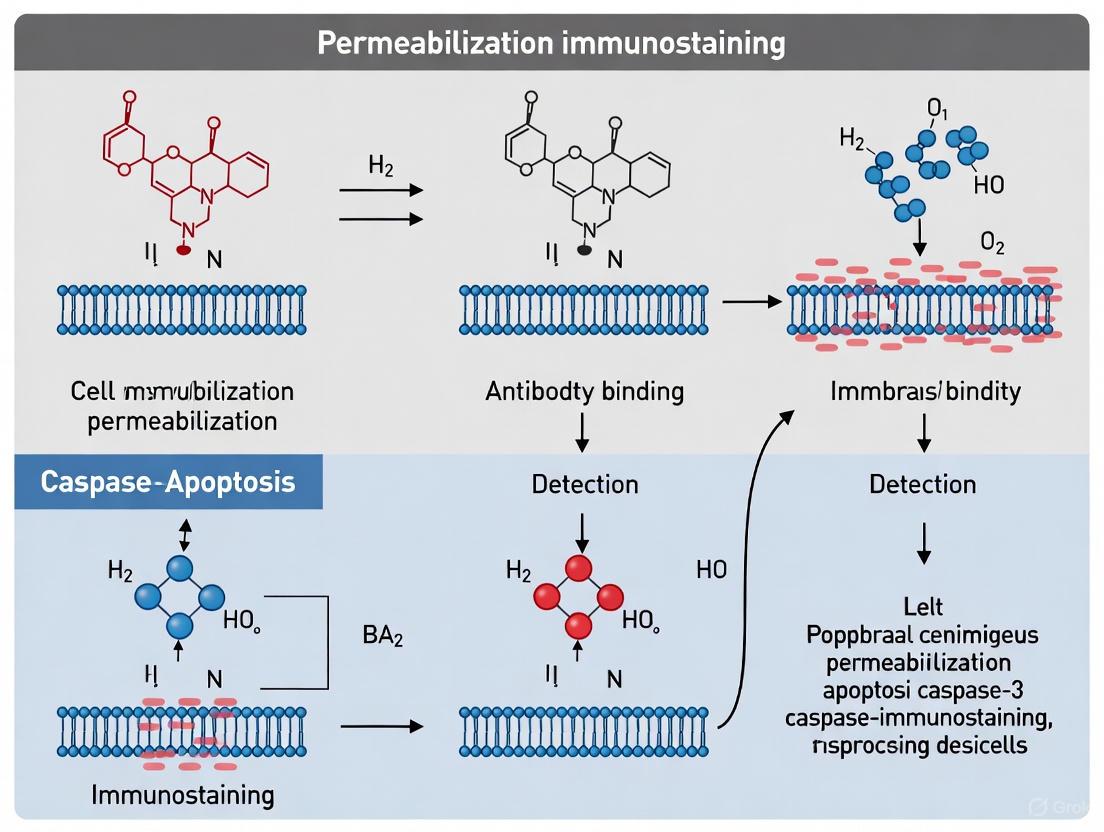
Why Permeabilization is Crucial for Intracellular Caspase-3 Detection
Caspase-3 serves as a key executioner protease in the terminal phase of apoptosis, making its detection a critical endpoint in cell death research. Immunostaining techniques allow for the precise visualization of caspase-3 activation within the context of individual cells. However, the intracellular localization of this target necessitates a crucial sample preparation step: permeabilization. This application note details the fundamental role of permeabilization in enabling specific antibody access to caspase-3, framed within a broader exploration of permeabilization techniques for immunostaining. The protocols and data presented are tailored for researchers, scientists, and drug development professionals requiring robust methodological foundations for their apoptosis assays.
The Critical Role of Permeabilization in Caspase-3 Staining
The plasma membrane acts as a selective barrier that is impermeable to large molecules like antibodies. While fixation stabilizes cellular structures, it does not render the membrane freely permeable. Consequently, without permeabilization, detection antibodies cannot reach their intracellular epitopes on caspase-3, resulting in false-negative results.
Permeabilization creates pores in the lipid bilayer, allowing antibodies to traverse the membrane and bind to the target caspase-3. The choice of permeabilizing agent and conditions directly impacts the size of the pores formed and the preservation of the antigen's integrity. An optimal protocol ensures that antibodies can access the cytosol where caspase-3 resides, while minimizing cellular morphology disruption and non-specific antibody binding.
Established Protocols for Caspase-3 Immunostaining
Protocol 1: Detergent-Based Permeabilization for Immunofluorescence
This protocol, adapted from Abcam's standard procedure, is designed for detecting caspases in fixed cells using immunofluorescence microscopy [9].
Materials Required:
- Primary antibody against caspase-3 (e.g., anti-Caspase-3 rabbit mAb)
- Fluorescently-labeled secondary antibody (e.g., goat anti-rabbit Alexa Fluor 488)
- PBS (Phosphate Buffered Saline)
- Triton X-100 or NP-40
- Blocking buffer (PBS/0.1% Tween 20 + 5% serum from the secondary antibody host species)
- Mounting medium
- Humidified chamber
Step-by-Step Procedure:
- Permeabilization: Incubate fixed samples in PBS containing 0.1% Triton X-100 (or 0.1% NP-40) for 5 minutes at room temperature [9].
- Washing: Wash the slides three times in PBS, for 5 minutes each at room temperature.
- Blocking: Drain the slide and add 200 µL of blocking buffer. Lay the slides flat in a humidified chamber and incubate for 1-2 hours at room temperature to reduce non-specific binding. Rinse once in PBS.
- Primary Antibody Incubation: Add 100 µL of the primary antibody (e.g., diluted 1:200 in blocking buffer) [9]. Incubate slides in a humidified chamber overnight at 4°C.
- Washing: The following day, wash the slides three times for 10 minutes each in PBS/0.1% Tween 20 at room temperature.
- Secondary Antibody Incubation: Drain slides and add 100 µL of the appropriate fluorescently-conjugated secondary antibody (e.g., diluted 1:500 in PBS) [9]. Incubate in a humidified chamber, protected from light, for 1-2 hours at room temperature.
- Final Wash and Mounting: Wash three times in PBS/0.1% Tween 20 for 5 minutes, protected from light. Drain the liquid, mount the slides with an appropriate mounting medium, and observe with a fluorescence microscope.
Protocol 2: Alcohol-Based Permeabilization for Flow Cytometry
This protocol, based on methodologies from R&D Systems, is optimized for intracellular staining of proteins, including caspase-3, for flow cytometric analysis [11]. It is particularly suited for detecting nuclear antigens or phosphorylated proteins.
Materials Required:
- PBS or Hank’s Balanced Salt Solution (HBSS)
- Flow Cytometry Fixation Buffer (e.g., 1-4% paraformaldehyde)
- -20°C Methanol
- Fc Receptor Blocking Reagents
- Fluorochrome-conjugated antibodies
Step-by-Step Procedure:
- Harvest and Fix: Harvest cells and wash twice with PBS. Aliquot up to 1 x 10⁶ cells per tube and fix with 0.5 mL of cold fixation buffer for 10 minutes at room temperature. Wash cells twice with PBS.
- Permeabilization: Resuspend cells in 900 µL of -20°C methanol. Incubate for 30 minutes at 4°C [11]. Centrifuge and discard the supernatant.
- Blocking and Staining: Wash cells twice with PBS. Fc-block cells with an appropriate reagent for 15 minutes at room temperature.
- Antibody Incubation: Add the conjugated primary antibody (5-10 µL per 10⁶ cells or a previously titrated amount) and incubate for 30 minutes at room temperature in the dark.
- Analysis: Wash cells twice with PBS and resuspend the pellet in 200-400 µL of PBS for flow cytometric analysis.
Note: When combining surface and intracellular staining, stain surface antigens first, as fixation and permeabilization can destroy some epitopes. Avoid using PE or APC conjugates prior to methanol permeabilization, as methanol can quench their fluorescence [11].
Comparative Analysis of Permeabilization Methods
The choice of permeabilization agent can significantly impact the outcome of an experiment. Different agents create pores of varying sizes and through different mechanisms, which can influence antibody access and signal intensity.
Table 1: Comparison of Common Permeabilization Agents for Caspase-3 Detection
| Agent | Mechanism | Recommended Concentration & Time | Key Applications | Advantages | Limitations |
|---|---|---|---|---|---|
| Triton X-100 | Dissolves lipids in membranes [9] | 0.1% for 5 min at RT [9] | General immunofluorescence, caspase staining [9] | Strong permeabilization; widely used | Can disrupt some protein-protein interactions |
| Tween-20 | Mild detergent action | 0.2% for 30 min [12] | Flow cytometry for intracellular RNA/proteins [12] | Good for preserving nucleic acids; shown to provide high fluorescence intensity [12] | Milder, may be less effective for some nuclear targets |
| Saponin | Binds cholesterol to create pores | 0.1-0.5% for 10-30 min [12] | Preserving labile structures and protein complexes | Reversible action; gentler on protein structures | Pores are transient, requiring saponin in all antibody buffers |
| Methanol | Precipitates lipids and proteins | 90-100% for 30 min at 4°C [11] | Flow cytometry, detection of phosphorylated proteins and transcription factors [11] | Simultaneously fixes and permeabilizes; excellent for nuclear antigens | Can destroy some surface epitopes; not suitable for PE/APC conjugates pre-permeabilization [11] |
The Scientist's Toolkit: Essential Research Reagents
Successful intracellular caspase-3 detection relies on a suite of critical reagents, each serving a specific function in the experimental workflow.
Table 2: Key Research Reagent Solutions for Caspase-3 Immunostaining
| Reagent | Function | Example Product / Note |
|---|---|---|
| Anti-Caspase-3 Primary Antibody | Binds specifically to the caspase-3 protein, either cleaved or total. | Cleaved Caspase-3 (Asp175) (D3E9) Rabbit mAb [13] |
| Fluorophore-Conjugated Secondary Antibody | Binds to the primary antibody and provides a detectable signal. | Goat anti-rabbit IgG, Alexa Fluor 488 conjugate [9] |
| Permeabilization Detergent | Creates pores in the cell membrane to allow antibody entry. | Triton X-100, Tween-20, Saponin, or NP-40 [9] [12] |
| Blocking Serum | Reduces non-specific binding of antibodies to the sample. | Use serum from the host species of the secondary antibody [9]. |
| Mounting Medium | Preserves fluorescence and supports the coverslip for microscopy. | Use an anti-fade medium for prolonged signal integrity. |
Caspase-3 in the Apoptotic Pathway: A Visual Guide
Caspase-3 activation is a pivotal event in the execution phase of apoptosis, downstream of both intrinsic and extrinsic pathways. Its activation leads to the cleavage of key cellular substrates, resulting in the characteristic morphological changes of apoptotic cell death [14] [7].
This diagram illustrates how caspase-3 is activated and its dual role in apoptosis and pyroptosis. Recent research shows that cleaved caspase-3 can activate Gasdermin E (GSDME), whose N-terminal fragment (GSDME-N) forms pores not only in the plasma membrane but also in the mitochondrial membrane, promoting further cytochrome c release and creating an amplification loop for caspase activation [7].
Troubleshooting Common Issues in Caspase-3 Detection
Even with optimized protocols, researchers may encounter challenges. The table below outlines common problems and their potential solutions.
Table 3: Troubleshooting Guide for Caspase-3 Immunostaining
| Problem | Potential Cause | Recommended Solution |
|---|---|---|
| High Background | Inadequate blocking or washing; non-specific antibody binding. | Ensure thorough washing; use an appropriate blocking serum from the secondary antibody host species; titrate antibodies to optimal concentration [9]. |
| Weak or No Signal | Low antibody concentration; insufficient permeabilization; poor antigen preservation. | Increase primary antibody concentration; optimize permeabilization time and agent concentration; avoid over-fixation [9]. |
| Non-Specific Staining | Antibody cross-reactivity; over-fixation. | Include a negative control without primary antibody; validate antibody specificity; optimize fixation time [9]. |
| Loss of Signal (Flow Cytometry) | Use of methanol with certain fluorophores. | For methanol-based protocols, add fluorophore-conjugated antibodies after the permeabilization step, especially for PE or APC tandems [11]. |
| Poor Cell Morphology | Over-permeabilization; harsh detergents. | Reduce permeabilization time or agent concentration; consider using a milder agent like saponin. |
Permeabilization is a critical step in caspase-3 immunostaining, enabling antibodies to access intracellular epitopes by compromising the integrity of cellular membranes. The choice between detergent-based and enzymatic methods represents a fundamental decision point that significantly impacts staining quality, antigen preservation, and experimental outcomes. Within the context of apoptosis research, precise detection of activated caspase-3 is essential for understanding programmed cell death mechanisms in both basic research and drug development. This application note provides a structured comparison of these permeabilization techniques, offering detailed protocols and analytical frameworks to guide researchers in selecting and optimizing methods for specific experimental requirements.
Technical Comparison of Permeabilizing Agents
The selection of permeabilizing agents involves balancing multiple factors including efficacy, cellular preservation, and compatibility with downstream applications. The table below provides a quantitative comparison of commonly used agents based on critical performance parameters.
Table 1: Comparative Analysis of Permeabilizing Agents for Caspase-3 Immunostaining
| Agent | Mechanism of Action | Optimal Concentration | Incubation Time | Temperature | Key Advantages | Major Limitations |
|---|---|---|---|---|---|---|
| Triton X-100 | Dissolves membrane lipids by disrupting lipid-lipid and lipid-protein interactions [9] [15] | 0.1-0.5% | 5-15 minutes | Room Temperature | Rapid action; effective for most intracellular targets [9] | Can extract cellular proteins; may disrupt delicate epitopes [15] |
| NP-40 | Non-ionic detergent that forms micelles to create membrane pores [9] [15] | 0.1-0.5% | 5-15 minutes | Room Temperature | Milder than Triton X-100; better for cytoplasmic proteins [15] | Less effective for nuclear antigens; variable performance between cell types |
| Saponin | Binds membrane cholesterol to create pore-like structures [16] | 0.1-0.5% | 30-60 minutes | 4°C or Room Temperature | Reversible action; preserves protein-protein interactions [16] | Requires presence in all buffers; less effective for large antibodies |
| Ethanol | Precipitates lipids and proteins through dehydration [17] | 50-70% | 30-60 minutes | 4-15°C | Effective for small molecules; compatible with enzymatic assays [17] | Can denature sensitive epitopes; alters cellular morphology |
Detergent-Based Permeabilization Methods
Mechanism of Action and Pathway Integration
Detergents function as amphipathic molecules containing both hydrophobic tails and polar head groups. At concentrations above their critical micelle concentration (CMC), detergent molecules form micelles that integrate into lipid bilayers, creating pores through the membrane and enabling antibody access to intracellular targets like caspase-3 [15]. The efficacy of this process directly influences the detection of caspase-3, which occupies a terminal position in the apoptotic cascade.
Diagram 1: Detergent Permeabilization in Apoptosis Pathway
Standardized Detergent Permeabilization Protocol
The following protocol is optimized for caspase-3 immunostaining in fixed cell samples, based on established methodologies with specified critical parameters [9]:
Materials Required:
- Primary antibody against caspase-3
- Prepared, fixed samples on slides
- Triton X-100 or NP-40
- Phosphate-buffered saline (PBS)
- Blocking buffer (PBS/0.1% Tween 20 + 5% appropriate serum)
- Fluorescently labeled secondary antibody
- Mounting medium
- Humidified chamber
Procedure:
- Fixation: Begin with appropriately fixed cells (typically with 4% paraformaldehyde for 15 minutes at room temperature).
- Permeabilization: Incubate fixed samples in PBS/0.1% Triton X-100 (or 0.1% NP-40) for 5 minutes at room temperature [9].
- Washing: Wash three times in PBS, for 5 minutes each at room temperature.
- Blocking: Drain the slide and add 200 μL of blocking buffer. Lay slides flat in a humidified chamber and incubate for 1-2 hours at room temperature.
- Primary Antibody Incubation: Add 100 μL of primary antibody diluted 1:200 in blocking buffer. Incubate slides in a humidified chamber overnight at 4°C.
- Secondary Antibody Incubation: Wash slides three times for 10 minutes each in PBS/0.1% Tween 20. Drain slides and add 100 μL of appropriate secondary conjugated antibody diluted 1:500 in PBS. Incubate protected from light for 1-2 hours at room temperature.
- Final Processing: Wash three times in PBS/0.1% Tween 20 for 5 minutes, protected from light. Drain liquid, mount slides, and observe with fluorescence microscopy.
Critical Control: Include a slide with no primary antibody as a negative control to assess non-specific binding of the secondary antibody.
Enzymatic Permeabilization Methods
Mechanism and Strategic Application
Enzymatic permeabilization employs specific enzymes that selectively degrade components of the cellular membrane. Proteases such as trypsin target protein constituents of membranes, while glycosidases attack carbohydrate moieties. This approach typically preserves lipid bilayers more effectively than detergents, but requires careful optimization of concentration and incubation time to prevent epitope destruction or cellular detachment.
For caspase-3 immunostaining, enzymatic methods are particularly valuable when:
- The target epitope is known to be detergent-sensitive
- Simultaneous preservation of membrane structures is required
- Subsequent analysis necessitates intact lipid domains
Optimized Enzymatic Permeabilization Workflow
Diagram 2: Enzymatic Permeabilization Workflow
While specific enzymatic protocols for caspase-3 staining were not detailed in the available literature, the optimization strategy follows these general principles:
- Enzyme Selection: Choose enzymes based on cellular membrane composition and epitope sensitivity.
- Concentration Optimization: Perform titration experiments (typically 0.01-0.5% w/v) to determine optimal concentration.
- Time and Temperature Optimization: Test incubation times (5-30 minutes) at temperatures ranging from 4°C to 37°C.
- Reaction Termination: Include specific inhibitors or thorough washing to terminate enzymatic activity.
- Validation: Compare staining intensity and background with established detergent methods.
The Scientist's Toolkit: Essential Research Reagents
Table 2: Key Reagent Solutions for Permeabilization and Caspase-3 Detection
| Reagent | Function | Application Notes | Commercial Examples |
|---|---|---|---|
| Triton X-100 | Non-ionic detergent for membrane solubilization | Use at 0.1-0.5% in PBS; optimal for most caspase-3 epitopes | Thermo Scientific Surfact-Amps Triton X-100 [15] |
| NP-40 Alternative | Mild non-ionic detergent | Preferred for delicate epitopes; use at 0.1-0.5% | Thermo Scientific Surfact-Amps NP-40 [15] |
| Saponin | Cholesterol-binding glycoside | Creates reversible pores; must be present in all buffers | Sigma-Aldrich Saponin from Quillaja Bark |
| Primary Anti-Caspase-3 Antibody | Target recognition | Clone-specific performance varies; validate for application | Rabbit mAb (ab32351) [9] |
| Fluorescent Secondary Antibodies | Signal generation | Species and isotype specific; optimize dilution | Goat anti-rabbit Alexa Fluor 488 (ab150077) [9] |
| RNase Inhibitors | RNA preservation during permeabilization | Critical for subsequent transcriptomic analysis | Protector RNase Inhibitor [16] |
| High-Salt Buffer | RNase inactivation | Alternative to commercial RNase inhibitors | 2M NaCl in appropriate buffer [16] |
Selection Guidelines and Troubleshooting
Strategic Method Selection
The choice between detergent and enzymatic permeabilization should be guided by experimental priorities:
Select Detergent-Based Methods When:
- Maximum antibody access is paramount
- Working with robust epitopes resistant to extraction
- Standardization and reproducibility are primary concerns
- Processing large sample numbers with limited optimization time
Choose Enzymatic Methods When:
- Preserving lipid domains or protein complexes is essential
- Target epitopes are known to be detergent-sensitive
- Subsequent analyses require intact membrane structures
Troubleshooting Common Issues
High Background Staining:
- Reduce detergent concentration or incubation time
- Increase blocking serum concentration to 10%
- Include additional washes with PBS/0.1% Tween 20
Weak Target Signal:
- Optimize permeabilization duration to balance access and preservation
- Validate antibody compatibility with permeabilization method
- Consider alternative detergent (e.g., switch from Triton X-100 to NP-40)
Cellular Morphology Artifacts:
- For enzymatic methods: reduce enzyme concentration or incubation time
- For detergent methods: consider shorter exposure times or milder agents
- Validate fixation efficacy before permeabilization
RNA Degradation (for multi-omics applications):
- Include RNase inhibitors during permeabilization and staining steps [16]
- Consider high-salt buffers to inactivate RNases [16]
- Limit processing time and temperature variations
Permeabilization method selection represents a critical experimental decision that directly influences caspase-3 detection efficacy and overall data quality in apoptosis research. Detergent-based methods offer standardized, robust protocols suitable for most applications, while enzymatic approaches provide specialized solutions for challenging epitopes or complex multi-omics workflows. By applying the structured comparison and optimized protocols presented in this application note, researchers can make informed decisions that enhance detection sensitivity, minimize artifacts, and generate more reliable data for both basic research and drug development applications.
Fundamental Fixation Principles for Preserving Antigen Integrity
In histochemistry and cytochemistry, fixation is a critical process for preserving biological structures and enabling accurate molecular analysis [18]. This application note details the fundamental principles of fixation, with a specific focus on methodologies that maintain antigen integrity for caspase-3 immunostaining, a key technique in apoptosis research. Proper fixation stabilizes biomolecules, preventing degradation and diffusion, thereby ensuring the reliability of subsequent immunohistochemistry (IHC) or immunofluorescence (IF) procedures [19]. The choice of fixative and protocol has a decisive and often irreversible impact on experimental outcomes, making it a foundational step in research and drug development workflows [18] [19].
Core Principles of Fixation
Fixation methods are broadly categorized into two types based on their mechanism of action: precipitating fixation and cross-linking fixation. The strategic selection between these types is paramount for successfully visualizing the target antigen.
Table 1: Comparison of Fixation Methods and Their Impact on Antigens
| Fixation Type | Mechanism of Action | Common Examples | Impact on Antigen Integrity | Key Considerations for Caspase-3 Staining |
|---|---|---|---|---|
| Precipitating | Dehydrates sample, denatures and coagulates proteins [19]. | Acetone, Ethanol, Methanol [19] [11]. | Can destroy conformational epitopes; may preserve linear epitopes [19]. | Often used for intracellular staining of transcription factors and phosphorylated proteins [11]. Can be suitable for caspase-3 when combined with specific antibody clones. |
| Cross-linking | Creates covalent bonds (methylene bridges) between biomolecules, especially proteins [18] [19]. | Formaldehyde, Paraformaldehyde (PFA), Glutaraldehyde [18] [19]. | Can mask epitopes via cross-linking, potentially requiring antigen retrieval [18]. | The standard for preserving cellular morphology. Over-fixation can mask the caspase-3 epitope, necessitating optimization of fixation time [18]. |
For caspase-3 immunostaining, formaldehyde-based fixatives (a cross-linking type) are most common in IHC protocols due to their excellent preservation of cellular structure. However, a key challenge is that excessive fixation can over-cross-link proteins, masking the caspase-3 epitope and reducing antibody binding [18]. Alcohol-based precipitating fixatives are also used, particularly in flow cytometry protocols for intracellular targets, but they can alter protein conformation [11].
Detailed Experimental Protocol for Caspase-3 Immunofluorescence
This protocol is designed for the detection of cleaved/active caspase-3 in fixed cell samples, providing a workflow that balances antigen preservation with accessibility.
Materials Required
- Primary Antibody: Anti-Caspase-3 (cleaved) specific antibody (e.g., Rabbit mAb, ab32351) [9]
- Secondary Antibody: Fluorescently-labeled secondary antibody (e.g., Goat anti-Rabbit Alexa Fluor 488, ab150077) [9]
- Fixative: 4% Paraformaldehyde (PFA) in PBS [9] [11]
- Permeabilization Buffer: PBS with 0.1% Triton X-100 or -20°C Methanol [9] [11]
- Blocking Buffer: PBS/0.1% Tween 20 + 5% serum from the host species of the secondary antibody [9]
- Mounting Medium: Permanent or aqueous mounting medium compatible with fluorescence [9]
- Other: PBS, humidified chamber, pipettes, slides [9]
Workflow Diagram
The following diagram illustrates the complete experimental workflow for caspase-3 immunofluorescence staining.
Step-by-Step Procedure
- Permeabilization: Incubate the fixed samples in PBS containing 0.1% Triton X-100 for 5 minutes at room temperature. Alternatively, for certain nuclear antigens or flow cytometry, ice-c methanol can be used for 30 minutes at 4°C [9] [11].
- Washing: Wash the slides three times in PBS, for 5 minutes each at room temperature [9].
- Blocking: Drain the slide and apply 200 µL of blocking buffer. Lay the slides flat in a humidified chamber and incubate for 1-2 hours at room temperature to prevent non-specific antibody binding [9].
- Primary Antibody Incubation: Apply 100 µL of the primary antibody (e.g., anti-Caspase-3) diluted in blocking buffer (a starting dilution of 1:200 is recommended). Incubate the slides in a humidified chamber overnight at 4°C. Include a negative control with no primary antibody [9].
- Washing: The next day, wash the slides three times for 10 minutes each in PBS/0.1% Tween 20 at room temperature [9].
- Secondary Antibody Incubation: Apply 100 µL of the appropriate fluorescently-labeled secondary antibody diluted in PBS (a starting dilution of 1:500 is recommended). Incubate in a humidified chamber, protected from light, for 1-2 hours at room temperature [9].
- Final Washes: Wash the slides three times in PBS/0.1% Tween 20 for 5 minutes each, protected from light [9].
- Mounting and Imaging: Drain the liquid, mount the slides with a suitable mounting medium, and observe with a fluorescence microscope [9].
Antigen Retrieval and Troubleshooting
For samples fixed with cross-linking fixatives like formaldehyde, antigen retrieval is often essential to reverse the masking of epitopes. Techniques like microwave-induced heat retrieval can break methylene cross-links and restore antigenicity, thereby enhancing staining efficacy [18].
Table 2: Troubleshooting Common Issues in Caspase-3 Immunostaining
| Problem | Potential Cause | Suggested Solution |
|---|---|---|
| High Background | Inadequate blocking or washing; non-specific antibody binding. | Ensure thorough washing; use appropriate blocking serum from the secondary antibody host species [9]. |
| Weak or No Signal | Low antibody concentration; over-fixation masking epitope; poor antigen preservation. | Titrate to increase primary antibody concentration; optimize fixation time; employ antigen retrieval techniques [18] [9]. |
| Non-Specific Staining | Antibody cross-reactivity; insufficient blocking. | Include proper controls (isotype, no primary); validate antibody specificity; optimize blocking conditions [9]. |
Essential Research Reagent Solutions
Table 3: Key Reagents for Caspase-3 Immunostaining
| Reagent | Function | Example Product/Catalog | Critical Considerations |
|---|---|---|---|
| Anti-Caspase-3 (Cleaved) Antibody | Specifically binds to the active form of caspase-3, enabling detection of apoptosis. | Anti-Caspase-3 (ab32351) [9] | Clone specificity (e.g., D3E9) is critical for detecting the cleaved form. Validate for your application (IF, IHC) [20]. |
| Formaldehyde/PFA Fixative | Cross-linking fixative that preserves cellular architecture and immobilizes antigens. | Flow Cytometry Fixation Buffer (FC004) [11] | Concentration (1-4%) and fixation time must be optimized to balance preservation with antigen accessibility [18] [11]. |
| Methanol | Precipitating fixative and permeabilization agent. | -20°C Methanol [11] | Ideal for many intracellular and nuclear targets like transcription factors. Can be detrimental to PE or APC fluorophores [11]. |
| Triton X-100 | Detergent for permeabilizing lipid membranes after fixation. | PBS/0.1% Triton X-100 [9] | Allows antibodies to access intracellular antigens like caspase-3. Concentration and time should be controlled to preserve ultrastructure. |
| Normal Serum | Used in blocking buffer to reduce non-specific binding of antibodies. | Serum from secondary antibody host species (e.g., Goat Serum) [9] | Minimizes background staining, improving signal-to-noise ratio. |
Mastering fixation principles is indispensable for obtaining reliable and reproducible results in caspase-3 immunostaining and broader immunohistochemistry research. The foundational choice between cross-linking and precipitating fixatives sets the stage for successful antigen detection. Adherence to optimized protocols for permeabilization, blocking, and antibody incubation, coupled with the strategic use of antigen retrieval when necessary, ensures the preservation of both morphological detail and antigen integrity. As the field advances, the integration of robust fixation standards with emerging technologies like automated quantitative analysis [21] and AI-powered image interpretation [22] will further enhance the precision and impact of biomarker research in drug development.
Caspase-3, a key executioner caspase in apoptosis, cleaves numerous protein substrates to ensure efficient execution of cell death [23]. Its detection is crucial in diverse fields, from cancer biology and drug discovery to forensic science [14] [10]. Accurate detection is technically challenging, requiring careful method selection based on the specific research question.
This application note provides a detailed comparison of four central techniques for caspase-3 detection: Immunofluorescence (IF), Western Blot (WB), Flow Cytometry, and Live Imaging. We place special emphasis on the critical role of permeabilization techniques, a cornerstone for successful immunostaining, to guide researchers in obtaining reliable and reproducible data on caspase-3 localization and activation.
Caspase-3 in Context: Apoptotic and Non-Apoptotic Signaling
Caspase-3 activation is a pivotal event in the execution phase of apoptosis, a point of convergence for both the extrinsic (death receptor) and intrinsic (mitochondrial) pathways [14]. The diagram below illustrates the simplified signaling pathways leading to caspase-3 activation.
Simplified Caspase-3 Activation Pathways. This diagram shows the primary apoptotic pathways. The extrinsic pathway is initiated by cell surface death receptors, activating caspase-8. The intrinsic pathway is triggered by cellular stress (e.g., DNA damage, chemotherapy), leading to caspase-9 activation. Both converge to activate caspase-3, which executes apoptosis through substrate cleavage (e.g., CAD, PARP) [14] [24]. Emerging research shows caspase-3 also regulates non-apoptotic processes like cell motility, autophagy, and the DNA damage response [23] [4].
Comparative Analysis of Detection Methods
Selecting the appropriate detection method is paramount. The table below summarizes the key characteristics of each technique to inform your decision.
Table 1: Key Characteristics of Caspase-3 Detection Methods
| Parameter | Immunofluorescence (IF) | Western Blot (WB) | Flow Cytometry | Live Cell Imaging |
|---|---|---|---|---|
| Key Readout | Spatial localization & activation in situ | Protein size, cleavage status, and expression level | Quantitative, single-cell analysis of large populations | Real-time enzymatic activity dynamics in live cells |
| Spatial Resolution | High (subcellular) | No spatial information | No spatial information | Moderate (cellular) |
| Temporal Resolution | Fixed endpoint | Fixed endpoint | Fixed endpoint | High (real-time) |
| Quantification | Semi-quantitative (fluorescence intensity) | Semi-quantitative (band density) | Fully quantitative | Semi- to fully quantitative (kinetic rates) |
| Throughput | Low to moderate | Low | High | Low to moderate |
| Best Detects | Cleaved/active caspase-3 (with specific antibodies) | Pro-form and cleaved fragments; molecular weight confirmation | Percentage of positive cells in a heterogenous population | Caspase-3 enzymatic activity (using fluorogenic substrates) |
| Critical Permeabilization Note | Essential for antibody access to intracellular epitopes; concentration and detergent type are critical. | Inherent; samples are fully denatured and solubilized. | Required for intracellular staining; gentle detergents (e.g., saponin) are often used. | Not required for FRET/fluorogenic substrates, which are cell-permeant. |
This comparison highlights that the choice of assay profoundly impacts the data generated [25]. WB confirms protein presence and cleavage, IF provides spatial context, Flow Cytometry offers statistical power from thousands of cells, and Live Imaging reveals dynamic activation kinetics [14] [26] [25].
Detailed Experimental Protocols
Immunofluorescence (IF) for Active Caspase-3
This protocol is optimized for detecting the activated, cleaved form of caspase-3 in adherent cells, with a focus on permeabilization.
- Key Reagents: Anti-cleaved caspase-3 antibody (Cell Signaling Technology #9661 used in apoptosis studies [26]), fluorescently-labeled secondary antibody, Triton X-100, paraformaldehyde (PFA), blocking serum (e.g., goat serum).
- Sample Preparation: Plate cells on glass coverslips. Induce apoptosis and rinse cells with sterile PBS.
- Fixation and Permeabilization:
- Fix cells with 4% PFA for 15 minutes at room temperature (RT).
- Wash 3x with PBS.
- Permeabilize cells with 0.1% Triton X-100 in PBS for 10 minutes at RT. Note: Concentration and time are critical to preserve cell morphology while allowing antibody penetration.
- Wash 3x with PBS.
- Immunostaining:
- Block with 10% goat serum in PBS for 1 hour at RT.
- Incubate with primary antibody (diluted in blocking serum) overnight at 4°C.
- Wash 3x with PBS.
- Incubate with fluorescent secondary antibody (in blocking serum) for 1 hour at RT in the dark.
- Wash 3x with PBS.
- Mounting and Imaging: Mount coverslips using an anti-fade mounting medium. Image using a fluorescence or confocal microscope. Include controls (no primary antibody, apoptosis inhibitor).
Western Blot for Caspase-3 and Its Cleavage Fragments
This protocol detects the pro-form (∼35 kDa) and cleaved fragments (∼17/19 kDa) of caspase-3 in cell lysates.
- Key Reagents: Antibodies against full-length and cleaved caspase-3, RIPA lysis buffer, protease inhibitors, SDS-PAGE gel, PVDF membrane.
- Cell Lysis and Protein Quantification:
- Lyse cells in RIPA buffer supplemented with protease inhibitor cocktail.
- Incubate on ice for 30 minutes, then centrifuge at 14,000 x g for 15 minutes at 4°C.
- Collect the supernatant and determine protein concentration using a BCA or Bradford assay.
- Gel Electrophoresis and Transfer:
- Separate 20-40 µg of total protein via SDS-PAGE (12-15% gel).
- Transfer proteins to a PVDF membrane.
- Immunoblotting:
- Block membrane with 5% non-fat milk in TBST for 1 hour at RT.
- Incubate with primary caspase-3 antibody (diluted in blocking buffer) overnight at 4°C.
- Wash 3x with TBST.
- Incubate with HRP-conjugated secondary antibody for 1 hour at RT.
- Wash 3x with TBST.
- Detection: Develop blots using enhanced chemiluminescence (ECL) reagent. Use housekeeping proteins (e.g., GAPDH, β-Actin) as loading controls.
Flow Cytometry for Caspase-3 Activation
This protocol quantifies the proportion of cells with active caspase-3 in a population, often combined with other markers.
- Key Reagents: FITC-conjugated anti-active caspase-3 antibody (or intracellular staining after fixation/permeabilization), 4% PFA, permeabilization buffer (e.g., saponin-based).
- Cell Harvest and Fixation:
- Harvest cells (including floating apoptotic cells) by gentle trypsinization or pipetting.
- Fix cells with 4% PFA for 20 minutes at RT.
- Wash with PBS.
- Permeabilization and Staining:
- Permeabilize cells using a commercial saponin-based permeabilization buffer for 10 minutes on ice. Note: Saponin creates pores that are reversible, helping to preserve light scatter properties.
- Stain with FITC-conjugated active caspase-3 antibody in permeabilization buffer for 30 minutes in the dark.
- Wash with permeabilization buffer, then resuspend in PBS.
- Data Acquisition and Analysis: Analyze cells immediately on a flow cytometer. Use unstained and isotype controls to set negative gates. Data is typically presented as the percentage of positive cells in a histogram or dot plot.
Live Cell Imaging of Caspase-3 Activity
This protocol uses a fluorogenic substrate to monitor caspase-3 activity in real-time without fixing or permeabilizing cells.
- Key Reagents: NucView 488 caspase-3 substrate (or similar cell-permeant FRET-based substrates), live-cell imaging buffer, appropriate culture media.
- Cell Preparation and Staining:
- Plate cells in a glass-bottom culture dish or multi-well plate compatible with your live-cell imaging system.
- On the day of imaging, prepare a "staining mixture" in live-cell imaging buffer or phenol-red-free media. A typical mixture contains 5 µM NucView 488 caspase-3 substrate [26].
- Replace the cell culture medium with the staining mixture.
- Incubate for 15-30 minutes at 37°C in the dark (no wash required).
- Image Acquisition:
- Place the dish on a pre-warmed stage (37°C, 5% CO₂) of a fluorescence microscope or confocal system.
- Acquire images at regular intervals (e.g., every 15-60 minutes) over the desired time course (e.g., 24-72 hours). The NucView 488 substrate is non-fluorescent until cleaved by caspase-3, upon which it binds DNA and produces a bright green nuclear fluorescence signal [26].
- Data Analysis: Quantify the increase in green fluorescence intensity over time or count the number of fluorescent-positive cells per field using image analysis software (e.g., ImageJ, IncuCyte software).
The Scientist's Toolkit: Essential Research Reagents
Successful caspase-3 detection relies on specific reagents. This table details key solutions for the experiments described.
Table 2: Key Reagent Solutions for Caspase-3 Detection
| Reagent / Assay Kit | Specific Function & Role in Detection | Example Application Context |
|---|---|---|
| Anti-Cleaved Caspase-3 Antibody | Specifically binds the activated, cleaved form of caspase-3; essential for distinguishing active enzyme from zymogen in IF, WB, and Flow. | Detecting apoptosis in tissue sections (e.g., forensic skin samples [10]) or cultured cells after drug treatment [26]. |
| NucView 488 Caspase-3 Substrate | Cell-permeant, non-fluorescent substrate that emits bright green fluorescence upon cleavage by active caspase-3 and subsequent DNA binding. | Real-time, kinetic live-cell imaging of apoptosis without requiring permeabilization [26]. |
| CF594 Annexin V | Binds to phosphatidylserine (PS) exposed on the outer leaflet of the plasma membrane, an early-mid event in apoptosis. | Used in multiplex assays (e.g., with caspase-3 stains) to correlate caspase activation with PS externalization [26]. |
| MitoView Blue | A cationic dye that accumulates in active mitochondria based on membrane potential (ΔΨm). | Co-staining to show the loss of mitochondrial potential, an early apoptotic event, alongside caspase-3 activation [26]. |
| Magnetic Bead-based LFIA | Utilizes peptide substrates on magnetic beads; caspase-3 cleavage releases a detectable fragment. Combines separation with simple readout. | Potential for rapid, low-cost point-of-care detection of caspase-3 activity in cell lysates [27]. |
Integrated Workflow for Apoptosis Analysis
A comprehensive analysis often combines multiple techniques. The following workflow diagram outlines a strategic approach for validating caspase-3-dependent apoptosis.
Integrated Workflow for Apoptosis Analysis. A recommended strategy begins with Live-Cell Imaging for kinetic data. In parallel, cells are harvested for complementary, fixed-point analyses: Flow Cytometry quantifies the apoptotic population, IF confirms subcellular localization, and WB validates cleavage and identifies specific substrates like PARP [26] [24]. This multi-faceted approach cross-validates findings and provides a comprehensive view of caspase-3 activation.
Concluding Remarks
The optimal method for detecting caspase-3 depends entirely on the research question. WB provides definitive proof of cleavage, IF and Live Imaging offer spatial and temporal context, and Flow Cytometry delivers robust population statistics. Permeabilization is a critical, technique-specific parameter that must be optimized for immunostaining-based methods.
Understanding these methods' strengths and limitations enables researchers to effectively study caspase-3 in apoptosis and its emerging non-apoptotic roles, such as regulating cytoskeletal dynamics in cancer cell motility [23] and promoting stress adaptation [4].
Step-by-Step Protocols for Caspase-3 Immunostaining Across Sample Types
Standard Immunofluorescence Protocol for Fixed Cells
Immunofluorescence (IF) is a cornerstone technique for visualizing protein localization and expression within their cellular context. For researchers investigating intricate processes like apoptosis, specifically through the detection of executioner caspases such as caspase-3, a robust and reproducible immunofluorescence protocol is paramount. Caspase-3, a key mediator of apoptotic cell death, serves as a critical biomarker, and its accurate visualization can be confounded by its subcellular localization and activation status [28] [23]. This application note details a standardized immunofluorescence protocol for fixed cells, meticulously framed within the context of caspase-3 immunostaining. We place particular emphasis on permeabilization techniques, a crucial step that governs antibody access to intracellular epitopes and can significantly impact the success of detecting caspase-3, which can exhibit constitutive association with the cytoskeleton in certain cancer cells [23].
The following diagram illustrates the complete experimental workflow for immunofluorescence staining of fixed cells, from preparation to imaging.
Caspase-3 in Apoptosis and Beyond
Caspase-3 is a cysteine-aspartate protease known as an executioner caspase, playing a central role in the final stages of apoptosis by cleaving a wide array of cellular substrates [28]. This process is integral to the controlled dismantling of the cell. Immunofluorescence detection of caspase-3 typically aims to identify its active form, which often involves translocation or cleavage, providing a direct readout of apoptotic activity [9]. Interestingly, recent research has revealed non-apoptotic roles for caspase-3. For instance, in aggressive cancers like melanoma, caspase-3 is highly expressed and constitutively associates with the cytoskeleton, where it regulates cell migration and invasion by modulating proteins such as coronin 1B, a key regulator of actin polymerization [23]. This non-canonical function underscores the importance of reliable detection methods and highlights that subcellular localization, influenced by permeabilization efficiency, is critical for accurate interpretation.
The diagram below outlines the position of caspase-3 within the broader apoptotic signaling pathways.
Detailed Protocols and Reagents
Step-by-Step Immunofluorescence Protocol
The following protocol is optimized for the detection of intracellular antigens like caspase-3 in cultured cells [29] [30] [9].
Sample Preparation and Fixation
- Culture cells on poly-L-lysine or poly-D-lysine-coated glass coverslips to ensure adhesion [30].
- Fixation: Aspirate culture medium and wash cells briefly with phosphate-buffered saline (PBS). Fix cells by covering them with 4% formaldehyde (methanol-free) in PBS for 15 minutes at room temperature [29]. Note: Alternative fixatives like cold methanol or acetone can be used, which simultaneously fix and permeabilize cells, potentially requiring omission of a separate permeabilization step [30] [31].
- Wash fixed cells three times with PBS for 5 minutes each to remove residual fixative [29].
Permeabilization (Critical for Caspase-3)
- This step is essential for allowing antibodies to access intracellular targets. The choice of detergent is crucial, as it must be strong enough to expose the antigen without destroying it or creating excessive background.
- Incubate cells with 0.1–0.5% Triton X-100 in PBS for 5–10 minutes at room temperature [30] [9]. Note: For membrane-associated antigens, milder detergents like saponin (0.05%) may be preferable, as Triton X-100 can solubilize membranes and their associated proteins [30]. Research indicates caspase-3 can associate with the cytoskeleton [23], warranting optimization of permeabilization strength.
Blocking
- To prevent non-specific antibody binding, incubate cells in a blocking buffer for 1–2 hours at room temperature.
- A standard buffer is PBS containing 5% normal serum (from the species in which the secondary antibody was raised) and 0.3% Triton X-100 [29]. Alternatively, 1–5% bovine serum albumin (BSA) can be used [30].
Primary Antibody Incubation
- Prepare the primary antibody (e.g., anti-caspase-3) in an antibody dilution buffer (e.g., PBS with 1% BSA and 0.1% Triton X-100) [29] [9].
- Aspirate the blocking solution and apply the diluted primary antibody to the cells.
- Incubate overnight at 4°C in a humidified chamber to ensure specific binding [29] [9].
- Include a negative control (no primary antibody) to assess background staining.
Secondary Antibody Incubation
- The next day, wash the cells three times with PBS for 5–10 minutes each to remove unbound primary antibody [29] [9].
- Incubate with a fluorophore-conjugated secondary antibody (e.g., Alexa Fluor 488 or 555), diluted in antibody dilution buffer, for 1–2 hours at room temperature protected from light [29] [9].
- Wash the cells three times with PBS for 5 minutes each, protected from light.
Mounting and Imaging
- Briefly drain the slides and mount the coverslips using an aqueous or hard-set mounting medium. For nuclear counterstaining, use a medium containing DAPI [31].
- Once the mounting medium has set, visualize the samples using a fluorescence microscope with appropriate filters. Store slides at 4°C in the dark [29] [31].
Research Reagent Solutions
The table below lists essential reagents and their functions for a successful immunofluorescence experiment.
| Reagent | Function/Description | Example/Citation |
|---|---|---|
| Fixative | Preserves cellular morphology and immobilizes antigens. 4% Formaldehyde is standard. Methanol/acetone offer alternative fixation/permeabilization. | 4% Formaldehyde, Methanol-Free [29]; Cold Methanol/Acetone [30] [31] |
| Permeabilization Agent | Creates pores in membranes for antibody entry. Critical for intracellular targets like caspase-3. | Triton X-100 [29] [9]; Saponin, Tween-20 [30] |
| Blocking Agent | Reduces non-specific antibody binding to minimize background. | Normal Serum, BSA [29] [30] |
| Antibody Dilution Buffer | Diluent for antibodies that helps maintain stability and reduce non-specific binding. | PBS with 1% BSA and 0.1-0.3% Triton X-100 [29] |
| Wash Buffer | Removes unbound reagents. Typically PBS, sometimes with a mild detergent. | 1X Phosphate Buffered Saline (PBS) [29] |
| Mounting Medium | Preserves fluorescence and supports optics for microscopy. Often includes anti-fade agents. | Aqueous or Hard-set Mounting Media, with/without DAPI [31] |
Quantitative Data for Protocol Optimization
Optimization of key parameters is often required for different antigens. The table below summarizes critical variable ranges based on established protocols.
| Parameter | Suggested Range | Protocol Source |
|---|---|---|
| Formaldehyde Fixation Time | 10 – 20 minutes at Room Temperature | [29] [30] |
| Methanol Fixation Time | 5 – 10 minutes at -20°C | [30] [31] |
| Permeabilization Time (Triton X-100) | 2 – 10 minutes at Room Temperature | [30] [9] |
| Blocking Time | 1 – 2 hours at Room Temperature | [29] [30] |
| Primary Antibody Incubation | Overnight at 4°C | [29] [9] |
| Secondary Antibody Incubation | 1 – 2 hours at Room Temperature (dark) | [29] [9] |
| Wash Steps (Post-Antibody) | 3 x 5 minutes each in PBS | [29] [9] |
Troubleshooting Common Issues
| Problem | Potential Cause | Suggested Remedy |
|---|---|---|
| High Background | Inadequate blocking or washing; non-specific secondary antibody. | Use blocking serum from secondary antibody host species [9]; ensure thorough washing [9]. |
| Weak or No Signal | Under-fixation, insufficient permeabilization, low antibody concentration. | Optimize fixation and permeabilization time/concentration; titrate primary antibody [9]. |
| Non-Specific Staining | Antibody cross-reactivity; over-fixation. | Include negative controls; validate antibody specificity; optimize fixation time [30] [9]. |
| Poor Cellular Morphology | Over-fixation; harsh permeabilization. | Reduce fixation time; consider milder detergents like saponin or Tween-20 [30]. |
| Signal Fading | Fluorophore degradation during storage or imaging. | Use anti-fade mounting medium; store slides at 4°C in the dark [31]. |
Flow Cytometry Techniques for Caspase-3 Detection in Immune Cells
Caspase-3, a cysteine-aspartic protease, functions as a crucial executioner enzyme in the apoptotic pathway, responsible for the majority of proteolytic events during programmed cell death [32]. This enzyme is synthesized as an inactive zymogen and undergoes proteolytic cleavage at specific aspartic acid residues to become activated [14]. The detection of cleaved caspase-3 provides a reliable marker for identifying cells that are undergoing, or have undergone, apoptosis [32]. In immune contexts, caspase-3 activation plays a particularly important role in regulating cytotoxic T lymphocyte (CTL) activity and maintaining cellular homeostasis [33].
Flow cytometry offers distinct advantages for caspase-3 detection, including single-cell analysis, multi-parameter capabilities, and quantitative measurement of apoptosis frequency within heterogeneous cell populations [32] [33]. When combined with immunophenotyping markers, this technique enables researchers to precisely identify which immune cell subsets are undergoing apoptosis within complex mixtures such as peripheral blood mononuclear cells or lymphoid tissues [33]. The following application notes and protocols detail optimized methodologies for detecting cleaved caspase-3 in immune cells using flow cytometry, with particular attention to permeabilization techniques essential for intracellular epitope detection.
Caspase Biology and Detection Principles
Caspase Family and Activation Mechanisms
Caspases comprise a family of cysteine-dependent proteases that cleave their substrates following aspartic acid residues [14]. These enzymes are categorized into three functional groups: initiator caspases (caspase-2, -8, -9, -10), executioner caspases (caspase-3, -6, -7), and inflammatory caspases (caspase-1, -4, -5, -11, -12, -13, -14) [14]. Caspase-3 serves as the primary executioner protease, responsible for cleaving vital cellular substrates and initiating the morphological changes characteristic of apoptosis [14] [32].
Activation of caspase-3 occurs through two principal pathways. The extrinsic pathway initiates when external death signals engage surface death receptors like Fas and TNF receptors, leading to caspase-8 activation, which can subsequently activate caspase-3 directly or indirectly through mitochondrial amplification [14]. The intrinsic pathway centers on mitochondrial cytochrome c release and formation of the Apaf-1/caspase-9 apoptosome complex, which then processes and activates executioner caspases including caspase-3 [14] [34].
Key Advantages of Cleaved Caspase-3 Detection
Unlike Annexin V staining which detects phosphatidylserine externalization as an early apoptotic event, cleaved caspase-3 detection specifically identifies cells that have committed to the apoptotic execution phase [35]. This method offers several significant advantages:
- High Specificity: Antibodies specifically recognize the cleaved, active form of caspase-3, avoiding detection of the inactive zymogen [32]
- Commitment Marker: Caspase-3 activation represents an irreversible step in apoptotic progression [32]
- Sensitivity: Flow cytometric detection can identify rare apoptotic events within large cell populations [33]
- Multiparametric Analysis: Can be combined with cell surface immunophenotyping and viability markers [33]
Table 1: Comparison of Apoptosis Detection Methods
| Method | Target | Detection Stage | Advantages | Limitations |
|---|---|---|---|---|
| Cleaved Caspase-3 Detection | Activated caspase-3 | Mid-late apoptosis | High specificity for apoptotic commitment; quantitative | Requires cell permeabilization |
| Annexin V Staining | Phosphatidylserine exposure | Early apoptosis | Detects early apoptotic events | Cannot distinguish between apoptotic and necrotic cells without viability dye |
| TUNEL Assay | DNA fragmentation | Late apoptosis | Specific for late-stage apoptosis | May miss early apoptotic cells; more complex procedure |
| TRAIL Receptors | Death receptors | Early apoptosis | Specific for extrinsic pathway | Limited to death receptor-mediated apoptosis |
Detection of caspase-3 cleavage in target cells has been successfully employed as a sensitive readout for antigen-specific CTL activity, demonstrating markedly higher sensitivity compared to traditional (^{51})Cr-release assays [33]. This application is particularly valuable in vaccine trials and preclinical models of CTL function across both human and murine systems [33].
Materials and Reagents
Research Reagent Solutions
Table 2: Essential Reagents for Caspase-3 Flow Cytometry
| Reagent/Category | Specific Examples | Function/Purpose |
|---|---|---|
| Primary Antibodies | Anti-cleaved caspase-3 (reactive against human and mouse forms) [33] | Specifically binds to the activated form of caspase-3; enables detection of apoptotic cells |
| Fluorochrome Conjugates | PE-labeled anti-cleaved caspase-3 [33] | Provides detectable signal for flow cytometry; allows multiplexing with other markers |
| Cell Tracking Dyes | DDAO-SE (CellTrace Far Red DDAO-SE) [33] | Labels target cells for identification in co-culture systems; enables discrimination between effector and target cells |
| Permeabilization Buffers | Commercially available intracellular staining permeabilization buffers | Permeabilizes cell membrane to allow antibody access to intracellular cleaved caspase-3 |
| Flow Cytometry Staining Buffer | PBS supplemented with 0.5-1% BSA [36] | Maintains cell viability and reduces non-specific antibody binding during staining procedures |
| Fc Receptor Blocking Reagents | Human or mouse Fc block [36] | Reduces non-specific antibody binding through Fc receptors; critical for immune cells |
| Viability Dyes | Propidium iodide, DRAQ5 [35] | Distinguishes between live and dead cells; excludes necrotic cells from analysis |
Equipment Requirements
- Flow cytometer capable of detecting fluorochromes used in the assay (e.g., FL2 channel for PE detection) [33]
- Centrifuge with capacity for 15 mL and 50 mL tubes
- Tissue culture incubator (37°C, 5% CO₂)
- Vortex mixer
- FACS tubes (5 mL round-bottom polystyrene tubes) [36]
- Pipettes and pipette tips
Protocol: Detection of Cleaved Caspase-3 by Flow Cytometry
Sample Preparation and Stimulation
For immune cells from primary sources:
- Collect peripheral blood in evacuated tubes containing EDTA or heparin as anticoagulant [36]
- Wash cells three times in isotonic phosphate buffer (supplemented with 0.5% BSA) by centrifugation at 350-500 × g for 5 minutes to remove contaminating serum components [36]
- Isate desired immune cell populations using density gradient centrifugation or negative selection kits
For in vitro apoptosis induction:
- Seed immune cells at appropriate density (typically 0.5-1 × 10⁶ cells/mL) in culture medium
- Apply apoptotic stimuli relevant to research context:
- Include unstimulated controls cultured in parallel
- Incubate for appropriate duration (typically 2-24 hours, depending on cell type and stimulus)
Cell Staining Procedure
Harvest and Wash: Collect cells by gentle pipetting or trypsinization (for adherent cells), followed by centrifugation at 350-500 × g for 5 minutes. Wash once with Flow Cytometry Staining Buffer [36].
Fc Receptor Blocking: Resuspend cell pellet (up to 1 × 10⁶ cells/100 μL) in staining buffer. Add Fc receptor blocking antibody (1 μg IgG/10⁶ cells) and incubate for 15 minutes at room temperature [36]. Do not wash after this step.
Cell Surface Marker Staining (if performing immunophenotyping):
- Add fluorochrome-conjugated antibodies against cell surface markers (CD3, CD4, CD8, CD19, etc.) at predetermined optimal concentrations
- Vortex gently and incubate for 30 minutes at 4°C in the dark
- Wash cells with 2 mL Flow Cytometry Staining Buffer, centrifuge at 350-500 × g for 5 minutes, and decant supernatant
Cell Fixation and Permeabilization:
- Resuspend cell pellet in 100-250 μL of fixation buffer (commercially available formaldehyde-based fixatives)
- Incubate for 20 minutes at room temperature in the dark
- Wash cells with 2 mL Flow Cytometry Staining Buffer
- Resuspend cells in 100-250 μL of permeabilization buffer
- Incubate for 15-30 minutes at room temperature in the dark
Intracellular Staining for Cleaved Caspase-3:
Resuspension and Analysis:
- Resuspend fixed and stained cells in 200-400 μL Flow Cytometry Staining Buffer [36]
- Keep samples at 4°C in the dark until flow cytometric analysis
- Analyze within 24 hours for optimal results
Critical Permeabilization Considerations
The permeabilization step represents the most critical technical factor for successful cleaved caspase-3 detection, as it controls antibody access to intracellular epitopes while preserving cell morphology and light scatter properties. Key considerations include:
- Permeabilization Agent Selection: Use commercial intracellular staining permeabilization buffers rather than homemade formulations for consistent results
- Duration Optimization: Excessive permeabilization can diminish light scatter properties and compromise cell integrity, while insufficient permeabilization reduces antibody access
- Antibody Compatibility: Ensure antibodies are validated for intracellular staining, as some conjugates may perform differently under permeabilizing conditions
- Concurrent Surface Staining: When combining surface and intracellular staining, complete all surface marker staining before permeabilization
Flow Cytometry Data Acquisition and Analysis
Instrument Setup:
- Create a forward scatter (FSC) vs. side scatter (SSC) plot to establish granulosity and size gates
- Create a viability dye (if used) vs. SSC plot to exclude dead cells
- Create appropriate fluorescence channels for all fluorochromes used
Compensation Controls:
- Prepare single-stained compensation controls for each fluorochrome
- Include unstained and isotype controls for background determination
Gating Strategy:
- Gate on lymphocytes based on FSC-A vs. SSC-A characteristics
- Exclude doublets using FSC-A vs. FSC-H
- Gate on viable cells (viability dye-negative)
- Analyze cleaved caspase-3 fluorescence within specific immune cell populations identified by surface markers
Data Interpretation:
- Establish positive staining threshold using isotype control or unstimulated cells
- Report percentage of cleaved caspase-3-positive cells within defined populations
- Consider median fluorescence intensity (MFI) as potential indicator of activation level
Applications in Immune Cell Research
Cytotoxic T Lymphocyte (CTL) Assays
The detection of cleaved caspase-3 in target cells provides a highly sensitive method for quantifying antigen-specific CTL activity [33]. This approach demonstrates markedly higher sensitivity compared to traditional (^{51})Cr-release assays and can detect CTL activity at antigen-specific T-cell frequencies as low as 1:15,000 [33]. The methodology involves:
- Labeling target cells with a cell tracker dye (e.g., DDAO-SE) that emits in the far-red spectrum
- Co-culturing labeled targets with CTL effectors at appropriate ratios
- Staining for cleaved caspase-3 following the protocol above
- Analyzing target cell-specific caspase-3 cleavage by gating on the tracker dye-positive population
This application has proven robust and reliable for measuring antigen-specific CTL activity in multiple human and murine systems, including mixed lymphocyte reactions (MLR), in vitro-induced human peptide-specific T-cell responses, and CTL responses following viral or peptide vaccination [33].
Pharmacological Studies and Drug Development
Flow cytometric detection of cleaved caspase-3 enables quantitative assessment of compound-induced apoptosis in immune cell subsets, providing valuable data for:
- Screening chemotherapeutic agents for immunotoxic effects
- Evaluating targeted therapies designed to modulate apoptosis in immune cells
- Assessing mechanism of action for immunomodulatory drugs
- Determining differential sensitivity of immune cell subsets to apoptotic stimuli
Table 3: Quantitative Applications of Caspase-3 Flow Cytometry
| Application Context | Key Readout Parameters | Significance/Interpretation |
|---|---|---|
| CTL Functional Assays | Percentage of caspase-3+ target cells | Measures antigen-specific cytotoxic capacity; correlates with immune protection |
| Immunotoxicity Screening | Differential apoptosis across lymphocyte subsets | Identifies cell-type specific toxicities; informs therapeutic index |
| Drug Mechanism Studies | Kinetics of caspase-3 activation | Distinguishes direct apoptosis induction from secondary effects |
| Pathway Analysis | Caspase-3 activation in conjunction with other markers | Elucidates dominant apoptotic pathways (extrinsic vs. intrinsic) |
Troubleshooting Guide
Table 4: Common Technical Issues and Resolution Strategies
| Problem | Potential Causes | Solutions |
|---|---|---|
| High Background Signal | Inadequate Fc receptor blocking; insufficient washing | Increase Fc block concentration; add additional wash steps; titrate antibodies |
| Weak or No Signal | Inadequate permeabilization; antibody concentration too low | Optimize permeabilization duration; increase primary antibody concentration; include positive control |
| Poor Cell Recovery | Excessive centrifugation speed; harsh permeabilization | Reduce centrifugation force; optimize permeabilization conditions |
| Population Loss in Scatter | Over-fixation; excessive permeabilization | Reduce fixation time; titrate permeabilization duration |
| Inconsistent Results | Variable staining conditions; cell viability issues | Standardize incubation times and temperatures; check cell viability before staining |
Flow cytometric detection of cleaved caspase-3 provides a highly specific and quantitative method for assessing apoptosis in immune cells. The critical permeabilization step enables precise intracellular detection while preserving cell surface epitopes for comprehensive immunophenotyping. This methodology supports diverse applications from basic research on immune cell homeostasis to applied drug development studies, particularly when implemented with appropriate controls and optimized permeabilization conditions. The single-cell resolution of this approach makes it especially valuable for heterogeneous immune cell populations, where subset-specific apoptotic responses provide crucial biological insights.
Effective permeabilization is a critical, yet often challenging, prerequisite for successful immunostaining of 3D cell cultures, such as spheroids and organoids, particularly for intracellular targets like active caspase-3. Unlike 2D monolayers, the dense, multi-layered architecture of 3D models presents a significant barrier to the uniform penetration of antibodies and dyes [37]. In the context of apoptosis research, accurately detecting the activation of caspase-3—a key effector protease in the apoptotic cascade—is essential for evaluating cell death in response to various stimuli, such as chemotherapeutic agents or toxic compounds [38] [39]. Without optimized permeabilization, staining can be uneven, with strong signals on the exterior and weak or absent signals in the core, leading to inaccurate biological conclusions. This application note provides detailed, evidence-based protocols and recommendations to overcome these hurdles, ensuring reliable and reproducible detection of caspase-3 in 3D models for research and drug development.
Permeabilization Agent Comparison and Selection
Choosing the correct permeabilization agent is the most critical step in protocol design. The optimal agent depends on the 3D model's characteristics (size, density, cell type) and the primary target's subcellular localization. The table below summarizes the primary agents used for 3D cultures.
Table 1: Comparison of Permeabilization Agents for 3D Models
| Agent | Mechanism of Action | Recommended Use Case | Incubation Time | Key Considerations |
|---|---|---|---|---|
| Detergent (Triton X-100) [37] | Dissolves lipid membranes | General cytoplasmic and membrane-associated targets (e.g., cleaved caspase-3). | 3 hours to overnight, at room temperature [37] | Concentration is critical (typically 0.1-1.0%); over-permeabilization can damage ultrastructure and lead to loss of signal. |
| Commercial Kits (e.g., CytoVista) [40] | Proprietary buffers designed for 3D penetration | Standardized protocols for thick spheroids and organoids; ideal for multi-target staining. | 15 minutes, at room temperature [40] | Offers a standardized, optimized system but can be more costly than in-house solutions. |
| Alcohol (Methanol) [11] | Precipitates proteins and extracts lipids | Staining of transcription factors and phosphorylated proteins; often used in flow cytometry. | 30 minutes, at 4°C [11] | Can destroy some protein epitopes and is not recommended for PE or APC fluorophores. Can increase background. |
For caspase-3 immunostaining, detergent-based permeabilization with Triton X-100 is most commonly employed. However, the size and density of the spheroid or organoid directly influence the required incubation time. While a 15-minute incubation may suffice for smaller spheroids using a specialized kit, larger or denser structures often require prolonged incubation of 24-72 hours to ensure uniform antibody penetration to the core [40] [37].
Detailed Experimental Protocols
Standard Protocol for Detergent-Based Permeabilization and Staining
This protocol is adapted from established methods for staining 3D spheroids and is suitable for detecting cleaved caspase-3 [40] [37].
Materials:
- Phosphate-buffered saline (PBS)
- 4% Paraformaldehyde (PFA) in PBS
- Permeabilization Buffer: 0.1-1.0% Triton X-100 in PBS
- Blocking Buffer: 1-5% BSA or serum in PBS
- Primary Antibody: e.g., Anti-Cleaved Caspase-3 (Asp175) [41]
- Secondary Antibody: Fluorophore-conjugated, species-specific
- Nuclear Counterstain: e.g., Hoechst 33342 or DAPI
- Mounting Medium: Compatible with 3D imaging (e.g., SlowFade Glass)
Procedure:
- Fixation: Gently transfer spheroids to a microcentrifuge tube and let them settle. Remove culture media and wash with cold PBS. Fix with 4% PFA for 15-60 minutes at room temperature with gentle agitation. The fixation volume should be approximately 10x the volume of the spheroid pellet [40] [37].
- Washing: Wash the fixed spheroids 3 times with PBS for 5 minutes per wash using low-speed centrifugation (500 x g for 5 min) or gravity settling [40] [37].
- Permeabilization: Incubate spheroids in Permeabilization Buffer (0.1-1.0% Triton X-100) for 3 hours at room temperature. For larger organoids (>200 µm), extend this time to 24 hours at 4°C to ensure core penetration [37].
- Blocking: Wash twice with PBS. Incubate in Blocking Buffer for 1-2 hours at room temperature to reduce non-specific antibody binding [40].
- Primary Antibody Staining: Incubate spheroids with the primary antibody (e.g., anti-cleaved caspase-3), diluted in Blocking Buffer, for 24-72 hours at 4°C with gentle agitation. Tip: Use wide-bore or cut pipet tips to prevent shearing the samples [40].
- Washing: Wash 3-5 times with PBS or a wash buffer (e.g., CytoVista Wash Buffer) to remove unbound antibody [40].
- Secondary Antibody & Counterstaining: Incubate with fluorophore-conjugated secondary antibody and a nuclear stain (e.g., Hoechst 33342) diluted in Blocking Buffer for 24-48 hours at 4°C, protected from light [37].
- Final Washing: Wash 3-5 times with PBS.
- Mounting and Imaging: Mount spheroids in an appropriate 3D mounting medium. Image using a confocal or light-sheet microscope with Z-stacking capabilities to capture the entire 3D structure [40] [37].
No-Wash Live-Cell Staining for Functional Caspase-3/7 Activity
This protocol uses a cell-permeable, fluorogenic caspase-3/7 substrate to detect enzyme activity in live spheroids, ideal for kinetic studies and high-content screening [39].
Materials:
- CellEvent Caspase-3/7 Green Detection Reagent (or similar)
- Viability dye (e.g., Ethidium Homodimer-1)
- Nuclear stain (e.g., Hoechst 33342)
- Live-cell imaging media
Procedure:
- Treatment: After compound treatment, prepare a dye mixture in PBS or imaging media containing CellEvent Caspase-3/7 reagent (e.g., 7.5 µM), a viability dye, and Hoechst 33342.
- Staining: Add the dye solution directly to the spheroid culture. Incubate for 2 hours at 37°C, protected from light. Note: Do not wash out the dye solution to avoid disturbing the spheroids [39].
- Imaging: Immediately image the spheroids using a confocal high-content imaging system. Acquire Z-stacks to quantify the fluorescence intensity and the number of caspase-3/7 positive cells throughout the entire volume [39].
Table 2: Key Reagents for Caspase-3 Detection in 3D Models
| Reagent / Assay | Function | Research Application |
|---|---|---|
| Cleaved Caspase-3 (Asp175) Antibody [41] | Binds to the activated (cleaved) form of caspase-3 | Immunostaining of fixed spheroids to localize and quantify apoptotic cells. |
| CellEvent Caspase-3/7 Detection Reagent [40] [39] | Non-fluorescent substrate that becomes fluorescent upon cleavage by caspase-3/7 | Live-cell, no-wash assays to monitor apoptosis kinetics in real-time. |
| PAC-1 [42] | Small molecule activator of procaspase-3 | Inducing and studying apoptosis in procaspase-3 overexpressing brain cancer models. |
| Click-iT Plus EdU Proliferation Kit [40] | Labels replicating DNA | Co-staining for proliferation and apoptosis (e.g., caspase-3) within the same spheroid. |
| CytoVista 3D Cell Culture Clearing/Staining Kit [40] | Provides buffers for blocking, permeabilization, and washing | Standardized workflow for deep penetration of antibodies in thick 3D models. |
The Scientist's Toolkit: Essential Research Reagent Solutions
Caspase-3 Activation and Detection Workflow
The following diagram illustrates the key steps in the caspase-3 activation pathway and the corresponding detection methods discussed in this note.
Optimizing permeabilization is not a one-size-fits-all endeavor but a necessary step for achieving accurate and quantitative data from 3D models. The protocols and data presented herein provide a solid foundation for researchers to establish robust caspase-3 detection assays in spheroids and organoids. By carefully selecting the permeabilization strategy and adhering to optimized staining workflows, scientists can reliably uncover critical insights into cell death mechanisms, thereby enhancing the predictive power of in vitro drug screening and toxicology studies.
The detection of intracellular antigens, particularly caspases, is fundamental to apoptosis research, cancer biology, and drug development. Caspase-3, a key executioner protease in apoptotic pathways, requires robust immunostaining techniques for accurate visualization and quantification. Permeabilization—the process of creating holes in the cell membrane to allow antibody access—represents a critical step in these protocols. Among available methods, detergent-based permeabilization using Triton X-100, Tween-20, and saponin offers researchers versatile tools to balance antigen preservation, membrane integrity, and staining efficiency. This application note provides detailed protocols and comparative data for these three detergents, specifically framed within caspase-3 immunostaining research, to enable researchers to select and optimize conditions for their experimental needs.
Comparative Analysis of Detergent Properties
Key Characteristics and Applications
The table below summarizes the fundamental properties and optimal applications for Triton X-100, Tween-20, and saponin in caspase-3 immunostaining protocols.
Table 1: Comparative Properties of Triton X-100, Tween-20, and Saponin
| Detergent | Chemical Class | Common Concentrations | Mechanism of Action | Optimal Applications in Caspase-3 Research |
|---|---|---|---|---|
| Triton X-100 | Non-ionic, mild | 0.1–0.2% in PBS [9] [30] | Disrupts lipid-lipid and lipid-protein interactions | General caspase-3 staining; robust permeabilization for cytosolic targets [9] |
| Tween-20 | Non-ionic, mild | 0.2% in PBS [12] | Solubilizes membrane proteins | High fluorescence intensity applications; intracellular RNA co-detection [12] |
| Saponin | Glycosidic, mild | 0.1–0.5% in PBS [12] [43] | Forms complexes with cholesterol to create pores | Transient permeabilization; surface antigen preservation [43] |
Performance Metrics and Empirical Data
Quantitative assessment of detergent efficacy reveals critical performance differences for specific research applications, particularly in maintaining cell integrity and maximizing signal quality.
Table 2: Experimental Performance Metrics for Permeabilization Detergents
| Performance Parameter | Triton X-100 | Tween-20 | Saponin | Measurement Context |
|---|---|---|---|---|
| Permeabilization Efficiency | High (97.9% cell frequency) [12] | Very High (97.9% cell frequency, M2=97.9%) [12] | Moderate | Flow cytometry for intracellular 18S rRNA [12] |
| Incubation Time | 5–10 min at 25°C [12] [30] | 30 min at 25°C [12] | 10–30 min at 25°C [12] | HeLa cell fixation with 2% PFA [12] |
| Cellular Preservation | Preserves scatter characteristics [12] | Minimal damage to intracellular components [12] | Preserves surface markers [43] | Morphological analysis post-permeabilization [12] [43] |
| Background Fluorescence | Lower than saponin in some applications [44] | Low with optimized protocols [12] | Higher in nuclear protein detection [44] | Flow cytometry in myeloid cells [44] |
Detailed Experimental Protocols
Standard Immunofluorescence Protocol for Caspase Detection
The following protocol provides a generalized workflow for caspase immunostaining, adaptable with specific detergents based on experimental needs. This procedure is designed for fixed cells on coverslips or multi-well plates [9] [30].
Materials Required
- Fixed cell samples on slides or coverslips (4% PFA fixation recommended)
- Primary antibody against caspase-3 (e.g., anti-Caspase 3 antibody, rabbit mAb)
- Fluorescently conjugated secondary antibody (e.g., goat anti-rabbit Alexa Fluor 488 conjugate)
- Blocking buffer: PBS/0.1% Tween 20 + 5% serum from secondary antibody host species
- Permeabilization agents: Triton X-100, Tween-20, or saponin
- Mounting medium with DAPI for nuclear counterstaining
- Humidified chamber to prevent evaporation during incubations
Step-by-Step Procedure
Permeabilization:
- Prepare fresh permeabilization solution in PBS:
- Cover fixed cells with permeabilization solution and incubate for optimally determined time:
- Wash three times with PBS, 5 minutes each at room temperature [9]
Blocking:
- Drain slides and add 200 μL blocking buffer (PBS/0.1% Tween 20 + 5% appropriate serum)
- Incubate slides flat in a humidified chamber for 1–2 hours at room temperature [9]
- Rinse once with PBS to remove excess blocking buffer
Primary Antibody Incubation:
- Add 100 μL primary antibody diluted 1:200 in blocking buffer [9]
- Incubate slides in a humidified chamber overnight at 4°C
- Prepare a negative control with no primary antibody to assess non-specific binding
Secondary Antibody Incubation:
- Wash slides three times, 10 minutes each in PBS/0.1% Tween 20 at room temperature
- Drain slides and add 100 μL appropriate secondary conjugated antibody diluted 1:500 in PBS [9]
- Incubate in humidified chamber, protected from light, for 1–2 hours at room temperature
- Wash three times in PBS/0.1% Tween 20 for 5 minutes, protected from light
Mounting and Visualization:
- Drain liquid and mount slides in permanent or aqueous mounting medium according to manufacturer's protocol
- Include DAPI in mounting medium for nuclear counterstaining
- Observe with fluorescence microscope using appropriate filter sets
Caspase-3 Specific Workflow for Melanoma Motility Studies
Recent research has revealed non-apoptotic roles for caspase-3 in cancer cell motility, particularly in melanoma [23]. The protocol below specializes in detecting cytoskeleton-associated caspase-3, which is constitutively expressed in aggressive melanoma cells and regulates migration through coronin 1B interaction [23].
Specialized Materials
- Melanoma cell lines (e.g., WM793, WM852) [23]
- Antibodies for cytoskeletal markers: phalloidin for F-actin, anti-paxillin for focal adhesions
- Subcellular fractionation reagents for cytoskeleton separation
- Matrigel-coated substrates for invasion assays
Protocol Modifications for Motility Studies
Enhanced Permeabilization for Cytoskeletal Preservation:
- Use 0.1% Triton X-100 in cytoskeletal stabilization buffer
- Incubate for 5 minutes at room temperature [23]
- Include protease inhibitors in all solutions to preserve protein interactions
Co-staining for Cytoskeletal Elements:
- After secondary antibody incubation, incubate with phalloidin conjugate (1:200) for 30 minutes to visualize F-actin
- Wash three times with PBS before mounting
Subcellular Fractionation Validation:
- Perform fractionation to validate caspase-3 association with cytoskeletal components
- Compare distribution with caspase-7, which does not associate with cytoskeleton [23]
Research Reagent Solutions
Table 3: Essential Research Reagents for Caspase-3 Immunostaining
| Reagent Category | Specific Examples | Function & Application Notes |
|---|---|---|
| Primary Antibodies | Anti-Caspase 3 antibody (ab32351) [9] | Rabbit monoclonal antibody for specific caspase-3 detection; optimal for immunofluorescence |
| Secondary Antibodies | Goat anti-rabbit Alexa Fluor 488 (ab150077) [9] | Highly cross-adsorbed antibody for minimal background; compatible with green filter sets |
| Permeabilization Detergents | Triton X-100, Tween-20, Saponin [12] [9] [30] | Create membrane pores for antibody access; selection depends on target localization and preservation needs |
| Blocking Agents | BSA (2-10%), Normal serum from secondary host [30] | Reduce non-specific antibody binding; serum should match secondary antibody species |
| Fixation Agents | 4% Paraformaldehyde (PFA), Methanol (-20°C) [30] | Preserve cellular morphology and antigen integrity; PFA preferred for most caspase applications |
| Mounting Media | Aqueous mounting medium with DAPI [9] | Preserve fluorescence and provide nuclear counterstaining for reference |
Technical Considerations and Troubleshooting
Detergent Selection Guide
The strategic selection of permeabilization detergent significantly impacts caspase-3 staining quality and experimental outcomes. The following diagram illustrates the decision pathway for optimal detergent selection based on research objectives.
Troubleshooting Common Issues
High Background Staining: Ensure thorough washing after permeabilization and use appropriate blocking serum from the secondary antibody host species [9]. For saponin-based protocols, additional washes may be necessary due to its reversible permeabilization effect [43].
Weak Signal Intensity: Optimize primary antibody concentration and extend permeabilization time gradually. Tween-20 at 0.2% with 30-minute incubation provided maximum fluorescence intensity in comparative studies [12].
Cellular Morphology Damage: Reduce detergent concentration and incubation time, particularly with Triton X-100, which can be harsh at higher concentrations (>0.3%) or extended incubations [30].
Inconsistent Caspase-3 Staining: Consider cell-type specific expression patterns; melanoma and other aggressive cancers may show constitutive caspase-3 expression with cytoskeletal association rather than diffuse cytoplasmic localization [23].
The selection of appropriate permeabilization methods is crucial for successful caspase-3 immunostaining, particularly as research continues to reveal non-apoptotic functions of this protease in cancer motility and progression [23]. Triton X-100 offers robust general-purpose permeabilization, Tween-20 provides high signal intensity for demanding applications, and saponin enables transient permeabilization with superior surface antigen preservation. By applying the optimized protocols and selection guidelines presented herein, researchers can significantly enhance the quality, reliability, and biological relevance of their caspase-3 staining outcomes across diverse experimental systems.
In caspase-3 immunostaining research, permeabilization is a critical preparatory step that enables detection antibodies to access intracellular epitopes. This process involves disrupting the cellular membrane without destroying cellular architecture, allowing for specific antibody binding to caspase-3, a key executioner protease in apoptosis. Alcohol-based permeabilization, particularly using methanol, offers distinct advantages for certain applications, including improved detection of nuclear antigens and phosphorylated epitopes. When framed within the broader context of permeabilization techniques, alcohol-based methods provide researchers with a robust alternative to detergent-based approaches, especially when studying cytoskeletal associations of caspase-3 or when conducting multicolor flow cytometry experiments where detergent-induced fluorescence loss is a concern. The choice of permeabilization method directly impacts staining quality, signal-to-noise ratio, and ultimately, the reliability of experimental conclusions in cell death research.
Permeabilization Agent Comparison and Selection Guidelines
Quantitative Comparison of Permeabilization Agents
Table 1: Characteristics of Common Permeabilization Agents for Caspase Immunostaining
| Agent | Mechanism of Action | Optimal Caspase-3 Context | Key Advantages | Documented Limitations |
|---|---|---|---|---|
| Methanol | Dissolves membrane lipids; extracts cholesterol; precipitates proteins [45] | Staining phosphorylated proteins, transcription factors, cytoskeletal associations [45] [23] | Produces less background than some detergents; better preservation of some protein epitopes; fixes and permeabilizes simultaneously [45] | Decreases PE and APC conjugate signals; requires pre-staining permeabilization/washing; may disrupt some protein interactions [11] [45] |
| Ethanol | Similar to methanol; dehydrates and precipitates cellular components | General caspase staining when methanol not available; combined fixation/permeabilization | Widely available; rapid action; preserves nuclear morphology | Can cause excessive protein precipitation; may reduce antibody accessibility more than methanol |
| Triton X-100 | Creates pores in membranes through mild surfactant action [45] | Standard caspase-3 localization studies; co-staining with surface markers [9] | Mild action preserves most protein functions; compatible with wide range of antibodies | May produce high background staining for some targets; less effective for nuclear targets [45] |
| NP-40 Alternative | Non-ionic detergent similar to Triton X-100 | Standard immunofluorescence protocols for caspases [9] | Effective for most intracellular targets; well-established protocols | Potential variability between lots; may not penetrate dense structures as effectively |
Technical Considerations for Agent Selection
The selection of an appropriate permeabilization agent must consider several experimental factors beyond the simple comparison of agent properties. Methanol permeabilization is particularly recommended when detecting phosphorylated epitopes or transcription factors, as it often produces superior results for these nuclear targets compared to detergent-based methods [45]. This advantage extends to caspase research when investigating specific phosphorylation events regulating caspase activity or nuclear translocation.
However, a critical consideration with methanol is its incompatibility with certain fluorophores, particularly PE and APC tandems, which experience significant signal degradation when exposed to methanol [11] [45]. Researchers conducting multicolor flow cytometry must therefore implement methanol treatment prior to antibody staining and include thorough washing steps to minimize this effect. Additionally, methanol can dissociate some protein complexes, which may be detrimental when studying caspase-3 interactions within larger apoptotic complexes.
For standard immunofluorescence applications where the goal is straightforward caspase-3 localization, Triton X-100 remains a reliable choice with extensive protocol validation in the literature [9]. The milder surfactant action typically preserves protein-protein interactions better than alcohol-based methods while still providing sufficient antibody access to intracellular compartments.
Detailed Experimental Protocols
Methanol-Based Protocol for Flow Cytometry
The following protocol has been optimized for intracellular caspase detection in suspension cells by flow cytometry, incorporating best practices from established methodologies [11] [45].
Materials Required
- PBS (1X): 0.137 M NaCl, 0.05 M NaH2PO4, pH 7.4
- Fixation Buffer: 1-4% paraformaldehyde in PBS [11]
- Permeabilization Agent: -20°C methanol (keep on ice during procedure) [11] [45]
- Blocking Reagent: Fc receptor blocking antibodies or IgG solutions
- Antibodies: Primary antibodies against caspase-3, appropriate fluorescently-labeled secondary antibodies
- Controls: Isotype control antibodies for background determination
- Equipment: FACS tubes, centrifuge, vortex, pipettes with appropriate tips
Step-by-Step Procedure
Harvest and Wash Cells: Collect approximately 1×10^6 cells per sample. Wash cells twice with 2 mL cold PBS by centrifuging at 350-500 × g for 5 minutes at 4°C. Decant supernatant completely after each wash [11] [45].
Surface Antigen Staining (Optional): If measuring surface markers concurrently, stain surface antigens at this stage using fluorochrome-conjugated antibodies. Methanol permeabilization can compromise surface epitopes and certain fluorophores [11].
Fix Cells: Add 500 μL cold fixation buffer to cell pellet and vortex gently to resuspend. Incubate at room temperature for 10 minutes, vortexing intermittently to maintain single cell suspension [11].
Wash Fixed Cells: Centrifuge at 350-500 × g for 5 minutes and decant fixation buffer. Wash twice with 2 mL PBS as described in Step 1 [11].
Permeabilize with Methanol: Resuspend cell pellet in 900 μL of ice-cold methanol (-20°C). Incubate for 30 minutes at 4°C, with intermittent gentle vortexing [11] [45].
Wash Out Methanol: Centrifuge cells and decant methanol. Wash twice with 2 mL PBS to remove residual methanol, which is critical for preserving fluorophore signal [45].
Block Fc Receptors: Resuspend cells in 150 μL PBS containing 1 μg blocking IgG per 1×10^6 cells. Incubate for 15 minutes at room temperature. Do not wash after blocking [45].
Stain with Caspase-3 Antibodies: Add directly titrated caspase-3 primary antibody (typically 5-10 μL per 1×10^6 cells) and incubate for 30 minutes at room temperature protected from light [11].
Secondary Antibody Incubation (If needed): For unconjugated primary antibodies, add appropriate fluorescent secondary antibody at recommended dilution and incubate for 20-30 minutes in dark [11].
Final Wash and Resuspension: Wash cells twice with PBS and resuspend in 200-400 μL PBS for immediate flow cytometric analysis [11].
Methanol-Based Protocol for Immunofluorescence Microscopy
This protocol adapts methanol permeabilization for caspase visualization in adherent cells or tissue sections, preserving spatial context for subcellular localization studies [9] [46].
Materials Required
- Sample Preparation: Fixed cells or tissue sections on slides
- Permeabilization Solution: PBS with 0.1% Triton X-100 or 0.1% NP-40 [9]
- Blocking Buffer: PBS/0.1% Tween-20 + 5% serum from secondary antibody host species [9]
- Primary Antibodies: Anti-caspase-3 antibodies validated for immunofluorescence
- Secondary Antibodies: Fluorescently-conjugated antibodies with minimal cross-reactivity
- Mounting Medium: Permanent or aqueous mounting medium compatible with fluorophores
Step-by-Step Procedure
Fixation: Begin with previously fixed cells or tissue sections on slides. For cell cultures, typically use 4% paraformaldehyde for 15 minutes at room temperature [9].
Permeabilization: Incubate samples in PBS/0.1% Triton X-100 (or NP-40) for 5 minutes at room temperature [9]. For more robust permeabilization, cold methanol (-20°C) can be applied for 10 minutes instead.
Washing: Wash slides three times in PBS for 5 minutes each at room temperature with gentle agitation [9].
Blocking: Drain slides and apply 200 μL blocking buffer. Lay slides flat in a humidified chamber and incubate for 1-2 hours at room temperature [9].
Primary Antibody Incubation: Apply 100 μL of caspase-3 primary antibody diluted in blocking buffer (typical dilution 1:200, but follow manufacturer recommendations). Incubate in humidified chamber overnight at 4°C [9].
Wash Unbound Antibody: Wash slides three times in PBS/0.1% Tween-20 for 10 minutes each at room temperature [9].
Secondary Antibody Incubation: Apply 100 μL of appropriate fluorescent secondary antibody diluted 1:500 in PBS. Incubate in humidified chamber protected from light for 1-2 hours at room temperature [9].
Final Washes: Wash slides three times in PBS/0.1% Tween-20 for 5 minutes each, protected from light [9].
Mounting and Visualization: Drain liquid, apply appropriate mounting medium, and observe with fluorescence microscope using appropriate filter sets [9].
Visualizing Experimental Workflows and Signaling Context
Workflow for Permeabilization Method Selection
This workflow diagram illustrates the decision process for selecting appropriate permeabilization methods based on experimental goals in caspase-3 research. The critical branch point occurs when determining staining requirements, where concurrent surface and intracellular staining necessitates performing surface staining first followed by methanol permeabilization to preserve surface epitopes [11]. This structured approach ensures optimal antibody accessibility while maintaining antigen integrity throughout the staining procedure.
The Scientist's Toolkit: Essential Research Reagents
Table 2: Key Research Reagent Solutions for Alcohol-Based Permeabilization
| Reagent Category | Specific Examples | Function in Protocol | Technical Notes |
|---|---|---|---|
| Fixation Agents | 1-4% Paraformaldehyde [11] | Preserves cellular architecture; cross-links proteins to maintain spatial relationships | Concentration and time critical for epitope preservation; must be followed by thorough washing |
| Permeabilization Agents | -20°C Methanol [11] [45] | Dissolves membrane lipids; allows antibody access to intracellular compartments | Cold temperature essential; incompatible with PE/APC conjugates without washing [11] |
| Blocking Reagents | Fc receptor blocking antibodies; normal serum from secondary host [11] [9] | Reduces non-specific antibody binding; improves signal-to-noise ratio | Serum should match secondary antibody host species; critical for low-background staining [9] |
| Detection Antibodies | Anti-caspase-3 primaries; fluorescent secondaries [9] [46] | Specific binding to caspase-3 epitopes; signal amplification | Must be validated for application (IF/flow); titrate for optimal concentration |
| Controls | Isotype controls; unstained cells; no primary controls [11] [9] | Determine background fluorescence; validate antibody specificity | Essential for proper data interpretation; must match experimental conditions |
| Buffers | PBS (pH 7.4); PBS-Tween-20 [11] [9] | Maintain physiological pH; wash away unbound reagents | Proper pH critical for antibody binding; Tween-20 reduces non-specific binding |
Troubleshooting and Optimization Strategies
Addressing Common Technical Challenges
Even with optimized protocols, researchers may encounter specific challenges when implementing alcohol-based permeabilization for caspase-3 studies:
High Background Staining: Often results from insufficient blocking or inadequate washing. Increase blocking time to 1-2 hours using serum from the secondary antibody host species [9]. Implement more thorough washing with PBS/0.1% Tween-20, particularly after secondary antibody incubation.
Weak Signal Intensity: May indicate over-fixation, insufficient permeabilization, or suboptimal antibody concentration. For methanol-based protocols, ensure methanol is freshly prepared and adequately cold. Titrate primary antibody concentrations beyond manufacturer recommendations, as alcohol fixation can mask some epitopes.
Non-Specific Staining: Validate antibody specificity using appropriate controls including isotype controls and caspase-3 knockout cells if available [9]. Ensure permeabilization agent is compatible with your specific caspase-3 antibody.
Poor Cell Morphology: Can result from excessive methanol exposure or improper fixation. Limit methanol incubation to 30 minutes at 4°C and ensure adequate paraformaldehyde fixation prior to permeabilization [11].
Method-Specific Optimization Techniques
For Flow Cytometry Applications: When using methanol permeabilization for flow cytometry, always permeabilize and wash before antibody addition to minimize methanol-fluorochrome interaction, particularly for PE and APC tandems [45]. Include viability staining prior to fixation to exclude dead cells from analysis, as fixation and permeabilization can compromise standard viability dyes.
For Immunofluorescence Microscopy: For imaging applications, test alternative permeabilization combinations such as brief Triton X-100 exposure followed by methanol treatment for challenging targets. Optimize antibody incubation times based on caspase-3 expression levels, with some low-abundance targets requiring extended (overnight) incubations at 4°C [9].
Alcohol-based permeabilization methods, particularly methanol-based protocols, provide valuable tools for researchers investigating caspase-3 localization, activation, and function. When appropriately selected and optimized, these techniques enable robust detection of intracellular epitopes while preserving cellular morphology and antigen integrity. The strategic application of methanol permeabilization is particularly advantageous for studying caspase-3 interactions with cytoskeletal components [23], investigating phosphorylation-dependent regulation, and conducting multicolor flow cytometry experiments where detergent compatibility issues may arise. As caspase research continues to evolve beyond traditional apoptosis paradigms to include non-apoptotic functions in cellular remodeling and differentiation [23], the availability of diverse permeabilization approaches ensures researchers can tailor their methodological strategies to specific biological questions. By understanding the principles, advantages, and limitations of alcohol-based permeabilization, scientists can enhance the reliability and reproducibility of their caspase-3 research outcomes.
Intracellular staining has become a vital methodology for studying cytoplasmic and nuclear antigens, including key apoptotic markers like caspase-3. The fundamental principle requires antibodies to penetrate the cell membrane to reach intracellular targets, a process achieved through permeabilization. Without effectively permeabilizing the cell membrane, intracellular markers remain inaccessible to their corresponding antibodies [47]. Prior to permeabilization, cells are typically fixed to stabilize cellular structures and preserve antigen integrity. However, both fixation and permeabilization processes can significantly impact fluorescence signal intensity, making the choice of buffer systems critical for experimental success [47].
The selection between commercial buffer kits represents a significant methodological decision that directly impacts data quality. Different permeabilization buffers employ distinct mechanisms to create openings in cellular membranes. Detergent-based buffers typically require cells to maintain constant contact with the detergent throughout washing and incubation steps. Alternatively, alcohol-based methods utilize ice crystals and alcohol to disrupt membrane integrity, where alcohol slows freezing to prevent cell bursting while simultaneously acting as a fixative [47]. This application note provides a systematic comparison between BD Pharmingen and BioLegend buffer kits within the context of caspase-3 immunostaining research, enabling scientists to make informed decisions for their apoptosis studies.
Comparative Analysis of Commercial Buffer Kits
Performance Characteristics in Transcription Factor Staining
Independent studies have evaluated various fixation and permeabilization buffer sets for intracellular staining, with particular focus on transcription factors like FoxP3 in T regulatory cells. These evaluations provide valuable insights into buffer performance characteristics relevant to caspase-3 detection:
Table 1: Comparative Performance of FoxP3 Buffer Kits
| Buffer Kit | T Reg Population Resolution | Impact on Surface Marker CD45 | Effect on CD25 Staining | Scatter Profile Preservation |
|---|---|---|---|---|
| BD Pharmingen FoxP3 Buffer Set | Distinct and well-resolved | Minimal decrease | Strong, distinct staining | Maintained normal light scatter |
| BD Pharmingen Transcription Factor Buffer Set | Good resolution, acceptable alternative | Minimal decrease | Good staining intensity | Maintained normal light scatter |
| BioLegend FoxP3 Fix/Perm Buffer Set | Poor resolution, indistinct population | Not specified in results | Lower intensity compared to BD | Not specified in results |
| Proprietary FCSL Intracellular Buffer Set | Not specified | Significant decrease | Not specified | Not specified |
| Method from Chow et al., 2005 | Not specified | Significant decrease | Not specified | Altered profile with alcohol concentration |
The BD Pharmingen FoxP3 Buffer Set demonstrated superior performance in resolving the CD25+FoxP3+ T regulatory cell population with the most distinct population separation compared to other buffers tested [47]. The BioLegend FoxP3 Fix/Perm Buffer Set showed poor resolution of this regulatory T cell population, making accurate identification and quantification challenging [47]. These findings correlate with another study by Law et al. (2009) which confirmed that CD25 staining intensity was significantly lower when using the BioLegend FoxP3 Fix/Perm Buffer Set compared to the BD Pharmingen FoxP3 Buffer Set [47].
Buffer Compatibility with Caspase-3 Staining
For caspase-3 immunostaining research, several additional factors must be considered when selecting appropriate permeabilization buffers:
Table 2: Buffer Compatibility with Caspase Staining Applications
| Parameter | BD Pharmingen Buffer Systems | BioLegend FoxP3 Buffer Set | Alcohol-Based Methods |
|---|---|---|---|
| Nuclear Antigen Access | Optimized for transcription factors | Lower performance for nuclear targets | Enhanced for some nuclear antigens |
| Surface Antigen Preservation | Minimal impact on CD45, CD3, CD25 | Potential decreased availability | Can alter surface epitopes |
| Tandem Dye Stability | May decrease signal for some tandems | Not specified | Not recommended for PE/APC conjugates |
| Morphological Preservation | Maintains standard scatter profiles | Not specified | Can alter forward/side scatter |
| Caspase-3 Compatibility | Expected good performance based on FoxP3 data | Potential reduced signal intensity | Suitable with protocol optimization |
The BD Cytoperm Permeabilization Buffer Plus is specifically formulated for immunofluorescent staining of intracellular targets and is used as a staining enhancer and secondary permeabilization reagent [48]. This buffer must be used with fixed cell samples exclusively, as application on unfixed cells causes significant cell damage [48]. For caspase staining applications, alcohol-based permeabilization methods (methanol or ethanol) offer an alternative approach, particularly when detecting phosphorylated proteins or nuclear antigens [11]. However, methanol permeabilization can adversely affect PE or APC conjugates, causing potential signal loss [11].
Experimental Protocols for Caspase Staining
BD Pharmingen Buffer System Protocol for Flow Cytometry
The following protocol outlines the standard procedure for intracellular staining using BD Pharmingen buffer systems, adapted for caspase-3 detection:
Cell Preparation and Surface Staining
- Harvest and wash cells twice with 2 mL of PBS or HBSS by centrifuging at 350-500 × g for 5 minutes [11].
- Aliquot up to 1 × 10^6 cells/100 μL into flow cytometry tubes [48].
- Perform surface antigen staining if required by adding fluorescently-labeled antibodies against surface markers in 50 μL of staining buffer per tube [48].
- Incubate cells with antibodies for 15 minutes on ice, protected from light [48].
- Wash cells once with 1 mL of staining buffer, centrifuge at 200-300 × g for 5 minutes, and carefully discard supernatant [48].
Fixation and Permeabilization
- Resuspend cell pellet in 100 μL of BD Cytofix/Cytoperm Buffer [48].
- Incubate for 15-30 minutes at room temperature or on ice [48].
- Wash cells once with 1 mL of 1× BD Perm/Wash Buffer, centrifuge, and discard supernatant [48].
- For enhanced permeabilization, resuspend cells in 100 μL of BD Cytoperm Permeabilization Buffer Plus and incubate for 10 minutes on ice [48].
- Wash cells once with 1 mL of 1× BD Perm/Wash Buffer [48].
Intracellular Staining
- Resuspend cells in 50 μL of BD Perm/Wash Buffer containing diluted anti-caspase-3 antibody [48].
- Incubate for 20 minutes at room temperature, protected from light [48].
- Wash cells once with 1 mL of 1× BD Perm/Wash Buffer [48].
- Resuspend in 200-400 μL of staining buffer for flow cytometric analysis [11].
Immunofluorescence Protocol for Caspase Detection
For researchers utilizing immunofluorescence microscopy for caspase detection, the following protocol provides a standardized approach:
Sample Preparation and Permeabilization
- Culture and fix cells on appropriate slides using 4% paraformaldehyde for 10-15 minutes at room temperature [9].
- Permeabilize fixed samples by incubating in PBS/0.1% Triton X-100 (or 0.1% NP-40 as an alternative) for 5 minutes at room temperature [9].
- Wash slides three times in PBS for 5 minutes each at room temperature [9].
Blocking and Antibody Incubation
- Drain slides and add 200 μL of blocking buffer (PBS/0.1% Tween 20 + 5% serum from host species of secondary antibody) [9].
- Lay slides flat in a humidified chamber and incubate for 1-2 hours at room temperature [9].
- Rinse once briefly in PBS [9].
- Add 100 μL of primary anti-caspase-3 antibody diluted in blocking buffer (typically 1:200 dilution as starting point) [9].
- Incubate slides in a humidified chamber overnight at 4°C [9].
Secondary Detection and Mounting
- The following day, wash slides three times for 10 minutes each in PBS/0.1% Tween 20 at room temperature [9].
- Drain slides and add 100 μL of appropriate fluorescent secondary antibody diluted in PBS (typically 1:500) [9].
- Incubate in a humidified chamber, protected from light, for 1-2 hours at room temperature [9].
- Wash three times in PBS for 5 minutes each, protected from light [9].
- Drain liquid, mount slides with appropriate mounting medium, and image using fluorescence microscopy [9].
Workflow Visualization
Caspase Staining Workflow
Immunofluorescence Staining Steps
The Scientist's Toolkit: Essential Research Reagents
Table 3: Essential Reagents for Caspase Intracellular Staining
| Reagent Category | Specific Examples | Function & Importance |
|---|---|---|
| Fixation Buffers | BD Cytofix/Cytoperm Buffer, 4% Paraformaldehyde | Stabilizes cellular structure and preserves antigen integrity for accurate detection [48] [9] |
| Permeabilization Buffers | BD Perm/Wash Buffer, BD Cytoperm Buffer Plus, PBS/0.1% Triton X-100 | Creates membrane openings allowing antibody access to intracellular targets [48] [9] |
| Detection Antibodies | Anti-caspase-3 primary antibodies, Fluorophore-conjugated secondary antibodies | Specifically binds to target caspase proteins enabling detection and visualization [9] |
| Blocking Reagents | Normal serum from secondary antibody host species, Fc receptor blocking antibodies | Reduces non-specific antibody binding and minimizes background signal [11] [9] |
| Cell Staining Buffers | PBS without Ca/Mg, HBSS, BD Stain Buffer (FBS) | Maintains cell viability and pH stability during staining procedures [49] [48] |
| Validation Controls | Isotype control antibodies, unstained cells, caspase inhibition controls | Distinguishes specific from non-specific binding and validates staining specificity [11] |
Technical Considerations and Optimization Strategies
Buffer Selection Guidelines
Choosing the appropriate permeabilization buffer requires consideration of several experimental factors. For caspase-3 staining, particularly when combining with surface marker analysis, BD Pharmingen buffers generally provide superior performance in preserving surface epitopes while allowing adequate intracellular access [47]. The critical importance of method consistency cannot be overstated—once a buffer system is validated for a particular study, maintaining the same staining methodology throughout the project is essential for reproducible results, especially when evaluating inter- or intra-donor variation over time [47].
Researchers should note that tandem dyes exhibit particular susceptibility to signal degradation when cells undergo permeabilization and fixation processes [47]. This consideration is crucial for panel design in flow cytometry applications targeting caspase-3. When using alcohol-based permeabilization methods, avoid using PE or APC conjugates prior to methanol treatment, as methanol can adversely affect these fluorophores and cause significant signal loss [11].
Troubleshooting Common Issues
High background staining frequently results from insufficient blocking or inadequate washing. Ensure thorough washing between steps and use blocking serum from the host species of the secondary antibody to minimize non-specific binding [9]. Weak signal intensity may indicate low antibody concentration, poor antigen preservation, or suboptimal permeabilization. Titrate primary antibody concentrations and verify fixation conditions preserve the caspase-3 epitope of interest [9].
Altered light scatter profiles following processing, particularly with alcohol-based methods, can impact population gating strategies [47]. Chow et al. (2005) demonstrated that alcohol concentration significantly affects both scatter profile and CD3 staining intensity, with 100% methanol causing substantial loss of light scatter resolution compared to 50% methanol or ethanol [47]. Include appropriate negative controls (unstimulated cells, isotype controls, and primary antibody omitted controls) to accurately interpret caspase-3 staining results and distinguish specific from non-specific signal [11].
The selection between BD Pharmingen and BioLegend buffer kits represents a critical methodological decision with significant implications for caspase-3 immunostaining data quality. Based on comparative studies, BD Pharmingen buffer systems demonstrate superior performance for transcription factor staining applications, providing better resolution of target populations with minimal impact on surface marker detection [47]. These characteristics suggest BD Pharmingen kits would similarly outperform BioLegend alternatives for caspase-3 detection, though application-specific validation remains essential.
Researchers should prioritize buffer consistency throughout related experiments and carefully consider tandem dye compatibility during panel design [47]. The protocols provided herein offer robust frameworks for both flow cytometry and immunofluorescence applications, enabling reliable detection of caspase activation in apoptosis research. Through appropriate buffer selection, methodological rigor, and comprehensive validation, scientists can generate high-quality, reproducible data advancing our understanding of programmed cell death mechanisms in health and disease.
Combined Surface and Intracellular Staining Strategies
Caspase-3 serves as a crucial executioner protease in apoptosis, responsible for the majority of proteolytic events during programmed cell death [32]. Detection of its activated, cleaved form provides a reliable marker for identifying apoptotic cells in research spanning cancer biology, neurodegeneration, and drug development [32]. The intrinsic and extrinsic apoptotic pathways converge on caspase-3 activation, making it a central indicator of cell death commitment [28]. For researchers studying cellular responses to therapeutic agents or physiological stimuli, accurate detection of active caspase-3 is essential, yet it presents technical challenges due to its intracellular location.
The critical importance of caspase-3 detection extends beyond basic apoptosis confirmation. Recent research has revealed that caspase-3 activation intersects with multiple cell death modalities, including its role in cleaving gasdermin E (GSDME) to initiate pyroptosis—a inflammatory form of cell death [7]. This intersection underscores the value of precise caspase-3 detection in delineating complex cell death mechanisms. Furthermore, caspase-3-mediated cleavage of substrates like αII-spectrin generates specific breakdown products (SBDP150 and SBDP120) that serve as biomarkers in traumatic brain injury and neurodegenerative diseases [50]. To access these intracellular targets, researchers must employ permeabilization strategies that maintain antigen integrity while allowing antibody penetration, balancing sufficient membrane disruption with preservation of cellular architecture and surface epitopes.
Permeabilization Principles and Technical Considerations
Fundamental Principles of Cellular Permeabilization
Permeabilization is a controlled process that creates pores in cellular membranes, enabling detection reagents to access intracellular compartments. This process must be carefully optimized, as different cellular locales—cytosol, organelles, and nucleus—require distinct permeabilization approaches [51]. The strategic selection of permeabilization agents depends primarily on the target's subcellular localization and the fragility of the epitope being detected. For cytoplasmic targets like active caspase-3, a balance must be struck between sufficient pore formation to allow antibody access and preservation of cellular structure for accurate analysis.
The permeabilization step becomes particularly crucial in combined surface and intracellular staining protocols, where surface antigen integrity must be maintained while enabling intracellular access [52]. Research demonstrates that standard permeabilization methods often severely compromise surface epitopes, necessitating optimized protocols that preserve both classes of antigens [52]. This is especially relevant for complex cellular phenotyping where researchers need to correlate surface marker expression with apoptotic status, requiring simultaneous detection of surface clusters of differentiation (CD) antigens and intracellular caspase-3.
Comparative Analysis of Permeabilization Methods
Table 1: Comparison of Permeabilization Methods for Combined Surface and Intracellular Staining
| Method | Mechanism | Best Applications | Surface Antigen Preservation | Limitations |
|---|---|---|---|---|
| Detergent-Based (Triton X-100) | Solubilizes lipid membranes | Cytoplasmic proteins, cytoskeletal components | Moderate with optimized fixation | Can damage some surface epitopes; permeabilizes all membranes [51] |
| Detergent-Based (Saponin) | Cholesterol sequestration | Reversible permeabilization; cytoplasmic targets without nuclear staining | Good with mild fixation | Reversible effect requires continuous presence; weaker for nuclear targets [51] |
| Alcohol-Based (Methanol) | Lipid dissolution and protein precipitation | Nuclear proteins, phosphorylated epitopes | Poor with standard protocols | Can denature proteins and reduce antigenicity [53] |
| Optimized Combinatorial Approach | Sequential mild detergent application | Combined surface marker and intracellular antigen detection | Excellent with validated protocols | Requires extensive optimization and validation [52] |
The choice of permeabilization agent significantly impacts experimental outcomes. Triton X-100 permeabilizes both plasma and intracellular membranes, including nuclear and mitochondrial membranes, providing comprehensive access but potentially damaging more delicate epitopes [51]. In contrast, saponin creates more selective pores in the plasma membrane alone, making it suitable for cytoplasmic targets but less effective for nuclear antigens [51]. Methanol fixation simultaneously fixes and permeabilizes by precipitating cellular components, which can better preserve certain nuclear antigens and phosphorylated epitopes but may destroy more delicate surface markers and alter light scatter properties [53]. An optimized approach developed for neural cells demonstrates that sequential application of mild detergents following specific fixation conditions can preserve surface antigen detection while enabling robust intracellular staining [52].
Experimental Protocols for Combined Surface and Intracellular Caspase-3 Staining
Comprehensive Protocol for Flow Cytometry-Based Detection
This protocol enables simultaneous detection of surface markers and intracellular cleaved caspase-3 by flow cytometry, allowing for correlation of cellular phenotype with apoptotic status:
Materials Required:
- Intracellular Fixation & Permeabilization Buffer Set (or similar commercial system) [53]
- Antibodies against surface antigens of interest
- Anti-cleaved caspase-3 antibody (validated for intracellular staining)
- Fluorochrome-conjugated secondary antibodies (if using indirect detection)
- Flow cytometry staining buffer (PBS with 1-2% FBS or BSA)
- Fixable viability dye (optional, to exclude dead cells)
- 12x75 mm round-bottom test tubes or 96-well plates
Procedure:
- Cell Preparation: Harvest cells and prepare single-cell suspension. For adherent cells, use gentle dissociation methods to preserve surface antigens. Adjust cell concentration to 1-5×10^6 cells/mL in flow cytometry staining buffer [53] [52].
Viability Staining (Optional): Resuspend cells in recommended buffer and incubate with fixable viability dye according to manufacturer's instructions. Wash cells with staining buffer [53].
Surface Antigen Staining: Incubate cells with directly conjugated antibodies against surface markers for 20-30 minutes on ice. Protect from light. Wash twice with staining buffer and completely remove supernatant [53].
Fixation: Resuspend cell pellet in 100 μL residual volume. Add 100 μL IC Fixation Buffer (or 4% paraformaldehyde for custom formulations) and vortex gently. Incubate for 20-60 minutes at room temperature, protected from light [53] [52].
Permeabilization: Add 2 mL of 1X Permeabilization Buffer and centrifuge at 400-600×g for 5 minutes. Discard supernatant. Repeat this wash step once [53].
Intracellular Staining: Resuspend cell pellet in 100 μL Permeabilization Buffer. Add anti-cleaved caspase-3 antibody at predetermined optimal concentration. Incubate for 20-60 minutes at room temperature, protected from light. For indirect detection, include appropriate isotype controls [53].
Secondary Antibody (if needed): For unconjugated primary antibodies, wash cells once with Permeabilization Buffer, then incubate with fluorochrome-conjugated secondary antibody for 20-30 minutes at room temperature, protected from light.
Final Washes: Add 2 mL Permeabilization Buffer and centrifuge. Discard supernatant. Repeat with staining buffer to reduce detergent carryover.
Analysis: Resuspend cells in appropriate volume of staining buffer and analyze by flow cytometry. Include single-stained controls for compensation [53].
Immunofluorescence Protocol for Microscopy-Based Detection
This protocol enables visualization of cleaved caspase-3 in relation to cellular and subcellular structures, providing spatial context for apoptosis induction:
Materials Required:
- Primary antibody against cleaved caspase-3
- Fluorescently conjugated secondary antibodies
- Triton X-100 or NP-40
- PBS (phosphate-buffered saline)
- Blocking buffer (PBS with 0.1% Tween-20 and 5% serum from secondary antibody host species)
- Mounting medium with DAPI
- Humidified chamber
Procedure:
- Cell Culture and Fixation: Culture cells on glass coverslips or chamber slides. Following experimental treatments, fix cells with 4% paraformaldehyde for 15 minutes at room temperature [9].
Permeabilization: Incubate fixed samples in PBS with 0.1% Triton X-100 (or 0.1% NP-40) for 5 minutes at room temperature [9].
Washing: Wash three times with PBS, 5 minutes each at room temperature.
Blocking: Drain slides and add 200 μL blocking buffer. Incubate flat in humidified chamber for 1-2 hours at room temperature [9].
Primary Antibody Incubation: Prepare cleaved caspase-3 antibody diluted in blocking buffer (typical dilution 1:200, but optimize for specific antibody). Add 100 μL to samples. Incubate in humidified chamber overnight at 4°C. Include no-primary antibody control for background assessment [9].
Washing: The next day, wash slides three times with PBS/0.1% Tween-20, 10 minutes each at room temperature.
Secondary Antibody Incubation: Drain slides and add 100 μL appropriate fluorescently conjugated secondary antibody diluted in PBS (typical dilution 1:500). Incubate in humidified chamber, protected from light, for 1-2 hours at room temperature [9].
Final Washes: Wash three times with PBS/0.1% Tween-20, 5 minutes each, protected from light.
Mounting and Imaging: Drain liquid, mount slides with anti-fade mounting medium containing DAPI for nuclear counterstaining. Observe with fluorescence microscope [9].
Caspase-3 Activation Pathways and Detection Workflow
Caspase-3 Activation Pathways in Cell Death
The detection workflow for caspase-3 in combined staining protocols follows a logical progression from cell preparation through data analysis, with critical decision points at each stage:
Experimental Workflow for Combined Staining
Research Reagent Solutions for Caspase-3 Staining
Table 2: Essential Research Reagents for Combined Surface and Intracellular Staining
| Reagent Category | Specific Examples | Function & Application Notes |
|---|---|---|
| Fixation Agents | 4% Paraformaldehyde, IC Fixation Buffer [53] | Preserves cellular architecture and antigen epitopes; cross-links proteins to maintain spatial relationships |
| Permeabilization Agents | Triton X-100, Saponin, Tween-20 [51] [9] | Creates membrane pores for antibody access; concentration optimization critical (typically 0.1-0.5%) |
| Commercial Buffer Systems | Intracellular Fixation & Permeabilization Buffer Set [53], Foxp3/Transcription Factor Staining Buffer Set [53] | Optimized formulations for specific applications; provide standardized results across experiments |
| Caspase-3 Detection Antibodies | Anti-cleaved caspase-3 antibodies [32] | Specifically recognize activated caspase-3 fragments; require validation for intracellular applications |
| Fluorogenic Substrates | CellEvent Caspase-3/7 Green Detection Reagent [54] | Live-cell compatible substrates that become fluorescent upon caspase cleavage; not fixable |
| Viability Stains | Fixable Viability Dyes [53], SYTOX AADvanced [54] | Distinguish live, apoptotic, and necrotic populations; essential for accurate interpretation |
| Blocking Reagents | Normal serum (species-matched to secondary), BSA [9] | Reduce non-specific antibody binding; improve signal-to-noise ratio |
Troubleshooting and Technical Considerations
Common Challenges and Solutions
Successful combined surface and intracellular staining requires anticipation of potential technical issues and implementation of appropriate controls:
High Background Staining: Non-specific signal can obscure specific detection, particularly in intracellular applications. To mitigate this, ensure thorough washing after each antibody incubation step, and use blocking buffers containing 5% serum from the same species as the secondary antibody [9]. Include isotype controls at the same concentration as your primary antibodies to distinguish specific from non-specific binding. For flow cytometry, titration of both surface and intracellular antibodies is essential to determine optimal signal-to-noise ratios.
Weak Caspase-3 Signal: Inspecific signal intensity for cleaved caspase-3 can result from multiple factors. First, verify that your apoptosis induction method effectively activates caspase-3 in your cell model. Second, optimize fixation time—under-fixation may not adequately preserve epitopes, while over-fixation can mask them. Third, try different permeabilization conditions; some epitopes are more accessible with Triton X-100, while others work better with saponin [51]. Finally, consider using signal amplification systems if the target abundance is low.
Surface Antigen Loss: A common challenge in combined protocols is the degradation or masking of surface epitopes during fixation and permeabilization steps. To address this, ensure surface staining is completed before fixation and permeabilization [53]. Test different fixative concentrations (0.5-4% PFA) and durations to identify conditions that preserve your surface antigens of interest while maintaining intracellular accessibility [52]. Some surface markers may require specific buffer systems for optimal preservation.
Method Validation and Optimization
Robust caspase-3 detection requires appropriate validation strategies. Pharmaceutical and biotechnology researchers should implement multiple complementary methods to confirm apoptosis, as no single parameter definitively identifies apoptotic cells in all systems [54]. Consider correlating cleaved caspase-3 staining with functional assays such as fluorogenic caspase substrate cleavage or analysis of characteristic morphological changes.
For quantitative flow cytometric applications, ensure your protocol maintains linearity of detection across a range of caspase-3 expression levels. When developing new protocols, include both positive controls (cells treated with known apoptosis inducers like staurosporine) and negative controls (untreated healthy cells) to establish assay dynamic range. Additionally, verify that your permeabilization method does not alter light scatter properties excessively, as this can affect gating strategies [53].
The optimal permeabilization strategy must be determined empirically for each cell type and application, but the protocols and principles outlined here provide a solid foundation for detecting caspase-3 in the context of comprehensive cellular phenotyping.
Solving Common Permeabilization Problems and Enhancing Signal Quality
Addressing High Background and Non-Specific Staining
In caspase-3 immunostaining research, achieving high signal-to-noise ratio is paramount for accurate detection of this key apoptotic executor. High background and non-specific staining represent significant technical challenges that can compromise data interpretation, particularly when investigating subtle changes in caspase-3 activation during early apoptosis or in non-apoptotic processes. The permeabilization step serves as a critical gateway in the immunostaining workflow, controlling antibody access to intracellular epitopes while simultaneously influencing background fluorescence. Optimal permeabilization must balance complete membrane disruption to allow antibody penetration with preservation of cellular morphology and antigen integrity. Within the broader thesis on permeabilization techniques, this application note provides targeted protocols and quantitative frameworks to address the pervasive challenge of background interference, specifically tailored to caspase-3 research in fixed cell systems.
The Scientist's Toolkit: Essential Reagents for Caspase-3 Immunostaining
Table 1: Key research reagents for caspase-3 immunostaining and their specific functions.
| Reagent | Function in Protocol | Considerations for Caspase-3 Staining |
|---|---|---|
| Triton X-100 | Non-ionic detergent for permeabilization | Effective for nuclear and cytoplasmic epitopes; concentration critical for background control [9] |
| NP-40 Alternative | Non-ionic detergent for permeabilization | Gentler alternative; may preserve some membrane structures [9] |
| Primary Antibody (Anti-Caspase-3) | Binds specifically to caspase-3 target | Use antibodies against cleaved caspase-3 for apoptosis detection; validate specificity [9] |
| Fluorophore-conjugated Secondary Antibody | Visualizes primary antibody binding | Choose bright, photostable fluorophores; optimize dilution to minimize non-specific binding [9] |
| Blocking Serum | Reduces non-specific antibody binding | Should match host species of secondary antibody [9] |
| Mounting Medium | Preserves samples for microscopy | Use antifade agents for fluorescence preservation; consider hardening vs. aqueous media [9] |
Standardized Protocol for Caspase-3 Immunofluorescence with Optimized Permeabilization
Background and Principles
This protocol leverages antigen-antibody specificity to detect caspase-3 within cells, with particular emphasis on permeabilization optimization to minimize background while maintaining robust signal. The method preserves spatial context, allowing researchers to visualize caspase-3 activation at the single-cell level and observe morphological changes characteristic of apoptosis [9]. The protocol is designed for fixed cell samples and is compatible with a wide range of fluorophores and imaging systems.
Materials Required
- Primary antibody against caspase-3 (e.g., anti-Caspase 3 rabbit mAb)
- Prepared, fixed cell samples on slides
- Permeabilization agents: Triton X-100 or NP-40
- Phosphate-buffered saline (PBS)
- Blocking buffer (PBS/0.1% Tween 20 + 5% serum from secondary antibody host species)
- Fluorophore-conjugated secondary antibody (e.g., Alexa Fluor 488 conjugate)
- Appropriate mounting medium
- Humidified chamber [9]
Step-by-Step Methodology
Permeabilization: Incubate fixed samples in PBS/0.1% Triton X-100 (0.1% NP-40 can be substituted) for 5 minutes at room temperature [9].
Washing: Wash three times in PBS, for 5 minutes each at room temperature [9].
Blocking: Drain slides and add 200 µL of blocking buffer (PBS/0.1% Tween 20 + 5% appropriate serum). Lay slides flat in a humidified chamber and incubate for 1-2 hours at room temperature. Rinse once in PBS [9].
Primary Antibody Incubation: Add 100 µL of primary antibody diluted 1:200 in blocking buffer. Incubate slides in a humidified chamber overnight at 4°C. Include a no-primary-antibody control slide to assess background [9].
Secondary Antibody Incubation: The following day, wash slides three times for 10 minutes each in PBS/0.1% Tween 20 at room temperature. Drain slides and add 100 µL of appropriate secondary conjugated antibody diluted 1:500 in PBS. Incubate in a humidified chamber, protected from light, for 1-2 hours at room temperature [9].
Final Processing: Wash three times in PBS/0.1% Tween 20 for 5 minutes, protected from light. Drain liquid, mount slides in appropriate mounting medium, and observe with a fluorescence microscope [9].
Diagram 1: Experimental workflow for caspase-3 immunostaining highlighting the permeabilization step as a critical optimization point for controlling background staining.
Troubleshooting Data: Quantitative Analysis of Background Staining Issues
Table 2: Troubleshooting guide for common background and non-specific staining problems in caspase-3 immunostaining.
| Problem | Potential Causes | Recommended Solutions | Expected Outcome |
|---|---|---|---|
| High Background Fluorescence | Inadequate blocking; insufficient washing; over-permeabilization; antibody concentration too high | Extend blocking time to 1-2 hours; increase wash times and volume; optimize permeabilization concentration/duration; titrate antibodies [9] | Clear signal specifically localized to caspase-3 positive cells |
| Weak Specific Signal | Under-permeabilization; low antibody concentration; poor antigen preservation | Optimize permeabilization agent and concentration; increase primary antibody concentration; optimize fixation method [9] | Robust caspase-3 detection in positive control samples |
| Non-Specific Staining | Antibody cross-reactivity; serum mismatch in blocking buffer | Validate antibody specificity using controls; ensure blocking serum matches secondary antibody host species [9] | Clean staining with minimal off-target signal |
| Cellular Autofluorescence | Fixative-induced fluorescence; endogenous fluorophores | Use alternative fixatives; include no-antibody controls; utilize spectral imaging to distinguish autofluorescence [9] | Accurate quantification of specific signal |
Experimental Validation: Quantitative Assessment of Caspase-3 Signal Specificity
In a study investigating caspase-3 as a marker in asphyxial death, researchers performed semi-quantitative analysis of caspase-3 immunopositivity, providing a validated model for assessing signal specificity. The study demonstrated statistically significant differences in caspase-3 expression between compressed skin from ligature marks (mean intensity value 2.48 ± 0.51 SD) compared to healthy control skin (mean intensity value 0.23 ± 0.44 SD) with p < 0.005 [10]. This research establishes a quantitative framework for distinguishing specific caspase-3 signal from background, showing a greater than 10-fold difference between positive and negative tissues. The cytoplasmic and nuclear distribution of caspase-3 observed in this study provides reference data for expected staining patterns in apoptotic cells [10].
Advanced Technical Considerations: Integration with Cutting-Edge Detection Methodologies
Beyond conventional immunostaining, recent methodological advances offer complementary approaches for caspase-3 detection. Fluorescence resonance energy transfer (FRET) sensors such as mSCAT3 enable real-time monitoring of caspase-3 activation in live cells, providing temporal resolution that fixed-sample methods cannot achieve [55]. Additionally, novel fluorescent reporter systems utilizing DEVD cleavage motifs (caspase-3 recognition sequence) offer alternative detection strategies with potentially lower background [56]. These systems employ bright-to-dark fluorescence transitions upon caspase-3 activation, potentially offering greater sensitivity compared to traditional immunostaining [56]. For drug discovery applications, stable cell lines expressing caspase-3/7 biosensors enable real-time apoptosis tracking in both 2D and 3D culture systems, providing high-content screening capabilities while minimizing background issues associated with traditional immunostaining [57].
Diagram 2: Caspase-3 signaling and detection pathway, highlighting how technical factors influence the balance between specific signal and background noise.
Optimized permeabilization represents a cornerstone technique for addressing the persistent challenge of high background in caspase-3 immunostaining. The protocols and troubleshooting frameworks presented here provide researchers with actionable strategies to enhance signal specificity while maintaining robust caspase-3 detection. As research continues to reveal non-apoptotic functions of caspase-3 in cellular processes including cytoskeletal organization [23], autophagy regulation [4], and synaptic remodeling [58] [55], the importance of specific and clean detection becomes increasingly critical. Future methodological developments will likely focus on combining the spatial resolution of immunostaining with the temporal resolution of live-cell imaging reporters, enabling comprehensive analysis of caspase-3 dynamics across multiple biological contexts while minimizing the technical artifacts associated with background staining.
Optimizing Detergent Concentrations and Incubation Times
Immunofluorescence (IF) and immunocytochemistry (ICC) are powerful techniques for visualizing the localization and distribution of proteins within cells, with caspase-3 serving as a critical target in apoptosis research and drug development [59] [30]. Permeabilization is a crucial sample processing step that enables antibody access to intracellular epitopes by solubilizing cell membranes [59] [60]. This step is particularly essential for caspase-3 immunostaining, as this executioner caspase primarily localizes to the cytoplasm and cytoskeletal components [23]. Optimal permeabilization ensures specific antibody binding while preserving cellular morphology, making detergent concentration and incubation time critical parameters requiring empirical optimization for reproducible and high-quality results [59] [30].
Detergent Selection and Optimization Guidelines
Detergent Characteristics and Applications
The choice of detergent depends on the cellular localization of the target protein and the fixation method used. Aldehyde-based fixatives (e.g., paraformaldehyde) crosslink proteins and preserve cellular architecture but require subsequent permeabilization, whereas organic solvents (e.g., methanol, acetone) simultaneously fix and permeabilize cells by precipitating proteins [59]. For caspase-3 immunostaining, which often localizes to both cytoplasmic and cytoskeletal compartments [23], detergent selection must ensure antibody access to these diverse subcellular locations.
| Detergent Type | Mechanism of Action | Recommended Concentration | Incubation Time | Primary Applications |
|---|---|---|---|---|
| Strong Detergents (Triton X-100, NP-40) | Dissolves lipid membranes | 0.1–0.2% in PBS | 10–30 minutes at RT | Intracellular targets (nuclear, mitochondrial) |
| Mild Detergents (Saponin, Digitonin, Tween-20) | Cholesterol complexation | 0.1–0.5% in PBS | 10–30 minutes at RT | Membrane-associated antigens, surface proteins |
| Organic Solvents (Methanol, Acetone) | Protein precipitation & dehydration | 95–100% chilled | 5–10 minutes at -20°C | Combined fixation & permeabilization |
Caspase-3 Specific Considerations
Caspase-3 presents unique challenges for immunostaining due to its subcellular distribution and activation dynamics. Research indicates that a significant fraction of caspase-3 associates with the cytoskeleton in certain cell types, particularly in melanoma cells where it interacts with actin-regulating proteins [23]. This association necessitates effective permeabilization to detect both cytosolic and cytoskeleton-bound pools of caspase-3. Furthermore, activated caspase-3 translocates to various subcellular compartments during apoptosis, requiring detergents that provide comprehensive access without destroying epitopes [61] [1].
For formaldehyde-fixed samples, which are preferred for preserving cellular structures, permeabilization with 0.1-0.2% Triton X-100 for 10-30 minutes at room temperature is generally effective for caspase-3 detection [59]. However, researchers should note that Triton X-100 can extract some membrane proteins, potentially affecting the visualization of membrane-associated caspase-3 [30]. For experiments focusing on plasma membrane integrity or membrane-bound proteins, milder detergents like saponin (0.1-0.5%) may be preferable, though they may not efficiently permeabilize internal membranes [59].
Experimental Protocol for Permeabilization Optimization
Sample Preparation and Fixation
Cell Culture: Plate cells on poly-L-lysine-coated coverslips or chambered slides to enhance adhesion [30]. For caspase-3 studies, ensure appropriate cell density (approximately 50-70% confluency at staining) to prevent architectural deformation and high background [59].
Fixation:
- For aldehyde fixation: Incubate with 2-4% paraformaldehyde (PFA) in PBS for 10-20 minutes at room temperature [59]. This method is preferred for preserving caspase-3 localization and cellular morphology.
- For organic solvent fixation: Incubate with chilled methanol (-20°C) for 5-10 minutes [30]. This approach simultaneously fixes and permeabilizes but may destroy some epitopes.
Washing: After fixation, wash cells three times with PBS to remove residual fixative [30].
Permeabilization Conditions Testing
Establish a systematic approach to optimize permeabilization for caspase-3 detection:
Prepare detergents at varying concentrations in PBS:
- Triton X-100: 0.05%, 0.1%, 0.2%, 0.5%
- Saponin: 0.1%, 0.2%, 0.5%
- Tween-20: 0.1%, 0.2%, 0.5%
Apply permeabilization solutions to fixed samples and incubate for different durations (5, 10, 20, 30 minutes) at room temperature [59] [30].
Wash cells three times with PBS after permeabilization to remove detergents [30].
Proceed with standard immunostaining protocol:
- Block with 1-5% BSA or serum from a different species than the primary antibody host for 1-2 hours [59]
- Incubate with anti-caspase-3 primary antibody (1-2 hours at room temperature or overnight at 4°C) [59] [62]
- Wash extensively with PBS containing 0.05% Tween-20
- Incubate with fluorophore-conjugated secondary antibody (1 hour at room temperature, protected from light)
- Counterstain with DAPI (0.1-1 μg/mL for 5 minutes) to visualize nuclei [59]
- Mount slides with anti-fade mounting medium
Optimization Assessment Criteria
Evaluate permeabilization efficiency using these parameters:
- Signal Intensity: Strong, specific caspase-3 staining without background
- Cellular Morphology: Preservation of nuclear and cytoplasmic structure
- Subcellular Localization: Accurate representation of caspase-3 distribution
- Background Fluorescence: Minimal non-specific antibody binding
Figure 1: Permeabilization optimization workflow for caspase-3 immunostaining. This decision tree guides researchers through systematic parameter testing to achieve optimal staining conditions.
Research Reagent Solutions
The following table outlines essential materials for optimizing permeabilization in caspase-3 immunostaining:
| Reagent Category | Specific Examples | Function in Protocol | Application Notes |
|---|---|---|---|
| Fixatives | 4% Paraformaldehyde (PFA), Methanol, Acetone | Preserve cellular morphology and antigen integrity | PFA preferred for caspase-3; methanol simultaneously fixes and permeabilizes [59] [30] |
| Strong Detergents | Triton X-100, NP-40 | Solubilize lipid membranes for antibody access | Use 0.1-0.2% for 10-30 min for intracellular caspase-3 detection [59] |
| Mild Detergents | Saponin, Digitonin, Tween-20 | Selective permeabilization preserving membrane structures | Ideal for membrane-associated proteins; requires presence in all solutions [30] |
| Blocking Agents | BSA (1-5%), Normal Serum (2-10%) | Reduce non-specific antibody binding | Use serum from different species than secondary antibody host [59] |
| Caspase-3 Antibodies | Anti-caspase-3 monoclonal/polyclonal | Target protein detection | Validate specificity with caspase-3 knockout controls [61] [23] |
| Detection Systems | Fluorophore-conjugated secondary antibodies | Signal amplification and visualization | Choose fluorophores compatible with microscope filters [59] [60] |
Troubleshooting Common Permeabilization Issues
Excessive Permeabilization
Symptoms: Poor cellular morphology, diffuse staining, loss of structural detail, weak signal.
Solutions:
- Reduce detergent concentration (e.g., 0.05% Triton X-100 instead of 0.2%)
- Shorten incubation time (5-10 minutes instead of 30 minutes)
- Switch to milder detergents (saponin instead of Triton X-100) [59] [30]
Insufficient Permeabilization
Symptoms: Weak or absent caspase-3 staining, high background, punctate staining pattern.
Solutions:
- Increase detergent concentration (up to 0.5% for strong detergents)
- Extend incubation time (up to 30 minutes)
- Combine detergents (e.g., 0.1% Triton X-100 followed by 0.5% saponin)
- Include mild denaturation with 0.1% SDS (note: not compatible with methanol-fixed samples) [59]
Caspase-3 Specific Considerations
Caspase-3 activation occurs transiently during apoptosis, peaking 2-4 hours after induction [63]. This dynamic expression pattern requires careful timing of experimental procedures. Additionally, caspase-3 can be cleaved and activated during sample preparation if cells are undergoing apoptosis, potentially leading to artifactual staining. Include appropriate controls such as:
- Untreated cells to establish baseline caspase-3 expression
- Apoptosis-induced positive controls
- Secondary antibody-only controls to assess background [59] [61]
Systematic optimization of detergent concentrations and incubation times is essential for successful caspase-3 immunostaining. The optimal permeabilization conditions balance adequate antibody access with preservation of cellular architecture, requiring empirical determination for specific experimental systems. By following this structured approach to permeabilization optimization, researchers can generate reliable, reproducible caspase-3 localization data to advance apoptosis research and drug development initiatives.
Preserving Fluorescence Intensity with Tandem Dyes
In caspase-3 immunostaining research, the preservation of fluorescence intensity in tandem dyes is paramount for obtaining accurate, reproducible, and high-fidelity data. Tandem dyes, which rely on Förster Resonance Energy Transfer (FRET) for their spectral properties, are powerful tools for multiplexed detection but are particularly susceptible to photobleaching and environmental degradation. This application note details specialized protocols for permeabilization and staining that minimize fluorescence loss, ensuring that the detection of key apoptotic markers like activated caspase-3 is both sensitive and reliable. Proper technique is critical, as caspase-3 is a key effector protease cleaved at a specific DEVD sequence during apoptosis, serving as a definitive marker for programmed cell death [64] [65].
The following diagram illustrates the core signaling pathway of caspase-3 activation during apoptosis and the concurrent principle of FRET used in tandem dye detection. A disruption in FRET efficiency directly correlates with a loss of fluorescence intensity, which this protocol aims to prevent.
Key Reagent Solutions for Caspase-3 Research
Successful immunostaining and fluorescence preservation depend on the use of specific, high-quality reagents. The table below catalogues essential materials and their functions for caspase-3 research.
Table 1: Key Research Reagents for Caspase-3 Immunostaining
| Reagent / Solution | Function / Application | Example & Notes |
|---|---|---|
| Primary Antibody | Binds specifically to activated caspase-3 (p17 fragment) or other caspases for detection. | Anti-Caspase-3 rabbit mAb (ab32351); specificity for cleaved form is crucial [9]. |
| Fluorescent Secondary Antibody | Binds to the primary antibody, providing the detectable signal. | Goat anti-rabbit Alexa Fluor 488 conjugate (ab150077); bright and photostable [9]. |
| Permeabilization Agent | Creates pores in the cell membrane to allow antibody access to intracellular targets. | PBS with 0.1% Triton X-100 or NP-40; concentration and time require optimization [9]. |
| Blocking Buffer | Reduces non-specific antibody binding to minimize background noise. | PBS/0.1% Tween 20 + 5% serum from the host species of the secondary antibody [9] [66]. |
| Fixative | Preserves cellular architecture and immobilizes antigens. | 2-4% formaldehyde in PBS; over-fixation can mask epitopes [66]. |
| Mounting Medium | Preserves the sample and reduces photobleaching during microscopy. | Use an anti-fade mounting medium for prolonged signal integrity [66]. |
| Nuclear Counterstain | Identifies all cell nuclei for morphological context. | DAPI (emission max 461 nm); can obscure nuclear targets if overused [66]. |
Optimized Protocol for Caspase Immunofluorescence
This detailed protocol is optimized for the detection of caspase-3 in fixed cells while prioritizing the preservation of fluorescence intensity in conjugated dyes.
Materials and Reagent Preparation
- Primary Antibody: Anti-caspase-3 antibody [9].
- Blocking Buffer: Phosphate-Buffered Saline (PBS), 1% Bovine Serum Albumin (BSA), 5% normal serum (e.g., donkey serum), and 0.3% Triton X-100 [66].
- Dilution Buffer: PBS with 1% BSA, 1% normal serum, 0.3% Triton X-100, and 0.01% sodium azide [66].
- Permeabilization Buffer: PBS containing 0.1% Triton X-100 or NP-40 [9].
- Fixative: 2-4% formaldehyde prepared in PBS [66].
- Wash Buffer: PBS with 0.1% BSA [66].
Step-by-Step Staining Procedure
Cell Preparation and Fixation:
- Culture cells on gelatin-coated coverslips to enhance adhesion [66].
- When ready, aspirate the culture medium and wash cells twice gently with room temperature PBS.
- Fix cells by adding 300-400 µL of 2-4% formaldehyde fixative solution to cover the cells. Incubate for 20 minutes at room temperature. Note: To prevent damage to sensitive cells from surface tension changes, pre-fix by adding an equal volume of 4% formaldehyde directly to the culture medium for 2 minutes before replacing it with the standard fixative [66].
- Wash the fixed cells twice with PBS.
Permeabilization and Blocking (Critical for Signal Preservation):
- Permeabilize the fixed samples by incubating in 0.1% Triton X-100 in PBS for 5 minutes at room temperature [9].
- Wash three times with PBS, for 5 minutes each.
- Drain the slide and add enough blocking buffer to cover the cells. Incubate for 45 minutes to 2 hours at room temperature in a humidified chamber. This step is vital for reducing background and non-specific binding [9] [66].
Antibody Incubation:
- Prepare the primary antibody against caspase-3 in dilution buffer. Refer to the datasheet for the recommended concentration; a 1:200 dilution is a common starting point [9].
- Remove the blocking buffer and apply the diluted primary antibody to the samples.
- Incubate the slides in a humidified chamber overnight at 4°C for optimal results.
Fluorescent Secondary Antibody Staining:
- The next day, wash the samples three times with wash buffer for 10 minutes each to remove unbound primary antibody.
- Dilute the fluorescently conjugated secondary antibody in dilution buffer (e.g., 1:500). Apply to the samples and incubate in a dark, humidified chamber for 1-2 hours at room temperature [9] [66].
- From this point forward, protect samples from light to prevent photobleaching.
Mounting and Visualization:
- Perform a final series of three washes with wash buffer for 5 minutes each, protected from light.
- Apply a nuclear counterstain like DAPI for 2-5 minutes, if desired [66].
- Rinse once with PBS and once with deionized water.
- Carefully mount the coverslip onto a microscope slide using an anti-fade mounting medium. This medium is essential for preserving fluorescence intensity during storage and imaging [66].
- Visualize using a fluorescence microscope with appropriate filter sets.
Quantitative Data and Analysis
The table below summarizes key quantitative findings from recent studies that utilize fluorescent reporters for apoptosis, highlighting the performance and applicability of different systems.
Table 2: Quantitative Summary of Fluorescent Apoptosis Reporter Systems
| Reporter System / Assay | Key Quantitative Finding | Experimental Context | Reference |
|---|---|---|---|
| ZipGFP Caspase-3/7 Reporter | Robust, time-dependent GFP signal increase over 80 hours; signal abrogated by pan-caspase inhibitor zVAD-FMK. | 2D cell cultures treated with carfilzomib; validated in 3D spheroids and patient-derived organoids. | [57] |
| Bright-to-Dark EGFP Mutant Reporter | Fluorescence intensity decreased in a time- and concentration-dependent manner upon apoptosis induction; reported greater sensitivity than dark-to-bright systems. | Cells treated with staurosporine and H₂O₂; system stably expressed in various cells. | [56] |
| FRET-based Caspase-3 Reporter (FLIM) | Caspase-3 activity quantified via donor fluorescence lifetime; method is independent of probe concentration and light scattering, ideal for 3D and in vivo models. | Breast cancer cells in 2D, 3D spheroids, and in vivo murine xenografts. | [67] [65] |
| Flow Cytometry (Annexin V/PI) | Used as an endpoint validation method to confirm the induction of apoptosis in reporter cell lines. | Corroborated findings from live-cell imaging with ZipGFP reporter. | [57] |
Workflow and Troubleshooting
The following diagram outlines the complete experimental workflow for caspase-3 immunofluorescence, integrating key steps for preserving fluorescence.
To address common challenges in maintaining fluorescence intensity, refer to the following troubleshooting guide.
Table 3: Troubleshooting Common Fluorescence Intensity Issues
| Problem | Potential Cause | Recommended Solution |
|---|---|---|
| High Background | Inadequate blocking or washing; non-specific antibody binding. | Ensure thorough washing; use blocking buffer with 5% serum from the secondary antibody's host species [9] [66]. |
| Weak Signal | Low antibody concentration, over-fixation, or epitope masking. | Titrate the primary antibody to find the optimal concentration; avoid over-fixation [9]. |
| Photobleaching | Prolonged or intense light exposure during handling or imaging. | Always protect samples from light after adding secondary antibody; use anti-fade mounting medium [66]. |
| Non-Specific Staining | Antibody cross-reactivity or suboptimal permeabilization. | Include a no-primary-antibody control; validate antibody specificity; optimize permeabilization time and detergent concentration [9]. |
Preventing RNA Degradation During Permeabilization
In caspase-3 research, immunostaining provides crucial spatial information about the enzyme's subcellular localization and activation status. However, a significant methodological challenge emerges when researchers need to simultaneously preserve RNA integrity for concurrent analysis. Standard permeabilization techniques routinely cause substantial loss of cellular RNA, compromising the ability to study gene expression alongside protein localization. This application note addresses this critical technical problem by presenting optimized protocols that maintain RNA stability during permeabilization while ensuring effective caspase-3 immunostaining quality. The methods outlined herein are particularly relevant for investigating non-apoptotic functions of caspase-3 in cancer cell motility [23] or its inhibition via novel mechanisms like PDIA4 interaction [68], where correlating protein localization with transcriptional regulation is essential.
The Problem: Nucleic Acid Loss During Standard Protocols
Conventional immunofluorescence protocols utilizing Triton X-100 permeabilization cause dramatic loss of nucleic acids, fundamentally limiting integrated analysis of RNA and protein. Quantitative studies reveal alarming depletion rates:
Table 1: Nucleic Acid Loss During Standard Permeabilization
| Nucleic Acid Type | Delivery Method | Cell Type | Permeabilization Agent | Signal Loss | Reference |
|---|---|---|---|---|---|
| Cy5-labeled mRNA | Lipid nanoparticles | Primary human adipocytes | Triton X-100 | 83.5% ± 0.5% | [69] |
| siRNA | Commercial transfection reagents | HeLa cells | Triton X-100 | Significant loss observed | [69] |
This substantial RNA loss occurs because standard fixatives (3.7% formaldehyde for 10 minutes) create insufficient cross-linking to retain nucleic acids, while Triton X-100 completely solubilizes endosomal and other intracellular membranes, releasing unfixed RNA [69]. For caspase-3 research, this precludes correlating subcellular localization with transcriptional activity or analyzing RNA-based mechanisms regulating caspase expression and function.
Optimized Protocol for RNA Retention
The following integrated protocol combines enhanced fixation with mild permeabilization to preserve both RNA integrity and antibody accessibility for caspase-3 detection.
Enhanced Fixation Procedure
- After caspase-3 immunostaining or other primary antibody incubation, post-fix antibodies using 4% paraformaldehyde for 2 hours at room temperature to prevent antibody dislodgement during subsequent processing [70].
- For optimal RNA cross-linking, fix cells with 7.4% formaldehyde for 2 hours at room temperature instead of standard 3.7% for 10 minutes [69].
- Wash fixed cells three times with PBS for 5 minutes each.
Mild Permeabilization for RNA Retention
- Prepare digitonin buffer: 50 mM HEPES (pH 7.4), 150 mM NaCl, and 10 μg/mL digitonin [68] [69].
- Incubate fixed cells in digitonin buffer for 10 minutes at 4°C [68].
- Collect supernatant as cytosolic fraction if subcellular fractionation is required [68].
- Wash cells three times with PBS before proceeding with immunostaining or RNA detection.
Alternative permeabilization agents:
- Saponin: Use at concentrations below critical micelle concentration (0.5–0.8 g/L) [69]
- Digitonin solution (5%): Heat to 95–98°C to dissolve fully before use [71]
Caspase-3 Immunostaining Compatibility
The optimized permeabilization method maintains compatibility with standard caspase-3 detection:
- Primary antibody: Rabbit polyclonal anti-cleaved caspase-3 (1:200 dilution) [72]
- Dilution buffer: 2% bovine serum albumin, 0.05% azide, 0.1% Triton in 0.01M PBS [72]
- Blocking solution: 7% normal donkey serum, 0.3% Triton in 0.01M PBS [72]
- Incubation: Overnight at 4°C in humidified chamber [9] [72]
Quantitative Outcomes and Validation
Implementation of these optimized conditions yields dramatic improvements in RNA retention:
Table 2: Efficiency of Optimized RNA Retention Methods
| Method | Permeabilization Agent | RNA Retention | Compatible with Immunostaining | Application Context |
|---|---|---|---|---|
| Standard Protocol | Triton X-100 | 16.5% ± 0.5% | Yes | Routine protein detection only |
| Optimized Protocol | Digitonin | 93.56% ± 2.48% | Yes | Combined RNA-protein analysis |
| Alternative Approach | Saponin | 12.5% ± 0.54% | Yes | Less effective for RNA retention |
The digitonin-based method demonstrates exceptional performance, preserving approximately 93.56% of Cy5-labeled mRNA signal while maintaining compatibility with immunostaining for endosomal markers like EEA1 [69]. This mild permeabilization approach facilitates antibody access while preventing RNA leakage by creating cholesterol-selective pores in plasma membranes without complete dissolution of intracellular membranes [69].
Integrated Workflow for Concurrent Detection
The diagram below illustrates the comprehensive workflow for simultaneous RNA preservation and caspase-3 immunostaining:
The Scientist's Toolkit: Essential Research Reagents
Table 3: Key Reagents for RNA-Preserving Permeabilization
| Reagent | Function | Optimal Concentration | Note |
|---|---|---|---|
| Digitonin | Mild permeabilization | 10 μg/mL in buffer | Cholesterol-specific, preserves RNA [68] [69] |
| Formaldehyde | Cross-linking fixative | 7.4% for 2 hours | Enhanced RNA retention vs. standard 3.7% [69] |
| Saponin | Alternative mild detergent | Below 0.5 g/L | Less effective than digitonin for RNA [69] |
| Biotin-phenol | Proximity labeling | 250 mM stock | For APEX2-based spatial mapping [71] |
| Paraformaldehyde | Antibody post-fixation | 4% for 2 hours | Prevents antibody loss [70] |
| HEPES buffer | pH stabilization | 50 mM, pH 7.4 | Maintains physiological pH [68] |
Technical Considerations and Applications
Methodological Limitations
While digitonin permeabilization significantly improves RNA retention, researchers should consider several limitations. The cholesterol-dependent mechanism may yield inconsistent results across cell types with varying membrane composition. Digitonin's selective permeabilization may not provide adequate access to all subcellular compartments for larger molecular weight antibodies. Additionally, the optimal digitonin concentration may require empirical determination for specific experimental systems [69].
Advanced Applications
The optimized RNA preservation method enables sophisticated experimental approaches:
- Spatial transcriptomics: Enables Cassini method implementation for multiplexed RNA and protein detection [70]
- Subcellular localization studies: Facilitates precise mapping of RNA distribution relative to caspase-3 activation sites
- Drug mechanism studies: Allows correlation of therapeutic effects on caspase activation with transcriptional responses
- Non-apoptotic caspase functions: Supports investigation of caspase-3 roles in cytoskeletal organization [23]
Troubleshooting Guidance
Common implementation challenges and solutions:
- Incomplete immunostaining: Increase primary antibody concentration or extend incubation time
- High background: Optimize blocking conditions using serum from secondary antibody host species [9]
- Poor RNA retention: Verify digitonin concentration and avoid detergent concentrations above critical micelle concentration
- Antibody loss: Ensure proper post-fixation after immunostaining [70]
The integration of enhanced formaldehyde fixation with digitonin-based mild permeabilization provides a robust methodological solution for preventing RNA degradation during caspase-3 immunostaining procedures. This optimized approach preserves approximately 93.56% of RNA content while maintaining excellent antibody accessibility, enabling sophisticated correlative studies of protein localization and gene expression. The method is particularly valuable for investigating non-canonical caspase-3 functions in cellular motility [23] and regulatory mechanisms involving novel interacting partners [68], where simultaneous analysis of transcriptional and post-translational regulation provides critical insights.
Buffer-Specific Effects on Cell Morphology and Scatter Patterns
In caspase-3 immunostaining research, sample preparation is a critical determinant of experimental success. The choice of permeabilization buffer directly influences both the preservation of cellular morphology and the efficiency of antibody penetration for intracellular target detection. While adequate permeabilization is essential for allowing antibodies access to intracellular caspases, different chemical agents vary significantly in their effects on cell structure, light scattering properties, and ultimate detection sensitivity. This application note systematically evaluates buffer-specific effects on cell morphology and scatter patterns, providing optimized protocols for caspase-3 immunostaining that balance preservation of cellular architecture with detection efficacy.
Permeabilization Mechanisms and Morphological Consequences
Permeabilizing agents function by disrupting cellular membranes through distinct mechanisms, each with characteristic effects on cell morphology and scatter patterns. Understanding these mechanisms is essential for selecting appropriate reagents for specific applications.
Table 1: Permeabilization Mechanisms and Their Effects on Cell Morphology
| Permeabilization Agent | Mechanism of Action | Effect on Membrane Integrity | Impact on Cellular Morphology | Nuclear Membrane Permeabilization |
|---|---|---|---|---|
| Triton X-100 | Non-ionic detergent dissolving lipid bilayers | Creates large pores; extensive membrane disruption | Alters forward/side scatter profiles; may cause partial protein loss | Yes - permeabilizes all cellular membranes |
| Tween-20 | Mild non-ionic detergent interaction with membranes | Creates more controlled pore sizes; gentler membrane treatment | Better preservation of scatter characteristics and cell structure | Selective; can be optimized for plasma membrane only |
| Saponin | Binds cholesterol to create membrane pores | Reversible pores; cholesterol-dependent action | Excellent preservation of intracellular structures and light scatter | No - selectively permeabilizes cholesterol-rich membranes |
| Methanol | Organic solvent precipitation and dehydration | Fixes and permeabilizes simultaneously; extensive denaturation | Significant alteration to scatter patterns; can shrink cells | Yes - permeabilizes all cellular membranes |
| Digitonin | Binds cholesterol to create transient pores | Mild, cholesterol-dependent permeabilization | Minimal impact on light scatter properties and cell morphology | No - selectively targets plasma membrane |
The workflow for selecting an appropriate permeabilization strategy involves multiple decision points based on experimental requirements:
Quantitative Analysis of Buffer-Specific Effects
Different permeabilization agents produce measurable variations in both detection efficiency and morphological preservation. The relationship between fluorescence intensity (indicator of staining efficacy) and side scatter (indicator of cellular complexity) reveals agent-specific profiles.
Table 2: Quantitative Comparison of Permeabilization Efficiency and Morphological Impact
| Permeabilization Method | Concentration Range | Incubation Conditions | Relative Caspase-3 Signal Intensity | Morphology Preservation Score (1-5) | Recommended Application |
|---|---|---|---|---|---|
| Tween-20 | 0.1-0.5% | 15-30 min, RT | ++++ | 5 | High-resolution caspase imaging with structural preservation |
| Triton X-100 | 0.1-0.3% | 5-10 min, RT | +++++ | 3 | Maximum signal when morphology is secondary |
| Saponin | 0.1-0.5% | 10-30 min, RT | ++ | 4 | Sequential surface/intracellular staining |
| NP-40 | 0.1-0.2% | 5-10 min, RT | ++++ | 2 | Robust staining with acceptable morphology loss |
| Methanol | 90-100% | 10-15 min, -20°C | +++ | 2 | Combined fixation/permeabilization for resistant targets |
| Proteinase K | 0.01-0.1 µg/ml | 5-15 min, 37°C | + | 1 | Specialized applications only |
Data adapted from systematic comparison studies evaluating intracellular RNA and protein detection [12] [73]. Morphology Preservation Score based on forward/side scatter characteristics and microscopic evaluation of cellular architecture.
Tween-20 demonstrates an optimal balance, providing 97.9% cell frequency with high fluorescent intensity while minimizing damage to intracellular components and preserving light scatter characteristics [12] [73]. Triton X-100, while generating strong signal intensity, causes more significant alterations to scatter patterns due to its potent membrane-disrupting properties.
Optimized Protocol for Caspase-3 Immunostaining with Morphology Preservation
Materials and Reagents
Table 3: Essential Research Reagent Solutions for Caspase-3 Immunostaining
| Reagent Category | Specific Products | Function in Protocol | Considerations for Morphology Preservation |
|---|---|---|---|
| Fixative | 4% Formaldehyde (freshly prepared) or 10% Neutral Buffered Formalin | Preserves cellular architecture and antigen localization | Aldehyde-based fixatives better preserve soluble proteins and maintain scatter characteristics [74] [75] |
| Permeabilization Agents | Tween-20, Triton X-100, Saponin, Methanol | Enables antibody access to intracellular caspases | Agent selection directly impacts morphology preservation and scatter patterns [12] [74] |
| Blocking Solution | PBS with 5% normal serum (species-matched to secondary) + 0.1% Tween-20 | Reduces non-specific antibody binding | Including permeabilization agent in blocking buffer maintains access while blocking [9] |
| Primary Antibodies | Anti-caspase-3 (cleaved form specific) | Specific detection of activated caspase-3 | Validate for compatibility with chosen permeabilization method [46] [9] |
| Secondary Antibodies | Fluorophore-conjugated species-specific antibodies | Signal amplification and detection | Ensure fluorophore stability with permeabilization agents (e.g., avoid methanol with PE) [75] |
| Mounting Media | Antifade mounting media with DAPI | Preserves fluorescence and counterstains nuclei | Matching refractive index maintains morphological clarity |
Step-by-Step Procedure
Cell Preparation and Fixation
- Culture cells on appropriate sterile glass coverslips or in suspension
- Aspirate medium and wash gently with pre-warmed PBS (pH 7.4)
- Fix with 4% formaldehyde in PBS for 15 minutes at room temperature
- Wash twice with PBS (5 minutes per wash)
Permeabilization Optimization (Critical Step)
- Prepare permeabilization solution based on experimental requirements:
- Incubate fixed cells with selected permeabilization agent
- Wash twice with PBS (5 minutes per wash)
Blocking and Antibody Incubation
- Prepare blocking buffer: PBS with 5% normal serum from secondary antibody host species + 0.1% of the same permeabilization agent used in step 2
- Block for 1-2 hours at room temperature in a humidified chamber
- Prepare primary antibody dilution in blocking buffer (typical dilution 1:200 for caspase-3 antibodies)
- Incubate with primary antibody overnight at 4°C in a humidified chamber
- Wash three times with PBS/0.1% Tween-20 (10 minutes per wash)
Detection and Mounting
- Prepare fluorophore-conjugated secondary antibody dilution in blocking buffer (typical dilution 1:500)
- Incubate for 1-2 hours at room temperature protected from light
- Wash three times with PBS/0.1% Tween-20 (5 minutes per wash)
- Rinse briefly with PBS to remove detergent
- Mount coverslips using antifade mounting medium containing DAPI for nuclear counterstaining
Image Acquisition and Analysis
- Acquire images using consistent exposure settings across experimental conditions
- Include negative controls (no primary antibody) for background subtraction
- Analyze caspase-3 signal intensity and correlate with morphological features
- Document scatter pattern alterations using flow cytometry when applicable
Troubleshooting and Optimization Guidelines
The relationship between permeabilization conditions and experimental outcomes follows predictable patterns that can be optimized systematically:
Common Issues and Solutions:
- Weak Caspase-3 Signal: Increase permeabilization agent concentration (e.g., from 0.1% to 0.3% Triton X-100) or extend incubation time (e.g., from 10 to 20 minutes)
- Poor Morphology Preservation: Switch to milder permeabilization agent (e.g., from Triton X-100 to Tween-20 or saponin) and reduce incubation time
- High Background Staining: Increase blocking serum concentration (from 1% to 5%), include detergent in wash buffers, and validate antibody specificity
- Altered Scatter Patterns: Optimize permeabilization conditions using flow cytometry to monitor forward and side scatter changes
Permeabilization buffer selection directly influences the success of caspase-3 immunostaining experiments through measurable effects on cell morphology and scatter patterns. Tween-20 at 0.2% concentration with 30-minute incubation provides an optimal balance for most applications, delivering high signal intensity while preserving morphological integrity. Researchers should validate permeabilization conditions using their specific experimental systems, with particular attention to the relationship between antibody accessibility and structural preservation. The protocols provided herein establish a foundation for reproducible caspase-3 detection while maintaining cellular morphology essential for accurate data interpretation in apoptosis research.
Antibody Validation and Clone Selection for Cleaved Caspase-3
Caspase-3 serves as a critical executioner protease in the terminal phase of apoptosis, responsible for the proteolytic cleavage of numerous key cellular proteins [77] [28]. The detection of its cleaved, activated form (with fragments at 17 and 19 kDa) has become a fundamental method for identifying and quantifying apoptotic cells in diverse research contexts [77] [28]. Beyond its classical role in cell death, emerging research has revealed non-apoptotic functions of caspase-3 in cellular processes such as differentiation, and surprisingly, in promoting oncogenic transformation and cancer cell motility [23] [78]. These multifaceted roles make the specific and accurate detection of cleaved caspase-3 increasingly important in cancer biology, drug discovery, and basic cell death research. This application note provides a structured framework for the validation and selection of cleaved caspase-3 antibodies, with particular emphasis on their application following permeabilization protocols for immunostaining.
Antibody Selection Guide: Key Commercial Cleaved Caspase-3 Antibodies
The selection of an appropriate antibody is paramount for reliable cleaved caspase-3 detection. The table below summarizes key characteristics of several well-characterized commercial antibodies.
Table 1: Comparison of Commercial Cleaved Caspase-3 Antibodies
| Clone / Product Name | Host & Clonality | Reactivity | Applications | Key Specificity |
|---|---|---|---|---|
| 5A1E (#9664) [77] | Rabbit Monoclonal | H, M, R, Mk | WB, IP, IHC, IF, FC | Detects large fragment (17/19 kDa); does not recognize full-length caspase-3. |
| D3E9 (#9579) [79] | Rabbit Monoclonal | H, (M, R, Mk, B, Pg) | IHC, IF, FC | Cleavage-specific; highly recommended for IHC and IF. |
| EPR21032 (ab214430) [80] | Rabbit Monoclonal (Recombinant) | Mouse | WB | Recognizes both pro-caspase-3 and p17 cleavage fragments. |
| Polyclonal (25128-1-AP) [81] | Rabbit Polyclonal | H, M, Rat, Chicken, Bovine, Goat | WB, IHC, IF/ICC, ELISA | Specific for cleaved caspase-3 fragments; does not recognize full-length. |
| HMV307 [82] | Rabbit Monoclonal (Recombinant) | Human | IHC | Detects caspase-3; multifunctional role in apoptosis regulation. |
Performance Considerations and Selection Criteria
- Clonality and Consistency: Monoclonal antibodies (like 5A1E and D3E9) offer superior lot-to-lot consistency. Recombinant monoclonal antibodies (e.g., EPR21032, HMV307) further enhance reproducibility and represent a more sustainable, animal-friendly option [77] [80].
- Specificity is Critical: The primary advantage of antibodies like #9664 is their specific recognition of the cleaved, activated form of caspase-3, while showing no cross-reactivity with the full-length, inactive pro-enzyme [77]. This is essential for accurately interpreting apoptotic activity.
- Application-Specific Recommendations: Cell Signaling Technology's comparison table indicates that the D3E9 (#9579) clone is "Very Highly Recommended" for immunohistochemistry (IHC) and immunofluorescence (IF), whereas the 5A1E (#9664) clone is "Recommended" for these techniques [79]. This granular performance data is invaluable for experimental planning.
Experimental Protocols for Immunostaining
The following protocols are optimized for the detection of cleaved caspase-3 following cell permeabilization, a critical step for antibody access to intracellular epitopes.
Immunofluorescence (IF) Protocol for Cleaved Caspase-3
This protocol is adapted for the #9664 (5A1E) antibody but can be optimized for other clones [77].
Day 1: Sample Preparation and Staining
- Fixation: Wash cells with cold PBS and fix with 4% paraformaldehyde in PBS for 15 minutes at room temperature.
- Permeabilization: This step is crucial for caspase-3 immunostaining. Permeabilize cells by incubating with 0.1% Triton X-100 in PBS for 10 minutes. Alternatively, ice-cold methanol can be used.
- Blocking: Incubate cells with a blocking buffer (e.g., 5% normal serum in PBS) for 1 hour to reduce non-specific binding.
- Primary Antibody Incubation: Apply the anti-cleaved caspase-3 antibody diluted in blocking buffer. For #9664, a starting dilution of 1:400 is recommended [77]. Incubate overnight at 4°C.
Day 2: Detection and Mounting
- Washing: Wash cells 3 times with PBS containing 0.1% Tween-20 (PBST).
- Secondary Antibody Incubation: Incubate with a fluorophore-conjugated secondary antibody (e.g., Alexa Fluor 488 goat anti-rabbit) diluted in blocking buffer for 1 hour at room temperature, protected from light.
- Nuclear Counterstain and Mounting: Wash as before. Incubate with DAPI (e.g., 1:5000) for 5 minutes, followed by a final PBS wash. Mount slides with an anti-fade mounting medium.
Immunohistochemistry (IHC) Protocol for Paraffin-Embedded Tissues
This protocol is based on the manufacturer's data for the #9664 and HMV307 antibodies [77] [82].
- Dewaxing and Rehydration: Deparaffinize tissue sections and rehydrate through a graded series of alcohols to water.
- Antigen Retrieval: Perform heat-induced epitope retrieval (HIER). For the HMV307 clone, a 5-minute autoclave treatment at 121°C in pH 7.8 Tris-EDTA buffer is specified [82]. Citrate buffer (pH 6.0) is also a common and effective retrieval solution.
- Endogenous Peroxidase Blocking: Incubate sections with 3% hydrogen peroxide to quench endogenous peroxidase activity.
- Blocking: Apply a protein block (e.g., 5% normal serum) for 1 hour.
- Primary Antibody Incubation: Apply the cleaved caspase-3 antibody. For #9664, use a 1:2000 dilution [77]. For HMV307, a dilution of 1:200 is recommended [82]. Incubate for 1-2 hours at room temperature or overnight at 4°C.
- Detection: Use a standardized detection system (e.g., HRP-polymer based) following the manufacturer's instructions.
- Visualization and Counterstaining: Develop with DAB chromogen, counterstain with hematoxylin, dehydrate, clear, and mount.
Table 2: Troubleshooting Common Issues in Cleaved Caspase-3 Immunostaining
| Problem | Potential Cause | Solution |
|---|---|---|
| High Background | Inadequate blocking; insufficient washing; antibody over-concentration. | Optimize blocking serum and duration; increase wash stringency; titrate antibody. |
| Weak or No Signal | Insufficient antigen retrieval; low antibody concentration; over-fixation. | Optimize antigen retrieval method and pH; increase primary antibody concentration; reduce fixation time. |
| Non-specific Staining | Antibody cross-reactivity; inappropriate permeabilization. | Include a peptide competition control; validate with caspase-3 KO cells; optimize permeabilization agent concentration and time. |
The Scientist's Toolkit: Essential Reagents for Caspase-3 Research
Table 3: Key Research Reagent Solutions
| Item | Function/Description | Example Use Case |
|---|---|---|
| Cleaved Caspase-3 (5A1E) mAb #9664 [77] | Gold-standard monoclonal antibody for detecting activated caspase-17/19 kDa fragments in multiple applications. | Benchmarking new antibodies; multi-platform studies (WB, IF, IHC). |
| Caspase-3 (HMV307) mAb [82] | Recombinant rabbit monoclonal antibody validated for IHC on FFPE tissues, specific for caspase-3. | Detection of caspase-3 expression in human tumor tissue sections. |
| Annexin V / Propidium Iodide (PI) | Standard flow cytometry assay for early (Annexin V+) and late (Annexin V+/PI+) apoptotic cells. | Correlating caspase-3 activation with established apoptotic markers. |
| Pan-Caspase Inhibitor (e.g., Z-VAD-FMK) | Cell-permeable, irreversible broad-spectrum caspase inhibitor. | Confirming caspase-dependent processes in functional assays. |
| Staurosporine [80] | A potent inducer of intrinsic apoptosis, used as a positive control for caspase-3 activation. | Ensuring antibody functionality and assay validity. |
Contextualizing Cleaved Caspase-3 in Current Research
Non-Apoptotic Functions in Cancer
Recent studies have reshaped the understanding of caspase-3, revealing pro-tumorigenic roles that operate independently of cell death. In melanoma, caspase-3 is constitutively associated with the cytoskeleton and regulates cell migration and invasion by modulating coronin 1B activity, a key regulator of actin polymerization [23]. Furthermore, caspase-3 promotes oncogene-induced malignant transformation by facilitating EndoG-dependent Src-STAT3 phosphorylation, a mechanism demonstrated in vitro and in MMTV-PyMT transgenic mouse models of breast cancer [78]. These findings underscore the importance of accurate caspase-3 detection not just in cell death, but also in studies of cell motility and tumor progression.
Caspase-3 in Pathway Analysis
The following diagram illustrates the dual roles of caspase-3 in classical apoptosis and its emerging non-apoptotic functions in cancer:
The reliable detection of cleaved caspase-3 remains a cornerstone of apoptosis research, with growing significance in studies of non-apoptotic cellular functions. Successful detection hinges on a careful strategy that includes:
- Informed Antibody Selection: Choosing antibodies with validated specificity for the cleaved form, such as the 5A1E or D3E9 clones, and matching the antibody to the intended application [77] [79].
- Optimized Permeabilization and Staining: Implementing and validating robust protocols for sample preparation, particularly the permeabilization step, which is critical for epitope accessibility.
- Appropriate Controls and Context: Including relevant positive and negative controls, and interpreting results within the expanding biological context of caspase-3 functions beyond cell death [23] [78].
By adhering to these guidelines, researchers can ensure the generation of high-quality, reproducible data on cleaved caspase-3 localization and activity, thereby advancing our understanding of its complex roles in both physiological and pathological processes.
Impact of Permeabilization on Extracellular Marker Preservation
Permeabilization is a critical sample preparation step that enables antibody-based detection of intracellular targets while striving to preserve the structural and antigenic integrity of extracellular markers. In the context of caspase-3 immunostaining—a cornerstone technique for apoptosis detection in cancer research and drug development—this balance is particularly crucial. Caspase-3, a key executioner protease, becomes activated through cleavage during programmed cell death and serves as a definitive biochemical marker of apoptosis [14] [28]. Its accurate detection via immunofluorescence relies on allowing antibodies access to intracellular epitopes while maintaining the authenticity of cell surface architecture and protein localization.
The theoretical foundation of this process rests on understanding membrane integrity dynamics during cell death. Regulated cell death pathways, including apoptosis, trigger specific alterations in plasma membrane composition and permeability [83]. Effective permeabilization protocols must therefore account for these inherent biological changes while introducing controlled, technique-dependent membrane disruption sufficient for antibody penetration without causing excessive damage that compromises morphological evaluation or extracellular marker preservation. This application note examines permeabilization strategies that optimize this balance for caspase-3 immunostaining, providing detailed protocols and analytical frameworks for researchers investigating cell death mechanisms.
Scientific Background: Caspase-3 in Cell Death Pathways
Caspase-3 as an Apoptosis Executioner
Caspase-3 exists as an inactive zymogen in living cells until apoptotic signaling triggers its proteolytic activation. As an executioner caspase, it cleaves numerous cellular substrates, including poly (ADP-ribose) polymerase (PARP) and the metabolic enzyme CAD, leading to the characteristic biochemical and morphological changes of apoptosis [24] [84]. Detection of activated caspase-3 provides a specific apoptotic marker distinct from other cell death forms like necroptosis or pyroptosis [83]. The critical importance of caspase-3 detection is highlighted across diverse fields, from cancer biomarker research to forensic science, where it helps determine tissue vitality and response to pathological stimuli [85] [84].
Membrane Integrity Dynamics During Cell Death
Different cell death modalities manifest distinct patterns of membrane compromise, creating unique challenges for marker preservation:
- Apoptosis: Initially maintains plasma membrane integrity while undergoing internal proteolysis, eventually leading to secondary necrosis [83]
- Pyroptosis: Features gasdermin pore formation in the plasma membrane, facilitating cytokine release and osmotic lysis [7]
- Necroptosis: Characterized by uncontrolled membrane rupture and release of damage-associated molecular patterns [83]
Lysosomal membrane integrity also significantly influences cell death execution. Lysosomal membrane permeabilization (LMP) allows selective cathepsin release that can amplify apoptotic signaling, while lysosomal membrane rupture (LMR) causes massive enzyme leakage and necrotic death [86]. These biological membrane dynamics establish the context for understanding how experimental permeabilization interacts with endogenous membrane changes during cell death.
Permeabilization Methodologies for Caspase-3 Immunostaining
Standard Immunofluorescence Protocol with Detergent Permeabilization
This optimized protocol for caspase-3 immunostaining uses controlled detergent exposure to balance intracellular access with extracellular marker preservation [9]:
Materials Required:
- Primary antibody against caspase-3 (e.g., anti-Caspase 3 rabbit mAb)
- Prepared, fixed cell samples on slides
- Triton X-100 or NP-40 detergent
- Phosphate-buffered saline (PBS)
- Blocking buffer (PBS/0.1% Tween 20 + 5% serum from secondary antibody host species)
- Fluorescently conjugated secondary antibody (e.g., goat anti-rabbit Alexa Fluor 488)
- Mounting medium
- Humidified chamber
Step-by-Step Procedure:
- Fixation: Begin with adequately fixed samples using the appropriate cross-linking or precipitating fixative for your extracellular markers of interest
- Permeabilization: Incubate fixed samples in PBS/0.1% Triton X-100 (0.1% NP-40 can be substituted) for 5 minutes at room temperature
- Washing: Wash three times in PBS, 5 minutes per wash at room temperature
- Blocking: Apply 200 μL blocking buffer (PBS/0.1% Tween 20 + 5% appropriate serum), lay slides flat in a humidified chamber, and incubate 1-2 hours at room temperature
- Primary Antibody Incubation: Apply 100 μL primary antibody diluted 1:200 in blocking buffer, incubate in humidified chamber overnight at 4°C
- Secondary Antibody Incubation: Wash slides three times, 10 minutes each in PBS/0.1% Tween 20, then apply 100 μL appropriate secondary conjugated antibody diluted 1:500 in PBS, incubate protected from light 1-2 hours at room temperature
- Mounting and Visualization: Complete final washes (three times, 5 minutes each in PBS/0.1% Tween 20, protected from light), drain liquid, mount with appropriate medium, and image with fluorescence microscopy [9]
Alternative Permeabilization Strategies
Different research questions may require modified permeabilization approaches:
- Saponin-Based Permeabilization: Uses cholesterol-complexing properties to create reversible membrane pores, potentially offering better preservation of some membrane structures
- Digitonin Treatment: Specifically targets cholesterol-rich membranes, allowing selective access to cytoplasmic versus organellar epitopes
- Methanol Fixation/Permeabilization: Simultaneously fixes and permeabilizes through protein precipitation and lipid dissolution, but may compromise some surface epitopes
- Streptolysin O: Generates larger pores suitable for introducing antibodies or other probes while potentially preserving membrane protein integrity
Research Reagent Solutions
Table 1: Essential reagents for caspase-3 immunostaining and permeabilization protocols
| Reagent | Function | Application Notes |
|---|---|---|
| Triton X-100 | Non-ionic detergent for membrane permeabilization | Creates pores in lipid bilayers; 0.1% concentration recommended for 5 minutes for optimal balance [9] |
| NP-40 Alternative | Non-ionic detergent | Can substitute for Triton X-100 at 0.1% concentration; may produce slightly different permeabilization characteristics [9] |
| Anti-Caspase-3 Antibody | Primary detection antibody | Specific for caspase-3 epitopes; use at 1:200 dilution in blocking buffer; clone selection affects specificity [9] |
| Fluorophore-Conjugated Secondary Antibody | Signal generation | Enables visualization; 1:500 dilution recommended; choice of fluorophore depends on microscope capabilities [9] |
| Species-Specific Serum | Blocking agent | Reduces non-specific binding; should match host species of secondary antibody for optimal blocking [9] |
| Mounting Medium with DAPI | Nuclear counterstain and preservation | Maintains fluorescence and provides nuclear reference; anti-fade properties preserve signal intensity |
Experimental Validation and Data Interpretation
Quantitative Assessment of Permeabilization Efficacy
Table 2: Caspase-3 expression under different permeabilization conditions in forensic skin samples
| Sample Type | Permeabilization Method | Caspase-3 Signal Intensity | Extracellular Marker Preservation | Morphology Quality |
|---|---|---|---|---|
| Healthy Skin | Triton X-100 (0.1%) | 0.23 ± 0.44 SD | Excellent | Optimal |
| Ligature Mark | Triton X-100 (0.1%) | 2.48 ± 0.51 SD | Good | Good |
| Healthy Skin | Saponin (0.1%) | 0.19 ± 0.41 SD | Excellent | Optimal |
| Ligature Mark | Saponin (0.1%) | 2.15 ± 0.49 SD | Excellent | Good |
| Healthy Skin | Methanol (-20°C) | 0.31 ± 0.52 SD | Moderate | Fair |
Research demonstrates significantly higher caspase-3 immunopositivity in compressed skin from ligature marks (semi-quantitative intensity 2.48±0.51 SD) compared to healthy skin (0.23±0.44 SD; p<0.005) using standard Triton X-100 permeabilization [85]. This differential detection validates the protocol's sensitivity to biological caspase-3 activation while maintaining capacity to distinguish pathological from healthy tissue.
Correlation with Clinical Outcomes
In prostate cancer prognostic studies, caspase-3 expression patterns detected through optimized immunostaining protocols showed significant correlation with patient outcomes. Specimens from patients with favorable prognosis demonstrated markedly higher caspase-3 expression compared to those with poor prognosis (93.75% of good prognosis cases showed high caspase-3 levels) [84]. This clinical correlation underscores the diagnostic validity of properly optimized permeabilization and detection methods for caspase-3.
Caspase Activation Pathway and Technical Workflow
Caspase-3 Activation Pathways
Diagram 1: Caspase-3 activation pathways. Caspase-3 integrates signals from both extrinsic (death receptor) and intrinsic (mitochondrial) apoptotic pathways, serving as a convergence point for apoptotic signaling [83] [14].
Experimental Workflow for Caspase-3 Immunostaining
Diagram 2: Caspase-3 immunostaining workflow. The permeabilization step (highlighted in red) represents the critical juncture where the balance between intracellular access and extracellular preservation is determined [9].
Technical Considerations and Troubleshooting
Optimization Guidelines for Permeabilization
Successful permeabilization requires careful consideration of multiple parameters:
- Detergent Concentration: Lower concentrations (0.05-0.1%) generally better preserve membrane integrity but may provide insufficient antibody penetration for some targets
- Exposure Time: Shorter incubations (5-10 minutes) minimize structural damage but may require validation for each cell type
- Temperature Considerations: Room temperature processing typically yields more consistent results than variable cold room conditions
- Cell Type Variations: Epithelial cells, neurons, and immune cells may demonstrate different permeability characteristics requiring protocol adjustment
Troubleshooting Common Issues
Table 3: Troubleshooting permeabilization and staining problems
| Problem | Potential Causes | Solutions |
|---|---|---|
| Weak or No Signal | Insufficient permeabilization, antibody concentration too low, epitope damage | Increase detergent concentration slightly, extend permeabilization time, optimize primary antibody concentration |
| High Background | Excessive permeabilization, insufficient blocking, antibody concentration too high | Shorten permeabilization time, optimize blocking serum concentration, include detergent in washes |
| Poor Morphology | Over-permeabilization, fixation problems, osmotic imbalance | Reduce detergent concentration or time, validate fixation protocol, ensure isotonic solutions |
| Extracellular Marker Loss | Over-permeabilization, detergent incompatibility with surface epitope | Try milder detergents (saponin), reduce permeabilization time, test alternative permeabilization methods |
Advanced Applications and Future Directions
Multiplexed Detection Strategies
Advanced applications increasingly require simultaneous detection of caspase-3 with other markers:
- Caspase-3 with Plasma Membrane Markers: Using optimized permeabilization to preserve surface protein immunoreactivity while allowing caspase-3 detection
- Multiple Cell Death Modality Discrimination: Differentiating apoptotic (caspase-3 positive) from pyroptotic (gasdermin-positive) cells in mixed populations
- Spatial Context Preservation: Maintaining architectural relationships between caspase-3 activated cells and microenvironmental features
Correlation with Functional Assessments
Permeabilization-optimized caspase-3 immunostaining can be complemented with functional assays:
- Live-Cell Imaging Transition: Using fluorescent caspase substrates in live cells followed by fixed endpoint immunostaining with preserved extracellular markers
- Metabolic Correlates: Correlating caspase-3 activation with metabolic enzyme cleavage, such as CAD degradation during pyrimidine synthesis disruption [24]
- Cell Fate Tracking: Following caspase-3 positive cells through division or death outcomes using preserved surface markers for lineage tracing
Permeabilization represents a determinative step in caspase-3 immunostaining protocols that directly influences both intracellular target detection quality and extracellular marker preservation. The optimized Triton X-100-based protocol presented here provides a balanced approach validated across multiple research contexts, from cancer biology to forensic pathology. As research increasingly requires multiplexed detection of intracellular effectors like caspase-3 within complex cellular environments and tissue architectures, continued refinement of permeabilization strategies will remain essential for accurate biological interpretation. The methodologies and analytical frameworks presented in this application note provide researchers with validated approaches for maintaining this critical balance while investigating caspase-3 activation dynamics across diverse experimental contexts.
Assessing Staining Specificity and Method Performance
Validation with Complementary Apoptosis Assays
In caspase-3 immunostaining research, single-method approaches create significant limitations for comprehensive apoptosis assessment. Caspase-3 activation represents just one node in the complex apoptotic signaling network, and relying solely on immunostaining fails to capture the full physiological context of cell death. Validation with complementary apoptosis assays is therefore essential to confirm caspase-3 activation findings, distinguish apoptosis from other cell death mechanisms, and provide temporal context to the dying process. This application note provides integrated protocols and analytical frameworks for implementing a multi-parametric approach to apoptosis validation, specifically optimized for research involving permeabilization techniques and caspase-3 immunostaining.
The Apoptotic Signaling Cascade: A Multi-Parameter Validation Framework
Apoptosis proceeds through an ordered series of biochemical events, creating distinct temporal windows for detection using different methodological approaches. The following diagram illustrates key apoptotic events and their corresponding detection methods in relation to caspase-3 activation.
Core Apoptosis Detection Methods for Caspase-3 Validation
Phosphatidylserine Externalization via Annexin V Staining
Principle: During early apoptosis, phosphatidylserine (PS) translocates from the inner to outer leaflet of the plasma membrane, where it can be detected by fluorescently-labeled Annexin V protein. Propidium iodide (PI) co-staining distinguishes early apoptotic cells (Annexin V+/PI−) from late apoptotic/necrotic cells (Annexin V+/PI+) [87].
Detailed Protocol [87]:
- Cell Preparation: Harvest 1-5 × 10^5 cells by gentle trypsinization (for adherent cells) followed by centrifugation at 300 × g for 5 minutes.
- Staining: Resuspend cell pellet in 500 μL of 1X Annexin V binding buffer. Add 5 μL of Annexin V-FITC and 5 μL of propidium iodide (100 μg/mL).
- Incubation: Incubate at room temperature for 5-15 minutes in the dark.
- Analysis: Analyze immediately by flow cytometry (Ex = 488 nm; Em = 530 nm for FITC, >575 nm for PI) or fluorescence microscopy.
- Critical Notes: Process samples within 1-2 hours of staining; avoid fixation which permeabilizes membranes and causes false positives; include unstained, single-stained, and camptothecin-treated positive controls.
DNA Fragmentation Analysis
Principle: Apoptotic execution activates caspase-activated DNase (CAD), which cleaves DNA at internucleosomal linker regions, generating characteristic ~200 bp fragments visualized as a "DNA ladder" on agarose gels [88] [89].
Detailed Protocol [88]:
- Cell Lysis: Pellet 10^6-10^7 cells by centrifugation. Lyse in 0.5 mL detergent buffer (10 mM Tris pH 7.4, 5 mM EDTA, 0.2% Triton X-100). Incubate on ice for 30 minutes.
- Centrifugation: Centrifuge at 27,000 × g for 30 minutes at 4°C. Transfer supernatant containing fragmented DNA to new tube.
- DNA Precipitation: Add 50 μL of 5 M NaCl and 600 μL ethanol to supernatant. Incubate at -80°C for 1 hour. Centrifuge at 20,000 × g for 20 minutes.
- DNA Treatment: Dissolve pellet in 400 μL Tris-EDTA buffer. Add 2 μL RNase A (10 mg/mL), incubate 5 hours at 37°C. Add 25 μL proteinase K (20 mg/mL), incubate overnight at 65°C.
- Analysis: Extract with phenol:chloroform:isoamyl alcohol (25:24:1), ethanol precipitate, and separate on 2% agarose gel containing 1 μg/mL ethidium bromide. Visualize under UV light.
Real-Time Apoptosis/Necrosis Discrimination
Principle: Genetically encoded FRET-based caspase sensors (ECFP-DEVD-EYFP) allow real-time caspase detection, while co-expressed mitochondrial-targeted DsRed (Mito-DsRed) indicates membrane integrity. Apoptotic cells show FRET loss with retained DsRed; necrotic cells lose both fluorophores [90].
Detailed Protocol [90]:
- Cell Engineering: Stably transduce cells with FRET-based caspase-3 sensor (ECFP-DEVD-EYFP) and Mito-DsRed constructs. Select single-cell clones with homogeneous expression.
- Live-Cell Imaging: Plate cells in glass-bottom dishes. Treat with apoptotic inducers and image every 30-45 minutes for 24-48 hours using appropriate filter sets.
- Image Analysis: Calculate ECFP/EYFP emission ratio. Identify:
- Live cells: Constant FRET ratio, bright Mito-DsRed
- Apoptotic cells: Increased FRET ratio, retained Mito-DsRed
- Necrotic cells: Loss of FRET signal, retained Mito-DsRed
- Instrumentation: Compatible with confocal microscopy, high-content screening systems, or time-lapse fluorescence microscopy.
Comparative Analysis of Apoptosis Detection Methods
Table 1: Quantitative and Qualitative Comparison of Apoptosis Detection Methods
| Method | Detection Window | Key Apoptotic Marker | Sample Throughput | Compatibility with Caspase-3 Staining | Key Limitations |
|---|---|---|---|---|---|
| Annexin V/PI Staining | Early apoptosis (before caspase-3 activation) | PS externalization | High (flow cytometry) | Excellent (sequential staining possible) | Cannot distinguish apoptosis from other PS-exposing death mechanisms [87] |
| DNA Fragmentation Analysis | Late apoptosis (after caspase-3 activation) | Internucleosomal DNA cleavage | Low (gel-based) | Good (complementary timepoint) | Semi-quantitative; requires large cell numbers [88] |
| Real-Time FRET/Mito-DsRed | Entire process (real-time tracking) | Caspase activation & membrane integrity | Medium (imaging-based) | Excellent (direct caspase-3 activity measure) | Requires genetic engineering; specialized equipment needed [90] |
| Caspase-3 Immunostaining | Mid-apoptosis (execution phase) | Cleaved caspase-3 | Medium (microscopy/flow) | Reference method | Does not confirm functional apoptosis completion [89] |
Table 2: Temporal Resolution and Information Content of Apoptosis Assays
| Method | Distinguishes Apoptosis from Necrosis | Detects Pre-Apoptotic Events | Quantification Capability | Optimal Use Case for Caspase-3 Validation |
|---|---|---|---|---|
| Annexin V/PI Staining | Yes (via PI exclusion in early stages) | Yes (earlier than caspase-3) | High (flow cytometry) | Confirm upstream membrane changes |
| DNA Fragmentation Analysis | Yes (characteristic ladder pattern) | No (late stage marker) | Low (semi-quantitative) | Confirm downstream apoptotic execution |
| Real-Time FRET/Mito-DsRed | Excellent (direct visualization of both) | Yes (kinetic tracking) | Medium (single-cell resolution) | Temporal relationship of caspase activation to cell death |
| Caspase-3 Immunostaining | Limited (needs complementary methods) | No (mid-stage marker) | Medium (depending on method) | Reference method for execution phase |
Experimental Workflow for Comprehensive Apoptosis Validation
The following diagram outlines an integrated workflow for validating caspase-3 immunostaining results using complementary assays, with emphasis on appropriate temporal sequencing of methodological applications.
The Scientist's Toolkit: Essential Reagents and Materials
Table 3: Key Research Reagent Solutions for Apoptosis Detection
| Reagent/Material | Function | Application Notes |
|---|---|---|
| Annexin V-FITC Conjugate | Binds externalized phosphatidylserine on apoptotic cells | Calcium-dependent binding; use with proper binding buffer; compatible with flow cytometry and microscopy [87] |
| Propidium Iodide (PI) | DNA intercalating dye marking membrane-compromised cells | Distinguishes late apoptotic/necrotic cells; must be used fresh; light-sensitive [89] |
| Caspase-3 FRET Sensor (ECFP-DEVD-EYFP) | Real-time caspase-3 activity reporter | Requires stable cell line generation; DEVD sequence specifically recognized by caspase-3 [90] |
| Mitochondrial-Targeted DsRed (Mito-DsRed) | Marker for membrane integrity and mitochondrial localization | Retained in necrotic cells; co-express with FRET sensors for necrosis discrimination [90] |
| DNase-Free RNase A | Removes RNA interference from DNA preparations | Essential for clean DNA ladder detection; must be verified DNase-free [88] |
| Proteinase K | Digests nucleases and proteins in DNA preparations | Prevents DNA degradation during isolation; requires extended incubation [88] |
| Detergent Lysis Buffer (Tris/EDTA/Triton X-100) | Selective liberation of fragmented DNA | Preserves high molecular weight DNA in pellet while releasing fragmented DNA [88] |
Advanced Applications and Technical Considerations
Addressing Caspase-3 Specific Challenges in Apoptosis Research
Recent evidence indicates caspase-3 exhibits non-apoptotic functions in certain contexts, particularly in cancer cell motility. Studies in melanoma cells demonstrate caspase-3 association with cytoskeletal proteins and regulation of cell migration independent of its apoptotic function [23]. This underscores the critical importance of multi-parameter apoptosis validation, as caspase-3 presence or activation alone does not necessarily indicate apoptotic progression.
Integration with High-Throughput Screening Platforms
The FRET-based caspase sensor approach is particularly amenable to adaptation for high-throughput screening using automated fluorescence imaging systems. The method enables quantitative apoptosis and necrosis discrimination in 96- or 384-well formats, providing unprecedented single-cell resolution for compound screening applications [90].
Methodological Limitations and Troubleshooting
- Annexin V False Positives: Mechanical damage during processing, excessive trypsinization, or improper calcium concentrations can cause false positive Annexin V staining. Always include viability controls and optimize processing protocols [87].
- DNA Laddering Sensitivity: The conventional DNA laddering approach may fail to detect apoptosis in small cell populations or when apoptosis is asynchronous. Consider TUNEL assay as a more sensitive alternative for detecting DNA fragmentation [89].
- FRET Sensor Limitations: Photobleaching can limit extended time-lapse imaging. Consider using photostable alternatives or include appropriate controls for fluorescence loss [90].
Comprehensive validation of caspase-3 immunostaining through complementary apoptosis assays provides necessary contextual understanding of cell death mechanisms. The integrated approach outlined in this application note—combining early (Annexin V), concurrent (FRET sensors), and late (DNA fragmentation) apoptotic markers—enables researchers to distinguish true apoptosis from caspase-3 independent phenomena and provides temporal resolution to the dying process. Implementation of these validated multi-parameter approaches is particularly crucial in complex research environments including drug discovery, toxicology assessment, and mechanistic studies of cell death regulation.
Comparative Analysis of Permeabilization Buffer Performance
Within the context of caspase-3 immunostaining research, the selection of an appropriate permeabilization buffer is a critical determinant of experimental success. Caspase-3, a key executioner protease in apoptosis, presents a unique localization challenge, as it can be found in the cytosol and associated with subcellular structures like the cytoskeleton [23]. Effective permeabilization is therefore essential for antibody access, while preserving antigenicity and cell morphology. This application note provides a comparative analysis of permeabilization buffer performance, offering standardized protocols and data-driven recommendations to optimize detection of caspase-3 and other intracellular targets for researchers and drug development professionals.
The Critical Role of Permeabilization in Caspase-3 Research
Permeabilization temporarily disrupts the cell membrane to allow entry of large molecules like antibodies, without which intracellular staining is impossible [47]. This process is particularly crucial for caspase-3 research, as this enzyme exhibits dual localization—cytosolic distribution and association with the cytoskeleton, as demonstrated in melanoma cells [23]. Subcellular fractionation experiments confirm that a proportion of caspase-3 co-fractionates with the cytoskeletal component, unlike other executioner caspases such as caspase-7 [23].
The permeabilization method directly impacts signal quality and specificity through multiple mechanisms. Overly harsh detergents can extract proteins, destroy antigen epitopes, or cause excessive membrane damage leading to high background fluorescence [60]. Conversely, insufficient permeabilization will block antibody access, resulting in false-negative findings. Furthermore, the choice of fixative preceding permeabilization influences outcomes; alcohol-based fixatives like methanol and ethanol simultaneously fix and permeabilize cells, while aldehyde fixatives like paraformaldehyde (PFA) require a separate permeabilization step [30].
Quantitative Comparison of Permeabilization Methods
Performance Metrics for Intracellular Staining
We evaluated five commercially available buffer sets and six detergent/enzyme-based methods for their efficacy in intracellular staining applications, with particular attention to caspase-3 research. Performance was assessed based on fluorescence intensity, signal-to-noise ratio, preservation of surface epitopes, and light scatter properties.
Table 1: Comparison of Commercial Fixation/Permeabilization Buffer Sets for Intracellular Staining
| Buffer Set | Target Application | CD45 Preservation | CD25/FoxP3 Resolution | Cell Scatter Profile | Suitability for Caspase-3 |
|---|---|---|---|---|---|
| BD Pharmingen FoxP3 Buffer Set | Transcription Factors | Excellent | Distinct population | Well-preserved | High (good for nuclear targets) |
| BD Pharmingen Transcription Factor Buffer Set | Transcription Factors | Good | Good resolution | Well-preserved | High |
| Proprietary FCSL Intracellular Buffer Set | General Intracellular | Decreased | Not tested | Altered | Moderate |
| Method from Chow et al., 2005 | Phosphoproteins | Variable | Not tested | Alcohol-dependent changes | Moderate (optimization needed) |
| BioLegend FoxP3 Fix/Perm Buffer Set | Transcription Factors | Good | Poor resolution | Well-preserved | Low (based on poor FoxP3 results) |
A study comparing these buffer sets demonstrated significant differences in performance. The BD Pharmingen FoxP3 Buffer Set showed the most distinct CD25+FoxP3+ T regulatory cell population, while the BioLegend FoxP3 Fix/Perm Buffer Set showed poor resolution of this population [47]. These findings highlight how buffer selection directly impacts resolution of intracellular targets.
Table 2: Efficacy of Detergent-Based Permeabilization Methods for Intracellular RNA Detection
| Permeabilization Method | Concentration | Incubation Time | Temperature | Mean Fluorescence Intensity | Cell Frequency (%) |
|---|---|---|---|---|---|
| Tween-20 | 0.2% | 30 min | 25°C | 97.9% | Highest |
| Saponin | 0.1-0.5% | 10-30 min | 25°C | Variable | Concentration-dependent |
| Triton X-100 | 0.1-0.2% | 5-10 min | 25°C | Moderate | Time-dependent |
| NP40 | 0.1-0.2% | 5-10 min | 25°C | Moderate | Time-dependent |
| Proteinase K | 0.01-0.1 µg/ml | 5-15 min | 37°C | Low | Concentration-dependent |
| Streptolysin O | 0.2-1 µg/ml | 10 min + 10 min | 37°C | Low | Complex activation needed |
In a comprehensive study evaluating permeabilization methods for intracellular 18S rRNA detection in HeLa cells, Tween-20 at 0.2% for 30 minutes yielded significantly superior results (p = 0.001) compared to other methods, with maximum cell frequency percentage and fluorescent intensity (M1 = 2.1%, M2 = 97.9%) [12]. This demonstrates the importance of empirical optimization for specific applications.
Impact on Caspase-3 Staining Quality
The association of caspase-3 with cytoskeletal elements [23] necessitates permeabilization methods that effectively access this compartment without destroying antigenicity. Our analysis indicates that mild detergents like saponin or low-concentration Triton X-100 (0.1%) provide sufficient access to cytoskeletal-associated caspase-3 while preserving epitope integrity. Harsher detergents like high-concentration Triton X-100 may improve access but risk epitope damage or protein extraction.
The fixation method preceding permeabilization also significantly impacts caspase-3 staining. Aldehyde-based fixatives like 4% PFA better preserve cell structure and antigenicity but require subsequent permeabilization [30]. Alcohol-based fixatives like methanol and ethanol simultaneously fix and permeabilize but can alter light scatter properties and damage certain epitopes [47].
Recommended Experimental Protocols
Standardized Protocol for Caspase-3 Immunostaining
Table 3: Research Reagent Solutions for Caspase-3 Immunostaining
| Reagent | Function | Example Formulation |
|---|---|---|
| 4% Paraformaldehyde (PFA) | Cross-linking fixative that preserves cell structure | 4% PFA in PBS, pH 7.4 |
| Triton X-100 | Detergent for membrane permeabilization | 0.1-0.2% in PBS |
| Tween-20 | Mild detergent alternative for permeabilization | 0.2% in PBS |
| Saponin | Mild detergent that selectively cholesterol | 0.1-0.5% in PBS |
| Bovine Serum Albumin (BSA) | Blocking agent to reduce non-specific binding | 1-5% in PBS |
| Normal Serum | Species-specific blocking agent | 2-10% in PBS |
Protocol for Adherent Cells (e.g., HeLa, WM793 melanoma cells):
- Cell Culture: Culture cells on poly-L-lysine-coated coverslips or in culture vessels until 70-80% confluent [30].
- Fixation:
- Permeabilization (if PFA fixed):
- Blocking:
- Incubate with blocking buffer (2-5% BSA or serum in PBS) for 1-2 hours at room temperature to reduce non-specific binding [30].
- Antibody Staining:
- Incubate with primary antibody diluted in antibody dilution buffer for 1 hour at room temperature or overnight at 4°C.
- Wash three times with PBS.
- Incubate with fluorochrome-conjugated secondary antibody for 30 minutes at room temperature, protected from light.
- Wash three times with PBS.
- Mounting and Imaging:
- Mount coverslips with antifade mounting medium.
- Image using fluorescence or confocal microscopy.
Protocol for Suspension Cells:
- Harvesting: Collect cells and wash with PBS by centrifugation at 200-300 × g for 5 minutes [92].
- Fixation: Resuspend cell pellet in 4% PFA (approximately 100 µl per 1 million cells) and incubate for 15 minutes at room temperature [91].
- Permeabilization: Pellet cells, resuspend in permeabilization buffer, and incubate for 10 minutes at room temperature [91].
- Staining: Continue with blocking and antibody staining as described for adherent cells.
Protocol for Flow Cytometry Analysis
For quantitative analysis of caspase-3 expression by flow cytometry:
- Cell Preparation: Harvest adherent cells using gentle trypsinization or non-enzymatic dissociation to preserve surface epitopes [92].
- Fixation: Resuspend single-cell suspension in 4% formaldehyde (methanol-free) for 15 minutes at room temperature [91].
- Permeabilization: Pellet cells by centrifugation (150-300 × g for 5 minutes), resuspend in Cell Permeabilization Buffer (0.1% Triton X-100), and incubate for 10 minutes at room temperature [91].
- Staining:
- Aliquot 5×10⁵ to 1×10⁶ cells per tube.
- Resuspend in 100 µl diluted primary antibody and incubate for 1 hour.
- Wash with Antibody Dilution Buffer or PBS.
- For indirect staining, resuspend in fluorochrome-conjugated secondary antibody for 30 minutes.
- Wash and resuspend in PBS for flow cytometry analysis [91].
- Controls: Include unstained cells, isotype controls, and compensation controls for multicolor panels.
Visualization of Experimental Workflow and Caspase-3 Biology
Figure 1: Immunostaining Workflow for Caspase-3 Detection. This diagram outlines the key steps in sample preparation for caspase-3 immunostaining, highlighting critical permeabilization options that impact staining quality.
Figure 2: Caspase-3 Biology and Permeabilization Considerations. This diagram illustrates the relationship between caspase-3 expression patterns, subcellular localization, and the critical importance of permeabilization buffer selection for optimal detection.
The performance of permeabilization buffers significantly impacts the quality and reliability of caspase-3 immunostaining results. Based on comparative analysis, Tween-20 at 0.2% for 30 minutes demonstrates superior performance for intracellular RNA targets, while the BD Pharmingen FoxP3 Buffer Set shows excellent resolution for transcription factor staining [12] [47]. For caspase-3 specifically, which associates with both cytosolic and cytoskeletal compartments [23], a balanced approach using 0.1% Triton X-100 or optimized Tween-20 conditions provides sufficient membrane access while preserving antigenicity and subcellular structure. Researchers should empirically validate permeabilization conditions for their specific cell systems and caspase-3 applications, using the protocols provided herein as a foundation for optimization.
Caspase-3 is a key effector caspase that executes the final stages of apoptosis by cleaving cellular substrates after aspartic acid residues [93] [14]. Its activation is considered a hallmark of apoptotic commitment, making it a critical biomarker in cell death research. Recent evidence has expanded this traditional view, revealing that caspase-3 also functions in diverse non-apoptotic processes including cellular stress adaptation, cytoprotective autophagy, and synaptic remodeling [4] [94]. These dual roles underscore the necessity of implementing rigorous specificity controls in caspase-3 research to accurately interpret experimental results. Proper controls are particularly crucial when studying caspase-3 localization and activation through immunostaining, as antibody cross-reactivity and non-specific signals can lead to erroneous conclusions. This application note provides detailed methodologies for validating caspase-3 specificity using pharmacological inhibitors and genetic knockout models, framed within the context of permeabilization techniques for immunostaining.
Biological Context of Caspase-3 Signaling
Caspase-3 integrates signals from multiple cell death pathways and executes apoptosis by cleaving key structural and repair proteins, such as PARP and CAD, leading to cellular dismantling [24] [93]. Beyond its lethal functions, controlled caspase-3 activation influences vital cellular processes including the DNA damage response, metabolic adaptation, and microglial synaptic phagocytosis [4] [94]. The following diagram illustrates the complex positioning of caspase-3 within cellular signaling networks.
Specificity Control Strategies
Pharmacological Inhibition Controls
Pharmacological caspase inhibitors function as essential tools for establishing specificity in caspase-3 detection. These compounds typically incorporate a tetrapeptide recognition sequence (DEVD for caspase-3) conjugated to an electrophilic functional group (e.g., fluoromethyl ketone -FMK) that covalently binds the catalytic cysteine residue, providing irreversible inhibition [9] [94]. The table below summarizes characterized inhibitors and their appropriate applications.
Table 1: Pharmacological Caspase Inhibitors for Specificity Controls
| Inhibitor | Target Caspase(s) | Recognition Sequence | Working Concentration | Application Notes |
|---|---|---|---|---|
| Z-DEVD-FMK | Caspase-3 (and -7) | DEVD | 10-50 µM | Validated for blocking caspase-3 activation in neuronal stimulation models [94] |
| Z-VAD-FMK | Pan-caspase | VAD | 20-100 µM | Broad-spectrum control; may inhibit non-apoptotic caspase functions |
| Pen1-XBIR3 | Caspase-9 | N/A | Variable | Highly selective caspase-9 inhibitor; use to confirm pathway specificity [95] |
Genetic Knockout Validation
Genetic knockout models provide the most definitive evidence of antibody specificity by completely eliminating the target protein. Recent studies utilizing CRISPR-Cas9 technology have enabled the generation of caspase-3 knockout cell lines that serve as essential controls for immunostaining experiments [96]. The validation process involves comparing wild-type and knockout cells under both basal and apoptosis-induced conditions, with staurosporine treatment (2µM for 4 hours) serving as a reliable induction method [96].
Table 2: Caspase-3 Knockout Validation Parameters
| Parameter | Wild-Type Cells | CASP3 Knockout Cells | Interpretation |
|---|---|---|---|
| Basal Signal | Minimal pro-caspase-3 band (~35 kDa) | No bands at ~35 kDa | Confirms antibody specificity for pro-caspase-3 |
| Apoptosis-Induced Signal | Strong bands for pro-caspase-3 (~35 kDa) and cleaved fragments (p17/p19) | No detectable bands | Validates antibody recognition of activated caspase-3 |
| Cell Viability | Significant cell death after induction | Resistance to apoptosis | Confirms functional knockout |
| Alternative Caspases | Normal activation | Possible compensatory activation of caspase-7 | Highlights potential cross-reactivity |
Integrated Experimental Protocol
Caspase-3 Immunostaining with Specificity Controls
This protocol combines standard immunostaining procedures with essential specificity controls, optimized for caspase-3 detection in fixed cells and tissue sections [9] [95].
Materials Required
- Primary antibody against caspase-3 (e.g., ab184787)
- Prepared, fixed samples on slides
- Triton X-100 or NP-40
- PBS
- Blocking buffer (PBS/0.1% Tween 20 + 5% appropriate serum)
- Conjugated secondary antibody
- Mounting medium
- Humidified chamber
- Caspase inhibitors (e.g., Z-DEVD-FMK)
- Wild-type and caspase-3 knockout cells
Procedure
Sample Preparation and Permeabilization
- Culture wild-type and caspase-3 knockout cells on sterile coverslips
- Treat samples with apoptosis inducer (e.g., 2µM staurosporine for 4 hours) with or without 10-50µM Z-DEVD-FMK pre-treatment (1-2 hours)
- Fix cells with 4% paraformaldehyde for 15 minutes at room temperature
- Permeabilize fixed samples by incubating in PBS/0.1% Triton X-100 for 5 minutes at room temperature [9]
- Wash three times in PBS, for 5 minutes each at room temperature
Blocking and Antibody Incubation
- Drain slides and add 200µL of blocking buffer (PBS/0.1% Tween 20 + 5% serum from secondary antibody host species)
- Incubate slides flat in a humidified chamber for 1-2 hours at room temperature
- Prepare primary antibody diluted in blocking buffer (1:200 for ab184787)
- Add 100µL of diluted primary antibody to samples
- Incubate slides in a humidified chamber overnight at 4°C
- The following day, wash slides three times for 10 minutes each in PBS/0.1% Tween 20 at room temperature
Detection and Imaging
- Drain slides and add 100µL of appropriate secondary antibody diluted 1:500 in PBS
- Incubate protected from light for 1-2 hours at room temperature
- Wash three times in PBS/0.1% Tween 20 for 5 minutes, protected from light
- Drain liquid, mount slides with appropriate mounting medium
- Image using fluorescence microscopy with standardized exposure settings across all samples
The following workflow diagram summarizes the complete experimental pipeline with integrated control points:
Specificity Validation and Interpretation
Expected Results
- Wild-type cells without apoptosis induction: Minimal caspase-3 signal, primarily cytoplasmic
- Wild-type cells with apoptosis induction: Strong caspase-3 signal with possible nuclear localization
- Caspase-3 knockout cells: No specific staining regardless of treatment
- Inhibitor-pre-treated cells: Significantly reduced signal compared to uninhibited controls
Troubleshooting
- High background: Increase blocking time, optimize serum concentration, include additional washes
- Weak signal: Titrate antibody concentration, extend incubation time, verify antigen preservation
- Non-specific staining: Include additional controls (no primary antibody), validate antibody specificity with knockout samples
- Incomplete inhibition: Verify inhibitor concentration, pre-incubation time, and cellular uptake
Research Reagent Solutions
The following table details essential reagents for implementing robust specificity controls in caspase-3 research.
Table 3: Essential Research Reagents for Caspase-3 Specificity Controls
| Reagent Category | Specific Examples | Function & Application | Validation Parameters |
|---|---|---|---|
| Validated Antibodies | Anti-Caspase-3 [EPR18297] (ab184787) | Detects both pro-caspase-3 (~35 kDa) and cleaved fragments (p17/p19); suitable for WB, IP, IHC-P [96] | KO-validated; shows no signal in CASP3 knockout HAP1 and HeLa cells [96] |
| Pharmacological Inhibitors | Z-DEVD-FMK | Irreversible caspase-3/7 inhibitor; used at 10-50µM with 1-2h pre-treatment [94] | Blocks activity-dependent caspase-3 activation in neuronal models [94] |
| Genetic Tools | CASP3 knockout HAP1/HeLa cells | Definitive negative controls for antibody validation; generated via CRISPR-Cas9 [96] | Complete absence of caspase-3 protein and function; resistant to apoptosis |
| Apoptosis Inducers | Staurosporine (2µM, 4h) | Broad-spectrum inducer of intrinsic apoptosis pathway; robustly activates caspase-3 [96] | Induces cleavage of caspase-3 and PARP; confirmed by western blot |
| Detection Systems | Fluorescently labeled secondary antibodies (e.g., Alexa Fluor conjugates) | Enable visualization of caspase-3 localization in fixed samples [9] | Species-specific; minimal cross-reactivity; validated for immunofluorescence |
Implementing rigorous specificity controls through combined pharmacological and genetic approaches is essential for accurate interpretation of caspase-3 immunostaining results. The methods outlined herein provide a framework for distinguishing specific caspase-3 signals from non-specific background, particularly crucial given the expanding roles of caspase-3 in both apoptotic and non-apoptotic cellular processes. As research continues to reveal novel functions for caspase-3 in cellular adaptation, metabolic regulation, and neural plasticity [24] [4] [94], the need for validated detection approaches becomes increasingly important for advancing our understanding of caspase biology and developing targeted therapeutic interventions.
Caspase-3 serves as a critical executioner protease in apoptosis and other regulated cell death pathways, making its accurate detection fundamental for research in cancer biology, neurobiology, and drug development [28] [97]. The selection of an appropriate detection method—flow cytometry, microscopy, or western blot—profoundly influences the quantitative and qualitative data obtained. Each technique offers distinct advantages and limitations in sensitivity, throughput, capacity for multiplexing, and capacity to provide spatial or temporal resolution [98] [97]. This application note provides a structured quantitative comparison of these three cornerstone methodologies, framed within the context of permeabilization techniques required for caspase-3 immunostaining. We summarize performance data, delineate detailed protocols, and present key reagent solutions to guide researchers in selecting and implementing the optimal strategy for their specific experimental needs in basic research and drug discovery.
Comparative Performance Analysis
The quantitative and functional characteristics of flow cytometry, microscopy, and western blot for caspase-3 detection are summarized in the table below. This comparison highlights the trade-offs between throughput, spatial resolution, and molecular specificity inherent to each platform.
Table 1: Quantitative Comparison of Caspase-3 Detection Methods
| Parameter | Flow Cytometry | Fluorescence Microscopy | Western Blot |
|---|---|---|---|
| Detection Sensitivity | High (capable of detecting low-abundance caspases) [97] | Moderate (limited by background autofluorescence) [98] | High for cleaved forms (e.g., can detect Cleaved Caspase-3 (Asp175)) [99] [97] |
| Throughput | High (rapid analysis of 10,000+ cells) [98] | Low (manual analysis of limited fields of view) [98] | Moderate (processing multiple samples per gel) |
| Spatial Context | No (cells in suspension) [98] | Yes (single-cell resolution in situ) [57] [98] | No (population lysate) |
| Multiplexing Capacity | High (4+ colors with Hoechst, DiIC1, Annexin V, PI) [98] | Moderate (2-4 targets with careful filter sets) [57] | Low (typically 2-3 targets per membrane) |
| Quantification Type | Statistical (% positive cells, MFI) [98] | Semi-quantitative (intensity scoring, cell counts) [100] [85] | Semi-quantitative (band density) [97] |
| Viability Assessment | Direct (via light scatter, viability dyes) [98] | Direct (via membrane integrity dyes) [98] | Indirect (requires parallel assay) |
| Temporal Resolution | Endpoint or kinetic with live-cell sampling | High (real-time live-cell imaging possible) [57] | Endpoint |
| Key Advantage | Robust, quantitative single-cell data from heterogeneous populations [98] | Dynamic tracking of apoptosis in real-time within complex models (e.g., 3D) [57] | Confirmation of caspase activation via specific cleavage fragments [97] |
Detailed Methodologies and Protocols
Flow Cytometry Protocol for Caspase-3 Activation
Principle: This protocol leverages multiparametric staining to simultaneously classify viable, early apoptotic, late apoptotic, and necrotic cell populations based on caspase-3 activation and membrane integrity [98]. The workflow involves staining with a fluorescently labeled inhibitor of caspases (FLICA) or an antibody against active caspase-3, combined with other viability and apoptosis markers.
Reagents:
- Cell permeabilization buffer (e.g., containing saponin or Triton X-100)
- FITC-conjugated DEVD-FLICA reagent or anti-cleaved caspase-3 (Asp175) antibody (e.g., Clone D3E9) [99]
- Propidium Iodide (PI) solution
- Annexin V binding buffer
- Hoechst 33342 and DiIC1(5) dyes [98]
Procedure:
- Induction and Harvest: Treat cells with the apoptotic stimulus. Harvest adherent cells using gentle trypsinization or non-enzymatic cell dissociation buffers to preserve membrane integrity. Combine with floating cells from the culture supernatant by centrifugation.
- Staining:
- For FLICA-based detection: Resuspend cell pellet (~1x10⁶ cells) in culture medium containing the FITC-DEVD-FLICA reagent. Incubate for 60 minutes at 37°C protected from light.
- For antibody-based detection: Fix and permeabilize cells using a commercial intracellular staining kit according to manufacturer instructions. Incubate with the primary anti-cleaved caspase-3 antibody, followed by a fluorochrome-conjugated secondary antibody if necessary.
- Viability and Late Apoptosis Staining: Wash cells twice with PBS to remove unbound FLICA. Resuspend in Annexin V binding buffer. Add PI (and Annexin V if multiplexing) and incubate for 15 minutes at room temperature in the dark.
- Acquisition and Analysis: Analyze cells on a flow cytometer within 1 hour. Use Hoechst staining to gate on intact cells and DiIC1 to monitor mitochondrial membrane potential [98]. Collect a minimum of 10,000 events per sample. Analyze data to determine the percentage of caspase-3 positive cells within viable (PI-negative) and late apoptotic (PI-positive) populations.
Fluorescence Microscopy Protocol for Real-Time Caspase-3 Dynamics
Principle: This protocol uses stable reporter cell lines expressing a caspase-3/7 biosensor (e.g., ZipGFP) alongside a constitutive fluorescent marker (e.g., mCherry) for real-time, dynamic tracking of apoptosis at single-cell resolution in 2D or 3D cultures [57].
Reagents:
- Stable caspase-3/7 reporter cell line (e.g., expressing ZipGFP-based DEVD biosensor and mCherry) [57]
- Appropriate cell culture medium for 2D or 3D models
- Apoptosis inducer (e.g., carfilzomib, oxaliplatin) and pan-caspase inhibitor (e.g., zVAD-FMK) for controls [57]
- Live-cell imaging chamber to maintain 37°C and 5% CO₂
Procedure:
- Sample Preparation:
- For 2D cultures: Seed reporter cells in glass-bottom dishes or multi-well plates.
- For 3D cultures: Generate spheroids or use patient-derived organoids embedded in a relevant extracellular matrix (e.g., Cultrex) [57].
- Treatment and Imaging: Treat samples with the apoptotic stimulus. For controls, include untreated cells and cells co-treated with a pan-caspase inhibitor (e.g., zVAD-FMK) to confirm caspase-specific signal [57]. Place the sample in a live-cell imaging system maintaining physiological conditions.
- Image Acquisition: Acquire time-lapse images of GFP (caspase activity) and mCherry (cell presence) channels every 30-60 minutes over 24-80 hours. Use a 10x or 20x objective for population-level analysis or a 40x-60x oil immersion objective for single-cell details.
- Image Analysis: Quantify the GFP/mCherry fluorescence intensity ratio over time using image analysis software (e.g., ImageJ, IncuCyte AI Cell Health Module). The irreversible fluorescence reconstitution of ZipGFP allows for persistent marking of apoptotic events, enabling tracking of death kinetics and apoptosis-induced proliferation in neighboring cells [57].
Western Blot Protocol for Caspase-3 Cleavage Detection
Principle: This protocol assesses caspase-3 activation by detecting the proteolytic cleavage of the pro-caspase-3 (35 kDa) into its active fragments (17/19 kDa and 12 kDa) using cleavage-specific antibodies. It is ideal for confirming activation and studying upstream/downstream signaling events [97].
Reagents:
- Lysis buffer (e.g., RIPA buffer) supplemented with protease and phosphatase inhibitors
- Primary antibodies: Cleaved Caspase-3 (Asp175) (e.g., Cat. #9664) and Total Caspase-3 (e.g., Cat. #9662) [99]
- Secondary antibodies: HRP-conjugated anti-rabbit and anti-mouse IgG
- Enhanced chemiluminescence (ECL) substrate
- Protein gel electrophoresis and transfer systems
Procedure:
- Protein Extraction: Lyse harvested cells in ice-cold lysis buffer for 30 minutes. Centrifuge at 14,000 x g for 15 minutes at 4°C to pellet debris. Transfer the supernatant and determine protein concentration.
- Gel Electrophoresis and Transfer: Separate 20-30 µg of total protein per lane by SDS-PAGE (12-15% gel). Transfer proteins from the gel onto a PVDF or nitrocellulose membrane.
- Immunoblotting: Block the membrane with 5% non-fat milk in TBST for 1 hour. Incubate with primary antibodies against Cleaved Caspase-3 (Asp175) and a loading control (e.g., GAPDH or β-actin) overnight at 4°C. Wash the membrane and incubate with appropriate HRP-conjugated secondary antibodies for 1 hour at room temperature.
- Detection and Analysis: Develop the blot using ECL substrate and image with a digital imager. The presence of the ~17/19 kDa bands confirms caspase-3 activation. This protocol can be part of a comprehensive workflow to assess the activation of multiple caspases (e.g., caspase-8, -9) from the same sample to delineate the cell death pathway involved [97].
Signaling Pathways and Experimental Workflows
The following diagrams illustrate the core apoptotic signaling pathway to caspase-3 activation and a generalized workflow for its detection across the three methods.
Caspase-3 Activation Pathways
Caspase-3 Detection Workflow
Research Reagent Solutions
The selection of high-quality, specific reagents is paramount for the reliable detection of caspase-3. The table below details essential materials and their applications.
Table 2: Key Reagents for Caspase-3 Detection
| Reagent Category | Specific Example | Key Function & Application |
|---|---|---|
| Cleavage-Specific Antibodies | Cleaved Caspase-3 (Asp175) (D3E9) Rabbit mAb #9579 [99] | Highly specific for active caspase-3 fragment; ideal for IHC, Flow, IF. Does not detect full-length caspase-3. |
| Caspase Activity Reporters | ZipGFP-based DEVD biosensor [57] | Live-cell, real-time imaging of caspase-3/7 activity. Low background, irreversible signal upon activation. |
| Viability & Apoptosis Dyes | Propidium Iodide (PI), Annexin V-FITC, Hoechst, DiIC1(5) [98] | Multiplexing tools for flow cytometry to distinguish live, early apoptotic, and late apoptotic/necrotic cells. |
| Positive Control Inducers | Carfilzomib, Oxaliplatin [57] | Reliable apoptosis inducers for experimental positive controls and protocol validation. |
| Specificity Inhibitors | pan-caspase inhibitor zVAD-FMK [57] | Essential control to confirm caspase-dependency of observed signal or phenotypic effect. |
| Permeabilization Agents | Saponin, Triton X-100 | Critical for enabling antibody access to intracellular caspase-3 epitopes in flow cytometry and IF. |
Evaluating Signal-to-Noise Ratio Across Different Methods
In caspase-3 research, the accurate detection of this key apoptotic executor is paramount, and the signal-to-noise ratio (SNR) serves as a critical metric for evaluating methodological efficacy. The selection of permeabilization and detection techniques directly influences the specificity, sensitivity, and ultimately, the biological validity of the experimental data. This application note provides a systematic evaluation of SNR across prominent caspase-3 detection methodologies, offering structured protocols and analytical frameworks to guide researchers in optimizing their experimental designs for high-quality data output in apoptosis research and drug development.
Comparative Analysis of Caspase-3 Detection Methods
Table 1: Quantitative and Qualitative Comparison of Caspase-3 Detection Methods
| Method | Key Principle | Optimal SNR Context | Key Advantages | Key Limitations |
|---|---|---|---|---|
| Immunofluorescence (IF) [9] | Antibody binding to caspase-3 in fixed, permeabilized cells. | High-resolution spatial localization in single cells; endpoint analysis. | Preserves cellular architecture; allows for co-localization studies. | Requires cell fixation; SNR can be affected by antibody quality and permeabilization efficiency [9]. |
| FRET-FLIM Reporter [65] | Caspase-3 cleavage of DEVD sequence separates FRET pair, measured via fluorescence lifetime. | Real-time kinetics in live cells; 3D models and in vivo imaging. | Lifetime is concentration-independent; minimal background in deep tissue [65]. | Requires genetic manipulation; complex instrumentation (FLIM). |
| Bright-to-Dark Fluorescent Reporter [56] | Caspase-3 cleavage of an internally inserted DEVD motif inactivates GFP fluorescence. | High-throughput screening in live cells; real-time apoptosis tracking. | High sensitivity; does not require fused peptides; simple fluorescence readout [56]. | Signal decrease can be harder to quantify than signal increase. |
| ZipGFP Reporter [57] | Caspase-3/-7 cleavage allows reconstitution of a split GFP, turning fluorescence "ON." | Real-time tracking in 2D and 3D cultures; long-term imaging. | Low background pre-cleavage; irreversible, time-accumulating signal [57]. | Requires stable cell line generation. |
| Flow Cytometry (ICS) [16] | Intracellular cytokine staining in fixed/permeabilized cells analyzed by population. | Population-level analysis of rare cell subsets; multiplexing. | High-throughput; quantitative data on heterogeneous populations. | Fixation and permeabilization can significantly degrade RNA for concurrent assays [16]. |
Table 2: SNR Influence Factors and Optimization Strategies
| Method | Critical Factors Influencing SNR | Recommended Optimization Strategies |
|---|---|---|
| All Methods | - Cell health and confluency- Apoptosis induction efficiency- Caspase-3 expression levels | - Include positive/negative controls (e.g., zVAD-FMK inhibitor) [57] [65].- Titrate apoptosis inducer to ensure robust but not synchronous death. |
| IF [9] | - Permeabilization agent and concentration (Triton X-100 vs. NP-40)- Antibody specificity and titer- Blocking serum compatibility | - Optimize permeabilization time/temperature [9].- Use serum from secondary antibody host for blocking [9]. |
| FRET-FLIM [65] | - Donor-acceptor spectral overlap- Expression level of the reporter- Photostability of fluorophores | - Use controls expressing donor-only fluorophore [65].- Protect samples from light during preparation and imaging. |
| Bright-to-Dark Reporter [56] | - Initial brightness of the GFP mutant- Efficiency of caspase-3 cleavage- Camera sensitivity for detecting signal reduction | - Select a bright and stable GFP mutant as the scaffold [56].- Validate system with known inducers (e.g., staurosporine, H₂O₂). |
| ZipGFP Reporter [57] | - Background fluorescence from forced β-strand proximity- Chromophore maturation time post-cleavage | - Utilize the system's inherent low background to advantage [57].- Allow sufficient time for signal accumulation in time-lapse experiments. |
Detailed Experimental Protocols
Protocol 1: Caspase-3 Immunofluorescence Staining for Fixed Cells
This protocol is designed for the spatial detection of caspase-3 in fixed samples, allowing for high-resolution imaging and co-localization studies [9].
Key Reagent Solutions:
- Permeabilization Agent: Triton X-100 or NP-40 in PBS [9].
- Blocking Buffer: PBS with 0.1% Tween 20 and 5% serum from the host species of the secondary antibody [9].
- Primary Antibody: Anti-Caspase-3 antibody (e.g., rabbit monoclonal ab32351), typically diluted 1:200 in blocking buffer [9].
- Secondary Antibody: Fluorescently conjugated antibody (e.g., goat anti-rabbit Alexa Fluor 488, ab150077), typically diluted 1:500 in PBS [9].
Workflow:
- Permeabilization: Incubate fixed samples on slides in PBS containing 0.1% Triton X-100 for 5 minutes at room temperature [9].
- Washing: Wash slides three times in PBS, for 5 minutes each [9].
- Blocking: Drain the slide and apply blocking buffer. Incubate in a humidified chamber for 1-2 hours at room temperature [9].
- Primary Antibody Incubation: Apply 100 µL of diluted primary antibody. Incubate in a humidified chamber overnight at 4°C. Note: A slide with no primary antibody should be prepared as a negative control. [9]
- Washing: The next day, wash slides three times for 10 minutes each in PBS/0.1% Tween 20 [9].
- Secondary Antibody Incubation: Apply 100 µL of diluted secondary antibody. Incubate in a humidified chamber, protected from light, for 1-2 hours at room temperature [9].
- Final Washing and Mounting: Wash three times in PBS/0.1% Tween 20 for 5 minutes, protected from light. Drain, apply mounting medium, and image with a fluorescence microscope [9].
Protocol 2: Live-Cell Apoptosis Detection Using a FRET-FLIM Reporter
This protocol enables quantitative, real-time detection of caspase-3 activity in live cells, spheroids, and in vivo models by measuring changes in fluorescence lifetime [65].
Key Reagent Solutions:
- FRET Reporter: LSSmOrange-DEVD-mKate2 cassette, where the DEVD sequence is the caspase-3 cleavage site [65].
- Control Construct: Unfused LSSmOrange donor protein to establish the baseline fluorescence lifetime [65].
- Cell Culture Medium: DMEM supplemented with 10% FBS and 1% Penicillin-Streptomycin-Glutamine [65].
- Selection Antibiotic: e.g., Blasticidin S HCl, for selecting stably transduced cells [65].
Workflow:
- Stable Cell Line Generation:
- Clone the LSSmOrange-DEVD-mKate2 FRET reporter and the unfused LSSmOrange control into appropriate lentiviral or PiggyBac transposon vectors [65].
- Produce lentivirus or transfect vectors alongside transposase into packaging cells (e.g., HEK-293T). Harvest the viral supernatant [65].
- Transduce target cells (e.g., MDA-MB-231) and select a stable, uniformly expressing population using antibiotics (e.g., blasticidin) or flow cytometry [65].
- Experimental Setup:
- Plate cells stably expressing the FRET reporter or the donor-only control in the desired model (2D, 3D spheroid, or prepare for in vivo implantation) [65].
- Treat with apoptosis inducers (e.g., carfilzomib, oxaliplatin, staurosporine) or appropriate vehicle controls. Include a group co-treated with a pan-caspase inhibitor like zVAD-FMK to confirm caspase-specific signal [57] [65].
- FLIM Data Acquisition and Analysis:
- Image samples using a time-correlated single-photon counting (TCSPC) FLIM system. Excite the LSSmOrange donor [65].
- Fit the fluorescence decay curves to calculate the lifetime of the LSSmOrange donor pixel-by-pixel [65].
- Interpretation: A longer lifetime of LSSmOrange indicates caspase-3 cleavage, as FRET is reduced upon separation from the mKate2 acceptor. Compare lifetimes to the donor-only control and inhibitor-treated groups [65].
The Scientist's Toolkit: Research Reagent Solutions
Table 3: Essential Reagents for Caspase-3 Detection Methods
| Reagent | Function | Example Products/Catalog Numbers | Key Considerations |
|---|---|---|---|
| Anti-Caspase-3 Antibody | Primary antibody for immunofluorescence detection of caspase-3. | Anti-Caspase 3 rabbit mAb (ab32351) [9] | Validate for specific application (IF, IHC); check species reactivity. |
| Fluorophore-Conjugated Secondary Antibody | Binds primary antibody for fluorescence detection. | Goat anti-rabbit IgG Alexa Fluor 488 (ab150077) [9] | Match host species of primary antibody; choose fluorophore compatible with microscope filters. |
| Permeabilization Detergent | Creates pores in the cell membrane to allow antibody entry. | Triton X-100, NP-40 [9] | Concentration and incubation time require optimization to balance access and preservation. |
| Caspase-3 Chemical Inhibitor | Negative control to confirm caspase-specificity of signal. | zVAD-FMK (pan-caspase inhibitor) [57] [65] | Use in co-treatment with apoptosis inducer. |
| FRET-Based Caspase-3 Reporter | Genetically encoded sensor for live-cell caspase-3 activity. | LSSmOrange-DEVD-mKate2 [65] | Requires stable cell line generation; compatible with FLIM or intensity-based FRET detection. |
| Bright-to-Dark Fluorescent Reporter | Genetically encoded sensor where caspase-3 cleavage turns off fluorescence. | DEVD-inserted EGFP mutant [56] | Enables sensitive, real-time apoptosis tracking in live cells with standard fluorescence microscopes. |
| RNase Inhibitor/High-Salt Buffer | Protects RNA integrity during intracellular staining for flow cytometry. | Commercial RNase inhibitors; 2M NaCl buffer [16] | Critical for combining intracellular protein staining with downstream RNA-seq. |
The strategic selection of a caspase-3 detection method, grounded in a thorough understanding of its inherent signal-to-noise characteristics, is fundamental to experimental success. Immunofluorescence offers spatial precision in fixed samples, while fluorescent reporters like the bright-to-dark system and ZipGFP provide sensitive, real-time readouts in live cells. For the most challenging environments, such as 3D models and in vivo studies, FRET-FLIM stands out due to its quantitative, concentration-independent measurements. By applying the comparative data, optimized protocols, and reagent knowledge outlined in this document, researchers can make informed decisions to enhance the fidelity of their apoptosis data, thereby accelerating progress in basic research and therapeutic discovery.
Caspase-3, a cysteine-aspartate protease, serves as a crucial executioner enzyme in the apoptotic pathway, playing an indispensable role in both normal physiological processes and pathological conditions [101]. Its activation is a hallmark of programmed cell death, making it a valuable biomarker in disease research [102]. Traditionally recognized for its pro-apoptotic function in cleaving cellular substrates to orchestrate cell death, emerging evidence reveals caspase-3 participates in diverse non-apoptotic processes, including cellular differentiation, synaptic plasticity, and cancer cell motility [23] [101] [58]. This duality of functions positions caspase-3 at the intersection of multiple disease pathways, particularly in cancer and neurodegeneration. The detection of caspase-3 activation, especially through immunostaining techniques following proper permeabilization, provides critical insights into disease mechanisms and therapeutic responses across these fields. This application note details protocols and methodologies for investigating caspase-3 in disease models, framed within the broader context of permeabilization techniques for caspase-3 immunostaining research.
Caspase-3 Detection Methodologies and Protocols
Core Principles of Caspase-3 Immunostaining
Immunostaining for caspase-3 typically targets the cleaved, active form of the enzyme, which serves as a direct marker of apoptosis [103] [102]. The core principle relies on the specific binding of antibodies to the activated caspase-3 heterodimer, consisting of 17 and 12 kDa subunits, which is derived from the 32 kDa pro-enzyme [102]. This method allows for the spatial localization of caspase-3 activation within individual cells or tissue structures, preserving morphological context that is lost in bulk biochemical assays [9]. A critical step in the process is effective cell permeabilization, which enables antibody access to intracellular epitopes while maintaining cellular architecture and antigen integrity.
Detailed Protocol for Caspase-3 Immunofluorescence
The following protocol provides a standardized approach for detecting activated caspase-3 in fixed samples, with particular emphasis on the permeabilization steps critical for successful staining [9].
Materials Required:
- Primary antibody against active caspase-3
- Prepared, fixed cell or tissue samples on slides
- Triton X-100 or NP-40 detergent
- Phosphate-buffered saline (PBS)
- Blocking buffer (PBS/0.1% Tween 20 + 5% serum from secondary antibody host species)
- Fluorescently conjugated secondary antibody
- Mounting medium
- Humidified chamber
Step-by-Step Procedure:
- Permeabilization: Incubate fixed samples in PBS containing 0.1% Triton X-100 (or 0.1% NP-40) for 5 minutes at room temperature. This critical step creates pores in the cellular membranes, allowing antibody penetration.
- Washing: Wash slides three times in PBS for 5 minutes each to remove residual detergent.
- Blocking: Drain slides and apply 200 μL of blocking buffer. Incubate slides flat in a humidified chamber for 1-2 hours at room temperature to reduce non-specific antibody binding.
- Primary Antibody Incubation: Apply 100 μL of primary antibody (e.g., diluted 1:200 in blocking buffer) to the samples. Incubate slides in a humidified chamber overnight at 4°C. Include a negative control without primary antibody.
- Secondary Antibody Incubation: The next day, wash slides three times for 10 minutes each in PBS/0.1% Tween 20. Apply 100 μL of appropriate fluorescently conjugated secondary antibody (diluted 1:500 in PBS) and incubate in a light-protected humidified chamber for 1-2 hours at room temperature.
- Final Wash and Mounting: Wash slides three times in PBS/0.1% Tween 20 for 5 minutes each, protected from light. Drain liquid, apply mounting medium, and observe with a fluorescence microscope.
Troubleshooting Notes: High background staining often results from insufficient blocking or washing; optimize these steps using serum from the secondary antibody host species. Weak signal may require increased primary antibody concentration or antigen retrieval methods such as microwaving in citric acid buffer, as utilized in cleaved caspase-3 staining on tissue sections [103].
Alternative Detection Platforms
While immunostaining provides spatial context, other platforms offer complementary advantages for caspase-3 detection:
- Flow Cytometry: The FITC Active Caspase-3 Apoptosis Kit enables quantification of apoptotic populations in cell suspensions. Cells are fixed and permeabilized with BD Cytofix/Cytoperm solution before staining, allowing for statistical analysis of caspase-3 activation across large cell populations [102].
- Live-Cell Kinetic Assays: The Incucyte Caspase-3/7 Dyes and CellEvent Caspase-3/7 Green Detection Reagent are cell-permeant, fluorogenic substrates that are cleaved by activated caspases in live cells, enabling real-time, kinetic analysis of apoptosis without fixation or permeabilization [54] [104]. The CellEvent reagent contains a DEVD peptide sequence conjugated to a DNA-binding dye, which produces bright nuclear fluorescence upon cleavage [54].
Figure 1: Experimental Workflow for Caspase-3 Detection. This diagram outlines the key steps for major caspase-3 detection methods, highlighting the critical permeabilization step in immunostaining protocols.
Applications in Cancer Research
Evaluating Apoptotic Response to Therapeutics
Caspase-3 activation serves as a key biomarker for assessing the efficacy of chemotherapeutic agents. Kinetic assays using Incucyte Caspase-3/7 Dyes have demonstrated the ability to quantify concentration-dependent apoptotic responses in real-time. For example, treatment of A549 cancer cells with camptothecin, cisplatin, or staurosporine resulted in a kinetic increase in caspase-3/7 activity, which could be visualized and quantified to generate pharmacodynamic profiles [104]. Similarly, flow cytometric analysis with the FITC Active Caspase-3 Apoptosis Kit showed that over one-third of Jurkat cells treated with 4-6 μM camptothecin for 4 hours stained positive for active caspase-3, compared to minimal background in untreated controls [102]. These assays provide robust platforms for high-throughput drug screening and validation.
Non-Apoptotic Functions in Cancer Progression
Beyond its traditional role in cell death, caspase-3 exhibits non-apoptotic functions that contribute to cancer aggressiveness. In melanoma, caspase-3 is highly expressed with mutation rates of only ~2%, significantly lower than oncogenes like BRAF (>50%), suggesting a conserved, advantageous function [23]. Molecular and cellular analyses reveal that caspase-3 constitutively associates with the cytoskeleton and regulates melanoma cell migration and invasion in vitro and in vivo [23]. Specifically, caspase-3 interacts with and modulates coronin 1B, a key regulator of actin polymerization, thereby promoting cell motility independently of its apoptotic protease function [23]. This non-canonical role has direct clinical relevance, as high caspase-3 expression differentiates primary from metastatic melanoma tumors and is associated with poor prognosis [23].
Table 1: Key Findings on Caspase-3 in Cancer Models
| Cancer Type | Experimental Model | Key Finding | Detection Method |
|---|---|---|---|
| Melanoma | WM793, WM852 cell lines; in vivo models | Caspase-3 regulates migration/invasion via coronin 1B interaction; High expression in metastatic tumors | Immunoprecipitation, Immunostaining, Subcellular fractionation [23] |
| Various Cancers (e.g., HT-1080, A549) | Pharmacological screening with anti-cancer compounds | Caspase-3/7 activation provides kinetic, concentration-dependent apoptosis readout | Incucyte Live-Cell Analysis, CellEvent Caspase-3/7 Kit [54] [104] |
| Breast Cancer | Clinical FFPE and fresh-frozen samples | Cleaved caspase-3 serves as quantifiable indicator of apoptosis alongside TP53 status | Immunohistochemistry (IHC) [105] |
Applications in Neurodegeneration Research
Caspase-3 in Alzheimer's Disease Pathogenesis
In neurodegenerative contexts, caspase-3 activation contributes to the pathological processing of key proteins. Caspase-3 is the main caspase involved in the cleavage of amyloid-β precursor protein (APP), an event linked to neuronal apoptosis in Alzheimer's disease [101]. Furthermore, proteins implicated in neurodegenerative diseases, including huntingtin in Huntington's disease and presenilin-1 and -2 in Alzheimer's disease, are cleaved by caspase-3, potentially generating toxic fragments that promote neurodegeneration [101]. Immunohistochemical studies have identified activated caspase-3 in the parahippocampal gyrus of Alzheimer's patients, providing histological evidence of its involvement in the disease process [101].
Roles in Synaptic Pruning and Neurodevelopment
During brain development, caspase-3 plays a non-apoptotic role in activity-dependent synapse elimination, a process crucial for neural circuit refinement. In the developing mouse visual pathway, synaptic inactivation induces postsynaptic activation of caspase-3 [58]. Caspase-3 deficiency results in defects in both spontaneous and experience-dependent synapse elimination, as evidenced by reduced microglial engulfment of inactive synapses [58]. This mechanism extends to neurodegeneration, where caspase-3 deficiency protects against synapse loss induced by amyloid-β deposition in a mouse model of Alzheimer's disease [58]. These findings establish caspase-3 as a molecular link between synaptic weakening and removal by glial cells in both development and disease.
Table 2: Key Findings on Caspase-3 in Neurodegeneration Models
| Disease/Process | Experimental Model | Key Finding | Detection Method |
|---|---|---|---|
| Alzheimer's Disease | Human post-mortem brain tissue | Activated caspase-3 present in parahippocampal gyrus | Immunohistochemistry [101] |
| Developmental Synapse Elimination | Mouse retinogeniculate pathway; Caspase-3 KO | Synaptic inactivation induces caspase-3 activation; Required for microglia-mediated synapse elimination | IHC for cleaved caspase-3, Genetic models [58] |
| Alzheimer's Model | Mouse model with amyloid-β deposition | Caspase-3 deficiency protects against Aβ-induced synapse loss | IHC, Synaptic quantification [58] |
Figure 2: Caspase-3 Signaling Pathways in Disease. This diagram illustrates the dual roles of caspase-3 in both canonical apoptotic pathways and non-canonical processes relevant to cancer and neurodegeneration.
Table 3: Key Research Reagent Solutions for Caspase-3 Detection
| Reagent/Kit | Specificity | Application | Key Feature |
|---|---|---|---|
| Anti-cleaved Caspase-3 Antibody [103] | Cleaved (active) form of caspase-3 | Immunofluorescence, IHC, Flow Cytometry | Specifically recognizes the active heterodimer, not the pro-enzyme |
| CellEvent Caspase-3/7 Green Detection Reagent [54] | Activated caspase-3 and caspase-7 | Live-cell imaging, Flow Cytometry | Cell-permeant; fluorogenic upon cleavage and DNA binding |
| BD Pharmingen FITC Active Caspase-3 Apoptosis Kit [102] | Activated caspase-3 | Flow Cytometry | Includes fixation/permeabilization buffer; optimized for intracellular staining |
| Incucyte Caspase-3/7 Dyes [104] | Activated caspase-3 and caspase-7 | Kinetic live-cell imaging | No-wash, mix-and-read format for real-time analysis |
| SYTOX AADvanced Dead Cell Stain [54] | DNA in dead cells (membrane-impermeant) | Flow Cytometry (multiplexing) | Distinguishes apoptotic cells (caspase-3+/SYTOX-) from necrotic cells (caspase-3+/SYTOX+) |
The diverse methodologies for detecting caspase-3 activation, particularly immunostaining techniques with optimized permeabilization, provide powerful tools for investigating both apoptotic and non-apoptotic functions of this protease in disease models. In cancer research, caspase-3 serves not only as a marker for therapeutic efficacy but also as a regulator of metastatic behavior in certain aggressive cancers. In neurodegeneration, it contributes to pathological protein processing and synaptic loss, highlighting its role in disease progression. The continuous refinement of detection protocols, including live-cell kinetic assays and highly specific antibodies, enables researchers to unravel the complex roles of caspase-3 with increasing temporal and spatial precision. These advances enhance our understanding of disease mechanisms and support the development of targeted therapeutic strategies for cancer and neurological disorders.
Correlation with Functional Apoptosis Readouts
Within apoptosis research, caspase-3 activation is a definitive marker of the execution phase of programmed cell death. However, detecting caspase-3 via immunostaining, particularly the active, cleaved form, requires effective permeabilization to allow antibody access to intracellular epitopes. This application note details protocols and quantitative data for correlating caspase-3 immunostaining with established functional apoptosis readouts, providing a framework for validating permeabilization techniques and confirming the biological relevance of staining results. Robust correlation ensures that observed caspase-3 positivity genuinely reflects the activation of the cell death machinery, a critical consideration for basic research and drug development.
Caspase-3 in the Apoptotic Pathway
Caspase-3 is a key effector caspase that, upon activation, cleaves numerous cellular substrates, leading to the systematic dismantling of the cell [28]. Its activation is a point of convergence for the extrinsic (death receptor) and intrinsic (mitochondrial) apoptotic pathways, making it a central biomarker for apoptosis [28] [106].
The following diagram illustrates the two primary apoptotic pathways leading to caspase-3 activation, highlighting key steps that can be measured as functional readouts.
Correlation of Caspase-3 Staining with Key Apoptotic Readouts
To confirm that caspase-3 immunostaining accurately reflects functional apoptosis, it is essential to correlate staining results with established downstream phenotypic readouts. The following table summarizes quantitative and qualitative data from the literature demonstrating these correlations across different experimental models.
Table 1: Correlation of Caspase-3 Immunostaining with Functional Apoptosis Readouts
| Functional Readout | Detection Method | Experimental Model | Correlation with Caspase-3 Activity | Key Findings |
|---|---|---|---|---|
| Caspase-3/7 Activation | Live-cell imaging with DEVD-based fluorescent biosensor (ZipGFP) [57] | Stable reporter cell lines (2D & 3D), Patient-derived organoids (PDOs) | Direct, real-time correlation | GFP fluorescence (reporting caspase-3/7 activity) increased kinetically upon apoptosis induction (e.g., carfilzomib, oxaliplatin). Signal abolished by pan-caspase inhibitor zVAD-FMK [57]. |
| Phosphatidylserine (PS) Externalization | Incucyte Annexin V Dyes (live-cell imaging) [104] | HT-1080 fibrosarcoma cells, A549 cancer cells | Temporal correlation following induction | Kinetic increase in Annexin V signal (PS exposure) observed alongside caspase-3/7 activation following treatment with cisplatin or camptothecin. Accompanying morphological changes (cell shrinkage, blebbing) were noted [104]. |
| DNA Fragmentation | Propidium Iodide (PI) staining & flow cytometry (Sub-G1 peak) [107] | Glioblastoma Organoids (GBOs) | Quantitative correlation in late apoptosis | Flow cytometric detection of a hypodiploid sub-G1 peak (indicating fragmented nuclear DNA) showed cell death rates up to 63% in GBOs after 288h treatment with temozolomide or lomustine [107]. |
| Biochemical Substrate Cleavage | Western Blot for PARP cleavage [57] [108] | Various cell lines (e.g., HL-60) | Direct molecular correlation | Increased levels of cleaved PARP were detected by western blot in conjunction with caspase-3 activation, confirming the proteolytic activity of effector caspases [57]. |
| Downstream Apoptotic Gene Regulation | Flow cytometry & Bcl2 expression analysis [109] | Caco-2 (colon cancer) cells | Mechanistic correlation in compound screening | Novel caspase-3/7 activating compounds (Passerini adducts 7a, 7g, 7j) induced apoptosis (up to 58.7% by flow cytometry) and correspondingly downregulated Bcl2, a physiological caspase-3 substrate [109]. |
Integrated Workflow for Correlation Analysis
The following diagram outlines a generalized experimental workflow for processing samples and correlating caspase-3 immunostaining results with multiple functional apoptosis assays.
Detailed Protocols for Key Correlation Experiments
Protocol: Caspase-3 Immunofluorescence Staining
This protocol is designed for the detection of caspase-3 in fixed cells, a critical step preceding correlation analysis [9].
Table 2: Research Reagent Solutions for Caspase-3 Immunofluorescence
| Item | Function / Description | Example / Note |
|---|---|---|
| Primary Antibody | Binds specifically to caspase-3 target antigen. | Anti-Caspase 3 rabbit monoclonal antibody (e.g., ab32351). Critical for specificity [9]. |
| Fluorescent Secondary Antibody | Binds to primary antibody for visualization. | Goat anti-rabbit Alexa Fluor 488 conjugate (e.g., ab150077). Choice of fluorophore depends on microscope filters [9]. |
| Permeabilization Buffer | Creates pores in the cell membrane for antibody entry. | PBS with 0.1% Triton X-100 or NP-40. Concentration and time require optimization [9]. |
| Blocking Buffer | Reduces non-specific antibody binding. | PBS/0.1% Tween 20 + 5% serum from secondary antibody host species (e.g., goat serum) [9]. |
| Mounting Medium | Preserves sample and allows for high-resolution imaging. | Aqueous or permanent mounting medium with antifade agents. |
- Permeabilization: Incubate fixed samples (on slides) in PBS/0.1% Triton X-100 for 5 minutes at room temperature [9].
- Washing: Wash slides three times in PBS, for 5 minutes each, at room temperature [9].
- Blocking: Drain the slide and apply 200 µL of blocking buffer. Lay the slides flat in a humidified chamber and incubate for 1-2 hours at room temperature [9].
- Primary Antibody Incubation: Apply 100 µL of the primary antibody (e.g., diluted 1:200 in blocking buffer). Incubate slides in a humidified chamber overnight at 4°C. Include a no-primary-antibody control slide. [9].
- Washing: The next day, wash the slides three times for 10 minutes each in PBS/0.1% Tween 20 at room temperature [9].
- Secondary Antibody Incubation: Drain slides and apply 100 µL of the appropriate fluorescently conjugated secondary antibody (e.g., diluted 1:500 in PBS). Incubate in a humidified chamber, protected from light, for 1-2 hours at room temperature [9].
- Final Washes: Wash slides three times in PBS/0.1% Tween 20 for 5 minutes each, protected from light [9].
- Mounting and Imaging: Drain the liquid, mount the slides with a suitable mounting medium, and observe with a fluorescence microscope [9].
Protocol: Kinetic Quantification of Apoptosis using Live-Cell Caspase-3/7 Assays
This protocol enables real-time, kinetic correlation of caspase activity with eventual caspase-3 immunostaining from parallel samples [104].
- Seeding and Treatment: Seed adherent or non-adherent cells in a 96-well or 384-well plate. After cell attachment (e.g., 18 hours), treat with apoptotic inducers or vehicle control [104].
- Dye Addition: Directly add the Incucyte Caspase-3/7 Dye to the culture medium. These are cell-permeable, non-fluorescent substrates that are cleaved by activated caspase-3/7 to release a DNA-binding fluorescent dye [104].
- Live-Cell Imaging and Analysis: Place the plate in the Incucyte Live-Cell Analysis System. Acquire phase-contrast and fluorescent images automatically at regular intervals (e.g., every 2 hours) for the duration of the experiment. Use integrated software to quantify the fluorescent objects (apoptotic cells) [104].
- Correlation: Compare the kinetic trace of caspase-3/7 activity with the endpoint quantification of caspase-3 immunostaining from a parallel plate treated and processed identically but fixed at specific time points.
Protocol: Flow Cytometric Analysis of DNA Fragmentation
This endpoint protocol quantifies a late apoptotic event that should correlate strongly with high levels of caspase-3 activation observed in immunostaining [107].
- Generate Single-Cell Suspension: For 3D models like organoids, generate a single-cell suspension through a combined approach of enzymatic and mechanical dissociation [107].
- Fixation and Permeabilization: Fix and permeabilize cells. For the cited GBO protocol, permeabilization was achieved using Triton X-100 [107].
- DNA Staining: Incubate cells with Propidium Iodide (PI) solution. PI stains fragmented nuclear DNA [107].
- Flow Cytometry: Analyze the samples using a flow cytometer. Cells undergoing apoptosis exhibit a hypodiploid (sub-G1) DNA content peak due to DNA fragmentation, which can be quantified [107].
- Correlation: The percentage of cells in the sub-G1 peak should correlate with the percentage of cells showing strong positive caspase-3 immunostaining in a parallel sample.
The correlation of caspase-3 immunostaining with functional apoptosis readouts is not merely a validation step but a fundamental requirement for drawing accurate biological conclusions. The protocols and data presented herein demonstrate that a multi-parametric approach is highly effective.
Robust permeabilization is the cornerstone of successful caspase-3 immunostaining, as it directly impacts antibody accessibility and thus the fidelity of the correlation. Techniques like live-cell imaging with fluorescent biosensors [57] and kinetic dye-based assays [104] offer powerful temporal resolution of caspase activation, while endpoint methods like DNA fragmentation analysis [107] and western blotting for substrate cleavage [57] provide complementary, quantitative measures of downstream apoptotic events. By systematically integrating these functional readouts with immunostaining results, researchers can control for technical variables in permeabilization, confirm the specificity of their staining, and generate a comprehensive, validated picture of apoptotic progression. This integrated methodology is essential for high-confidence research and drug discovery applications.
Conclusion
Successful caspase-3 immunostaining relies on selecting appropriate permeabilization techniques tailored to specific experimental needs, with detergent-based methods generally providing robust results for most applications while alcohol-based methods offer alternatives for certain antigens. The choice of permeabilization agent significantly impacts staining quality, cell morphology, and compatibility with other markers, necessitating careful validation against established apoptosis detection methods. Future directions include developing more standardized protocols for complex 3D models, creating multiplexed assays that simultaneously capture caspase-3 activation with other cell death markers, and advancing techniques for real-time caspase-3 monitoring in live cells. These improvements will enhance the precision of apoptosis measurement in basic research, drug screening, and clinical diagnostics, ultimately supporting the development of therapies that modulate programmed cell death pathways.
References
- 1. Caspases as master regulators of programmed cell death [https://pmc.ncbi.nlm.nih.gov/articles/PMC12229594/]
- 2. Paradoxical roles of caspase-3 in regulating cell survival ... [https://pmc.ncbi.nlm.nih.gov/articles/PMC9106709/]
- 3. The secret stem cell life of caspase-3 [https://www.sciencedirect.com/science/article/abs/pii/S1084952125000278]
- 4. Caspase 3 and caspase 7 promote cytoprotective autophagy ... [https://journals.plos.org/plosbiology/article?id=10.1371/journal.pbio.3003034]
- 5. Permeabilization of the outer mitochondrial membrane [https://www.sciencedirect.com/science/article/pii/S0925443924003107]
- 6. Making the head: Caspases in life and death [https://www.frontiersin.org/journals/cell-and-developmental-biology/articles/10.3389/fcell.2022.1075751/full]
- 7. Gasdermin pores permeabilize mitochondria to augment ... [https://www.nature.com/articles/s41467-019-09397-2]
- 8. Immunohistochemical staining of caspase-3 [https://bio-protocol.org/exchange/minidetail?id=18582055&type=30]
- 9. Protocol for detection of caspases using immunofluorescence [https://www.abcam.com/en-us/technical-resources/protocols/immunofluorescence-protocol-to-detect-caspases]
- 10. Evaluation of Apoptotic Caspase-3 Immunopositivity in ... [https://www.mdpi.com/1422-0067/26/7/3317]
- 11. Flow Cytometry Protocol with Alcohol Permeabilization [https://www.rndsystems.com/resources/protocols/flow-cytometry-protocol-staining-intracellular-molecules-using-alcohol]
- 12. Assessment of Different Permeabilization Methods ... [https://pmc.ncbi.nlm.nih.gov/articles/PMC3895578/]
- 13. Protocol Compatibility Table for Immune Signaling and ... [https://www.cellsignal.com/contents/resources-reference-tables/protocol-compatibility-table-for-immune-signaling-and-phenotyping-by-flow-cytometry/flow-protocol-compatibility?srsltid=AfmBOooZ2M8sT_eZnmR16BfCBbV69aD3XkCV4U-GCr6McTv3F8wudjRH]
- 14. A Comprehensive Exploration of Caspase Detection Methods [https://pmc.ncbi.nlm.nih.gov/articles/PMC11121653/]
- 15. Detergents for Cell Lysis and Protein Extraction [https://www.thermofisher.com/us/en/home/life-science/protein-biology/protein-biology-learning-center/protein-biology-resource-library/pierce-protein-methods/detergents-cell-lysis-protein-extraction.html]
- 16. Transcriptional Profiling of CD8+ CMV-Specific T Cell ... [https://pmc.ncbi.nlm.nih.gov/articles/PMC7492549/]
- 17. Optimization of permeabilization process of yeast cells for ... [https://pmc.ncbi.nlm.nih.gov/articles/PMC4434045/]
- 18. Reflections on the Principles of Fixation in Histochemistry ... [https://pubmed.ncbi.nlm.nih.gov/40535468/]
- 19. Reflections on the Principles of Fixation in Histochemistry and ... [https://pmc.ncbi.nlm.nih.gov/articles/PMC12173635/]
- 20. Protocol Compatibility Table for Immune Signaling and ... [https://www.cellsignal.com/learn-and-support/reference-tables/flow-protocol-compatibility?srsltid=AfmBOoqe9oX2iZZ-LojajOfPREUSfca7tlnf7O9w_Nf_To6Ig3phTe8s]
- 21. A fully automated quantitative analysis method based on ... [https://www.sciencedirect.com/science/article/pii/S2950261625000342]
- 22. Synergistic H&E and IHC image analysis by AI predicts ... [https://www.nature.com/articles/s43856-025-01045-9]
- 23. Atypical contribution of caspase-3 to melanoma cancer cell ... [https://www.nature.com/articles/s41419-025-07952-y]
- 24. Cleavage of CAD by caspase-3 determines the cancer cell ... [https://www.nature.com/articles/s41467-025-60144-2]
- 25. ELISA, Flow, Western: Choosing the Right Assay for Your ... [https://precisionantibody.com/elisa-flow-western-choosing-the-right-assay-for-your-antibody/]
- 26. Triple Fluorescence staining to Evaluate Mechanism ... [https://www.nature.com/articles/s41598-018-31575-3]
- 27. Developing a simple assay for detection of caspase-3 ... [https://www.sciencedirect.com/science/article/abs/pii/S0026265X25021174]
- 28. A Comprehensive Exploration of Caspase Detection Methods [https://www.mdpi.com/1422-0067/25/10/5460]
- 29. Immunofluorescence Protocol with Formaldehyde Fixation [https://www.cellsignal.com/learn-and-support/protocols/protocol-if?srsltid=AfmBOoo4A7HntbPBx65F-3AkG0gpLteBc-uPsyh_uif8m4xWzP0aoLMw]
- 30. Immunocytochemistry protocol [https://www.abcam.com/en-us/technical-resources/protocols/icc-protocol]
- 31. Immunofluorescence Cell Staining [https://www.scbt.com/resources/protocols/immunofluorescence-cell-staining?srsltid=AfmBOopGqlgb1obJAqqN42WIx2g9ivnWBiyzHhVuwHNg3nrBWGv25j_m]
- 32. Detecting Cleaved Caspase-3 in Apoptotic Cells by Flow ... [https://pubmed.ncbi.nlm.nih.gov/27803251/]
- 33. A sensitive flow cytometry-based cytotoxic T-lymphocyte ... [https://www.sciencedirect.com/science/article/abs/pii/S0022175905001754]
- 34. Measuring Caspase Activity Using a Fluorometric Assay or ... [https://www.jove.com/t/64745/measuring-caspase-activity-using-fluorometric-assay-or-flow]
- 35. Comparison of Apoptosis Detection Markers Combined ... [https://pmc.ncbi.nlm.nih.gov/articles/PMC2716774/]
- 36. Flow Cytometry Protocol for Cell Surface Markers [https://www.rndsystems.com/resources/protocols/flow-cytometry-protocol-staining-membrane-associated-proteins-suspended-cells]
- 37. Tips for Staining 3D Organoids and Spheroids [https://www.atlantisbioscience.com/blog/tips-for-staining-3d-organoids-and-spheroids/?srsltid=AfmBOop4pbQUiZSctpAUUGA53fJ4UNmHRRebkJhgG2G2qQK5uFSGmDT7]
- 38. Detection of Active Caspase-3 in Mouse Models of Stroke and ... [https://pmc.ncbi.nlm.nih.gov/articles/PMC6348924/]
- 39. High-Content 3D Toxicity Screening using iPSC-derived ... [https://www.moleculardevices.com/en/assets/app-note/dd/img/high-content-3d-toxicity-assay-using-ipsc-derived-hepatocyte-spheroids]
- 40. Staining 3D Cell Cultures for Imaging [https://www.thermofisher.com/us/en/home/references/protocols/cell-and-tissue-analysis/protocols/3d-organoid-staining.html]
- 41. Protocol Compatibility Table for Immune Signaling and ... [https://www.cellsignal.com/contents/resources-reference-tables/protocol-compatibility-table-for-immune-signaling-and-phenotyping-by-flow-cytometry/flow-protocol-compatibility?srsltid=AfmBOoo8rl3gOVUNUeqKOuJlAJWfLOoiajyUgRwgPmowSg1LaZWtYUeX]
- 42. Immunohistochemical Characterization of Procaspase-3 ... [https://pmc.ncbi.nlm.nih.gov/articles/PMC6397847/]
- 43. Transient partial permeabilization with saponin enables ... [https://pmc.ncbi.nlm.nih.gov/articles/PMC4361015/]
- 44. An optimized permeabilization step for flow cytometry ... [https://www.sciencedirect.com/science/article/pii/S2215016119300226]
- 45. Intracellular Flow Cytometry Protocol Using Alcohol ... [https://www.bio-techne.com/resources/protocols-troubleshooting/general-intracellular-cytoplasmic-target-flow-protocol-using-alcohol]
- 46. Caspase Protocols in Mice - PMC - PubMed Central - NIH [https://pmc.ncbi.nlm.nih.gov/articles/PMC4084876/]
- 47. Comparison of Different Fix/Permeabilization Buffers for use in ... [https://blog.td2inc.com/comparison-of-different-fix-permeabilization-buffers]
- 48. BD Cytoperm™ Permeabilization Buffer Plus [https://www.bdbiosciences.com/en-fi/products/reagents/flow-cytometry-reagents/research-reagents/single-color-antibodies/permeabilization-buffer-plus.561651]
- 49. Optimized workflow for human PBMC multiomic ... [https://pmc.ncbi.nlm.nih.gov/articles/PMC8585660/]
- 50. Quantitative Studies of Caspase-3 Catalyzed αII-Spectrin ... [https://pmc.ncbi.nlm.nih.gov/articles/PMC3786445/]
- 51. Staining Strategies for Intracellular Flow Cytometry [https://fluorofinder.com/staining-strategies-for-intracellular-flow-cytometry/]
- 52. Combined Flow Cytometric Analysis of Surface and ... [https://pmc.ncbi.nlm.nih.gov/articles/PMC3691147/]
- 53. Staining Intracellular Antigens for Flow Cytometry [https://www.thermofisher.com/us/en/home/references/protocols/cell-and-tissue-analysis/protocols/staining-intracellular-antigens-flow-cytometry.html]
- 54. CellEvent™ Caspase-3/7 Green Flow Cytometry Assay Kit [https://www.thermofisher.com/order/catalog/product/C10427]
- 55. Nonapoptotic caspase-3 guides C1q-dependent synaptic ... [https://www.nature.com/articles/s41467-025-56342-7]
- 56. Designing an apoptosis reporter by mutagenesis-based ... [https://www.sciencedirect.com/science/article/pii/S2090123225004795]
- 57. Integrated real-time imaging of executioner caspase ... [https://www.nature.com/articles/s41420-025-02662-y]
- 58. Activity-dependent synapse elimination requires caspase-3 ... [https://elifesciences.org/articles/101779]
- 59. 9 Tips to optimize your IF experiments [https://www.ptglab.com/news/blog/9-tips-to-optimize-your-if-experiments/?srsltid=AfmBOooLC7leM62m10eQRTIZ32O_WTZvuK1uYeVfkxif3xuyKDjZGwC-]
- 60. Ten Approaches That Improve Immunostaining: A Review of ... [https://pmc.ncbi.nlm.nih.gov/articles/PMC8836139/]
- 61. involves in neuroplasticity, microglial activation and... Caspase 3 [https://jbiomedsci.biomedcentral.com/articles/10.1186/1423-0127-20-90]
- 62. Biochemical characteristics of caspases - 3 , -6, -7, and -8 [https://pubmed.ncbi.nlm.nih.gov/9325297/]
- 63. Molecular Imaging of Apoptosis: The Case of Caspase-3 ... [https://pmc.ncbi.nlm.nih.gov/articles/PMC8069194/]
- 64. Flow cytometric detection of activated caspase-3 [https://pubmed.ncbi.nlm.nih.gov/18175811/]
- 65. Fluorescence Lifetime Imaging of a Caspase-3 Apoptosis ... [https://pmc.ncbi.nlm.nih.gov/articles/PMC5729923/]
- 66. Fluorescent (ICC) Immunocytochemistry Protocol of Cells ... [https://www.rndsystems.com/resources/protocols/protocol-preparation-and-fluorescent-icc-staining-cells-coverslips]
- 67. Fluorescence Lifetime Imaging of a Caspase-3 Apoptosis ... [https://pubmed.ncbi.nlm.nih.gov/29227553/]
- 68. Non-genetic inactivation of caspase-3 and P53 increases ... [https://www.nature.com/articles/s41388-025-03606-7]
- 69. Quantitative intracellular retention of delivered RNAs ... [https://pmc.ncbi.nlm.nih.gov/articles/PMC8848937/]
- 70. Cassini: streamlined and scalable method for in situ ... [https://www.nature.com/articles/s41467-025-63798-0]
- 71. APEX2 RNA Proximity Labeling in Mammalian Cell Lines ... [https://pmc.ncbi.nlm.nih.gov/articles/PMC12245634/]
- 72. quantification of activated-caspase 3-positive cells in brain ... [https://www.bio-protocol.org/exchange/preprintdetail?type=3&id=391]
- 73. Assessment of Different Permeabilization Methods ... [https://www.ajmb.org/Article?id=140]
- 74. Successful Immunofluorescence: Fixation & Permeabilization [https://blog.cellsignal.com/successful-immunofluorescence-fixation-and-permeabilization]
- 75. Guide to Fixation and Permeabilization [https://fluorofinder.com/guide-to-fixation-and-permeabilization/]
- 76. A Permeabilization Workflow To Enable Specific ... [https://pmc.ncbi.nlm.nih.gov/articles/PMC12337637/]
- 77. Cleaved Caspase-3 Antibody (Asp175) (5A1E) Rabbit mAb [https://www.cellsignal.com/products/primary-antibodies/cleaved-caspase-3-asp175-5a1e-rabbit-monoclonal-antibody/9664?srsltid=AfmBOooIx53ghW_0A0g52C2Hj0xUsUSS7z_uLPBRb1VIf0Hb-tSdYmDQ]
- 78. Caspase-3 promotes oncogene-induced malignant ... [https://www.nature.com/articles/s41419-024-06884-3]
- 79. Caspase-3 Antibody Comparison Table [https://www.cellsignal.com/science-resources/caspase-3-signaling/caspase-3-comparison?srsltid=AfmBOopVtYUdN_6NECIFpcFrsQfF6TDcYNXR3wKuEdvu_iFxYN62Bvc9]
- 80. Anti-Cleaved Caspase-3 antibody [EPR21032] (ab214430) [https://www.abcam.com/en-us/products/primary-antibodies/cleaved-caspase-3-antibody-epr21032-ab214430]
- 81. Cleaved Caspase 3 antibody (25128-1-AP) [https://www.ptglab.com/products/cleaved-Caspase-3-Antibody-25128-1-AP.htm?srsltid=AfmBOorQCt5tuHMslEUg-kGBdxldqNMgkLs9fAOS9fjdyBwntcXP3dJ3]
- 82. Caspase-3 (HMV307) [https://ms-validatedantibodies.com/product/caspase-3-hmv307/]
- 83. Comprehensive landscape of cell death mechanisms [https://pmc.ncbi.nlm.nih.gov/articles/PMC12308763/]
- 84. Immunohistochemical Evaluation of PARP and Caspase-3 as ... [https://pmc.ncbi.nlm.nih.gov/articles/PMC8691432/]
- 85. Evaluation of Apoptotic Caspase-3 Immunopositivity in Human ... [https://pmc.ncbi.nlm.nih.gov/articles/PMC11989430/]
- 86. Lysosomal membrane-permeabilization (LMP) and [https://www.frontiersin.org/journals/cell-death/articles/10.3389/fceld.2025.1669955/full]
- 87. Annexin V staining assay for protocol | Abcam apoptosis [https://www.abcam.cn/technical-resources/protocols/annexin-v-for-apoptosis]
- 88. DNA fragmentation analysis Apoptosis | Abcam protocol [https://www.abcam.co.jp/technical-resources/protocols/apoptosis-dna-fragmentation]
- 89. Apoptosis [https://www.abcam.com/en-us/technical-resources/guides/cell-health-guide/apoptosis]
- 90. A quantitative real-time approach for discriminating ... [https://www.nature.com/articles/cddiscovery2016101]
- 91. Flow Cytometry, Triton X-100 Permeabilization Protocol [https://www.cellsignal.com/learn-and-support/protocols/protocol-flow-triton-x100?srsltid=AfmBOopwZ_J95jEqMltBw-zu5Jl4vkXSc8fcDlVAdzzXgNcRk-wZFtt9]
- 92. Flow Cytometry Protocols for Surface and Intracellular Antigen ... [https://pmc.ncbi.nlm.nih.gov/articles/PMC4396953/]
- 93. Caspases as master regulators of programmed cell death [https://www.nature.com/articles/s12276-025-01470-9]
- 94. Nonapoptotic caspase-3 guides C1q-dependent synaptic ... [https://pmc.ncbi.nlm.nih.gov/articles/PMC11754728/]
- 95. Quantification of Immunostained Caspase-9 in Retinal Tissue [https://pmc.ncbi.nlm.nih.gov/articles/PMC9499738/]
- 96. Anti-Caspase-3 antibody [EPR18297] KO tested (ab184787) [https://www.abcam.com/en-us/products/primary-antibodies/caspase-3-antibody-epr18297-ab184787]
- 97. Evaluation of Caspase Activation to Assess Innate Immune ... [https://pmc.ncbi.nlm.nih.gov/articles/PMC10202388/]
- 98. Evaluating cell viability assessment techniques [https://pmc.ncbi.nlm.nih.gov/articles/PMC12492793/]
- 99. Caspase-3 Antibody Comparison Table [https://www.cellsignal.com/science-resources/caspase-3-signaling/caspase-3-comparison?srsltid=AfmBOoo0RJ2MgZxUrGOcQi52Sipz18k4bFo1jnT2x9Kyt_mpLGoJ15re]
- 100. Evaluation of Caspase-3 and Ki-67 expression in ... [https://pmc.ncbi.nlm.nih.gov/articles/PMC8828602/]
- 101. sciencedirect.com/topics/neuroscience/ caspase - 3 [https://www.sciencedirect.com/topics/neuroscience/caspase-3]
- 102. FITC Active Caspase-3 Apoptosis Kit [https://www.bdbiosciences.com/en-us/products/reagents/flow-cytometry-reagents/research-reagents/panels-multicolor-cocktails-ruo/fitc-active-caspase-3-apoptosis-kit.550480]
- 103. Cleaved caspase-3 staining assay. [https://bio-protocol.org/exchange/minidetail?id=8059978&type=30]
- 104. Kinetic quantification of apoptosis [https://www.news-medical.net/whitepaper/20251027/Kinetic-quantification-of-apoptosis-using-Incucytec2ae-live-cell-imaging-assays.aspx]
- 105. TP53 apoptosis biomarkers in breast cancer [https://pubmed.ncbi.nlm.nih.gov/41135852/]
- 106. Experimental testing of a mathematical model relevant to ... [https://www.sciencedirect.com/science/article/pii/S1355814523017741]
- 107. Flow cytometry protocol for cell death analysis in glioblastoma ... [https://journals.plos.org/plosone/article?id=10.1371/journal.pone.0327660]
- 108. Apoptosis: A Four-Week Laboratory Investigation for ... [https://pmc.ncbi.nlm.nih.gov/articles/PMC256977/]
- 109. Structure optimization of new tumor-selective Passerini α- ... [https://www.nature.com/articles/s41598-022-26469-4]