PARP-1 Cleavage in Apoptosis: From a Molecular Switch to Diagnostic and Therapeutic Opportunities
This article provides a comprehensive analysis of the critical functional differences between full-length and cleaved PARP-1 during apoptosis, a key event in cellular fate.
PARP-1 Cleavage in Apoptosis: From a Molecular Switch to Diagnostic and Therapeutic Opportunities
Abstract
This article provides a comprehensive analysis of the critical functional differences between full-length and cleaved PARP-1 during apoptosis, a key event in cellular fate. Aimed at researchers and drug development professionals, it explores the foundational biology of PARP-1 domains and cleavage by caspases-3/7, detailing how this proteolytic event transforms PARP-1 from a DNA repair enzyme into a facilitator of cell death. The content covers established and emerging methodologies for detecting PARP-1 fragments, addresses common experimental challenges, and validates findings through comparative analysis of cleavage products. By synthesizing current research, this review highlights the significant implications of PARP-1 cleavage for understanding disease mechanisms and developing novel biomarkers and targeted therapies.
The Molecular Anatomy of PARP-1: Domains, Activation, and the Cleavage Switch
Poly(ADP-ribose) polymerase 1 (PARP1) is a highly abundant chromatin-associated enzyme present in the nuclei of all higher eukaryotic cells. As a central stress sensor, PARP1 plays decisive roles in maintaining genomic integrity, chromatin remodeling, and transcriptional control, ultimately determining cell fate in response to DNA damage [1] [2] [3]. The multifaceted functions of PARP1 are encoded within its modular domain architecture, which enables the enzyme to detect DNA strand breaks, undergo activation through automodification, and catalyze the synthesis of poly(ADP-ribose) (PAR) chains on target proteins. During apoptosis, caspase-mediated cleavage of PARP1 produces distinct fragments that exhibit altered functions compared to the full-length protein, creating a critical regulatory switch in cell death pathways [4]. This technical guide provides a comprehensive analysis of the structure-function relationships within full-length PARP1 and contrasts these properties with the cleaved form to elucidate their distinct roles in cellular physiology and apoptosis.
Full-length human PARP1 is a 116-kDa protein consisting of 1,014 amino acids organized into three primary functional regions [5] [3]. The modular design follows a "beads on a string" arrangement with independent structural domains connected by flexible linkers, allowing for coordinated conformational changes upon DNA binding [6].
Table 1: Domain Organization of Full-Length Human PARP1
| Domain Region | Structural Components | Amino Acid Residues | Primary Functions |
|---|---|---|---|
| N-terminal DNA-Binding Domain (DBD) | Zinc Finger 1 (F1), Zinc Finger 2 (F2), Zinc Finger 3 (F3), Nuclear Localization Signal (NLS), Caspase-3 Cleavage Site (DEVD) | ~1-372 | DNA damage recognition, nuclear localization, apoptosis regulation |
| Central Auto-modification Domain (AMD) | BRCT domain, Leucine Zipper, WGR domain | ~373-662 | Protein-protein interactions, PARP1 dimerization, signal transduction |
| C-terminal Catalytic Domain (CAT) | Helical subdomain (HD), ADP-ribosyltransferase (ART) | ~663-1014 | NAD+ binding, PAR synthesis, enzyme activity regulation |
The transition from full-length PARP1 to its apoptotic fragments represents a fundamental functional shift. During apoptosis, caspase-3 cleaves full-length PARP1 at D214 within the DEVD motif, generating two major fragments: a 24-kDa DNA-binding fragment and an 89-kDa truncated PARP1 (tPARP1) fragment [4] [5]. This cleavage event dismantles the integrated domain architecture, redistributing PARP1 functions between the separate fragments.
DNA-Binding Domain: Structure and Mechanism
Zinc Finger Motifs and DNA Recognition
The N-terminal DNA-binding domain of PARP1 contains three zinc finger motifs that specifically recognize DNA strand breaks [1] [5]. Zinc fingers F1 and F2 are structurally independent in the absence of DNA and share highly similar structural folds despite having only 25% sequence identity [1]. These fingers belong to a highly unusual zinc finger type characterized by a CCHC ligand pattern and an exceptionally long sequence separation (26-37 residues) between ligands 2 and 3 [1].
Table 2: Functional Specialization of PARP1 Zinc Fingers
| Zinc Finger | Structure | DNA Recognition Specificity | Functional Role |
|---|---|---|---|
| F1 | CCHC motif, α-helical fold | Binds 5' end of DNA break | Cooperates with F2 for high-affinity binding, signal relay to catalytic domain |
| F2 | CCHC motif, α-helical fold | Binds 3' end of DNA break, primary damage contact | Primary DNA damage sensor, interacts much more strongly with nicked/gapped DNA than F1 |
| F3 | Distinct fold from F1/F2 | Not involved in DNA binding | Essential for PARP1 activation, potentially mediates protein-protein interactions |
Biophysical studies using NMR spectroscopy and analytical ultracentrifugation have demonstrated that the F1+F2 fragment (PARP1 1-214) recognizes DNA single-strand breaks as a monomer in a single orientation [1]. This finding contradicts earlier models proposing dimerization upon DNA binding. The DBD recognizes different DNA lesions (nicks, gaps, double-strand breaks) in highly similar conformations, enabling PARP1 to participate in multiple steps of DNA single-strand break repair and base excision repair pathways [1].
Experimental Approaches for Studying DNA Binding
Electrophoretic Mobility Shift Assay (EMSA): This technique demonstrates PARP1 binding to DNA structures containing strand breaks. The F1+F2 fragment produces a discrete band shift with nicked or gapped DNA, confirming specific binding. Recombinant PARP1 DBD (residues 1-214) is incubated with DNA substrates (e.g., 34-bp DNA with a single-nick) in binding buffer (20 mM HEPES, pH 7.5, 50 mM NaCl, 1 mM DTT) for 30 minutes at 4°C before separation on a 6% non-denaturing polyacrylamide gel [1].
Sedimentation Velocity Analytical Ultracentrifugation (SV-AUC): This method determines the stoichiometry of PARP1-DNA complexes. Experiments with F1+F2 and nicked DNA show a sedimentation coefficient consistent with a 1:1 monomeric complex, providing direct evidence against dimerization models [1].
NMR Spectroscopy: Solution NMR with 15N-labeled F1 and F2 fingers identifies specific chemical shift perturbations upon DNA binding. Titration experiments reveal that F2 interacts much more strongly with nicked or gapped DNA than F1, with the F2-DNA interface essentially identical whether F2 is isolated or in the two-finger fragment [1].
Auto-modification Domain: Regulation and Signaling
Domain Organization and Modification Sites
The central auto-modification domain serves as a regulatory hub that controls PARP1 function through post-translational modification and protein-protein interactions [5] [7]. This region contains the BRCT domain, which mediates phosphopeptide binding and protein interactions, and the WGR domain, which acts as a core structural element linking DNA damage detection to catalytic activation [5].
A critical breakthrough in understanding PARP1 regulation came from the identification of specific serine residues (S499, S507, and S519) as primary auto-modification sites [7]. These sites undergo HPF1-dependent serine ADP-ribosylation, which counters PARP1 trapping on chromatin and contributes to cellular tolerance of PARP inhibitors [7]. The efficient modification of these serine residues promotes PARP1 release from DNA damage sites and prevents prolonged chromatin association.
Experimental Analysis of Auto-modification
Site-Directed Mutagenesis: Generation of PARP1 mutants with serine-to-alanine substitutions at positions 499, 507, and 519 creates an auto-modification-deficient PARP1 that retains catalytic activity. This separation-of-function mutant reveals that auto-modification controls replication fork speed and Okazaki fragment maturation while being dispensable for repair factor recruitment [8] [7].
Immunoblotting with PAR-Specific Antibodies: Cells expressing wild-type or auto-modification-deficient PARP1 are treated with DNA-damaging agents (e.g., H2O2, laser microirradiation). PAR synthesis is detected using antibodies specific for poly(ADP-ribose), with auto-modification-deficient mutants showing reduced PAR signal despite normal recruitment to damage sites [7].
Chromatin Fractionation: This technique assesses PARP1 retention on chromatin. Cells expressing auto-modification-deficient PARP1 show prolonged chromatin association after DNA damage compared to wild-type PARP1, demonstrating that auto-modification promotes timely PARP1 release [8] [7].
Catalytic Domain: Mechanism of PAR Synthesis
Structural Basis of Catalysis
The C-terminal catalytic domain of PARP1 contains the conserved ADP-ribosyltransferase (ART) motif that defines the PARP family [6] [5]. This region is composed of a helical subdomain (HD) that functions as an auto-inhibitory module and the ART subdomain that houses the active site [6]. In the absence of DNA damage, the HD maintains PARP1 in an auto-inhibited state by blocking NAD+ access to the active site [6].
The ART subdomain contains the essential catalytic triad (H862, Y896, E988) that coordinates NAD+ binding and catalysis [6]. Glu988 is particularly critical as it polarizes the donor NAD+ molecule for nucleophilic attack, and its mutation eliminates PAR elongation activity, converting PARP1 to a mono(ADP-ribosyl) transferase [6]. Additional structural elements, including the donor loop (D-loop) and acceptor loop, further regulate catalytic activity and polymer length [6].
Catalytic Mechanism and DNA Dependence
PARP1 catalyzes the transfer of ADP-ribose units from NAD+ to glutamate, aspartate, or serine residues on acceptor proteins, initiating with attachment of the first ADP-ribose unit (initiation) followed by successive additions (elongation) and occasional branching [6]. The reaction mechanism involves:
- DNA-Induced Activation: Binding to DNA strand breaks through the zinc fingers induces PARP1 self-assembly, with each step reducing conformational entropy [6].
- HD Destabilization: Interdomain communication provides free energy for local destabilization of the helical subdomain, exposing the NAD+-binding site [6].
- NAD+ Binding: The donor NAD+ molecule positions in the active site through interactions with the catalytic triad [6].
- PAR Chain Elongation: New ADP-ribose units are added to the distal terminus of growing PAR chains through a "protein-distal" mechanism [6].
PARP1 Cleavage in Apoptosis: Functional Consequences
During apoptosis, caspase-3-mediated cleavage of PARP1 at D214 separates the DNA-binding domain from the auto-modification and catalytic domains, generating two primary fragments with distinct properties and functions [4] [5].
Table 3: Functional Comparison of Full-Length vs. Cleaved PARP1
| Property | Full-Length PARP1 | 24-kDa Fragment (DBD) | 89-kDa Fragment (tPARP1) |
|---|---|---|---|
| Subcellular Localization | Nuclear | Nuclear | Cytosolic |
| DNA Binding | High affinity for strand breaks | Retains DNA binding, acts as dominant-negative | Loses DNA binding capacity |
| Catalytic Activity | DNA-dependent PAR synthesis | No catalytic activity | Altered substrate specificity, gains RNA Pol III targeting |
| Primary Functions | DNA repair, transcriptional regulation, chromatin modulation | Blocks DNA repair, occupies DNA breaks | Activates RNA Pol III, facilitates IFN-β production, enhances apoptosis |
| Domain Composition | All domains (ZnF1-3, BRCT, WGR, CAT) | ZnF1-2, NLS | ZnF3, BRCT, WGR, CAT |
The 24-kDa N-terminal fragment retains the zinc fingers F1 and F2, enabling it to recognize and occupy DNA breaks. However, lacking the catalytic domain, it cannot initiate repair and instead acts as a dominant-negative inhibitor that suppresses DNA end sensing and PARP1-mediated DNA damage repair during apoptosis [4]. This function ensures that valuable cellular resources are not wasted on DNA repair in cells committed to die.
The 89-kDa tPARP1 fragment represents a functionally reprogrammed enzyme with altered subcellular localization and substrate specificity. tPARP1 translocates from the nucleus to the cytoplasm, where it interacts with the RNA polymerase III (Pol III) complex through its BRCT domain [4]. This interaction enables tPARP1 to catalyze ADP-ribosylation of Pol III, which facilitates IFN-β production and enhances apoptosis during innate immune responses to foreign DNA [4]. This moonlighting function of tPARP1 represents a remarkable evolutionary adaptation that converts a DNA repair enzyme into a pro-apoptotic signaling molecule.
Research Reagent Solutions
Table 4: Essential Research Reagents for PARP1 Studies
| Reagent/Category | Specific Examples | Function/Application |
|---|---|---|
| PARP Inhibitors | Olaparib, Talazoparib, Veliparib, Niraparib, Rucaparib | Catalytic inhibition, PARP trapping studies, therapeutic applications |
| DNA Damage Agents | H2O2, MNNG, laser microirradiation, etoposide | PARP1 activation, DNA repair pathway studies |
| Activity Assays | NAD+ consumption measurements, PAR immunodetection | Quantifying PARP1 enzymatic activity, auto-modification |
| Antibodies | Anti-PARP1 (full-length and cleaved), anti-PAR, anti-γH2AX | Detection of PARP1 expression, cleavage, and activity; DNA damage assessment |
| Cell Lines | PARP1 KO HeLa cells, BRCA-deficient lines, PARP1 reconstitution systems | Structure-function studies, drug sensitivity assays, synthetic lethality |
| Expression Constructs | Wild-type PARP1, separation-of-function mutants (e.g., S499/507/519A, E988K) | Domain function analysis, mechanistic studies |
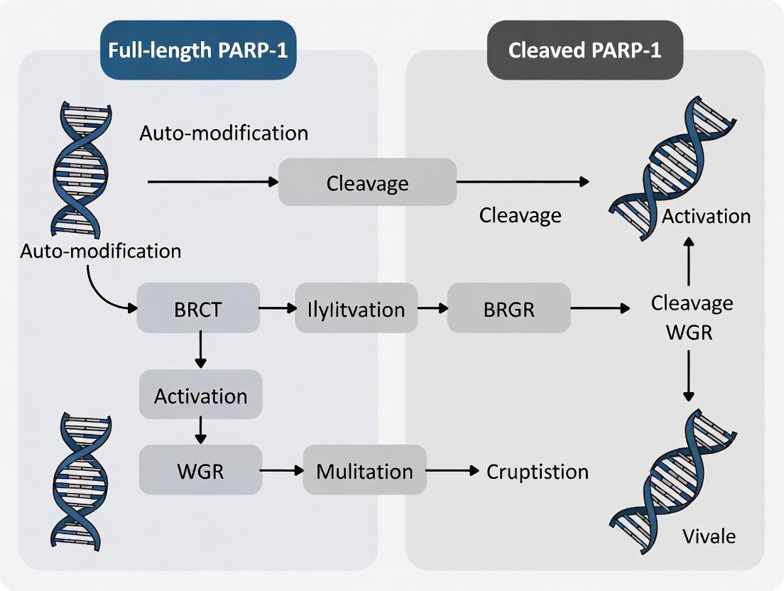
Visualization of PARP1 Domain Architecture and Apoptotic Cleavage
The domain structure of full-length PARP1 represents a sophisticated molecular machine that integrates DNA damage sensing with catalytic output through coordinated domain interactions. The caspase-mediated cleavage of PARP1 during apoptosis represents a fundamental reprogramming event that dismantles this integrated architecture, converting a DNA repair enzyme into a pro-apoptotic signaling system. Understanding these structural and functional relationships provides critical insights for developing PARP-targeted therapies, particularly for cancer treatment where PARP inhibitors have shown significant clinical success. The contrasting properties of full-length and cleaved PARP1 highlight the remarkable functional plasticity of this essential enzyme and its central role in determining cellular fate in response to stress signals.
In the molecular landscape of programmed cell death, the cleavage of poly(ADP-ribose) polymerase-1 (PARP-1) stands as a definitive biomarker of apoptosis. This proteolytic event, mediated primarily by effector caspases-3 and -7 at the highly specific DEVD214/G motif, represents a critical point of commitment to the apoptotic pathway. The recognition and cleavage at this site not only inactivates PARP-1's DNA repair functions but also generates fragments with distinct biological activities that differentiate them from the full-length protein. Within apoptosis research, understanding the functional consequences of this cleavage event provides crucial insights into cell fate decisions, as the generated fragments possess unique properties not shared by their full-length precursor. This technical guide examines the molecular mechanism of caspase-mediated PARP-1 cleavage, its functional consequences, and the experimental approaches for studying this fundamental apoptotic event.
Molecular Mechanism of Caspase Recognition and Cleavage
The DEVD214 Recognition Motif
Caspase-3 and caspase-7, as executioner caspases, recognize and cleave PARP-1 at the conserved amino acid sequence DEVD214↓G (where ↓ indicates the cleavage site) [9] [10]. This tetrapeptide sequence conforms to the canonical caspase recognition motif, with aspartic acid (D) residues at positions P1, P2, and P4, and valine (V) at position P3 relative to the cleavage site:
- P4: Aspartic acid (D)
- P3: Valine (V)
- P2: Aspartic acid (D)
- P1: Aspartic acid (D214) - Cleavage occurs C-terminal to this residue
- P1': Glycine (G) - First amino acid of the C-terminal fragment
The aspartic acid at the P1 position is absolutely required for caspase recognition, as caspases are cysteine proteases with strict specificity for cleavage after aspartic acid residues [11]. This recognition motif is situated within the nuclear localization signal (NLS) of PARP-1's DNA-binding domain, strategically positioning the cleavage event to alter the subcellular localization of the resulting fragments [10].
Structural Domains and Cleavage Products
PARP-1 comprises several functional domains that determine the differential functions of full-length versus cleaved PARP-1:
Table 1: PARP-1 Domains and Cleavage Products
| Domain/Feature | Location (AA) | Function | Location in Fragments |
|---|---|---|---|
| Zinc Finger 1 (ZnF1) | 1-97 | DNA damage recognition | 24 kDa fragment |
| Zinc Finger 2 (ZnF2) | 98-207 | DNA damage recognition | 24 kDa fragment |
| Nuclear Localization Signal | ~210-214 | Nuclear targeting | Contains cleavage site |
| DEVD214 Cleavage Site | 214-215 | Caspase-3/7 recognition | Between fragments |
| BRCT Domain | 385-479 | Protein-protein interactions | 89 kDa fragment |
| WGR Domain | 498-525 | DNA binding coordination | 89 kDa fragment |
| Catalytic Domain | 656-1014 | PAR synthesis activity | 89 kDa fragment |
Proteolytic cleavage at DEVD214 generates two primary fragments:
- 24 kDa N-terminal fragment (amino acids 1-214): Contains ZnF1 and ZnF2 domains with intact DNA-binding capability but lacking catalytic activity
- 89 kDa C-terminal fragment (amino acids 215-1014): Contains the BRCT domain, WGR domain, and catalytic domain [9] [4] [10]
This cleavage event occurs within the nuclear localization signal, explaining the differential subcellular localization of the resulting fragments post-cleavage [10].
Functional Consequences of PARP-1 Cleavage
Differential Functions: Full-length vs. Cleaved PARP-1
The biological consequences of PARP-1 cleavage extend far beyond simple inactivation of DNA repair, with the fragments acquiring novel functions distinct from the full-length protein:
Table 2: Functional Differences Between Full-length and Cleaved PARP-1
| Parameter | Full-length PARP-1 | 24 kDa Fragment | 89 kDa Fragment |
|---|---|---|---|
| Subcellular Localization | Nuclear | Nuclear | Cytoplasmic (after cleavage) |
| DNA Binding | Yes (via ZnF1/ZnF2) | Yes (retains ZnF1/ZnF2) | Impaired (lacks ZnF1/ZnF2) |
| Catalytic Activity | Fully active | None | Reduced but potentially functional |
| DNA Repair Role | Promotes repair | Dominant-negative inhibitor of repair | Limited involvement |
| Apoptotic Function | Anti-apoptotic (via DNA repair) | Pro-apoptotic (blocks repair) | Context-dependent (see below) |
| Novel Functions | Transcriptional regulation | - | Cytosolic PAR carrier; RNA Pol III regulation |
Novel Biological Activities of Cleavage Fragments
Recent research has revealed unexpectedly diverse functions for the PARP-1 cleavage fragments:
The 24 kDa fragment acts as a trans-dominant inhibitor of DNA repair by occupying DNA strand breaks and preventing recruitment of intact PARP-1 and other repair factors, thereby conserving cellular ATP pools during apoptosis [9] [10].
The 89 kDa fragment (tPARP1) translocates to the cytoplasm where it recognizes the RNA polymerase III (Pol III) complex via its BRCT domain and can catalyze mono-ADP-ribosylation of Pol III subunits, potentially facilitating IFN-β production during innate immune responses to foreign DNA [4].
Poly(ADP-ribosyl)ated 89 kDa fragments can serve as cytoplasmic PAR carriers that bind apoptosis-inducing factor (AIF), facilitating AIF release from mitochondria and its translocation to the nucleus, thereby promoting a caspase-independent cell death pathway known as parthanatos [12].
Differential effects on cell survival have been demonstrated, where expression of the 24 kDa fragment confers protection from ischemic damage in neuronal models, while the 89 kDa fragment exhibits cytotoxic properties and enhances NF-κB-mediated inflammatory responses [10].
Experimental Analysis of PARP-1 Cleavage
Detection Methodologies
The following experimental approaches are commonly employed to study PARP-1 cleavage in apoptosis research:
Western Blot Analysis
- Procedure: Separate protein extracts from apoptotic and control cells by SDS-PAGE (8-12% gel), transfer to PVDF membrane, and incubate with PARP-1 antibodies
- Antibody Selection: Use antibodies recognizing either the N-terminal (detects full-length and 24 kDa fragment) or C-terminal regions (detects full-length and 89 kDa fragment)
- Expected Results: Full-length PARP-1 (116 kDa) decreases during apoptosis with concomitant appearance of 89 kDa fragment; 24 kDa fragment may be less consistently detected due to rapid degradation or poor transfer
- Validation: Co-detection of caspase-3 cleavage (activation) and other apoptotic markers [9] [10]
Immunofluorescence and Microscopy
- Procedure: Fix cells, permeabilize, and stain with PARP-1 antibodies alongside nuclear markers; can combine with TUNEL staining for DNA fragmentation
- Key Observation: Redistribution of PARP-1 immunoreactivity from nuclear to cytoplasmic compartments during apoptosis
- Advanced Applications: Proximity ligation assays to study fragment interactions; colocalization studies with AIF or other binding partners [13] [12]
Flow Cytometry with Annexin V/PI Staining
- Procedure: Dual staining with Annexin V-FITC and propidium iodide to quantify apoptotic populations correlated with PARP-1 cleavage by Western blot
- Utility: Provides quantitative assessment of apoptosis progression in parallel with molecular cleavage events [4]
Research Reagent Solutions
Table 3: Essential Research Reagents for PARP-1 Cleavage Studies
| Reagent/Category | Specific Examples | Function/Application | Key Features |
|---|---|---|---|
| PARP-1 Antibodies | Anti-PARP-1 C-terminal, Anti-PARP-1 N-terminal, Cleaved PARP-1 (Asp214) specific | Detection of full-length and cleaved fragments by WB, IF | Species specificity, domain specificity |
| Caspase Inhibitors | Z-VAD-FMK (pan-caspase), Z-DEVD-FMK (caspase-3/7 specific) | Inhibit PARP-1 cleavage to establish causality | Cell-permeable, irreversible |
| Apoptosis Inducers | Staurosporine, Actinomycin D, Etoposide, Trail | Activate caspases to trigger PARP-1 cleavage | Various initiation pathways |
| PARP-1 Constructs | PARP-1 WT, PARP-1 UNCL (D214A), PARP-1 24kDa, PARP-1 89kDa | Functional studies of cleavage fragments | Non-cleavable mutant for control |
| Cell Lines | PARP-1-deficient 293T, SH-SY5Y, Primary cortical neurons | Loss-of-function and reconstitution studies | Genetic background control |
| Activity Assays | PAR ELISA, NAD+ consumption assays | Correlate cleavage with functional changes | Quantitative functional readouts |
Signaling Pathway Visualization
PARP-1 Cleavage in Apoptosis Pathway
Technical Protocols for Key Experiments
Induction and Detection of PARP-1 Cleavage
Staurosporine-Induced Apoptosis Protocol
- Cell Preparation: Plate cells at 60-70% confluence in appropriate growth medium 24 hours before treatment
- Treatment: Add staurosporine (0.5-2 μM final concentration) or actinomycin D (0.5-5 μM) directly to culture medium
- Time Course: Incubate for 2-8 hours at 37°C; include DMSO vehicle control
- Harvesting: Collect cells by trypsinization or direct scraping, followed by centrifugation at 500 × g for 5 minutes
- Lysis: Resuspend cell pellet in RIPA buffer (50 mM Tris-HCl pH 7.4, 150 mM NaCl, 1% NP-40, 0.5% sodium deoxycholate, 0.1% SDS) with protease and phosphatase inhibitors
- Western Blot: Separate 20-30 μg total protein on 8% SDS-PAGE gel, transfer to PVDF membrane, block with 5% non-fat milk, and incubate with anti-PARP-1 antibody (1:1000) overnight at 4°C
- Detection: Use HRP-conjugated secondary antibody (1:5000) and chemiluminescent substrate; expected results show full-length PARP-1 (116 kDa) decreasing with 89 kDa fragment increasing over time [12]
Subcellular Localization Assessment
Cellular Fractionation Protocol
- Harvesting: Collect 1-5 × 10⁶ cells by gentle scraping in PBS
- Plasma Membrane Lysis: Resuspend in hypotonic buffer (10 mM HEPES pH 7.9, 10 mM KCl, 1.5 mM MgCl₂, 0.5 mM DTT) with 0.1% IGEPAL CA-630, incubate 10 minutes on ice
- Centrifugation: Spin at 1000 × g for 5 minutes at 4°C; collect supernatant (cytoplasmic fraction)
- Nuclear Extraction: Resuspend pellet in high-salt buffer (20 mM HEPES pH 7.9, 420 mM NaCl, 1.5 mM MgCl₂, 0.2 mM EDTA, 25% glycerol), vortex vigorously, incubate 30 minutes on ice
- Clearance: Centrifuge at 20,000 × g for 10 minutes; collect supernatant (nuclear fraction)
- Analysis: Perform Western blot on both fractions using PARP-1 antibodies; validate fractionation with compartment-specific markers (e.g., Lamin B1 for nuclear, GAPDH for cytoplasmic)
- Expected Outcome: Full-length PARP-1 primarily nuclear; 89 kDa fragment appears in cytoplasmic fraction during apoptosis [4] [12]
Research Implications and Therapeutic Applications
The differential functions of full-length versus cleaved PARP-1 fragments extend beyond basic apoptosis research into therapeutic development:
Cancer Therapeutics: PARP inhibitors used in BRCA-deficient cancers may exert effects through modulation of both full-length and cleaved PARP-1 functions; understanding fragment biology could inform combination therapies [14]
Neuroprotection: The cytotoxic properties of the 89 kDa fragment suggest therapeutic targeting of this fragment in neurodegenerative conditions, while the protective 24 kDa fragment might offer neuroprotective strategies [10]
Inflammatory Regulation: Given the role of the 89 kDa fragment in enhancing NF-κB activity and inflammatory responses, fragment-specific inhibitors might provide novel anti-inflammatory approaches [10]
Viral Infection Response: The interaction between the 89 kDa fragment and RNA Pol III complex reveals potential antiviral mechanisms that could be harnessed therapeutically [4]
The DEVD214 cleavage site in PARP-1 represents not merely an inactivation mechanism but a critical molecular switch that converts a DNA repair enzyme into mediators of cell death with diverse biological functions. Continuing research into the differential roles of full-length and cleaved PARP-1 promises to reveal new insights into cell fate decisions and novel therapeutic opportunities across multiple disease contexts.
Poly(ADP-ribose) polymerase-1 (PARP-1) is a 116-kDa nuclear enzyme that plays a central role in detecting and repairing DNA damage. As a critical DNA damage sensor, PARP-1 becomes activated upon binding to DNA strand breaks, catalyzing the transfer of ADP-ribose units from NAD+ to target proteins, including itself, in a process known as poly(ADP-ribosyl)ation. This post-translational modification serves as a signal for the recruitment of DNA repair machinery. However, during apoptosis, PARP-1 undergoes proteolytic cleavage at the hands of executioner caspases, generating two principal fragments: a 24-kDa DNA-binding fragment and an 89-kDa catalytic fragment. This cleavage event represents a biochemical hallmark of apoptosis and serves to fundamentally alter PARP-1's function from DNA repair facilitator to apoptosis promoter. The distinct biological activities of these cleavage fragments have profound implications for cell fate decisions and represent a critical switching mechanism between survival and death pathways in response to cellular stress.
Structural Basis of PARP-1 Cleavage
Domain Architecture of Full-Length PARP-1
PARP-1 is a modular protein comprising several functionally specialized domains. The N-terminal region contains a DNA-binding domain (DBD) with three zinc finger motifs (ZnF1, ZnF2, and ZnF3) that recognize DNA strand breaks. The ZnF1 and ZnF2 domains are particularly crucial for detecting DNA damage, while ZnF3 contributes to inter-domain communication. The DBD is followed by a nuclear localization signal (NLS) and the caspase cleavage site (DEVD214), which is situated within this localization signal. The central automodification domain (AMD), also known as the BRCT domain, serves as the primary acceptor site for poly(ADP-ribose) chains and facilitates protein-protein interactions. The C-terminal region houses the catalytic domain (CAT), which contains the NAD+-binding site and is responsible for synthesizing poly(ADP-ribose) polymers.
Proteolytic Cleavage and Fragment Generation
During apoptosis, executioner caspases-3 and -7 recognize and cleave PARP-1 at the DEVD214 motif located between the DNA-binding domain and the automodification domain. This proteolytic event generates two distinct fragments with separate cellular fates and functions. The 24-kDa N-terminal fragment (amino acids 1-214) encompasses ZnF1, ZnF2, and the nuclear localization signal. The 89-kDa C-terminal fragment (amino acids 215-1014) contains ZnF3, the BRCT domain, the WGR domain, and the catalytic domain. This cleavage event fundamentally alters the properties, localization, and functions of the PARP-1 protein, redirecting its activity from DNA repair to facilitation of the apoptotic process.
Table 1: Structural Composition of PARP-1 Fragments
| Feature | 24-kDa Fragment | 89-kDa Fragment | Full-Length PARP-1 |
|---|---|---|---|
| Molecular Weight | 24 kDa | 89 kDa | 116 kDa |
| Domains Contained | ZnF1, ZnF2, NLS | ZnF3, BRCT, WGR, CAT | All domains (ZnF1-3, NLS, BRCT, WGR, CAT) |
| Caspase Cleavage Site | C-terminal end (DEVD214) | N-terminal end (DEVD214) | Intact (DEVD214) |
| Primary Localization | Nuclear | Cytosolic translocation | Nuclear |
| DNA Binding | High affinity | Greatly reduced | High affinity (activated by DNA breaks) |
| Catalytic Activity | None | Basal activity (DNA-independent) | DNA-dependent activation |
Functional Consequences of PARP-1 Cleavage
The 24-kDa DNA-Binding Fragment: A Trans-Dominant Inhibitor
The 24-kDa fragment retains the zinc finger motifs responsible for high-affinity DNA binding but lacks catalytic activity. This fragment remains tightly bound to DNA strand breaks in the nucleus, where it acts as a trans-dominant inhibitor of DNA repair. By occupying DNA damage sites, the 24-kDa fragment effectively blocks access for intact PARP-1 molecules and other DNA repair proteins to these sites. This competitive inhibition prevents the initiation of DNA repair processes that might otherwise impede the apoptotic program. Recent research has demonstrated that the 24-kDa fragment can also trans-dominantly inhibit PARP2, a closely related family member involved in DNA repair, thereby amplifying the shutdown of DNA repair activities during apoptosis. Furthermore, this fragment can serve as an acceptor for poly(ADP-ribosyl)ation, which reduces its DNA binding affinity and may provide a regulatory mechanism for its inhibitory function.
The 89-kDa Catalytic Fragment: A PAR Carrier in Cell Death
The 89-kDa fragment contains the automodification and catalytic domains but has dramatically reduced DNA binding capacity due to the loss of ZnF1 and ZnF2. While this fragment cannot be stimulated by DNA damage, it retains basal catalytic activity that is sufficient for limited poly(ADP-ribose) synthesis. Following cleavage, the 89-kDa fragment translocates from the nucleus to the cytoplasm, where it can participate in alternative cell death pathways. Notably, this fragment can function as a poly(ADP-ribose) carrier during apoptosis, shuttling PAR polymers to the cytoplasm. These PAR polymers can then bind to apoptosis-inducing factor (AIF), facilitating its release from mitochondria and subsequent translocation to the nucleus, where it contributes to large-scale DNA fragmentation. This pathway represents a crucial intersection between caspase-dependent apoptosis and AIF-mediated cell death.
Opposing Roles in Cell Survival and Death
Research examining the individual expression of these fragments reveals their opposing functions in cell fate decisions. Expression of the 24-kDa fragment confers protection against ischemic injury in neuronal models, similar to the effect observed with an uncleavable PARP-1 mutant. In contrast, expression of the 89-kDa fragment is explicitly cytotoxic and promotes cell death pathways. These differential effects extend to inflammatory responses, with the 89-kDa fragment increasing NF-κB activity and expression of pro-inflammatory genes like iNOS and COX-2, while the 24-kDa fragment has the opposite effect. This divergence highlights how PARP-1 cleavage generates fragments with antagonistic functions that collectively promote the apoptotic program while suppressing survival signals.
Experimental Analysis of PARP-1 Cleavage
Detecting PARP-1 Cleavage
The cleavage of PARP-1 is most commonly detected by Western blot analysis using antibodies that recognize different epitopes of the protein. Full-length PARP-1 migrates at approximately 116 kDa, while the cleavage fragments appear as 89-kDa and 24-kDa bands. Antibodies specific to the N-terminal region will detect both full-length PARP-1 and the 24-kDa fragment, whereas antibodies against the C-terminal region will detect full-length PARP-1 and the 89-kDa fragment. The appearance of the 89-kDa fragment is considered a biochemical hallmark of apoptosis and is widely used as an indicator of caspase activation in experimental systems.
Diagram 1: Experimental workflow for detecting PARP-1 cleavage fragments by Western blot.
Functional Assays for Fragment Activity
Several experimental approaches have been developed to characterize the distinct functions of the PARP-1 cleavage fragments:
- DNA Binding Assays: Electrophoretic mobility shift assays (EMSAs) and chromatin immunoprecipitation (ChIP) can evaluate the DNA-binding capacity of the 24-kDa fragment and its ability to compete with full-length PARP-1 for DNA damage sites.
- Catalytic Activity Measurements: In vitro PARylation assays using recombinant fragments and NAD+ as substrate can quantify the basal enzymatic activity of the 89-kDa fragment and its regulation by PAR polymers.
- Localization Studies: Immunofluorescence and subcellular fractionation techniques can track the nuclear retention of the 24-kDa fragment and the cytosolic translocation of the 89-kDa fragment following apoptosis induction.
- Cell Death Analysis: Expression vectors encoding individual fragments can be transfected into cells to assess their effects on viability under stress conditions, using assays such as MTT, Annexin V staining, or propidium iodide uptake.
Table 2: Key Experimental Reagents for PARP-1 Cleavage Research
| Reagent | Function/Application | Key Features |
|---|---|---|
| Caspase-3/7 Inhibitors (zVAD-fmk, DEVD-CHO) | Inhibit PARP-1 cleavage; validate caspase-dependence | Distinguish caspase-dependent apoptosis from other cell death forms |
| PARP Inhibitors (PJ34, ABT-888, Olaparib) | Block PARP catalytic activity; study fragment functions | Tool compounds for investigating PARP-1 enzymatic function |
| PARP-1 shRNA/siRNA | Knock down endogenous PARP-1; study fragment effects | Enable clean background for exogenous fragment expression studies |
| Cleavage-Specific Antibodies | Detect 89-kDa fragment specifically | Avoid cross-reactivity with full-length PARP-1 or other proteins |
| Recombinant PARP-1 Fragments | In vitro studies of fragment functions | Purified 24-kDa and 89-kDa fragments for biochemical assays |
| Uncleavable PARP-1 Mutant (PARP-1UNCL) | Study consequences of preventing cleavage | DEVD motif mutation prevents caspase cleavage |
Signaling Pathways Involving PARP-1 Cleavage Fragments
The cleavage of PARP-1 and the generation of its distinct fragments influence multiple cell death pathways through specific molecular mechanisms. The following diagram illustrates the key pathways involving these fragments during apoptotic cell death:
Diagram 2: Signaling pathways involving PARP-1 cleavage fragments in apoptosis.
Integration with Cell Death Pathways
PARP-1 cleavage represents a critical commitment point in apoptotic cell death, with the fragments executing distinct yet complementary functions. The 24-kDa fragment promotes apoptosis primarily through inhibition of DNA repair, preventing the potentially deleterious repair of DNA fragmentation that occurs during apoptosis. This ensures the irreversibility of the cell death process. Meanwhile, the 89-kDa fragment can engage in cross-talk with parthanatos, a PAR-mediated cell death pathway, through its function as a PAR carrier that facilitates AIF release from mitochondria. This dual mechanism—simultaneously blocking survival pathways while actively promoting death pathways—ensures efficient execution of apoptosis once the decision has been made.
Research Implications and Therapeutic Perspectives
The distinct functions of PARP-1 cleavage fragments have significant implications for both basic research and therapeutic development. From a research perspective, these fragments serve as valuable biomarkers for differentiating apoptosis from other forms of cell death, such as necrosis, which produces different PARP-1 cleavage patterns (e.g., a 50-kDa fragment generated by lysosomal proteases). The opposing functions of the fragments in cell survival and inflammatory responses suggest complex regulatory networks that fine-tune cellular responses to stress.
From a therapeutic standpoint, understanding fragment biology may inform drug development strategies. PARP inhibitors are already established in cancer therapy, particularly for tumors with BRCA mutations, but a more nuanced approach targeting specific fragment functions could expand therapeutic opportunities. For instance, strategies that modulate the balance between fragment activities might influence cell fate decisions in neurological disorders, ischemic conditions, or inflammatory diseases where PARP-1 plays a pathogenic role. The recent discovery that the 89-kDa fragment can regulate RNA polymerase III activity and innate immune responses further expands potential therapeutic applications into viral infections and autoimmune disorders.
The cleavage of PARP-1 into 24-kDa and 89-kDa fragments represents a critical biochemical event in apoptosis that effectively converts a DNA repair enzyme into a facilitator of cell death. These fragments execute distinct and opposing functions: the 24-kDa fragment acts as a trans-dominant inhibitor of DNA repair, while the 89-kDa fragment can propagate death signals through its catalytic activity and function as a PAR carrier. This division of labor ensures an efficient and irreversible commitment to apoptosis while preventing conflicting survival signals. Continued investigation into the biology of these fragments will undoubtedly yield new insights into cell death regulation and potentially uncover novel therapeutic strategies for a range of human diseases.
Poly(ADP-ribose) polymerase-1 (PARP-1) is a multifunctional nuclear enzyme central to maintaining genomic stability. During apoptosis, caspase-mediated cleavage of PARP-1 represents a critical biochemical event that serves as more than merely a hallmark of programmed cell death. This proteolytic processing generates distinct fragments that execute divergent biological functions: it inactivates the DNA repair capacity of full-length PARP-1 while simultaneously enabling novel signaling roles for the cleavage products. This review comprehensively examines the functional consequences of PARP-1 cleavage, contrasting the well-established DNA repair termination with emerging evidence of gained signaling functions that influence inflammatory responses, innate immunity, and cell fate decisions. Understanding this dualism provides crucial insights for therapeutic targeting in cancer and neurodegenerative diseases.
PARP-1 is a modular protein comprising several functional domains that dictate its cellular activities. The N-terminal region contains two zinc finger motifs (ZnF1 and ZnF2) that facilitate DNA damage recognition, followed by a third zinc finger (ZnF3) crucial for inter-domain communication and enzymatic activation [10] [15]. The central auto-modification domain (AMD), also known as the BRCT domain, serves as a target for poly(ADP-ribosyl)ation and mediates protein-protein interactions [16] [15]. The C-terminal region houses the catalytic domain (CAT), which transfers ADP-ribose units from NAD+ to target proteins [16].
During apoptosis, executioner caspases-3 and -7 cleave PARP-1 at the conserved DEVD214↓G motif, located between ZnF2 and ZnF3 [10] [15] [17]. This proteolysis generates two major fragments:
- A 24 kDa fragment containing the ZnF1 and ZnF2 DNA-binding domains
- An 89 kDa fragment (truncated PARP-1 or tPARP-1) containing ZnF3, AMD, and the catalytic domain [10] [15] [4]
Table 1: PARP-1 Domains and Cleavage Products
| Domain/Feature | Location | Function | Status in Fragments |
|---|---|---|---|
| Zinc Fingers 1 & 2 (ZnF1/2) | N-terminal (1-214) | DNA damage recognition | 24 kDa fragment |
| Zinc Finger 3 (ZnF3) | Adjacent to cleavage site | Inter-domain communication, enzymatic activation | 89 kDa fragment (tPARP1) |
| Auto-modification Domain (AMD/BRCT) | Central | Protein-protein interactions, auto-ribosylation | 89 kDa fragment (tPARP1) |
| Catalytic Domain (CAT) | C-terminal | Poly(ADP-ribose) polymerization | 89 kDa fragment (tPARP1) |
| Caspase Cleavage Site | DEVD214↓G | Caspase-3/7 recognition | Cleavage point |
Inactivation of DNA Repair Functions
The canonical consequence of PARP-1 cleavage is the termination of its DNA repair activities, primarily through two complementary mechanisms.
Separation of DNA-Binding from Catalytic Domains
Proteolytic cleavage physically separates the N-terminal DNA-binding domain (residing in the 24 kDa fragment) from the C-terminal catalytic domain (in the 89 kDa fragment) [15] [17]. This separation prevents PARP-1 from executing its coordinated response to DNA damage, where DNA binding triggers catalytic activation. The 24 kDa fragment, retaining the nuclear localization signal, remains tightly bound to DNA strand breaks but lacks catalytic activity [15]. This fragment acts as a trans-dominant inhibitor of DNA repair by occupying DNA damage sites and blocking access by intact PARP-1 and other repair enzymes [15].
Conservation of Cellular Energetics
Full-length PARP-1 activation consumes substantial NAD+ and ATP pools during massive DNA damage, potentially leading to energy depletion and necrotic cell death [18] [17]. PARP-1 cleavage prevents this "energy catastrophe" by terminating poly(ADP-ribose) synthesis [17]. Experimental evidence demonstrates that prevention of PARP-1 cleavage through mutation of the caspase site (PARP-1UNCL) increases cellular sensitivity to necrotic cell death, while cells expressing cleavable PARP-1 maintain ATP levels and execute apoptosis efficiently [17].
Initiation of Novel Signaling Roles
Emerging research reveals that PARP-1 cleavage fragments are not merely inert byproducts but actively participate in signaling pathways, potentially representing an evolutionary conservation of functions observed in lower organisms where PARP-1 naturally lacks N-terminal zinc fingers [4].
Regulation of Inflammatory Responses via NF-κB
PARP-1 cleavage products differentially modulate NF-κB activity and subsequent inflammatory signaling. Research demonstrates that the 89 kDa fragment (tPARP-1) enhances NF-κB activation and increases expression of NF-κB-dependent genes such as inducible nitric oxide synthase (iNOS) and cyclooxygenase-2 (COX-2) [10]. In contrast, the 24 kDa fragment and uncleavable PARP-1 (PARP-1UNCL) suppress inflammatory signaling and upregulate anti-apoptotic proteins like Bcl-xL [10]. These findings establish PARP-1 cleavage as a molecular switch that modulates the inflammatory response during cell death.
Cytosolic Signaling in Innate Immunity
A groundbreaking discovery reveals that the 89 kDa tPARP-1 translocates to the cytoplasm during apoptosis and interacts with the RNA polymerase III (Pol III) complex [4]. Through its BRCT domain, tPARP-1 recognizes and mono-ADP-ribosylates Pol III, enhancing its ability to transcribe foreign DNA and potentiate interferon-β (IFN-β) production [4]. This mechanism connects PARP-1 cleavage to innate immune responses during cellular stress and pathogen challenge.
Diagram 1: PARP-1 Cleavage and Resulting Signaling Pathways. Caspase-mediated cleavage generates fragments with distinct signaling functions.
Opposing Effects on Cell Survival and Death
The dual consequences of PARP-1 cleavage create a sophisticated regulatory mechanism that influences cellular fate decisions.
Differential Impact on Cell Viability
Experimental models using oxygen/glucose deprivation (OGD) to simulate ischemic conditions demonstrate that expression of uncleavable PARP-1 (PARP-1UNCL) or the 24 kDa fragment provides significant protection from cell death [10]. Conversely, expression of the 89 kDa fragment (tPARP-1) is explicitly cytotoxic and enhances cell death susceptibility [10]. This differential effect underscores the opposing functions of PARP-1 cleavage products in survival signaling.
Molecular Switch Between Apoptosis and Necrosis
PARP-1 cleavage acts as a molecular switch between apoptotic and necrotic cell death pathways [17]. When PARP-1 remains intact and active during death receptor signaling (e.g., TNF stimulation), it depletes cellular ATP reserves, forcing cells toward necrosis [17]. In contrast, caspase-mediated cleavage of PARP-1 during apoptosis conserves cellular energy, enabling the ATP-dependent execution of apoptotic programmed cell death [17].
Table 2: Functional Comparison of PARP-1 Forms in Cell Survival
| PARP-1 Form | Impact on Cell Viability | NF-κB Activity | DNA Repair Capacity | Key Downstream Effects |
|---|---|---|---|---|
| Full-length PARP-1 | Context-dependent | Baseline | Fully functional | DNA repair, energy consumption |
| Uncleavable PARP-1 (PARP-1UNCL) | Cytoprotective | Decreased | Functional (no termination) | Increased Bcl-xL, decreased iNOS/COX-2 |
| 24 kDa Fragment | Cytoprotective | Decreased | Inhibitory (dominant-negative) | Blocks DNA repair complexes |
| 89 kDa Fragment (tPARP1) | Cytotoxic | Increased | None | Increased iNOS/COX-2, activates Pol III, enhances IFN-β |
Experimental Approaches and Research Toolkit
Investigating PARP-1 cleavage requires specific methodological approaches and research tools.
Key Experimental Models
- In vitro ischemia models: Oxygen/glucose deprivation (OGD) with restoration in neuronal cell lines (SH-SY5Y) and primary cortical neurons [10]
- Apoptosis induction: Poly(dA-dT) transfection to mimic cytosolic DNA sensing, death receptor activation (TNF, CD95), and genotoxic agents [17] [4]
- Genetic constructs: Expression vectors for PARP-1 variants (wild-type, uncleavable PARP-1D214N, 24 kDa, 89 kDa fragments) [10] [17] [4]
Essential Research Reagents
Table 3: Key Research Reagents for PARP-1 Cleavage Studies
| Reagent / Method | Function / Application | Experimental Utility |
|---|---|---|
| Caspase inhibitors (zVAD-fmk) | Pan-caspase inhibitor | Distinguishes caspase-dependent vs independent cleavage [17] |
| PARP-1UNCL (D214N mutant) | Caspase-resistant PARP-1 | Studying consequences of prevented cleavage [10] [17] |
| siRNA-PARP-1 | Knockdown of endogenous PARP-1 | Background reduction for transfection studies [10] |
| Anti-PARP-1 antibodies | Cleavage detection (full-length vs 89 kDa) | Apoptosis assessment, Western blot analysis [10] [4] |
| 3-aminobenzamide (3-AB) | PARP catalytic inhibitor | Assessing enzymatic vs scaffolding functions [19] [17] |
| Annexin V/PI staining | Apoptosis quantification | Flow cytometry-based cell death measurement [4] |
Diagram 2: Experimental Workflow for PARP-1 Cleavage Studies. Key methodological approaches for investigating PARP-1 cleavage consequences.
Discussion and Therapeutic Implications
The dual functional consequences of PARP-1 cleavage represent an elegant evolutionary adaptation that optimizes cell fate decisions during stress. The inactivation of DNA repair conserves cellular resources and prevents aberrant survival of damaged cells, while the acquired signaling functions potentially alert neighboring cells to danger and coordinate tissue-level responses.
Therapeutically, understanding these mechanisms provides opportunities for targeted interventions. In neurodegenerative diseases where excessive PARP-1 activation contributes to pathology, promoting appropriate cleavage may mitigate energy depletion [18] [2]. Conversely, in cancer therapy, manipulating the balance between PARP-1's DNA repair and signaling functions could enhance the efficacy of genotoxic treatments [19] [16] [20].
The discovery of tPARP-1's role in innate immunity through Pol III activation [4] further expands potential therapeutic applications in viral diseases and inflammation. Future research should focus on precisely mapping the structural determinants of these gained functions and developing compounds that can selectively modulate specific PARP-1 fragments without affecting others.
PARP-1 cleavage during apoptosis initiates a sophisticated functional transition from DNA damage responder to signaling modulator. The 24 kDa and 89 kDa fragments execute distinct biological programs that collectively influence cellular fate, inflammatory responses, and immune signaling. This paradigm shift in understanding PARP-1 biology—from a DNA repair enzyme to a multifaceted signaling regulator—opens new avenues for therapeutic innovation in cancer, neurodegeneration, and inflammatory diseases. The contrasting functions of PARP-1 cleavage products exemplify the complex economy of cellular signaling, where proteolytic processing converts one functional entity into multiple effectors with distinct, and sometimes opposing, biological activities.
PARP-1 Cleavage as a Hallmark Biochemical Biomarker of Apoptosis
Poly (ADP-ribose) polymerase-1 (PARP-1), also known as ARTD1, is a nuclear enzyme that functions as a primary DNA damage sensor and facilitates DNA repair through poly(ADP-ribosyl)ation (PARylation) of nuclear acceptor proteins [9] [12]. Beyond its DNA repair functions, PARP-1 participates in transcription, inflammation, and learning and memory [9]. However, PARP-1's role as a definitive biomarker for apoptosis emerges through its specific proteolytic cleavage by activated cell death proteases. During apoptosis, PARP-1 is a preferred substrate for caspase proteases, and its cleavage is considered a hallmark biochemical event that distinguishes apoptotic from necrotic cell death [9] [21] [17]. This proteolytic inactivation prevents excessive NAD+ and ATP consumption, facilitating the apoptotic process [17]. The cleavage of PARP-1 generates specific signature fragments—a 24-kDa DNA-binding fragment and an 89-kDa catalytic fragment—whose functions extend beyond the mere inactivation of DNA repair [9] [10] [12]. This technical guide examines the biochemical nature, functional consequences, and research applications of PARP-1 cleavage, framing it within the critical distinction between full-length and cleaved PARP-1 in apoptosis research.
Structural Biology of PARP-1 and Cleavage Sites
Domain Architecture of Full-Length PARP-1
PARP-1 is a modular protein comprising several functional domains:
- DNA-Binding Domain (DBD): A 46-kD N-terminal domain containing two zinc finger motifs that confer high-affinity binding to DNA strand breaks, cruciforms, and nucleosomes [9]. A third zinc finger motif, located between the second zinc finger and the auto-modification domain, is vital for inter-domain interactions and enzymatic action [9].
- Auto-Modification Domain (AMD): A 22-kD central domain containing a BRCT fold (a motif found in many DNA repair proteins) that functions as a target for covalent auto-modification and facilitates protein-protein interactions [9].
- Catalytic Domain (CD): A 54-kD C-terminal domain that polymerizes linear or branched ADP-ribose units from NAD+ onto target proteins [9].
PARP-1 is an abundant nuclear enzyme with approximately 1-2 million copies per cell, accounting for ~85% of total cellular PARP activity [9].
Caspase Cleavage Site and Fragment Generation
The primary cleavage site for apoptotic caspases (caspase-3 and -7) is located within the DEVD214 motif in the DBD, specifically situated within a nuclear localization signal (NLS) [10]. Cleavage at this site separates the N-terminal DNA-binding domain from the C-terminal catalytic domain, generating two primary fragments:
- 24-kDa Fragment: Contains the two N-terminal zinc-finger DNA-binding motifs and the NLS. It remains tightly bound to DNA strand breaks in the nucleus [9] [10].
- 89-kDa Fragment: Contains the third zinc finger, BRCT domain, WGR domain, and the C-terminal catalytic domain [9] [4]. This fragment translocates to the cytoplasm under certain conditions [12].
Table 1: PARP-1 Fragments Generated by Caspase-Mediated Cleavage
| Fragment | Molecular Weight | Domains Contained | Cellular Localization Post-Cleavage | Primary Functions |
|---|---|---|---|---|
| Full-Length PARP-1 | 116-kDa | DBD (ZnF1, ZnF2, ZnF3), AMD, CD | Nucleus | DNA damage sensing, DNA repair, transcription regulation |
| 24-kDa Fragment | 24-kDa | DBD (ZnF1, ZnF2) | Nuclear | Acts as trans-dominant inhibitor of PARP-1; occupies DNA breaks |
| 89-kDa Fragment | 89-kDa | ZnF3, BRCT, WGR, CD | Cytoplasmic (translocates) | Serves as PAR carrier; induces AIF-mediated apoptosis; ADP-ribosylates cytoplasmic targets |
Diagram 1: Caspase-mediated cleavage of PARP-1. Caspase-3/7 cleaves full-length PARP-1 at the DEVD214 site, separating the 24-kDa DNA-binding fragment from the 89-kDa fragment containing the catalytic domain.
Functional Consequences of PARP-1 Cleavage
Classical Model: Inactivation of DNA Repair
The traditional understanding of PARP-1 cleavage centers on the strategic inactivation of DNA repair to facilitate apoptotic execution:
- The 24-kDa fragment retains the DNA-binding zinc fingers and acts as a trans-dominant inhibitor by irreversibly binding to DNA strand breaks, thereby blocking access for DNA repair enzymes including intact PARP-1 [9].
- The 89-kDa catalytic fragment displays significantly reduced DNA binding capacity and is liberated from the nucleus into the cytosol [9].
- This cleavage event conserves cellular ATP pools by preventing PARP-1 hyperactivation and consequent NAD+ depletion, which would otherwise shift cell death toward necrosis [9] [17].
Emerging Paradigms: Gain-of-Function for Cleavage Fragments
Recent research reveals that PARP-1 fragments are not merely inactive degradation products but acquire novel signaling functions:
The 89-kDa Fragment as a Cytoplasmic PAR Carrier in Parthanatos
The 89-kDa fragment, when poly(ADP-ribosyl)ated, can translocate to the cytoplasm and function as a PAR carrier that induces apoptosis-inducing factor (AIF) release from mitochondria [12] [22]. This cascade, termed parthanatos, represents a caspase-independent programmed cell death pathway distinct from both apoptosis and necrosis [12]. AIF binding to PAR attached to the 89-kDa PARP-1 fragment facilitates its translocation to the nucleus, culminating in chromatin condensation and large-scale DNA fragmentation [12] [22].
Truncated PARP-1 in Innate Immune Signaling
The 89-kDa truncated PARP-1 (tPARP1) recognizes and mono-ADP-ribosylates the RNA polymerase III (Pol III) complex in the cytosol during poly(dA-dT)-stimulated apoptosis [4]. This ADP-ribosylation facilitates IFN-β production and enhances apoptosis during innate immune responses to foreign DNA [4]. The interaction is mediated by the BRCT domain of tPARP1 [4].
Regulation of NF-κB Activity
PARP-1 cleavage fragments differentially modulate inflammatory responses:
- The 89-kDa fragment increases NF-κB activity and expression of downstream pro-inflammatory genes (iNOS, COX-2) while decreasing anti-apoptotic Bcl-xL expression [10].
- In contrast, the 24-kDa fragment and uncleavable PARP-1 decrease iNOS and COX-2 while increasing Bcl-xL, conferring a protective phenotype in ischemic models [10].
Table 2: Functional Differences Between Full-Length and Cleaved PARP-1
| Functional Aspect | Full-Length PARP-1 | 24-kDa Fragment | 89-kDa Fragment |
|---|---|---|---|
| DNA Repair | Promotes base excision and single-strand break repair | Inhibits DNA repair by blocking DNA ends | Catalytically impaired in DNA repair context |
| Energy Metabolism | Can deplete NAD+/ATP upon overactivation | Prevents energy depletion | Limited impact on energy stores |
| Cell Death Regulation | Can promote either apoptosis or necrosis based on activation level | Cytoprotective in ischemia models | Cytotoxic; promotes AIF-mediated parthanatos |
| Inflammatory Response | Cofactor for NF-κB; moderate activity | Decreases NF-κB activity and pro-inflammatory genes | Increases NF-κB activity and pro-inflammatory genes |
| Innate Immunity | Limited role in cytoplasmic DNA sensing | Not involved | Activates Pol-III-dependent IFN-β production |
Detection Methodologies and Experimental Protocols
Western Blot Analysis for PARP-1 Cleavage
Principle: Separation of full-length and cleaved PARP-1 fragments via SDS-PAGE followed by immunodetection.
Key Reagents:
- Antibodies: Primary antibodies recognizing either the N-terminal (detects full-length and 24-kDa fragment) or C-terminal regions (detects full-length and 89-kDa fragment) of PARP-1. Cleavage-specific antibodies that exclusively recognize the neo-epitopes created by caspase cleavage are also available.
- Cell Lysis Buffer: RIPA buffer or similar, supplemented with protease inhibitors and caspase inhibitors to prevent post-lysis cleavage.
Protocol:
- Induce apoptosis in cells using appropriate stimuli (e.g., staurosporine, actinomycin D, etoposide).
- Harvest cells at various time points and lyse in appropriate buffer.
- Separate proteins (20-50 μg per lane) on 8-10% SDS-PAGE gels.
- Transfer to PVDF or nitrocellulose membranes.
- Block with 5% non-fat milk or BSA in TBST.
- Incubate with primary anti-PARP-1 antibody (1:1000 dilution) overnight at 4°C.
- Incubate with HRP-conjugated secondary antibody (1:2000-5000) for 1 hour at room temperature.
- Detect using enhanced chemiluminescence substrate.
- Expected Results: Apoptotic samples show decreased full-length PARP-1 (116-kDa) with corresponding appearance of the 89-kDa fragment. The 24-kDa fragment may be less consistently detected depending on the antibody used.
PAR Immunoassay for Target Engagement
Principle: Measures poly(ADP-ribose) (PAR) levels as an indicator of PARP-1 enzymatic activity.
Applications:
- Pharmacodynamic Studies: Evaluating target engagement of PARP inhibitors in clinical trials [23].
- Cell Death Mode Determination: High PAR levels indicate PARP-1 activation (often associated with necrosis), while PARP-1 cleavage in apoptosis reduces PAR levels.
Protocol Considerations:
- Sample Types: Tumor biopsies, PBMCs, tissue homogenates.
- Technical Challenges: PAR levels in human biopsies are often lower than in xenograft models, requiring sensitivity optimization [23].
- Controls: Essential to include PAR polymer standards and assay controls for cross-laboratory reproducibility [23].
Multiparameter Apoptosis Assays
Combined Approaches:
- Annexin V/PI Staining with PARP-1 Cleavage: Correlate externalization of phosphatidylserine with PARP-1 cleavage.
- γH2AX and PARP-1 Cleavage: Detect DNA double-strand breaks alongside apoptotic signaling.
- Caspase Activity Assays with PARP-1 Cleavage: Measure caspase-3/7 activity concurrently with PARP-1 cleavage.
Diagram 2: Experimental workflow for detecting PARP-1 cleavage. Multiple complementary methods are used to confirm apoptosis and analyze cell death pathways.
The Scientist's Toolkit: Essential Research Reagents
Table 3: Key Research Reagents for PARP-1 Cleavage Studies
| Reagent Category | Specific Examples | Research Application | Technical Considerations |
|---|---|---|---|
| PARP-1 Antibodies | Anti-N-terminal, Anti-C-terminal, Cleavage-specific | Western blot, Immunofluorescence, IHC | Region-specific antibodies distinguish full-length from fragments; cleavage-specific antibodies provide highest specificity |
| Caspase Inhibitors | zVAD-fmk (pan-caspase), DEVD-CHO (caspase-3/7) | Inhibiting PARP-1 cleavage to study alternative functions | zVAD can potentiate TNF-induced necrosis by preventing PARP-1 cleavage [17] |
| PARP Inhibitors | Veliparib, Olaparib, 3-aminobenzamide (3-AB) | Studying PARP-1 enzymatic function; cancer therapy | PAR immunoassay used to measure target engagement in clinical trials [23] |
| Apoptosis Inducers | Staurosporine, Actinomycin D, Etoposide, Anti-FAS | Inducing caspase-dependent PARP-1 cleavage | Different inducers may activate distinct apoptotic pathways |
| PAR Detection Reagents | Anti-PAR antibody, PAR polymer standards | Measuring PARP-1 enzymatic activity | Commercial kits available (e.g., Trevigen); HPLC used for PAR standard characterization [23] |
| Cell Lines | PARP-1-deficient cells, Non-cleavable PARP-1 mutants (D214N) | Functional studies of cleavage fragments | Uncleavable PARP-1 confers protection in some ischemia models [10] [17] |
| Vectors | PARP-1WT, PARP-124, PARP-189 expression constructs | Studying individual fragment functions | Fragment expression reveals opposing roles in cell viability [10] |
Research Applications and Clinical Implications
Basic Research Applications
PARP-1 cleavage serves as a fundamental biomarker for:
- Apoptosis Quantification: The appearance of the 89-kDa fragment provides a sensitive, early marker of apoptotic commitment.
- Cell Death Mechanism Discrimination: Distinguishes caspase-dependent apoptosis (PARP-1 cleavage) from caspase-independent parthanatos (PARP-1 activation and PAR translocation) and necrosis (PARP-1 activation without cleavage) [12] [17].
- Therapeutic Screening: Evaluating efficacy of pro-apoptotic agents in cancer research and neurodegenerative disease models.
Clinical and Translational Applications
Cancer Therapeutics
- PARP Inhibitors in Oncology: PARP inhibitors (e.g., olaparib, veliparib) are approved for BRCA-mutant cancers, exploiting synthetic lethality [24]. PAR immunoassays monitor target engagement in clinical trials [23].
- Biomarker for Treatment Response: PARP-1 cleavage in tumor biopsies may indicate effective apoptosis induction by chemotherapeutic agents.
- Melanoma and Other Cancers: High PARP-1 expression correlates with aggressive melanoma characteristics and poor patient outcomes, making it a potential prognostic marker and therapeutic target [24].
Neurodegenerative Diseases and Ischemic Injury
- Neuroprotection: PARP inhibition attenuates injury in cerebral ischemia, trauma, and excitotoxicity, demonstrating PARP-1's central role in these pathologies [9] [10].
- Inflammatory Regulation: The differential effects of PARP-1 fragments on NF-κB activity suggest therapeutic opportunities for modulating inflammation in neurological disorders [10].
PARP-1 cleavage remains a cornerstone biomarker of apoptosis with expanding functional significance beyond its classical role in DNA repair inactivation. The distinct functions of the 24-kDa and 89-kDa fragments—from trans-dominant inhibition of DNA repair to cytoplasmic PAR carrier functions and immune signaling—reveal a complex regulatory network centered on PARP-1 proteolysis. The emerging paradigm recognizes these fragments as active signaling molecules with specific roles in cell fate decisions, rather than mere inert byproducts of caspase activity. For researchers and drug development professionals, understanding the nuanced functions of full-length versus cleaved PARP-1 provides critical insights for developing targeted therapies, interpreting mechanistic studies of cell death, and advancing biomarker applications in clinical oncology and neurology. The continued investigation of PARP-1 cleavage signatures promises to yield novel therapeutic strategies for cancer, neurodegenerative conditions, and inflammatory disorders where regulated cell death pathways are disrupted.
Detecting the Switch: Techniques for PARP-1 Cleavage Analysis in Research and Diagnostics
Poly(ADP-ribose) polymerase-1 (PARP-1) is a 116 kDa nuclear enzyme that plays a critical role in DNA damage repair and maintenance of genomic integrity [10] [9]. During the early stages of apoptosis, PARP-1 serves as a primary substrate for executioner caspases-3 and -7, which cleave the full-length protein at the conserved aspartic acid residue 214 within the DEVD sequence [9] [25]. This proteolytic cleavage generates two characteristic signature fragments: a 24 kDa DNA-binding domain fragment and an 89 kDa catalytic domain fragment [10] [26]. The detection of these fragments via Western blotting has become a gold standard biochemical marker for identifying apoptotic cells in research and drug development. This cleavage event serves to inactivate PARP-1's DNA repair function, conserving cellular energy for the systematic execution of the apoptotic program [9] [27]. This technical guide provides detailed methodologies for reliably detecting these signature fragments, framed within the critical distinction between full-length and cleaved PARP-1 in apoptosis research.
Biological Significance of PARP-1 Cleavage
Functional Consequences of Cleavage
The cleavage of PARP-1 represents more than just a biomarker; it signifies a fundamental shift in cellular fate. The 24 kDa fragment, containing the nuclear localization signal and zinc finger DNA-binding domains, remains tightly bound to DNA strand breaks. This binding acts as a trans-dominant inhibitor of the BER pathway by blocking access to DNA damage sites for other repair factors [9] [26]. Meanwhile, the 89 kDa fragment, comprising the automodification and catalytic domains, translocates to the cytoplasm where it can participate in novel signaling functions [26] [4].
Recent research has revealed that the 89 kDa fragment can serve as a poly(ADP-ribose) (PAR) carrier to the cytoplasm, where it facilitates apoptosis-inducing factor (AIF) release from mitochondria—a critical step in certain cell death pathways [26] [12]. Additionally, this fragment can mono-ADP-ribosylate RNA polymerase III in the cytosol, potentially linking apoptosis to innate immune responses through IFN-β production [4]. These findings underscore that PARP-1 cleavage fragments are not merely inert byproducts but active participants in coordinating cell death.
Table 1: Characteristics of Full-Length PARP-1 and Its Cleavage Fragments
| Form | Molecular Weight | Domains Present | Localization | Primary Function |
|---|---|---|---|---|
| Full-Length PARP-1 | 116 kDa | DNA-binding, Automodification, Catalytic | Nuclear | DNA damage repair, transcriptional regulation |
| 24 kDa Fragment | 24 kDa | DNA-binding domain (Zn fingers 1 & 2) | Nuclear | Dominant-negative inhibitor of DNA repair |
| 89 kDa Fragment | 89 kDa | Automodification domain, Catalytic domain | Cytoplasmic | PAR carrier, AIF-mediated apoptosis, RNA Pol III regulation |
PARP-1 Cleavage in Different Cell Death Paradigms
The role of PARP-1 cleavage varies significantly across different cell death modalities. In caspase-dependent apoptosis, cleavage serves to inactivate DNA repair and facilitate cellular dismantling [9] [26]. However, in parthanatos—a caspase-independent programmed cell death pathway—PARP-1 overactivation leads to substantial PAR synthesis without cleavage, resulting in energy depletion and AIF-mediated DNA fragmentation [26] [12]. This distinction is crucial for researchers interpreting Western blot results, as the presence or absence of cleavage fragments can help differentiate between cell death mechanisms.
Antibody Selection and Experimental Design
Critical Reagents for Detection
The cornerstone of successful PARP-1 cleavage detection is appropriate antibody selection. Antibodies such as Cell Signaling Technology's PARP Antibody #9542 are specifically validated to detect endogenous levels of full-length PARP-1 (116 kDa) and the large cleavage fragment (89 kDa) [25]. This antibody was generated using a synthetic peptide corresponding to the caspase cleavage site in human PARP-1, making it ideal for detecting the cleavage event [25].
For researchers requiring detection of the 24 kDa fragment, antibodies targeting the N-terminal DNA-binding domain are necessary. It is critical to verify species reactivity for the model system being studied, with most commercial antibodies validated for human, mouse, and rat samples [25].
Table 2: Essential Research Reagents for PARP-1 Cleavage Detection
| Reagent | Specification | Function/Application |
|---|---|---|
| PARP Antibody #9542 | Rabbit monoclonal, detects 116 kDa and 89 kDa forms [25] | Primary antibody for Western blotting |
| Caspase-3 | Activated executioner caspase | PARP-1 cleavage enzyme; activity can be measured to confirm apoptosis |
| Staurosporine | 0.1-1 μM for 4-24 hours [26] | Apoptosis inducer; positive control for cleavage |
| PJ34 or ABT-888 | PARP inhibitors (e.g., 10 μM) [26] | Negative controls for PARP-1 activation |
| zVAD-fmk | Pan-caspase inhibitor (e.g., 20-50 μM) [26] | Inhibits PARP-1 cleavage; confirms caspase-dependence |
Experimental Controls and Design
Robust experimental design requires appropriate controls to accurately interpret PARP-1 cleavage data. Essential controls include:
- Untreated cells: Baseline full-length PARP-1 expression
- Apoptosis-positive control: Cells treated with known inducers (staurosporine, actinomycin D, or other relevant compounds) [26] [12]
- Caspase inhibitor control: Cells pre-treated with zVAD-fmk followed by apoptosis inducer to confirm caspase-dependent cleavage [26]
- PARP inhibitor control: Cells treated with PARP inhibitors to distinguish between different PARP-1-mediated cell death pathways [26]
Time-course experiments are particularly valuable, as PARP-1 cleavage typically precedes other late apoptotic markers. Sampling at 0, 2, 4, 6, 8, and 24 hours post-treatment can capture the dynamics of cleavage in response to various stimuli.
Detailed Western Blot Methodology
Sample Preparation and Electrophoresis
Cell Lysis and Protein Extraction
- Harvest cells by gentle scraping or trypsinization
- Wash twice with cold PBS
- Lyse cells in RIPA buffer (150 mM NaCl, 1% NP-40, 0.5% sodium deoxycholate, 0.1% SDS, 50 mM Tris, pH 8.0) supplemented with:
- Protease inhibitors (e.g., 1 mM PMSF, 1 μg/mL aprotinin, 1 μg/mL leupeptin)
- Phosphatase inhibitors (e.g., 1 mM NaF, 1 mM Na₃VO₄)
- PARP inhibitor (optional, to prevent automodification during extraction)
- Incubate on ice for 30 minutes with occasional vortexing
- Centrifuge at 14,000 × g for 15 minutes at 4°C
- Transfer supernatant to fresh tubes and determine protein concentration using BCA assay
Gel Electrophoresis
- Prepare 4-12% Bis-Tris gradient gels for optimal separation of both high and low molecular weight fragments
- Load 20-50 μg of total protein per lane
- Include pre-stained protein molecular weight markers spanning 20-120 kDa
- Run gel in MOPS or MES buffer at 100-150V for 60-90 minutes
Membrane Transfer and Immunodetection
Transfer Conditions
- Transfer proteins to PVDF membrane using wet or semi-dry transfer systems
- For simultaneous detection of all fragments (116 kDa, 89 kDa, and 24 kDa), transfer at 100V for 60-90 minutes at 4°C
- Confirm transfer efficiency with Ponceau S staining
Blocking and Antibody Incubation
- Block membrane with 5% non-fat dry milk or BSA in TBST for 1 hour at room temperature
- Incubate with primary antibody diluted in blocking buffer:
- PARP Antibody #9542 at 1:1000 dilution [25]
- Incubate overnight at 4°C with gentle agitation
- Wash membrane 3 × 10 minutes with TBST
- Incubate with HRP-conjugated secondary antibody (1:2000-1:5000) for 1 hour at room temperature
- Wash 3 × 10 minutes with TBST
Detection and Optimization
- Develop blots using enhanced chemiluminescence (ECL) substrate
- For weak signals, use high-sensitivity ECL substrates
- Optimize exposure times to avoid saturation, particularly for the strong 89 kDa fragment signal
- Stripping and re-probing for loading controls (β-actin, GAPDH, or histone H3) is recommended
Troubleshooting and Data Interpretation
Common Challenges and Solutions
Weak or No Signal
- Confirm antibody specificity and expiration date
- Optimize protein loading concentration
- Test different blocking agents (BSA vs. non-fat milk)
- Increase primary antibody incubation time
High Background
- Increase number and duration of washes
- Optimize blocking conditions (concentration and duration)
- Titrate primary antibody to find optimal dilution
Incomplete Transfer of Fragments
- For simultaneous detection of all fragments, verify transfer efficiency using reversible protein stains
- Consider transferring for longer durations or using pre-cut gels to separately optimize transfer conditions for different molecular weight ranges
Quantitative Analysis
Densitometric analysis of Western blot bands allows for quantification of PARP-1 cleavage:
- Calculate cleavage ratio as: 89 kDa band intensity / (116 kDa + 89 kDa band intensities)
- Normalize all values to loading controls
- Express results as fold-change compared to untreated controls
- Statistical analysis should include at least three independent experiments
Advanced Applications and Integrated Approaches
Subcellular Localization Studies
The distinct subcellular localization of PARP-1 fragments enables more sophisticated experimental designs. Fractionation studies separating nuclear and cytoplasmic components can provide additional validation of apoptosis. The 24 kDa fragment remains nuclear, bound to DNA damage sites, while the 89 kDa fragment translocates to the cytoplasm [26] [4]. This translocation can be visualized using complementary techniques such as immunofluorescence or cell fractionation followed by Western blotting.
Correlative Assays
To strengthen conclusions about apoptotic engagement, PARP-1 cleavage should be correlated with other apoptotic markers:
- Caspase-3/7 activation assays
- Annexin V/propidium iodide staining by flow cytometry [4]
- DNA fragmentation analysis (TUNEL assay)
- Mitochondrial membrane potential assessment
Visualizing PARP-1 Cleavage and Apoptotic Signaling
The following diagram illustrates the process of PARP-1 cleavage during apoptosis and the subsequent functions of its signature fragments:
PARP-1 Cleavage in Apoptosis
The experimental workflow for detecting PARP-1 cleavage fragments is summarized below:
PARP Cleavage Detection Workflow
The detection of PARP-1 cleavage fragments via Western blotting remains an essential technique for apoptosis research and drug development. The 89 kDa and 24 kDa fragments serve as critical biomarkers that distinguish between functional PARP-1 in DNA repair and its inactivation during programmed cell death. By following the detailed protocols outlined in this guide—including proper antibody selection, controlled experimental conditions, and appropriate data interpretation—researchers can reliably detect and quantify these signature fragments. The continuing discovery of novel functions for these fragments, particularly the cytoplasmic roles of the 89 kDa fragment, underscores the importance of this assay in understanding cell death mechanisms and evaluating therapeutic efficacy in cancer treatment and other disease models.
Immunohistochemistry and Immunofluorescence for Spatial Localization of Fragments
In apoptosis research, the cleavage of full-length poly(ADP-ribose) polymerase-1 (PARP-1) serves as a critical biochemical hallmark that distinguishes programmed cell death from other forms of cellular demise. During apoptosis, executioner caspases, primarily caspase-3, proteolytically cleave the 116-kD full-length PARP-1 into signature fragments of 24-kD and 89-kD [9] [4]. This cleavage event represents more than merely a biomarker; it signifies a fundamental shift in cellular fate, where DNA repair machinery is dismantled in favor of apoptotic execution. The spatial localization of these fragments—with the 24-kD DNA-binding domain remaining nuclear and the 89-kD catalytic fragment translocating to the cytoplasm—unveils novel biological functions that extend beyond PARP-1's traditional nuclear roles [4]. This technical guide provides detailed methodologies for visualizing and quantifying this spatial redistribution using immunohistochemistry (IHC) and immunofluorescence (IF), enabling researchers to precisely map PARP-1 cleavage dynamics within the native architectural context of cells and tissues.
PARP-1 Biology and Fragment Significance
Domain Architecture and Cleavage Sites
PARP-1 possesses a modular six-domain architecture that dictates its function and cleavage fate during apoptosis. The N-terminal region contains two zinc finger domains (F1 and F2) that facilitate DNA damage binding, followed by a third zinc finger (F3), a BRCA1 C-terminus (BRCT) domain-containing automodification domain, and a WGR domain. The C-terminal region houses the catalytic domain (CAT) comprising the helical subdomain (HD) and ADP-ribosyltransferase (ART) active site [28]. During apoptosis, caspase-3 cleaves PARP-1 at aspartate residue 214 within the sequence DEVD, separating the N-terminal DNA-binding domain (24-kD fragment containing F1 and F2) from the C-terminal automodification and catalytic domains (89-kD fragment) [9] [4]. This proteolytic event fundamentally alters PARP-1's cellular functions and spatial distribution.
Table 1: PARP-1 Domains and Fragment Characteristics
| Domain/Fragment | Molecular Weight | Key Components | Localization | Function |
|---|---|---|---|---|
| Full-length PARP-1 | 116-kD | All domains (F1, F2, F3, BRCT, WGR, CAT) | Nuclear | DNA damage repair, transcriptional regulation |
| N-terminal Fragment | 24-kD | Zinc fingers F1 & F2 (DNA-binding domain) | Nuclear | Dominant-negative inhibitor of DNA repair [9] |
| C-terminal Fragment (tPARP1) | 89-kD | Third zinc finger, BRCT, WGR, catalytic domain | Cytoplasmic | Novel signaling functions, including Pol III ADP-ribosylation [4] |
Functional Consequences of Cleavage
The biological significance of PARP-1 cleavage extends beyond the inactivation of its DNA repair function. While the 24-kD fragment acts as a trans-dominant inhibitor by irreversibly binding to DNA strand breaks and blocking repair complex assembly [9], the 89-kD truncated PARP-1 (tPARP1) translocates to the cytoplasm where it acquires novel functions. Recent research has revealed that tPARP1 recognizes the RNA polymerase III (Pol III) complex in the cytosol through its BRCT domain and mediates ADP-ribosylation of Pol III during innate immune responses [4]. This modification facilitates IFN-β production and enhances apoptosis, demonstrating that PARP-1 fragments actively participate in signaling pathways rather than merely representing inert cleavage products. This functional dichotomy underscores the importance of spatially resolving these fragments to fully understand their roles in cell fate decisions.
Technical Approaches for Spatial Localization
Antibody Selection and Validation
The specific detection of PARP-1 fragments requires carefully validated antibodies that recognize either the full-length protein, specific fragments, or post-translational modifications associated with cleavage events.
Table 2: Key Antibodies for PARP-1 Fragment Detection
| Target | Clone/Product | Applications | Specificity | Key Features |
|---|---|---|---|---|
| PARP1 C-terminal | Monoclonal Antibody (123) (#436400) [29] | WB, IHC, ICC/IF, Flow, IP | C-terminal region (aa ~600-1014) | Detects both full-length and 89-kD fragment; suitable for multiple applications |
| Cleaved PARP-1 | Anti-cleaved PARP (Asp214) | IF, IHC, WB | Neo-epitope at caspase cleavage site | Specific for apoptotic fragment; does not recognize full-length PARP-1 |
| PARP1 N-terminal | Various commercial clones | WB, IP, IF | N-terminal zinc finger domain | Detects full-length and 24-kD fragment; nuclear retention studies |
For immunofluorescence and immunohistochemistry, antibody validation is crucial and should include:
- Specificity confirmation using PARP-1 knockout or knockdown cells [30]
- Fragment recognition verification via western blotting of apoptotic cell lysates
- Cross-reactivity assessment with related PARP family members
- Optimal dilution determination for each application and tissue type
Immunofluorescence Protocol for PARP-1 Fragment Localization
Cell Culture and Apoptosis Induction:
- Culture appropriate cells (e.g., A549, HeLa, or primary cells) on glass coverslips until 70% confluent.
- Induce apoptosis using 0.5-1.0 μM staurosporine for 4-6 hours or cell-type specific apoptosis inducers.
- Include controls: untreated cells and caspase inhibitor (Z-VAD-FMK, 20-50 μM) pre-treated cells.
Cell Fixation, Permeabilization, and Blocking:
- Rinse cells with PBS and fix with 4% paraformaldehyde for 15 minutes at room temperature.
- Permeabilize with 0.25% Triton X-100 for 10 minutes [29].
- Block with 5% BSA for 1 hour at room temperature to reduce non-specific binding.
Antibody Staining and Imaging:
- Incubate with primary antibodies (e.g., PARP1 Mouse Monoclonal Antibody #436400 at 1-2 μg/mL) diluted in 1% BSA for 3 hours at room temperature or overnight at 4°C [29].
- Wash 3× with PBS + 0.1% Tween-20 (5 minutes each).
- Incubate with appropriate secondary antibodies (e.g., Goat anti-Mouse IgG Alexa Fluor 488 conjugate at manufacturer's recommended dilution) for 1 hour at room temperature in the dark.
- Counterstain nuclei with DAPI (0.5-1 μg/mL) for 5 minutes.
- Mount coverslips with antifade mounting medium and image using confocal microscopy.
Critical Considerations:
- Include controls: no primary antibody, isotype control, and caspase inhibitor-treated cells
- For co-localization studies with cytoplasmic markers, use organelle-specific dyes (e.g., MitoTracker for mitochondria)
- Optimize exposure times to prevent signal saturation and bleed-through between channels
Multiplex Immunofluorescence for Spatial Biology
For advanced spatial biology applications, multiplex immunofluorescence (mIF) enables simultaneous detection of PARP-1 fragments alongside immune cell markers and spatial context markers. The Akoya Phenoptics platform and similar technologies use tyramide signal amplification (TSA) for sequential staining with antibody stripping between rounds [31].
Key Steps for PARP-1 Multiplexing:
- Perform standard IF through secondary antibody detection as above.
- Image the specific fluorescence channel.
- Strip antibodies using low-pH buffer (10-100 mM glycine, pH 2.0) or high-salt buffer.
- Block again and proceed with next primary antibody targeting a different marker.
- Repeat cycles for 4-9 markers typically.
- Use multispectral imaging and unmixing to separate overlapping signals.
Panel Design Considerations:
- Include lineage markers (e.g., pan-cytokeratin for tumor cells, CD45 for immune cells)
- Incorporate functional markers (e.g., cleaved caspase-3 for apoptosis, Ki-67 for proliferation)
- Add spatial context markers (e.g., collagen for stroma, DAPI for nuclei)
- Validate antibody compatibility with stripping conditions
Immunohistochemistry Protocol for FFPE Tissues
Tissue Preparation and Sectioning:
- Use formalin-fixed paraffin-embedded (FFPE) tissue sections cut at 4-5 μm thickness.
- Deparaffinize through xylene and graded ethanol series (100%, 95%, 70%).
- Perform antigen retrieval using citrate buffer (pH 6.0) or EDTA (pH 8.0) with heat mediation (pressure cooker, microwave, or steamer).
Staining and Detection:
- Block endogenous peroxidase with 3% H₂O₂ for 10-15 minutes.
- Block with protein block (5% normal serum) for 30 minutes.
- Apply primary antibody (e.g., PARP1 #436400 at 1:10-1:50 dilution) for 1 hour at room temperature or overnight at 4°C [29].
- Detect with HRP-conjugated secondary antibody and DAB chromogen.
- Counterstain with hematoxylin, dehydrate, clear, and mount.
Quantification and Analysis:
- Use automated image analysis platforms for objective quantification
- Develop algorithms for nuclear vs. cytoplasmic localization
- Score staining intensity (0-3+) and distribution (nuclear, cytoplasmic, both)
- Correlate fragment patterns with morphological features of apoptosis
Research Reagent Solutions
Table 3: Essential Research Reagents for PARP-1 Fragment Studies
| Reagent Category | Specific Examples | Application/Function | Considerations |
|---|---|---|---|
| PARP-1 Antibodies | Clone 123 (Thermo Fisher #436400) [29] | Detects C-terminal region in IF, IHC, WB | Recognizes both full-length and 89-kD fragment |
| Apoptosis Inducers | Staurosporine, RSL3 [32], poly(dA-dT) [4] | Activate caspase-dependent and independent apoptosis | RSL3 connects ferroptosis-apoptosis crosstalk [32] |
| Caspase Inhibitors | Z-VAD-FMK (pan-caspase inhibitor) | Negative control for caspase-dependent cleavage | Confirms caspase-specific PARP-1 cleavage |
| Detection Systems | Tyramide signal amplification (Akoya) [31] | Signal amplification for low-abundance targets | Essential for multiplex fluorescence applications |
| Microscopy Platforms | Akoya Phenoptics, confocal systems | High-resolution spatial imaging | Spectral unmixing capabilities for multiplexing |
Data Interpretation and Analytical Considerations
Quantification of Spatial Patterns
The accurate interpretation of PARP-1 fragment localization requires robust quantification methodologies:
Nuclear-to-Cytoplasmic Ratio Analysis:
- Segment individual nuclei based on DAPI staining
- Create cytoplasmic ring expansions (2-5 μm) from nuclear boundaries
- Measure mean fluorescence intensity in nuclear and cytoplasmic compartments
- Calculate N:C ratio for full-length PARP-1 vs. fragments
- Establish threshold values for significant translocation events
Spatial Context Analysis:
- Identify tissue compartments (tumor, stroma, immune infiltrates)
- Quantify fragment distribution across compartments
- Assess proximity relationships between PARP-1 fragments and specific cell types
- Correlate spatial patterns with treatment response or clinical outcomes
Technical Validation and Controls
Rigorous experimental controls are essential for reliable fragment detection:
Required Controls:
- Caspase inhibition: Z-VAD-FMK (20-50 μM) should prevent fragment generation
- Knockdown/knockout validation: PARP-1 deficient cells [30] confirm antibody specificity
- Fragment specificity: Antibodies targeting neo-epitopes created by cleavage
- Apoptosis positive control: Staurosporine-treated cells verify assay sensitivity
- Multiplexing validation: Single-stain controls for spectral unmixing accuracy [31]
Applications in Therapeutic Development
The spatial localization of PARP-1 fragments has significant implications for drug development, particularly in oncology and neurodegenerative diseases. In PARP inhibitor (PARPi) resistant tumors, monitoring PARP-1 cleavage patterns can provide insights into alternative cell death pathways. Recent studies show that RSL3, a ferroptosis inducer, triggers both caspase-dependent PARP-1 cleavage and reduced full-length PARP-1 through translational suppression, offering dual pathways to overcome PARPi resistance [32]. Additionally, the discovery of cytoplasmic tPARP1 functions in innate immune activation through Pol III ADP-ribosylation [4] suggests novel therapeutic targets beyond traditional DNA repair roles. These applications highlight the value of spatial fragment analysis in understanding therapeutic mechanisms and developing biomarker strategies for treatment selection and monitoring.
The spatial localization of PARP-1 fragments through immunohistochemistry and immunofluorescence provides critical insights into cellular fate decisions that extend far beyond conventional apoptosis detection. The distinct biological functions of the 24-kD and 89-kD fragments—with the former acting as a dominant-negative inhibitor of DNA repair in the nucleus and the latter acquiring novel signaling functions in the cytoplasm—underscore the importance of maintaining spatial context in molecular analyses. As research continues to unveil the non-canonical functions of these fragments, particularly in immune activation and inter-organellar communication, the methodologies described in this guide will enable researchers to precisely map these events within the architectural integrity of cells and tissues. The integration of these spatial techniques with emerging multiplexing platforms and automated image analysis will further enhance our understanding of PARP-1's multifaceted roles in health and disease, ultimately informing therapeutic development across a spectrum of pathological conditions.
The Molecular Dichotomy of PARP-1: From DNA Repair to Apoptosis Execution
Poly (ADP-ribose) polymerase 1 (PARP-1) is a 116 kDa nuclear enzyme that serves as a critical sentinel of genomic integrity, playing dual roles in DNA damage repair and apoptosis initiation. Its function is determined by its structural state: full-length PARP-1 facilitates DNA repair, while its cleaved forms promote apoptotic execution. The full-length protein consists of three primary domains: an N-terminal DNA-binding domain (DBD) containing two zinc finger motifs, a central automodification domain (AMD), and a C-terminal catalytic domain (CAT) [5]. The DBD is responsible for recognizing and binding to DNA strand breaks, while the CAT domain catalyzes poly(ADP-ribosyl)ation of target proteins using NAD+ as a substrate [9] [5].
During apoptosis, PARP-1 undergoes proteolytic cleavage at the aspartate-glutamate-valine-aspartic acid (DEVD) sequence, specifically between Asp214 and Gly215, mediated primarily by executioner caspases-3 and -7 [33] [9] [34]. This cleavage event separates the 24 kDa DNA-binding domain from the 89 kDa catalytic fragment, effectively dismantling the enzyme's functional integrity [9] [12]. The biological consequences of this cleavage are profound: the 24 kDa fragment remains nuclear and acts as a trans-dominant inhibitor of DNA repair by irreversibly binding to DNA breaks, while the 89 kDa fragment translocates to the cytoplasm where it acquires novel signaling functions [9] [4] [12].
Table 1: Characteristics of Full-Length and Cleaved PARP-1 Fragments
| Parameter | Full-Length PARP-1 | 24 kDa Fragment | 89 kDa Fragment |
|---|---|---|---|
| Molecular Weight | 116 kDa | 24 kDa | 89 kDa |
| Cellular Localization | Nuclear | Nuclear | Cytoplasmic |
| Primary Function | DNA damage repair, transcriptional regulation | Dominant-negative inhibitor of DNA repair | Cytoplasmic signaling, RNA Pol III activation, AIF-mediated apoptosis |
| Detection Antibodies | Anti-PARP1 (C-terminal specific) | Cleavage-site specific (e.g., ab4830) | Anti-PARP (e.g., #9542), detects 89 kDa fragment |
| Caspase Cleavage Site | Not applicable | Asp214-Gly215 | Asp214-Gly215 |
| DNA Binding Capacity | Yes | Yes (irreversible) | Greatly reduced |
Experimental Methodologies for Detecting PARP-1 Cleavage in Apoptosis Assays
Western Blot Protocol for PARP-1 Cleavage Detection
Sample Preparation:
- Harvest cells following apoptotic induction (e.g., 1-48 hours post-treatment)
- Lyse cells in RIPA buffer supplemented with protease and phosphatase inhibitors
- Determine protein concentration using BCA assay and normalize samples
- Prepare 20-40 μg of total protein per lane for SDS-PAGE [32] [33]
Electrophoresis and Transfer:
- Separate proteins using 8-12% SDS-PAGE gels
- Transfer to PVDF or nitrocellulose membranes using standard wet or semi-dry transfer systems
Antibody Detection:
- Block membranes with 5% non-fat milk or BSA in TBST for 1 hour
- Incubate with primary antibodies specific for PARP-1 cleavage:
- Incubate with appropriate HRP-conjugated secondary antibodies (1:2000-1:14000 dilution)
- Develop using enhanced chemiluminescence substrate [32] [33] [34]
Interpretation: Apoptotic samples demonstrate both the 116 kDa full-length band and the 89 kDa cleavage fragment, with the ratio increasing with apoptosis progression. The 24 kDa fragment is less frequently detected due to its small size and potential epitope masking [33] [9].
Multiparameter Apoptosis Assays Correlating PARP-1 Cleavage
To establish robust correlation between PARP-1 cleavage and other apoptotic markers, researchers should implement parallel assays:
Caspase-3/7 Activation Measurement:
- Use fluorogenic substrates (e.g., DEVD-AFC or DEVD-R110)
- Measure activity in cell lysates or live cells over time
- Correlate caspase activation kinetics with PARP-1 cleavage appearance [9] [5]
Annexin V/Propidium Iodide Staining:
- Harvest cells at intervals post-treatment
- Stain with FITC-Annexin V and PI according to manufacturer protocols
- Analyze by flow cytometry to distinguish early apoptotic (Annexin V+/PI-) from late apoptotic/necrotic (Annexin V+/PI+) populations [32] [4]
Mitochondrial Membrane Potential Assessment:
- Use JC-1 dye according to established protocols
- Monitor transition from red (aggregated, high ΔΨm) to green (monomeric, low ΔΨm) fluorescence
- Correlate mitochondrial depolarization with PARP-1 cleavage [32]
Nuclear Morphology and DNA Fragmentation:
- Fix and stain cells with DAPI or Hoechst dyes
- Score for chromatin condensation and nuclear fragmentation
- Perform TUNEL assay to detect DNA strand breaks [17] [9]
Table 2: Temporal Correlation of Apoptotic Markers with PARP-1 Cleavage
| Apoptotic Marker | Detection Method | Typical Onset Relative to PARP-1 Cleavage | Key Interpretation Considerations |
|---|---|---|---|
| Phosphatidylserine Externalization | Annexin V-FITC/PI flow cytometry | Precedes or coincides with early PARP-1 cleavage | Distinguish from secondary necrosis; requires live cell analysis |
| Caspase-3/7 Activation | Fluorogenic substrate cleavage | Immediately precedes PARP-1 cleavage (minutes) | Direct mechanistic relationship; use specific inhibitors for validation |
| Mitochondrial Membrane Depolarization | JC-1, TMRE, or Mitotracker dyes | Variable (intrinsic pathway: precedes; extrinsic pathway: may follow) | Pathway-specific timing relationships |
| Chromatin Condensation | DAPI/Hoechst staining | Follows PARP-1 cleavage (30 mins - 2 hours) | Late apoptotic marker; indicates irreversible commitment |
| DNA Fragmentation | TUNEL assay | Follows PARP-1 cleavage (1-3 hours) | Correlates with 24 kDa fragment function |
Apoptotic Signaling Leading to PARP-1 Cleavage
Advanced Technical Considerations and Pathway Integration
PARP-1 Cleavage in Alternative Cell Death Pathways
Beyond classical apoptosis, PARP-1 cleavage fragments participate in interconnected cell death pathways:
Parthanatos Integration: The 89 kDa PARP-1 fragment serves as a carrier for poly(ADP-ribose) (PAR) polymers to the cytoplasm during parthanatos, a caspase-independent cell death pathway. This fragment facilitates apoptosis-inducing factor (AIF) release from mitochondria, creating a convergence point between apoptotic and parthanatos pathways [12]. Researchers should monitor AIF translocation in conjunction with PARP-1 cleavage to identify this cross-talk.
Ferroptosis-Apoptosis Crosstalk: Recent evidence indicates that ferroptosis inducers like RSL3 can trigger PARP-1 cleavage through dual mechanisms: direct caspase activation and inhibition of METTL3-mediated m6A modification of PARP1 mRNA, reducing full-length PARP1 translation [32]. This represents a novel regulatory layer connecting redox stress to apoptotic signaling.
Protease Specificity Signatures: Different proteases generate characteristic PARP-1 cleavage fragments: caspases produce 89 kDa and 24 kDa fragments, calpains generate a 55-62 kDa fragment, granzyme A creates a 50 kDa fragment, and cathepsins yield a 35-40 kDa fragment [9]. Using cleavage-site specific antibodies in combination with protease inhibitors enables discrimination between apoptosis and alternative cell death modalities.
The Scientist's Toolkit: Essential Research Reagents
Table 3: Key Reagents for PARP-1 Cleavage and Apoptosis Research
| Reagent Category | Specific Examples | Application & Function | Technical Considerations |
|---|---|---|---|
| PARP-1 Antibodies | Anti-cleaved PARP-1 (Asp214) [33] [35]; PARP Antibody #9542 [34] | Western blot, ICC, flow cytometry detection of cleaved and full-length PARP-1 | Validate specificity with PARP-1 knockout cells; optimize dilution for each application |
| Caspase Inhibitors | Z-VAD-FMK (pan-caspase) [32] [17]; DEVD-CHO (caspase-3 specific) | Mechanism validation; distinguish caspase-dependent vs independent death | Use multiple concentrations; assess potential off-target effects on other proteases |
| Apoptosis Inducers | RSL3 [32]; Staurosporine [33] [12]; Etoposide [33]; Actinomycin D [12] | Positive controls for PARP-1 cleavage assays | Titrate for optimal signal-to-noise; consider mechanism-specific effects |
| Detection Kits | FITC Annexin V Apoptosis Detection Kit [32]; Caspase-3/7 Glo Assay; MTT Cell Viability Assay [32] | Multiparameter apoptosis assessment | Establish kinetic profiles; correlate with PARP-1 cleavage timeline |
| PARP Activity Assays | PARP Activity Assay Kits; PARG Inhibitors | Measure PAR formation and turnover | Assess activity before and after cleavage; link functional to structural changes |
Experimental Workflow for PARP-1 Cleavage Analysis
Interpretation Guidelines and Technical Validation
Quantification and Normalization Strategies
Accurate quantification of PARP-1 cleavage requires robust normalization methods:
- Express cleaved PARP-1 as a ratio to full-length PARP-1 to control for loading variations
- Use housekeeping proteins (e.g., GAPDH, actin) for total protein normalization, but verify their stability under apoptotic conditions
- Implement positive controls (apoptosis-induced samples) in every experiment
- For temporal studies, calculate cleavage index: (89 kDa band intensity / (116 kDa + 89 kDa band intensities)) × 100
Troubleshooting Common Experimental Challenges
Incomplete Cleavage Detection:
- Optimize antibody concentrations and exposure times for ECL detection
- Verify apoptosis induction efficiency through complementary assays
- Test multiple timepoints to capture cleavage kinetics
Non-Specific Bands:
- Validate antibodies using PARP-1 knockout cells or siRNA knockdown
- Include peptide competition controls for cleavage-specific antibodies
- Optimize membrane blocking conditions (BSA vs. non-fat milk)
Discordant Marker Correlation:
- Consider cell-type specific variations in apoptotic signaling
- Assess potential contribution of alternative cell death pathways
- Evaluate caspase-independent PARP-1 cleavage mechanisms
The correlation between PARP-1 cleavage and other apoptotic markers provides a robust framework for assessing cell death mechanisms in experimental models. By implementing multiparameter assays and understanding the temporal relationships between these events, researchers can accurately interpret apoptotic progression and identify potential therapeutic interventions for apoptosis-related pathologies.
The cleavage of poly (ADP-ribose) polymerase-1 (PARP-1) by caspases during apoptosis generates distinct fragments with unique biological activities, complicating the interpretation of cell death signaling pathways. The development of an uncleavable PARP-1 mutant (PARP-1UNCL), where the caspase cleavage site is genetically inactivated, provides researchers with a critical tool for delineating the specific functions of full-length versus cleaved PARP-1. This technical guide explores the foundational role of PARP-1UNCL in apoptosis research, detailing its applications in mechanistic studies, validation of cleavage-dependent phenomena, and its emerging utility in cancer biology and therapeutic development. We provide comprehensive experimental methodologies, quantitative data comparisons, and essential resource tables to enable researchers to effectively implement this powerful control tool in their investigative workflows.
PARP-1 is a nuclear enzyme with well-characterized roles in DNA repair, transcriptional regulation, and cell death signaling. During apoptosis, executioner caspases-3 and -7 cleave PARP-1 at the conserved aspartate-glutamate-valine-aspartate (DEVD214) motif within its nuclear localization signal, generating two primary fragments: a 24-kDa DNA-binding fragment (PARP-124) and an 89-kDa catalytic fragment (PARP-189) [9] [10]. This proteolytic event serves as a recognized biomarker for apoptosis, but its functional consequences extend beyond a mere indicator of caspase activation.
The traditional interpretation suggests that PARP-1 cleavage inactivates DNA repair processes, conserving cellular energy for the apoptotic program [9]. However, emerging evidence indicates that the cleavage fragments themselves may possess unique biological activities that actively participate in cell death signaling. The 24-kDa fragment retains DNA-binding capacity and may act as a trans-dominant inhibitor of DNA repair by occupying DNA strand breaks [9], while the 89-kDa fragment has been implicated in parthanatos, a caspase-independent cell death pathway, through its translocation to the cytoplasm and facilitation of apoptosis-inducing factor (AIF) release [22]. This complexity necessitates precise experimental tools to dissect the specific contributions of PARP-1 cleavage fragments versus the full-length protein in cell death pathways.
Biological Rationale for PARP-1UNCL
The Uncleavable PARP-1 Mutant Design
The PARP-1UNCL mutant is generated through site-directed mutagenesis of the caspase cleavage site (DEVD214), typically altering the aspartate residue to prevent caspase recognition and proteolysis [10]. This strategic modification creates a molecular tool that retains the structural and functional capabilities of full-length PARP-1 while resisting apoptotic fragmentation, enabling researchers to isolate the specific effects of PARP-1 cleavage in experimental systems.
Comparative Functions of PARP-1 Forms
Table 1: Functional Characteristics of PARP-1 and Its Cleavage Fragments
| PARP-1 Form | Size | Primary Localization | Key Functions | Effect on Cell Viability |
|---|---|---|---|---|
| Full-length PARP-1 | 116 kDa | Nucleus | DNA repair, NF-κB coactivation, energy consumption | Context-dependent [10] [36] |
| PARP-1UNCL | 116 kDa | Nucleus | DNA binding, resistance to caspase cleavage | Cytoprotective [10] |
| PARP-124 | 24 kDa | Nucleus | Dominant-negative inhibitor of DNA repair, DNA binding | Cytoprotective [10] |
| PARP-189 | 89 kDa | Cytoplasm/Nucleus | AIF-mediated parthanatos, NF-κB hyperactivation | Cytotoxic [22] [10] |
Experimental Applications and Workflows
Validating Cleavage-Dependent Phenomena
PARP-1UNCL serves as an essential control for determining whether observed phenomena are specifically attributable to PARP-1 cleavage fragments. In studies of parthanatos, a caspase-independent cell death pathway characterized by AIF translocation, researchers can employ PARP-1UNCL to distinguish this pathway from classical apoptosis [22]. The 89-kDa PARP-1 fragment functions as a cytoplasmic poly(ADP-ribose) (PAR) carrier that facilitates AIF release from mitochondria, creating a positive feedback loop that amplifies cell death signals. By expressing PARP-1UNCL in experimental models, researchers can confirm whether this specific cell death pathway requires PARP-1 cleavage or can proceed through full-length PARP-1 activation.
Experimental Protocol:
- Transfect cells with PARP-1UNCL, PARP-1WT, and vector control constructs
- Induce DNA damage using appropriate stimuli (e.g., H₂O₂, etoposide, or γ-irradiation)
- Monitor PARP-1 cleavage by western blotting using antibodies specific for full-length PARP-1 and cleavage fragments
- Assess AIF translocation via immunofluorescence and subcellular fractionation
- Quantify cell death using Annexin V/PI staining and caspase activity assays
Dissecting Inflammatory Signaling in Cell Death
PARP-1 is a known cofactor for NF-κB, and its cleavage status significantly influences inflammatory responses during cell death [10]. Research demonstrates that PARP-1UNCL and PARP-124 expression decreases inducible nitric oxide synthase (iNOS) and cyclooxygenase-2 (COX-2) while increasing anti-apoptotic Bcl-xL expression. In contrast, PARP-189 expression heightens NF-κB activity and promotes pro-inflammatory mediator expression. These findings position PARP-1UNCL as a critical tool for delineating how PARP-1 cleavage modulates inflammatory signaling during cell death, particularly in ischemia-reperfusion injury and neurodegenerative conditions.
Table 2: PARP-1UNCL in Inflammatory Pathway Analysis
| Experimental Readout | PARP-1UNCL Effect | PARP-189 Effect | Research Implications |
|---|---|---|---|
| NF-κB activation | Similar to PARP-1WT | Significantly enhanced | PARP-189 hyperactivates inflammatory pathways [10] |
| iNOS/COX-2 expression | Decreased | Increased | PARP-1 cleavage shifts balance to pro-inflammatory state [10] |
| Bcl-xL expression | Increased | Decreased | Uncleavable PARP-1 promotes anti-apoptotic signaling [10] |
Interrogating Cross-Talk Between Cell Death Pathways
The development of combination therapies that target multiple cell death pathways represents an emerging strategy for overcoming treatment resistance in cancer. PARP-1UNCL enables researchers to investigate how PARP-1 cleavage influences the interplay between apoptosis, parthanatos, and ferroptosis. Recent studies with RSL3, a ferroptosis inducer with pro-apoptotic activity, demonstrate that it triggers both caspase-dependent PARP-1 cleavage and reduced full-length PARP-1 through translational suppression [37]. PARP-1UNCL provides a means to dissect these parallel pathways and determine their relative contributions to cell death, particularly in PARP inhibitor-resistant malignancies.
Detailed Experimental Protocols
Generating and Validating PARP-1UNCL Expression Systems
Molecular Cloning:
- Obtain wild-type PARP-1 plasmid (commercially available from repositories such as the Harvard Institute of Proteomics)
- Perform site-directed mutagenesis to alter the DEVD214 caspase cleavage site (typically D214A or D214N mutation)
- Clone PARP-1UNCL into appropriate expression vectors (e.g., pcDNA for transient expression, lentiviral vectors for stable expression, or adeno-associated viruses for primary neuronal cultures) [10]
- Sequence confirm the mutation to ensure accuracy
Cell Culture and Transfection:
- Maintain human neuroblastoma SH-SY5Y cells in Dulbecco's modified Eagle's medium (DMEM) supplemented with 10% fetal bovine serum and antibiotics
- Generate tetracycline-inducible stable transfectants to control PARP-1UNCL expression
- For primary cortical neurons, isolate from Sprague-Dawley rats at postnatal day 2 and culture in Neurobasal Medium-A supplemented with B27
- Transduce primary neurons with adeno-associated viruses carrying PARP-1UNCL 3 days after isolation, with experiments conducted at day 6 post-transduction [10]
Ischemic Challenge Models Using PARP-1UNCL
Oxygen/Glucose Deprivation (OGD) Protocol:
- Culture PARP-1UNCL-expressing cells to 80-90% confluence
- Replace standard medium with deoxygenated, glucose-free buffer solution
- Place cells in a hypoxic chamber (1% O₂, 5% CO₂, balance N₂) at 37°C for 6 hours
- For OGD/restoration of oxygen and glucose (ROG) experiments, return cells to normal oxygenated, glucose-containing medium for 15 hours [10]
- Include appropriate controls (PARP-1WT, PARP-124, PARP-189, and empty vector)
Assessment of Outcomes:
- Viability Analysis: Utilize MTT assay, LDH release, or Annexin V/PI staining
- PARP-1 Cleavage: Western blot with antibodies targeting full-length PARP-1 and cleavage fragments (24-kDa and 89-kDa)
- NF-κB Activation: Electrophoretic mobility shift assay (EMSA) for DNA binding, reporter gene assays, or nuclear translocation by immunofluorescence
- Inflammatory Mediators: Quantify iNOS and COX-2 protein levels by western blot and mRNA levels by RT-qPCR
The Scientist's Toolkit: Essential Research Reagents
Table 3: Key Research Reagents for PARP-1 Cleavage Studies
| Reagent/Category | Specific Examples | Research Application | Technical Notes |
|---|---|---|---|
| PARP-1 Constructs | PARP-1WT, PARP-1UNCL, PARP-124, PARP-189 | Comparative functional studies | Harvard Institute of Proteomics is source for WT plasmid [10] |
| Cell Lines | SH-SY5Y (neuroblastoma), Primary cortical neurons | Ischemia-reperfusion studies | Primary neurons require AAV transduction [10] |
| Apoptosis Inducers | Staurosporine, Actinomycin D, Etoposide phosphate (VP-16) | Caspase activation and PARP-1 cleavage | Concentration and timing vary by cell type [9] [22] |
| PARP Inhibitors | Olaparib, other clinical PARP inhibitors | Assessing therapeutic combinations | Useful in PARP inhibitor resistance models [37] |
| Detection Antibodies | Anti-PARP-1 (full-length), Anti-cleaved PARP-1 (89 kDa, 24 kDa) | Western blot, immunofluorescence | Critical for validating cleavage events [10] [4] |
Signaling Pathways and Experimental Workflows
Diagram 1: Experimental workflow for comparing cell death pathways using PARP-1UNCL. The pathway demonstrates how PARP-1UNCL enables researchers to distinguish between cleavage-dependent and cleavage-independent phenomena in apoptotic signaling.
The PARP-1UNCL mutant represents an indispensable control tool in cell death research, enabling precise dissection of cleavage-dependent versus independent functions of PARP-1. Its implementation has revealed unexpected complexities in PARP-1 biology, including the dual roles of cleavage fragments in both promoting and inhibiting cell death, and their significant influence on inflammatory responses. As research advances, PARP-1UNCL will continue to provide critical insights into the interplay between different cell death pathways, particularly in the context of therapeutic resistance in cancer and neurodegenerative diseases. Furthermore, this tool holds promise for developing novel combination therapies that strategically manipulate PARP-1 cleavage to achieve specific therapeutic outcomes, particularly in apoptosis-refractory malignancies where parthanatos represents an alternative cell death pathway.
Poly (ADP-ribose) polymerase-1 (PARP-1) is a nuclear enzyme with well-established roles in DNA repair, genomic stability, and transcriptional regulation. As a key substrate for multiple protease families, the cleavage of PARP-1 serves as a fundamental biomarker in cell death research. The differentiation between apoptotic cleavage and other proteolytic events represents a critical technical challenge with significant implications for accurately interpreting experimental results and understanding disease mechanisms. Within apoptosis research, the distinction between full-length and cleaved PARP-1 provides crucial insights into cellular fate decisions, energy status, and death pathway activation. This technical guide examines emerging techniques for differentiating caspase-mediated PARP-1 cleavage from cleavage by other proteases, particularly calpains, within the broader context of understanding the functional consequences of PARP-1 proteolysis in health and disease.
The full-length 116-kDa PARP-1 protein contains several structurally and functionally distinct domains: an N-terminal DNA-binding domain (DBD) containing three zinc finger motifs, a central automodification domain (AMD) with a BRCT (BRCA1 C-terminal) fold, and a C-terminal catalytic domain (CAT) that mediates poly(ADP-ribosyl)ation [9] [5]. During apoptosis, caspase cleavage separates the DNA-binding domain from the catalytic domain, producing signature fragments that have distinct biological activities compared to the full-length protein. Understanding these differences is essential for proper interpretation of PARP-1 cleavage in experimental models.
Molecular Signatures of PARP-1 Cleavage by Different Protease Families
Caspase-Mediated Cleavage: The Apoptotic Signature
Caspase-mediated cleavage of PARP-1 represents a hallmark of apoptotic cell death. Executioner caspases-3 and -7 specifically cleave PARP-1 at the conserved DEVD214↓G215 motif located within the nuclear localization signal of the DBD [9] [10]. This proteolytic event generates two signature fragments:
- 24-kDa fragment: Contains the first two zinc finger motifs (DBD) and remains tightly bound to DNA, acting as a trans-dominant inhibitor of DNA repair by blocking access to DNA strand breaks [9].
- 89-kDa fragment: Comprises the third zinc finger, BRCT, WGR, and catalytic domains, which translocates to the cytoplasm during apoptosis [9] [4].
The biological consequences of caspase-mediated cleavage include inhibition of the catalytic activity of PARP-1, prevention of NAD+ and ATP depletion, and facilitation of the apoptotic process [17]. This cleavage event serves as a biochemical switch that redirects cellular resources from DNA repair to programmed cell death execution.
Calpain-Mediated Cleavage: The Necrotic and Excitotoxic Signature
Calpains, calcium-activated cysteine proteases, cleave PARP-1 at distinct sites from caspases, generating different signature fragments associated with alternative cell death pathways. During excitotoxic stress and calcium dysregulation, calpain cleavage produces:
- 40-45-kDa fragment: Corresponds to the N-terminal DBD [9]
- 55-62-kDa fragment: Contains the C-terminal catalytic domain [9] [38]
Calpain-mediated PARP-1 cleavage occurs in pathological conditions such as cerebral ischemia, traumatic brain injury, and neurodegenerative diseases, often in conjunction with AIF (apoptosis-inducing factor)-mediated caspase-independent cell death [38]. The cross-talk between PARP-1 and calpain signaling creates a feed-forward loop where PARP-1 activation promotes calcium dysregulation, which in turn activates calpains that cleave PARP-1 and other substrates [38].
Other Proteolytic Events
Beyond caspases and calpains, PARP-1 serves as a substrate for several other proteases under specific conditions:
- Granzyme A: Cleaves PARP-1 at a unique site, generating 50-kDa and 64-kDa fragments during cytotoxic T-cell-mediated cell death [9]
- Cathepsins: Lysosomal proteases that cleave PARP-1 during lysosome-mediated cell death pathways [9]
- Matrix Metalloproteinases (MMPs): Can generate specific PARP-1 fragments in extracellular contexts [9]
Table 1: Characteristic PARP-1 Cleavage Fragments by Different Protease Families
| Protease | Cleavage Site | Fragment Sizes | Primary Function | Cell Death Context |
|---|---|---|---|---|
| Caspase-3/7 | DEVD214↓G215 | 24 kDa + 89 kDa | Apoptosis execution | Apoptosis |
| Calpain | Multiple distinct sites | 40-45 kDa + 55-62 kDa | Calcium-mediated signaling | Necrosis, excitotoxicity |
| Granzyme A | Unique site | 50 kDa + 64 kDa | Immune-mediated killing | Cytotoxic T-cell response |
| Cathepsins | Not fully characterized | Variable | Lysosomal pathways | Lysosome-mediated death |
| MMPs | Not fully characterized | Variable | Extracellular remodeling | Tissue remodeling |
Technical Approaches for Differentiating PARP-1 Cleavage Events
Western Blotting Strategies with Cleavage-Specific Antibodies
Western blotting remains the gold standard for detecting and differentiating PARP-1 cleavage fragments. The following technical considerations are essential for accurate interpretation:
Antibody Selection:
- Full-length PARP-1 antibodies: Target epitopes in the catalytic domain (e.g., C-terminal) or automodification domain to detect intact 116-kDa protein
- Cleavage-specific antibodies: Recognize neo-epitopes created by caspase cleavage at D214/G215, specifically detecting the 89-kDa fragment without cross-reacting with full-length PARP-1 [39]
- Domain-specific antibodies: Target specific domains (zinc fingers, BRCT, WGR) to infer cleavage patterns based on fragment size
Electrophoretic Conditions:
- Use 4-20% gradient gels for optimal resolution of full-length and cleavage fragments
- Include high-range molecular weight markers to accurately distinguish 89-kDa, 62-kDa, and 24-kDa fragments
- Run samples at 100-120V for 90-120 minutes to ensure proper separation of similar-sized fragments
Controls and Validation:
- Include apoptotic positive controls (staurosporine-treated cells) and necrotic positive controls (H2O2-treated cells)
- Use caspase inhibitors (z-VAD-fmk) and calpain inhibitors (MDL-28170) to confirm protease-specific cleavage events
- Normalize to loading controls (β-actin, GAPDH) and express cleavage as ratio of fragment to full-length PARP-1
Functional Assays for Differentiating Cell Death Pathways
Beyond fragment detection, functional assays provide critical context for interpreting PARP-1 cleavage patterns:
Metabolic and Viability Assays:
- ATP quantification: Caspase-mediated cleavage preserves ATP levels, while calpain activation often correlates with ATP depletion [17]
- NAD+ measurements: PARP-1 activation consumes NAD+; caspase cleavage prevents this depletion
- Alamar Blue assay: Measures metabolic activity and can differentiate apoptotic vs. necrotic profiles [38]
Protease Activity Profiling:
- Caspase-3/7 activity assays: Use fluorogenic substrates (DEVD-AFC) to quantify executioner caspase activity
- Calpain activity assays: Employ fluorescent substrates specific for calpain cleavage [38]
- Multiplex protease screening: Simultaneously measure multiple protease activities in the same sample
Subcellular Localization:
- Cellular fractionation: Separate nuclear, cytoplasmic, and mitochondrial fractions to track fragment localization
- Immunofluorescence: Visualize nuclear-to-cytoplasmic translocation of the 89-kDa fragment during apoptosis [4]
Table 2: Experimental Approaches for Differentiating PARP-1 Cleavage Pathways
| Method | Key Parameters | Apoptotic Signature | Calpain Signature | Technical Considerations |
|---|---|---|---|---|
| Western Blot | Fragment size pattern | 89 kDa + 24 kDa | 40-45 kDa + 55-62 kDa | Use high-resolution gels, validate antibodies |
| Caspase Activity | DEVD-ase activity | High activity | Low to moderate activity | Use specific inhibitors as controls |
| Calpain Activity | Calcium-dependent proteolysis | Low activity | High activity | Measure in calcium-buffered conditions |
| Subcellular Fractionation | Fragment localization | 89 kDa cytoplasmic shift | Nuclear retention | Validate fraction purity with markers |
| Metabolic Profiling | ATP/NAD+ levels | Maintained ATP | Depleted ATP | Normalize to cell number |
Signaling Pathway Integration and Cross-Talk
The differentiation of PARP-1 cleavage events requires understanding the broader signaling context in which these proteolytic events occur. The following diagrams illustrate key pathways and their experimental analysis:
PARP-1 Cleavage Pathways in Cell Death
Experimental Workflow for PARP-1 Cleavage Analysis
Advanced Techniques and Emerging Methodologies
Live-Cell Imaging and FRET-Based Sensors
Recent advances in live-cell imaging have enabled real-time monitoring of PARP-1 cleavage events:
- FRET-based PARP-1 biosensors: Engineered constructs with fluorophore pairs that lose FRET efficiency upon cleavage, allowing kinetic analysis of proteolysis
- Caspase-specific fluorescent reporters: Transgenic expression of CFP-DEVD-YFP constructs that cleave during apoptosis, providing temporal correlation with PARP-1 cleavage
- Automated time-lapse microscopy: Track individual cell fates while monitoring PARP-1 cleavage, enabling single-cell resolution of death pathway activation
Mass Spectrometry-Based Proteomics
Proteomic approaches offer unbiased identification of PARP-1 cleavage events:
- N-terminal terminomics: Selective enrichment and identification of protein N-termini to map cleavage sites with amino acid resolution
- PARP-1 interactome mapping: Quantitative proteomics to identify changes in PARP-1 binding partners before and after cleavage
- Post-translational modification analysis: Comprehensive mapping of ADP-ribosylation sites and their regulation by cleavage events
Single-Cell Analysis Technologies
Emerging single-cell methods address heterogeneity in PARP-1 cleavage responses:
- Single-cell western blotting: Microfluidic platforms enabling PARP-1 cleavage analysis at single-cell resolution
- Mass cytometry (CyTOF): Metal-tagged antibodies against PARP-1 fragments combined with other death signaling markers
- Single-cell RNA sequencing: Transcriptomic profiling of cells with different PARP-1 cleavage status to identify regulatory networks
Research Reagent Solutions for PARP-1 Cleavage Studies
Table 3: Essential Reagents for PARP-1 Cleavage Differentiation
| Reagent Category | Specific Examples | Application | Key Features |
|---|---|---|---|
| PARP-1 Antibodies | Anti-PARP-1 (C-terminal), Cleaved PARP-1 (Asp214) | Western blot, ICC | Fragment specificity, species reactivity |
| Protease Inhibitors | z-VAD-fmk (pan-caspase), MDL-28170 (calpain) | Pathway inhibition | Specificity, membrane permeability |
| Activity Assays | Caspase-Glo 3/7, Calpain-Glo | Functional profiling | Sensitivity, luminescence detection |
| Cell Death Inducers | Staurosporine, H2O2, NMDA | Positive controls | Pathway specificity, concentration optimization |
| Validation Tools | PARP-1 knockout cells, Uncleavable PARP-1 mutants | Specificity controls | Genetic validation of findings |
| Metabolic Assays | CellTiter-Glo, NAD/NADH-Glo | Energy status assessment | Correlation with cleavage patterns |
The differentiation of apoptotic PARP-1 cleavage from other proteolytic events remains a critical challenge in cell death research. The emerging techniques described in this guide—ranging from advanced western blotting strategies to live-cell imaging and single-cell analysis—provide researchers with powerful tools to accurately dissect PARP-1 cleavage patterns in complex biological systems. The functional consequences of PARP-1 cleavage extend beyond a simple cell death marker, influencing inflammatory responses, transcriptional regulation, and cellular energy homeostasis. As research continues to reveal the multifaceted roles of different PARP-1 fragments, the technical ability to differentiate their generation through specific proteolytic events becomes increasingly important for both basic research and drug development applications. The integration of multiple complementary approaches, as outlined in this guide, offers the most robust framework for interpreting PARP-1 cleavage within the broader context of cell signaling and fate decisions.
Resolving Ambiguity: Overcoming Challenges in PARP-1 Cleavage Interpretation and Specificity
In the field of apoptosis research, the cleavage of Poly(ADP-ribose) polymerase-1 (PARP-1) serves as a critical biochemical marker distinguishing programmed cell death from other forms of cellular demise. The 116 kDa full-length PARP-1 plays well-established roles in DNA repair and maintenance of genomic integrity [2] [40]. During apoptosis, however, executioner caspases (primarily caspase-3 and -7) cleave PARP-1 at the DEVD214↓G215 motif, generating two characteristic fragments: a 24 kDa DNA-binding domain (DBD) and an 89 kDa catalytic fragment [10] [15] [41]. This proteolytic event represents more than mere protein degradation; it constitutes a fundamental molecular switch that alters cellular fate by terminating DNA repair capacity and potentially initiating new signaling functions [42] [4] [22]. For researchers investigating cell death mechanisms, accurately distinguishing these cleaved fragments from the full-length protein is paramount, yet methodologically challenging. This technical guide addresses the common pitfalls in detecting PARP-1 cleavage and provides validated experimental frameworks to ensure data reliability within the broader context of apoptosis research.
Biological Significance: From DNA Repair Fragments to Apoptotic Signals
The cleavage of PARP-1 serves dual purposes in the apoptotic cascade: it inactivates the DNA repair function while potentially activating novel signaling modalities. Understanding this functional transition is essential for proper experimental interpretation.
Functional Consequences of Cleavage
Inactivation of DNA Repair: The separation of the 24 kDa DBD from the 89 kDa catalytic fragment effectively halts PARP-1's role in DNA repair. The 24 kDa fragment remains tightly bound to DNA strand breaks, acting as a trans-dominant inhibitor that blocks further DNA repair attempts by intact PARP-1 molecules [15] [42].
Energy Conservation: By preventing PARP-1's catalytic activity, which consumes NAD+ and ATP, this cleavage event conserves cellular energy pools necessary for the efficient execution of the apoptotic program [42].
Potential Gain-of-Function: Emerging evidence suggests that the cleavage fragments may acquire novel signaling functions. The 89 kDa fragment translocates to the cytoplasm and may function as a carrier of poly(ADP-ribose) (PAR) polymers, potentially facilitating apoptosis-inducing factor (AIF)-mediated cell death (parthanatos) [22]. Recent research also indicates that truncated PARP-1 can mono-ADP-ribosylate RNA Polymerase III in the cytosol, potentially amplifying innate immune responses during apoptosis [4].
Table 1: Characteristics of Full-Length vs. Cleaved PARP-1 Fragments
| Parameter | Full-Length PARP-1 (116 kDa) | 24 kDa Fragment (DBD) | 89 kDa Fragment (Catalytic) |
|---|---|---|---|
| Cellular Localization | Nuclear | Nuclear | Cytoplasmic (after cleavage) |
| DNA Binding | Yes (via zinc fingers) | Yes (irreversible) | Minimal |
| Catalytic Activity | Fully active | None | Reduced/Modified |
| Primary Function | DNA repair, transcription regulation | Dominant-negative inhibitor of DNA repair | Potential signaling roles, PAR carrier |
| Detection Method | Antibodies to C-terminal or internal epitopes | Antibodies to N-terminal epitopes | Antibodies to C-terminal epitopes or cleavage-specific |
PARP-1 Cleavage in Cell Death Pathways
The differential detection of PARP-1 fragments provides crucial insights into the specific cell death pathway activated:
- Apoptosis: Characterized by caspase-mediated cleavage at Asp214, producing the definitive 89 kDa and 24 kDa fragments [15] [41].
- Parthanatos: Involves PARP-1 overactivation and PAR polymer formation, with possible secondary cleavage events [22].
- Necrosis: Typically lacks the specific caspase-mediated cleavage pattern, though other proteolytic events may occur [15].
The diagram below illustrates the proteolytic processing of PARP-1 and the key functional consequences in apoptosis:
Common Pitfalls and Methodological Challenges
Incomplete Cleavage Detection
Technical Basis: During apoptosis, PARP-1 cleavage is typically rapid and complete. However, experimental conditions or weak apoptotic stimuli may result in partial cleavage, creating interpretation challenges.
Specific Challenges:
- Mixed Cell Populations: In heterogeneous samples (e.g., tumor biopsies), both cleaved and full-length PARP-1 may be detected, reflecting varying degrees of apoptosis across the cell population.
- Suboptimal Stimulus Intensity: Insufficient apoptotic stimulation can yield intermediate cleavage patterns that are difficult to interpret quantitatively.
- Temporal Dynamics: Cleavage is a dynamic process, and sampling at a single timepoint may capture incomplete progression.
Solutions:
- Implement time-course experiments to capture cleavage dynamics.
- Use positive controls (e.g., cells treated with known apoptosis inducers like staurosporine or etoposide).
- Combine with additional apoptotic markers (e.g., caspase-3 activation, Annexin V staining) for validation.
Non-Specific Antibodies
Technical Basis: Antibody specificity is paramount for accurately distinguishing full-length PARP-1 from its cleavage fragments and from other PARP family members.
Common Issues:
- Cross-reactivity with PARP isoforms: Commercial antibodies may recognize multiple PARP family members due to conserved domains.
- Failure to distinguish cleavage states: Some antibodies recognize both full-length and cleaved PARP-1, complicating quantification.
- Lot-to-lot variability: Particularly problematic with polyclonal antibodies.
Validation Strategies:
- Use Knockout Controls: Test antibodies in PARP-1-deficient cells (e.g., PARP1-deficient 293T cells) [4].
- Verify with Recombinant Fragments: Validate antibody specificity against expressed 24 kDa and 89 kDa fragments.
- Employ Cleavage-Specific Antibodies: Utilize antibodies specifically designed to recognize the neo-epitope created by caspase cleavage (e.g., anti-cleaved PARP Asp214) [41].
Table 2: Commercial Antibodies for PARP-1 Cleavage Detection
| Antibody Target | Clone/Product | Specificity | Recommended Applications | Key Validation Data |
|---|---|---|---|---|
| Cleaved PARP (Asp214) | #9541 (CST) | 89 kDa fragment only | WB, Simple Western | Does not recognize full-length PARP-1 [41] |
| Cleaved PARP1 | ab4830 (Abcam) | 85 kDa fragment | WB | Detects caspase-cleaved fragment in apoptotic cells [33] |
| PARP1 (C-terminal) | Various | Full-length & 89 kDa fragment | WB, IP | Detects both full-length and cleaved catalytic fragment |
| PARP1 (N-terminal) | Various | Full-length & 24 kDa fragment | WB, IP | Detects both full-length and DNA-binding fragment |
Degradation Artifacts
Technical Basis: Cellular proteases released during sample preparation can create artifactual fragments that mimic specific cleavage events.
Identification and Prevention:
- Pattern Recognition: True caspase cleavage generates specific 24 kDa and 89 kDa fragments. Additional or smeared bands suggest non-specific degradation.
- Sample Handling: Always process samples on ice with fresh protease inhibitors.
- Rapid Processing: Lyse cells immediately after collection; avoid repeated freeze-thaw cycles.
- Inclusion of Protease Inhibitors: Use broad-spectrum protease inhibitor cocktails, particularly targeting caspases, calpains, and cathepsins when studying cell death.
Experimental Protocols for Reliable Detection
Cell Culture and Apoptosis Induction
Materials:
- SH-SY5Y human neuroblastoma cells or primary rat cortical neurons [10]
- Apoptosis inducers: Staurosporine (0.5-1 μM, 4-16h), Etoposide (50-100 μM, 16-24h), or Actinomycin D (0.5-1 μg/mL, 16-24h) [33] [22]
- Poly(dA-dT) transfection for cytosolic DNA-induced apoptosis [4]
Protocol:
- Culture cells in appropriate medium (DMEM complete for SH-SY5Y; Neurobasal-A with B27 for cortical neurons).
- Induce apoptosis using optimized concentrations of inducer for determined timepoints.
- Include untreated controls and caspase inhibitor (e.g., Z-VAD-FMK, 20-50 μM) conditions to confirm caspase-dependent cleavage.
Sample Preparation for Western Blotting
Lysis Buffer Composition:
- 50 mM Tris-HCl (pH 7.5)
- 150 mM NaCl
- 1% NP-40 or RIPA buffer
- Complete protease inhibitor cocktail (add fresh)
- 1 mM PMSF
- 1 mM sodium orthovanadate (phosphatase inhibitor)
Procedure:
- Place culture dishes on ice and wash cells with cold PBS.
- Add appropriate volume of lysis buffer (e.g., 100-200 μL for a 35 mm dish).
- Scrape cells and transfer to pre-chilled microcentrifuge tubes.
- Incubate on ice for 15-30 minutes with occasional vortexing.
- Centrifuge at 14,000 × g for 15 minutes at 4°C.
- Transfer supernatant to new tubes and determine protein concentration.
- Boil samples with Laemmli buffer for 5 minutes before SDS-PAGE.
Western Blot Analysis
Electrophoresis and Transfer:
- Use 4-12% Bis-Tris gradient gels for optimal separation of full-length (116 kDa) and cleaved (89 kDa) PARP-1.
- Transfer to PVDF membrane using standard protocols.
Antibody Incubation:
- Primary antibodies: Dilute in TBST with 5% BSA
- Anti-cleaved PARP (Asp214): 1:1000 [41]
- Anti-PARP-1 (full-length): 1:1000-1:2000
- Secondary antibodies: HRP-conjugated, 1:2000-1:5000
- Develop with enhanced chemiluminescence substrate
Essential Controls:
- Loading control: GAPDH, β-actin, or histone H3
- Apoptosis positive control: Staurosporine-treated cells
- Caspase inhibition control: Z-VAD-FMK treated
- Molecular weight markers: To verify fragment sizes
The following diagram outlines the key decision points in experimental workflow for accurate PARP-1 cleavage analysis:
Complementary Assays for Validation
Flow Cytometry with Annexin V/PI:
- Perform in parallel to quantify apoptotic population
- Correlate percentage of Annexin V-positive cells with degree of PARP-1 cleavage
Caspase Activity Assays:
- Measure caspase-3/7 activity using fluorogenic substrates (e.g., DEVD-AFC)
- Confirm temporal correlation with PARP-1 cleavage
Immunofluorescence Microscopy:
- Visualize subcellular localization of PARP-1 fragments
- Demonstrate nuclear-to-cytoplasmic translocation of 89 kDa fragment [22]
The Scientist's Toolkit: Essential Research Reagents
Table 3: Key Research Reagents for PARP-1 Cleavage Studies
| Reagent Category | Specific Examples | Function/Application | Technical Notes |
|---|---|---|---|
| Apoptosis Inducers | Staurosporine (0.5-1 μM), Etoposide (50-100 μM), Actinomycin D (0.5-1 μg/mL) | Induce caspase-dependent PARP-1 cleavage | Titrate for cell type-specific response; include time course |
| Caspase Inhibitors | Z-VAD-FMK (pan-caspase inhibitor, 20-50 μM) | Confirm caspase-dependent cleavage | Pre-treat 1-2h before apoptosis induction |
| Validated Antibodies | Cleaved PARP (Asp214) #9541 (CST); Anti-Cleaved PARP1 ab4830 (Abcam) | Specific detection of 89 kDa fragment | Validate with knockout controls; optimize dilution |
| Cell Lines | SH-SY5Y, Jurkat, HeLa, PARP1-deficient 293T | Model systems for apoptosis studies | PARP1-deficient cells crucial for antibody validation [4] |
| Protease Inhibitors | PMSF, Complete Mini EDTA-free protease inhibitor cocktail | Prevent artifactual proteolysis during preparation | Always add fresh; keep samples on ice |
| Positive Controls | Staurosporine-treated cell lysates | Reference for cleaved PARP-1 | Include on every blot for consistency |
Troubleshooting Guide
Problem: Multiple non-specific bands
- Potential cause: Antibody cross-reactivity or protein degradation
- Solution: Include PARP-1 knockout controls; check sample preparation conditions
Problem: Weak or no cleavage signal despite apoptosis
- Potential cause: Suboptimal antibody dilution or non-caspase-mediated cell death
- Solution: Titrate antibody; confirm caspase activation with complementary assays
Problem: Inconsistent results between experiments
- Potential cause: Variability in apoptosis induction or sample processing
- Solution: Standardize apoptosis induction protocol; use fresh reagents
Accurate detection of PARP-1 cleavage remains a cornerstone of apoptosis research, providing critical insights into cell death mechanisms. The distinction between full-length PARP-1 and its caspase-generated fragments is not merely technical but reflects fundamental biological transitions in cellular fate. By implementing the rigorous methodologies outlined in this guide—including appropriate controls, validated reagents, and complementary assays—researchers can navigate the common pitfalls of incomplete cleavage detection, non-specific antibodies, and degradation artifacts. As research continues to reveal new functions for PARP-1 fragments in signaling pathways [4] [22], the precise discrimination of these proteolytic products becomes increasingly important for both basic research and drug development targeting cell death pathways.
Optimizing Sample Preparation to Preserve Cleavage Fragments and Prevent Post-Lysis Proteolysis
In apoptosis research, the cleavage of poly (ADP-ribose) polymerase 1 (PARP-1) serves as a definitive biochemical hallmark, distinguishing programmed cell death from other forms of cellular demise. The specific cleavage fragments generated—particularly the canonical 24 kDa and 89 kDa fragments—function as precise signatures for identifying activation of specific proteases and cell death pathways. However, accurate detection and interpretation of these fragments are critically dependent on sample preparation methodologies that preserve their integrity. This technical guide provides in-depth protocols and strategic frameworks for optimizing sample preparation to prevent post-lysis proteolysis, thereby ensuring reliable analysis of PARP-1 cleavage fragments in apoptosis research. Through implementation of rigorous inhibitor cocktails, temperature control, and rapid processing techniques, researchers can maintain the delicate balance between full-length and cleaved PARP-1, enabling more accurate investigation of cell death mechanisms in both basic research and drug development contexts.
PARP-1 is a nuclear enzyme with well-established roles in DNA damage repair and maintenance of genomic integrity. During apoptosis, PARP-1 undergoes specific proteolytic cleavage that serves as a biochemical signature for caspase activation [15]. This cleavage event represents a critical regulatory mechanism that disables the DNA repair capacity of the cell, thereby facilitating the apoptotic process. The proteolytic processing of PARP-1 generates distinct fragments that vary depending on the activating protease and cellular context, with the best-characterized cleavage being mediated by caspases-3 and -7, which yields 24 kDa and 89 kDa fragments [15] [22].
The differential functions of full-length versus cleaved PARP-1 underscore the importance of accurately preserving and distinguishing these species during sample preparation. Full-length PARP-1 (116 kDa) acts as a DNA damage sensor and facilitator of DNA repair through its catalytic activity of adding poly (ADP-ribose) polymers to target proteins [15]. In contrast, the 24 kDa fragment, which contains the DNA-binding domain, remains nuclear-associated and may act as a trans-dominant inhibitor of DNA repair by competing with full-length PARP-1 for DNA damage sites [15]. The 89 kDa fragment, containing the auto-modification and catalytic domains, translocates to the cytoplasm where recent research has revealed surprising functions, including serving as a carrier for poly(ADP-ribose) (PAR) polymers to induce apoptosis-inducing factor (AIF)-mediated cell death [22] [12] and facilitating ADP-ribosylation of RNA polymerase III during innate immune responses [4].
Beyond classical apoptosis, PARP-1 cleavage occurs in other programmed cell death pathways, including pyroptosis, where inflammasome-activated caspase-1 and caspase-7 contribute to PARP-1 processing [43]. These contextual variations in PARP-1 cleavage patterns necessitate meticulous sample preparation to preserve the authentic fragment profile that exists within the cellular environment at the time of lysis.
PARP-1 Cleavage Fragments: Significance and Detection Challenges
Signature Fragments Across Cell Death Pathways
PARP-1 serves as a substrate for multiple proteases activated during different cell death programs, each generating characteristic cleavage fragments that serve as biomarkers for specific death pathways:
PARP-1 Cleavage in Cell Death Pathways
The 89 kDa fragment generated during apoptosis has recently been shown to translocate to the cytoplasm where it functions as a carrier for PAR polymers, facilitating AIF release from mitochondria and contributing to caspase-independent cell death pathways [22] [12]. Meanwhile, the 24 kDa DNA-binding fragment remains nuclear and may act as a dominant-negative inhibitor of DNA repair by competing with intact PARP-1 for DNA strand breaks [15].
Technical Challenges in Fragment Preservation
The accurate detection and quantification of PARP-1 cleavage fragments face several significant technical challenges:
Post-Lysis Proteolysis: Cellular lysis liberates compartmentalized proteases that can generate artifactual cleavage fragments or degrade existing fragments, obscuring the true in vivo cleavage pattern [44] [45].
Fragment Stability: Certain PARP-1 fragments may exhibit differential stability compared to the full-length protein, leading to preferential degradation during sample processing.
Aggregation Tendencies: Proteolytic fragments, particularly those containing intrinsically disordered regions, may form aggregates that complicate extraction and analysis [45].
Translocation Dynamics: The subcellular localization of fragments—such as the cytoplasmic translocation of the 89 kDa fragment—requires careful fractionation protocols to preserve compartment-specific distributions [22] [12].
Table 1: PARP-1 Cleavage Fragments in Different Cell Death Contexts
| Protease | Cleavage Fragments | Cell Death Pathway | Biological Consequences |
|---|---|---|---|
| Caspase-3/7 | 24 kDa + 89 kDa | Apoptosis | 24 kDa fragment acts as trans-dominant inhibitor of DNA repair; 89 kDa fragment translocates to cytoplasm as PAR carrier [15] [22] |
| Caspase-1 | 89 kDa | Pyroptosis | Inflammasome-mediated cleavage; contributes to pro-inflammatory cell death [43] |
| Calpain | Variable fragments | Necrosis/Excitotoxicity | Calcium-dependent cleavage; associated with pathological cell death [15] |
| Cathepsins | Multiple fragments | Lysosomal cell death | Contribute to necrosis-like cell death programs [15] |
| Granzyme A | 50 kDa + 66 kDa | Immune-mediated killing | Lymphocyte-induced apoptosis through distinct cleavage pattern [15] |
Strategic Framework for Preventing Post-Lysis Proteolysis
Protease Inhibition Cocktails
Comprehensive protease inhibition is the cornerstone of preserving PARP-1 cleavage patterns. The inhibitor selection should be tailored to neutralize the specific proteases that can cleave PARP-1 or degrade its fragments:
Essential Inhibitor Components:
- Caspase Inhibitors: Although caspases are primarily associated with pre-lytic PARP-1 cleavage, their unexpected activation during processing should be prevented using broad-spect caspase inhibitors (e.g., Z-VAD-FMK) or specific caspase-3/7 inhibitors.
- Calpain Inhibitors: Calcium-activated proteases represent a significant threat during lysis; include calpain inhibitors (e.g., ALLN, calpeptin) especially when working with neuronal tissues or ischemia models.
- Lysosomal Protease Inhibitors: Cathepsins released from lysosomes during lysis can generate artifactual fragments; employ inhibitors such as E-64, leupeptin, and pepstatin A.
- Serine Protease Inhibitors: PMSF or AEBSF should be included to neutralize trypsin-like proteases.
- Metalloprotease Inhibitors: EDTA or EGTA chelate metal cofactors required for metalloprotease activity.
- Ubiquitin-Proteasome Inhibitors: MG-132 or lactacystin may be beneficial for preserving certain fragments susceptible to proteasomal degradation.
Practical Considerations:
- Prepare fresh inhibitor cocktails immediately before use, as many compounds have limited stability in aqueous solution.
- Consider cell-type specific protease expression patterns when designing inhibitor cocktails.
- Validate inhibitor efficacy through time-course experiments comparing fragment stability.
Temperature and Temporal Control
Stringent temperature control and minimized processing times are critical factors in preventing post-lytic proteolysis:
Optimal Conditions:
- Perform all cell lysis and initial processing steps at 0-4°C using pre-chilled buffers and equipment.
- Limit the duration between cell lysis and complete denaturation to less than 15 minutes.
- Use rapid-denaturation lysis buffers that include SDS or other denaturants when possible.
- Avoid multiple freeze-thaw cycles of protein extracts, as this can promote protease activation and fragment degradation.
Lysis Buffer Optimization
The composition of the lysis buffer significantly impacts protease activity and fragment preservation:
Key Components:
- Denaturants: Inclusion of 1-2% SDS effectively denatures proteases but may interfere with subsequent applications.
- Salt Concentration: Moderate ionic strength (150-200 mM NaCl) helps maintain solubility while minimizing protease activation.
- Detergents: Non-ionic detergents (NP-40, Triton X-100) at 0.1-1% facilitate membrane protein extraction while maintaining some protein structure.
- Reducing Agents: DTT or β-mercaptoethanol prevent oxidation but may activate certain proteases; optimize concentration carefully.
- Osmolarity Regulators: Sucrose (250 mM) or glycerol (10%) can help stabilize protein structure and inhibit certain proteases.
Table 2: Comprehensive Protease Inhibition Strategy for PARP-1 Fragment Preservation
| Protease Category | Specific Threats to PARP-1 | Recommended Inhibitors | Working Concentration | Stability in Solution |
|---|---|---|---|---|
| Caspases | Artificial generation of 89 kDa fragment | Z-VAD-FMK (pan-caspase) | 20-50 µM | Stable 4°C, 1 week |
| Calpains | Calcium-dependent cleavage during homogenization | ALLN, Calpeptin | 10-50 µM | Fresh preparation recommended |
| Cathepsins | Lysosomal proteases post-lysis | E-64, Leupeptin | 10-20 µM | Stable 4°C, several days |
| Serine Proteases | Broad-spectrum proteolysis | PMSF, AEBSF | 0.1-1 mM | Very unstable (PMSF) |
| Metalloproteases | Divalent cation-dependent cleavage | EDTA, EGTA | 1-5 mM | Stable long-term |
| Proteasomal | Fragment degradation | MG-132 | 10-20 µM | DMSO stock stable at -20°C |
Experimental Protocols for PARP-1 Fragment Preservation
Rapid Denaturation Lysis Protocol
This protocol prioritizes speed and complete protease inactivation, making it ideal for preserving the authentic in vivo PARP-1 cleavage pattern:
Reagents Required:
- 2× SDS Lysis Buffer: 4% SDS, 120 mM Tris-HCl (pH 6.8), 20% glycerol, 200 mM DTT
- Protease Inhibitor Cocktail: Prepare 100× stock in DMSO containing 10 mM AEBSF, 10 mM E-64, 1 mM MG-132, 5 mM EDTA
- Benzonase Nuclease (optional, for viscous sample reduction)
- PBS (ice-cold)
Procedure:
- Pre-warm 2× SDS Lysis Buffer to 95°C in a heating block.
- Aspirate culture media and immediately rinse cells with ice-cold PBS.
- Completely remove PBS and add pre-warmed 2× SDS Lysis Buffer directly to cells (50-100 µL per 10⁶ cells).
- Immediately scrape cells and transfer lysate to a microcentrifuge tube.
- Vortex vigorously for 15 seconds, then incubate at 95°C for 5 minutes.
- For viscous samples (high DNA content), add 1 µL Benzonase nuclease per 100 µL lysate and incubate at 37°C for 10 minutes.
- Cool samples to room temperature, then store at -80°C or proceed to protein quantification.
Validation: Compare time-course samples to ensure fragment patterns remain consistent, indicating successful protease inhibition.
Subcellular Fractionation Protocol
This protocol preserves the localization of PARP-1 fragments, which is critical for understanding their functional consequences:
Reagents Required:
- Cytoplasmic Extraction Buffer: 10 mM HEPES (pH 7.9), 10 mM KCl, 0.1 mM EDTA, 0.1 mM EGTA, 1 mM DTT, protease inhibitors
- Nuclear Extraction Buffer: 20 mM HEPES (pH 7.9), 400 mM NaCl, 1 mM EDTA, 1 mM EGTA, 1 mM DTT, protease inhibitors
- Membrane Extraction Buffer: Cytoplasmic Extraction Buffer + 1% IGEPAL CA-630
- Dounce homogenizer with tight-fitting pestle
Procedure:
- Harvest cells by gentle scraping and pellet at 500 × g for 5 minutes at 4°C.
- Wash cell pellet with ice-cold PBS and resuspend in 5 volumes of Cytoplasmic Extraction Buffer.
- Incubate on ice for 15 minutes to swell cells.
- Homogenize with 25-30 strokes of a Dounce homogenizer (confirm >90% cell breakage by microscopy).
- Centrifuge at 1,000 × g for 10 minutes at 4°C; supernatant represents cytoplasmic fraction.
- Resuspend nuclear pellet in Nuclear Extraction Buffer and vortex vigorously.
- Rotate at 4°C for 30 minutes, then centrifuge at 12,000 × g for 15 minutes.
- Collect supernatant as nuclear fraction.
- For membrane-associated proteins, treat the initial cytoplasmic fraction with Membrane Extraction Buffer and ultracentrifuge at 100,000 × g for 30 minutes.
Application: This method enables separate analysis of nuclear 24 kDa fragments and cytoplasmic 89 kDa fragments [22] [12], providing insights into their distinct biological functions.
Filter-Based Purification of Full-Length PARP-1
For studies requiring intact full-length PARP-1, this innovative filter-based method rapidly separates full-length protein from proteolytic fragments:
Principle: Proteolytic fragments often form aggregates that can be retained by filters, while full-length protein passes through [45].
Procedure:
- Prepare cell lysate using mild non-denaturing lysis buffer.
- Pre-wet appropriate molecular weight cut-off filters (recommended: 100 kDa MWCO) with lysis buffer.
- Apply lysate to filter device and centrifuge according to manufacturer's instructions.
- Collect flow-through containing enriched full-length PARP-1.
- Analyze both retained and flow-through fractions by immunoblotting to assess separation efficiency.
Optimization Notes:
- Filter material (cellulose vs. polyethersulfone) may affect recovery rates.
- Centrifugation speed and time require empirical optimization for different cell types.
- Method works particularly well for proteins with intrinsically disordered regions [45].
The Scientist's Toolkit: Essential Research Reagents
Table 3: Research Reagent Solutions for PARP-1 Cleavage Studies
| Reagent Category | Specific Products | Application Purpose | Technical Notes |
|---|---|---|---|
| PARP-1 Antibodies | Anti-PARP-1 (C-terminal specific), Cleaved PARP-1 (Asp214) | Detection of full-length vs. cleaved PARP-1 | C-terminal antibodies detect both full-length and 89 kDa fragment; cleavage-site specific antibodies detect only cleaved forms |
| Caspase Inhibitors | Z-VAD-FMK (pan-caspase), DEVD-CHO (caspase-3/7) | Preventing artificial cleavage during processing | Include in lysis buffers when preserving pre-lytic state is critical |
| Protease Inhibitor Cocktails | Complete Mini (Roche), Halt (Thermo Scientific) | Broad-spectrum protease inhibition | Commercial cocktails provide convenience but may require supplementation for specific proteases |
| Subcellular Fractionation Kits | NE-PER Kit (Thermo Scientific), ProteoExtract Subcellular Proteome Kit | Compartment-specific analysis of PARP-1 fragments | Enables separation of nuclear 24 kDa and cytoplasmic 89 kDa fragments |
| Rapid Lysis Systems | SDS sample buffer, RIPA buffer with enhanced inhibitors | Immediate denaturation at harvest | Pre-heating lysis buffer to 95°C improves instantaneous denaturation |
| Protein Stabilization Reagents | ProteaseArrest (Calbiochem), Glycerol-based stabilizers | Maintaining protein integrity during storage | Particularly important for preserving cleavage fragment ratios in stored samples |
| Activity Assays | PARP Assay Kit (Trevigen), PAR Antibodies (Millipore) | Assessing PARP-1 enzymatic activity | Activity measurements complement cleavage analysis |
Quality Control and Validation Methods
Fragment Pattern Verification
Establishing the authenticity of PARP-1 cleavage fragments requires multiple validation approaches:
- Time-Course Analysis: Compare fragment patterns immediately after lysis versus after various incubation periods at 4°C to detect post-lytic processing.
- Inhibitor Titration: Systematically omit individual protease inhibitors from cocktails to identify which proteases pose the greatest threat in your specific system.
- Positive Controls: Include samples with known PARP-1 cleavage patterns (e.g., staurosporine-treated cells for apoptosis) to validate detection methods.
- Genetic Controls: When possible, utilize PARP-1 knockout cells or caspase-deficient cells to confirm antibody specificity and fragment identity.
Quantitative Assessment
Accurate quantification of cleavage fragments relative to full-length PARP-1 provides crucial information about the extent of cell death activation:
- Ensure detection methods remain in the linear range for both full-length and fragment detection.
- Normalize PARP-1 signals to appropriate loading controls that are unaffected by cell death processes.
- Calculate cleavage ratios (fragment:full-length) to standardize comparisons across experiments.
- Consider using imaging systems with wide dynamic range to detect both abundant full-length and less abundant fragments.
The meticulous preservation of PARP-1 cleavage fragments through optimized sample preparation represents a critical methodological foundation for apoptosis research. The differential functions of full-length PARP-1 versus its proteolytic fragments—from DNA repair regulation to cytoplasmic signaling roles—demand technical approaches that maintain the authentic in vivo state of these proteins. Through implementation of comprehensive protease inhibition strategies, temperature-controlled processing, and appropriate fractionation techniques, researchers can reliably capture the delicate balance between PARP-1 forms that exists at the moment of cellular lysis. As research continues to reveal novel functions for PARP-1 fragments in diverse cell death pathways, the methods outlined in this technical guide will enable more accurate investigation of these processes, ultimately advancing both basic biological understanding and therapeutic development in cell death-related diseases.
Strategies to Differentiate Apoptosis-Associated Cleavage from PARP-1 Overactivation in Parthanatos
Within cell death research, poly(ADP-ribose) polymerase-1 (PARP-1) represents a critical molecular switch whose activation state dictates dramatically different cellular outcomes. In the context of a broader thesis on the differential functions of full-length and cleaved PARP-1 in apoptosis research, this technical guide addresses the precise experimental strategies required to distinguish between two distinct PARP-1-mediated pathways: caspase-dependent cleavage during apoptosis and PARP-1 hyperactivation in parthanatos. While both processes involve PARP-1, they represent fundamentally different cell death mechanisms with unique morphological features, biochemical signatures, and functional consequences [46] [15]. The accurate differentiation between these pathways is not merely academic; it carries significant implications for understanding disease pathogenesis, particularly in neurological disorders, stroke, and cancer, and for developing targeted therapeutic interventions [46] [47].
This guide provides researchers with a comprehensive experimental framework, integrating current molecular understanding with practical methodologies to unequivocally identify these pathways in experimental systems. We present detailed protocols, analytical approaches, and key reagents that enable precise pathway discrimination, emphasizing the distinct roles played by full-length and cleaved PARP-1 forms in each death subroutine.
Molecular Signatures: Comparative Analysis of PARP-1 in Apoptosis vs. Parthanatos
The following table summarizes the key differentiating characteristics between apoptosis-associated PARP-1 cleavage and parthanatos-associated PARP-1 overactivation.
Table 1: Key Characteristics Differentiating Apoptosis-Associated PARP-1 Cleavage from Parthanatos
| Parameter | Apoptosis (PARP-1 Cleavage) | Parthanatos (PARP-1 Overactivation) |
|---|---|---|
| Primary Initiators | Death receptor signaling, trophic factor withdrawal, mild DNA damage [46] | Excessive DNA damage (ROS/RNS, alkylating agents, excitotoxicity) [46] [47] |
| Key Proteases | Caspases-3 and -7 [15] [10] | Not protease-driven; calcium-activated proteases may contribute secondarily [15] |
| PARP-1 Fragments | 24 kDa (DBD) and 89 kDa (CAT + AMD) fragments [15] [10] | Full-length hyperactivated PARP-1; no characteristic cleavage fragments [46] |
| PAR Accumulation | Transient or minimal due to caspase-mediated inhibition of PARP-1 activity [15] | Massive, sustained PAR polymer accumulation [46] [22] |
| AIF Localization | Primarily mitochondrial; no nuclear translocation [22] | AIF translocates from mitochondria to nucleus [46] [22] |
| Energy Status | ATP-dependent process [46] | NAD+ and ATP depletion [46] [47] |
| Nuclear Morphology | Nuclear condensation and fragmentation into apoptotic bodies [46] | Nuclear condensation without apoptotic body formation [46] |
| Mitochondrial Changes | Cytochrome c release, maintained membrane potential early [46] | Mitochondrial depolarization, AIF release [46] |
Experimental Strategies for Pathway Differentiation
Detection of PARP-1 Cleavage Fragments and PAR Polymers
Western Blot Analysis for PARP-1 Fragments
- Sample Preparation: Prepare whole-cell lysates using RIPA buffer supplemented with protease inhibitors (including caspase inhibitors like Z-VAD-FMK to prevent post-lysis cleavage) and PARP inhibitors (like PJ-34) to preserve PARylation states [10].
- Electrophoresis: Use 4-12% Bis-Tris gradient gels for optimal separation of full-length PARP-1 (116 kDa) and its cleavage fragments (89 kDa and 24 kDa).
- Antibody Selection:
- Primary Antibodies: Use antibodies targeting different PARP-1 epitopes: N-terminal antibodies (detect full-length and 24 kDa fragment), C-terminal antibodies (detect full-length and 89 kDa fragment), and cleavage-specific antibodies that recognize the neo-epitope created after caspase cleavage [15] [10].
- Secondary Antibodies: HRP-conjugated antibodies suitable for your primary antibody host species.
- Detection Method: Enhanced chemiluminescence with quantification via densitometry. The ratio of cleaved to full-length PARP-1 provides a quantitative measure of caspase activation [15].
Immunofluorescence Detection of PAR Polymers
- Cell Fixation: Fix cells with 4% paraformaldehyde for 15 minutes at room temperature. Avoid methanol fixation as it may destroy certain epitopes.
- Permeabilization: Use 0.1% Triton X-100 in PBS for 10 minutes.
- Antibody Staining: Incubate with anti-PAR antibody (e.g., 10H) followed by appropriate fluorescent secondary antibody [46] [22].
- Counterstaining and Imaging: Counterstain nuclei with DAPI and image using fluorescence microscopy. Parthanatos exhibits intense nuclear PAR accumulation, while apoptosis shows minimal PAR signal [46].
Subcellular Localization Studies
AIF Translocation Analysis
- Protocol: Perform subcellular fractionation to separate nuclear, mitochondrial, and cytosolic components, followed by Western blotting for AIF [46] [22]. Alternatively, use immunofluorescence with mitochondrial markers (e.g., COX IV) and nuclear staining to visualize AIF redistribution.
- Interpretation: Nuclear AIF localization is characteristic of parthanatos, while its retention in mitochondria suggests alternative death pathways [46] [22].
Tracking the 89-kDa PARP-1 Fragment
- Protocol: As described in Mashimo et al. (2021), perform subcellular fractionation after induction of cell death and probe for the 89-kDa fragment using C-terminal specific PARP-1 antibodies [22] [12].
- Interpretation: In parthanatos triggered by caspase activation, the poly(ADP-ribosyl)ated 89-kDa fragment translocates to the cytoplasm, where it serves as a PAR carrier facilitating AIF release from mitochondria [22] [12].
Functional and Pharmacological Inhibition assays
Pharmacologic Probes
- Caspase Inhibition: Use Z-VAD-FMK (pan-caspase inhibitor) at 20-50 µM. Apoptotic PARP-1 cleavage is abolished, while parthanatos proceeds unaffected [15].
- PARP Inhibition: Employ PARP inhibitors (e.g., PJ-34, olaparib) at concentrations of 1-10 µM. These prevent parthanatos but do not inhibit apoptotic PARP-1 cleavage [46] [47].
Genetic Approaches
- PARP-1 KO Cells: Utilize PARP-1 deficient cells or PARP-1 knockdown with siRNA. These models are resistant to parthanatos but remain susceptible to apoptosis with normal PARP-1 cleavage in wild-type controls [46].
- Caspase-3 KO Cells: Cells lacking executioner caspases exhibit impaired PARP-1 cleavage during apoptosis but remain sensitive to parthanatos [15].
Signaling Pathway Visualization
Diagram 1: PARP-1 Pathways in Apoptosis and Parthanatos. The diagram illustrates the distinct signaling cascades in apoptosis (red) and parthanatos (blue), highlighting the caspase-mediated cleavage of PARP-1 in apoptosis versus PARP-1 hyperactivation in parthanatos. Cross-talk between pathways (purple) shows how caspase activation under certain conditions can lead to parthanatos features via the 89-kDa fragment.
The Scientist's Toolkit: Essential Research Reagents
Table 2: Key Research Reagents for Differentiating PARP-1-Mediated Cell Death Pathways
| Reagent Category | Specific Examples | Research Application | Mechanistic Insight |
|---|---|---|---|
| PARP Inhibitors | PJ-34, Olaparib, Nicotinamide | Inhibit PARP catalytic activity; prevent parthanatos but not apoptosis [46] [47] | Confirms PARP-1 hyperactivation dependency in cell death |
| Caspase Inhibitors | Z-VAD-FMK (pan-caspase), DEVD-CHO (caspase-3) | Block apoptotic PARP-1 cleavage; distinguish caspase-dependent death [15] | Identifies caspase-mediated events in cell death pathway |
| PARP-1 Antibodies | N-terminal, C-terminal, cleavage-specific neoepitope | Detect full-length vs. cleaved PARP-1; fragment localization [15] [10] | Differentiates apoptosis (cleavage) vs. parthanatos (full-length hyperactivation) |
| PAR Antibodies | 10H antibody, other anti-PAR clones | Detect PAR accumulation characteristic of parthanatos [46] [22] | Visualizes and quantifies PARP-1 overactivation |
| AIF Antibodies | Mitochondrial, total AIF antibodies | Track AIF subcellular localization [46] [22] | Confirms parthanatos execution (nuclear translocation) |
| Genetic Models | PARP-1 KO cells, Caspase-3 KO cells, AIF mutant cells | Determine genetic dependency of death pathway [46] [15] | Provides definitive evidence for pathway specificity |
| Activity Assays | NAD+/ATP quantification kits, PARP activity assays | Measure metabolic consequences of PARP activation [46] [47] | Quantifies energy depletion in parthanatos |
Advanced Techniques and Emerging Research Directions
Monitoring Truncated PARP-1 Functions
Recent research has revealed that the 89-kDa PARP-1 fragment generated during apoptosis is not merely an inactive byproduct. This truncated PARP-1 (tPARP-1) can translocate to the cytoplasm and mediate novel functions, including:
- Interaction with RNA Polymerase III: tPARP1 can recognize and mono-ADP-ribosylate the RNA polymerase III complex in the cytosol during poly(dA-dT)-stimulated apoptosis, facilitating IFN-β production [4].
- PAR Carrier Function: The caspase-generated 89-kDa fragment with covalently attached PAR polymers can translocate to the cytoplasm and facilitate AIF release from mitochondria, creating a cross-talk between apoptosis and parthanatos pathways [22] [12].
These findings necessitate more sophisticated experimental approaches when differentiating cell death pathways, as the presence of PARP-1 cleavage fragments does not always exclude parthanatos-like mechanisms.
Metabolic and Energetic Profiling
Quantitative assessment of energy metabolites provides functional evidence for differentiating PARP-1 mediated death pathways:
- NAD+ and ATP Measurement: Use commercial kits to quantify NAD+ and ATP levels at various time points after death induction. Parthanatos exhibits rapid NAD+ depletion followed by ATP loss, while apoptosis typically maintains energy levels until late stages [46] [47].
- Glycolytic Flux Analysis: Measure extracellular acidification rate (ECAR) as an indicator of glycolytic activity. Cells undergoing parthanatos may show compensatory glycolysis before energy collapse.
The precise differentiation between apoptosis-associated PARP-1 cleavage and parthanatos remains a critical challenge in cell death research with significant implications for understanding disease mechanisms and developing targeted therapies. The integrated experimental framework presented here, combining molecular, biochemical, and pharmacological approaches, provides researchers with a robust methodology for unambiguous identification of these distinct cell death pathways. As research continues to reveal unexpected complexities in PARP-1 biology, including novel functions for cleavage fragments and pathway cross-talk, the strategic application of these discriminative techniques will be essential for advancing our understanding of cell fate decisions in health and disease.
For decades, the cleavage of poly(ADP-ribose) polymerase 1 (PARP-1) during apoptosis has been primarily viewed as an inactivation mechanism to prevent DNA repair and conserve cellular energy. However, emerging research reveals a more complex narrative, positioning the 89 kDa cleavage fragment not as a mere byproduct but as an active signaling molecule with distinct functions that diverge from its full-length parent protein. This whitepaper synthesizes recent findings on the multifaceted roles of the 89 kDa PARP-1 fragment, detailing its function as a cytoplasmic poly(ADP-ribose) (PAR) carrier in parthanatos, its novel catalytic activities in innate immune signaling, and its regulatory dynamics. We provide comprehensive experimental methodologies for investigating these phenomena and present key reagent solutions to facilitate further research into this biologically significant fragment with implications for therapeutic development in cancer, neurodegeneration, and inflammatory diseases.
PARP-1 is a 116 kDa nuclear protein with a well-established role in DNA damage repair. Its modular architecture comprises three zinc finger domains (ZnF1, ZnF2, ZnF3) responsible for DNA binding, a BRCT domain facilitating protein-protein interactions, a WGR domain essential for nucleic acid binding, and a C-terminal catalytic domain (CAT) that mediates poly(ADP-ribosyl)ation [15] [48]. Upon detecting DNA strand breaks, PARP-1 undergoes a dramatic conformational change, activating its catalytic domain to synthesize PAR chains on nuclear acceptor proteins, thereby recruiting DNA repair machinery [48] [49].
During apoptosis, PARP-1 becomes a substrate for effector caspases-3 and -7, which cleave at the DEVD214 motif located within the nuclear localization signal near the DNA-binding domain [22] [15]. This proteolytic event generates two fragments: a 24 kDa N-terminal fragment containing ZnF1 and ZnF2, and an 89 kDa C-terminal fragment (PARP1ΔZnF1-2) comprising ZnF3, BRCT, WGR, and CAT domains [50]. While the 24 kDa fragment remains nuclear-bound and acts as a trans-dominant inhibitor of DNA repair, the 89 kDa fragment translocates to the cytoplasm, initiating its distinct functional repertoire [22] [50].
Table 1: Key Characteristics of Full-Length PARP-1 and Its Apoptotic Fragments
| Parameter | Full-Length PARP-1 | 24 kDa Fragment | 89 kDa Fragment |
|---|---|---|---|
| Molecular Weight | 116 kDa | 24 kDa | 89 kDa |
| Domains Contained | ZnF1, ZnF2, ZnF3, BRCT, WGR, CAT | ZnF1, ZnF2 | ZnF3, BRCT, WGR, CAT |
| Primary Localization | Nuclear | Nuclear | Cytoplasmic |
| DNA Binding | High affinity (via ZnF1-ZnF3) | Irreversible binding to DNA breaks | Minimal capacity |
| Catalytic Activity | DNA-dependent stimulation | None | Basal activity, PAR-inhibited |
| Primary Functions | DNA damage repair, transcription regulation | Trans-dominant inhibition of DNA repair | PAR carrier, AIF release, Pol III regulation |
Active Signaling Roles of the 89 kDa Fragment
Cytoplasmic PAR Carrier in Parthanatos
The 89 kDa fragment serves as a critical cytoplasmic PAR carrier that bridges caspase-dependent apoptosis with PAR-mediated cell death (parthanatos). Research by Mashimo et al. demonstrated that during staurosporine- and actinomycin D-induced apoptosis, caspase activation triggers PARP-1 automodification before cleavage, generating poly(ADP-ribosyl)ated 89 kDa fragments [22] [26]. These PAR-decorated fragments translocate to the cytoplasm, whereas the 24 kDa fragments remain associated with DNA lesions [22].
In the cytoplasm, the PAR polymers attached to the 89 kDa fragment facilitate its interaction with apoptosis-inducing factor (AIF) anchored to mitochondrial membranes [22] [26]. This binding induces AIF release and subsequent translocation to the nucleus, where it associates with nucleases to execute large-scale DNA fragmentation—a hallmark of parthanatos [22] [26]. This pathway represents a crucial caspase-mediated interaction between classical apoptosis and parthanatos, expanding our understanding of programmed cell death networks.
Novel Catalytic Functions in Innate Immunity
Beyond its role as a PAR carrier, the 89 kDa fragment exhibits unexpected catalytic activities in cytosolic innate immune signaling. Recent research reveals that truncated PARP1 (tPARP1) recognizes and mono-ADP-ribosylates RNA polymerase III (Pol III) in the cytosol during poly(dA-dT)-stimulated apoptosis [4]. This modification facilitates IFN-β production and enhances apoptosis—functions not observed with full-length PARP-1.
The interaction between tPARP1 and Pol III is mediated by the BRCT domain of the 89 kDa fragment, with key residue F473 being essential for this protein-protein interaction [4]. This finding is evolutionarily significant, as PARP-1 orthologs in lower eukaryotes naturally lack the first two zinc fingers, suggesting that the 89 kDa fragment represents a functionally conserved form of the enzyme with specialized biological roles [4].
Regulatory Dynamics and Inter-Fragment Complementation
The 89 kDa and 24 kDa fragments maintain a complex regulatory relationship after cleavage. The 89 kDa fragment (PARP1ΔZnF1-2) exhibits basal catalytic activity but cannot be stimulated by DNA damage due to the absence of the primary DNA-binding domains (ZnF1-ZnF2) [50]. Interestingly, PAR polymers strongly inhibit this basal activity, suggesting a feedback regulation mechanism [50].
Remarkably, the fragments can reassemble, with the 24 kDa fragment (ZnF1-2PARP1) complementing PARP1ΔZnF1-2 to partially restore DNA-dependent activation [50]. This inter-fragment complementation requires ZnF1, which is necessary for PAR-dependent stimulation of PARP-1, and both fragments serve as PARylation acceptors [50]. Additionally, ZnF1-2PARP1 trans-dominantly inhibits the DNA-dependent stimulation of PARP2, potentially creating a broader suppression of DNA repair during apoptosis [50].
Figure 1: Signaling Pathways of the 89 kDa PARP-1 Fragment. The diagram illustrates how the 89 kDa fragment translocates to the cytoplasm and participates in multiple cell death pathways, including AIF-mediated parthanatos and RNA Polymerase III-dependent innate immune signaling.
Experimental Methodologies for Fragment Characterization
Inducing and Detecting PARP-1 Cleavage
Staurosporine and Actinomycin D Treatment:
- Protocol: Treat cells (e.g., HeLa, HL-60, MCF-7) with 0.5-1 μM staurosporine or 5-10 μg/mL actinomycin D for 4-8 hours to induce caspase-dependent apoptosis [22] [26].
- PARP-1 Cleavage Detection: Use Western blotting with antibodies recognizing either full-length PARP-1 and the 89 kDa fragment (e.g., anti-PARP-1 C-terminal antibody) or specific neo-epitopes on the 89 kDa fragment [26] [4].
- Validation: Include caspase inhibitors (zVAD-fmk, 20-50 μM) to confirm caspase-dependent cleavage, and PARP inhibitors (PJ34, ABT-888, 1-10 μM) to assess PAR synthesis involvement [26].
Poly(dA-dT) Transfection for Innate Immune Signaling:
- Protocol: Transfert cells with synthetic poly(dA-dT) (0.5-2 μg/mL) using lipofectamine to mimic cytosolic DNA and induce apoptosis through innate immune activation [4].
- Apoptosis Assessment: Measure PARP-1 cleavage, Annexin V/FITC and propidium iodide staining by flow cytometry, and morphological changes characteristic of apoptosis [4].
- IFN-β Production: Quantify IFN-β mRNA levels by RT-qPCR or secreted protein by ELISA to confirm innate immune activation [4].
Tracking Fragment Localization and Interactions
Subcellular Fractionation and Immunofluorescence:
- Cellular Fractionation: Separate nuclear and cytoplasmic fractions using hypotonic lysis and centrifugation. Validate purity with markers like Lamin B1 (nuclear) and GAPDH (cytoplasmic) [22] [26].
- Immunofluorescence: Fix cells with 4% paraformaldehyde, permeabilize with 0.1% Triton X-100, and stain with specific antibodies against PARP-1 fragments and AIF to visualize co-localization [26].
- Quantification: Measure nuclear shrinkage and AIF translocation in multiple microscopic fields, comparing treated versus control cells [26].
Protein Interaction Mapping:
- Co-immunoprecipitation: Express SFB- or HA-tagged 89 kDa fragment (with catalytic mutation E988A to trap substrates) in PARP-1-deficient cells. Immunoprecipitate with anti-FLAG or anti-HA beads and identify interacting partners by mass spectrometry [4].
- Domain Mapping: Generate internal truncation mutants of the 89 kDa fragment (e.g., ΔBRCT, ΔWGR) and point mutations (e.g., F473A in BRCT) to identify interaction domains [4].
Table 2: Quantitative Effects of PARP-1 Fragment Manipulation in Cellular Models
| Experimental Condition | Effect on Cell Viability | PAR Synthesis | AIF Translocation | NF-κB Activity |
|---|---|---|---|---|
| PARP-189 Expression | Decreased ~60% (toxic) [10] | Unchanged | Increased | Significantly elevated [10] |
| PARP-124 Expression | Increased ~30% (protective) [10] | Unchanged | Decreased | Lowered [10] |
| PARP-1UNCL Expression | Increased ~40% (protective) [10] | Unchanged | Decreased | Lowered [10] |
| PARP-1 shRNA | Increased ~35% (protective) [26] | Abolished | Abolished | Not reported |
| Caspase Inhibition (zVAD-fmk) | Increased ~80% (protective) [26] | Abolished | Abolished | Not reported |
Biochemical Characterization of Fragment Activity
In Vitro PARylation Assays:
- Protocol: Incubate purified 89 kDa fragment with NAD+ and potential substrates (e.g., Pol III subunits, histone proteins) in reaction buffer (50 mM Tris-HCl pH 8.0, 50 mM NaCl, 10 mM MgCl2) for 30 minutes at 30°C [50].
- PAR Inhibition Studies: Add PAR polymers (0.1-1 μM) to assess inhibition of basal catalytic activity of the 89 kDa fragment [50].
- Detection: Resolve reactions by SDS-PAGE and detect ADP-ribosylation by autoradiography (using 32P-NAD+) or Western blotting with PAR antibodies [50].
DNA Binding and Competition Assays:
- Electrophoretic Mobility Shift Assay (EMSA): Incubate 24 kDa and 89 kDa fragments with fluorescently-labeled DNA oligonucleotides containing strand breaks. Resolve protein-DNA complexes by native gel electrophoresis [50].
- PARP2 Competition: Pre-incubate DNA breaks with the 24 kDa fragment before adding PARP2 to assess trans-dominant inhibition of PARP2 DNA binding and activation [50].
The Scientist's Toolkit: Essential Research Reagents
Table 3: Key Research Reagents for Investigating the 89 kDa PARP-1 Fragment
| Reagent/Category | Specific Examples | Research Application | Functional Role |
|---|---|---|---|
| Apoptosis Inducers | Staurosporine (0.5-1 μM), Actinomycin D (5-10 μg/mL), poly(dA-dT) (0.5-2 μg/mL) | Induce caspase-dependent PARP-1 cleavage | Activate caspases-3/7 to generate 89 kDa fragment [22] [4] |
| Pharmacological Inhibitors | zVAD-fmk (20-50 μM), PJ34 (1-10 μM), ABT-888 (1-10 μM) | Determine caspase-dependence and PAR synthesis role | zVAD-fmk inhibits caspases; PJ34/ABT-888 inhibit PARP catalytic activity [26] |
| Expression Constructs | PARP-1UNCL (uncleavable), PARP-189, PARP-124, PARP1ΔZnF1-2, domain mutants | Express specific fragments and mutants | Isolate fragment-specific functions; determine domain requirements [4] [10] [50] |
| Cell Models | PARP-1-deficient 293T, MCF-7 (caspase-3 deficient), HL-60, Primary cortical neurons | Study fragment functions in different contexts | Provide systems lacking endogenous PARP-1 or specific caspases [4] [10] [51] |
| Detection Antibodies | Anti-PARP-1 (C-terminal), anti-PARP-1 (cleaved specific), anti-PAR, anti-AIF | Detect fragments, PAR synthesis, and downstream effectors | Identify 89 kDa fragment localization and functional states [22] [26] [4] |
The 89 kDa PARP-1 fragment represents a paradigm shift in apoptosis research, transforming from a perceived inert byproduct to an active participant in multiple cell death pathways. Its functions as a cytoplasmic PAR carrier in parthanatos, a regulator of innate immunity through Pol III interaction, and a modulated enzyme with distinct regulatory properties highlight the functional divergence between full-length PARP-1 and its cleavage fragments.
These findings have substantial implications for therapeutic development. Targeting the specific activities of the 89 kDa fragment could provide new avenues for treating conditions where programmed cell death is dysregulated, including cancer, neurodegenerative diseases, and inflammatory disorders. The experimental frameworks and reagent tools outlined in this whitepaper provide a foundation for further investigation into this biologically significant fragment, whose complexities we are only beginning to understand.
Troubleshooting Guide for Inconsistent Results Across Different Cell and Disease Models
The interpretation of poly(ADP-ribose) polymerase-1 (PARP-1) data presents a significant challenge in apoptosis research, primarily due to the dualistic nature of its full-length and cleaved forms. These molecular entities not only exhibit distinct functions but also operate within complex, overlapping cell death pathways that vary across experimental models. Inconsistencies in research outcomes often stem from this intricate functional dichotomy, where full-length PARP-1 primarily facilitates DNA repair and cell survival, while its cleavage fragments can paradoxically promote multiple forms of cell death, including apoptosis, parthanatos, and inflammatory responses [47] [9] [12]. This guide provides a systematic framework for troubleshooting experimental variability by delineating the context-specific functions of PARP-1 forms and offering standardized methodologies for their accurate detection and interpretation.
Molecular Dichotomy: Full-Length versus Cleaved PARP-1
Structural Forms and Functional Consequences
PARP-1 undergoes specific proteolytic processing that generates fragments with distinct biological activities. Understanding these structural forms is fundamental to interpreting experimental results.
Table 1: Characteristics of Full-Length and Cleaved PARP-1 Forms
| PARP-1 Form | Molecular Weight | Primary Domains Retained | Cellular Localization | Primary Functions |
|---|---|---|---|---|
| Full-Length | 116 kDa | All domains: DNA-binding domain (ZnF1, ZnF2, ZnF3), BRCT, WGR, Catalytic | Nuclear | DNA damage repair, transcriptional regulation, NAD+ consumption in overactivation [9] [52] |
| 24-kDa Fragment | 24 kDa | ZnF1 and ZnF2 (DNA-binding domain) | Nuclear | Dominant-negative inhibitor of DNA repair, binds irreversibly to DNA breaks [9] [12] |
| 89-kDa Fragment (tPARP1) | 89 kDa | ZnF3, BRCT, WGR, Catalytic domain | Cytoplasmic | Caspase-dependent apoptosis: PAR carrier inducing AIF-mediated parthanatos; Caspase-independent: ADP-ribosylation of cytoplasmic targets (e.g., Pol III) [4] [12] |
The functional divergence between these forms creates a complex regulatory network. The 24-kDa fragment acts as a trans-dominant inhibitor of DNA repair by occupying DNA breaks, thereby conserving cellular ATP and facilitating apoptotic progression [9]. Conversely, the 89-kDa fragment (tPARP1) translocates to the cytoplasm where it exhibits gain-of-function activities, including serving as a PAR carrier to trigger AIF release from mitochondria and catalyzing ADP-ribosylation of non-nuclear targets such as RNA polymerase III, thereby influencing innate immune responses [4] [12].
Proteases Generating PARP-1 Signature Fragments
Multiple proteases cleave PARP-1 at specific sites, generating signature fragments that serve as biomarkers for particular cell death pathways.
Table 2: Proteases Cleaving PARP-1 and Their Signature Fragments
| Protease | Cleavage Site | Signature Fragments | Associated Cell Death Pathway | Biological Consequences |
|---|---|---|---|---|
| Caspase-3/7 | DEVD↑G | 24 kDa + 89 kDa | Apoptosis [9] | Inactivation of DNA repair, promotion of apoptotic dismantling |
| Calpain | Multiple sites | 55 kDa + 62 kDa variants | Necrosis, excitotoxicity | Alternative cell death modulation [9] |
| Granzyme A | Unknown | 50 kDa + 64 kDa | Immune-mediated cytotoxicity | Lymphocyte-induced cell death [9] |
| Cathepsins | Unknown | 35 kDa + 65 kDa | Lysosomal-mediated cell death | Alternative proteolytic processing [9] |
| MMP-2/9 | Unknown | 55 kDa + 62 kDa | Extracellular matrix remodeling | Limited nuclear localization [9] |
The detection of specific PARP-1 fragments can thus serve as a diagnostic signature for the activation of particular proteases and their associated cell death pathways [9]. For instance, the classic 89-kDa and 24-kDa fragment pair indicates caspase-mediated apoptosis, while alternative fragment patterns suggest activation of other proteolytic systems.
Diagram 1: PARP-1 Cleavage Pathways and Functional Consequences. This diagram illustrates how different proteolytic systems process full-length PARP-1 into distinct fragments with specialized functions in cell death pathways.
Cross-Talk Between Cell Death Pathways
The functional outcomes of PARP-1 cleavage are further complicated by extensive cross-talk between different cell death mechanisms. The RSL3 compound, traditionally known as a ferroptosis inducer, exemplifies this complexity by simultaneously activating both ferroptosis and apoptosis through PARP-1-dependent mechanisms [32]. RSL3 triggers two parallel apoptotic pathways: (1) caspase-dependent PARP-1 cleavage, and (2) DNA damage-dependent apoptosis resulting from reduced full-length PARP-1 levels via inhibition of METTL3-mediated m6A modification and subsequent suppression of PARP-1 translation [32].
This pathway cross-talk creates significant potential for experimental variability, as the dominant pathway activated may depend on cell type, stimulus intensity, metabolic context, and the cellular repertoire of death machinery components. The detection of PARP-1 cleavage fragments must therefore be interpreted within the broader context of concurrent cell death activation.
Diagram 2: RSL3-Induced Crosstalk Between Ferroptosis and Apoptosis Pathways. This diagram shows how a single stimulus can engage multiple cell death mechanisms simultaneously through PARP-1-dependent mechanisms, creating potential sources of experimental variability.
Troubleshooting Experimental Inconsistencies
Model-Specific Variability Factors
Different cellular and disease models exhibit inherent characteristics that significantly influence PARP-1 processing and function:
Cancer Model Variations: PARP inhibitor (PARPi)-resistant malignancies demonstrate altered PARP-1 dependency. RSL3 retains pro-apoptotic function in PARPi-resistant cells by bypassing conventional resistance mechanisms through ferroptosis-apoptosis crosstalk [32].
Neuronal versus Cancer Cells: In neurodegenerative models, PARP-1 overactivation depletes NAD+ and ATP, leading to caspase-independent parthanatos, whereas in cancer models, caspase-dependent apoptosis predominates [47].
Cell Type-Specific Cleavage Patterns: The basal expression levels of PARP-1 and its regulatory proteases vary significantly across cell types, resulting in different cleavage kinetics and fragment accumulation patterns [9].
Technical Considerations for Detection and Interpretation
Antibody Selection: Utilize antibodies with validated specificity for different PARP-1 domains. Antibodies targeting the N-terminal region detect both full-length and the 24-kDa fragment, while C-terminal-specific antibodies detect full-length and the 89-kDa fragment [4] [12].
Fragment Stability Assessment: The 89-kDa fragment may be rapidly modified or degraded post-translocation to the cytoplasm. Include proteasome inhibitors (e.g., MG132) and PARP activity inhibitors in lysis buffers when investigating these fragments [53].
Subcellular Localization Analysis: Perform fractionation studies to distinguish nuclear retention of the 24-kDa fragment from cytoplasmic translocation of the 89-kDa fragment, as their functions are compartment-specific [12].
Standardized Experimental Protocols
Comprehensive PARP-1 Cleavage Analysis
Materials Required:
- Cell lines of interest with appropriate controls
- Apoptosis inducers (e.g., staurosporine, actinomycin D) and/or ferroptosis inducers (e.g., RSL3)
- Protease inhibitors: caspase inhibitor (Z-VAD-FMK), calpain inhibitor (ALLN), proteasome inhibitor (MG132)
- Lysis buffer with complete protease inhibitor cocktail
- Antibodies: anti-PARP-1 (N-terminal and C-terminal specific), anti-cleaved PARP-1 (Asp214), loading control (GAPDH, Histone H3)
- Subcellular fractionation kit
Procedure:
- Treatment Optimization: Treat cells with titrated concentrations of death inducers (e.g., 0.1-10 µM staurosporine, 0.5-5 µM RSL3) for 2-24 hours based on preliminary kinetics studies.
- Inhibitor Controls: Pre-treat cells with 20 µM Z-VAD-FMK (caspase inhibitor) for 2 hours or 10 µM ferrostatin-1 (ferroptosis inhibitor) for 1 hour before adding primary inducers.
- Subcellular Fractionation: Separate nuclear and cytoplasmic fractions using commercial kits according to manufacturer protocols.
- Western Blotting: Load 20-30 µg protein per lane, transfer to PVDF membrane, and probe with PARP-1 antibodies.
- Quantification: Calculate cleavage ratios (fragment:full-length) using densitometry analysis from three independent experiments.
Functional Validation of PARP-1 Fragments
Approaches:
- Express truncated PARP-1 constructs (24-kDa and 89-kDa) in PARP-1 deficient cells to assess their individual effects on cell viability and death pathway activation [54].
- Monitor PARP-1 fragment interactions using co-immunoprecipitation with known binding partners (e.g., AIF for 89-kDa fragment, DNA repair proteins for 24-kDa fragment) [12].
- Assess functional consequences through complementary assays: Annexin V/PI staining for apoptosis, PI staining for necrosis, MDA measurement for lipid peroxidation in ferroptosis.
The Scientist's Toolkit: Essential Research Reagents
Table 3: Key Research Reagents for PARP-1 Apoptosis Studies
| Reagent Category | Specific Examples | Function/Application | Considerations |
|---|---|---|---|
| PARP-1 Antibodies | Anti-PARP-1 (N-terminal), Anti-PARP-1 (C-terminal), Anti-cleaved PARP-1 (Asp214) | Detection of full-length and specific fragments | Validate specificity for intended fragments; confirm cross-reactivity across models |
| Cell Death Inducers | Staurosporine (apoptosis), RSL3 (ferroptosis/apoptosis), Actinomycin D (transcription inhibitor) | Activate specific cell death pathways | Use concentration gradients; pre-test time courses |
| Protease Inhibitors | Z-VAD-FMK (caspase), ALLN (calpain/proteasome), MG132 (proteasome) | Inhibit specific cleavage events | Use appropriate controls to verify inhibitor efficacy |
| PARP Activity Modulators | Olaparib (PARP inhibitor), PJ-34 (PARP inhibitor), NAD+ precursors | Modulate PARP-1 catalytic function | Consider differential effects on full-length vs. fragments |
| Cell Lines | PARP-1 proficient vs. deficient lines, Isogenic resistant vs. sensitive pairs | Control for PARP-1 specificity | Verify genetic background consistency |
| Detection Kits | Annexin V/PI apoptosis detection, MTT cell viability, Caspase-3/7 activity assays | Functional validation of death pathways | Correlate with PARP-1 cleavage patterns |
The inconsistent results observed across different cell and disease models in PARP-1 apoptosis research stem from legitimate biological complexity rather than mere technical artifact. The functional antagonism between full-length PARP-1 and its cleavage fragments, combined with extensive cross-talk between cell death pathways, creates a landscape where contextual factors dramatically influence experimental outcomes. By implementing the standardized methodologies and troubleshooting approaches outlined in this guide—including rigorous detection protocols, appropriate controls, and careful interpretation of fragment-specific functions—researchers can navigate this complexity and generate more reproducible, biologically meaningful data. The key to success lies in recognizing that PARP-1 processing represents not a single endpoint, but rather a dynamic molecular switch whose position dictates cellular fate across diverse experimental systems.
Functional Dichotomy: A Comparative Analysis of Full-Length and Cleaved PARP-1 Roles
Within the intricate landscape of programmed cell death, poly(ADP-ribose) polymerase-1 (PARP-1) serves as a critical molecular switch, directing cellular fate in response to stress and damage. The functional capacity of this enzyme is fundamentally transformed through proteolytic cleavage, creating distinct biological entities with often opposing roles in life-and-death decisions. This whitepaper provides a comprehensive technical comparison between full-length and cleaved PARP-1, focusing on their contrasting properties within apoptosis research. For drug development professionals and researchers, understanding this dichotomy is essential for developing targeted therapies, particularly for apoptosis-refractory cancers and neurodegenerative conditions. The cleavage of PARP-1, primarily by executioner caspases, represents more than a mere biomarker of apoptosis; it constitutes a fundamental reprogramming event that inactivates the DNA repair functions of the full-length enzyme while generating cleaved fragments with novel, pro-apoptotic activities [15] [4].
Molecular Structures and Domains
The functional divergence between full-length and cleaved PARP-1 originates from their distinct molecular architectures, which dictate their subcellular localization, interaction partners, and catalytic capabilities.
Full-Length PARP-1 Structure
Full-length PARP-1 is a 116 kDa nuclear protein comprising three primary functional domains [55] [3]:
- DNA-Binding Domain (DBD): Located at the N-terminus, this 46 kDa domain contains three zinc-finger motifs that act as "nick sensors," enabling recognition of DNA strand breaks and facilitating rapid binding to damaged DNA [55].
- Automodification Domain (AMD): This central 22 kDa region contains a BRCT motif (found in many DNA repair proteins) that facilitates protein-protein interactions and serves as the primary site for auto-poly(ADP-ribosyl)ation, which regulates PARP-1's release from DNA [55].
- Catalytic Domain (CD): The C-terminal 54 kDa region contains the highly conserved "PARP signature" motif that catalyzes poly(ADP-ribose) synthesis using NAD+ as substrate [55].
Cleaved PARP-1 Fragments
During apoptosis, executioner caspases-3 and -7 cleave PARP-1 at a specific aspartate residue (D214 in human PARP-1), generating two primary fragments [15] [4]:
- 24 kDa Fragment: Comprises the N-terminal DBD containing two zinc-finger motifs and the nuclear localization signal, causing it to remain nuclear.
- 89 kDa Fragment (tPARP1): Contains the third zinc-finger, BRCT domain, WGR domain, and the intact catalytic domain, enabling cytoplasmic translocation [4].
Table 1: Structural and Biochemical Properties Comparison
| Property | Full-Length PARP-1 | Cleaved PARP-1 Fragments |
|---|---|---|
| Molecular Weight | 116 kDa | 24 kDa (DBD) + 89 kDa (Catalytic) |
| Domain Composition | Full DBD (ZnF1, ZnF2, ZnF3), AMD, CD | 24 kDa: DBD (ZnF1, ZnF2); 89 kDa: ZnF3, AMD, CD |
| Primary Localization | Nuclear | 24 kDa: Nuclear; 89 kDa: Cytoplasmic |
| Catalytic Activity | Full PARylation capacity | 89 kDa: Retains catalytic activity with altered specificity |
| DNA Binding | High affinity for strand breaks | 24 kDa: Irreversible binding to DNA ends; 89 kDa: Reduced DNA binding |
| Automodification | Intact (self-regulatory) | 89 kDa: Retained but functionally altered |
Functional Roles in Apoptosis
The full-length and cleaved forms of PARP-1 operate in opposing directions within cell fate decisions, with the full-length enzyme promoting survival and the cleaved fragments actively driving apoptotic progression.
Full-Length PARP-1: DNA Guardian and Survival Promoter
Full-length PARP-1 functions as a first responder to genomic insults, detecting DNA damage and orchestrating repair processes through multiple mechanisms [55] [3]:
- DNA Damage Sensor: The DBD recognizes DNA strand breaks with high affinity, triggering rapid activation.
- Repair Complex Recruitment: PARP-1 catalyzes extensive poly(ADP-ribosyl)ation of itself and nuclear proteins, creating a chromatin scaffold that recruits DNA repair factors like XRCC1 [32] [55].
- Chromatin Relaxation: Extensive PARylation of histones promotes chromatin decondensation, facilitating access for repair machinery to damage sites [3].
- Energy Metabolism Regulation: Severe DNA damage causes PARP-1 hyperactivation, depleting cellular NAD+ and ATP pools, which can trigger a caspase-independent cell death pathway involving apoptosis-inducing factor (AIF) release from mitochondria [18].
Cleaved PARP-1: Apoptosis Executioner
PARP-1 cleavage serves as a committed step in apoptotic progression, generating fragments with distinct pro-apoptotic functions [15] [4]:
- DNA Repair Inhibition: The 24 kDa fragment acts as a trans-dominant inhibitor of DNA repair by irreversibly binding to DNA strand breaks, preventing recruitment and function of repair complexes [15].
- Energy Conservation: Cleavage terminates PARP-1's catalytic activity, preventing NAD+/ATP depletion and preserving energy for the ordered execution of apoptosis [15].
- Novel Signaling Functions: The 89 kDa fragment (tPARP1) translocates to the cytoplasm where it mono-ADP-ribosylates RNA Polymerase III, enhancing its ability to transcribe foreign DNA and stimulate IFN-β production, thereby amplifying apoptotic signaling during pathogen response [4].
Diagram 1: PARP-1 Fate Determination in Cell Stress Response
Experimental Analysis and Methodologies
Detection and Characterization Methods
Researchers employ multiple complementary approaches to distinguish between full-length and cleaved PARP-1 in experimental systems:
Immunoblotting Techniques:
- Antibody Selection: Use antibodies targeting different PARP-1 epitopes for specific detection:
- N-terminal antibodies: Detect both full-length and the 24 kDa fragment
- C-terminal antibodies: Detect full-length and the 89 kDa fragment
- Cleavage-specific antibodies: Specifically recognize the neo-epitope created by caspase cleavage
- Electrophoretic Separation: SDS-PAGE optimization for resolving 116 kDa (full-length), 89 kDa, and 24 kDa fragments [32] [4].
Functional Assays:
- PARylation Activity: Measure catalytic function in immunoprecipitated samples using NAD+ incorporation assays.
- Subcellular Fractionation: Separate nuclear and cytoplasmic fractions to track fragment localization [4].
- DNA Binding Capacity: Electrophoretic mobility shift assays (EMSA) to assess DNA binding properties of fragments.
Table 2: Research Reagent Solutions for PARP-1 Analysis
| Reagent/Cell Line | Specific Application | Experimental Function |
|---|---|---|
| PARP-1 deficient 293T cells | Functional complementation | Background-free expression of PARP-1 mutants [4] |
| Caspase-3/7 inhibitors (Z-VAD-FMK) | Apoptosis modulation | Inhibits PARP-1 cleavage to study full-length functions |
| RSL3 (Ferroptosis inducer) | Apoptosis induction | Triggers PARP-1 cleavage via ROS-mediated pathways [32] |
| Poly(dA-dT) | Innate immune apoptosis model | Stimulates cytosolic DNA sensing pathway involving tPARP1 [4] |
| Anti-PARP-1 cleavage site antibodies | Cleavage detection | Specific recognition of caspase-cleaved PARP-1 fragments |
| SFB/SBP Tandem Affinity Tags | Protein interaction studies | Isolation of tPARP1 complexes for proteomic analysis [4] |
Protocol: Analyzing PARP-1 Cleavage During Apoptosis
This standardized protocol enables researchers to detect and quantify PARP-1 cleavage in response to apoptotic stimuli:
Cell Treatment and Lysis:
- Treat cells with apoptosis inducer (e.g., RSL3 at 1-10 μM, staurosporine at 1 μM, or poly(dA-dT) at 1 μg/mL for cytosolic DNA response) for 2-24 hours [32] [4].
- Include caspase inhibitor control (Z-VAD-FMK at 20-50 μM) to confirm caspase-dependent cleavage.
- Harvest cells and prepare lysates using RIPA buffer supplemented with protease inhibitors.
- Perform protein quantification via BCA assay.
Immunoblotting Procedure:
- Separate 20-30 μg protein by SDS-PAGE (8-12% gradient gel optimal for resolving fragments).
- Transfer to PVDF membrane and block with 5% non-fat milk.
- Probe with primary antibodies:
- Anti-PARP-1 C-terminal (1:1000) to detect full-length and 89 kDa fragment
- Anti-PARP-1 N-terminal (1:1000) to detect full-length and 24 kDa fragment
- Anti-cleaved PARP-1 (Asp214) (1:500) for specific cleavage detection
- Anti-β-actin (1:5000) or other loading control
- Incubate with appropriate HRP-conjugated secondary antibodies.
- Develop with ECL substrate and image.
Data Interpretation:
- Apoptotic samples show decreased full-length (116 kDa) band with corresponding appearance of 89 kDa and/or 24 kDa fragments.
- Caspase inhibitor should prevent fragment appearance.
- Densitometric analysis enables quantification of cleavage efficiency.
Pathological and Therapeutic Implications
Cancer Therapeutics and Resistance Mechanisms
The functional dichotomy of PARP-1 has significant implications for cancer therapy, particularly in the context of PARP inhibitor (PARPi) resistance:
- Synthetic Lethality: PARP inhibitors trap full-length PARP-1 on DNA, preventing repair and inducing replication stress in HR-deficient cancers [8].
- Resistance Mechanisms: Reduced PARP-1 expression or acquisition of PARP-1 mutations can confer PARPi resistance [32].
- Therapeutic Opportunities: RSL3 and other ferroptosis inducers can overcome PARPi resistance by triggering PARP-1 cleavage and apoptosis through ROS-mediated pathways, independent of traditional DNA repair mechanisms [32].
Neurodegenerative Disorders
In neurodegenerative conditions, PARP-1 cleavage patterns serve as diagnostic signatures for specific cell death pathways:
- Apoptotic Clearance: Efficient PARP-1 cleavage facilitates controlled removal of damaged neurons.
- Pathological Significance: Impaired cleavage or excessive PARP-1 activation contributes to neuronal loss in cerebral ischemia, Alzheimer's disease, and Parkinson's disease [15].
Diagram 2: Therapeutic Strategies Targeting PARP-1 Forms
The functional comparison between full-length and cleaved PARP-1 reveals a sophisticated molecular switch governing cellular fate decisions. While full-length PARP-1 serves as a essential guardian of genomic integrity through its DNA repair functions, its cleavage during apoptosis generates fragments that actively promote cell death through both inhibitory mechanisms (24 kDa fragment) and gain-of-function activities (89 kDa tPARP1). This dichotomy presents multiple therapeutic opportunities, from PARP inhibition strategies that exploit the full-length enzyme's functions in DNA repair to novel approaches that trigger or enhance PARP-1 cleavage in apoptosis-refractory malignancies. For researchers and drug development professionals, recognizing these distinct functional properties is essential for designing targeted therapies and interpreting PARP-1-related biomarkers in both oncological and neurological contexts. The continuing elucidation of novel functions for PARP-1 fragments, particularly the emerging role of tPARP1 in innate immune activation, promises to reveal new therapeutic avenues for manipulating this critical cell fate regulator.
Poly(ADP-ribose) polymerase 1 (PARP1) plays a dual role in cellular homeostasis, functioning as an essential DNA repair enzyme and, upon proteolytic cleavage, generating fragments that actively regulate cell death pathways. The apoptotic 24-kDa fragment of PARP1, which comprises the DNA-binding domain, accumulates in the nucleus and functions as a trans-dominant inhibitor of base excision repair (BER). This whitepaper delineates the mechanism by which this fragment disrupts DNA repair pathways, summarizes key experimental evidence, and provides detailed methodologies for its detection and functional analysis. The formation of this fragment represents a critical commitment point in the cellular shift from DNA repair to programmed cell death, with significant implications for cancer research and therapeutic development.
PARP1 is a nuclear enzyme with a well-characterized role in the DNA damage response, particularly in facilitating base excision repair (BER). Its functional domains include an N-terminal DNA-binding domain (DBD) containing two zinc finger motifs, a central automodification domain (AMD), and a C-terminal catalytic domain (CD) responsible for poly(ADP-ribose) (PAR) synthesis [9] [56]. Upon detection of DNA strand breaks, PARP1 becomes activated and catalyzes the polymerization of ADP-ribose units from NAD+ onto target proteins, including itself. This PARylation serves as a signal for the recruitment of DNA repair machinery [56] [57].
During caspase-dependent apoptosis, executioner caspases-3 and -7 cleave PARP1 at a specific site within the nuclear localization signal, producing two characteristic fragments: an 89-kDa fragment containing the automodification and catalytic domains, and a 24-kDa fragment comprising the DNA-binding domain [9] [22]. While the 89-kDa fragment may translocate to the cytoplasm and participate in alternative cell death pathways such as parthanatos [22], the 24-kDa fragment remains tightly bound to DNA strand breaks in the nucleus. It is this nuclear retention and persistent DNA binding that enables the 24-kDa fragment to function as a potent trans-dominant inhibitor of BER, effectively shutting down DNA repair during apoptotic progression [58] [9].
Mechanistic Insights: How the 24-kDa Fragment Inhibits BER
Competitive Inhibition at DNA Damage Sites
The 24-kDa PARP1 fragment contains the complete DNA-binding domain with two zinc finger motifs that confer high-affinity binding to various DNA structures, including single-strand breaks, double-strand breaks, and cruciform structures [9] [56]. This fragment binds irreversibly to DNA strand breaks but lacks the catalytic domain necessary for PAR synthesis and subsequent repair protein recruitment. By occupying DNA damage sites without executing the repair functions of full-length PARP1, the 24-kDa fragment physically blocks access for intact DNA repair enzymes, including full-length PARP1 and other BER components [58] [9].
Differential Impact on BER Subpathways
Research demonstrates that the 24-kDa fragment exhibits a more potent inhibitory effect on long-patch (LP) BER compared to short-patch (SP) BER. In SP BER, which involves the replacement of a single nucleotide, the inhibitory effect can be partially overcome by adding DNA polymerase β to experimental systems. However, in LP BER, which requires strand-displacement DNA synthesis and involves enzymes such as FEN1 and PCNA, the 24-kDa fragment effectively suppresses repair by competing with these essential factors [58].
Table 1: Differential Inhibition of BER Pathways by the 24-kDa PARP1 Fragment
| BER Subpathway | Key Enzymes Involved | Inhibition by 24-kDa Fragment | Potential Rescue Mechanisms |
|---|---|---|---|
| Short-Patch BER | DNA polymerase β, XRCC1-Ligase III complex | Partial inhibition | Addition of DNA polymerase β |
| Long-Patch BER | FEN1, PCNA, DNA polymerase δ/ε | Strong inhibition | Not effectively rescued by FEN1 or PCNA addition |
Biological Consequences in Apoptotic Cells
The irreversible binding of the 24-kDa fragment to DNA breaks serves multiple functions in committed apoptotic cells: it conserves cellular ATP by preventing PARP1 hyperactivation, ensures the irreversibility of the cell death commitment by blocking DNA repair, and may facilitate the nuclear dismantling process by maintaining DNA in a damaged state [9]. This mechanism represents a sophisticated cellular switch that transitions from DNA preservation to programmed elimination.
Experimental Evidence and Quantitative Data
Key Findings from Nuclear Extract Studies
Seminal research using bovine testis nuclear extracts demonstrated that exogenous addition of the 24-kDa PARP1 fragment significantly suppressed BER reactions. The fragment showed particular efficacy in inhibiting strand-displacement DNA synthesis and FEN1 activity in long-patch BER. When the 24-kDa fragment was added to nuclear extracts at approximately 150-200 nM concentration, it reduced BER efficiency by 60-80% for long-patch substrates, while short-patch BER was less affected (30-50% reduction) [58].
Table 2: Quantitative Effects of 24-kDa PARP1 Fragment on BER Activities
| BER Activity Measured | DNA Substrate | Inhibition by 24-kDa Fragment | Experimental System |
|---|---|---|---|
| Gap filling | One-nucleotide gap with 5'-phosphate | ~35% inhibition | Bovine testis nuclear extract |
| Nick sealing | Nicked DNA | ~40% inhibition | Bovine testis nuclear extract |
| Strand-displacement synthesis | DNA with 5'-furan at nick | ~75% inhibition | Bovine testis nuclear extract |
| FEN1 activity | DNA with 5'-flap | ~70% inhibition | Bovine testis nuclear extract |
Detection Methods and Signature Patterns
Western blot analysis remains the primary method for detecting PARP1 cleavage fragments. The appearance of the 24-kDa fragment alongside the 89-kDa fragment serves as a biochemical signature of caspase-mediated apoptosis. Antibodies specific for the N-terminal region of PARP1 are required to detect the 24-kDa fragment, while C-terminal-specific antibodies detect the 89-kDa fragment [39] [59]. Advanced techniques such as the Single-cell Electrophoresis-based Viability and Protein (SEVAP) assay enable simultaneous detection of DNA fragmentation and PARP1 cleavage at the single-cell level, providing enhanced resolution of apoptotic progression [59].
Experimental Protocols for Detection and Functional Analysis
Western Blot Detection of PARP1 Cleavage Fragments
Cell Lysis and Protein Extraction
- Harvest cells and wash with ice-cold PBS
- Lyse cells in RIPA buffer (50 mM Tris-HCl pH 7.4, 150 mM NaCl, 1% NP-40, 0.5% sodium deoxycholate, 0.1% SDS) supplemented with protease inhibitors (e.g., 1 mM PMSF, 10 μg/mL aprotinin) and caspase inhibitors (if aiming to prevent further cleavage during processing)
- Incubate on ice for 30 minutes, then centrifuge at 14,000 × g for 15 minutes at 4°C
- Collect supernatant and determine protein concentration using a Bradford or BCA assay
Electrophoresis and Immunoblotting
- Separate 20-50 μg of total protein on 4-20% gradient SDS-PAGE gels
- Transfer to PVDF or nitrocellulose membranes using standard wet or semi-dry transfer systems
- Block membranes with 5% non-fat milk in TBST (Tris-buffered saline with 0.1% Tween-20) for 1 hour at room temperature
- Incubate with primary antibodies specific for the N-terminal region of PARP1 (for detecting the 24-kDa fragment) diluted in blocking buffer overnight at 4°C
- Common antibodies include: rabbit anti-PARP1 N-terminal antibodies (e.g., ab32138) at 1:1000 dilution
- Wash membranes 3× with TBST, then incubate with appropriate HRP-conjugated secondary antibodies (1:5000) for 1 hour at room temperature
- Develop using enhanced chemiluminescence substrate and visualize with imaging system
Important Considerations: Include controls such as untreated cells and cells treated with known apoptosis inducers (e.g., staurosporine). Normalize signals using housekeeping proteins like β-actin or GAPDH. The 24-kDa fragment may require longer exposure times for detection compared to full-length PARP1 [39].
Functional BER Inhibition Assay
Preparation of Nuclear Extracts
- Harvest cells and wash with ice-cold PBS
- Resuspend cell pellet in hypotonic buffer (10 mM HEPES pH 7.9, 1.5 mM MgCl₂, 10 mM KCl, 0.5 mM DTT) and incubate on ice for 15 minutes
- Lys cells with Dounce homogenizer (15-20 strokes)
- Centrifuge at 3000 × g for 15 minutes to pellet nuclei
- Extract nuclear proteins with high-salt buffer (20 mM HEPES pH 7.9, 25% glycerol, 0.42 M NaCl, 1.5 mM MgCl₂, 0.2 mM EDTA, 0.5 mM DTT) for 30 minutes on ice
- Centrifuge at 14,000 × g for 30 minutes and collect supernatant as nuclear extract
In vitro BER Assay
- Prepare DNA substrates containing specific lesions (e.g., uracil, abasic sites, or single-strand breaks)
- Set up 25 μL reaction mixtures containing: 2 μg nuclear extract, 0.5 μg DNA substrate, 25 mM HEPES pH 7.9, 100 mM KCl, 10 mM MgCl₂, 0.5 mM EDTA, 2 mM DTT, 2 mM ATP, 20 μM each dNTP, 100 μM NAD+
- Add recombinant 24-kDa PARP1 fragment (50-200 nM) to test groups
- Incubate at 30°C for 30-60 minutes
- Stop reactions with proteinase K and EDTA
- Analyze DNA repair products using denaturing or native gel electrophoresis followed by autoradiography or fluorescent detection [58]
The Scientist's Toolkit: Essential Research Reagents
Table 3: Key Reagents for Studying the 24-kDa PARP1 Fragment
| Reagent/Solution | Function/Application | Examples/Specifications |
|---|---|---|
| Anti-PARP1 N-terminal Antibodies | Detection of 24-kDa fragment in Western blot | Rabbit monoclonal antibodies specific to amino acids 1-300 of human PARP1 |
| Recombinant 24-kDa PARP1 Fragment | Functional studies of BER inhibition | Purified fragment encompassing amino acids 1-214 (DNA-binding domain) |
| Caspase-3/7 Recombinant Enzyme | In vitro generation of PARP1 fragments | Active enzymes for cleavage assays |
| BER DNA Substrates | Measuring BER activity in presence of 24-kDa fragment | Oligonucleotides with specific lesions (gaps, nicks, flaps) |
| PARP Inhibitors | Control experiments to distinguish PARP1 functions | Talazoparib, olaparib, veliparib |
| Apoptosis Inducers | Positive controls for PARP1 cleavage | Staurosporine, actinomycin D, temozolomide |
Pathway Visualization
Diagram 1: PARP1 Cleavage and BER Inhibition Pathway. This diagram illustrates the sequence of events from DNA damage detection to PARP1 cleavage and subsequent BER inhibition by the 24-kDa fragment, culminating in apoptotic commitment.
Diagram 2: Experimental Workflow for Functional Analysis. This diagram outlines the key methodological steps for investigating the inhibitory effects of the 24-kDa PARP1 fragment on BER activities.
The nuclear retention of the 24-kDa PARP1 fragment represents a critical mechanism for ensuring the irreversibility of apoptotic commitment through its trans-dominant inhibition of BER. This fragment effectively competes with essential DNA repair proteins, with particularly potent effects on the long-patch BER pathway. The detection and functional characterization of this fragment provides researchers with a valuable biomarker for distinguishing apoptotic pathways and understanding the molecular switch between DNA repair and cell death. Further investigation into the regulation of this process may yield novel therapeutic strategies for cancer treatment, particularly in leveraging the differential DNA repair capacities between normal and malignant cells.
Poly(ADP-ribose) polymerase 1 (PARP1) is a critical nuclear enzyme involved in DNA damage repair and cell death signaling. In apoptosis research, the functional distinction between full-length PARP1 and its cleavage fragments represents a fundamental paradigm. Full-length PARP1 (116 kDa) functions primarily as a DNA damage sensor and repair catalyst, while its caspase-cleaved 89 kDa fragment acquires novel cytosolic functions that actively promote cell death [12] [26] [22]. This cleavage event, mediated by executioner caspases-3 and -7 at the DEVD214 site, serves as a well-established biochemical hallmark of apoptosis [15] [10]. However, recent research has revealed that the 89 kDa fragment is not merely an inactivated enzyme but rather a multifunctional signaling molecule that orchestrates cytoplasmic death pathways through mechanisms including poly(ADP-ribose) (PAR) transport and RNA polymerase III regulation [26] [4]. This whitepaper examines these novel cytosolic functions within the broader context of apoptosis research, highlighting how PARP1 cleavage transforms a nuclear repair enzyme into an active participant in cell death execution.
Structural Transformation: From DNA Repair Enzyme to Cytosolic Signaling Molecule
The functional conversion of PARP1 upon caspase cleavage results from profound structural changes that alter its localization, interaction partners, and biological activities.
Domain Architecture and Cleavage Consequences
Table 1: Domain Composition and Functions of Full-Length PARP1 and Its 89 kDa Fragment
| Protein Form | Domains Present | Molecular Weight | Primary Localization | Key Functions |
|---|---|---|---|---|
| Full-Length PARP1 | DNA-Binding Domain (ZnF1, ZnF2, ZnF3), BRCT, WGR, Catalytic Domain | 116 kDa | Nuclear | DNA damage sensing, PAR synthesis, DNA repair initiation, Transcription regulation |
| 24 kDa Fragment | ZnF1, ZnF2, Nuclear Localization Signal | 24 kDa | Nuclear | Dominant-negative inhibitor of DNA repair, DNA end binding |
| 89 kDa Fragment | ZnF3, BRCT, WGR, Catalytic Domain | 89 kDa | Cytosolic | PAR carrier to cytoplasm, AIF-mediated apoptosis, RNA Pol III regulation, Innate immune activation |
Caspase-mediated cleavage at aspartate 214 occurs within the nuclear localization signal (NLS) located between the DNA-binding domain and automodification domain [12] [26]. This strategic cleavage site ensures the 24 kDa fragment retains the NLS and remains nuclear, while the 89 kDa fragment loses its nuclear targeting signal and translocates to the cytoplasm [12] [26] [22]. The 89 kDa fragment contains the third zinc finger, BRCT domain, WGR domain, and the catalytic domain, enabling it to perform specialized functions despite lacking full DNA-binding capability [4].
Novel Cytosolic Functions of the 89 kDa PARP1 Fragment
The 89 kDa Fragment as a Cytoplasmic PAR Carrier in AIF-Mediated Apoptosis
The 89 kDa PARP1 fragment serves as a vehicle for transporting PAR polymers to the cytoplasm, where they trigger mitochondrial apoptosis-inducing factor (AIF) release—a crucial mechanism connecting caspase activation to AIF-mediated DNA fragmentation [12] [26] [22].
Mechanistic Insights: Upon caspase activation during staurosporine- or actinomycin D-induced apoptosis, PARP1 undergoes auto-poly(ADP-ribosyl)ation prior to cleavage [26]. The resulting 89 kDa fragment retains covalently attached PAR polymers and translocates to the cytoplasm [12] [26]. Once cytoplasmic, the PAR polymers bound to the 89 kDa fragment interact with mitochondrial membrane-anchored AIF, facilitating its release [26] [22]. The liberated AIF then translocates to the nucleus and associates with DNAase, leading to large-scale DNA fragmentation and nuclear shrinkage [26]. This pathway represents a significant convergence point between caspase-dependent apoptosis and PARthanatos, as inhibition of either caspases or PARP1 prevents AIF translocation and nuclear condensation [26].
Table 2: Key Experimental Evidence for the PAR Carrier Function
| Experimental Approach | Key Findings | Biological Significance |
|---|---|---|
| Pharmacological inhibition (PJ34, ABT888) | Reduced staurosporine-induced cytotoxicity and AIF translocation | Confirms PARP1 catalytic activity is required for this pathway |
| Caspase inhibition (zVAD-fmk) | Completely blocked cell death, PAR synthesis, and AIF translocation | Demonstrates caspase-dependence of PARP1 activation and cleavage |
| PARP1 shRNA knockdown | Abolished PAR synthesis, AIF translocation, and nuclear shrinkage | Validates PARP1-specific role in this death pathway |
| Subcellular fractionation + Western blot | Confirmed cytoplasmic translocation of 89 kDa fragment with attached PAR | Direct evidence of PAR carrier function |
| Immunofluorescence microscopy | Visualized AIF nuclear translocation and nuclear shrinkage | Correlates 89 kDa fragment translocation with apoptotic morphology |
RNA Polymerase III Regulation and Innate Immune Activation
Beyond its PAR carrier function, the 89 kDa fragment regulates cytosolic RNA polymerase III (Pol III) during innate immune responses to pathogenic DNA, representing a completely novel function disconnected from its traditional nuclear roles [4].
Mechanistic Insights: During poly(dA-dT)-stimulated apoptosis, which mimics pathogenic DNA infection, the 89 kDa fragment translocates to the cytoplasm and interacts with the Pol III complex via its BRCT domain [4]. The fragment catalyzes mono-ADP-ribosylation of Pol III subunits, enhancing the complex's ability to transcribe foreign DNA into double-stranded RNA [4]. This transcription activity triggers type I interferon responses, specifically IFN-β production, and amplifies apoptotic signaling [4]. Importantly, full-length PARP1 and non-cleavable PARP1 mutants cannot perform this function, demonstrating the unique functional capacity of the 89 kDa fragment [4]. Evolutionary analysis supports the biological significance of this mechanism, as PARP1 orthologs in lower organisms naturally lack the first two zinc fingers, resembling the 89 kDa fragment structure [4].
Experimental Approaches and Methodologies
Key Experimental Protocols
Validating the PAR Carrier Function:
- Induction of Apoptosis: Treat cells (e.g., HeLa, Jurkat) with staurosporine (0.5-1 μM) or actinomycin D (0.5-1 μg/mL) for 1-6 hours to activate caspases and induce PARP1 cleavage [26].
- Pharmacological Inhibition: Pre-treat cells with PARP inhibitors (PJ34, ABT888; 10-20 μM) or caspase inhibitor (zVAD-fmk; 20-50 μM) 1 hour before apoptosis inducers to validate pathway specificity [26].
- Subcellular Fractionation: Separate nuclear and cytoplasmic fractions using differential centrifugation and detergent-based extraction methods [26].
- Western Blot Analysis: Detect PARP1 fragments (using antibodies recognizing the C-terminal catalytic domain), PAR polymers (anti-PAR antibodies), and AIF translocation across fractions [26].
- Immunofluorescence Microscopy: Visualize AIF subcellular localization and nuclear morphology changes using anti-AIF antibodies and DNA stains (DAPI, Hoechst) [26].
Investigating RNA Pol III Regulation:
- Apoptosis Induction with Foreign DNA: Transfect cells with poly(dA-dT) (0.5-2 μg/mL) using lipofectamine to mimic pathogenic DNA and stimulate innate immune response [4].
- Tandem Affinity Purification: Express SFB-tagged catalytic mutant of 89 kDa PARP1 (E988A) in PARP1-deficient 293T cells to trap interacting proteins [4].
- Co-immunoprecipitation: Validate interactions between 89 kDa PARP1 and Pol III subunits (POLR3A, POLR3B, POLR3F) using anti-HA and anti-myc antibodies [4].
- In vitro ADP-ribosylation Assay: Assess mono-ADP-ribosylation of Pol III using purified components, NAD+ as substrate, and autoradiography or anti-MAR antibodies for detection [4].
- IFN-β Promoter Activity: Measure IFN-β production using luciferase reporter assays and qRT-PCR to quantify mRNA levels [4].
The Scientist's Toolkit: Essential Research Reagents
Table 3: Key Research Reagents for Studying 89 kDa PARP1 Functions
| Reagent/Category | Specific Examples | Research Applications | Mechanistic Insight |
|---|---|---|---|
| Apoptosis Inducers | Staurosporine, Actinomycin D, poly(dA-dT) | Activate caspases and trigger PARP1 cleavage | Initiate the signaling cascade leading to 89 kDa fragment formation |
| Pharmacological Inhibitors | PJ34, ABT888 (PARP); zVAD-fmk (caspase); XAV939 (tankyrase) | Pathway dissection and validation | Determine specific enzyme dependencies in death pathways |
| Cell Lines | HeLa, Jurkat, PARP1-deficient 293T, SH-SY5Y | Model systems for apoptosis and PARP1 function | Provide cellular contexts for studying fragment localization and interactions |
| Antibodies | Anti-PARP1 (C-terminal), anti-PAR, anti-AIF, anti-POLR3A/B/F | Detection of proteins, modifications, and localization | Enable visualization and quantification of key pathway components |
| Expression Constructs | Wild-type PARP1, non-cleavable PARP1 (PARP-1UNCL), 89 kDa fragment (PARP-189) | Functional complementation and domain mapping | Define structure-function relationships through mutational analysis |
| Protein Interaction Tools | Tandem affinity purification, Co-immunoprecipitation, Yeast two-hybrid | Identification of novel binding partners | Uncover unexpected functions through interaction networks |
Schematic Diagrams of Key Signaling Pathways
PAR Carrier Function in AIF-Mediated Apoptosis
RNA Polymerase III Regulation During Innate Immune Response
Discussion: Implications for Therapeutic Development
The emerging roles of the 89 kDa PARP1 fragment in cytoplasmic signaling pathways present significant implications for drug development, particularly in cancer therapy and the treatment of neurodegenerative diseases. PARP inhibitors (PARPi) have shown considerable success in treating BRCA-deficient cancers, but resistance remains a challenge [37]. Understanding the non-canonical functions of PARP1 fragments may reveal new therapeutic opportunities to overcome this resistance. The discovery that the 89 kDa fragment regulates RNA Pol III and innate immune signaling suggests potential applications in immunotherapy and antiviral drug development [4]. Furthermore, the fragment's role in AIF-mediated apoptosis provides a mechanistic basis for neuroprotective strategies in conditions involving parthanatos, such as Parkinson's disease and stroke [12] [26]. Future research should explore whether different apoptotic stimuli generate functionally distinct 89 kDa fragments through alternative cleavage patterns or post-translational modifications, potentially opening new avenues for targeted interventions in cell death pathways.
The cleavage of poly (ADP-ribose) polymerase-1 (PARP-1) by proteases during cellular stress generates specific fragments with distinct biological activities. This review provides an in-depth analysis of two key cleavage products: the 24-kDa fragment (PARP-124) and the 89-kDa fragment (PARP-189). Contrary to the historical view that PARP-1 cleavage primarily serves to inactivate the enzyme, emerging evidence reveals that these fragments actively regulate cell fate, with PARP-124 exhibiting cytoprotective properties and PARP-189 promoting cytotoxicity. This technical guide synthesizes current knowledge on the mechanisms underlying these opposing functions, details experimental approaches for their study, and discusses the implications for apoptosis research and therapeutic development.
PARP-1, a nuclear enzyme central to DNA damage repair and cell survival, becomes a substrate for proteolytic cleavage during programmed cell death. Caspase-3 and -7 cleave PARP-1 at the DEVD214 site, generating two major fragments: a 24-kDa DNA-binding domain (PARP-124) and an 89-kDa catalytic fragment (PARP-189) [10] [15]. For years, this cleavage event was considered merely an apoptotic biomarker that inactivated DNA repair to facilitate cell death. However, research within the broader context of apoptosis now reveals a more complex picture, where these fragments possess intrinsic, opposing functions that actively influence cell fate decisions [10] [42].
The discovery that these fragments are not merely inert byproducts but active regulators of cellular survival pathways has significant implications for understanding cell death mechanisms and developing novel therapeutic strategies, particularly for cancer and neurodegenerative diseases.
Structural and Functional Characteristics of PARP-1 Fragments
Domain Architecture of PARP-1 and its Cleavage Products
PARP-1 is a modular protein comprising three primary functional domains. The N-terminal DNA-binding domain (DBD) contains two zinc finger motifs that recognize DNA strand breaks. The central automodification domain (AMD) features a BRCT fold involved in protein-protein interactions. The C-terminal catalytic domain (CD) houses the NAD+ binding site responsible for poly(ADP-ribose) synthesis [15] [60]. Cleavage at DEVD214 by caspases separates the DBD (PARP-124, 24 kDa) from the fragment containing both the AMD and CD (PARP-189, 89 kDa) [15].
Table 1: Structural Domains and Properties of PARP-1 Fragments
| Feature | PARP-1 (Full-length) | PARP-124 (24-kDa Fragment) | PARP-189 (89-kDa Fragment) |
|---|---|---|---|
| Molecular Weight | 113 kDa | 24 kDa | 89 kDa |
| Domains Present | DBD, AMD, CD | DBD only | AMD and CD |
| DNA Binding | Yes, transient | Yes, strong and irreversible | Greatly reduced |
| Catalytic Activity | Fully active | None | Inactive (disputed) |
| Cellular Localization | Nuclear | Nuclear | Cytosolic (upon cleavage) |
| Primary Function | DNA damage sensing/repair | Dominant-negative inhibitor | Cytotoxic effector |
Mechanistic Basis of Opposing Functions
The structural differences between the fragments underpin their divergent biological roles. PARP-124, containing the intact DBD but lacking catalytic activity, binds irreversibly to DNA strand breaks. This binding creates a steric blockade that prevents access by DNA repair enzymes, including full-length PARP-1, thereby acting as a trans-dominant inhibitor of DNA repair [42]. This function conserves cellular ATP pools that would otherwise be depleted by PARP-1 overactivation, thereby preventing energy failure-induced necrosis and promoting the apoptotic cascade [42].
In contrast, PARP-189, which contains the automodification and catalytic domains, exhibits altered cellular properties. While some studies suggest it loses DNA-dependent catalytic activity [42], its expression in cellular models induces cytotoxicity, potentially through mechanisms involving aberrant protein-protein interactions or interference with normal transcriptional regulation [10].
Experimental Models and Methodologies
Key Experimental Systems for Functional Studies
Research characterizing PARP-124 and PARP-189 has employed multiple model systems:
- In vitro OGD/ROG models: Oxygen/glucose deprivation followed by restoration of oxygen/glucose in neuronal cells (SH-SY5Y human neuroblastoma cells and rat primary cortical neurons) effectively mimics ischemic stress and has been instrumental in demonstrating the cytoprotective effects of PARP-124 and the cytotoxic nature of PARP-189 [10].
- TALEN-generated PARP1 knock-out cells: Genetically engineered HeLa cells with PARP1 deletion provide a clean background for reconstitution studies with PARP-1 variants, allowing precise structure-function analyses without interference from endogenous PARP-1 [61].
- Caspase cleavage assays: In vitro systems using recombinant caspases (caspase-3 and -7) and PARP-1 enable the production and characterization of the cleavage fragments and their biochemical properties [15] [42].
Detailed Protocol: Assessing Fragment Function in Ischemic Models
Objective: To evaluate the cytoprotective versus cytotoxic effects of PARP-124 and PARP-189 under ischemic conditions.
Materials:
- SH-SY5Y human neuroblastoma cells or rat primary cortical neurons
- Tetracycline-inducible expression constructs for PARP-1WT, PARP-1UNCL (uncleavable mutant), PARP-124, and PARP-189 [10]
- Dulbecco's Modified Eagle Medium (DMEM) complete and glucose-free DMEM
- Anaerobic chamber for oxygen deprivation
- Cell viability assay kits (MTT, LDH release, or similar)
- Antibodies for detecting PARP-1 fragments (specific to DBD and catalytic domains)
- NF-κB activity reporter system
Methodology:
- Cell culture and transfection: Culture SH-SY5Y cells in complete DMEM. Establish stable tetracycline-inducible transfectants for PARP-1WT, PARP-124, and PARP-189 constructs [10].
- Expression induction: Add tetracycline (1μg/mL) to induce fragment expression 24 hours before ischemic insult.
- Oxygen/glucose deprivation (OGD): Replace medium with glucose-free DMEM and place cells in an anaerobic chamber (1% O2, 5% CO2, 94% N2) for 6 hours [10].
- Restoration of oxygen/glucose (ROG): Return cells to normal oxygen conditions with complete medium for 15-24 hours recovery.
- Viability assessment: Quantify cell viability using MTT assay and cytotoxicity via LDH release. Compare fragment-expressing cells to controls.
- NF-κB pathway analysis: Measure NF-κB translocation (immunofluorescence), DNA-binding activity (EMSA), and transcriptional activity of NF-κB-dependent promoters (reporter assays) [10].
- Downstream effector quantification: Assess protein expression of iNOS, COX-2, and Bcl-xL by Western blotting.
Table 2: Expected Experimental Outcomes in OGD/ROG Models
| Parameter | PARP-124 | PARP-189 | PARP-1WT | PARP-1UNCL |
|---|---|---|---|---|
| Cell Viability | Increased | Decreased | Intermediate | Increased |
| NF-κB Activation | Similar to WT | Significantly higher | Baseline | Similar to WT |
| iNOS/COX-2 Expression | Decreased | Increased | Baseline | Decreased |
| Bcl-xL Expression | Increased | Decreased | Baseline | Increased |
| PAR Formation | No significant change | No significant change | Increased during DNA damage | No significant change |
Signaling Pathways and Molecular Interactions
The opposing effects of PARP-124 and PARP-189 on cell fate involve distinct interactions with key signaling pathways, particularly the NF-κB system, which regulates inflammation and cell survival.
Figure 1: Signaling Pathways of PARP-1 Fragments in Cell Fate Decisions
NF-κB Regulation by PARP-1 Fragments
PARP-1 serves as a cofactor for NF-κB, and its cleavage products differentially regulate NF-κB-mediated transcription:
- PARP-124 and cytoprotection: Expression of PARP-124 decreases protein levels of pro-inflammatory mediators iNOS and COX-2 while increasing expression of the anti-apoptotic protein Bcl-xL. This occurs despite similar NF-κB nuclear translocation compared to wild-type PARP-1, suggesting PARP-124 modulates the specificity of NF-κB transcriptional programs rather than overall activation [10].
- PARP-189 and cytotoxicity: In contrast, PARP-189 expression induces significantly higher NF-κB transcriptional activity and enhances binding to the iNOS promoter. This is accompanied by elevated protein expression of COX-2 and iNOS with concurrent reduction in Bcl-xL, creating a pro-death cellular environment [10].
The Scientist's Toolkit: Essential Research Reagents
Table 3: Key Research Reagents for Studying PARP-1 Cleavage Fragments
| Reagent/Category | Specific Examples | Function/Application | Experimental Use |
|---|---|---|---|
| Expression Constructs | PARP-1WT, PARP-1UNCL (uncleavable), PARP-124, PARP-189 [10] | Functional studies of fragments | Transfection, viral transduction |
| Cell Lines | SH-SY5Y neuroblastoma, HeLa PARP1 KO [10] [61] | Cellular models for functional assays | Viability, signaling studies |
| Primary Cells | Rat cortical neurons [10] | Physiologically relevant models | Neurodegeneration research |
| Protease Inhibitors | Caspase-3/7 inhibitors, calpain inhibitors | Block specific cleavage events | Mechanism determination |
| Detection Antibodies | Anti-PARP-1 DBD, anti-PARP-1 catalytic domain | Fragment identification | Western blot, immunofluorescence |
| Activity Assays | PAR detection kits, NAD+ measurement | Assess PARP enzymatic activity | Functional consequence studies |
| Viability Assays | MTT, LDH release, Annexin V/PI | Quantify cell death/survival | Functional outcome measurement |
Research Implications and Future Directions
The dichotomous functions of PARP-1 cleavage fragments have significant implications for therapeutic development. Strategies that modulate the balance between these fragments could potentially influence cell fate decisions in pathological conditions:
- Neurodegenerative diseases: Promoting the cytoprotective PARP-124 pathway while inhibiting PARP-189 formation or function may represent a novel neuroprotective strategy in stroke, traumatic brain injury, and neurodegenerative disorders [10] [15].
- Cancer therapy: Understanding these pathways may inform combination therapies that enhance cancer cell death while protecting healthy tissues. The development of PARP inhibitors that specifically modulate cleavage patterns could potentially overcome certain resistance mechanisms [62] [63].
- Inflammatory conditions: As PARP-189 appears to drive pro-inflammatory signaling, selectively targeting this fragment could provide anti-inflammatory benefits in conditions where PARP-1 activation contributes to pathology.
Future research should focus on identifying the precise structural determinants within each fragment that mediate their opposing functions, developing selective modulators of these pathways, and exploring the therapeutic potential of manipulating the PARP-1 cleavage balance in disease models.
The cleavage of PARP-1 during cellular stress generates fragments with autonomous, biologically active functions that actively participate in cell fate decisions. PARP-124 promotes cytoprotection through dominant-negative inhibition of DNA repair and modulation of survival pathways, while PARP-189 drives cytotoxicity through enhanced inflammatory signaling and potentially other mechanisms. This paradigm shift in understanding PARP-1 cleavage—from a mere apoptotic biomarker to an active regulatory mechanism—opens new avenues for therapeutic intervention in cancer, neurodegenerative diseases, and other conditions characterized by dysregulated cell death. The experimental approaches and reagents detailed in this technical guide provide the foundation for further exploration of this biologically and therapeutically significant area of research.
Poly(ADP-ribose) polymerase-1 (PARP-1) is a multifaceted nuclear enzyme that functions as a critical molecular stress sensor in the cell. This chromatin-associated protein becomes activated in response to various cellular insults, particularly DNA damage, and catalyzes the synthesis of poly(ADP-ribose) (PAR) chains from nicotinamide adenine dinucleotide (NAD+) substrate [55] [64]. Beyond its established roles in DNA repair and genome maintenance, PARP-1 serves as a key modulator of cell fate decisions through its influence on vital pathways including NF-κB-mediated transcription, apoptosis-inducing factor (AIF) release, and innate immune signaling [55] [4] [64].
A pivotal event in PARP-1 biology occurs during apoptosis when caspase-3 cleaves PARP-1 at the DEVD214 site, generating two fragments: a 24 kDa N-terminal fragment and an 89 kDa C-terminal fragment (truncated PARP-1 or tPARP-1) [10] [4]. This cleavage event was historically viewed primarily as an apoptotic marker that inactivated PARP-1 to prevent energy depletion. However, emerging research reveals that the cleavage products possess distinct and often opposing biological functions compared to the full-length protein [10] [4]. This whitepaper provides an in-depth technical analysis of the differential modulation of key cellular pathways by full-length versus cleaved PARP-1, with particular emphasis on implications for therapeutic development.
PARP-1 Structure and Cleavage: From Domain Architecture to Functional Fragments
Structural Domains of Full-Length PARP-1
Full-length PARP-1 is a 116 kDa protein comprising 1014 amino acids with three primary functional domains [55]:
- DNA-binding domain (DBD): An N-terminal 46-kDa domain containing three zinc-finger motifs that recognize DNA damage and direct PARP-1 binding to damaged DNA [55].
- Automodification domain (AMD): A central 22-kDa domain serving as a regulatory segment with a breast cancer susceptibility protein C-terminus motif and sites for covalent PAR attachment [55].
- Catalytic domain: A C-terminal 54-kDa region that executes the enzymatic function of PARP-1, synthesizing PAR polymers using NAD+ as substrate [55].
Caspase-Mediated Cleavage and Fragment Generation
During apoptosis, activated caspase-3 and -7 cleave PARP-1 at DEVD214 within the DBD, producing two major fragments [10] [4]:
- 24 kDa fragment: Contains the first two zinc-finger motifs and nuclear localization signal, remains nuclear localized [4].
- 89 kDa fragment (tPARP-1): Comprises the third zinc-finger, BRCT domain, WGR domain, and catalytic domain, translocates to the cytoplasm [4].
Table 1: Characteristics of Full-Length PARP-1 and Its Cleavage Fragments
| Form | Molecular Weight | Domains Present | Cellular Localization | Primary Functions |
|---|---|---|---|---|
| Full-length PARP-1 | 116 kDa | All three domains (DBD, AMD, Catalytic) | Nuclear | DNA damage repair, NF-κB coactivation, Energy metabolism |
| 24 kDa fragment | 24 kDa | First two zinc fingers, NLS | Nuclear | Dominant-negative DNA end binding, Apoptosis marker |
| 89 kDa fragment (tPARP-1) | 89 kDa | Third zinc finger, BRCT, WGR, Catalytic domain | Cytoplasmic | RNA Pol III activation, Innate immune signaling, IFN-β production |
Differential Modulation of NF-κB Activity
Full-Length PARP-1 as NF-κB Coactivator
Full-length PARP-1 functions as an essential cofactor for NF-κB-mediated transcription. PARP-1 facilitates diverse inflammatory responses by promoting inflammation-relevant gene expression, such as cytokines, oxidation-reduction-related enzymes, and adhesion molecules [55]. The mechanism involves:
- Direct interaction with NF-κB components
- PARylation of NF-κB subunits
- Chromatin remodeling at NF-κB target genes
- Recruitment of additional transcriptional coactivators
In experimental models, PARP-1 suppression by genetic deletion or pharmacological inhibitors reduces NF-κB-mediated proinflammatory gene expression and decreases inflammatory cell recruitment [55].
Contrasting Effects of PARP-1 Cleavage Products
Research using engineered PARP-1 constructs reveals that cleavage fragments differentially regulate NF-κB activity and downstream effectors [10]:
- Uncleavable PARP-1 (PARP-1UNCL): Confers protection from ischemic damage and decreases expression of pro-inflammatory proteins like iNOS and COX-2 while increasing anti-apoptotic Bcl-xL [10].
- PARP-124 fragment: Mimics the protective effects of PARP-1UNCL, reducing iNOS and COX-2 expression [10].
- PARP-189 fragment: Induces significantly higher NF-κB activity than wild-type PARP-1, accompanied by increased protein expression of COX-2 and iNOS and decreased Bcl-xL [10].
Table 2: Comparative Effects of PARP-1 Forms on NF-κB Pathway and Downstream Targets
| PARP-1 Form | NF-κB Activity | iNOS Expression | COX-2 Expression | Bcl-xL Expression | Overall Cellular Outcome |
|---|---|---|---|---|---|
| Full-length (WT) | Baseline activation | Baseline | Baseline | Baseline | Context-dependent |
| Uncleavable (UNCL) | Similar to WT | Decreased | Decreased | Increased | Cytoprotective |
| 24 kDa fragment | Similar to WT | Decreased | Decreased | Increased | Cytoprotective |
| 89 kDa fragment | Significantly enhanced | Increased | Increased | Decreased | Cytotoxic |
Experimental Protocol: Assessing NF-κB Activation in PARP-1 Modulated Cells
Purpose: To evaluate how different PARP-1 forms influence NF-κB transcriptional activity during stress conditions.
Methodology:
- Cell Culture & Transfection: Utilize SH-SY5Y human neuroblastoma cells or primary rat cortical neurons. Generate tetracycline-inducible stable transfectants expressing PARP-1WT, PARP-1UNCL, PARP-124, or PARP-189 constructs [10].
- Ischemic Challenge: Subject cells to oxygen/glucose deprivation (OGD) for 6 hours, with or without restoration of oxygen and glucose (ROG) [10].
- NF-κB Translocation Assay:
- Perform subcellular fractionation to separate nuclear and cytoplasmic components.
- Detect NF-κB subunits (p65/p50) in nuclear fractions via Western blotting.
- Alternatively, use immunofluorescence staining with confocal microscopy to visualize NF-κB nuclear translocation.
- NF-κB Transcriptional Activity:
- Transfert cells with NF-κB reporter luciferase construct prior to OGD.
- Measure luciferase activity after OGD/ROG treatment.
- DNA Binding Activity:
- Perform electrophoretic mobility shift assay (EMSA) with nuclear extracts using κB consensus oligonucleotides.
- Specifically assess NF-κB-dependent iNOS promoter binding activity [10].
- Downstream Target Analysis:
- Quantify iNOS and COX-2 protein levels by Western blot.
- Measure iNOS transcript levels by quantitative RT-PCR.
- Evaluate Bcl-xL protein expression by Western blot [10].
PARP-1 in Cell Death Pathways: AIF Release and Apoptosis
Full-Length PARP-1 and AIF-Mediated Cell Death
Excessive activation of full-length PARP-1 constitutes a mechanism for exacerbating inflammation and tissue injury through induction of mitochondria-associated cell death [55]. This PARP-1-dependent cellular suicide mechanism involves:
- Massive NAD+ Depletion: Over-activation of PARP-1 consumes NAD+ pools, leading to energy crisis [64].
- ATP Depletion: Attempts to resynthesize NAD+ consume 2-4 ATP molecules per NAD+ molecule, depleting cellular energy reserves [64].
- AIF Translocation: PARP-1 over-activation induces translocation of apoptosis-inducing factor (AIF) from mitochondria to nucleus, causing DNA condensation and fragmentation [64].
- Caspase-Independent Death: AIF-mediated cell death occurs independently of caspases, representing a primary necrotic death pathway [64].
Cleavage Products and Apoptotic Regulation
PARP-1 cleavage during apoptosis was traditionally thought to inactivate the enzyme to conserve energy, but emerging evidence reveals more complex functions:
- The 24 kDa fragment acts as a dominant-negative by occupying DNA breaks but lacking catalytic activity [4].
- The 89 kDa fragment (tPARP-1) translocates to cytoplasm and gains novel functions in innate immune signaling [4].
- Uncleavable PARP-1 confers protection from various insults including endotoxic shock and ischemia/reperfusion damage [10].
Experimental Protocol: Evaluating PARP-1-Mediated Cell Death Pathways
Purpose: To assess the contribution of different PARP-1 forms to cell death mechanisms, particularly AIF release and apoptosis.
Methodology:
- Cell Death Induction:
- Treat PARP-1 modulated cells with DNA-damaging agents (e.g., H₂O₂, MNNG) or apoptotic inducers (e.g., staurosporine).
- For innate immunity context, transfert with poly(dA-dT) to mimic pathogenic DNA [4].
- Cell Viability Assessment:
- Perform MTT assay or similar viability tests at various time points.
- Use Annexin V-FITC/PI staining with flow cytometry to quantify apoptosis [4].
- PARP-1 Cleavage Detection:
- Prepare whole cell lysates at different time points.
- Perform Western blot with antibodies recognizing:
- Full-length PARP-1 (116 kDa)
- Cleavage fragments (89 kDa and 24 kDa)
- Specific antibody detecting only tPARP1 [4].
- AIF Translocation Analysis:
- Perform subcellular fractionation to isolate mitochondrial, cytoplasmic, and nuclear fractions.
- Detect AIF distribution by Western blot across fractions.
- Confirm via immunofluorescence microscopy using AIF antibody and mitochondrial markers.
- Caspase Activation Assessment:
- Measure caspase-3/7 activity using fluorogenic substrates (e.g., DEVD-AFC).
- Detect cleaved caspase-3 by Western blot.
- Metabolic Status Evaluation:
- Measure NAD+ levels using enzymatic cycling assays.
- Assess ATP levels via luciferase-based assays.
- Quantify PAR formation by Western blot or immunofluorescence [10].
PARP-1 Cleavage Products in Innate Immune Signaling
Novel Function of tPARP-1 in Cytosolic DNA Sensing
The 89 kDa cleavage product (tPARP-1) translocates to the cytoplasm during apoptosis and acquires a previously unrecognized function in innate immune signaling [4]. Key discoveries include:
- RNA Polymerase III Interaction: tPARP-1 recognizes and binds to the RNA polymerase III (Pol III) complex in the cytosol via its BRCT domain [4].
- ADP-ribosylation of Pol III: tPARP-1 mono-ADP-ribosylates RNA Pol III, facilitating its activation [4].
- Enhanced IFN-β Production: tPARP-1-mediated Pol III activation promotes interferon-beta production during poly(dA-dT)-stimulated apoptosis [4].
- Apoptosis Amplification: This novel pathway creates a feed-forward loop that amplifies apoptotic signaling in response to pathogenic DNA [4].
Comparative Analysis: Full-Length vs. Cleaved PARP-1 in Immunity
Table 3: Immune Functions of Full-Length Versus Cleaved PARP-1
| Parameter | Full-Length PARP-1 | 89 kDa Fragment (tPARP-1) |
|---|---|---|
| Subcellular Localization | Nuclear | Cytoplasmic |
| Primary Immune Function | NF-κB-mediated inflammatory gene expression | RNA Pol III-mediated IFN-β production |
| DNA Sensing | Nuclear DNA damage recognition | Cytosolic foreign DNA sensing |
| Downstream Output | Cytokine, adhesion molecule, enzyme production | Type I interferon response |
| Pathway Partners | NF-κB subunits, histone modifiers | POLR3A, POLR3B, POLR3F |
| Biological Context | Tissue injury, sepsis, inflammation | Pathogen infection, cytosolic DNA stress |
Experimental Protocol: Investigating tPARP-1 in Innate Immune Signaling
Purpose: To characterize the novel role of tPARP-1 in cytosolic DNA sensing and interferon response.
Methodology:
- Apoptosis Induction with DNA Stimuli:
- Transfect cells with poly(dA-dT) using lipofectamine to mimic pathogenic DNA [4].
- Alternatively, infect with DNA viruses or transfert with other immunostimulatory DNA.
- tPARP-1-Pol III Interaction Studies:
- Subcellular Localization Analysis:
- Perform immunofluorescence staining for tPARP1 and Pol III subunits.
- Use fractionation studies to confirm cytoplasmic co-localization.
- ADP-ribosylation Assays:
- Conduct in vitro ADP-ribosylation assays with purified tPARP1 and Pol III components.
- Detect modification using Western blot with PAR antibodies or autoradiography with 32P-NAD+.
- IFN-β Output Measurement:
- Quantify IFN-β mRNA levels by quantitative RT-PCR.
- Measure IFN-β protein secretion by ELISA.
- Use IFN-β reporter assays to assess pathway activation.
- Functional Validation:
- Employ siRNA knockdown of Pol III subunits to validate pathway specificity.
- Compare cells expressing non-cleavable PARP1 versus wild-type [4].
The Scientist's Toolkit: Essential Research Reagents
Table 4: Key Reagents for Studying PARP-1 Functions in Apoptosis and Immune Signaling
| Reagent Category | Specific Examples | Function/Application | Technical Notes |
|---|---|---|---|
| PARP-1 Constructs | PARP-1WT, PARP-1UNCL (uncleavable), PARP-124, PARP-189 | Functional studies of different PARP-1 forms | Express in PARP-1-deficient backgrounds for clean results [10] |
| Cell Models | PARP-1-/- 293T cells, SH-SY5Y neuroblastoma, Primary cortical neurons | Loss-of-function and cell-type specific studies | Primary neurons better reflect physiological responses [10] |
| Apoptosis Inducers | Poly(dA-dT), Staurosporine, H₂O₂, DNA-damaging agents | Activate PARP-1 and induce cleavage | Poly(dA-dT) specifically engages innate immune aspects [4] |
| Detection Antibodies | Anti-PARP-1 (full-length and cleavage-specific), Anti-AIF, Anti-POLR3A/B/F | Protein detection, localization, interaction studies | Cleavage-specific antibodies essential for tracking fragments [4] |
| Viability Assays | MTT, Annexin V-FITC/PI, Caspase-3/7 activity kits | Quantify cell death and apoptosis | Combine multiple assays for comprehensive assessment [10] [4] |
| Pathway Reporters | NF-κB luciferase, IFN-β luciferase | Measure transcriptional activity of key pathways | Normalize to constitutive controls for accuracy [10] |
| Inhibitors | PARP inhibitors (e.g., PJ34, Olaparib), Caspase inhibitors (Z-VAD-FMK) | Pathway inhibition studies | Use to validate mechanism and pathway specificity [55] |
The dichotomy between full-length PARP-1 and its cleavage products represents a sophisticated regulatory mechanism that differentially controls critical cellular pathways including NF-κB activity, AIF-mediated cell death, and innate immune signaling. Full-length PARP-1 primarily functions in DNA damage response and NF-κB-mediated inflammation, while its cleavage during apoptosis generates fragments with distinct and often opposing functions. The 89 kDa tPARP-1 fragment translocates to the cytoplasm and activates novel pathways involving RNA Pol III and interferon response, revealing an unexpected connection between apoptotic signaling and innate immunity.
These findings have profound implications for therapeutic development, particularly in cancer, inflammatory diseases, and neurodegenerative conditions. Understanding the context-dependent functions of different PARP-1 forms enables more precise targeting strategies. For instance, PARP inhibitors in cancer therapy must be evaluated for their effects on both full-length and cleaved PARP-1 functions, while therapeutic approaches targeting inflammation might benefit from selective modulation of PARP-1's transcriptional coactivator role without affecting its cytoplasmic immune functions. Future research should focus on developing techniques to specifically track and manipulate these distinct PARP-1 forms in physiological and pathological contexts.
Conclusion
The cleavage of PARP-1 is far more than a passive biomarker of apoptosis; it represents a decisive functional switch that actively redirects cellular fate. The full-length protein is a guardian of genomic integrity, while its cleavage fragments acquire distinct and potent pro-death functions—the 24 kDa fragment halts DNA repair, and the 89 kDa fragment orchestrates cytoplasmic death signaling, including AIF-mediated parthanatos and modulation of inflammatory responses. This nuanced understanding opens transformative avenues for biomedical research. Future directions should focus on exploiting this cleavage event for therapeutic gain, such as developing strategies to steer cell death in cancer, or to inhibit specific fragment functions in neurodegenerative and inflammatory diseases. Furthermore, the differential roles of the fragments present a compelling case for developing fragment-specific inhibitors and diagnostics, moving beyond the paradigm of total PARP inhibition to usher in a new era of precision cell death medicine.
References
- 1. The DNA-Binding Domain of Human PARP-1 Interacts with ... [https://pmc.ncbi.nlm.nih.gov/articles/PMC3094755/]
- 2. PARP-1, a determinant of cell survival in response to DNA ... [https://www.sciencedirect.com/science/article/pii/S0301472X03000833]
- 3. cellular stress signaling through poly(ADP-ribose) and PARP-1 [https://genesdev.cshlp.org/content/26/5/417.full]
- 4. Truncated PARP1 mediates ADP-ribosylation of RNA ... [https://www.nature.com/articles/s41421-021-00355-1]
- 5. Biological function, mediate cell death pathway and their ... [https://www.frontiersin.org/journals/nutrition/articles/10.3389/fnut.2022.1093939/full]
- 6. Poly(ADP-ribosyl)ation by PARP1: reaction mechanism and ... [https://pmc.ncbi.nlm.nih.gov/articles/PMC6486540/]
- 7. Serine-linked PARP1 auto-modification controls PARP ... [https://www.nature.com/articles/s41467-021-24361-9]
- 8. PARP1 auto-modification promotes faithful Okazaki ... [https://www.sciencedirect.com/science/article/pii/S1097276525007476]
- 9. PARP-1 cleavage fragments: signatures of cell-death ... [https://pmc.ncbi.nlm.nih.gov/articles/PMC3022541/]
- 10. Poly(ADP-ribose) polymerase-1 and its cleavage products ... [https://pmc.ncbi.nlm.nih.gov/articles/PMC4013233/]
- 11. Caspases as master regulators of programmed cell death [https://pmc.ncbi.nlm.nih.gov/articles/PMC12229594/]
- 12. The 89-kDa PARP1 cleavage fragment serves as a ... [https://www.sciencedirect.com/science/article/pii/S0021925820000320]
- 13. Transcription factor AP-2α triggers apoptosis in cardiac ... [https://www.nature.com/articles/4401383]
- 14. Caspase 3 and caspase 7 promote cytoprotective autophagy ... [https://journals.plos.org/plosbiology/article?id=10.1371/journal.pbio.3003034]
- 15. PARP-1 cleavage fragments: signatures of cell-death ... [https://biosignaling.biomedcentral.com/articles/10.1186/1478-811X-8-31]
- 16. Poly(ADP-ribose) polymerase-1 (PARP1)-based dual ... [https://www.sciencedirect.com/science/article/abs/pii/S0223523425007020]
- 17. Activation and Caspase-mediated Inhibition of PARP - PMC [https://pmc.ncbi.nlm.nih.gov/articles/PMC99613/]
- 18. PARP and the Release of Apoptosis-Inducing Factor ... - NCBI [https://www.ncbi.nlm.nih.gov/books/NBK6179/]
- 19. PARP1 Inhibitors: antitumor drug design - PMC [https://pmc.ncbi.nlm.nih.gov/articles/PMC4610162/]
- 20. Targeting PARP1: A Promising Approach for Next- ... [https://pmc.ncbi.nlm.nih.gov/articles/PMC12162764/]
- 21. Poly(ADP-ribose) polymerase-1 cleavage during apoptosis [https://link.springer.com/article/10.1023/A:1016119328968]
- 22. The 89-kDa PARP1 cleavage fragment serves as a ... - PubMed [https://pubmed.ncbi.nlm.nih.gov/33168626/]
- 23. Implementation of Validated Pharmacodynamic Assays in ... [https://pmc.ncbi.nlm.nih.gov/articles/PMC4096894/]
- 24. PARP1 in melanoma: Mechanistic insights and ... [https://www.sciencedirect.com/science/article/pii/S0304383525001636]
- 25. PARP Antibody #9542 [https://www.cellsignal.com/products/primary-antibodies/parp-antibody/9542?srsltid=AfmBOopYy4fbZFRnP6tGw0jEEW_7xObG8AUHtrJ6bEEjEwdbZiR9mbnU]
- 26. The 89-kDa PARP1 cleavage fragment serves as a ... [https://pmc.ncbi.nlm.nih.gov/articles/PMC7948984/]
- 27. Role of Poly(ADP-ribose) Polymerase (PARP) Cleavage in ... [https://www.sciencedirect.com/science/article/pii/S0021925819652385]
- 28. PARP-1 activation requires local unfolding of an ... [https://pmc.ncbi.nlm.nih.gov/articles/PMC4712911/]
- 29. Monoclonal Antibody (123) (436400) PARP 1 [https://www.thermofisher.com/antibody/product/PARP1-Antibody-clone-123-Monoclonal/436400]
- 30. - PARP negatively regulates nucleolar protein pool and mitochondrial... 1 [https://genesenvironment.biomedcentral.com/articles/10.1186/s41021-024-00312-w]
- 31. Companion diagnostic requirements for spatial biology using multiplex... [https://pmc.ncbi.nlm.nih.gov/articles/PMC9948403/]
- 32. RSL3 promotes PARP1 apoptotic functions by distinct ... [https://pmc.ncbi.nlm.nih.gov/articles/PMC12492750/]
- 33. Anti-Cleaved PARP1 antibody (ab4830) [https://www.abcam.com/en-us/products/primary-antibodies/cleaved-parp1-antibody-ab4830]
- 34. PARP Antibody #9542 [https://www.cellsignal.com/products/primary-antibodies/parp-antibody/9542?srsltid=AfmBOor2CYTuYY8WZEDe3tX_ctbpGt4_Wv_KeemZ2bbU35SVRPKWb1JU]
- 35. Anti-PARP1 (cleaved Asp214) Antibodies | Invitrogen [https://www.thermofisher.com/antibody/primary/target/parp1%20(cleaved%20asp214)]
- 36. PARP1: Structural Insights and Pharmacological Targets ... [https://pmc.ncbi.nlm.nih.gov/articles/PMC8206044/]
- 37. RSL3 promotes PARP1 apoptotic functions by distinct ... [https://cmbl.biomedcentral.com/articles/10.1186/s11658-025-00785-9]
- 38. Cross-talk Between PARP-1- and Calpain- Signaling ... [https://pmc.ncbi.nlm.nih.gov/articles/PMC2710414/]
- 39. Apoptosis western blot guide [https://www.abcam.com/en-us/knowledge-center/western-blot/western-blot-analysis-of-apoptosis]
- 40. Multifaceted Role of PARP1 in Maintaining Genome ... [https://www.sciencedirect.com/science/article/pii/S0022283623003066]
- 41. Cleaved PARP (Asp214) Antibody #9541 [https://www.cellsignal.com/products/primary-antibodies/cleaved-parp-asp214-antibody/9541?srsltid=AfmBOoqElZS3mzgN6SbjOo7eFKZfFsTxEXPAowq5YyfN65rfiMP4v804]
- 42. Gain-of-function of poly(ADP-ribose) polymerase-1 upon ... [https://pubmed.ncbi.nlm.nih.gov/11707529/]
- 43. Proteolytic inactivation of poly (ADP-ribose) polymerase 1 ... [https://pmc.ncbi.nlm.nih.gov/articles/PMC3104018/]
- 44. Avoiding Proteolysis During Protein Purification [https://pubmed.ncbi.nlm.nih.gov/27730548/]
- 45. Separating full-length protein from aggregating proteolytic ... [https://www.sciencedirect.com/science/article/abs/pii/S0003269716302895]
- 46. Molecular Mechanisms of Parthanatos and Its Role in Diverse ... [https://pmc.ncbi.nlm.nih.gov/articles/PMC9266317/]
- 47. Roles and therapeutic potential of PARP-1 in ... [https://www.sciencedirect.com/science/article/abs/pii/S0006295225006380]
- 48. Structural basis for DNA-dependent poly(ADP-ribosyl)ation by ... [https://pmc.ncbi.nlm.nih.gov/articles/PMC3532513/]
- 49. Structural Basis of Detection and Signaling of DNA Single- ... [https://www.sciencedirect.com/science/article/pii/S1097276515008230]
- 50. Regulatory apoptotic fragment of PARP1 complements ... [https://www.sciencedirect.com/science/article/abs/pii/S1568786423001477]
- 51. Cleavage of Automodified Poly(ADP-ribose) Polymerase ... [https://www.sciencedirect.com/science/article/pii/S0021925819520610]
- 52. Multiple functions of PARP1 in the repair of DNA double ... [https://pmc.ncbi.nlm.nih.gov/articles/PMC12494070/]
- 53. PARP1-dependent DNA-protein crosslink repair [https://www.nature.com/articles/s41467-024-50912-x]
- 54. Poly(ADP-ribose) polymerase-1 and its cleavage products ... [https://www.sciencedirect.com/science/article/pii/S0167488913004242]
- 55. Signaling Mechanism of Poly(ADP-Ribose) Polymerase-1 ... [https://pmc.ncbi.nlm.nih.gov/articles/PMC3069822/]
- 56. Poly(ADP-ribosyl)ation by PARP-1: `PAR-laying' NAD+ into a ... [https://genesdev.cshlp.org/content/19/17/1951.full]
- 57. PARP1-MGMT complex underpins pathway crosstalk in O 6 [https://jhoonline.biomedcentral.com/articles/10.1186/s13045-022-01367-4]
- 58. Suppression of base excision repair reactions by apoptotic ... [https://pubmed.ncbi.nlm.nih.gov/17236819/]
- 59. Programmed Cell-Death Mechanism Analysis Using Same-Cell... [https://pubmed.ncbi.nlm.nih.gov/34939076/]
- 60. Polyadenosine diphosphate-ribose polymerase inhibitors [https://pmc.ncbi.nlm.nih.gov/articles/PMC10726039/]
- 61. Analyzing structure-function relationships of artificial and ... [https://pubmed.ncbi.nlm.nih.gov/27694308/]
- 62. Clinical approaches to overcome PARP inhibitor resistance [https://molecular-cancer.biomedcentral.com/articles/10.1186/s12943-025-02355-1]
- 63. Deciphering the mechanisms of PARP inhibitor resistance ... [https://www.sciencedirect.com/science/article/pii/S0753332225001490]
- 64. Poly(ADP-ribose) polymerase 1 at the crossroad of ... [https://www.aging-us.com/article/100052/text]