Strategies for Enhancing Mitochondrial Viability in Transplantation: From Isolation to Clinical Application
Mitochondrial transplantation has emerged as a transformative therapeutic strategy for restoring cellular bioenergetics in diseases ranging from ischemia-reperfusion injury to neurodegenerative disorders.
Strategies for Enhancing Mitochondrial Viability in Transplantation: From Isolation to Clinical Application
Abstract
Mitochondrial transplantation has emerged as a transformative therapeutic strategy for restoring cellular bioenergetics in diseases ranging from ischemia-reperfusion injury to neurodegenerative disorders. However, the clinical translation of this approach faces significant challenges related to maintaining mitochondrial viability, structural integrity, and functionality from isolation through delivery. This article comprehensively examines recent advances in mitochondrial preservation technologies, including novel isolation techniques that preserve membrane potential, innovative cryopreservation methods maintaining >80% post-thaw functionality, and biotechnological approaches using surface modifications and protective vesicles. We evaluate these strategies through the lens of preclinical and early clinical applications in cardiac, neurological, and organ transplantation contexts, providing researchers and drug development professionals with a roadmap for optimizing mitochondrial transplantation protocols and accelerating therapeutic development.
The Viability Imperative: Understanding Mitochondrial Stress Responses and Functional Requirements
Frequently Asked Questions (FAQs) on Post-Isolation Mitochondrial Viability
Q1: What is the typical functional lifespan of isolated mitochondria, and what is the primary cause of decline? Isolated mitochondria experience a significant decline in respiratory function after approximately 2 hours post-isolation when stored in standard isolation buffers on ice [1]. The primary causes are the loss of mitochondrial membrane integrity and the gradual depletion of essential substrates and cofactors outside the cellular environment. This leads to a rapid drop in the mitochondrial membrane potential, which is critical for ATP production [2] [3].
Q2: How can I quickly assess the viability of my mitochondrial preparation? A combination of membrane potential staining and protein content measurement provides a rapid and reliable assessment. Staining with potential-sensitive fluorescent probes like TMRM, TMRE, or JC-1, combined with a general mitochondrial stain like MitoTracker Green, allows for the quantification of the proportion of viable mitochondria in your preparation [2]. The table below summarizes key methods for assessing mitochondrial function and their applications.
Table 1: Key Methods for Assessing Mitochondrial Function Post-Isolation
| Assessment Method | Key Metric | Technological Platform | Primary Application |
|---|---|---|---|
| Membrane Potential Assay | ΔΨm (Membrane Potential) | Fluorescence microscopy / plate readers (e.g., JC-1, TMRM) | Viability and functional integrity screening [2] [3] |
| High-Resolution Respirometry | Oxygen Consumption Rate (OCR) | Oroboros O2k, Seahorse XF Analyzer | Detailed analysis of ETC function and coupling efficiency [4] [5] |
| ATP Production Assay | ATP Content / Synthesis Rate | Luminescence (luciferase-based) assays | Direct measurement of energetic output [2] [3] |
| Enzymatic Activity Assays | Individual Complex I-V Activity | Spectrophotometry | Pinpointing defects in specific ETC complexes [3] |
Q3: What are the critical parameters to measure to confirm mitochondrial quality before transplantation? Before transplantation, you should confirm three critical parameters:
- Membrane Integrity: Assessed via membrane potential dyes and by measuring the oxygen consumption rate after adding cytochrome c; a large increase indicates damaged outer membranes [2] [5].
- Respiratory Competence: Measured as the ADP-stimulated oxygen consumption rate (OXPHOS capacity) and the maximum uncoupled respiration rate (ETS capacity) [5].
- Purity: Confirmed via Western blot analysis for contaminants from other organelles (e.g., cytosol: GAPDH, nucleus: histone H3) [2].
Q4: My isolated mitochondria have high membrane potential but low ATP production. What could be the issue? This discrepancy often points to a specific dysfunction in ATP synthase (Complex V) or the presence of an uncoupling agent. The high membrane potential indicates the electron transport chain (Complexes I-IV) is functioning to pump protons. However, if ATP synthase is impaired or if a chemical uncoupler (like FCCP) is creating a proton leak, the proton-motive force will not be efficiently converted to ATP [3]. We recommend testing ATP synthase activity directly or performing a respirometry protocol to check the coupling efficiency between oxygen consumption and ATP production.
Troubleshooting Guide: Mitochondrial Functional Decline
Table 2: Troubleshooting Common Issues with Isolated Mitochondria
| Problem | Potential Causes | Solutions & Recommendations |
|---|---|---|
| Rapid Decline in Membrane Potential (<30 min) | - Isolation buffer contamination or incorrect pH/osmolarity.- Overly aggressive homogenization or centrifugation.- High levels of reactive oxygen species (ROS). | - Prepare fresh, sterile, and filtered isolation buffer; verify pH (7.0-7.4) and osmolarity.- Use a Teflon-glass homogenizer with a clear clearance and minimize homogenization strokes.- Add antioxidants (e.g., 0.1-1.0 mM glutathione) to the isolation and storage buffers [6]. |
| Low ATP Synthesis Despite Normal OCR | - Depletion of endogenous substrates in the matrix.- Dysfunction of ATP synthase (Complex V) or adenine nucleotide translocator.- Contamination with inhibitory substances. | - Supplement respiration medium with essential substrates (e.g., glutamate, malate, succinate, ADP).- Perform a direct assay for ATP synthase activity.- Ensure all reagents are of high purity and use ultrapure water. |
| Poor Engraftment & Functional Integration in Recipient Cells | - Mitochondrial aggregation or swelling in storage.- Inefficient cellular uptake mechanisms.- Immune recognition and clearance of allogeneic mitochondria. | - Use a rapid filtration-based isolation method to minimize damage [2].- Consider surface modification with cell-penetrating peptides (e.g., Pep-1) to enhance uptake [1].- Where possible, use autologous mitochondria sourced from the patient's own tissue (e.g., skeletal muscle) [2] [7]. |
Experimental Protocols for Functional Assessment
Protocol 1: Rapid Viability Staining with Membrane Potential Probes
This protocol allows for the quick quantification of the proportion of viable mitochondria in a preparation using a flow cytometer or fluorescence microscope [2].
- Preparation: Isolate mitochondria using your preferred method (e.g., differential centrifugation).
- Staining: Resuspend the mitochondrial pellet in incubation buffer. Prepare two aliquots:
- Aliquot A (Total Mitochondria): Stain with 100-200 nM MitoTracker Green for 15 minutes at 4°C. This dye stains all mitochondria regardless of membrane potential.
- Aliquot B (Viable Mitochondria): Co-stain with 100-200 nM MitoTracker Green and a membrane potential-sensitive dye like 50-100 nM TMRM or TMRE for 15 minutes at 4°C, protected from light.
- Analysis: Analyze the samples using flow cytometry. The population that is positive for both MitoTracker Green and TMRM represents viable, respiration-competent mitochondria. The population positive for MitoTracker Green but negative for TMRM represents mitochondria that have lost their membrane potential.
Protocol 2: Assessing Respiratory Function via High-Resolution Respirometry
This protocol outlines a substrate-uncoupler-inhibitor titration (SUIT) scheme to probe different states of mitochondrial respiration, adapted for both fresh and frozen tissues [5].
- Mitochondrial Preparation: Isolate mitochondria and resuspend in a suitable respiration buffer (e.g., MiR05). Keep on ice and use within 2 hours of isolation.
- Instrument Calibration: Calibrate the oxygen sensor in the respirometry chamber (Oroboros O2k) according to the manufacturer's instructions.
- Titration Scheme:
- LEAK State (Basal): Add mitochondria to the chamber. Add Complex I-linked substrates (e.g., glutamate and malate). This measures state 2 respiration, driven by proton leak.
- OXPHOS State (ATP-Linked): Add a saturating amount of ADP. This measures state 3 respiration, reflecting the capacity for ATP production.
- ETS Capacity (Maximum): Titrate the chemical uncoupler FCCP in steps to collapse the proton gradient and achieve the maximum electron transfer system capacity.
- Complex II Inhibition: Add rotenone to inhibit Complex I, then add succinate to measure Complex II-linked respiration.
- Residual Oxygen Consumption: Add antimycin A to inhibit Complex III, revealing any non-mitochondrial oxygen consumption.
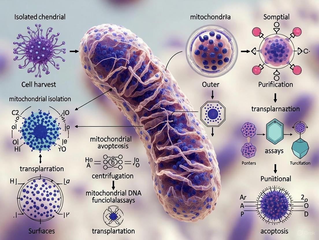
Visualization of Mitochondrial Assessment Workflow
The following diagram illustrates the logical workflow for isolating and assessing mitochondria to determine their viability for transplantation.
The Scientist's Toolkit: Key Research Reagent Solutions
Table 3: Essential Reagents for Mitochondrial Isolation and Functional Assessment
| Reagent / Kit | Function / Target | Brief Explanation & Application |
|---|---|---|
| TMRM / TMRE Dyes | Mitochondrial Membrane Potential (ΔΨm) | Cationic fluorescent dyes that accumulate in active mitochondria; loss of fluorescence indicates depolarization and functional decline [2] [3]. |
| JC-1 Dye | Mitochondrial Membrane Potential (ΔΨm) | Ratiometric dye that forms red J-aggregates in high ΔΨm and green monomers in low ΔΨm, allowing for qualitative and quantitative analysis of viability [3]. |
| ATP Luminescence Assay Kit | ATP Content | Utilizes the luciferin-luciferase reaction to produce light proportional to ATP concentration; provides a direct readout of energetic output with high sensitivity [2] [3]. |
| Digitonin | Plasma Membrane Permeabilization | Used to create permeabilized cells or fibers, allowing substrates and reagents direct access to mitochondria while preserving the intracellular architecture for respirometry studies [5]. |
| Substrate-Inhibitor Cocktails | Electron Transport Chain (ETC) | Specific compounds (e.g., Glutamate/Malate for CI, Succinate for CII, Rotenone, Antimycin A) used in SUIT protocols to dissect the function of individual respiratory complexes [5]. |
| Cell-Penetrating Peptides (e.g., Pep-1) | Mitochondrial Surface Modification | Enhances the cellular uptake and functional integration of transplanted mitochondria by facilitating fusion with the recipient mitochondrial network [1]. |
Technical Troubleshooting Guides
Troubleshooting Mitochondrial Calcium Handling
Table 1: Common Issues in Mitochondrial Calcium Resilience Experiments
| Problem | Potential Cause | Solution |
|---|---|---|
| Low calcium retention capacity | Depleted energy substrates (e.g., ADP, ATP) | Add 5mM glutamate and 2mM malate to incubation buffer to support substrate-driven respiration [8]. |
| Impaired Electron Transport Chain (ETC) function | Validate ETC complex I and IV activity via oxygen consumption rate (OCR) profiling [9]. | |
| Premature mitochondrial permeability transition pore (mPTP) opening | High levels of oxidative stress | Include 100µM Trolox or other antioxidants in the assay buffer to mitigate ROS [2]. |
| Low membrane potential (ΔΨm) | Confirm ΔΨm is polarized (-180mV) using TMRM or TMRE fluorescent probes prior to calcium challenge [8]. | |
| Unstable mitochondrial membrane potential (ΔΨm) during calcium pulsing | Calcium overload via MCU | Use Ru360 (a specific MCU inhibitor) at 10µM to confirm calcium influx is MCU-dependent [8]. |
| Inefficient calcium efflux via NCLX | Ensure presence of 5-10mM NaCl in buffer to support NCLX-mediated calcium efflux [8]. |
Troubleshooting Mitochondrial Isolation for Transplantation
Table 2: Challenges in Isocating Functional Mitochondria
| Problem | Potential Cause | Solution |
|---|---|---|
| Low yield of intact mitochondria | Overly aggressive homogenization | Use a Teflon homogenizer with precisely controlled strokes (e.g., 40 strokes); avoid blender-based methods [10]. |
| Poor post-transplantation viability | Mitochondrial damage during isolation | Implement the iMIT isolation technique using low-dose digitonin (30µM) to selectively weaken the plasma membrane, preserving outer membrane integrity [10]. |
| Rapid loss of function after isolation | Natural decay of isolated mitochondria | Use mitochondria within 2 hours of isolation for transplantation, as respiratory function significantly declines thereafter [1]. |
| Contamination with other cellular components | Inefficient purification | Employ density gradient centrifugation with Percoll layers (e.g., 15%, 23%, 40%) for highly enriched mitochondrial fractions [2]. |
Frequently Asked Questions (FAQs)
Q1: What are the primary proteins regulating mitochondrial calcium influx and efflux, and how do they interact?
The mitochondrial calcium uniporter (MCU) complex is the primary channel for Ca²⁺ influx, driven by the highly negative inner mitochondrial membrane potential (ΔΨm ~ -180 mV) [8]. Its activity is regulated by proteins like MICU1 and MICU2, which act as gatekeepers. Conversely, the mitochondrial Na⁺/Ca²⁺ exchanger (NCLX) is the primary pathway for Ca²⁺ efflux [8]. The matrix Ca²⁺ concentration ([Ca²⁺]m) is thus a dynamic balance between MCU-mediated influx and NCLX-mediated efflux.
Q2: How can I quickly assess the health and membrane potential of my isolated mitochondrial preparation before a calcium challenge?
You can rapidly assess viability using fluorescent dyes and a fluorescence microscope or plate reader. Use a combination of MitoTracker Green (labels all mitochondria) and a membrane potential-sensitive probe like TMRM, TMRE, or MitoTracker Red CMXRos [2] [10]. A high proportion of mitochondria stained by both indicates a healthy preparation. A preparation where over 90% of mitochondria show a polarized membrane is considered excellent [10].
Q3: Our transplanted mitochondria show poor uptake into recipient cells. What strategies can improve this?
Several biotechnological strategies can enhance uptake:
- Surface Modification: Conjugating isolated mitochondria with cell-penetrating peptides (CPPs) like Pep-1 or TAT can significantly improve cellular internalization efficiency [1].
- Vesicle Encapsulation: Packaging mitochondria within extracellular vesicles (EVs) or artificial liposomes can protect them from the inhospitable extracellular environment and facilitate delivery [1] [11].
- Optimized Co-culture: Leveraging natural intercellular transfer mechanisms, such as tunneling nanotubes (TNTs), by co-culturing mitochondria with recipient cells can also be effective [2].
Q4: What are the best practices for cryopreserving isolated mitochondria without significant loss of function?
Rapid thawing is critical. A protocol demonstrating stable freeze-thaw cycles showed that when thawing is completed in under 1.5 minutes, the proportion of polarized mitochondria decreases by only about 10% [10]. Slow thawing leads to extensive ice crystal formation and irreversible damage.
Q5: How does the extracellular calcium concentration in pathological states (e.g., ischemia) impact transplanted mitochondria?
In pathological conditions like ischemia-reperfusion injury, the extracellular environment can feature calcium overload [1] [7]. This high calcium concentration can stress transplanted mitochondria immediately upon delivery, potentially triggering mPTP opening and swelling before they are internalized by cells. Using delivery systems that offer protection, such as vesicles, is particularly important in these contexts [1].
Experimental Protocols
Protocol: Assessing Calcium Retention Capacity (CRC)
This protocol measures the amount of calcium mitochondria can accumulate before undergoing the mitochondrial permeability transition (mPT), a key indicator of resilience [8].
- Isolate Mitochondria: Use a preferred method (e.g., differential centrifugation or iMIT) to obtain intact mitochondria from tissue (e.g., liver, skeletal muscle) [2] [10].
- Prepare Assay Buffer: Prepare 2 mL of assay buffer (e.g., 125 mM KCl, 10 mM HEPES, 2 mM K₂HPO₄, 1 mM MgCl₂, pH 7.2) containing 5 mM glutamate and 2 mM malate as substrates.
- Load Dye and Mitochondria: Add 0.5-1 mg of mitochondrial protein and the calcium-sensitive dye Calcium Green-5N (1-2 µM) to the buffer in a stirred cuvette at 37°C.
- Calcium Challenge: Using a fluorometer, take a baseline recording. Then, add sequential, small pulses of CaCl₂ (e.g., 10 nmoles per pulse) every 60-90 seconds.
- Data Analysis: Monitor the fluorescence. Each pulse will cause a sharp increase followed by a rapid decrease as mitochondria take up calcium. The CRC is the total amount of calcium added before a sudden, permanent increase in fluorescence, indicating mPTP opening and failure to sequester calcium.
Protocol: The iMIT (intact Mitochondria Isolation Technique)
This method minimizes damage to mitochondrial membranes, which is crucial for transplantation research [10].
- Preparation: Culture cells in 150-mm dishes to ~80% confluence. Wash twice with 10 mL of ice-cold Tris-isolation buffer (10 mM Tris-HCl, 250 mM sucrose, 0.5 mM EGTA, pH 7.4).
- Digitonin Treatment: Incubate cells with 9 mL of Tris-isolation buffer containing 30 µM digitonin at 4°C for 3 minutes. Note: This step selectively weakens the plasma membrane without increasing its permeability.
- Wash and Incubate: Wash cells twice with Tris-isolation buffer to remove digitonin. Incubate in the same buffer at 4°C for an additional 10 minutes.
- Cell Disruption: Detach the cells by gentle pipetting. Agitate the resulting cell suspension several times by pipetting to selectively rupture the plasma membrane and release mitochondria.
- Differential Centrifugation:
- Centrifuge the suspension at 500 × g for 10 minutes at 4°C to pellet nuclei and cell debris.
- Transfer the supernatant to a new tube and centrifuge at 3,000 × g for 10 minutes at 4°C to pellet the mitochondrial fraction.
- Resuspension: Gently resuspend the final pellet in a small volume of Tris-isolation buffer. Keep on ice and use for transplantation within 2 hours.
The Scientist's Toolkit
Table 3: Essential Research Reagent Solutions
| Reagent | Function/Brief Explanation | Key Application |
|---|---|---|
| TMRM / TMRE | Cationic fluorescent dyes that accumulate in the mitochondrial matrix in a manner dependent on the membrane potential (ΔΨm). | Real-time assessment of mitochondrial health and ΔΨm collapse during calcium challenge [2] [10]. |
| Digitonin | A mild detergent used at low concentrations to selectively permeabilize the cholesterol-rich plasma membrane without damaging mitochondrial membranes. | Key component of the iMIT isolation protocol for obtaining mitochondria with high outer membrane integrity [10]. |
| Ru360 | A highly specific and potent membrane-permeant inhibitor of the Mitochondrial Calcium Uniporter (MCU). | Tool for confirming that calcium influx is occurring specifically through the MCU pathway [8]. |
| CGP-37157 | A selective inhibitor of the mitochondrial Na⁺/Ca²⁺ exchanger (NCLX). | Used to probe the role of calcium efflux in modulating matrix calcium levels and mPTP opening [8]. |
| Percoll | A colloidal silica gel used to form density gradients for ultra-purification of cellular organelles. | Density gradient centrifugation with Percoll layers (e.g., 15%/23%/40%) isolates mitochondria with very low synaptosome and myelin contamination [2]. |
| Cell-Penetrating Peptides (e.g., Pep-1) | Short peptides that facilitate the transport of molecular cargo across cellular membranes. | Conjugated to isolated mitochondria to enhance their uptake into recipient cells during transplantation experiments [1]. |
Signaling Pathways & Experimental Workflows
Mitochondrial Calcium Regulation Pathway
Diagram Title: Mitochondrial Calcium Regulation and mPTP Pathway
Mitochondrial Isolation & Transplantation Workflow
Diagram Title: Mitochondrial Isolation and Transplantation Workflow
A fundamental challenge in mitochondrial transplantation research is the frequent discrepancy between measurements of structural integrity and functional capacity. Isolated mitochondria may appear structurally sound yet be functionally compromised, or vice versa. This discrepancy directly impacts the success of transplantation, as only fully viable mitochondria can integrate into recipient cells and restore bioenergetics. Research indicates that dye-based methods alone may underestimate structural damage, while some functional assays might not reflect the true capacity for long-term energy production in a new cellular environment [12]. Resolving these assessment discrepancies is critical for advancing the therapeutic potential of mitochondrial transplantation, a promising strategy for conditions ranging from cardiovascular diseases to neurodegenerative disorders and surgical wound healing [13] [1] [14].
Troubleshooting Guides
Guide 1: Discrepancy Between Membrane Potential and ATP Production
Problem: Isolated mitochondria show high membrane potential (indicating health) but fail to produce adequate ATP in functional assays.
| Possible Cause | Verification Experiment | Recommended Solution |
|---|---|---|
| Uncoupling of the Electron Transport Chain [9] | Measure oxygen consumption rate (OCR) with and without ADP. A high OCR without ATP synthesis indicates a proton leak. | Ensure isolation buffers are at correct pH and osmolarity. Include bovine serum albumin (BSA) to absorb fatty acids that can act as uncouplers. |
| Insufficient Substrate Availability [9] | Repeat ATP assay with a substrate cocktail (e.g., pyruvate, malate, and succinate). | Supplement the respiration medium with multiple metabolic substrates (e.g., 5mM Pyruvate, 2mM Malate, 10mM Succinate) to fuel both complex I and II. |
| Calcium-Induced Damage [12] | Assess structural integrity via Coulter counter analysis alongside fluorescence assays. | Chelate excess calcium in solutions using EGTA (1-2 mM). Isolate and store mitochondria in calcium-free buffers. For transplantation, consider the calcium concentration of the target environment [12]. |
| Loss of Matrix Components | Assess integrity via electron microscopy and Western Blot for matrix proteins [13]. | Optimize centrifugation speed and duration during isolation to prevent overly harsh pelleting. Use a gentle, density gradient-based isolation protocol. |
Guide 2: Inconsistent Results Between Staining and Respiration Assays
Problem: Mitochondria stain positively with viability dyes (e.g., MitoTracker) but show poor respiratory control in an Oroboros system.
| Possible Cause | Verification Experiment | Recommended Solution |
|---|---|---|
| Dye-Specific Artifacts [12] | Compare results from multiple dyes (e.g., MitoTracker Red FM, JC-1, TMRM) and correlate with a functional readout like ATP concentration [13]. | Do not rely on a single dye. Always corroborate membrane potential measurements with a direct functional assay, such as ATP production or oxygen consumption. |
| Presence of Non-Functional but Structurally Intact Organelles [12] | Perform a combined assessment of membrane potential (fluorescence) and structural integrity (Coulter counter) on the same sample. | Implement a dual-parameter quality control check that mandates passing both a structural (e.g., size distribution) and a functional (e.g., ATP synthesis rate) threshold. |
| Assay Condition Discrepancy | Compare the buffer composition and temperature between the staining and respiration assays. | Standardize assay conditions across all assessments. Ensure respiration medium matches the ionic and substrate composition used in transplantation experiments. |
Guide 3: Poor Post-Transplantation Outcomes Despite Good In Vitro Viability
Problem: Mitochondria pass all in-vitro quality controls but fail to improve cellular bioenergetics or integrate after transplantation.
| Possible Cause | Verification Experiment | Recommended Solution |
|---|---|---|
| Susceptibility to Extracellular Stress [12] | Pre-incubate mitochondria in a calcium-rich buffer (e.g., 1.3 mM) mimicking the extracellular environment and re-measure function. | Pre-condition mitochondria by briefly exposing them to a physiologically relevant calcium level (e.g., 1.3 mM) before transplantation to select a more robust population [12]. |
| Inefficient Cellular Uptake [1] | Label mitochondria with a lipophilic dye (e.g., MitoTracker) and use flow cytometry or microscopy to quantify uptake by recipient cells. | Use a surface modification strategy, such as conjugating cell-penetrating peptides (e.g., Pep-1 or TAT) to the mitochondrial membrane to enhance cellular internalization [1]. |
| Lysosomal Degradation Post-Uptake [1] | Use confocal microscopy with lysosomal and mitochondrial markers to track if co-localization occurs post-transplantation. | Co-deliver mitochondria with agents that temporarily inhibit lysosomal activity (e.g., chloroquine). Alternatively, use a magnetically-guided delivery system to force faster entry into the cytoplasm. |
Frequently Asked Questions (FAQs)
Q1: What is the most reliable single metric for assessing mitochondrial viability? There is no single perfect metric. The most reliable approach is a combined assessment that includes at least one structural and one functional metric. For instance, correlating membrane potential (using MitoTracker Red FM) with direct ATP production provides a more comprehensive picture than either metric alone. Research shows that relying solely on dye-based methods can significantly overestimate the population of viable organelles [12].
Q2: Why do my isolated mitochondria fail in a calcium-rich environment despite good membrane potential? Mitochondrial membrane integrity is highly vulnerable to calcium overload. Supraphysiologic calcium concentrations (e.g., 2.6 mM) can cause a progressive loss of function and integrity, even if the initial membrane potential appears strong. A substantial proportion of mitochondria can remain viable at physiologic blood calcium levels (~1.3 mM), but the population must be tested under these specific conditions to ensure translational relevance [12].
Q3: How can I quickly check if my isolation protocol is causing structural damage? Impedance-based Coulter counter analysis is a rapid and effective method to assess structural integrity and size distribution, providing a quantitative measure of damage that fluorescence assays may miss. This can be complemented by transmission electron microscopy (TEM) for detailed visualization of membrane structures and cristae, which is considered a gold standard [12] [13].
Q4: Are frozen-thawed mitochondria viable for transplantation? Current evidence strongly suggests that frozen-thawed mitochondria are not effective. Studies indicate that thawed or broken mitochondria are non-viable and fail to provide cytoprotection following transplantation. Successful transplantation and bioenergetic rescue consistently depend on the use of freshly isolated, structurally intact mitochondria [13].
Q5: What are the key markers to confirm mitochondrial integrity after isolation? A robust quality control check should include Western Blot analysis for key protein markers. Essential outer membrane markers include TOM20, while the presence and integrity of all five Oxidative Phosphorylation (OXPHOS) complexes (I-V) should be confirmed. This verifies that the machinery for energy production is intact [13].
The following table consolidates key experimental data on how different stressors impact mitochondrial viability, highlighting the discrepancy between assessment methods.
Table 1: Impact of Stressors on Mitochondrial Viability Metrics
| Stressor Condition | Membrane Potential Retention (Fluorescence Assay) | Structural Integrity Retention (Coulter Counter) | Functional Outcome (ATP Production / Transplantation) | Citation |
|---|---|---|---|---|
| Calcium Exposure (1.3 mM for 12h) | 90-95% retention | More extensive loss than fluorescence suggests | Supports feasibility for intracoronary transplantation | [12] |
| Calcium Exposure (2.6 mM for 12h) | Progressive loss, near freeze-thaw control levels | Significant structural damage | Not viable for transplantation | [12] |
| Freeze-Thaw Cycle | Significant loss | Significant structural damage | Non-viable and ineffective for cytoprotection; does not increase cellular ATP [13] | [13] |
| Transplantation into Cells | N/A | N/A | Dose-dependent increase in cellular metabolic activity and ATP levels [13] | [13] |
Experimental Protocols
Protocol: Combined Viability Assessment (Membrane Potential & Structural Integrity)
This protocol is designed to directly identify discrepancies between functional and structural readouts.
- Isolate Mitochondria: Use a standard differential centrifugation protocol from a cell source (e.g., L6 cells or hMSCs). Keep mitochondria on ice in isolation buffer [12] [13].
- Apply Stressor: Divide the mitochondrial suspension into aliquots. Expose them to the stressor of interest (e.g., 1.3 mM or 2.6 mM CaCl₂) for a set duration (e.g., 1-12 hours). Maintain a control group in a stress-free buffer.
- Parallel Assessment:
- Membrane Potential (Functional): Incubate a sample with MitoTracker Red FM (or similar dye) according to manufacturer instructions. Measure fluorescence using a plate reader or flow cytometer [12].
- Structural Integrity (Structural): Analyze a separate sample of the same preparation using an impedance-based Coulter counter. This provides a direct count and size distribution, identifying swollen or ruptured organelles [12].
- Correlate with ATP Synthesis: As a definitive functional check, measure the ATP production rate of the stressed mitochondria using a luciferase-based ATP assay kit in the presence of ADP and substrates [13].
Protocol: Quality Control for Transplantation-Grade Mitochondria
A mandatory quality control workflow to ensure mitochondria are suitable for transplantation.
- Isolation from hMSCs: Isolate mitochondria from human Mesenchymal Stromal Cells using a standardized, gentle isolation kit. Perform all steps at 4°C to preserve function [13].
- Concentration & Sizing: Determine mitochondrial concentration and validate size distribution (expected range: 0.1-1.2 μm) using interferometric light microscopy (e.g., Videodrop) or dynamic light scattering [13].
- Structural Integrity Check (TEM): Fix a small aliquot in glutaraldehyde and process for Transmission Electron Microscopy. Image to confirm the presence of intact outer and inner membranes, including cristae [13].
- Protein Marker Validation (Western Blot): Lyse an aliquot and perform Western Blot analysis. Confirm the presence of outer membrane marker TOM20 and subunits from all five OXPHOS complexes (I-V). The corresponding supernatant (SN) from isolation should show minimal signal for these markers [13].
- Functional Potency Assay: Co-culture a defined number of mitochondria with metabolically stressed recipient cells (e.g., starved HCEC-1CT colonic epithelial cells). After 24-48 hours, measure the rescue of metabolic activity using an Alamar Blue assay and confirm a concomitant increase in intracellular ATP levels [13].
Visualization of Workflows
Mitochondrial Viability Assessment Workflow
This diagram outlines the parallel assessment of structural and functional metrics to identify viable mitochondria for transplantation.
Post-Transplantation Viability Investigation
This diagram illustrates common causes of failure after transplantation and their potential solutions.
The Scientist's Toolkit: Research Reagent Solutions
Table 2: Essential Reagents for Mitochondrial Viability Assessment
| Reagent / Material | Function / Application | Key Consideration for Discrepancies |
|---|---|---|
| MitoTracker Red FM [12] | Fluorescent dye to assess mitochondrial membrane potential (functional metric). | Can overestimate viability; always pair with a structural assay. |
| Coulter Counter / Impedance Analyzer [12] | Provides quantitative data on mitochondrial size and count, directly measuring structural integrity. | Reveals structural loss that fluorescence assays may miss. |
| ATP Assay Kit (Luciferase-based) [13] | Directly measures ATP concentration, the ultimate functional output of mitochondria. | The definitive test for functional competence; correlates with transplantation success. |
| Antibodies for OXPHOS Complexes & TOM20 [13] | Western Blot analysis to confirm the presence and integrity of key protein complexes (structural and functional). | Validates that the electron transport chain machinery is intact post-isolation. |
| Cell-Penetrating Peptides (e.g., Pep-1, TAT) [1] | Conjugated to isolated mitochondria to enhance uptake by recipient cells, improving transplantation efficacy. | Addresses the issue of poor functional outcomes due to failed cellular integration. |
| Calcium Chelators (e.g., EGTA) [12] | Added to isolation and storage buffers to protect against calcium-induced permeability transition and swelling. | Prevents a common cause of functional degradation despite initially good membrane potential. |
For researchers in mitochondrial transplantation, assessing the viability of isolated mitochondria is a critical step that directly impacts experimental success and therapeutic outcomes. Viability transcends mere structural integrity; it is the functional capacity of mitochondria to perform their essential bioenergetic functions. The core indicators of this viability are adenosine triphosphate (ATP) production and the maintenance of mitochondrial membrane potential (ΔΨm). These two parameters are deeply interdependent: the ΔΨm is the electrochemical gradient that drives ATP synthesis, while functional ATP production reflects a well-coupled and intact electron transport chain. Accurately measuring these functions provides the most reliable assessment of whether isolated mitochondria are viable and capable of rescuing bioenergetic deficits in recipient cells or tissues.
This guide addresses the key technical challenges and frequently asked questions in evaluating mitochondrial bioenergetics, providing standardized protocols and troubleshooting advice to ensure the reliability of your data in the context of transplantation research.
Core Functional Assays & Methodologies
Measuring Mitochondrial Membrane Potential (ΔΨm)
Detailed Experimental Protocol:
- Dye Loading: Resuspend the isolated mitochondrial pellet or cultured cells in an appropriate incubation buffer (e.g., containing succinate or other relevant substrates). Load with a potential-sensitive dye such as tetramethylrhodamine, ethyl ester (TMRE) or MitoTracker Red FM at a concentration of 50-500 nM. The optimal concentration and incubation time (typically 15-30 minutes at 37°C) should be determined empirically.
- Washing and Preparation: Gently wash the samples to remove excess, unincorporated dye. Resuspend in fresh buffer.
- Measurement:
- Fluorescence Microscopy/Image Cytometry: Image the samples using the appropriate excitation/emission filters (e.g., ~549/575 nm for TMRE). For single-cell analysis in neuronal cultures, this can be performed in time-lapse to track dynamics [15] [16].
- Flow Cytometry: Analyze the fluorescence intensity of the mitochondrial population. A rightward shift in fluorescence indicates a higher, healthier ΔΨm.
- Plate Reader Assay: Transfer the suspension to a clear-bottom black-walled microplate and measure the fluorescence.
- Validation with Controls: To confirm the specificity of the signal, include a control sample treated with an uncoupler like FCCP (e.g., 10 μM for 4 hours [15]), which dissipates ΔΨm and should result in a significant loss of fluorescence.
Measuring ATP Production
Detailed Experimental Protocol (Bioluminescent Assay): This method uses firefly luciferase's ATP-dependent light emission to quantify production rates [17] [18].
- Reaction Setup: In a luminometer tube or plate, prepare a reaction mix containing:
- Isolation buffer (e.g., with Mg2+ and phosphate)
- Luciferin (substrate)
- Luciferase (enzyme)
- Key substrates for the electron transport chain:
- Complex I-driven: Malate + Glutamate or Pyruvate
- Complex II-driven: Succinate (often with rotenone to inhibit complex I backflow)
- Initiation: Start the reaction by adding a small volume of isolated mitochondria. The final volume is typically 100-200 μL.
- Data Acquisition: Immediately place the sample in the luminometer and record the light output continuously for several minutes. The initial rate of increase in luminescence is proportional to the rate of ATP production [17].
- Calibration: A standard curve with known concentrations of ATP must be run in parallel to convert relative light units (RLU) into absolute ATP concentration.
Quantitative Data from Core Assays
The following table summarizes expected outcomes and critical controls for these core assays, integrating data from recent studies.
Table 1: Key Parameters and Controls for Bioenergetic Assays
| Assay | Healthy Mitochondria Indicator | Positive Control (Damaged) | Key Confounding Factors | Typical Experimental Outputs |
|---|---|---|---|---|
| ΔΨm (TMRE) | High fluorescence intensity | FCCP (Uncoupler) causes >80% signal loss [15] | Dye overloading, quenching, non-specific binding, temperature fluctuations | Fluorescence units, images (relative intensity) |
| ATP Production (Biolum.) | High linear ATP production rate | No substrate (basal) or Antimycin A (inhibits ETC) | Substrate specificity, mitochondrial preparation integrity (see Table 3), ADP availability | ATP production rate (nmol/min/mg protein) |
| Oxygen Consumption (Respirometry) | High respiratory control ratio (State 3/State 4) | Uncouplers (FCCP) for maximal capacity; inhibitors (KCN) | Instrument calibration, substrate permeability, medium composition | Oxygen Consumption Rate (OCR), pmol/(s*mg) |
The Scientist's Toolkit: Research Reagent Solutions
Table 2: Essential Reagents for Mitochondrial Functional Assessment
| Reagent / Material | Function / Application | Key Considerations |
|---|---|---|
| TMRE / MitoTracker Red FM | Fluorescent dyes for quantifying ΔΨm. Accumulate in active mitochondria. | TMRE is more reversible; MitoTracker can be fixed. Both are susceptible to photobleaching. |
| FCCP | Proton ionophore used as an uncoupler positive control to dissipate ΔΨm. | Titrate concentration for complete depolarization without inducing swelling. |
| Luciferase/Luciferin Kit | Bioluminescent detection system for quantifying ATP production. | Ensure reagent freshness and avoid repeated freeze-thaw cycles. |
| Succinate / Malate / Glutamate | Mitochondrial substrates to fuel specific ETC complexes (II and I, respectively). | Use appropriate inhibitors (e.g., rotenone with succinate) to isolate complex-specific activity. |
| High-Resolution Respirometer | Instrument for precise, real-time measurement of mitochondrial oxygen consumption. | Requires careful calibration and small amounts of sample [17]. |
| Cell-Penetrating Peptides (Pep-1) | Enhances mitochondrial uptake into recipient cells during transplantation studies [1]. | Optimize peptide-to-mitochondria ratio (e.g., 1750:1 w/w) to avoid toxicity [1]. |
Troubleshooting Common Experimental Issues
FAQ 1: My isolated mitochondria show a good membrane potential but low ATP production. What could be the cause? This discrepancy often points to uncoupling or specific damage to ATP synthase.
- Cause A: The proton circuit may be compromised. The proton-motive force is being generated (maintaining ΔΨm) but is not efficiently coupled to ATP synthesis by the F1Fo ATP synthase. This can be tested by assessing the P/O ratio (ATP produced per oxygen atom consumed) [17].
- Cause B: The isolation procedure may have damaged the ATP synthase complex or made the inner membrane leaky to protons. Compare different isolation buffers and ensure gentle homogenization.
- Action Plan: Perform a respirometry experiment to measure the Respiratory Control Ratio (RCR). A low RCR indicates poor coupling between oxidation and phosphorylation.
FAQ 2: Why do I get inconsistent results with the MTT assay when testing mitochondrial viability? The MTT assay is often misapplied as a direct viability assay. It measures total cellular metabolic activity, not just mitochondrial viability [19].
- Cause A: The MTT reagent can be reduced by enzymes outside the mitochondria and by biomolecules like ascorbic acid and glutathione [19].
- Cause B: The formation and extrusion of formazan crystals are complex and depend on cell density, MTT concentration, and incubation time, which are often not optimized [19].
- Action Plan: For a purer assessment of mitochondrial function, rely on direct measures like ATP production or ΔΨm. If using MTT, rigorously optimize the protocol and interpret results as "cellular metabolic activity" rather than viability.
FAQ 3: How stable are isolated mitochondria for transplantation, and what factors affect their survival? Mitochondrial longevity post-isolation is a major bottleneck for clinical translation.
- Cause: Isolated mitochondria rapidly lose respiratory function, often within 2 hours, and are sensitive to the extracellular environment (e.g., high calcium, ROS) [1].
- Action Plan:
- Timing: Use mitochondria as quickly as possible after isolation (ideally within 30-60 minutes).
- Calcium Stress: Recent data shows mitochondria can retain ~90-95% membrane potential for 12 hours at physiologic calcium (1.3 mM), but higher concentrations (2.6 mM) cause progressive failure [20]. Use Coulter counter analysis alongside dye-based methods to better assess structural integrity [20].
- Advanced Delivery Systems: Utilize biotechnological approaches like surface modification with cell-penetrating peptides (e.g., Pep-1) or encapsulation in extracellular vesicles to enhance protection and targeting [1].
FAQ 4: How does the mitochondrial source (species, tissue) impact viability and function for transplantation? The source is a critical variable influencing metabolic compatibility.
- Evidence: Studies show that mitochondria from diverse species (e.g., Vero, MDBK, MDCK cells) can be internalized by human cells without significant immune response. However, the therapeutic outcome in disease models varies even when baseline ATP and ΔΨm are similar, suggesting metabolic compatibility between donor mitochondria and recipient cells is crucial [21].
- Action Plan: Select mitochondrial sources based on the disease model. Test multiple sources and prioritize metabolically matched mitochondria for optimal functional integration.
Workflow and Relationship Visualizations
Mitochondrial Viability Assessment Workflow
Bioenergetic Integrity Relationships
Critical Considerations for Mitochondrial Transplantation
The ultimate test of mitochondrial viability is successful functional integration in a recipient system. Beyond standard viability assays, consider these factors specific to transplantation:
- Metabolic Compatibility: Mitochondria with matching metabolic characteristics can provide improved therapeutic outcomes, even in the absence of superior baseline function [21].
- Isolation Method Integrity: The method of isolation (e.g., hypotonic vs. isotonic) critically impacts the structural and functional integrity of the outer and inner membranes, which in turn affects energy metabolism [17] [18]. Isotonic isolation generally preserves function better.
- Assessment Timing: Functional assessments should be performed immediately before transplantation to ensure the mitochondria are still viable, as their functional lifespan ex vivo is limited [1].
Advanced Protocols and Biotechnological Solutions for Mitochondrial Preservation
This technical support guide addresses a core challenge in mitochondrial transplantation research: obtaining high yields of intact, functional mitochondria. The viability of isolated mitochondria is paramount for successful outcomes in therapeutic applications, such as treating ischemia-reperfusion injury or neurodegenerative diseases [12] [22].
The premise of using digitoxin, a cardiac glycoside, is based on its known mechanism of action: inhibition of the Na+/K+ ATPase pump on the plasma membrane [23]. The proposed method aims to leverage this effect to weaken the cellular membrane, thereby facilitating easier disruption and release of mitochondria during the isolation process, potentially leading to higher yields and better preserved function.
Experimental Protocol: Digitoxin-Based Isolation Workflow
The following protocol details the pre-treatment and isolation steps. All steps post-digitoxin treatment should be performed on ice or at 4°C to preserve mitochondrial integrity [24].
Step-by-Step Methodology
Cell Pre-treatment:
- Culture cells (e.g., HepG2, HEK293T, or AC16) to 80% confluence in standard culture dishes [25] [24].
- Prepare a digitoxin solution in DMSO or culture medium. A critical optimization step is to titrate the digitoxin concentration (e.g., 0.1-10 µM) and incubation time (e.g., 15-60 minutes) to achieve plasma membrane permeabilization without damaging mitochondrial membranes.
- Remove the growth medium and add the digitoxin solution to the cells. Incubate under normal culture conditions for the determined time.
- Termination: After incubation, promptly remove the digitoxin solution and wash the cells twice with ice-cold, sterile Phosphate Buffer Saline (PBS) to stop the reaction [24].
Mitochondrial Isolation:
- Harvest digitoxin-treated cells using a cell lifter or trypsin-EDTA, and collect them by centrifugation at 500 × g for 3 minutes at 4°C [24].
- Resuspend the cell pellet in 2 mL of ice-cold Homogenization Buffer (e.g., containing 320 mM sucrose, 1 mM EDTA, 10 mM Tris-HCl, pH 7.4) supplemented with a protease inhibitor like PMSF [24].
- Homogenize the cell suspension using a Dounce tissue grinder. The number of strokes (e.g., ~20 for HEK293T) must be optimized. Monitor efficiency by Trypan Blue staining, aiming for ~80% cell disruption [24].
- Centrifuge the homogenate at 1,200 × g for 3 minutes at 4°C to remove intact cells, nuclei, and heavy debris. Transfer the supernatant to a new tube.
- Centrifuge the supernatant at a high speed (15,000 × g for 2-10 minutes at 4°C) to pellet the crude mitochondrial fraction [25] [24].
- Wash the mitochondrial pellet by resuspending it in a suitable buffer (e.g., Buffer A or respiratory buffer) and repeat the high-speed centrifugation [24]. The final pellet contains the isolated mitochondria.
The workflow below summarizes the key stages of this protocol.
Research Reagent Solutions
The table below lists essential reagents and their functions for this protocol.
| Reagent | Function / Rationale in Protocol |
|---|---|
| Digitoxin | Primary agent for plasma membrane weakening via Na+/K+ ATPase inhibition [23]. |
| Dounce Homogenizer | Mechanical disruption of digitoxin-weakened cells; gentler on mitochondria than other methods [25] [24]. |
| Homogenization Buffer (e.g., Sucrose/EDTA/Tris) | Isotonic buffer to prevent osmotic damage to mitochondria during isolation [24]. |
| Phenylmethylsulfonyl Fluoride (PMSF) | Serine protease inhibitor added to buffers to protect mitochondrial proteins from degradation [24]. |
| JC-1 Dye | Fluorescent probe for ratiometric assessment of mitochondrial membrane potential (ΔΨm), a key indicator of function [25] [26]. |
| MitoTracker Red FM | Alternative fluorescent dye for assessing mitochondrial membrane potential and viability [12]. |
| Respiratory Buffer (e.g., with Succinate) | Buffer for post-isolation functional assays to measure mitochondrial activity and ROS production [25]. |
Troubleshooting Guide & FAQs
Table 1: Common Problems and Solutions
| Problem | Potential Cause | Solution |
|---|---|---|
| Low Mitochondrial Yield | • Insufficient digitoxin concentration/time.• Inefficient homogenization.• Incomplete cell harvesting. | • Titrate digitoxin dose and time.• Optimize number of Dounce strokes; check with Trypan Blue.• Ensure complete cell scraping and pelleting. |
| Poor Mitochondrial Function/Low Membrane Potential | • Digitoxin concentration too high, damaging mitochondria.• Extended processing time on ice.• Hypotonic or incorrect isolation buffer. | • Reduce digitoxin exposure; ensure thorough washing post-treatment.• Minimize time from homogenization to final pellet.• Verify osmolarity and composition of all buffers. |
| Contamination with Other Organelles | • Inefficient low-speed centrifugation.• Over-homogenization. | • Perform multiple low-speed spins until supernatant is clear.• Avoid excessive Dounce strokes. |
| High ROS in Isolated Mitochondria | • Physical stress during isolation.• Lack of substrates in storage/resuspension buffer. | • Use gentler homogenization.• Resuspend final mitochondrial pellet in a respiratory buffer containing substrates like succinate [25]. |
Frequently Asked Questions (FAQs)
Q1: What is the ideal digitoxin concentration to start with for my cell line? A1: There is no universal concentration. You must empirically determine the optimal dose. Begin with a low-concentration range (e.g., 0.1-1.0 µM) and a short incubation time (15-30 minutes). Assess viability and yield compared to a non-digitoxin control. The goal is to find a condition that increases yield without compromising function, as measured by membrane potential assays [12].
Q2: How can I quickly verify if my isolated mitochondria are intact and functional? A2: The most common and robust method is to measure the mitochondrial membrane potential (ΔΨm). This can be done using ratiometric fluorescent dyes like JC-1 [25] or FRET-based probes [26]. A high ΔΨm indicates healthy, functional mitochondria. Additionally, a Coulter counter can be used to assess structural integrity and quantify mitochondrial loss, which may reveal damage that fluorescence assays miss [12].
Q3: My isolated mitochondria work in assays, but fail to provide therapeutic benefit in transplantation models. Why? A3: Functional assays may not capture all aspects of viability required for in vivo efficacy. Ensure the mitochondria can survive in the transplantation environment. For example, mitochondria for intracoronary delivery must withstand extracellular calcium levels (~1.3 mM). Test their resilience in physiologically relevant conditions; one study showed supraphysiologic calcium (2.6 mM) caused progressive loss of function [12]. Furthermore, consider metabolic compatibility between the donor mitochondria source and the recipient cells, as this can significantly impact therapeutic outcome [21].
Q4: Are there alternatives to digitoxin for membrane weakening in isolation protocols? A4: Yes. The core of most standard mitochondrial isolation protocols is differential centrifugation combined with mechanical homogenization (using a Dounce or Potter-Elvehjem homogenizer) without chemical pre-treatments [25] [24]. Several commercial kits (e.g., Qproteome from Qiagen, MITOISO2 from Sigma-Aldrich) are also available and have been compared in the literature, often yielding mitochondria with high inner-membrane integrity and activity [25]. The digitoxin method is an investigative approach aimed at enhancing these established techniques.
Quality Control and Validation Assays
Rigorous validation is required to confirm that digitoxin pre-treatment enhances yield without harming function. The following diagram and table outline the key parallel assessments needed.
Table 2: Key Quality Control Metrics for Isolated Mitochondria
| Assessment Metric | Methodology | Interpretation / Desired Outcome |
|---|---|---|
| Total Protein Yield | Micro-Lowry assay on mitochondrial pellet [25]. | Higher yield in digitoxin-treated vs. control indicates successful method. |
| mtDNA Copy Number | Quantitative Real-Time PCR (e.g., for tRNALeu gene) [25]. | Confirms nucleic acid content and, indirectly, quantity. |
| Structural Integrity | Impedance-based Coulter counter analysis [12]. | Quantifies intact mitochondrial particles; can reveal structural damage missed by fluorescence. |
| Membrane Potential (ΔΨm) | Ratiometric fluorescence (JC-1 or FRET probes) [25] [26]. | High potential (red/green ratio for JC-1) indicates functional integrity and health. |
| Mitochondrial Activity | DCFH-DA staining for ROS production or ATP production assays [25] [21]. | Confirms metabolic competence. Healthy mitochondria show controlled, measurable activity. |
For mitochondrial transplantation research, the ultimate success criterion is the functional integration of transplanted organelles into recipient cells. The mitochondrial membrane potential (ΔΨm) is a direct indicator of mitochondrial health and its capacity to produce ATP. Preserving a high ΔΨm (>90%) post-thaw is therefore not merely a metric of survival, but a prerequisite for therapeutic efficacy. This technical support center provides targeted guidance to achieve this benchmark, ensuring your cryopreserved mitochondria are viable and functionally competent for advanced research and therapeutic development.
Troubleshooting Guides
Low Post-Thaw Membrane Potential
A decline in ΔΨm indicates compromised mitochondrial integrity, leading to reduced ATP synthesis and impaired cellular rescue capabilities in transplantation models.
- Problem: Post-thaw membrane potential is consistently below 80%.
- Investigation & Solutions:
| Potential Cause | Investigation | Recommended Solution |
|---|---|---|
| Slow Thawing Process | Time the thaw process from removal from storage to complete ice dissolution. | Implement an ultra-rapid thawing protocol where thawing is completed in under 1.5 minutes [10]. |
| Cryoprotectant Toxicity | Verify the concentration and type of cryoprotectant used. | Use DMSO at a standardized concentration (typically 10%). Ensure cryopreservation media is cooled to 2-8°C before use [27]. |
| Isolation-Induced Damage | Assess outer membrane integrity post-isolation using Western Blot for intermembrane space proteins like cytochrome c [10]. | Adopt a gentle isolation method like the iMIT technique, which uses low-dose digitonin to pre-weaken the plasma membrane, minimizing mechanical shear and preserving membrane integrity [10]. |
| Suboptimal Post-Thaw Culture | Check the osmolarity and composition of the thaw/resuspension media. | Resuspend mitochondria in a specialized Tris-isolation buffer (e.g., 10 mM Tris-HCl, 250 mM sucrose, 0.5 mM EGTA, pH 7.4) to provide immediate ionic and osmotic support [10]. |
Poor Functional Recovery Post-Thaw
Even with adequate ΔΨm, mitochondria may fail to restore bioenergetics in recipient cells.
- Problem: Mitochondria integrate but fail to improve cellular ATP levels or reduce oxidative stress in recipient cells.
- Investigation & Solutions:
| Potential Cause | Investigation | Recommended Solution |
|---|---|---|
| Loss of Intermembrane Proteins | Analyze the supernatant during isolation for markers like adenylate kinase 2 (AK2) or cytochrome c [10]. | Optimize isolation protocol to retain intermembrane space proteins. The iMIT method has been shown to better preserve these compared to homogenization [10]. |
| Oxidative Stress During Thaw | Measure ROS levels immediately post-thaw. | Consider adding antioxidants (e.g., superoxide dismutase mimics) to the thawing medium. However, validate they do not interfere with recipient cell signaling [28]. |
| Inefficient Uptake by Recipient Cells | Quantify mitochondrial uptake using fluorescent tags (e.g., MitoTracker) over time. | Enhance delivery using biotechnological strategies, such as surface modifying mitochondria with cell-penetrating peptides (CPPs) like Pep-1 to improve cellular internalization [1]. |
Frequently Asked Questions (FAQs)
Q1: What is the single most critical factor for preserving membrane potential during thawing? A: The speed of the thawing process is paramount. Evidence indicates that completing the thaw in under 1.5 minutes is critical to minimize activity loss, with the proportion of polarized mitochondria decreasing by only about 10% when this is achieved [10].
Q2: Our isolation yield is high, but post-thaw function is low. Could the issues be linked? A: Absolutely. Conventional isolation methods like homogenization can inflict structural damage that compromises the outer membrane, making mitochondria more susceptible to cryo-injury. Switching to a gentler, integrity-focused method like the iMIT technique can significantly improve post-thaw outcomes [10].
Q3: Are there any methods to improve the uptake of our thawed mitochondria into target cells? A: Yes. Biotechnology approaches are key. Surface modification of isolated mitochondria with cell-penetrating peptides (CPPs), such as Pep-1 or TAT, can significantly enhance the precision and efficiency of cellular uptake and internalization after thawing [1].
Q4: How long can we store cryopreserved mitochondria without significant loss of function? A: While the search results do not specify a maximum storage duration, they emphasize that stable storage conditions are critical. Fluctuations in temperature during storage can negatively impact viability [29]. For long-term storage, liquid nitrogen is recommended [29].
Q5: Why is membrane potential a more important metric than just mitochondrial count post-thaw? A: A high mitochondrial count is meaningless if the organelles are dysfunctional. The membrane potential (ΔΨm) is a direct, functional readout of the proton gradient across the inner membrane, which is essential for ATP production. Intact ΔΨm is a strong indicator that the mitochondria are metabolically active and capable of supporting bioenergetic rescue in recipient cells [9] [30].
Detailed Protocol: iMIT Isolation & Rapid Thaw
This protocol is adapted from methods proven to preserve >90% membrane potential post-thaw [10].
Part A: Isolation of Intact Mitochondria using the iMIT Technique
- Preparation: Culture cells to 80-90% confluence in 150-mm dishes. Wash cells twice with 10 mL of ice-cold Tris-isolation buffer (10 mM Tris-HCl, 250 mM sucrose, 0.5 mM EGTA, pH 7.4).
- Plasma Membrane Weakening: Incubate cells with 9 mL of Tris-isolation buffer containing 30 μM digitonin at 4°C for 3 minutes.
- Wash & Equilibrate: Gently wash the cells twice with plain Tris-isolation buffer to remove excess digitonin. Incubate in fresh buffer at 4°C for 10 minutes.
- Cell Disruption: Detach the cells by gentle pipetting and agitate the suspension several times through a pipette to selectively rupture the pre-weakened plasma membrane.
- Differential Centrifugation:
- Centrifuge the suspension at 500 × g for 10 minutes at 4°C to pellet nuclei and cell debris.
- Transfer the supernatant to a new tube and centrifuge at 3,000 × g for 10 minutes at 4°C to pellet the mitochondrial fraction.
- Resuspension: Gently resuspend the final mitochondrial pellet in a small volume of Tris-isolation buffer. Keep on ice at all times.
Part B: Cryopreservation and Rapid Thaw Protocol
- Cryopreservation:
- Dilute the mitochondrial suspension to a concentration of ~500 μg protein/mL in a cryopreservation medium (e.g., Tris-isolation buffer with 10% DMSO and 90% FCS, pre-cooled to 2-8°C) [27] [10].
- Dispense 0.1-1.0 mL aliquots into cryovials.
- Freeze gradually at 1°C/minute using a controlled-rate freezer or isopropanol-filled container placed at -80°C for 4+ hours [29].
- Transfer vials to long-term storage in liquid nitrogen [29].
- Rapid Thawing:
- Critical Step: Rapidly retrieve the vial from storage and immediately place it in a 37°C water bath with gentle agitation.
- Ensure the sample is completely thawed in under 1.5 minutes [10].
- Immediately upon ice dissolution, transfer the vial to wet ice.
The following table summarizes key performance metrics from the referenced isolation and cryopreservation study [10].
| Metric | iMIT Isolation Method (Pre-freeze) | Conventional Homogenization (Pre-freeze) | Post Rapid Thaw (<1.5 min) |
|---|---|---|---|
| Mitochondria with Polarized Inner Membranes | ~90% | Lower than iMIT | Decrease of only ~10% points |
| Outer Membrane Integrity | High (Preserved) | Often Compromised | Data not explicitly stated, but implied high |
| Intermembrane Space Protein Retention | High (e.g., Cytochrome c) | Lower | Data not explicitly stated, but implied high |
Workflow and Pathway Diagrams
Optimal Mitochondrial Workflow
This diagram contrasts the optimal protocol for high membrane potential retention against a suboptimal one, highlighting the critical decision points.
Mitochondrial Dysfunction Cascade
This diagram illustrates the logical sequence of events when the rapid thaw protocol is not followed, leading to mitochondrial dysfunction.
The Scientist's Toolkit: Essential Research Reagents
This table details key reagents and their critical functions for successful mitochondrial cryopreservation.
| Reagent | Function / Rationale |
|---|---|
| Digitonin | A mild detergent used in the iMIT protocol to selectively weaken the plasma membrane without damaging mitochondrial membranes, enabling the release of intact organelles [10]. |
| Tris-isolation Buffer | Provides an isotonic, pH-stable environment for mitochondria. The sucrose maintains osmolarity, while EGTA chelates calcium to prevent permeability transition pore opening [10]. |
| Dimethyl Sulfoxide (DMSO) | A standard cryoprotectant that penetrates cells and organelles, reducing ice crystal formation during freezing. A concentration of 10% is commonly used [27]. |
| Fetal Calf Serum (FCS) | Often used as a component (e.g., 90%) in cryopreservation media. It provides proteins and other macromolecules that offer additional membrane stabilization and cryoprotection [27]. |
| Tetramethylrhodamine Ethyl Ester (TMRE) | A cell-permeant, fluorescent dye that accumulates in active mitochondria based on their membrane potential (ΔΨm). It is a key tool for quantifying functional viability post-thaw [10]. |
| Cell-Penetrating Peptides (CPPs) | Peptides like Pep-1 and TAT. Used to coat isolated mitochondria post-thaw to significantly enhance their uptake and delivery into recipient target cells in transplantation assays [1]. |
This technical support guide details the methodology for implementing the "mito-condition" culture medium, a breakthrough protocol for the large-scale fabrication of functional mitochondria in Mesenchymal Stem Cells (MSCs). The primary goal of this protocol is to overcome a critical bottleneck in mitochondrial transplantation research: the sustainable production of high-quality, energetic mitochondria. The optimized condition achieves an 854-fold increase in total mitochondrial yield after 15 days of culture, coupled with a 5.71-fold enhancement in ATP production capacity compared to mitochondria from typical culture conditions [31]. This guide provides detailed protocols, troubleshooting, and FAQs to ensure robust replication of these results for your transplantation studies.
Core Experimental Protocol & Workflow
The following section outlines the key methodology for establishing the mito-condition culture and quantifying its output.
Detailed Protocol: Establishing Mito-Condition MSCs (mc-MSCs)
Objective: To expand human adipose-derived MSCs in a customized serum-free medium that simultaneously enhances cell proliferation and mitochondrial biogenesis (mitobiogenesis).
Key Materials:
- Cell Source: Human adipose-derived Mesenchymal Stem Cells (MSCs).
- Base Medium: A previously established serum-free culture expansion system [31].
- Mito-Condition Components: The optimized medium consists of nine key factors.
Procedure:
- Preparation of Mito-Condition Medium: Supplement the base serum-free medium with the following nine components [31]:
- Basic Fibroblast Growth Factor (bFGF)
- Sodium Bicarbonate (NaHCO₃)
- Lipid Concentrate
- Insulin-Transferrin-Selenium (ITS)
- Progesterone (Prog)
- Hydrocortisone (Hc)
- Vitamin C (Vc)
- Heparin Sodium (HS)
- Human Platelet Lysate (HPL)
- Cell Culture and Passaging: Culture the MSCs in the prepared mito-condition medium. Maintain cells in a standard humidified incubator at 37°C with 5% CO₂.
- Continuous Expansion: Passage the cells upon reaching 80-90% confluence. The mc-MSCs exhibit a significantly reduced population doubling time (PDT) of approximately 21.23 hours, compared to 38.45 hours for typical-condition MSCs (tc-MSCs) [31].
- Harvesting and Validation: After 5 passages (approximately 15 days), harvest the cells for mitochondrial isolation. Validate the success of the culture using the quality control assays detailed in Section 2.2.
Detailed Protocol: Mitochondrial Isolation and Functional Assays
Objective: To isolate mitochondria from expanded mc-MSCs and assess their quantity and functional quality.
Key Materials:
- Mitochondrial Isolation Kit (differential centrifugation-based).
- TOMM20 antibody for immunostaining.
- Reagents for qPCR (for mtDNA copy number analysis).
- Transmission Electron Microscopy (TEM) reagents.
- ATP Assay Kit.
- Seahorse XF Analyzer or similar instrument for Oxygen Consumption Rate (OCR) profiling [32].
Procedure:
- Mitochondrial Isolation: Use a standard mitochondrial isolation kit, following the manufacturer's instructions for mammalian cells, to isolate intact mitochondria from the harvested mc-MSCs.
- Quantification of Mitochondrial Yield:
- Mitochondrial Content: Perform immunostaining for the mitochondrial outer membrane protein TOMM20 and quantify fluorescence intensity. mc-MSCs show a 2.83-fold increase in mitochondrial content per cell [31].
- mtDNA Copy Number: Extract total DNA and perform quantitative PCR (qPCR) for a mitochondrial gene (e.g., MT-ND1) normalized to a nuclear gene (e.g., 18S rRNA). mc-MSCs show an 8.69-fold increase in mtDNA copies per cell [31].
- Mitochondrial Protein: Perform a protein assay on the isolated mitochondrial fraction. mc-MSCs show a 1.70-fold increase in mitochondrial protein per cell [31].
- Assessment of Mitochondrial Function:
- ATP Production: Use a luminescent ATP assay kit on isolated mitochondria. mc-MSC mitochondria exhibit a 5.71-fold higher ATP production level [31].
- Respiratory Function: Utilize the Resipher system or a Seahorse XF Analyzer to perform a mitochondrial stress test on isolated mitochondria or 3D-cultured cells to profile key parameters like basal respiration, ATP-linked respiration, and spare respiratory capacity [32].
The Scientist's Toolkit: Research Reagent Solutions
Table 1: Essential Reagents for the Mito-Condition Protocol
| Reagent/Factor | Function & Rationale in Mito-Condition |
|---|---|
| Human Platelet Lysate (HPL) | A serum alternative that promotes both robust cell proliferation and enhances mitochondrial function [31]. |
| Basic Fibroblast Growth Factor (bFGF) | Stimulates cell proliferation and has been reported to promote mitochondrial biogenesis [31]. |
| Hydrocortisone (Hc) | A glucocorticoid that supports cell growth and has been implicated in promoting mitochondrial biogenesis [31]. |
| Vitamin C (Vc) | An antioxidant that reduces oxidative stress and can support mitochondrial health and biogenesis [31]. |
| Insulin-Transferrin-Selenium (ITS) | Provides essential components for cell growth and metabolism, supporting overall cell health during rapid expansion. |
| Lipid Concentrate | Supplies necessary lipids for membrane synthesis, crucial for the massive expansion of both cells and their organelles. |
| Resipher System / Seahorse XF Analyzer | Instrumental for real-time, high-resolution measurement of Oxygen Consumption Rate (OCR) to validate mitochondrial respiratory function [32]. |
| TOMM20 Antibody | A marker for the mitochondrial outer membrane, used for quantifying mitochondrial content via immunostaining [31]. |
Troubleshooting Guides & FAQs
Frequently Asked Questions (FAQs)
Q1: What is the single most critical component of the mito-condition medium? A: While all components are part of an optimized cocktail, the phenotypic screening showed that Human Platelet Lysate (HPL) significantly boosted both cell proliferation and total mitochondrial content. However, omitting any single factor led to a significant decrease in mitochondrial intensity, indicating the formulation works as a synergistic whole [31].
Q2: How does this method solve the scalability problem in mitochondrial transplantation? A: Traditional methods rely on mitochondria isolated directly from tissues, which is unsustainable (doses can require over 10⁹ mitochondria per patient) and offers poor quality control. This method uses stem cells as a scalable "biofactory," producing ~10¹³ off-the-shelf mitochondria within 15 days from a single donor sample, with consistently high energetic function [31].
Q3: Are the mitochondria produced by mc-MSCs functionally superior in vivo? A: Yes. In a mouse model of osteoarthritis, transplantation of mitochondria fabricated using this mito-condition protocol resulted in significant cartilage regeneration over a 12-week period, demonstrating their therapeutic efficacy in vivo [31].
Q4: We observe variability in mitochondrial yield between donors. How can this be normalized? A: Donor-related variability in starting material is a known challenge in cell manufacturing. The mito-condition protocol is designed to be applied during the ex vivo expansion phase, which can help normalize these differences. Ensuring consistent cell passage number, seeding density, and strict adherence to the medium formulation is critical to minimize variability.
Troubleshooting Common Experimental Issues
Table 2: Troubleshooting Guide for the Mito-Condition Protocol
| Problem | Potential Causes | Solutions & Recommendations |
|---|---|---|
| Insufficient Mitochondrial Yield |
|
|
| Poor Cell Proliferation |
|
|
| High Mitochondrial Quantity but Low Function (e.g., Low ATP) |
|
|
| Inconsistent Results Post-Transplantation |
|
Key Signaling Pathways and Molecular Mechanisms
The efficacy of the mito-condition medium is mediated by a specific cellular reprogramming. Mechanistic studies revealed that the medium establishes a novel cellular state that prioritizes mitochondrial fabrication by enhancing proliferation and mitobiogenesis while suppressing other energy-consuming activities [31].
- AMPK Activation: The mito-condition was shown to activate the AMP-activated protein kinase (AMPK) pathway, a master sensor of cellular energy status [31] [34].
- Transcriptional Cascade: Activated AMPK upregulates the transcriptional coactivator PGC-1α (PPARγ coactivator-1α) [35] [34]. PGC-1α in turn co-activates transcription factors like NRF-1 and NRF-2 (Nuclear Respiratory Factors), which drive the expression of nuclear genes encoding mitochondrial proteins [9] [34].
- mtDNA Replication: A key downstream target is the induction of TFAM (Transcription Factor A, Mitochondrial), which is essential for mtDNA transcription and replication, directly leading to the observed increase in mtDNA copy number and mitochondrial mass [9] [31] [34].
- Cellular State: This signaling cascade establishes a metabolic program that suppresses non-essential energy-consuming activities and diverts resources toward proliferation and mitochondrial fabrication [31].
Cell-penetrating peptides (CPPs) are short peptides, typically 5-30 amino acids long, characterized by their unique ability to cross cell membranes while preserving cargo integrity [36]. These peptides represent a promising non-invasive strategy for delivering therapeutic molecules, including proteins, nucleic acids, and even organelles, into cells [36] [37]. In mitochondrial transplantation research, CPPs have emerged as powerful tools to enhance the delivery and uptake of isolated mitochondria into recipient cells, addressing a critical challenge in restoring cellular bioenergetics [38] [1].
Mitochondrial transplantation has demonstrated potential for treating various conditions linked to mitochondrial dysfunction, including cardiovascular diseases, neurodegenerative disorders, and idiopathic inflammatory myopathy [22] [39] [40]. However, clinical translation faces limitations due to inefficient mitochondrial uptake by target cells and poor retention of transplanted mitochondria [1]. Surface modification of mitochondria using CPPs offers a innovative biotechnological approach to overcome these barriers by enhancing mitochondrial delivery precision, cellular internalization, and functional integration [1].
CPP Classifications and Mechanisms of Action
CPP Classification Systems
CPPs can be classified based on their physicochemical properties and origins, which influence their interactions with cellular membranes and internalization mechanisms [37]. The table below summarizes the primary classification systems for CPPs.
Table 1: Classification of Cell-Penetrating Peptides
| Classification Basis | CPP Category | Key Characteristics | Representative Examples |
|---|---|---|---|
| Physicochemical Properties | Cationic | Rich in basic residues (Arg, Lys); net positive charge | TAT(47-57), Penetratin, R9 [36] [37] |
| Amphipathic | Contain hydrophobic and hydrophilic regions; form structured helices | MPG, Transportan, Pep-1 [36] [37] | |
| Hydrophobic | Comprise non-polar residues; few charged amino acids | TP10, SG3, C105Y [36] [37] | |
| Origin | Protein-derived | Derived from natural protein sequences | TAT (HIV-1), Penetratin (Antennapedia) [36] [37] |
| Synthetic | Artificially designed sequences | MPG, Pep-1 [36] [37] | |
| Chimeric | Combinations of natural/synthetic sequences | Transportan, LP-C18 [36] [37] |
Mechanisms of Cellular Uptake
CPPs employ diverse mechanisms to traverse cellular membranes, with the specific pathway dependent on CPP properties, concentration, cargo type, and target cell characteristics [36] [37]. The primary mechanisms include:
Direct Penetration: Some CPPs, particularly amphipathic and hydrophobic varieties, can cross plasma membranes directly through transient pore formation or inverted micelle mechanisms, especially at higher concentrations [36] [37]. This pathway enables immediate cytosolic delivery without entrapment in endosomal compartments.
Endocytosis: Most CPPs utilize various endocytic pathways for cellular entry, including macropinocytosis, clathrin-mediated endocytosis, and caveolae-mediated endocytosis [36] [37]. The initial step typically involves electrostatic interactions between positively charged CPP residues and negatively charged glycosaminoglycans on cell surfaces [37].
The cellular uptake process begins with electrostatic interactions between cationic CPP residues and negatively charged membrane components, particularly heparan sulfate proteoglycans [37]. This interaction triggers internalization through various pathways, with subsequent endosomal escape being a critical step for functional cargo delivery [36].
Figure 1: CPP-Mediated Mitochondrial Uptake Workflow
Troubleshooting Guides: Addressing Common Experimental Challenges
Low Mitochondrial Uptake Efficiency
Problem: Despite CPP modification, mitochondrial uptake remains inefficient in target cells.
Possible Causes and Solutions:
Cause: Insufficient CPP-mitochondria complex formation due to suboptimal conjugation conditions.
- Solution: Optimize the weight ratio of CPP to mitochondria. For Pep-1 mediated mitochondria delivery, use a weight ratio of 1750:1 with incubation at 37°C for 30 minutes [1].
Cause: Incompatible CPP type for specific recipient cells.
Cause: Loss of mitochondrial membrane potential during isolation, reducing cellular acceptance.
- Solution: Implement the iMIT (intact mitochondria isolation technique) using digitonin treatment, which preserves inner membrane polarization in approximately 90% of mitochondria [10].
Rapid Loss of Mitochondrial Function Post-Isolation
Problem: Isolated mitochondria lose functionality before successful transplantation.
Possible Causes and Solutions:
Cause: Mitochondrial damage during isolation procedures.
- Solution: Adopt gentle isolation methods like iMIT that use digitonin to selectively weaken plasma membranes instead of harsh mechanical disruption [10].
Cause: Inadequate preservation conditions during storage.
- Solution: Implement optimized cryopreservation with rapid thawing (completed in under 1.5 minutes), which decreases the proportion of polarized mitochondria by only about 10% [10].
Cause: Extended processing time exceeding mitochondrial viability window.
- Solution: Minimize isolation-to-transplantation interval to under 2 hours, as significant respiratory function loss occurs beyond this timeframe [1].
Inconsistent Experimental Results
Problem: High variability in mitochondrial uptake and functional outcomes between experiments.
Possible Causes and Solutions:
Cause: Inconsistent mitochondrial quality from different preparations.
- Solution: Establish quality control metrics including mitochondrial membrane potential assessment (using TMRE or similar dyes), outer membrane integrity tests, and intermembrane space protein retention assays [10].
Cause: Variable CPP binding efficiency across mitochondrial preparations.
- Solution: Standardize CPP conjugation protocols and validate using fluorescence microscopy or flow cytometry with labeled CPPs [1].
Cause: Heterogeneous recipient cell populations with varying uptake capacities.
- Solution: Synchronize cell culture conditions and monitor metabolic states, as energy-deficient cells exhibit lower mitochondrial uptake efficiency [39].
Frequently Asked Questions (FAQs)
Q1: What are the key advantages of using CPPs for mitochondrial transplantation compared to other delivery methods?
CPPs offer several advantages: (1) Non-invasive delivery without requiring viral vectors that pose immunogenicity and genomic integration risks [36]; (2) Versatility in cargo type and size, capable of delivering entire organelles [38] [1]; (3) Enhanced biocompatibility and reduced cytotoxicity compared to chemical transfection methods [36] [37]; (4) Ability to facilitate both covalent and non-covalent complex formation with cargo [36].
Q2: How can I quantify and validate successful mitochondrial uptake in my experiments?
Multiple validation approaches include: (1) Microscopy-based tracking using mitochondria-specific dyes (MitoTracker) in conjunction with labeled CPPs [21] [1]; (2) Functional assessments including ATP production measurements, mitochondrial membrane potential assays, and oxygen consumption rates [10] [21]; (3) Long-term tracking of protein and RNA production from transplanted mitochondria, with evidence showing persistence for at least 4 weeks post-transplantation [38].
Q3: What factors influence the choice of CPP for mitochondrial surface modification?
Key considerations include: (1) Recipient cell type - different cells show varying efficiency for specific CPP classes [37]; (2) Desired uptake mechanism - direct penetration versus endocytic pathways [36]; (3) Cargo characteristics - mitochondrial size and membrane properties [1]; (4) Application purpose - therapeutic applications may require CPPs with minimal immunogenicity [21] [1].
Q4: Are there safety concerns associated with CPP-mediated mitochondrial transplantation?
While generally considered safe, important considerations include: (1) Potential immune responses, though studies show no significant IL-6, IL-10, or TNF-α level changes with most interspecies mitochondrial transplantations [21]; (2) Batch-to-batch consistency in mitochondrial and CPP preparation [1]; (3) Long-term fate and functionality of transplanted mitochondria, with evidence supporting sustained function for at least 4 weeks [38]; (4) Possible metabolic incompatibility, though metabolic matching can enhance therapeutic outcomes [21].
Experimental Protocols
Standard Protocol: CPP-Mediated Mitochondrial Transplantation
This protocol outlines the procedure for modifying isolated mitochondria with CPPs and subsequent cellular delivery, based on established methodologies [10] [1].
Table 2: Key Research Reagent Solutions
| Reagent/Material | Function/Purpose | Example Sources/Formats |
|---|---|---|
| Digitonin | Selective plasma membrane permeabilization for mitochondrial isolation | Promega, Sigma-Aldrich [10] |
| Tris-isolation Buffer | Mitochondrial preservation medium (10 mM Tris-HCl, 250 mM sucrose, 0.5 mM EGTA, pH 7.4) | Standard laboratory preparation [10] |
| CPPs (TAT, Pep-1, Penetratin) | Enhance mitochondrial cellular uptake | Commercial synthesis or in-house production [36] [1] |
| TMRE/MitoTracker Dyes | Assess mitochondrial membrane potential and tracking | Thermo Fisher Scientific [10] [21] |
| Cell Culture Media | Maintain recipient cells during transplantation | Thermo Fisher Scientific, Sigma-Aldrich [10] |
Step-by-Step Procedure:
Mitochondrial Isolation:
- Culture cells until 80% confluence in 150-mm dishes [10].
- Wash cells twice with 10 mL Tris-isolation buffer [10].
- Incubate with Tris-isolation buffer containing 30 μM digitonin at 4°C for 3 minutes [10].
- Wash twice with Tris-isolation buffer and incubate in the same buffer at 4°C for 10 minutes [10].
- Detach cells by gentle pipetting and centrifuge suspension at 500×g for 10 minutes at 4°C [10].
- Collect supernatant and centrifuge at 3,000×g for 10 minutes at 4°C [10].
- Resuspend mitochondrial pellet in Tris-isolation buffer [10].
CPP-Mitochondria Complex Formation:
- Prepare CPP solution in appropriate buffer (concentration varies by CPP type) [1].
- For Pep-1: Combine with mitochondria at weight ratio of 1750:1 (Pep-1:mitochondria) [1].
- Incubate mixture at 37°C for 30 minutes with gentle agitation [1].
- Validate complex formation using microscopy if using fluorescently labeled CPPs [1].
Cellular Delivery:
- Prepare recipient cells at appropriate density (typically 70-80% confluence) [1].
- Replace culture medium with fresh medium containing CPP-mitochondria complexes.
- Incubate under normal culture conditions (typically 37°C, 5% CO₂) for 4-24 hours depending on application [38] [1].
- Replace with fresh medium to remove non-internalized complexes.
Validation and Assessment:
- Monitor uptake using fluorescence microscopy for labeled mitochondria or CPPs [21] [1].
- Assess functional integration through ATP production assays, mitochondrial membrane potential measurements, and metabolic profiling [10] [21].
- Evaluate long-term persistence via mitochondrial RNA and protein production analyses [38].
Figure 2: Mitochondrial Isolation & CPP Modification Workflow
Quality Control Assessment Protocol
Mitochondrial Viability Assessment:
- Membrane Potential Measurement: Use TMRE or JC-1 dyes to assess inner membrane polarization. Aim for >85% polarized mitochondria [10].
- Outer Membrane Integrity: Evaluate cytochrome c retention via Western blotting to confirm outer membrane preservation [10].
- Functional Capacity: Measure oxygen consumption rates using a Seahorse Analyzer or similar system to verify respiratory function [10] [21].
CPP-Mitochondria Complex Validation:
- Complex Stability: Assess using size-exclusion chromatography or dynamic light scattering to confirm uniform complex formation [1].
- Binding Efficiency: Quantify using fluorescence-based methods if using labeled CPPs, aiming for consistent batch-to-batch binding [1].
Advanced Applications and Future Directions
The application of CPP-modified mitochondria continues to evolve with several promising advances:
Metabolic Compatibility Optimization: Recent research indicates that matching donor mitochondrial characteristics with recipient cell metabolic requirements enhances therapeutic outcomes [21]. Screening mitochondria from different tissue sources for specific applications may improve efficacy.
Hybrid Mitochondrial Generation: Studies demonstrate that fusion of mitochondria from different germlines can create hybrid mitochondria with enhanced therapeutic potential, particularly for complex disease models [21].
Surface Modification Innovations: Beyond CPPs, emerging strategies include mitochondrial encapsulation in extracellular vesicles or artificial vesicles, hydrogel-based retention systems, and surface functionalization with hydrophilic polymers [1].
The integration of CPP technology with mitochondrial transplantation represents a promising frontier for addressing mitochondrial dysfunction across various disease contexts. As protocols standardize and safety profiles establish, this approach holds significant potential for clinical translation in regenerative medicine and metabolic disorder treatment.
Troubleshooting Guides and FAQs
Extracellular Vesicles (EVs) for Mitochondrial Delivery
FAQ: Why do my isolated extracellular vesicles (EVs) show low mitochondrial encapsulation? How can I improve the yield?
Answer: Low mitochondrial encapsulation in EVs is a common challenge, often due to insufficient endogenous signaling that triggers the natural packaging process. The yield of mitochondria-containing EVs (EV-Mito) is regulated by specific cellular pathways.
- Root Cause: The release of EV-Mito from donor cells, such as Mesenchymal Stem Cells (MSCs), is regulated by a calcium-dependent mechanism involving the CD38/IP3R/Ca2+ pathway [41]. Inadequate activity in this pathway limits EV-Mito production.
- Solution: Genetically engineer donor cells to become "super donors". By using a non-viral gene delivery vector (e.g., CAP polymer) to upregulate CD38 expression in MSCs, you can activate this pathway. This approach has been shown to increase the yield of EV-Mito by threefold compared to untreated cells [41].
- Protocol: Genetically Engineering MSCs for Enhanced EV-Mito Production
- Vector Complex Formation: Combine the CAP polymer with a CD38 plasmid DNA (pCD38) at a weight ratio of 5:1 (CAP:pDNA) to form stable CAP/pCD38 complexes [41].
- Cell Transfection: Transfert the CAP/pCD38 complexes into MSCs. The CAP vector degrades in the intracellular reductive environment, efficiently releasing the pCD38 [41].
- EV-Mito Collection: After confirming CD38 upregulation, culture the engineered MSCs and collect the conditioned medium. Isolate the EV-Mito fraction using differential centrifugation [41].
FAQ: How can I characterize and confirm the presence of mitochondria in my EV preparation?
Answer: A combination of techniques is required to confirm mitochondrial entrapment and functionality.
- Size and Morphology: Use Transmission Electron Microscopy (TEM) to identify electron-dense mitochondrial structures within vesicles larger than 200 nm, which are classified as medium-to-large EVs (m/lEVs) [42].
- Mitochondrial Markers: Perform Western Blot analysis on the isolated EV fraction for mitochondrial proteins (e.g., TOM40). The presence of these markers, coupled with the absence of exosomal markers, confirms m/lEVs with mitochondrial cargo [42].
- Functional Bioenergetics: Assess the respiratory capacity of recipient cells after co-culture with your EV-Mito preparation using a Seahorse XF Analyzer. An increase in Oxygen Consumption Rate (OCR) and ATP production indicates the transfer of functional mitochondria [42] [43].
Table 1: Troubleshooting Low EV-Mito Yield
| Problem | Potential Cause | Solution | Key Performance Indicator |
|---|---|---|---|
| Low mitochondrial encapsulation | Insufficient CD38/IP3R/Ca2+ pathway activity | Upregulate CD38 in donor MSCs using CAP/pCD38 transfection [41] | 3-fold increase in EV-Mito yield [41] |
| Isolated mitochondria are non-functional | Rapid inactivation post-isolation | Use m/lEVs for natural encapsulation and protection [42] | Increased ATP in recipient cells [42] |
| Inefficient transfer to recipient cells | Lack of selective targeting | Leverage innate selectivity of EV-Mito for damaged cells [41] | Improved uptake in diseased cell models vs. healthy cells [41] |
Liposomes for Mitochondrial Targeting
FAQ: What is the most effective way to functionalize liposomes to ensure they target mitochondria?
Answer: Surface functionalization with Triphenylphosphonium (TPP) is a widely used and effective strategy. TPP is a lipophilic cation that accumulates in mitochondria due to the highly negative mitochondrial membrane potential (ΔΨm), which is even more pronounced in cancer cells [44].
- Protocol: Preparing TPP-Conjugated Mitochondria-Targeted Liposomes (TPPLs)
- Synthesize DSPE-PEG-TPP Polymer: Conjugate (3-carboxypropyl)triphenylphosphonium bromide (TPP) to 1,2-distearoyl-sn-glycero-3-phosphoethanolamine-N-[amino(polyethylene glycol)-2000] (DSPE-PEG2000-NH2) using NHS and EDCI coupling chemistry in chloroform. Purify the resulting DSPE-PEG-TPP polymer conjugate via dialysis and lyophilization [44].
- Formulate Liposomes: Prepare liposomes using a standard lipid composition (e.g., L-α-Phosphatidylcholine and Cholesterol) and incorporate the synthesized DSPE-PEG-TPP polymer (e.g., at 5 mol%) via thin-film hydration or ethanol injection methods [44].
- Load Drug and Characterize: Load your therapeutic agent (e.g., Doxorubicin) into the liposomes. Characterize the TPPLs for size, zeta potential, and stability. Spherical liposomes with a size of ~100-200 nm and a positive zeta potential are typical [44].
FAQ: My mitochondria-targeted liposomes show high cytotoxicity even without a drug payload. What could be wrong?
Answer: Empty TPP-conjugated carriers are typically non-toxic. The issue may lie with the drug release profile or off-target effects.
- Root Cause: The therapeutic agent might be releasing prematurely in the cytoplasm or affecting non-mitochondrial targets.
- Solution:
- Confirm Targeting: Use a fluorescent marker (e.g., MitoTracker) to confirm the co-localization of your TPPLs with mitochondria in the target cells. TPPLs should show clear mitochondrial localization, whereas non-targeted PEGylated liposomes (PPLs) will not [44].
- Optimize Release Kinetics: Design liposomes for a pH-dependent release. The drug release from TPPLs should be higher at the acidic tumor pH (e.g., 5.6) compared to physiological pH (7.4), ensuring targeted delivery [44].
- Verify Empty Carrier Toxicity: Perform a cell viability assay (e.g., CCK-8) with your empty TPPLs. They should show no significant cytotoxicity compared to untreated cells [44].
Table 2: Key Characterization Data for Doxorubicin-Loaded TPP-Liposomes (TPPLs) [44]
| Parameter | Dox-Loaded TPPLs | Dox-Loaded PPLs (Non-targeted) | Free Doxorubicin |
|---|---|---|---|
| Size (nm) | Appropriate, spherical (e.g., ~100-200 nm) | Appropriate, spherical (e.g., ~100-200 nm) | N/A |
| Mitochondrial Co-localization | Yes | No | N/A |
| IC50 Value (HCT116 cells) | 1.62-fold lower than Free Dox | 1.17-fold lower than Free Dox | Baseline |
| Reactive Oxygen Species (ROS) | Significantly increased | Moderate increase | Baseline |
Hydrogels for Mitochondrial Delivery and Protection
FAQ: How can I create a hydrogel system that protects mitochondria during delivery and controls its release?
Answer: Thermo-gelling, erodible hydrogels are ideal for this purpose, as they provide a protective microenvironment and allow for sustained, localized release.
- Protocol: Preparing a Methylcellulose-Hyaluronic Acid (HA-MC) Hydrogel for Mitochondrial Delivery
- Hydrogel Formulation: Prepare a homogeneous mixture of Methylcellulose (MC) and Sodium Hyaluronate (HA) in an isotonic mitochondrial isolation buffer (e.g., containing mannitol, sucrose, and HEPES). Manually mix the solution and cycle it through refrigeration (4°C for 30 min), mixing, and centrifugation 2-3 times to achieve a homogeneous gel. Store the final gel at 4°C [45].
- Loading Mitochondria: Gently mix the isolated mitochondria with the cold (liquid) HA-MC hydrogel solution to ensure uniform distribution [45].
- In Situ Gelation and Release: Upon injection/inapplication to the target site (at 37°C), the hydrogel will solidify. The hydrogel erodes over time, releasing the mitochondria. One study showed that 70% of loaded mitochondria were released within 20 minutes at 37°C [45].
FAQ: The mitochondria I release from the hydrogel seem to have lost their respiratory function. How can I preserve their viability?
Answer: Loss of function can occur due to damage during isolation or from the harsh extracellular environment. The hydrogel itself should act as a protective barrier.
- Root Cause: Isolated mitochondria are fragile and can quickly lose respiratory function outside the cell.
- Solution:
- Validate Hydrogel Protection: Compare the respiratory capacity of mitochondria released from the hydrogel against "naked" mitochondria incubated in buffer for the same duration. The hydrogel-released mitochondria should maintain significantly higher respiratory function over time (e.g., over 60 minutes) [45].
- Optimize Isolation Buffer: Use an isolation buffer that contains mannitol and sucrose to maintain osmolarity, HEPES to maintain pH, and EGTA as a calcium chelator to prevent calcium overload during the isolation process [45].
- Confirm Viability Pre-transplantation: Always assess the viability of your mitochondrial preparation before loading into the hydrogel, using a respirometry assay (e.g., Seahorse Analyzer) [45].
Table 3: Experimental Outcomes of Hydrogel-Based Mitochondrial Delivery [45]
| Experiment | Key Finding | Implication for Experiment Design |
|---|---|---|
| In Vitro Release Profile | 70% of mitochondria released from HA-MC hydrogel within 20 min at 37°C [45]. | The hydrogel allows for rapid release, suitable for localized delivery. |
| Respiratory Capacity | Mitochondria released from hydrogel showed greater respiratory function over 60 min than those without gel protection [45]. | Hydrogels provide a protective microenvironment, preserving mitochondrial viability. |
| In Vitro Metabolic Activity | Transplantation of allogeneic cell-line-derived mitochondria into recipient cells significantly increased metabolism at 6- and 24-hours [45]. | Delivered mitochondria are functionally integrated and boost host cell bioenergetics. |
The Scientist's Toolkit: Research Reagent Solutions
Table 4: Essential Reagents for Mitochondrial Delivery Systems
| Reagent / Material | Function / Application | Example from Literature |
|---|---|---|
| CAP Polymer | A non-viral gene delivery vector (CBA-Agm-PAEBS) used to engineer MSCs. Offers high transfection efficiency and low toxicity for creating "super donor" cells [41]. | Used to deliver pCD38 plasmid to MSCs, boosting EV-Mito production via the CD38/IP3R/Ca2+ pathway [41]. |
| DSPE-PEG-TPP Polymer | A key conjugate for formulating mitochondria-targeted liposomes. The TPP moiety drives accumulation inside mitochondria [44]. | Synthesized and incorporated into liposomes for targeted delivery of Doxorubicin to mitochondrial membranes [44]. |
| HA-MC Hydrogel | A thermogelling, erodible hydrogel system composed of Hyaluronic Acid and Methylcellulose. Protects isolated mitochondria and allows for controlled, localized release [45]. | Served as a delivery vehicle for mitochondria, preserving their bioenergetic capacity and facilitating release in situ [45]. |
| Mitochondrial Isolation Buffer | An isotonic buffer for extracting functional mitochondria. Typically contains Mannitol, Sucrose, HEPES, and EGTA to maintain osmolarity, pH, and prevent calcium overload [45]. | Used for isolating mitochondria from cell lines (e.g., SH-SY5Y) and tissues (e.g., brain, spinal cord) prior to transplantation [45]. |
| CD38 Plasmid (pCD38) | Genetic material for upregulating CD38 expression in donor cells. This activates the key signaling pathway that enhances EV-Mito secretion [41]. | Delivered via CAP polymer to MSCs to genetically engineer them for increased mitochondrial vesicle release [41]. |
Pathway and Workflow Visualizations
Diagram Title: Engineering Super Donor MSCs for Enhanced EV-Mito Production
Diagram Title: Workflow for Mitochondrial Delivery Strategies
Overcoming Translational Barriers: Scalability, Storage, and Host Compatibility
Mitochondrial transplantation has emerged as a promising therapeutic strategy for rescuing cells and tissues affected by mitochondrial dysfunction. This innovative approach involves transferring healthy, functional mitochondria into diseased cells to restore bioenergetics, improve cellular viability, and mitigate disease pathology. The technique has shown potential across diverse medical applications, including treatment of cardiovascular diseases, neurodegenerative disorders, and ocular conditions [46] [1].
However, the transition from laboratory research to clinical application faces a significant scalability bottleneck. Current mitochondrial production methods yield only limited quantities with variable quality, sufficient for just a single treatment at best. The immense clinical demand—requiring up to one billion mitochondria per patient—far exceeds what conventional approaches can provide [47]. This supply challenge is compounded by issues of mitochondrial viability, functional preservation during isolation, and efficient delivery to target tissues. Overcoming these limitations requires innovative solutions for mass-producing high-quality mitochondria while maintaining their structural integrity and bioenergetic capacity.
Current Methods and Limitations
Existing Mitochondrial Isolation and Delivery Techniques
Several methods have been developed for mitochondrial isolation and delivery, each with distinct advantages and limitations for clinical translation:
Table 1: Mitochondrial Delivery Methods and Their Limitations
| Delivery Method | Mechanism | Efficiency | Key Limitations |
|---|---|---|---|
| Direct Co-incubation | Spontaneous cellular uptake of isolated mitochondria | Variable, cell-type dependent | Low efficiency; limited control over uptake [48] |
| Microinjection | Direct injection into target tissues or cells | High for targeted areas | Technically challenging; invasive; requires specialized equipment [48] |
| Systemic Injection | Introduction into bloodstream for distribution | Low (~10% reaches target cells) | Nonspecific distribution; immune reactions; rapid clearance [1] |
| Peptide-Mediated Delivery (PMD) | Use of cell-penetrating peptides (e.g., Pep-1) | Significantly higher than co-culture | Requires conjugation optimization; potential immunogenicity [1] [48] |
| Extracellular Vesicle (EV) Encapsulation | Natural vesicular carriers for mitochondrial components | Moderate; cell-type dependent | Standardization challenges; heterogeneous EV populations [1] [49] |
| Liposome Encapsulation | Artificial lipid bilayer vesicles for protection | Improved over direct delivery | Manufacturing complexity; potential stability issues [48] |
Critical Bottlenecks in Scalability
The path to clinical-grade mitochondrial production faces several interconnected challenges:
- Short Functional Lifespan: Isolated mitochondria significantly lose respiratory function after approximately 2 hours, creating a narrow therapeutic window for transplantation [1].
- Limited Starting Material: Conventional methods rely on extracting mitochondria from donor tissues, yielding insufficient quantities for widespread clinical application [47].
- Quality Variability: Isolation methods must minimize loss of function and structural integrity, but current protocols produce inconsistent mitochondrial quality [1].
- Hostile Extracellular Environment: Transplanted mitochondria face challenging conditions including high calcium concentrations and reactive oxygen species (ROS) in pathological environments [1].
- Cellular Integration Barriers: Only a small proportion (approximately 10%) of injected mitochondria successfully reach and integrate into target cells, with limited specificity for intended recipient cells [1].
Breakthrough Solutions for Scalable Production
Stem Cell-Based "Mitochondria Factory"
A groundbreaking approach developed by researchers at Zhejiang University School of Medicine leverages human mesenchymal stem cells as a sustainable source for mass-producing mitochondria. By refining stem cell culture conditions using a specially designed "mito-condition" medium, the team achieved remarkable improvements in both mitochondrial quantity and quality [47].
Key Components of the Mito-Condition Medium:
- Growth factors and human platelet lysate
- Nine essential components optimized for mitochondrial biogenesis
- AMPK pathway activators to drive mitochondrial production
- Factors that upregulate mitochondrial biogenesis genes (TFAM, PGC-1α)
This innovative system functions as a "mitochondria factory," achieving an extraordinary 854-fold increase in mitochondrial production compared to conventional methods. The manufactured mitochondria demonstrated enhanced functionality, producing 5.7 times more ATP than naturally occurring mitochondria while maintaining stable performance even post-isolation [47].
Experimental Protocol: Scalable Mitochondrial Production
Materials and Methods for High-Yield Mitochondrial Generation:
Cell Culture Setup:
- Utilize human mesenchymal stem cells (hMSCs) as the mitochondrial source
- Culture cells in standard expansion medium until 70-80% confluency
Mitochondrial Biogenesis Induction:
- Replace standard medium with "mito-condition" medium containing nine optimized components
- Incubate for 15 days, monitoring cell viability and mitochondrial density
Mitochondrial Isolation:
- Harvest cells using gentle enzymatic dissociation
- Homogenize cells using a balanced osmotic buffer (e.g., Mannitol-Sucrose-HEPES-EDTA)
- Perform differential centrifugation: 800g for 10 minutes (pellet debris) followed by 10,000g for 15 minutes (pellet mitochondria)
- Resuspend mitochondrial pellet in appropriate transplantation buffer
Quality Assessment:
- Measure ATP production rate via luminescence assays
- Assess membrane potential using fluorescent dyes (e.g., JC-1, TMRM)
- Evaluate structural integrity via transmission electron microscopy
- Analyze respiratory chain complex activity using enzymatic assays
This protocol represents a significant advancement over traditional mitochondrial isolation methods, which typically require approximately 90 minutes and yield limited quantities sufficient only for small-scale applications [48].
Troubleshooting Common Experimental Challenges
FAQ 1: How can I maintain mitochondrial function during the isolation process?
Problem: Rapid functional decline of isolated mitochondria, with significant respiratory function loss within 2 hours [1].
Solutions:
- Optimized Buffer Composition: Use isolation buffers containing Mannitol (225mM), Sucrose (75mM), HEPES (5mM), and EDTA (0.1mM) to maintain osmotic balance and minimize calcium-induced damage [1].
- Temperature Control: Perform isolations at 4°C to slow metabolic activity and preserve function.
- Rapid Processing: Complete the entire isolation and transplantation procedure within 2 hours to maximize functional retention.
- Quality Assessment: Implement real-time functional assessment using markers like membrane potential (JC-1 staining) and ATP production rates before transplantation.
Preventive Measures:
- Pre-chill all equipment and solutions
- Use protease-free reagents to prevent mitochondrial protein degradation
- Avoid repeated freeze-thaw cycles of mitochondrial preparations
FAQ 2: What strategies improve mitochondrial uptake by recipient cells?
Problem: Low mitochondrial transfer efficiency, with only approximately 10% of injected mitochondria reaching target cells [1].
Solutions:
- Surface Modification: Conjugate mitochondria with cell-penetrating peptides (CPPs) like Pep-1 or TAT. Protocol: Prepare Pep-1/mitochondria complex at a weight ratio of 1750:1 by incubation at 37°C for 30 minutes [1].
- EV Encapsulation: Package mitochondria in extracellular vesicles for natural delivery. Isolate EVs from donor cells, incubate with mitochondria, and purify via density gradient centrifugation.
- Hydrogel Encapsulation: Embed mitochondria in biocompatible hydrogels for controlled release and improved retention at target sites.
- Receptor-Targeting Ligands: Functionalize mitochondrial surfaces with targeting moieties specific to recipient cells.
Validation Methods:
- Label mitochondria with MitoTracker dyes before transplantation
- Use confocal microscopy to visualize intracellular localization
- Measure functional improvement in recipient cells (ATP production, ROS reduction)
FAQ 3: How can I minimize immune responses to transplanted mitochondria?
Problem: Immune activation due to recognition of transplanted mitochondria as foreign entities, potentially triggering inflammatory responses [1].
Solutions:
- Autologous Sources: When possible, use patient-derived mitochondria to avoid allogeneic immune responses.
- Surface Polymer Coating: Modify mitochondrial surfaces with biocompatible polymers (e.g., PEG) to reduce immune recognition.
- Immunomodulatory Protocols: Administer low-dose immunosuppressants (e.g., cyclosporine A) around the transplantation procedure.
- Damage Minimization: Ensure mitochondrial viability before transplantation, as damaged mitochondria release DAMPs (Damage-Associated Molecular Patterns) that activate immune responses [1].
Assessment Methods:
- Monitor inflammatory cytokines (IL-6, IL-10, TNF-α) via ELISA post-transplantation
- Check for immune cell infiltration in transplanted tissues
- Evaluate mitochondrial function in recipient cells over time
Quality Assessment and Validation
Essential Quality Metrics for Clinical-Grade Mitochondria
Table 2: Comprehensive Mitochondrial Quality Assessment Parameters
| Parameter | Assessment Method | Acceptance Criteria | Clinical Relevance |
|---|---|---|---|
| Structural Integrity | Transmission Electron Microscopy | Intact outer and inner membranes; clearly visible cristae | Ensures functional competence [47] |
| Membrane Potential | JC-1 or TMRM fluorescence | High red/green fluorescence ratio (JC-1) | Indicates ATP production capacity [21] |
| ATP Production Rate | Luminescence-based assays | >5.7x baseline (vs. natural mitochondria) | Primary therapeutic function [47] |
| Respiratory Capacity | Oxygen consumption rate (OCR) | Robust response to ADP (State 3 respiration) | Predictive of in vivo performance [9] |
| Purity | Western blot (cytochrome c, VDAC) | Minimal non-mitochondrial protein contamination | Reduces immune activation risk [48] |
| Functional Persistence | Time-course ATP measurements | >80% function retained at 2 hours post-isolation | Ensures therapeutic window [1] |
Signaling Pathways in Enhanced Mitochondrial Biogenesis
Figure 1: Signaling Pathway in Enhanced Mitochondrial Biogenesis
The Researcher's Toolkit: Essential Reagents and Materials
Table 3: Key Research Reagents for Mitochondrial Transplantation Studies
| Reagent/Material | Function | Application Notes |
|---|---|---|
| Mito-Condition Medium | Enhances mitochondrial biogenesis in stem cells | Contains 9 optimized components; activates AMPK pathway [47] |
| Cell-Penetrating Peptides (Pep-1, TAT) | Improves mitochondrial uptake by recipient cells | Use at 1750:1 weight ratio with mitochondria; 37°C incubation [1] |
| MitoTracker Probes | Fluorescent mitochondrial labeling | CMXRos for membrane potential; Green for mass [50] |
| JC-1 Dye | Mitochondrial membrane potential assessment | Red fluorescence (aggregates) indicates high potential [21] |
| Isolation Buffer (MSHE) | Maintains mitochondrial integrity during isolation | Mannitol-Sucrose-HEPES-EDTA formulation; ice-cold [48] |
| Extracellular Vesicle Isolation Kits | EV purification for mitochondrial encapsulation | Enables natural delivery route; improves biocompatibility [49] |
| ATP Luminescence Assay Kits | Functional assessment of ATP production | Critical for pre-transplantation quality control [47] |
| Platelet Lysate | Growth supplement for mito-condition medium | Enhances mitochondrial production in stem cell factories [47] |
The development of scalable mitochondrial production systems represents a paradigm shift in regenerative medicine. The stem cell-based "mitochondria factory" approach, capable of generating an 854-fold increase in mitochondrial yield, directly addresses the critical supply bottleneck that has hindered clinical translation [47]. This breakthrough, combined with improved delivery methods and comprehensive quality assessment protocols, paves the way for mitochondrial transplantation to transition from experimental concept to widely accessible clinical therapy.
Future research directions should focus on further optimizing mitochondrial specificity for different target tissues, developing cryopreservation protocols to extend shelf life, and establishing standardized good manufacturing practice (GMP) protocols for clinical-grade mitochondrial production. Additionally, exploring the potential of engineered mitochondria with enhanced functions or specific metabolic characteristics could expand therapeutic applications across a broader spectrum of mitochondrial dysfunction-related diseases.
As these scalable production methods continue to refine and validate, mitochondrial transplantation holds promise not only for treating rare mitochondrial diseases but also for addressing more common conditions such as cardiovascular disorders, neurodegenerative diseases, and age-related degenerative processes where mitochondrial dysfunction plays a central role. The convergence of biotechnology, stem cell science, and mitochondrial medicine is poised to unlock new frontiers in regenerative therapeutics, potentially benefiting millions of patients worldwide.
Mitochondrial transplantation has emerged as a novel therapeutic strategy for restoring cellular function in injured tissues and organs by introducing healthy, functional mitochondria into diseased cells. The fundamental premise involves isolating mitochondria from donor sources and delivering them to recipient cells where mitochondrial dysfunction contributes to pathology. While early research focused primarily on demonstrating the feasibility of mitochondrial transplantation, recent advances have revealed that metabolic compatibility between donor mitochondria and recipient cells serves as a critical determinant of therapeutic success. The concept of metabolic matching extends beyond simple species compatibility to encompass the functional metabolic characteristics of both donor mitochondria and recipient cells.
Therapeutic efficacy in mitochondrial transplantation is profoundly influenced by how well the bioenergetic profile of donor mitochondria aligns with the metabolic requirements of recipient cells. Research has demonstrated that mitochondria with matching metabolic characteristics provide improved therapeutic outcomes, even when compared to mitochondria with similar basic functional parameters. This matching principle operates through multiple mechanisms, including enhanced integration into existing mitochondrial networks, optimized adenosine triphosphate (ATP) production aligned with cellular demand, reduced reactive oxygen species (ROS) generation, and improved calcium buffering capacity. Understanding and applying principles of metabolic matching represents a significant advancement in the field, moving toward precision mitochondrial therapies tailored to specific disease contexts and metabolic environments.
Troubleshooting Guides: Addressing Common Experimental Challenges
Poor Mitochondrial Integration Following Transplantation
Problem: After transplantation, exogenous mitochondria fail to integrate into the mitochondrial network of recipient cells, resulting in limited functional improvement and rapid clearance.
Solutions:
- Verify Membrane Potential: Assess mitochondrial membrane potential (ΔΨm) of isolated mitochondria using JC-1 or tetramethylrhodamine methyl ester (TMRM) staining before transplantation. Mitochondria with low ΔΨm (<100-120 mV) will have reduced integration capacity. Isolated mitochondria should maintain a membrane potential of at least 120-140 mV for optimal integration [21].
- Optimize Isolation Timeline: Complete the entire isolation and transplantation process within 30-45 minutes to preserve mitochondrial integrity and function. Mitochondrial respiratory function significantly declines after approximately 2 hours post-isolation [51] [1].
- Utilize Surface Modification: Employ cell-penetrating peptides (CPPs) such as TAT or Pep-1 to enhance cellular uptake. Conjugate peptides with mitochondria at a weight ratio of 1750:1 by incubation at 37°C for 30 minutes prior to transplantation. This approach has been shown to improve mitochondrial delivery efficiency by up to 55-fold compared to unmodified mitochondria [1].
- Apply Polymer Coatings: Implement Dextran-TPP (triphenylphosphonium) coatings to modify mitochondrial surface charge from -44 mV to approximately -4 mV, significantly enhancing cellular uptake and reducing required dosages [51].
Inconsistent Therapeutic Outcomes Across Disease Models
Problem: Mitochondrial transplantation produces variable results depending on the specific disease model or cell type being treated, despite using standardized mitochondrial isolation protocols.
Solutions:
- Implement Metabolic Profiling: Characterize the metabolic phenotype of recipient cells before selecting donor mitochondria. Cells under oxidative stress (e.g., H2O2-treated BMDMs) show better response to mitochondria from metabolically compatible sources, such as MDCK in experimental models, which demonstrated significantly reduced mitochondrial ROS levels and increased glutathione compared to other sources [21].
- Match Metabolic Characteristics: Select donor mitochondria based on metabolic compatibility rather than species similarity alone. In studies with CCCP-treated AC16 cells, MDBK-derived mitochondria provided superior therapeutic effects despite similar basic functional parameters, significantly enhancing cell viability and reducing inflammatory markers (IL-6 and IL-10) compared to other sources [21].
- Consider Hybrid Mitochondria: Generate hybrid mitochondria through cell fusion (e.g., HL1 + H9C2) to create mitochondria with multi-germline characteristics. These hybrids demonstrate enhanced dynamic activity with significantly higher expression of Opa1, Drp1, and Mfn1, potentially offering broader therapeutic efficacy across different disease contexts [21].
- Validate Functional Compatibility: Assess metabolic parameters beyond basic function, including respiratory chain complex activities, lipid oxidation capacity, and glycolytic support function, to ensure alignment with recipient cell requirements [21] [22].
Rapid Loss of Mitochondrial Function Post-Isolation
Problem: Isolated mitochondria quickly lose functional capacity during the isolation and storage process, compromising therapeutic potential.
Solutions:
- Optimize Isolation Buffer: Utilize ice-cold mitochondrial isolation buffer containing 75mM sucrose, 225mM mannitol, 1mM EGTA, 5mM HEPES (pH 7.4), and 0.1% fatty acid-free BSA to maintain structural integrity and function [51].
- Implement Metabolic Dormancy: Apply Dextran-TPP coatings to induce temporary metabolic dormancy, reducing respiratory control ratio and LEAK state during storage while allowing functional recovery post-transplantation. This approach extends viable storage time while preserving function [51].
- Maintain Temperature Control: Keep mitochondria at 4°C throughout isolation and immediately prior to transplantation. Avoid repeated freeze-thaw cycles, as these disrupt membrane integrity and reduce ATP production capacity [51] [1].
- Functional Validation: Regularly assess mitochondrial viability using dual-fluorescence staining with MitoTracker Green FM (membrane-independent) and MitoTracker Red CMXRos (membrane potential-dependent). Functional mitochondria should maintain a red:green fluorescence ratio >2.5:1 [52].
Frequently Asked Questions (FAQs)
Q1: What defines "metabolic matching" in mitochondrial transplantation, and how is it quantified?
Metabolic matching refers to the functional compatibility between donor mitochondria and recipient cells based on their metabolic profiles and requirements. This encompasses alignment in bioenergetic demands, substrate utilization preferences, ROS management capabilities, and dynamic behavior. Quantification involves assessing multiple parameters: (1) Oxidative Phosphorylation Capacity measured via oxygen consumption rates (OCR) in different metabolic states; (2) Metabolic Flexibility evaluated through the ability to utilize various substrates (pyruvate, glutamate, fatty acids); (3) ROS Production and Scavenging capacity determined by fluorescent probes; and (4) Membrane Dynamics assessed through fusion/fission protein expression (Opa1, Drp1, Mfn1/2). Optimal matching occurs when donor mitochondrial functional profiles complement the specific metabolic deficiencies of recipient cells [21] [22].
Q2: How does species divergence impact mitochondrial transplantation efficacy?
Research demonstrates that mitochondrial transplantation is not strictly limited by species barriers. Studies utilizing mitochondria from 13 different species (including Vero, MDBK, MDCK, and PK15 cells) showed successful internalization and functional integration into human cardiomyocytes (AC16), hepatoma cells (HepG2), and mouse fibroblasts (L929) without significant immune activation. However, substantial phylogenetic divergence (e.g., plant mitochondria from Vaucheria litorea) presents greater challenges due to isolation difficulties and potential immune responses over extended periods (15+ days). While species compatibility is not the primary concern, metabolic and functional compatibility outweigh species origin in determining therapeutic efficacy [21].
Q3: What are the key differences between autologous and allogeneic mitochondrial transplantation?
Autologous transplantation involves transferring mitochondria within the same individual, typically isolated from a healthy tissue (e.g., skeletal muscle) to a damaged area (e.g., heart). This approach minimizes immune recognition and has demonstrated clinical success in pediatric patients with cardiogenic shock. Allogeneic transplantation uses mitochondria from foreign donors, which offers the advantage of selecting mitochondria with specific functional enhancements but carries greater risk of immune recognition. Current evidence suggests that allogeneic mitochondria can be effectively utilized if selected for metabolic compatibility, though long-term immune responses require further investigation [53] [51].
Q4: What methods enhance mitochondrial delivery to specific target tissues?
Several advanced delivery approaches improve mitochondrial targeting: (1) Surface Modification with cell-penetrating peptides (TAT, Pep-1) or Dextran-TPP enhances cellular uptake and tissue retention; (2) Biomaterial-Assisted Delivery using hydrogels (hyaluronic acid, chitosan) provides sustained local release; (3) Vesicle Encapsulation within extracellular vesicles or liposomes protects mitochondria during systemic delivery; (4) Direct Injection ensures localized delivery but is invasive; (5) Intranasal Administration bypasses the blood-brain barrier for neurological applications; and (6) Machine Perfusion enables ex vivo organ delivery during transplantation procedures [51] [1].
Quantitative Data on Metabolic Matching
Table 1: Therapeutic Efficacy of Mitochondria from Different Sources in Disease Models
| Recipient Cell Model | Mitochondrial Source | Key Functional Parameters | Therapeutic Outcomes | Mechanistic Insights |
|---|---|---|---|---|
| H₂O₂-treated BMDMs (Oxidative Stress) | MDCK (canine kidney) | MMP: 125-135 mV; ATP: 28-32 nmol/mg | Enhanced cell viability; Significant reduction in mitochondrial ROS; Decreased lipid peroxidation; Increased glutathione levels | Metabolic compatibility with oxidative stress response pathways; Enhanced antioxidant support |
| H₂O₂-treated BMDMs (Oxidative Stress) | MDBK (bovine kidney) | MMP: 128-138 mV; ATP: 29-33 nmol/mg | Moderate therapeutic effect | General bioenergetic support without specific metabolic alignment |
| CCCP-treated AC16 (Cardiomyocyte) | MDBK (bovine kidney) | MMP: 130-140 mV; ATP: 30-34 nmol/mg | Superior cell viability; Reduced IL-6 compared to Vero; Reduced IL-10 compared to MDCK | Enhanced anti-inflammatory effects; Metabolic alignment with cardiac cell requirements |
| CCCP-treated AC16 (Cardiomyocyte) | Vero (monkey kidney) | MMP: 126-136 mV; ATP: 27-31 nmol/mg | Moderate therapeutic effect | Basic functional support without inflammatory modulation advantages |
| LPS-treated HepG2 (Hepatocyte) | Species-matched | MMP: 132-142 mV; ATP: 31-35 nmol/mg | Optimal metabolic support; Enhanced functional recovery | Tissue-specific metabolic alignment; Improved integration efficiency |
Table 2: Functional Comparison of Mitochondrial Isolation and Enhancement Methods
| Method/Approach | Isolation Time | Functional Preservation | Integration Efficiency | Key Advantages | Clinical Applicability |
|---|---|---|---|---|---|
| Differential Centrifugation | 45-60 minutes | Moderate (60-70% function retained) | Standard (baseline) | High purity; Established protocol | Well-suited for research applications |
| Differential Filtration | 30 minutes | High (75-85% function retained) | Good (1.2x baseline) | Rapid isolation; Clinical use demonstrated | Used in current clinical trials [51] |
| Surface Modification (Dextran-TPP) | +15-20 minutes | Enhanced (90-95% function retained) | Excellent (up to 55x baseline) | Induces metabolic dormancy; Enhanced uptake | Promising for clinical translation |
| Cell-Penetrating Peptides (Pep-1) | +30 minutes | High (80-90% function retained) | Excellent (3-5x baseline) | Specific targeting; Reduced immune recognition | Preclinical validation ongoing |
| Hybrid Mitochondria | Varies | Very High (85-95% function retained) | Enhanced (1.5-2x baseline) | Multi-germline characteristics; Enhanced dynamics | Experimental stage |
Experimental Protocols for Metabolic Matching
Mitochondrial Isolation Protocol Using Differential Filtration
Principle: This method utilizes sequential filtration to rapidly isolate functional mitochondria while preserving structural integrity and function, making it suitable for clinical applications.
Reagents and Equipment:
- Mitochondrial Isolation Buffer: 250 mM sucrose, 10 mM HEPES, 1 mM EGTA, pH 7.4
- Cell Homogenizer (Dounce or mechanical)
- Filtration system with 40 μm, 10 μm, and 5 μm filters
- Refrigerated Centrifuge
- Protein Assay Kit (e.g., BCA assay)
- Fluorescent dyes: MitoTracker Red CMXRos, JC-1
Procedure:
- Tissue/Cell Preparation: Harvest donor tissue (e.g., skeletal muscle biopsy) or cells at 70-80% confluence. Wash with ice-cold PBS and resuspend in mitochondrial isolation buffer.
- Homogenization: Gently homogenize tissue/cells with 15-20 strokes in a Dounce homogenizer or mechanical homogenizer. Monitor efficiency by trypan blue staining (approximately 50% cell disruption optimal).
- Sequential Filtration:
- Pass homogenate through 40 μm filter to remove large debris
- Filtrate through 10 μm filter to remove smaller debris and nuclei
- Final filtration through 5 μm filter to collect mitochondria-rich filtrate
- Concentration: Centrifuge filtrate at 12,000 × g for 10 minutes at 4°C. Discard supernatant and gently resuspend mitochondrial pellet in transplantation buffer.
- Quantification and Quality Control:
- Determine protein concentration using BCA assay (target: 5-15 mg/mL)
- Assess membrane potential using JC-1 staining (red:green ratio >2.5:1 indicates high membrane potential)
- Validate structural integrity via electron microscopy if available
Timing Considerations: Complete entire process within 30 minutes to maximize functional preservation. Mitochondria should be transplanted immediately after isolation [51].
Metabolic Profiling Protocol for Donor-Recipient Matching
Principle: Comprehensive assessment of metabolic parameters to guide optimal mitochondrial donor selection for specific recipient environments.
Reagents and Equipment:
- Seahorse XF Analyzer or equivalent oxygenph system
- Substrate cocktails: Glucose (10 mM), Pyruvate (1 mM), Glutamine (2 mM), Fatty acids (palmitate-BSA, 100 μM)
- Inhibitors: Oligomycin (1 μM), FCCP (1-2 μM), Rotenone/Antimycin A (0.5 μM)
- Fluorescent probes: MitoSOX Red (mitochondrial ROS), TMRM (membrane potential)
- ATP detection kit (luciferase-based)
Procedure:
- Recipient Cell Metabolic Phenotyping:
- Seed recipient cells in specialized microplates (20,000-50,000 cells/well)
- Perform mitochondrial stress test: Measure basal OCR, ATP-linked respiration, maximal respiratory capacity, and spare respiratory capacity
- Conduct glycolytic stress test: Measure basal glycolysis, glycolytic capacity, and glycolytic reserve
- Determine substrate flexibility by substituting different energy sources
Donor Mitochondria Functional Assessment:
- Isolate mitochondria as described in Protocol 5.1
- Perform substrate oxidation assays: Measure OCR with specific substrates (pyruvate, succinate, fatty acids)
- Assess ATP production rate under different metabolic conditions
- Quantify ROS production and scavenging capacity using MitoSOX Red and antioxidant enzyme assays
Compatibility Scoring:
- Calculate alignment scores for key parameters: ATP production rate match, ROS handling capacity, substrate utilization preferences
- Prioritize donor sources that complement specific deficiencies in recipient cells (e.g., high antioxidant capacity for oxidative stress models)
Data Interpretation: Optimal matching occurs when donor mitochondrial functional profile addresses the primary metabolic limitations of recipient cells, rather than simply maximizing individual parameters [21] [22].
Signaling Pathways and Metabolic Integration
Diagram 1: Mitochondrial Internalization and Functional Integration Pathways. This diagram illustrates the sequential process from mitochondrial transplantation through cellular uptake, metabolic integration, and resulting functional outcomes. Key uptake mechanisms include macropinocytosis, receptor-mediated endocytosis, direct membrane fusion, and tunneling nanotubes. Successful integration involves fusion with existing mitochondrial networks, metabolic replenishment, and genome complementation, ultimately leading to improved bioenergetics, redox balance, calcium homeostasis, and apoptosis regulation [21] [51] [1].
Research Reagent Solutions
Table 3: Essential Reagents for Mitochondrial Transplantation Research
| Reagent/Category | Specific Examples | Function/Application | Key Considerations |
|---|---|---|---|
| Mitochondrial Isolation Kits | Cell Mitochondria Isolation Kit (Beyotime, C3601) | Isolation of functional mitochondria via differential centrifugation | Maintain strict temperature control (4°C); Complete within 30 minutes for optimal function [52] |
| Membrane Potential Probes | JC-1, TMRM, MitoTracker Red CMXRos | Assessment of mitochondrial health and function | Use ratio-metric probes (JC-1) for quantitative assessment; Validate with CCCP controls |
| Surface Modification Agents | Dextran-TPP, TAT peptide, Pep-1 peptide | Enhance mitochondrial stability and cellular uptake | Optimize conjugation ratios (e.g., 1750:1 for Pep-1); Confirm unchanged structure post-modification [51] [1] |
| Viability Assessment Tools | MitoTracker Green FM, ATP detection kits, OCR measurement systems | Functional validation pre- and post-transplantation | Combine multiple assessment methods; Correlate membrane potential with ATP production |
| Metabolic Modulators | Oligomycin, FCCP, Rotenone, Antimycin A | Mitochondrial stress testing and functional characterization | Establish dose-response curves for specific cell types; Include appropriate controls |
| Cell-Penetrating Peptides | HIV-1 TAT, Pep-1, Custom CPP sequences | Enhanced mitochondrial delivery and uptake efficiency | Screen for cell-type specific efficacy; Balance uptake enhancement with potential toxicity |
FAQ: Understanding Immune Recognition of Transplanted Mitochondria
What are the primary immune triggers in mitochondrial transplantation? The host immune system can be activated by several components of transplanted mitochondria. Key triggers include mitochondrial damage-associated molecular patterns (mtDAMPs), such as mitochondrial DNA (mtDNA) and formyl peptides, which are recognized by pattern recognition receptors (e.g., Toll-like receptors) on immune cells [54]. Furthermore, the intrinsic immunogenicity of the mitochondria, which are of bacterial origin, can provoke an inflammatory response. Circulating extracellular mitochondria from donors have been shown to activate recipient endothelial cells, inducing adhesion molecules and inflammatory cytokines, thereby exacerbating allograft rejection [54].
Is the immune response different for autologous versus allogeneic mitochondria? Research indicates that the source of mitochondria can influence the immune response. Studies have shown that both autologous and non-autologous mitochondria can be used for transplantation, with non-autologous transplantation also demonstrating the ability to improve cellular energy metabolism in recipient cells with damaged mitochondria [55]. However, the persistence of the bioenergetic enhancement might be transient in normal cells. A 2025 study highlighted the "universality" of mitochondrial transplantation across different species, finding no significant immune or inflammatory responses in vitro to mitochondria derived from a wide range of species, suggesting a degree of immune tolerance [21]. Despite this, ensuring metabolic and immunogenic compatibility remains a crucial consideration.
How can the release of mtDAMPs be mitigated during transplantation? Therapeutic strategies are being developed to reduce the release and impact of mtDAMPs. These include pharmacological agents like tranexamic acid, which has shown potential in preclinical models to reduce mtDAMP release [54]. Another approach is the induction of PINK1-mediated mitophagy, a quality control process that can clear damaged mitochondria before they release mtDAMPs [54]. Additionally, administration of β2-glycoprotein I has been demonstrated to facilitate the clearance of extracellular mitochondria by promoting their binding and phagocytosis by macrophages, potentially reducing their immunogenic impact [54].
Troubleshooting Guides
Issue 1: Inflammatory Response in Recipient Tissue
Potential Causes:
- High mtDAMP Load: The mitochondrial isolation or transplantation process may have caused significant damage, leading to a high release of mtDNA and formyl peptides [54].
- Contaminants: The mitochondrial preparation may be contaminated with other cellular components from the source tissue.
- Source Incompatibility: The donor mitochondria may be immunologically or metabolically incompatible with the recipient cells [21].
Solutions:
- Optimize Isolation Protocol: Use rapid, cold-temperature isolation methods and affinity purification techniques (e.g., MACS with anti-TOMM22 beads) to maximize mitochondrial purity and integrity while minimizing damage [55].
- Pharmacological Intervention: Pre-treat the recipient with agents known to mitigate mtDAMP effects, such as tranexamic acid, or utilize β2-glycoprotein I to enhance the clearance of extracellular mitochondria [54].
- Source Selection: Consider using autologous mitochondria where feasible. If using allogeneic sources, screen for metabolic compatibility to reduce immunogenic conflict [21].
Issue 2: Poor Engraftment and Transient Functional Benefit
Potential Causes:
- Inefficient Delivery: The delivery method may not facilitate efficient uptake of mitochondria by the target cells.
- Hostile Microenvironment: The recipient cells may have a high level of oxidative stress, which can damage the transplanted mitochondria before they are internalized.
- Lack of Integration: The transplanted mitochondria may not be effectively incorporating into the endogenous mitochondrial network of the host cell.
Solutions:
- Route of Administration: Based on preclinical studies, intracoronary injection has been identified as one of the most effective delivery routes for cardiac applications [14]. Explore optimized delivery vectors, such as PEP-TPP complexes, which dissociate in ischemic conditions to enhance mitochondrial internalization [55].
- Co-treatment with Antioxidants: Reduce the oxidative stress in the target tissue prior to transplantation to improve the survival of the delivered mitochondria.
- Utilize Engineered Carriers: Employ nanoparticles or extracellular vesicles to encapsulate and protect mitochondria during delivery, enhancing their uptake and longevity [55].
Experimental Protocols & Data
Detailed Methodology: Mitochondrial Isolation via Differential Centrifugation
This protocol is for isolating functional mitochondria from cultured cells or fresh tissue under cold conditions to preserve function [55].
- Homogenization: Mince fresh tissue or collect cultured cells. Place them in a chilled isolation buffer (typically containing sucrose, mannitol, EGTA, and HEPES at pH 7.4) and homogenize using a Dounce homogenizer. The buffer osmolality must be carefully maintained to prevent mitochondrial swelling.
- Differential Centrifugation:
- Centrifuge the homogenate at 1,000 × g for 10 minutes at 4°C to pellet nuclei and unbroken cells.
- Transfer the supernatant to a new tube and centrifuge at 12,000 × g for 15 minutes at 4°C to pellet the crude mitochondrial fraction.
- Washing: Gently resuspend the mitochondrial pellet in fresh, cold isolation buffer and repeat the high-speed centrifugation (12,000 × g for 15 minutes at 4°C). This wash step removes residual contaminants.
- Resuspension: Finally, resuspend the purified mitochondrial pellet in a small volume of isolation buffer or respiration buffer.
- Quality Control: Immediately assess mitochondrial viability and function using assays for membrane potential (e.g., MitoTracker Red CMXRos), ATP production, and oxygen consumption rate [56] [55].
Table 1: Efficacy of Mitochondrial Transplantation in Preclinical Models of Ischemia-Reperfusion Injury
| Organ/Model | Intervention | Key Functional Outcomes | Reference |
|---|---|---|---|
| Porcine DCD Heart | Autologous mitochondria via coronary delivery | ↑ LV peak development pressure: 79.4-100.2 mmHg vs 55.8 mmHg (Vehicle). ↑ Max dP/dt: 569.6-1012.9 mmHg/s vs 345.4 mmHg/s (Vehicle). ↓ Infarct size: ~5.5% vs 23.9% (Vehicle). | [14] |
| Pediatric Porcine DCD Heart | Mitochondrial supplementation during ex vivo perfusion | ↑ Left Ventricular Developed Pressure (LVDP): 92.7 mmHg vs 66.5 mmHg (Vehicle). ↑ Max dP/dt: 3253 mmHg/s vs 1883 mmHg/s (Vehicle). ↑ Fractional Shortening: 36.2% vs 20.1% (Vehicle). | [14] |
| Murine Corneal Acid Burn | Subconjunctival injection of MSC-derived mitochondria | Enhanced repair of corneal epithelium. Decreased apoptosis and oxidative stress in recipient cells. | [56] |
Research Reagent Solutions
Table 2: Essential Materials for Mitochondrial Transplantation Research
| Reagent/Material | Function/Application | Example |
|---|---|---|
| Mitochondria Isolation Kits | Isolate pure and functional mitochondria from cells or tissues. | Kits based on differential centrifugation or affinity purification (e.g., Anti-TOMM22 magnetic beads) [56] [55]. |
| Fluorescent Mitochondrial Probes | Label and track isolated mitochondria for uptake and localization studies. | MitoTracker Green FM (total mitochondria), MitoTracker Red CMXRos (active mitochondria) [56] [21]. |
| Magnetic Cell Separation (MACS) System | High-purity isolation of mitochondria using affinity purification. | MACS separator and columns for TOMM22-based isolation [55]. |
| ATP Assay Kit | Quantify mitochondrial functional activity by measuring ATP production. | Luminescence-based ATP assay kits [56] [21]. |
| Annexin V Apoptosis Kit | Assess cytoprotective effects of transplantation by measuring apoptosis. | Flow cytometry-based detection of apoptotic cells [56]. |
Signaling Pathways and Workflows
Mitochondrial Immune Recognition Pathway
Mitochondrial Transplantation Workflow
For researchers focused on improving the viability of isolated mitochondria for transplantation, selecting the optimal delivery route is a critical determinant of experimental success. The administration pathway directly influences mitochondrial uptake, functional integration, and therapeutic outcomes by navigating complex biological barriers. This technical support center provides targeted guidance on the trade-offs between intracoronary, intramuscular, and systemic administration methods, offering practical troubleshooting advice and standardized protocols to enhance reproducibility in mitochondrial transplantation research.
Frequently Asked Questions (FAQs)
Q1: What are the primary considerations when choosing between intracoronary, intramuscular, and systemic administration routes for mitochondrial transplantation?
The choice depends on your target tissue, precision requirements, and the specific challenges of your disease model. Intracoronary injection delivers mitochondria directly to heart tissue with high efficiency and is ideal for cardiac applications [14]. Intramuscular injection offers a less invasive approach for targeting skeletal muscle but may exhibit more variable absorption patterns [57]. Systemic administration (typically intravenous) allows broader distribution but faces significant challenges including immune clearance, non-specific distribution, and potential entrapment in filtering organs, with studies showing only approximately 10% of injected mitochondria reaching target cells [1].
Q2: How can I improve mitochondrial viability and function during the isolation and transplantation process?
Maintaining mitochondrial viability requires optimized isolation protocols and careful handling. Isolated mitochondria significantly lose respiratory function after approximately 2 hours, necessitating rapid processing from donor tissue to recipient [1]. Use isolation buffers that minimize functional loss and preserve membrane integrity. Biotechnological approaches such as surface modification with cell-penetrating peptides (e.g., TAT, Pep-1) or encapsulation in protective carriers like extracellular vesicles can enhance stability and cellular uptake [1]. Ensuring mitochondrial quality before transplantation is crucial—damaged mitochondria can trigger immune responses through released damage-associated molecular patterns [1].
Q3: What techniques can enhance delivery efficiency specifically for intracoronary administration?
For intracoronary delivery, technical precision is paramount. Preclinical studies demonstrate that controlled infusion pressure and rate prevent vascular damage and ensure uniform distribution. Serial injections may improve outcomes compared to single injections, as shown in pig models where serial injection resulted in better functional recovery (LV pressure of 100.2 ± 13.6 mmHg vs. 79.4 ± 4.3 mmHg for single injection) [14]. Combining intracoronary delivery with ex vivo perfusion systems creates a synergistic effect, particularly for donor organ preservation, by maintaining physiological conditions during mitochondrial integration [14].
Q4: Our team is encountering inconsistent results with intramuscular mitochondrial delivery. What factors might contribute to this variability?
Intramuscular injection variability often stems from technical execution and anatomical factors. Erratic absorption occurs if mitochondria are deposited in fascia rather than muscle tissue [57]. Improper deposition in nerves, blood vessels, or between muscle bundles also contributes to inconsistent results [57]. Standardize injection techniques using anatomical landmarks (ventrogluteal, vastus lateralis, etc.) and consider ultrasound guidance for precise localization. The Z-track technique can prevent leakage back into subcutaneous tissues, particularly important for concentrated mitochondrial preparations [57].
Q5: What immune responses should we anticipate with allogeneic mitochondrial transplantation, and how can we mitigate them?
Mitochondria contain mitochondrial DNA (mtDNA) and are recognized by the immune system as foreign entities, potentially triggering inflammatory responses [1]. While comprehensive immune profiling in humans remains limited, preclinical studies suggest that mitochondrial surface modification with biocompatible polymers (like polyethylene glycol) can reduce immune recognition [1]. Using autologous mitochondria (isolated from the recipient's own tissue) when possible represents the most effective strategy to circumvent immune rejection, though this isn't always feasible in acute clinical scenarios.
Troubleshooting Guides
Problem: Low Mitochondrial Retention in Target Tissue
Potential Causes and Solutions:
- Cause: Rapid clearance by immune cells or dispersion from injection site.
- Solution: Utilize surface-modified mitochondria with CPPs (e.g., Pep-1) to enhance cellular uptake efficiency [1]. For intramuscular injections, employ the Z-track technique to minimize leakage [57].
- Cause: Suboptimal injection technique or delivery parameters.
- Solution: For intracoronary delivery, optimize infusion pressure and rate. Consider serial injections rather than single bolus administration to improve distribution and retention [14].
Problem: Poor Functional Integration of Transplanted Mitochondria
Potential Causes and Solutions:
- Cause: Compromised mitochondrial viability due to prolonged isolation or improper storage.
- Solution: Strictly limit the time between isolation and transplantation to under 2 hours. Use validated quality control measures to assess membrane potential and ATP synthesis capability before transplantation [1] [14].
- Cause: Hostile recipient microenvironment (e.g., high reactive oxygen species).
- Solution: Pre-condition recipient tissue with antioxidant treatments when possible. Consider using mitochondrial carriers like hydrogels or extracellular vesicles that provide protective microenvironments during delivery [1].
Problem: Inconsistent Therapeutic Outcomes Across Experiments
Potential Causes and Solutions:
- Cause: Variable mitochondrial quality between preparations.
- Solution: Standardize isolation protocols across all experiments. Implement rigorous quality assessment including measurement of oxygen consumption rates and membrane integrity for every mitochondrial batch [1].
- Cause: Inconsistent delivery techniques between operators.
- Solution: Establish detailed standard operating procedures for each administration route. Provide training on anatomical landmarks for intramuscular injections and technical proficiency for intracoronary procedures [57] [14].
Quantitative Data Comparison
Table 1: Comparative Analysis of Mitochondrial Delivery Routes
| Parameter | Intracoronary | Intramuscular | Systemic (Intravenous) |
|---|---|---|---|
| Targeting Precision | High (direct organ delivery) [14] | Moderate (muscle-specific) [57] | Low (systemic distribution) [1] |
| Mitochondrial Retention Rate | 13.03 ± 3.39% EF recovery in mice [14] | Variable (technique-dependent) [57] | ~10% reaches target cells [1] |
| Technical Complexity | High (requires specialized skills) [14] | Moderate (standard clinical technique) [57] | Low (standard injection) |
| Invasiveness | High (invasive procedure) | Low to Moderate [57] | Low |
| Immune Exposure Risk | Moderate (localized delivery) | Moderate [57] | High (systemic circulation) [1] |
| Volume Capacity | Limited by vascular volume | Limited (typically <2mL in humans) [57] | Higher volume capacity |
Table 2: Functional Recovery Metrics by Delivery Route in Preclinical Models
| Model System | Delivery Route | Functional Outcome | Timeframe |
|---|---|---|---|
| Pig DCD Heart [14] | Intracoronary | LVDP: 92.66 ± 4.27 mmHg vs. 66.53 ± 3.94 mmHg (control) | 4 hours post-perfusion |
| Mouse Heart [14] | Intracoronary | EF: 13.03 ± 3.39% vs. 1.15 ± 0.75% (control) | 3 hours post-injection |
| Pig DCD Heart [14] | Intracoronary (Serial) | dP/dt max: 1012.9 ± 216.1 mmHg/s vs. 345.4 ± 36.4 mmHg/s (control) | 2 hours post-injection |
| Parkinson's Model [1] | Pep-1 Enhanced | Improved neuronal survival | Disease-dependent |
Experimental Protocols
Standardized Mitochondrial Isolation Protocol
Objective: Obtain functional, viable mitochondria from donor tissue for transplantation studies.
Materials:
- Mitochondrial Isolation Buffer (MIB): 300mM sucrose, 10mM K-HEPES, 1mM K-EGTA, pH 7.4
- Tissue homogenizer (Dounce or mechanical)
- Refrigerated centrifuge
- BCA protein assay kit for quantification
- Oxygraph system for functional assessment
Procedure:
- Tissue Preparation: Mince donor tissue (cardiac/skeletal muscle) into small pieces in ice-cold MIB.
- Homogenization: Use 10-15 strokes with a Dounce homogenizer on ice. Avoid foam formation.
- Differential Centrifugation:
- Centrifuge at 800 × g for 10 minutes at 4°C to remove nuclei and debris.
- Transfer supernatant to new tube and centrifuge at 8,000 × g for 10 minutes at 4°C.
- Discard supernatant and gently resuspend mitochondrial pellet in fresh MIB.
- Quality Assessment:
- Determine protein concentration using BCA assay.
- Assess mitochondrial membrane potential using JC-1 or TMRM dyes.
- Measure oxygen consumption rate using an oxygraph system.
- Transplantation Preparation: Use mitochondria within 2 hours of isolation, maintaining on ice until administration.
Optimized Intracoronary Delivery Protocol
Objective: Achieve efficient mitochondrial distribution in cardiac tissue with minimal damage.
Materials:
- Customized injection catheter system
- Perfusion apparatus
- Mitochondrial suspension (0.5-1mg protein/mL in isolation buffer)
- Stereotactic apparatus for small animals
Procedure:
- Cannulation: Cannulate the aortic root and secure the preparation.
- Mitochondrial Preparation: Keep mitochondrial suspension on ice until moment of injection.
- Infusion Parameters:
- Use controlled infusion pressure (80-100 mmHg).
- Inject mitochondrial suspension (100-200μL for mouse heart).
- For serial injections: Administer 3-4 aliquots with 2-minute intervals between doses.
- Post-infusion Monitoring: Monitor functional parameters (LVDP, dP/dt) to assess immediate response.
- Validation: Confirm mitochondrial delivery using fluorescent tags (e.g., MitoTracker) or immunohistochemistry.
Signaling Pathways and Workflows
Diagram 1: Mitochondrial Transplantation Experimental Workflow
Diagram 2: Mitochondrial Function Restoration Pathway
Research Reagent Solutions
Table 3: Essential Research Reagents for Mitochondrial Transplantation Studies
| Reagent/Category | Specific Examples | Function/Application |
|---|---|---|
| Mitochondrial Isolation | Sucrose-HEPES-EGTA Buffer | Maintains osmotic balance and mitochondrial integrity during isolation [1] |
| Viability Assessment | JC-1, TMRM dyes | Fluorescent probes for measuring mitochondrial membrane potential [1] |
| Delivery Enhancement | Cell-penetrating peptides (TAT, Pep-1) | Enhance cellular uptake of transplanted mitochondria [1] |
| Targeted Delivery | Extracellular vesicles, Liposomal carriers | Provide protective microenvironment for mitochondria during delivery [1] |
| Functional Assessment | Oxygraph systems, ATP assays | Measure mitochondrial respiration and energy production capacity [14] |
| Tracking & Localization | MitoTracker dyes, GFP-tagged mitochondria | Enable visualization and tracking of transplanted mitochondria [14] |
What is a potency assay and why is it critical for mitochondrial transplantation research?
A potency assay is defined as a quantitative test that measures the biological activity of a therapeutic product, demonstrating its specific ability to effect a given result consistent with its intended mechanism of action (MoA) [58]. For mitochondrial transplantation, potency represents the "strength" of the isolated mitochondrial preparation to achieve its therapeutic purpose, such as restoring bioenergetics or reducing oxidative stress in recipient cells.
Regulatory agencies including the U.S. Food and Drug Administration (FDA) and European Medicines Agency (EMA) require validated potency assays before product release to support consistency in the strength of all released products [58]. In the context of mitochondrial transplantation, implementing robust potency assays is essential because:
- Merely confirming mitochondrial identity and quantity does not necessarily correlate with biological activity [58]
- Mitochondria are sensitive to isolation, processing, and storage conditions, which can cause them to lose biological function despite maintaining structural integrity [58]
- These assays serve during product development and manufacturing for comparability, stability testing, and quality evaluation [58]
How do potency assays fit into the broader thesis on improving mitochondrial viability?
Establishing potency assays is fundamentally intertwined with efforts to improve the viability of isolated mitochondria for transplantation research. A well-designed potency testing strategy directly supports mitochondrial viability enhancement by:
- Providing quantitative metrics to compare different isolation and preservation techniques
- Enabling the selection of optimal mitochondrial sources and preparation methods
- Facilitating the development of standardized protocols that maximize functional retention
- Creating benchmarks for evaluating novel delivery systems and formulations
Without meaningful potency measurements, claims of improved viability remain speculative rather than scientifically validated.
Key Technical Concepts and Definitions
What critical quality attributes (CQAs) should be measured for mitochondrial potency?
Critical Quality Attributes (CQAs) are physical, chemical, biological, or microbiological properties that should be within an appropriate limit, range, or distribution to ensure the desired product quality [58]. For mitochondrial transplantation, CQAs include:
Table 1: Critical Quality Attributes for Mitochondrial Potency Assessment
| Category | Specific Attributes | Measurement Techniques |
|---|---|---|
| Structural Integrity | Membrane potential, morphological integrity, completeness of respiratory chain complexes | TMRE/JC-1 staining, electron microscopy, Western blot for complex proteins [56] [21] |
| Bioenergetic Function | ATP production, oxygen consumption rate (OCR), respiratory control ratio | ATP assays, Seahorse XF Analyzer, Clark-type oxygen electrode [56] [21] |
| Metabolic Competence | NAD+/NADH ratio, ROS management, calcium buffering capacity | Fluorescent probes, enzymatic assays [1] |
| Functional Integration | Fusion with endogenous networks, restoration of recipient cell function | Live-cell imaging, metabolic profiling of recipient cells [56] [50] |
What mechanisms of action (MoAs) should potency assays target?
The development of potency assays must be guided by the intended Mechanism of Action (MoA) of the mitochondrial transplantation. Key MoAs identified in recent literature include:
- Bioenergetic Rescue: Restoration of cellular ATP production and improvement of oxidative phosphorylation in metabolically compromised cells [1] [21] [55]
- Redox Homeostasis: Reduction of oxidative stress levels and restoration of balanced reactive oxygen species (ROS) management [56] [46]
- Anti-apoptotic Effects: Decrease in apoptosis and prevention of cell death under stress conditions [56] [55]
- Metabolic Reprogramming: Alteration of recipient cell metabolism to combat different disease states [21]
- Cellular Protection: Enhanced resistance to damage in healthy cells through bioenergetic enhancement [21]
Experimental Protocols for Potency Assessment
Protocol 1: Comprehensive Mitochondrial Function Assessment
Objective: To evaluate multiple parameters of mitochondrial function in isolated preparations prior to transplantation.
Materials and Reagents:
- Mitochondria Isolation Kit (e.g., Invitrogen #89874) [56]
- MitoTracker Green FM (1 µM, #M7512, Thermo Fisher Scientific) for total mitochondria labeling [56]
- MitoTracker Red CMXRos (1 µM, #M7514, Thermo Fisher Scientific) for active mitochondria labeling [56]
- Mitochondrial Membrane Potential Detection Kit (e.g., C2001S, Beyotime) [56]
- ATP Assay Kit (e.g., S0027, Beyotime) [56]
- Antibodies for mitochondrial markers: TOM20 (1:2000, #11802-1-AP, Proteintech), cytochrome C oxidase (1:2000, #11967, Cell Signaling Technology) [56]
- Respiratory chain complex antibodies: NDUFV2, SDHB, UQCRC2, COX IV, Anti-ATPB [56]
Procedure:
- Isolate mitochondria from source cells (e.g., mesenchymal stem cells) using ultracentrifugation with a Dounce homogenizer [56].
- Assess purity via Western blot analysis for mitochondrial markers (TOM20, cytochrome C oxidase) and cytoplasmic markers (Tubulin, GAPDH) to confirm minimal contamination [56].
- Evaluate structural integrity using transmission electron microscopy to visualize mitochondrial ultrastructure [21].
- Measure membrane potential using the Mitochondrial Membrane Potential Detection Kit according to manufacturer instructions, reading fluorescence with a fluorescent microplate reader [56].
- Quantify ATP production using the ATP Assay Kit following manufacturer protocols [56].
- Analyze respiratory chain complexes via Western blot using specific antibodies against complex subunits [56].
- Determine the ratio of active mitochondria by co-staining with MitoTracker Green FM and MitoTracker Red CMXRos for 30 minutes, followed by fluorescence microscopy observation [56].
Troubleshooting Tip: If mitochondrial membrane potential is low, ensure isolation is performed quickly at 4°C and use mitochondria within 2 hours of isolation, as respiratory function significantly declines after this period [1].
Protocol 2: Functional Integration Assessment in Recipient Cells
Objective: To evaluate the capacity of transplanted mitochondria to integrate into and restore function in recipient cells.
Materials and Reagents:
- Primary human corneal epithelial cells (pHCECs) or other relevant cell type [56]
- Mito-COX8-GFP lentivirus (SBI, Cat# Cyto102-PA-1) for mitochondrial labeling [56]
- Rotenone (Sigma, Cat# R8875) for inducing mitochondrial injury [56]
- Annexin V Apoptosis Detection Kit (#559763, BD Biosciences) [56]
- Crystal violet staining solution (Beyotime Institute of Biotechnology) [56]
- Live cell imaging system (e.g., high-resolution microscope Elyra7) [56]
Procedure:
- Establish mitochondrial injury model by treating recipient cells with 10 µM rotenone for 2 hours to induce mitochondrial dysfunction [56].
- Label donor mitochondria by transducing source cells with Mito-COX8-GFP lentivirus to generate GFP-tagged mitochondria [56].
- Co-culture isolated mitochondria with injured recipient cells for 24 hours.
- Monitor mitochondrial uptake using live cell imaging systems with Brightfield and Alexa 488 channels, recording continuously for 24 hours with 10-minute intervals [56].
- Quantify transplantation rate through flow cytometry analysis of GFP-positive cells [56].
- Assess functional improvement by measuring:
Troubleshooting Tip: If mitochondrial uptake is low, confirm that recipient cells are under oxidative stress conditions, which enhances their capacity to internalize exogenous mitochondria [56].
Troubleshooting Guides: Common Experimental Challenges
Why is our mitochondrial transplantation efficiency low, and how can we improve it?
Table 2: Troubleshooting Low Mitochondrial Transplantation Efficiency
| Problem | Potential Causes | Solutions |
|---|---|---|
| Poor mitochondrial quality | Extended isolation time, improper temperature control, outdated reagents | Isolate mitochondria quickly at 4°C, use fresh buffers, validate isolation reagents regularly [1] [55] |
| Insufficient recipient cell receptivity | Cells not sufficiently stressed, incorrect cell state | Induce mild oxidative stress in recipient cells before transplantation [56] |
| Suboptimal delivery conditions | Incorrect mitochondrial concentration, inadequate co-culture time | Titrate mitochondrial concentration (e.g., 50-100 µg/mL), extend co-culture period to 24-48 hours [56] |
| Mitochondrial aggregation | High calcium in extracellular environment, improper suspension | Use calcium-free buffers, gently agitate during co-culture period [1] |
How can we address inconsistent potency results between batches?
Issue: Significant variability in potency measurements between different mitochondrial preparations.
Investigation Steps:
- Compare donor cell sources: Ensure consistent passage number and viability of source cells [58].
- Evaluate isolation techniques: Standardize homogenization intensity and centrifugation parameters across batches [55].
- Monitor environmental conditions: Document temperature fluctuations, buffer pH, and timing variations during isolation [55].
- Implement process controls: Introduce quality checkpoints at each stage of mitochondrial preparation.
Solutions:
- Implement affinity-based isolation methods using magnetic beads conjugated with antibodies against TOMM22 for higher purity and consistency [55].
- Establish internal reference standards from a master mitochondrial bank to normalize between experiments [58].
- Develop an assay matrix approach that uses multiple complementary tests rather than relying on a single potency measure [58].
Advanced Technical Considerations
How can we enhance mitochondrial stability and shelf-life for clinical applications?
Current Limitations: Isolated mitochondria significantly lose respiratory function after approximately 2 hours, creating a critical challenge for clinical translation [1].
Advanced Stabilization Strategies:
Table 3: Strategies for Enhancing Mitochondrial Stability
| Strategy | Methodology | Evidence of Efficacy |
|---|---|---|
| Cryopreservation | Freeze tissues in buffer containing 20% glycerol or DMSO with fatty acid-free BSA at -80°C | No significant differences in membrane potential generation and ATP production compared to fresh tissue [55] |
| Surface Modification | Conjugate mitochondria with cell-penetrating peptides (CPPs) like Pep-1 or TAT | Enhanced mitochondrial precision delivery and internalization; Pep-1/mitochondria complex prepared at 1750:1 weight ratio by incubation at 37°C for 30 min [1] |
| Encapsulation Approaches | Use extracellular vesicles (EVs), liposomes, or hydrogels to protect mitochondria | Provides protective microenvironment, shields from enzymatic degradation and immune detection [1] |
| Polymer Functionalization | Coat mitochondrial surfaces with hydrophilic, biocompatible polymers | Enhances biocompatibility and reduces immune recognition [1] |
What bioengineering approaches can improve mitochondrial delivery and integration?
Innovative Delivery Systems:
- FluidFM Technology: A microfluidic approach that combines atomic force microscopy with nanofluidics to extract and inject organelles with subcellular spatial resolution, achieving functional transplantation between individual cells [50].
- Platelet-derived mitochondria: Using mitochondria isolated from platelets, which have demonstrated therapeutic effects in diabetes-associated cognitive dysfunction by suppressing cuproptosis [59].
- Targeted polypeptide systems: PEP-triphenylphosphonium cations (TPP) complex bound to mitochondria that dissociates in ischemic conditions, enabling effective internalization [55].
The Scientist's Toolkit: Essential Research Reagents
Table 4: Key Research Reagent Solutions for Mitochondrial Potency Assessment
| Reagent/Category | Specific Examples | Function/Application |
|---|---|---|
| Isolation Kits | Mitochondria Isolation Kit for Cultured Cells (Invitrogen #89874) | Standardized mitochondrial extraction from source cells [56] |
| Viability Indicators | MitoTracker Green FM (#M7512), MitoTracker Red CMXRos (#M7514) | Discrimination between total and active mitochondria populations [56] |
| Membrane Potential Probes | Mitochondrial Membrane Potential Detection Kit (C2001S, Beyotime) | Assessment of mitochondrial membrane integrity and function [56] |
| Bioenergetic Assays | ATP Assay Kit (S0027, Beyotime) | Quantification of mitochondrial ATP production capacity [56] |
| Mitochondrial Markers | Antibodies against TOM20, cytochrome C oxidase, respiratory complex subunits | Validation of mitochondrial purity and structural completeness [56] |
| Apoptosis Detection | Annexin V Apoptosis Detection Kit (#559763, BD Biosciences) | Measurement of anti-apoptotic effects following transplantation [56] |
| Cell Line Sources | Human corneal epithelial cells, mesenchymal stem cells (Nuwacell, Cat#RC02003) | Standardized recipient and donor cell systems [56] |
Regulatory and Standardization Framework
What are the regulatory requirements for potency assay validation?
Key Regulatory Considerations:
- Relevance: The assay must measure biological activity related to the proposed MoA [58].
- Reliability: Demonstrate accuracy, sensitivity, specificity, and reproducibility with appropriate reference materials [58].
- Quantitative Measurement: Provide results in continuous scale rather than pass/fail when possible [58].
- Stability Indicating: Ability to detect changes in product quality over time and under stress conditions [58].
Validation Parameters:
- Accuracy: How close measured values are to true values
- Precision: Repeatability and reproducibility across operators and time
- Specificity: Ability to measure specific analyte in presence of other components
- Linearity and Range: Concentration interval over which measurements are accurate
- Robustness: Capacity to remain unaffected by small, deliberate variations
Comprehensive Workflow Diagram
Diagram 1: Comprehensive mitochondrial potency assessment workflow
Frequently Asked Questions (FAQs)
FAQ 1: What is the minimum set of potency assays required for early-stage mitochondrial therapy development?
For early-stage development, focus on these core assessments:
- Membrane potential measurement using JC-1 or TMRE staining as a primary indicator of functional integrity [56]
- ATP production capacity as a direct measure of bioenergetic function [56] [21]
- Purity assessment via Western blot for mitochondrial markers (TOM20, COX IV) and exclusion of cytoplasmic contaminants [56]
- Basic functional rescue in a standardized cellular injury model (e.g., rotenone-induced damage) [56]
These core assays cover the fundamental MoAs while being practical for resource-limited early development.
FAQ 2: How do we handle the challenge of multiple mechanisms of action when designing potency assays?
The "assay matrix" approach is recommended when a single potency assay cannot fully represent multiple MoAs [58]. This involves:
- Identifying all relevant MoAs through preliminary studies
- Developing targeted assays for each major MoA
- Assigning appropriate weightings to each assay based on its relative importance
- Establishing a composite potency score that incorporates results from the entire matrix
This approach acknowledges the complexity of mitochondrial therapeutics while providing a comprehensive assessment framework.
FAQ 3: What acceptance criteria should we establish for mitochondrial potency?
Acceptance criteria should be:
- Numerically defined with upper and lower limits based on extensive characterization data [58]
- Correlated with in vivo efficacy through animal model studies
- Statistically justified based on multiple batch analyses
- Stability-indicating to ensure maintained potency throughout product shelf-life
- Periodically re-evaluated as clinical experience accumulates
Initial criteria might include: ≥60% maintained membrane potential compared to fresh isolates, ≥50% improvement in ATP production in impaired cells, and ≥30% reduction in apoptosis markers in standard injury models.
FAQ 4: How can we address interspecies variability in mitochondrial function when translating from animal models to humans?
Recent evidence suggests significant universality in mitochondrial transplantation across species [21]. However, to address potential variability:
- Conduct comparative functionality studies using mitochondria from different species
- Focus on metabolic compatibility between donor mitochondria and recipient cells [21]
- Consider creating "hybrid mitochondria" with enhanced therapeutic effects by combining characteristics from multiple sources [21]
- Validate key potency metrics across species barriers during translational development
FAQ 5: What are the most promising new technologies for improving potency assessment?
Emerging technologies include:
- FluidFM systems for single-organelle manipulation and transplantation [50]
- Multi-omics approaches (transcriptomics, proteomics, metabolomics) for comprehensive molecular profiling [60]
- Advanced imaging techniques like fluorescence lifetime imaging ophthalmoscopy (FLIO) for non-invasive functional assessment [46]
- Biosensors for real-time monitoring of mitochondrial function in recipient cells
- Microphysiological systems (organ-on-a-chip) for more predictive potency modeling
These technologies offer exciting opportunities for more sophisticated and predictive potency assessment as the field advances.
Therapeutic Efficacy and Clinical Validation Across Disease Models
Troubleshooting Guide: FAQs on Mitochondrial Isolation and Function
Q1: My isolated mitochondrial preparations show low viability and poor membrane potential. What are the potential causes and solutions?
A1: Low viability can stem from isolation technique or source tissue quality.
- Cause 1: Overly aggressive tissue homogenization or prolonged centrifugation. These mechanical stresses can physically damage the outer and inner mitochondrial membranes.
- Cause 2: The source tissue itself has high levels of pre-existing mitochondrial damage, which is common in ischemia-reperfusion models.
- Solution: Implement a rigorous quality control check using fluorescent probes post-isolation. Use a combination of a membrane potential-independent probe (e.g., MitoTracker Green) and a membrane potential-sensitive probe (e.g., TMRM, TMRE). This allows you to identify the proportion of viable mitochondria in your preparation [61] [2].
- Cause 3: Inefficient purification leading to cytoplasmic contaminants that can activate deleterious pathways.
Q2: When assessing single mitochondria via flow cytometry, I get a high degree of heterogeneity in morphology and function. Is this expected, and how should I gate my populations?
A2: Yes, heterogeneity is a key characteristic, especially in pathological conditions like IRI.
- Explanation: Flow cytometry of single mitochondria reveals subpopulations with varying sizes, internal complexity, membrane potential, and ROS production. Ischemia-reperfusion increases this heterogeneity, with a larger population of swollen, low-potential, high-ROS mitochondria [61].
- Gating Strategy: First, identify your mitochondrial population of interest based on light scatter (FSC for size, SSC for complexity) and positive staining with a universal mitochondrial marker like Nonyl acridine orange (NAO), which binds to cardiolipin independent of membrane potential [61]. Subsequently, within this gated population, you can create sub-gates using dyes like TMRM (for Δψm) and DHR 123 (for ROS) to quantify the proportion of dysfunctional mitochondria [61].
Q3: In DCD heart transplantation, what defines the critical warm ischemic time, and how do preservation techniques like taNRP mitigate its impact?
A3: The critical window is the functional warm ischemic time (f-WIT), defined as the period from withdrawal of life-sustaining treatment (WLST) and the subsequent drop in systolic blood pressure below 50 mmHg or oxygen saturation below 70%, to the initiation of in-situ or ex-situ reperfusion [62] [63].
- The Problem: During f-WIT, the heart actively depletes its energy stores, leading to ischemic damage that jeopardizes post-transplant function [62].
- Mitigation via taNRP: Thoraco-abdominal Normothermic Regional Perfusion (taNRP) significantly shortens the effective cardiac ischemic time. After circulatory death is declared, the donor is connected to a bypass circuit, and the heart is reperfused in-situ with warm, oxygenated blood. This allows for metabolic resuscitation and functional assessment of the heart before procurement, potentially reversing ischemic damage. Studies show that within ~20 minutes of taNRP, substantial metabolic recovery can occur [62].
Experimental Protocols for Mitochondrial and Functional Analysis
Protocol: Rapid Isolation of Mitochondria from Skeletal Muscle for Transplantation
This protocol is optimized for speed and viability, crucial for transplantation research [2].
- Tissue Collection: Obtain a fresh tissue sample (e.g., ~6 mm biopsy from rectus abdominis or gastrocnemius muscle).
- Homogenization: Immediately place tissue in ice-cold isolation buffer. Homogenize using an automated homogenizer for 1 minute.
- Enzymatic Digestion: Add Subtilisin A to the homogenate and incubate on ice for 10 minutes.
- Differential Filtration:
- Filter the digested homogenate through a pre-wetted 40 μm mesh filter.
- Pass the filtrate through a new pre-wetted 40 μm filter.
- Finally, pass it through a pre-wetted 10 μm filter.
- The entire filtration process requires only 2-3 minutes.
- Concentration (Optional): Centrifuge the final filtrate at 9,000 x g for 10 minutes at 4°C to pellet mitochondria for concentration.
- Quality Control: Resuspend the mitochondrial pellet and assess viability using TMRM and MitoTracker Green staining [2].
Protocol: Multiparametric Analysis of Single Mitochondrion by Flow Cytometry
This protocol assesses morphology, membrane potential, and ROS in individual mitochondria from heart tissue [61].
- Mitochondrial Isolation: Isolate mitochondria from left ventricular tissue using standard differential centrifugation in a sucrose-mannitol buffer.
- Staining: Resuspend 10 μg of mitochondrial protein in 1 mL of assay buffer. Add fluorescent probes:
- NAO (50 nM): To identify the total mitochondrial population.
- TMRM (100 nM): To measure mitochondrial membrane potential (Δψm).
- DHR 123 (5 μM): To detect reactive oxygen species (ROS).
- Incubation: Incubate for 15-20 minutes at room temperature in the dark.
- Flow Cytometry Analysis:
- Use a flow cytometer equipped with appropriate lasers and filters.
- Collect forward scatter (FSC) and side scatter (SSC) data for morphology.
- Detect NAO in the FL-1 channel (e.g., 530/30 nm), TMRM in the FL-2 channel (e.g., 585/42 nm), and DHR 123 in the FL-1 channel.
- Analyze a minimum of 10,000 events per sample.
- Data Interpretation: Gate on the NAO-positive population. Within this gate, analyze TMRM fluorescence (shift to the right indicates higher Δψm) and DHR 123 fluorescence (shift to the right indicates higher ROS production). Compare the median fluorescence intensity or percentage of positive populations between experimental groups (e.g., SHAM vs. IRI vs. POST-conditioned hearts).
Protocol: Functional Assessment of DCD Hearts via taNRP
This outlines the clinical procedure for in-situ recovery of DCD hearts [62] [63].
- Withdrawal of Life-Sustaining Treatment (WLST): After family consent and authorization, WLST is performed in a controlled setting (OR or ICU).
- Declaration of Circulatory Death: After WLST, the donor is monitored for circulatory arrest. Death is declared following a mandatory "stand-off" period (typically 5 minutes) confirming the irreversible absence of circulation.
- Cannulation and Initiation of taNRP: A median sternotomy is performed. The donor's right atrium and ascending aorta are cannulated. The aortic arch vessels are clamped to exclude cerebral circulation. Thoraco-abdominal normothermic regional perfusion (taNRP) is initiated using a cardiopulmonary bypass (CPB) circuit.
- Metabolic and Functional Resuscitation: The heart is reperfused with warm, oxygenated blood for approximately 45-60 minutes. During this time, cardiac function is monitored to allow for metabolic recovery.
- Viability Assessment: Cardiac function is formally assessed using transesophageal echocardiogram. Lactate levels in the coronary sinus may also be monitored.
- Procurement: If the heart meets viability criteria, it is arrested with cold cardioplegia and procured for transplantation, either placed in cold static storage or on an ex-situ machine perfusion device like the OCS.
Data Presentation: Quantitative Parameters and Reagents
Table 1: Key Functional Parameters in Mitochondrial and DCD Heart Research
| Parameter | Measurement Technique | Typical Value in Healthy/Control Conditions | Interpretation in Disease/IRI | Source |
|---|---|---|---|---|
| Mitochondrial Membrane Potential (Δψm) | Flow cytometry with TMRM | High median fluorescence intensity | Decreased intensity indicates depolarization, a sign of early dysfunction [61]. | [61] |
| ROS Production | Flow cytometry with DHR 123 | Low median fluorescence intensity | Increased intensity indicates oxidative stress [61]. | [61] |
| Respiratory Control Index (RCI) | Oxygraphy (State 3/State 4 respiration) | >4 (substrate and species-dependent) | Decreased RCI indicates uncoupling of oxidative phosphorylation [61]. | [61] |
| Functional Warm Ischemic Time (f-WIT) | Clinical observation & monitoring | Target: As short as possible (e.g., <30 min) | Longer f-WIT is associated with greater ischemic injury in DCD hearts [62] [63]. | [62] [63] |
| 1-Year Post-Transplant Survival (DCD Hearts) | Patient follow-up | 91-93.2% (comparable to DBD) | Indicates successful recovery and mitigation of IRI [62] [63]. | [62] [63] |
Table 2: Research Reagent Solutions for Mitochondrial Studies
| Reagent | Function/Biological Role | Example Application in Experiments |
|---|---|---|
| TMRM (Tetramethylrhodamine, methyl ester) | Cell-permeant dye that accumulates in active mitochondria based on membrane potential (Δψm). | Quantifying mitochondrial depolarization in IRI models using flow cytometry or fluorescence microscopy [61]. |
| NAO (Nonyl Acridine Orange) | Stains mitochondrial membrane cardiolipin, independent of Δψm. | Identifying the total mitochondrial population in flow cytometry, used for initial gating [61]. |
| DHR 123 (Dihydrorhodamine 123) | Cell-permeant, non-fluorescent compound that fluoresces upon oxidation by ROS (primarily H₂O₂). | Measuring intramitochondrial reactive oxygen species production in isolated mitochondria [61]. |
| Subtilisin A | Proteolytic enzyme. | Used in rapid mitochondrial isolation protocols to digest connective tissue, improving yield and purity from muscle samples [2]. |
| DilC1(5) | Carbocyanine dye used to assess mitochondrial mass and integrity. | An alternative to NAO for staining mitochondria in flow cytometry; accumulation is dependent on Δψm [61]. |
Visualization of Workflows
Mitochondrial Quality Control by Flow Cytometry
DCD Heart Recovery via taNRP
Critical limb ischemia (CLI), the most severe form of peripheral artery disease, represents a significant clinical challenge characterized by chronic leg pain, tissue necrosis, and a high risk of limb amputation or death [64]. Traditional treatments have primarily focused on surgical revascularization and pharmacological approaches to restore blood flow, but a substantial number of patients are not candidates for these procedures, creating an urgent need for novel therapeutic strategies [65]. Within this context, regenerative medicine has emerged as a promising alternative, with whole cell therapies and the more recent approach of mitochondrial transplantation representing two distinct generations of therapeutic innovation.
Cell therapy, particularly using mesenchymal stem cells (MSCs), has demonstrated potential by leveraging the cells' ability to secrete pro-angiogenic factors, cytokines, and genetic material that promote tissue repair and vascular regeneration [65]. However, this approach faces limitations related to cell survival, engraftment efficiency, and functional variability in diseased microenvironments. Mitochondrial transplantation represents a paradigm shift—a cell-free approach that directly targets mitochondrial dysfunction, a central mechanism in ischemic pathogenesis [64]. This comparative analysis examines both modalities within the context of limb ischemia, providing technical guidance for researchers exploring these innovative therapies.
Experimental Protocols: Methodologies for Direct Comparison
Mitochondrial Isolation and Transplantation Protocol
The efficacy of mitochondrial transplantation hinges on the isolation of functionally intact mitochondria. The following protocol, adapted from PD-MSC mitochondrial isolation studies, provides a reliable methodology [64]:
- Cell Culture and Harvesting: Culture placenta-derived mesenchymal stem cells (PD-MSCs) in α-MEM supplemented with 10% FBS, penicillin/streptomycin, and basic fibroblast growth factor. At 80-90% confluency (passage 7), detach cells using 0.05% trypsin-EDTA and centrifuge at 1500 rpm for 5 minutes to obtain a pellet.
- Mitochondrial Isolation: Resuspend the cell pellet in 400 μl of ice-cold SHE(+) buffer (0.25 M sucrose, 20 mM HEPES pH 7.4, 2 mM EGTA, 10 mM KCl, 1.5 mM MgCl2, and 0.1% defatted BSA) supplemented with protease inhibitors. Lyse cells by passing them through a 26G syringe 100 times on ice. Dilute the lysate with 1.6 ml of SHE(+) buffer and centrifuge at 1,000 × g for 5 minutes at 4°C to remove debris. Collect the supernatant and centrifuge at 12,000 × g for 10 minutes at 4°C to pellet the mitochondria.
- Mitochondrial Washing and Resuspension: Wash the mitochondrial pellet twice—first with SHE(-) buffer (without BSA) and then with Dulbecco's Phosphate Buffered Saline (DPBS). Perform a final centrifugation at 20,000 × g for 20 minutes at 4°C. Resuspend the final mitochondrial pellet in 200 μl of DPBS and maintain on ice for immediate use. Mitochondrial integrity and concentration should be verified before transplantation.
- In Vivo Transplantation in Murine CLI Model: Utilize a murine critical limb ischemia model induced by femoral artery ligation. Immediately after surgery, intramuscularly inject 20 μg of freshly isolated mitochondria (resuspended in 40 μl total volume of DPBS) into four distinct sites in the ischemic hindlimb. The control groups should include: (1) Normal (no surgery), (2) HLI (surgery only), (3) HLI+MSC (injected with 2 × 10^6 PD-MSCs) [64].
Whole Cell Therapy Administration Protocol
For comparative studies, the following protocol for whole cell therapy can be implemented:
- Cell Preparation: Culture MSCs (bone marrow-derived or other sources) under standard conditions. At the time of transplantation, harvest cells using standard methods and resuspend in an appropriate vehicle solution (e.g., saline or PBS).
- In Vivo Administration: For the HLI+MSC control group, intramuscularly inject 2 × 10^6 cells (in 40 μl total volume) into the ischemic hindlimb using the same injection sites as the mitochondrial group [64]. Functional outcomes are typically evaluated at 7 days post-transplantation.
Quantitative Comparison: Efficacy in Limb Ischemia Models
Therapeutic Efficacy and Functional Outcomes
Table 1: Comparative Analysis of Therapeutic Outcomes in Murine Hindlimb Ischemia Models
| Parameter | Mitochondrial Transplantation | Whole Cell (MSC) Therapy | Assessment Method |
|---|---|---|---|
| Blood Flow Recovery | Significantly improved perfusion ratio | Improved perfusion, but less effective than MT | Laser Doppler Perfusion Imaging (LDPI) [64] |
| Muscle Regeneration | Significantly enhanced | Moderate improvement | Histological analysis [64] |
| ATP Production | Enhanced ATP production & OXPHOS complex levels | Moderate indirect effect | CellTiter-Glo Luminescent Assay [64] |
| Oxidative Stress | Reduced mROS levels & increased SOD-2 expression | Some antioxidant effect | MitoSOX staining [64] |
| Apoptosis | Decreased Bax, cytosolic cytochrome c, cleaved caspase-3 | Some anti-apoptotic effect | Western blot, immunohistochemistry [64] |
| Angiogenesis | Promoted angiogenesis & increased vascular density | Promotes angiogenesis via paracrine factors | Immunofluorescence (CD31) [64] |
| Inflammation | Reduced inflammation | Immunomodulatory properties | Cytokine analysis [64] |
Clinical Translation and Long-Term Outcomes
Table 2: Clinical and Preclinical Translation Potential
| Aspect | Mitochondrial Transplantation | Whole Cell Therapy |
|---|---|---|
| Mechanism of Action | Direct bioenergetic support, ROS reduction, anti-apoptosis [64] [22] | Paracrine signaling, immunomodulation, differentiation [65] |
| Onset of Action | Rapid (addresses immediate bioenergetic crisis) [64] | Delayed (requires cell secretion/engraftment) |
| Therapeutic Scope | Acute ischemia, ischemia-reperfusion injury [66] | Chronic ischemia, tissue regeneration [67] |
| Limb Salvage Evidence | Preclinical models show reduced infarct size, enhanced function [66] | 16-year clinical study: 68.2% limb salvage rate in no-option CLTI [67] |
| Key Limiting Factors | Mitochondrial quality, storage, delivery efficiency [22] | Cell survival, engraftment, patient comorbidities [67] |
| Immunogenicity | Low (hypoimmunogenic) [21] | Low with autologous; variable with allogeneic [65] |
The Scientist's Toolkit: Essential Research Reagents
Table 3: Key Reagents for Mitochondrial Transplantation Research
| Reagent / Material | Function / Application | Specific Example |
|---|---|---|
| SHE Buffer System | Mitochondrial isolation and preservation of structural integrity [64] | 0.25 M sucrose, 20 mM HEPES, 2 mM EGTA, 10 mM KCl, 1.5 mM MgCl₂, 0.1% BSA |
| Protease Inhibitors | Prevent mitochondrial protein degradation during isolation [64] | Added to SHE(+) buffer during initial isolation steps |
| CellTiter-Glo Assay | Quantify ATP production capacity of isolated mitochondria [64] | Luminescent measurement of ATP generation capacity |
| MitoSOX Red | Detect mitochondrial reactive oxygen species (mROS) in tissues [64] | Fluorescent staining of superoxide in muscle sections |
| Transmission Electron Microscopy | Validate structural integrity of isolated mitochondria & intracellular localization [64] | Imaging mitochondrial ultrastructure at 120 kV |
| Nanogold Labeling | Track and confirm localization of transplanted mitochondria in host tissue [64] | Covalent linkage to thiol groups on mitochondrial membrane |
| Laser Doppler Perfusion Imager | Quantify blood flow recovery in ischemic limbs [64] | MoorLDI system with region of interest analysis |
Troubleshooting Guides and FAQs
Mitochondrial Quality and Functionality
Q: How can I quickly verify the quality and functionality of isolated mitochondria before transplantation? A: Implement a multi-parameter validation approach:
- Structural Integrity: Use transmission electron microscopy to confirm intact double-membrane structure [64].
- Membrane Potential: Assess using JC-1 or TMRM staining to ensure polarized membranes indicative of healthy mitochondria [21].
- Functional Capacity: Measure ATP production rate using a CellTiter-Glo assay with ADP supplementation [64].
- Purity: Confirm absence of significant cellular debris through protein assays and microscopy.
Q: What are the critical steps to prevent mitochondrial damage during isolation? A: The most critical factors are:
- Temperature Maintenance: Perform all procedures on ice or at 4°C.
- Rapid Processing: Isolate and transplant mitochondria within a short timeframe (preferably immediately after isolation).
- Gentle Mechanical Disruption: Optimize the number of passages through the syringe (typically 100 times) to ensure complete cell lysis without excessive shear force [64].
- Appropriate Buffer System: Use SHE buffer with BSA during initial isolation to preserve mitochondrial integrity.
Transplantation Efficiency and Validation
Q: How can I confirm successful mitochondrial uptake in target tissues? A: Several tracking methods are available:
- Nanogold Labeling: Pre-label mitochondria with electron-dense Nanogold particles (1.4 nm) that covalently link to thiol groups on the mitochondrial outer membrane, allowing clear identification via TEM post-transplantation [64].
- Fluorescent Labeling: Use MitoTracker dyes (e.g., MitoTracker Red CMXRos) that bind to mitochondrial membranes. Note that these dyes may dilute over time and affect function.
- Genetic Tags: For advanced applications, isolate mitochondria from cells expressing mitochondrial-targeted fluorescent proteins (e.g., mito-GFP).
Q: What is the optimal dosing strategy for mitochondrial transplantation in murine models? A: Based on current literature:
- Dosing Range: Effective doses range from 1×10^6 to 1×10^9 mitochondria per gram of wet tissue weight, showing efficacy across this broad range [66].
- Clinical Limb Ischemia Model: A dose of 20 μg of protein-equivalent mitochondria injected intramuscularly in four distinct sites of the ischemic hindlimb has demonstrated efficacy [64].
- Injection Timing: Administer mitochondria shortly after induction of ischemia (e.g., 1 hour post-surgery) to mitigate acute ischemic injury [64].
Technical Challenges and Optimization
Q: The therapeutic effects of my mitochondrial transplants are inconsistent. What factors should I investigate? A: Inconsistency often stems from:
- Mitochondrial Source: Different mitochondrial sources (e.g., liver vs. muscle) may have varying metabolic compatibility with recipient tissues [21].
- Ischemic Microenvironment: The degree of inflammation and oxidative stress in the target tissue can affect mitochondrial uptake and function.
- Recipient Cell Metabolic State: Ensure metabolic compatibility between donor mitochondria and recipient cells, as this significantly impacts therapeutic outcomes [21].
- Isolation Technique Variability: Standardize isolation protocols rigorously across experiments.
Q: Can mitochondria from different species be used in preclinical models, and does this trigger immune responses? A: Research indicates broad interspecies compatibility:
- Species Universality: Mitochondria from diverse species (from primates to fish) can be internalized by recipient cells without significant immune activation, as demonstrated by stable IL-6, IL-10, and TNF-α levels post-transplantation [21].
- Metabolic Considerations: While immunogenicity is low, metabolic compatibility between the mitochondrial source and recipient tissue may influence therapeutic efficacy [21].
Visualizing Mechanisms and Workflows
Mechanism of Action in Ischemic Tissue
Mechanism of Mitochondrial Transplantation Therapy
Experimental Workflow for Comparative Studies
Comparative Study Experimental Workflow
FAQs: Overcoming Key Experimental Challenges
FAQ 1: What are the primary mechanisms for transporting therapeutic entities across the BBB, and which is most relevant for isolated mitochondria?
The BBB permits transport via several pathways, which can be leveraged for therapeutic delivery. Isolated mitochondria, due to their large size, primarily rely on methods that temporarily loosen the tight junctions between endothelial cells or utilize specific transport cells [68] [69].
- Paracellular Diffusion (TJ Modulation): This involves the temporary, reversible opening of the tight junctions between endothelial cells. Clinically, this is achieved using hyperosmotic solutions like 25% mannitol, which causes endothelial cells to shrink and stretches the junctions, creating a transient window for larger entities to pass through [68] [70] [69].
- Transcellular Pathways:
- Adsorptive-mediated Transcytosis: This is triggered by the electrostatic interaction between positively charged molecules and the negatively charged cell membrane. While relevant for cationic peptides, it is less specific for mitochondria [68].
- Cell-Mediated Transcytosis: Certain cells, like leukocytes, can cross the BBB. This innate capacity is a key reason mesenchymal stem cells (MSCs) are often used as mitochondrial donors; they possess the inherent ability to traverse the BBB, particularly toward sites of injury [70] [69].
FAQ 2: The viability and function of our isolated mitochondria drop rapidly after isolation. What are the key factors for maintaining health ex vivo?
The short functional lifespan of isolated mitochondria is a major bottleneck. The viability window is typically narrow, with significant loss of respiratory function occurring after about 2 hours post-isolation [1]. Key parameters to control are:
- Isolation Protocol: Use gentle, optimized ultracentrifugation protocols with isotonic buffers to minimize structural damage. The use of a Dounce homogenizer is common. Always purify the final preparation through a cell strainer (e.g., 10 µm) to remove debris [56].
- Functional Validation: Do not assume functionality post-isolation. Always confirm it using:
- Membrane Potential Assays: Use dyes like MitoTracker Red CMXRos and JC-1 to measure ΔΨm. A high potential indicates healthy mitochondria [56].
- ATP Production: Quantify ATP output using an ATP assay kit as a direct measure of bioenergetic capacity [56].
- Purity Check: Use Western blotting to confirm the presence of mitochondrial markers (e.g., TOM20, COX IV) and the absence of cytoplasmic contaminants (e.g., GAPDH, Tubulin) [56].
FAQ 3: After transplantation, how can we distinguish and track the activity of donor mitochondria from host mitochondria in vivo?
Tracking and quantifying the uptake and function of transplanted mitochondria is critical for evaluating efficacy.
- Fluorescent Labeling: Donor mitochondria can be pre-labeled with fluorescent tags, such as MitoTracker dyes or by transducing the donor cells (e.g., MSCs) with a lentivirus expressing a fluorescent protein targeted to the mitochondria (e.g., Mito-GFP). Engraftment can then be visualized via confocal microscopy and quantified using flow cytometry to determine the percentage of recipient cells that are fluorescence-positive [56].
- Functional Integration: To confirm that the transplanted mitochondria are not just present but functional, assess the rescue of bioenergetic parameters in recipient cells. This includes measuring the restoration of ATP levels, reduction in oxidative stress (ROS), and a decrease in apoptosis markers (e.g., Annexin V) in treated groups versus impaired controls [56] [1].
Troubleshooting Guides
Problem: Low Efficiency of Mitochondrial Engraftment in Target Neurons
| Symptom | Possible Cause | Solution |
|---|---|---|
| Low fluorescence in recipient cells after labeled mitochondrial delivery. | Mitochondria aggregate and fail to interact with cell membranes. | Implement surface modification of mitochondria with cell-penetrating peptides (CPPs) like Pep-1 or TAT to enhance cellular adhesion and uptake [1]. |
| High background signal in non-target tissues. | Non-specific systemic distribution after intravenous injection. | Switch to a more localized delivery route, such as direct injection or intranasal administration, which bypasses the BBB and reduces systemic exposure [1]. |
| Mitochondia detected in lysosomes. | Transplanted mitochondria are being degraded rather than integrating into the network. | Focus on improving mitochondrial health and membrane integrity at the time of transplantation. Biotechnological carriers (e.g., extracellular vesicles) can also offer protection from lysosomal degradation [1]. |
Problem: Inconsistent Neuronal Rescue in In Vitro Models
| Symptom | Possible Cause | Solution |
|---|---|---|
| Poor rescue of neurite outgrowth despite confirmed mitochondrial uptake. | The in vitro neuronal impairment model is not robust or consistent. | Standardize the impairment model. Use rotenone (e.g., 10 µM for 2 hours) to reliably induce mitochondrial dysfunction. Confirm impairment via reduced ATP and increased ROS before rescue attempts [56] [71]. |
| High variability in neurite length measurements. | Assay readout is not objective or quantitative. | Replace qualitative microscopy with high-content, automated imaging systems. Use parameters like total neurite length, root number, and maximum neurite length for quantitative analysis [71]. |
| Rescue effect is masked by high cell death. | The impairment model is too severe. | Titrate the insult (e.g., rotenone concentration) to create a sub-lethal model of dysfunction that impairs neurite outgrowth without causing widespread cell death, allowing rescue to be measurable [71]. |
Experimental Protocols & Data Presentation
Protocol 1: Isolating and Validating Functional Mitochondria from MSCs
This is a core methodology for generating viable therapeutic mitochondria [56].
- Cell Culture: Grow human MSCs to 80-90% confluence using standard DMEM/F12 medium supplemented with 5% fetal bovine serum.
- Isolation: Use a commercial Mitochondria Isolation Kit for Cultured Cells. Harvest MSCs, wash with PBS, and resuspend in isolation reagent. Homogenize the cell suspension gently using a Dounce homogenizer (e.g., 10-15 strokes).
- Purification: Centrifuge the homogenate at low speed (e.g., 1,000 x g for 10 minutes) to remove nuclei and unbroken cells. Transfer the supernatant to a new tube and centrifuge at high speed (e.g., 12,000 x g for 15 minutes) to pellet the mitochondria.
- Filtration: Resuspend the mitochondrial pellet and pass it through a 10 µm cell filter to remove aggregates and ensure a pure preparation.
- Validation:
- Purity: Analyze via Western blot for mitochondrial markers (TOM20, COX IV) and absence of cytoplasmic markers (GAPDH).
- Activity: Stain with MitoTracker Red CMXRos (active) and MitoTracker Green FM (total) to confirm membrane potential.
- Function: Use an ATP Assay Kit to measure bioenergetic output.
Protocol 2: Parallel Artificial Membrane Permeability Assay (PAMPA-BBB)
This high-throughput assay predicts the passive BBB penetration potential of small molecules, useful for screening adjunct therapies [71].
- Preparation: Use a commercial 96-well stirwell sandwich plate. The proprietary BBB-1 lipid solution (porcine brain lipid extract in alkane) is immobilized on the PVDF filter separating the donor and acceptor compartments.
- Loading: Dilute test compounds (e.g., from a 10 mM DMSO stock) to 0.05 mM in aqueous phosphate buffer (pH 7.4). The final DMSO concentration should be ≤0.5%. Add the solution to the donor compartment. The acceptor compartment contains a proprietary "brain sink" buffer.
- Incubation: Assemble the plate and incubate at room temperature for 60 minutes with constant stirring using the Gutbox system to reduce the aqueous boundary layer.
- Analysis: Measure the concentration of the test article in both donor and acceptor compartments using a UV plate reader. The permeability (Pe) is calculated automatically by the instrument's software and expressed in units of 10⁻⁶ cm/s.
Quantitative Data from Key Assays
Table 1: Interpretation of PAMPA-BBB Permeability Results [71]
| Permeability (Pe) 10⁻⁶ cm/s | BBB Penetration Classification |
|---|---|
| Pe > 4.0 | High (CNS permeable) |
| 2.0 < Pe ≤ 4.0 | Moderate |
| Pe ≤ 2.0 | Low (CNS impermeable) |
Application: In a screen of 1700 natural products, 255 compounds (15%) demonstrated moderate to high BBB permeability (Pe > 2.0 x 10⁻⁶ cm/s), identifying them for further neurotoxicity testing [71].
Table 2: Neurite Outgrowth Inhibition Assay Results [71]
| Compound Subset | Result | Implication |
|---|---|---|
| 255 BBB-permeable NPs | 83 compounds (~35%) showed significant neurite outgrowth inhibition | Highlights the critical need to screen for neurotoxicity, even for naturally derived compounds. |
| Active compounds | 53% had AC₅₀ values < 10 µM | Indicates potent neurotoxicity for a substantial portion of these permeable compounds. |
The Scientist's Toolkit: Research Reagent Solutions
Table 3: Essential Reagents for Mitochondrial Transplantation & BBB Research
| Reagent / Kit | Function | Example & Catalog # |
|---|---|---|
| Mitochondria Isolation Kit | Isolates intact mitochondria from cultured cells. | Invitrogen #89874 [56] |
| MitoTracker Probes (CMXRos, Green FM) fluorescently labels total and active mitochondria for uptake and health tracking. | MitoTracker Red CMXRos (#M7514) & Green FM (#M7512), Thermo Fisher [56] | |
| PAMPA-BBB Assay Kit | Predicts passive blood-brain barrier penetration of small molecules. | Pion Inc. #110672 (BBB-1 lipid) [71] |
| Annexin V Apoptosis Kit | Detects apoptotic cells in response to mitochondrial impairment or rescue. | BD Biosciences #559763 [56] |
| ATP Assay Kit | Quantifies cellular or mitochondrial ATP production as a functional readout. | Beyotime #S0027 [56] |
| iPSC-derived Human Neurons | Provides a physiologically relevant in vitro model for neurite outgrowth and neurotoxicity studies. | BrainXell (CAG-GFP neurons) [71] |
Signaling Pathways and Experimental Workflows
Quantitative Evidence for Cross-Species Mitochondrial Compatibility
The following table summarizes key experimental evidence demonstrating mitochondrial transfer and functional compatibility across different species.
Table 1: Evidence for Cross-Species Mitochondrial Function
| Donor Species | Recipient Species/System | Key Findings | Efficiency/Outcome | Citation |
|---|---|---|---|---|
| Mouse (MSCs) | Human ρ0 cancer cells | Isolated mitochondria were taken up and rescued aerobic respiration. | Confirmed functional uptake | [72] |
| Human (Platelets) | Human ρ0 cells | mtDNA delivery rescued aerobic respiration in cybrid cells. | Successful respiration rescue | [72] |
| Human (Sperm) | Human ρ0 cells | mtDNA delivery rescued aerobic respiration in cybrid cells. | Successful respiration rescue | [72] |
| Mouse | Human ρ0 cells | Isolated mitochondria were taken up by human cells. | Fusion occurred but not maintained long-term | [72] |
| Canine Host Cells | Canine Transmissible Venereal Tumour cells | In vivo mitochondrial transfer from host to tumour cells. | Documented in vivo transfer | [72] |
| Human (MSCs) | Human A549 ρ0 lung carcinoma cells | Mitochondria delivery rescued cell-intrinsic defects in aerobic respiration. | First evidence of metabolic rescue | [72] |
Experimental Protocols for Cross-Species Mitochondrial Research
This protocol is used to obtain functional mitochondria for transplantation experiments.
- Cell Culture: Culture human MSCs in Dulbecco's modified medium/F12 supplemented with 5% fetal bovine serum. Maintain cultures at 37°C in a humidified atmosphere of 95% air and 5% CO₂, changing the medium every 2-3 days.
- Isolation: Use a Mitochondria Isolation Kit for Cultured Cells with a Dounce homogenizer, following the manufacturer's instructions.
- Purification: After extraction, filter the mitochondrial preparation through a 10 µm cell filter to purify it.
- Viability Assessment:
- Purity: Analyze mitochondrial markers (TOM20, cytochrome C oxidase) and cytoplasmic contaminants (Tubulin, GAPDH) via Western blot.
- Function: Assess membrane potential using MitoTracker Red CMXRos (labels active mitochondria) and MitoTracker Green FM (labels all mitochondria). Measure oxygen consumption rate and ATP content.
- Completeness: Examine respiratory chain complexes (NDUFV2, SDHB, UQCRC2, COX IV, Anti-ATPB) by Western blot.
This methodology tests the capacity of recipient cells to internalize isolated mitochondria and assesses subsequent functional recovery.
- Labeling and Co-culture:
- Label isolated mitochondria (e.g., from MSCs) with a fluorescent tag, such as MitoTracker dyes or by transducing donor cells with Mito-COX8-GFP lentivirus.
- Seed recipient cells (e.g., primary human corneal epithelial cells) in a culture plate.
- Add the labeled mitochondria to the culture medium and co-incubate for 24 hours.
- Tracking Internalization:
- Live Imaging: Use a live fluorescence imaging system to record mitochondrial uptake in real-time over 24 hours.
- Confocal Microscopy: Fix cells and use confocal laser scanning microscopy to visualize internalized mitochondria.
- Flow Cytometry: Quantify the proportion of cells that have taken up fluorescently-labeled mitochondria.
- Functional Assessment:
- Apoptosis: Measure apoptosis-related markers using an Annexin V Apoptosis Detection Kit and Western blotting.
- Oxidative Stress: Evaluate levels of reactive oxygen species (ROS).
- Metabolic Function: Assess mitochondrial membrane potential and ATP levels using specific assay kits.
- Proliferation: Use clonogenic assays (crystal violet staining) to evaluate cell survival and proliferation.
This method prioritizes speed and viability, suitable for therapeutic use.
- Homogenization: Collect tissue (e.g., a 6 mm biopsy sample). Homogenize in isolation buffer using an automated homogenizer for 1 minute.
- Digestion: Digest the homogenate with Subtilisin A on ice for 10 minutes.
- Differential Filtration: Successively filter the digested homogenate through pre-wetted 40 µm, 40 µm, and 10 µm mesh filters. This process typically requires 2-3 minutes.
- Concentration (Optional): Concentrate the purified mitochondria by centrifugation at 9000 × g for 10 minutes at 4°C.
Research Reagent Solutions
Table 2: Essential Reagents for Mitochondrial Transplantation Research
| Reagent/Catalog Number | Function/Application | Key Features |
|---|---|---|
| MitoTracker Red CMXRos / #M7514 [56] | Labels active mitochondria based on membrane potential. | Fluorescent probe for viability assessment. |
| MitoTracker Green FM / #M7512 [56] | Labels all mitochondria, independent of membrane potential. | Used with MitoTracker Red to quantify active fraction. |
| Mitochondria Isolation Kit / #89874 [56] | Isolates mitochondria from cultured cells. | Used with Dounce homogenizer for standard isolation. |
| Mito-COX8-GFP lentivirus / Cyto102-PA-1 [56] | Genetically labels mitochondria with GFP for tracking. | Enables live-cell imaging of mitochondrial transfer. |
| Annexin V Apoptosis Detection Kit / #559763 [56] | Detects apoptotic cells post-intervention. | Assesses cellular health after mitochondrial uptake. |
| Mitochondrial Membrane Potential Detection Kit / C2001S [56] | Measures mitochondrial health via membrane potential. | Fluorescent microplate reader compatible. |
| ATP Assay Kit / S0027 [56] | Quantifies cellular ATP content. | Luminescence-based measurement of metabolic function. |
| Subtilisin A [2] | Enzyme for tissue digestion in rapid isolation protocols. | Used in rapid filtration-based isolation methods. |
Experimental Workflow and Signaling Pathways
Troubleshooting Guides and FAQs
FAQ 1: What is the fundamental evidence that mitochondria can function across species boundaries?
Evidence from multiple studies confirms that mitochondrial transfer can occur across species, though with some limitations. Foundational work shows that isolated mouse mitochondria can be taken up by human ρ0 cells (lacking mtDNA) and temporarily restore aerobic respiration [72]. The formation of "cybrids" (cytoplasmic hybrid cells) using mtDNA from platelets or sperm of healthy individuals to rescue respiration in ρ0 cells further supports cross-species metabolic complementation [72]. However, one critical consideration is mito-nuclear conflict; while mouse mitochondria can fuse with human mitochondria inside a cell, they are often not maintained over long periods due to genetic incompatibility between the donated mitochondrial genome and the host cell's nuclear genome [72] [53].
FAQ 2: What are the primary safety concerns associated with transplanting mitochondria across phylogenetic boundaries?
The major safety concerns extend beyond immediate immune rejection to include profound effects on cellular programming.
- Immune Recognition: Introducing foreign mitochondria may trigger an innate immune response, as mitochondria share bacterial origins [73].
- Nuclear Genome Remodeling: A significant concern is the potential for foreign mtDNA to remodel the nuclear genome, including the nuclear epigenome and transcriptome [53]. This mito-nuclear crosstalk could have unintended long-term consequences on cell function and identity.
- Off-Target Effects: The use of engineered delivery systems, such as TALE proteins, has been associated with off-target effects on the nuclear genome in some platforms [74].
- Durability and Fate: The long-term fate and functional durability of transplanted xenogenic mitochondria are not fully understood. They may be susceptible to selective mitophagy or fail to replicate sustainably [72] [53].
FAQ 3: Which mitochondrial delivery method has the highest translational potential for in vivo applications?
Subconjunctival injection has emerged as a minimally invasive and effective route for delivering mitochondria to ocular tissues, demonstrating successful repair of corneal acid burns in mouse models [56]. For other organs, intravenous injection is a viable approach, as respiratory-competent mitochondria naturally exist in the blood [72] [2]. The choice of method depends on the target tissue, with a general consensus favoring direct injection into or adjacent to the target site for high local engraftment, or intravenous delivery for systemic distribution [56] [72] [2].
FAQ 4: How can I quickly assess the viability and functionality of isolated mitochondria before transplantation?
A combination of fluorescent dyes and functional assays is recommended for a comprehensive assessment [2].
- Membrane Potential (Viability): Use potential-dependent probes like TMRM, TMRE, or MitoTracker Red CMXRos. Only viable, respiring mitochondria retain these dyes.
- Total Mitochondrial Mass: Use MitoTracker Green FM, which labels mitochondria independently of membrane potential.
- Combined Assessment: Co-staining with MitoTracker Green and a potential-sensitive probe (e.g., MitoTracker Red) allows you to identify the proportion of viable mitochondria in the preparation [56] [2].
- Functional Tests: For critical applications, directly measure the Oxygen Consumption Rate (OCR) using a Clark-type electrode and quantify ATP content via a luminescence assay to confirm coupled respiration and oxidative phosphorylation capacity [2].
FAQ 5: Our lab is new to mitochondrial transplantation. What is the most critical step to ensure experimental success?
The single most critical step is preserving mitochondrial viability during isolation. The isolation process must be optimized for speed and gentle handling to prevent damage. The shift from time-consuming, repetitive centrifugation methods to rapid differential filtration techniques can significantly improve outcomes by reducing isolation time and mechanical stress, resulting in highly purified, viable, and intact mitochondria suitable for transplantation [2]. Always perform immediate viability checks post-isolation before proceeding with delivery experiments.
Mitochondrial transplantation (MT) is an emerging therapeutic strategy aimed at restoring cellular function by introducing healthy, functional mitochondria into cells or tissues with compromised mitochondrial activity [22]. This approach represents a paradigm shift in treating a wide range of diseases linked to mitochondrial dysfunction, including cardiovascular conditions, neurodegenerative disorders, and metabolic diseases [53] [34]. The fundamental premise of MT is to replenish or replace damaged mitochondria to restore bioenergetic capacity, attenuate oxidative stress, and promote cellular repair and survival [22] [1].
The transition of MT from promising preclinical results to clinical application hinges on addressing critical challenges related to the viability and functionality of isolated mitochondria [1]. Isolated mitochondria rapidly lose their respiratory function, with significant degradation occurring within approximately 2 hours post-isolation, creating a narrow therapeutic window for administration [1]. Furthermore, once introduced into the recipient, these mitochondria must navigate an often inhospitable extracellular environment characterized by high calcium concentrations and reactive oxygen species (ROS), avoid aggregation and structural changes, successfully cross cellular barriers, and ultimately integrate functionally into the existing mitochondrial network of recipient cells while avoiding lysosomal degradation [1]. This technical support document addresses these core viability challenges through troubleshooting guidance and frequently asked questions, providing researchers with practical frameworks for advancing MT research toward clinical application.
Current Clinical Trial Landscape and Early Human Data
The clinical translation of mitochondrial transplantation is still in its early stages, but promising preliminary data has begun to emerge from initial human trials, particularly in cardiovascular applications.
Table 1: Early Clinical Trial Evidence for Mitochondrial Transplantation
| Clinical Context | Reported Outcomes | Delivery Method | Citation |
|---|---|---|---|
| Pediatric Patients on ECMO Support | Beneficial outcomes substantiated | Not Specified | [34] |
| Patients with Heart Disease | Procedure found safe and potentially effective | Not Specified | [14] |
| DCD Heart Transplantation | Early human data shows promise; additional clinical validation required | Intracoronary Injection | [14] |
Early clinical trials in patients with heart disease have found the mitochondrial transplantation procedure to be safe and potentially effective [14]. Furthermore, endeavors across various animal models and pediatric patients who received central extracorporeal membrane oxygenation (ECMO) support have substantiated the beneficial outcomes of these interventions [34]. In the specific context of donation after circulatory death (DCD) heart transplantation, clinical translation is underway with promising early human data [14]. The intracoronary injection of mitochondria has been demonstrated as the most effective delivery route based on preclinical studies [14].
Technical Challenges & Troubleshooting Guides
Troubleshooting Guide: Maintaining Mitochondrial Viability
Problem: Rapid Decline in Mitochondrial Function Post-Isolation Isolated mitochondria significantly lose respiratory function after about 2 hours, creating a critical limitation for clinical applications [1].
Table 2: Troubleshooting Mitochondrial Viability
| Problem | Potential Cause | Solution | Considerations |
|---|---|---|---|
| Short functional lifespan | Natural degradation after isolation; loss of membrane potential. | Use biotechnological strategies like hydrogels for a protective microenvironment during transit [1]. | Optimizes the narrow therapeutic window; requires material biocompatibility testing. |
| Low uptake by recipient cells | Limited cellular internalization; negative surface charge reducing interaction with cell membranes. | Surface modification with Cell-Penetrating Peptides (CPPs) like TAT or Pep-1 [1]. | Enhances precision of delivery and internalization; must assess impact on mitochondrial integrity. |
| Loss of function in extracellular environment | High calcium and ROS in pathological conditions; aggregation and swelling. | Encapsulation in extracellular vesicles (EVs) or artificial vesicles [1]. | Shields from enzymatic degradation, immune detection, and oxidative damage. |
| Poor integration into recipient mitochondrial network | Hostile cytoplasmic environment; lysosomal degradation of transplanted mitochondria. | Ensure mitochondria are isolated with protocols that minimize loss of function/vitality and structure [1]. | Use of viable and functional mitochondria is critical; damaged mitochondria can trigger immune responses. |
Experimental Protocol: Mitochondrial Isolation and Purification
A critical first step for successful mitochondrial transplantation is the isolation of functional, intact mitochondria. The following protocol is adapted from methods used in preclinical studies, including those on porcine models [14].
Objective: To isolate viable and functional mitochondria from donor tissue (e.g., cervical muscle, autologous sources) with preserved membrane potential and ATP synthesis capability.
Materials:
- Donor Tissue: Autologous or allogeneic tissue source (e.g., skeletal muscle).
- Isolation Buffer: Mannitol, sucrose, EDTA, HEPES, fatty-acid free BSA (e.g., 5mg/ml).
- Reagents: Protease inhibitors, ATP, Succinate.
- Equipment: Potter-Elvehjem homogenizer, refrigerated centrifuge, fluorescence-activated cell sorter (FACS) for purification.
Procedure:
- Tissue Mincing: Rapidly harvest and mince the donor tissue on ice into fine pieces (~1mm³) using a sterile scalpel.
- Homogenization: Suspend the tissue in ice-cold isolation buffer and homogenize using a Potter-Elvehjem homogenizer with a tight-fitting pestle. Apply 10-12 slow, controlled strokes. Note: Avoid excessive force to prevent damaging mitochondrial membranes.
- Differential Centrifugation:
- Centrifuge the homogenate at 800 x g for 10 minutes at 4°C to pellet nuclei and unbroken cells.
- Carefully transfer the supernatant to a new tube.
- Centrifuge the supernatant at 12,000 x g for 15 minutes at 4°C to pellet the mitochondrial fraction.
- Washing: Discard the supernatant and gently resuspend the mitochondrial pellet in fresh, cold isolation buffer. Repeat the high-speed centrifugation step (12,000 x g, 15 minutes). This wash step removes residual contaminants.
- Resuspension: Resuspend the final, purified mitochondrial pellet in a small volume of isolation buffer supplemented with 5mM ATP and 5mM succinate to support membrane integrity and energy state.
- Quality Control (Critical Step):
- Viability Assessment: Use a fluorescent dye (e.g., JC-1 or Tetramethylrhodamine Methyl Ester (TMRM)) to confirm a stable, high membrane potential. A high membrane potential is indicative of healthy, functional mitochondria [14].
- Purity Check: Use flow cytometry (FACS) to confirm the isolation of a pure mitochondrial population if required by the experimental design.
- Protein Quantification: Determine mitochondrial concentration using a protein assay (e.g., BCA assay).
Isolated mitochondria should be used immediately for transplantation to ensure optimal function.
The Scientist's Toolkit: Essential Research Reagents & Materials
Table 3: Key Research Reagent Solutions for Mitochondrial Transplantation
| Reagent/Material | Function/Application | Example & Notes |
|---|---|---|
| Cell-Penetrating Peptides (CPPs) | Enhance mitochondrial delivery and internalization into recipient cells. | TAT, Pep-1; Can be conjugated to mitochondrial surface [1]. |
| Extracellular Vesicles (EVs) / Artificial Vesicles | Act as natural delivery vehicles, protecting mitochondria during transit and enhancing biocompatibility. | Engineered vesicles can be functionalized with targeting ligands [1]. |
| Hydrogel Matrices | Provide a protective microenvironment for isolated mitochondria, potentially extending viability and enabling controlled release. | Can be used to create a scaffold for mitochondrial retention and delivery [1]. |
| Membrane Potential-Sensing Dyes (e.g., JC-1, TMRM) | Critical for quality control; assess the health and viability of isolated mitochondria pre-transplantation. | A stable, high membrane potential is a key indicator of functional integrity [14]. |
| Mannitol-Sucrose Based Isolation Buffers | Maintain osmotic balance and structural integrity of mitochondria during the isolation process. | Often supplemented with EDTA (chelator) and BSA (binds free fatty acids) [14]. |
| ATP and Succinate | Supplemented in resuspension buffers to provide substrates that support mitochondrial energy state and membrane potential post-isolation. | Helps maintain functionality during the window between isolation and transplantation [14]. |
Regulatory & Long-Term Consideration FAQs
Q1: What are the primary regulatory hurdles for clinical trials involving mitochondrial transplantation? While specific regulatory frameworks for mitochondria-based therapies are still evolving, key hurdles will undoubtedly include demonstrating consistent safety, purity, and potency of the mitochondrial product [1]. This requires standardized, reproducible protocols for mitochondrial isolation, storage, and delivery. A major concern is the potential for immune responses, as mitochondria are recognized by the immune system as foreign entities, which could compromise therapeutic efficacy and safety [1]. Furthermore, long-term safety and efficacy data, including the fate and functional period of transplanted mitochondria, are currently limited and will be a focus of regulatory scrutiny [53] [1].
Q2: How can the potential for immune rejection of transplanted mitochondria be mitigated in research settings? Strategies to mitigate immune responses are a critical area of investigation. Using autologous mitochondria (harvested from the patient's own body, such as skeletal muscle) is the most straightforward way to avoid immune recognition [14]. For allogeneic sources, biotechnological approaches are being explored. These include encapsulating mitochondria in extracellular vesicles (EVs) or functionalizing mitochondrial surfaces with hydrophilic, biocompatible polymers, which can shield them from immune detection [1].
Q3: What are the key unknown long-term effects of mitochondrial transplantation that require further study? Several long-term effects remain unknown and are active research areas. A primary question is the long-term survival and functional period of transplanted mitochondria within the recipient cells [1]. Furthermore, the impact of introducing foreign mitochondrial DNA (mtDNA) on mito-nuclear crosstalk is crucial. This transfer is expected to remodel the nuclear genome, particularly the nuclear epigenome and transcriptome, and the long-term consequences of this remodeling must be thoroughly explored [53]. Understanding whether transplanted mitochondria persist, replicate, or are eventually turned over is essential for assessing the durability of the therapy.
Visualizing Workflows and Signaling Pathways
Mitochondrial Transplantation Experimental Workflow
Mitochondrial Quality Control & Mito-Nuclear Signaling
Conclusion
The field of mitochondrial transplantation is rapidly evolving from proof-of-concept studies toward clinically viable therapeutic applications. The convergence of improved isolation techniques, advanced cryopreservation protocols, and innovative delivery systems has significantly enhanced the viability and functional persistence of transplanted mitochondria. Critical insights emerge: physiological calcium concentrations are well-tolerated, metabolic compatibility may outweigh phylogenetic differences, and mitochondrial quality directly correlates with therapeutic outcomes. The demonstrated efficacy across cardiac, neurological, and ischemic disease models, coupled with emerging clinical validation, positions mitochondrial transplantation as a promising modality for addressing diseases rooted in bioenergetic failure. Future research must prioritize standardized potency assays, scalable manufacturing processes, and long-term safety assessment to fully realize the clinical potential of this innovative approach. The integration of mitochondrial transplantation with existing therapeutic platforms, particularly in organ preservation and regenerative medicine, represents the next frontier in transforming patient care for conditions with limited treatment options.
References
- 1. Biotechnological approaches and therapeutic potential of ... [https://www.nature.com/articles/s41467-025-61239-6]
- 2. Mitochondrial transplantation as a promising therapy for ... [https://pmc.ncbi.nlm.nih.gov/articles/PMC10031255/]
- 3. Comprehensive Guide to Mitochondrial Function Assays [https://www.elabscience.com/resources/cell-metabolism-technical-topics/1954?srsltid=AfmBOorXu4e-6hQ5Vq_DHfbRV0A3Z85MRsTZGv6wwURSlegtW7T57LF4]
- 4. Overview of methods that determine mitochondrial function ... [https://www.sciencedirect.com/science/article/pii/S0026049525001696]
- 5. A robust method for assessing mitochondrial function in ... [https://www.nature.com/articles/s42003-025-08608-5]
- 6. Isolation of Mitochondria From Fresh Mice Lung Tissue [https://pmc.ncbi.nlm.nih.gov/articles/PMC8670177/]
- 7. From preservation to repair: Mitochondrial transplantation ... [https://www.sciencedirect.com/science/article/abs/pii/S1499387225001729]
- 8. Mitochondrial Calcium and the Regulation of Metabolism in ... [https://pmc.ncbi.nlm.nih.gov/articles/PMC6534814/]
- 9. Mitochondrial transplant: Unravelling a promising treatment for ... [https://pmc.ncbi.nlm.nih.gov/articles/PMC12615307/]
- 10. A method for isolating and cryopreserving intact ... [https://pmc.ncbi.nlm.nih.gov/articles/PMC12226260/]
- 11. Extracellular vesicles meet mitochondria: Potential roles in ... [https://www.sciencedirect.com/science/article/pii/S1043661824002524]
- 12. Assessment of mitochondrial viability under calcium Stress [https://pubmed.ncbi.nlm.nih.gov/41238093/]
- 13. Therapeutic potential of human mesenchymal stromal cell ... [https://www.nature.com/articles/s41598-025-13887-3]
- 14. Exploring mitochondrial health and transplantation strategies ... [https://translational-medicine.biomedcentral.com/articles/10.1186/s12967-025-06805-8]
- 15. Preservation of mitochondrial membrane potential is ... [https://pmc.ncbi.nlm.nih.gov/articles/PMC10400507/]
- 16. Guidelines on experimental methods to assess ... [https://www.nature.com/articles/s41418-017-0020-4]
- 17. Functional Assessment of Isolated Mitochondria In Vitro [https://pmc.ncbi.nlm.nih.gov/articles/PMC2782617/]
- 18. ATP production in isolated mitochondria of procyclic ... [https://www.sciencedirect.com/science/article/abs/pii/S0166685100003030]
- 19. The MTT Assay: Utility, Limitations, Pitfalls, and ... [https://pmc.ncbi.nlm.nih.gov/articles/PMC8657538/]
- 20. Assessment of mitochondrial viability under calcium Stress [https://www.sciencedirect.com/science/article/abs/pii/S1567724925000959?via%3Dihub]
- 21. Mitochondrial transplantation: adaptive bio-enhancement [https://www.nature.com/articles/s41419-025-07643-8]
- 22. Mitochondrial Transplantation: A Novel Therapeutic Approach ... [https://pmc.ncbi.nlm.nih.gov/articles/PMC12152381/]
- 23. Digitoxin - an overview [https://www.sciencedirect.com/topics/biochemistry-genetics-and-molecular-biology/digitoxin]
- 24. for Protocol and sub-cellular localization... mitochondrial isolation [https://pmc.ncbi.nlm.nih.gov/articles/PMC9932554/]
- 25. Comparison of three methods for mitochondria isolation ... [https://pmc.ncbi.nlm.nih.gov/articles/PMC4833849/]
- 26. Monitoring mitochondrial membrane potential by FRET ... [https://www.sciencedirect.com/science/article/abs/pii/S0003267019313595]
- 27. Technical pitfalls when collecting, cryopreserving, thawing ... [https://pmc.ncbi.nlm.nih.gov/articles/PMC11133553/]
- 28. Mitochondrial Dysfunction and Oxidative Stress Caused by ... [https://pmc.ncbi.nlm.nih.gov/articles/PMC7996228/]
- 29. Tips For Cell Cryopreservation [https://www.creative-bioarray.com/support/tips-for-cell-cryopreservation.htm]
- 30. Mitochondrial transplantation in brain disorders [https://www.sciencedirect.com/science/article/pii/S0149763424004408]
- 31. Organelle-tuning condition robustly fabricates energetic ... [https://pmc.ncbi.nlm.nih.gov/articles/PMC11914688/]
- 32. Protocol for real-time measurement of mitochondrial ... [https://www.sciencedirect.com/science/article/pii/S2666166725000577]
- 33. Mitochondrial transplantation as a promising therapy for ... [https://www.sciencedirect.com/science/article/pii/S2211383522004257]
- 34. Mitochondrial dysfunction: mechanisms and advances in ... [https://www.nature.com/articles/s41392-024-01839-8]
- 35. Targeting mitochondrial quality control: new therapeutic ... [https://pmc.ncbi.nlm.nih.gov/articles/PMC11337860/]
- 36. The Different Cellular Entry Routes for Drug Delivery Using ... [https://pmc.ncbi.nlm.nih.gov/articles/PMC12149500/]
- 37. Exploring the Chemical Features and Biomedical ... [https://www.mdpi.com/1422-0067/26/1/59]
- 38. Development of a cell-penetrating peptide-based ... - PubMed [https://pubmed.ncbi.nlm.nih.gov/39991470/]
- 39. A crucial strategy for remodeling cellular metabolic function [https://pmc.ncbi.nlm.nih.gov/articles/PMC12478445/]
- 40. Mitochondrial transplantation as a novel therapeutic ... [https://www.sciencedirect.com/science/article/pii/S0003496725000706]
- 41. Super mitochondria -enriched extracellular enable... vesicles [https://www.nature.com/articles/s41467-025-64486-9?error=cookies_not_supported&code=e1ff6820-1a08-4bf7-9a84-1cd6f2a4a4ed]
- 42. -mediated Extracellular : Premise and... vesicle mitochondria delivery [https://pmc.ncbi.nlm.nih.gov/articles/PMC12158986/]
- 43. Therapeutic transplantation of mitochondria and Extracellular ... [https://pubmed.ncbi.nlm.nih.gov/40570985/]
- 44. Mitochondria-Targeted Liposomes for Drug Delivery to ... [https://pmc.ncbi.nlm.nih.gov/articles/PMC11280384/]
- 45. Erodible thermogelling hydrogels for localized mitochondrial ... [https://pmc.ncbi.nlm.nih.gov/articles/PMC9154311/]
- 46. Mitochondrial transplant: Unravelling a promising treatment ... [https://www.sciencedirect.com/science/article/pii/S221323172500415X]
- 47. New method creates 854x more mitochondria, offering ... [https://www.eurekalert.org/news-releases/1078780]
- 48. The Research Progress of Mitochondrial Transplantation in ... [https://www.mdpi.com/1422-0067/25/2/1175]
- 49. Targeting Mitochondrial Dynamics via EV Delivery in ... [https://pmc.ncbi.nlm.nih.gov/articles/PMC12609785/]
- 50. Mitochondria transplantation between living cells - PMC [https://pmc.ncbi.nlm.nih.gov/articles/PMC8942278/]
- 51. Bridging the gap between in vitro and in vivo models: a way ... [https://stemcellres.biomedcentral.com/articles/10.1186/s13287-024-03771-8]
- 52. Mitochondrial transplantation sensitizes chemotherapy to ... [https://www.cancerbiomed.org/content/early/2025/06/19/j.issn.2095-3941.2024.0596]
- 53. Mitochondrial transplantation: Triumphs, challenges, and ... [https://www.sciencedirect.com/science/article/pii/S156772492500039X]
- 54. The future role of mitochondrial drugs in vascularized ... [https://pmc.ncbi.nlm.nih.gov/articles/PMC12340357/]
- 55. Mitochondrial transplantation: an overview of a promising ... [https://pmc.ncbi.nlm.nih.gov/articles/PMC10547968/]
- 56. Isolated Mitochondrial Transplantation as a Novel Treatment ... [https://pmc.ncbi.nlm.nih.gov/articles/PMC11895849/]
- 57. Intramuscular Route of Drug Administration: Advantages and Disadvantages [https://www.pharmapproach.com/intramuscular-route-of-drug-administration-advantages-and-disadvantages/]
- 58. The Essential Need for a Validated Potency for Cell-Based... Assay [https://pmc.ncbi.nlm.nih.gov/articles/PMC8599250/]
- 59. Peripheral mitochondrial transplantation alleviates ... [https://www.sciencedirect.com/science/article/pii/S0361923025000577]
- 60. Frontiers | New insights on potency from recent advances and... assays [https://www.frontiersin.org/journals/immunology/articles/10.3389/fimmu.2025.1597888/full]
- 61. Isolated Mitochondria State after Myocardial Ischemia ... [https://pmc.ncbi.nlm.nih.gov/articles/PMC10053810/]
- 62. The international experience of in-situ recovery of the DCD ... [https://pmc.ncbi.nlm.nih.gov/articles/PMC9995283/]
- 63. Emerging Frontier in Heart Transplantation: Donation After ... [https://www.acc.org/Latest-in-Cardiology/Articles/2022/11/21/13/31/Emerging-Frontier-in-Heart-Transplantation]
- 64. Comparative analysis of mitochondrial and mesenchymal ... [https://pmc.ncbi.nlm.nih.gov/articles/PMC12163251/]
- 65. Cell Therapy for Critical Limb Ischemia - PubMed Central - NIH [https://pmc.ncbi.nlm.nih.gov/articles/PMC7925447/]
- 66. Mitochondrial transplantation ameliorates acute limb ... [https://www.sciencedirect.com/science/article/pii/S0741521419311887]
- 67. Efficacy of autologous cell therapy on limb salvage in patients ... [https://stemcellres.biomedcentral.com/articles/10.1186/s13287-025-04493-1]
- 68. Analytical and Biological Methods for Probing the Blood-Brain ... [https://pmc.ncbi.nlm.nih.gov/articles/PMC3744104/]
- 69. The blood–brain barrier: Structure, regulation and drug ... [https://www.nature.com/articles/s41392-023-01481-w]
- 70. Permeating the Blood Brain Barrier and Abrogating ... [https://pmc.ncbi.nlm.nih.gov/articles/PMC3411920/]
- 71. Investigating blood–brain barrier penetration and ... [https://www.nature.com/articles/s41598-025-90888-2]
- 72. Recommendations for mitochondria transfer and ... [https://www.nature.com/articles/s42255-024-01200-x]
- 73. Recommendations for mitochondria transfer and ... [https://www.sciencedaily.com/releases/2025/01/250127201037.htm]
- 74. Strand-selective base editing of human mitochondrial DNA ... [https://www.nature.com/articles/s41587-023-01791-y]