TMRE Mitochondrial Membrane Potential Analysis: A Comprehensive Guide for Biomedical Research and Drug Discovery
This article provides a comprehensive resource for researchers, scientists, and drug development professionals on the application of Tetramethylrhodamine Ethyl Ester (TMRE) for analyzing mitochondrial membrane potential (ΔΨm).
TMRE Mitochondrial Membrane Potential Analysis: A Comprehensive Guide for Biomedical Research and Drug Discovery
Abstract
This article provides a comprehensive resource for researchers, scientists, and drug development professionals on the application of Tetramethylrhodamine Ethyl Ester (TMRE) for analyzing mitochondrial membrane potential (ΔΨm). It covers the foundational principles of ΔΨm as a key indicator of cellular health and mitochondrial function, detailed protocols for assay setup in various experimental models (including 2D cultures, 3D spheroids, and primary cells), and advanced troubleshooting strategies to ensure data validity. Furthermore, it explores validation techniques, compares TMRE with alternative probes, and discusses its critical role in pre-clinical drug evaluation, particularly in mechanisms involving energy disruption and apoptosis.
Understanding Mitochondrial Membrane Potential: The Foundation of Cellular Energetics and Why It Matters
The Bioenergetic Role of ΔΨm in Oxidative Phosphorylation and ATP Production
Mitochondrial membrane potential (ΔΨm) is a fundamental component of cellular bioenergetics, representing the electrical potential difference across the inner mitochondrial membrane. This potential results from the electrochemical gradient generated by proton pumps during electron transport chain (ETC) activity and serves as a key intermediate form of energy storage [1]. The primary function of ΔΨm is to drive ATP synthesis through oxidative phosphorylation, making it essential for meeting cellular energy demands, particularly under aerobic conditions [2]. In the broader context of mitochondrial membrane potential analysis with TMRE research, understanding the precise bioenergetic role of ΔΨm provides critical insights for drug development targeting metabolic diseases, neurodegenerative disorders, and cancer [3].
Theoretical Foundation of ΔΨm in Bioenergetics
Generation and Maintenance of ΔΨm
The mitochondrial membrane potential is generated through redox transformations associated with the Krebs cycle and electron transport chain activity. As electrons pass through complexes I, III, and IV of the ETC, protons are pumped from the mitochondrial matrix to the intermembrane space, creating both a chemical (ΔpH) and electrical (ΔΨm) gradient [1]. Together, these components form the proton motive force (Δp), with ΔΨm constituting approximately 80% of this potential energy [4]. The direction of the membrane potential is negative inside, creating a driving force preferred for inward transport of cations and outward transport of anions [1].
ΔΨm as the Driving Force for ATP Production
The central bioenergetic role of ΔΨm lies in its ability to power ATP synthesis through chemiosmotic coupling. The F₁F₀ ATP synthase (Complex V) harnesses the energy stored in ΔΨm by allowing protons to flow back into the mitochondrial matrix through its membrane-embedded F₀ subunit, driving the phosphorylation of ADP to ATP in the F₁ subunit [1] [2]. This coupling mechanism ensures efficient energy transfer from nutrient oxidation to ATP synthesis, with the magnitude of ΔΨm directly influencing the rate and efficiency of ATP production [4].
Table 1: Key Components Involved in ΔΨm Generation and Utilization
| Component | Function in ΔΨm Dynamics | Impact on ATP Production |
|---|---|---|
| Complex I, III, IV | Generate ΔΨm by pumping protons from matrix to intermembrane space | Establish proton motive force essential for ATP synthase function |
| ATP Synthase | Consumes ΔΨm to phosphorylate ADP to ATP | Directly produces ATP; rate-limited by ΔΨm consumption capacity |
| Adenine Nucleotide Translocase (ANT) | Exchanges ATP⁴⁻ for ADP³⁻ consuming one net charge equivalent to 1 H⁺ | Links mitochondrial ATP production to cellular energy demands |
| Uncoupling Proteins (UCPs) | Induce proton leak, dissipating ΔΨm as heat | Decrease ATP synthesis efficiency; regulate ROS production |
Non-Energy Related Functions of ΔΨm
Beyond ATP production, ΔΨm serves as a critical driving force for multiple essential mitochondrial processes. It enables the transport of ions (such as Ca²⁺ and Fe²⁺) and proteins necessary for healthy mitochondrial functioning [1]. Additionally, ΔΨm plays a key role in mitochondrial quality control through selective elimination of dysfunctional mitochondria via mitophagy [1]. The potential also facilitates the import of nucleic acids, including tRNAs, which are essential for mitochondrial gene expression and function [1].
Experimental Protocols for ΔΨm Analysis Using TMRE
TMRE-Based ΔΨm Measurement by Microplate Spectrophotometry
This protocol enables quantitative assessment of ΔΨm in live cells using tetramethylrhodamine ethyl ester (TMRE), a cell-permeant, cationic dye that accumulates in active mitochondria in a potential-dependent manner [3].
Materials and Reagents
- TMRE-Mitochondrial Membrane Potential Assay Kit (e.g., ab113852) [3]
- Sterile 96-well black microplate (e.g., Corning Costar) [5]
- PBS with 0.02% w/v BSA
- Carbonyl cyanide 4-(trifluoromethoxy)phenylhydrazone (FCCP)
- Plate reader capable of fluorescence measurements (Ex/Em: 549/575 nm)
Procedure
- Cell Seeding: Seed cells at 30,000 cells/well onto a sterile 96-well black microplate 48 hours prior to experimentation [5].
- Treatment Application: After 24 hours, refresh media and apply experimental treatments. Include a control group treated with 1-5 μM FCCP for 10 minutes at 37°C to dissipate ΔΨm [5] [3].
- Dye Preparation: Reconstitute TMRE in sterile DMSO to yield a 100 μM stock solution, then dilute in PBS (0.02% w/v BSA) to a final working concentration of 100-500 nM [5] [3].
- Staining: Wash cells with PBS (0.02% w/v BSA), add TMRE working solution, and incubate for 15-30 minutes at 37°C with 5% CO₂ [5] [3].
- Washing and Measurement: Wash cells 3× with PBS (0.02% w/v BSA) and immediately record fluorescence at Ex/Em 548/574 nm using a plate reader [5].
- Data Normalization: Normalize fluorescence values against total cellular protein content using a BCA protein assay on the same plate [5].
TMRE-Based ΔΨm Measurement by Fluorescent Microscopy
This protocol enables qualitative and semi-quantitative assessment of ΔΨm with subcellular resolution, allowing visualization of mitochondrial distribution and heterogeneity.
Materials and Reagents
- TMRE dye (100-500 nM working concentration)
- Glass-bottom culture dishes or sterile coverslips
- HEPES-buffered salt solution (HBPS) with 0.02% BSA
- Fluorescence microscope with appropriate filter sets (Ex/Em: 549/575 nm)
Procedure
- Cell Preparation: Seed cells at 10,000 cells/well on glass-bottom dishes or coverslips and allow to adhere for 24-48 hours [5].
- Staining: Incubate cells with 100 nM TMRE in HBPS (0.02% BSA) for 20-30 minutes at 37°C [5] [3].
- Washing: Wash cells briefly with pre-warmed PBS or HBPS to remove excess dye [3].
- Imaging: Immediately image using a fluorescence microscope with 20× or higher magnification and appropriate filter sets [5].
- Image Analysis: Quantify fluorescence intensity using image analysis software (e.g., Zen2 Lite, ImageJ) [5].
The Scientist's Toolkit: Research Reagent Solutions
Table 2: Essential Reagents for ΔΨm Analysis in TMRE Research
| Reagent/Kit | Function | Application Notes |
|---|---|---|
| TMRE (Tetramethylrhodamine ethyl ester) | Cationic dye that accumulates in active mitochondria proportional to ΔΨm | Use 100-500 nM working concentration; compatible with live cells only; not fixable [3] |
| FCCP (Carbonyl cyanide 4-(trifluoromethoxy)phenylhydrazone) | Protonophore uncoupler that dissipates ΔΨm; serves as negative control | Use 1-5 μM for 10 min pretreatment; validates ΔΨm-dependent staining [5] [3] |
| TMRE-Mitochondrial Membrane Potential Assay Kit (ab113852) | Complete kit with optimized TMRE and FCCP concentrations | Includes protocol and controls; validated for flow cytometry, microplate reading, and microscopy [3] |
| Oligomycin | ATP synthase inhibitor that increases ΔΨm by preventing consumption | Use to assess ΔΨm dependence on ATP synthase activity; typically 1-5 μg/mL [4] |
| BSA (Bovine Serum Albumin) | Prevents non-specific binding of TMRE; improves washing efficiency | Use at 0.02% in PBS or HBPS washing buffers [5] |
Data Interpretation and Technical Considerations
Quantitative Analysis of ΔΨm Measurements
When interpreting TMRE fluorescence data, researchers must consider the complex relationship between ΔΨm and oxidative phosphorylation parameters. As illustrated in Table 3, ΔΨm values must be interpreted in the context of overall mitochondrial function rather than as isolated measurements [4].
Table 3: Interpreting ΔΨm Changes in the Context of OXPHOS Parameters
| ΔΨm Measurement | O₂ Consumption | ATP Production | Biological Interpretation |
|---|---|---|---|
| Increased ΔΨm | Decreased | Decreased | Restricted proton flow through ATP synthase (e.g., oligomycin treatment) [4] |
| Increased ΔΨm | Increased | Variable | Enhanced ETC activity exceeding ATP synthase capacity (e.g., beta-cells with high glucose) [4] |
| Decreased ΔΨm | Increased | Increased | Elevated ATP demand driving coupled OXPHOS [4] |
| Decreased ΔΨm | Decreased | Decreased | ETC impairment or uncoupling (e.g., FCCP treatment) [1] [4] |
Common Artifacts and Troubleshooting
Several technical considerations are essential for accurate ΔΨm assessment using TMRE:
Dye Concentration Optimization: Excessive TMRE concentrations can induce artifactual fluorescence due to dye aggregation and potential mitochondrial toxicity. Perform concentration curves for each cell type [3].
Timing Considerations: TMRE fluorescence should be measured immediately after staining, as prolonged incubation or delayed measurement can lead to signal loss due to dye leakage or photobleaching [5].
Validation with Controls: Always include FCCP-treated controls to confirm ΔΨm-dependent staining. A minimum 50% reduction in fluorescence with FCCP treatment validates the specificity of the measurement [3].
Instrument Calibration: Regularly calibrate fluorescence detectors using reference standards to ensure inter-experiment comparability, particularly for longitudinal studies.
Advanced Applications in Drug Development Research
The analysis of ΔΨm using TMRE provides critical insights for pharmaceutical research, particularly in screening compounds that modulate mitochondrial function. In neurodegenerative disease research, TMRE-based assays can identify compounds that protect against ΔΨm collapse induced by disease-related toxins [3]. In cancer drug development, researchers can screen for compounds that selectively induce ΔΨm dissipation in cancer cells with altered metabolic profiles [3]. For metabolic disorders, ΔΨm analysis enables assessment of compounds that enhance coupling efficiency and reduce proton leak, potentially improving metabolic efficiency [4] [6].
When implementing these protocols for drug screening, include appropriate controls and validation experiments to distinguish specific mitochondrial effects from non-specific cytotoxicity. Combine TMRE measurements with assessments of oxygen consumption rates and ATP production to obtain a comprehensive view of compound effects on mitochondrial function [4].
ΔΨm as a Central Integrator of Cell Health, Stress Signaling, and Apoptosis
The mitochondrial membrane potential (ΔΨm), a electrical potential difference across the inner mitochondrial membrane, serves as a fundamental indicator of cellular bioenergetics and health. Maintained at approximately -180 mV in healthy cells, this potential is generated by the electron transport chain (ETC) which actively pumps protons from the matrix into the intermembrane space, creating an electrochemical gradient [7]. This gradient represents a key component of the proton motive force that drives ATP synthesis through the rotation of ATP synthase, coupling substrate oxidation to cellular energy production [4]. Beyond its fundamental role in bioenergetics, ΔΨm has emerged as a central integrator of cellular stress signaling and a decisive factor in the intrinsic apoptosis pathway, making it a critical parameter for assessing mitochondrial function in health and disease [8] [9].
The significance of ΔΨm extends far beyond energy production, as it regulates multiple essential mitochondrial processes including protein import, ion homeostasis, and metabolic signaling [10]. Recent research has revealed that chronic alterations in ΔΨm, particularly hyperpolarization, can induce pervasive molecular and genomic changes, including nuclear DNA hypermethylation and extensive transcriptional reprogramming [10]. This positions ΔΨm as a key signaling intermediary that communicates mitochondrial status to the rest of the cell, influencing fate decisions ranging from proliferation to programmed cell death. Consequently, accurate measurement and interpretation of ΔΨm provides invaluable insights into cellular health, pharmacological responses, and disease mechanisms, making it an essential tool for researchers across biomedical disciplines.
Key Applications and Functional Significance of ΔΨm Analysis
ΔΨm as a Marker of Mitochondrial Function and Cellular Energetics
ΔΨm serves as a sensitive indicator of mitochondrial coupling efficiency and overall bioenergetic capacity. In coupled mitochondria, the generation of ΔΨm by the ETC is balanced by its consumption through ATP synthase activity to produce ATP [4]. However, this relationship is not always straightforward, as different perturbations to oxidative phosphorylation (OXPHOS) can produce similar changes in ΔΨm, highlighting the need for complementary measurements to fully interpret bioenergetic status. For instance, inhibition of ATP synthase with oligomycin typically increases ΔΨm while decreasing oxygen consumption, whereas increased ATP demand can stimulate both respiration and ΔΨm consumption, potentially leading to a decrease in ΔΨm despite enhanced mitochondrial function [4]. This complexity underscores that ΔΨm must be interpreted within the specific cellular context, considering that both hyperpolarization and depolarization can indicate pathological or adaptive states depending on the underlying mechanism.
ΔΨm in Stress Signaling and Cellular Adaptation
Recent evidence has established that chronic alterations in ΔΨm trigger extensive cellular reprogramming beyond immediate bioenergetic effects. Studies using genetic models of mitochondrial hyperpolarization (IF1-KO cells) demonstrate that sustained elevation in ΔΨm induces nuclear DNA hypermethylation and remodeling of phospholipid composition, subsequently modulating the transcription of genes involved in mitochondrial function, carbohydrate metabolism, and lipid processing [10]. These transcriptional changes include downregulation of ETC components and mitoribosome genes, suggesting a compensatory adaptation to chronic hyperpolarization [10]. Importantly, these effects can be replicated in wild-type cells exposed to environmental chemicals that cause hyperpolarization, indicating a conserved mechanism through which mitochondrial stress signals can epigenetically reshape cellular phenotype, with potential implications for chemical toxicity and disease pathogenesis.
ΔΨm in Apoptosis Regulation
The role of ΔΨm in apoptosis represents one of its most clinically significant functions. During the intrinsic apoptosis pathway, mitochondrial outer membrane permeabilization (MOMP) leads to the release of cytochrome c and other pro-apoptotic factors from the intermembrane space into the cytosol [8]. Cytochrome c release impairs electron shuttle between Complex III and IV, resulting in rapid dissipation of ΔΨm, which often serves as a surrogate marker for this committed step in apoptosis [7]. This permeability transition is regulated by Bcl-2 family proteins, which determine the threshold for apoptosis induction in response to diverse cellular stresses [8]. Notably, research has demonstrated that cytochrome c release and ΔΨm loss can be functionally dissociated in some apoptotic contexts, with cytochrome c release occurring independently of complete ΔΨm collapse in granzyme B-induced apoptosis [9]. This nuanced relationship highlights the importance of multi-parameter assessment when studying apoptotic mechanisms.
Table 1: Key Functional Roles of ΔΨm in Cellular Physiology
| Functional Domain | Specific Role | Physiological Significance | Pathological Associations |
|---|---|---|---|
| Bioenergetics | Drives ATP synthesis through proton motive force | Maintains cellular energy homeostasis | Neurodegeneration, metabolic syndromes |
| Calcium Signaling | Facilitates mitochondrial Ca²⁺ uptake through the electrophoretic uniporter | Regulates TCA cycle dehydrogenases, shapes cytosolic Ca²⁺ transients | Calcium overload conditions, excitotoxicity |
| Reactive Oxygen Species | Threshold-dependent ROS production at high ΔΨm | Redox signaling, oxidative damage | Aging, inflammatory diseases, cancer |
| Apoptosis Regulation | Loss associated with cytochrome c release and MOMP | Determines cellular fate in response to stress | Cancer chemoresistance, degenerative disorders |
| Epigenetic Modulation | Hyperpolarization-linked nuclear DNA methylation changes | Gene expression regulation, cellular adaptation | Environmental toxicant effects, cancer epigenetics |
TMRE-Based Analysis of ΔΨm: Detailed Experimental Protocol
TMRE Staining Principle and Advantages
Tetramethylrhodamine ethyl ester (TMRE) is a cell-permeant, cationic fluorescent dye that accumulates in active mitochondria in a ΔΨm-dependent manner [7]. In healthy cells with intact ΔΨm, TMRE enters the mitochondrial matrix and emits strong red fluorescence due to the negative charge of the mitochondrial interior. As ΔΨm dissipates, TMRE accumulation decreases, resulting in diminished fluorescence signal [7]. This property makes TMRE an excellent probe for monitoring changes in mitochondrial polarization status across various experimental conditions. Compared to alternative dyes, TMRE offers several advantages, including relatively low toxicity to live cells, reversible binding, and compatibility with various detection platforms including fluorescence microscopy, flow cytometry, and plate-based assays. The quantitative nature of TMRE fluorescence intensity allows for robust comparison of ΔΨm between experimental groups when proper normalization procedures are followed.
Step-by-Step TMRE Staining Protocol for Flow Cytometry
The following protocol provides a standardized approach for ΔΨm measurement using TMRE staining and flow cytometric analysis, adapted from established methodologies [7] [11]:
Preparation of Staining Solution: Create a 50 μM intermediate dilution of TMRE in complete cell culture medium from a 10 mM DMSO stock solution. Further dilute to the working concentration of 250 nM in pre-warmed culture medium. Protect from light during preparation and use.
Cell Staining Procedure:
- Culture cells in appropriate growth conditions until they reach 70-80% confluence.
- For suspension cells: Collect cells by gentle centrifugation (300 × g for 5 minutes) and resuspend in TMRE staining solution at a density of 0.5-1 × 10⁶ cells/mL.
- For adherent cells: Aspirate culture medium and add TMRE staining solution directly to the culture vessel.
- Incubate cells for 30 minutes at 37°C in a CO₂ incubator protected from light.
Sample Processing:
- For suspension cells: Centrifuge stained cells (300 × g for 5 minutes), carefully aspirate supernatant, and resuspend in fresh pre-warmed culture medium or PBS.
- For adherent cells: Gently wash twice with pre-warmed PBS or culture medium after incubation, then trypsinize and resuspend in fresh medium.
- Keep samples on ice or at room temperature protected from light until analysis (typically within 1 hour).
Flow Cytometry Acquisition:
- Use a flow cytometer equipped with a 488 nm or 561 nm laser for excitation.
- Detect TMRE fluorescence using a 575/26 nm or similar bandpass filter (PE channel).
- Collect a minimum of 10,000 events per sample for statistical robustness.
- Include appropriate controls: unstained cells, FCCP-treated depolarized controls (50-100 μM for 15-30 minutes prior to staining), and potential inhibitor controls as needed.
Data Analysis:
- Gate on viable cells based on forward and side scatter properties to exclude debris and dead cells.
- Compare mean or median fluorescence intensity (MFI) of TMRE staining between experimental conditions.
- Normalize data to negative controls (FCCP-treated cells) and express as percentage of control or fold-change relative to baseline.
Table 2: Essential Controls for TMRE-Based ΔΨm Assays
| Control Type | Purpose | Preparation Method | Expected Outcome |
|---|---|---|---|
| Unstained Cells | Autofluorescence baseline | Cells without TMRE staining | Defines negative fluorescence threshold |
| FCCP-treated (50-100 μM) | Maximum depolarization control | Pre-incubate 15-30 min before TMRE staining | 70-90% reduction in TMRE signal |
| Oligomycin (1-10 μM) | Hyperpolarization control | Pre-incubate 15-30 min before TMRE staining | 20-40% increase in TMRE signal |
| Valinomycin (1-10 μM) | K⁺-specific depolarization | Pre-incubate 15-30 min before TMRE staining | Concentration-dependent depolarization |
| DMSO Vehicle | Solvent control | Same DMSO concentration as experimental treatments | Validates specific drug effects vs. solvent artifacts |
Critical Optimization Parameters and Troubleshooting
Successful implementation of TMRE staining requires careful optimization of several key parameters:
Dye Concentration Titration: While 250 nM works for many cell types, optimal concentration should be determined empirically for each cell type by testing a range from 50-500 nM. Select the lowest concentration that provides robust signal-to-noise ratio.
Loading Time and Temperature: Standard incubation is 30 minutes at 37°C, but certain cell types may require adjustment (15-60 minutes). Lower temperatures or shorter incubations may be necessary for highly active cells with significant dye efflux.
Compatibility with Multiplexing: TMRE can be combined with other probes in multiparametric assays. When measuring apoptosis concurrently with ΔΨm, annexin V-FITC (for phosphatidylserine exposure) can be used with TMRE, with careful compensation between FITC and PE channels [12].
Normalization Strategies: For more quantitative comparisons, normalize TMRE fluorescence to mitochondrial mass using concurrent staining with mitochondrial dyes such as MitoTracker Green (incubated at 50-200 nM for 30 minutes), which accumulates independently of ΔΨm [10].
Instrument Calibration: Regular calibration with fluorescent beads ensures consistent performance across experiments. Verify laser alignment and detector sensitivity before each acquisition session.
Complementary Methods for ΔΨm Assessment
JC-1: A Ratiometric Alternative for ΔΨm Measurement
The JC-1 dye represents a powerful alternative to TMRE, particularly when ratiometric measurements are desired. JC-1 exhibits potential-dependent accumulation in mitochondria, forming red fluorescent J-aggregates (~590 nm emission) at hyperpolarized potentials, while remaining as green fluorescent monomers (~529 nm emission) at depolarized potentials [13]. This emission shift enables quantitative assessment of ΔΨm through the red/green fluorescence ratio, which minimizes potential artifacts related to mitochondrial density, dye loading efficiency, or cell size [13]. The MitoProbe JC-1 Assay Kit provides an optimized system for flow cytometric applications, with established protocols for detecting apoptosis-induced mitochondrial depolarization [13]. For imaging applications, JC-1 enables visualization of heterogeneous mitochondrial populations within single cells, with polarized mitochondria appearing orange-red and depolarized mitochondria appearing green [13].
Multiparametric Approaches for Comprehensive Cell Health Assessment
Integrating ΔΨm measurement with complementary parameters provides a more comprehensive assessment of cellular status. A robust flow cytometry-based methodology has been developed that simultaneously evaluates proliferation (BrdU or CellTrace Violet), cell cycle distribution (propidium iodide), apoptosis (annexin V), and mitochondrial depolarization (JC-1) from a single sample [12]. This multiparametric approach enables researchers to distinguish whether changes in cell numbers result from altered proliferation or increased cell death, and whether mitochondrial dysfunction underlies these phenotypic changes [12]. For specialized applications, additional parameters such as caspase activation, ROS production (using DCFDA or DHR), or DNA damage (γH2AX) can be incorporated to address specific research questions [12].
Data Interpretation and Methodological Considerations
Interpreting ΔΨm in the Context of OXPHOS Function
Proper interpretation of ΔΨm measurements requires understanding its relationship to overall mitochondrial physiology. As recently emphasized in methodological commentaries, ΔΨm has limited sensitivity and specificity for reporting changes in OXPHOS activity in coupled mitochondria [4]. Different perturbations to the OXPHOS system can produce similar ΔΨm signatures—for example, both inhibition of ATP synthase and stimulation of electron transport can cause hyperpolarization through distinct mechanisms [4]. Consequently, researchers should complement ΔΨm measurements with assessments of oxygen consumption rates where possible, and carefully design experimental controls to distinguish between specific bioenergetic perturbations. The interpretation framework should consider whether observed ΔΨm changes reflect alterations in ΔΨm generation (ETC activity), consumption (ATP synthesis demand), or coupling efficiency (proton leak).
Common Pitfalls and Technical Considerations
Several methodological considerations are essential for robust ΔΨm measurement:
Dye Toxicity and Artifacts: High concentrations of potentiometric dyes can themselves induce mitochondrial toxicity or uncoupling. Always use the minimum effective concentration and include vehicle controls.
Instrument Sensitivity: Ensure flow cytometer detectors can adequately resolve the dynamic range of TMRE fluorescence, which may require PMT voltage optimization for each cell type.
Cell Health Status: Stress during cell processing can significantly impact ΔΨm. Maintain consistent handling procedures and minimize processing time.
Appropriate Gating: Exclude debris, dead cells, and aggregates through careful forward/side scatter gating and potentially viability dye exclusion.
Contextual Interpretation: Consider cell-type specific differences in baseline ΔΨm and response to stimuli when comparing across experimental systems.
Table 3: Key Research Reagent Solutions for ΔΨm Analysis
| Reagent/Assay | Primary Application | Key Features | Example Sources |
|---|---|---|---|
| TMRE | ΔΨm measurement by flow cytometry, microscopy | Low toxicity, reversible binding, compatible with multiplexing | Thermo Fisher, Sigma-Aldrich, Cayman Chemical |
| JC-1 (MitoProbe Kit) | Ratiometric ΔΨm assessment | Potential-dependent emission shift (green/red), quantitative ratio measurements | Thermo Fisher (M34152), Abcam, Cayman Chemical |
| TMRM | ΔΨm measurement with lower toxicity | Similar to TMRE but with potentially reduced toxicity in sensitive cells | Thermo Fisher, Sigma-Aldrich, AAT Bioquest |
| m-MPI Assay | High-throughput screening | Homogenous format, red/green ratio compatible with 1536-well plates | Codex BioSolutions [14] |
| FCCP | Depolarization control | Protonophore, uncouples OXPHOS, validates ΔΨm-dependent staining | Sigma-Aldrich, Tocris, Cayman Chemical |
| Oligomycin | Hyperpolarization control | ATP synthase inhibitor, increases ΔΨm by reducing consumption | Sigma-Aldrich, Tocris, Cayman Chemical |
| MitoTracker Green | Mitochondrial mass normalization | ΔΨm-independent staining, normalizes for mitochondrial content | Thermo Fisher, Abcam |
| Annexin V Conjugates | Apoptosis detection (multiplexing) | Detects phosphatidylserine exposure, combined with ΔΨm for apoptosis staging | Thermo Fisher, BioLegend, Abcam |
Integrated ΔΨm Signaling Network - This diagram illustrates how ΔΨm functions as a central integrator in cellular stress response pathways, influencing both adaptive signaling and commitment to apoptotic cell death.
TMRE ΔΨm Analysis Workflow - This workflow outlines the key steps in TMRE-based ΔΨm assessment, highlighting the integration of essential experimental controls throughout the procedure.
The mitochondrial membrane potential (ΔΨm) is a fundamental component of cellular bioenergetics, generated by the electron transport chain (ETC) as protons are pumped across the inner mitochondrial membrane. This creates an electrochemical gradient with a typical value of -150 to -180 mV (matrix negative) relative to the cytosol [15] [16]. This potential difference accounts for the majority of the proton motive force (Δp) that drives ATP synthesis through ATP synthase (Complex V) [15]. The maintenance of ΔΨm is critical not only for ATP production but also for mitochondrial calcium homeostasis, reactive oxygen species regulation, and overall cellular health assessment in biomedical research [15] [17].
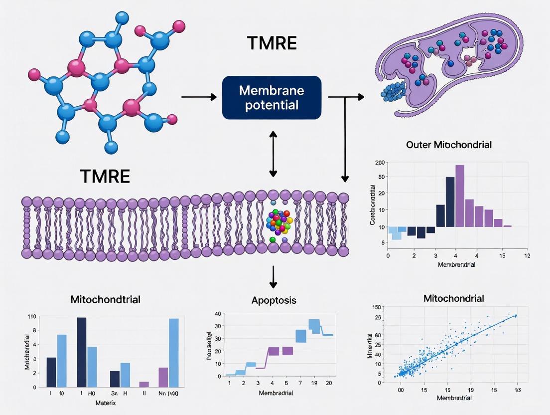
Tetramethylrhodamine ethyl ester (TMRE) belongs to a class of lipophilic cationic dyes that serve as sensitive reporters of ΔΨm in live cells. As a cell-permeant, positively-charged fluorophore, TMRE distributes across membranes in response to electrical gradients, following the principles of the Nernst equation [15] [16]. In the context of mitochondrial function assessment, particularly in cancer research and drug development, TMRE provides researchers with a valuable tool for monitoring metabolic alterations and cellular stress responses [17] [3]. Its application spans multiple detection platforms including fluorescence microscopy, flow cytometry, and microplate-based assays, making it versatile for various experimental setups in basic biological research and pharmaceutical screening [3].
Electrochemical Principles of TMRE Accumulation
The Nernstian Distribution Principle
The accumulation of TMRE in mitochondria follows the Nernst equation, which describes the equilibrium distribution of a permeant ion across a membrane under an electrical potential gradient. For a monovalent cation like TMRE at 37°C, the Nernst equation is expressed as:
ΔΨ = -61.5 log([TMRE]m/[TMRE]c)
Where ΔΨ represents the membrane potential in millivolts, [TMRE]m is the TMRE concentration in the mitochondrial matrix, and [TMRE]c is the concentration in the cytosol [15] [16]. This relationship demonstrates that TMRE accumulates exponentially with increasing membrane potential. A typical resting mitochondrial potential of -180 mV would theoretically result in approximately a 1000-fold higher concentration of TMRE in the matrix compared to the cytosol, though actual accumulation is influenced by additional factors including dye binding to mitochondrial membranes [16] [18].
The driving force for TMRE accumulation is fundamentally electrophoretic - the positively charged dye molecules are attracted to the negatively charged mitochondrial interior [15] [19]. This potential-dependent accumulation makes TMRE fluorescence intensity a sensitive indicator of changes in ΔΨm, with depolarization (less negative potential) causing dye release and decreased fluorescence, while hyperpolarization (more negative potential) enhances dye uptake and fluorescence signal [15].
TMRE Properties and Spectroscopic Characteristics
TMRE is a red-orange fluorescent dye with excitation/emission maxima of approximately 549/575 nm [3]. Its chemical structure includes a delocalized positive charge that facilitates membrane permeability and a lipophilic moiety that promotes accumulation in lipid environments like mitochondrial membranes [15] [16]. Unlike some other mitochondrial dyes, TMRE exhibits a red shift in both absorption and emission spectra when it accumulates in the hydrophobic mitochondrial environment, providing a potential mechanism for rationetric measurements [20].
A critical operational consideration is the concentration-dependent behavior of TMRE. At low concentrations (typically 1-30 nM), TMRE operates in non-quenching mode, where fluorescence intensity is directly proportional to dye concentration and thus to ΔΨm [15] [18]. At higher concentrations (>50-100 nM), TMRE enters quenching mode due to dye aggregation, where fluorescence is self-quenched at high intramitochondrial concentrations, and depolarization leads to dye release and consequent fluorescence dequenching (increased fluorescence) [15]. For most quantitative applications, the non-quenching mode is preferred as it provides a more straightforward relationship between fluorescence intensity and membrane potential [15] [18].
Table 1: Key Properties of TMRE and Related ΔΨm Probes
| Probe | Spectra (Ex/Em) | Operating Modes | Key Advantages | Potential Limitations |
|---|---|---|---|---|
| TMRE | ~549/575 nm [3] | Non-quenching (1-30 nM) or quenching (>50-100 nM) [15] | Low mitochondrial binding and minimal ETC inhibition; suitable for long-term and acute studies [15] | Requires careful concentration optimization; potential phototoxicity at high laser powers |
| TMRM | Similar to TMRE | Same as TMRE | Similar to TMRE but with slightly less membrane binding [15] [20] | Similar to TMRE |
| Rhodamine 123 | ~507/529 nm | Primarily quenching mode (~1-10 μM) [15] | Slow equilibration makes quenching/unquenching easier to detect [15] | More ETC inhibition than TMRM/TMRE [15] |
| JC-1 | 514/529 nm (monomer); 585/590 nm (J-aggregate) | Dual-emission rationetric | "Yes/No" discrimination of polarization state; internal rationetric control [15] | Very sensitive to concentration; aggregate form sensitive to non-ΔΨm factors [15] |
Practical Application Guides
TMRE Staining Protocol for ΔΨm Assessment
The following protocol summarizes established methodologies for TMRE staining to assess mitochondrial membrane potential in live cells using various detection platforms [21] [3] [18]:
Cell Preparation: Seed cells at appropriate density (e.g., 5,000-50,000 cells per well in 96-well plates) and culture for 24-48 hours to reach desired confluence [21].
Experimental Treatment: Apply test compounds or interventions according to experimental design. Include appropriate controls:
- Negative Control: Cells treated with mitochondrial uncouplers such as FCCP (carbonyl cyanide-4-(trifluoromethoxy)phenylhydrazone) at 10-50 μM for 20-30 minutes to completely depolarize mitochondria and establish baseline fluorescence [21] [3].
- Vehicle Control: Cells treated with dye solvent (typically DMSO) at equivalent concentration to account for solvent effects [21].
TMRE Loading:
- Prepare TMRE working solution in pre-warmed culture medium or PBS at concentrations typically ranging from 50-500 nM, optimized for specific cell type and detection method [21] [3] [18].
- Incubate cells with TMRE solution for 15-45 minutes at 37°C in the dark [3].
- For non-quenching mode, use lower TMRE concentrations (1-30 nM); for quenching mode, use higher concentrations (>50-100 nM) [15].
Washing and Preparation for Imaging:
Detection and Analysis:
- Microplate Reader: Read fluorescence at excitation/emission of 544/584 nm [21].
- Flow Cytometry: Use 488 nm laser for excitation and detect emission at ~575 nm [3].
- Fluorescence Microscopy: Use appropriate filter sets for tetramethylrhodamine; widefield, confocal, or multiphoton microscopy can be employed [17].
Table 2: TMRE Staining Conditions Across Different Experimental Systems
| Cell Type | TMRE Concentration | Incubation Time | Detection Method | Key Application |
|---|---|---|---|---|
| Jurkat cells [3] | 100-500 nM | 30-45 minutes | Flow cytometry, microplate reader | Apoptosis studies, drug screening |
| HeLa cells [3] | 200 nM | 20 minutes | Fluorescence microscopy | Cancer cell metabolism |
| Smooth muscle cells [18] | 2.5-25 nM | 10 minutes + equilibration | High-speed 3D imaging | Mitochondrial flicker analysis |
| P19 neurons [3] | 500 nM | 30-45 minutes | Microplate reader | Neurotoxicity assessment |
| MSCs [22] | 50 nM | Not specified | Flow cytometry | Mitochondrial transfer studies |
Critical Controls and Validation Experiments
Proper interpretation of TMRE fluorescence requires implementation of critical controls to ensure that observed fluorescence changes genuinely reflect alterations in ΔΨm rather than confounding factors:
Uncoupler Control: Treatment with protonophores like FCCP (10-50 μM) that collapse the proton gradient and depolarize mitochondria provides a baseline for minimal ΔΨm-dependent staining [21] [3]. A significant decrease in TMRE fluorescence after FCCP treatment validates that dye accumulation is potential-dependent.
Inhibitor Controls: Using compounds that affect ETC function helps contextualize results:
Concentration Optimization: Perform TMRE titration experiments to determine the optimal concentration for specific cell types and experimental conditions, ensuring operation in the desired mode (quenching vs. non-quenching) [15] [18].
Membrane Potential Independence Testing: When investigating potential non-specific dye effects, compare TMRE staining with mitochondrial proteins tagged with fluorescent proteins (e.g., COX8a-GFP, TOM20-GFP) that localize to mitochondria independent of ΔΨm [22].
Cell Health Assessment: Combine TMRE staining with viability markers to exclude fluorescence changes resulting from plasma membrane permeability or cell death [3].
Experimental Workflow for TMRE-Based ΔΨm Assessment
Technical Considerations and Potential Limitations
Common Pitfalls and Troubleshooting
Despite its widespread use, TMRE-based ΔΨm assessment presents several technical challenges that require careful consideration:
Dye Concentration Effects: Inappropriate TMRE concentration is a frequent source of erroneous interpretation. Excessive dye concentrations can cause artifactual fluorescence due to non-specific binding or induce mitochondrial toxicity by inhibiting electron transport chain function [15] [20]. TMRE has been shown to suppress mitochondrial respiratory control, with inhibition being more pronounced than with the closely related TMRM [20]. Always perform initial concentration titration for new cell types or experimental conditions.
Non-Specific Staining: While TMRE is considered relatively specific for mitochondria due to their high membrane potential, recent evidence suggests that cationic dyes can accumulate in other cellular compartments with membrane potential, including endoplasmic reticulum and plasma membrane, particularly when used at higher concentrations [22]. This non-specific accumulation can lead to overestimation of mitochondrial mass or potential.
Photobleaching and Phototoxicity: TMRE is susceptible to photobleaching under prolonged or intense illumination, potentially leading to underestimation of fluorescence intensity [17]. Furthermore, light exposure can generate reactive oxygen species that indirectly affect ΔΨm. Implement appropriate controls for photobleaching and use minimal necessary light exposure during imaging.
Influence of Plasma Membrane Potential: Changes in plasma membrane potential can affect TMRE uptake into the cell, consequently influencing mitochondrial accumulation independent of ΔΨm [18]. For precise ΔΨm measurements, researchers have voltage-clamped the plasma membrane to 0 mV to eliminate this confounding factor [18].
Dye Leakage and Redistribution: TMRE can leak out of cells over time or redistribute during experimental manipulations, particularly after fixation [19]. This necessitates careful timing of measurements after loading and avoidance of fixatives for live-cell imaging.
Limitations in Interpreting TMRE Fluorescence
Several fundamental limitations affect the interpretation of TMRE fluorescence data:
ΔΨm vs. ΔpHm Distinction: TMRE and related cationic dyes measure only the electrical component (ΔΨm) of the total proton motive force (Δp). The pH gradient (ΔpHm) constitutes a significant portion of Δp (typically 30-60 mV out of 180-220 mV total) but is not detected by these dyes [15]. Changes in mitochondrial pH can occur independently of ΔΨm alterations, potentially leading to misinterpretation of mitochondrial energetic status [15].
Non-Protonic Charge Effects: Intracellular ion changes, particularly calcium fluxes, can influence ΔΨm measurements independently of protonic gradients. Studies have demonstrated conditions where mitochondrial hyperpolarization detected by TMRE occurred concurrently with matrix acidification, contrary to expected coupling between electrical and chemical gradients [15]. This highlights that TMRE cannot distinguish between charge contributions from protons versus other ions like Ca²⁺ [15].
Quantitative Challenges: While TMRE distribution follows Nernstian principles, quantitative determination of absolute ΔΨm values is complicated by dye binding to mitochondrial membranes, which varies with temperature and mitochondrial physiological state [18] [20]. Binding effectively increases the apparent accumulation beyond that predicted by the Nernst equation for the free dye concentration [20].
Artifacts in Mitochondrial Transfer Studies: Recent investigations have revealed significant limitations using TMRE and similar dyes as surrogates for actual mitochondrial transfer between cells. Comparative studies demonstrate that TMRE signal transfers between cells at much higher efficiency than mitochondrial-targeted fluorescent proteins, suggesting direct dye transfer rather than organelle movement [22]. This calls for caution in interpreting dye redistribution as evidence of mitochondrial trafficking.
TMRE Behavior: Principle and Confounding Factors
Table 3: Key Research Reagent Solutions for TMRE-Based ΔΨm Analysis
| Reagent/Resource | Function/Application | Key Considerations |
|---|---|---|
| TMRE Assay Kits (e.g., ab113852) [3] | Complete kits including TMRE and FCCP control | Provide standardized protocols and optimized reagent concentrations; suitable for multi-platform detection |
| FCCP [21] [3] | Proton ionophore; mitochondrial uncoupler | Used as negative control to collapse ΔΨm; typically used at 10-50 μM for 20-30 minutes |
| Oligomycin [15] | ATP synthase inhibitor | Used to induce hyperpolarization; helps distinguish ΔΨm changes related to ATP synthesis |
| Carbonyl cyanide m-chlorophenyl hydrazone (CCCP) | Alternative mitochondrial uncoupler | Similar function to FCCP; different potency and solubility profile |
| MitoTracker Probes [19] | Fixable mitochondrial dyes | Useful for comparison studies; some variants (e.g., MitoTracker Green FM) show different potential dependence |
| CellLight Mitochondrial Fluorescent Proteins [19] | Genetic labeling of mitochondria | Provide potential-independent mitochondrial localization; useful for normalization and control experiments |
| MitoSOX Red [19] | Mitochondrial superoxide indicator | Can be combined with TMRE for multi-parameter assessment of mitochondrial function |
| Rotenone and Antimycin A | ETC complex inhibitors | Used to investigate specific sites of respiratory chain dysfunction affecting ΔΨm |
Advanced Applications and Future Perspectives
TMRE-based ΔΨm assessment continues to evolve with advancing imaging technologies and experimental approaches. High-speed 3D imaging techniques have enabled the quantification of spontaneous, transient mitochondrial depolarizations ("flickers") with millivolt resolution, revealing heterogeneous mitochondrial behavior within individual cells [18]. These flickers, ranging from <10 mV to >100 mV in amplitude, represent dynamic mitochondrial responses to various physiological stimuli and stressors [18].
The integration of TMRE staining with advanced microscopy modalities including multiphoton microscopy and fluorescence lifetime imaging (FLIM) provides enhanced spatial resolution and quantitative capabilities, particularly valuable for investigating mitochondrial heterogeneity in complex tissues and cancer models [17]. Furthermore, the combination of TMRE with other fluorescent indicators of mitochondrial function (e.g., Ca²⁺ sensors, ROS probes) enables multi-parameter assessment of mitochondrial physiology in live cells [17] [19].
Emerging concerns about potential limitations of TMRE and similar dyes in specific applications, particularly in mitochondrial transfer studies [22], highlight the importance of complementary approaches using genetic fluorescent protein tags for definitive mitochondrial tracking. Future methodological developments will likely focus on improving specificity, reducing phototoxicity, and enabling absolute quantification of ΔΨm rather than relative changes.
When appropriately applied with necessary controls and awareness of its limitations, TMRE remains a powerful tool for investigating mitochondrial function in health and disease, contributing significantly to our understanding of cellular bioenergetics in basic research and drug discovery applications.
Mitochondrial membrane potential (ΔΨm), the electrical gradient across the inner mitochondrial membrane, is a central parameter of mitochondrial function and cellular health [23]. It is generated by the electron transport chain (ETC), which pumps protons from the matrix into the intermembrane space, creating an electrochemical gradient that drives ATP synthesis [24] [25]. This proton motive force is fundamental for energy production, reactive oxygen species (ROS) regulation, calcium buffering, and apoptotic signaling [24] [25]. Consequently, deviations in ΔΨm are critical biomarkers in pathologies ranging from neurodegeneration to cancer. This application note details the central role of ΔΨm analysis, specifically using the fluorescent probe Tetramethylrhodamine Ethyl Ester (TMRE), within research frameworks investigating neurodegenerative diseases, cancer biology, and toxicological screening.
Table 1: Key Functional Roles of Mitochondrial Membrane Potential
| Functional Role | Biological Significance | Pathological Consequences of Dysregulation |
|---|---|---|
| ATP Synthesis | Drives proton flux through F1F0 ATP synthase to produce cellular energy [24]. | Energy depletion, impaired cellular function [25]. |
| Calcium Buffering | Facilitates mitochondrial calcium uptake, regulating cytosolic calcium levels and signaling [25]. | Disrupted calcium homeostasis, exacerbation of excitotoxicity [25]. |
| ROS Production | ΔΨm above ~140 mV exponentially increases ROS production, particularly at ETC complexes I and III [24]. | Oxidative stress, damage to cellular macromolecules [23] [24]. |
| Apoptotic Regulation | ΔΨm collapse often precedes cytochrome c release and caspase activation [23]. | Dysregulated cell death; either excessive (neurodegeneration) or impaired (cancer) [23] [24]. |
| Protein Import | Required for the import of nuclear-encoded proteins into the mitochondrial matrix [25]. | Defective mitochondrial biogenesis and function [25]. |
ΔΨm in Biological Contexts
ΔΨm in Neurodegeneration
In neurons, ΔΨm is not uniform; a spatial gradient exists where the potential is highest in the soma and decreases along the axons, making distal synaptic mitochondria inherently more vulnerable to stress [23]. Neurodegeneration often proceeds via a "two-hit" model [23]. The first hit is this pre-existing lower ΔΨm at synapses. A second hit, such as expression of mutant proteins (e.g., amyloid-β in Alzheimer's disease), oxidative stress, or aging, can push these vulnerable mitochondria over the threshold, triggering synaptic degeneration through sub-lethal caspase activation and cytokine production [23]. Furthermore, ΔΨm is essential for mitochondrial dynamics—fusion, fission, and trafficking—processes critical for neuronal health. Sustained depolarization excludes mitochondria from the fusion pool, targeting them for autophagic removal (mitophagy) [25].
ΔΨm in Cancer Biology
Cancer cells frequently exhibit an abnormally high ΔΨm (hyperpolarization) compared to their normal counterparts [24]. This elevated potential is associated with decreased susceptibility to apoptosis and enhanced invasive and metastatic potential [24]. In vivo models show that cancer cells with high ΔΨm lead to a greater metastatic burden than those with low ΔΨm [24]. The hyperpolarized state can also drive excessive ROS production, which can act as a signaling molecule to promote proliferative pathways [24]. Furthermore, ΔΨm is implicated in therapeutic resistance, as a decrease in ΔΨm has been identified as an indicator of radioresistant cancer cells [26]. Recent evidence also positions ΔΨm as a retrograde signal that regulates cell cycle progression, where decreased ΔΨm delays the G1-to-S phase transition [27].
ΔΨm in Toxicity Screening and Drug Development
ΔΨm is a sensitive indicator of drug-induced mitochondrial toxicity. Many pharmacological agents, including certain phenylpropanoids studied in neurodegeneration, exhibit a biphasic, concentration-dependent effect on ΔΨm [23]. At low concentrations, compounds like EGCG or quercetin can protect ΔΨm and restore mitochondrial function. However, at higher concentrations, they may induce ΔΨm dissipation and apoptosis [23]. This underscores the critical importance of dose-response study designs in toxicological screening. Assays measuring ΔΨm are therefore vital for identifying both protective compounds and off-target mitochondrial toxicities in drug development pipelines.
Table 2: Pharmacological Modulators of ΔΨm in Research
| Compound | Target/Activity | Effect on ΔΨm | Research Context |
|---|---|---|---|
| FCCP/CCCP | Protonophore (Uncoupler) | Depolarization (↓ ΔΨm) [28] | Positive control for depolarization; induces mitophagy [29]. |
| Oligomycin | ATP Synthase Inhibitor | Hyperpolarization (↑ ΔΨm) [28] [30] | Inhibits proton flux back into matrix, increasing ΔΨm but reducing ATP production. |
| Antimycin A | Complex III Inhibitor | Depolarization (↓ ΔΨm) [30] | Inhibits ETC, reduces proton pumping. Can increase ROS [24]. |
| BAM15 | Uncoupler | Depolarization (↓ ΔΨm) [27] | Dissipates ΔΨm without depolarizing plasma membrane [27]. |
| PMI | P62-mediated mitophagy inducer | Independent (No change) [29] | Indces mitophagy without collapsing ΔΨm, avoiding toxicity [29]. |
| EGCG/Quercetin | Polyphenols (Biphasic) | Low conc.: Protection; High conc.: Depolarization [23] | Models for concentration-dependent toxicity and therapeutic windows. |
TMRE-Based Analysis of ΔΨm: Protocols and Applications
TMRE Staining Protocol for Live-Cell Imaging
TMRE is a cell-permeant, cationic fluorescent dye that accumulates in active mitochondria in a ΔΨm-dependent manner. The following protocol is adapted for rat cortical neurons [28] but is applicable to various cell lines with minimal modifications.
Key Reagents:
- TMRE Stock Solution: 10 mM in anhydrous DMSO. Store in aliquots at -20°C, protected from light [28].
- Tyrode's Buffer (TB): 145 mM NaCl, 5 mM KCl, 10 mM Glucose, 1.5 mM CaCl₂, 1 mM MgCl₂, 10 mM HEPES; adjust pH to 7.4 with NaOH [28].
Staining Procedure:
- Cell Preparation: Culture cells on glass-bottom dishes (e.g., MatTek). Wash cells 3 times with pre-warmed TB or culture medium to remove serum [28].
- Dye Loading:
- Post-Incubation: For imaging, after incubation, mount the dish on the microscope stage in fresh TB with 50 nM TMRE to maintain equilibrium distribution [28] [30]. For flow cytometry, resuspend dye-loaded cells in cold DPBS with 1% FBS and analyze immediately [31].
Live-Cell Imaging and Data Analysis
Image Acquisition:
- Use a confocal laser-scanning microscope with a 63x oil immersion objective [28] [30].
- Excitation/Emission: 561 nm / 570-610 nm for TMRE [28] [30].
- Use low laser power (1-5%) and resolution (256 x 256) to minimize photobleaching and phototoxicity [28].
- Acquire a time-series to establish a stable baseline (e.g., 5-10 images over 2-5 minutes).
Pharmacological Validation:
- Apply 1 μM FCCP (uncoupler) at the end of the baseline acquisition. A successful experiment shows a rapid and significant decrease in TMRE fluorescence intensity, confirming the ΔΨm-dependent nature of the signal [28].
Quantitative Analysis:
- Region of Interest (ROI): Select ROIs over mitochondrial regions or entire cell bodies [28].
- Background Subtraction: Measure fluorescence intensity in a cell-free region and subtract this value from the cellular ROI intensities [28].
- Normalization: Normalize fluorescence intensity (F) to the average baseline fluorescence (F₀) for each ROI using the formula: ΔF/F₀ (%) = (F - F₀)/F₀ × 100 [28].
- Heterogeneity Analysis: Calculate the coefficient of variation (CV) from single-cell measurements to assess population heterogeneity [30].
The Scientist's Toolkit: Key Research Reagent Solutions
Table 3: Essential Reagents for ΔΨm Research
| Reagent / Assay Kit | Function / Specificity | Key Considerations |
|---|---|---|
| TMRE (Tetramethylrhodamine Ethyl Ester) | Potentiometric ΔΨm indicator [28] [30]. | Use in non-quenching mode (low nM range). Fluorescence intensity proportional to ΔΨm. |
| TMRE-Mitochondrial Membrane Potential Assay Kit | Complete kit including TMRE dye and optimized buffer [31]. | Streamlines workflow; includes protocols for flow cytometry. |
| JC-1 | Rationetric ΔΨm indicator; forms J-aggregates (red) in high ΔΨm [23]. | More complex signal interpretation due to aggregation; can be affected by mitochondrial morphology. |
| FCCP / CCCP | Proton ionophores; positive control for complete ΔΨm depolarization [28]. | Use at 0.5-1 μM for intact cells. CCCP may have broader cellular effects than FCCP. |
| Oligomycin | ATP synthase inhibitor; positive control for ΔΨm hyperpolarization [28] [30]. | Use at 1-2 μg/ml. Hyperpolarization is due to inhibition of proton consumption by ATP synthase. |
| H2DCF-DA | Cell-permeant indicator for general oxidative stress/ROS [28]. | Useful for parallel assessment of ROS, a key parameter linked to ΔΨm [24] [28]. |
| MitoTracker Probes (e.g., Deep Red) | Covalent mitochondrial labels; independent of ΔΨm for long-term tracking [23]. | Ideal as a morphological reference stain for mitochondrial location and mass. |
The analysis of mitochondrial membrane potential using TMRE is an indispensable tool for probing cellular health and function across diverse research fields. The precise protocols and contextual data provided in this application note equip researchers to effectively apply this technique. As evidenced, ΔΨm serves as a critical node linking metabolic state to fundamental cellular outcomes in neurodegeneration, cancer, and drug-induced toxicity, making its accurate measurement vital for advancing both basic science and therapeutic development.
A Practical Guide to TMRE Assays: From Basic Protocols to Advanced High-Throughput Applications
Mitochondrial membrane potential (ΔΨm) is the electrical potential difference across the inner mitochondrial membrane, a key parameter reflecting mitochondrial health and cellular energy status [3]. It is generated by the proton pumps of the electron transport chain and is essential for ATP production through oxidative phosphorylation [3]. Dysregulation of ΔΨm is a hallmark of cellular dysfunction and is implicated in various diseases, including neurodegenerative disorders, cancer, and metabolic syndromes [3] [32].
Tetramethylrhodamine ethyl ester (TMRE) is a cell-permeant, cationic, fluorescent dye that readily accumulates in active mitochondria due to their relative negative charge [3]. The intensity of TMRE fluorescence is directly proportional to the ΔΨm. Depolarized or inactive mitochondria exhibit a decreased membrane potential and fail to sequester TMRE, resulting in a diminished fluorescence signal [3] [33]. This property makes TMRE an ideal probe for monitoring changes in mitochondrial function in live cells across various experimental platforms, including flow cytometry, fluorescence microscopy, and microplate readers [3].
Principle of TMRE Assay and Key Signaling Pathways
The TMRE assay leverages the electrochemical gradient across the inner mitochondrial membrane. The positively charged TMRE molecule is electrophoretically taken up into the mitochondrial matrix in a manner dependent on the membrane potential. In healthy, polarized mitochondria, this results in the accumulation of the dye and a strong fluorescent signal. A loss of ΔΨm, which can occur during cellular stress or the early stages of apoptosis, prevents this accumulation, leading to a reduction in fluorescence [3] [34].
The following diagram illustrates the core principle of the TMRE assay and its connection to key cellular pathways, culminating in the experimental readout.
The Scientist's Toolkit: Essential Reagents and Materials
Successful execution of the TMRE assay requires careful preparation of reagents and access to appropriate instrumentation. The table below details the core components of the research toolkit.
Table 1: Key Research Reagent Solutions and Materials for TMRE Assay
| Item | Function/Description | Examples / Notes |
|---|---|---|
| TMRE | Cell-permeant, cationic fluorescent dye that accumulates in polarized mitochondria. | Often supplied as a stock solution in DMSO [3] [11]. |
| FCCP | Proton ionophore uncoupler; used as a positive control to dissipate ΔΨm and validate the assay [3]. | Carbonyl cyanide 4-(trifluoromethoxy)phenylhydrazone [3]. |
| Assay Buffers | For washing cells and diluting dyes (e.g., PBS). PBS with 0.2% BSA is recommended for washing steps to reduce background [3]. | Krebs-Ringer-Hepes (KRH) buffer can be used for calcium-related studies [32]. |
| Live Cells | The assay is exclusively for use with live, unfixed cells [3]. | Adherent (e.g., HeLa) or suspension (e.g., Jurkat) cells [3]. |
| Detection Instruments | Equipment for measuring fluorescence signal. | Fluorescent microplate reader, microscope, or flow cytometer [3] [35]. |
Core Experimental Protocols
This section provides detailed, step-by-step methodologies for assessing ΔΨm using TMRE across three common platforms.
General Staining Protocol
The foundational staining procedure is consistent across all detection methods and must be optimized for specific cell types.
- Preparation of Staining Solution: Dilute TMRE in pre-warmed culture medium to the desired working concentration. A typical working range is 50-500 nM [3] [32]. Note: Using TMRE at concentrations <200 nM is recommended to avoid fluorescence quenching and artifactual signals [32].
- Positive Control Preparation: Treat control cell samples with the uncoupler FCCP (e.g., 1-100 µM) for 10 minutes at 37°C prior to and during TMRE staining to depolarize mitochondria [3] [34].
- Staining Incubation: Remove culture medium from cells and add the TMRE staining solution. Incubate for 15-30 minutes at 37°C in a 5% CO₂ incubator [3] [32].
- Washing: After incubation, carefully pellet suspension cells or remove the staining solution from adherent cells. Gently wash the cells 1-3 times with PBS, preferably containing 0.2% BSA, to remove excess, non-specific dye [3] [32].
- Immediate Analysis: Resuspend or overlay the cells with fresh culture medium or PBS and proceed immediately with analysis on your chosen platform. Do not fix the cells, as this will disrupt the potential-dependent staining [3] [34].
Platform-Specific Protocols and Parameters
The following table summarizes the critical instrument settings and procedural notes for each detection method.
Table 2: Platform-Specific Parameters for TMRE Analysis
| Parameter | Flow Cytometry | Fluorescence Microscopy | Microplate Reader |
|---|---|---|---|
| Key Steps | 1. Prepare single-cell suspension after staining [3].2. Resuspend in PBS for analysis.3. Acquire at least 10,000 events per sample. | 1. Culture cells on glass-bottom dishes or chamber slides [35].2. After staining and washing, add a small volume of fresh medium for imaging.3. Image promptly to maintain cell viability. | 1. Seed cells in sterile, clear-bottom, black-walled microplates [35].2. Include blanks (wells with medium but no cells) for background subtraction. |
| Excitation/Emission | 488 nm laser excitation; detection with a 575 nm filter (e.g., PE channel) [3] [35]. | Standard TRITC filter set [11]. Ex/Em ~549/575 nm [3]. | Ex/Em = 549/575 nm [3]. |
| Data Output | Fluorescence intensity per cell (histograms). Enables quantification of heterogeneous responses within a population [3] [34]. | Qualitative and spatial information on mitochondrial localization and morphology within single cells [3] [32]. | Mean Fluorescent Intensity (MFI) per well, providing a population-average measurement [3]. |
| Critical Validation | Compare fluorescence intensity histograms of untreated vs. FCCP-treated cells. A clear left-shift (signal decrease) should be observed in the FCCP-treated sample [3] [34]. | Visually confirm loss of punctate mitochondrial staining and overall signal reduction in FCCP-treated cells compared to the bright, granular pattern in healthy cells [3]. | The MFI of FCCP-treated controls should be significantly lower than that of untreated cells. Data is often presented as MFI +/- standard deviation [3]. |
The workflow for all three platforms is consolidated in the following experimental roadmap.
Troubleshooting and Best Practices
Even with a robust protocol, researchers may encounter challenges. The table below outlines common issues and recommended solutions.
Table 3: Troubleshooting Guide for TMRE Assays
| Problem | Potential Cause | Recommended Solution |
|---|---|---|
| High Background / Nonspecific Signal | Incomplete washing of excess dye [32]. | Increase the number of washes with PBS/0.2% BSA [32]. |
| TMRE concentration is too high [32]. | Titrate the TMRE dose. Use lower concentrations (e.g., 50-100 nM) and avoid exceeding 200 nM to prevent quenching [32]. | |
| Weak or No Signal | Loss of ΔΨm due to cell death or excessive stress. | Check cell viability and health. Ensure cultures are not over-confluent. |
| Photobleaching from prolonged light exposure [32]. | Minimize light exposure during staining and analysis; use lower laser power or shorter exposure times [32]. | |
| Inconsistent Results | Inadequate FCCP control validation. | Always include an FCCP-treated control. If this control does not show a strong signal decrease, the assay is not functioning correctly [3] [32]. |
| Dye precipitation or degradation. | Ensure TMRE stock is properly stored at -20°C and avoid repeated freeze-thaw cycles. Centrifuge staining solution before use if precipitation is suspected. | |
| Poor Mitochondrial Localization (Microscopy) | Probe accumulation in non-mitochondrial compartments [32]. | Confirm mitochondrial localization by co-staining with a validated mitochondrial marker (e.g., MitoTracker) [32]. |
Mitochondrial membrane potential (ΔΨm) is a critical indicator of mitochondrial health and cellular viability, serving as a key parameter in fields ranging from fundamental cell biology to drug development. Tetramethylrhodamine, ethyl ester (TMRE) is a cell-permeant, cationic dye that accumulates in active mitochondria in a ΔΨm-dependent manner, making it a vital tool for assessing mitochondrial function. The reliability of TMRE-based assays, however, is highly dependent on the meticulous optimization of several key parameters. This application note provides a detailed framework for optimizing TMRE concentration, incubation time, and cell density to ensure robust, reproducible, and accurate assessment of ΔΨm. Proper optimization is not merely a technical exercise; it is fundamental to generating high-quality data that can accurately inform on mitochondrial responses to pharmacological treatments or genetic modifications, thereby supporting critical decisions in the research and development pipeline [4].
The Critical Role of Mitochondrial Membrane Potential
The mitochondrial membrane potential, generated by the electron transport chain (ETC), represents approximately 80% of the proton motive force (Δp) that drives ATP synthesis. Maintaining ΔΨm is essential for mitochondrial functions, including protein import, ion homeostasis, and ATP production [4] [10]. Notably, ΔΨm is not a static metric. It dynamically reflects the balance between its generation by the ETC and its consumption primarily by ATP synthase. This means that a change in ΔΨm must be interpreted carefully: a decrease could indicate either a loss of ETC function or an increase in ATP demand and turnover [4].
Hyperpolarization and Depolarization: While mitochondrial depolarization is a well-established hallmark of dysfunction and a trigger for mitophagy, chronic mitochondrial hyperpolarization is increasingly recognized for its profound cellular effects. Research using IF1-knockout cell models has demonstrated that sustained hyperpolarization can trigger extensive transcriptional reprogramming, alter nuclear DNA methylation, and remodel phospholipids, influencing processes from epigenetics to cell cycle progression [10]. This underscores that both increases and decreases in ΔΨm can be biologically significant, necessitating precise and reliable measurement techniques.
TMRE as a Measurement Tool: TMRE is a potentiometric dye that distributes across the mitochondrial membrane according to the Nernst equation. In healthy, polarized mitochondria, the negatively charged interior attracts and concentrates the cationic TMRE, resulting in intense fluorescence. A loss of ΔΨm prevents this accumulation, leading to a diffuse distribution of the dye in the cytosol and a corresponding decrease in fluorescent signal. This property makes TMRE an excellent indicator of mitochondrial function, but its accurate application requires careful protocol standardization [11].
Optimizing Key Parameters for TMRE Staining
Successful ΔΨm measurement with TMRE hinges on a balanced interplay between dye concentration, incubation time, and cell density. The following section provides optimized parameters and a structured protocol to guide researchers.
Parameter Optimization Guidelines
Table 1: Key Parameters for TMRE Staining Optimization
| Parameter | Recommended Range | Key Considerations | Impact of Sub-Optimal Conditions |
|---|---|---|---|
| TMRE Working Concentration | 150 - 500 nM | A common effective concentration is 250 nM [11]. The optimal concentration should be determined empirically for each cell type. | Too High: Can induce mitochondrial toxicity and uncoupling.Too Low: Results in a weak fluorescent signal and poor resolution. |
| Incubation Time | 15 - 30 minutes | A standard incubation time is 30 minutes at 37°C [11]. Ensure consistent timing across all samples. | Too Short: Incomplete dye loading and underestimation of ΔΨm.Too Long: Potential for dye toxicity and artifactual results. |
| Cell Density | 50 - 80% confluency | Ensure cells are healthy and not over-confluent to avoid nutrient depletion and stress-induced changes in ΔΨm. | Too Dense: Leads to nutrient competition, contact inhibition, and altered metabolism.Too Sparse: Inconsistent imaging fields and higher well-to-well variability. |
| Post-Staining Wash | 2-3 washes with clear buffer | Use pre-warmed PBS or other clear saline-based buffer. Image promptly after washing. | Insufficient Washing: High background fluorescence from unincorporated dye.Excessive Washing/Delays: Risk of dye leakage from mitochondria. |
Detailed Staining Protocol
This protocol is designed for a single well of a 6-well plate or a 35 mm dish. Scale volumes accordingly [11].
Preparation of Staining Solution
- Prepare a 10 mM stock solution of TMRE in DMSO and store it at -20°C.
- On the day of the experiment, prepare an intermediate dilution (e.g., 50 µM) in complete cell culture medium.
- Prepare the final working staining solution (e.g., 250 nM) by diluting the intermediate stock in pre-warmed complete medium. Protect from light.
Cell Staining
- Culture cells according to standard practices, ensuring they are at an optimal density (e.g., 50-80% confluency) at the time of staining.
- Aspirate the culture medium from the live cells.
- Add the prepared TMRE staining solution to the cells.
- Incubate for 30 minutes in a 37°C incubator (5% CO₂), protected from light.
Washing and Imaging
- After incubation, carefully aspirate the TMRE staining solution.
- Gently wash the cells 2-3 times with pre-warmed PBS or another clear, buffered saline solution.
- After the final wash, add a small volume of pre-warmed clear buffer or FluoroBrite DMEM to the cells.
- Image the cells immediately using a fluorescence microscope equipped with a TRITC (or Cy3) filter set. For live-cell imaging, maintain the cells at 37°C during image acquisition.
Validation and Controls
Including appropriate controls is non-negotiable for validating the specificity of the TMRE signal.
- Depolarization Control: Treat a separate sample of cells with a mitochondrial uncoupler such as FCCP (1-10 µM) or a combination of oligomycin (1 µM) and antimycin A (1 µM) for 15-30 minutes prior to and during TMRE staining. A valid assay will show a marked reduction in punctate mitochondrial fluorescence in the control-treated sample [4].
- Viability Control: Always confirm cell viability under the chosen staining conditions, as excessive TMRE concentrations or prolonged incubation can be toxic.
- Quenching vs. Non-Quenching Mode: Be aware of the operational mode. The recommended concentrations (e.g., 250 nM) are typically for the non-quenching mode, where fluorescence intensity is proportional to ΔΨm. High, self-quenching concentrations require a different interpretation and are not recommended for standard assays [36].
The Scientist's Toolkit: Essential Reagents and Materials
Table 2: Key Research Reagent Solutions for TMRE-based ΔΨm Analysis
| Reagent / Material | Function / Purpose | Example & Notes |
|---|---|---|
| TMRE | Potentiometric fluorescent dye used to measure mitochondrial membrane potential. | Tetramethylrhodamine, ethyl ester; supplied as a powder, typically made into a 10 mM stock in DMSO [11]. |
| FCCP / CCCP | Protonophore uncouplers; used as a key validation control to dissipate ΔΨm. | Confirms that TMRE signal is ΔΨm-dependent. A working concentration is often 1-10 µM. |
| Oligomycin | ATP synthase inhibitor; used in control experiments to manipulate ΔΨm. | Inhibits the forward activity of ATP synthase, which can lead to a hyperpolarization of the membrane [4] [10]. |
| Cell Permeabilizer | For assays on isolated mitochondria or controlled substrate conditions. | e.g., Digitonin. Used in conjunction with substrates like succinate to energize mitochondria [10]. |
| MitoTracker Green (MTG) | A ΔΨm-independent mitochondrial mass dye. | Useful for normalizing TMRE fluorescence to mitochondrial content, helping to distinguish changes in potential from changes in mass [10]. |
| Live-Cell Imaging Medium | A clear, low-fluorescence buffer for maintaining cells during imaging. | e.g., FluoroBrite DMEM or HBSS. Helps reduce background fluorescence and maintains physiological pH. |
Visualizing the Workflow and Key Relationships
The following diagrams illustrate the core principles of the TMRE assay and the experimental workflow.
Diagram 1: TMRE Experimental Workflow. This flowchart outlines the key steps for a standard TMRE staining experiment, from sample preparation to data analysis.
Diagram 2: TMRE Signal Response to ΔΨm. This diagram illustrates the fundamental principle of how TMRE distribution and fluorescence signal change in response to the health status of the mitochondria.
Integrated Analysis: Connecting ΔΨm to Broader Cellular Phenotypes
Changes in ΔΨm do not occur in isolation. Integrating TMRE-based measurements with other cellular assays provides a systems-level understanding of how mitochondrial status influences cell fate. For instance, a decrease in cell number observed in a pharmacological screen could be due to either increased cell death or decreased proliferation. A multiparametric flow cytometry approach that simultaneously measures ΔΨm (using JC-1, a dye analogous to TMRE), apoptosis (annexin V/PI), and proliferation (BrdU or CellTrace Violet) can dissect these mechanisms [12]. This integrated workflow can reveal, for example, that a drug-induced mitochondrial depolarization triggers the intrinsic apoptosis pathway, leading to increased cell death. Furthermore, changes in ΔΨm can modulate nuclear gene expression through mechanisms like phospholipid remodeling, creating a feedback loop that influences the cell's long-term adaptive response [10]. Therefore, TMRE staining serves as a critical entry point for a deeper, more comprehensive investigation into cellular bioenergetics and health.
Mitochondrial membrane potential (ΔΨm) is a key indicator of cellular health, serving as a critical parameter for evaluating mitochondrial function. The electrochemical gradient across the inner mitochondrial membrane drives ATP production and is essential for maintaining cellular homeostasis [37] [4]. A significant loss of ΔΨm is an early event in apoptosis and other pathological conditions, rendering cells depleted of energy with subsequent death [28] [7]. Fluorescent dyes such as TMRE (tetramethylrhodamine ethyl ester) are widely used to monitor ΔΨm in live cells. However, the specificity of these dyes for ΔΨm-dependent staining must be rigorously validated using appropriate controls. This application note details the use of mitochondrial uncouplers FCCP (carbonyl cyanide-p-trifluoromethoxyphenylhydrazone) and CCCP (carbonyl cyanide 3-chlorophenylhydrazone) as essential controls to confirm that observed fluorescence changes genuinely reflect alterations in ΔΨm rather than non-specific artifacts [3] [28].
Theoretical Background: The Role of FCCP/CCCP in Validating ΔΨm Measurements
Mechanism of Protonophore Action
FCCP and CCCP are protonophores that function as mitochondrial uncouplers by dissipating the proton gradient across the inner mitochondrial membrane. These lipophilic weak acids shuttle protons across the mitochondrial membrane, effectively collapsing the electrochemical gradient that constitutes ΔΨm [3] [4]. This action decouples substrate oxidation from ATP synthesis, leading to maximum electron transport chain activity without ATP production. When used as positive controls in TMRE staining experiments, FCCP/CCCP treatment should result in a marked decrease in TMRE fluorescence, confirming that the dye accumulation is ΔΨm-dependent [3] [28].
Importance of Experimental Controls
Without proper controls, fluorescence changes attributed to ΔΨm may actually result from non-specific factors including dye loading variability, changes in mitochondrial mass, plasma membrane potential alterations, or non-specific binding. The inclusion of FCCP/CCCP controls provides a critical benchmark for distinguishing ΔΨm-specific staining from these confounding factors [37] [4]. This validation is particularly important when investigating the effects of novel compounds on mitochondrial function in drug development contexts, where accurate assessment of mitochondrial toxicity is essential [14].
TMRE Staining Protocol with FCCP/CCCP Controls
Principle of TMRE Staining
TMRE is a cell-permeant, cationic, red-orange fluorescent dye that accumulates in active mitochondria due to their relative negative charge [3] [38]. The dye enters mitochondria in a membrane potential-dependent manner and is retained at higher concentrations in polarized mitochondria. Depolarized or inactive mitochondria exhibit decreased membrane potential and fail to sequester TMRE, resulting in reduced fluorescence intensity [3] [7]. TMRE is suitable for quantitative measurement of membrane potential using the Nernst equation and can be applied with various detection platforms including fluorescence microscopy, flow cytometry, and microplate fluorometry [38].
Reagent Preparation
Staining Procedure for Adherent Cells
- Cell Preparation: Plate adherent cells in appropriate culture vessels (e.g., 96-well plates, chamber slides) and culture until 70-90% confluent [3] [39].
- Positive Control Setup: Add FCCP or CCCP to designated positive control wells at final concentrations typically ranging from 1-50 μM and incubate for 10-30 minutes at 37°C [3] [28].
- TMRE Loading: Prepare TMRE working solution in pre-warmed culture medium or buffer at concentrations typically between 50-500 nM [3] [28]. Remove culture medium from cells and replace with TMRE-containing solution.
- Incubation: Incubate cells with TMRE for 15-45 minutes at 37°C protected from light [3] [35].
- Washing: Remove TMRE solution and wash cells 1-2 times with PBS or assay buffer to remove excess dye [3] [39].
- Imaging/Analysis: Immediately analyze samples using an appropriate detection platform.
Staining Procedure for Suspension Cells
- Cell Preparation: Harvest suspension cells and centrifuge at 400 × g for 5 minutes [37]. Resuspend in pre-warmed culture medium or buffer at approximately 1 × 10^6 cells/mL [37].
- Positive Control Setup: Add FCCP or CCCP to aliquot of cells (final concentration 1-50 μM) and incubate for 10-30 minutes at 37°C [3].
- TMRE Loading: Add TMRE to both untreated and FCCP/CCCP-treated cells (final concentration 50-500 nM) and incubate for 15-45 minutes at 37°C protected from light [3].
- Washing: Centrifuge cells at 400 × g for 5 minutes, remove supernatant, and resuspend in fresh buffer [37].
- Analysis: Analyze by flow cytometry or other appropriate methods.
Detection Methods
- Flow Cytometry: Use 488 nm excitation with emission detection at ~575 nm [3] [35].
- Fluorescence Microscopy: Use standard TRITC/Rhodamine filter sets (Ex/Em ~549/575 nm) [3] [28].
- Microplate Reader: Measure fluorescence with Ex/Em 549/575 nm [3] [35].
Quantitative Data and Optimization
Table 1: TMRE Staining Conditions Across Cell Types
| Cell Type | TMRE Concentration | Incubation Time | FCCP/CCCP Concentration | Reference |
|---|---|---|---|---|
| Cortical Neurons | 20 nM | 45 min | 1 μM FCCP | [28] |
| Jurkat Cells | 100-400 nM | 15-30 min | 10-100 μM FCCP | [3] |
| HeLa Cells | 200 nM | 20 min | Not specified | [3] |
| HepG2 Cells | Not specified | 30 min | 3.5-6.9 μM FCCP | [14] |
| SJK Cells | Not specified | 15 min | 0.6-50 μM CCCP | [39] |
Table 2: Expected Fluorescence Changes with FCCP/CCCP Treatment
| Condition | TMRE Fluorescence | ΔΨm Status | Biological Interpretation |
|---|---|---|---|
| Untreated Control | High | Polarized (-180 mV) | Healthy, functional mitochondria |
| FCCP/CCCP Treated | Low (70-95% reduction) | Depolarized | Complete mitochondrial uncoupling |
| Apoptotic Cells | Intermediate | Partially depolarized | Early apoptotic event |
| ATP Synthase Inhibition | Increased | Hyperpolarized | Reduced proton flux through ATP synthase |
Troubleshooting and Technical Considerations
Common Issues and Solutions
- Excessive Background Fluorescence: Reduce TMRE concentration or increase wash steps to minimize non-specific staining [28].
- Incomplete Depolarization with FCCP/CCCP: Verify uncoupler concentration and pre-incubation time; consider titration experiments [3] [39].
- Cellular Toxicity: Limit TMRE incubation time and concentration; TMRE is generally non-toxic at recommended concentrations [38].
- Photobleaching: Minimize light exposure during staining and imaging; use antifade reagents if necessary [28].
Critical Optimization Parameters
- Dye Concentration: Use the lowest effective TMRE concentration (typically 50-500 nM) to avoid artifacts [28].
- Loading Time: Determine optimal incubation time for each cell type (typically 15-45 minutes) [3].
- Uncoupler Titration: Establish FCCP/CCCP concentration that produces maximal depolarization without cellular toxicity [39].
- Temporal Considerations: For kinetic assays, establish baseline fluorescence before uncoupler addition [28].
Research Reagent Solutions
Table 3: Essential Materials for TMRE-based ΔΨm Assays
| Reagent/Equipment | Function/Specification | Examples/Notes |
|---|---|---|
| TMRE | Cationic fluorescent dye that accumulates in polarized mitochondria | Available as standalone reagent or in kit formats; excitation/emission ~549/575 nm [3] [38] |
| FCCP/CCCP | Mitochondrial uncouplers for positive control validation | Protonophores that dissipate ΔΨm; typically used at 1-50 μM [3] [28] |
| Appropriate Buffer Systems | Maintain physiological conditions during staining | PBS, HBSS, or culture media; may include 0.2% BSA for suspension cells [3] [28] |
| Detection Instrumentation | Fluorescence measurement | Flow cytometer with 488 nm laser [3], fluorescence microscope [28], or microplate reader [35] |
| Cell Culture Vessels | Platform-specific sample containers | Black-walled clear-bottom plates for microplate readers [14], chambered coverslips for microscopy [28] |
The inclusion of FCCP/CCCP controls is essential for validating the specificity of TMRE-based ΔΨm measurements. These mitochondrial uncouplers provide a critical benchmark for distinguishing ΔΨm-dependent fluorescence changes from non-specific artifacts, ensuring accurate interpretation of experimental results. The protocols outlined herein provide researchers with robust methodologies for implementing these essential controls across various experimental platforms and cell types. Proper application of these validation strategies will enhance the reliability of mitochondrial function assessment in basic research and drug development contexts.
Mitochondrial membrane potential (ΔΨm) is a key parameter for evaluating mitochondrial function, generated by the electrochemical gradient across the inner mitochondrial membrane during oxidative phosphorylation [14]. This potential drives ATP synthesis and serves as a crucial indicator of cellular health, with its dysregulation implicated in various disorders including cancer, neurodegenerative diseases, and drug-induced toxicity [14]. Tetramethylrhodamine ethyl ester (TMRE) is a cell-permeant, positively-charged fluorescent dye that readily accumulates in active mitochondria due to their relative negative charge, making it one of the most reliable probes for monitoring ΔΨm [3] [40]. Unlike other dyes, TMRE is less prone to artifacts associated with mitochondrial membrane binding or inhibition of the electron transport chain [40].
The transition from traditional 2D cell cultures to three-dimensional (3D) models like spheroids and organoids represents a significant advancement in drug discovery and cellular research. Compared to 2D monolayers, 3D models better recapitulate tissue-specific architecture, mechanical and biochemical cues, and cell-to-cell interactions, making them more predictive of in vivo drug responses [41]. However, this increased physiological relevance introduces technical challenges for fluorescent staining and imaging, particularly for dyes like TMRE that require precise accumulation in mitochondria within thick, complex structures. This protocol addresses these challenges by providing optimized methods for adapting TMRE staining to 3D spheroid models and complex co-culture systems.
Technical Considerations for 3D Models
Advantages and Challenges of 3D Culture Systems
Three-dimensional cell cultures, including multicellular spheroids, organoids, and scaffold-based systems, offer significant advantages over conventional 2D cultures. They develop gradients of oxygen, nutrients, and metabolites, creating heterogeneous cell populations that more closely mimic in vivo conditions [41]. This heterogeneity is particularly relevant for cancer research, where cells in different regions of a tumor experience varying microenvironments. For instance, compared with 2D culture, colon cancer HCT-116 cells in 3D culture demonstrate increased resistance to chemotherapeutic agents such as melphalan, fluorouracil, oxaliplatin, and irinotecan—a phenomenon consistently observed in vivo [41].
However, several practical challenges accompany spheroid culture and analysis, including the development and maintenance of spheroids with uniform size, precise control of specific cell ratios in co-culture systems, and the lack of reliable, standardized assays compatible with high-throughput screening [41]. Additionally, staining and imaging 3D models presents unique obstacles. Unlike 2D cultures which can easily be visualized by light transmission, 3D cultures may be too thick for light to effectively pass through, requiring specialized clearing reagents and imaging techniques for optimal clarity [42].
TMRE Staining Principles in 3D Environments
TMRE functions as a ΔΨm-sensitive probe due to its cationic nature and lipophilic properties. In healthy, polarized mitochondria, the negative charge of the mitochondrial matrix drives TMRE accumulation, resulting in intense fluorescence. Depolarized or inactive mitochondria with decreased membrane potential fail to sequester TMRE, resulting in diminished fluorescence [3]. The protonophore FCCP (carbonyl cyanide 4-(trifluoromethoxy)phenylhydrazone) is commonly used as a control treatment, as it eliminates mitochondrial membrane potential and abolishes TMRE staining [3] [14].
In 3D models, several factors complicate TMRE staining. Dye penetration becomes limited by diffusion barriers through multiple cell layers and extracellular matrix components. The metabolic heterogeneity within spheroids—including proliferating, quiescent, and hypoxic regions—creates varying levels of ΔΨm that must be accurately captured. Furthermore, light scattering and absorption in thick samples can compromise fluorescence detection and quantification. These challenges necessitate modifications to standard TMRE protocols used in 2D cultures, particularly regarding dye concentration, incubation time, and penetration enhancement strategies.
Table 1: Comparison of 2D vs. 3D Cell Culture Models for TMRE Staining
| Parameter | 2D Monolayer Cultures | 3D Spheroid Models |
|---|---|---|
| Physiological Relevance | Limited cell-cell and cell-matrix interactions | In vivo-like architecture and microenvironment |
| Metabolic Heterogeneity | Relatively uniform | Gradients of oxygen, nutrients, metabolites |
| Drug Response | Often overestimates efficacy | Better predicts in vivo resistance |
| TMRE Penetration | Rapid and uniform | Limited by diffusion barriers |
| Staining Optimization | Straightforward protocol | Requires penetration enhancement |
| Imaging & Analysis | Simple widefield microscopy | Often requires confocal microscopy and 3D deconvolution |
| Experimental Reproducibility | High | Can be variable without standardized protocols |
Optimized Protocols for TMRE Staining in 3D Models
3D Spheroid Generation
Multiple approaches exist for generating 3D spheroids, each with advantages and limitations. The choice of method depends on experimental requirements, including throughput needs, cost considerations, and desired spheroid characteristics.
Ultra-Low Attachment (ULA) Plates: These plates feature a specially treated surface to minimize cell adhesion, promoting self-aggregation into spheroids. The round or tapered bottom geometry helps position a single spheroid within each well, enabling formation and assaying within the same plate [41]. Protocol:
- Seed cells at appropriate density (e.g., 1,000-10,000 cells/well depending on cell type and desired spheroid size) in ULA 96-well or 384-well plates.
- Centrifuge plates at 300-500×g for 5-10 minutes to aggregate cells at the bottom of wells.
- Culture for 3-7 days, monitoring spheroid formation daily.
- Replace 50-70% of media every 2-3 days using careful pipetting to avoid disrupting spheroids.
Hanging Drop Plates: This method uses gravity to aggregate cells in droplets suspended from the top of specialized plates, producing highly uniform spheroids [41]. Protocol:
- Prepare cell suspension at 2-5 times the final desired density.
- Dispense 20-50 μL droplets containing the cell suspension onto the inner surface of the plate lid.
- Invert the lid and place over a humidified chamber to prevent evaporation.
- Culture for 3-5 days until spheroids form.
- Transfer spheroids to a standard plate for experimentation by washing with media.
Matrix-Embedded Culture: This approach provides extracellular matrix (ECM) support that better mimics the in vivo microenvironment. Materials like Matrigel offer important advantages for reflecting biological features but require careful handling due to temperature sensitivity [43]. Protocol:
- Keep Matrigel on ice and pre-chill tips and tubes to prevent premature gelling.
- Mix cells with cold Matrigel at appropriate density (e.g., 1,000-5,000 cells/50 μL).
- Dispense cell-Matrigel mixture into wells or onto pillar plates using an automated spotter for high-throughput applications [43].
- Incubate at 37°C for 20-30 minutes to allow gelation.
- Carefully add culture media without disrupting the gel.
Micropatterned Plates: These surfaces contain nanoscale scaffolds that control cell adhesion and migration, enabling spheroid cultures with little well-to-well variation, making them compliant with high-throughput screening [41].
TMRE Staining Protocol for Live 3D Spheroids
Materials and Reagents:
- TMRE Mitochondrial Membrane Potential Assay Kit (e.g., ab113852) [3] or TMRE powder
- Dimethyl sulfoxide (DMSO)
- Phosphate-buffered saline (PBS)
- FCCP (carbonyl cyanide 4-(trifluoromethoxy)phenylhydrazone) for control
- Live cell imaging media
- 3D spheroids cultured in appropriate plates
- Low-binding microcentrifuge tubes (if processing spheroids in suspension)
Staining Procedure:
- Prepare TMRE working solution: Dilute TMRE in pre-warmed culture media or imaging media to a final concentration of 100-500 nM. Higher concentrations (up to 1 μM) may be needed for larger spheroids (>300 μm). The optimal concentration should be determined empirically for each cell type and spheroid size. Note that TMRE is only suitable for use with live cells and is not compatible with fixation [3].
Prepare control samples: Add FCCP to control spheroids at a final concentration of 10-50 μM and incubate for 10-30 minutes at 37°C before TMRE staining. FCCP uncouples oxidative phosphorylation, eliminating mitochondrial membrane potential and serving as a negative control for TMRE staining [3] [14].
Equilibration: Pre-warm TMRE working solution and culture media to 37°C to prevent temperature shock to spheroids.
Staining incubation:
- Carefully remove culture media from spheroids.
- Add TMRE working solution, ensuring complete coverage of spheroids.
- Incubate for 30-90 minutes at 37°C, protected from light. Longer incubation times may be necessary for larger spheroids, but should be optimized to avoid dye toxicity.
Washing:
- Gently remove TMRE solution.
- Wash 2-3 times with pre-warmed PBS or culture media containing 0.2% bovine serum albumin (BSA). Cut pipette tips to widen openings to prevent shearing of spheroids during fluid handling [42].
- For spheroids in suspension, centrifuge at 500×g for 5 minutes between washes [42].
Post-staining imaging: Add fresh pre-warmed imaging media and proceed with imaging within 1-2 hours. For longer imaging sessions, maintain spheroids at 37°C with 5% CO2.
Table 2: TMRE Staining Optimization for Different Spheroid Sizes
| Spheroid Size | TMRE Concentration | Incubation Time | Wash Steps | Imaging Considerations |
|---|---|---|---|---|
| Small (<100 μm) | 100-200 nM | 30-45 minutes | 2 x 5 minutes | Widefield microscopy may suffice |
| Medium (100-300 μm) | 200-500 nM | 45-75 minutes | 3 x 5 minutes | Confocal recommended |
| Large (>300 μm) | 500-1000 nM | 75-120 minutes | 3-4 x 10 minutes | Multiphoton or light sheet microscopy |
TMRE Staining in Co-culture Systems
Co-culture systems incorporating multiple cell types, such as tumor cells with immune or stromal cells, provide even more physiologically relevant models. The following protocol adapts TMRE staining for these complex systems:
Generate co-culture spheroids using desired cell ratios. Common approaches include:
- Pre-mixing cell types before spheroid formation
- Sequential addition of different cell types
- Using specialized systems like micropatterned plates to control spatial organization
Stain with TMRE following the general protocol above, potentially extending incubation times to account for increased structural complexity.
Combine with cell-type-specific markers to distinguish ΔΨm in different populations:
- Pre-stain specific cell types with fluorescent cell trackers before spheroid formation
- Use transgenic cell lines expressing fluorescent proteins under cell-type-specific promoters
- Perform immunostaining after TMRE imaging for fixed samples (note: TMRE staining is incompatible with fixation)
Image and analyze using multi-channel acquisition to separate TMRE signal from cell identification markers.
A recent study demonstrated the power of this approach by combining automated image analysis and machine learning to discriminate melanoma cells from macrophages in co-culture and analyze their mitochondrial membrane potentials separately [40].
Imaging and Data Analysis
Imaging Techniques for TMRE-Labeled 3D Samples
Confocal Microscopy: Essential for accurate TMRE imaging in 3D spheroids due to its optical sectioning capability, which reduces out-of-focus light and enables reconstruction of 3D structure. Recommended settings:
- Use 543 nm or 561 nm laser lines for excitation
- Collect emission at 570-620 nm
- Set Z-stack intervals at 1-5 μm depending on spheroid size and resolution requirements
- Use appropriate pinhole size (1 Airy unit typically optimal)
High-Content Screening Systems: For higher throughput applications, systems like the ImageXpress Micro Widefield High Content Screening system can be used with computational deconvolution to improve image clarity [14]. These systems enable multiplexed analysis of multiple parameters alongside TMRE fluorescence.
Multiphoton Microscopy: Advantages include deeper penetration into thick samples and reduced phototoxicity, making it ideal for large spheroids (>400 μm) [40].
Image Processing: Use 3D deconvolution algorithms to improve image resolution and contrast. Software packages like Celleste 6 Image Analysis Software offer 2D/3D deconvolution features specifically designed for 3D cell culture imaging [42].
Quantitative Analysis of TMRE Fluorescence
Intensity-Based Quantification:
- Measure mean or median TMRE fluorescence intensity within regions of interest (ROIs)
- Normalize to background fluorescence or FCCP-treated controls
- Calculate TMRE/MitoTracker ratio to account for variations in mitochondrial mass [44]
Heterogeneity Analysis:
- Segment spheroids into concentric regions (e.g., core, intermediate, periphery) to assess spatial gradients in ΔΨm
- Calculate coefficient of variation or entropy metrics to quantify heterogeneity
Single-Cell Analysis Within Spheroids:
- Use nuclear staining to identify individual cells
- Apply segmentation algorithms to define cell boundaries
- Extract TMRE intensity on a per-cell basis for population analysis
Multi-Parametric Analysis:
- Combine TMRE with other fluorescent probes for viability (e.g., calcein AM), apoptosis (e.g., caspase indicators), or proliferation (e.g., EdU)
- Employ machine learning approaches to classify cell states based on multiplexed readouts [40]
Diagram 1: Experimental workflow for TMRE analysis in 3D spheroids, showing key stages from model generation to data interpretation.
Research Reagent Solutions
Table 3: Essential Reagents and Tools for TMRE Staining in 3D Models
| Reagent/Tool | Function/Purpose | Examples/Specifications |
|---|---|---|
| TMRE Assay Kit | Complete solution for ΔΨm measurement | Includes TMRE and FCCP control (e.g., ab113852) [3] |
| Ultra-Low Attachment Plates | Promote spheroid self-assembly | Round-bottom wells for single spheroid formation (e.g., Corning #3471, Nunclon Sphera) [45] [42] |
| Extracellular Matrix | Provide 3D scaffolding for embedded culture | Matrigel, collagen, synthetic hydrogels [43] |
| Clearing Reagents | Enhance light penetration for imaging | Reduce scattering in thick samples (e.g., CytoVista kit) [42] |
| Cell Viability Assays | Multiplex with TMRE to assess toxicity | LIVE/DEAD kit, Calcein AM [42] |
| Metabolic Inhibitors | Control treatments for ΔΨm modulation | FCCP (uncoupler), Oligomycin (ATP synthase inhibitor) [3] [14] |
| Automated Dispensing | Precise handling of 3D cultures | Automated spotters for consistent spheroid generation (e.g., ASFA Spotter) [43] |
| High-Content Imagers | 3D-capable imaging systems | Confocal microscopes, spinning disk systems [45] [40] |
Applications in Drug Discovery and Development
The adaptation of TMRE staining for 3D models has significant implications for drug discovery, particularly in assessing mitochondrial toxicity and therapy efficacy. Three-dimensional aggregated spheroid models (3D-ASM) enable more selective drug efficacy analysis compared to conventional 2D-high throughput screening (HTS) [43]. For instance, 3D-HTS demonstrates a broader range of drug efficacy analyses for hepatocellular carcinoma cell lines and enables selective drug efficacy analysis for FDA-approved drugs like sorafenib [43].
In immunotherapy development, TMRE staining in 3D models has revealed critical insights. Studies of CD19 CAR-T cells have shown that products leading to complete response in patients had significantly higher mitochondrial function irrespective of mitochondrial content [44]. Furthermore, manipulating culture conditions by replacing glucose with galactose increased mitochondrial activity in CAR-T cells and improved their in vivo efficacy, demonstrating how metabolic interventions can enhance cellular therapies [44].
The integration of TMRE staining with other functional assays in 3D models creates powerful platforms for comprehensive drug evaluation. Combining ΔΨm measurement with assessments of reactive oxygen species, apoptosis, and proliferation provides multiparametric insights into drug mechanisms and toxicities that better predict in vivo outcomes [42].
Diagram 2: Drug screening workflow incorporating TMRE-based ΔΨm assessment in 3D models, highlighting the integration of mitochondrial function analysis in early discovery stages.
The adaptation of TMRE staining for 3D spheroid models and complex co-cultures represents a significant advancement in mitochondrial research and drug discovery. The protocols outlined here address the key challenges of dye penetration, heterogeneous staining, and accurate quantification in thick samples. When properly implemented, these methods enable researchers to leverage the enhanced physiological relevance of 3D models while maintaining robust assessment of mitochondrial membrane potential. As 3D culture technologies continue to evolve, with advancements in organoid systems, organs-on-chips, and 3D bioprinting, the integration of TMRE and other functional probes will remain essential for bridging the gap between in vitro models and in vivo physiology.
High-content analysis (HCA) represents a transformative approach in biomedical research, integrating automated imaging, multiparametric data collection, and machine learning for comprehensive cellular characterization. This application note details robust methodologies for investigating mitochondrial membrane potential (ΔΨm) using TMRM staining within the context of HCA platforms. We provide validated protocols for sample preparation, image acquisition, and multivariate data analysis that enable researchers to precisely quantify mitochondrial functional states alongside other critical cellular parameters. The implementation of artificial intelligence-driven image analysis significantly enhances the objectivity, reproducibility, and throughput of these assays, making them particularly valuable for drug discovery and toxicology screening. By framing these techniques within a complete workflow from experimental design to data interpretation, this guide serves as an essential resource for researchers aiming to leverage HCA for advanced mitochondrial function assessment in physiological and pathological contexts.
High-content analysis (HCA) has emerged as a powerful technological platform that combines automated microscopy with multiparametric fluorescence detection and computational analysis to extract quantitative data from biological systems. This integrated approach enables researchers to simultaneously monitor multiple cellular processes at single-cell resolution, providing unprecedented insights into complex biological phenomena [46]. Within this framework, the assessment of mitochondrial function—particularly mitochondrial membrane potential (ΔΨm)—serves as a critical parameter for evaluating cellular health, metabolic status, and response to pharmacological interventions [47] [4].
The integration of machine learning with HCA has revolutionized our ability to interpret complex multiparametric datasets, moving beyond simple fluorescence intensity measurements to sophisticated pattern recognition and predictive modeling [48] [49]. This advancement is particularly relevant for mitochondrial analysis, where ΔΨm must be contextualized within broader cellular states including cell cycle progression, apoptosis, and proliferation dynamics [12]. This application note provides comprehensive methodologies for implementing ΔΨm analysis using TMRM within HCA platforms, with emphasis on experimental design, technical validation, and integration with complementary cellular assays to ensure biologically meaningful interpretation of results.
Materials and Methods
Research Reagent Solutions
Table 1: Essential reagents for mitochondrial membrane potential analysis
| Reagent | Function | Application Notes |
|---|---|---|
| TMRM (Tetramethylrhodamine Methyl Ester) | ΔΨm-sensitive fluorescent probe | Use in 20-100 nM range for non-quenching mode; preferred over JC-1 for HCA due to more linear response [4] |
| HCS CellMask Deep Red | Cytoplasmic stain for segmentation | Validated for use with TMRM without significant spectral overlap |
| NucBlue Live ReadyProbes Reagent (Hoechst 33342) | Nuclear counterstain | Essential for identifying individual cells in confluent cultures |
| MitoSOX Red | Mitochondrial superoxide indicator | Can be multiplexed with TMRM to correlate ΔΨm with oxidative stress [47] |
| Annexin V-FITC | Apoptosis detection | Compatible with TMRM for multiplexed cell death assays [12] |
| CellTrace Violet | Proliferation tracking | Enables correlation of ΔΨm with cell division history [12] |
| Carbonyl cyanide-p-trifluoromethoxyphenylhydrazone (FCCP) | Mitochondrial uncoupler | Positive control for ΔΨm dissipation (1-5 μM, 10-15 min pretreatment) |
| Oligomycin | ATP synthase inhibitor | Negative control for ΔΨm hyperpolarization (1-5 μM, 15-30 min pretreatment) |
Instrumentation and Software Platforms
The protocols described herein have been validated on multiple HCA platforms including the Yokogawa CQ1 Confocal Quantitative Image Cytometer and CellVoyager CV8000 systems [50]. For image analysis, both vendor-specific software (e.g., CellPathfinder) and open-source solutions (e.g., CellProfiler) can be employed. Machine learning implementation is facilitated through Genedata Screener, which provides automated workflow solutions for complex multiparametric data [49].
Experimental Protocol: Multiparametric Assessment of Mitochondrial Function
Cell Preparation and Staining
- Cell Seeding: Plate cells in collagen-coated 96-well or 384-well HCA-compatible microplates at optimized densities (typically 5,000-15,000 cells/well for adherent lines) to achieve 50-70% confluency at time of staining.
- Compound Treatment: After 24-hour attachment, apply experimental compounds with appropriate vehicle controls. Include reference controls: FCCP (1-5 μM) for ΔΨm dissipation, oligomycin (1-5 μM) for hyperpolarization, and staurosporine (1 μM) for apoptosis induction.
- Staining Solution Preparation: Prepare live-cell staining solution in pre-warmed culture medium containing:
- 50 nM TMRM
- 2.5 μM HCS CellMask Deep Red
- 1:1000 dilution NucBlue Live
- 5 μM MitoSOX Red (if measuring ROS)
- Staining Procedure:
- Remove culture medium and replace with staining solution (100 μL/well for 96-well plates)
- Incubate for 30 minutes at 37°C, 5% CO₂
- Replace staining solution with fresh pre-warmed medium containing 10 nM TMRM (maintenance concentration)
- Image immediately without fixation to preserve ΔΨm
Image Acquisition Parameters
- Microscope Configuration: Utilize confocal or widefield HCA systems with environmental control (37°C, 5% CO₂) for live-cell imaging.
- Channel Settings:
- Nuclear stain: Ex 365 nm/Em 445-450 nm (Hoechst)
- Cytoplasmic stain: Ex 635 nm/Em 655-760 nm (CellMask)
- TMRM: Ex 540-560 nm/Em 570-620 nm
- MitoSOX (if used): Ex 510 nm/Em 580-620 nm
- Acquisition Protocol: Acquire 9-16 fields/well (20× objective) to capture ≥5,000 cells/condition. Set exposure times using untreated controls, ensuring no pixel saturation. Include Z-stack acquisition (3-5 slices at 1 μm intervals) if using confocal systems.
Machine Learning-Enhanced Image Analysis
- Image Preprocessing: Apply flat-field correction and background subtraction using reference images from unstained controls.
- Cell Segmentation:
- Use nuclear stain to identify individual cells
- Apply cytoplasm mask using CellMask signal
- Generate mitochondrial mask from TMRM channel using intensity-based thresholding
- Feature Extraction: Extract >100 morphological and intensity features per cell, including:
- TMRM Intensity Features: Mean, median, and total mitochondrial TMRM intensity
- Morphological Features: Mitochondrial area, perimeter, form factor, and aspect ratio
- Textural Features: Granularity and spatial distribution patterns of TMRM signal
- Contextual Features: Nuclear size, cytoplasmic area, and nuclear-cytoplasmic ratio
- Population Classification: Implement random forest or support vector machine algorithms to identify subpopulations based on ΔΨm and morphological profiles.
Diagram 1: HCA workflow for mitochondrial analysis
Results and Data Interpretation
Quantitative Parameters for Mitochondrial Health Assessment
Table 2: Key parameters extracted from TMRM-based HCA
| Parameter Category | Specific Metrics | Biological Interpretation |
|---|---|---|
| ΔΨm Intensity | Mean mitochondrial TMRM intensity | Overall energetic capacity; decreased in dysfunction |
| TMRM intensity heterogeneity (CV) | Mitochondrial population heterogeneity | |
| Morphology | Mitochondrial area/cell | Mass of mitochondrial network |
| Mitochondrial form factor | Complexity of mitochondrial structures | |
| Branch length & number | Reticulation vs. fragmentation | |
| Spatial Distribution | Perinuclear vs. peripheral distribution | Subcellular localization patterns |
| Mitochondrial-cytoskeletal alignment | Organizational integrity | |
| Cellular Context | ΔΨm vs. cell cycle phase | Metabolic changes through division |
| ΔΨm vs. apoptosis markers | Relationship to cell death pathways |
Integration with Complementary Cellular Assays
The true power of HCA emerges when ΔΨm measurements are contextualized within broader cellular states. Our multiplexing approach enables simultaneous assessment of:
- Cell Cycle Status: Through DNA content analysis using Hoechst staining intensity, revealing how ΔΨm varies between G1, S, and G2/M phases [12].
- Proliferation Capacity: Using CellTrace Violet dye dilution to correlate ΔΨm with division history and identify metabolically restricted subpopulations.
- Apoptosis Initiation: Through annexin V staining to determine temporal relationships between ΔΨm loss and phosphatidylserine externalization.
- Oxidative Stress: Via MitoSOX Red to establish connections between ΔΨm collapse and mitochondrial superoxide production [47].
Diagram 2: ΔΨm relationships with cellular processes
Machine Learning-Enabled Population Analysis
Unsupervised clustering of multiparametric HCA data routinely identifies functionally distinct subpopulations that would be obscured in bulk analyses. In primary T cells analyzed using 33-parameter flow cytometry, distinct CD8+ T cell clusters marked by unique ΔΨm profiles were associated with enhanced activation, cytotoxicity, and tissue infiltration potential [51]. Similar approaches in HCA imaging data enable identification of rare subpopulations with pathological ΔΨm signatures that may represent early responders to therapeutic intervention or resistance precursors.
Critical Considerations for ΔΨm Interpretation
Hallmark Principles of ΔΨm Biology
When interpreting TMRM-based ΔΨm measurements, researchers must consider four fundamental principles of mitochondrial physiology [4]:
ΔΨm is Necessary But Not Sufficient for ATP Synthesis: High ΔΨm can persist even when ATP synthesis is compromised (e.g., with oligomycin treatment), demonstrating that ΔΨm alone does not indicate functional OXPHOS.
The Finite Range of Physiologic ΔΨm: In coupled mitochondria, ΔΨm operates within a narrow range (typically ~150-180 mV). Apparent "hyperpolarization" may actually represent pathological inability to consume ΔΨm for ATP production.
Context Determines ΔΨm Meaning: The same ΔΨm value may reflect different physiological states depending on cellular ATP demand, nutrient availability, and stress conditions.
Multimodal Validation is Essential: ΔΨm measurements should be corroborated with additional parameters such as oxygen consumption rate, ATP production, and mitochondrial calcium levels where possible [47].
Technical Validation and Quality Control
- Stain Specificity: Verify mitochondrial localization of TMRM through pattern recognition and co-localization with mitochondrial markers.
- Linearity and Dynamic Range: Establish that TMRM intensity responds appropriately to FCCP (complete dissipation) and oligomycin (moderate increase).
- Photostability: Quantify photobleaching rates during extended acquisition and implement compensation strategies if signal decay exceeds 15% over imaging period.
- Assay Robustness: Calculate Z'-factors (>0.5) using positive and negative controls to ensure assay suitability for screening applications.
Advanced Applications
Microphysiological Systems Integration
The principles outlined herein can be adapted to complex 3D model systems, including blood-brain barrier-on-a-chip platforms [52] and TumorGraft3D co-culture systems [53]. These advanced models present unique challenges for HCA, including light scattering in thick tissues and probe penetration limitations, but provide more physiologically relevant contexts for evaluating mitochondrial function in disease-specific microenvironments.
High-Throughput Screening
The protocols described can be scaled for compound screening through automation-compatible steps and reduced staining volumes. In 384-well format, a single operator can process 50-100 plates per week, generating data for >10 million individual cells. The integration with AI-based analysis pipelines, such as those implemented in Genedata Screener, enables efficient hit identification and stratification based on multiparametric mitochondrial profiles [49].
This application note provides a comprehensive framework for implementing robust mitochondrial membrane potential analysis within high-content screening platforms. By integrating TMRM-based ΔΨm measurement with multiparametric feature extraction and machine learning classification, researchers can move beyond simplistic intensity measurements to truly multidimensional assessment of mitochondrial function in relevant biological contexts. The methodologies outlined enable direct correlation of energetic status with cell cycle progression, apoptosis commitment, proliferative capacity, and oxidative stress—delivering a systems-level view of cellular responses to genetic or pharmacological perturbations. As HCA technology continues to evolve with improved resolution, faster acquisition speeds, and more sophisticated AI-driven analytics, these approaches will become increasingly essential for deciphering complex mitochondrial biology in health and disease.
Solving Common TMRE Assay Challenges: A Troubleshooting and Optimization Framework
Addressing High Background and Non-Specific Staining
In the broader context of mitochondrial membrane potential (ΔΨm) analysis, tetramethylrhodamine ethyl ester (TMRE) serves as a crucial tool for assessing cellular health, apoptosis, and metabolic function in live cells [7] [3]. This cationic, lipophilic dye accumulates in active mitochondria driven by the negative charge of the inner membrane, providing a quantitative readout of mitochondrial function [3]. However, researchers frequently encounter technical challenges with high background fluorescence and non-specific staining that can compromise data interpretation. These issues are particularly problematic in flow cytometry and fluorescence microscopy applications where signal specificity is paramount for accurate assessment of ΔΨm. This application note details the primary causes of these artifacts and provides optimized protocols to ensure reliable, reproducible results in drug development and basic research settings.
Primary Causes and Biological Mechanisms
High background in TMRE staining typically arises from several specific technical and biological factors:
Dye Aggregation and Adherence: TMRE exhibits a well-documented tendency to adhere to polystyrene surfaces of common laboratory plasticware, leading to inconsistent dye availability and increased background signal [54]. This non-specific binding creates reservoirs of dye that cannot be adequately removed during washing steps.
Insufficient Washing or Improper Buffers: The use of suboptimal wash buffers that lack critical components like serum albumin can fail to effectively remove unincorporated dye from cellular samples [3] [54]. Albumin acts as a scavenger for free dye molecules, reducing extracellular background.
Excessive Dye Concentration: Using TMRE at concentrations above the optimal range (typically 20-200 nM) saturates mitochondrial membranes and increases non-specific binding to other cellular structures [54] [55]. Over-staining overwhelms the potential-dependent accumulation mechanism.
Loss of Membrane Potential in Control Cells: Inadequate validation of ΔΨm sensitivity using uncoupler controls (e.g., FCCP) makes it impossible to distinguish specific from non-specific staining [3] [54]. FCCP collapses the proton gradient, abolishing potential-dependent TMRE accumulation.
Impact on Experimental Outcomes
Artifactual TMRE staining directly impacts data interpretation in key applications:
False Negatives in Apoptosis Detection: Early apoptotic cells with diminished ΔΨm may be misclassified if background signal obscures the genuine decrease in TMRE retention [7] [55].
Overestimation of Cell Viability: Compromised cells that should exhibit reduced TMRE fluorescence may appear healthy due to non-specific dye binding, skewing viability assessments in toxicity studies [55].
Reduced Assay Sensitivity: High background fluorescence compresses the dynamic range between polarized and depolarized mitochondrial populations, diminishing the statistical power to detect subtle ΔΨm changes in response to pharmacological interventions [3] [55].
Optimized Reagents and Experimental Conditions
Research Reagent Solutions
Table 1: Essential Reagents for TMRE-Based Mitochondrial Membrane Potential Assays
| Reagent | Function/Purpose | Key Considerations |
|---|---|---|
| TMRE (Tetramethylrhodamine ethyl ester) | Cell-permeant, cationic dye that accumulates in active mitochondria in a membrane potential-dependent manner [3] | Prepare stock in DMSO; aliquot and store at ≤ -20°C protected from light; working concentrations typically 20-200 nM [54] |
| FCCP (Carbonyl cyanide 4-(trifluoromethoxy)phenylhydrazone) | Proton ionophore that uncouples oxidative phosphorylation; used as positive control to collapse ΔΨm and validate specific staining [3] | Typically used at 1-50 µM for 10-20 min pre-incubation; prepare fresh stock solutions in DMSO [3] [54] |
| Stain Buffer with FBS | Wash buffer containing fetal bovine serum; proteins reduce non-specific dye binding [54] | Superior to PBS for reducing background; albumin acts as scavenger for unincorporated dye [54] |
| Polypropylene Labware | Sample tubes for staining procedures | Prevents TMRE adhesion to tube walls; polystyrene surfaces bind TMRE and increase background [54] |
| Serum-containing Medium | Staining medium for live cells | Serum proteins help minimize non-specific dye interactions with cellular membranes [11] [3] |
Optimized Staining Conditions
Table 2: TMRE Staining Parameters Across Detection Platforms
| Parameter | Flow Cytometry | Fluorescence Microscopy | Microplate Fluorometry |
|---|---|---|---|
| Typical TMRE Concentration | 20-200 nM [54] | 100-200 nM [3] [56] | 200-500 nM [3] |
| Staining Duration | 15-30 minutes [54] | 20-30 minutes [3] [56] | 15-30 minutes [3] |
| Incubation Temperature | 37°C [54] | 37°C [3] | 37°C [3] |
| Wash Buffer | Stain Buffer with FBS or PBS with 0.2% BSA [3] [54] | DPBS or complete medium [54] | PBS or HBSS with 0.2% BSA [3] |
| Critical Control | FCCP (1-50 µM, 10-20 min pre-treatment) [54] | FCCP (1-50 µM, 10-20 min pre-treatment) [3] | FCCP (5 µM, 10 min pre-treatment) [3] |
Figure 1: Troubleshooting workflow identifying primary causes of high TMRE background and their corresponding solutions.
Detailed Experimental Protocols
Optimized TMRE Staining Protocol for Flow Cytometry
Principle: This protocol minimizes non-specific staining through appropriate labware selection, optimized dye concentration, and effective washing procedures [54].
Materials:
- TMRE stock solution (0.2-1 mM in DMSO, stored at ≤ -20°C)
- Pre-warmed complete culture medium
- Pre-warmed Stain Buffer with FBS or PBS with 0.2% BSA
- Polypropylene tubes (prevents TMRE adhesion)
- Mitochondrial uncoupler (FCCP, 50 mM stock in DMSO) for controls
Procedure:
Sample Preparation:
Control Setup:
Staining Process:
- Add TMRE stock solution directly to cell suspension to achieve final concentration of 20-200 nM [54].
- Critical Note: Titrate TMRE concentration for each cell type to find the lowest concentration that provides clear resolution between polarized and depolarized populations [54].
- Incubate for 15-30 minutes at 37°C protected from light [54].
Washing and Preparation for Analysis:
Instrument Configuration:
- Excite TMRE with blue (488 nm) or yellow-green (561 nm) laser [54].
- Detect fluorescence with filters used for phycoerythrin (PE) (e.g., 575/26 or 582/15 nm) [54].
- Spillover Consideration: Be aware of TMRE spillover into PE-CF594, BV605, BV650, or PerCP-Cy5.5 detectors; use lowest effective dye concentration to minimize this issue [54].
Validation Protocol for Staining Specificity
Principle: This procedure verifies that observed TMRE fluorescence specifically reflects ΔΨm-dependent accumulation rather than non-specific binding [3] [55].
Procedure:
Uncoupler Control:
- Split cell sample into two aliquots.
- Treat one aliquot with 1-50 µM FCCP for 20 minutes at 37°C [54].
- The second aliquot serves as untreated control with intact ΔΨm.
- Stain both samples with identical TMRE concentrations and conditions.
Specificity Assessment:
- Analyze both samples using flow cytometry or fluorescence microscopy.
- FCCP-treated cells should exhibit ≥80% reduction in TMRE fluorescence compared to untreated controls [3].
- Residual fluorescence in FCCP-treated samples represents non-specific background.
Dye Titration Optimization:
Figure 2: Experimental workflow for validating TMRE staining specificity using FCCP control.
Advanced Troubleshooting and Technical Notes
Addressing Persistent Background Issues
For particularly challenging cell types or experimental conditions, consider these advanced approaches:
Extended Washing: Implement additional wash steps with buffer containing 0.2% BSA, with 5-10 minute incubations between washes to allow equilibrium redistribution of dye [3] [56].
Competitive Displacement: Include a brief (5-10 minute) incubation with unlabeled precursors (e.g., rhodamine derivatives) following TMRE staining to displace non-specifically bound dye molecules.
Temperature Optimization: For temperature-sensitive processes, perform staining at room temperature with extended incubation times (45-60 minutes) to reduce fluid-phase pinocytosis that contributes to background [56].
Image-Based Compensation: In microscopy applications, include FCCP-treated controls in each experiment to digitally subtract background using image analysis software [3] [56].
Critical Experimental Considerations
Fixation Incompatibility: TMRE staining is not compatible with aldehyde-based fixation methods. Cells must be maintained alive throughout staining and analysis procedures [3] [54].
Kinetic Measurements: For time-course experiments, include parallel FCCP-treated controls at each time point as background may vary with experimental duration [55].
Cell Type-Specific Optimization: Different cell types (suspension vs. adherent, primary vs. immortalized) often require distinct TMRE concentrations and incubation conditions; always validate for each model system [54] [55].
Multiplexing Considerations: When combining TMRE with other fluorescent probes, consider potential spectral overlap and implement appropriate compensation controls [54].
Effective management of background and non-specific staining is essential for robust TMRE-based assessment of mitochondrial membrane potential in live cells. The optimized protocols presented here emphasize proper labware selection, rigorous control strategies, and systematic dye titration to maximize signal-to-noise ratio. Implementation of these methods will enhance data quality and reliability in diverse applications ranging from basic mitochondrial biology to drug discovery and toxicology screening.
Managing Artifacts from Multidrug Resistance Pumps with Inhibitors like Cyclosporin H
The analysis of mitochondrial membrane potential (ΔΨm) using potentiometric dyes like TMRE (Tetramethylrhodamine, Ethyl Ester) is a cornerstone of cellular bioenergetics and apoptosis research. However, a significant technical artifact can compromise data interpretation: the active efflux of these cationic dyes by multidrug resistance (MDR) pumps, particularly P-glycoprotein (P-gp/ABCB1). These ATP-Binding Cassette (ABC) transporters recognize and pump out a wide array of lipophilic compounds, including common ΔΨm probes. In cells expressing these pumps, dye efflux leads to diminished intracellular fluorescence, which can be misinterpreted as mitochondrial depolarization. This application note details a protocol using inhibitors like Cyclosporin H to counteract these artifacts, thereby ensuring the accurate assessment of mitochondrial function.
Scientific Background and Rationale
Multidrug Resistance (MDR) Pumps
MDR pumps are transmembrane proteins that utilize ATP hydrolysis to export xenobiotics from cells. Key transporters involved in dye efflux include:
- P-glycoprotein (P-gp/ABCB1): The most well-characterized pump, which transports a wide range of cationic and neutral lipophilic substrates [57].
- Multidrug Resistance-Associated Proteins (MRPs/ABCC family)
- Breast Cancer Resistance Protein (BCRP/ABCG2)
These transporters are constitutively expressed in many cancer cell lines and certain primary cells (e.g., hematopoietic, renal, and hepatic cells), and their expression can be upregulated in response to chemical stress [57].
Interference with TMRE Staining
TMRE is a cell-permeant, cationic dye that accumulates in the mitochondrial matrix in a Nernstian fashion, dependent on the highly negative ΔΨm (typically around -180 mV) [58]. Its fluorescence intensity is directly proportional to the ΔΨm. However, as a lipophilic cation, TMRE is a substrate for P-gp. In cells with high P-gp activity, TMRE is actively extruded, leading to:
- Reduced overall fluorescence intensity, complicating the distinction between true mitochondrial depolarization and mere dye efflux.
- Inaccurate kinetic measurements of ΔΨm changes.
- False-positive indications of apoptosis in assays measuring ΔΨm loss.
The Role of Cyclosporin H
Cyclosporin H is a non-immunosuppressive analog of Cyclosporin A. While Cyclosporin A is a known first-generation P-gp inhibitor [57], Cyclosporin H is specifically recognized for its potent and selective inhibition of P-gp without the significant calcineurin inhibition associated with Cyclosporin A. By blocking P-gp, Cyclosporin H prevents the active export of TMRE, allowing the dye to reach its intra-mitochondrial equilibrium concentration, which faithfully reflects the true ΔΨm.
Table 1: Common MDR Inhibitors and Their Properties
| Inhibitor | Primary Target | Generation | Key Characteristics | Considerations |
|---|---|---|---|---|
| Cyclosporin H | P-gp | 1st | Potent, non-immunosuppressive analog of Cyclosporin A [57]. | Preferred for functional studies to avoid immunosuppressive side effects. |
| Verapamil | P-gp | 1st | Calcium channel blocker; one of the first P-gp inhibitors identified [57]. | Has its own potent pharmacological activity which may confound results. |
| Tariquidar | P-gp | 3rd | Highly potent and specific; does not inhibit other ABC transporters like BCRP [57]. | Compound of choice for definitive P-gp inhibition studies. |
| Elacridar | P-gp, BCRP | 3rd | Dual inhibitor of P-gp and BCRP [57]. | Useful when multiple transporters are involved. |
| Flavonoids (e.g., Quercetin) | P-gp, BCRP | 4th (Natural) | Multi-target polyphenols; also inhibit signaling pathways involved in MDR [57]. | Less potent; can have additional, off-target biological effects. |
Materials and Reagents
Research Reagent Solutions
Table 2: Essential Reagents for MDR Inhibition Assays
| Item | Function/Description | Example Catalog Number / Source |
|---|---|---|
| TMRE | Potentiometric dye for measuring mitochondrial membrane potential. | TMRM/TMRE from commercial suppliers (e.g., PotentiometricProbes.com) [58]. |
| Cyclosporin H | Selective P-glycoprotein (P-gp) inhibitor to prevent dye efflux. | Available from major biochemical suppliers. |
| Carbonyl cyanide 4-(trifluoromethoxy)phenylhydrazone (FCCP) | Protonophore uncoupler; positive control for complete mitochondrial depolarization. | Commonly stocked mitochondrial toxin. |
| Dimethyl Sulfoxide (DMSO) | Vehicle solvent for TMRE, Cyclosporin H, and FCCP. | High-grade, sterile DMSO. |
| Phosphate-Buffered Saline (PBS) | Washing and dye dilution buffer. | - |
| Cell Culture Medium | Phenol-red-free medium is recommended for fluorescence assays. | - |
| Annexin V Binding Buffer | For apoptosis assays combined with TMRE staining. | - |
Experimental Protocols
Preliminary Assessment of MDR Interference
Before embarking on a full study, it is crucial to determine if your cell model exhibits significant MDR activity that could interfere with TMRE staining.
Procedure:
- Cell Preparation: Harvest and wash your cells. Adjust concentration to 1-2 x 10^6 cells/mL in appropriate, pre-warmed culture medium or PBS.
- Dye Loading: Incubate cells with a working concentration of TMRE (typically 20-200 nM) for 15-30 minutes at 37°C in the dark [58] [15].
- Flow Cytometry Analysis:
- Immediately analyze fluorescence using a flow cytometer with a 488 nm laser and a detector for PE (e.g., 575/26 nm bandpass filter).
- Without washing: Acquire data for 1-2 minutes to get an initial fluorescence reading.
- With continued incubation: Continue to monitor the fluorescence intensity of the unstained population over 10-20 minutes without washing the dye out of the buffer.
- Interpretation:
- Low MDR Activity: A stable, bright fluorescence signal that does not decrease over time.
- High MDR Activity: A rapid decrease in the mean fluorescence intensity over time, as the pumps actively remove the dye from the cells.
Optimized Protocol for ΔΨm Measurement with Cyclosporin H
This protocol is designed for a flow cytometry-based assessment of ΔΨm in the presence of an MDR inhibitor.
Step-by-Step Workflow:
- Cell Treatment and Preparation:
- Culture and treat cells according to your experimental design.
- Harvest cells gently (using trypsin/EDTA or non-enzymatic dissociation for adherent cells) and wash with PBS.
- Count cells and resuspend in pre-warmed, phenol-red-free culture medium or PBS at a density of 0.5-1 x 10^6 cells/mL.
Inhibition of MDR Pumps:
- Pre-incubate the cell suspension with a titrated concentration of Cyclosporin H (1-10 µM) for 15-20 minutes at 37°C in the dark. Note: The optimal concentration should be determined empirically for each cell line.
- Include a vehicle control (e.g., DMSO at the same dilution as used for Cyclosporin H) and a positive control for depolarization (e.g., 10-20 µM FCCP).
TMRE Staining:
- Add TMRE directly to the cell suspension to a final concentration of 50-100 nM.
- Continue the incubation for another 15-30 minutes at 37°C in the dark.
Data Acquisition by Flow Cytometry:
- If possible, analyze cells without washing to maintain a steady-state equilibrium of the dye; otherwise, wash once gently with PBS and resuspend in fresh, pre-warmed buffer.
- Acquire data immediately on a flow cytometer using a 488 nm laser.
- Measure TMRE fluorescence in the PE channel (e.g., 575/26 nm).
- Collect data for at least 10,000 events per sample.
Data Analysis:
- Gate on the viable cell population based on forward and side scatter.
- Compare the geometric mean fluorescence intensity (MFI) of the PE channel between experimental groups.
- The efficacy of Cyclosporin H is demonstrated by a significant increase in TMRE MFI compared to the vehicle control, without a change in the FCCP-treated (depolarized) control.
Multiparametric Apoptosis Assay Incorporating MDR Inhibition
For a more comprehensive analysis of cell health, this protocol can be integrated with apoptosis markers.
Procedure:
- Follow steps 1-3 of the optimized protocol above (Cell preparation, Cyclosporin H pre-incubation, and TMRE staining).
- Annexin V Staining:
- After TMRE staining, wash cells once in 1X PBS.
- Resuspend the cell pellet in 100 µL of Annexin V Binding Buffer.
- Add a fluorochrome-conjugated Annexin V (e.g., Annexin V-FITC or Annexin V-APC) as per manufacturer's instructions.
- Incubate for 15 minutes at room temperature in the dark.
- Propidium Iodide (PI) Staining:
- Add PI to a final concentration of 0.5-1 µg/mL to the cell suspension from the previous step, 1-5 minutes before acquisition.
- Flow Cytometry Acquisition and Analysis:
- Use a flow cytometer equipped with lasers and filters suitable for TMRE (PE), Annexin V (e.g., FITC or APC), and PI (e.g., PerCP-Cy5-5 or equivalent).
- Apply careful compensation due to the spectral overlap of fluorophores [59].
- Analyze the population to distinguish:
- Viable cells: Annexin V⁻ / PI⁻ / TMREhi
- Early Apoptotic cells: Annexin V⁺ / PI⁻ / TMRElo
- Late Apoptotic/Dead cells: Annexin V⁺ / PI⁺ / TMRElo
Flowchart for MDR Artifact Management
Data Interpretation and Troubleshooting
Expected Results and Validation
- Successful MDR Inhibition: A successful experiment with Cyclosporin H will show a marked increase in TMRE fluorescence in the treated sample compared to the vehicle control, without a concomitant increase in the FCCP-treated cells. This indicates that the initial low signal was due to efflux artifact and not true depolarization.
- Validation with Apoptosis Inducers: When using an apoptotic stimulus, you should observe a coordinated shift in the population from TMREhi/Annexin V⁻ to TMRElo/Annexin V⁺, confirming a bona fide loss of ΔΨm.
Troubleshooting Common Issues
Table 3: Troubleshooting Guide for MDR Inhibition Assays
| Problem | Potential Cause | Solution |
|---|---|---|
| No increase in fluorescence with Cyclosporin H | 1. Insufficient inhibitor concentration.2. Dye efflux mediated by non-P-gp transporters (e.g., BCRP).3. Genuine mitochondrial depolarization. | 1. Titrate Cyclosporin H (1-20 µM).2. Test a dual inhibitor like Elacridar or a 3rd generation inhibitor [57].3. Validate with FCCP. |
| High background or non-specific staining | 1. TMRE concentration too high.2. Excessive incubation time.3. Cell death leading to non-specific dye binding. | 1. Titrate TMRE (20-200 nM); use the lowest concentration that gives a robust signal [15].2. Optimize incubation time.3. Gate out dead cells using a viability dye. |
| Poor separation between populations | 1. Spectral overlap in multicolor panels.2. Low signal-to-noise ratio. | 1. Optimize laser and filter settings; use careful compensation [59].2. Use brighter fluorophores for low-abundance targets and ensure MDR inhibition is effective. |
| Variable results between replicates | 1. Inconsistent cell handling or dye loading.2. Inaccurate preparation of inhibitor/dye stocks. | 1. Standardize harvesting, incubation times, and temperatures.2. Prepare fresh, concentrated stocks in DMSO and use consistent dilution factors. |
The confounding effects of MDR pumps pose a significant challenge in the accurate measurement of mitochondrial membrane potential. The strategic use of selective inhibitors like Cyclosporin H is essential to unmask these artifacts and reveal the true bioenergetic status of cells. The protocols detailed herein provide a robust framework for researchers to validate their experimental systems and acquire reliable, interpretable data, thereby strengthening conclusions drawn in the context of drug development, toxicology, and fundamental cell biology research.
Mitochondrial membrane potential (ΔΨm) is a critical indicator of mitochondrial health and function, reflecting the proton gradient generated by the electron transport chain that drives ATP production [4]. Tetramethylrhodamine ethyl ester (TMRE) has emerged as one of the most reliable fluorescent probes for monitoring ΔΨm in live cells due to its minimal interference with mitochondrial function and its ability to operate in two distinct measurement modes: quenching and non-quenching [60] [20]. The choice between these modes significantly impacts the quality, interpretation, and biological relevance of acquired data. This application note provides a structured framework for selecting the appropriate TMRE imaging mode based on specific experimental requirements in drug development and basic research contexts.
Fundamental Principles of TMRE Staining
TMRE is a cell-permeant, cationic dye that accumulates in the mitochondrial matrix in proportion to the ΔΨm, following the Nernst equation [60]. The highly negative charge of the mitochondrial interior relative to the cytoplasm creates an electrochemical gradient that drives TMRE accumulation. The fundamental difference between quenching and non-quenching modes lies in the dye concentration used and the resulting fluorescence behavior:
- Quenching Mode: Utilizes high TMRE concentrations (typically 100-500 nM) where dye accumulation in mitochondria leads to self-quenching of fluorescence. Mitochondrial depolarization causes dye release into the cytoplasm, decreasing quenching and increasing overall fluorescence [60].
- Non-Quenching Mode: Employs low TMRE concentrations (typically 5-50 nM) where fluorescence intensity directly correlates with ΔΨm. Mitochondrial depolarization reduces mitochondrial TMRE accumulation and decreases fluorescence intensity without quenching artifacts [28] [60].
Table 1: Key Characteristics of Quenching vs. Non-Quenching TMRE Measurement Modes
| Parameter | Quenching Mode | Non-Quenching Mode |
|---|---|---|
| TMRE Concentration | High (100-500 nM) [60] | Low (5-50 nM) [28] [60] |
| Fluorescence Signal Relationship to ΔΨm | Inverse (depolarization increases signal) [60] | Direct (depolarization decreases signal) [28] |
| Primary Measurement | Fluorescence de-quenching upon depolarization [60] | Fluorescence intensity loss upon depolarization [60] |
| Signal Dynamic Range | Suitable for detecting large ΔΨm changes [60] | Ideal for detecting subtle, real-time ΔΨm changes [60] |
| Technical Considerations | Potential dye toxicity at high concentrations; requires careful optimization [20] | Minimal impact on mitochondrial respiration; more physiologically relevant [20] |
| Optimal Applications | Endpoint measurements, detecting major depolarization events [60] | Kinetic studies, monitoring subtle physiological changes [28] [60] |
Diagram 1: TMRE Signal Pathways in Quenching vs. Non-Quenching Modes
Experimental Protocols
TMRE Stock Solution Preparation
Materials Required:
- TMRE powder (commercially available as standalone reagent or in kits, e.g., ab113852) [3]
- Anhydrous dimethylsulfoxide (DMSO)
- Aliquot tubes (light-protected)
Procedure:
- Prepare a 10 mM stock solution by dissolving TMRE powder in anhydrous DMSO [28].
- Vortex the solution for 1 minute to ensure complete dissolution.
- Aliquot into single-use portions to minimize freeze-thaw cycles.
- Store aliquots at -20°C, protected from light. Under these conditions, the stock solution remains stable for approximately one month [28].
Non-Quenching Mode Protocol for Kinetic Measurements
This protocol is optimized for detecting subtle changes in ΔΨm in live cells, such as during drug treatment or physiological stimulation [28] [60].
Additional Materials:
- Live cells cultured in appropriate vessel (glass-bottom dish, 96-well plate)
- Tyrode's buffer or appropriate staining buffer
- Control compounds: FCCP (50 mM stock in DMSO) for depolarization control, Oligomycin (5 mg/mL stock in DMSO) for hyperpolarization control [28] [60]
Staining Procedure:
- Prepare working solution: Dilute TMRE stock in pre-warmed cell culture medium or buffer to a final concentration of 20 nM [28].
- Wash cells: Gently wash cells 3 times with Tyrode's buffer to remove serum-containing medium [28].
- Dye loading: Incubate cells with the 20 nM TMRE working solution for 45 minutes in the dark at room temperature [28].
- Control preparation: For parallel control samples, pre-treat cells with 1 μM FCCP for 10 minutes prior to TMRE staining to dissipate ΔΨm [28] [3].
- Post-staining: For non-quenching measurements, do not wash after staining to maintain equilibrium [60]. Mount immediately for imaging.
Data Acquisition and Analysis:
- Microscope settings: Use confocal laser scanning microscopy with excitation at 514-549 nm and emission detection at 570-575 nm [28] [3].
- Acquisition parameters: Apply low laser power (1-5%) and resolution (256 × 256) to minimize photobleaching and cellular damage [28].
- Kinetic measurement: Use time-series program to acquire images at regular intervals before and after experimental treatments.
- Data extraction: Select regions of interest (ROIs) corresponding to mitochondrial regions using image analysis software.
- Quantification: Calculate average fluorescence intensities for all ROIs at each time point. Subtract background intensity from non-cellular regions.
- Normalization: Normalize TMRE fluorescence intensity to baseline using the formula: ΔF = (F - Fo)/Fo × 100, where F = fluorescence intensity at any time point, Fo = baseline fluorescence [28].
Quenching Mode Protocol for Endpoint Measurements
This protocol is suitable for detecting large-scale ΔΨm changes, typically in endpoint assays where maximal signal difference between conditions is desired [60].
Procedure:
- Prepare working solution: Dilute TMRE stock in pre-warmed cell culture medium to a final concentration of 200-400 nM [3] [60].
- Wash cells: Gently wash cells with buffer to remove serum.
- Dye loading: Incubate cells with the high-concentration TMRE working solution for 20-30 minutes in the dark at 37°C [3].
- Post-staining: Briefly wash cells with PBS containing 0.2% BSA to remove excess dye [3].
- Imaging: Acquire images immediately using standard fluorescence microscope settings with Ex/Em of 549/575 nm.
Table 2: Troubleshooting Common Issues in TMRE Staining
| Problem | Potential Cause | Solution |
|---|---|---|
| High Background Signal | Incomplete washing, excessive dye concentration | Optimize washing steps; titrate dye concentration; validate with FCCP control [28] [3] |
| No Signal Change with FCCP | Improper FCCP preparation, loss of ΔΨm before staining | Prepare fresh FCCP stocks; verify cell viability; check mitochondrial function [28] |
| Rapid Photobleaching | Excessive laser power, prolonged exposure | Reduce laser power to 1-5%; use shorter exposure times; include anti-fade agents [28] |
| Inconsistent Staining Between Cell Types | Variable P-glycoprotein expression | Pre-treat with P-glycoprotein inhibitors (e.g., 1 μM PSC833) for 10 minutes prior to TMRE staining [61] |
| Abnormal Morphology After Staining | Dye toxicity at high concentrations | Switch to non-quenching mode with lower dye concentrations; reduce incubation time [20] |
The Scientist's Toolkit: Essential Reagents and Materials
Table 3: Key Research Reagent Solutions for TMRE-Based ΔΨm Analysis
| Reagent/Material | Function/Application | Examples/Specifications |
|---|---|---|
| TMRE | Fluorescent potentiometric dye for ΔΨm measurement | Available as standalone reagent (e.g., ab274305) or in kit formats (e.g., ab113852, MT-TMRE) [3] [35] |
| FCCP | Protonophore uncoupler; positive control for depolarization | Used at 1-100 μM to dissipate ΔΨm; validates TMRE response [28] [3] |
| Oligomycin | ATP synthase inhibitor; control for hyperpolarization | Used at 2 μg/mL; blocks proton re-entry increasing ΔΨm [28] [60] |
| PSC833 | P-glycoprotein inhibitor; prevents dye efflux in T-cells and other immune cells | Pre-treatment with 1 μM for 10 minutes improves staining in P-gp expressing cells [61] |
| Specialized Cell Culture Vessels | Optimized platform for live-cell imaging | Glass-bottom dishes (e.g., MatTek), 96-well clear bottom plates with dark sides [28] [35] |
| Commercial TMRE Assay Kits | Complete validated solutions for ΔΨm measurement | Include TMRE, FCCP control, and optimized protocols (e.g., RayBio MT-TMRE, Abcam ab113852) [3] [35] |
Diagram 2: Decision Framework for TMRE Mode Selection
Technical Considerations and Validation
Addressing Technical Confounders
Several factors can compromise TMRE-based ΔΨm measurements if not properly controlled:
P-glycoprotein Interference: Certain cell types, particularly immune cells like T-cells and invariant Natural Killer T (iNKT) cells, express high levels of P-glycoprotein efflux pumps that can actively export TMRE, leading to artificially low fluorescence signals that do not reflect true ΔΨm [61]. Pre-treatment with P-glycoprotein inhibitors such as PSC833 (1 μM for 10 minutes) during TMRE staining corrects this discrepancy [61].
Dye-Induced Toxicity: At high concentrations required for quenching mode, TMRE and related dyes can suppress mitochondrial respiratory control, with TMRE showing greater suppression than TMRM [20]. This is particularly relevant for prolonged kinetic studies where maintaining physiological function is essential.
Non-Specific Binding: The partially hydrophobic nature of TMRE can cause non-specific binding to phospholipids, creating fluorescence artifacts [60]. Using the lowest effective dye concentration and including appropriate controls minimizes this confounder.
Experimental Validation
Proper validation of TMRE measurements requires pharmacological confirmation of ΔΨm dependence:
Depolarization Control: FCCP (carbonyl cyanide 4-(trifluoromethoxy)phenylhydrazone) applied at 1-100 μM concentrations should cause rapid and near-complete loss of TMRE fluorescence in non-quenching mode, or increased fluorescence in quenching mode [28] [3]. The optimal FCCP concentration should be determined empirically for each cell type.
Hyperpolarization Control: Oligomycin (2 μg/mL), an ATP synthase inhibitor, should increase TMRE fluorescence in non-quenching mode by blocking proton re-entry into the matrix, thereby increasing ΔΨm [28] [60].
Complementary Assays: For critical validation, combine TMRE measurements with other mitochondrial parameters such as oxygen consumption rate [4], mitochondrial DNA quantification [61], or proteomic analysis of mitochondrial proteins [61] to ensure consistent interpretation of mitochondrial function.
Application Scenarios in Drug Development
The selection between quenching and non-quenching modes has particular implications in pharmaceutical research:
High-Content Screening: Non-quenching mode enables kinetic assessment of compound effects on ΔΨm in real-time, providing both potency and time-course information for lead optimization [60].
Toxicity Assessment: Quenching mode offers robust endpoint measurements for screening compound libraries for mitochondrial toxicity, with larger signal changes facilitating automated analysis [60].
Immunometabolism Studies: Given the high P-glycoprotein expression in immune cells [61], non-quenching mode with P-gp inhibition is essential for accurate assessment of mitochondrial function in T-cell activation studies or immunotherapy development.
Complex Model Systems: Advanced applications in 3D models (spheroids, organoids) and co-culture systems benefit from non-quenching mode combined with computational approaches like machine learning to resolve cell-type-specific effects [60].
Mitigating Probe Toxicity and Inhibition of the Electron Transport Chain
Mitochondrial membrane potential (ΔΨm) is a key indicator of cellular health, generated by the electrochemical gradient across the inner mitochondrial membrane during oxidative phosphorylation [14]. This potential drives ATP synthesis and is essential for mitochondrial function. Tetramethylrhodamine ethyl ester (TMRE) is a cationic fluorescent dye widely used to assess ΔΨm in live cells. As a cell-permeant dye, TMRE accumulates in active mitochondria due to their relative negative charge, with fluorescence intensity directly correlating with ΔΨm [3].
A critical challenge in mitochondrial research involves distinguishing true physiological changes in ΔΨm from artifactual measurements caused by probe toxicity or direct electron transport chain (ETC) inhibition. Many compounds, including some mitochondrial probes and environmental toxins, can directly impair ETC complexes, leading to reduced ΔΨm and compromised cellular function [62] [63]. This application note provides detailed methodologies to mitigate these confounding factors, ensuring accurate interpretation of TMRE-based assays within mitochondrial membrane potential research.
Quantitative Data on ETC Inhibitors and Toxins
Understanding specific inhibitors and toxins that affect the ETC is crucial for experimental design and data interpretation. The table below summarizes key compounds, their targets, and effects:
Table 1: Common ETC Inhibitors and Mitochondrial Toxins
| Compound | Primary Target | Effect on ETC | Effect on MMP | Typical Working Concentration |
|---|---|---|---|---|
| FCCP | ATP Synthase / Uncoupler | Dissipates proton gradient | Decrease [3] [14] | 1-10 µM [3] [21] |
| Rotenone | Complex I | Inhibition [64] | Decrease | Varies by cell type |
| Antimycin A | Complex III | Inhibition [64] | Decrease | Varies by cell type |
| Oligomycin | Complex V (ATP Synthase) | Inhibition [64] | Variable | 1 µM [64] |
| Arsenic | Succinate Dehydrogenase | Inhibits Complex II [62] | Decrease | Environmental exposure |
| Atrazine | Multiple Complexes | Inhibits Complexes I-V [62] | Decrease | Environmental exposure |
| Statins | Coenzyme Q10 Synthesis | Reduces CoQ10 levels [62] | Decrease | Pharmacological |
Beyond these specific inhibitors, environmental toxicants like bisphenols and phthalates can also decrease mitochondrial membrane potential by increasing oxidative stress and impairing dehydrogenase activity [62]. The integration of transcriptomic and metabolomic profiling has revealed that down-regulation of electron transport from cytochrome c to oxygen (Complex IV) is a crucial mitochondrial alteration in pathologies like pulmonary arterial hypertension [65].
Research Reagent Solutions
A carefully selected toolkit of reagents is essential for conducting robust TMRE assays and investigating ETC function. The following table outlines key solutions:
Table 2: Essential Research Reagents for TMRE and ETC Studies
| Reagent / Kit | Primary Function | Application Note |
|---|---|---|
| TMRE Assay Kit (e.g., ab113852) | Quantitative ΔΨm measurement in live cells [3] | Contains TMRE and FCCP control; compatible with plate readers, microscopy, and flow cytometry. |
| FCCP (Carbonyl cyanide 4-(trifluoromethoxy)phenylhydrazone) | Positive control; uncouples OXPHOS to dissipate ΔΨm [3] [14] | Used to validate assay sensitivity and confirm TMRE response to depolarization. |
| Mito Stress Test Kit (e.g., for Seahorse XF Analyzer) | Measures OCR to profile ETC function [64] | Uses sequential injections of oligomycin, FCCP, and rotenone/antimycin A. |
| N-Acetyl Cysteine (NAC) | Antioxidant; boosts glutathione reserves [62] | Mitigates oxidative stress from toxins; can protect mitochondrial function. |
| Saracatinib / Dasatinib | Src kinase inhibitors [64] | Research tool to investigate Src-mediated regulation of ETC Complex I. |
| CellTiter-Glo Luminescent Assay | Cell viability assessment [14] | Multiplex with TMRE to distinguish ΔΨm loss from cytotoxicity. |
Optimized Experimental Protocols
TMRE Staining Protocol for Microplate Reading
This protocol is optimized for detecting changes in ΔΨm while controlling for probe toxicity and ETC inhibition, based on established methodologies [3] [21] [35].
- Cell Seeding: Plate cells in a sterile, black-walled, clear-bottom 96-well or 1536-well microplate. Allow cells to adhere overnight under standard culture conditions (37°C, 5% CO₂).
- Compound Treatment:
- Treat test compounds in relevant concentration ranges, noting that high concentrations may induce ETC inhibition or toxicity.
- Positive Control: Treat control wells with 1-10 µM FCCP (from a 50 mM DMSO stock) for 20-30 minutes prior to TMRE staining to fully depolarize mitochondria [3] [21].
- Vehicle Control: Include wells treated with DMSO equivalent to the highest volume used in compound treatments.
- TMRE Staining:
- Prepare TMRE staining solution by diluting TMRE in pre-warmed culture medium to a final concentration of 100-500 nM [3] [21]. Note: The optimal concentration must be determined empirically to avoid probe-induced toxicity.
- Remove culture medium from wells and replace with the TMRE staining solution.
- Incubate for 15-45 minutes at 37°C in the dark.
- Washing:
- Carefully aspirate the TMRE solution.
- Gently wash cells with 1x PBS supplemented with 0.2% BSA (w/v) to remove excess dye. Critical: Overt washing or harsh pipetting may dislodge cells.
- Add a final volume of 1x PBS-0.2% BSA or clear culture medium to wells.
- Fluorescence Measurement:
- Immediately read fluorescence on a microplate reader using Ex/Em = 549/575 nm [3] [35] or Ex/Em = 544/590 nm [3].
- For ratiometric assessment of mitochondrial health, some indicators like m-MPI can be used, which shifts emission from 590 nm (red, aggregated) to 535 nm (green, monomeric) upon depolarization [14].
Multiplexed Viability Assessment
To directly control for cytotoxicity confounding ΔΨm measurements:
- Following TMRE fluorescence reading, add an equal volume of CellTiter-Glo reagent to each well.
- Incubate at room temperature for 10-30 minutes to stabilize the luminescent signal.
- Measure luminescence, which is proportional to the amount of ATP present and thus the number of viable cells [14].
- Normalize TMRE fluorescence values to the viability luminescence values for a more accurate representation of ΔΨm per viable cell.
ETC Functional Assessment via Seahorse XF Analyzer
This protocol assesses the functional integrity of the ETC, independent of fluorescent probes [64].
- Cell Preparation: Seed appropriate cell numbers (e.g., 50,000-80,000 cells/well for a XF24 plate) and culture overnight.
- Assay Medium: On the day of the assay, replace growth medium with XF base medium supplemented with 17.5 mM glucose, 2 mM L-glutamine, and 10 mM sodium pyruvate. Incubate cells for 1 hour in a non-CO₂ incubator at 37°C.
- Mitochondrial Stress Test:
- Measure baseline Oxygen Consumption Rate (OCR).
- Sequentially inject through the instrument's ports:
- Oligomycin (1 µM final): Inhibits ATP synthase; reveals ATP-linked respiration.
- FCCP (0.25-1 µM final, concentration must be optimized): Uncouples mitochondria to measure maximal respiratory capacity.
- Rotenone & Antimycin A (e.g., 0.5 µM each final): Inhibits Complex I and III, shutting down mitochondrial respiration and revealing non-mitochondrial oxygen consumption.
Signaling Pathways and Experimental Workflow
The following diagrams illustrate the key signaling pathways involved in ETC regulation and a logical workflow for mitigating confounding factors in TMRE assays.
CDCP1-mtSrc-ETC Signaling Axis
Experimental Workflow for Mitigating Confounding Factors
Discussion and Technical Considerations
The CDCP1/mitochondrial Src axis represents a specific signaling pathway that regulates ETC function, particularly by stimulating Complex I activity to potentiate oxidative phosphorylation (OXPHOS) and promote cancer cell migration [64]. When investigating such pathways using TMRE, it is crucial to differentiate the signaling-mediated effects on ΔΨm from direct ETC inhibition or probe toxicity.
Several technical considerations are paramount for robust data. First, TMRE concentration optimization is critical, as excessively high concentrations can induce artifactual ΔΨm dissipation due to probe toxicity [3]. Second, the inclusion of proper controls in every experiment—most importantly, FCCP to define maximal depolarization and vehicle controls to establish baseline potential—is non-negotiable [3] [14]. Third, multiplexing with a viability assay provides a direct correlation between ΔΨm and cell health, ensuring that observed reductions in fluorescence are not secondary to cell death [14]. Finally, if test compounds are suspected of directly impairing the ETC, follow-up investigations using a functional respirometry assay like the Seahorse XF Analyzer are essential to pinpoint the specific site of inhibition within the electron transport chain [64].
Mitochondrial membrane potential analysis with TMRE remains a powerful technique for assessing cellular metabolic state. By implementing the protocols and controls outlined in this application note, researchers can confidently mitigate the confounding effects of probe toxicity and ETC inhibition, thereby generating more reliable and physiologically relevant data for the drug development pipeline.
The mitochondrial membrane potential (ΔΨm) is a central intermediate in oxidative energy metabolism, serving as a key indicator of mitochondrial health and function [66]. In live-cell imaging, the fluorescent probe TMRM (Tetramethylrhodamine methyl ester) and its close relative TMRE (Tetramethylrhodamine ethyl ester) are widely considered the most reliable indicators for ΔΨm due to their minimal interference with mitochondrial function compared to other dyes [60] [4]. These lipophilic cations accumulate within the mitochondrial matrix in a manner predicted by the Nernst equation, theoretically providing a direct readout of the electrochemical gradient across the inner mitochondrial membrane [60].
However, fluorescence intensity measurements from these probes are influenced by multiple interrelated factors beyond ΔΨm itself. A simplistic interpretation of fluorescence signals can lead to significant experimental errors and erroneous biological conclusions [4]. This Application Note details the major technical pitfalls researchers encounter when distinguishing genuine ΔΨm changes from artifactual fluorescence variations and provides robust methodological frameworks to ensure data accuracy.
Key Technical Challenges and Confounding Factors
Plasma Membrane Potential (ΔΨP) Dependence
The distribution of TMRM across mitochondrial membranes is profoundly influenced by the plasma membrane potential (ΔΨP). As lipophilic cations, these probes must first cross the plasma membrane before accumulating in mitochondria.
- Mechanism of Interference: The cellular uptake of TMRM is driven by the negative potential inside the cell relative to the extracellular space. Changes in ΔΨP directly affect the cytosolic concentration of TMRM available for mitochondrial uptake, thereby influencing the final fluorescent signal attributed to mitochondria [66].
- Impact on Data: A hyperpolarized plasma membrane can enhance TMRM accumulation in the cytosol, leading to increased mitochondrial loading and fluorescence that might be misinterpreted as mitochondrial hyperpolarization. Conversely, plasma membrane depolarization can diminish TMRM entry, reducing mitochondrial fluorescence independently of actual ΔΨm changes [66].
Probe Binding and Local Microenvironment
The fluorescence signal of TMRM is not solely dependent on its concentration but is also significantly affected by its local environment within the cell.
- Binding Effects: TMRM exhibits both high- and low-affinity binding to mitochondrial membranes and proteins. This binding can alter its fluorescent properties, including quantum yield and spectral characteristics, leading to non-linear relationships between actual probe concentration and measured fluorescence intensity [66].
- Quenching vs. Non-Quenching Modes: TMRM can be used in two distinct modes:
- Quenching Mode: High probe concentrations lead to aggregation and fluorescence quenching within mitochondria.
- Non-Quenching Mode: Low probe concentrations prevent aggregation, making fluorescence intensity more directly proportional to concentration [60]. Misapplication of these modes or failure to account for quenching effects can severely compromise data interpretation, particularly when comparing different cell types or treatment conditions.
Volume and Concentration Considerations
The absolute fluorescence intensity from TMRM-loaded mitochondria depends on several physical and optical factors unrelated to ΔΨm.
- Matrix-to-Cell Volume Ratio: Cells with higher mitochondrial density or larger mitochondrial volume will naturally accumulate more TMRM, producing a stronger fluorescence signal that does not necessarily reflect a higher ΔΨm [66].
- Optical Dilution: Variations in focal plane, sample thickness, and light scattering can create fluorescence intensity differences that are mistakenly attributed to changes in membrane potential [66].
- Activity Coefficients: The effective concentration of the probe in the mitochondrial matrix, which drives the Nernstian distribution, is influenced by local activity coefficients, requiring careful calibration for quantitative measurements [66].
Specificity and Sensitivity Limitations of ΔΨm
Interpreting TMRM fluorescence requires understanding what ΔΨm can and cannot report about mitochondrial function.
- Finite Dynamic Range: ΔΨm operates within a finite physiological range (typically -120 to -160 mV in cells), limiting its sensitivity to report changes in oxidative phosphorylation (OXPHOS). Significant metabolic alterations can occur with only minimal shifts in ΔΨm [4].
- Low Specificity for OXPHOS Status: The same ΔΨm value can be associated with divergent metabolic states. For instance, both resting state and inhibited ATP synthase (e.g., with oligomycin) can result in a hyperpolarized ΔΨm, but represent fundamentally different bioenergetic conditions [4]. Therefore, a hyperpolarized signal should not be universally equated with "healthier" or "more active" mitochondria without additional validation.
Table 1: Major Confounding Factors in TMRM Fluorescence Assays
| Confounding Factor | Underlying Mechanism | Impact on TMRM Signal | False Interpretation Risk |
|---|---|---|---|
| Plasma Membrane Potential (ΔΨP) | Alters cytosolic TMRM availability for mitochondrial uptake | ΔΨP depolarization decreases signal; hyperpolarization increases signal | Misattribution of plasma membrane changes to ΔΨm |
| Probe Binding & Microenvironment | Alters quantum yield and causes aggregation/quenching | Alters fluorescence intensity independently of [TMRM] | Over/under-estimation of true ΔΨm |
| Mitochondrial Volume/Density | Changes total TMRM loading capacity per cell | Higher density increases total signal | High density misinterpreted as hyperpolarization |
| Optical Factors | Variations in light path, focus, and dye concentration | Affects detected photon count | Technical artifacts misread as biological changes |
Recommended Experimental Strategies and Protocols
Parallel Measurement of Plasma Membrane Potential
To deconvolute the contributions of ΔΨP and ΔΨm, implement co-staining with a ΔΨP-sensitive probe.
- Recommended Probe: Use a bis-oxonol type plasma membrane potential indicator (PMPI), which is an anionic dye that distributes across the plasma membrane according to ΔΨP [66].
- Experimental Workflow:
- Load cells with both TMRM (20–50 nM) and the PMPI dye according to manufacturer recommendations.
- Acquire simultaneous or rapidly alternating images using appropriate filter sets for both fluorophores.
- Use a biophysical model that accounts for the Eyring rate theory of probe redistribution to deconvolute the separate contributions of ΔΨP and ΔΨm to the TMRM fluorescence signal [66].
Diagram 1: Workflow for parallel ΔΨm and ΔΨP measurement.
Calibration for Quantitative Absolute Measurements
For studies requiring absolute ΔΨm values in millivolts, rather than relative changes, a comprehensive calibration protocol is essential.
- Principle: This method converts fluorescence intensity into absolute membrane potential by accounting for volume ratios, binding, and activity coefficients [66].
- Step-by-Step Protocol:
- Cell Preparation: Culture cells on glass-bottom dishes or plates suitable for high-resolution imaging.
- Dye Loading: Incubate cells with a low concentration of TMRM (e.g., 20 nM) in non-quenching mode for 30-45 minutes at 37°C to reach equilibrium.
- System Calibration:
- Apply 5 µM FCCP (a protonophore) to fully depolarize mitochondria and record minimum fluorescence (F~min~).
- Apply 2 µM oligomycin (ATP synthase inhibitor) in the presence of substrate to induce maximal hyperpolarization and record maximum fluorescence (F~max~). Validate with other inhibitors if necessary [4] [67].
- Parameter Determination:
- Determine mitochondrial volume fraction via confocal microscopy with a mitochondrial marker (e.g., MitoTracker Green).
- Quantify background fluorescence and non-specific probe binding in the presence of depolarizing agents.
- Calculation: Apply the Nernst equation and biophysical model to calculate absolute ΔΨm, incorporating calibration parameters for volume ratio, binding affinities, and activity coefficients [66].
Table 2: Key Reagents for TMRM-based ΔΨm Assays
| Reagent / Tool | Function / Purpose | Example Usage & Concentration |
|---|---|---|
| TMRM / TMRE | Cationic, fluorescent ΔΨm indicator | 20-50 nM for non-quench mode; 100-500 nM for quench mode [60] [67] |
| Bis-oxonol dyes | Anionic ΔΨP indicator for parallel measurement | Co-staining to correct for plasma membrane potential effects [66] |
| FCCP | Protonophore; positive control for depolarization | 1-5 µM to collapse ΔΨm and establish F~min~ [60] [67] |
| Oligomycin A | ATP synthase inhibitor; positive control for hyperpolarization | 1-2 µM to inhibit proton reflux and establish F~max~ [4] [67] |
| MitoTracker Green | ΔΨm-insensitive mitochondrial mass/volume stain | Used to normalize TMRM signal to mitochondrial content [10] [60] |
Orthogonal Validation with Metabolic Inhibitors
Always confirm TMRM fluorescence responses using specific pharmacological modulators of mitochondrial function.
- Standard Validation Protocol:
- Baseline Recording: Acquire TMRM fluorescence under baseline conditions.
- Inhibitor Application:
- Interpretation: A valid ΔΨm measurement should show the expected directional response to these controls. The absence of these responses suggests the signal is dominated by non-specific artifacts.
Ratiometric and FRET-Based Probes as Alternatives
For applications where absolute quantification is challenging, consider advanced probe designs that offer internal calibration.
- JC-1: This probe forms J-aggregates (red fluorescence) at high ΔΨm and monomers (green fluorescence) at low ΔΨm, providing a built-in ratiometric output. However, it is prone to artifacts due to its concentration-dependent aggregation and can form large aggregates in aqueous solution [68] [69].
- FRET-based Probes: Newer designs utilize two probes that undergo Förster resonance energy transfer (FRET) when co-localized in polarized mitochondria. Upon depolarization, the probes migrate to different cellular compartments, reducing FRET efficiency and providing a ratiometric signal that is less sensitive to probe concentration and instrumental variations [68].
Diagram 2: FRET-based probe mechanism for ratiometric ΔΨm sensing.
Accurate measurement of mitochondrial membrane potential using TMRM requires moving beyond simplistic interpretations of fluorescence intensity. Researchers must systematically account for the confounding influences of plasma membrane potential, probe binding characteristics, mitochondrial volume, and optical factors. By implementing the rigorous experimental strategies outlined here—including parallel ΔΨP measurement, full quantitative calibration, orthogonal pharmacological validation, and considering ratiometric methods—scientists can significantly enhance the reliability and biological relevance of their ΔΨm data, leading to more robust conclusions in mitochondrial research and drug development.
Beyond TMRE: Validation Strategies and Comparative Analysis with Other ΔΨm Probes
How to Validate TMRE Findings with Complementary Assays (e.g., ATP levels, OCR)
Tetramethylrhodamine ethyl ester (TMRE) is a widely used fluorescent dye for assessing mitochondrial membrane potential (ΔΨm), a critical parameter of mitochondrial health and function. However, relying solely on TMRE staining can yield misleading conclusions due to its sensitivity to various confounding factors. Recent research demonstrates that ΔΨm does not always correlate directly with functional metabolic outputs like ATP production [70]. Furthermore, technical artifacts such as P-glycoprotein (P-gp)-mediated efflux can significantly skew TMRE staining intensity, particularly in immune cells [61]. This Application Note provides detailed protocols and frameworks for validating TMRE findings through complementary assays measuring ATP levels and oxygen consumption rate (OCR) to ensure robust and biologically relevant conclusions in mitochondrial research and drug development.
The Critical Need for Validation: Why TMRE Alone is Insufficient
TMRE accumulates in active mitochondria in a membrane potential-dependent manner, making it a valuable tool for estimating ΔΨm [7]. Nevertheless, several critical limitations necessitate its use in a multi-assay framework.
- Discordance between ΔΨm and ATP Production: A foundational study imaging neurons revealed that mitochondrial membrane potential (IMPmito) and ATP levels (ATPmito) can be transiently correlated but frequently dissociate. For instance, during mitochondrial fission, IMPmito depolarized while ATPmito levels remained unchanged. This demonstrates that a polarized membrane does not guarantee high ATP synthesis [70].
- P-glycoprotein Interference: TMRE is a substrate for the xenobiotic efflux pump P-gp. Cell populations with varying P-gp expression (e.g., different T cell subsets) can show dramatically different TMRE signals independent of their true ΔΨm. This can lead to profoundly erroneous conclusions about mitochondrial activity [61].
- Assumption-Driven Pitfalls: Interpreting TMRE data often relies on the assumption that fluorescence intensity is linearly related to ΔΨm and, by extension, metabolic activity. The discrepancies highlighted above show that this assumption is frequently invalid, underscoring the need for direct measurement of functional outputs [70] [61] [71].
The following diagram illustrates the primary confounding factors and the recommended validation path.
Essential Research Reagent Solutions
The table below catalogues key reagents essential for executing the TMRE and validation assays described in this note.
Table 1: Key Research Reagents for Mitochondrial Function Analysis
| Reagent / Assay | Primary Function | Key Considerations |
|---|---|---|
| TMRE (Tetramethylrhodamine Ethyl Ester) | Fluorescent indicator of mitochondrial membrane potential (ΔΨm) [7]. | Positively charged, accumulates in active mitochondria; susceptible to P-gp efflux [61]. |
| TMRM (Tetramethylrhodamine Methyl Ester) | Analogous to TMRE; used for ΔΨm measurement. | Also reported to be a P-gp substrate; use with appropriate controls is critical [11] [61]. |
| PSC833 | Potent and specific P-glycoprotein (P-gp) inhibitor. | Use during dye staining to block TMRE/TMRM efflux and confirm P-gp interference [61]. |
| CellTiter-Glo / ATPlite | Luminescence-based assays for quantifying cellular ATP levels. | Provides a direct readout of energetic status; can discord with cell number under certain treatments [71]. |
| Seahorse XF Analyzer | Platform for real-time measurement of Oxygen Consumption Rate (OCR) and Extracellular Acidification Rate (ECAR). | Gold standard for profiling mitochondrial respiration and glycolytic function in live cells. |
| Oligomycin A | ATP synthase (Complex V) inhibitor. | Used in validation protocols to dissipate ΔΨm and challenge ATP production capacity [70] [72]. |
| FCCP | Mitochondrial uncoupler. | Collapses ΔΨm while maximally stimulating OCR; used to test electron transport chain capacity. |
| Rapamycin | mTORC1 pathway inhibitor. | Used in research to modulate cellular energy allocation; can preserve ATP under stress [72]. |
Detailed Experimental Protocols
Protocol 1: TMRE Staining with P-gp Inhibition
This protocol is adapted from standard TMRE staining guides [7] [11] and incorporates a critical step to account for P-gp-mediated efflux, as demonstrated in T-cell studies [61].
Workflow Overview:
Detailed Procedure:
- TMRE Stock Solution: Prepare a 10 mM stock of TMRE in high-quality DMSO. Aliquot and store at -20°C protected from light. Avoid repeated freeze-thaw cycles.
- Staining Solution Preparation: On the day of the experiment, dilute TMRE to the desired working concentration (typically 5-250 nM) in pre-warmed complete cell culture medium. Note: The optimal working concentration must be determined empirically for each cell type.
- P-gp Inhibition (Critical Step): Prepare a 1 mM stock of PSC833 in DMSO. Add it to the cell suspension or culture medium at a final concentration of 1 µM. Pre-incubate the cells for 10 minutes at 37°C before adding the TMRE staining solution [61].
- Staining Incubation: Add the TMRE staining solution (with or without PSC833) to the cells. Incubate for 15-30 minutes at 37°C in a cell culture incubator protected from light.
- Washing: After incubation, carefully remove the staining solution and wash the cells three times with a clear, pre-warmed buffer such as PBS. It is critical to maintain cells at 37°C until analysis to prevent potential-dependent dye redistribution.
- Analysis: Analyze the cells immediately using flow cytometry (with a TRITC/PE filter set) or fluorescence microscopy. Key Validation: Compare the TMRE fluorescence intensity in the presence and absence of PSC833. A significant increase in signal with PSC833 indicates substantial P-gp interference, and data from the PSC833-treated samples should be considered more reliable [61].
Protocol 2: ATP Level Quantitation as a Functional Validation
This protocol uses a luminescent assay to directly measure cellular ATP content, providing a crucial functional correlate to the TMRE signal.
Workflow Overview:
Detailed Procedure:
- Cell Plating: Seed cells at an optimal density in a 384-well assay plate. Allow cells to adhere and recover overnight under standard culture conditions.
- Compound Treatment: Apply the experimental compounds, positive controls (e.g., 1-10 µM Oligomycin A [70] [72]), and vehicle controls. The incubation time can vary from 1 to 72 hours depending on the biological question.
- ATP Assay (e.g., CellTiter-Glo):
- Equilibrate the assay plate to room temperature for approximately 20 minutes.
- Add an equal volume of CellTiter-Glo reagent to each well.
- Mix the contents thoroughly on an orbital shaker for 2 minutes to induce cell lysis.
- Incubate the plate at room temperature for an additional 10-30 minutes to stabilize the luminescent signal.
- Measure the luminescence using a plate reader (e.g., PerkinElmer Envision) [71].
- Data Analysis: Normalize luminescence values to the vehicle control (e.g., DMSO). Data can be expressed as a percentage of control for dose-response studies.
Protocol 3: Oxygen Consumption Rate (OCR) Profiling
The Seahorse XF Analyzer provides a real-time, dynamic profile of mitochondrial function, offering a direct readout of the electron transport chain activity that generates ΔΨm.
Key Metrics from an OCR Profile:
- Basal Respiration: The baseline OCR under assay conditions.
- ATP-linked Respiration: The OCR fraction used for ATP production, calculated as the drop in OCR after injection of Oligomycin (ATP synthase inhibitor).
- Maximal Respiration: The maximum respiratory capacity, measured after injection of the uncoupler FCCP.
- Non-Mitochondrial Respiration: The residual OCR after injection of Rotenone & Antimycin A (inhibitors of Complex I and III).
Data Integration and Interpretation Framework
Integrating data from TMRE, ATP, and OCR assays is essential for drawing accurate conclusions about mitochondrial function. The table below outlines common experimental scenarios and their multi-assay signatures.
Table 2: Integrated Interpretation of TMRE, ATP, and OCR Data
| Experimental Scenario | TMRE Signal (ΔΨm) | ATP Levels | OCR Profile | Biological Interpretation & Next Steps |
|---|---|---|---|---|
| Healthy/Coupled Mitochondria | High | High | High Basal & ATP-linked OCR | ETC and ATP synthesis are functionally coupled. TMRE signal is a valid indicator of metabolic state. |
| ETC Inhibition (e.g., Rotenone) | Low | Low | Low across all parameters | General mitochondrial depression. All assays concordantly show dysfunction. |
| Uncoupling (e.g., FCCP) | Low | Low | High Maximal OCR (if uncoupler titrated), Low ATP-linked OCR | ΔΨm is collapsed and cannot drive ATP synthesis. ETC is hyperactive attempting to compensate. Direct validation of TMRE dissipation. |
| ATP Synthase Inhibition (e.g., Oligomycin) | High [70] [72] | Low [70] [72] | High Basal OCR, Zero ATP-linked OCR | ΔΨm is hyperpolarized due to blocked proton flow, but ATP production fails. Classic discordance showing TMRE can be misleading. |
| Compensatory Bioenergetic Stress (e.g., Rapamycin) | Variable | Preserved/Increased under stress [72] | Variable | mTOR inhibition reduces energy-consuming processes (e.g., protein synthesis), preserving ATP despite potential insults [72]. Highlights that ATP is the ultimate functional metric. |
| P-gp Interference | Artificially Low | Normal | Normal | The low TMRE signal is not due to low ΔΨm but to active dye efflux. Repeat TMRE staining with PSC833 inhibitor [61]. |
Validating TMRE-based findings is not an optional step but a fundamental requirement for rigorous mitochondrial research. The dissociation between membrane potential and ATP levels, coupled with technical artifacts like P-gp efflux, means that TMRE should be interpreted as a component of a functional signature, not a standalone measure of mitochondrial health. By integrating the detailed protocols for TMRE (with P-gp checks), ATP quantitation, and OCR profiling provided in this note, researchers can build a comprehensive and reliable picture of mitochondrial function, leading to more robust conclusions in basic research and drug discovery.
The analysis of mitochondrial membrane potential (ΔΨm) is a cornerstone of cellular bioenergetics, providing critical insights into cell health, metabolic status, and the early stages of apoptosis. Among the various tools available for this purpose, tetramethylrhodamine ethyl ester (TMRE) and tetramethylrhodamine methyl ester (TMRM) stand out as two of the most reliable fluorescent cationic dyes used for monitoring ΔΨm in live cells and isolated mitochondria. These lipophilic cations accumulate within the mitochondrial matrix in proportion to the membrane potential, following the principles of the Nernst equation [15] [73]. Their fluorescence intensity therefore serves as a quantitative indicator of mitochondrial polarization state.
While TMRE and TMRM share similar chemical structures and operating principles, subtle differences between them significantly influence their experimental application, particularly in studies requiring minimal perturbation of mitochondrial function. This application note provides a direct comparison of these essential research tools, focusing on their spectral properties, binding characteristics, and effects on mitochondrial respiration. Within the broader context of TMRE research, understanding these distinctions enables researchers to select the optimal probe for specific experimental conditions, from basic phenomenological observations to quantitative determinations of absolute membrane potential.
Comparative Properties of TMRE and TMRM
Spectral Characteristics and Practical Considerations
TMRE and TMRM exhibit nearly identical spectral profiles, making them compatible with standard fluorescence microscopy filter sets designed for tetramethylrhodamine.
Table 1: Spectral Properties of TMRE and TMRM
| Property | TMRE | TMRM |
|---|---|---|
| Chemical Name | Tetramethylrhodamine Ethyl Ester | Tetramethylrhodamine Methyl Ester |
| Peak Excitation | ~549 nm [74] | ~548 nm [74] / 552 nm [75] |
| Peak Emission | ~574 nm [74] | ~573 nm [74] / 574 nm [75] |
| Relative Brightness | Brighter [74] | Slightly less bright [74] |
| Compatible Laser Lines | 488 nm, 543 nm, 561 nm [74] | 488 nm, 543 nm, 561 nm [74] |
The minimal difference in their excitation and emission maxima means that, spectrally, the dyes are virtually interchangeable. The practical distinction lies in TMRE's marginally greater fluorescence intensity, which can be beneficial in applications with low signal-to-noise ratios [74].
Mitochondrial Binding and Respiratory Inhibition
The most critical distinctions between TMRE and TMRM lie not in their spectra, but in their biochemical interactions with mitochondria. These interactions directly impact data interpretation and mitochondrial health in live-cell assays.
Table 2: Functional Comparison of TMRE and TMRM in Biological Applications
| Characteristic | TMRE | TMRM |
|---|---|---|
| Mitochondrial Binding | High binding [20] | Lowest binding [20] [15] |
| Temperature Dependence | Binding is temperature-dependent [20] | Binding is temperature-dependent [20] |
| Inhibition of Electron Transport Chain (ETC) | Significant suppression of respiration [20] | Minimal suppression at low concentrations [20] [15] |
| Order of Binding & Inhibition | TMRE > R123 > TMRM [20] | TMRE > R123 > TMRM [20] |
| Preferred Application Context | Endpoint assays or shorter-term imaging [20] | Chronic/long-term studies and quantitative measurements [20] [15] |
Research indicates that both dyes bind to the inner and outer aspects of the inner mitochondrial membrane, leading to accumulation that exceeds predictions from the Nernst equation alone [20]. This binding is temperature-dependent, and the extent of binding follows the order TMRE > R123 > TMRM [20]. Consequently, TMRE causes greater suppression of mitochondrial respiratory control compared to TMRM [20]. TMRM is therefore preferred for experiments where minimal perturbation of mitochondrial function is paramount, especially in long-term or chronic studies [15].
Experimental Protocols for ΔΨm Measurement
General Workflow for Using TMRE/TMRM
The following diagram illustrates the core decision-making workflow for designing an experiment using TMRE or TMRM.
Key Staining Modalities: Quenching vs. Non-Quenching
TMRE and TMRM can be used in two distinct modes, which dictate dye concentration, experimental setup, and data interpretation.
Non-Quenching Mode: In this mode, low dye concentrations (typically ~1–30 nM for TMRM; use the lowest possible concentration) are used [15]. The dye distributes across membranes according to the Nernst equation without significant self-quenching. An increase in ΔΨm leads to increased mitochondrial fluorescence, and a decrease in ΔΨm leads to decreased fluorescence. This mode is best for measuring pre-existing ΔΨm and is suitable for both acute and chronic studies [15].
Quenching Mode: High dye concentrations (>50–100 nM) are used, leading to aggregation and fluorescence quenching within the mitochondrial matrix [15] [60]. In this configuration, mitochondrial depolarization causes the dye to redistribute into the cytosol, leading to an increase in overall cellular fluorescence due to de-quenching, while hyperpolarization causes a decrease [15] [60]. This mode is highly sensitive to large changes in potential.
Detailed Staining Protocol for Live-Cell Imaging
The following protocol is adapted from standardized methods for single-cell fluorescence imaging in primary neurons and other cellular models [76].
Procedure:
- Dye Preparation: Prepare a 1 mM stock solution of TMRE or TMRM in DMSO. Aliquot and store at -20°C protected from light.
- Dye Loading: Dilute the stock solution in pre-warmed cell culture medium to a working concentration of 5–50 nM for non-quenching mode, or 100–500 nM for quenching mode.
- Incubation: Incubate cells with the dye-containing medium for 15–30 minutes at 37°C in a cell culture incubator. The optimal time and concentration are cell-type dependent and should be determined empirically.
- Washing (Optional): For non-quenching mode, the dye can be washed out to eliminate background signal. For quenching mode, the dye is typically left in the bath during imaging to prevent redistribution artifacts [15].
- Image Acquisition: Acquire images using a fluorescence microscope equipped with a standard TRITC/Rhodamine filter set. For time-lapse experiments, minimize laser exposure to prevent phototoxicity and bleaching.
- Controls: Always include validation controls.
- Depolarization Control: Apply 1–2 µM FCCP (a protonophore) at the end of the experiment to fully collapse ΔΨm, resulting in a loss of mitochondrial fluorescence in non-quenching mode [15] [76].
- Hyperpolarization Control (Optional): Apply 1–5 µM Oligomycin (an ATP synthase inhibitor) to induce hyperpolarization, observed as an increase in mitochondrial fluorescence [60].
High-Throughput and Multiparametric Analysis
Recent advancements have integrated TMRE/TMRM staining into high-content screening platforms. This approach allows for the unbiased, large-scale profiling of ΔΨm kinetics across multiple samples and complex models, including 3D spheroids and co-cultures [60]. By combining automated image analysis with machine learning, researchers can deconvolve heterogeneous cellular responses and correlate ΔΨm with other parameters like mitochondrial morphology and ATP levels [60] [77].
The Scientist's Toolkit: Essential Reagent Solutions
Table 3: Key Research Reagents for Mitochondrial Membrane Potential Assays
| Reagent / Dye | Function / Description | Key Application Note |
|---|---|---|
| TMRM | Cationic, lipophilic dye; minimal binding/toxicity. | Preferred for long-term, chronic studies and quantitative measurements of absolute ΔΨm [20] [15]. |
| TMRE | Cationic, lipophilic dye; brighter but higher binding. | Suitable for shorter-term assays and endpoint measurements where signal intensity is a priority [20] [74]. |
| JC-1 | Ratiometric, dual-emission cationic dye. | Ideal for "yes/no" discrimination of polarization state, e.g., in apoptosis studies by flow cytometry [15] [74]. |
| Rhodamine 123 | Cationic dye; more slowly permeant. | Well-suited for fast-resolving acute studies in quenching mode [15]. |
| FCCP | Protonophore; uncoupler. | Positive control for complete mitochondrial depolarization [15] [76]. |
| Oligomycin | ATP synthase inhibitor. | Control for inducing mitochondrial hyperpolarization [60]. |
| MitoTracker Green FM | Mitochondria-selective stain. | Used as a morphology reference in super-resolution studies of membrane potential gradients [77]. |
Advanced Concepts: From Gross Potential to Cristae-Level Gradients
Cutting-edge research using super-resolution microscopy (e.g., SIM, STED) has revealed that the inner mitochondrial membrane is not a uniform electrical field. The membrane potential differs between the cristae membranes (CM, where proton pumps are located) and the inner boundary membranes (IBM) [77]. The distribution of TMRM fluorescence between these sub-compartments can be analyzed to study these gradients.
The following diagram illustrates how TMRM distribution reports on the membrane potential gradient across the inner mitochondrial membrane at the nanoscale.
This relationship means that at low, non-quenching concentrations (e.g., 1.35–5.4 nM), TMRM predominantly accumulates in the cristae space, which typically has a higher potential (ΔΨC) than the IBM (ΔΨIBM) [77]. At high concentrations, the cristae become saturated, and TMRM redistributes to the IBM. This principle allows researchers to probe cristae-level bioenergetics, for instance, observing cristae-specific hyperpolarization in response to mitochondrial calcium uptake [77].
TMRE and TMRM remain indispensable tools in the mitochondrial researcher's arsenal. The choice between them hinges on the specific experimental requirements. TMRM, with its lower binding and minimal respiratory inhibition, is the superior choice for quantitative measurements, long-term live-cell imaging, and studies where preserving native mitochondrial function is critical. TMRE, being brighter, can be advantageous in applications requiring high signal intensity, such as endpoint assays or detecting subtle changes in cells with low membrane potential.
Future research will continue to leverage the unique properties of these dyes, particularly as microscopy techniques advance. The ability to use TMRM in super-resolution modes to dissect intracristae potentials is a prime example of how classic probes can find new life in addressing fundamental biological questions. A thorough understanding of their comparative spectra, binding, and inhibitory properties ensures that these powerful tools are used to their fullest potential, yielding reliable and insightful data on mitochondrial function in health and disease.
The mitochondrial membrane potential (ΔΨm) is a key indicator of mitochondrial health and cellular viability, generated by the electrochemical gradient across the inner mitochondrial membrane [78] [4]. This potential, primarily negative inside, is crucial for driving ATP synthesis and is a well-established early marker in apoptosis, where its dissipation precedes nuclear fragmentation [78] [79]. Accurate measurement of ΔΨm is therefore paramount in fundamental research and pharmaceutical development, particularly for screening compounds that may induce mitochondrial toxicity. Fluorescent probes that accumulate electrophoretically within the mitochondrial matrix in a potential-dependent manner provide a powerful tool for assessing ΔΨm in living cells [78] [80]. Among the most commonly used dyes are TMRE, JC-1, Rhodamine 123, and DiOC6(3), each with distinct photophysical properties, advantages, and limitations. This application note provides a detailed comparison of these four probes, offering structured protocols and data to guide researchers in selecting the optimal dye for their specific experimental context within mitochondrial membrane potential analysis.
Probe Comparison and Selection Guide
Selecting the appropriate fluorescent probe is a critical first step in experimental design. The choice depends on the required assay format (e.g., plate reader, flow cytometry, microscopy), the need for ratiometric capability, and the specific cellular model. The table below summarizes the core characteristics of the four probes to facilitate this decision.
Table 1: Comparative Analysis of Fluorescent Probes for Mitochondrial Membrane Potential
| Probe Name | Mechanism & Signal Response | Optimal Excitation/Emission | Key Advantages | Documented Limitations & Specificity Concerns |
|---|---|---|---|---|
| JC-1 | Ratiometric; Forms green-fluorescent monomers (~Ex/Em 514/529 nm) at low ΔΨm and red-fluorescent J-aggregates (~Ex/Em 585/590 nm) at high ΔΨm [81] [80]. | Monomer: ~485/535 nmJ-aggregate: ~540/590 nm [78] | - Ratiometric measurement corrects for artifacts like dye loading, cell size, and well-to-well variability [80].- High specificity for mitochondrial vs. plasma membrane potential [80].- Reliable for detecting ΔΨm changes in apoptosis [82]. | - Low water solubility and tendency to form precipitates [78].- Requires careful optimization of concentration and loading [81]. |
| TMRE | Intensity-Based; Cell-permeant cation that accumulates in active mitochondria. Signal loss indicates depolarization [79]. | ~549/574 nm [78] | - Simpler intensity-based readout.- Good for kinetic assays and high-resolution imaging.- Used in validated assay kits with CCCP controls [79]. | - Signal intensity depends on dye concentration and cell number, requiring careful controls [4].- Can be pumped out by multi-drug resistance transporters [78]. |
| Rhodamine 123 | Intensity-Based; Enters cells and accumulates in energized mitochondria. Signal loss indicates depolarization [78]. | ~507/529 nm [78] | - Widely available and historically well-characterized. | - Lower sensitivity to ΔΨm changes compared to JC-1 and other dyes [82].- Can be extruded by multi-drug resistance pumps, complicating interpretation [78]. |
| DiOC6(3) | Intensity-Based (can be ratiometric in bacteria); Stains mitochondria at low concentrations (<100 nM). Emission can shift to red at high potentials in some systems [80]. | ~484/501 nm (Green);Red-shift upon aggregation [80] | - Can be used for ratiometric (red/green) analysis in bacteria [80]. | - Highly sensitive to changes in plasma membrane potential [82].- Can inhibit mitochondrial respiration and is relatively cytotoxic [80]. |
To further aid in the selection process, the following decision pathway outlines a logical workflow for choosing the most suitable probe based on key experimental parameters.
Detailed Experimental Protocols
TMRE-Based MMP Assay Protocol for a Microplate Reader
This protocol is optimized for a homogenous assay in a 96-well plate format using adherent cells, adapted from commercial kit instructions and high-throughput screening publications [78] [79].
Table 2: Key Reagents and Equipment for the TMRE Assay
| Item | Function / Description | Example Source / Specification |
|---|---|---|
| TMRE | Cell-permeant, cationic dye that accumulates in active mitochondria; fluorescence loss indicates depolarization. | Mitochondrial Membrane Potential Assay Kit (II) #13296 (Cell Signaling Technology) [79] |
| CCCP | Protonophore uncoupler; dissipates ΔΨm for use as a technical positive control. | Included in assay kit #13296 [79] |
| Cell Culture Plate | Black-walled, clear-bottom 96-well plate. | Tissue culture treated, suitable for fluorescence reading |
| Fluorescence Microplate Reader | Instrument to detect TMRE fluorescence. | Equipped with ~549 nm excitation / ~574 nm emission filters [78] |
Step-by-Step Procedure:
- Cell Plating: Plate adherent cells (e.g., HepG2) at an optimal density (e.g., 8,000-10,000 cells per well in 100 µL culture medium) in a 96-well plate. Incubate overnight at 37°C and 5% CO₂ to allow cell attachment [78].
- Compound Treatment: Transfer your test compounds or vehicle control (DMSO) to the cells. Include wells for a negative control (vehicle only) and a positive control (e.g., 10-50 µM CCCP). Incubate for the desired time (e.g., 1-5 hours) [78] [79].
- Staining with TMRE:
- Prepare a TMRE working solution in pre-warmed assay buffer or culture medium. The optimal final concentration should be determined by titration but often ranges from 100-500 nM [78] [79].
- Add the TMRE working solution directly to each well. Do not wash the cells beforehand to create a homogenous assay.
- Incubate the plate for 15-30 minutes at 37°C in the dark.
- Fluorescence Measurement: Read the plate using a fluorescence microplate reader with the appropriate settings (e.g., Ex ~549 nm / Em ~574 nm). Do not allow the wells to dry out during reading.
- Data Analysis: Calculate the fluorescence intensity for each well. The signal from CCCP-treated positive control wells (depolarized) should be significantly lower than the negative control. Data can be expressed as raw fluorescence or normalized to the negative control.
JC-1-Based MMP Assay Protocol for Flow Cytometry
This protocol leverages the unique ratiometric property of JC-1, ideal for detecting heterogeneous cellular responses using flow cytometry [81] [80].
Step-by-Step Procedure:
- Cell Preparation and Treatment: Harvest and treat your cells (e.g., Jurkat cells in suspension) according to the experimental design. Include negative and positive controls (e.g., 10-50 µM CCCP or FCCP) [81] [80].
- Staining with JC-1:
- Washing and Resuspension: After incubation, centrifuge the cells (e.g., 400 x g for 5 minutes) and carefully aspirate the supernatant. Wash the cells once with assay buffer or PBS, then resuspend in a suitable volume of buffer for flow cytometric analysis [81].
- Flow Cytometry Acquisition:
- Use a flow cytometer with a 488 nm laser.
- Detect the JC-1 monomer (green) fluorescence using a 530/30 nm bandpass filter (FITC channel).
- Detect the J-aggregate (red) fluorescence using a 585/42 nm bandpass filter (PE channel).
- Collect data for at least 10,000 events per sample [12].
- Data Analysis:
- Analyze the data by plotting the red fluorescence (J-aggregates) against the green fluorescence (monomers).
- Healthy, polarized cells will display high red and low-to-medium green fluorescence.
- Apoptotic or depolarized cells will show decreased red fluorescence and a concomitant increase in green fluorescence.
- The results are best expressed as the ratio of aggregate (red) to monomer (green) fluorescence, which provides a robust, quantitative measure of ΔΨm independent of mitochondrial mass and cell size [81] [80].
Critical Considerations for Accurate MMP Interpretation
While fluorescent probes are accessible and powerful, their data can be misinterpreted without a thorough understanding of mitochondrial physiology and dye limitations.
Hallmark 1: ΔΨm and OXPHOS are Not Linearly Correlated. It is a common oversimplification to equate a higher ΔΨm with increased mitochondrial activity and a lower ΔΨm with dysfunction. In reality, ΔΨm has low sensitivity and specificity for reporting changes in oxidative phosphorylation (OXPHOS) activity in coupled mitochondria [4]. For instance, an increase in ATP demand can lead to a slight decrease in ΔΨm as the proton gradient is consumed by ATP synthase, while inhibition of ATP synthase with oligomycin will cause ΔΨm to increase despite a halt in ATP production [4]. Therefore, a standalone ΔΨm measurement is often insufficient to conclude on overall mitochondrial function; it should be complemented with other assays, such as oxygen consumption rate measurements, for a complete bioenergetic profile [4].
Hallmark 2: Probe Specificity and Cellular Context are Paramount. The choice of probe is critical, as some are more specific than others. JC-1 is recognized as more specific for mitochondrial versus plasma membrane potential and more consistent in its response to depolarization than DiOC6(3) or Rhodamine 123 [82] [80]. Furthermore, dyes like Rhodamine 123 and DiOC6(3) can be substrates for multidrug resistance efflux pumps (P-glycoprotein), which can confound results by reducing intracellular dye concentration independent of ΔΨm [78] [80]. DiOC6(3) is also noted for its potential cytotoxicity and sensitivity to plasma membrane potential changes [82] [80]. These factors must be considered and controlled for in experimental design.
Hallmark 3: Both Hyperpolarization and Depolarization are Biologically Relevant. Much of the focus in apoptosis research is on ΔΨm loss. However, mitochondrial membrane hyperpolarization is also a significant biological state, observed in certain cancers and in response to some environmental chemicals [10]. Chronic hyperpolarization can trigger widespread transcriptional and epigenetic changes, including nuclear DNA hypermethylation, altering the expression of metabolic genes [10]. Assays using ratiometric probes like JC-1 are particularly well-suited for detecting both increases and decreases in ΔΨm.
The electrochemical gradient across the inner mitochondrial membrane, known as the mitochondrial membrane potential (ΔΨm), is a fundamental indicator of cellular health and function. This potential, typically measuring approximately -180 mV in healthy mitochondria, is essential for ATP production through oxidative phosphorylation and serves as a critical regulator of apoptotic pathways [7]. In cancer research, monitoring ΔΨm provides invaluable insights into drug mechanisms, particularly for compounds that target mitochondrial function. Tetramethylrhodamine, ethyl ester (TMRE) is a cell-permeant, cationic fluorescent dye that accumulates in active mitochondria in a membrane potential-dependent manner, making it an essential tool for quantifying drug-induced changes in mitochondrial health [3].
The repurposing of niclosamide, an FDA-approved anthelmintic drug, for cancer therapy exemplifies the utility of TMRE staining in elucidating drug mechanisms. Niclosamide has demonstrated potent anti-neoplastic activity across diverse cancer lineages, including colon, breast, and prostate cancers, primarily through its function as a mitochondrial uncoupling agent [83] [84]. This case study details the application of TMRE staining to investigate niclosamide's mechanism of action, providing researchers with robust protocols for quantifying mitochondrial membrane potential changes in response to therapeutic interventions.
Fundamental Mechanisms of TMRE Accumulation
TMRE operates as a potentiometric fluorescent dye due to its chemical properties and charge characteristics. The dye is positively charged and lipid-soluble, allowing it to freely permeate cellular membranes and accumulate in the mitochondrial matrix in response to the negative charge maintained by active mitochondria [3]. The Nernst equation governs this electrophoretic distribution, where the dye concentration in mitochondria is proportional to the membrane potential. In healthy, polarized mitochondria with intact ΔΨm, TMRE accumulates efficiently, producing intense red-orange fluorescence when excited at approximately 549 nm with emission detected at around 575 nm [3]. Conversely, when ΔΨm is dissipated—as occurs during apoptosis or in response to uncoupling agents like niclosamide—TMRE accumulation diminishes, resulting in decreased fluorescence intensity that can be quantified using various detection platforms.
The validity of TMRE measurements depends on using appropriate controls and understanding its limitations. A critical control involves treating cells with protonophores such as carbonyl cyanide-4-(trifluoromethoxy)phenylhydrazone (FCCP), which completely collapses ΔΨm and establishes baseline fluorescence [3]. TMRE is suitable only for live-cell applications, as fixation protocols disrupt membrane integrity and dye retention. Furthermore, potential dye toxicity must be considered during extended incubations, though standard protocols (15-30 minutes) typically maintain cell viability [11] [3].
Comparative Properties of Mitochondrial Membrane Potential Probes
Table 1: Comparison of Common Mitochondrial Membrane Potential Detection Dyes
| Dye Name | Excitation/Emission Maxima | Detection Methods | Key Advantages | Primary Limitations |
|---|---|---|---|---|
| TMRE | ~549/575 nm [3] | Fluorescence microscopy, microplate reader, flow cytometry | Low phototoxicity, suitable for long-term imaging, quantitative measurements | Not compatible with fixed cells, potential for self-quenching at high concentrations |
| TMRM | ~548/573 nm [11] | Fluorescence microscopy, microplate reader, flow cytometry | Reduced phototoxicity compared to some dyes, good for kinetic studies | Not compatible with fixed cells, requires optimization of loading conditions |
| JC-1 | 514/529 nm (monomer); 585/590 nm (J-aggregate) [3] | Fluorescence microscopy, microplate reader, flow cytometry | Ratiometric measurement (shift from green to red with polarization) | More prone to artifacts, complex data interpretation, potential dye precipitation |
| JC-10 | ~510/525 nm (monomer); ~560/595 nm (J-aggregate) [3] | Fluorescence microscopy, microplate reader, flow cytometry | Improved solubility over JC-1, ratiometric measurement | Requires careful calibration, more expensive than single-wavelength dyes |
Niclosamide: Mechanisms of Anticancer Action
Mitochondrial Uncoupling as a Primary Anticancer Mechanism
Niclosamide functions as a protonophore, effectively shuttling protons across the inner mitochondrial membrane and dissipating the proton motive force essential for ATP synthesis [83]. This uncoupling activity disrupts oxidative phosphorylation, forcing the electron transport chain to operate at accelerated rates while diminishing ATP production. The resulting energy depletion activates AMP-activated protein kinase (AMPK), a master regulator of cellular energy homeostasis that inhibits anabolic processes and promotes cell cycle arrest and apoptosis [83] [85]. TMRE staining directly captures this uncoupling effect as a measurable decrease in fluorescence intensity, providing direct visual and quantitative evidence of niclosamide's primary mitochondrial mechanism.
Beyond energy disruption, niclosamide-induced uncoupling generates substantial reactive oxygen species (ROS) due to incomplete electron reduction at complexes I and III of the respiratory chain [83]. These potent oxidants damage cellular macromolecules including proteins, lipids, and DNA, ultimately triggering apoptotic pathways. Crucially, niclosamide's disruption of ΔΨm leads to mitochondrial matrix condensation and cytochrome c release—an initiating event in the intrinsic apoptotic pathway [83] [85]. This cascade activates executioner caspases that systematically dismantle the cell, demonstrating how TMRE staining serves as an early apoptotic indicator.
Multi-Pathway Inhibition in Cancer Cells
While mitochondrial uncoupling represents niclosamide's fundamental mechanism, its anticancer activity extends to disruption of multiple signaling pathways commonly dysregulated in cancer:
STAT3 Signaling Inhibition: Niclosamide suppresses the Signal Transducer and Activator of Transcription 3 (STAT3) pathway, which normally promotes tumorigenesis, immune evasion, and apoptotic resistance when constitutively activated [83] [85]. By inhibiting STAT3 activation and nuclear translocation, niclosamide counteracts expression of proliferative and angiogenic genes.
Wnt/β-Catenin Pathway Disruption: The Wnt/β-catenin pathway regulates cellular proliferation and differentiation, with aberrant activation observed in numerous cancers. Niclosamide promotes degradation of low-density lipoprotein receptor-related protein 6 (LRP6), a critical Wnt co-receptor, thereby inhibiting downstream signaling [83].
mTOR Pathway Modulation: Niclosamide disrupts mTORC1 signaling through activation of the tuberous sclerosis complex (TSC), reducing activity of this key promoter of cellular growth and survival [83] [85]. This mechanism synergizes with its energy-depleting effects through mitochondrial uncoupling.
NF-κB Pathway Suppression: By inhibiting IκB kinase (IKK), niclosamide prevents nuclear factor-kappa B (NF-κB) activation and subsequent transcription of anti-apoptotic genes [83].
Diagram 1: Niclosamide's multifaceted anticancer mechanisms converge on apoptosis induction through mitochondrial uncoupling and multiple signaling pathway disruptions.
Experimental Protocols: TMRE Staining for Drug Mechanism Elucidation
TMRE Staining Protocol for Live-Cell Imaging
The following protocol provides optimized methodology for assessing niclosamide-induced changes in ΔΨm using TMRE staining and fluorescence microscopy:
Materials Required:
- TMRE-Mitochondrial Membrane Potential Assay Kit (e.g., ab113852 from Abcam) [3] or purified TMRE powder
- Appropriate cell culture vessels (e.g., 6-well plates, 35 mm dishes, or chambered coverslips)
- Complete cell culture medium
- Phosphate-buffered saline (PBS)
- Niclosamide stock solution (e.g., 10 mM in DMSO)
- FCCP (carbonyl cyanide 4-(trifluoromethoxy)phenylhydrazone) for uncoupling control
- Fluorescence microscope with TRITC filter set (Ex/Em: ~549/575 nm) [11] [3]
Staining Procedure:
- Cell Preparation: Plate cells at appropriate density (e.g., 1-2×10^5 cells per well in 6-well plates containing coverslips) and culture for 24-48 hours to reach 70-80% confluence.
Drug Treatment: Treat cells with niclosamide at predetermined concentrations (typically 0.5-5 μM for in vitro studies based on efficacy [84]) for desired duration. Include untreated controls and FCCP-treated controls (5-50 μM for 10 minutes prior to staining) [3].
TMRE Staining Solution Preparation: Dilute TMRE in pre-warmed complete culture medium to achieve working concentration of 100-500 nM [56] [3]. For initial optimization, test multiple concentrations within this range.
Staining Incubation:
Washing:
- Following incubation, carefully remove TMRE staining solution.
- Gently wash cells twice with PBS or clear buffer (e.g., HBSS with 0.2% bovine serum albumin) [3].
- For live-cell imaging, maintain a small volume of PBS or buffer to prevent drying.
Imaging:
- Mount coverslips if necessary and image immediately using identical settings across all conditions.
- For confocal microscopy, utilize single focal plane imaging with standardized laser power, gain, and exposure settings [56].
- Collect multiple images per condition for statistical analysis.
Diagram 2: TMRE staining workflow for live-cell imaging provides a systematic approach to assess drug effects on mitochondrial membrane potential.
Quantitative TMRE Analysis Using Microplate Reader
For higher-throughput quantification of ΔΨm changes in response to niclosamide treatment, microplate reader detection offers robust quantitative data:
Materials Required:
- Black-walled, clear-bottom 96-well microplates
- Fluorescent microplate reader capable of measuring Ex/Em: 549/575 nm
- TMRE staining solution (100-400 nM in complete medium)
- Niclosamide dilution series
- FCCP control (50 μM)
Quantification Procedure:
- Cell Seeding: Seed cells in 96-well plates at optimal density (e.g., 1-5×10^4 cells per well) and culture for 24 hours.
Drug Treatment: Treat cells with niclosamide concentration series (typically 0.1-10 μM) for predetermined time periods. Include untreated controls and FCCP-treated controls (add 50 μM FCCP 10 minutes before staining).
Staining: Replace medium with 100 μL TMRE staining solution (100-400 nM in complete medium) and incubate for 15-30 minutes at 37°C [3].
Washing: Remove TMRE solution and wash twice with 100 μL PBS or HBSS with 0.2% BSA.
Measurement: Add 100 μL fresh PBS or HBSS to each well and measure fluorescence using microplate reader (Ex/Em: 549/575 nm).
Data Analysis: Calculate relative ΔΨm as percentage of untreated controls after subtracting FCCP background values.
Protocol Optimization and Troubleshooting
Critical Optimization Parameters:
- TMRE Concentration: Excessive TMRE can cause artifactual uncoupling or self-quenching. Perform concentration titration (50-500 nM) to identify optimal signal-to-noise ratio [3].
Incubation Time: Insufficient incubation under-stains mitochondria, while excessive incubation increases potential cytotoxicity. Standard incubations range 15-30 minutes [11] [3].
Cell Density: Overconfluent cultures may exhibit altered metabolism and inconsistent staining. Maintain 70-80% confluence at time of staining.
Imaging Parameters: Use identical microscope settings across all conditions for valid comparisons. For quantitative comparisons, ensure signals are within linear detection range.
Troubleshooting Common Issues:
- High Background Fluorescence: Increase washing stringency or reduce dye concentration.
- Poor Signal Intensity: Increase TMRE concentration or incubation time; verify dye solubility and storage conditions.
- Inconsistent Staining Between Replicates: Ensure uniform cell seeding and consistent handling across conditions.
- Rapid Signal Fading: Reduce illumination intensity or exposure time; include antifade reagents compatible with live cells.
Data Interpretation: Linking TMRE Findings to Biological Outcomes
Quantitative Assessment of Niclosamide-Induced ΔΨm Disruption
TMRE fluorescence quantification reveals niclosamide's concentration-dependent and time-dependent effects on mitochondrial membrane potential. Representative data from colon cancer models demonstrates significant ΔΨm reduction at niclosamide concentrations as low as 0.5 μM, with near-complete dissipation observed at 2-5 μM [84]. This correlates with its established uncoupling activity and provides a quantitative framework for understanding its potency across different cancer cell lineages.
Table 2: Quantitative Effects of Niclosamide on Mitochondrial Parameters in Cancer Models
| Cell Line | Niclosamide Concentration | Exposure Time | ΔΨm Reduction | Correlative Effects |
|---|---|---|---|---|
| Colon Cancer MC38 | 0.5 μM | 4-24 hours | ~25-40% | Increased pyruvate flux to mitochondria, reduced PPP activity [84] |
| Colon Cancer HCT116 | 0.5-2 μM | 4-24 hours | ~30-60% | Inhibition of cell proliferation, reduced clonogenicity [84] |
| Various Cancer Lineages | 1-10 μM | 24-72 hours | ~40-80% | Activation of AMPK, cell cycle arrest, apoptosis induction [83] |
| Hepatic Metastasis Model | 50-100 mg/kg (oral) | 2-4 weeks | Not directly measured | Reduced hepatic metastasis, decreased tumor burden [84] |
Correlation with Metabolic and Apoptotic Markers
The utility of TMRE staining extends beyond confirming mitochondrial uncoupling to providing insights into downstream metabolic consequences. Research combining TMRE staining with metabolomic approaches demonstrates that niclosamide-induced ΔΨm dissipation correlates with fundamental metabolic reprogramming:
Enhanced Mitochondrial Pyruvate Oxidation: NMR-based metabolomic studies reveal that niclosamide treatment increases pyruvate dehydrogenase to pyruvate carboxylase ratio by approximately 3-5 fold, indicating redirected pyruvate flux from lactate production to mitochondrial oxidation [84].
Suppressed Anabolic Pathways: Niclosamide reduces pentose phosphate pathway activity by 60-80%, concurrently diminishing serine and glycine production—key building blocks for nucleotide synthesis and one-carbon metabolism [84].
Energy Stress Response: The energy depletion consequent to uncoupling activates AMPK, inhibiting mTORC1 signaling and suppressing cellular biosynthetic processes [83] [85].
These metabolic alterations create an cellular environment incompatible with sustained proliferation, ultimately activating the intrinsic apoptotic pathway through cytochrome c release and caspase activation [83] [7].
Research Reagent Solutions
Table 3: Essential Research Reagents for TMRE-Based Mechanistic Studies
| Reagent/Category | Specific Examples | Research Function | Application Notes |
|---|---|---|---|
| TMRE Assay Kits | TMRE-Mitochondrial Membrane Potential Assay Kit (Abcam ab113852) [3], RayBio TMRE Mitochondrial Membrane Potential Assay Kit [35] | Complete kits providing TMRE and FCCP control for standardized ΔΨm measurements | Optimized for flow cytometry, microplate reader, or fluorescence microscopy; include critical controls |
| Mitochondrial Dyes | Tetramethylrhodamine, ethyl ester (TMRE), TMRM, JC-1, JC-10 [11] [3] | Fluorescent detection of ΔΨm changes in live cells | TMRE offers low phototoxicity; JC-1/JC-10 provide ratiometric measurements but with more complex protocols |
| Uncoupling Controls | FCCP (Carbonyl cyanide 4-(trifluoromethoxy)phenylhydrazone) [3] | Positive control for complete ΔΨm dissipation | Used at 5-50 μM for 10 minutes prior to TMRE staining to establish baseline fluorescence |
| Niclosamide Formulations | Niclosamide ethanolamine salt (NEN) [84], Niclosamide piperazine salt [85], Niclosamide-loaded lipid nanocapsules (NIC-LNCs) [86] | Enhanced solubility formulations for in vitro and in vivo studies | NEN shows ~13,000-fold higher water solubility than niclosamide (21 g/L vs 0.0016 g/L) while maintaining anticancer activity [85] |
| Detection Platforms | Confocal microscopy (e.g., Nikon Eclipse Ti) [56], Fluorescent microplate readers, Flow cytometers | Quantification of TMRE fluorescence | Confocal microscopy enables subcellular localization; plate readers offer higher throughput; flow cytometry provides single-cell resolution |
TMRE staining represents an indispensable methodology for elucidating the mechanisms of mitochondrial-targeting compounds like niclosamide in cancer research. The precise quantification of ΔΨm dissipation provides direct evidence of niclosamide's protonophoric activity, while correlation with metabolic and apoptotic markers establishes the functional consequences of mitochondrial uncoupling. The protocols outlined in this application note—encompassing live-cell imaging, quantitative microplate detection, and proper controls—enable researchers to rigorously investigate drug effects on mitochondrial function.
The case of niclosamide demonstrates how TMRE-based assessment can bridge molecular mechanism and therapeutic potential, revealing concentration-dependent ΔΨm collapse that correlates with metabolic reprogramming and apoptosis induction across diverse cancer lineages. As drug delivery strategies evolve to overcome niclosamide's bioavailability limitations—including novel salt formulations and nanotechnological approaches—TMRE staining will continue to provide critical mechanistic validation of anti-cancer efficacy at the mitochondrial level.
The proton-motive force (Δμ̃H+) across the inner mitochondrial membrane is the central energy transducer of oxidative phosphorylation. According to the chemiosmotic theory, this force consists of two primary components: the electrical potential (ΔΨm), negative inside, and the chemical pH gradient (ΔpHm), alkaline inside [87] [88]. The relationship is summarized by the equation: Δμ̃H+ = ΔΨm - 59ΔpHm (at 25°C), where ΔpHm is expressed in mV [87]. The total Δμ̃H+ typically ranges from 170 to 200 mV in many biological systems [88]. Critically, ΔΨm and ΔpHm are thermodynamically equivalent in contributing to the total proton-motive force that drives ATP synthesis [87]. However, they are kinetically distinct and can exert different influences on various mitochondrial transporters, ion channels, and metabolic processes [88]. A common misconception in mitochondrial physiology is to treat measurements of ΔΨm as a direct proxy for the total proton gradient. This application note clarifies the distinct nature of these two components, explains why ΔΨm is not a direct measure of ΔpHm, and provides protocols for their independent assessment, with a special focus on research involving the potentiometric dye TMRE.
Quantitative Differences Between ΔΨm and ΔpHm
Relative Contributions to the Proton-Motive Force
The contribution of ΔΨm and ΔpHm to the total proton-motive force is not fixed and can vary significantly depending on experimental conditions, cell type, and metabolic status. However, under many physiological conditions, the electrical component constitutes the majority of the force.
Table 1: Typical Relative Contributions of ΔΨm and ΔpHm
| Parameter | Typical Contribution | Reported Range | Experimental Context |
|---|---|---|---|
| ΔΨm (Electrical) | 80-85% (~140-170 mV) | Majority contributor [88] | Isolated mitochondria, intact cells [88] |
| ΔpHm (Chemical) | 15-20% (~30-40 mV, ~0.5-0.7 pH units) | 0.05 to >1 pH unit [87] [88] | Highly dependent on buffer capacity and ion transport [87] |
| Total Δμ̃H+ | 170-200 mV | 170-220 mV [87] [88] | Sum of ΔΨm and ΔpHm components |
It is crucial to note that these values are dynamic. For instance, during cytosolic Ca²⁺ elevations, one study recorded a drop in the resting matrix pH from ~7.6 to ~7.2 and a consequent decrease in ΔpHm, while ΔΨm could be maintained or even increased due to stimulated respiration [87]. This decoupling of the two components demonstrates that they can be regulated independently.
Key Factors That Differentially Affect ΔΨm and ΔpHm
Several biological and experimental factors preferentially influence one component over the other, leading to potential misinterpretations if only ΔΨm is monitored.
Table 2: Factors Differentially Affecting ΔΨm and ΔpHm
| Factor | Effect on ΔΨm | Effect on ΔpHm | Mechanism |
|---|---|---|---|
| K⁺ Ionophores (Valinomycin) | Decreases | Increases (in isolated mitochondria) | Collapses ΔΨm by K⁺ influx; ΔpHm increases to maintain total Δμ̃H+ [87] |
| K⁺/H⁺ Exchanger (Nigericin) | Increases | Collapses | Collapses ΔpHm by electroneutral K⁺/H⁺ exchange; ΔΨm increases [87] |
| Cytosolic Ca²⁺ Increase | May increase due to stimulated respiration | Decreases | Plasma membrane Ca²⁺-ATPases acidify cytosol; acidification transmitted to matrix [87] |
| Inorganic Phosphate (Pi) Transport | Minor or slight increase | Decreases | Pi enters via electroneutral symport with H⁺, dissipating ΔpHm [88] |
| ATP Synthase Activity (High ATP demand) | Decreases transiently | Relatively stable | Rapid H⁺ influx through synthase lowers ΔΨm; ΔpHm is less affected initially |
| Proton Leak / Uncouplers (FCCP) | Collapses | Collapses | Provides a pathway for H⁺ to bypass ATP synthase, dissipating both gradients |
Figure 1: Differential Regulation of Force Components. Changes in ionic and metabolic conditions can cause ΔΨm and ΔpHm to shift in opposite directions, demonstrating their independent regulation.
Advanced Protocols for Independent Measurement
To accurately assess the individual contributions of ΔΨm and ΔpHm, specific protocols must be employed. Below are detailed methodologies for simultaneous measurement and for using potentiometric dyes like TMRE correctly.
Protocol: Simultaneous Measurement of Matrix pH and ΔΨm
This protocol is adapted from studies that dynamically measured ΔpHm in living cells using a targeted pH sensor alongside a ΔΨm-sensitive dye [87].
Objective: To concurrently monitor mitochondrial matrix pH (pHmito) and ΔΨm in intact cells, enabling direct calculation of ΔpHm.
Key Reagents and Functions:
- SypHer (or similar ratiometric pH-sensitive YFP): A genetically encoded, mitochondrially targeted fluorescent protein whose excitation/emission ratio changes with matrix pH [87].
- TMRE (Tetramethylrhodamine, ethyl ester): A cationic, fluorescent dye that accumulates in mitochondria in a ΔΨm-dependent manner.
- 5-(and-6)-Carboxy-SNARF-1 (SNARF): A ratiometric fluorescent indicator loaded into the cytosol (acetoxymethyl ester form) to report cytosolic pH (pHcyto) [87].
- Ionomycin/Ca²⁺ or Histamine: To induce controlled cytosolic Ca²⁺ elevations.
- Plasma Membrane Ca²⁺-ATPase Inhibitors (e.g., Orthovanadate): To block Ca²⁺-induced cytosolic acidification.
Procedure:
- Cell Preparation and Transfection:
- Culture cells (e.g., HeLa or HEK293) on glass-bottom dishes suitable for high-resolution fluorescence microscopy.
- Transfect cells with a plasmid encoding mito-SypHer to target the pH sensor to the mitochondrial matrix. Allow 24-48 hours for expression.
- Dye Loading:
- On the day of imaging, load cells with 5 μM SNARF-AM in complete medium for 20-30 minutes at 37°C to label the cytosol.
- Wash cells twice with a pre-warmed imaging buffer (e.g., Hanks' Balanced Salt Solution with HEPES).
- Subsequently, load cells with 20-50 nM TMRE in imaging buffer for 20 minutes at 37°C. Note: Use low TMRE concentrations to avoid artifacts and toxicity.
- Microscopy and Data Acquisition:
- Use a fluorescence microscope (widefield or confocal) equipped with appropriate filter sets and environmental control (37°C).
- SypHer (pHmito): Acquire ratiometric images (excitation at 420/490 nm, emission at 535 nm).
- SNARF (pHcyto): Acquire ratiometric images (excitation at 540/580 nm, emission at 640 nm).
- TMRE (ΔΨm): Acquire intensity-based images (excitation ~540 nm, emission ~590 nm).
- Collect a 2-3 minute baseline recording.
- Experimental Intervention:
- Add a Ca²⁺ mobilizing agent (e.g., 100 μM histamine) to the imaging buffer and continue acquisition for 10-15 minutes. Observe the transient changes in all three fluorescence signals.
- As a control, repeat the experiment after pre-incubating cells with 100 μM orthovanadate for 30 minutes to inhibit PMCA and prevent Ca²⁺-induced acidification.
- Data Analysis and Calculation:
- Calibration: Perform an in situ calibration for SypHer and SNARF at the end of each experiment using high-K⁺ buffers of known pH containing ionophores (nigericin and monensin) [87].
- Calculate pHmito and pHcyto: Convert the SypHer and SNARF fluorescence ratios to pH values using the calibration curves.
- Calculate ΔpHm: ΔpHm = pHmito - pHcyto. Convert to mV using the Nernst equation: ΔpHm (mV) = -59 * ΔpHm.
- Correlate with ΔΨm: Plot the calculated ΔpHm (in mV) and the normalized TMRE fluorescence intensity (proxy for ΔΨm) over time to visualize their dynamic relationship.
Figure 2: Workflow for Simultaneous pH and Potential Measurement. This protocol allows for the direct, dynamic measurement of both components of the proton-motive force in live cells.
Protocol: Validating TMRE-Based ΔΨm Measurements and Controlling for Confounders
TMRE is a widely used dye for measuring ΔΨm, but its signal can be influenced by factors other than membrane potential, such as dye efflux by transporters like P-glycoprotein (P-gp) [61]. This protocol ensures robust interpretation of TMRE data.
Objective: To measure changes in ΔΨm using TMRE while controlling for non-specific effects, particularly in cell types with high efflux pump activity.
Key Reagents and Functions:
- TMRE: ΔΨm-sensitive fluorescent dye.
- Carbonyl cyanide m-chlorophenyl hydrazone (FCCP): Protonophore uncoupler that collapses ΔΨm; serves as a critical control for specificity.
- PSC833 (or other P-gp inhibitor): Specifically inhibits P-glycoprotein to prevent artifactual TMRE efflux [61].
- MitoTracker Green (MTG): A ΔΨm-insensitive mitochondrial dye used to normalize TMRE fluorescence for mitochondrial mass [10].
Procedure:
- Cell Preparation:
- Harvest and resuspend cells in pre-warmed, complete culture medium or PBS at a density of ~1 x 10⁶ cells/mL. Divide the cell suspension into aliquots for experimental and control conditions.
- Inhibition of Efflux Pumps (Critical for T cells, cancer cells):
- Pre-incubate the cell aliquots with or without 1 μM PSC833 for 10 minutes at 37°C [61]. This step is essential for cell types known to express high levels of P-gp.
- TMRE Staining:
- Add 20-100 nM TMRE (from a DMSO stock solution) directly to the cell suspensions. Include a control sample that will be treated with FCCP.
- Incubate for 15-30 minutes at 37°C in the dark.
- Controls and Uncoupling:
- After the initial staining, add 1-5 μM FCCP to the designated control sample and incubate for an additional 5-10 minutes. This sample defines the "depolarized" background signal.
- Analysis by Flow Cytometry or Microscopy:
- Analyze the cells immediately by flow cytometry or microscopy without washing (to avoid disturbing the equilibrium distribution of the dye).
- Flow Cytometry: Measure TMRE fluorescence using a detector for phycoerythrin (PE) or similar (Ex/Em ~549/575 nm). If using MTG for normalization, stain a separate aliquot of cells with MTG (Ex/Em ~490/516 nm) according to the manufacturer's protocol.
- Microscopy: Image TMRE fluorescence. The use of MTG for normalization requires a second channel and careful analysis to avoid bleed-through.
Data Interpretation and Validation:
- The TMRE signal in the FCCP-treated sample represents non-ΔΨm-related fluorescence (e.g., background, non-specific binding). Subtract this value from all experimental samples.
- A significant increase in the TMRE signal upon PSC833 pre-treatment indicates that P-gp activity was artificially lowering the perceived ΔΨm [61].
- Normalize the FCCP-corrected TMRE fluorescence to the MTG signal to account for variations in mitochondrial mass between cell populations [10]. The final metric is ΔΨm ∝ (TMREsignal - TMREFCCP) / MTGsignal.
The Scientist's Toolkit: Key Research Reagents
Table 3: Essential Reagents for Investigating ΔΨm and ΔpHm
| Reagent / Assay Kit | Primary Function | Key Considerations |
|---|---|---|
| TMRE / TMRM | Potentiometric dye for measuring ΔΨm. Accumulates in mitochondria based on negative charge. | Use low concentrations (nM range); requires uncoupler control (FCCP); sensitive to P-gp efflux [61] [11]. |
| JC-1 Dye | Ratiometric potentiometric dye. Forms red J-aggregates at high ΔΨm and green monomers at low ΔΨm. | Red/green ratio is potential-dependent; less sensitive to artifacts like dye concentration or mitochondrial density [89] [90]. |
| MitoTracker Green FM (MTG) | Mitochondrial mass stain; stains mitochondria independently of ΔΨm. | Used to normalize potentiometric dye signals; not a measure of function, only quantity [10]. |
| FCCP | Proton ionophore (uncoupler). Collapses both ΔΨm and ΔpHm by equalizing H⁺ across the membrane. | Essential negative control for all potentiometric dye experiments to confirm specificity [14]. |
| PSC833 | Potent and specific P-glycoprotein (P-gp) inhibitor. | Critical for accurate ΔΨm assessment in P-gp-expressing cells (e.g., T cells, cancer cells) [61]. |
| SypHer / mtAlpHi | Genetically encoded, ratiometric fluorescent sensors for matrix pH. | Enables direct, dynamic measurement of ΔpHm when used with a cytosolic pH indicator [87]. |
| Nigericin | K⁺/H⁺ ionophore. Collapses ΔpHm while increasing ΔΨm. | Useful tool for experimentally dissecting the contributions of ΔΨm and ΔpHm to the total force [87]. |
| Mito-MPI Assay Kit | Commercial kit for high-throughput screening of mitochondrial toxicity. | Reports MMP via a fluorescent dye that shifts from red aggregates to green monomers upon depolarization [14]. |
Accurately interpreting mitochondrial bioenergetics data requires a clear understanding that the mitochondrial membrane potential (ΔΨm) and the proton gradient (ΔpHm) are separable, dynamically regulated components of the proton-motive force. Relying solely on ΔΨm measurements, particularly from single-dye assays, can lead to incorrect conclusions, especially in cell models with active ion transport or efflux pumps. The protocols and tools outlined herein provide a framework for researchers, particularly in drug development, to dissect these components more precisely, leading to a more robust and accurate understanding of mitochondrial function in health and disease.
Conclusion
TMRE analysis remains a powerful and versatile method for assessing mitochondrial health, providing critical insights into cellular bioenergetics and the mechanisms of drug action and disease. Mastering its application—from robust foundational protocols to sophisticated troubleshooting and validation—is essential for generating reliable data in basic research and pre-clinical drug development. Future directions will leverage high-content, high-throughput platforms to dissect ΔΨm heterogeneity within complex tissue models and patient-derived samples, further cementing its role in advancing translational medicine, personalized therapeutics, and our understanding of mitochondrial biology in health and disease.
References
- 1. Mitochondrial membrane potential - PMC [https://pmc.ncbi.nlm.nih.gov/articles/PMC5792320/]
- 2. 19.3: Regulation of Oxidative Phosphorylation [https://bio.libretexts.org/Bookshelves/Biochemistry/Fundamentals_of_Biochemistry_(Jakubowski_and_Flatt)/02%3A_Unit_II-_Bioenergetics_and_Metabolism/19%3A_Oxidative_Phosphorylation/19.03%3A_Regulation_of_Oxidative_Phosphorylation]
- 3. TMRE-Mitochondrial Membrane Potential Assay Kit [https://www.abcam.com/en-us/products/assay-kits/tmre-mitochondrial-membrane-potential-assay-kit-ab113852]
- 4. Measuring mitochondrial membrane potential | The EMBO ... [https://link.springer.com/article/10.1038/s44318-025-00632-9]
- 5. 4.9. Mitochondrial Membrane Potentials Analysis [https://bio-protocol.org/exchange/minidetail?id=9606999&type=30]
- 6. Mitochondrial Bioenergetics and Cardiac Rehabilitation [https://www.mdpi.com/2077-0383/14/11/3949]
- 7. Measuring Mitochondrial Transmembrane Potential by ... [https://pubmed.ncbi.nlm.nih.gov/27934682/]
- 8. The Role of Mitochondria in Apoptosis - PMC - PubMed Central [https://pmc.ncbi.nlm.nih.gov/articles/PMC4762029/]
- 9. Functional dissociation of ΔΨm and cytochrome c release ... [https://www.nature.com/articles/4401772]
- 10. Mitochondrial membrane hyperpolarization modulates ... [https://www.nature.com/articles/s41467-025-59427-5]
- 11. Functional Mitochondrial Staining Protocol [https://www.thermofisher.com/us/en/home/life-science/cell-analysis/cell-analysis-learning-center/molecular-probes-school-of-fluorescence/imaging-basics/protocols-troubleshooting/protocols/functional-mitochondrial-staining.html]
- 12. From proliferation to mitochondrial membrane potential ... [https://pmc.ncbi.nlm.nih.gov/articles/PMC11933298/]
- 13. JC-1 Dye for Mitochondrial Membrane Potential [https://www.thermofisher.com/us/en/home/life-science/cell-analysis/cell-viability-and-regulation/apoptosis/mitochondria-function/jc-1-dye-mitochondrial-membrane-potential.html]
- 14. Mitochondrial Membrane Potential Assay - PMC [https://pmc.ncbi.nlm.nih.gov/articles/PMC5375165/]
- 15. Mitochondrial membrane potential probes and the proton ... [https://pmc.ncbi.nlm.nih.gov/articles/PMC3115691/]
- 16. TMRE & TMRM | Explore Membrane Potentials [https://www.potentiometricprobes.com/tmre-tmrm?srsltid=AfmBOorS7GA6-6EDz_pirA87-RZYPZaPDzrVNcycSjXBfHVj_qnQ3cQT]
- 17. Fluorescence microscopy imaging of mitochondrial ... [https://pmc.ncbi.nlm.nih.gov/articles/PMC10326048/]
- 18. Quantitative Analysis of Spontaneous Mitochondrial ... [https://pmc.ncbi.nlm.nih.gov/articles/PMC1303612/]
- 19. Probes for Mitochondria—Section 12.2 [https://www.thermofisher.com/us/en/home/references/molecular-probes-the-handbook/probes-for-organelles/probes-for-mitochondria.html]
- 20. Measurement of Mitochondrial Membrane Potential Using ... [https://www.sciencedirect.com/science/article/pii/S0006349599772140]
- 21. TMRE mitochondrial membrane potential assay. [https://bio-protocol.org/exchange/minidetail?id=7815306&type=30]
- 22. Exploring the limitations of mitochondrial dye as a genuine ... [https://www.nature.com/articles/s42003-024-05964-6]
- 23. Mitochondrial Membrane Potential - an overview | ScienceDirect Topics [https://www.sciencedirect.com/topics/neuroscience/mitochondrial-membrane-potential]
- 24. Intracellular and microenvironmental regulation of ... [https://pmc.ncbi.nlm.nih.gov/articles/PMC10176868/]
- 25. Mitochondrial Membrane Potential and Dynamics | SpringerLink [https://link.springer.com/chapter/10.1007/978-0-85729-701-3_8]
- 26. Decreased mitochondrial membrane potential is an ... [https://www.sciencedirect.com/science/article/abs/pii/S0024320521010389]
- 27. Mitochondrial membrane potential acts as a retrograde signal to regulate cell cycle progression | Life Science Alliance [https://www.life-science-alliance.org/content/6/12/e202302091]
- 28. Determination of Mitochondrial Membrane Potential and ... [https://pmc.ncbi.nlm.nih.gov/articles/PMC3143685/]
- 29. PMI: A ΔΨm Independent Pharmacological Regulator of ... [https://www.sciencedirect.com/science/article/pii/S1074552114003706]
- 30. Quantitative analysis of mitochondrial membrane potential ... [https://pmc.ncbi.nlm.nih.gov/articles/PMC7871195/]
- 31. Mitochondrial membrane potential measurement [https://bio-protocol.org/exchange/minidetail?id=823658&type=30]
- 32. Analysis of mitochondrial membrane potential, ROS, and ... [https://pmc.ncbi.nlm.nih.gov/articles/PMC12271852/]
- 33. TMRE Mitochondrial Membrane Potential Assay Kit [https://www.lumiprobe.com/k/tmre-mitochondrial-membrane-potential-assay-kit]
- 34. Flow cytometric determination of mitochondrial membrane ... [https://www.sciencedirect.com/science/article/abs/pii/S002217590500270X]
- 35. TMRE Mitochondrial Membrane Potential Assay Kit [https://www.raybiotech.com/tmre-mitochondrial-membrane-potential-assay-kit-mt-tmre]
- 36. Analysis of the Mitochondrial Density and Longitudinal ... [https://www.jove.com/t/65306/analysis-mitochondrial-density-longitudinal-distribution-rat-live]
- 37. Analysis of the Mitochondrial Membrane Potential Using ... [https://pmc.ncbi.nlm.nih.gov/articles/PMC6343665/]
- 38. TMRE, 2 mM in DMSO [https://biotium.com/product/tmre-2-mm-dmso/]
- 39. Monitor mitochondrial membrane potential in cancer cell ... [https://www.moleculardevices.com/en/assets/app-note/dd/img/monitor-mitochondrial-membrane-potential-in-cancer-cell-lines-with-dual-emission-fluorescent-dye]
- 40. High-Throughput Microscopy Analysis of Mitochondrial ... [https://www.mdpi.com/2073-4409/12/7/1089]
- 41. Three-Dimensional Cell Cultures in Drug Discovery and ... [https://pmc.ncbi.nlm.nih.gov/articles/PMC5448717/]
- 42. Staining 3D Cell Cultures for Imaging [https://www.thermofisher.com/us/en/home/references/protocols/cell-and-tissue-analysis/protocols/3d-organoid-staining.html]
- 43. Optimization of 3D-aggregated spheroid model (3D-ASM) ... [https://www.nature.com/articles/s41598-022-23474-5]
- 44. Improved CAR-T cell activity associated with increased ... [https://pmc.ncbi.nlm.nih.gov/articles/PMC10557609/]
- 45. Protocol for 3D tumor spheroid generation, immunostaining ... [https://pmc.ncbi.nlm.nih.gov/articles/PMC12615730/]
- 46. Imaging flow cytometry: from high - resolution ... [https://www.frontiersin.org/journals/bioengineering-and-biotechnology/articles/10.3389/fbioe.2025.1580749/full]
- 47. Analysis of mitochondrial membrane potential, ROS, and ... [https://www.sciencedirect.com/science/article/pii/S1016847825000627]
- 48. High-content imaging and flow cytometry 2025 [https://elrig.org/portfolio/high-content-imaging-and-flow-cytometry-2025/]
- 49. ELRIG High Content Imaging and Flow Cytometry 2025 [https://www.genedata.com/resources/connect/details/conference/elrig-high-content-imaging-and-flow-cytometry-2025]
- 50. ELRIG High-Content Imaging and Flow Cytometry 2025 [https://www.yokogawa.com/eu/news/events/2025/high-content-imaging-and/]
- 51. Multi-parametric profiling of IL-7-augmented GD2.CART ... [https://pmc.ncbi.nlm.nih.gov/articles/PMC12589891/]
- 52. Recent advances in 3D models for multiparametric blood ... [https://pubs.rsc.org/en/content/articlehtml/2025/tb/d4tb02499k]
- 53. High throughput phenotyping | Cancer Drug Screening [https://www.championsoncology.com/2025aacr-high-throughput-phenotyping-and-drug-discovery-using-the-tumorgraft3d-co-culture-platform-and-flow-cytometry]
- 54. BD Pharmingen™ MitoStatus TMRE [https://www.bdbiosciences.com/en-us/products/reagents/flow-cytometry-reagents/research-reagents/single-color-antibodies-ruo/mitostatus-tmre.564696]
- 55. Mitochondrial Staining Allows Robust Elimination of ... [https://pmc.ncbi.nlm.nih.gov/articles/PMC3966287/]
- 56. TMRE (mitochondrial membrane potential) staining and ... [https://bio-protocol.org/exchange/minidetail?id=16739227&type=30]
- 57. Pharmacological Strategies for Overcoming Multidrug Resistance to Chemotherapy - PMC [https://pmc.ncbi.nlm.nih.gov/articles/PMC9838346/]
- 58. TMRE & TMRM | Explore Membrane Potentials [https://www.potentiometricprobes.com/tmre-tmrm?srsltid=AfmBOoow-ipNfO0pEDxVL_FFbFZrrMpj8LLhPOFEG8nBimwuke4H5u0z]
- 59. Designing a multicolor flow cytometry protocol [https://www.abcam.com/en-us/technical-resources/guides/flow-cytometry-guide/designing-a-multicolor-protocol]
- 60. High-Throughput Microscopy Analysis of Mitochondrial ... [https://pmc.ncbi.nlm.nih.gov/articles/PMC10093082/]
- 61. P-glycoprotein expression skews mitochondrial dye ... [https://pmc.ncbi.nlm.nih.gov/articles/PMC12213397/]
- 62. Strategies for Protecting Mitochondria From Metals and ... [https://pmc.ncbi.nlm.nih.gov/articles/PMC9173845/]
- 63. Mitochondrial Toxins and Healthy Lifestyle Meet at the ... [https://www.e-dmj.org/journal/view.php?number=659]
- 64. CDCP1/mitochondrial Src axis increases electron transport ... [https://www.nature.com/articles/s41416-025-03163-6]
- 65. Dysfunction in mitochondrial electron transport chain ... - PMC [https://pmc.ncbi.nlm.nih.gov/articles/PMC11749457/]
- 66. Quantitative measurement of mitochondrial membrane ... [https://pmc.ncbi.nlm.nih.gov/articles/PMC3448152/]
- 67. Mitochondrial Membrane Potential Assay [https://www.sartorius.com/en/applications/life-science-research/live-cell-assays/mitochondrial-membrane-potential]
- 68. Monitoring mitochondrial membrane potential by FRET ... [https://www.sciencedirect.com/science/article/abs/pii/S0003267019313595]
- 69. Comprehensive analysis of cellular metrics [https://www.nature.com/articles/s41420-025-02391-2]
- 70. Transitional correlation between inner-membrane potential and ATP ... [https://www.nature.com/articles/s41598-018-21109-2?error=cookies_not_supported&code=ec5617ba-de3e-4011-a169-068c9aac76a8]
- 71. A Simple High-Content Cell Cycle Assay Reveals... | PLOS One [https://journals.plos.org/plosone/article?id=10.1371/journal.pone.0063583]
- 72. Alleviation of neuronal energy deficiency by mTOR inhibition as... [https://elifesciences.org/articles/13378]
- 73. TMRE & TMRM | Explore Membrane Potentials [https://www.potentiometricprobes.com/tmre-tmrm?srsltid=AfmBOoouQ5LNsVOYbo-gAlrFUU1yBsF6Nra4pkLAbDErucDAISHQPDng]
- 74. What is the difference between MitoPT TMRE and JC-1? [https://www.antibodiesinc.com/blogs/news/ict-answers-your-questions-5?srsltid=AfmBOorjR3u9bYZYoRKTVxtShFMT93TFHAAw8Hi7yBReYY1ZVScOm-Tc]
- 75. Spectrum [TMRM (Tetramethylrhodamine methyl ester)] [https://www.aatbio.com/fluorescence-excitation-emission-spectrum-graph-viewer/tmrm_tetramethylrhodamine_methyl_ester]
- 76. Guidelines on experimental methods to assess ... [https://www.nature.com/articles/s41418-017-0020-4]
- 77. Implications of mitochondrial membrane potential gradients ... [https://www.nature.com/articles/s41598-024-65595-z]
- 78. Application of a homogenous membrane potential assay to ... [https://pmc.ncbi.nlm.nih.gov/articles/PMC3426425/]
- 79. Mitochondrial Membrane Potential Assay Kit (II) #13296 [https://www.cellsignal.com/products/cellular-assay-kits/mitochondrial-membrane-potential-assay-kit-ii/13296?srsltid=AfmBOooErxpmhXkr3Y_Dt5NChubYIjYr6ABC6E97Co4oT2hhdkbKPxSC]
- 80. Slow-Response Probes—Section 22.3 [https://www.thermofisher.com/us/en/home/references/molecular-probes-the-handbook/probes-for-membrane-potential/slow-response-probes.html]
- 81. JC-1 Mitochondrial Membrane Potential Assay [https://www.creative-bioarray.com/support/jc-1-mitochondrial-membrane-potential-assay.htm]
- 82. JC-1, but not DiOC6(3) or rhodamine 123, is a reliable ... [https://pubmed.ncbi.nlm.nih.gov/9247146/]
- 83. Optimizing Niclosamide for Cancer Therapy: Improving ... [https://www.mdpi.com/2072-6694/16/20/3548]
- 84. Effect of mitochondrial uncouplers niclosamide ... [https://www.nature.com/articles/s41419-017-0092-6]
- 85. Optimizing Niclosamide for Cancer Therapy: Improving ... [https://pmc.ncbi.nlm.nih.gov/articles/PMC11506536/]
- 86. Quantification and Engineering of Lipid Nanocapsule ... [https://pubmed.ncbi.nlm.nih.gov/40947739/]
- 87. Dynamic Regulation of the Mitochondrial Proton Gradient ... [https://pmc.ncbi.nlm.nih.gov/articles/PMC3064219/]
- 88. Control Over the Contribution of the Mitochondrial ... [https://pmc.ncbi.nlm.nih.gov/articles/PMC2662253/]
- 89. Analysis of the Mitochondrial Membrane Potential Using ... [https://en.bio-protocol.org/en/bpdetail?id=3128&type=0]
- 90. Comprehensive Guide to Mitochondrial Function Assays [https://www.elabscience.com/resources/cell-metabolism/1954?srsltid=AfmBOop1Ebiad-Jpc2kyMhSUoZ32bDwgjhLpv3c_ZvHlw5bnOVpGzPMK]