TUNEL vs. Cleaved Caspase-3: A Strategic Guide for Apoptosis Detection in Research and Diagnostics
This article provides a comprehensive comparison of two cornerstone apoptosis detection methods: the TUNEL assay, which identifies DNA fragmentation, and cleaved caspase-3 analysis, which targets a key executioner protease.
TUNEL vs. Cleaved Caspase-3: A Strategic Guide for Apoptosis Detection in Research and Diagnostics
Abstract
This article provides a comprehensive comparison of two cornerstone apoptosis detection methods: the TUNEL assay, which identifies DNA fragmentation, and cleaved caspase-3 analysis, which targets a key executioner protease. Tailored for researchers and drug development professionals, we explore the foundational principles, morphological hallmarks, and biochemical pathways of apoptosis to establish a robust theoretical framework. The piece delivers detailed methodological protocols, highlights recent innovations like spatial proteomics compatibility, and addresses common pitfalls such as false positives and the phenomenon of anastasis (cell recovery). By synthesizing troubleshooting advice with a direct comparison of specificity, sensitivity, and application suitability, this guide empowers scientists to make informed, reliable choices in their experimental and clinical workflows.
The Biology of Cell Death: Understanding Apoptosis Pathways and Hallmarks
Programmed cell death (PCD) is a fundamental biological process essential for development, tissue homeostasis, and immune function. While apoptosis has long been recognized as the primary form of PCD, recent research has identified additional regulated cell death pathways, including necroptosis and pyroptosis, each with distinct molecular mechanisms and physiological roles [1]. Understanding these pathways is crucial for biomedical research, particularly in cancer biology and therapeutic development.
This guide provides a comparative analysis of TUNEL assay and caspase cleavage detection, two fundamental methods for identifying apoptotic cells. We objectively evaluate their performance, supported by experimental data, to inform researchers and drug development professionals in selecting appropriate methodologies for specific research contexts.
Molecular Mechanisms of Programmed Cell Death
Apoptosis: The Silent Pathway
Apoptosis, known as type I cell death, is characterized by cell shrinkage, chromatin condensation, and formation of apoptotic bodies that are phagocytosed without triggering inflammation [1]. This process occurs through two main pathways:
- Extrinsic Pathway: Initiated when external ligands (e.g., TNF-α or FasL) bind to death receptors, forming the death-inducing signaling complex (DISC) that activates caspase-8 and caspase-10, which then activate executioner caspases-3, -6, and -7 [1].
- Intrinsic Pathway: Triggered by internal cellular disturbances like DNA damage or oxidative stress, leading to mitochondrial outer membrane permeabilization (MOMP) and release of cytochrome c, which forms the apoptosome with APAF1, activating caspase-9 and subsequently executioner caspases [2].
Both pathways converge on caspase-3 activation, which cleaves cellular substrates including PARP, leading to controlled cellular dismantling [1].
Necroptosis: Programmed Necrosis
Necroptosis represents a caspase-independent form of regulated necrosis characterized by cytoplasmic swelling, plasma membrane rupture, and release of cellular contents that trigger inflammation [2]. This pathway is typically initiated when caspase-8 activity is inhibited during death receptor activation [3].
The core mechanism involves RIPK1 and RIPK3 interaction through their RHIM domains, forming a complex that phosphorylates MLKL. Phosphorylated MLKL oligomerizes and translocates to the plasma membrane, forming pores that disrupt membrane integrity and lead to cellular rupture [2].
Pyroptosis: Inflammatory Cell Death
Pyroptosis is an inflammatory form of PCD characterized by cell swelling, plasma membrane pore formation, and lytic cell death resulting in release of proinflammatory cytokines [4]. This process is executed by gasdermin family proteins, particularly GSDMD, which is cleaved by inflammatory caspases [4].
The mechanism involves canonical inflammasome activation (caspase-1) or non-canonical pathways (caspase-4/5 in humans, caspase-11 in mice) that cleave GSDMD, releasing its N-terminal domain that binds to membrane lipids and forms large transmembrane pores [4]. These pores disrupt ionic gradients and facilitate water influx, leading to cell swelling and eventual lysis.
Figure 1: Core signaling pathways of apoptosis, necroptosis, and pyroptosis. Each pathway initiates through specific triggers but converges on executioner mechanisms that determine morphological outcomes and immunological consequences.
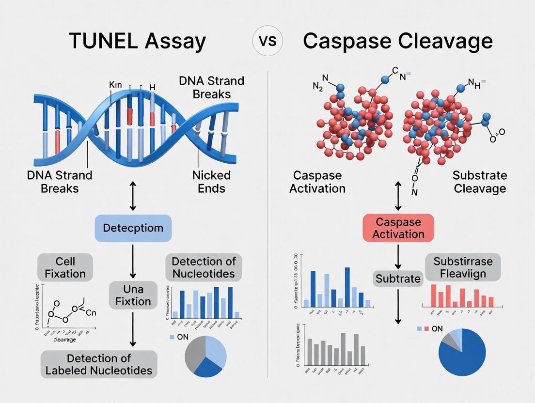
Detection Methods: TUNEL Assay vs. Caspase Cleavage
TUNEL Assay: Principle and Applications
The Terminal deoxynucleotidyl transferase dUTP nick end labeling (TUNEL) assay detects DNA fragmentation, a hallmark of late-stage apoptosis [5]. This method utilizes terminal deoxynucleotidyl transferase (TdT) to incorporate modified nucleotides (BrdUTP, EdUTP, or fluorescently-labeled dUTP) at the 3'-OH ends of fragmented DNA [5].
Key Detection Strategies:
- Click Chemistry-Based: EdUTP incorporation detected via copper-catalyzed azide-alkyne cycloaddition with fluorescent or colorimetric readouts [5]
- Antibody-Based: BrdUTP incorporation detected using anti-BrdU antibodies [5]
- Direct Labeling: Fluorescein-dUTP incorporation for direct fluorescence detection [5]
The TUNEL assay is particularly valuable for spatial localization of cell death in tissue contexts and has been widely adopted for in situ apoptosis detection [6].
Caspase Cleavage Detection: Principles and Applications
Caspase activation represents a committed step in apoptosis execution, making its detection a reliable indicator of ongoing PCD [7]. Multiple methods exist for detecting caspase activity:
Antibody-Based Methods:
- Western blotting to detect caspase cleavage and activation [7]
- Immunofluorescence for spatial localization of active caspases [7]
- Immunohistochemistry for tissue-based detection [7]
Activity-Based Methods:
- Fluorescent-labeled inhibitors for live imaging [7]
- FRET-based caspase activity sensors [7]
- Mass spectrometry for identifying caspase substrates and cleavage products [7]
Caspase detection offers the advantage of identifying early apoptosis events before morphological changes become apparent [7].
Table 1: Comparative Analysis of TUNEL Assay and Caspase Cleavage Detection
| Parameter | TUNEL Assay | Caspase Cleavage Detection |
|---|---|---|
| Target | DNA fragmentation | Caspase enzyme activity or cleavage |
| Detection Stage | Late apoptosis | Early to mid apoptosis |
| Specificity for Apoptosis | Moderate (can detect other DNA damage) | High when specific caspases are targeted |
| Multiplexing Compatibility | Compatible with spatial proteomics after protocol optimization [6] | Highly compatible with multiplexed protein detection |
| Throughput | Moderate | High with plate-based activity assays |
| Tissue Preservation | Requires optimization of antigen retrieval (pressure cooker preferred over proteinase K) [6] | Excellent tissue preservation |
| Quantification | Semi-quantitative | Highly quantitative with activity assays |
| Key Limitations | Cannot distinguish apoptosis from other DNA fragmentation events; proteinase K treatment reduces protein antigenicity [6] | May detect caspase activity in non-apoptotic processes; context-dependent interpretation needed |
Experimental Data and Performance Comparison
Protocol Optimization and Compatibility Studies
Recent investigations have systematically evaluated TUNEL protocol variations for compatibility with multiplexed spatial proteomic methods. Sherman et al. demonstrated that replacing proteinase K with pressure cooker treatment preserves both TUNEL signal and protein antigenicity, enabling seamless integration with Multiple Iterative Labeling by Antibody Neodeposition (MILAN) and Cyclic Immunofluorescence (CycIF) [6].
Key Experimental Findings:
- Proteinase K treatment consistently reduced or abrogated protein antigenicity, limiting multiplexing capabilities [6]
- Pressure cooker treatment enhanced protein antigenicity while maintaining TUNEL sensitivity [6]
- Antibody-based TUNEL with pressure cooker retrieval was successfully integrated into MILAN staining series [6]
- TUNEL signal was completely erased using 2-ME/SDS treatment, allowing restaining with protein markers [6]
These findings establish that TUNEL can be effectively harmonized with spatial proteomics through careful protocol optimization, addressing a previous major limitation [6].
Specificity Concerns and Biological Context
Both detection methods require careful interpretation within appropriate biological contexts:
TUNEL Specificity Considerations:
- Detects DNA fragmentation occurring during both apoptosis and necrosis [8]
- May label cells undergoing DNA repair or other non-lethal DNA damage [8]
- Recent evidence shows TUNEL-positive cells can recover through anastasis, reversing apoptotic progression [8]
Caspase Detection Considerations:
- Caspases participate in non-apoptotic processes including differentiation and inflammation [7]
- Inflammatory caspases (caspase-1, -4, -5, -11) execute pyroptosis rather than apoptosis [4]
- Cross-talk between cell death pathways can complicate interpretation [1]
Table 2: Quantitative Performance Metrics of Detection Methods
| Performance Metric | TUNEL Assay | Caspase Activity Assays | Caspase Cleavage Western | Caspase Immunofluorescence |
|---|---|---|---|---|
| Sensitivity | High (detects 0.1-1% apoptotic cells) | Very high (fmole sensitivity) | Moderate | Moderate to high |
| Time to Result | 2-4 hours | 1-2 hours | 6-24 hours | 6-24 hours |
| Spatial Resolution | Excellent (single cell in tissue) | Poor (population average) | Poor (tissue homogenate) | Excellent (single cell) |
| Dynamic Range | ~2 log | ~3-4 log | ~1.5 log | ~2 log |
| Reproducibility | Moderate (depends on tissue quality) | High | Moderate | Moderate |
| Cost per Sample | Medium | Low to medium | Low | Medium to high |
Detailed Experimental Protocols
Optimized TUNEL Protocol for Multiplexed Applications
Based on recent methodological advances [6], the following protocol enables TUNEL integration with spatial proteomics:
Sample Preparation:
- Use formalin-fixed paraffin-embedded (FFPE) tissue sections (4-5 μm thickness)
- Deparaffinize and rehydrate through xylene and graded ethanol series
- Perform antigen retrieval using pressure cooker (20 min at 95°C in citrate buffer, pH 6.0)
- Critical: Avoid proteinase K treatment to preserve protein antigenicity
TUNEL Reaction:
- Apply TUNEL reaction mixture containing EdUTP and TdT enzyme
- Incubate for 60 minutes at 37°C in a humidified chamber
- For Click-iT detection: Add click reaction mixture with fluorescent azide
- Counterstain with Hoechst 33342 or DAPI
Multiplexing with Protein Detection:
- After TUNEL imaging, erase signal with 2-ME/SDS treatment (66°C for 1-2 hours)
- Proceed with standard immunofluorescence for protein targets of interest
- For MILAN applications: Repeat erasure and staining cycles as needed
Caspase Activity Measurement Protocol
Caspase-3/7 Activity Assay (Fluorometric):
- Prepare cell lysates or tissue homogenates in appropriate buffer
- Incubate with caspase-specific fluorogenic substrates (e.g., DEVD-AFC for caspase-3)
- Measure fluorescence emission over 30-60 minutes (Ex/Em: 400/505 nm)
- Calculate activity relative to protein concentration and control samples
In Situ Caspase Detection:
- Fix cells or tissue sections with 4% paraformaldehyde
- Permeabilize with 0.1% Triton X-100
- Block with 5% normal serum
- Incubate with antibody against active caspase-3 (cleaved form)
- Detect with fluorescent secondary antibodies
- Counterstain and image
Figure 2: Experimental workflows for TUNEL assay and caspase detection methods. The optimized TUNEL protocol includes critical steps for multiplexing compatibility, while caspase detection offers multiple readout options depending on research needs.
The Scientist's Toolkit: Essential Research Reagents
Table 3: Key Research Reagents for Programmed Cell Death Detection
| Reagent Category | Specific Examples | Function/Application | Key Considerations |
|---|---|---|---|
| TUNEL Assay Kits | Click-iT Plus TUNEL Assay (Thermo Fisher) | Detection of DNA fragmentation in situ | Copper concentration optimized for fluorescent protein compatibility [5] |
| APO-BrdU TUNEL Assay (Thermo Fisher) | Flow cytometry or imaging of apoptosis | Uses BrdUTP incorporation with Alexa Fluor 488-anti-BrdU detection [5] | |
| Caspase Substrates | DEVD-AFC, DEVD-AMC | Fluorometric caspase-3/7 activity assays | Cell-permeable (AMC) vs. impermeable (AFC) variants available |
| LEHD-AFC, IETD-AFC | Fluorometric caspase-9 and -8 activity assays | Specific for initiator caspases in intrinsic/extrinsic pathways | |
| Caspase Antibodies | Anti-cleaved caspase-3 | Detection of activated caspase-3 in tissues | High specificity for apoptosis execution phase |
| Anti-caspase-8 | Detection of initiator caspase in extrinsic pathway | Can detect both pro-form and cleaved forms | |
| Cell Death Inducers | Staurosporine | Broad-spectrum kinase inducer of intrinsic apoptosis | Positive control for apoptosis assays [5] |
| TNF-α + Cycloheximide | Extrinsic apoptosis induction via death receptors | Requires sensitization for optimal effect | |
| RSL3 | Ferroptosis inducer via GPX4 inhibition | Useful for specificity controls [9] | |
| Inhibitors | Z-VAD-FMK | Pan-caspase inhibitor | Broad inhibition of apoptotic caspases |
| Z-AEAD-FMK | Novel pan-caspase inhibitor targeting AEAD motif | Inhibits caspases-1, -3, -6, -7, -8, -9 [10] | |
| Necrostatin-1 | RIPK1 inhibitor for necroptosis suppression | Specific for necroptosis pathway inhibition | |
| Gasdermin Reagents | Anti-GSDMD (full length/cleaved) | Pyroptosis detection | Specific for pyroptosis execution phase [4] |
| Disulfiram | Gasdermin pore formation inhibitor | Blocks pyroptosis execution [4] |
The comparative analysis of TUNEL assay and caspase cleavage detection reveals complementary strengths that researchers should leverage based on specific experimental needs. TUNEL excels in spatial localization of cell death within tissue architecture, while caspase detection provides higher specificity for apoptotic commitment and earlier detection capability.
Recent methodological advances, particularly the replacement of proteinase K with pressure cooker antigen retrieval, have resolved previous incompatibilities between TUNEL and multiplexed spatial proteomics [6]. This enables researchers to contextualize cell death within complex tissue microenvironments while preserving molecular information.
For comprehensive apoptosis assessment, we recommend a combined approach utilizing caspase activation as an early marker and TUNEL as a confirmatory late-stage indicator, with careful attention to biological context and potential cross-talk between cell death pathways. This integrated strategy provides the most robust framework for programmed cell death research in both basic and translational applications.
Apoptosis, or programmed cell death, is a fundamental biological process characterized by a sequence of highly specific morphological changes within the cell. These hallmarks include chromatin condensation, cell shrinkage, membrane blebbing, and the formation of apoptotic bodies [3]. The accurate detection of this form of cell death is critical in numerous fields of biological research, from understanding embryonic development to evaluating the efficacy of cancer therapeutics. Among the various techniques developed, the TUNEL (Terminal deoxynucleotidyl transferase dUTP nick end labeling) assay and caspase cleavage detection have emerged as two of the most prominent methods. The TUNEL assay operates by detecting the DNA fragmentation that occurs in the later stages of apoptosis, while caspase cleavage methods target the activation of caspases, which are key protease mediators early in the apoptotic cascade [11] [12]. This guide provides an objective, data-driven comparison of these two methodologies, equipping researchers with the information necessary to select the most appropriate technique for their specific experimental context.
TUNEL Assay
The TUNEL assay identifies apoptotic cells by labeling the 3'-hydroxyl termini of DNA double-strand breaks that are generated during the endonucleolytic cleavage of genomic DNA [13]. This is achieved using the enzyme terminal deoxynucleotidyl transferase (TdT), which catalyzes the attachment of deoxynucleotides—tagged with a fluorochrome or another marker—to these 3'-OH ends [8] [13]. While historically a gold standard, it is crucial to note that DNA strand breaks can also occur in other cell death processes, such as necrosis, and even in non-lethal cellular activities like DNA repair, which can potentially lead to false-positive results if the assay is not carefully controlled and interpreted [8] [13].
Caspase Cleavage Detection
Caspases are a family of cysteine-aspartic proteases that serve as central regulators and executioners of apoptosis [12]. Their activation triggers a proteolytic cascade that leads to the characteristic morphological hallmarks of apoptosis. Detection methods typically utilize antibodies that specifically recognize the cleaved, active form of caspases (such as caspase-3) or their cleaved substrates (like cleaved cytokeratin 18 or PARP) [11] [12]. This approach provides a more direct measurement of the core apoptotic machinery and often identifies cells at an earlier stage of the death process compared to the TUNEL assay [11].
Table 1: Core Principles of TUNEL and Caspase Detection Methods
| Feature | TUNEL Assay | Caspase Cleavage Detection |
|---|---|---|
| Primary Target | DNA strand breaks (3'-OH ends) | Activated caspases or caspase-cleaved protein products |
| Key Reagent | Terminal deoxynucleotidyl transferase (TdT) | Antibodies specific to cleaved caspase-3, CK18, PARP, etc. |
| Detection Stage | Mid-to-late apoptosis (after DNA fragmentation) | Early-to-mid apoptosis (during/after caspase activation) |
| Biological Specificity | Detects a consequence of apoptosis | Detects a key mediator of apoptosis |
Head-to-Head Comparative Data
Numerous independent studies have directly compared the performance of these two assays, providing quantitative data on their correlation and reliability.
A pivotal study on prostate cancer (PC-3) xenografts quantified apoptosis using multiple methods and found an excellent correlation (R = 0.89) between apoptotic indices obtained with activated caspase-3 immunohistochemistry and cleaved cytokeratin 18 immunohistochemistry [11]. The correlation between activated caspase-3 and the TUNEL assay was also found to be good, though slightly lower (R = 0.75) [11]. The authors concluded that activated caspase-3 immunohistochemistry was an "easy, sensitive, and reliable method" for quantifying apoptosis in their model [11].
Another study on clinically localized prostate cancer compared several apoptotic markers, including ACINUS (a caspase-cleaved protein), caspase-3, and TUNEL, for their utility in calculating tumor growth rates. The study found that both ACINUS and caspase-3 were better predictors of clinical cancer aggressiveness than TUNEL. In logistic regression models, the area under the curve (AUC) was 0.677 for ACINUS and 0.694 for caspase-3, compared to 0.669 for TUNEL [14].
Table 2: Summary of Key Comparative Study Findings
| Study Model | Comparative Metric | TUNEL Assay Performance | Caspase Cleavage Performance | Conclusion |
|---|---|---|---|---|
| PC-3 Xenografts [11] | Correlation with Activated Caspase-3 | R = 0.75 (Good) | Self (Reference) | Caspase-3 is a sensitive and reliable standard. |
| Clinical Prostate Cancer [14] | Predictive Value (AUC) | AUC = 0.669 | AUC = 0.694 (Caspase-3) | Caspase-3 was a superior predictor of aggressiveness. |
Critical Considerations and Limitations
Specificity and the Challenge of Anastasis
A significant consideration for the TUNEL assay is its potential lack of absolute specificity for apoptotic cell death. The assay can label cells with DNA damage from various causes, not solely apoptosis [8]. Furthermore, compelling evidence has shown that cells can recover from the brink of apoptotic death, a process termed anastasis. Cells exhibiting classic hallmarks of late-stage apoptosis, including caspase activation and genomic DNA breakage (detectable by TUNEL), have been observed to reverse the process and survive [8]. This phenomenon indicates that a positive signal in either assay does not irrevocably equate to cell demise and cautions against the simplistic interpretation of "percent apoptosis" based on a single time-point measurement [8].
Practical Workflow and Technical Nuances
From a practical standpoint, caspase detection via immunohistochemistry or immunofluorescence is often integrated more seamlessly into standard pathology workflows, as it is similar to other antibody-based staining techniques. The TUNEL assay, while commercially available in kit form, can involve multiple washing and incubation steps that are time-consuming and require operation in the dark [15]. Moreover, the original TUNEL assay protocols were known to suffer from technical issues that could label necrotic cells as apoptotic; however, the method has been substantially improved over the years to enhance its specificity for apoptosis when correctly performed [13].
The Scientist's Toolkit: Essential Reagents
Table 3: Key Research Reagent Solutions for Apoptosis Detection
| Reagent / Kit | Function / Target | Key Application Notes |
|---|---|---|
| Terminal Deoxynucleotidyl Transferase (TdT) | Enzyme that adds labeled nucleotides to 3'-OH DNA ends. | Core component of TUNEL assay kits; critical for labeling DNA breaks. |
| Anti-Cleaved Caspase-3 Antibody | Specifically binds the activated form of caspase-3. | Highly cited marker for early-to-mid apoptosis in IHC/IF [11] [12]. |
| Anti-Cleaved PARP Antibody | Detects caspase-cleaved Poly (ADP-ribose) polymerase. | Another major caspase substrate; serves as a verification marker. |
| Anti-Cleaved Cytokeratin 18 (M30) Antibody | Recognizes a caspase-cleaved epitope of CK18. | Especially useful in epithelial-derived cancers; correlates well with caspase-3 [11]. |
| BrdUTP or Fluorescein-dUTP | Labeled nucleotides incorporated by TdT. | BrdUTP with secondary detection offers high sensitivity [13]. |
| Annexin V Conjugates | Binds externalized phosphatidylserine. | Used for flow cytometry to detect early apoptosis, often combined with viability dyes. |
Visualizing Apoptosis Detection Pathways
The following diagrams illustrate the fundamental principles and workflows of the two detection methods, highlighting their distinct targets within the apoptotic process.
Diagram 1: Core detection principles for TUNEL and caspase assays.
Diagram 2: Apoptosis timeline and optimal assay detection windows.
Detailed Experimental Protocols
To ensure reproducibility, below are detailed methodologies for key experiments cited in this guide.
- Tissue Preparation: Use formalin-fixed, paraffin-embedded tissue sections (5 µm thickness).
- Deparaffinization and Antigen Retrieval: Deparaffinize and rehydrate sections through a graded alcohol series. Perform heat-induced antigen retrieval using Citra buffer (pH 6.0) for 10 minutes at 120°C and 21 PSI.
- Blocking and Primary Antibody Incubation: Block endogenous peroxidase with 3% H₂O₂. Incubate sections with serum block to reduce non-specific binding. Apply anti-activated caspase-3 or anti-cleaved cytokeratin 18 primary antibody at optimized dilution (e.g., 1:50 to 1:500) for 1-2 hours at 37°C.
- Detection and Visualization: Use a dextran polymer-based detection system (e.g., EnVision) conjugated with horseradish peroxidase. Visualize with diaminobenzidine (DAB) to produce a brown precipitate, or with Fast Red for a red precipitate.
- Counterstaining and Analysis: Counterstain lightly with hematoxylin to visualize nuclei. Dehydrate, clear, and mount. Quantify apoptotic indices using computer-assisted image analysis.
- Sample Preparation: Deparaffinize and rehydrate formalin-fixed, paraffin-embedded sections as above.
- Proteinase Digestion: Treat sections with proteinase K (20 µg/mL) for 15 minutes at room temperature to expose DNA breaks.
- Quenching Endogenous Peroxidase: Incubate sections in 3% H₂O₂ for 10 minutes to block endogenous peroxidase activity, which is critical for reducing background in subsequent enzymatic detection.
- Enzymatic Labeling: Apply the TUNEL reaction mixture containing TdT enzyme and labeled dUTP (e.g., biotin-dUTP or fluorescein-dUTP) to the sections. Incubate in a humidified chamber for 60 minutes at 37°C.
- Signal Detection: For biotin-dUTP, apply a streptavidin-horseradish peroxidase conjugate. For fluorescein-dUTP, use an anti-fluorescein antibody conjugate. Develop with DAB or a compatible fluorescent mounting medium.
- Analysis: Use microscopy or automated image analysis to quantify TUNEL-positive nuclei.
Concluding Comparison and Recommendations
The choice between TUNEL and caspase cleavage assays is not a matter of identifying a universally superior technique, but rather of selecting the most appropriate tool for the specific research question and context.
- For Specificity and Early Detection: Caspase cleavage detection, particularly using antibodies against activated caspase-3, is generally recommended for its high specificity as it targets a central component of the apoptotic machinery. It is less susceptible to false positives from non-apoptotic DNA damage and can identify cells in the earlier phases of death [11] [12].
- For Detecting Later Stages and DNA Fragmentation: The TUNEL assay remains a valuable tool for specifically visualizing the DNA fragmentation that is a hallmark of mid-to-late apoptosis. Its utility is highest when this specific readout is required, provided the assay is meticulously performed and interpreted with an awareness of its limitations [13].
- For Robustness and Confirmation: The most reliable approach for quantifying apoptosis, especially in preclinical studies, is to employ multiple assays that target different points in the cell death pathway [8] [12]. A combination of caspase activation (e.g., cleaved caspase-3 IHC) and a well-validated TUNEL assay, correlated with standard morphological analysis (e.g., H&E staining for chromatin condensation), provides the most compelling and accurate data. This multi-parametric strategy helps account for the dynamic and sometimes reversible nature of apoptosis and ensures a more comprehensive analysis.
Apoptosis, or programmed cell death, is a fundamental biological process critical for maintaining tissue homeostasis, eliminating damaged cells, and ensuring proper embryonic development. This highly regulated cell death pathway occurs through two principal signaling cascades: the intrinsic and extrinsic apoptosis pathways. The intrinsic pathway (mitochondrial pathway) initiates internally from cellular stress signals, such as DNA damage or oxidative stress, leading to mitochondrial outer membrane permeabilization (MOMP) and cytochrome c release. The extrinsic pathway (death receptor pathway) begins externally when extracellular ligands bind to death receptors on the cell surface, triggering intracellular caspase activation. Both pathways converge on the activation of executioner caspases, including caspase-3 and caspase-7, which orchestrate the systematic dismantling of cellular components, resulting in the characteristic morphological changes of apoptosis, including cell shrinkage, chromatin condensation, and DNA fragmentation.
Understanding these pathways is particularly crucial in cancer research and therapeutic development, as dysregulated apoptosis is a hallmark of cancer. Researchers rely on various detection methods to study apoptosis, with TUNEL (Terminal deoxynucleotidyl transferase dUTP nick end labeling) and caspase cleavage assays representing two of the most widely used techniques. This guide provides a comprehensive comparison of these methods, examining their principles, applications, and performance characteristics to inform researchers' experimental design and interpretation.
Principles of Detection: TUNEL vs. Caspase Cleavage Assays
TUNEL Assay Fundamentals
The TUNEL assay detects apoptosis by identifying DNA fragmentation, a hallmark late-stage apoptotic event. This method relies on the enzyme terminal deoxynucleotidyl transferase (TdT), which catalyzes the addition of labeled dUTP nucleotides to the 3'-hydroxyl termini of DNA breaks. The labeling can be direct (using fluorescently-tagged nucleotides) or indirect (using hapten-labeled nucleotides detected with secondary reagents) [16] [17]. During apoptosis, endonucleases cleave DNA into fragments of 180-200 base pairs, generating abundant DNA breaks that are selectively labeled by the TUNEL reaction, allowing visualization and quantification of apoptotic cells in tissue sections or cell cultures.
The TUNEL assay typically involves several key steps: sample fixation, permeabilization, antigen retrieval (often using proteinase K or heat-induced epitope retrieval), TdT-mediated nucleotide incorporation, and signal detection. A critical advancement in TUNEL methodology addresses its compatibility with modern spatial proteomics. Recent research demonstrates that proteinase K treatment, commonly used in TUNEL protocols, significantly diminishes protein antigenicity for subsequent multiplexed protein detection. Replacing proteinase K with pressure cooker-based antigen retrieval preserves both TUNEL signal intensity and protein antigenicity, enabling integration with multiplexed iterative staining techniques like MILAN (Multiple Iterative Labeling by Antibody Neodeposition) [6].
Caspase Cleavage Assay Fundamentals
Caspase cleavage assays detect apoptosis by measuring the activation of executioner caspases, particularly caspase-3 and caspase-7, which represent a commitment to the apoptotic process. These proteases cleave specific amino acid sequences (primarily after aspartic acid residues) in numerous cellular substrates, including poly ADP ribose polymerase (PARP) and cytosolic CAD, the rate-limiting enzyme for de novo pyrimidine synthesis [18] [19]. Caspase activation occurs upstream of DNA fragmentation in the apoptotic cascade, making these assays valuable for detecting earlier apoptotic events compared to TUNEL.
The most common caspase detection method uses antibodies specific to the cleaved, activated forms of caspases (e.g., anti-cleaved caspase-3). Alternatively, fluorogenic or luminogenic substrates containing caspase cleavage sites (such as DEVD sequences) provide functional activity measurements. When caspase-3 cleaves these substrates, it releases a fluorophore or luminophore, generating detectable signals proportional to caspase activity [18]. These assays are particularly amenable to high-throughput screening formats, with luminogenic assays offering approximately 20-50-fold higher sensitivity than fluorogenic versions [18].
Comparative Performance Analysis
Sensitivity and Specificity
The table below summarizes the key performance characteristics of TUNEL and caspase cleavage assays based on comparative studies:
Table 1: Performance Comparison of TUNEL and Caspase Cleavage Assays
| Parameter | TUNEL Assay | Caspase Cleavage Assay |
|---|---|---|
| Detection Target | DNA fragmentation (3'-OH ends) | Activated executioner caspases (caspase-3/7) |
| Apoptosis Stage Detected | Late-stage apoptosis | Mid-to-late stage apoptosis |
| Sensitivity | High sensitivity for advanced apoptosis; can detect a single apoptotic cell [20] | High sensitivity; luminogenic versions 20-50x more sensitive than fluorescent substrates [18] |
| Specificity Concerns | Can label necrotic cells, cells with DNA repair, or autolytic processes; requires careful interpretation [8] [16] | Highly specific for apoptotic pathway; caspase activation considered "point of no return" [18] |
| Quantification Readouts | Number of positive cells, stained area (apoptotic index) [20] | Caspase activity (RLU/RFU), number of positive cells, intensity measurement |
| Compatibility with Multiplexing | Compatible with pressure cooker retrieval; proteinase K reduces protein antigenicity [6] | Highly compatible with multiplex protein detection [6] |
Experimental Considerations and Limitations
Both assays present distinct advantages and limitations that influence their application in research settings. TUNEL staining provides direct histological visualization of apoptotic cells within tissue architecture, making it invaluable for spatial contextualization. However, concerns regarding specificity persist, as DNA strand breaks can occur in various non-apoptotic contexts, including necrosis, DNA repair, and even gene transcription [8] [16]. Furthermore, emerging evidence of anastasis (recovery from late-stage apoptosis) challenges the assumption that TUNEL-positive cells are irrevocably committed to death, as cells exhibiting caspase activation, DNA fragmentation, and apoptotic morphology can potentially recover under certain conditions [8].
Caspase cleavage assays offer earlier detection of apoptosis and greater specificity for the programmed cell death pathway. The detection of activated caspase-3, for instance, is considered a highly specific apoptotic marker since caspase-1 (involved in pyroptosis) does not participate in apoptotic pathways [3]. However, caspase activation may be transient in some apoptotic processes, potentially leading to false negatives if sampling occurs outside activation windows. Additionally, the cleavage of specific caspase substrates like CAD at Asp1371 represents a commitment step in apoptosis execution, connecting caspase activation directly to metabolic cessation [19].
Experimental Protocols and Methodologies
Detailed TUNEL Assay Protocol with MILAN Compatibility
The following protocol represents an optimized TUNEL methodology compatible with multiplexed protein detection:
Table 2: Key Research Reagent Solutions for TUNEL Assay
| Reagent | Function | Example Products/Formats |
|---|---|---|
| Terminal Deoxynucleotidyl Transferase (TdT) | Catalyzes nucleotide addition to DNA 3'-OH ends | Recombinant TdT enzyme |
| Labeled dUTP | Detection of DNA breaks | FITC-dUTP, Biotin-dUTP, BrdU-dUTP |
| Antigen Retrieval Solution | Unmasking of epitopes | Citrate buffer, EDTA buffer |
| Detection Reagents | Signal visualization | Streptavidin-HRP, anti-FITC-HRP, anti-BrdU antibodies |
| Chromogenic/Fluorogenic Substrates | Signal generation | DAB, AEC, TMB (chromogenic); Fluorescein, TMR (fluorescent) |
- Sample Preparation: Use formalin-fixed paraffin-embedded (FFPE) tissue sections (4-5 μm thickness) mounted on charged slides. Deparaffinize and rehydrate through xylene and graded ethanol series.
- Antigen Retrieval: Perform heat-induced epitope retrieval using pressure cooker in citrate buffer (pH 6.0) for 10 minutes at 95-100°C. Critical note: Avoid proteinase K treatment to preserve protein antigenicity for subsequent multiplexed staining [6].
- Permeabilization: Treat slides with 0.1% Triton X-100 in PBS for 15 minutes at room temperature.
- TUNEL Reaction: Prepare TUNEL reaction mixture according to manufacturer's instructions (e.g., ApopTag Red In Situ Apoptosis Detection Kit). Apply to tissue sections and incubate in a humidified chamber for 60 minutes at 37°C.
- Detection: For indirect detection methods, apply appropriate secondary detection reagents (e.g., anti-fluorescein antibody conjugates for FITC-labeled nucleotides).
- Counterstaining and Mounting: Counterstain with DAPI or methyl green, and mount with appropriate mounting medium.
- Erasure for Multiplexing (Optional): For integration with MILAN, erase antibodies by incubating specimens in 2-mercaptoethanol/sodium dodecyl sulfate (2-ME/SDS) at 66°C, then proceed with subsequent antibody staining cycles [6].
Detailed Caspase Cleavage Assay Protocol
The following protocol outlines both immunohistochemical and activity-based approaches for caspase detection:
Table 3: Key Research Reagent Solutions for Caspase Cleavage Assay
| Reagent | Function | Example Products/Formats |
|---|---|---|
| Anti-Cleaved Caspase Antibodies | Specific detection of activated caspases | Anti-cleaved caspase-3, anti-cleaved Dcp-1 |
| Caspase Substrates | Measure caspase enzymatic activity | DEVD-AMC, DEVD-AFC, DEVD-R110, Z-DEVD-aminoluciferin |
| Cell Lysis Buffer | Extract proteins while maintaining activity | RIPA buffer, specialized caspase lysis buffers |
| Detection Reagents | Signal generation | HRP-conjugated secondary antibodies, luciferase reagent |
Immunohistochemical Detection (Cleaved Caspase-3):
- Sample Preparation: Use FFPE tissue sections prepared as described above.
- Antigen Retrieval: Perform heat-induced epitope retrieval using pressure cooker in citrate buffer (pH 6.0) for 10 minutes.
- Blocking: Block endogenous peroxidase activity with 3% H₂O₂, then block nonspecific binding with protein block serum for 30 minutes.
- Primary Antibody Incubation: Apply anti-cleaved caspase-3 antibody (diluted according to manufacturer's instructions) and incubate overnight at 4°C.
- Detection: Apply appropriate biotinylated secondary antibody followed by streptavidin-HRP complex. Develop with DAB chromogen and counterstain with hematoxylin.
Activity-Based Detection (Caspase-Glo 3/7 Assay):
- Cell Preparation: Plate cells in white-walled 96-well or 384-well plates. Treat with experimental compounds.
- Equilibration: Equilibrate Caspase-Glo 3/7 reagent and cell plates to room temperature.
- Reagent Addition: Add equal volume of Caspase-Glo 3/7 reagent to each well.
- Incubation: Mix contents gently using a plate shaker and incubate at room temperature for 30-60 minutes.
- Measurement: Record luminescence using a plate-reading luminometer [18].
Biochemical Pathway Visualization
Apoptosis Pathways and Detection Methods
This diagram illustrates the two main apoptosis pathways and their connection to detection methods. The extrinsic pathway initiates from death receptor activation, while the intrinsic pathway responds to cellular stress signals. Both pathways converge on caspase-3 activation, which cleaves various cellular substrates, including CAD, leading to DNA fragmentation. Caspase assays detect active caspase-3/7, representing mid-to-late stage apoptosis, while TUNEL detects the subsequent DNA fragmentation, representing late-stage apoptosis.
Research Applications and Contextual Selection
Application-Specific Method Selection
Choosing between TUNEL and caspase cleavage assays depends on research objectives, sample types, and experimental context:
- For Histological Spatial Analysis: TUNEL remains preferred for visualizing apoptotic cells within tissue architecture, especially when combined with pressure-cooker antigen retrieval for multiplexed protein detection [6].
- For High-Throughput Screening: Caspase activity assays, particularly luminogenic formats, offer superior sensitivity, miniaturization capability, and compatibility with automated screening platforms [18].
- For Early Apoptosis Detection: Caspase cleavage assays detect commitment to apoptosis before morphological manifestations, providing earlier intervention assessment.
- For Multiplexed Spatial Proteomics: The harmonized TUNEL protocol with pressure cooker retrieval enables integration with MILAN and CycIF, allowing rich contextualization of cell death within complex tissue environments [6].
- For Specific Apoptosis Confirmation: Caspase-3/7 detection provides higher specificity for apoptotic programmed cell death compared to TUNEL, which may label non-apoptotic DNA fragmentation.
Emerging Trends and Future Directions
The apoptosis detection field continues to evolve with several emerging trends. Multiplexing capabilities are advancing, with recent demonstrations that TUNEL can be successfully integrated with spatial proteomic methods like MILAN and cyclic immunofluorescence (CycIF) [6]. Artificial intelligence and automated image analysis are addressing quantification challenges, with platforms like CASQITO (Computer Assisted Signal Quantification Including Threshold Options) providing semi-automated processing for both TUNEL and caspase staining images [20]. Furthermore, the commercial apoptosis assay market is expanding, projected to grow from USD 6.5 billion in 2024 to USD 14.6 billion by 2034, driving innovation in assay sensitivity, specificity, and convenience [21].
Both TUNEL and caspase cleavage assays provide valuable, complementary approaches for apoptosis detection in research and drug development. TUNEL offers direct histological visualization of late-stage apoptotic cells with DNA fragmentation, while caspase assays detect earlier commitment to the apoptotic process through caspase-3/7 activation. The recent harmonization of TUNEL with spatial proteomics through pressure cooker antigen retrieval represents a significant advancement, enabling multidimensional analysis of cell death within its tissue context. Researchers should select methods based on their specific experimental needs, considering factors such as detection timing, specificity requirements, sample type, and desired multiplexing capabilities. As both technologies continue to evolve, they will undoubtedly provide increasingly sophisticated tools for unraveling the complex biochemical cascades of intrinsic and extrinsic apoptosis pathways in health and disease.
Caspase-3 is a frequently activated death protease that serves as the central executioner of apoptosis, catalyzing the specific cleavage of many key cellular proteins and ultimately leading to the dismantling of the cell [22]. As a member of the cysteine-aspartic acid protease (caspase) family, caspase-3 exists as an inactive proenzyme in the cytosol and is activated through proteolytic cleavage during apoptosis [23]. This enzyme performs its functions by catalyzing the cleavage of peptide bonds following aspartic acid residues in target proteins [23]. Unlike initiator caspases that begin the apoptotic cascade, caspase-3 is classified as an executioner caspase (along with caspases-6 and -7) and is positioned at the terminal end of the caspase cascade, where it is activated by both intrinsic and extrinsic death pathways [24] [23]. The activation of caspase-3 leads to characteristic apoptotic morphological changes including plasma membrane blebbing, chromatin condensation, DNA cleavage, and exposure of phosphatidylserine on the extracellular side of the plasma membrane [23].
The essential nature of caspase-3 in normal development has been demonstrated in knockout mice, which exhibit profound defects in brain development and altered cellular kinetics, underscoring its non-redundant functions in apoptosis [22]. In cancer research, caspase-3 activation represents a crucial mechanism through which many chemotherapeutic agents exert their cytotoxic effects on tumor cells, making it a protein of significant interest in both basic research and drug development [23]. Recent evidence has also revealed that caspase-3 plays a role beyond classical apoptosis, serving as a switch between apoptosis and pyroptosis through its cleavage of Gasdermin E (GSDME), thereby expanding our understanding of its functions in cellular fate decisions [23].
Caspase-3 Activation Pathways
Caspase-3 activation occurs through two well-characterized apoptotic pathways that converge on this key executioner protease. The specific pathways and their components are detailed below and illustrated in Figure 1.
The Extrinsic (Death Receptor) Pathway
The extrinsic pathway, also known as the death receptor pathway, is initiated by the binding of extracellular death ligands (such as Fas ligand or TNF-α) to their corresponding cell surface death receptors [25] [23]. This binding induces receptor clustering and formation of the death-inducing signaling complex (DISC), which recruits and activates initiator caspase-8 [25] [23]. Once activated, caspase-8 can directly cleave and activate procaspase-3, initiating the execution phase of apoptosis [23]. Additionally, caspase-8 can proteolytically cleave Bid, a member of the Bcl-2 family, generating truncated Bid (tBid) that translocates to mitochondria and amplifies the death signal through the intrinsic pathway [23].
The Intrinsic (Mitochondrial) Pathway
The intrinsic pathway, also referred to as the mitochondrial pathway, is triggered by intracellular stress signals including DNA damage, oxidative stress, or growth factor withdrawal [25] [23]. These stimuli cause mitochondrial outer membrane permeabilization, leading to the release of cytochrome c from the mitochondrial intermembrane space into the cytoplasm [25] [23]. Cytochrome c then binds to Apaf-1 (apoptotic protease-activating factor-1), promoting the formation of a multiprotein complex called the apoptosome [25] [23]. The apoptosome recruits and activates the initiator caspase-9, which in turn cleaves and activates executioner caspase-3 [26] [23]. The activation of caspase-3 represents the point of convergence between the extrinsic and intrinsic pathways, positioning it as the central executioner of apoptosis [23].
Figure 1: Caspase-3 activation pathways
Caspase-3 Substrate Specificity and Key Targets
Substrate Recognition Motif
Caspase-3 exhibits a strong preference for cleaving substrates C-terminal to aspartic acid residues, with the optimal recognition sequence being DEVD (Asp-Glu-Val-Asp) [24]. This tetrapeptide motif represents the canonical cleavage site that caspase-3 recognizes in target proteins, with the scissile bond located between the second aspartic acid and the subsequent amino acid [24]. The substrate specificity of caspase-3 was initially characterized using positional scanning synthetic combinatorial library methods with fluorogenic tetrapeptide substrates [24]. While the DEVD sequence represents the optimal recognition motif, it's important to note that this exact sequence appears in less than 1% of the total protein cleavage sites targeted by caspase-3 in native cellular environments, indicating that contextual factors beyond the primary sequence influence substrate selection [24].
Table 1: Caspase Family Substrate Preferences
| Enzyme | Peptide Substrate Preference | Protein Substrate Preference | Biological Role |
|---|---|---|---|
| Caspase-1 | WEHD | YVHD/FESD | Inflammatory |
| Caspase-2 | VDVAD | XDEVD | Initiator |
| Caspase-3 | DEVD | DEVD | Executioner |
| Caspase-6 | VQVD | VEVD | Executioner |
| Caspase-7 | DEVD | DEVD | Executioner |
| Caspase-8 | LETD | XEXD | Initiator |
| Caspase-9 | (W/L)EHD | - | Initiator |
| Caspase-10 | LEHD | LEHD | Initiator |
Data derived from peptide library and proteomic studies [24]
Key Cellular Substrates of Caspase-3
Caspase-3 catalyzes the specific cleavage of numerous key cellular proteins, with recent proteomic studies identifying hundreds of potential substrates [24] [26]. The functional consequences of these cleavage events contribute to the characteristic morphological and biochemical changes observed during apoptosis.
Table 2: Key Validated Substrates of Caspase-3
| Substrate Category | Representative Substrates | Functional Consequences of Cleavage |
|---|---|---|
| DNA Repair Enzymes | PARP (Poly-ADP ribose polymerase) | Inactivation of DNA repair; preservation of ATP pools |
| Structural Proteins | Lamin A, Lamin B1 | Nuclear membrane disintegration |
| Cytoskeletal Proteins | Gelsolin, α-Fodrin | Membrane blebbing, cell shrinkage |
| Caspase Family | Procaspase-6, Procaspase-7 | Amplification of protease cascade |
| Kinases | PKCδ, PAK2 | Propagation of death signals |
| DNAse Inhibitors | ICAD/DFF45 | Activation of CAD nuclease; DNA fragmentation |
| Gasdermin Family | GSDME (Gasdermin E) | Switch from apoptosis to pyroptosis |
The breadth of caspase-3 substrates reflects its role as a central executioner protease that coordinates the systematic dismantling of cellular structures. Proteolytic cleavage of these targets typically results in either activation or inactivation of the protein, with some cleavages producing dominant-negative or dominant-positive fragments that further promote the apoptotic process [24]. For example, cleavage of the DNAse inhibitor ICAD releases the active CAD endonuclease, which is responsible for internucleosomal DNA cleavage and the characteristic DNA laddering observed in apoptosis [22]. Similarly, cleavage of GSDME by caspase-3 represents a molecular switch that can convert the apoptotic program to pyroptosis, an inflammatory form of cell death, particularly in conditions where GSDME is highly expressed [23].
Recent advances in proteomic technologies, particularly subtiligase N-terminomics, have dramatically expanded our knowledge of caspase-3 substrates. This method enables global identification of proteolytic cleavage events by labeling and enriching for neo-N-termini generated by protease activity [26]. Using this approach, researchers have identified 906 putative protein substrates for caspase-3, far exceeding the number known just a decade ago [26]. This expansive substrate pool highlights the central role of caspase-3 in coordinating apoptotic events, though the functional significance of many of these cleavage events remains to be fully elucidated [24].
Comparative Detection Methods: Caspase Cleavage vs. TUNEL Assay
Caspase-3 Activity-Based Detection
The detection of active caspase-3 provides a specific and early marker of apoptosis commitment. Modern methods for detecting caspase-3 activation leverage its enzymatic activity or specific epitopes that become exposed upon proteolytic activation.
Table 3: Caspase-3 Detection Methods and Protocols
| Method | Principle | Key Reagents | Detection Platform | Advantages |
|---|---|---|---|---|
| Immunohistochemistry for activated caspase-3 | Antibodies recognizing cleaved/activated caspase-3 | Anti-activated-caspase-3 antibody | Light microscopy | Excellent correlation with apoptosis; specific for early apoptosis [11] |
| Fluorogenic substrate assays (CellEvent) | DEVD peptide linked to nucleic acid binding dye; cleavage releases fluorescent dye | CellEvent Caspase-3/7 Green/Red reagent | Fluorescence microscopy, flow cytometry, microplate readers | No-wash, real-time monitoring in live cells; fixable [27] |
| FRET-based reporters | Caspase cleavage site between FRET pair; cleavage disrupts energy transfer | DEVD sequence linked to CFP/YFP or other FRET pairs | Fluorescence microscopy, flow cytometry | Real-time kinetics in live cells; genetic encoding possible [28] |
| Fluorescent inhibitor probes (Image-iT) | Cell-permeant fluorochrome-labeled inhibitors bind active caspase | FAM-DEVD-FMK, SR-DEVD-FMK | Fluorescence microscopy, HCS | Specific active enzyme labeling; fixable [27] |
The experimental protocol for detecting caspase-3 activation using fluorogenic substrates typically involves incubating cells with the cell-permeant reagent (e.g., CellEvent Caspase-3/7 reagent at 5-10 μM) for 30-60 minutes at 37°C, followed by visualization using fluorescence microscopy or quantification by flow cytometry [27]. For immunohistochemical detection in tissue sections, specific antibodies against the activated form of caspase-3 are applied, followed by appropriate secondary antibodies and colorimetric development [11]. Controls should include cells treated with caspase-3 inhibitors (such as Z-DEVD-fmk) to confirm specificity, and unstained cells to establish background fluorescence levels [27] [28].
TUNEL Assay Methodology
The TUNEL (Terminal deoxynucleotidyl transferase dUTP Nick End Labeling) assay detects DNA fragmentation, a characteristic biochemical feature of late-stage apoptosis [11] [29]. The technique is based on the ability of terminal deoxynucleotidyl transferase (TdT) to catalyze the addition of fluorescently labeled dUTP to the 3'-hydroxyl termini of DNA fragments [29]. The experimental workflow involves fixing and permeabilizing cells or tissue sections, followed by incubation with TdT enzyme and labeled nucleotides, and finally detection via fluorescence microscopy or flow cytometry [11] [29]. While widely used, the TUNEL assay has limitations including potential false positives from necrotic cells or DNA damage unrelated to apoptosis, and it primarily detects later stages of cell death when DNA fragmentation has already occurred [11] [29].
Table 4: Comparison of Caspase-3 Detection and TUNEL Assay
| Parameter | Caspase-3 Cleavage Detection | TUNEL Assay |
|---|---|---|
| Biological process detected | Early commitment to apoptosis | Late-stage DNA fragmentation |
| Timing in apoptosis | Early event, precedes morphological changes | Late event, after caspase activation |
| Specificity for apoptosis | High (when using activity-based assays) | Moderate (can label necrotic cells) |
| Tissue context preservation | Excellent with IHC | Good with careful optimization |
| Quantification ease | Good (flow cytometry, plate readers) | Moderate (requires careful thresholding) |
| Live cell monitoring | Possible with fluorogenic substrates | Not applicable |
| Correlation with apoptosis | Excellent (R=0.89 with cleaved CK18) [11] | Good (R=0.75 with activated caspase-3) [11] |
A comparative study evaluating immunohistochemistry for activated caspase-3 and the TUNEL method for apoptosis quantification in PC-3 subcutaneous xenografts found that activated caspase-3 immunohistochemistry was an easy, sensitive, and reliable method for detecting and quantifying apoptosis [11]. The study reported an excellent correlation (R = 0.89) between apoptotic indices obtained using activated caspase-3 and cleaved cytokeratin 18 immunostaining, and a good correlation (R = 0.75) between activated caspase-3 immunostaining and the TUNEL assay [11].
Figure 2: Experimental workflow for apoptosis detection methods
Research Reagent Solutions for Caspase-3 Detection
The following table provides key research reagents and their applications for studying caspase-3 activation and activity in experimental systems.
Table 5: Essential Research Reagents for Caspase-3 Detection
| Reagent Category | Specific Examples | Mechanism of Action | Research Applications |
|---|---|---|---|
| Fluorogenic Substrates | CellEvent Caspase-3/7 Green (Ex/Em: 502/530 nm) | DEVD peptide linked to nucleic acid binding dye; cleavage enables DNA binding and fluorescence | Real-time monitoring of caspase-3/7 activity in live cells; no-wash protocol [27] |
| Fluorescent Inhibitors | FAM-DEVD-FMK, SR-DEVD-FMK | Irreversible binding to active site of caspase-3; fluorophore allows detection | End-point detection of active caspase-3; can be combined with other markers [27] |
| Activity Assay Kits | Image-iT LIVE Caspase Detection Kits | Fluorochrome-labeled inhibitors for caspases with DEVD recognition | Multiplexing with cell viability dyes; fixed cell applications [27] |
| Activation-State Antibodies | Anti-activated caspase-3 antibodies | Specific recognition of cleaved/activated caspase-3 | Immunohistochemistry, Western blotting; specific detection in tissue sections [11] |
| Caspase Inhibitors | Z-DEVD-fmk, Ac-DEVD-CHO | Competitive inhibition of caspase-3 active site | Control experiments to confirm specificity; therapeutic modulation studies [28] |
| Genetically Encoded Reporters | VC3AI (Venus-based C3AI) | Cyclized fluorescent protein with DEVD cleavage site; fluorescence activated upon cleavage | Long-term monitoring in genetically modified cells; spatial-temporal studies [28] |
Caspase-3 stands as the central executioner protease in apoptotic pathways, integrating signals from both extrinsic and intrinsic activation routes to coordinate the systematic dismantling of cellular structures through cleavage of hundreds of protein substrates. Its detection via activity-based assays or cleavage-specific antibodies provides a specific and early marker of apoptosis commitment, offering advantages over traditional TUNEL assays that detect later DNA fragmentation events. The continuing identification of novel caspase-3 substrates through advanced proteomic approaches like N-terminomics expands our understanding of its diverse functions in both apoptotic and non-apoptotic processes. For researchers and drug development professionals, caspase-3 detection methods represent robust tools for assessing therapeutic efficacy and mechanistic outcomes in experimental systems, particularly when employed as part of a multi-parametric approach to cell death analysis.
The detection of apoptosis, or programmed cell death, is fundamental to biomedical research, playing a critical role in understanding development, disease progression, and therapeutic efficacy. Among the most established biochemical hallmarks of apoptosis are DNA fragmentation, characterized by the nucleosomal ladder, and the generation of DNA breaks with 3'-OH termini. This guide provides an objective comparison of two dominant methodological approaches for detecting these events: the TUNEL assay, which identifies 3'-OH ends, and assays detecting caspase-cleaved substrates, which target upstream executioner events. We summarize performance data, detail experimental protocols, and contextualize these methods within the evolving landscape of apoptosis research, providing scientists with the information necessary to select the most appropriate tool for their specific applications.
Apoptosis is a highly regulated form of cell death essential for maintaining tissue homeostasis, eliminating potentially harmful cells, and supporting proper embryogenesis [3]. Its deregulation is a hallmark of numerous diseases, including cancer, neurodegenerative disorders, and autoimmune conditions [30]. The apoptotic process is characterized by a cascade of morphological and biochemical events, culminating in the systematic disassembly of the cell. Two key biochemical hallmarks are the activation of a family of cysteine-aspartic proteases known as caspases and the fragmentation of the cell's genomic DNA [3] [15].
The intrinsic apoptosis pathway converges on mitochondrial outer membrane permeabilization (MOMP), leading to the release of cytochrome c and the formation of the apoptosome complex, which activates initiator caspase-9. This, in turn, activates the executioner caspases-3 and -7 [30]. These executioner caspases then cleave over a thousand cellular substrates, including key structural proteins and the inhibitor of the caspase-activated DNase (ICAD). The cleavage of ICAD releases its inhibitory hold on the CAD endonuclease, allowing it to enter the nucleus and cleave DNA [31].
CAD-mediated DNA cleavage produces two distinct but related signatures:
- The Nucleosomal Ladder: Internucleosomal cleavage of DNA generates a characteristic "ladder" pattern of DNA fragments in multiples of approximately 180-200 base pairs when separated by agarose gel electrophoresis [32]. This is considered a late-stage biochemical hallmark of apoptosis.
- 3'-OH Ends: The endonucleolytic activity creates a vast number of DNA strand breaks, both double and single-stranded, each featuring free 3'-hydroxyl (3'-OH) groups [31]. The detection of these ends forms the basis of the TUNEL assay.
The following diagram illustrates the core apoptotic pathway leading to these DNA fragmentation signatures.
Methodological Principles and Comparison
The TUNEL Assay: Detecting DNA Fragmentation
The Terminal deoxynucleotidyl transferase (TdT)-mediated dUTP nick-end labeling (TUNEL) assay is a mainstay method for detecting DNA fragmentation in situ, first described in 1992 [6]. The core principle relies on the enzyme Terminal deoxynucleotidyl Transferase (TdT), which catalyzes the template-independent addition of deoxyribonucleotide triphosphates (dNTPs) to the 3'-hydroxyl termini of DNA fragments.
Principle: In a typical TUNEL reaction, TdT is used to incorporate labeled dUTP (e.g., fluorescein-dUTP or biotin-dUTP) onto the 3'-OH ends of DNA strands breaks. The incorporated label is then visualized using fluorescence microscopy, flow cytometry, or colorimetric detection, allowing for the identification and quantification of cells undergoing apoptosis [8] [32].
Key Considerations:
- Specificity: While a hallmark of apoptosis, DNA strand breaks can also occur in other forms of cell death, such as necrosis, and even in non-lethal cellular processes like DNA repair [8] [6]. The pattern of staining (e.g., focal vs. pan-nuclear) and tissue context can help distinguish apoptosis from necrosis.
- Reversibility: A critical caveat is that the detection of DNA fragmentation via TUNEL does not invariably signify irreversible cell death. Cells can recover from early and even late stages of apoptosis, a process termed anastasis, through a process called anastasis [8]. This demonstrates that DNA breakage, once considered a point-of-no-return, can be compatible with cell survival.
- Protocol Evolution: Traditional TUNEL protocols use proteinase K for antigen retrieval, which can degrade protein epitopes and limit multiplexing with immunofluorescence. Recent advances have shown that pressure cooker-based antigen retrieval can effectively replace proteinase K, preserving both TUNEL signal and protein antigenicity for sophisticated spatial proteomic techniques like MILAN (Multiple Iterative Labeling by Antibody Neodeposition) and CycIF (Cyclic Immunofluorescence) [6].
Caspase-Cleavage Assays: Detecting Upstream Apoptotic Events
As an alternative to detecting DNA fragmentation, assays that target the activation of executioner caspases-3 and -7 offer a more upstream and specific measurement of apoptotic commitment. These proteases cleave their substrates after aspartic acid residues, and a common target sequence is DEVD [33] [34].
Principle: Caspase-cleavage assays typically use:
- Antibodies against Cleaved Proteins: Immunohistochemistry or immunofluorescence with antibodies specifically recognizing caspase-cleaved forms of proteins, such as activated caspase-3 itself or its cleavage products like cleaved cytokeratin 18 [11].
- Fluorescent Reporter Constructs: Genetically encoded biosensors where a fluorescent protein (e.g., GFP) is engineered to be fluorescent only upon caspase-mediated cleavage. These can be "dark-to-bright" systems, where cleavage activates fluorescence, or "bright-to-dark" systems, where fluorescence is quenched by cleavage, allowing for real-time tracking of apoptosis in live cells [33] [34].
Direct Performance Comparison
The table below summarizes a direct, quantitative comparison between the TUNEL assay and caspase-3 immunohistochemistry (IHC) from a controlled study using PC-3 prostate cancer xenografts [11].
Table 1: Quantitative Comparison of TUNEL and Caspase-3 IHC in PC-3 Xenografts
| Assay Method | Principle of Detection | Correlation with Morphology | Apoptotic Index (Mean ± SD) | Correlation with Cleaved CK18 IHC (R-value) |
|---|---|---|---|---|
| TUNEL Assay | Labels 3'-OH ends of DNA strand breaks | Good | Reported as Apoptotic Index | 0.75 |
| Activated Caspase-3 IHC | Binds activated caspase-3 protein | Excellent | Reported as Apoptotic Index | 0.89 |
Key Findings from Comparative Data:
- The study found that activated caspase-3 immunohistochemistry was an "easy, sensitive, and reliable method" for quantifying apoptosis in tissue sections [11].
- The correlation between apoptotic indices obtained from activated caspase-3 and another caspase-cleavage marker, cleaved cytokeratin 18, was excellent (R=0.89), suggesting high concordance between different caspase-derived signals.
- While a good correlation (R=0.75) existed between caspase-3 IHC and TUNEL, TUNEL's reliance on a later downstream event and potential for labeling non-apoptotic DNA breaks can make it less specific than direct caspase detection [11].
Experimental Protocols in Practice
Detailed Protocol: TUNEL Assay for Histological Sections
This protocol is adapted for formalin-fixed paraffin-embedded (FFPE) tissue sections and can be modified for cell pellets or frozen sections [6] [11].
Research Reagent Solutions:
- Terminal Deoxynucleotidyl Transferase (TdT): The core enzyme that catalyzes the addition of labeled nucleotides to 3'-OH ends.
- Labeled dUTP (e.g., Fluorescein-dUTP): The reporter molecule incorporated into DNA breaks.
- TdT Reaction Buffer: Provides optimal ionic and pH conditions for TdT activity, often containing cobalt cofactor.
- Proteinase K or Antigen Retrieval Reagents: For unmasking DNA breaks in FFPE tissues. Pressure cooker-based retrieval is recommended for multiplexing with protein markers.
- DNase I (Optional): Used as a positive control to introduce DNA breaks in all nuclei.
- Blocking Solution (e.g., BSA): To reduce non-specific background staining.
Workflow:
- Dewaxing and Rehydration: Deparaffinize FFPE sections in xylene and rehydrate through a graded ethanol series to water.
- Antigen Retrieval: Perform heat-induced epitope retrieval using a pressure cooker in an appropriate buffer (e.g., citrate, EDTA) as an alternative to proteinase K digestion to preserve protein integrity [6].
- Quenching of Endogenous Peroxidases: (For enzyme-based detection) Incubate with 3% H₂O₂ to block endogenous peroxidase activity.
- Equilibration: Rinse slides in TdT reaction buffer.
- Labeling Reaction: Incubate sections with the TUNEL reaction mixture (TdT enzyme + labeled dUTP in TdT buffer) in a humidified chamber at 37°C for 60 minutes.
- Termination and Washing: Stop the reaction by immersing slides in a stop/wash buffer.
- Detection:
- For Fluorescent Labels: Apply a nuclear counterstain (e.g., DAPI) and mount with an anti-fade medium. Visualize by fluorescence microscopy.
- For Enzyme-based Labels: Incubate with a peroxidase-conjugated antibody against the label (e.g., anti-fluorescein HRP), followed by a chromogenic substrate (e.g., DAB). Counterstain with haematoxylin, dehydrate, and mount.
- Controls:
- Positive Control: Treat a sample section with DNase I to induce DNA breaks.
- Negative Control: Omit the TdT enzyme from the reaction mixture.
The following workflow diagram visualizes the key steps of the TUNEL protocol.
Detailed Protocol: Caspase-3 Activation by Immunohistochemistry
This protocol details the detection of activated caspase-3 in FFPE tissues, a method shown to be highly specific for apoptosis [11].
Research Reagent Solutions:
- Primary Antibody against Activated Caspase-3: Rabbit or mouse monoclonal antibody that specifically recognizes the cleaved, active form of caspase-3.
- Secondary Antibody (HRP-conjugated): Anti-rabbit or anti-mouse immunoglobulin conjugated to horseradish peroxidase.
- Antigen Retrieval Buffer (e.g., Citrate Buffer, pH 6.0): For unmasking the target epitope.
- Blocking Solution: Normal serum or protein block to reduce non-specific binding.
- Chromogenic Substrate (e.g., DAB): Produces an insoluble brown precipitate upon reaction with HRP.
- Hematoxylin: For nuclear counterstaining.
Workflow:
- Dewaxing and Rehydration: As described for the TUNEL protocol.
- Antigen Retrieval: Perform heat-induced epitope retrieval in a pressure cooker or water bath using the appropriate buffer.
- Blocking: Incubate sections with a blocking solution for 10-20 minutes at room temperature to minimize background.
- Primary Antibody Incubation: Apply the anti-activated caspase-3 antibody at the optimal dilution. Incubate in a humidified chamber for 1 hour at room temperature or overnight at 4°C.
- Washing: Rinse slides with a wash buffer (e.g., PBS with Tween).
- Secondary Antibody Incubation: Apply the HRP-conjugated secondary antibody and incubate for 30-60 minutes at room temperature.
- Washing: Rinse slides thoroughly.
- Signal Detection: Incubate with DAB substrate solution until the desired stain intensity develops. Monitor under a microscope.
- Counterstaining and Mounting: Rinse slides in water, counterstain lightly with hematoxylin, dehydrate, clear, and mount with a permanent mounting medium.
Advanced Techniques and Future Directions
The field of apoptosis detection continues to evolve with the integration of novel technologies that provide greater spatial context, dynamic range, and multiplexing capabilities.
- Spatial Proteomics Integration: The harmonization of TUNEL with multiplexed iterative staining methods like MILAN and CycIF represents a significant advancement [6]. By replacing proteinase K with pressure cooker retrieval, researchers can now profile the expression of dozens of proteins in the precise tissue microenvironment surrounding a TUNEL-positive cell, enabling deep phenotyping of cell death in complex tissues.
- Real-Time Live-Cell Imaging: Genetically encoded fluorescent reporters for caspase-3/7 activity allow for the dynamic tracking of apoptosis in live cells and complex 3D models like organoids [33]. These systems often use a DEVD-based biosensor where caspase cleavage causes a fluorescent signal to turn on (dark-to-bright) or off (bright-to-dark), enabling single-cell resolution kinetics and the study of heterogeneous cell responses to treatment [34].
- Label-Free and Innovative Assays: Newer strategies aim to simplify detection and reduce cost. One example is a label-free assay that utilizes TdT to synthesize poly-adenosine (poly-A) sequences from the 3'-OH ends in apoptotic cells. The poly-A sequences then form a complex with the small molecule coralyne, resulting in a measurable fluorescent enhancement [15].
Both DNA fragmentation markers and caspase cleavage events provide robust, albeit distinct, windows into the apoptotic process. The TUNEL assay, targeting the nucleosomal ladder's 3'-OH ends, is a powerful tool for identifying late-stage DNA breakdown but requires careful interpretation due to potential lack of absolute specificity for apoptotic death and the emerging understanding of anastasis. In contrast, detecting activated caspase-3 or its cleavage products offers a more upstream, mechanistically specific readout of apoptotic commitment and shows excellent correlation with morphological criteria.
Selection Guide:
- For histological analysis where specificity is paramount and tissue is fixed, activated caspase-3 IHC is highly recommended [11].
- For confirming late-stage apoptosis or when caspase-independent pathways are suspected, TUNEL remains a valuable tool, especially when paired with morphological assessment.
- For live-cell imaging, kinetic studies, or 3D models, fluorescent caspase reporters are the gold standard.
- For highly multiplexed, deep spatial phenotyping of cell death in precious clinical samples, the harmonized TUNEL-MILAN protocol represents the cutting edge [6].
The choice between these methods should be guided by the specific research question, the biological context, the required specificity, and the available experimental model.
From Principle to Practice: Protocols and Evolving Applications of TUNEL and Caspase-3 Assays
The Terminal deoxynucleotidyl transferase dUTP Nick-End Labeling (TUNEL) assay stands as a long-established and widely used technique for detecting programmed cell death, playing a crucial role in research from basic developmental biology to preclinical drug evaluation [35] [36]. This guide objectively examines the TUNEL assay workflow, its performance against alternative apoptosis detection methods like caspase cleavage detection, and its specific applications in modern research. While initially celebrated as a specific assay for apoptosis, further research has clarified that TUNEL detects the DNA fragmentation that is a hallmark of late-stage apoptosis but can also occur in other forms of cell death, making its contextual interpretation essential [35]. This characteristic positions TUNEL as a broad indicator of cell death-associated DNA fragmentation rather than a strictly apoptosis-specific probe, a critical distinction when comparing it to earlier apoptotic markers like activated caspases.
The fundamental principle of the TUNEL assay relies on the enzymatic labeling of DNA strand breaks. During the final stages of apoptosis, endogenous endonucleases cleave genomic DNA, generating abundant DNA fragments with free 3'-hydroxyl (3'-OH) ends [37] [36]. The TUNEL assay exploits this phenomenon by using the enzyme terminal deoxynucleotidyl transferase (TdT), which catalyzes the template-independent addition of labeled deoxynucleotides (dUTPs) to these 3'-OH termini [17]. The incorporated labels, whether fluorescent or colorimetric, enable the visualization and quantification of cells undergoing this terminal stage of cell death.
Core TUNEL Assay Workflow: A Step-by-Step Guide
The standard TUNEL protocol involves a series of critical steps to reliably preserve cellular structure, allow enzyme access, specifically label DNA breaks, and detect the signal. The workflow below illustrates the complete process from sample preparation to analysis.
Sample Preparation and Fixation
The initial phase focuses on preserving cellular morphology and preventing DNA degradation. For adherent cells, culture media is removed, and cells are washed with phosphate-buffered saline (PBS) before fixation with 4% paraformaldehyde (PFA) for 15-30 minutes at room temperature [37] [38]. For tissue samples, two main approaches exist: formalin-fixed paraffin-embedded (FFPE) tissues require deparaffinization and rehydration through xylene and graded ethanol series, while frozen tissues are directly fixed with 4% PFA for 15-30 minutes [37] [36]. Fixation cross-links proteins and preserves the nuclear architecture, locking the fragmented DNA in place for subsequent detection. Proper fixation is crucial as under-fixation may fail to preserve morphology, while over-fixation can mask DNA breaks or reduce antigenicity for potential multiplexing.
Permeabilization and Controls
Following fixation, permeabilization is essential to allow the TdT enzyme (approximately 150 kDa) to access the nuclear DNA. For cultured cells, incubation with 0.1-0.5% Triton X-100 in PBS for 5-15 minutes on ice is typically effective [37] [38]. Tissue sections often require harsher permeabilization, frequently using 20 µg/mL Proteinase K for 10-20 minutes at room temperature [37] [36]. This step must be carefully optimized—insufficient permeabilization limits enzyme access causing false negatives, while excessive treatment can damage nuclear structure.
Implementing proper controls at this stage is mandatory for reliable interpretation:
- Positive Control: Treat a sample with DNase I (1 µg/mL for 15-30 minutes) to artificially create DNA strand breaks, ensuring all nuclei stain positive and validating assay sensitivity [37] [38].
- Negative Control: Process a sample identically but omit the TdT enzyme from the reaction mix. This control identifies non-specific background staining or antibody binding [37].
TdT Labeling Reaction and Signal Detection
The core labeling reaction involves incubating samples with the TdT enzyme and modified dUTPs for 60 minutes at 37°C in a humidified chamber [37] [38]. The choice of dUTP modification dictates the subsequent detection strategy, with major approaches summarized below:
Table 1: TUNEL Signal Detection Methodologies
| Detection Method | dUTP Modification | Detection Strategy | Advantages | Limitations |
|---|---|---|---|---|
| Direct Fluorescence | Fluorescein-dUTP, CF-Dye-dUTP | Direct visualization via fluorescence microscopy/flow cytometry | Fastest protocol (fewer steps) [17] | Potentially lower signal amplification |
| Click Chemistry | EdUTP (alkyne-modified) | Copper-catalyzed azide-alkyne cycloaddition with fluorescent azides [5] | Small label size improves penetration; bright, photostable signal [5] [38] | Copper catalyst may affect fluorescent proteins or phalloidin [5] |
| Indirect (BrdU-based) | BrdUTP | Detection with fluorochrome-conjugated anti-BrdU antibody [5] [17] | Signal amplification via antibody binding [17] | Additional steps and potential for non-specific antibody binding |
| Colorimetric (IHC) | Biotin-dUTP | Streptavidin-HRP + DAB substrate produces brown precipitate [5] [17] | Compatible with brightfield microscopy; permanent slides [5] | No multiplexing capability; requires careful blocking for endogenous biotin [17] |
Following the labeling reaction, stop/wash buffer is applied, followed by appropriate detection reagents for indirect methods. Finally, a nuclear counterstain such as DAPI (for fluorescence) or Methyl Green/Eosin (for colorimetric) is applied to visualize all cell nuclei, enabling accurate identification of TUNEL-positive cells and assessment of tissue architecture [5] [37].
Analysis and Quantification
TUNEL assay results are typically analyzed using fluorescence microscopy for fluorescent detection or brightfield microscopy for colorimetric detection [17] [37]. Apoptotic cells display bright nuclear fluorescence (fluorescent methods) or dark brown nuclear staining (colorimetric methods), while non-apoptotic cells show only the counterstain.
Quantification approaches vary from manual counting of TUNEL-positive cells to sophisticated automated image analysis. The apoptotic index is commonly calculated as the percentage of TUNEL-positive cells among the total cell population [39]. Automated quantification using software like Fiji/ImageJ enhances objectivity and reproducibility, particularly for large sample sets [39]. However, manual verification remains recommended to distinguish specific staining from artifacts, especially in complex tissue contexts.
Performance Comparison: TUNEL vs. Caspase Cleavage Detection
Direct comparison of TUNEL and caspase cleavage detection reveals fundamental differences in their biological targets, temporal positioning within the apoptosis pathway, and specificity. The pathway below illustrates the relationship between these detection methods in the context of apoptotic progression.
Table 2: Comparative Performance: TUNEL vs. Caspase Cleavage Detection
| Parameter | TUNEL Assay | Caspase Cleavage Detection |
|---|---|---|
| Biological Target | DNA strand breaks (3'-OH ends) from endonuclease activity [35] [36] | Activated caspase-3 (cleaved form) [11] [39] |
| Apoptosis Stage Detected | Late stage (after CAD activation) [36] | Early/Mid stage (executioner caspase activation) [11] |
| Specificity for Apoptosis | Lower - detects all DNA fragmentation (apoptosis, necrosis, ferroptosis, pyroptosis) [35] | Higher - specific to apoptotic caspase cascade [11] |
| Correlation with Apoptosis | Good correlation with caspase-3 (R=0.75 in PC-3 xenografts) [11] | Excellent correlation with apoptosis (gold standard) [11] |
| Utility in Growth Rate Calculation | Less predictive (AUC=0.669, P=0.110) [14] | Better predictor (AUC=0.694, P=0.038) [14] |
| Multiplexing Compatibility | High (with fluorescent proteins using optimized kits like Click-iT Plus) [5] | High (standard IHC/IF protocols) [39] |
| Key Advantages | Universal for cell death with DNA fragmentation; sensitive; works in all cell/tissue types [35] | High specificity for apoptosis; earlier detection; simpler protocol [11] |
Experimental Evidence and Performance Data
Comparative studies provide quantitative insights into the relative performance of these methodologies. In prostate cancer research, a study comparing biomarkers for calculating tumor growth rates found that caspase-3 demonstrated superior predictive value (AUC = 0.694, P = 0.038) compared to TUNEL (AUC = 0.669, P = 0.110) for assessing clinical cancer aggressiveness [14]. Another study in PC-3 subcutaneous xenografts reported an excellent correlation (R = 0.89) between apoptotic indices obtained using activated caspase-3 and cleaved cytokeratin 18 immunostaining, while a good correlation (R = 0.75) was observed between caspase-3 and TUNEL [11]. This supports caspase detection as a more specific and reliable method for quantifying apoptosis in tissue sections.
The timing of detection also differs significantly. Caspase-3 activation represents an earlier apoptotic event preceding DNA fragmentation, allowing intervention studies to detect commitment to cell death before irreversible DNA degradation [11]. In contrast, TUNEL identifies cells at a later, often irreversible stage of apoptosis, potentially explaining its higher sensitivity but lower specificity in certain contexts [35] [36].
Essential Research Reagent Solutions
Successful implementation of the TUNEL assay requires specific reagents and kits optimized for different sample types and detection needs. The table below catalogues key solutions and their applications.
Table 3: Essential Research Reagent Solutions for TUNEL Assay
| Reagent/Kit Name | Manufacturer | Key Features | Optimal Application | Detection Method |
|---|---|---|---|---|
| Click-iT Plus TUNEL Assay | Thermo Fisher Scientific [5] | Copper-optimized chemistry; compatible with fluorescent proteins and phalloidin [5] | Multiplexed imaging with GFP or other fluorescent protein tags [5] | Fluorescence (Alexa Fluor dyes) |
| Click-iT TUNEL Alexa Fluor Imaging Assays | Thermo Fisher Scientific [5] [38] | Fast (2-hour protocol); high sensitivity with EdUTP; small label size for better penetration [38] | High-content screening and microscope imaging of cultured cells [5] | Fluorescence (Alexa Fluor 488, 594, 647) |
| Click-iT TUNEL Colorimetric IHC Detection Kit | Thermo Fisher Scientific [5] | Biotin-azide with streptavidin-HRP and DAB; compatible with hematoxylin and methyl green [5] | Brightfield microscopy analysis of tissue sections [5] | Colorimetric (DAB) |
| APO-BrdU TUNEL Assay | Thermo Fisher Scientific [5] | Two-color flow cytometry; includes propidium iodide for DNA content [5] | Apoptosis detection by flow cytometry [5] | Fluorescence (Alexa Fluor 488) |
| In Situ Cell Death Detection Kit | Roche [40] | TMR red or fluorescein direct labeling; commonly used in model organisms [40] | TUNEL staining in Drosophila and other model organisms [40] | Fluorescence (TMR red, Fluorescein) |
| ApopTag Red In Situ Apoptosis Detection Kit | Merck-Millipore [39] | Peroxidase or fluorescence detection; well-established protocol | General tissue apoptosis detection [39] | Fluorescence or Peroxidase |
The TUNEL assay remains a powerful, sensitive technique for detecting late-stage cell death characterized by DNA fragmentation, with particular utility in organs with high endogenous DNase activity like the kidney [35]. However, its application requires careful consideration of its limitations, particularly its inability to distinguish between apoptotic and other forms of cell death involving DNA fragmentation. The experimental evidence clearly indicates that for studies specifically focused on apoptosis, caspase cleavage detection methods, particularly for caspase-3, offer superior specificity and predictive value [11] [14].
For researchers requiring comprehensive cell death assessment, the optimal approach often involves combining both techniques—using caspase detection for specific apoptosis quantification and TUNEL for broad detection of DNA-damaging cell death processes. This multi-parametric analysis provides a more complete understanding of cell death dynamics in experimental models, from developmental studies to therapeutic efficacy testing in cancer research. The development of improved TUNEL methodologies, such as copper-optimized Click-iT Plus kits that maintain fluorescent protein signals during multiplexing, continues to enhance the utility of this established technique in sophisticated experimental designs [5].
In the field of apoptosis research, accurately detecting programmed cell death is fundamental to understanding cancer biology, neurodegenerative diseases, and drug development. For decades, the TUNEL (terminal deoxynucleotidyl transferase dUTP nick end labeling) assay has been a widely used technique for identifying apoptotic cells by detecting DNA fragmentation. However, with advancing knowledge of apoptotic mechanisms, caspase cleavage—particularly of executioner caspase-3—has emerged as a more specific and earlier apoptotic marker. This guide objectively compares the performance of cleaved caspase-3 detection methods against TUNEL and other apoptotic assays, providing researchers with experimental data and protocols to inform their methodological selections.
The Apoptotic Signaling Pathway and Key Detection Points
The complex process of apoptosis features multiple signaling pathways and key molecular events that can be leveraged for detection. The flowchart below illustrates the major pathways and highlights where different detection methods, including TUNEL and caspase-3 assays, intercept the process.
This diagram illustrates how caspase-3 activation serves as a convergence point for both intrinsic and extrinsic apoptotic pathways, occurring before the DNA fragmentation detected by TUNEL assays. Cleaved caspase-3 then proteolytically cleaves numerous cellular substrates, including PARP and cytokeratin 18, leading to the characteristic morphological changes of apoptosis [41].
Comparative Performance of Apoptosis Detection Methods
Selecting the appropriate apoptosis detection method requires careful consideration of specificity, sensitivity, and technical requirements. The table below summarizes the key characteristics of major detection methodologies.
| Method | Detection Principle | Specificity for Apoptosis | Advantages | Limitations |
|---|---|---|---|---|
| Cleaved Caspase-3 IHC | Antibody detection of activated caspase-3 fragments | High (directly targets key executioner caspase) | Early apoptosis detection, excellent correlation with morphology, easy quantification [11] | Requires specific antibodies, fixed tissue |
| TUNEL Assay | Labels 3'-OH ends of fragmented DNA | Moderate (can detect late-stage apoptosis and necrosis) | Widely used, works on tissue sections and cells [5] | Can produce false positives from necrosis or DNA damage [11] [14] |
| Caspase-Cleaved CK18 Detection | Antibody detection of caspase-cleaved cytokeratin 18 | High (specific caspase cleavage product) | Excellent correlation with activated caspase-3 (R=0.89) [11] | Limited to cells expressing cytokeratin 18 |
| ACINUS Detection | Antibody detection of caspase-cleaved nuclear protein | High (specific caspase-3 substrate) | Suitable for automated image analysis, nuclear localization [14] | Less established, requires validation |
Quantitative studies directly comparing these methods have revealed important performance characteristics. Research on PC-3 subcutaneous xenografts demonstrated an excellent correlation (R=0.89) between apoptotic indices obtained using activated caspase-3 and cleaved cytokeratin 18 immunohistochemistry, along with a good correlation (R=0.75) between activated caspase-3 and TUNEL [11]. Another study evaluating apoptosis biomarkers for automated image analysis found that caspase-3 and ACINUS were better predictors than TUNEL, with caspase-3 showing an area under the curve (AUC) of 0.694 compared to TUNEL's AUC of 0.669 [14].
Detection Methods for Cleaved Caspase-3
Immunohistochemistry (IHC) and Immunocytochemistry (ICC)
Immunohistochemistry and immunocytochemistry represent cornerstone techniques for detecting cleaved caspase-3 in tissue sections and cultured cells, respectively.
Experimental Protocol for Cleaved Caspase-3 IHC [11] [14]:
- Tissue Preparation: Fix tissues in 10% neutral buffered formalin and embed in paraffin. Section at 4-5μm thickness.
- Deparaffinization and Rehydration: Deparaffinize sections in xylene and rehydrate through graded ethanol series to water.
- Antigen Retrieval: Use citrate-based buffer (pH 6.0) with heat-induced epitope retrieval (10 minutes at 120°C and 21 PSI).
- Blocking: Block endogenous peroxidase with 3% H₂O₂ and nonspecific binding with serum block.
- Primary Antibody Incubation: Apply anti-cleaved caspase-3 antibody (optimal dilution typically 1:50-1:500) for 1-2 hours at 37°C.
- Detection: Use dextran polymer-enzyme conjugate with DAB chromogen for visualization.
- Counterstaining: Counterstain with hematoxylin, dehydrate, and mount.
Performance Data: In comparative studies, activated caspase-3 immunohistochemistry proved to be an "easy, sensitive, and reliable method for detecting and quantifying apoptosis" in prostate cancer xenograft models [11]. The method demonstrated superior specificity for apoptosis compared to TUNEL, which can detect DNA fragmentation from non-apoptotic processes.
ELISA-Based Detection
ELISA methods provide quantitative analysis of cleaved caspase-3 levels in cell lysates and tissue homogenates.
Experimental Protocol for Caspase-3 ELISA:
- Sample Preparation: Lyse cells in RIPA buffer with protease inhibitors. Centrifuge at 10,000×g for 10 minutes and collect supernatant.
- Plate Coating: Coat 96-well plates with capture antibody specific to cleaved caspase-3 in carbonate buffer overnight at 4°C.
- Blocking: Block plates with 1% BSA in PBS for 1 hour at room temperature.
- Incubation: Add samples and standards to wells. Incubate 2 hours at room temperature.
- Detection Antibody: Add biotinylated detection antibody for 1 hour.
- Signal Development: Add streptavidin-HRP conjugate followed by TMB substrate. Stop reaction with sulfuric acid.
- Quantification: Measure absorbance at 450nm and interpolate concentrations from standard curve.
Advantages and Limitations: ELISA offers high sensitivity for detecting low caspase-3 concentrations and enables rapid analysis of multiple samples. However, it lacks spatial information and requires cell lysis, preventing morphological correlation [42].
Activity Assays
Caspase-3 activity assays measure enzymatic function rather than mere presence, providing functional validation of apoptosis.
Experimental Protocol for Caspase-3 Activity Assay:
- Sample Preparation: Prepare cell lysates in lysis buffer (e.g., 50mM HEPES, pH 7.4, 5mM CHAPS, 5mM DTT).
- Reaction Setup: Mix lysates with reaction buffer containing caspase-3 fluorogenic substrate (Ac-DEVD-AFC or Ac-DEVD-pNA).
- Incubation: Incubate at 37°C for 1-2 hours protected from light.
- Measurement: For fluorogenic substrates, measure fluorescence (Ex=400nm, Em=505nm). For colorimetric substrates, measure absorbance at 405nm.
- Specificity Controls: Include caspase-3 inhibitor (Ac-DEVD-CHO) in parallel reactions to confirm specificity.
Functional Considerations: Activity assays directly measure the catalytic function of cleaved caspase-3, confirming not just presence but functional apoptosis execution. These assays can detect early apoptosis before morphological changes become evident.
Advanced Detection: Integrating TUNEL with Multiplexed Imaging
Recent methodological advances have addressed the challenge of combining TUNEL with multiplexed protein detection. Traditional TUNEL protocols using proteinase K for antigen retrieval diminish protein antigenicity, limiting multiplexing capabilities. However, replacing proteinase K with pressure cooker treatment preserves both TUNEL sensitivity and protein antigenicity [43].
Harmonized Protocol for TUNEL with Multiplexed Immunofluorescence [43]:
- Tissue Preparation: Use formalin-fixed paraffin-embedded sections.
- Antigen Retrieval: Perform pressure cooker-based retrieval instead of proteinase K digestion.
- TUNEL Reaction: Use Click-iT-based TUNEL assay with EdUTP incorporation.
- Click Chemistry Detection: Detect incorporated EdUTP using fluorescent azides.
- Multiplexed Immunofluorescence: Continue with standard MILAN (multiple iterative labeling by antibody neodeposition) or CycIF (cyclic immunofluorescence) protocols.
This harmonized approach enables rich spatial contextualization of cell death within complex tissue architectures while simultaneously detecting cell-type-specific markers.
Research Reagent Solutions
Selecting appropriate reagents is crucial for successful apoptosis detection experiments. The table below outlines essential research tools for detecting cleaved caspase-3.
| Reagent/Category | Specific Examples | Function and Application |
|---|---|---|
| Cleaved Caspase-3 Antibodies | Anti-cleaved caspase-3 (Asp175) monoclonal antibodies | Specifically recognizes activated caspase-3 fragment for IHC, ICC, and western blot [41] |
| Caspase-3 Activity Assay Kits | Fluorogenic DEVD-based substrates (Ac-DEVD-AFC) | Measures enzymatic activity of caspase-3 in cell lysates using fluorescence detection |
| ELISA Kits | Human cleaved caspase-3 ELISA kits | Quantifies cleaved caspase-3 concentration in cell culture supernatants or tissue homogenates |
| Positive Controls | Staurosporine-treated cell lysates, camptothecin-treated cells | Provides reliable positive control for assay validation [5] |
| TUNEL Assay Kits | Click-iT Plus TUNEL assays, APO-BrdU TUNEL assays | Detects DNA fragmentation; modern kits offer improved compatibility with protein detection [5] [43] |
| Multiplexing Reagents | Click-iT TUNEL Alexa Fluor assays | Enables simultaneous detection of apoptosis and cell type markers [5] |
The detection of cleaved caspase-3 through antibodies, ELISA, and activity assays offers specific and early apoptosis measurement advantages over traditional TUNEL methods. While TUNEL detects later apoptotic events and can be less specific, cleaved caspase-3 detection directly targets a central executioner caspase, providing higher specificity and earlier detection capability. The choice among antibody-based, ELISA, or activity assays depends on specific research needs: IHC/ICC for spatial context in tissues, ELISA for quantification across many samples, and activity assays for functional confirmation. Recent advancements integrating TUNEL with multiplexed protein detection demonstrate how these approaches can be complementary rather than mutually exclusive, enabling comprehensive apoptosis analysis within complex tissue environments. As apoptosis research continues evolving, these detection methods will remain fundamental tools for understanding cell death mechanisms in development, homeostasis, and disease.
Apoptosis, or programmed cell death, is a fundamental biological process essential for development, tissue homeostasis, and disease prevention. The accurate detection of apoptosis is particularly crucial in cancer research and therapeutic development, where evasion of cell death represents a key hallmark of malignancy. Within the apoptotic cascade, caspase-3 functions as a central executioner protease, activated in response to diverse death stimuli through both intrinsic and extrinsic pathways. Its activation triggers the systematic cleavage of cellular proteins, leading to the characteristic morphological changes of apoptosis. Traditional methods for apoptosis detection, including the TUNEL (terminal deoxynucleotidyl transferase dUTP nick end labeling) assay, have significant limitations—they provide merely snapshot endpoint data and suffer from specificity issues in distinguishing apoptotic cells from those undergoing other forms of death.
Advanced genetically encoded reporters for caspase-3 represent a transformative technological development, enabling real-time, dynamic monitoring of apoptotic events within living cells and physiological model systems. These biosensors have evolved from conventional fluorescence resonance energy transfer (FRET)-based designs to sophisticated single-fluorophore systems with enhanced sensitivity and specificity. This guide provides a comprehensive comparison of these advanced caspase reporters, detailing their working mechanisms, experimental applications, and performance metrics relative to traditional methods, thereby equipping researchers with the knowledge to select optimal detection strategies for their specific experimental contexts.
Caspase-3 Activation and Detection Principles
The Molecular Pathway of Caspase-3 Activation
Caspase-3 exists as an inactive zymogen in living cells until apoptotic signaling triggers its proteolytic activation. This process initiates when initiator caspases (such as caspase-8 or -9) are activated by upstream death signals, subsequently cleaving the pro-form of caspase-3 to generate active enzyme. The activated caspase-3 then recognizes and cleaves target substrates at specific tetra-peptide sequences, most notably DEVD (Asp-Glu-Val-Asp), leading to the orchestrated dismantling of the cell [3] [44]. This precise molecular recognition forms the foundational mechanism exploited by virtually all genetically encoded caspase-3 reporters.
Comparative Limitations of Traditional Apoptosis Detection
The TUNEL assay has historically been a widely used method for apoptosis detection, functioning by labeling DNA strand breaks characteristic of late-stage apoptosis. However, this method presents substantial limitations for dynamic apoptosis research. As a terminal endpoint assay, TUNEL cannot provide kinetic data on caspase activation within individual living cells [11]. Furthermore, concerns regarding specificity persist, as DNA fragmentation can occasionally occur in non-apoptotic cell death contexts [45]. Comparative studies have demonstrated that while TUNEL staining shows general correlation with caspase activation markers (R=0.75), immunohistochemistry for activated caspase-3 provides superior specificity and reliability for apoptosis quantification in tissue sections [11]. These limitations underscore the necessity for more specific, dynamic detection systems capable of monitoring the earliest stages of apoptosis in living systems.
Figure 1: Caspase-3 Activation and Reporter Detection Pathway. This diagram illustrates the molecular cascade from apoptotic stimulus to caspase-3 activation, culminating in DEVD sequence cleavage that triggers fluorescence in genetically encoded reporters.
Comparison of Advanced Genetically Encoded Caspase Reporters
Working Mechanisms of Major Reporter Classes
Genetically encoded caspase reporters utilize diverse protein engineering strategies to convert caspase-3 activation into measurable fluorescent signals. FRET-based reporters employ two fluorescent proteins connected by a DEVD-containing linker; when intact, excitation of the donor fluorophore transfers energy to the acceptor, but caspase cleavage separates the pair, reducing FRET efficiency [46] [47]. Single FP-based reporters utilize circularly permuted fluorescent proteins (cpFPs) where new termini are created near the chromophore and connected via a DEVD sequence; cleavage permits proper chromophore maturation and fluorescence emission [47]. Split-fluorescent protein systems keep complementary FP fragments in proximity until DEVD cleavage allows their separation and subsequent fluorescent complementation [28] [33]. Bright-to-dark switches represent an innovative approach where caspase cleavage actually diminishes fluorescence by disrupting the fluorophore environment [34].
Table 1: Comparison of Genetically Encoded Caspase-3 Reporter Systems
| Reporter Type | Example Constructs | Working Mechanism | Caspase Specificity | Key Advantages | Key Limitations |
|---|---|---|---|---|---|
| FRET-Based | LSS-mOrange-DEVD-mKate2 [46] | Cleavage disrupts energy transfer between FRET pair | Caspase-3/7 | Ratiometric measurement; Well-established | Small signal change; Affected by microenvironment |
| Single FP (Switch-On) | VC3AI (Venus-based) [28], ZipGFP [33] | Cleavage allows FP refolding/maturation | Caspase-3/7 | Large signal-to-noise; Minimal background | Irreversible activation; Cannot track transient activation |
| Single FP (Switch-Off) | Mutant EGFP [34] | Cleavage disrupts fluorophore environment | Caspase-3 | High sensitivity; No added peptides needed | Signal decrease harder to quantify |
| Fluorescence Translocation | pCasFSwitch [44] | Cleavage releases FP from membrane to nucleus | Caspase-3 | Spatial information; Easy quantification | Requires cellular compartmentalization |
Quantitative Performance Comparison
Direct comparison of reporter performance reveals critical differences in sensitivity, activation kinetics, and dynamic range. The bright-to-dark mutant EGFP system demonstrated superior sensitivity compared to conventional dark-to-bright systems, detecting apoptosis at earlier stages and lower stimulus concentrations [34]. The Venus-based C3AI (VC3AI) exhibited extremely low background fluorescence, with no detectable signal in healthy cells even under high exposure settings (10 seconds), while showing robust fluorescence induction upon TNF-α-induced apoptosis [28]. The ZipGFP-based caspase-3/7 reporter platform enabled quantitative tracking of apoptosis over extended periods (up to 120 hours), demonstrating progressive signal increase following oxaliplatin treatment that was completely abrogated by the pan-caspase inhibitor zVAD-FMK [33].
Table 2: Quantitative Performance Metrics of Caspase-3 Reporters
| Reporter | Background Fluorescence | Signal Increase After Induction | Time to Detectable Signal | Inhibition by Caspase Inhibitors | Application in 3D Models |
|---|---|---|---|---|---|
| VC3AI [28] | Undetectable (even with 10s exposure) | Strong fluorescence in dead cells | Real-time activation monitoring | Complete inhibition by Z-DEVD-fmk (200μM) and Z-VAD-fmk | Demonstrated in modified soft agar assay |
| FRET-FLIM Reporter [46] | Low (efficient FRET in uncleaved state) >50% lifetime change | Minutes to hours post-treatment | Not explicitly quantified | Validated in spheroids and in vivo tumor xenografts | |
| ZipGFP System [33] | Minimal (prevented folding) | Robust, time-dependent induction | Progressive over 80-120 hours | Abrogated by zVAD-FMK co-treatment | Demonstrated in spheroids and patient-derived organoids |
| Mutant EGFP [34] | High (diminished by cleavage) | Fluorescence decrease | Faster than dark-to-bright systems | Confirmed by caspase inhibitor studies | Applicable to various species and models |
Experimental Protocols for Key Reporter Systems
Protocol 1: VC3AI Reporter System for Real-Time Caspase-3-like Activity Monitoring
The VC3AI (Venus-based Caspase-3 Activity Indicator) employs a cyclized chimera containing a caspase-3 cleavage site (DEVDG) as a molecular switch [28]. The experimental workflow involves:
Cell Line Development: Generate stable cell lines constitutively expressing VC3AI. The construct is created by truncating N- and C-termini of Venus (a YFP variant) and linking them with the DEVDG polypeptide, with split Npu DnaE intein fused to the ends to facilitate cyclization [28].
Validation Experiments: Treat VC3AI-expressing cells (e.g., MCF-7) with apoptosis inducers (e.g., TNF-α) alongside caspase inhibitors (Z-DEVD-fmk or Z-VAD-fmk) to confirm specificity. In MCF-7 cells (caspase-3 deficient), caspase-7 activation is responsible for VC3AI cleavage, verified by caspase-7 knockdown suppressing TNF-α-induced fluorescence [28].
Imaging and Analysis: Perform live-cell imaging to monitor fluorescence emergence. In MCF-7/VC3AI cells, fluorescence appears specifically in apoptotic cells, while control cells (VCAIcon with GSGCG instead of DEVDG) remain non-fluorescent. Quantitative analysis reveals fluorescence intensity increases proportional to caspase activation [28].
Protocol 2: FLIM-FRET Caspase-3 Reporter for 3D and In Vivo Imaging
The FLIM-FRET (Fluorescence Lifetime Imaging-Förster Resonance Energy Transfer) approach utilizes a caspase-3 reporter linking LSS-mOrange (donor) and mKate2 (acceptor) via a DEVD sequence [46]:
Stable Cell Line Generation: Create lentiviral vectors containing LSS-mOrange-DEVD-mKate2 and transduce target cells (e.g., MDA-MB-231). Select uniformly expressing populations using blasticidin resistance or fluorescence-activated cell sorting (FACS) [46].
FLIM Imaging Parameters: Perform FLIM measurements using appropriate excitation (e.g., two-photon microscopy for in vivo applications). The lifetime of LSS-mOrange increases upon DEVD cleavage and separation from mKate2. FLIM offers advantages over intensity-based measurements as it is independent of reporter concentration or imaging depth [46].
Application in Complex Models: Apply the reporter to 2D cultures, 3D spheroids, and in vivo tumor xenografts. Treat with apoptotic inducers (e.g., chemotherapeutic agents) and monitor lifetime changes at single-cell resolution to capture heterogeneous responses within populations [46].
Protocol 3: Integrated ZipGFP Reporter for Multiplexed Cell Death Analysis
The ZipGFP-based platform enables simultaneous monitoring of caspase-3/7 activation alongside other cell death parameters [33]:
Reporter Design:
- Utilize a split-GFP architecture where the eleventh β-strand is tethered via a flexible linker containing DEVD.
- Co-express constitutive mCherry as a cell presence marker.
- Clone into lentiviral vectors for stable cell line generation.
Multiparameter Assaying:
- Treat reporter cells with death inducers (e.g., carfilzomib, oxaliplatin) in presence or absence of caspase inhibitors.
- Monitor GFP fluorescence (caspase activation) and mCherry (cell presence) via live-cell imaging.
- Combine with proliferation dyes to detect apoptosis-induced proliferation (AIP) in neighboring cells.
- Perform endpoint calreticulin staining to assess immunogenic cell death potential [33].
Application in 3D Models:
- Adapt to patient-derived organoids (PDOs) and spheroid cultures.
- Embed in extracellular matrix substitutes (e.g., Cultrex).
- Image using confocal microscopy to capture spatial heterogeneity of apoptotic responses [33].
Figure 2: Generalized Workflow for Implementing Caspase-3 Reporter Systems. This diagram outlines the key experimental stages from initial reporter design to final data analysis, highlighting critical steps at each phase.
The Scientist's Toolkit: Essential Research Reagents and Materials
Table 3: Key Research Reagent Solutions for Caspase Reporter Studies
| Reagent/Material | Function/Application | Examples/Specifications |
|---|---|---|
| Fluorescent Proteins | Reporter construction | Venus [28], LSS-mOrange [46], mKate2 [46], mCherry [33] |
| Apoptosis Inducers | Activate caspase pathways | TNF-α [28], carfilzomib [33], oxaliplatin [33], staurosporine [34] |
| Caspase Inhibitors | Specificity controls | Z-DEVD-fmk (caspase-3/7 inhibitor) [28], Z-VAD-fmk (pan-caspase inhibitor) [28] [33] |
| Cell Culture Systems | Experimental platforms | 2D monolayers, 3D spheroids [46] [33], patient-derived organoids (PDOs) [33] |
| Selection Agents | Stable cell line generation | Blasticidin [46], puromycin [33] |
| Imaging Systems | Signal detection | Confocal microscopy [44], FLIM systems [46], live-cell imaging systems (e.g., IncuCyte) [33] |
Comparative Analysis and Research Applications
Advantages Over Traditional Detection Methods
Genetically encoded caspase reporters offer substantial advantages compared to traditional apoptosis detection methods. Unlike the TUNEL assay, which identifies late-stage DNA fragmentation, caspase reporters detect earlier molecular events in the apoptotic cascade, providing superior temporal resolution and the capability for real-time kinetic analysis [11] [3]. These systems enable single-cell resolution analysis, revealing population heterogeneity in apoptotic responses that would be masked by bulk measurement techniques [46] [33]. Furthermore, their genetic encoding permits longitudinal tracking of the same cells throughout the apoptotic process, eliminating sampling variability and enabling direct observation of cellular fate decisions following therapeutic interventions [28] [33].
The application of these advanced reporters in physiologically relevant 3D model systems represents a particular strength. The ZipGFP platform has been successfully implemented in patient-derived organoids (PDOs), demonstrating localized caspase activation within complex tissue-like structures in response to chemotherapeutic agents [33]. Similarly, FLIM-FRET reporters have enabled quantitative apoptosis assessment in tumor xenografts, providing unprecedented spatial and temporal resolution of treatment responses in vivo [46]. This capability to function in complex microenvironments addresses a critical limitation of traditional methods, which often suffer from poor penetration and signal heterogeneity in 3D contexts [33] [3].
Selection Guidelines for Research Applications
Choosing the appropriate caspase reporter requires careful consideration of experimental goals and model systems. For high-content screening applications requiring simple readouts, single FP switch-on systems (VC3AI, ZipGFP) offer excellent signal-to-noise ratios and straightforward quantification [28] [33]. When multiplexing with other fluorescent markers is necessary, FRET-FLIM reporters provide spectral separation and intensity-independent measurements [46]. For studies focusing on kinetic analysis of rapid caspase activation, bright-to-dark systems may offer superior temporal resolution and sensitivity [34]. In complex 3D models and in vivo applications, FLIM-based detection or reporters with red-shifted spectra are preferable due to reduced autofluorescence and better tissue penetration [46] [33].
Researchers should also consider validation requirements when implementing these systems. Specificity should be confirmed using caspase inhibitors (Z-DEVD-fmk for caspase-3/7, Z-VAD-fmk for pan-caspase inhibition) and correlation with traditional apoptotic markers (annexin V, PARP cleavage) [28] [33]. For quantitative studies, incorporation of constitutive fluorescent markers (e.g., mCherry in the ZipGFP system) enables normalization for cell presence and viability, improving accuracy in longitudinal experiments [33].
Advanced genetically encoded caspase-3 reporters represent a significant technological advancement over traditional apoptosis detection methods, offering unprecedented capabilities for real-time, dynamic monitoring of cell death processes in living systems. While each reporter class—FRET-based, single FP switch-on/off, and translocation systems—has distinct advantages and limitations, all provide superior temporal resolution and single-cell analysis compared to endpoint assays like TUNEL. The implementation of these tools in increasingly complex physiological models, including patient-derived organoids and in vivo imaging contexts, is deepening our understanding of apoptotic heterogeneity and therapeutic responses. As these technologies continue to evolve, incorporating multicolor capabilities and expanded parameter detection, they will undoubtedly play an increasingly vital role in fundamental cell death research and preclinical drug development pipelines.
The detection of programmed cell death is a cornerstone of biomedical research, with implications ranging from cancer biology to toxicology. For decades, the TUNEL (Terminal deoxynucleotidyl transferase dUTP nick-end labeling) assay has been a fundamental method for identifying cell death in situ, while caspase activation has served as a key biochemical marker of apoptosis. However, both approaches have traditionally been limited in their ability to contextualize cell death within complex tissue microenvironments. This guide explores a groundbreaking methodological advancement: the harmonization of TUNEL with modern multiplexed spatial proteomic platforms, specifically MILAN (multiple iterative labeling by antibody neodeposition) and CycIF (cyclic immunofluorescence). We provide a comprehensive comparison of this integrated approach against traditional caspase detection methods, supported by experimental data and detailed protocols to empower researchers in making informed methodological decisions.
Programmed cell death is a fundamental biological process essential for development, tissue homeostasis, and disease pathogenesis [3]. Apoptosis, the most well-studied form of programmed cell death, is characterized by specific morphological changes including cell shrinkage, chromatin condensation, nuclear fragmentation, and formation of apoptotic bodies [3] [48]. Two principal methodological approaches have emerged for detecting apoptotic cells: (1) the TUNEL assay, which identifies DNA fragmentation as a terminal event, and (2) caspase activation assays, which detect earlier biochemical events in the apoptotic cascade.
Caspases are cysteine-dependent proteases that serve as critical regulators and effectors of apoptosis [7]. They are synthesized as inactive zymogens (procaspases) and undergo proteolytic processing upon apoptotic stimulation [49]. Caspases can be broadly categorized into initiator caspases (caspase-2, -8, -9, -10) and executioner caspases (caspase-3, -6, -7) [7]. Activation occurs through two main pathways: the extrinsic (death receptor-mediated) pathway and the intrinsic (mitochondrial) pathway [49]. Caspase-3 is considered a key executioner protease responsible for cleaving numerous cellular substrates and orchestrating the final stages of apoptosis [7].
The TUNEL assay detects DNA fragmentation, a hallmark of late-stage apoptosis, by utilizing terminal deoxynucleotidyl transferase (TdT) to label 3'-OH ends of DNA breaks with modified nucleotides [50]. While traditionally used to identify apoptotic cells, TUNEL can also detect necrotic cell death and has been criticized for potential false positives in certain contexts [50].
The integration of TUNEL with multiplexed spatial proteomics represents a paradigm shift, enabling researchers to simultaneously visualize cell death and dozens of protein markers within the architectural context of intact tissues.
Technical Comparison: TUNEL with Spatial Proteomics vs. Caspase Detection Methods
The following comparison examines the technical capabilities, advantages, and limitations of integrated TUNEL-spatial proteomics approaches versus traditional caspase detection methods.
Table 1: Technical Comparison of Integrated TUNEL-Spatial Proteomics vs. Caspase Detection Methods
| Parameter | TUNEL with MILAN/CycIF | Caspase Detection Methods |
|---|---|---|
| Multiplexing Capacity | 40-60 protein targets simultaneously with cell death detection [51] | Typically 1-3 targets simultaneously with caspase detection [7] |
| Spatial Context | Preserved tissue architecture with single-cell resolution [6] | Often requires tissue disruption (WB, flow cytometry) or limited multiplexing in situ [49] |
| Cell Death Stage Detected | Late-stage apoptosis/necrosis (DNA fragmentation) [50] | Earlier activation (caspase cleavage) [11] |
| Compatibility with FFPE | Fully compatible [6] | Variable; some antibodies require specific fixation [49] |
| Throughput | Medium-high (depends on imaging platform) [51] | Low-high (varies by method) [7] |
| Key Technical Challenge | Tissue loss over multiple cycles [51] | Specificity and activity preservation [49] |
| Quantification Capability | Single-cell level for both death and phenotype [6] | Population or single-cell (depending on method) [7] |
Table 2: Performance Metrics in Experimental Models
| Method | Sensitivity | Specificity | Correlation with Gold Standard | Experimental Validation |
|---|---|---|---|---|
| TUNEL-MILAN | High (with pressure cooker retrieval) [6] | High (tissue-specific patterns) [6] | Matches commercial TUNEL kits [6] | APAP hepatotoxicity, dexamethasone-induced adrenal apoptosis [6] |
| Activated Caspase-3 IHC | High [11] | High [11] | Good correlation with TUNEL (R=0.75) [11] | PC-3 xenografts [11] |
| Annexin V | High [52] | High [52] | Similar to TUNEL by flow cytometry [52] | Model cell culture systems [52] |
| Lamin B | Lower [52] | Variable [52] | Less reliable than TUNEL or annexin V [52] | Model cell culture systems [52] |
Methodological Breakthrough: Resolving the TUNEL and Spatial Proteomics Incompatibility
The Core Challenge: Proteinase K Antigen Degradation
Traditional TUNEL protocols rely on proteinase K for antigen retrieval to make DNA breaks accessible to TdT enzyme [6]. However, this treatment creates a fundamental incompatibility with multiplexed spatial proteomics. Recent research demonstrates that proteinase K treatment "consistently reduced or even abrogated protein antigenicity" for the targets tested, making subsequent antibody-based protein detection unreliable or impossible [6].
The Solution: Pressure Cooker-Based Antigen Retrieval
The key innovation enabling TUNEL integration with spatial proteomics is the replacement of proteinase K with heat-mediated antigen retrieval using a pressure cooker [6]. This approach:
- Preserves protein antigenicity: Pressure cooker treatment "enhanced protein antigenicity for the targets tested" while maintaining TUNEL sensitivity [6]
- Enables full protocol integration: Antibody-based TUNEL with pressure cooker retrieval "could be flexibly integrated into a MILAN staining series" [6]
- Maintains signal integrity: TUNEL signal was "reliably produced independent of the antigen retrieval method" with only "tissue-specific minor differences in signal to noise" compared to proteinase K-based methods [6]
Experimental Workflow for TUNEL-MILAN Integration
Diagram 1: TUNEL-MILAN Integrated Workflow. The process begins with pressure cooker antigen retrieval, followed by TUNEL staining and imaging, then iterative cycles of antibody staining, imaging, and erasure.
Experimental Protocols and Validation Data
Detailed Protocol: Pressure Cooker-Based TUNEL for MILAN Integration
Reagents and Equipment:
- Formal-fixed paraffin-embedded (FFPE) tissue sections
- Pressure cooker with citrate-based antigen retrieval solution
- Terminal deoxynucleotidyl transferase (TdT) enzyme
- Modified nucleotides (e.g., BrdU, EdU)
- Anti-BrdU/EdU antibodies with fluorescent conjugates
- MILAN staining reagents: primary antibodies, fluorescent secondary antibodies, erasure buffer (2-ME/SDS)
Step-by-Step Procedure:
Deparaffinization and Rehydration:
- Process FFPE sections through xylene and graded ethanol series
- Rinse in PBS
Pressure Cooker Antigen Retrieval:
- Place slides in preheated citrate buffer in pressure cooker
- Process for 15 minutes at full pressure
- Cool slides for 20 minutes before proceeding
TUNEL Reaction:
- Prepare TUNEL reaction mixture per manufacturer's instructions
- Apply to tissue sections and incubate at 37°C for 60 minutes
- Wash with PBS
TUNEL Detection:
- Apply anti-BrdU/EdU primary antibody for 60 minutes at room temperature
- Wash and apply fluorescent secondary antibody
- Image using appropriate fluorescence channels
MILAN Integration:
- After TUNEL imaging, erase antibodies using 2-ME/SDS at 66°C
- Confirm erasure by re-imaging
- Proceed with standard MILAN protocol for multiplexed protein detection [51]
Validation in Experimental Models
Researchers validated this integrated approach in two well-characterized models of cell death:
- Acetaminophen (APAP)-induced hepatocyte necrosis: Demonstrates robust TUNEL staining in a spatially restricted pattern around central veins [6]
- Dexamethasone-induced adrenocortical apoptosis: Provides a model of classical apoptosis for method validation [6]
The harmonized protocol successfully enabled "rich spatial contextualization of cell death in complex tissues" while preserving the ability to detect "20–80 protein targets in one tissue specimen" using modern spatial proteomic methods [51].
The Scientist's Toolkit: Essential Research Reagents
Table 3: Key Research Reagent Solutions for TUNEL-Spatial Proteomics Integration
| Reagent/Category | Specific Examples | Function/Purpose | Considerations |
|---|---|---|---|
| Spatial Proteomics Platforms | MILAN, CyCIF, CODEX | Multiplexed protein detection in tissue context | MILAN offers compatibility with conventional antibodies and no special equipment [51] |
| Antigen Retrieval Methods | Pressure cooker, proteinase K | Expose epitopes for antibody binding | Pressure cooker preserves protein antigenicity [6] |
| TUNEL Kits | Click-iT Plus TUNEL, In-house protocols | Detect DNA fragmentation | Antibody-based detection enables erasure compatibility [6] |
| Antibody Erasure Solutions | 2-ME/SDS buffer | Remove antibodies between cycles | Enables iterative staining; 66°C incubation [6] |
| Fluorophores | Alexa Fluor 488, 555, 647, 750 | Signal detection | Must be quenchable with H₂O₂ for CyCIF [51] |
| Validation Controls | DNase-treated tissues, apoptosis models | Assay specificity and sensitivity | Essential for protocol optimization [6] |
Caspase Signaling Pathways: Molecular Context for Detection Methods
Understanding the molecular pathways of caspase activation provides essential context for interpreting both caspase-based and TUNEL-based detection results.
Diagram 2: Caspase Activation Pathways Leading to TUNEL Detection. The extrinsic and intrinsic pathways converge on executioner caspases that cleave cellular substrates, ultimately leading to DNA fragmentation detected by TUNEL.
The harmonization of TUNEL with multiplexed spatial proteomics represents a significant advancement over traditional caspase detection methods by enabling researchers to contextualize cell death within the complex cellular ecosystem of intact tissues. While caspase detection methods remain valuable for identifying early apoptotic events, the integrated TUNEL-MILAN/CycIF approach provides unparalleled ability to correlate cell death with cell phenotype, signaling state, and spatial location in disease and development.
The key innovation of replacing proteinase K with pressure cooker antigen retrieval resolves the fundamental incompatibility between TUNEL and iterative immunofluorescence, opening new possibilities for spatial systems biology approaches to cell death research. As multiplexed imaging technologies continue to evolve, this integration provides a framework for increasingly comprehensive analysis of cell death in its native tissue context.
Comparing TUNEL Assay with Caspase Cleavage for Apoptosis Detection
This guide provides an objective comparison between the TUNEL assay and caspase cleavage detection, two fundamental techniques for identifying apoptotic cells. We evaluate their performance, applications, and limitations across key research areas including cancer research, neurodegeneration, and cardiovascular disease to inform method selection for research and drug development.
The table below summarizes the core characteristics of each method, highlighting their primary applications and limitations.
| Feature | TUNEL Assay | Caspase Cleavage Detection |
|---|---|---|
| Core Principle | Detects DNA fragmentation by labeling 3'-OH ends in DNA breaks [35] | Detects activation of key apoptotic proteases (caspases), notably caspase-3/-7 [33] |
| Primary Readout | DNA strand breaks (biochemical hallmark) [3] | Caspase enzyme activity (biochemical initiator/executor) [3] [53] |
| Key Applications | Universal marker of irreversible cell death; used in fixed cells/tissues [35] | Specific marker of apoptotic pathway activation; suitable for live and fixed cells [53] [33] |
| Main Limitations | Not specific to apoptosis; detects various cell death modes; potential for false positives [8] [35] | Does not confirm ultimate cell demise (recovery via anastasis is possible) [8] |
Technical Performance and Experimental Data
Understanding the technical capabilities and validation data of each method is crucial for experimental design.
TUNEL Assay Performance
The TUNEL assay is highly sensitive for detecting DNA fragmentation. However, its specificity for apoptosis has been a major point of contention, as it can label DNA breaks from various processes [35]. It is considered a marker for irreversible cell death [35]. A significant advancement is the harmonization of TUNEL with modern spatial proteomics methods like MILAN and CycIF. Replacing the traditional proteinase K antigen retrieval step with pressure cooking preserves protein antigenicity, allowing for rich, multiplexed spatial contextualization of cell death within complex tissues [6] [43].
Caspase Cleavage Detection Performance
Caspase detection, particularly of executioners like caspase-3/-7, is a specific marker for the activation of the apoptotic pathway. Modern approaches use genetically encoded fluorescent reporters (e.g., ZipGFP with a DEVD cleavage motif) for real-time, single-cell tracking of caspase dynamics in 2D and 3D cultures [33]. A critical limitation is that caspase activation does not guarantee cell death. Cells can recover from late-stage apoptosis through a process called anastasis, even after displaying caspase activation, DNA fragmentation, and membrane blebbing [8].
Detailed Experimental Protocols
Protocol 1: TUNEL Assay for Tissue Analysis
This protocol is adapted for compatibility with multiplexed iterative immunofluorescence (MILAN) [6] [43].
- Sample Preparation: Use formalin-fixed, paraffin-embedded (FFPE) tissue sections (4-5 µm) mounted on slides.
- Deparaffinization and Rehydration: Deparaffinize slides in xylene and rehydrate through a graded ethanol series to water.
- Antigen Retrieval (Critical Step): Perform heat-mediated antigen retrieval using a pressure cooker in a suitable buffer (e.g., citrate, pH 6.0). Avoid proteinase K to preserve protein antigenicity for subsequent multiplexing [6].
- TUNEL Reaction:
- Prepare the TUNEL reaction mixture containing Terminal Deoxynucleotidyl Transferase (TdT) and fluorochrome-labeled or hapten-labeled dUTP (e.g., BrdUTP, EdU).
- Apply the mixture to the tissue sections and incubate in a humidified chamber at 37°C for 60-90 minutes.
- Detection (if using hapten-labeled dUTP): If using BrdUTP, detect the incorporated label with a fluorescently conjugated anti-BrdU antibody. A Click-iT reaction is used for EdU [6].
- Multiplexed Immunofluorescence: Proceed with the standard MILAN or CycIF protocol for iterative antibody staining to co-detect protein markers [6].
Protocol 2: Real-Time Caspase-3/7 Activity Detection in Live Cells
This protocol utilizes a stable fluorescent reporter system for live-cell imaging [33].
- Cell Line Preparation: Generate or obtain a stable cell line expressing a caspase-3/7 reporter (e.g., a ZipGFP-based biosensor with a DEVD cleavage site) and a constitutive fluorescent marker (e.g., mCherry) for normalization.
- Culture and Treatment: Seed reporter cells into multi-well imaging plates. After adherence, treat with the apoptotic stimulus of interest. Include controls (vehicle and pan-caspase inhibitor, e.g., zVAD-FMK).
- Live-Cell Imaging: Place the plate in a live-cell imaging system (e.g., IncuCyte) maintained at 37°C and 5% CO₂.
- Image Acquisition: Acquire images of GFP (caspase activity) and mCherry (cell presence) channels at regular intervals (e.g., every 2-4 hours) over the desired timeframe (e.g., 48-120 hours).
- Quantitative Analysis: Use integrated software to quantify the GFP fluorescence intensity, normalized to the mCherry signal, to track the kinetics of caspase activation over time.
Research Reagent Solutions
The table below lists key reagents and their functions for implementing these apoptosis detection methods.
| Reagent / Assay | Function / Application | Key Considerations |
|---|---|---|
| Terminal Deoxynucleotidyl Transferase (TdT) | Enzyme core to the TUNEL assay; adds labeled dUTPs to 3'-OH DNA ends [35] | Essential for in-situ labeling of DNA breaks. |
| Click-iT Plus TUNEL Assay | Commercial kit for TUNEL detection using EdU click chemistry [6] | Often uses proteinase K, which is incompatible with protein multiplexing [6]. |
| Caspase-3/7 Reporter (ZipGFP-DEVD) | Genetically encoded biosensor for real-time, specific detection of executioner caspase activity [33] | Enables live-cell imaging and single-cell analysis in 2D and 3D models. |
| Annexin V Conjugates | Binds to phosphatidylserine (PS) externalized on the cell membrane during early apoptosis [54] [53] | Often used with viability dyes (e.g., Propidium Iodide) to distinguish early/late apoptosis and necrosis. |
| Propidium Iodide (PI) | Membrane-impermeant dye that stains DNA in cells with compromised membranes [54] | Used to identify late-stage apoptotic and necrotic cells; can be combined with TUNEL or Annexin V. |
| Pan-Caspase Inhibitor (zVAD-FMK) | Cell-permeable inhibitor that covalently binds to active caspases [33] | Critical control to confirm caspase-dependent processes in experimental validation. |
Apoptosis Signaling and Detection Workflow
The following diagram illustrates the key events in the apoptotic pathway and where the TUNEL assay and caspase detection methods act upon it.
Application Across Disease Contexts
Cancer Research
In cancer, the choice of assay can lead to different biological interpretations. The TUNEL assay is widely used to quantify tumor cell death in response to chemotherapeutic agents in fixed tissue samples [3]. However, it cannot distinguish between apoptosis, necrosis, or other forms of cell death like pyroptosis or ferroptosis, which may have different implications for tumor immunity and treatment efficacy [3] [35]. Furthermore, TUNEL-positive cells are not always fated to die, as cancer cells can recover through anastasis, a potential mechanism for tumor repopulation [8]. Caspase activation assays, especially live-cell reporters, are ideal for high-content screening of pro-apoptotic drugs and for studying dynamic resistance mechanisms like apoptosis-induced proliferation (AIP), where dying cells stimulate their neighbors to divide [33].
Neurodegeneration
In diseases like Alzheimer's and multiple sclerosis, neuronal apoptosis is a key pathological feature. The TUNEL assay has been historically used to demonstrate DNA fragmentation in post-mortem brain tissues [54]. Its compatibility with archival FFPE tissues makes it valuable for human studies. A significant challenge is the low-grade, chronic nature of cell death in these diseases, which requires highly sensitive and quantitative methods. Caspase activation assays can provide earlier detection signals than DNA fragmentation. The development of real-time caspase reporters is crucial for modeling neurodegeneration in vitro and for screening neuroprotective compounds in complex systems like 3D organoids [33].
Cardiovascular Disease
In cardiovascular contexts such as myocardial infarction and reperfusion injury, cardiomyocyte apoptosis contributes to heart failure. The TUNEL assay is extensively applied to heart tissue to quantify apoptotic loss after ischemic injury [35]. A major caveat is that ischemia-reperfusion can induce multiple forms of cell death, including necrosis and ferroptosis, which TUNEL may also detect, potentially leading to an overestimation of apoptosis. Caspase inhibitors and specific caspase activity assays are necessary to confirm the apoptotic component. Multiplexed TUNEL with protein markers (e.g., using the pressure cooker protocol) allows researchers to identify the specific cell types undergoing death and investigate death signaling pathways within the complex architecture of the heart muscle [6] [43].
Navigating Pitfalls and Enhancing Reproducibility in Apoptosis Detection
The accurate detection of programmed cell death is fundamental to research in oncology, neurobiology, and drug development. For decades, the TUNEL (Terminal deoxynucleotidyl transferase dUTP Nick End Labeling) assay has been a cornerstone technique for identifying apoptotic cells in situ by labeling the 3'-hydroxyl (3'-OH) termini of fragmented DNA [55] [56]. Despite its widespread use, a critical challenge persists: the assay lacks absolute specificity for apoptosis and can produce false positive signals from cells undergoing necrosis, autolytic death, or even active DNA repair [56] [57] [58]. This limitation is particularly problematic when distinguishing between different modes of cell death, a common requirement in mechanistic studies and therapeutic efficacy evaluations.
Within the context of comparing TUNEL with caspase cleavage detection, it is crucial to recognize that these assays target fundamentally different biochemical events in the cell death cascade. Caspase activation represents an upstream, commitment step in apoptosis, while DNA fragmentation detected by TUNEL is a downstream, terminal event [33] [3]. This temporal and mechanistic distinction underpins their differing specificities and susceptibility to false positives. This guide provides a detailed comparison of these methodologies, supported by experimental data and optimized protocols, to empower researchers in making informed decisions for their apoptosis detection needs.
Comparative Analysis: TUNEL vs. Caspase Cleavage Detection
The following table summarizes the core characteristics of each method, highlighting their distinct advantages and limitations for apoptosis detection.
Table 1: Fundamental Comparison between TUNEL Assay and Caspase Cleavage Detection
| Feature | TUNEL Assay | Caspase Cleavage Detection |
|---|---|---|
| Target | 3'-OH ends of fragmented DNA [56] | Activated executioner caspases (e.g., caspase-3/-7); specific cleavage events (e.g., PARP) [33] [3] |
| Primary Application | Detecting late-stage apoptosis and other forms of cell death with DNA fragmentation [56] [58] | Detecting mid-stage apoptosis commitment; differentiating apoptosis from caspase-independent death [33] [34] |
| Key Advantage | Universal marker for irreversible cell death; works across all death mechanisms involving DNA breakage [56] | High mechanistic specificity for apoptotic pathways; can be engineered for real-time, live-cell imaging [33] [34] |
| Major Challenge | False positives from necrosis, DNA repair, and over-digestion [57] [58] | May miss apoptosis in cell types with alternative executioner pathways (e.g., caspase-7 in MCF-7 cells) [33] |
| Multiplexing Potential | Compatible with spatial proteomics (e.g., MILAN) when Proteinase K is replaced with heat-induced epitope retrieval [6] | Highly compatible with multiplexed assays for immunogenic cell death markers (e.g., surface calreticulin) [33] |
Quantitative data further illuminates the performance differences between these assays. A comparison of modified dUTP types revealed that the Click-iT TUNEL assay utilizing EdUTP detected a significantly higher percentage of apoptotic cells under identical conditions compared to traditional BrdUTP or fluorescein-dUTP methods [5]. Furthermore, a critical study demonstrated that the standard TUNEL protocol's reliance on Proteinase K (ProK) for antigen retrieval is a major source of incompatibility with iterative immunofluorescence, as ProK treatment "consistently reduced or even abrogated protein antigenicity" [6]. Replacing ProK with pressure cooker-based retrieval resolved this issue, enabling seamless integration of TUNEL with multiplexed spatial proteomic methods like MILAN (Multiple Iterative Labeling by Antibody Neodeposition) without compromising TUNEL sensitivity [6].
Experimental Data and Methodological Insights
Protocol Optimization to Mitigate False Positives
A primary source of TUNEL false positives is the inappropriate use of permeabilization reagents. A 1998 study highlighted that incubation time with Proteinase K directly influenced the number of TUNEL-positive nuclei in liver tissue, with longer digestions generating false positives in necrotic areas induced by CCl4 [57]. The authors demonstrated that this artifact could be abolished by pre-treating tissue sections with diethyl pyrocarbonate (DEPC), an endonuclease inhibitor, confirming that ProK was liberating endogenous enzymes that subsequently damaged DNA [57].
Modern adaptations of the TUNEL protocol directly address this historical problem. Sherman et al. (2025) systematically evaluated this issue and established that pressure cooker antigen retrieval could effectively replace ProK digestion. In their models of acetaminophen-induced hepatocyte necrosis and dexamethasone-induced adrenocortical apoptosis, pressure cooker treatment not only preserved but enhanced protein antigenicity for subsequent multiplexed imaging, whereas "Proteinase K treatment consistently reduced or even abrogated protein antigenicity" [6]. This protocol harmonization allows researchers to colocalize TUNEL signals with dozens of protein markers, providing rich spatial context and an internal validation mechanism to distinguish apoptosis from necrosis based on complementary markers.
Caspase-Based Reporting as a Specific Alternative
To overcome the specificity limitations of TUNEL, fluorescent reporter systems based on caspase activity have been developed. These systems often use a DEVD peptide sequence (the cleavage site for caspase-3/-7) engineered into a fluorescent protein. Upon caspase activation, the reporter is cleaved, leading to a change in fluorescence—either a "dark-to-bright" signal in FRET-based reporters or a "bright-to-dark" signal in mutagenesis-based designs [33] [34].
One such bright-to-dark reporter demonstrated superior sensitivity compared to a conventional dark-to-bright caspase-activatable GFP, enabling more sensitive detection of apoptosis induced by staurosporine and H₂O₂ [34]. Furthermore, these caspase reporter systems have been successfully deployed in complex 3D culture models, including patient-derived organoids, allowing for real-time tracking of apoptotic dynamics at single-cell resolution within a physiologically relevant tissue context [33]. This capability for long-term live-cell imaging provides kinetic data that is simply unattainable with endpoint assays like TUNEL.
Essential Workflows and Signaling Pathways
TUNEL Assay Workflow
The diagram below outlines the key steps in a standard TUNEL assay procedure, highlighting critical control points to ensure result validity.
Apoptosis Signaling and Detection Targets
This pathway diagram illustrates the key events in apoptosis, contextualizing where TUNEL and caspase-based assays act within the cell death cascade.
Research Reagent Solutions Toolkit
The following table catalogs essential reagents and their optimized applications for both TUNEL and caspase-based apoptosis detection, based on cited experimental data.
Table 2: Essential Reagents for Apoptosis Detection Assays
| Reagent / Kit | Primary Function | Key Optimization Notes |
|---|---|---|
| Terminal Deoxynucleotidyl Transferase (TdT) | Enzyme that incorporates modified dUTPs at 3'-OH DNA ends [55] [5] | Critical for TUNEL signal generation. Maintain optimal dUTP:TdT molar ratio (~5:1) and incubate at 37°C for 30-60 min [55] |
| Click-iT Plus TUNEL Assay | Utilizes EdUTP and copper-catalyzed "click" chemistry for detection [6] [5] | Offers superior compatibility with fluorescent proteins and phalloidin staining vs. classic TUNEL. Pressure cooker retrieval recommended over Proteinase K [6] [5] |
| Proteinase K | Protease for antigen retrieval and permeabilization in FFPE tissues [55] [58] | Major source of false positives and antigen loss [6] [57]. Titrate carefully (e.g., 20 µg/mL, 15-25°C, 15 min) or replace with pressure cooker retrieval [6] [55] |
| Caspase-3/7 Reporter Cell Lines | Stable reporters for real-time caspase activity (e.g., ZipGFP-based DEVD biosensor) [33] | Enables dynamic, single-cell tracking of apoptosis in 2D and 3D models. Confirmed specific via zVAD-FMK inhibition [33] |
| Anti-Cleaved Caspase-3 Antibodies | Immunofluorescence detection of activated caspase-3 [33] [58] | Provides high specificity for apoptosis. Ideal for multiplexing with other markers to validate TUNEL results [58] |
| DNase I | Enzyme for creating positive control samples [55] [58] | Treatment (1 µg/mL, 15-30 min) artificially fragments all DNA; validates TUNEL assay functionality [58] |
The choice between TUNEL and caspase cleavage detection is not merely a technical preference but a strategic decision grounded in the biological question. The TUNEL assay serves as a broad indicator of terminal cell death associated with DNA fragmentation but requires rigorous controls and optimized protocols—notably the replacement of Proteinase K with pressure cooker retrieval—to mitigate false positives from necrosis and technical artifacts [6] [57]. In contrast, caspase-based methods provide high specificity for the apoptotic machinery itself and are invaluable for kinetic studies and confirming the mechanism of death [33] [34].
For the most robust conclusions, particularly in complex disease models or therapeutic screens, the integrated use of both techniques is highly recommended. A synergistic approach, where caspase activation confirms apoptotic engagement and TUNEL signals late-stage execution, provides a more comprehensive and reliable assessment of cell death, ensuring that research findings and drug development decisions are built upon a solid experimental foundation.
The accurate detection of programmed cell death is fundamental to advancing our understanding of cancer biology, neurodegenerative diseases, and therapeutic development. For decades, the Terminal deoxynucleotidyl transferase dUTP nick-end labeling (TUNEL) assay has served as a cornerstone method for visualizing apoptosis in tissue sections, providing crucial spatial context for cell death events. However, a critical methodological limitation has constrained its utility in modern multiplexed spatial proteomics: the routine use of proteinase K (ProK) for antigen retrieval. Traditional TUNEL protocols rely on ProK digestion to expose DNA nicks for terminal deoxynucleotidyl transferase (TdT) enzyme access, but this enzymatic treatment consistently diminishes or abrogates protein antigenicity, preventing comprehensive colocalization studies with protein biomarkers [6]. This technical incompatibility has created a significant barrier to integrating TUNEL with powerful spatial proteomic methods like multiplexed iterative labeling by antibody neodeposition (MILAN) and cyclic immunofluorescence (CycIF), which require preservation of protein epitopes for multiplexed antibody staining.
Recent investigative work has identified a transformative solution: replacing proteinase K with heat-induced antigen retrieval using a pressure cooker. This methodological shift represents more than a simple protocol adjustment—it constitutes a critical advancement that bridges classical apoptosis detection with contemporary multiplexed spatial biology. By resolving the fundamental incompatibility between TUNEL and iterative immunofluorescence, pressure cooker retrieval enables researchers to contextualize cell death within the complex tissue microenvironment while preserving precious clinical specimens for extensive analysis [6]. This comparative guide examines the experimental evidence, procedural methodologies, and performance data supporting this critical protocol modification, framing it within the broader context of apoptosis detection research that increasingly values caspase cleavage assays for their specificity in marking apoptotic events.
Experimental Comparisons and Performance Data
Quantitative Comparison of Antigen Retrieval Methods
Table 1: Performance Comparison of Proteinase K versus Pressure Cooker Antigen Retrieval
| Parameter | Proteinase K Retrieval | Pressure Cooker Retrieval |
|---|---|---|
| TUNEL Signal Quality | Reliable signal production [6] | Reliable signal production with tissue-specific minor differences in signal-to-noise [6] |
| Protein Antigenicity | Consistently reduced or abrogated [6] | Enhanced for targets tested [6] |
| Compatibility with Multiplexed Proteomics | Incompatible with MILAN and CycIF [6] | Fully compatible with iterative immunofluorescence [6] |
| Spatial Contextualization | Limited | Enables rich spatial contextualization of cell death in situ [6] |
| Method Flexibility | Fixed protocol with limited optimization potential | Flexible integration into staining series [6] |
| Experimental Workflow | Requires extra fixation step after retrieval [6] | Can be integrated with standard IHC workflow |
Comparison with Caspase Cleavage Detection Methods
Table 2: TUNEL versus Caspase Cleavage Assays for Apoptosis Detection
| Detection Method | Principle | Advantages | Limitations |
|---|---|---|---|
| TUNEL with Pressure Cooker Retrieval | Detects DNA fragmentation via TdT-mediated dUTP labeling | Preserves protein antigenicity for multiplexing; provides spatial context in tissues [6] | Does not distinguish between apoptotic and necrotic cell death [6] |
| Activated Caspase-3 Immunohistochemistry | Detects cleaved/activated caspase-3 using specific antibodies | Specific marker for apoptosis; excellent correlation with apoptosis morphology [11] | May miss early apoptotic stages; requires specific validated antibodies |
| Cleaved Cytokeratin 18 Detection | Detects caspase-cleaved CK18 fragments using specific antibodies | Specific for epithelial apoptosis; excellent correlation with caspase-3 staining [11] | Limited to epithelial cells and tissues |
| FRET-Based Caspase Biosensors | Measures caspase activity via cleavage of FRET-based bioprobes [59] | Enables real-time monitoring in live cells; quantitative activation kinetics [59] | Requires specialized equipment and probe delivery; not for fixed tissues |
| Fluorescent Reporter Systems | Uses GFP-based reporters with caspase cleavage motifs (DEVD) [33] [34] | Allows dynamic tracking in 2D and 3D models; suitable for high-content screening [33] | Genetically encoded requiring stable cell lines; not applicable to primary tissues |
Methodological Protocols
Pressure Cooker Antigen Retrieval for TUNEL Assay
The following protocol details the optimized method for integrating pressure cooker antigen retrieval with TUNEL staining, based on experimental validation across multiple tissue types including acetaminophen-induced hepatocyte necrosis and dexamethasone-induced adrenocortical apoptosis [6].
Tissue Section Preparation:
- Cut formalin-fixed paraffin-embedded (FFPE) tissue sections at 4-5μm thickness
- Mount on charged slides and dry overnight at 37°C or 1 hour at 60°C
- Deparaffinize through xylene and graded ethanol series (100%, 95%, 70%) to water
Pressure Cooker Antigen Retrieval:
- Prepare antigen retrieval buffer (10 mM citrate buffer, pH 6.0, or Tris-EDTA buffer, pH 9.0)
- Fill pressure cooker with buffer and preheat until boiling
- Place slides into the pressure cooker and close lid tightly
- Process at maximum pressure (~121°C) for 3-10 minutes (optimize for specific tissues)
- Cool down cooker with tap water and open when pressure drops
- Remove slides and cool at room temperature for 20 minutes [60]
TUNEL Staining:
- Perform TUNEL reaction using commercial kit or in-house antibody-based protocol
- For in-house protocol: Apply TdT enzyme with modified nucleotides (e.g., BrdU-UTP)
- Detect incorporated nucleotides with anti-BrdU antibody conjugated to fluorophore [6]
Multiplexed Immunofluorescence:
- Proceed directly with MILAN, CycIF, or standard immunofluorescence protocols
- No additional antigen retrieval required after TUNEL staining [6]
Instant Pot as an Accessible Alternative
Research demonstrates that common kitchen appliances like the Instant Pot provide a valid, economical alternative to laboratory-grade pressure cookers for antigen retrieval:
- Use 3-quart mini Instant Pot Ultra filled with antigen retrieval buffer
- Set temperature to 97°C using "ultra" setting
- Prewarm buffer-filled Coplin jars for 25 minutes before adding slides
- Perform antigen retrieval for 25 minutes at 95°C [61] [62]
- This method achieves temperatures within 2-3°C of set point and works effectively with both cell surface (CD4, CD8, CD31) and intracellular (transcription factors) antigens [61]
Caspase-3 Activity Assay Protocol
For comparison, below is a standard protocol for caspase-3 detection using immunohistochemistry:
Antigen Retrieval:
- Use pressure cooker method as described above with citrate buffer (pH 6.0)
Immunostaining:
- Block endogenous peroxidase activity with 3% H₂O₂
- Block nonspecific binding with 5% normal serum
- Incubate with anti-activated caspase-3 primary antibody (1-2 hours at room temperature or overnight at 4°C)
- Apply species-appropriate secondary antibody
- Detect with chromogenic substrate (DAB) or fluorescent conjugate [11]
Quantification:
- Calculate apoptotic index by counting caspase-3-positive cells per total cells in multiple high-power fields [11]
Signaling Pathways and Experimental Workflows
Apoptosis Signaling Pathways and Detection Methods
Diagram Title: Apoptosis Pathways and Detection Methods
This diagram illustrates the key apoptotic signaling pathways and where different detection methods target the process. The extrinsic pathway initiates through death receptors, while the intrinsic pathway involves mitochondrial components. Both converge on executioner caspases-3/7, which cleave cellular substrates including DNA fragmentation factors and structural proteins like cytokeratin 18. TUNEL detects the resultant DNA fragmentation, while caspase-3 and cleaved cytokeratin 18 immunohistochemistry detect specific proteolytic cleavage events, providing more specific markers of apoptotic execution [3] [11].
Experimental Workflow for Multiplexed Apoptosis Detection
Diagram Title: Antigen Retrieval Impact on Multiplexing
This workflow diagram contrasts the experimental outcomes when using proteinase K versus pressure cooker antigen retrieval for TUNEL assays. The critical divergence occurs at the antigen retrieval step, where proteinase K treatment leads to protein antigen loss and limited multiplexing capability, while pressure cooker treatment preserves protein antigenicity and enables advanced spatial proteomics through methods like MILAN and CycIF [6].
The Scientist's Toolkit: Essential Research Reagents
Table 3: Key Reagents for Advanced Apoptosis Detection
| Reagent/Category | Specific Examples | Function and Application |
|---|---|---|
| Antigen Retrieval Buffers | Citrate buffer (pH 6.0), Tris-EDTA buffer (pH 8.0-9.0) | Unmask cross-linked epitopes in FFPE tissues; pH selection affects efficiency for different targets [60] |
| TUNEL Assay Components | TdT enzyme, Modified nucleotides (BrdU-UTP, EdU), Anti-BrdU antibodies | Label DNA strand breaks for apoptosis detection; antibody-based detection enables erasure compatibility [6] |
| Caspase Detection Reagents | Anti-activated caspase-3 antibodies, Cleaved CK18 antibodies (M30) | Specifically detect apoptotic execution phase; more specific than TUNEL for apoptosis [11] |
| Fluorescent Reporters | FRET-based caspase biosensors [59], DEVD-ZipGFP reporters [33] | Real-time caspase activity monitoring in live cells; suitable for dynamic studies and high-content screening |
| Erasure Solutions | 2-Mercaptoethanol with SDS (2-ME/SDS) | Remove antibodies between staining cycles in MILAN; enables iterative multiplexing on same sample [6] |
| Pressure Cooking Equipment | Laboratory pressure cookers, Instant Pot | Heat-induced epitope retrieval; accessible alternatives to specialized equipment [61] [62] |
Discussion and Future Perspectives
The methodological shift from proteinase K to pressure cooker antigen retrieval represents a critical advancement in apoptosis detection technology, particularly for researchers seeking to integrate classical TUNEL staining with contemporary spatial proteomics. Experimental evidence consistently demonstrates that pressure cooker treatment quantitatively preserves TUNEL signal while simultaneously enhancing protein antigenicity for multiplexed biomarker detection [6]. This protocol modification effectively resolves the longstanding incompatibility between TUNEL and iterative immunofluorescence methods, enabling unprecedented spatial contextualization of cell death within complex tissues.
When evaluating apoptosis detection methods, caspase-based assays offer distinct advantages in specificity for apoptotic execution. Research demonstrates excellent correlation between activated caspase-3 immunohistochemistry and cleaved cytokeratin 18 detection (R=0.89), with good correlation to TUNEL assay results (R=0.75) [11]. While caspase detection provides more specific markers of apoptotic activation, TUNEL retains value for detecting later-stage DNA fragmentation events and certain necrotic processes. The optimized pressure cooker protocol now enables researchers to simultaneously visualize both events alongside dozens of protein biomarkers within the same tissue section, providing comprehensive cellular context.
Future methodological developments will likely focus on enhancing multiplexing capabilities through improved erasure protocols [6], expanding live-cell apoptosis monitoring with advanced fluorescent reporters [33] [34] [59], and standardizing antigen retrieval across diverse tissue types. The adoption of accessible alternatives like the Instant Pot for antigen retrieval [61] [62] may further democratize these advanced techniques, making robust multiplexed apoptosis detection available to broader research communities. As spatial biology continues to evolve, the integration of apoptosis detection with comprehensive tissue mapping will undoubtedly yield new insights into disease mechanisms and therapeutic responses across diverse pathological contexts.
The long-standing dogma in cell biology has been that apoptosis—a programmed cell death process—is an irreversible cascade, committing cells to demolition once they pass critical checkpoints such as mitochondrial outer membrane permeabilization (MOMP) and executioner caspase activation [63] [64]. This paradigm is now being fundamentally challenged by the discovery of anastasis (Greek for "rising to life"), a natural cell recovery phenomenon that rescues cells from the brink of apoptotic death [63] [65]. Anastasis demonstrates that cells can survive even after exhibiting hallmarks of late-stage apoptosis, including caspase activation, DNA damage, and phosphatidylserine externalization [63] [8].
This cellular recovery process has profound implications for biomedical research, particularly in cancer therapy where apoptosis-inducing treatments may inadvertently select for more resilient and potentially aggressive cell populations through anastasis [64] [66]. Understanding anastasis necessitates a critical re-evaluation of how we detect and quantify apoptosis. This guide provides a comprehensive comparison of two fundamental apoptosis detection methods—the TUNEL assay and caspase cleavage analysis—within the context of identifying reversible apoptosis and anastasis.
Technical Comparison: TUNEL vs. Caspase Cleavage Detection
Methodological Principles and Workflows
TUNEL (Terminal deoxynucleotidyl transferase dUTP Nick End Labeling) detects DNA fragmentation by labeling the free 3'-hydroxyl termini in DNA breaks using the enzyme terminal deoxynucleotidyl transferase (TdT) [8] [39]. This method identifies double-stranded DNA breaks that occur during late-stage apoptosis, but it can also detect DNA damage from other sources, including necrosis and certain cellular stress responses [8] [39].
Caspase cleavage detection methods target the activated forms of executioner caspases (primarily caspase-3 and caspase-7) that serve as central apoptosis orchestrators [11] [39]. These methods typically use antibodies specific to cleaved, activated caspase epitopes or fluorescent biosensors engineered with caspase cleavage motifs (such as DEVD) [67] [33]. The recently developed ZipGFP-based reporter system utilizes a split-GFP architecture where caspase cleavage separates β-strands, allowing GFP refolding and fluorescence recovery, providing irreversible marking of caspase activation events [33].
Table 1: Fundamental Characteristics of Apoptosis Detection Methods
| Parameter | TUNEL Assay | Caspase Cleavage Detection |
|---|---|---|
| Detection Target | DNA strand breaks with 3'-OH ends | Activated executioner caspases (caspase-3/7) |
| Apoptosis Stage Detected | Late stage (after caspase-activated DNase action) | Mid to late stage (execution phase) |
| Specificity for Apoptosis | Lower (detects other DNA damage) | Higher (specific to caspase activation) |
| Reversibility Indicator | Limited (marks DNA damage that may be repaired) | More relevant (caspase activation can be halted) |
| Tissue Section Compatibility | Well-established [11] | Excellent with validated antibodies [11] [39] |
| Live-Cell Imaging | Not suitable | Suitable with fluorescent reporters [67] [33] |
Performance in Anastasis Research Context
The critical distinction between these methods emerges when studying anastasis, as they capture different biological events with varying implications for cellular recovery potential.
TUNEL Limitations in Anastasis Research:
- DNA damage detected by TUNEL may be repaired during anastasis, but the assay provides no information about the concurrent caspase activity status [8]
- TUNEL-positive cells have been demonstrated to retain clonogenic capacity, confirming that DNA fragmentation alone does not preclude recovery [8]
- The method cannot distinguish between cells committed to death and those undergoing anastasis, as both may display similar DNA fragmentation patterns [8]
Caspase Detection Advantages for Reversible Apoptosis:
- Directly targets the central execution machinery of apoptosis, which anastatic cells must inactivate to survive [63] [65]
- Real-time caspase activity reporters enable tracking of single-cell caspase dynamics, capturing both activation and subsequent deactivation during recovery [67] [33]
- Caspase activation without immediate cell death provides direct evidence of apoptotic reversal potential [63] [8]
Table 2: Detection Capabilities for Anastasis-Related Phenomena
| Biological Phenomenon | TUNEL Detection | Caspase Cleavage Detection |
|---|---|---|
| Early Apoptosis Reversal | Poor (detects later events) | Good (can capture caspase activation reversal) |
| Late-Stage Anastasis | Limited (DNA damage may persist) | Moderate (caspase activity may cease) |
| Anastasis-Related Mutagenesis | Excellent (marks DNA damage) | Poor (does not directly detect DNA damage) |
| Single-Cell Recovery Dynamics | Not feasible in live cells | Excellent with fluorescent reporters [33] |
| Correlation with Cell Fate | Weak (TUNEL+ cells can recover) | Moderate (caspase+ cells can recover) |
Experimental Protocols for Anastasis Research
Integrated Detection Workflow for Reversible Apoptosis
For comprehensive anastasis research, a combined approach leveraging both methods provides the most complete understanding. The following workflow integrates traditional endpoint detection with modern live-cell imaging:
Protocol Details for Combined Detection
Sample Preparation:
- Culture cells on appropriate substrates (glass coverslips for imaging, chamber slides for parallel staining)
- Induce apoptosis using stimulus of choice (e.g., 1-5μM staurosporine, 100-500μM ethanol, or specific chemotherapeutic agents) [63] [65]
- For anastasis studies, remove death stimulus after 2-6 hours (timing depends on cell type and stimulus intensity) [63] [64]
- Allow recovery periods from 24 hours to several days to capture anastasis outcomes
Simultaneous TUNEL and Cleaved Caspase Staining:
- Fix cells with 4% paraformaldehyde for 15 minutes at room temperature
- Permeabilize with 0.1% Triton X-100 in PBS for 10 minutes
- Block with 2% BSA in PBS for 1 hour
- Incubate with primary antibody against cleaved caspase-3 (Cell Signaling Technology #9661, 1:400 dilution) overnight at 4°C [11] [39]
- Apply secondary antibody (Alexa Fluor-conjugated, 1:500) for 2 hours at room temperature
- Perform TUNEL staining according to manufacturer's protocol (e.g., ApopTag Red In Situ Apoptosis Detection Kit) [39]
- Counterstain nuclei with DAPI and mount for imaging
Image Acquisition and Analysis:
- Acquire images using confocal microscopy with consistent settings across conditions
- For quantification, use automated image analysis pipelines (e.g., Fiji/ImageJ with custom macros) [39]
- Classify cells into four populations based on staining patterns to identify anastasis candidates (caspase+/TUNEL+ cells that subsequently recover)
Molecular Mechanisms of Anastasis and Detection Implications
Understanding anastasis mechanisms provides context for interpreting detection method results. Anastatic cells employ multiple strategies to reverse the apoptotic cascade:
The molecular signature of anastasis includes upregulation of XIAP to arrest caspase-mediated destruction, AKT1 and pro-survival BCL2 family members to suppress MOMP-mediated apoptotic signals, MDM2 to suppress p53-mediated death signaling, DFF45/ICAD and HSPs to arrest apoptotic DNases, PARP-1 and GADD45G to repair DNA damage, HO-1 to neutralize free radicals, and ATG12 and SQSTM1 to remove damaged cellular components via autophagy [64] [65]. This complex recovery program enables cells to survive despite having initiated the apoptotic cascade.
Table 3: Essential Research Tools for Anastasis Studies
| Reagent/Cell Line | Specific Example | Research Application | Anastasis Relevance |
|---|---|---|---|
| Caspase-3/7 Fluorescent Reporter | ZipGFP-DEVD construct [33] | Real-time apoptosis monitoring in live cells | Tracks caspase dynamics during recovery |
| Anti-Cleaved Caspase-3 Antibody | Cell Signaling #9661 [11] [39] | Immunohistochemistry endpoint detection | Identifies cells that activated execution caspase |
| TUNEL Assay Kit | ApopTag Red In Situ [39] | DNA fragmentation detection | Marks late apoptotic DNA damage |
| Apoptosis Inducers | Staurosporine, ethanol, chemotherapeutic agents [63] [65] | Trigger controlled apoptosis induction | Standardized death stimulus for recovery studies |
| Caspase Inhibitor | zVAD-FMK [33] | Caspase activity inhibition | Control for caspase-dependent effects |
| Anastasis-Prone Cell Lines | HeLa, MCF-7, primary hepatocytes [63] [65] | Model systems for recovery studies | Cell-type specific anastasis mechanisms |
The comparative analysis of TUNEL and caspase cleavage detection methods reveals a critical paradigm for apoptosis research: caspase activation detection provides earlier and more specific markers for reversible apoptosis, while TUNEL identifies later-stage DNA damage that may persist in recovering cells. For anastasis research, integrated approaches that combine real-time caspase activity monitoring with endpoint TUNEL and viability assays offer the most comprehensive understanding of this phenomenon.
The implications extend fundamentally to cancer therapy research, where anastasis may explain how tumor cells survive treatment intervals between chemotherapy cycles [64] [66] [68]. This understanding necessitates more sophisticated apoptosis assessment in therapeutic development, moving beyond simple "apoptotic index" measurements toward dynamic single-cell fate tracking that can distinguish between complete death and temporary recovery. Future research should focus on developing standardized anastasis models and identifying molecular targets to prevent cancer cell recovery through this fascinating survival mechanism.
Regulated cell death, or apoptosis, is a fundamental process in tissue homeostasis, development, and disease pathogenesis. Accurate detection and quantification of apoptosis are therefore critical for biomedical researchers and drug development professionals. Among the various techniques available, the TUNEL (Terminal deoxynucleotidyl transferase dUTP Nick-End Labeling) assay and caspase cleavage detection have emerged as two prominent methodologies, each with distinct advantages and limitations. The TUNEL assay detects DNA fragmentation, a hallmark of late-stage apoptosis, while caspase cleavage methods measure the activation of caspase-3 and -7, key executioner enzymes in the apoptotic cascade.
This guide provides an objective comparison of these approaches, focusing specifically on experimental validation strategies, appropriate controls, and correlation with morphological features. We present synthesized experimental data and detailed protocols to inform method selection and optimization, framed within the broader context of validating apoptosis detection systems for research and therapeutic development.
Fundamental Principles: Signaling Pathways and Methodological Workflows
Core Apoptotic Signaling Pathways
The following diagram illustrates the key apoptotic signaling pathways and detection points for TUNEL and caspase-based methods:
Figure 1: Apoptotic signaling pathway showing key detection points. Caspase detection occurs earlier in the pathway (green), while TUNEL detects later DNA fragmentation (red). Morphological confirmation remains essential for validation.
Experimental Workflow Comparison
The following workflow illustrates the parallel processes for TUNEL and caspase detection methods, highlighting critical control points:
Figure 2: Parallel experimental workflows for TUNEL and caspase detection methods. Note the different antigen retrieval requirements and detection methodologies.
Comparative Experimental Data and Validation Strategies
Performance Comparison in Tissue Contexts
Table 1: Comparative performance of apoptosis detection markers in human tissue studies
| Detection Method | Target | Tonsils (AC/Germinal Center) | Atherosclerotic Plaques (AC/mm²) | Compatibility with Morphological Correlation | Detection Stage |
|---|---|---|---|---|---|
| TUNEL | DNA fragmentation | 17 ± 2 [45] | 85 ± 10 (whole mount) [45] | High (pan-nuclear staining pattern) [45] [6] | Late apoptosis |
| Cleaved caspase-3 | Activated caspase-3 | 71 ± 13 [45] | 48 ± 8 [45] | Moderate (cytoplasmic staining) [45] | Early-to-mid apoptosis |
| Cleaved PARP-1 | Cleaved PARP-1 p85 fragment | Data not quantified [45] | 53 ± 3 [45] | Moderate (nuclear staining) [45] | Mid apoptosis |
Methodological Specificity and Technical Considerations
Table 2: Technical specifications and validation requirements for apoptosis detection methods
| Parameter | TUNEL Assay | Caspase Cleavage Detection | Morphological Analysis |
|---|---|---|---|
| Primary Target | DNA strand breaks [45] | Activated caspase-3/7 [33] [67] | Cellular and nuclear condensation [45] |
| Key Controls Required | DNase I treatment (positive), enzyme omission (negative), macrophage colocalization [45] [6] | zVAD-FMK inhibition (negative), induced apoptosis (positive) [33] | Professional histological assessment |
| Optimal Antigen Retrieval | Pressure cooker (preserves protein antigenicity) [6] | Pressure cooker or protease [6] | Standard H&E processing |
| Compatibility with Multiplexing | Compatible with MILAN and CycIF after protocol optimization [6] | Highly compatible with standard immunofluorescence [33] | Foundation for all correlations |
| Potential Artefacts | Necrosis detection, false positives from proteinase K [45] [6] | Upstream activation without completion of apoptosis [45] | Subject to interpreter experience |
Essential Research Reagent Solutions
Table 3: Key research reagents and their applications in apoptosis detection validation
| Reagent / Assay | Primary Function | Validation Role | Example Applications |
|---|---|---|---|
| Recombinant DNase I | Positive control for TUNEL assay [6] | Verifies TUNEL reagent functionality [6] | Chromatin digestion controls [69] |
| zVAD-FMK (pan-caspase inhibitor) | Inhibits caspase activation [33] | Confirms caspase dependence of apoptosis [33] | Specificity validation for caspase reporters [33] |
| Click-iT Plus TUNEL Assay | Gold-standard DNA fragmentation detection [6] | Benchmark for TUNEL protocol development [6] | Apoptosis quantification in tissue sections [6] |
| Anti-cleaved caspase-3 antibodies | Detect activated caspase-3 [45] | Specific marker of caspase activation [11] [45] | Immunohistochemistry and Western blot [45] |
| Proteinase K | Antigen retrieval for DNA exposure [6] | Traditional TUNEL protocol step [6] | DNA unmasking (note: reduces protein antigenicity) [6] |
| Pressure cooker method | Heat-mediated antigen retrieval [6] | Alternative to proteinase K preserving protein epitopes [6] | Multiplexed TUNEL and protein detection [6] |
Detailed Experimental Protocols for Validation
Optimized TUNEL Assay with Morphological Correlation
Protocol Adaptation from Sherman et al., 2025 [6]
Tissue Preparation: Use formalin-fixed paraffin-embedded (FFPE) sections (4-5μm) mounted on charged slides. Bake slides at 60°C for 30 minutes to ensure adhesion.
Deparaffinization and Rehydration:
- Xylene: 2 changes, 5 minutes each
- 100% Ethanol: 2 changes, 2 minutes each
- 95% Ethanol: 2 minutes
- 70% Ethanol: 2 minutes
- Distilled water: 5 minutes
Antigen Retrieval (Pressure Cooker Method):
- Place slides in preheated target retrieval solution (10mM Sodium Citrate, pH 6.0)
- Process in pressure cooker for 10 minutes at full pressure
- Cool slides for 20 minutes at room temperature
- Rinse with distilled water Rationale: Pressure cooker method preserves protein antigenicity for subsequent multiplexing compared to proteinase K [6]
TUNEL Reaction Mixture:
- Terminal deoxynucleotidyl transferase (TdT): 80-100 U/section
- Fluorescein-12-dUTP: 0.5-1.0 nmol/section
- TdT reaction buffer: 1X concentration
- Incubate in humidified chamber at 37°C for 60-90 minutes
Detection and Visualization:
- Wash slides with PBS (3 changes, 2 minutes each)
- Apply anti-fluorescein antibody conjugated with peroxidase (1:300 dilution)
- Incubate for 45 minutes at room temperature
- Visualize with AEC chromogen
- Counterstain with hematoxylin for morphological assessment
Critical Controls:
- Positive control: Treat separate section with DNase I (1μg/mL in 50mM Tris-HCl, pH 7.5) for 10 minutes at room temperature before TUNEL reaction
- Negative control: Omit TdT enzyme from reaction mixture
- Morphological correlation: Assess nuclear condensation, cell shrinkage, and apoptotic body formation by trained histologist
Caspase Activation Detection with Inhibition Validation
Protocol Adaptation from Integrated Caspase Dynamics Studies [33]
Cell Culture and Treatment:
- Plate cells in appropriate growth medium 24 hours before experiment
- Treat with apoptosis inducer (e.g., 1-10μM carfilzomib or 100-500μM oxaliplatin)
- Include control group with pan-caspase inhibitor (zVAD-FMK, 20-50μM)
Caspase-3/7 Activity Measurement:
- For fluorescent reporters: Monitor GFP fluorescence recovery hourly using live-cell imaging [33]
- For immunohistochemistry: Proceed with standard IHC protocol using anti-cleaved caspase-3 antibodies
- For Western blot: Detect cleaved caspase-3 (17-19 kDa fragment) and cleaved PARP (89 kDa fragment)
Validation with Caspase Inhibition:
- Pre-treat cells with zVAD-FMK for 1-2 hours before apoptosis induction
- Confirm absence of caspase activation in inhibitor-treated group
- Use this control to verify specificity of caspase-dependent apoptosis
Morphological Correlation:
- Assess chromatin condensation using Hoechst 33342 (1μg/mL)
- Evaluate membrane blebbing and apoptotic body formation by phase-contrast microscopy
- Quantify percentage of cells with apoptotic morphology across multiple fields
Integration with Modern Multiplexing Approaches
Recent methodological advances enable integration of apoptosis detection with spatial proteomics, significantly enhancing contextual analysis of cell death. Sherman et al. (2025) demonstrated that TUNEL can be effectively harmonized with Multiple Iterative Labeling by Antibody Neodeposition (MILAN) by replacing proteinase K with pressure cooker antigen retrieval [6]. This adaptation preserves both TUNEL sensitivity and protein antigenicity, allowing for iterative staining of 20+ protein markers alongside apoptosis detection in the same tissue section.
For caspase detection, fluorescent reporter systems like the ZipGFP-based caspase-3/7 biosensor enable real-time tracking of apoptosis dynamics in both 2D and 3D culture models [33]. These systems provide irreversible fluorescent marking upon caspase activation, allowing single-cell resolution tracking of apoptotic events over time, particularly valuable for therapeutic efficacy studies in cancer research and drug development.
Based on the comparative data and validation protocols presented, we recommend:
For definitive apoptosis quantification: Combine TUNEL with caspase detection and morphological assessment, as each method targets different stages of the apoptotic process [45].
For therapeutic screening applications: Implement caspase activation assays with real-time reporters for early apoptosis detection, complemented by endpoint TUNEL analysis for confirmation [33] [67].
For complex tissue analysis: Adopt pressure cooker-based TUNEL protocols to enable multiplexing with protein markers, providing rich spatial context for cell death analysis [6].
Always include appropriate controls: DNase I treatment for TUNEL validation, caspase inhibition for specificity confirmation, and professional morphological assessment as the gold standard reference [45] [6].
The optimal apoptosis detection strategy employs orthogonal validation methods tailored to specific research questions, with careful attention to control experiments that verify methodological specificity and reliability in each experimental system.
Accurately detecting programmed cell death is fundamental to biomedical research, particularly in cancer biology and therapeutic development. Among the various techniques available, the TUNEL (Terminal deoxynucleotidyl transferase dUTP Nick End Labeling) assay and caspase cleavage detection have emerged as two predominant methods for identifying apoptotic cells. However, significant variability in protocols, reagents, and detection systems has complicated the comparison of results across studies and laboratories. The TUNEL assay detects DNA fragmentation, a late-stage apoptotic event characterized by oligonucleosomal DNA cleavage, while caspase cleavage methods target the activation of key apoptotic proteases, primarily caspase-3 and -7, which occur earlier in the apoptotic cascade [45] [3]. This methodological comparison guide objectively evaluates the performance characteristics of these approaches, focusing specifically on standardization challenges and inter-kit variability that impact experimental reproducibility and data interpretation in both basic research and drug development contexts.
Methodological Principles and Detection Targets
TUNEL Assay Fundamentals
The TUNEL assay operates on the principle of enzymatically labeling DNA strand breaks that occur during apoptosis. Terminal deoxynucleotidyl transferase (TdT) catalyzes the addition of modified dUTP nucleotides to the 3'-hydroxyl termini of fragmented DNA, which are then detected via fluorescence, chemiluminescence, or colorimetry [55]. A key advantage of this method is its ability to detect late-stage apoptosis through a hallmark biochemical event: internucleosomal DNA cleavage that produces abundant 3'-OH termini approximately every 200 base pairs, resulting in a 50-100-fold increase in signal intensity compared to random DNA breaks in necrotic cells [55]. This technique can be applied to tissue sections, cell cultures, and flow cytometry samples, though optimal conditions vary significantly by sample type and require careful protocol optimization.
Caspase Cleavage Detection Methods
Caspase detection methods target the activation of cysteine-aspartic proteases that serve as central executioners of apoptosis. These approaches typically identify either the cleavage of caspase substrates (such as PARP-1) or the activated caspase fragments themselves through antibody-based detection [45] [7]. Caspase-3 activation represents a commitment point in the apoptotic cascade, occurring after initiator caspases but before morphological changes become apparent [3]. Contemporary approaches include fluorescent-labeled inhibitors for live imaging, FRET-based caspase activity sensors, and executioner caspase biosensors that enable real-time tracking of apoptosis dynamics in living cells and organoids [7] [33]. Unlike TUNEL, which detects a terminal apoptotic event, caspase activation represents a potentially reversible stage in the cell death process, offering different biological insights but also introducing distinct variability challenges.
Table 1: Fundamental Characteristics of Apoptosis Detection Methods
| Characteristic | TUNEL Assay | Caspase Cleavage Detection |
|---|---|---|
| Primary Detection Target | DNA fragmentation (3'-OH ends) | Activated caspases or caspase cleavage products |
| Stage of Apoptosis Detected | Late stage ("point of no return") | Mid-stage (execution phase) |
| Key Reagents | Terminal deoxynucleotidyl transferase (TdT), Modified dUTP | Caspase-specific antibodies, Fluorescent substrates |
| Sample Compatibility | Fixed tissues, cells | Live or fixed cells, tissue lysates, live imaging |
| Potential Cross-Reactivity | Necrosis, autophagy, DNA damage | Non-apoptotic caspase functions, pyroptosis |
Comparative Performance and Variability Assessment
Sensitivity and Detection Timing
Substantial differences emerge when comparing the sensitivity and detection timing of TUNEL versus caspase-based methods. Studies examining human tissues with known apoptotic frequencies reveal that TUNEL consistently identifies fewer apoptotic cells than caspase cleavage markers. In advanced human atherosclerotic plaques, TUNEL detection yielded 85±10 positive cells per whole mount section, while cleaved PARP-1 and caspase-3 immunostaining identified 53±3 and 48±8 positive cells per mm², respectively [45]. This discrepancy partly reflects biological reality—TUNEL detects later apoptotic events after DNA fragmentation, while caspase activation occurs earlier—but also highlights differential method sensitivity. The timing difference is further corroborated by kinetic studies showing annexin V binding (an early apoptotic marker) detects maximum apoptosis 4-5 hours earlier than morphological changes and 8 hours before DNA fragmentation becomes detectable by conventional methods [70].
Inter-Kit and Inter-Laboratory Variability
Commercial kit variability represents a significant standardization challenge for both detection methods. For TUNEL assays, sensitivity variations of up to 5-fold have been reported between different vendors' kits, substantially impacting apoptotic index measurements and potentially leading to different biological interpretations [55]. This variability stems from differences in TdT enzyme purity and activity, dUTP labeling efficiency, detection system sensitivity, and proprietary buffer compositions. Similarly, caspase detection kits exhibit substantial inter-kit variability due to antibody specificity differences, variable cleavage site recognition, and differential detection of active versus pro-caspase forms. The growing adoption of spatial proteomics methods has further complicated standardization, as traditional TUNEL proteinase K digestion severely compromises protein antigenicity for multiplexed applications, necessitating methodological adaptations like pressure-cooker antigen retrieval that introduce additional variability [6].
Table 2: Quantitative Comparison of Detection Method Performance
| Performance Parameter | TUNEL Assay | Caspase Cleavage Detection |
|---|---|---|
| Inter-kit variability | Up to 5-fold sensitivity differences [55] | Not quantified but significant antibody lot variability |
| Dynamic range | 1000-fold linear range with ApoqPCR variants [71] | Limited by antibody affinity and substrate kinetics |
| Detection threshold | 50-100× signal vs. background [55] | Dependent on activation level and detection method |
| Tissue context preservation | Excellent with optimized fixation [45] | Good, but antigen retrieval critical |
| Compatibility with multiplexing | Limited with traditional protocols [6] | Excellent with antibody-based multiplex panels |
Standardization Approaches and Protocol Harmonization
TUNEL Assay Optimization Strategies
Standardizing TUNEL assays requires meticulous attention to multiple technical parameters. Fixation conditions significantly impact results, with 4% paraformaldehyde for 20 minutes representing the optimal balance between tissue preservation and antigen accessibility; prolonged fixation causes progressive antigen masking and diminished signal [55]. Proteinase K digestion presents a particular challenge, as over-digestion disrupts cellular morphology while under-digestion reduces labeling efficiency—typically 20μg/mL for 15 minutes at 25°C provides optimal results for paraffin sections [55]. Emerging approaches replace proteinase K with heat-mediated antigen retrieval using pressure cooking, which preserves protein antigenicity for subsequent multiplexed analyses without compromising TUNEL sensitivity [6]. The TdT enzymatic reaction itself requires precise optimization of dUTP:TdT molar ratios (optimal at approximately 5:1) and incubation conditions (37°C for 30-60 minutes) to maximize specific signal while minimizing background [55].
Caspase Detection Standardization
Caspase detection methods present distinct standardization challenges, primarily revolving around antibody specificity and activity normalization. Western blotting for caspase cleavage remains the gold standard for confirming specific activation but provides limited quantitative accuracy and poor spatial context [7]. For immunohistochemical applications, antigen retrieval methods must be optimized for each tissue type and fixation protocol, with citrate buffer microwave treatment often providing superior results. Live-cell caspase sensors based on FRET or split-GFP technologies (such as the ZipGFP caspase-3/7 reporter) enable real-time kinetic assessments but require careful normalization to cell number and viability markers like constitutive mCherry expression [33]. Importantly, caspase-3 deficient cell lines (e.g., MCF-7) still activate caspase-7, which cleaves the same DEVD recognition motif, potentially confounding quantitative interpretations if not properly controlled [33].
Reference Standards and Controls
Both methodologies require robust controls to ensure experimental validity. For TUNEL assays, essential controls include DNase I-treated samples as positive controls and TdT omission as negative controls [55]. Combining TUNEL with morphological assessment of nuclear condensation (30-50% size reduction), chromatin margination, and membrane blebbing helps distinguish specific apoptosis from nonspecific DNA damage [55]. Caspase detection assays should include cells treated with known apoptosis inducers (e.g., staurosporine) as positive controls and caspase inhibitors (zVAD-FMK) to confirm specificity [33]. For quantitative applications, recombinant active caspase standards and calibrated apoptotic DNA preparations enable more accurate cross-experiment comparisons [71]. The emerging ApoqPCR method, which provides absolute quantitation of apoptotic DNA via ligation-mediated qPCR, represents a promising approach for standardizing apoptosis measurements across laboratories with a 1000-fold linear dynamic range and sensitivity for samples equivalent to 100 cells or less [71].
Experimental Workflows and Technical Visualization
TUNEL Assay Workflow
The following diagram illustrates the key decision points and standardization requirements in a typical TUNEL assay workflow:
Caspase Detection Pathway Integration
The following diagram illustrates caspase activation within apoptotic pathways and detection methodological relationships:
Essential Research Reagent Solutions
The following table catalogizes key reagents essential for implementing standardized apoptosis detection protocols:
Table 3: Essential Research Reagents for Apoptosis Detection
| Reagent/Category | Function/Purpose | Standardization Considerations |
|---|---|---|
| Terminal deoxynucleotidyl transferase (TdT) | Catalyzes dUTP addition to 3'-OH DNA ends | Enzyme purity and specific activity vary between suppliers; critical source of inter-kit variability |
| Modified dUTP (FITC, Digoxigenin, EdU) | Label incorporation for detection | Different labeling efficiencies and detection sensitivities; Click-chemistry EdU offers 10× sensitivity [55] |
| Caspase-specific antibodies | Detect cleaved/activated caspases IHC/WB | Variable specificity between lots; validation with knockout cells recommended |
| Caspase fluorescent substrates (DEVD-AMC) | Measure caspase activity in lysates | Susceptible to non-caspase proteases; inhibitor controls essential |
| Live-cell caspase reporters (ZipGFP) | Real-time caspase activity in living cells | Requires constitutive normalization marker (e.g., mCherry) [33] |
| Proteinase K | Antigen retrieval for TUNEL | Concentration and timing critical; over-digestion destroys morphology [6] |
| Apoptotic DNA standards | Quantitation calibration for ApoqPCR | Enables absolute quantitation with 3-4 log improved sample economy [71] |
Standardizing apoptosis detection methodologies remains challenging but essential for producing comparable, reproducible research findings. The inherent biological differences between TUNEL and caspase detection targets mean these methods provide complementary rather than interchangeable apoptosis assessments. TUNEL excels at identifying late-stage, committed apoptotic cells in fixed tissues, while caspase detection offers earlier event detection and live-cell application capabilities. Significant inter-kit variability affects both platforms, with TUNEL kits showing up to 5-fold sensitivity differences and caspase detection suffering from antibody specificity issues. Emerging approaches like pressure-cooker antigen retrieval for TUNEL multiplexing, ApoqPCR for absolute DNA fragmentation quantitation, and standardized live-cell caspase reporters represent promising solutions to current standardization challenges. As the field progresses toward increasingly multiplexed spatial biology applications, harmonizing these methodologies with minimal antigenicity compromise will be essential for comprehensive cell death assessment in complex tissue environments.
A Head-to-Head Comparison: Selecting the Right Assay for Your Research Goals
The accurate detection of programmed cell death (PCD) is fundamental to biomedical research, influencing everything from basic mechanistic studies to drug discovery and toxicology screening. Among the various forms of PCD, apoptosis is the most well-studied, characterized by a cascade of biochemical events driven by caspases and culminating in DNA fragmentation. This guide provides an objective, data-driven comparison of two principal methodological approaches for apoptosis detection: the Terminal deoxynucleotidyl transferase dUTP nick-end labeling (TUNEL) assay and assays targeting caspase cleavage. The TUNEL assay detects a late-stage morphological hallmark—DNA fragmentation—while caspase-cleavage assays, such as immunohistochemistry for activated caspase-3, target a central biochemical event in the apoptotic cascade. Understanding the quantitative differences in their specificity, sensitivity, and technical performance is critical for researchers to select the optimal tool for their experimental models and biological questions.
Fundamental Mechanisms and Signaling Pathways
Apoptosis is executed through a tightly regulated sequence of events. The core machinery involves a family of cysteine-aspartic proteases known as caspases, which are synthesized as inactive zymogens (procaspases) and activated via proteolytic cleavage. Initiator caspases (e.g., caspase-8, -9) activate executioner caspases (e.g., caspase-3, -7), which then cleave over a hundred cellular substrates, leading to the characteristic morphological changes of apoptosis, including chromatin condensation and DNA fragmentation [3].
The Caspase-Cleavage Pathway: This detection method targets the activated, cleaved forms of executioner caspases. For example, upon activation, caspase-3 cleaves key cellular proteins, such as cytokeratin 18, and activates DNA fragmentation factors [11] [12]. Immunodetection using antibodies specific to the cleaved form of caspase-3 provides a direct measure of the central enzymatic activity driving apoptosis.
The TUNEL Assay Pathway: This method identifies the 3'-OH ends of DNA fragments generated during the final stages of apoptosis. DNA fragmentation is primarily carried out by endonucleases like DNase I, which are activated downstream of caspase signaling. The assay uses the enzyme Terminal deoxynucleotidyl transferase (TdT) to label these DNA breaks [56].
The diagram below illustrates the relationship between the caspase cascade and the two detection methods.
Head-to-Head Quantitative Comparison
Direct comparative studies in model systems provide the most robust data for evaluating these assays. The table below summarizes key performance metrics from empirical comparisons.
Table 1: Quantitative Comparison of TUNEL and Caspase-Cleavage Assays
| Performance Metric | TUNEL Assay | Caspase-Cleavage Assays | Experimental Context & Model System |
|---|---|---|---|
| Specificity for Apoptosis | Lower. Detects any DNA fragmentation, including in necroptosis, pyroptosis, and necrosis [56]. | Higher. Specifically detects the activated caspase core of the apoptotic pathway [11] [20]. | Prostate cancer PC-3 xenografts; Drosophila wing imaginal discs [11] [20]. |
| Correlation with Morphology | Good, but can overcount due to non-apoptotic DNA damage [11]. | Excellent. Shows strong concordance with morphological hallmarks of apoptosis [11]. | Histological sections of PC-3 subcutaneous xenografts [11]. |
| Sensitivity to Image Processing | Robust. Quantification is less sensitive to variations in image processing parameters [20]. | More Sensitive. Accurate quantification requires careful optimization of thresholding and processing [20]. | Drosophila wing imaginal discs; image analysis with Fiji [20]. |
| Inter-Assay Correlation | Good correlation with caspase-3 (R=0.75) [11]. | Excellent correlation with other caspase targets (e.g., cleaved CK18, R=0.89) [11]. | Prostate cancer PC-3 xenografts; computer-assisted image analysis [11]. |
| Key Advantage | Universal marker of irreversible cell death; sensitive [56]. | High specificity; marks an early, committed step in apoptosis [11] [12]. | N/A |
| Key Limitation | Lack of mechanistic specificity; can label non-apoptotic cells [56]. | Does not detect caspase-independent cell death pathways [3]. | N/A |
Beyond specificity, the temporal relationship between caspase activation and DNA fragmentation means these assays can report on different stages of the cell death process. Caspase-cleavage assays generally detect cells earlier in the apoptotic process, while TUNEL identifies cells at a later, often irreversible, stage [12] [56]. The choice of readout (e.g., number of positive cells vs. stained area) also significantly impacts quantification accuracy and should align with the staining pattern and biological question [20].
Detailed Experimental Protocols
To ensure reproducible and reliable results, adherence to validated experimental protocols is essential. Below are detailed methodologies for direct comparison and individual application of these techniques, as derived from the literature.
Protocol for Direct Comparative Analysis
This protocol is adapted from a study comparing TUNEL and anti-cleaved caspase staining in Drosophila wing imaginal discs, a model for studying apoptosis in development and disease [20].
Sample Preparation and Staining:
- Fixation: Dissect larval tissues and fix with 3.7% formaldehyde in PBS for 20 minutes at room temperature.
- Immunostaining First: Perform immunostaining first using a primary antibody against the cleaved form of an executioner caspase (e.g., anti-cleaved Dcp-1 for Drosophila, or anti-cleaved caspase-3 for mammalian systems). Follow with incubation using a fluorescently conjugated secondary antibody.
- TUNEL Staining Second: After washing, perform the TUNEL staining reaction according to the manufacturer's instructions (e.g., using the ApopTag Red In Situ Apoptosis Detection Kit).
- Mounting: Mount samples in an anti-fading mounting medium (e.g., ProLong Diamond).
Image Acquisition:
- Acquire images using a confocal microscope.
- Use identical laser power, gain, and offset settings for all samples within the same experiment.
- Acquire signals for both assays from the exact same tissue zone or field of view.
Image Processing & Quantification (Semi-Automated):
- Use open-source software like Fiji/ImageJ.
- Apply a consistent processing protocol, such as the CASQITO (Computer Assisted Signal Quantification Including Threshold Options) macro [20].
- For the caspase channel, careful thresholding is critical to distinguish specific signal from background.
- For the TUNEL channel, thresholding is generally more robust.
- Standard readouts include the number of apoptotic objects (cells) or the total stained area.
The workflow for this integrated protocol is visualized below.
Protocol for Caspase-3 Immunohistochemistry in Tissues
This protocol is derived from a study on prostate cancer xenografts that established activated caspase-3 immunohistochemistry (IHC) as a sensitive and reliable method for quantifying apoptosis in tissue sections [11].
- Tissue Sectioning: Cut formalin-fixed, paraffin-embedded (FFPE) tissue into thin sections (e.g., 4-5 µm) and mount on slides.
- Deparaffinization and Rehydration: Bake slides, then pass through xylene and a graded series of ethanol to water.
- Antigen Retrieval: Perform heat-induced epitope retrieval (HIER) using a suitable buffer (e.g., citrate-based, pH 6.0).
- Blocking: Block endogenous peroxidase activity and apply a protein block to reduce non-specific binding.
- Primary Antibody Incubation: Incubate sections with a validated primary antibody specific for the activated (cleaved) form of caspase-3.
- Detection: Use a standardized detection system (e.g., horseradish peroxidase-conjugated secondary antibody and DAB chromogen).
- Counterstaining and Quantification: Counterstain with hematoxylin, dehydrate, and clear the sections. Quantify apoptotic indices by counting caspase-3-positive cells manually or via computer-assisted image analysis across multiple representative fields.
Protocol for TUNEL Assay in Kidney Injury Models
The TUNEL assay is particularly powerful for evaluating cell death in organs with high endogenous nuclease activity, such as the kidney. This protocol is based on its application in kidney injury evaluation [56].
- Sample Preparation: Use frozen tissue sections or fixed cells/tissues. For FFPE tissues, follow standard deparaffinization and rehydration steps.
- Proteinase Digestion: Treat sections with proteinase K to permeabilize the tissue and expose DNA.
- TdT Reaction Mixture Incubation: Apply the TUNEL reaction mixture containing Terminal deoxynucleotidyl Transferase (TdT) and fluorescently or enzymatically labeled dUTP to the sections. Incubate in a humidified chamber.
- Washing and Signal Detection: Wash the sections thoroughly to stop the reaction. For fluorescent labels, proceed to mounting. For enzymatic labels, add the appropriate substrate for color development.
- Analysis and Interpretation: Analyze using fluorescence or bright-field microscopy. A positive signal indicates the presence of DNA fragmentation. Crucially, the pattern of staining (e.g., diffuse vs. punctate nuclear) can provide clues about the mechanism of cell death and should be interpreted in the context of other morphological features [56].
Research Reagent Solutions
The following table catalogues essential reagents and tools used in the featured experiments, providing a practical resource for assay setup.
Table 2: Key Research Reagents for Apoptosis Detection Assays
| Reagent / Tool | Function & Application | Example Product / Source |
|---|---|---|
| Anti-cleaved Caspase-3 Antibody | Primary antibody for IHC; specifically binds the activated form of caspase-3 for highly specific apoptosis detection in tissue sections. | Cell Signaling Technology #9664 [11] [12] |
| Anti-cleaved Dcp-1 Antibody | Primary antibody for detecting activated executioner caspases in Drosophila model systems. | Cell Signaling Technology (Asp216) [20] |
| ApopTag TUNEL Kit | Complete kit containing TdT enzyme and labeled dUTP for standardized and sensitive detection of DNA fragmentation. | Merck-Millipore [20] [56] |
| CASQITO Fiji Macro | Open-source, semi-automated image analysis protocol for processing and quantifying apoptosis assay images, reducing user bias. | Fiji Macro [20] |
| Annexin V Apoptosis Detection Kit | Flow cytometry-based assay for detecting phosphatidylserine externalization, an early marker of apoptosis. Often used in conjunction with other assays. | Thermo Fisher Scientific Annexin V-FITC Kit [72] [12] |
| Caspase-GFP Reporter | Genetically encoded sensor (bright-to-dark) for real-time, live-cell imaging of caspase-3 activity. | Mutant EGFP with DEVD caspase-3 cleavage motif [34] |
The comparative data from model systems clearly delineates the applications for TUNEL and caspase-cleavage assays. Caspase-cleavage assays, particularly IHC for activated caspase-3, offer superior specificity for the apoptotic pathway and are recommended for studies where distinguishing apoptosis from other forms of cell death is critical [11]. In contrast, the TUNEL assay serves as a highly sensitive, universal marker for irreversible cell death associated with DNA fragmentation, making it invaluable for quantifying overall tissue injury, as in kidney toxicology studies [56]. The optimal choice is context-dependent: for mechanistic studies of apoptotic signaling, caspase detection is more definitive, while for broad assessment of cell death in tissues, TUNEL is highly effective. For the most comprehensive analysis, a multi-parametric approach, using both assays in parallel alongside morphological examination, is considered best practice to confirm the presence and stage of apoptosis and to generate robust, quantifiable data [20] [12].
Apoptosis, or programmed cell death, is a fundamental cellular process crucial for maintaining tissue homeostasis, embryogenesis, and immune function [3]. Its detection is paramount in diverse fields ranging from cancer drug development to cardiovascular disease research. The timing of detection—whether an assay captures the initial stages or final phases of apoptosis—profoundly influences experimental interpretation and therapeutic conclusions. Two predominant methodological approaches have emerged: caspase cleavage assays, which target early enzymatic events, and TUNEL (terminal deoxynucleotidyl transferase-mediated dUTP nick end labeling), which identifies late-stage DNA fragmentation [3] [15]. This guide provides a systematic comparison of these methods, focusing on their temporal resolution, and offers researchers a framework for selecting context-appropriate detection strategies supported by experimental data and protocols.
Apoptosis Signaling Pathways and Key Detection Points
The Molecular Cascade of Apoptosis
Apoptosis proceeds through an orchestrated molecular cascade, primarily via two pathways: the extrinsic (death receptor) pathway and the intrinsic (mitochondrial) pathway [3] [7]. Both converge to activate executioner caspases, which systematically dismantle the cell. The extrinsic pathway initiates when external ligands bind to death receptors (e.g., Fas, TNF receptors), triggering the activation of initiator caspase-8 [7]. The intrinsic pathway activates in response to internal cellular damage (e.g., DNA damage, oxidative stress), leading to mitochondrial outer membrane permeabilization (MOMP), cytochrome c release, and formation of the apoptosome, which activates initiator caspase-9 [3] [7]. These initiator caspases then activate executioner caspases-3 and -7, which cleave over 600 cellular substrates, including structural proteins and DNA repair enzymes [7]. Finally, caspase-activated DNase (CAD) is activated, cleaving DNA into the characteristic oligonucleosomal fragments that are detected in the late stages of apoptosis [15].
Visualization of Apoptosis Pathways and Detection Windows
The following diagram illustrates the sequential nature of apoptosis, highlighting the distinct temporal windows targeted by caspase and TUNEL detection methods:
Comparative Analysis of Detection Methods
Temporal and Mechanistic Characteristics
The table below summarizes the fundamental characteristics of caspase cleavage assays and the TUNEL method, emphasizing their temporal relationship to the apoptotic process:
Table 1: Core Characteristics of Apoptosis Detection Methods
| Feature | Caspase Cleavage Assays | TUNEL Assay |
|---|---|---|
| Detection Target | Activated caspase enzymes (e.g., caspase-3, -7) or their cleavage products (e.g., cleaved cytokeratin 18) [7] [11] | DNA strand breaks with free 3'-OH termini [8] [15] |
| Temporal Stage | Early to mid-apoptosis [7] | Late apoptosis [8] [15] |
| Molecular Process | Proteolytic cascade initiation and execution [7] | Endonuclease-mediated DNA fragmentation [15] |
| Key Advantage | High specificity for apoptotic commitment; earlier detection [7] [11] | Direct visualization in tissue sections and single cells [6] |
| Primary Limitation | Caspase activity does not always guarantee cell demise [8] | Not specific to apoptosis; detects necrosis, autolysis, and DNA repair [8] [73] |
Experimental Data Comparison
Direct comparative studies provide quantitative evidence for the performance differences between these detection methods. A study on prostate cancer PC-3 xenografts revealed strong correlation between activated caspase-3 immunohistochemistry and cleaved cytokeratin 18 staining (R=0.89), with a good but lower correlation between caspase-3 and TUNEL (R=0.75) [11]. More strikingly, research in myocardial infarction models demonstrated that while TUNEL staining was marked, cleaved caspase-3 staining and apoptosis defined by electron microscopy were significantly less prevalent, indicating TUNEL overestimates apoptotic cell death in this context [73].
Table 2: Experimental Performance Comparison
| Experimental Context | Caspase Assay Findings | TUNEL Assay Findings | Interpretation |
|---|---|---|---|
| PC-3 Xenografts [11] | Specific staining of apoptotic cells; excellent correlation with caspase-cleaved CK18 | Good correlation with caspase-3; detected late-stage apoptosis | Caspase-3 immunohistochemistry recommended as sensitive, reliable method |
| Myocardial Infarction [73] | Significantly less cleaved caspase-3 staining; minimal caspase-3/8 activity | Marked TUNEL staining not confirmed by other methods | TUNEL results not specific; apoptosis extent overestimated by TUNEL |
| Reversibility Studies [8] | Detects potentially reversible stages (anastasis) | Early TUNEL positivity can be reversed; cells may recover | Neither method exclusively indicates irreversible commitment to death |
Detection Methodologies and Protocols
Caspase Activation Workflow
The following diagram outlines the key procedural steps for detecting apoptosis through caspase activity, incorporating both traditional and advanced approaches:
Detailed Protocol: Caspase-3 Immunohistochemistry [11]
- Tissue Preparation: Fix tissues in formalin and embed in paraffin (FFPE). Section at 4-6μm thickness.
- Deparaffinization and Antigen Retrieval: Deparaffinize slides in xylene and rehydrate through graded ethanol series. Perform heat-mediated antigen retrieval using citrate buffer (pH 6.0) in a pressure cooker or microwave.
- Blocking and Primary Antibody Incubation: Block endogenous peroxidase with 3% H₂O₂. Block nonspecific binding with 5% normal serum. Incubate with anti-activated caspase-3 antibody (optimal dilution determined empirically) overnight at 4°C.
- Detection and Visualization: Apply biotinylated secondary antibody followed by HRP-conjugated streptavidin. Develop with DAB chromogen to generate brown precipitate. Counterstain with hematoxylin.
- Analysis: Quantify apoptotic indices by counting caspase-3-positive cells per total cells in multiple high-power fields using computer-assisted image analysis.
TUNEL Assay Workflow
The TUNEL assay procedure involves specific steps to detect DNA fragmentation, with recent improvements enhancing its compatibility with multiplexed protein detection:
Detailed Protocol: Fluorescence TUNEL Assay [6] [74]
- Sample Fixation: Fix cells or tissues in 4% paraformaldehyde for 15-30 minutes at room temperature. For FFPE tissues, deparaffinize and rehydrate as described above.
- Permeabilization Optimization: Treat with Proteinase K (10-20 μg/mL) for 15-30 minutes at room temperature. Note: Recent evidence indicates proteinase K drastically reduces protein antigenicity; pressure cooker antigen retrieval in citrate buffer (pH 6.0) preserves both TUNEL signal and protein epitopes for multiplexing [6].
- TUNEL Reaction Mixture: Prepare reaction mixture containing: TdT reaction buffer, fluorescently-labeled dUTP (e.g., FITC-dUTP), and TdT enzyme (approximately 0.25 Units/μL final concentration). Incubate for 60 minutes at 37°C in a humidified dark chamber.
- Washing and Counterstaining: Wash slides thoroughly with PBS containing 0.05% Tween 20 to reduce background. Counterstain with DAPI (1 μg/mL) to visualize all nuclei.
- Controls: Include positive control (DNase I-treated sample: incubate with 1 μg/mL DNase I for 10 minutes), negative control (omit TdT enzyme), and untreated control.
- Imaging and Quantification: Visualize using fluorescence microscopy with appropriate filter sets. Calculate apoptotic index as (TUNEL-positive cells / total DAPI-stained cells) × 100%.
Research Reagent Solutions
Table 3: Essential Reagents for Apoptosis Detection
| Reagent/Category | Specific Examples | Function and Application |
|---|---|---|
| Caspase Antibodies | Anti-activated caspase-3; Anti-cleaved PARP; Anti-cleaved cytokeratin 18 [11] | Immunohistochemical and Western blot detection of specific caspase activation events |
| Caspase Substrates | DEVD-p-nitroaniline (pNA); DEVD-AFC (7-amino-4-trifluoromethylcoumarin) [7] | Colorimetric or fluorometric measurement of caspase enzyme activity in cell lysates |
| Live-Cell Reporters | DEVD-ZipGFP caspase-3/7 biosensor; FRET-based SCAT reporters [33] | Real-time, single-cell imaging of caspase activation dynamics in live cells |
| TUNEL Kits | Fluorescein-dUTP based kits; Biotin-dUTP with enzyme-based detection [74] | Detection of DNA fragmentation in situ with various readout options |
| Critical Enzymes | Terminal deoxynucleotidyl transferase (TdT); DNase I (for positive controls) [15] [74] | Catalyzes nucleotide addition to 3'-OH DNA ends; validates assay functionality |
| Caspase Inhibitors | zVAD-FMK (pan-caspase inhibitor); Z-AEAD-FMK (novel pan-caspase inhibitor) [10] [33] | Experimental controls to confirm caspase-dependent processes |
The selection between caspase cleavage assays and TUNEL staining should be guided by research objectives, required temporal resolution, and concern for specificity. Caspase detection methods, particularly activated caspase-3 immunohistochemistry and live-cell reporters, offer superior specificity and earlier detection windows, making them ideal for mechanistic studies and therapeutic screening where early apoptotic commitment must be distinguished from other forms of cell death [7] [11] [33]. TUNEL retains utility for detecting late-stage apoptosis in tissue contexts, particularly when combined with morphological analysis, but researchers must acknowledge its limitations in specificity and temporal resolution [8] [73].
For highest reliability, the field increasingly recommends a multimodal approach:
- For definitive apoptosis quantification: Use caspase activation markers (especially activated caspase-3) as primary metrics, supplemented with morphological analysis [11].
- For spatial context in complex tissues: Implement optimized TUNEL with pressure cooker retrieval followed by multiplexed protein detection (e.g., MILAN, CycIF) to richly contextualize cell death [6].
- For dynamic studies: Employ live-cell caspase reporters in 2D and 3D model systems to capture the kinetics of cell death and potential recovery events [33].
This stratified approach ensures appropriate temporal and mechanistic resolution, advancing more accurate interpretation of apoptotic regulation in both physiological and therapeutic contexts.
The accurate quantification of apoptotic cells within intact tissue sections is a cornerstone of research in cancer biology, neurobiology, and therapeutic development. This guide provides an objective comparison between two principal methodological approaches for in situ apoptosis detection: the TUNEL assay and immunohistochemical methods targeting caspase cleavage. The TUNEL (Terminal deoxynucleotidyl transferase dUTP Nick End Labeling) assay detects DNA fragmentation, a late-stage apoptotic event, by labeling the 3'-hydroxyl termini of broken DNA strands [5]. In contrast, caspase-cleavage detection, often through antibodies against activated caspase-3 or cleaved cytokeratin 18, identifies an earlier biochemical event in the apoptotic cascade—the proteolytic activation of executioner caspases [11]. The spatial context provided by these in situ techniques is critical, as it preserves the architectural relationship between dying cells and their tissue microenvironment, information that is lost in solution-based assays.
Core Technologies and Detection Principles
TUNEL Assay
The TUNEL assay operates on the principle of enzymatically labeling DNA strand breaks. The enzyme Terminal deoxynucleotidyl Transferase (TdT) catalyzes the addition of modified nucleotides (e.g., dUTP labeled with biotin, fluorescein, or an alkyne moiety like EdUTP) to the 3'-OH ends of fragmented DNA [5] [75]. These incorporated nucleotides are then visualized using colorimetric or fluorescent detection strategies. A key advancement is the Click-iT Plus TUNEL assay, which uses a copper-catalyzed "click" chemistry reaction to attach the detection azide, offering improved compatibility with multiplexing alongside fluorescent proteins and phalloidin stains [5]. While TUNEL is a widely established and relatively easy-to-perform technique, a significant caveat is that DNA fragmentation can also occur in necrotic cell death, potentially leading to false-positive results if not carefully controlled [8] [75].
Caspase-Cleavage Detection
This method leverages the central role of caspases as key mediators of apoptosis. Executioner caspases-3 and -7 cleave cellular substrates after aspartic acid residues within specific motifs, most commonly DEVD [76] [30]. Detection is typically performed using cleavage-specific antibodies that recognize a neoantigen created by caspase-mediated cleavage of a target protein, such as the activated form of caspase-3 itself or a cleavage product like cleaved cytokeratin 18 [11]. Alternatively, innovative fluorescent reporter systems have been developed that incorporate the DEVD sequence into a protein scaffold like GFP; cleavage by caspase-3/-7 separates fluorescent protein fragments, leading to a measurable change in fluorescence (either a dark-to-bright or bright-to-dark transition) that can be monitored in real-time [76] [34]. This approach targets an earlier, more specific event in the apoptotic cascade than TUNEL.
Table 1: Fundamental Comparison of Detection Principles
| Feature | TUNEL Assay | Caspase-Cleavage Detection |
|---|---|---|
| Primary Target | DNA strand breaks / 3'-OH ends [5] | Activated executioner caspases (e.g., Casp-3, -7) or their cleavage products [11] [76] |
| Detection Basis | Enzymatic labeling by Terminal Deoxynucleotidyl Transferase (TdT) [5] | Immunohistochemistry with cleavage-specific antibodies or genetically encoded FRET/biosensors [11] [76] |
| Stage of Apoptosis | Late stage (DNA fragmentation) [75] | Mid-stage (caspase activation) [11] |
| Key Specificity Concern | Can label DNA breaks from necrosis or other processes (e.g., chromothripsis) [8] | Highly specific to apoptotic caspase activation pathway [11] |
Experimental Protocols for In Situ Detection
Protocol: TUNEL Assay for Tissue Sections
The following protocol is adapted for formalin-fixed, paraffin-embedded (FFPE) tissue sections using a fluorescent Click-iT Plus TUNEL assay [5].
- Dewaxing and Rehydration: Cut tissue sections to 5-8 μm thickness. Deparaffinize in xylene and rehydrate through a graded series of ethanol to distilled water.
- Antigen Retrieval and Permeabilization: Perform antigen retrieval using a suitable buffer (e.g., citrate, EDTA) with a heating method (microwave or steam). Permeabilize cells by incubating with a detergent solution (e.g., 0.1-0.5% Triton X-100 in PBS) for 15-20 minutes at room temperature.
- TUNEL Reaction Mixture Incubation: Prepare the TUNEL reaction mixture containing TdT enzyme and EdUTP (alkyne-modified dUTP) according to the manufacturer's instructions. Apply the mixture to the tissue sections and incubate in a humidified chamber for 60-90 minutes at 37°C.
- Click Chemistry Detection: After washing, prepare the Click-iT reaction cocktail containing a fluorescent azide (e.g., Alexa Fluor 488 or 594 azide). Apply the cocktail to the sections and incubate for 30 minutes at room temperature, protected from light.
- Counterstaining and Mounting: Wash sections thoroughly. Apply a nuclear counterstain such as Hoechst 33342 or DAPI. Mount coverslips using an anti-fade mounting medium.
- Imaging and Analysis: Visualize and acquire images using a fluorescence or confocal microscope. Apoptotic cells will display distinct nuclear fluorescence.
Protocol: Activated Caspase-3 Immunohistochemistry
This protocol details the detection of apoptosis via immunohistochemistry (IHC) for activated caspase-3 on FFPE tissue sections [11] [14].
- Sample Preparation: Cut, dewax, and rehydrate FFPE tissue sections as described in the TUNEL protocol.
- Antigen Retrieval: Perform heat-induced epitope retrieval (HIER) using a high-pH buffer (e.g., Tris-EDTA) to unmask the caspase-3 cleavage site.
- Blocking and Primary Antibody Incubation: Block endogenous peroxidase activity with 3% H₂O₂. Block non-specific binding with a serum-based protein block. Incubate sections with a primary antibody specific for the cleaved (activated) form of caspase-3 overnight at 4°C in a humidified chamber.
- Detection and Visualization: The following day, apply a species-appropriate secondary antibody conjugated to an enzyme (e.g., HRP) or a fluorescent tag. For enzymatic detection, use a chromogen like DAB to develop a colored precipitate. For fluorescence, use a fluorophore-conjugated secondary.
- Counterstaining and Mounting: Counterstain nuclei with hematoxylin (for colorimetric IHC) or a fluorescent stain like DAPI. Mount sections with an appropriate medium.
- Quantification: Use manual counting or automated image analysis to determine the apoptotic index (percentage of caspase-3-positive cells) [20].
Comparative Performance Data
Independent studies have directly compared the performance of these two methods in various tissue models, providing critical quantitative data for informed selection.
Table 2: Quantitative Performance Comparison in Preclinical Models
| Study Model | Comparison Metric | TUNEL Assay Performance | Caspase-Cleavage Performance | Key Finding |
|---|---|---|---|---|
| PC-3 Prostate Cancer Xenografts [11] | Correlation of Apoptotic Indices (AI) | Reference Method | AI from activated caspase-3 IHC showed excellent correlation with cleaved CK18 IHC (R=0.89) and good correlation with TUNEL (R=0.75). | Activated caspase-3 IHC is a sensitive and reliable method for quantifying apoptosis in tissue sections. |
| Clinical Prostate Cancer Biopsies [14] | Predictive Value for Cancer Aggressiveness (AUC) | AUC = 0.669 (P = 0.110) | AUC for caspase-3 = 0.694 (P = 0.038). Another marker, ACINUS (a caspase-3 substrate), showed similar predictive power. | Caspase-3 was a statistically significant predictor of clinical aggressiveness, while TUNEL was not. |
| Drosophila Wing Imaginal Discs [20] | Suitability for Automated Image Analysis | Robust to variations in image processing parameters. | Anti-cleaved caspase staining was more sensitive to image processing and required careful handling for accurate quantification. | TUNEL was more robust for semi-automated quantification protocols in this model system. |
The Scientist's Toolkit: Essential Research Reagents
Table 3: Key Reagent Solutions for Apoptosis Detection
| Reagent / Kit | Core Function | Specific Application Note |
|---|---|---|
| Click-iT Plus TUNEL Assay [5] | Labels DNA breaks via TdT and click chemistry. | Optimized for tissue; compatible with fluorescent protein and phalloidin multiplexing. |
| Anti-Cleaved Caspase-3 Antibody [11] [14] | Binds specifically to the activated form of caspase-3. | High specificity for apoptosis; requires validated antibody and optimized antigen retrieval. |
| Caspase-3/-7 Fluorescent Reporter (ZipGFP) [76] | Genetically encoded sensor for real-time caspase activity. | Enables live-cell, dynamic imaging of apoptosis; suitable for 2D, 3D, and organoid cultures. |
| Annexin V Conjugates | Binds phosphatidylserine externalized on the cell surface. | Detects early apoptosis; often used in flow cytometry but can be adapted for imaging. |
| Propidium Iodide (PI) | Stains DNA in cells with compromised membranes. | Distinguishes late apoptosis/necrosis (PI-positive) from early apoptosis (PI-negative). |
Integrated Signaling Pathways and Experimental Workflow
The following diagrams illustrate the biochemical pathways these assays target and a generalized workflow for their application in tissue analysis.
Diagram 1: Apoptosis pathway and assay detection points. The caspase-cleavage assay detects an upstream signaling event (caspase activation), while the TUNEL assay detects the downstream consequence (DNA fragmentation).
Diagram 2: Parallel workflows for TUNEL and caspase-cleavage IHC on tissue sections. While initial steps are similar, the core detection chemistry differs significantly.
The choice between TUNEL and caspase-cleavage assays is not a matter of one being universally superior, but rather which is most fit-for-purpose within a specific research context.
- For Specificity and Early Detection: When the highest specificity for apoptotic cells is required, or when aiming to detect the initiation of cell death, caspase-cleavage detection is the stronger choice. Its correlation with clinically relevant outcomes in cancer studies further supports its use in translational research [11] [14].
- For Robust Workflows and Late-Stage Confirmation: The TUNEL assay remains a valuable tool, particularly when a robust, well-established protocol is needed and the experimental system is known to have minimal confounding necrosis. Its performance in automated quantification pipelines can be an advantage in high-throughput settings [20].
- For Dynamic and Live-Cell Analysis: When working with live cells, organoids, or when kinetic data on apoptosis is crucial, fluorescent caspase biosensors (e.g., ZipGFP) are unparalleled, providing real-time, single-cell resolution data not possible with endpoint assays like TUNEL or standard IHC [76].
A strategic approach for high-confidence validation involves using these methods in a complementary manner; for instance, corroborating key findings with both caspase activation and TUNEL positivity can provide powerful confirmation of apoptotic cell death within its spatial tissue context.
In apoptosis research, detecting programmed cell death in isolation provides an incomplete picture of the complex tissue microenvironment. The ability to multiplex cell death assays with other protein and cellular markers is crucial for understanding spatial context, identifying cell types undergoing death, and unraveling mechanistic pathways in physiological and disease states. Among the various techniques available, the TUNEL (terminal deoxynucleotidyl transferase dUTP nick end labeling) assay and caspase cleavage detection represent two foundational approaches for identifying apoptotic cells, yet they differ significantly in their compatibility with multiplexing strategies. This comparison guide objectively evaluates the multiplexing potential of these methodologies, drawing on experimental data to inform researchers, scientists, and drug development professionals seeking to implement these techniques in complex experimental designs.
The TUNEL assay detects DNA fragmentation, a hallmark of late-stage apoptosis, by labeling the 3'-hydroxyl termini of double-strand DNA breaks using the enzyme terminal deoxynucleotidyl transferase (TdT) [8] [77]. In contrast, caspase cleavage detection, particularly for executioner caspases like caspase-3/7, identifies earlier apoptotic events through proteolytic activity measurement or cleavage-specific antibodies [7] [18]. These fundamental differences in target recognition directly impact their integration with other detection methods.
Table 1: Core Characteristics of TUNEL and Caspase Cleavage Detection Methods
| Feature | TUNEL Assay | Caspase Cleavage Detection |
|---|---|---|
| Primary Target | DNA strand breaks [8] | Activated caspases (e.g., caspase-3/7) or cleaved substrates (e.g., PARP) [7] [45] |
| Apoptosis Stage Detected | Mid to late stage [8] | Early to mid stage [7] |
| Key Challenge for Multiplexing | Proteinase K treatment severely degrades protein antigenicity [6] | Generally uses standard immunofluorescence protocols [7] |
| Key Advantage for Multiplexing | Recent protocols enable antigen retrieval without Proteinase K (e.g., pressure cooker) [6] | Broad compatibility with standard multiplexing workflows [11] [7] |
Multiplexing Compatibility with Protein Markers
TUNEL Assay Compatibility
Traditional TUNEL protocols have faced significant limitations in multiplexing due to their reliance on Proteinase K for antigen retrieval. This enzyme treatment effectively unmasks DNA breaks for TdT enzyme access but simultaneously degrades protein epitopes, rendering many protein markers undetectable by immunohistochemistry or immunofluorescence [6]. Experimental data demonstrates that Proteinase K treatment "consistently reduced or even abrogated protein antigenicity" for the targets tested, creating a fundamental barrier to co-detection [6].
However, recent methodological advancements have substantially improved TUNEL's multiplexing potential. Researchers have successfully replaced Proteinase K with heat-mediated antigen retrieval methods, particularly pressure cooker treatment, which preserves both TUNEL signal and protein antigenicity. In validation studies, this modified protocol showed that "pressure cooker may replace proteinase K without compromising TUNEL sensitivity" while maintaining full compatibility with iterative immunofluorescence techniques [6]. This breakthrough enables researchers to perform TUNEL alongside spatial proteomic methods like multiple iterative labeling by antibody neodeposition (MILAN) and cyclic immunofluorescence (CycIF), allowing for the comprehensive contextualization of cell death within complex tissues [6].
Caspase Cleavage Assay Compatibility
Caspase detection methods generally exhibit superior inherent compatibility with protein marker multiplexing. Since most caspase detection relies on antibodies targeting cleaved caspases (e.g., activated caspase-3) or their substrates (e.g., cleaved PARP), these assays seamlessly integrate into standard immunofluorescence workflows without requiring destructive preprocessing steps [11] [7] [45]. The experimental protocol typically involves standard antigen retrieval methods (e.g., citrate buffer or heat-induced epitope retrieval) that preserve the integrity of other protein targets, enabling straightforward co-localization studies.
Comparative studies have confirmed this compatibility advantage. Research comparing apoptosis markers in human tissues demonstrated that caspase cleavage markers could be effectively combined with macrophage immunostaining (CD68) and other cellular markers without significant protocol modifications [45]. This compatibility has established caspase cleavage detection as a preferred method for studies requiring simultaneous analysis of cell death and cell-type identification, particularly in heterogeneous tissue environments like atherosclerotic plaques and lymphoid tissues [45].
Integration with Spatial Proteomics and Live-Cell Imaging
Spatial Proteomics Integration
The compatibility of TUNEL and caspase assays with advanced spatial proteomics varies significantly:
TUNEL: With protocol modifications, TUNEL can be successfully integrated with MILAN and CycIF.
Caspase Cleavage: Offers more straightforward compatibility with spatial biology techniques.
Table 2: Comparison of Multiplexing Performance in Experimental Systems
| Experimental Application | TUNEL Assay Performance | Caspase Cleavage Performance |
|---|---|---|
| Formalin-Fixed Paraffin-Embedded (FFPE) Tissues | Compatible with pressure cooker retrieval; shows preserved protein antigenicity [6] | Highly compatible with standard FFPE protocols [11] |
| Identification of Apoptotic Cell Types | Possible when combined with cell-specific markers using modified protocols [6] | Directly compatible with cell lineage markers; confirmed in macrophage clearance studies [45] |
| Live-Cell Imaging | Not applicable (endpoint assay) | Compatible via FRET sensors and fluorescent inhibitors [7] |
| High-Throughput Screening | Less amenable due to multi-step, non-homogeneous format [18] | Highly amenable via luminogenic caspase-3/7 assays (e.g., Caspase-Glo) [18] |
Live-Cell Imaging and Dynamic Monitoring
For longitudinal studies tracking apoptosis in real-time, caspase detection holds distinct advantages:
Caspase Activity Reporters:
- FRET-based sensors detect caspase activity through cleavage-induced fluorescence changes [7].
- Mutagenized fluorescent proteins incorporating caspase cleavage motifs (e.g., DEVDG) transition from bright to dark upon cleavage, enabling real-time apoptosis monitoring [34].
- These tools allow researchers to track the temporal dynamics of caspase activation in live cells and model organisms [34].
TUNEL Limitations:
- The TUNEL assay remains fundamentally an endpoint technique requiring cell fixation and permeabilization [77].
- It cannot provide temporal resolution of DNA fragmentation events in living cells.
Experimental Protocols for Multiplexed Applications
Modified TUNEL Protocol for Multiplexing
The following protocol, adapted from Sherman et al. (2025), enables successful TUNEL multiplexing by replacing Proteinase K with heat-induced antigen retrieval [6]:
- Tissue Preparation: Cut formalin-fixed paraffin-embedded (FFPE) tissue sections at 4-5μm thickness and mount on slides.
- Deparaffinization and Rehydration: Process through xylene and graded ethanol series to water.
- Antigen Retrieval: Perform heat-mediated retrieval in pressure cooker with appropriate buffer (e.g., citrate, pH 6.0 or Tris-EDTA, pH 9.0) for 15-20 minutes.
- Permeabilization: Treat with 0.25% Triton X-100 in PBS for 20 minutes at room temperature.
- TUNEL Reaction: Prepare TUNEL reaction mixture per manufacturer instructions and apply to tissues. Incubate for 60 minutes at 37°C.
- Click Reaction: For Click-iT TUNEL assays, prepare reaction cocktail and incubate for 30 minutes at room temperature.
- Immunofluorescence: Block with 3% BSA, then incubate with primary antibodies against protein targets of interest overnight at 4°C.
- Detection: Apply fluorescent secondary antibodies, counterstain with DAPI, and mount.
This protocol's key advantage is the complete avoidance of Proteinase K, which "consistently reduced or even abrogated protein antigenicity" in comparative tests [6].
Caspase Cleavage Multiplexing Protocol
Standard immunofluorescence protocol for cleaved caspase detection in multiplexed applications [11] [45]:
- Tissue Preparation: Cut FFPE tissue sections and mount as above.
- Deparaffinization and Rehydration: Process through standard xylene and ethanol series.
- Antigen Retrieval: Perform heat-induced epitope retrieval using pressure cooker or water bath with appropriate buffer.
- Blocking: Incubate with protein block (e.g., 3% BSA or normal serum) for 30 minutes.
- Primary Antibody Incubation: Apply anti-cleaved caspase antibody (e.g., cleaved caspase-3) alone or in combination with other primary antibodies for 60 minutes at room temperature or overnight at 4°C.
- Detection: Apply species-appropriate fluorescent secondary antibodies.
- Counterstaining and Mounting: Counterstain with DAPI or Hoechst and mount with antifade medium.
This protocol demonstrates the straightforward integration of caspase cleavage detection into standard multiplexed immunofluorescence workflows without requiring specialized modifications.
Apoptosis Signaling Pathways and Detection Methods
Apoptosis Signaling and Detection Methods
This diagram illustrates the key apoptotic signaling pathways and where TUNEL and caspase cleavage detection methods intersect with the biochemical cascade. The extrinsic pathway initiates through death receptor activation, while the intrinsic pathway responds to cellular stress signals [7]. Both converge on executioner caspase activation (primarily caspase-3/7), which represents the detection point for caspase cleavage assays [7] [18]. These executioner caspases then mediate downstream events including substrate cleavage and DNA fragmentation, with the latter being detected by the TUNEL assay [8] [45].
Research Reagent Solutions
Table 3: Essential Research Reagents for TUNEL and Caspase Detection
| Reagent Category | Specific Examples | Function and Application |
|---|---|---|
| TUNEL Assay Kits | Click-iT TUNEL Alexa Fluor Assays [6] [77] | Utilizes click chemistry with alkyne-modified dUTP for sensitive apoptosis detection; compatible with multiplexing when using appropriate antigen retrieval. |
| Caspase Antibodies | Anti-cleaved caspase-3 [11] [45] | Targets activated caspase-3 fragments; ideal for immunohistochemistry and immunofluorescence in multiplex panels. |
| Caspase Activity Assays | Caspase-Glo 3/7 Assay [18] | Homogeneous luminogenic assay measuring caspase-3/7 activity; suitable for high-throughput screening. |
| Caspase Reporters | FRET-based sensors [7]Mutagenized GFP with DEVD motif [34] | Enables real-time monitoring of caspase activity in live cells; useful for kinetic studies. |
| Antigen Retrieval Reagents | Proteinase K [6] [77]Citrate Buffer (pH 6.0) [6] | Proteinase K: Traditional TUNEL antigen retrieval (degrades protein antigens). Citrate Buffer: Heat-induced retrieval (preserves protein antigens). |
| Detection Components | Terminal deoxynucleotidyl transferase (TdT) [77]EdUTP or BrdUTP [6] [77] | TdT enzyme incorporates modified nucleotides into DNA breaks; essential for TUNEL reaction. |
The multiplexing potential of TUNEL and caspase cleavage detection methods must be carefully considered within specific experimental contexts:
- For fixed tissue studies requiring spatial context of cell death, the modified TUNEL protocol with pressure cooker antigen retrieval offers a robust solution that preserves protein marker integrity [6].
- For live-cell imaging, high-throughput screening, or straightforward multiplexing with protein markers, caspase detection methods provide superior flexibility and integration [7] [18].
- When studying phagocytic clearance of apoptotic cells, TUNEL offers advantages for identifying non-ingested apoptotic cells, while caspase markers may detect earlier phagocytosed cells [45].
Researchers should select the appropriate method based on their specific multiplexing requirements, considering that caspase cleavage detection generally offers broader compatibility with protein markers, while TUNEL provides specific information about late-stage apoptotic events that can be multiplexed with protocol modifications.
Apoptosis, or programmed cell death, is a fundamental biological process essential for development, tissue homeostasis, and immune regulation. Accurate detection of apoptotic cells is crucial for life science research, particularly in fields like cancer biology, neurobiology, and drug development. The TUNEL (Terminal deoxynucleotidyl transferase dUTP nick end labeling) assay and caspase-3 cleavage detection represent two cornerstone methodologies for identifying apoptotic cells, each targeting different events in the apoptotic cascade. This guide provides an objective comparison of these techniques, including their underlying principles, experimental applications, and limitations, to help researchers select the most appropriate approach for their specific research context.
Technical Principles and Biological Targets
TUNEL Assay: Detecting DNA Fragmentation
The TUNEL assay identifies apoptotic cells by labeling the 3'-hydroxyl termini of DNA fragments generated during apoptosis-associated DNA degradation.
- Core Principle: The assay utilizes terminal deoxynucleotidyl transferase (TdT), an enzyme that catalyzes the addition of modified deoxynucleotides (e.g., dUTP labeled with fluorescein, biotin, or EdUTP) to the 3'-OH ends of DNA strand breaks [5] [78].
- Detection Strategies:
- Direct Detection: Fluorochrome-tagged nucleotides (e.g., fluorescein-dUTP) are directly incorporated and visualized [45].
- Indirect Detection: Hapten-labeled nucleotides (e.g., BrdUTP, EdUTP) are incorporated and subsequently detected with a conjugate (e.g., streptavidin-HRP or an antibody) [5] [78].
- Click Chemistry: An alkyne-modified EdUTP is incorporated and detected via a copper-catalyzed azide-alkyne cycloaddition reaction, offering high specificity and signal-to-noise ratio [5].
- Biological Context: DNA fragmentation is a late-stage event in apoptosis, resulting from the activation of endonucleases [78]. The presence of non-phagocytized TUNEL-positive cells in tissue can serve as a marker of impaired phagocytic clearance by macrophages, as demonstrated in human atherosclerotic plaques [45].
Caspase-3 Cleavage: Targeting Proteolytic Execution
Caspase-3 is a key "executioner" protease that becomes activated during apoptosis and cleaves numerous cellular substrates, including poly(ADP-ribose) polymerase-1 (PARP-1) [45].
- Core Principle: Activation involves the proteolytic cleavage of inactive caspase-3 zymogen into active fragments (p20 and p12) [79]. Detection typically uses antibodies specific to the cleaved, active form of caspase-3 or its cleavage products (e.g., cleaved PARP-1 p85 fragment) [45] [80].
- Biological Context: Caspase-3 activation represents a commitment to the apoptotic program, occurring before DNA fragmentation [79]. It is part of the caspase cascade that systematically dismantles the cell. In mouse models of cerebral ischemia, cleaved caspase-3 immunoreactivity appears in neurons several hours before TUNEL-positive staining becomes detectable [79].
Novel Fluorescent Reporters for Real-Time Imaging
Recent advancements include genetically encoded fluorescent reporters for real-time apoptosis monitoring.
- Design Principle: These biosensors incorporate a caspase-3/7 cleavage site (DEVD) into a fluorescent protein structure. Caspase-mediated cleavage causes a conformational change that switches fluorescence on or off [67] [33] [28].
- Applications: Such reporters enable dynamic, single-cell tracking of apoptosis kinetics in 2D cultures, 3D spheroids, and organoids, overcoming limitations of endpoint assays [33].
The following diagram illustrates the key events in the apoptosis cascade targeted by these detection methods and their relative timing.
Comparative Performance Data
The choice between TUNEL and caspase-3 detection is informed by their differing sensitivities, specificities, and the biological context of the study. The table below summarizes key comparative data from direct experimental comparisons.
Table 1: Quantitative Comparison of TUNEL and Caspase-3 Cleavage Detection
| Detection Marker | Tissue / Model System | Detection Specificity | Key Quantitative Findings | Interpretation & Caveats |
|---|---|---|---|---|
| TUNEL | Human Atherosclerotic Plaques [45] | DNA strand breaks | 85 ± 10 TUNEL-positive AC in whole mount sections [45] | Suitable marker for impaired phagocytosis; non-phagocytized AC indicate poor clearance [45]. |
| Cleaved Caspase-3 | Human Atherosclerotic Plaques [45] | Activated caspase-3 | 48 ± 8 positive cells per mm² [45] | Not a reliable marker for phagocytosis efficiency; activation occurs in non-phagocytized AC [45]. |
| Cleaved PARP-1 | Human Atherosclerotic Plaques [45] | Caspase-cleaved PARP-1 p85 fragment | 53 ± 3 positive cells per mm² [45] | Similar limitations as cleaved caspase-3 for assessing phagocytosis [45]. |
| TUNEL | Human Tonsils (Physiological Apoptosis) [45] | DNA strand breaks | 17 ± 2 TUNEL-positive AC per germinal center [45] | Confirms efficient clearance under physiological conditions. |
| Cleaved Caspase-3 & TUNEL | Mouse Brain (Cerebral Ischemia) [79] | Activated caspase-3 & DNA breaks | Cleaved caspase-3 p20 immunoreactivity appears before (at reperfusion) and is later visualized in TUNEL-positive cells (12-24 hr) [79]. | Demonstrates the earlier activation of caspase-3 in the apoptotic timeline. |
Experimental Protocols
TUNEL Assay Protocol for Flow Cytometry (Br-dUTP Method)
This protocol, adapted from Darzynkiewicz et al. (2008), outlines the highly sensitive Br-dUTP labeling method for the detection of DNA strand breaks by flow cytometry [78].
- Cell Fixation and Permeabilization:
- Suspend 1–2 × 10⁶ cells in 0.5 ml PBS.
- Fix by adding the cell suspension to 4.5 ml of ice-cold 1% formaldehyde (methanol-free) in PBS. Incubate for 15 minutes on ice.
- Centrifuge, rinse with PBS, and resuspend the cell pellet in 0.5 ml PBS.
- Permeabilize by adding the suspension to 4.5 ml of ice-cold 70% ethanol. Cells can be stored in ethanol at -20°C for several weeks.
- DNA Strand Break Labeling:
- Centrifuge ethanol-suspended cells and rinse with PBS.
- Resuspend the cell pellet (~1 × 10⁶ cells) in 50 µl of TdT reaction buffer containing: TdT reaction buffer (e.g., 0.1 M potassium cacodylate, 0.1 mM DTT), 2.5 mM CoCl₂, 0.5 nmol Br-dUTP, and 10-20 U of TdT enzyme.
- Incubate for 40 minutes at 37°C.
- Add 1.5 ml of washing buffer (e.g., PBS with 0.1% Triton X-100) and centrifuge to terminate the reaction.
- Immunocytochemical Detection:
- Resuspend the cell pellet in 100 µl of an anti-BrdU antibody solution (e.g., FITC-conjugated anti-BrdU monoclonal antibody) in PBS containing 0.5% Tween 20 and 1% BSA.
- Incubate for 1 hour at room temperature, protected from light.
- Add 1.5 ml of washing buffer, centrifuge, and resuspend in a propidium iodide (PI) or DAPI solution (e.g., 1-5 µg/ml PI in PBS) for DNA content staining.
- Analyze by flow cytometry. The FITC fluorescence (DNA strand breaks) is measured versus DNA content (PI fluorescence) [78].
Combined TUNEL and Active Caspase-3 Double-Labeling Protocol
This protocol enables the simultaneous detection of two distinct apoptotic features in tissue sections, providing higher specificity for apoptosis over necrosis [80].
- TUNEL Staining (Steps 1-10):
- Rehydrate deparaffinized tissue sections in PBS for 10 minutes.
- Wash in DNase-free water for 5 minutes.
- Digest with Proteinase K in a humidity chamber for 30 minutes at room temperature.
- Wash twice in DNase-free water.
- Block endogenous peroxidase with H₂O₂ for 10 minutes.
- Wash in DNase-free water.
- Equilibrate sections in TdT labeling buffer for 5 minutes.
- Apply TdT labeling mixture (containing TdT and labeled nucleotide). Incubate for 1 hour at 37°C in a humidity chamber.
- Rinse and incubate with Stop Buffer for 5 minutes.
- Wash in PBS for 5 minutes.
- Incubate with Streptavidin-HRP (if using an indirect label) for 30 minutes.
- Wash in PBS.
- Develop color using DAB chromogen (produces a dark brown nuclear stain). Monitor under a microscope.
- Wash in PBS for 20 minutes.
- Active Caspase-3 Immunostaining (Steps 11-23):
- Block endogenous peroxidase again with H₂O₂.
- Rinse in PBS and apply avidin/biotin blocking reagents.
- Wash in PBS.
- Incubate sections with anti-active caspase-3 antibody (e.g., 5-15 µg/mL) overnight at 2-8°C.
- Wash three times in PBS for 15 minutes each.
- Incubate with a biotinylated anti-rabbit secondary antibody for 30-60 minutes.
- Wash in PBS.
- Incubate with Streptavidin-HRP for 30 minutes.
- Wash in PBS.
- Develop color using AEC chromogen (produces a red cytoplasmic stain). Monitor under a microscope.
- Rinse, mount with aqueous mounting medium, and dry slides [80].
The workflow for this combined approach is summarized below.
The Scientist's Toolkit: Key Reagents and Materials
Table 2: Essential Reagents for Apoptosis Detection Assays
| Reagent / Kit | Primary Function | Specific Application Note |
|---|---|---|
| Terminal Deoxynucleotidyl Transferase (TdT) | Catalyzes the addition of labeled nucleotides to 3'-OH ends of DNA breaks. | Essential enzyme for the TUNEL reaction [45] [78]. |
| Modified Nucleotides (e.g., Br-dUTP, Fluorescein-dUTP, EdUTP) | Serve as markers for incorporation at DNA break sites. | Br-dUTP offers high sensitivity for flow cytometry; EdUTP enables flexible click-chemistry detection [5] [78]. |
| Anti-BrdU Antibody, FITC-conjugated | Immunocytochemical detection of incorporated Br-dUTP. | Does not require DNA denaturation when used in the TUNEL context [78]. |
| Click-iT Plus TUNEL Assay | A complete kit for in situ apoptosis detection using click chemistry. | Optimized for tissue sections and multiplexing with fluorescent proteins; available with different Alexa Fluor azides [5]. |
| Anti-Active Caspase-3 Antibody | Specifically binds the cleaved, activated form of caspase-3. | Critical for immunohistochemical or immunofluorescence detection of caspase-3 activation [45] [80]. |
| Caspase Inhibitors (e.g., z-DEVD-fmk, z-VAD-fmk) | Irreversibly inhibit caspase activity (DEVD-fmk for caspase-3/7; VAD-fmk for pan-caspase). | Used as control to confirm caspase-dependent apoptosis or reporter activation [79] [33] [28]. |
| ZipGFP-based Caspase-3/7 Reporter | Genetically encoded biosensor for real-time caspase activation. | Allows dynamic, single-cell tracking of apoptosis in live cells and 3D models [33]. |
Selecting the appropriate apoptosis detection method depends on the research question, experimental model, and required information. The following workflow can guide this decision.
Critical Considerations and Limitations
- TUNEL Assay Caveats: TUNEL can label DNA breaks not associated with apoptosis, such as those in necrotic cells, during DNA repair, or in senescent cells [8]. Furthermore, cells with TUNEL-positive DNA fragmentation can sometimes recover through a process called anastasis, meaning a positive signal does not always equate to irreversible cell death [8].
- Caspase-3 Detection Caveats: Caspase activation is not always a point of no return and can be transient. Its utility for assessing the efficiency of apoptotic cell clearance by phagocytes (efferocytosis) is limited, as caspase-3 can be activated in cells that have not yet been phagocytosed [45].
- Combined Approach Superiority: Using TUNEL and cleaved caspase-3 detection together provides a more robust and specific identification of apoptotic cells by confirming two key biochemical events in the pathway. This is particularly valuable for distinguishing apoptosis from necrosis and for increasing confidence in quantitative analyses [80].
In conclusion, there is no single "best" method for all scenarios. The optimal choice hinges on a clear understanding of the apoptotic timeline, the specific biological context of the study, and a critical awareness of the limitations inherent in each technique.
Conclusion
The choice between TUNEL and cleaved caspase-3 detection is not a matter of identifying a single superior technique, but of selecting the most appropriate tool for the specific biological question and experimental context. TUNEL excels in providing spatial context for late-stage apoptosis in tissues, especially when integrated with modern multiplexed imaging. In contrast, cleaved caspase-3 detection offers higher specificity for the commitment phase of apoptosis and is more adaptable to live-cell and high-throughput kinetic studies. Critically, researchers must be aware of the limitations of each method, including the potential for TUNEL to mark reversible apoptosis (anastasis) and the crucial need for optimized protocols to preserve sample integrity. Future directions will be shaped by the integration of these methods with AI-driven image analysis, single-cell multi-omics, and 3D cell culture models, further solidifying their indispensable role in advancing therapeutic discovery and precision medicine.
References
- 1. Insights on the crosstalk among different cell death ... [https://www.nature.com/articles/s41420-025-02328-9]
- 2. Targeting regulated cell death: Apoptosis, necroptosis ... [https://pmc.ncbi.nlm.nih.gov/articles/PMC11921819/]
- 3. Programmed cell death detection methods: a systematic ... [https://link.springer.com/article/10.1007/s10495-022-01735-y]
- 4. Pyroptosis: molecular mechanisms and roles in disease [https://www.nature.com/articles/s41422-025-01107-6]
- 5. TUNEL Assays | Thermo Fisher Scientific - US [https://www.thermofisher.com/us/en/home/life-science/cell-analysis/cell-viability-and-regulation/apoptosis/tunel-assays.html]
- 6. Harmonizing TUNEL with multiplexed iterative ... [https://pmc.ncbi.nlm.nih.gov/articles/PMC12146639/]
- 7. A Comprehensive Exploration of Caspase Detection Methods [https://pmc.ncbi.nlm.nih.gov/articles/PMC11121653/]
- 8. Do TUNEL and Other Apoptosis Assays Detect Cell Death ... [https://pmc.ncbi.nlm.nih.gov/articles/PMC7730366/]
- 9. Pan‐Cancer Analyses of Necroptosis, Pyroptosis and ... [https://pmc.ncbi.nlm.nih.gov/articles/PMC12283246/]
- 10. Identification of a novel caspase cleavage motif AEAD [https://www.sciencedirect.com/science/article/pii/S1995820X24001159]
- 11. Comparison of immunohistochemistry for activated caspase-3 and cleaved cytokeratin 18 with the TUNEL method for quantification of apoptosis in histological sections of PC-3 subcutaneous xenografts - PubMed [https://pubmed.ncbi.nlm.nih.gov/12533835/]
- 12. Seven Assays to Detect Apoptosis - CST Blog [https://blog.cellsignal.com/blog.cellsignal.com/cell-process-seven-assays-to-detect-apoptosis]
- 13. TUNEL assay - Wikipedia [https://en.wikipedia.org/wiki/TUNEL_assay]
- 14. Comparison of ACINUS, Caspase-3, and TUNEL as Apoptotic Markers in Determination of Tumor Growth Rates of Clinically Localized Prostate Cancer Using Image Analysis - PMC [https://pmc.ncbi.nlm.nih.gov/articles/PMC4348696/]
- 15. In situ label-free and sensitive detection assay for cell ... [https://www.sciencedirect.com/science/article/abs/pii/S000326972100230X]
- 16. TUNEL Assay - an overview [https://www.sciencedirect.com/topics/biochemistry-genetics-and-molecular-biology/tunel-assay]
- 17. TUNEL staining [https://www.abcam.com/en-us/technical-resources/guides/cell-health-guide/tunel-staining-or-tunel-assay]
- 18. Apoptosis Marker Assays for HTS - NCBI [https://www.ncbi.nlm.nih.gov/books/NBK572437/]
- 19. Cleavage of CAD by caspase-3 determines the cancer cell ... [https://www.nature.com/articles/s41467-025-60144-2]
- 20. Apoptosis Quantification in Tissue: Development of a Semi ... [https://pmc.ncbi.nlm.nih.gov/articles/PMC8533694/]
- 21. Apoptosis Assay Market Size Report, 2025 – 2034 [https://www.gminsights.com/industry-analysis/apoptosis-assay-market]
- 22. Emerging roles of caspase-3 in apoptosis [https://pubmed.ncbi.nlm.nih.gov/10200555/]
- 23. The caspase-3/GSDME signal pathway as a switch ... [https://www.nature.com/articles/s41420-020-00349-0]
- 24. Caspases and their substrates - PMC [https://pmc.ncbi.nlm.nih.gov/articles/PMC5520456/]
- 25. Detection of apoptosis : A review of conventional and novel ... [https://pubs.rsc.org/en/content/articlehtml/2010/ay/c0ay00247j]
- 26. Deorphanizing Caspase-3 and Caspase-9 Substrates In ... [https://pmc.ncbi.nlm.nih.gov/articles/PMC9116730/]
- 27. Caspase Assays | Thermo Fisher Scientific - US [https://www.thermofisher.com/us/en/home/life-science/cell-analysis/cell-viability-and-regulation/apoptosis/caspase-assays.html]
- 28. Visualization of caspase-3-like activity in cells using a ... [https://www.nature.com/articles/ncomms3157]
- 29. Apoptosis detection: a purpose-dependent approach ... [https://pmc.ncbi.nlm.nih.gov/articles/PMC8208110/]
- 30. Current trends in luminescence-based assessment of apoptosis [https://pubs.rsc.org/en/content/articlehtml/2023/ra/d3ra05809c]
- 31. Chromatin collapse during caspase-dependent apoptotic ... [https://pubmed.ncbi.nlm.nih.gov/23430749/]
- 32. DNA Laddering - an overview [https://www.sciencedirect.com/topics/neuroscience/dna-laddering]
- 33. Integrated real-time imaging of executioner caspase ... [https://www.nature.com/articles/s41420-025-02662-y]
- 34. Designing an apoptosis reporter by mutagenesis-based ... [https://www.sciencedirect.com/science/article/pii/S2090123225004795]
- 35. TUNEL Assay: A Powerful Tool for Kidney Injury Evaluation [https://pmc.ncbi.nlm.nih.gov/articles/PMC7795088/]
- 36. Video: The TUNEL Assay [https://www.jove.com/v/5651/the-tunel-assay]
- 37. Mastering Apoptosis Detection: A Step-by-Step TUNEL Assay ... [https://www.clyte.tech/post/mastering-apoptosis-detection-a-step-by-step-tunel-assay-protocol?srsltid=AfmBOorPhLbln-uzSVs5DOf3ABMynppEIbWWBNGdGwj6fePleQADujmN]
- 38. Click-iT TUNEL Alexa Fluor Imaging Assay Protocol [https://www.thermofisher.com/us/en/home/references/protocols/cell-and-tissue-analysis/apoptosis-protocol/tunel-protocols/click-it-tunel-alexa-fluor-imaging-assay-protocol.html]
- 39. Apoptosis Quantification in Tissue: Development of a Semi- ... [https://www.mdpi.com/2218-273X/11/10/1523]
- 40. Protocol to study cell death using TUNEL assay in ... [https://pmc.ncbi.nlm.nih.gov/articles/PMC8808282/]
- 41. Apoptosis antibodies: Tools for cell death detection [https://www.abcam.com/en-us/knowledge-center/antibodies/apoptosis-antibodies]
- 42. ELISA vs Western Blot: When to Use Each Immunoassay ... [https://www.thermofisher.com/blog/life-in-the-lab/western-blot-or-elisa/]
- 43. Harmonizing TUNEL with multiplexed iterative ... [https://www.sciencedirect.com/science/article/pii/S2667237525000839]
- 44. Development of a fluorescence-based cellular apoptosis ... [https://pmc.ncbi.nlm.nih.gov/articles/PMC6372133/]
- 45. Comparison of Apoptosis Detection Markers Combined ... [https://pmc.ncbi.nlm.nih.gov/articles/PMC2716774/]
- 46. A Caspase-3 Reporter for Fluorescence Lifetime Imaging ... [https://pmc.ncbi.nlm.nih.gov/articles/PMC6025355/]
- 47. Live-cell imaging of cell signaling using genetically encoded ... [https://pmc.ncbi.nlm.nih.gov/articles/PMC5730511/]
- 48. Guidelines for the use and interpretation of assays ... [https://pmc.ncbi.nlm.nih.gov/articles/PMC2757140/]
- 49. Chapter Eight Caspases: Determination of Their Activities ... [https://www.sciencedirect.com/science/article/abs/pii/S0076687908014080]
- 50. TUNEL Assay - an overview [https://www.sciencedirect.com/topics/medicine-and-dentistry/tunel-assay]
- 51. A framework for multiplex imaging optimization and ... [https://www.nature.com/articles/s42003-022-03368-y]
- 52. Comparison of the TUNEL, lamin B and annexin V ... [https://pubmed.ncbi.nlm.nih.gov/12553704/]
- 53. Analyzing Apoptosis – A Review of Analytical Techniques [https://bitesizebio.com/20737/analyzing-apoptosis-a-review-of-analytical-techniques/]
- 54. Comparison of several techniques for the detection ... [https://pmc.ncbi.nlm.nih.gov/articles/PMC6495494/]
- 55. TUNEL Assay: The Gold Standard Technique for Apoptosis ... [https://www.antbioinc.com/blogs/technical-articles/tunel-assay-the-gold-standard-technique-for-apoptosis-detection]
- 56. TUNEL Assay: A Powerful Tool for Kidney Injury Evaluation [https://www.mdpi.com/1422-0067/22/1/412]
- 57. False positive staining in the TUNEL assay to detect ... [https://pmc.ncbi.nlm.nih.gov/articles/PMC395637/]
- 58. Mastering Apoptosis Detection: A Step-by-Step TUNEL Assay ... [https://www.clyte.tech/post/mastering-apoptosis-detection-a-step-by-step-tunel-assay-protocol?srsltid=AfmBOor0yosMi81Kla9xMYPJMb6jGUmybqpQqQFeFXr-BQysLHwvr2pY]
- 59. Evaluation of Caspase‐3 Activity During Apoptosis with ... [https://pmc.ncbi.nlm.nih.gov/articles/PMC7738394/]
- 60. Tips for Antigen retrieval [https://www.biossusa.com/blogs/news/tips-for-antigen-retrieval?srsltid=AfmBOopHI5Z5fPwirw0m-4r_3izZFIh4TW4M6FKb8NQr4sRrmqE91Erq]
- 61. Instant Pot for antigen retrieval: a simple, safe and economical ... [https://pmc.ncbi.nlm.nih.gov/articles/PMC10265304/]
- 62. Bringing antigen retrieval to you in an instant…pot? [https://www.biotechniques.com/lab-design-machinery/bringing-antigen-retrieval-to-you-in-an-instantpot/]
- 63. Anastasis: recovery from the brink of cell death - PMC [https://pmc.ncbi.nlm.nih.gov/articles/PMC6170572/]
- 64. Unravelling the pathological roles of anastasis in cancer ... [https://pmc.ncbi.nlm.nih.gov/articles/PMC12187405/]
- 65. Anastasis: cell recovery mechanisms and potential role in ... [https://biosignaling.biomedcentral.com/articles/10.1186/s12964-022-00880-w]
- 66. Anastasis, recovery from the brink of death as a ... [https://www.sciencedirect.com/science/article/abs/pii/B9780128213100000048]
- 67. "Reading cell death with light: Real-time visualization of ... [https://www.eurekalert.org/news-releases/1096590]
- 68. When Therapy-Induced Cancer Cell Apoptosis Fuels ... [https://www.mdpi.com/2673-7523/4/1/3]
- 69. Renal Dnase1 Enzyme Activity and Protein Expression Is ... [https://pmc.ncbi.nlm.nih.gov/articles/PMC2938370/]
- 70. Comparative Analysis of Different Methodological ... [https://pmc.ncbi.nlm.nih.gov/articles/PMC1867030/]
- 71. A new way of measuring apoptosis by absolute quantitation of ... [https://pmc.ncbi.nlm.nih.gov/articles/PMC3424536/]
- 72. North America Apoptosis Assay Market Trends 2025–2034 [https://www.gminsights.com/industry-analysis/north-america-apoptosis-assay-market]
- 73. The magnitude and temporal dependence of apoptosis ... [https://pmc.ncbi.nlm.nih.gov/articles/PMC2660641/]
- 74. How Much Do You Know About TUNEL Staining? A ... - Yeasen [https://www.yeasenbio.com/blogs/cell/how-much-do-you-know-about-tunel-staining-a-troubleshooting-guide-for-abnormal-staining-results]
- 75. Programmed cell death detection methods - PubMed Central [https://pmc.ncbi.nlm.nih.gov/articles/PMC9308588/]
- 76. Integrated real-time imaging of executioner caspase dynamics ... [https://pmc.ncbi.nlm.nih.gov/articles/PMC12328661/]
- 77. Click-iT TUNEL Alexa Fluor Imaging Assay Protocol [https://www.thermofisher.com/br/en/home/references/protocols/cell-and-tissue-analysis/apoptosis-protocol/tunel-protocols/click-it-tunel-alexa-fluor-imaging-assay-protocol.html]
- 78. Analysis of apoptosis by cytometry using TUNEL assay [https://www.sciencedirect.com/science/article/abs/pii/S1046202307002174]
- 79. Activation and Cleavage of Caspase-3 in Apoptosis Induced ... [https://pmc.ncbi.nlm.nih.gov/articles/PMC6793169/]
- 80. Detection of Apoptosis with Double Labeling: TUNEL and ... [https://www.rndsystems.com/resources/protocols/technical-notes-detection-apoptosis-double-labeling-tunel-and-active-caspase-3]