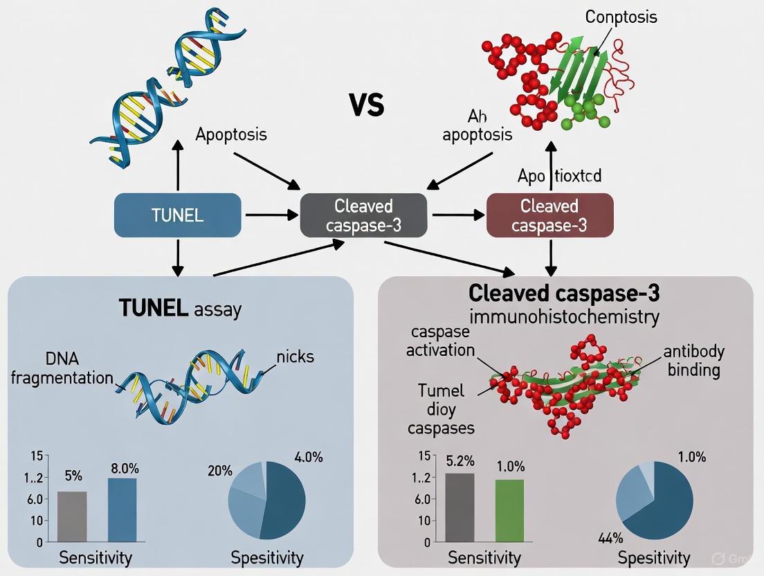
TUNEL vs Cleaved Caspase-3 IHC: A Sensitive and Specific Apoptosis Detection Guide for Researchers
This article provides a comprehensive comparison of TUNEL and cleaved caspase-3 immunohistochemistry for detecting apoptosis in biomedical research.
TUNEL vs Cleaved Caspase-3 IHC: A Sensitive and Specific Apoptosis Detection Guide for Researchers
Abstract
This article provides a comprehensive comparison of TUNEL and cleaved caspase-3 immunohistochemistry for detecting apoptosis in biomedical research. Tailored for scientists and drug development professionals, it explores the foundational mechanisms, detailed protocols, troubleshooting strategies, and validation data for both techniques. By synthesizing current research, the content clarifies the superior specificity of cleaved caspase-3 for early-to-mid apoptosis and the application of TUNEL for late-stage DNA fragmentation, empowering researchers to select and optimize the most sensitive and reliable methods for their experimental models in cancer, toxicology, and disease pathophysiology.
Understanding Apoptosis Pathways: The Biological Basis for TUNEL and Caspase-3
Programmed cell death, or apoptosis, is a fundamental biological process essential for embryonic development, normal tissue turnover, and the elimination of damaged or infected cells. This genetically controlled, energy-dependent pathway facilitates the orderly dismantling of a cell, preventing the release of harmful contents and avoiding inflammatory responses. The precise identification of apoptosis relies heavily on recognizing its distinctive morphological features, which stand in stark contrast to those of accidental cell death (necrosis). Within research and clinical diagnostics, a variety of techniques are employed to detect these changes, with the TUNEL assay and cleaved caspase-3 immunohistochemistry being two prominent methods. This guide provides an objective comparison of these techniques, framed within the context of sensitivity research, to aid scientists in selecting the most appropriate methodology for their experimental or diagnostic objectives.
The Hallmark Morphological Features of Apoptosis
The definitive identification of apoptosis is rooted in observing a series of characteristic structural alterations within the cell. These changes represent a consistent and evolutionarily conserved pattern that can be visualized using various microscopic techniques.
Nuclear Changes
The most diagnostic features of apoptosis occur within the nucleus. Chromatin condensation begins with the aggregation of nuclear material beneath the nuclear membrane, culminating in pyknosis, a state where the nucleus becomes intensely basophilic and shrunken [1]. This is followed by karyorrhexis, the fragmentation of the condensed nucleus into discrete, membrane-bound apoptotic bodies containing nuclear debris [2] [1]. These nuclear events are often the most readily identifiable indicators of apoptosis in stained tissue sections.
Cytoplasmic and Membrane Changes
Concurrent with nuclear disintegration, the cytoplasm undergoes profound remodeling. A hallmark early event is cell shrinkage and loss of cell-cell contacts, driven by controlled ion movement and ATPase activity [2]. This is accompanied by extensive membrane blebbing, where the plasma membrane forms outward protrusions due to the dissociation of the cytoskeleton from the membrane and activation of myosin light-chain kinase [2]. The cell eventually separates into multiple, compact apoptotic bodies, which are neatly packaged vesicles containing cytoplasm and intact organelles [3] [1]. Crucially, the integrity of the plasma membrane is maintained throughout this process, preventing the leakage of cellular contents and subsequent inflammation [1].
Table 1: Key Morphological Changes in Apoptosis and Necrosis
| Feature | Apoptosis | Necrosis |
|---|---|---|
| Cell Size | Shrinkage | Swelling |
| Nucleus | Pyknosis and Karyorrhexis | Karyolysis |
| Cell Membrane | Intact, with blebbing | Disrupted |
| Cellular Contents | Retained in apoptotic bodies | Released |
| Inflammatory Response | None | Present |
| Tissue Affected | Single cells or small clusters | Contiguous cells |
Detection Methodologies: A Focus on TUNEL and Cleaved Caspase-3 IHC
Accurate quantification of apoptosis in tissue sections requires reliable methods. Below are detailed protocols for two widely used techniques, highlighting their underlying principles.
TUNEL Assay Protocol
The Terminal deoxynucleotidyl transferase (TdT) dUTP Nick-End Labeling (TUNEL) assay detects DNA fragmentation, a late-stage event in apoptosis.
- Principle: The enzyme Terminal deoxynucleotidyl transferase (TdT) catalyzes the addition of labeled dUTP to the 3'-hydroxyl termini of double- or single-stranded DNA breaks that are characteristic of apoptotic DNA cleavage [4] [5].
- Procedure:
- Tissue Preparation: Deparaffinize and rehydrate formalin-fixed, paraffin-embedded (FFPE) tissue sections.
- Proteinase Digestion: Treat sections with proteinase K to expose DNA strands.
- TdT Reaction: Incubate sections with a reaction mixture containing TdT enzyme and labeled nucleotides (e.g., fluorescein-dUTP).
- Detection: For fluorescent labels, visualize directly under a fluorescence microscope. For chromogenic detection, incubate with an enzyme-conjugated antibody (e.g., anti-fluorescein peroxidase) and then with a chromogen substrate like DAB.
- Counterstaining: Use a light counterstain (e.g., hematoxylin) to visualize all cells.
- Output: Apoptotic nuclei are identified by positive staining (brown with DAB, fluorescent with fluorescein). The percentage of TUNEL-positive epithelial cells can be calculated for quantification [5].
Cleaved Caspase-3 Immunohistochemistry (IHC) Protocol
This method detects the activated form of caspase-3, a key "executioner" caspase in the apoptotic cascade, offering a more specific measure of the cell death program.
- Principle: Antibodies are used that specifically recognize a neo-epitope on the large fragment of caspase-3 that is only exposed after its proteolytic cleavage and activation during apoptosis [4].
- Procedure:
- Tissue Preparation: Deparaffinize and rehydrate FFPE tissue sections.
- Antigen Retrieval: Heat treat sections in a citrate or EDTA-based buffer to unmask the target epitope.
- Blocking: Incubate with a protein block (e.g., normal serum) to reduce non-specific antibody binding.
- Primary Antibody Incubation: Apply a monoclonal or polyclonal antibody specific for cleaved caspase-3.
- Detection: Use a standardized detection system (e.g., horseradish peroxidase-conjugated secondary antibody and DAB chromogen).
- Counterstaining: Counterstain with hematoxylin.
- Output: Cells undergoing apoptosis show cytoplasmic and/or nuclear immunoreactivity for cleaved caspase-3. The staining intensity and distribution can be graded (e.g., 0 to 3+) for semi-quantitative analysis [5].
Comparative Analysis: TUNEL vs. Cleaved Caspase-3 IHC
While both methods aim to identify apoptotic cells, they target different stages and biochemical events in the process, leading to significant differences in specificity, sensitivity, and applicability.
Quantitative Data Comparison
A direct comparative study in PC-3 subcutaneous xenografts provides objective data for evaluating these two methods [4].
Table 2: Quantitative Comparison of Apoptosis Detection Methods in PC-3 Xenografts
| Method | Target Process | Correlation with Activated Caspase-3 | Key Advantage | Key Limitation |
|---|---|---|---|---|
| Cleaved Caspase-3 IHC | Caspase activation (mid-stage) | Self (Gold Standard) | High specificity, detects early commitment to apoptosis | May miss very late-stage apoptotic cells |
| Cleaved CK18 IHC | Caspase-mediated keratin cleavage | Excellent (R = 0.89) | High specificity, correlates well with caspase-3 | Tissue and cell type specific |
| TUNEL Assay | DNA fragmentation (late-stage) | Good (R = 0.75) | Widely adopted, labels late-stage nuclei | Can yield false positives from necrosis or autolysis |
Sensitivity and Specificity Assessment
- Cleaved Caspase-3 IHC demonstrates superior specificity as it directly detects a central component of the apoptotic execution machinery [4]. It is an "easy, sensitive, and reliable method" for quantifying apoptosis, identifying cells that have irreversibly committed to the death pathway but may not yet exhibit DNA fragmentation.
- TUNEL Assay, while sensitive, has been criticized for its potential lack of specificity. The assay can label DNA breaks occurring in other contexts, such as necrosis, autolysis, DNA repair, or even gene transcription, potentially leading to false-positive results [4] [5]. The study on lung tissues from Idiopathic Interstitial Pneumonias required additional validation with proliferation markers to confirm that TUNEL positivity was not due to DNA repair [5].
The Scientist's Toolkit: Essential Reagents for Apoptosis Detection
Successful experimentation relies on high-quality, specific reagents. The following table outlines key materials used in the featured methodologies.
Table 3: Research Reagent Solutions for Apoptosis Detection
| Reagent / Kit | Function / Target | Application Context |
|---|---|---|
| Anti-Cleaved Caspase-3 Antibody | Binds specifically to activated caspase-3 fragment | Immunohistochemistry, Western Blot |
| TUNEL Assay Kit | Labels DNA strand breaks with fluorescent or chromogenic tags | In-situ apoptosis detection on tissue sections or cells |
| Annexin V-FITC/PI Apoptosis Kit | Detects phosphatidylserine exposure (early apoptosis) and membrane integrity | Flow cytometry, fluorescence microscopy |
| Antibody Cocktails (e.g., pro/p17-caspase-3 + cleaved PARP) | Simultaneously detect multiple apoptosis markers in a single assay | Streamlined Western Blot analysis |
| Hoechst 33342 / DAPI | Fluorescent dyes that bind DNA, highlighting nuclear condensation and fragmentation | Fluorescence microscopy for morphological assessment |
Signaling Pathways and Experimental Workflow
The decision to use TUNEL or cleaved caspase-3 IHC is informed by their respective positions within the apoptotic cascade. The following diagrams illustrate the biochemical context of these markers and a generalized workflow for their evaluation.
Apoptosis Signaling Pathways and Detection Marker Context
Experimental Decision Workflow for Apoptosis Detection
The choice between TUNEL and cleaved caspase-3 immunohistochemistry is not merely a technical preference but a strategic decision based on the biological question and required level of specificity. Cleaved caspase-3 IHC emerges as the more specific and reliable method for quantifying apoptosis in histological sections, as it directly targets the core apoptotic protease and demonstrates an excellent correlation with the gold standard of morphological assessment [4]. It is particularly powerful for identifying cells in the early and middle stages of apoptosis.
The TUNEL assay, while a historically valuable tool for detecting the late stages of cell death, requires careful interpretation and stringent controls to avoid false positives from non-apoptotic DNA fragmentation. For the most definitive results, a multimodal approach combining cleaved caspase-3 IHC with morphological analysis is strongly recommended. This combination leverages the specificity of the molecular marker with the irrefutable evidence provided by the classic hallmarks of apoptosis, such as cell shrinkage, chromatin condensation, and the formation of apoptotic bodies.
Caspase-3 is a cysteine-aspartic protease that functions as a key executioner protein in the apoptotic pathway, cleaving cellular substrates at specific aspartic acid residues to orchestrate controlled cellular dismantling [6]. As the most prominent effector caspase, it occupies a central position where multiple cell death pathways converge, serving as the final common mediator before the characteristic morphological changes of apoptosis occur [7]. This enzyme is synthesized as an inactive zymogen (caspase-3p32) that requires proteolytic activation into p20 and p12 fragments to gain catalytic function [8]. Caspase-3 demonstrates the highest homology to the CED-3 protein in Caenorhabditis elegans, underscoring its evolutionary conservation and fundamental role in programmed cell death across species [8].
The essential nature of caspase-3 is dramatically illustrated in knockout models, where caspase-3-deficient mice exhibit profoundly abnormal brain development and die within three weeks after birth, establishing its non-redundant functions in neural development [8]. Beyond its classical role in apoptosis, emerging research reveals caspase-3 participates in diverse non-apoptotic processes including stem cell differentiation, tissue regeneration, and even cancer cell motility, expanding its biological significance beyond cell death execution [7] [9].
Caspase-3 Activation and the Apoptotic Cascade
Molecular Activation Pathways
Caspase-3 activation occurs through two principal signaling cascades: the extrinsic (death receptor) pathway and the intrinsic (mitochondrial) pathway [7]. The extrinsic pathway initiates when extracellular ligands bind to death receptors belonging to the tumor necrosis factor (TNF) receptor superfamily, such as Fas (CD95), TNFR1, and TRAIL receptors [7]. This receptor activation recruits adaptor proteins like FADD (Fas-associated death domain protein), which subsequently activates initiator caspase-8. The intrinsic pathway, conversely, is triggered by intracellular stress signals that cause mitochondrial outer membrane permeabilization (MOMP), leading to cytochrome c release and formation of the apoptosome complex with Apaf-1 and pro-caspase-9 [6].
Both pathways ultimately converge on caspase-3 activation, though through different mechanisms. Caspase-8 from the extrinsic pathway can directly cleave and activate caspase-3, or in certain cell types, engage the intrinsic pathway by cleaving Bid, a BH3-only protein, which promotes additional cytochrome c release and caspase-9 activation [7]. Caspase-9 from the intrinsic pathway serves as the primary activator of caspase-3 in the mitochondrial pathway [6]. This convergence ensures amplification of the apoptotic signal, as active caspase-3 can further propagate the cascade through positive feedback loops that activate additional caspases, ensuring efficient and irreversible commitment to cell death [7].
Caspase-3 Cleavage Specificity and Targets
As an executioner caspase, caspase-3 recognizes specific amino acid sequences in target proteins, predominantly cleaving at DEVD (Asp-Glu-Val-Asp) motifs [10]. Recent research has identified additional cleavage motifs, including the novel AEAD sequence, expanding our understanding of its substrate specificity [11]. Upon activation, caspase-3 systematically dismantles the cell by cleaving numerous structural and functional proteins, including:
- Nuclear proteins: Poly-ADP ribose polymerase (PARP), lamin proteins, and caspase-activated DNase (CAD) inhibitor (ICAD) [6] [12]
- Cytoskeletal components: αII- and βII-spectrin, gelsoin, and other structural proteins [8]
- DNA repair enzymes: Multiple enzymes involved in DNA damage response and repair [6]
The cleavage of ICAD is particularly significant as it releases CAD, which then translocates to the nucleus and catalyzes the internucleosomal DNA fragmentation that produces the characteristic apoptotic DNA laddering [12]. Similarly, caspase-3-mediated cleavage of the protein ACINUS induces chromatin condensation prior to DNA fragmentation, further facilitating the packaging of cellular contents into apoptotic bodies [12]. This systematic substrate cleavage results in the classic morphological hallmarks of apoptosis, including cell shrinkage, chromatin condensation, DNA fragmentation, and membrane blebbing [7].
Figure 1: Caspase-3 Activation Pathways. Caspase-3 serves as the convergence point for extrinsic (death receptor) and intrinsic (mitochondrial) apoptotic pathways, executing cell death through systematic substrate cleavage.
Detection Methodologies: TUNEL vs. Cleaved Caspase-3 IHC
Terminal Deoxynucleotidyl Transferase dUTP Nick End Labeling (TUNEL)
The TUNEL assay has been extensively used for apoptosis detection in histological sections, exploiting the DNA fragmentation that occurs during late-stage apoptosis [4] [12]. This method employs terminal deoxynucleotidyl transferase (TdT) to incorporate labeled dUTP at the 3'-ends of DNA fragments created during apoptotic degradation [12]. The technique then uses immunohistochemical detection to visualize these labeled DNA ends, identifying cells undergoing programmed cell death.
While widely adopted, the TUNEL method faces significant limitations. Its interpretation and specificity have been controversial, as DNA fragmentation can occur in other cell death processes beyond classical apoptosis, including necrosis and autolysis [4]. Additionally, the TUNEL assay detects apoptosis at a relatively late stage after significant DNA degradation has already occurred, potentially missing earlier phases of the apoptotic process [4] [12]. Technical challenges also include the potential for false positives from non-apoptotic DNA damage, requiring careful interpretation alongside morphological analysis [13].
Cleaved Caspase-3 Immunohistochemistry
Cleaved caspase-3 immunohistochemistry represents a more direct approach to apoptosis detection by targeting the activated form of the enzyme itself [4]. This method uses antibodies specifically recognizing the proteolytically processed p20 fragment of caspase-3 that is generated during activation, providing direct evidence of caspase cascade engagement [4] [8]. The detection of this specific cleavage fragment offers superior specificity for apoptosis compared to TUNEL, as it directly measures enzyme activation rather than a downstream consequence that might be shared with other death pathways.
The methodology typically involves antigen retrieval on formalin-fixed, paraffin-embedded tissue sections, incubation with primary antibodies specific for the cleaved form of caspase-3, amplification with appropriate detection systems, and visualization with chromogens like diaminobenzidine (DAB) [12]. This approach allows precise cellular localization of active caspase-3 and enables quantification of apoptotic indices through computer-assisted image analysis [4]. A significant advantage of this method is its ability to detect cells in earlier stages of apoptosis before morphological changes become evident or DNA fragmentation occurs [4].
Comparative Experimental Data
Direct comparisons between these methodologies reveal important differences in performance characteristics. A comparative study using prostate cancer PC-3 subcutaneous xenografts demonstrated that activated caspase-3 immunohistochemistry provides superior sensitivity and reliability for apoptosis quantification compared to TUNEL [4]. The research found an excellent correlation (R = 0.89) between apoptotic indices obtained using activated caspase-3 and cleaved cytokeratin 18 immunostaining, while a good but lower correlation (R = 0.75) existed between activated caspase-3 and TUNEL results [4].
Table 1: Comparative Performance of Apoptosis Detection Methods
| Parameter | TUNEL Assay | Cleaved Caspase-3 IHC |
|---|---|---|
| Target | DNA fragmentation | Activated caspase-3 enzyme |
| Detection Stage | Late apoptosis | Early to mid apoptosis |
| Specificity for Apoptosis | Moderate (can detect other death forms) | High (specific to caspase-mediated apoptosis) |
| Correlation with Caspase-3 | R = 0.75 [4] | Reference standard |
| Morphological Context | Requires additional assessment | Can be correlated with morphology |
| Adaptability to Automation | Moderate [12] | High [12] |
In prostate cancer research, caspase-3 demonstrated better predictive value for clinical aggressiveness than TUNEL, with area under the curve (AUC) values of 0.694 for caspase-3 versus 0.669 for TUNEL in logistic regression models [12]. Another study of cerebral ischemia models revealed that while TUNEL detected widespread DNA fragmentation throughout ischemic regions, active caspase-3 immunoreactivity was predominantly observed in specific neuronal populations, suggesting differences in sensitivity and cellular context [13].
Experimental Protocols for Caspase-3 Detection
Immunohistochemistry Protocol for Cleaved Caspase-3
The detection of cleaved caspase-3 through immunohistochemistry follows a standardized protocol with specific considerations for optimal results. For formalin-fixed, paraffin-embedded tissues, sections of 5μm thickness are typically prepared [12]. The protocol proceeds with deparaffinization and rehydration through an alcohol gradient, followed by antigen retrieval using appropriate buffers such as Citra buffer, often employing heat-induced epitope retrieval methods (10 minutes at 120°C and 21 PSI) [12].
After cooling, sections undergo peroxidase blocking with 3% H₂O₂ for 5 minutes at 37°C to quench endogenous peroxidase activity, followed by avidin-biotin blocking if necessary [12]. The critical step involves incubation with primary antibodies specific for cleaved caspase-3—anti-caspase-3 antibodies diluted typically at 1:500 for 2 hours at 37°C [12]. Multiple commercial antibodies are available that specifically recognize the activated form without cross-reacting with the full-length zymogen.
Following primary antibody incubation, sections are processed with appropriate detection systems, such as peroxidase-labeled polymer systems, and visualized with chromogens like diaminobenzidine (DAB) [12]. Counterstaining with hematoxylin provides nuclear context, followed by dehydration and mounting for microscopic analysis. Appropriate negative controls (omitting primary antibody) and positive controls (tissues with known apoptosis) should be included in each run to validate results [12].
Caspase Activity Assay Protocol
Beyond immunohistochemical localization, biochemical assessment of caspase-3 activity provides quantitative data on enzyme function. The caspase-like DEVDase activity assay utilizes synthetic substrates containing the DEVD recognition sequence to specifically measure caspase-3/7 activity [8]. In this protocol, tissue samples are homogenized in appropriate buffer (25mM HEPES, pH 7.5, containing 0.1% Triton X-100, 5mM MgCl₂, 2mM dithiothreitol) with protease inhibitors [8].
The homogenate is centrifuged at 50,000 × g, and the supernatant is incubated with fluorogenic substrates such as zDEVD-afc (N-benzyloxycarbonyl-Asp-Glu-Val-Asp-7-amino-4-trifluoromethyl coumarin) at 12.5μM concentration [8]. Enzyme activity is measured by monitoring fluorescence increase (400-505nm excitation-emission) at 5-minute intervals, with calculations based on the slope of fluorescence units per milligram of protein per minute [8]. This assay demonstrates optimal activity at physiological pH (7.4), with substantial reduction (>80%) at slightly acidic pH (6.8-7.0) [8].
Specificity should be confirmed using caspase inhibitors such as zDEVD-fmk, which completely blocks activity at 200μM concentration [10]. This biochemical approach complements immunohistochemical findings by providing quantitative kinetic data on caspase activation, particularly useful for tracking temporal progression of apoptosis in experimental models [8].
Figure 2: Cleaved Caspase-3 IHC Workflow. Standardized experimental protocol for detecting activated caspase-3 in formalin-fixed, paraffin-embedded (FFPE) tissue sections.
The Scientist's Toolkit: Essential Research Reagents
Table 2: Key Research Reagents for Caspase-3 Detection
| Reagent/Category | Specific Examples | Function & Application |
|---|---|---|
| Primary Antibodies | Anti-caspase-3 (R&D Systems), Anti-cleaved caspase-3 | Specifically recognizes activated caspase-3; essential for IHC detection [12] |
| Activity Assay Substrates | zDEVD-afc, zDEVD-fmk (fluorogenic/inhibitor) | Fluorogenic substrate for measuring caspase-3/7 activity; inhibitor for specificity controls [8] [10] |
| Detection Systems | EnVision Doublestain System, HRP-labeled polymers | Signal amplification and visualization for immunohistochemistry [12] |
| Positive Control Tissues | PC-3 xenografts, ischemic brain tissue, skin compression samples | Tissues with known apoptosis for assay validation [4] [14] |
| Caspase Inhibitors | zDEVD-fmk, zVAD-fmk (pan-caspase inhibitor) | Specific and broad-spectrum caspase inhibitors for mechanistic studies [8] [10] |
| Fluorescent Biosensors | VC3AI (Venus-based C3AI) | Genetically encoded indicator for real-time caspase-3 activity monitoring in live cells [10] |
Applications in Disease Research and Diagnostic Contexts
Neurodegenerative Diseases and Cerebral Ischemia
Caspase-3 activation plays a crucial role in neuronal cell death following ischemic injury and in neurodegenerative conditions. In transient focal ischemia models, caspase-3 activation demonstrates a time-dependent evolution, with elevated caspase-like enzyme activity detected within 30-60 minutes after reperfusion, followed by immunoreactivity for the caspase-3p20 fragment at 1-12 hours, and eventual DNA fragmentation appearing 6-24 hours post-reperfusion [8]. This temporal progression underscores caspase-3's role in the execution phase of ischemic neuronal death and positions it as an earlier marker than DNA fragmentation for detecting apoptotic commitment.
Research in rat models of transient middle cerebral artery occlusion reveals that active caspase-3 immunoreactivity primarily localizes to scattered neurons in narrow zones near the edges of ischemic infarcts, particularly in striatal regions, while being largely absent from cortical neurons within the infarct core [13]. This patterned activation suggests selective neuronal vulnerability and potentially different death mechanisms in various brain regions following ischemic insult. The detection of caspase-3 activation in these models has therapeutic implications, as caspase inhibitors like zDEVD-fmk demonstrate neuroprotective effects and improved neurological outcomes in animal studies [8].
Cancer Biology and Prognostic Assessment
In cancer research, caspase-3 detection serves both prognostic and mechanistic purposes. Studies in prostate cancer demonstrate that caspase-3 immunostaining provides superior predictive value for clinical aggressiveness compared to TUNEL, with automated image analysis of caspase-3-positive cells enabling calculation of tumor growth rates that show statistically significant linear trends across clinical aggressiveness categories [12]. This application highlights the utility of caspase-3 as a biomarker for tumor behavior assessment.
Paradoxically, some aggressive cancers including melanoma and colon cancer exhibit high caspase-3 expression despite its pro-apoptotic function [9]. In melanoma, caspase-3 expression differentiates primary from metastatic tumors and associates with poor prognosis, suggesting non-apoptotic functions [9]. Recent research reveals caspase-3 regulates melanoma cell motility through interactions with cytoskeletal proteins like coronin 1B, promoting migration and invasion independently of its apoptotic function [9]. This unexpected role expands the functional repertoire of caspase-3 beyond cell death and complicates its interpretation in cancer contexts.
Forensic Applications and Vitality Assessment
In forensic pathology, caspase-3 detection has emerged as a valuable tool for determining vitality in ligature marks in hanging cases, where its presence indicates an ante-mortem response to compression [14]. Studies demonstrate that caspase-3 levels in compressed skin from ligature marks are significantly elevated compared to healthy skin (p < 0.005), establishing it as a reliable marker of supravitality [14]. Since caspase-3 activation is an ATP-dependent process, its detection indicates the compressive force was applied while the victim was still alive with sufficient cellular energy for the apoptotic cascade to initiate.
The apoptotic process in ischemic epidermal cells begins promptly with mechanical stress, with caspase-3 expression varying from minutes after the initial stress input [14]. This rapid response makes it a sensitive indicator of vital reactions to injury, with semi-quantitative analyses revealing strong caspase-3 expression in basal epidermal cells within ligature furrows, exhibiting a granular cytoplasmic and nuclear distribution pattern [14]. The reliability of caspase-3 for vitality assessment surpasses traditional histological examination alone and provides objective evidence for distinguishing ante-mortem from post-mortem injuries.
In the study of programmed cell death, the temporal sequence of molecular events is fundamental to accurately interpreting experimental data. Apoptosis is characterized by a cascade of biochemical events, beginning with the activation of cysteine-aspartic proteases (caspases) and culminating in the systematic dismantling of the cell, including the hallmark fragmentation of nuclear DNA. This article positions the TUNEL (Terminal deoxynucleotidyl transferase dUTP Nick End Labeling) assay within this temporal framework, objectively comparing its performance with the detection of cleaved caspase-3 as a earlier-phase apoptotic marker. The core thesis is that while TUNEL is a valuable technique, it detects a late-stage event in the apoptotic process, a characteristic that fundamentally defines its sensitivity, specificity, and appropriate application in research and drug development [4] [15].
Understanding this sequence is critical. The activation of executioner caspases, particularly caspase-3, represents a commitment point where the cell irreversibly initiates its own degradation. One of the key downstream actions of activated caspase-3 is the cleavage of inhibitors of DNA-breaking enzymes, leading to the oligonucleosomal DNA fragmentation that the TUNEL assay is designed to detect [12] [15]. Consequently, a TUNEL-positive signal manifests hours after the initial caspase activation, a delay that has direct implications for the interpretation of cell death studies in contexts ranging from cancer therapy response to neurological disease.
Comparative Analysis of Key Apoptotic Markers
The following table summarizes the core characteristics of the TUNEL assay and cleaved caspase-3 immunohistochemistry (IHC) based on empirical comparisons.
Table 1: Direct Comparison of TUNEL and Cleaved Caspase-3 Detection Methods
| Feature | TUNEL Assay | Cleaved Caspase-3 IHC |
|---|---|---|
| Detected Event | DNA strand breaks (late-stage apoptosis, necrosis) [15] | Activation of key executioner caspase (early-to-mid apoptosis) [4] [16] |
| Temporal Position | Late; follows caspase activation and substrate cleavage [8] | Early/Mid; precedes and triggers DNA fragmentation [4] |
| Specificity for Apoptosis | Moderate; can label DNA breaks from necrotic cell death [15] | High; directly detects a central mediator of apoptotic execution [4] [17] |
| Correlation with Morphology | Good in clear-cut cases | Excellent; strong correlation with morphological apoptosis [4] |
| Key Advantage | Flags ultimate apoptotic outcome (DNA destruction) | Identifies cells committed to, but not yet fully dismantled by, apoptosis [18] |
Quantitative data reinforces this temporal and functional relationship. A comparative study on prostate cancer (PC-3) xenografts found that while there was a good correlation (R=0.75) between apoptotic indices obtained via cleaved caspase-3 IHC and the TUNEL assay, the caspase-3 method was deemed more sensitive and reliable for quantification. The correlation between two caspase-dependent markers—activated caspase-3 and caspase-cleaved cytokeratin 18—was even stronger (R=0.89), underscoring the consistency of detecting early caspase activity [4]. Furthermore, in assessing prostate cancer aggressiveness, cleaved caspase-3 was a better predictor (AUC=0.694, P=0.038) than TUNEL (AUC=0.669, P=0.110), highlighting its potential clinical utility [12].
Experimental Data and Protocols
Key Supporting Experiments
The following table outlines foundational experiments that have directly compared these two detection methods across various biological models.
Table 2: Summary of Key Comparative Experiments
| Experimental Model | Treatment / Condition | Key Finding | Citation |
|---|---|---|---|
| PC-3 Prostate Cancer Xenografts | (Spontaneous apoptosis in model) | Caspase-3 IHC is more sensitive and reliable for apoptosis quantification than TUNEL, with a good correlation (R=0.75) between the two. | [4] |
| Pig Lymphoid Organs | Normal physiological apoptosis | Both methods detect physiological apoptosis; TUNEL stains apoptotic bodies in macrophages, while CCasp3 labels individual lymphocytes earlier in the process. | [16] |
| Human Prostate Cancer Biopsies | Newly diagnosed cancers of varying aggressiveness | Caspase-3 was a statistically significant predictor of clinical aggressiveness, while TUNEL was not (P=0.110). | [12] |
| Mouse Brain Model | Transient cerebral ischemia | Cleaved caspase-3 (p20) immunoreactivity appears at reperfusion, while TUNEL-positive cells and DNA laddering are detected 6–24 hours later. | [8] |
Detailed Experimental Protocol
To illustrate how such comparative data is generated, the protocol from the seminal PC-3 xenograft study is detailed below [4]. This serves as a robust template for researchers seeking to validate these methods in their own models.
Protocol: Comparison of Cleaved Caspase-3 IHC and TUNEL on Formalin-Fixed Paraffin-Embedded (FFPE) Tissues
Tissue Processing:
- Induce apoptosis in the experimental model (e.g., treat xenografts with a chemotherapeutic agent known to trigger apoptosis).
- Harvest tissues and fix immediately in 10% neutral buffered formalin for 16-24 hours. Prolonged fixation can mask epitopes and should be avoided.
- Process fixed tissues through a graded ethanol series, clear with xylene, and embed in paraffin.
- Section tissues at 4-5 μm thickness and mount on charged glass slides.
Cleaved Caspase-3 Immunohistochemistry:
- Deparaffinization and Rehydration: Bake slides, then deparaffinize in xylene and rehydrate through a descending ethanol series to distilled water.
- Antigen Retrieval: Perform heat-induced epitope retrieval using a citrate-based buffer (pH 6.0) or Tris-EDTA buffer (pH 9.0) in a pressure cooker or water bath. This step is critical for unmasking the caspase-3 epitope.
- Endogenous Peroxidase Blocking: Incubate sections with 3% hydrogen peroxide to quench endogenous peroxidase activity.
- Protein Blocking: Apply a serum or protein block from the same species as the secondary antibody to reduce non-specific binding.
- Primary Antibody Incubation: Incubate sections with a polyclonal or monoclonal anti-cleaved caspase-3 antibody (e.g., Cell Signaling Technology #9661) at a predetermined optimal dilution (e.g., 1:100 to 1:500) for 1-2 hours at room temperature or overnight at 4°C.
- Detection: Use a standard detection system such as the EnVision/HRP system with a dextran polymer conjugated with secondary antibodies and horseradish peroxidase. Visualize the signal with 3,3'-Diaminobenzidine (DAB), which produces a brown precipitate.
- Counterstaining and Mounting: Counterstain lightly with hematoxylin to visualize nuclei, then dehydrate, clear, and mount with a permanent mounting medium.
TUNEL Assay:
- Section Preparation: Deparaffinize and rehydrate adjacent tissue sections as described above.
- Proteinase Digestion: Treat sections with Proteinase K (e.g., 20 μg/mL) for 15-20 minutes at room temperature to permeabilize the tissue and expose DNA.
- Endogenous Peroxidase Blocking: Apply 3% hydrogen peroxide as in the IHC protocol.
- Labeling Reaction: Incubate sections with the TUNEL reaction mixture containing TdT (Terminal deoxynucleotidyl Transferase) enzyme and fluorescein- or biotin-labeled dUTP (e.g., from the In Situ Cell Death Detection Kit, POD, Roche) for 60 minutes at 37°C.
- Signal Detection and Visualization: For fluorescent labels, apply an anti-fluorescein antibody conjugated with horseradish peroxidase (POD), then develop with DAB. For biotin labels, use a streptavidin-HRP complex followed by DAB.
- Counterstaining and Mounting: Counterstain with hematoxylin, dehydrate, clear, and mount.
Quantification and Analysis:
- Acquire images of stained sections using a brightfield microscope.
- Calculate the Apoptotic Index (AI) for each method by counting the number of positively stained cells per 1000 total cells or per unit area, preferably in a blinded manner.
- Use computer-assisted image analysis software for objective and reproducible quantification across all samples.
- Perform statistical analysis (e.g., Pearson correlation) to compare the apoptotic indices derived from the two methods.
The Scientist's Toolkit: Essential Research Reagents
Successful execution of the protocols above relies on a set of specific, high-quality reagents.
Table 3: Key Reagent Solutions for Apoptosis Detection
| Reagent / Kit | Function | Key Consideration |
|---|---|---|
| Anti-Cleaved Caspase-3 Antibody | Specifically binds the activated (cleaved) form of caspase-3, avoiding the inactive pro-enzyme. | Antibody specificity must be validated. Polyclonal antibodies often provide a stronger signal but may have higher background. |
| TUNEL Assay Kit | Enzymatically labels the 3'-OH ends of fragmented DNA. | Kits from vendors like Roche/Merck or Thermo Fisher are standardized. The choice of fluorescence vs. colorimetric detection depends on the application and available equipment. |
| Citrate or EDTA-based Antigen Retrieval Buffer | Unmasks hidden epitopes in FFPE tissue by reversing formaldehyde cross-links. | The pH and buffer type can dramatically impact signal intensity and must be optimized for the primary antibody. |
| HRP-based Detection System (e.g., EnVision) | Amplifies the primary antibody signal for visual detection under a microscope. | Polymer-based systems are highly sensitive and reduce non-specific background compared to avidin-biotin systems. |
| DAB (3,3'-Diaminobenzidine) Chromogen | A substrate for HRP that produces an insoluble, brown precipitate at the site of the target antigen. | DAB is carcinogenic and must be handled with appropriate safety measures. |
Integrated Signaling Pathways and Experimental Workflow
The biochemical relationship between caspase-3 activation and DNA fragmentation, and the corresponding detection methods, can be visualized in the following pathway. Cleaved caspase-3, an executioner caspase, directly cleaves and inactivates the inhibitor of Caspase-Activated DNase (ICAD). This releases the active CAD enzyme, which then translocates to the nucleus and cleaves DNA into the oligonucleosomal fragments that are the hallmark of apoptosis and the target of the TUNEL assay [12] [15].
Diagram 1: The Apoptotic Pathway from Caspase-3 Activation to DNA Fragmentation. The diagram illustrates the causal relationship where active caspase-3 cleaves ICAD to release CAD, which subsequently fragments nuclear DNA. The dashed lines indicate the specific detection events for cleaved caspase-3 IHC (an early/mid event) and the TUNEL assay (a late event).
The objective comparison of TUNEL and cleaved caspase-3 IHC confirms a central tenet of apoptosis biology: DNA fragmentation is a late-stage event. The experimental data consistently shows that cleaved caspase-3 is a more sensitive and specific marker for the commitment phase of apoptotic execution, appearing earlier in the timeline and providing a stronger correlation with clinical outcomes in disease models like cancer [4] [12].
For the researcher, this has direct practical implications. The choice of assay should be guided by the experimental question. If the goal is to identify cells that have initiated the irreversible execution phase of apoptosis—for instance, to evaluate the early efficacy of a pro-apoptotic drug—cleaved caspase-3 IHC is the superior tool. Conversely, the TUNEL assay remains valuable for confirming that the cell death process has reached its terminal stage of nuclear disintegration. A comprehensive approach, utilizing cleaved caspase-3 IHC to mark the initiation of execution and TUNEL to confirm its completion, provides the most powerful strategy for quantifying and understanding apoptosis in physiological and pathological contexts. This nuanced understanding is essential for advancing research in cancer biology, neuroscience, and drug development.
Apoptosis, or programmed cell death, is a tightly regulated process essential for development, tissue homeostasis, and disease prevention. The morphological changes characteristic of apoptosis—including cell shrinkage, chromatin condensation, and nuclear fragmentation—are largely executed by a family of cysteine proteases known as caspases [19]. Among these, caspase-3 serves as a key convergence point for the two principal apoptotic signaling pathways: the intrinsic (mitochondrial) pathway and the extrinsic (death receptor) pathway [20] [21].
Both pathways initiate caspase-3 activation through distinct mechanisms and signaling cascades, yet they ultimately coordinate the dismantling of the cell through the cleavage of vital cellular components. This comparative guide examines the mechanistic details, temporal dynamics, and detection methodologies associated with caspase-3 activation via these two pathways, providing researchers with objective data to inform experimental design and interpretation in drug development and basic research.
Pathway Mechanisms: From Initiation to Execution
The Intrinsic (Mitochondrial) Pathway
The intrinsic pathway is activated in response to internal cellular stressors such as DNA damage, oxidative stress, growth factor deprivation, and oncogene activation [20]. These signals converge on the mitochondria, initiating a cascade of protein interactions that ultimately lead to caspase activation.
Key Molecular Events:
- Stress Sensing and Signaling: Cellular stresses, particularly DNA damage, are sensed by proteins like ATM and Chk2, which stabilize and activate the p53 tumor suppressor protein. p53 then transcriptionally activates pro-apoptotic Bcl-2 family members, including Bax, Noxa, and PUMA [20].
- Mitochondrial Outer Membrane Permeabilization (MOMP): Activated pro-apoptotic proteins like Bax and Bak integrate into the mitochondrial outer membrane, forming pores that lead to MOMP. This critical event is counterbalanced by anti-apoptotic Bcl-2 family members (Bcl-2, Bcl-xL) [20] [19].
- Cytochrome c Release and Apoptosome Formation: MOMP permits the release of cytochrome c from the mitochondrial intermembrane space into the cytosol. Cytochrome c binds to Apaf-1, forming a complex called the apoptosome in the presence of dATP/ATP [20].
- Caspase-9 and Caspase-3 Activation: The apoptosome recruits and activates initiator caspase-9, which then proteolytically processes and activates the executioner caspase, caspase-3 [20] [21].
The Extrinsic (Death Receptor) Pathway
The extrinsic pathway is initiated outside the cell through the engagement of death receptors by their cognate ligands, providing a mechanism for the immune system to direct cell elimination.
Key Molecular Events:
- Death Receptor Ligand Binding: Death receptors belonging to the Tumor Necrosis Factor Receptor (TNFR) superfamily (e.g., Fas, TNFR1) are activated upon binding to their trimeric ligands (e.g., FasL, TNF-α) [20].
- Death-Inducing Signaling Complex (DISC) Formation: Receptor activation leads to the intracellular recruitment of adapter proteins such as FADD (Fas-Associated via Death Domain) and the initiator caspase, procaspase-8. This multi-protein complex is known as the DISC [20].
- Caspase-8 Activation: Within the DISC, procaspase-8 undergoes proximity-induced autoprocessing and activation [20].
- Downstream Caspase Activation: Activated caspase-8 can propagate the death signal through two primary routes:
- Direct Cleavage Pathway: Caspase-8 directly cleaves and activates procaspase-3 [20].
- Amplification Loop via Mitochondria: Caspase-8 cleaves the Bcl-2 family protein Bid to its active form, tBid. tBid translocates to mitochondria, inducing MOMP and cytochrome c release, thereby engaging the intrinsic pathway to amplify caspase activation [20].
The following diagram illustrates the key steps in both pathways and their convergence on caspase-3 activation:
Quantitative Comparison of Caspase-3 Activation
The intrinsic and extrinsic pathways demonstrate distinct kinetic profiles and activation thresholds for caspase-3. The following table summarizes key quantitative differences and characteristics based on experimental data.
Table 1: Quantitative Comparison of Caspase-3 Activation via Intrinsic and Extrinsic Pathways
| Parameter | Intrinsic Pathway | Extrinsic Pathway | Experimental Context |
|---|---|---|---|
| Key Initiators | Cellular stress (DNA damage, hypoxia), p53, Bax/Bak | Death receptors (Fas, TNFR1), Ligands (FasL, TNF-α) | Defined molecular components of each pathway [20] |
| Primary Activator of Caspase-3 | Caspase-9 (via apoptosome) | Caspase-8 (directly or via Bid/mitochondria) | Hierarchical caspase activation cascade [21] |
| Activation Kinetics at Single-Cell Level | Rapid completion (~5 min) once initiated [22] | Rapid execution; can be faster than intrinsic | FRET-based live-cell imaging [22] |
| Temperature Sensitivity | Inhibited at room temperature (25°C) [23] | Functional at room temperature (25°C) [23] | Studies in Jurkat cells with various stimuli [23] |
| Regulatory Core | Bcl-2 family balance, IAPs (XIAP), SMAC/Diablo [20] [24] | FLIP, FADD, IAPs [20] | Identification of key regulatory proteins [20] [24] |
| Synergy with Proteasome Inhibition | Amplification of caspase activity observed [25] | Amplification of caspase activity observed [25] | Proteomic analysis and combinatorial drug studies [25] |
Detection Methodologies and Sensitivity
Detecting caspase-3 activation accurately is crucial for apoptosis research. The two most common methods are TUNEL, which identifies late-stage DNA fragmentation, and immunohistochemistry (IHC) for cleaved caspase-3, which detects the active protease itself.
TUNEL Assay: Principles and Protocols
The Terminal deoxynucleotidyl transferase dUTP nick-end labeling (TUNEL) assay detects DNA fragmentation, a hallmark of late-stage apoptosis [19].
Core Protocol:
- Sample Preparation: Fix cells or tissue sections (e.g., formalin-fixed paraffin-embedded, FFPE).
- Antigen Retrieval: A critical step to expose DNA nicks. Traditional protocols use Proteinase K digestion, but this can degrade protein epitopes, preventing subsequent multiplexed protein detection [26].
- Labeling Reaction: Incubate samples with terminal deoxynucleotidyl transferase (TdT) enzyme and labeled dUTP (e.g., fluorophore-conjugated).
- Detection: Visualize labeled DNA fragments via fluorescence microscopy or flow cytometry.
Compatibility Note: Recent advancements demonstrate that replacing Proteinase K with heat-mediated antigen retrieval (e.g., pressure cooker) preserves TUNEL signal sensitivity while maintaining protein antigenicity for multiplexed spatial proteomics methods like MILAN (Multiple Iterative Labeling by Antibody Neodeposition) and CycIF (Cyclic Immunofluorescence) [26].
Cleaved Caspase-3 Immunohistochemistry
This method uses antibodies specific to the activated form of caspase-3 (which lacks the prodomain), providing a direct measure of this key executioner caspase's activity [24].
Core Protocol:
- Sample Preparation: Fix cells or tissue sections (FFPE recommended).
- Antigen Retrieval: Typically employs heat-induced epitope retrieval (HIER) in citrate or EDTA buffer.
- Blocking and Antibody Incubation: Block nonspecific sites and incubate with primary antibody specific for cleaved caspase-3, followed by species-appropriate secondary antibodies conjugated to reporters (enzymes or fluorophores).
- Signal Detection: Develop signal using chromogenic or fluorescent substrates.
Comparative Sensitivity and Specificity
Table 2: Comparison of TUNEL and Cleaved Caspase-3 IHC for Apoptosis Detection
| Feature | TUNEL Assay | Cleaved Caspase-3 IHC |
|---|---|---|
| Detects | DNA fragmentation (late apoptosis/necrosis) | Activated caspase-3 protein (mid-late apoptosis) |
| Stage of Detection | Late | Mid to Late |
| Specificity for Apoptosis | Lower (also stains necrotic cells) [19] | Higher (directly measures apoptotic pathway activation) |
| Multiplexing Compatibility | Possible with pressure cooker retrieval [26] | Excellent with standard IHC/IF protocols |
| Key Consideration | DNA fragmentation is a final, committal step | Caspase-3 activation is a regulatory point; also has non-apoptotic roles [27] |
The Scientist's Toolkit: Essential Research Reagents
Successful investigation of apoptotic pathways requires a suite of reliable reagents. The following table outlines essential tools for studying caspase-3 activation.
Table 3: Key Research Reagent Solutions for Apoptosis and Caspase-3 Studies
| Reagent / Assay | Primary Function | Application Context |
|---|---|---|
| TUNEL Assay Kits | Label 3'OH ends of fragmented DNA in situ | Detecting late-stage apoptosis/necrosis in cells and tissues [26] [19] |
| Anti-Cleaved Caspase-3 Antibodies | Specifically recognize the active, large fragment of caspase-3 | IHC, IF, and Western blot detection of caspase-3 activation [19] |
| Caspase Inhibitors (e.g., z-DEVD-FMK) | Cell-permeable, irreversible inhibitor of caspase-3 | Validating caspase-3 functional role; inhibiting apoptosis [22] [27] |
| FRET-Based Caspase Sensors (e.g., SCAT3/mSCAT3) | Rationetric live-cell biosensor for caspase activity | Real-time, single-cell kinetic analysis of caspase-3 activation [22] [27] |
| Annexin V Staining Kits | Detect phosphatidylserine externalization on cell surface | Flow cytometry detection of early apoptosis [19] |
| Mitochondrial Membrane Potential Dyes (e.g., TMRE) | Assess loss of mitochondrial membrane potential (ΔΨm) | Early indicator of intrinsic pathway activation [22] [19] |
| BH3 Mimetics (e.g., Venetoclax) | Inhibit anti-apoptotic Bcl-2 proteins | Inducing intrinsic apoptosis; therapeutic research [19] |
Advanced Concepts and Research Applications
Non-Apoptotic Roles of Caspase-3
Beyond its well-established role in cell death, caspase-3 exhibits non-apoptotic functions. Recent research using high-resolution live imaging reveals that localized, non-apoptotic activation of caspase-3 at presynaptic sites guides complement (C1q)-dependent microglial phagocytosis of synapses, contributing to neuronal circuit remodeling without inducing cell death [27]. This underscores the importance of using multiple assays to distinguish the context and consequences of caspase-3 activation.
Molecular Regulation of Caspase-3 Activation
The activation of caspase-3 is a tightly controlled proteolytic process. The zymogen (inactive form) consists of an N-terminal prodomain, a large subunit (p20), and a small subunit (p10) [24]. Research using inducible expression systems reveals that while complete removal of the prodomain (Δ28) does not render caspase-3 constitutively active, it lowers its activation threshold, making cells more susceptible to death signals [24]. Furthermore, specific amino acids within the prodomain, particularly aspartic acid at position 9 (D9), are critical for its removal and full caspase activation, suggesting a regulated two-step cleavage process for activation [24].
Apoptosis, or programmed cell death, is a tightly regulated process essential for development and tissue homeostasis. A defining biochemical feature of apoptosis is the systematic fragmentation of nuclear DNA into nucleosomal units. For decades, the Terminal deoxynucleotidyl transferase dUTP nick-end labeling (TUNEL) assay has been a cornerstone method for detecting this DNA fragmentation in tissue sections, serving as a key indicator of apoptotic cell death. However, the molecular event that precipitates this DNA cleavage is the activation of a specific class of cysteine proteases known as executioner caspases.
This guide establishes the direct temporal and mechanistic sequence wherein the activation of caspase-3 precedes and is required for apoptotic DNA fragmentation. We will objectively compare the detection of activated caspase-3 and DNA fragmentation (TUNEL), providing experimental data and protocols that underscore the central role of caspase-3 as the primary activator of the apoptotic DNA degradation machinery. This relationship is not merely sequential but causal, a concept critical for accurate interpretation of cell death assays in research and drug development.
The Molecular Pathway: From Caspase-3 to DNA Cleavage
The link between caspase-3 and DNA fragmentation is mediated by a specific biochemical pathway. Caspase-3, upon its activation, cleaves various cellular substrates. One crucial substrate is the Inhibitor of Caspase-Activated DNase (ICAD), which exists in a complex with its counterpart, the Caspase-Activated DNase (CAD), also known as DNA Fragmentation Factor 40 (DFF40).
- Inactive Complex: In healthy cells, CAD/DFF40 is bound and kept in an inactive state by its inhibitor, ICAD [28] [29].
- Caspase-3 Activation: Upon receipt of an apoptotic signal, caspase-3 is activated.
- ICAD Cleavage: Active caspase-3 proteolytically cleaves ICAD, disrupting the ICAD-CAD complex [30] [29].
- CAD/DFF40 Activation: Once released from ICAD, CAD/DFF40 becomes active and gains its DNase activity [28].
- DNA Fragmentation: Activated CAD/DFF40 translocates to the nucleus and cleaves chromosomal DNA into the characteristic oligonucleosomal fragments, which are subsequently detected by the TUNEL assay [28] [29].
This pathway is summarized in the following diagram:
Direct Comparative Evidence: Caspase-3 IHC vs. TUNEL
A direct comparative study provides compelling evidence for the superiority and temporal precedence of caspase-3 detection. This study quantitatively evaluated activated caspase-3 and cleaved cytokeratin 18 immunohistochemistry against the TUNEL method in prostate cancer (PC-3) xenografts [4].
Key Experimental Findings:
- Superior Correlation: Activated caspase-3 immunohistochemistry showed an excellent correlation (R = 0.89) with another caspase-based method (cleaved CK18 immunostaining) [4].
- Good Correlation with TUNEL: A good, though less perfect, correlation (R = 0.75) was found between activated caspase-3 immunostaining and the TUNEL assay, consistent with caspase-3 activation being an earlier event [4].
- Conclusion: The study recommended activated caspase-3 immunohistochemistry as "an easy, sensitive, and reliable method for detecting and quantifying apoptosis" in histological sections [4].
Table 1: Quantitative Comparison of Apoptosis Detection Methods from Comparative Study [4]
| Detection Method | Principle | Correlation with Activated Caspase-3 (R value) | Key Advantage |
|---|---|---|---|
| Activated Caspase-3 IHC | Detects active form of key executioner caspase | 1.00 (Reference) | Direct marker of apoptosis; early event |
| Cleaved Cytokeratin 18 IHC | Detects caspase-cleaved intermediate filament | 0.89 | Excellent correlation with caspase-3 activation |
| TUNEL Assay | Detects DNA strand breaks | 0.75 | Labels late-stage apoptotic cells |
Experimental Protocol: Double Labeling for Caspase-3 and TUNEL
To simultaneously visualize both events in the same tissue section, a double-labeling protocol can be employed. The following workflow is adapted from a technical note detailing the sequential use of TUNEL and active caspase-3 immunohistochemistry [31].
Workflow Overview:
- Tissue Preparation: Deparaffinize and rehydrate formalin-fixed, paraffin-embedded (FFPE) tissue sections.
- TUNEL Assay:
- Proteinase K Digestion: Incubate sections with Proteinase K to expose DNA nicks.
- TdT Labeling: Apply Terminal Deoxynucleotidyl Transferase (TdT) enzyme with labeled dUTP to mark DNA breaks.
- Signal Development: Use Streptavidin-HRP and DAB chromogen to produce a dark-brown nuclear stain.
- Caspase-3 Immunohistochemistry:
- Blocking: Block endogenous peroxidase and apply avidin-biotin blocking reagents.
- Primary Antibody Incubation: Incubate with anti-active caspase-3 antibody overnight.
- Signal Development: Use a secondary antibody, Streptavidin-HRP, and AEC chromogen to produce a red cytoplasmic stain.
- Interpretation: Apoptotic cells are identified by dark-brown nuclei (TUNEL-positive) and red-stained cytoplasm (active caspase-3-positive) [31].
This protocol visually demonstrates the temporal sequence: cells in early apoptosis may be caspase-3 positive but TUNEL negative, while late apoptotic cells will be positive for both.
Advanced Techniques and Mechanistic Validation
Modern genetic and biochemical approaches further solidify the causal relationship and allow for real-time observation of this sequence.
Real-Time Imaging of Caspase Dynamics
Advanced reporter systems have been developed to visualize caspase activation dynamically. One such platform uses a stable fluorescent reporter for caspase-3/-7 activity [18]. The system is based on a biosensor where a caspase cleavage site (DEVD) is inserted into a circularly permuted fluorescent protein. Only upon cleavage by active caspase-3/-7 does the biosensor fluoresce, allowing for real-time tracking of apoptosis initiation long before membrane permeabilization or full DNA fragmentation occurs [18] [10]. This technology enables high-content screening of drug-induced apoptosis and the study of cell death kinetics in physiologically relevant 3D models like spheroids and patient-derived organoids [18].
Key Evidence from Inhibition and Genetic Studies
Mechanistic validation comes from studies where the caspase-3/CAD pathway is disrupted.
- Caspase Inhibition: Treating cells with the caspase-3/7 inhibitor Z-DEVD-fmk blocks the cleavage of ICAD and subsequent DNA fragmentation in response to diverse apoptotic stimuli, confirming the pathway's dependency on caspase activity [29] [10].
- Genetic Evidence: Introducing a mutated, non-cleavable form of ICAD into cells prevents apoptotic DNA fragmentation upon exposure to etoposide, UV radiation, or growth factor withdrawal. Crucially, the cells still die, proving that DNA fragmentation is a consequence of caspase-3-mediated ICAD cleavage, not the cause of cell death itself [29].
Table 2: Key Research Reagents for Studying Caspase-3 and DNA Fragmentation
| Reagent / Assay | Function / Specificity | Example Application |
|---|---|---|
| Anti-Active Caspase-3 Antibodies | Specifically binds the cleaved, active form of caspase-3. | Immunohistochemistry (IHC) and Western Blot for detecting apoptosis in tissues (e.g., FFPE sections) or cell lysates [4] [31] [32]. |
| TUNEL Assay Kits | Labels 3'-OH ends of fragmented DNA with TdT enzyme. | Histological detection of mid-to-late stage apoptosis and necrosis in tissue sections [4] [26] [31]. |
| Caspase-3/-7 Fluorescent Reporters (e.g., ZipGFP, VC3AI) | Genetically encoded biosensors that fluoresce upon caspase-mediated cleavage. | Real-time, live-cell imaging of apoptosis kinetics in 2D and 3D culture models [18] [10]. |
| Caspase Inhibitors (e.g., Z-DEVD-fmk) | Irreversibly inhibits caspase-3 and caspase-7 activity. | Mechanistic studies to confirm the role of caspase-3/7 in a specific apoptotic pathway [18] [10]. |
| PAC-1 and Derivatives | Small molecule procaspase-3 activator that chelates inhibitory zinc ions. | Experimental anti-cancer strategy to directly induce apoptosis in cancer cells [33]. |
The experimental data from multiple, independent fields—immunohistochemistry, biochemistry, genetics, and live-cell imaging—converge on a single model: caspase-3 activation is a primary and causative event that precedes DNA fragmentation in apoptosis.
- Timing and Specificity: Detecting active caspase-3 identifies cells in the early executive phase of apoptosis. In contrast, TUNEL detects a later downstream biochemical consequence. This makes caspase-3 IHC a more specific and earlier marker for apoptosis quantification, as it is less likely to detect necrotic cells or other types of DNA damage [4] [31] [32].
- Mechanistic Insight: The pathway from caspase-3 activation through ICAD cleavage to CAD/DFF40-mediated DNA fragmentation is well-defined and supported by loss-of-function experiments [30] [28] [29].
- Practical Implications: For researchers and drug development professionals, the choice of assay has profound implications. Screening for compounds that induce caspase-3 activation (e.g., using PAC-1 [33] or fluorescent reporters [18]) provides an earlier and more direct measure of a drug's pro-apoptotic efficacy. Meanwhile, understanding the complete sequence is vital for deciphering mechanisms of drug action and resistance.
In conclusion, while the TUNEL assay remains a valuable tool, its results must be interpreted within the context of the apoptotic cascade. The evidence unequivocally shows that caspase-3 activation is the initiating trigger, and DNA fragmentation is the definitive, demolitive consequence. Framing apoptosis detection within this temporal sequence ensures greater accuracy and biological relevance in scientific research and therapeutic development.
Bench Protocols: Implementing TUNEL and Cleaved Caspase-3 IHC in Research
Within apoptosis research, immunohistochemistry (IHC) for cleaved caspase-3 has emerged as a highly specific method for detecting programmed cell death. This guide objectively compares leading cleaved caspase-3 antibodies, providing standardized protocol data and contextualizing its performance against the traditional TUNEL assay, with a focus on sensitivity and specificity for research and drug development applications.
The accurate detection of apoptosis in tissue sections is fundamental to understanding disease mechanisms and evaluating the efficacy of therapeutic interventions. For years, the terminal deoxynucleotidyl transferase dUTP nick-end labelling (TUNEL) method was the go-to technique. However, its interpretation can be controversial due to potential false positives from non-apoptotic DNA fragmentation [4]. The discovery of caspases, the key executioners of apoptosis, enabled a more direct and specific approach. Among them, caspase-3 is a critical effector that, upon activation, is cleaved into specific fragments, including a 17/19 kDa large fragment [34]. Immunohistochemistry targeting this cleaved, activated form (cleaved caspase-3) provides a powerful tool to identify cells undergoing apoptosis with high specificity. This guide compares leading cleaved caspase-3 antibodies and details protocols that underscore its advantages, particularly when framed within comparative sensitivity research against TUNEL.
Commercial Cleaved Caspase-3 Antibody Comparison
We summarize the key characteristics of three widely cited cleaved caspase-3 antibodies from different suppliers to facilitate an objective comparison. The table below consolidates product specifications and recommended dilutions for IHC.
Table 1: Key Specifications of Commercial Cleaved Caspase-3 Antibodies
| Feature | Cell Signaling Technology (CST) #9661 | Proteintech 25128-1-AP | Abcepta AP63081 |
|---|---|---|---|
| Host Species | Rabbit [34] | Rabbit [35] | Rabbit [36] |
| Clonality | Polyclonal [34] | Polyclonal [35] | Polyclonal [36] |
| Reactivities (Tested) | Human, Mouse, Rat, Monkey [34] | Human, Mouse [35] | Human, Mouse, Rat [36] |
| Immunogen | Synthetic peptide adjacent to Asp175 in human caspase-3 [34] | Peptide (Specific sequence not detailed) [35] | Not explicitly specified |
| Recommended IHC Dilution | 1:400 [34] | 1:50 - 1:500 [35] | 1:50 - 1:300 [36] |
| Molecular Weight of Target | 17, 19 kDa (large fragment) [34] | 17-25 kDa [35] | 31 kDa (calculated for full-length) [36] |
Performance and Validation Insights
Beyond specifications, practical performance data is critical for selection. A comparative study noted that CST #9661 is highly specific for the large fragment of activated caspase-3 and does not recognize full-length caspase-3 or other cleaved caspases [34]. However, users should be aware that the datasheet notes it may detect non-specific substrates by western blot and show background in specific healthy cell types in frozen tissues [34].
In contrast, a verified customer review for Proteintech 25128-1-AP provides a direct performance comparison. The researcher stated that while they "trouble getting a signal" with CST's antibody at a 1:250 dilution, the Proteintech antibody provided a quality result at a 1:1000 dilution for Western blot on HK-2 cells [35]. This highlights that optimal antibody performance can be cell line and application-dependent, and independent validation is crucial.
Direct Sensitivity Comparison: Cleaved Caspase-3 IHC vs. TUNEL
The transition from TUNEL to cleaved caspase-3 IHC is driven by the need for greater specificity in quantifying apoptosis. A foundational 2003 study directly addressed this by comparing the two methods in prostate cancer PC-3 xenografts [4].
The key findings are summarized in the table below:
Table 2: Key Findings from Comparative Study of Caspase-3 IHC and TUNEL
| Method | Principle of Detection | Key Advantages | Reported Correlation |
|---|---|---|---|
| Activated Caspase-3 IHC | Detects cleaved, activated caspase-3 protein [4] | High specificity; marks early phase of apoptosis [4] | R = 0.89 with cleaved CK18 [4] |
| TUNEL Assay | Detects DNA strand breaks [4] | Well-established, broad use [4] | R = 0.75 with activated caspase-3 [4] |
The study concluded that activated caspase-3 immunohistochemistry was an "easy, sensitive, and reliable method for detecting and quantifying apoptosis" and recommended it for this purpose in tissue sections [4]. The good but imperfect correlation (R=0.75) suggests that while the methods often identify the same pool of dying cells, caspase-3 IHC may detect cells at an earlier stage of apoptosis before DNA fragmentation becomes extensive.
Protocol Harmonization for Multiplexing
A significant advancement is the harmonization of cleaved caspase-3 IHC or TUNEL with modern spatial proteomics. A recent motivation study identified that a key incompatibility is the use of proteinase K for antigen retrieval in TUNEL, which "consistently reduced or even abrogated protein antigenicity" for subsequent antibody staining [37]. The study found that replacing proteinase K with pressure cooker treatment not only preserved TUNEL signal but enhanced protein antigenicity, enabling seamless integration of cell death detection with multiplexed iterative immunofluorescence techniques [37]. This protocol adjustment is equally relevant for combining cleaved caspase-3 IHC with other protein markers in complex panels.
The Scientist's Toolkit: Essential Reagents for Cleaved Caspase-3 IHC
Table 3: Key Reagents for Cleaved Caspase-3 Immunohistochemistry
| Item | Function / Description | Example / Note |
|---|---|---|
| Primary Antibody | Binds specifically to the cleaved (Asp175) fragment of caspase-3 [34] | CST #9661; Proteintech 25128-1-AP [34] [35] |
| Antigen Retrieval Buffer | Re-exposes epitopes masked by tissue fixation. | Citrate buffer (pH 6.0) or TE buffer (pH 9.0) [35] |
| Detection System | A multistep visualization method (e.g., HRP-based) to detect bound primary antibody. | Often includes secondary antibody and chromogen (e.g., DAB). |
| Pressure Cooker / Autoclave | A method for heat-induced epitope retrieval (HIER). | Can be used to replace proteinase K for better multiplexing [37]. |
The Apoptosis Signaling Pathway and Caspase-3 Activation
The following diagram illustrates the central role of caspase-3 in the apoptosis execution pathway, highlighting its position downstream of both intrinsic and extrinsic death signals and its key substrates.
Caspase-3 Activation in Apoptosis
As depicted, caspase-3 exists as an inactive zymogen that is proteolytically processed by initiator caspases (e.g., Caspase-8 or -9) in response to upstream death signals [34]. This cleavage event, adjacent to Asp175, generates the active p17 and p19 fragments [34]. The activated cleaved caspase-3 then orchestrates the demolition phase of apoptosis by cleaving key cellular proteins, such as PARP, leading to the characteristic morphological changes of apoptotic cell death [34] [36].
Experimental Protocol: Standardized IHC for Cleaved Caspase-3
This protocol is optimized for formalin-fixed, paraffin-embedded (FFPE) tissues and can be adapted based on the specific antibody selected.
- Dewaxing and Rehydration: Deparaffinize tissue sections in xylene (or substitute) and rehydrate through a graded series of ethanol to water.
- Antigen Retrieval: Perform heat-induced epitope retrieval (HIER) using a pressure cooker or decloaking chamber. Recommended: Citrate buffer (pH 6.0) or TE buffer (pH 9.0), as used in validated protocols [35]. Heat for 20-30 minutes and allow to cool.
- Blocking: Block endogenous peroxidase activity with 3% H₂O₂. Block nonspecific protein binding with a normal serum blocker (e.g., from the detection kit).
- Primary Antibody Incubation: Apply the cleaved caspase-3 antibody at the optimized dilution. * CST #9661: Dilute 1:400 in antibody diluent [34]. * Proteintech 25128-1-AP: Titrate within 1:50 - 1:500 [35]. Incorate overnight at 4°C.
- Detection: Use a standard HRP-polymer detection system (e.g., anti-rabbit) according to the manufacturer's instructions. Visualize with DAB chromogen and counterstain with hematoxylin.
- Mounting and Analysis: Dehydrate, clear, and mount slides with a permanent mounting medium. Analyze using brightfield microscopy.
The selection of a cleaved caspase-3 antibody requires careful consideration of both vendor specifications and independent validation data. As the comparative studies show, cleaved caspase-3 IHC offers a highly specific and sensitive method for apoptosis detection, often outperforming the TUNEL assay in specificity. The ongoing harmonization of this protocol with advanced spatial proteomics methods ensures that cleaved caspase-3 will remain a cornerstone biomarker for contextualizing cell death within the complex tissue microenvironment, providing critical insights for basic research and drug development.
The TUNEL (Terminal deoxynucleotidyl transferase dUTP nick end labeling) assay stands as a widely used technique in molecular biology and cell death research for detecting DNA fragmentation, a hallmark event in the late stages of apoptosis [38]. This method enables researchers to visualize and quantify apoptotic cells in tissue samples or cultured cells, providing crucial insights into physiological and pathological processes [38]. Within the context of comparative methodologies for apoptosis detection, the TUNEL assay represents a key approach that identifies the ultimate biochemical endpoint of the cell death cascade—DNA fragmentation. This positioning makes it particularly valuable for comparison with earlier apoptotic markers such as cleaved caspase-3, with research indicating good correlation (R = 0.75) between these methods in prostate cancer xenograft models [4]. The fundamental principle underlying the TUNEL assay involves the enzymatic labeling of the 3'-hydroxyl termini of DNA fragments using terminal deoxynucleotidyl transferase (TdT), which attaches modified deoxynucleotides to these broken DNA ends [39]. This review will comprehensively examine the TUNEL workflow from sample preparation through final detection, compare its performance with alternative methodologies, and present experimental data positioning its relative sensitivity and applicability in modern apoptosis research.
Core Principles of the TUNEL Assay
The TUNEL assay operates on the fundamental principle of detecting DNA strand breaks that characterize the final stages of apoptosis. During programmed cell death, endogenous endonucleases become activated and cleave genomic DNA into fragments between nucleosomes, generating abundant 3'-hydroxyl (3'-OH) termini [38]. The assay utilizes the enzyme terminal deoxynucleotidyl transferase (TdT), which catalyzes the template-independent addition of deoxynucleotides to these 3'-OH ends of DNA fragments [39]. Unlike DNA polymerases, TdT does not require a template strand, enabling it to add nucleotides to any available 3'-OH terminus without primer binding.
The modified nucleotides incorporated during the TUNEL reaction form the basis for detection. These nucleotides can be tagged either directly with fluorescent dyes (such as FITC) or with haptens including biotin, bromine (BrdU), or digoxigenin for indirect detection methods [39]. The detection strategy employed significantly impacts the assay's sensitivity, specificity, and compatibility with other staining techniques. Direct methods using fluorescently-labeled nucleotides are faster with fewer steps, while indirect methods employing hapten-labeled nucleotides can provide signal amplification through secondary detection systems, potentially enhancing sensitivity for detecting lower levels of apoptosis [39].
Table: Comparison of TUNEL Detection Methodologies
| Detection Method | Principle | Relative Popularity* | Advantages | Limitations |
|---|---|---|---|---|
| Direct FITC-dUTP | Fluorescent dye directly conjugated to nucleotide | 50% | Fastest protocol; minimal steps | Less signal amplification |
| Biotin-dUTP/Streptavidin-HRP | Biotinylated nucleotide detected with enzyme-streptavidin conjugates | 15% | Signal amplification; compatible with brightfield microscopy | Requires endogenous biotin blocking |
| BrdU/Anti-BrdU | Brominated nucleotide detected with specific antibodies | 8% | Potentially brighter signal | More processing steps |
| Click Chemistry | EdUTP detected via copper-catalyzed azide-alkyne cycloaddition | - | Efficient penetration; high sensitivity | Copper sensitivity for some applications |
Data from survey of 50 research publications from 2017 [39]
The molecular specificity of the TUNEL assay stems from its targeting of double-stranded DNA breaks with 3'-OH termini, which are abundantly generated during apoptotic DNA fragmentation. However, it is crucial to recognize that DNA strand breaks can also occur in other biological contexts, including necrosis, autophagy, DNA repair, and transcription, necessitating careful experimental controls and correlation with morphological features of apoptosis for accurate interpretation [4].
Detailed TUNEL Workflow: Step-by-Step Protocol
Sample Preparation and Fixation
Proper sample preparation is critical for preserving cellular morphology and maintaining DNA integrity for accurate TUNEL staining. For cultured cells grown on coverslips or in multi-well plates, the initial step involves careful removal of culture media followed by a gentle wash with phosphate-buffered saline (PBS) to remove residual components that might interfere with subsequent steps [40]. If cells are prone to detachment, proceeding directly to fixation without this wash step may be necessary to prevent loss of apoptotic cells that often exhibit reduced adhesion [40]. Fixation is typically performed using freshly prepared 4% paraformaldehyde in PBS for 15 minutes at room temperature, which effectively preserves nuclear structures while maintaining antigen accessibility [41]. Following fixation, cells are permeabilized with 0.25% Triton X-100 in PBS for 20 minutes at room temperature to allow reagent penetration to the nuclear compartment [40]. For tissue sections, particularly formalin-fixed paraffin-embedded (FFPE) samples, additional steps of deparaffinization and rehydration are required prior to permeabilization [38].
The following diagram illustrates the complete TUNEL assay workflow:
TUNEL Assay Complete Workflow
TdT Reaction and Nick-End Labeling
The core labeling reaction involves incubating fixed and permeabilized samples with terminal deoxynucleotidyl transferase (TdT) and modified nucleotides. The TUNEL reaction mixture typically consists of TdT reaction buffer, the TdT enzyme, and modified nucleotides such as EdUTP (5-ethynyl-2'-deoxyuridine) or BrdUTP (brominated deoxyuridine) [38] [40]. For the Click-iT TUNEL assay format, the EdUTP nucleotide incorporation occurs first, followed by a copper-catalyzed detection reaction using azide-modified fluorophores [38]. This two-step approach has demonstrated enhanced detection efficiency compared to single-step methods, identifying a higher percentage of apoptotic cells under identical conditions [38]. The reaction is typically performed for 1-2 hours at 37°C, with optimal incubation time varying based on sample type and extent of DNA fragmentation [40] [41].
Detection Strategies and Visualization
Following the TdT-mediated incorporation of modified nucleotides, detection proceeds according to the labeling strategy employed. For direct detection using fluorescently-tagged dUTP (such as FITC-dUTP), samples may be immediately visualized after appropriate washing to remove unincorporated nucleotides [39]. For click chemistry-based detection, samples are incubated with fluorescent azide derivatives in a copper-catalyzed cycloaddition reaction that specifically links the fluorophore to the incorporated EdUTP [38]. The Click-iT Plus TUNEL assay features optimized copper concentrations that preserve fluorescence of fluorescent proteins like GFP and maintain compatibility with phalloidin staining, enabling multiplex analyses [38]. For indirect detection methods, additional incubation steps with streptavidin-enzyme conjugates (for biotin-dUTP) or specific antibodies (for BrdU-dUTP) are required, followed by chromogenic or fluorescent development [39]. Finally, samples are typically counterstained with nuclear dyes like Hoechst 33342 or DAPI to visualize total cellularity and assess morphological features, then mounted for microscopy analysis [40].
Research Reagent Solutions Toolkit
Table: Essential Reagents for TUNEL Assay Implementation
| Reagent/Category | Specific Examples | Function & Application Notes |
|---|---|---|
| Core TUNEL Kits | Click-iT TUNEL Alexa Fluor Imaging Assays [38], APO-BrdU TUNEL Assay [38], In Situ Cell Death Detection Kit (Roche) [41] | Provide optimized reagent combinations for specific platforms; include TdT enzyme, modified nucleotides, and detection components |
| Terminal Deoxynucleotidyl Transferase (TdT) | Recombinant TdT [40] | Key enzymatic component that adds modified nucleotides to 3'-OH DNA ends; quality and activity critically affect assay performance |
| Modified Nucleotides | EdUTP [38], BrdUTP [38], FITC-dUTP [39], Biotin-dUTP [39] | Serve as substrates for TdT; modification type determines compatible detection method |
| Detection Components | Alexa Fluor azides [38] [40], Anti-BrdU antibodies [38], Streptavidin-HRP [39] | Enable visualization of incorporated nucleotides; choice affects sensitivity, multiplexing capability, and background |
| Fixation & Permeabilization | 4% Paraformaldehyde [40] [41], 70% Ethanol [40], Triton X-100 [40] [41] | Preserve cellular architecture while allowing reagent access to nuclear compartment |
| Control Reagents | DNase I [40], Staurosporine [40] | Validate assay performance; DNase I creates intentional DNA breaks for positive controls |
| Counterstains & Mounting | Hoechst 33342 [40], DAPI [39], Propidium Iodide [38], Vectashield [41] | Visualize total nuclei and provide cellular context; mounting media preserve fluorescence |
Comparative Performance Analysis: TUNEL vs. Alternative Methods
Sensitivity Comparison with Cleaved Caspase-3 IHC
The relative sensitivity of TUNEL versus cleaved caspase-3 immunohistochemistry has been directly evaluated in comparative studies. Research utilizing PC-3 prostate cancer subcutaneous xenografts demonstrated that activated caspase-3 immunohistochemistry provides an easy, sensitive, and reliable method for detecting and quantifying apoptosis in tissue sections [4]. This study found an excellent correlation (R = 0.89) between apoptotic indices obtained using activated caspase-3 and cleaved cytokeratin 18 immunostaining, while a good correlation (R = 0.75) was observed between activated caspase-3 immunostaining and the TUNEL assay [4]. This differential correlation highlights the distinct biological targets of these methods—TUNEL detects the final DNA fragmentation event, while caspase-3 detection identifies earlier enzymatic activation in the apoptosis cascade.
The positioning of these methods within the apoptosis timeline creates complementary strengths. Caspase-3 activation represents a commitment point in the apoptotic pathway, making it a marker for earlier stages of programmed cell death, while TUNEL identifies cells in later stages where DNA fragmentation has already occurred [4]. This temporal relationship explains why caspase-3 immunohistochemistry typically detects a broader population of apoptotic cells compared to TUNEL in synchronized models, as it captures both early and mid-phase apoptotic cells before DNA fragmentation becomes extensive enough for TUNEL detection.
Table: Quantitative Comparison of Apoptosis Detection Methods
| Method | Biological Target | Detection Stage | Correlation with Caspase-3 IHC | Advantages | Limitations |
|---|---|---|---|---|---|
| TUNEL Assay | DNA strand breaks with 3'-OH ends | Late apoptosis | R = 0.75 [4] | Detects ultimate apoptotic endpoint; widely established | Potential false positives from non-apoptotic DNA breaks |
| Cleaved Caspase-3 IHC | Activated caspase-3 enzyme | Early to mid apoptosis | Self-comparison | Specific to apoptotic pathway; earlier detection | Misses late-stage apoptotic cells |
| Cleaved CK18 IHC | Caspase-cleaved cytokeratin 18 | Mid apoptosis | R = 0.89 with caspase-3 [4] | Excellent tissue morphology preservation | Limited to epithelial cells containing CK18 |
Technical Performance of TUNEL Methodologies
Direct comparison of TUNEL methodologies reveals significant differences in detection efficiency and practical implementation. Data from Thermo Fisher Scientific demonstrates that their Click-iT TUNEL assay utilizing EdUTP with click chemistry detection identified a higher percentage of apoptotic cells compared to both BrdUTP and fluorescein-dUTP methods under identical experimental conditions [38]. In HeLa cells treated with 0.5 μM staurosporine for 4 hours, the Click-iT EdUTP method detected approximately 70% more apoptotic cells than one fluorescein-dUTP system and nearly double the percentage detected by another fluorescein-dUTP product [38]. This enhanced sensitivity is attributed to the smaller size of the alkyne modification on EdUTP compared to bulky fluorophores, enabling more efficient incorporation by TdT and better penetration into tissue samples [40].
Time-course experiments further substantiate these performance differences. When HeLa cells were treated with staurosporine and assessed at various time points, the Click-iT TUNEL assay consistently detected a higher percentage of apoptotic cells compared to a fluorescein-dUTP system across all time points from 2 to 6 hours post-treatment [40]. This performance advantage was particularly pronounced at earlier time points, suggesting enhanced sensitivity for detecting initial DNA fragmentation events. The practical implementation time also varies significantly between methods, with direct fluorescence detection requiring approximately 2 hours [40], while indirect methods utilizing biotin-streptavidin amplification or antibody-based detection typically require 3 hours or more [42] [39].
Advanced Applications and Multiplexing Strategies
The TUNEL assay demonstrates significant versatility through integration with other detection methodologies, enabling comprehensive analysis of cell death within tissue context. Advanced multiplexing approaches combine TUNEL staining with cell type-specific markers, cytoskeletal elements, and other apoptotic indicators to provide richer biological insights. The Click-iT Plus TUNEL assay has been specifically optimized for compatibility with fluorescent proteins like GFP and staining with phalloidin for F-actin visualization through copper concentration optimization [38]. This enables researchers to simultaneously analyze transgene expression, cellular architecture, and apoptotic status within the same sample.
Research applications demonstrate sophisticated multiplexing capabilities. In studies of formalin-fixed, paraffin-embedded tissue from transgenic mice expressing GFP in intestinal muscle, the Click-iT Plus TUNEL assay successfully detected apoptotic cells while preserving GFP fluorescence, and allowed concurrent staining with Hoechst 33342 for nuclear visualization and Alexa Fluor 647 phalloidin for F-actin identification [38]. Similarly, colorimetric TUNEL detection using DAB development can be combined with eosin Y and methyl green counterstains to provide morphological context for apoptotic cells in brightfield microscopy [38]. For flow cytometry applications, the APO-BrdU TUNEL assay facilitates two-color analysis of DNA fragmentation and total cellular DNA content, enabling cell cycle analysis of apoptotic populations [38].
In Drosophila research, TUNEL staining has been effectively combined with immunohistochemistry for Elav (an neuronal marker) to quantify cell death in specific neuronal populations within third-instar larval eye-antennal imaginal discs [41]. This protocol utilizes TMR red-based TUNEL detection with Cy5-conjugated secondary antibodies for simultaneous analysis, taking care to avoid spectral overlap between TUNEL signals and secondary antibody detection [41]. Such multiplexing approaches require careful selection of fluorophores with minimal spectral overlap and strategic sequencing of staining procedures to maintain antigenicity and signal integrity throughout the multi-step process.
Methodological Considerations and Troubleshooting
Successful implementation of the TUNEL assay requires attention to several methodological considerations to ensure specific and reproducible detection of apoptotic cells. Fixation represents a critical parameter that balances preservation of morphology with maintenance of DNA integrity and enzyme accessibility. Under-fixation may compromise structural integrity, while over-fixation (particularly with cross-linking agents like paraformaldehyde) can mask DNA breaks or reduce TdT accessibility, potentially leading to false-negative results [41]. The standard fixation protocol recommends 15-20 minutes in 4% paraformaldehyde at room temperature for most cell cultures and tissue sections [40] [41].
Appropriate controls are essential for validating TUNEL assay results and avoiding misinterpretation. The inclusion of both positive and negative controls in every experiment is strongly recommended. Positive controls typically involve treating samples with DNase I to create intentional DNA strand breaks, confirming that the TUNEL reagents are functioning properly [40]. Negative controls should omit the TdT enzyme from the reaction mixture to assess non-specific incorporation or background signal [39]. Additional specificity controls may include comparison with morphological assessment of apoptosis (chromatin condensation, nuclear fragmentation) and correlation with alternative apoptotic markers such as caspase activation [4].
Several common challenges may arise during TUNEL assay implementation. Excessive background signal can result from inadequate washing, over-fixation, or endogenous enzyme activities, and can often be mitigated by optimizing permeabilization conditions, including additional washes, or incorporating blocking steps with serum albumin [39]. Weak specific signal may indicate enzyme inactivity, suboptimal nucleotide incorporation, or insufficient DNA fragmentation, potentially addressed by verifying reagent freshness, extending incubation times, or including appropriate positive controls to validate the assay system [40]. For tissue sections, careful attention to antigen retrieval methods may be necessary to expose DNA breaks while maintaining tissue architecture, with citrate-based buffers often providing effective retrieval [41]. When combining TUNEL with other detection methods, careful consideration of reagent compatibility and staining sequence is essential to preserve signal integrity and specificity across all channels.
The precise detection of programmed cell death is a cornerstone of research in cancer biology, toxicology, and drug development. For decades, the TUNEL (Terminal deoxynucleotidyl transferase dUTP nick end labeling) assay has been a fundamental tool for identifying cells with fragmented DNA, a hallmark of late-stage apoptosis. However, a significant challenge in the field has been the technique's inability to consistently distinguish between apoptotic and necrotic cell death, alongside its variable compatibility with advanced multiplexed proteomic methods. Within this context, cleaved caspase-3 immunohistochemistry (IHC) has emerged as a powerful complementary technique, targeting an earlier, more specific event in the apoptotic cascade. This guide provides an objective comparison of these methodologies, with a specific focus on the modernized Click-iT TUNEL platform and its application in sophisticated double-labeling protocols, providing researchers with the data needed to select the optimal detection strategy for their experimental goals.
Technical Comparison: Click-iT TUNEL vs. Cleaved Caspase-3 IHC
Fundamental Principles and Workflows
The Click-iT TUNEL assay and cleaved caspase-3 IHC operate on distinct principles, detecting different biochemical events in the apoptotic pathway.
Click-iT TUNEL Assay: This method detects the 3'-hydroxyl termini of double-stranded DNA breaks, which are a ultimate determinate of apoptosis [40] [38]. The assay utilizes terminal deoxynucleotidyl transferase (TdT) to incorporate EdUTP (5-ethynyl-2'-deoxyuridine), a dUTP modified with an alkyne moiety, into the fragmented DNA. The incorporated EdUTP is then detected via a copper-catalyzed "click" reaction with a fluorescent or colorimetric azide dye [38] [43]. This two-step detection strategy is noted for its high sensitivity and efficiency.
Cleaved Caspase-3 IHC: This antibody-based method detects a specific neo-epitope created when the inactive caspase-3 zymogen is proteolytically cleaved to become active [44]. Caspase-3 is a key effector caspase that is activated in response to most apoptotic triggers, and its cleavage is a committed step in the apoptotic cascade [45]. The detection relies on antibodies specifically designed to recognize this cleavage-induced epitope, providing high specificity for apoptosis.
Table 1: Core Principle and Workflow Comparison
| Feature | Click-iT TUNEL Assay | Cleaved Caspase-3 IHC |
|---|---|---|
| Detection Target | 3'-OH ends of fragmented DNA [40] | Caspase-induced neo-epitope on activated caspase-3 [44] |
| Primary Reaction | Enzymatic incorporation of EdUTP by TdT [38] | Antigen-antibody binding |
| Detection Chemistry | Click reaction (azide-alkyne cycloaddition) [43] | Enzymatic (e.g., HRP) or fluorescent secondary antibody |
| Key Biological Event | Late-stage apoptosis (DNA fragmentation) | Mid-stage apoptosis (caspase activation) |
Sensitivity, Specificity, and Comparative Performance
Direct comparative studies have provided quantitative data on the performance of these two techniques.
Sensitivity and Correlation: A seminal comparative study in PC-3 prostate cancer xenografts found an excellent correlation ( R = 0.89 ) between apoptotic indices obtained using activated caspase-3 immunohistochemistry and another apoptosis marker, cleaved cytokeratin 18. A good correlation ( R = 0.75 ) was also observed between activated caspase-3 and TUNEL assay results [4]. This indicates that while both methods identify overlapping populations of apoptotic cells, caspase-3 IHC may offer a more specific and reliable quantification in certain models.
Specificity Considerations: A key advantage of cleaved caspase-3 detection is its high specificity for the apoptotic process, as caspase-3 activation is a central event in the apoptotic cascade [44]. In contrast, DNA fragmentation detected by TUNEL, while a hallmark of apoptosis, can also occur in necrotic cells, potentially leading to false-positive identification if used as a sole method [15] [44]. Therefore, for discriminating apoptosis from necrosis, cleaved caspase-3 is considered more specific.
Table 2: Experimental Performance Data from a Comparative Study [4]
| Detection Method | Correlation with Cleaved CK18 | Key Advantage | Key Limitation |
|---|---|---|---|
| Activated Caspase-3 IHC | R = 0.89 (Excellent) | High specificity for apoptosis; detects an earlier event | Does not detect late-stage, caspase-independent cell death |
| TUNEL Assay | R = 0.75 (Good) | Detects the ultimate DNA fragmentation | Can label necrotic cells; potentially less specific |
Diagram 1: Simplified Apoptosis Pathway with Key Detection Points. Green (Caspase-3) and red (DNA Fragmentation) nodes indicate the specific biochemical events detected by cleaved caspase-3 IHC and TUNEL assays, respectively.
Advanced Integration: Double-Labeling and Multiplexing
Rationale and Workflow for Double-Labeling
To overcome the limitations of single-method detection and gain a more comprehensive understanding of cell death, researchers often employ double-labeling techniques that combine TUNEL with cleaved caspase-3 IHC [31]. This approach allows for the simultaneous identification of cells undergoing DNA fragmentation and those with activated caspase-3 within the same tissue section. The combined readout significantly enhances the specificity of apoptosis detection, as a cell positive for both markers is unequivocally apoptotic. Furthermore, it can reveal the temporal sequence of events, distinguishing cells in early (caspase-3 positive only) from late (caspase-3 and TUNEL positive) stages of apoptosis.
A proven protocol for this double-labeling involves a sequential staining process [31]:
- TUNEL Staining First: The process begins with the completion of the TUNEL staining protocol, including proteinase K treatment, TdT reaction with a labeled nucleotide (e.g., biotin-dUTP), and detection with a chromogen like DAB (which produces a dark brown signal) [31].
- Blocking and Antibody Incubation: Following TUNEL, endogenous peroxidase is blocked again. The tissue is then incubated with a primary antibody against active caspase-3.
- Caspase-3 Detection: The caspase-3 signal is developed using a different chromogen, such as AEC, which produces a distinct red color [31].
This sequence and the use of contrasting chromogens allow for clear visualization of single- and double-labeled cells.
Diagram 2: Double-Labeling Experimental Workflow. This sequential protocol ensures clear and distinct detection of both TUNEL and cleaved caspase-3 signals on the same tissue section [31].
Compatibility with Spatial Proteomics
A critical advancement in TUNEL methodology is the recent demonstration of its compatibility with modern spatial proteomic methods like multiplexed iterative labeling by antibody neodeposition (MILAN). A key finding is that the traditional use of proteinase K (ProK) for antigen retrieval in TUNEL consistently reduces or abrogates protein antigenicity, preventing subsequent iterative antibody staining. Researchers have successfully harmonized these protocols by replacing proteinase K with pressure cooker-based antigen retrieval, which preserves both TUNEL sensitivity and protein antigenicity for dozens of additional markers [26]. This breakthrough allows for the rich spatial contextualization of cell death within complex tissue environments.
The Scientist's Toolkit: Essential Research Reagents
Successful execution of these advanced apoptosis detection assays requires a suite of specific reagents.
Table 3: Key Research Reagent Solutions for Click-iT TUNEL and Double-Labeling
| Reagent / Kit | Primary Function | Key Features |
|---|---|---|
| Click-iT TUNEL Alexa Fluor Imaging Assay [40] [38] | Fluorescent detection of DNA fragmentation in cultured cells. | Utilizes click chemistry for sensitive detection; available in multiple fluorophores (Alexa Fluor 488, 594, 647). |
| Click-iT Plus TUNEL Assay [38] | Fluorescent detection of apoptosis in tissue sections and cells. | Optimized copper chemistry preserves fluorescent protein signals and allows multiplexing with phalloidin. |
| Anti-Active Caspase-3 Antibody [31] [44] | Specific detection of the cleaved (activated) form of caspase-3. | Essential for IHC and double-labeling; confirms apoptotic commitment. |
| TACS TdT DAB Apoptosis Detection Kit [31] | Colorimetric (DAB) detection of DNA fragmentation. | Used in established double-labeling protocols to generate a brown nuclear signal. |
| Proteinase K [26] [31] | Enzyme for antigen retrieval in traditional TUNEL protocols. | Can degrade protein antigens; pressure cooker retrieval is now recommended for multiplexed workflows [26]. |
| Biotin Azide [43] | Detection reagent for click chemistry-based colorimetric TUNEL. | Binds to incorporated EdUTP; subsequently detected by streptavidin-HRP conjugates. |
The choice between Click-iT TUNEL and cleaved caspase-3 IHC is not a matter of one being universally superior, but rather of selecting the right tool for the specific research question. Cleaved caspase-3 IHC offers high specificity for apoptosis and is ideal for confirming apoptotic commitment and detecting earlier events in the cascade. The Click-iT TUNEL assay provides robust detection of the terminal stage of apoptosis and, with recent protocol modifications using pressure cooker retrieval, is now fully compatible with cutting-edge spatial proteomics. For the highest level of specificity and mechanistic insight, double-labeling protocols that combine both techniques are highly recommended, allowing researchers to visualize the interplay between caspase activation and DNA fragmentation within the complex architecture of intact tissues.
Within the critical field of apoptosis research, particularly for investigating the sensitivity of biomarkers like TUNEL and cleaved caspase-3, the initial choice of tissue preservation method is a fundamental determinant of experimental success. The debate between using Formalin-Fixed Paraffin-Embedded (FFPE) and fresh frozen tissues is central to optimizing the accuracy and reliability of immunohistochemistry (IHC) outcomes. This guide provides an objective comparison of these two core methodologies, framing the analysis within the context of detecting DNA fragmentation (via TUNEL) and caspase activation (via cleaved caspase-3). The selection between FFPE and frozen sections influences every subsequent stage, from antigen preservation and accessibility to the final interpretation of staining patterns, making it a crucial consideration for researchers, scientists, and drug development professionals aiming to draw definitive conclusions about cellular death mechanisms [46] [47]. The overarching goal is to equip scientists with the data needed to align their tissue preparation strategy with their specific research objectives, whether for diagnostic purposes, biomarker discovery, or therapeutic assessment.
Fundamental Principles: FFPE vs. Frozen Tissue Processing
The processes for creating FFPE and frozen tissue blocks are fundamentally different, each imposing specific effects on tissue macromolecules.
- FFPE Processing: This method involves immersing tissue in neutral buffered formalin, which cross-links proteins and nucleic acids, effectively preserving cellular morphology and creating a stable, durable tissue block that can be stored at room temperature for decades. The extensive cross-linking, while excellent for morphology, masks epitopes and often necessitates a decalcification step for bony specimens, which can further affect antigenicity [48] [47]. Prior to IHC, the paraffin must be removed with organic solvents (deparaffinization), and a critical antigen retrieval step is required. This step, often using heat (Heat-Induced Epitope Retrieval, or HIER) or proteolytic enzymes (Enzyme-Induced Epitope Retrieval, or EIER), is designed to break the cross-links and unmask the antibody-binding sites [47] [49].
- Frozen Processing: This technique uses snap-freezing tissue in liquid nitrogen, followed by storage at -80°C. This rapid freezing halts cellular processes without creating protein cross-links, thereby preserving antigens in a native state and maintaining the integrity of DNA and RNA. While this avoids the need for antigen retrieval, it can lead to the formation of ice crystals that disrupt cellular morphology. Frozen tissues are embedded in a special medium and sectioned using a cryostat, and the sections are typically fixed briefly in cold acetone or alcohol before staining [50] [51].
The following diagram illustrates the core workflows and key molecular consequences of each method.
Direct Comparison for Apoptosis Assay Sensitivity
The choice between FFPE and frozen tissue has a direct and measurable impact on the sensitivity and specificity of apoptosis detection methods. The key biomarkers, TUNEL and cleaved caspase-3, are affected differently by the two preservation methods.
A high-specificity immunofluorescence assay developed for FFPE tissue highlights the capability to distinguish between apoptosis and direct drug-induced DNA damage by measuring the co-expression of γH2AX (a DNA double-strand break marker) and cleaved caspase-3 associated with membrane blebbing (CC3(bleb)). This assay was successfully validated in xenograft models and canine lymphoma biopsy specimens, demonstrating that quantitative, mechanism-based apoptosis detection is feasible in FFPE tissues [52]. Furthermore, a comparative study on prostate cancer xenografts found that immunohistochemistry for activated caspase-3 was a sensitive and reliable method for quantifying apoptosis, showing a good correlation (R=0.75) with TUNEL assay results [4]. This suggests that for caspase-3 detection, FFPE tissue can yield robust results.
To aid in selection, the following table summarizes the performance characteristics of each method in the context of TUNEL and cleaved caspase-3 IHC.
Table 1: Performance Comparison for Apoptosis Marker Detection
| Parameter | FFPE Sections | Frozen Sections |
|---|---|---|
| Antigen Preservation | Epitopes are cross-linked and masked; requires antigen retrieval [47]. | Native, unmodified epitopes; no retrieval needed [50]. |
| Morphology Quality | Superior. Excellent preservation of cellular and architectural detail [50]. | Variable. Ice crystals can disrupt cellular structure [50]. |
| TUNEL Assay Suitability | Suitable, though DNA fragmentation may be affected by cross-linking and fixation time [31]. | Excellent, as DNA is less chemically modified and more accessible [31]. |
| Cleaved Caspase-3 IHC | Requires optimized antigen retrieval; specific protocols (e.g., CC3(bleb)) show high specificity [52] [4]. | Generally more straightforward with high antigenicity, but background can be an issue [52]. |
| Biomarker Co-localization | Well-suited for multiplex assays (e.g., γH2AX/CC3) to define mechanism of action [52]. | Possible, but potential for antigen diffusion and poorer morphology can complicate analysis. |
| Long-Term Storage | Room temperature; decades-long stability [50]. | Requires -80°C freezers; vulnerable to power failures [50] [51]. |
| Best For | Clinical archives, retrospective studies, and high-resolution morphological correlation. | Molecular studies, sensitive DNA/RNA analysis, and rapid intra-operative assessment. |
Experimental Data and Protocol Analysis
Supporting data from direct comparisons and optimized protocols provide a foundation for evidence-based decision-making.
Supporting Comparative Data
Studies have demonstrated that with proper optimization, FFPE tissues can perform comparably to frozen tissues even in demanding applications like next-generation sequencing (NGS), which is indicative of macromolecule preservation. One study showed that whole exome sequencing (WES) from FFPE-derived tumor DNA produced alterations comparable to those from fresh frozen samples [51]. In RNA-Seq, optimized pipelines for FFPE samples allowed for distinguishing between breast cancer subtypes with gene expression data consistent with public databases from fresh tissues [51]. This underscores that the challenges associated with FFPE tissues can be mitigated with tailored protocols.
Key Protocols for Apoptosis Detection
The technical protocol for detecting apoptosis markers varies significantly based on the tissue type.
Table 2: Core Protocol Differences for Apoptosis Assays
| Protocol Step | FFPE Sections | Frozen Sections |
|---|---|---|
| Fixation | 24-48 hours in 10% Neutral Buffered Formalin [48] [47]. | Brief post-fixation in cold acetone or ethanol (5-10 minutes) [31]. |
| Antigen Retrieval | Essential. Requires Heat-Induced (HIER) or Enzyme-Induced (EIER) methods [47] [49]. | Typically not required. |
| Permeabilization | Often part of antigen retrieval; Proteinase K used in TUNEL protocols [53] [31]. | Mild detergent treatment (e.g., Triton X-100) may be used. |
| Double-Labeling (TUNEL & Caspase-3) | Feasible with sequential staining, requiring careful optimization to balance epitope retrieval for both targets [31]. | More straightforward due to lack of cross-linking, but morphology may limit precision. |
A representative double-labeling protocol for FFPE tissue involves deparaffinization, rehydration, and Proteinase K digestion for the TUNEL reaction. After the TUNEL detection steps, the protocol continues with blocking and incubation with an anti-active caspase-3 primary antibody, followed by a secondary antibody with a different chromogen to visualize the second target [31].
The Scientist's Toolkit: Essential Reagent Solutions
Successful apoptosis detection relies on a suite of specific reagents and kits. The following table details key solutions for the featured experiments.
Table 3: Key Research Reagent Solutions for Apoptosis Detection
| Reagent / Kit | Function / Application | Key Feature |
|---|---|---|
| Anti-Active Caspase-3 Antibody [4] [31] | Specific detection of the cleaved, activated form of caspase-3 in IHC. | High specificity for apoptosis execution phase; superior to diffuse cytoplasmic CC3 [52]. |
| TUNEL Assay Kit [53] [31] | Labels DNA strand breaks for in situ detection of apoptotic cells. | Can be combined with IHC for caspase-3 to confirm apoptotic mechanism [31]. |
| CC3(bleb) IFA [52] | High-specificity immunofluorescence assay for apoptosis in FFPE tissue. | Detects cleaved caspase-3 associated with membrane blebbing, reducing background from non-apoptotic CC3 [52]. |
| HIER Buffer (Citrate, pH 6.0 / EDTA, pH 8.0) [47] [49] | Unmasking cross-linked epitopes in FFPE tissue for antibody binding. | Critical for successful IHC on FFPE; buffer pH must be optimized for the target antigen [49]. |
| Proteinase K [53] [31] | Enzyme-based antigen retrieval and permeabilization for FFPE sections. | Digests proteins to expose masked epitopes; digestion time must be carefully optimized [47]. |
The comparison between FFPE and frozen sections reveals a clear trade-off: FFPE offers superior morphology and unparalleled archival stability for retrospective studies, while frozen sections provide superior biomolecule integrity for a wider array of biochemical assays. Within the specific context of TUNEL versus cleaved caspase-3 IHC sensitivity, the decision matrix is straightforward. For studies where precise cellular localization and correlation with detailed histopathology are paramount, FFPE tissue, with rigorously optimized antigen retrieval protocols, is the definitive choice. Conversely, if the research priority is maximum assay sensitivity for DNA fragmentation or preserving the native state of the caspase-3 epitope without the variable of antigen retrieval, frozen sections are the optimal path.
Future developments in digital pathology and artificial intelligence (AI) are poised to further refine the value of FFPE tissues. AI algorithms can assist in the automated interpretation of complex staining patterns from IHC, providing more quantitative and reproducible data [46]. Furthermore, continued innovation in antigen retrieval techniques and antibody validation will keep improving the sensitivity and specificity of apoptosis detection in FFPE tissues, solidifying their role in both clinical and research settings. Ultimately, the "best" tissue is not an absolute, but a variable defined by the precise experimental question at hand.
The accurate quantification of apoptotic indices is fundamental to biomedical research, enabling scientists to understand tumor growth rates, treatment efficacy, and fundamental cellular processes in development and disease. The balance between cellular proliferation and apoptosis determines pathological progression and therapeutic response, making precise measurement of apoptotic rates essential for both basic research and drug development [12]. For decades, the terminal deoxynucleotidyl transferase dUTP nick-end labeling (TUNEL) assay has been a cornerstone methodology for detecting apoptotic cell death in histological sections. However, questions regarding its specificity and compatibility with modern analytical platforms have prompted rigorous comparison with alternative methods, particularly immunohistochemistry (IHC) for cleaved caspase-3, a key executor of apoptosis [4].
This guide objectively compares the performance of TUNEL versus cleaved caspase-3 immunohistochemistry for sensitivity and suitability in automated image analysis systems. As research increasingly moves toward high-throughput screening and spatial proteomics, understanding the technical capabilities and limitations of these apoptosis detection methods becomes crucial for researchers, scientists, and drug development professionals. We present experimental data, detailed methodologies, and analytical frameworks to inform selection of appropriate apoptosis detection methods for specific research contexts, with particular emphasis on quantitative applications requiring automated analysis.
Molecular Foundations of Apoptosis Detection
Key Signaling Pathways in Apoptosis
Apoptosis proceeds through two main pathways that converge on caspase activation. The intrinsic pathway (mitochondrial) is initiated by internal stimuli such as DNA damage or oxidative stress, leading to mitochondrial outer membrane permeabilization and release of cytochrome c into the cytosol. Cytochrome c then forms the apoptosome with Apaf-1 and procaspase-9, activating caspase-9 which subsequently activates executioner caspases-3 and -7 [54]. The extrinsic pathway (death receptor) begins with ligand binding to death receptors (e.g., Fas, DR4/5) on the cell membrane, resulting in formation of the death-inducing signaling complex (DISC) and activation of caspase-8. Caspase-8 can directly activate executioner caspases or amplify the death signal through cleavage of Bid, which engages the mitochondrial pathway [54]. Both pathways converge on the activation of caspase-3, the primary executioner caspase that cleaves numerous cellular substrates including ACINUS, leading to the characteristic morphological changes of apoptosis such as chromatin condensation and DNA fragmentation [12].
Molecular Targets for Detection Methods
TUNEL assay detects DNA fragmentation, a late-stage event in apoptosis where caspase-activated DNase (CAD) cleaves DNA between nucleosomes. During apoptosis, inhibitor of CAD (ICAD) is cleaved by active caspase-3, releasing active CAD that produces oligonucleosomal DNA fragments with exposed 3'-OH ends. The TUNEL assay uses terminal deoxynucleotidyl transferase (TdT) to incorporate labeled dUTP at these exposed ends [12]. Cleaved caspase-3 immunohistochemistry detects the activated form of caspase-3 itself. Caspase-3 exists as an inactive zymogen that is proteolytically cleaved during apoptosis to generate activated fragments. Antibodies specific for these cleaved forms (but not the full-length precursor) allow direct detection of the executing protease [4]. ACINUS immunohistochemistry detects a caspase-3 substrate involved in chromatin condensation. ACINUS is cleaved by caspase-3 during apoptosis to produce a p23 fragment that induces chromatin condensation prior to DNA fragmentation, representing an intermediate event between caspase-3 activation and DNA degradation [12].
Comparative Performance Analysis
Quantitative Comparison of Apoptotic Indices
Multiple studies have directly compared the performance of TUNEL and cleaved caspase-3 immunohistochemistry for apoptosis quantification. In a seminal comparative study using PC-3 subcutaneous xenografts, apoptotic indices were calculated by computer-assisted image analysis following identification of apoptotic cells by morphological analysis, TUNEL assay, activated caspase-3, and cleaved cytokeratin 18 immunohistochemistry [4].
Table 1: Correlation Between Apoptotic Indices Determined by Different Methods
| Comparison | Correlation Coefficient (R) | Statistical Significance | Reference |
|---|---|---|---|
| Activated caspase-3 vs. Cleaved CK18 | 0.89 | High | [4] |
| Activated caspase-3 vs. TUNEL | 0.75 | Good | [4] |
| Cleaved caspase-3 vs. ACINUS | Statistically significant linear trend (P=0.046) | Better than TUNEL for predicting clinical aggressiveness | [12] |
| TUNEL vs. Clinical Aggressiveness | AUC=0.669 (P=0.110) | Not statistically significant | [12] |
| Caspase-3 vs. Clinical Aggressiveness | AUC=0.694 (P=0.038) | Statistically significant | [12] |
The study concluded that activated caspase-3 immunohistochemistry provided an "easy, sensitive, and reliable method for detecting and quantifying apoptosis" in their model system and recommended it for detection and quantification in tissue sections [4]. This superior performance is attributed to caspase-3 detection representing an earlier, more specific event in the apoptotic cascade compared to DNA fragmentation, which can also occur in necrotic cell death.
Technical Compatibility with Automated Analysis Platforms
Compatibility with automated image analysis systems represents a critical consideration for high-throughput apoptosis screening. A study comparing ACINUS, caspase-3, and TUNEL for automated analysis of prostate cancer biopsies found significant differences in suitability for automated quantification [12].
Table 2: Technical Compatibility with Automated Analysis Systems
| Parameter | TUNEL Assay | Cleaved Caspase-3 IHC | ACINUS IHC |
|---|---|---|---|
| Localization | Nuclear | Cytoplasmic | Nuclear |
| Signal Resolution | Variable, can be diffuse | Well-defined | Well-defined |
| Background Issues | Moderate to high (necrosis) | Low | Low |
| Compatibility with Automated Image Analysis | Moderate | High | High |
| Multiplexing Potential | Limited with traditional protocols | High | High |
| Prediction of Clinical Aggressiveness | Limited (AUC=0.669) | Good (AUC=0.694) | Good (P=0.046) |
The nuclear localization of both ACINUS and TUNEL signals theoretically facilitates automated detection, but TUNEL's potential for false positivity and technical variability reduced its predictive value for clinical outcomes compared to caspase-3 and ACINUS [12]. The study found a statistically significant linear trend across clinical prostate cancer aggressiveness categories when tumor growth rates were calculated using ACINUS, while TUNEL showed limited predictive value.
Experimental Protocols and Methodologies
Cleaved Caspase-3 Immunohistochemistry Protocol
Tissue Preparation: Formalinfixed, paraffin-embedded tissue sections are cut at 5μm thickness and mounted on charged slides. Sections are deparaffinized through xylene and rehydrated through graded alcohols to water [12]. Antigen Retrieval: Slides are incubated with Citra buffer (or similar antigen retrieval solution) and heated for 10 minutes at 120°C and 21 PSI using a decloaking chamber or pressure cooker. Cool slides for 20-30 minutes before proceeding [12]. Immunostaining:
- Block endogenous peroxidase with 3% H₂O₂ for 5-10 minutes at 37°C
- Apply serum block for non-specific binding reduction
- Incubate with anti-caspase-3 primary antibody (dilution 1:500) for 2 hours at 37°C
- Amplify signal using peroxidase-labeled polymer system
- Visualize with diaminobenzidine (DAB) chromogen
- Counterstain with hematoxylin
- Dehydrate, clear, and mount with permanent mounting medium [12]
Automated Quantification: Images from immunostained sections are analyzed using automated image analysis software (e.g., Java-based software for automated quantitative nuclear analysis). The software identifies positive staining based on intensity thresholds and morphological parameters, calculating apoptotic indices as the percentage of positive cells within the tumor region [12].
TUNEL Assay Protocol with Modern Modifications
Traditional TUNEL Protocol: Deparaffinize and rehydrate tissue sections as above. Incubate with 20μg/ml proteinase K for 15 minutes at room temperature. Block endogenous peroxidase with 3% H₂O₂ for 10 minutes. Apply TUNEL reaction mixture containing TdT enzyme and labeled dUTP. Incubate in humidified chamber for 60 minutes at 37°C. Detect labeled nucleotides with peroxidase-conjugated antibody and DAB chromogen. Counterstain, dehydrate, clear, and mount [12]. Modern Modified Protocol for Multiplexing: Recent advances have addressed compatibility issues between TUNEL and spatial proteomics methods. The key modification replaces proteinase K with pressure cooker antigen retrieval: after deparaffinization and rehydration, perform antigen retrieval using pressure cooker instead of proteinase K digestion. This preservation of protein antigenicity enables integration with multiplexed iterative staining methods like Multiple Iterative Labeling by Antibody Neodeposition (MILAN) and Cyclic Immunofluorescence (CycIF) [26] [37].
The harmonized protocol enables rich spatial contextualization of cell death in complex tissues while preserving protein epitopes for subsequent iterative staining rounds, addressing a major limitation of traditional TUNEL methodology [26].
Advanced Detection Technologies
Fluorescent Reporter Systems for Live-Cell Imaging
Genetically encoded fluorescent reporters enable real-time monitoring of caspase activation in live cells. These systems typically utilize fluorescence resonance energy transfer (FRET) or split-fluorescent protein designs:
FRET-Based Reporters: A popular design links donor and acceptor fluorescent proteins (e.g., CFP/YFP or LSS-mOrange/mKate2) via a caspase cleavage motif (DEVD for caspase-3/7). Before apoptosis, FRET occurs between the fluorophores. Upon caspase activation, cleavage separates the proteins, eliminating FRET and altering emission ratios [55]. Split-Fluorescent Protein Systems: These utilize caspase-cleavable linkers within engineered fluorescent proteins. For example, the ZipGFP system incorporates a DEVD cleavage motif between two segments of GFP that self-assemble. Caspase cleavage triggers structural changes that activate fluorescence, creating a "dark-to-bright" transition [18]. Bright-to-Dark Systems: Recent innovations create mutants of fluorescent proteins that contain embedded caspase cleavage sites. These constitutively fluorescent proteins lose fluorescence upon caspase-mediated cleavage, potentially offering greater sensitivity than dark-to-bright systems [56].
These reporter systems enable dynamic tracking of apoptotic events at single-cell resolution in both 2D and 3D culture systems, including spheroids and organoids, providing superior temporal resolution compared to endpoint assays like TUNEL or IHC [18].
Fluorescence Lifetime Imaging Microscopy (FLIM)
FLIM detects apoptosis by measuring fluorescence lifetime changes in FRET-based caspase reporters. Unlike intensity-based measurements, fluorescence lifetime is independent of probe concentration, excitation light intensity, and photon scattering, making it particularly suitable for imaging in thick tissues and 3D environments [55]. In frequency-domain FLIM, changes in fluorescence lifetime are derived from phase delay and modulation ratio of emitted light relative to excitation light. This approach allows rapid acquisition of fluorescence lifetime images, enabling real-time imaging of dynamic apoptotic events in living systems [55]. FLIM with phasor analysis has successfully identified treatment-induced apoptosis in single breast cancer cells in 2D cultures, 3D spheroids, and living mice with orthotopic human breast cancer xenografts, demonstrating its applicability across model systems [55].
Automated Image Analysis Algorithms
Advanced computational approaches have been developed specifically for apoptosis quantification. One automated algorithm in MATLAB implements vision-based, tunable analysis of signal translocation in single or multiple cells. This approach:
- Avoids pitfalls of simple image statistics
- Identifies extractable features that coincide with human perspective
- Achieves >90% precision and >85% sensitivity
- Can be scaled to single-cell analysis or high-throughput batch analysis [54]
Commercial automated imaging systems (e.g., ImageXpress Pico) offer preconfigured "Apoptosis" protocol templates for easy image acquisition and analysis, calculating parameters such as number and percentage of apoptotic cells automatically [57].
The Scientist's Toolkit: Essential Research Reagents
Table 3: Key Research Reagents for Apoptosis Detection
| Reagent/Method | Detection Target | Application Context | Key Features |
|---|---|---|---|
| Anti-Cleaved Caspase-3 Antibodies | Activated caspase-3 fragments | IHC, IF on fixed tissues | High specificity for executing protease |
| TUNEL Kits | DNA strand breaks | IHC, IF on fixed tissues | Classical apoptosis marker; modern versions compatible with multiplexing |
| DEVD-Based Fluorescent Reporters | Caspase-3/7 activity | Live-cell imaging | Real-time kinetics in living cells |
| EarlyTox Caspase-3/7 NucView 488 | Caspase-3/7 activity | Live-cell imaging, HTS | Fluorogenic substrate; nuclear localization upon cleavage |
| Annexin V Conjugates | Phosphatidylserine externalization | Flow cytometry, live-cell imaging | Early apoptosis marker |
| ACINUS Antibodies | Caspase-cleaved ACINUS | IHC, IF on fixed tissues | Nuclear marker; suitable for automated analysis |
| ZipGFP Caspase Reporter | Caspase-3/7 activity | Live-cell imaging, 3D models | Split-GFP design with minimal background |
| LSS-mOrange-DEVD-mKate2 | Caspase-3 activity | FLIM, deep tissue imaging | FRET pair optimized for lifetime imaging |
Integrated Workflow for Apoptosis Quantification
The following workflow diagram illustrates an integrated approach for apoptosis quantification that combines multiple detection modalities to maximize accuracy and information content:
The comparative analysis of apoptotic indices quantification methods reveals that cleaved caspase-3 immunohistochemistry provides superior specificity and reliability for automated image analysis compared to traditional TUNEL assays. While TUNEL remains a valuable tool for detecting DNA fragmentation, its limitations in specificity and compatibility with multiplexed spatial proteomics can be mitigated through protocol modifications that replace proteinase K with pressure cooker antigen retrieval [26]. For high-throughput screening and automated analysis applications, cleaved caspase-3 detection offers well-defined localization, excellent correlation with morphological apoptosis assessment, and robust predictive value for clinical outcomes [4] [12].
Emerging technologies including fluorescent reporter systems, FLIM, and sophisticated automated algorithms are expanding our capacity to monitor apoptotic dynamics in real-time within physiologically relevant 3D models. The optimal choice of detection method depends on specific research requirements: endpoint tissue analysis favors cleaved caspase-3 IHC, while kinetic studies in live cells benefit from genetically encoded reporters. Integration of multiple complementary approaches provides the most comprehensive assessment of apoptotic indices, enabling researchers to capture the full complexity of cell death processes in health and disease.
Resolving Technical Challenges and Enhancing Assay Specificity
Accurately discriminating between apoptosis, necrosis, and autophagy represents a fundamental challenge in biomedical research, with significant implications for understanding disease mechanisms and developing therapeutic strategies. The Terminal deoxynucleotidyl transferase dUTP nick end labeling (TUNEL) assay has been extensively used for detecting programmed cell death since its introduction, particularly in histopathological contexts [58]. However, the interpretation and specificity of this assay have been controversial, as it cannot reliably distinguish between different cell death modalities based solely on DNA strand break detection [4] [59]. This limitation becomes critically important in the context of a broader thesis on TUNEL versus cleaved caspase-3 immunohistochemistry sensitivity, where methodological specificity directly impacts experimental validity and translational relevance.
The Nomenclature Committee on Cell Death (NCCD) has emphasized that a cell should be considered 'dead' when it has lost plasma membrane integrity, undergone complete disintegration, or been engulfed by neighboring cells in vivo [59]. However, TUNEL detects DNA fragmentation that can occur in multiple cell death pathways, including both apoptosis and necrosis, as well as in other biological processes not associated with cell death execution [58] [15]. This lack of specificity poses significant challenges for researchers investigating cell death mechanisms in complex physiological and pathological contexts, particularly in cancer research where discriminating between cell death modalities informs therapeutic development [58] [15].
Understanding Cell Death Pathways: Morphological and Biochemical Hallmarks
Comparative Pathology of Cell Death Modalities
The three major forms of cell death—apoptosis, necrosis, and autophagy—exhibit distinct morphological and biochemical characteristics that theoretically enable their discrimination, though significant overlap can complicate interpretation [58] [15].
Table 1: Characteristic Features of Different Cell Death Modalities
| Parameter | Apoptosis | Necrosis | Autophagy |
|---|---|---|---|
| Cell Morphology | Cell shrinkage, membrane blebbing | Cellular swelling, organelle dilation | Massive cytoplasmic vacuolization |
| Nuclear Changes | Chromatin condensation, nuclear fragmentation | Nuclear dehydration (pyknosis) | Limited chromatin condensation |
| DNA Fragmentation | Internucleosomal cleavage (180-200 bp) | Random digestion | Not characteristic |
| Plasma Membrane | Integrity maintained until late stages | Rapid rupture | Integrity generally maintained |
| Inflammatory Response | Non-inflammatory | Pro-inflammatory | Generally non-inflammatory |
| Key Biochemical Markers | Caspase activation, phosphatidylserine externalization | RIPK1/RIPK3/MLKL activation (necroptosis) | LC3-I to LC3-II conversion, autophagosome formation |
| Phagocytic Recognition | Efficient engulfment by phagocytes | Spillage of contents, no controlled engulfment | Limited association with phagocytes |
Molecular Signaling Pathways in Cell Death
The execution of different cell death modalities follows distinct molecular pathways, though significant cross-talk exists between them [15]. Apoptosis can be triggered through either the extrinsic (death receptor) or intrinsic (mitochondrial) pathways, both culminating in caspase activation [58] [15]. The intrinsic pathway involves mitochondrial outer membrane permeabilization, cytochrome c release, and formation of the apoptosome complex, which activates executioner caspases-3, -6, and -7 [15]. In contrast, necroptosis represents a programmed form of necrosis mediated by receptor-interacting protein kinases RIPK1 and RIPK3, culminating in phosphorylation of mixed lineage kinase domain-like (MLKL) protein and plasma membrane rupture [15]. Autophagy involves the formation of double-membrane autophagosomes that engulf cellular components for degradation by lysosomes, with extensive cross-talk existing between autophagy and apoptotic signaling [58].
Figure 1: Molecular Pathways in Cell Death. The diagram illustrates key signaling pathways in apoptosis, necrosis/necroptosis, and autophagy, highlighting potential cross-talk between different modalities.
TUNEL Assay: Principles, Applications, and Limitations
Technical Basis of TUNEL Detection
The TUNEL assay operates on the principle of labeling DNA strand breaks that occur during cell death [60] [61]. The technique utilizes terminal deoxynucleotidyl transferase (TdT), an enzyme that catalyzes the template-independent addition of deoxynucleotides to the 3'-hydroxyl termini of DNA fragments [61] [62]. These modified nucleotides are typically conjugated to fluorochromes or other detection moieties, enabling visualization of labeled cells by microscopy or flow cytometry [62]. During late apoptosis, activated endonucleases cleave DNA between nucleosomes, generating abundant double-strand breaks with exposed 3'-OH groups that serve as substrates for TdT [62]. Since normal or proliferating cells contain minimal DNA fragmentation, they theoretically exhibit little to no labeling [62].
The standard TUNEL protocol involves multiple critical steps: sample fixation, permeabilization, enzymatic labeling, and detection [61]. Fixation typically employs 4% paraformaldehyde in PBS to preserve cellular architecture while maintaining accessibility to DNA breaks [62]. Permeabilization using proteinase K or detergent solutions enables reagent penetration to nuclear DNA [61]. The TdT-mediated labeling reaction then incorporates modified nucleotides at DNA break sites, followed by visualization using appropriate detection systems [61].
Established Pitfalls and Limitations of TUNEL
Despite its widespread use, TUNEL assay suffers from several significant limitations that compromise its specificity for detecting apoptosis:
Lack of Specificity for Apoptosis: The most critical limitation of TUNEL is its inability to reliably distinguish between apoptosis and other processes involving DNA fragmentation [4] [59] [58]. DNA strand breaks occur not only in apoptosis but also in necrosis, necroptosis, and even during DNA repair processes [58] [15]. This fundamental lack of specificity means that TUNEL-positive signal alone cannot confirm apoptotic cell death.
Technical Artifacts and False Positives: Multiple technical factors can generate false-positive TUNEL signals [60] [61] [62]. Improper fixation using acidic fixatives or prolonged fixation times can cause DNA degradation that is misinterpreted as apoptosis [62]. Certain cell types with high intrinsic nuclease activity, such as smooth muscle cells, may show false-positive staining [62]. Additionally, mechanical tissue damage, oxidative stress, and hypermetabolic states can induce DNA strand breaks detected by TUNEL [62].
False Negatives and Sensitivity Issues: The TUNEL assay can also produce false-negative results due to insufficient permeabilization, enzyme inactivation, inappropriate fixation methods, or fluorescence quenching [60] [61]. Incomplete permeabilization prevents reagent access to nuclear DNA, while excessive cross-linking from over-fixation can mask DNA breaks [62]. These technical issues can lead to underestimation of apoptosis, particularly in suboptimally processed samples.
Incompatibility with Modern Multiplexing Approaches: Traditional TUNEL protocols using proteinase K for antigen retrieval substantially diminish protein antigenicity, preventing integration with advanced spatial proteomic methods [26]. This limitation restricts the ability to contextualize cell death within complex tissue microenvironments using multiplexed protein markers.
Cleaved Caspase-3 Immunohistochemistry: A Superior Alternative for Apoptosis Detection
Biochemical Basis and Technical Advantages
Immunohistochemistry for activated caspase-3 provides a more specific approach for detecting apoptosis by targeting a key executioner caspase in the apoptotic pathway [4]. Caspase-3 exists as an inactive zymogen (32-35 kDa) that undergoes proteolytic cleavage during apoptosis into activated fragments (17 kDa and 12 kDa) [4] [15]. Antibodies specifically recognizing the cleaved form of caspase-3 provide high specificity for apoptotic cells, as caspase-3 activation represents a committed step in the apoptotic cascade [4].
The methodological protocol for cleaved caspase-3 immunohistochemistry follows standard immunohistochemical techniques [4]. Tissue sections are typically subjected to antigen retrieval to unmask epitopes, followed by incubation with primary antibodies specific for the cleaved form of caspase-3 [4]. Detection employs standard enzymatic or fluorescence-based systems compatible with routine histopathological evaluation [4].
Comparative Experimental Data: TUNEL vs. Cleaved Caspase-3
Table 2: Experimental Comparison of TUNEL and Cleaved Caspase-3 Immunohistochemistry
| Parameter | TUNEL Assay | Cleaved Caspase-3 IHC | Experimental Evidence |
|---|---|---|---|
| Sensitivity | Detects late-stage apoptosis | Detects early to mid-stage apoptosis | Caspase-3 activation precedes DNA fragmentation in apoptosis timeline [4] |
| Specificity for Apoptosis | Low (labels apoptosis, necrosis, DNA repair) | High (specific for apoptotic caspase activation) | Excellent correlation (R=0.89) with cleaved cytokeratin 18; good correlation (R=0.75) with TUNEL [4] |
| Detection Range | Limited to stages with DNA fragmentation | Broader detection across activation timeline | Activated caspase-3 IHC recommended as more reliable for apoptosis quantification [4] |
| Compatibility with Multiplexing | Limited with traditional protocols | High compatibility with standard IHC | Pressure-cooker antigen retrieval enables TUNEL integration with spatial proteomics [26] |
| Technical Reliability | Variable due to multiple artifacts | Consistent with proper controls | Activated caspase-3 described as "easy, sensitive, and reliable" [4] |
| Background Staining | Common issue requiring optimization | Typically low with specific antibodies | High background in TUNEL requires careful optimization of TdT concentration and reaction time [60] [61] |
Experimental Protocols for Cell Death Detection
Standard TUNEL Assay Protocol
The following protocol represents a standardized approach for TUNEL staining based on current methodologies [61] [62]:
Sample Preparation: Fix cells or tissues in 4% paraformaldehyde in PBS (pH 7.4) for 25 minutes at 4°C. Avoid acidic fixatives or prolonged fixation times.
Deparaffinization and Hydration (for FFPE sections): Bake slides at 60°C for 30 minutes, followed by xylene treatment (2 × 10 minutes) and gradient ethanol hydration (100%, 95%, 70%, 50%).
Permeabilization: Treat with proteinase K (20 μg/mL) for 10-30 minutes at room temperature. Optimal time varies with sample thickness.
Equilibration: Apply equilibration buffer containing Mg²⁺ for 10-30 minutes to prepare samples for labeling.
TdT Labeling Reaction: Prepare TUNEL reaction mixture containing TdT enzyme and fluorescein-dUTP. Apply to samples and incubate at 37°C for 60 minutes in a humidified chamber.
Termination and Washes: Stop reaction with stop/wash buffer and perform multiple PBS washes (3-5 times) to reduce background.
Detection and Counterstaining: Apply appropriate detection reagents and counterstain with DAPI or other nuclear stains.
Microscopy and Analysis: Visualize using fluorescence microscopy with appropriate filter sets. Include positive controls (DNase-treated samples) and negative controls (omitting TdT enzyme).
Activated Caspase-3 Immunohistochemistry Protocol
The following protocol details optimized detection of cleaved caspase-3 [4]:
Tissue Processing: Fix tissues in neutral-buffered formalin for 24 hours followed by standard paraffin embedding.
Sectioning and Deparaffinization: Cut 4-5 μm sections, deparaffinize in xylene, and rehydrate through graded alcohols.
Antigen Retrieval: Perform heat-induced epitope retrieval using citrate buffer (pH 6.0) or Tris-EDTA buffer (pH 9.0) in a pressure cooker or decloaking chamber.
Peroxidase Blocking: Incubate with 3% hydrogen peroxide for 10 minutes to quench endogenous peroxidase activity.
Protein Blocking: Apply protein block (serum or protein-based) for 30 minutes to reduce non-specific binding.
Primary Antibody Incubation: Apply anti-cleaved caspase-3 antibody (dilution optimized per manufacturer's recommendations) overnight at 4°C.
Detection: Use appropriate detection system (e.g., HRP-polymer systems) followed by DAB chromogen development.
Counterstaining and Mounting: Counterstain with hematoxylin, dehydrate, clear, and mount with permanent mounting medium.
Research Reagent Solutions
Table 3: Essential Reagents for Cell Death Detection Assays
| Reagent | Function | Application Notes |
|---|---|---|
| Terminal Deoxynucleotidyl Transferase (TdT) | Catalyzes addition of labeled nucleotides to 3'-OH DNA ends | Key enzyme in TUNEL; concentration critical for signal-to-noise ratio [61] |
| Proteinase K | Proteolytic enzyme for tissue permeabilization | Enables reagent access to nuclear DNA; over-digestion causes tissue detachment [61] [26] |
| Fluorescein-dUTP | Modified nucleotide for DNA break labeling | Substrate for TdT enzyme; alternative labels available (biotin, other fluorophores) [61] |
| Anti-Cleaved Caspase-3 Antibody | Specific detection of activated caspase-3 | Preferentially recognizes large fragment (17/19 kDa) of cleaved caspase-3 [4] |
| Equilibration Buffer | Provides optimal reaction conditions | Contains Mg²⁺ to reduce background or Mn²⁺ to enhance staining efficiency [61] |
| DNase I | Induction of DNA strand breaks for positive controls | Validates TUNEL assay performance in each experiment [61] |
Methodological Integration and Best Practices
Integrated Workflow for Comprehensive Cell Death Assessment
Figure 2: Integrated Workflow for Cell Death Assessment. The diagram outlines a comprehensive approach combining morphological assessment with specific biochemical markers for accurate cell death classification.
Guidelines for Method Selection and Interpretation
Based on comparative analysis of TUNEL and cleaved caspase-3 immunohistochemistry, the following evidence-based recommendations emerge for researchers investigating cell death:
Employ Multiple Methodological Approaches: Relying on a single detection method is insufficient for definitive cell death classification [59] [58]. The Nomenclature Committee on Cell Death emphasizes that "cell death should always be measured using multiple methods, preferably in the same sample, combining biochemical and morphological assays when possible" [58].
Prioritize Caspase Activation for Apoptosis Detection: For specific apoptosis detection, cleaved caspase-3 immunohistochemistry provides superior specificity compared to TUNEL [4]. The excellent correlation between activated caspase-3 and cleaved cytokeratin 18 (R=0.89) supports its reliability as an apoptosis marker [4].
Contextualize TUNEL with Morphological Assessment: When utilizing TUNEL, always correlate findings with careful morphological examination using H&E staining or other cytological assessments [59] [58]. This integrated approach helps distinguish apoptotic nuclei from necrotic cells or technical artifacts.
Implement Rigorous Controls: Include appropriate positive controls (DNase-treated samples for TUNEL, known apoptotic tissues for caspase-3) and negative controls (omission of primary antibody or TdT enzyme) in every experiment [61]. Proper controls are essential for validating assay performance and interpreting results accurately.
Consider Modifications for Advanced Applications: For studies requiring integration with spatial proteomics or multiplexed imaging, consider modified TUNEL protocols that replace proteinase K with pressure-cooker antigen retrieval to preserve protein epitopes [26].
The critical comparison between TUNEL and cleaved caspase-3 immunohistochemistry reveals fundamental differences in their specificity, reliability, and applications for cell death detection. While TUNEL detects DNA fragmentation common to multiple cell death modalities, cleaved caspase-3 immunohistochemistry provides specific identification of apoptotic cells through detection of a key executioner caspase activation [4]. The experimental evidence demonstrates that activated caspase-3 immunohistochemistry offers superior specificity for apoptosis detection, with excellent correlation to other apoptotic markers and good—though not perfect—correlation with TUNEL [4].
These methodological distinctions have profound implications for cell death research and therapeutic development. In cancer research, where discriminating between apoptosis and necrosis informs mechanism of action studies for chemotherapeutic agents, cleaved caspase-3 provides more definitive evidence of apoptotic response [4] [58]. Similarly, in neurodegenerative diseases where mixed cell death patterns often occur, specific apoptosis detection enables more accurate pathological assessment [59] [15].
The broader thesis on TUNEL versus cleaved caspase-3 immunohistochemistry sensitivity thus concludes that while TUNEL retains utility as a screening tool, cleaved caspase-3 immunohistochemistry represents a more reliable and specific method for apoptosis detection in most experimental contexts. Future advancements in cell death detection will likely emphasize multiplexed approaches that simultaneously evaluate multiple cell death pathways, enabling comprehensive characterization of complex cell death responses in physiological and pathological states.
Caspase-3, a cysteine-dependent aspartate-specific protease, functions as a central executioner protease in apoptotic pathways [63]. Its activation represents a committed step in programmed cell death, making it a critical biomarker for apoptosis research in cancer biology, neurobiology, and drug development [63]. Unlike its inactive precursor (procaspase-3), activated caspase-3 contains a proteolytically cleaved p17 subunit, which serves as a definitive marker of apoptosis [64]. Traditional apoptosis detection methods like the TUNEL (terminal deoxynucleotidyl transferase dUTP nick end labeling) assay have faced challenges regarding specificity, as they detect DNA fragmentation that can occur in various cell death processes [4]. This technical comparison guide examines specialized reagents and methodologies that enable precise discrimination of cleaved caspase-3 from full-length protein, providing researchers with essential tools for accurate apoptosis quantification in physiological and pathological contexts.
Caspase-3 Structure and Activation Mechanisms
Caspase-3 exists as an inactive zymogen (procaspase-3) in living cells, requiring proteolytic activation at specific aspartic acid residues to form the active enzyme [63]. Structurally, procaspase-3 consists of a prodomain and large (p20) and small (p10) catalytic subunits [63] [9]. During apoptosis, initiator caspases (caspase-8, -9, -10) cleave procaspase-3 at specific aspartate residues, resulting in the formation of the active heterotetramer comprising two p17 and two p12 subunits [63] [65]. This structural reorganization exposes the active site containing the conserved pentapeptide motif QACXG (where X represents R, Q, or G), enabling proteolytic activity against cellular substrates [63].
The following diagram illustrates the transition from inactive procaspase-3 to the activated form, highlighting the structural changes that generate the p17 subunit epitope targeted by specific antibodies:
Figure 1: Caspase-3 Activation and Detection Mechanism. The diagram illustrates the proteolytic cleavage of procaspase-3 into active caspase-3, generating the p17 subunit that contains the epitope specifically recognized by anti-p17 antibodies.
Caspase-3 activation occurs through two primary apoptotic pathways [63]. The extrinsic pathway initiates through cell surface death receptors (e.g., Fas, TNF receptors), leading to caspase-8 activation, which directly processes procaspase-3 [63]. The intrinsic pathway involves mitochondrial outer membrane permeabilization and cytochrome c release, forming the apoptosome complex with Apaf-1 and procaspase-9, which then activates procaspase-3 [63]. Both pathways converge on caspase-3 activation, which subsequently cleaves numerous cellular substrates, including poly(ADP-ribose) polymerase (PARP) and cytokeratin 18, executing the apoptotic program [63] [4].
Comparative Analysis of Caspase-3 Detection Methods
Antibody-Based Detection Systems
Antibody-based methods represent the most widely used approach for detecting caspase-3 activation, with antibodies targeting different epitopes providing varying levels of specificity:
Table 1: Antibody-Based Caspase-3 Detection Methods
| Method | Target Epitope | Specificity | Applications | Advantages | Limitations |
|---|---|---|---|---|---|
| Anti-p17 Antibodies | Cleaved p17 subunit | High specificity for activated caspase-3 | IHC, WB, Flow Cytometry, ICC [64] | Detects only active form; minimal cross-reactivity with full-length protein | May miss early activation stages |
| Anti-cleaved caspase-3 neo-epitope | Caspase-generated cleavage site | High specificity for activated caspase-3 | IHC, WB [66] | Specific to novel epitope created by cleavage; excellent for in situ detection | Requires proper tissue fixation and antigen retrieval |
| Pan-caspase-3 antibodies | Both full-length and cleaved protein | Low specificity for activation status | WB, Immunoprecipitation | Detects total caspase-3 protein | Cannot distinguish active from inactive forms |
Anti-p17 antibodies specifically recognize the 17 kDa subunit generated during caspase-3 activation, providing high specificity for the cleaved, active form while showing minimal reactivity with the full-length protein [64]. These antibodies enable researchers to specifically detect apoptosis-associated caspase-3 activation without signal interference from the procaspase precursor [64]. Similarly, anti-cleaved caspase-3 neo-epitope antibodies target novel epitopes exposed only after proteolytic cleavage, offering exceptional specificity for the activated form in tissue sections [66].
Activity-Based Detection Methods
Activity-based detection methods leverage caspase-3's enzymatic function rather than epitope recognition:
Table 2: Activity-Based Caspase-3 Detection Methods
| Method | Principle | Detection Range | Sensitivity | Advantages | Limitations |
|---|---|---|---|---|---|
| Fluorogenic substrates (DEVD-ase) | Cleavage of DEVD sequence releases fluorophore | 0.01-100 U/mL | Moderate | Functional activity measurement; adaptable to HTS | Does not distinguish caspase-3 from caspase-7 |
| Electrochemical biosensors | Caspase cleavage exposes amine groups for GO-MB binding | 0.1-100 pg/mL | Ultra-high (0.06 pg/mL) [67] | Extreme sensitivity; minimal sample volume | Specialized equipment required; not for tissue localization |
| FRET-based probes | Cleavage separates FRET pair, altering emission | Varies with probe design | High | Real-time monitoring in live cells | Probe delivery challenges; potential cellular toxicity |
Electrochemical approaches represent cutting-edge sensitivity for caspase-3 activity detection. One innovative platform employs an N-terminal blocked peptide (Ac-Gly-Gly-His-Asp-Glu-Val-Asp-His-Gly-Gly-Gly-Cys) containing the DEVD recognition sequence covalently immobilized on a gold electrode [67]. Upon caspase-3-mediated cleavage, new N-terminal amine groups become exposed, enabling graphene oxide (GO) attachment via EDC/NHS chemistry [67]. The large surface area of GO subsequently binds numerous methylene blue (MB) molecules through π-π stacking and electrostatic interactions, resulting in significantly amplified electrochemical signals proportional to caspase-3 concentration [67]. This method achieves exceptional sensitivity with a detection limit of 0.06 pg/mL, approximately 10³-10⁵ times more sensitive than conventional approaches [67].
Direct Comparison: TUNEL vs. Cleaved Caspase-3 Immunohistochemistry
The transition from TUNEL to cleaved caspase-3 IHC represents a significant advancement in apoptosis detection specificity. A seminal comparative study using prostate cancer PC-3 xenografts demonstrated clear performance differences:
Table 3: Quantitative Comparison of Apoptosis Detection Methods in PC-3 Xenografts
| Detection Method | Biological Basis | Correlation with Morphology | Advantages | Limitations |
|---|---|---|---|---|
| TUNEL Assay | DNA fragmentation detection | Moderate | Established methodology; widely used | Can detect non-apoptotic DNA fragmentation; later apoptotic event |
| Activated Caspase-3 IHC | Direct caspase activation marker | Excellent (R=0.89 with cleaved CK18) [4] | Early apoptosis detection; high specificity | Requires specific antibody validation |
| Cleaved CK18 IHC | Caspase-cleaved intermediate filament | Excellent (R=0.89 with activated caspase-3) [4] | Specific apoptosis marker; epithelial cells | Restricted to epithelial-derived cells |
This comprehensive comparison revealed that activated caspase-3 immunohistochemistry provided an easy, sensitive, and reliable method for detecting and quantifying apoptosis in tissue sections [4]. The strong correlation (R=0.89) between apoptotic indices obtained using activated caspase-3 and cleaved cytokeratin 18 immunostaining confirmed its reliability, while the moderate correlation (R=0.75) with TUNEL findings highlighted the improved specificity of caspase-3 detection for authentic apoptotic events [4].
The following diagram illustrates the experimental workflow for comparing these apoptosis detection methods:
Figure 2: Experimental Workflow for Apoptosis Detection Method Comparison. The diagram outlines the systematic approach used to evaluate different apoptosis detection methods, leading to the conclusion that activated caspase-3 immunohistochemistry provides superior specificity for apoptosis quantification.
Technical Considerations for Optimal Specificity
Experimental Protocol: Activated Caspase-3 Immunohistochemistry
For optimal detection of cleaved caspase-3 with minimal cross-reactivity to full-length protein, follow this detailed protocol:
Tissue Preparation and Fixation
- Use fresh tissue samples immediately fixed in 4% paraformaldehyde for 24 hours
- Process through graded ethanol series and embed in paraffin
- Section at 4-5μm thickness and mount on charged slides
Antigen Retrieval
- Deparaffinize sections and rehydrate through xylene and graded alcohols
- Perform heat-induced epitope retrieval using citrate buffer (pH 6.0) or EDTA buffer (pH 8.0)
- Maintain sub-boiling temperature for 20 minutes followed by 20-minute cool-down
Immunostaining
- Block endogenous peroxidase activity with 3% H₂O₂ for 10 minutes
- Apply protein block (serum-free) for 20 minutes to reduce non-specific binding
- Incubate with primary anti-cleaved caspase-3 antibody (1:100-1:500 dilution) overnight at 4°C
- Use appropriate species-specific secondary antibody conjugated to enzyme (HRP) or fluorophore
- Develop with DAB chromogen or fluorescent substrate
Counterstaining and Mounting
- Counterstain with hematoxylin (for DAB) or DAPI (for fluorescence)
- Dehydrate through graded alcohols and xylene (for DAB)
- Mount with permanent mounting medium
Quantification
- Use computer-assisted image analysis systems for objective quantification
- Count caspase-3-positive cells in at least 10 high-power fields (400x)
- Calculate apoptotic index: (number of positive cells/total cells) × 100
Specificity Validation Controls
To confirm antibody specificity exclusively for cleaved caspase-3:
- Include tissue sections with known apoptosis levels as positive controls
- Pre-absorb antibody with blocking peptide to demonstrate specificity
- Compare staining patterns with caspase-3 knockout tissues or cells
- Validate with multiple antibodies targeting different cleaved caspase-3 epitopes
- Correlate with morphological features of apoptosis (cell shrinkage, chromatin condensation)
The Scientist's Toolkit: Essential Reagents for Caspase-3 Detection
Table 4: Key Research Reagents for Cleaved Caspase-3 Detection
| Reagent/Category | Specific Examples | Function/Application | Specificity Considerations |
|---|---|---|---|
| Anti-p17 Antibodies | Rabbit polyclonal anti-caspase-3 p17 [64] | Detect activated caspase-3 in IHC, WB, Flow Cytometry | High specificity for p17 subunit of cleaved caspase-3 |
| Anti-cleaved caspase-3 | Neo-epitope specific monoclonal antibodies [66] | IHC detection of apoptosis in tissue sections | Recognizes novel epitope exposed only after cleavage |
| Fluorogenic Substrates | DEVD-AFC, DEVD-AMC [67] | Caspase-3/7 activity measurement in cell lysates | Recognizes DEVD sequence; does not distinguish caspase-3 from -7 |
| Electrochemical Peptide | Ac-GGHDEVDHGGGC [67] | Ultra-sensitive caspase-3 activity detection | Contains caspase-3-specific DEVD cleavage sequence |
| Caspase Inhibitors | Z-VAD-fmk, Ac-DEVD-CHO [65] [68] | Specific inhibition for caspase activity validation | DEVD-based inhibitors show preference for caspase-3 but lack absolute specificity |
Specific detection of cleaved caspase-3, as opposed to full-length protein, remains essential for accurate apoptosis assessment in experimental and clinical pathology. Antibodies targeting the p17 subunit or caspase-generated neo-epitopes provide superior specificity compared to pan-caspase-3 antibodies, while activity-based methods offer complementary functional data. When compared to traditional TUNEL assays, cleaved caspase-3 immunohistochemistry demonstrates enhanced specificity for apoptotic cells, earlier detection capability, and better correlation with morphological apoptosis criteria. The continued refinement of caspase-3 detection methodologies, including emerging ultrasensitive platforms like graphene oxide-assisted electrochemical sensors, will further advance apoptosis research and therapeutic development across diverse disease contexts.
In the precise world of immunohistochemistry (IHC), signal optimization is paramount for accurate data interpretation, especially in sensitive applications like apoptosis detection. Within the specific research context comparing TUNEL (Terminal deoxynucleotidyl transferase dUTP nick-end labeling) and cleaved caspase-3 immunohistochemistry, the techniques of antigen retrieval and permeabilization become critical determinants of assay sensitivity and reliability. These sample preparation steps directly influence antibody access to intracellular targets, thereby affecting the observed apoptotic indices and the subsequent biological conclusions [69]. Failures in optimization can lead to high background, weak staining, or false negatives, compromising data integrity [70]. This guide provides a structured comparison of current methodologies and protocols to empower researchers in making informed decisions for their specific experimental needs.
Core Concepts and Their Role in Apoptosis Detection
Antigen Retrieval
The process of chemical fixation, particularly with aldehydes like formalin, can create protein cross-links that mask epitopes, preventing antibody binding [71]. Antigen retrieval reverses this masking, and the chosen method significantly impacts the detection of key apoptotic markers.
- Heat-Induced Epitope Retrieval (HIER): This gentler method uses elevated temperature (approx. 95°C) in a specific buffer to break cross-links [71]. Common buffers include Sodium Citrate (pH 6.0), Tris-EDTA (pH 9.0), and EDTA (pH 8.0), with the optimal pH being both antigen- and antibody-dependent [72] [71]. Heating is typically done using a pressure cooker, microwave, or steaker for 10-20 minutes [72].
- Protease-Induced Epitope Retrieval (PIER): This method employs enzymes like proteinase K, trypsin, or pepsin to digest proteins and expose epitopes [72] [71]. It is performed at 37°C for 5-30 minutes [71]. A critical consideration is that proteinase K treatment can drastically diminish protein antigenicity for other targets, which is a major incompatibility for multiplexed spatial proteomic studies [26] [37].
Permeabilization
Permeabilization is required for antibodies to access intracellular targets, which is essential for detecting proteins like cleaved caspase-3. This step involves using solvents or detergents to create holes in cell membranes [71].
- Detergents: These are the most common agents. Harsh detergents like Triton X-100 or NP-40 (used at 0.1-0.2%) are very effective but can disrupt tissue morphology [71] [73]. Milder detergents like Tween-20, saponin, or digitonin (used at 0.2-0.5%) are less damaging but may be insufficient for some nuclear targets [71] [70].
- Solvents: Reagents like acetone and methanol can also permeabilize tissues, often during a combined fixation and permeabilization step [71].
- Permeabilization-Free Methods: Notably, recent advances demonstrate that with careful optimization of fixation to preserve extracellular space (ECS), antibodies can penetrate thick tissue sections without detergents, thereby preserving ultrastructural integrity for correlative microscopy [73].
The following diagram illustrates the decision pathway for selecting and optimizing these critical steps.
Comparative Analysis of Methodologies
Direct Comparison of Antigen Retrieval Methods
The choice between HIER and PIER involves trade-offs between epitope retrieval efficacy, tissue morphology preservation, and compatibility with downstream applications. The table below summarizes the core characteristics of each method.
Table 1: Comparison of Antigen Retrieval Methods
| Parameter | Heat-Induced Epitope Retrieval (HIER) | Protease-Induced Epitope Retrieval (PIER) |
|---|---|---|
| Principle | Heat energy breaks cross-links [71] | Enzymatic digestion of proteins [71] |
| Key Advantage | Gentler; more definable parameters; superior for multiplexed proteomics [71] [26] | Useful for epitopes resistant to heat retrieval [71] |
| Key Disadvantage | Can damage tissue adhesion; uneven heating in microwaves [71] | Can degrade protein antigenicity and damage tissue morphology [71] [26] |
| Typical pH | Citrate (pH 6.0), Tris-EDTA (pH 9.0); target-dependent [72] [71] | Typically neutral (pH ~7.4) [71] |
| Temperature | ~95-98°C [72] [71] | 37°C [71] |
| Incubation Time | 10-20 minutes (commonly) [72] [71] | 5-30 minutes (10-15 common) [72] [71] |
| Compatibility with Spatial Proteomics | High (Pressure cooker method recommended) [26] [37] | Low (Proteinase K abrogates protein antigenicity) [26] [37] |
Direct Comparison of Permeabilization Agents
Selecting a permeabilization agent requires balancing the need for antibody penetration against the preservation of cellular structures.
Table 2: Comparison of Common Permeabilization Agents
| Agent | Category | Typical Concentration | Incubation Time | Key Considerations |
|---|---|---|---|---|
| Triton X-100 | Harsh Detergent [71] | 0.1 - 0.2% [71] | 10 minutes only [71] | Effective for nuclear targets; can significantly disrupt membrane integrity [71] [73] [70] |
| Tween 20 | Mild Detergent [71] | 0.2 - 0.5% [70] | 10 - 30 minutes [71] | Milder, better for cytoplasmic staining; may be insufficient for some nuclear antigens [71] [70] |
| Saponin | Mild Detergent [71] | 0.2 - 0.5% [71] | 10 - 30 minutes [71] | Mild; often requires presence in all subsequent buffers to maintain permeabilization. |
| Acetone | Solvent [71] | 100% (often used cold) | 5 - 10 minutes | Fixes and permeabilizes simultaneously; can be harsh and cause brittleness. |
| ECS-Preservation | Permeabilization-Free [73] | N/A | N/A | Enables antibody penetration in thick sections without detergents, preserving ultrastructure for EM [73]. |
Impact on Apoptosis Assay Sensitivity: TUNEL vs. Cleaved Caspase-3
The optimization of antigen retrieval and permeabilization is not merely technical but has a direct and measurable impact on the sensitivity and specificity of biological assays, particularly in the context of apoptosis detection.
Correlation of Apoptotic Indices in Comparative Studies
Research directly comparing apoptotic biomarkers highlights how methodological choices influence results. A 2003 study on PC-3 xenografts found that while activated caspase-3 immunohistochemistry and the TUNEL assay showed a good correlation (R=0.75), caspase-3 was recommended as a more direct and reliable method for quantifying apoptosis [4]. A later study on prostate cancer biopsies further validated this, showing that both ACINUS (a caspase-3 cleavage target) and caspase-3 were better predictors of clinical aggressiveness than TUNEL [12].
Advancing Multiplexing: The Proteinase K Incompatibility
A key recent advancement, published in 2025, identified a major incompatibility between traditional TUNEL protocols and modern spatial proteomics methods like MILAN (Multiple Iterative Labeling by Antibody Neodeposition) and CycIF (Cyclic Immunofluorescence) [26] [37]. The standard TUNEL protocol uses proteinase K (a PIER method) for antigen retrieval, which was found to consistently reduce or abrogate subsequent protein antigenicity [26] [37]. This prevents the simultaneous contextualization of cell death with multiple protein markers in the same tissue section.
The resolution to this problem was to replace proteinase K with pressure cooker-based HIER. This substitution quantitatively preserved the TUNEL signal without compromising protein antigenicity, enabling seamless integration of TUNEL into powerful multiplexed spatial proteomic workflows [26] [37]. This harmonization allows for rich spatial contextualization of cell death within complex tissues.
Detailed Experimental Protocols
To ensure reproducibility, below are detailed protocols for key techniques discussed in this guide.
- Deparaffinization and Rehydration: For FFPE sections, deparaffinize in xylene and rehydrate through a graded ethanol series (100% → 70% → 50%) to water.
- Buffer Selection: Place slides in a suitable container filled with retrieval buffer (e.g., 10 mM Sodium Citrate pH 6.0, 1 mM EDTA pH 8.0, or 10 mM Tris/1 mM EDTA pH 9.0).
- Heating: Using a pressure cooker, microwave, or steamer, boil the slides in buffer and maintain the temperature at approximately 98°C for 15-20 minutes.
- Cooling: Remove the container from the heat source and allow the slides to cool completely in the buffer for 20-30 minutes.
- Washing: Rinse the slides briefly with distilled water and then transfer to wash buffer (e.g., PBS or TBS) before proceeding to permeabilization or blocking.
This protocol is designed for thick (up to 1 mm) acutely immersion-fixed tissue sections where the extracellular space (ECS) is preserved.
- Fixation: Immerse freshly dissected tissue in a primary fixative containing 4% PFA and 0.005% glutaraldehyde for 12-24 hours.
- Sectioning: Cut tissue into 300 μm thick sections using a vibratome.
- Blocking (Optional): Incubate sections in an isotonic antibody incubation buffer. The utility of blocking serums may be tissue and antibody-dependent.
- Primary Antibody Incubation: Incubate sections with primary antibody (e.g., anti-NeuN) diluted to 33-66 nM in isotonic incubation buffer for 72 hours at room temperature with gentle agitation.
- Washing: Wash sections thoroughly with isotonic buffer multiple times over several hours to remove unbound antibody.
- Secondary Antibody Incubation (for indirect detection): Incubate with fluorophore-conjugated secondary antibody in isotonic buffer for 24-48 hours at room temperature, protected from light.
- Clearing and Mounting: Refix with 2% PFA, then clear using a fructose-based method (SeeDB) to enable deep-tissue imaging while preserving ultrastructure.
This protocol replaces proteinase K with HIER to maintain compatibility with spatial proteomics.
- Sample Preparation: Perform on FFPE tissue sections mounted on slides after standard deparaffinization and rehydration.
- Antigen Retrieval: Perform HIER using a pressure cooker in an appropriate buffer (e.g., Citrate pH 6 or Tris-EDTA pH 9). Do not use proteinase K.
- TUNEL Reaction: Follow the steps of an antibody-based (e.g., BrdU-labeled) TUNEL assay according to the manufacturer's instructions, excluding any proprietary permeabilization or retrieval steps.
- TUNEL Detection: Apply a fluorophore-conjugated anti-BrdU antibody or similar to detect the incorporated nucleotides.
- Multiplexed Immunofluorescence (MILAN/CycIF): After imaging the TUNEL signal, proceed with the chosen spatial proteomics method. The 2-ME/SDS erasure step in MILAN effectively removes the TUNEL detection antibodies, allowing for subsequent rounds of standard antibody staining for protein targets on the same tissue section.
The Scientist's Toolkit: Key Research Reagent Solutions
The following table lists essential reagents and their critical functions for optimizing antigen retrieval and permeabilization.
Table 3: Essential Reagents for Antigen Retrieval and Permeabilization Optimization
| Reagent / Tool | Primary Function | Key Consideration for Optimization |
|---|---|---|
| Sodium Citrate Buffer (pH 6.0) | A common buffer for HIER [72]. | Often a good starting point for many antibodies, but not universal [71]. |
| Tris-EDTA Buffer (pH 9.0) | A high-pH buffer for HIER [72]. | Essential for many phospho-specific targets; requires empirical testing [72] [71]. |
| Proteinase K | Enzyme for PIER in standard TUNEL protocols [26]. | Compromises protein antigenicity; avoid for multiplexed spatial proteomics [26] [37]. |
| Pressure Cooker | Device for consistent, uniform HIER [26]. | Superior to microwave for even heating and results; enables TUNEL multiplexing [26]. |
| Triton X-100 | Harsh detergent for robust permeabilization [71]. | Use at low concentration (0.1-0.2%) for short time to minimize membrane damage [71] [73]. |
| Tween 20 | Mild detergent for gentle permeabilization [71]. | Preferred for cytoplasmic epitopes or when preserving membrane integrity is a priority [70]. |
| Anti-fade Mounting Medium | Preserves fluorescence signal during imaging and storage [72]. | Critical for immunofluorescence; seal coverslip edges for long-term slide storage [72] [70]. |
| 2-Mercaptoethanol/SDS (2-ME/SDS) | Antibody erasure buffer for iterative staining (MILAN) [26]. | Enables erasure of TUNEL detection antibodies and subsequent protein staining on the same slide [26]. |
The optimization of antigen retrieval and permeabilization is a foundational element of robust and reproducible immunohistochemistry. As demonstrated in the context of TUNEL versus cleaved caspase-3 sensitivity, these steps are not generic but require careful, target-aware selection. The move towards heat-induced methods over enzymatic retrieval, driven by the need for multiplex compatibility, represents a significant methodological evolution. Furthermore, the development of permeabilization-free techniques for specialized applications highlights the ongoing innovation in the field. By systematically comparing these methods and applying the detailed protocols provided, researchers can significantly enhance the quality of their data, leading to more accurate and insightful biological conclusions, particularly in the critical assessment of complex processes like apoptosis.
Within apoptosis research, the accurate in-situ detection of cell death is fundamental for understanding disease mechanisms and evaluating therapeutic efficacy. Two cornerstone methodologies—the Terminal deoxynucleotidyl transferase dUTP nick-end labeling (TUNEL) assay and immunohistochemistry (IHC) for cleaved caspase-3—offer distinct approaches for visualizing apoptosis in tissue sections. However, their sensitivity is frequently compromised by technical artifacts, principally false-positive results and non-specific background staining. Framed within a broader thesis comparing the sensitivity of TUNEL versus cleaved caspase-3 IHC, this guide objectively details the sources of these inaccuracies and presents validated solutions. The content, grounded in comparative experimental data and tailored for researchers and drug development professionals, aims to empower laboratories to achieve highly specific and reproducible staining, thereby enhancing the reliability of their apoptotic cell death data.
Technical Comparison of Apoptosis Detection Methods
Table 1: Comparative Analysis of TUNEL and Cleaved Caspase-3 IHC
| Feature | TUNEL Assay | Cleaved Caspase-3 IHC |
|---|---|---|
| Target | DNA strand breaks (3'-OH ends) [74] | Activated (cleaved) executioner caspase-3 [4] |
| Primary Cause of False Positives | Necrotic cell death, autolysis, active DNA repair processes, and proliferating cells [74] | Non-specific antibody binding (e.g., to endogenous biotin, Fc receptors) or cross-reactivity [75] [76] |
| Primary Cause of Background | Proteinase K overdigestion damaging tissue architecture; suboptimal TdT enzyme concentration [26] [74] | Inadequate blocking, over-fixation masking epitopes, excessive primary antibody concentration, or over-development of chromogen [75] [76] |
| Key Specificity Validation | Correlate with apoptotic morphology (karyorrhexis, pyknosis); use DNase-treated positive control [26] [77] | Correlate with apoptotic morphology; use tissues/cells known to express the antigen as a positive control [4] [46] |
| Quantitative Performance | In gut epithelium models, TUNEL showed higher apoptosis levels in severely injured tissue versus other methods [77] | In gut epithelium models, active caspase-3 demonstrated lower levels of apoptosis versus other methods; excellent correlation with cleaved CK18 (R=0.89) [4] [77] |
| Compatibility with Multiplexing | Compatible with spatial proteomics (MILAN, CycIF) when Proteinase K is replaced with pressure-cooker retrieval [26] | Highly compatible with standard multiplexed IHC and immunofluorescence protocols [26] [46] |
Experimental Protocols for Optimal Staining
Harmonized TUNEL Protocol for Multiplexing
Recent research demonstrates that the key incompatibility between TUNEL and modern spatial proteomic methods like MILAN is the use of Proteinase K (ProK) for antigen retrieval. ProK treatment consistently reduces or abrogates protein antigenicity, preventing subsequent iterative antibody staining [26]. The following harmonized protocol enables robust TUNEL staining while preserving tissue for multiplexed protein detection.
- Tissue Preparation: Use formalin-fixed, paraffin-embedded (FFPE) tissue sections (4-5 μm thick) mounted on adhesive slides [26] [75].
- Deparaffinization and Rehydration: Deparaffinize slides in xylene and rehydrate through a graded ethanol series to distilled water [75].
- Antigen Retrieval (Critical Step): Perform heat-induced epitope retrieval (HIER) using a pressure cooker and appropriate buffer (e.g., citrate buffer pH 6.0). Do not use Proteinase K. Pressure cooking enhances TUNEL signal and preserves protein antigenicity [26].
- TUNEL Reaction: Apply the TUNEL reaction mixture containing terminal deoxynucleotidyl transferase (TdT) and a modified nucleotide (e.g., BrdUTP or EdUTP) to the tissue sections. Incubate in a humidified chamber according to kit specifications [26] [74].
- Signal Detection: For antibody-based detection (e.g., using anti-BrdU), apply the primary and secondary antibodies with appropriate washes [26].
- Erasure and Multiplexing (Optional): For iterative staining in MILAN, erase antibodies by incubating specimens in 2-mercaptoethanol with sodium dodecyl sulfate (2-ME/SDS) at 66°C. This step completely removes TUNEL antibodies, allowing the section to be restained for protein targets [26].
Optimized Cleaved Caspase-3 IHC Protocol
- Tissue Fixation: Fix tissues in 10% neutral buffered formalin for 24 hours at room temperature. Avoid over-fixation, which can mask the caspase-3 epitope [75] [76].
- Sectioning and Storage: Cut sections at 4 μm. Perform IHC on freshly cut sections to prevent epitope degradation during storage [75].
- Antigen Retrieval: Dewax and rehydrate sections. Block endogenous peroxidase activity with 3% H₂O₂. Perform HIER by microwaving slides in citrate buffer (pH 6.0) for optimal epitope unmasking [77].
- Blocking: Incubate sections with a protein block (e.g., 5-10% normal serum from the secondary antibody species) for 30 minutes to reduce non-specific background [75] [76].
- Antibody Incubation: Incubate with anti-cleaved caspase-3 primary antibody at the optimally determined dilution in a humidified chamber. This must be determined by titration to balance signal and background [76]. Follow with incubation with a compatible secondary antibody.
- Detection and Counterstaining: Apply chromogen (e.g., DAB), monitor development closely under a microscope, and stop the reaction promptly. Counterstain with hematoxylin, dehydrate, and mount [75].
Experimental Workflow and Signaling Pathways
The following diagram illustrates the critical decision points for selecting and optimizing an apoptosis detection method, based on the specific experimental goals and requirements.
The Scientist's Toolkit: Key Reagent Solutions
Table 2: Essential Reagents for Apoptosis Detection and Troubleshooting
| Reagent | Function | Consideration for Optimal Use |
|---|---|---|
| Validated Primary Antibodies | Specifically binds the target of interest (e.g., cleaved caspase-3). | Use antibodies rigorously validated for IHC and your specific application (e.g., FFPE tissue). Always run a positive control [76] [46]. |
| Terminal Deoxynucleotidyl Transferase (TdT) | Enzyme that catalyzes the addition of labeled dUTP to 3'-OH ends of DNA in the TUNEL assay [74]. | The type of labeled nucleotide (e.g., BrdUTP vs. biotin-dUTP) impacts detection sensitivity [74]. |
| Protein Blocking Serum | Reduces non-specific background staining by blocking hydrophobic interactions and Fc receptors [75] [76]. | Use 5-10% normal serum from the same species as the secondary antibody. Ensure sufficient washing after blocking to remove excess protein [75]. |
| Antigen Retrieval Buffers | Unmask epitopes cross-linked by formalin fixation [75]. | The optimal buffer (e.g., Citrate pH 6.0, Tris-EDTA pH 9.0) and method (pressure cooker, microwave) must be determined empirically for each antibody [75] [76]. |
| Detection System (e.g., HRP-DAB) | Visualizes the antibody-antigen binding. | Ensure the system is active. Monitor DAB development under a microscope and stop the reaction promptly to prevent high background [76]. |
| Autofluorescence Quenchers | Reduces unwanted background signal in fluorescent IHC/TUNEL. | Apply reagents like Sudan Black B or commercial kits before imaging, especially in aged tissues containing lipofuscin [76]. |
Accurate detection of apoptotic cell death is fundamental to biomedical research, particularly in oncology and drug development. For decades, the terminal deoxynucleotidyl transferase dUTP nick-end labeling (TUNEL) assay has served as a historical cornerstone for identifying apoptosis in tissue sections. However, its interpretation and specificity have remained controversial, as DNA fragmentation can occasionally occur in non-apoptotic cell death contexts [4]. With advancing understanding of apoptosis mechanisms, cleaved caspase-3 immunohistochemistry (IHC) has emerged as a more specific marker, detecting a key executioner caspase in the apoptotic cascade [17]. Research directly comparing these methods reveals that while TUNEL and caspase-3 detection often correlate well (R = 0.75), caspase-3 immunohistochemistry demonstrates superior specificity for the apoptotic process [4] [78].
The limitations of relying on a single apoptosis marker have driven the adoption of multiplexing strategies that combine multiple detection methods. This approach leverages the complementary strengths of different markers, enabling more confident interpretation of apoptosis by capturing distinct biochemical events within the same biological sample [79]. For researchers and drug development professionals, these strategies provide a powerful tool for validating mechanism of action in preclinical models and clinical trials, ultimately leading to more reliable assessment of therapeutic efficacy.
Table 1: Core Apoptosis Markers and Their Detection Windows
| Marker | Target Process | Detection Window | Primary Strength | Key Limitation |
|---|---|---|---|---|
| TUNEL | DNA fragmentation | Late-stage apoptosis | Highlights irreversible commitment to cell death | Can detect non-apoptotic DNA strand breaks [4] |
| Cleaved Caspase-3 | Caspase activation | Mid-stage apoptosis | High specificity for apoptotic pathway [4] | May miss caspase-independent apoptosis [17] |
| Cleaved PARP | Caspase substrate cleavage | Mid-to-late apoptosis | Key substrate of executioner caspases [17] | Dependent on caspase activation |
| ACINUS | Chromatin condensation | Mid-stage apoptosis | Detects nuclear event prior to DNA fragmentation [12] | Less established in clinical applications |
| M30/Cleaved CK18 | Cytoskeletal breakdown | Mid-stage apoptosis | Epithelium-specific; early caspase activation [4] | Restricted to epithelial-derived tissues |
Experimental Comparisons: Quantitative Data on Marker Performance
Direct Sensitivity Comparisons in Prostate Cancer Models
A comparative study using PC-3 prostate cancer xenografts provided quantitative data on the performance of different apoptosis detection methods. Researchers calculated apoptotic indices using various techniques and found that activated caspase-3 immunohistochemistry correlated excellently (R = 0.89) with cleaved cytokeratin 18 staining, another caspase-cleavage dependent marker. The correlation between activated caspase-3 and TUNEL, while still good (R = 0.75), was notably lower, suggesting differences in detection sensitivity or specificity [4].
In clinical prostate cancer specimens, a comparison of ACINUS, caspase-3, and TUNEL for automated image analysis revealed that ACINUS and caspase-3 were statistically significant predictors of clinical cancer aggressiveness, while TUNEL did not reach significance. The area under the curve (AUC) values from logistic regression analysis were 0.677 for ACINUS, 0.694 for caspase-3, and 0.669 for TUNEL, demonstrating the superior predictive power of caspase-related markers over DNA fragmentation detection alone [12].
Multiplexed High-Content Screening Applications
The evolution toward multiplexed apoptosis assessment is exemplified by recent developments in high-content screening. A 384-well multiplexed assay for human neural progenitor cells simultaneously measures proliferation (BrdU incorporation), apoptosis (caspase-3/7 activation), and cell viability. This approach represents a significant advancement over traditional 96-well formats that required separate plates for each endpoint, demonstrating how multiplexing increases throughput while decreasing time, labor, and material costs [80].
Table 2: Performance Metrics of Apoptosis Detection Methods
| Method | Sensitivity | Specificity for Apoptosis | Compatibility with Multiplexing | Best Application Context |
|---|---|---|---|---|
| TUNEL Alone | High for late apoptosis | Moderate (detects DNA breaks) [4] | Traditional IHC/IF limited to 2-3 targets | Initial apoptosis screening |
| Caspase-3 IHC Alone | High for mid-apoptosis | High (specific to caspase activation) [4] | Good with standard IHC | Specific apoptosis confirmation |
| TUNEL + Caspase-3 Multiplex | Highest across stages | Highest (dual pathway confirmation) | Requires protocol optimization [26] | Definitive apoptosis assessment |
| Cleaved PARP + Caspase-3 | High for execution phase | High (substrate + enzyme) [17] | Excellent with standard IHC | Mechanistic studies |
| Spatial Proteomics with TUNEL | High | High with protein context | Compatible with MILAN/CycIF [26] | Complex tissue environments |
Advanced Multiplexing Methodologies: Integrating TUNEL with Spatial Proteomics
The Proteinase K Compatibility Challenge
A critical advancement in apoptosis multiplexing emerged from addressing a fundamental technical limitation: the incompatibility between conventional TUNEL protocols and modern spatial proteomic methods. Traditional TUNEL assays use proteinase K (ProK) digestion for antigen retrieval, which systematically degrades protein epitopes and abrogates subsequent antibody-based detection [26]. This limitation has historically prevented the integration of TUNEL with sophisticated multiplexed protein analysis techniques.
Pressure Cooker-Based Antigen Retrieval Solution
Research demonstrated that replacing proteinase K with pressure cooker-based heat-induced antigen retrieval quantitatively preserves TUNEL signal without compromising protein antigenicity. This methodological innovation enables seamless integration of TUNEL with multiple iterative labeling by antibody neodeposition (MILAN) and cyclic immunofluorescence (CycIF), two leading spatial proteomic methods [26]. The harmonized protocol maintains TUNEL sensitivity in both apoptotic (dexamethasone-induced adrenocortical apoptosis) and necrotic (acetaminophen-induced hepatocyte necrosis) contexts while enabling the detection of dozens of protein targets in the same tissue section.
Experimental Protocol: TUNEL-MILAN Integration
The optimized workflow for combining TUNEL with spatial proteomics includes:
- Tissue Preparation: Formalin-fixed, paraffin-embedded (FFPE) tissue sections mounted on slides.
- Antigen Retrieval: Pressure cooker treatment in appropriate buffer (e.g., Citra buffer) at 120°C for 10 minutes instead of proteinase K digestion.
- TUNEL Reaction: Application of TdT enzyme and modified nucleotides (e.g., EdUTP) to label DNA strand breaks.
- Click Chemistry: Reaction with fluorescent azide for TUNEL detection.
- MILAN Cycling: Iterative rounds of immunofluorescence staining using primary antibodies, secondary detection, imaging, and gentle antibody removal with 2-mercaptoethanol/SDS at 66°C.
- Image Registration: Computational alignment of all imaging rounds to build a multiplexed dataset [26].
This protocol successfully harmonizes TUNEL with spatial proteomics, enabling rich contextualization of cell death within complex tissue environments while preserving precious clinical specimens for extensive analysis.
Figure 1: Integrated TUNEL-MILAN Workflow for Spatial Apoptosis Analysis
Reagent Solutions and Detection Platforms for Multiplexed Apoptosis Analysis
Research Reagent Solutions
Successful implementation of multiplexed apoptosis detection requires specific reagent systems designed for compatibility and sensitivity:
- Click-iT TUNEL Colorimetric IHC Detection Kits: Utilize click chemistry for detection of DNA strand breaks, enabling compatibility with histology stains including hematoxylin and eosin, methyl green, and Movat's pentachrome [43].
- CellEvent Caspase-3/7 Green Detection Reagent: A fluorogenic substrate for activated caspases that replaces luminescence-based caspase assays and enables multiplexing with immunocytochemistry in high-content screening applications [80].
- Bio-Plex Pro RBM Apoptosis Assays: Magnetic bead-based multiplex immunoassays capable of measuring panels of Bcl-2 family proteins (both pro- and anti-apoptotic) from total cell lysates or subcellular fractions, providing pathway-level analysis rather than single-marker detection [81].
- Antibodies to Cleaved Caspase-3 and Cleaved PARP: Well-characterized antibodies that specifically recognize the activated forms of these apoptosis mediators, suitable for IHC and immunofluorescence in FFPE tissues [17] [79].
- ACINUS Antibodies: Detect chromatin condensation events prior to DNA fragmentation, providing earlier detection of nuclear apoptosis than TUNEL [12].
Instrument Platforms for Multiplexed Analysis
- High-Content Imaging Systems: Automated microscopy platforms capable of acquiring and analyzing multiplexed apoptosis markers in 384-well formats, significantly increasing throughput over traditional 96-well assays [80].
- Suspension Array Systems: Bio-Plex 200, MAGPIX, and 3D systems that analyze magnetic bead-based multiplex immunoassays, providing quantitative data on multiple apoptosis regulators simultaneously from limited sample volumes [81].
- Multispectral Imaging Systems: Platforms that enable spectral separation of multiple fluorescence labels, critical for distinguishing overlapping signals in highly multiplexed tissue imaging [26].
Strategic Implementation Guidelines for Confident Apoptosis Interpretation
Marker Selection Based on Biological Context
The optimal combination of apoptosis markers depends heavily on the experimental model and biological context. For epithelial-derived tissues, combining cleaved caspase-3 with the M30 antibody (detecting caspase-cleaved cytokeratin 18) provides excellent specificity, as both targets are directly generated by executioner caspase activity [4]. In neural systems, multiplexing BrdU (proliferation) with caspase-3/7 activation effectively captures the balance between cell generation and death, a critical parameter in neurodevelopmental toxicity testing [80].
When investigating caspase-independent cell death pathways, incorporating markers beyond the canonical caspase cascade becomes essential. In such contexts, TUNEL combined with morphological assessment and necrosis markers (e.g., HMGB1 release) can help distinguish alternative cell death mechanisms [79].
Validation and Interpretation Framework
To maximize confidence in apoptosis interpretation, implement a sequential validation framework:
- Primary Screening: Begin with a single, highly specific marker (typically cleaved caspase-3 IHC) to establish baseline apoptosis levels.
- Confirmatory Multiplexing: Add complementary markers (TUNEL, cleaved PARP, or ACINUS) to confirm apoptosis in ambiguous cases or to capture different temporal stages.
- Spatial Contextualization: Incorporate cell-type-specific markers to determine which populations are undergoing apoptosis within complex tissues [26].
- Morphological Correlation: Always correlate biomarker positivity with classical morphological features of apoptosis (chromatin condensation, cell shrinkage, membrane blebbing) to exclude false positives.
Figure 2: Temporal Sequence of Apoptotic Events and Corresponding Detection Methods
Troubleshooting Common Multiplexing Challenges
- Antigen Retrieval Compatibility: When combining TUNEL with protein immunodetection, avoid proteinase K and implement pressure cooker-based retrieval to preserve protein epitopes [26].
- Signal Bleed-Through: In fluorescence multiplexing, carefully select fluorophores with minimal spectral overlap and implement sequential imaging with spectral unmixing when necessary.
- Validation of Specificity: Include appropriate controls: (1) DNase-treated sections for TUNEL, (2) caspase inhibitor-treated samples for caspase detection, and (3) knockout/knockdown cells where possible.
- Quantification Approach: For automated image analysis, nuclear markers (TUNEL, ACINUS, cleaved caspase-3) provide more reliable quantification than cytoplasmic markers.
Multiplexed apoptosis detection represents a significant evolution beyond single-marker approaches, providing the comprehensive data needed for confident interpretation of cell death in complex biological systems. The integration of TUNEL with caspase activation markers across various technological platforms—from traditional IHC to advanced spatial proteomics—creates a powerful framework for apoptosis assessment. As the field continues to advance, the harmonization of these methods with evolving multiplexing technologies will further enhance our ability to contextualize cell death within the intricate architecture of tissues and disease processes, ultimately accelerating drug development and improving diagnostic accuracy.
Head-to-Head Comparison: Sensitivity, Specificity, and Clinical Correlation
Within cell death research, accurately detecting apoptosis in its earliest stages is fundamental for advancing our understanding of disease mechanisms and therapeutic responses. For years, the Terminal deoxynucleotidyl transferase dUTP nick-end labeling (TUNEL) assay has been a widely used histological method for identifying apoptotic cells. However, its specificity has been questioned, as it detects DNA fragmentation that can occur in various forms of cell death, not exclusively apoptosis [82]. In contrast, cleaved caspase-3 immunohistochemistry (IHC) has emerged as a more direct marker of apoptosis, targeting a key protease activated early in the apoptotic cascade [4]. This guide provides an objective, data-driven comparison of these two techniques, framing the analysis within ongoing research on their relative sensitivity and reliability.
Methodological Comparison
Core Principles and Experimental Protocols
Understanding the fundamental principles and standard protocols for each method is crucial for interpreting experimental data.
Cleaved Caspase-3 Immunohistochemistry
- Principle: This method uses antibodies specifically designed to recognize the activated, cleaved form of caspase-3. Caspase-3 is an executioner caspase, a key mediator in the apoptosis pathway that cleaves numerous cellular substrates, making its activation a definitive early step in the apoptotic process [83] [15].
- Protocol Overview: Formalin-fixed, paraffin-embedded tissue sections are deparaffinized and rehydrated. Antigen retrieval is performed using a citrate-based buffer under high temperature and pressure (e.g., 120°C for 10 minutes) to unmask epitopes. Sections are then incubated with a primary antibody against cleaved caspase-3, followed by amplification and visualization using a detection system (e.g., dextran polymer with diaminobenzidine (DAB)). Counterstaining with hematoxylin is typically performed to provide histological context [12].
TUNEL Assay
- Principle: The TUNEL assay detects 3'-OH DNA termini, which are characteristic of DNA fragmentation. The enzyme terminal deoxynucleotidyl transferase (TdT) catalyzes the addition of labeled dUTP to the 3'-OH ends of fragmented DNA, allowing visualization of cells undergoing this process [82].
- Protocol Overview: After deparaffinization and rehydration, tissue sections are treated with proteinase K (e.g., 20 µg/ml for 15 minutes) to permeabilize the tissue and make DNA accessible. Endogenous peroxidase is blocked with H₂O₂. The TdT enzyme and labeled nucleotide mixture are then applied to the sections. The reaction is visualized through immunohistochemical methods appropriate for the label used [12] [82].
Key Research Reagent Solutions
The following table details essential reagents and their functions for these apoptosis detection methods.
Table 1: Key Research Reagents for Apoptosis Detection
| Reagent / Assay | Primary Function | Specific Example |
|---|---|---|
| Anti-Cleaved Caspase-3 Antibody | Specifically binds to the activated form of caspase-3 for IHC detection. | Primary antibody from R&D Systems [12]. |
| TUNEL Assay Kit | Provides TdT enzyme and labeled nucleotides for detecting DNA strand breaks. | In Situ Apoptosis Detection Kit (Chemicon International) [12]. |
| Caspase-3 Inhibitor | Validates the specificity of caspase-3-dependent activity in functional studies. | Z-DEVD-FMK (used at 10 µM) [27]. |
| FRET-based Caspase-3 Reporter | Enables real-time, live-cell imaging of caspase-3 activation dynamics. | mSCAT3/synaptophysin-mSCAT3 probe [27]. |
Direct Sensitivity and Reliability Data
Quantitative Correlation and Apoptotic Index Analysis
A direct comparative study in prostate cancer PC-3 xenografts provided quantitative data on the performance of these assays. Apoptotic indices were calculated using computer-assisted image analysis, yielding the following correlations:
Table 2: Correlation of Apoptotic Indices from a PC-3 Xenograft Study [4]
| Comparison of Methods | Correlation Coefficient (R) |
|---|---|
| Activated Caspase-3 IHC vs. Cleaved Cytokeratin 18 IHC | 0.89 |
| Activated Caspase-3 IHC vs. TUNEL Assay | 0.75 |
The study concluded that activated caspase-3 IHC was an "easy, sensitive, and reliable method for detecting and quantifying apoptosis," and recommended it over the TUNEL method for tissue sections [4].
Predictive Value in Clinical Aggressiveness
Further evidence from prostate cancer research supports the clinical relevance of caspase-3. A study evaluating biomarkers for calculating tumor growth rates found that both caspase-3 and ACINUS (another caspase-3 substrate) were better predictors of clinical cancer aggressiveness than TUNEL.
Table 3: Biomarker Performance in Predicting Prostate Cancer Aggressiveness [12]
| Biomarker | Area Under the Curve (AUC) | P-value |
|---|---|---|
| Caspase-3 | 0.694 | 0.038 |
| ACINUS | 0.677 | 0.048 |
| TUNEL | 0.669 | 0.110 (not significant) |
Mechanisms and Specificity: A Pathway Perspective
The superior sensitivity and specificity of caspase-3 as a marker stem from its precise role in the core apoptotic machinery, as illustrated below.
Diagram 1: The Apoptotic Pathway and Detection Methods. Caspase-3 activation is an early, committed step in apoptosis, cleaving key substrates like CAD (which drives DNA fragmentation) and cytokeratin 18. Caspase-3 IHC detects this specific early event, while TUNEL detects the later consequence of DNA fragmentation, which can also occur in other forms of cell death.
The Specificity Challenge of TUNEL
As shown in the pathway, TUNEL detects the end result of DNA degradation. This is a limitation because DNA fragmentation is not exclusive to apoptosis. The TUNEL assay can also yield positive results in other modes of cell death, including necrosis, pyroptosis, and ferroptosis, as well as in processes involving DNA repair [82] [15]. This lack of specificity can lead to the overestimation of apoptotic cells and misinterpretation of cell death mechanisms.
Non-Apoptotic Functions of Caspase-3
Recent research has revealed that caspase-3 activation is not always a commitment to death. Sublethal, non-apoptotic activation of caspase-3 plays critical roles in diverse biological processes, further highlighting its utility as a sensitive indicator of cellular activity.
- Oncogenic Transformation: Caspase-3 activation can facilitate oncogene-induced malignant transformation. Mechanistically, active caspase-3 triggers the translocation of Endonuclease G (EndoG), which activates the Src-STAT3 phosphorylation pathway to promote transformation. Genetic ablation of caspase-3 significantly delayed tumor formation in models of breast cancer [84].
- Synaptic Pruning in the Brain: In neuronal development, localized, non-apoptotic activation of caspase-3 at presynapses guides complement-dependent microglial phagocytosis of synapses. This process, essential for circuit refinement, is triggered by increased neuronal activity and does not lead to the death of the neuron [27].
- Cellular Differentiation: Caspases, including caspase-3, contribute to the differentiation of various cell types, such as hematopoietic stem cells, osteoclasts, and induced pluripotent stem cells [84].
The diagram below summarizes these non-apoptotic roles.
Diagram 2: Non-Apoptotic Roles of Caspase-3. Detection of cleaved caspase-3 should be interpreted in context, as it can signify pro-survival or remodeling functions beyond cell death.
The collective experimental data, including direct correlation analyses and assessments of clinical predictive power, consistently demonstrate that cleaved caspase-3 IHC is a more sensitive and reliable early marker of apoptosis than the TUNEL assay. Its specificity for the core apoptotic cascade allows it to detect apoptosis before irreversible DNA fragmentation occurs and reduces false positives from other cell death pathways. While the discovery of non-apoptotic caspase-3 functions adds a layer of complexity to its interpretation, it simultaneously underscores the sensitivity of this protein as a marker for significant cellular events. For researchers and drug development professionals requiring accurate quantification of apoptosis in tissue sections, cleaved caspase-3 IHC represents the superior methodological choice.
Apoptosis, or programmed cell death, is a fundamental physiological process crucial for development, immune response, and tissue homeostasis. Its dysregulation is a hallmark of diseases including cancer, neurodegenerative disorders, and autoimmune conditions. The detection and quantification of apoptosis are therefore critical for basic research and drug development. A multitude of biochemical and immunohistochemical techniques have been developed to identify apoptotic cells, with the TUNEL (Terminal deoxynucleotidyl transferase dUTP nick-end labeling) assay and immunohistochemistry for cleaved caspase-3 being among the most prominent. However, the specificity of these methods can be compromised by cross-reactivity with non-apoptotic cell death processes, such as necrosis, or by technical artifacts. This has established correlation with classic morphological features as the indispensable gold standard for verifying true apoptotic events. This guide objectively compares the performance of TUNEL and cleaved caspase-3 immunohistochemistry, framing their utility within the critical context of morphological confirmation for researchers and drug development professionals.
Core Principles of Apoptosis and Detection Methods
Morphological and Biochemical Hallmarks of Apoptosis
Apoptosis is characterized by a tightly orchestrated series of morphological changes that distinguish it from other forms of cell death, such as necrosis. These morphological hallmarks include cell shrinkage, chromatin condensation (pyknosis), nuclear fragmentation (karyorrhexis), plasma membrane blebbing, and formation of apoptotic bodies that are phagocytosed by neighboring cells without inciting an inflammatory response [85]. Biochemically, apoptosis involves the activation of a cascade of proteases, most notably caspases, which cleave key cellular substrates. The executioner caspase-3 is a central figure, and its cleaved, active form is a definitive biochemical marker. Simultaneously, endonucleases are activated, leading to the cleavage of nuclear DNA into oligonucleosomal fragments, which is the basis for the TUNEL assay [85].
Signaling Pathways and Key Biomarkers
The apoptotic machinery is triggered through two primary pathways. The extrinsic (death receptor) pathway is initiated by the ligation of cell surface receptors, leading to the activation of initiator caspase-8. The intrinsic (mitochondrial) pathway is activated by cellular stress, resulting in mitochondrial outer membrane permeabilization, release of cytochrome c, and activation of initiator caspase-9. Both pathways converge on the activation of executioner caspase-3 and caspase-7, which orchestrate the dismantling of the cell [85]. The following diagram illustrates these pathways and the points where key biomarkers are detected.
Comparative Analysis of TUNEL and Cleaved Caspase-3 IHC
The following tables provide a detailed comparison of the TUNEL assay and cleaved caspase-3 immunohistochemistry (IHC), summarizing their core principles and a direct, data-driven performance comparison based on published research.
Table 1: Fundamental characteristics of TUNEL and cleaved caspase-3 IHC
| Feature | TUNEL Assay | Cleaved Caspase-3 IHC |
|---|---|---|
| Detection Principle | Enzymatic labeling of DNA strand breaks (3'-hydroxyl ends) by terminal deoxynucleotidyl transferase (TdT) [26] | Immunological detection of a neo-epitope exposed upon proteolytic cleavage of caspase-3 [4] |
| Primary Target | DNA fragmentation | Activated (cleaved) executioner caspase-3 |
| Key Reagents | TdT enzyme, labeled nucleotides (e.g., dUTP) | Primary antibody specific for cleaved caspase-3 |
| Stage of Apoptosis | Mid-to-late stage | Execution phase (active cell death) |
Table 2: Performance comparison based on experimental data
| Parameter | TUNEL Assay | Cleaved Caspase-3 IHC | Supporting Evidence & Context |
|---|---|---|---|
| Sensitivity | High for late-stage apoptosis | High for the execution phase | Both methods are capable of detecting significant levels of apoptosis in model systems [4]. |
| Specificity for Apoptosis | Lower. Can label necrotic cells and cells undergoing DNA repair, leading to false positives [86] | Higher. Specifically identifies cells with activated caspase-3, a core apoptotic protease [4] | A comparative study found activated caspase-3 IHC to be a more specific method for quantifying apoptosis vs. TUNEL [4]. |
| Correlation with Morphology | Requires careful morphological validation to distinguish true apoptosis from false positives [86] | Strong correlation. Cleaved caspase-3 positive cells often display apoptotic morphology [4] | The same study reported an excellent correlation (R=0.89) between cleaved caspase-3 and another apoptotic biomarker (cleaved cytokeratin 18), which aligns with morphology [4]. |
| Key Technical Challenge | Tissue fixation and protease (e.g., Proteinase K) pretreatment can massively degrade protein antigenicity, compromising subsequent multiplexed proteomic studies [26] | Limited to caspase-dependent apoptosis; not effective in caspase-independent cell death pathways [85] | A 2025 study identified Proteinase K as a key incompatibility for TUNEL in spatial proteomics, suggesting pressure-cooker retrieval as an alternative [26]. |
| Compatibility with Multiplexing | Traditional protocols are challenging, but antibody-based TUNEL with pressure-cooker retrieval can be integrated into iterative staining (e.g., MILAN) [26] | Excellent. Standard IHC protocol is highly compatible with multiplexed immunofluorescence and spatial proteomics [26] | Harmonized protocols enable rich spatial contextualization of cell death alongside dozens of other protein targets [26]. |
Experimental Protocols for Key Methodologies
TUNEL Assay Protocol with MILAN Compatibility
This protocol is adapted from recent work demonstrating compatibility with multiplexed iterative staining, a key advancement for spatial proteomics [26].
- Tissue Preparation: Use formalin-fixed, paraffin-embedded (FFPE) tissue sections (4-5 µm) mounted on slides.
- Deparaffinization and Rehydration: Deparaffinize slides in xylene and rehydrate through a graded ethanol series to distilled water.
- Antigen Retrieval (Critical Step): Avoid Proteinase K. Instead, perform heat-mediated antigen retrieval using a pressure cooker in a suitable buffer (e.g., citrate buffer, pH 6.0). This preserves protein epitopes for subsequent multiplexed imaging [26].
- TUNEL Reaction Mixture: Prepare the reaction mixture per kit instructions. For an in-house antibody-based readout, the mixture typically contains Terminal deoxynucleotidyl transferase (TdT), reaction buffer, and a modified nucleotide (e.g., BrdU-dUTP).
- Incubation: Apply the TUNEL reaction mixture to the tissue sections and incubate in a humidified chamber at 37°C for 60 minutes.
- Detection of Incorporated Nucleotide: For BrdU-based detection, incubate with an anti-BrdU primary antibody, followed by a fluorophore- or enzyme-conjugated secondary antibody.
- Erasure and Multiplexing (for MILAN): To erase the TUNEL signal for subsequent staining rounds, incubate slides in a solution of 2-mercaptoethanol and sodium dodecyl sulfate (2-ME/SDS) at 66°C. This removes antibodies without destroying tissue antigenicity, allowing for iterative staining (MILAN) or cyclic immunofluorescence (CycIF) [26].
Cleaved Caspase-3 Immunohistochemistry Protocol
- Tissue Preparation: Use FFPE tissue sections.
- Deparaffinization and Rehydration: As described in the TUNEL protocol.
- Antigen Retrieval: Perform heat-induced epitope retrieval (HIER) using a pressure cooker or water bath in a target-specific buffer (e.g., EDTA, pH 8.0-9.0).
- Blocking: Block endogenous peroxidase activity (for enzyme-based detection) and incubate with a protein block (e.g., normal serum) to reduce non-specific binding.
- Primary Antibody Incubation: Incubate with a validated rabbit monoclonal anti-cleaved caspase-3 (Asp175) antibody overnight at 4°C.
- Detection: Detect the bound primary antibody using a labeled polymer system (e.g., HRP-conjugated) and a chromogenic substrate (e.g., DAB). Alternatively, use a fluorophore-conjugated secondary antibody for immunofluorescence.
- Counterstaining and Mounting: Counterstain with hematoxylin (for DAB) or DAPI (for fluorescence), dehydrate, clear, and mount.
The Scientist's Toolkit: Essential Reagent Solutions
The following table catalogs key reagents and their critical functions for the experiments described in this guide.
Table 3: Key research reagents for apoptosis detection
| Reagent / Assay Name | Function / Principle | Application Note |
|---|---|---|
| Terminal deoxynucleotidyl transferase (TdT) | Enzyme that catalyzes the template-independent addition of labeled nucleotides to 3'-OH ends of DNA fragments [26] | Core component of the TUNEL assay. |
| Anti-Cleaved Caspase-3 (Asp175) Antibody | Primary antibody that specifically recognizes the large fragment of activated caspase-3, a key executioner protease [79] | High-specificity marker for the execution phase of apoptosis. |
| Click-iT Plus TUNEL Assay Kit | Commercial assay utilizing a click chemistry reaction for detection of incorporated EdU, potentially offering improved sensitivity [26] | A standard for comparison in TUNEL optimization studies. |
| M30 Apoptosense ELISA | Serological assay detecting a caspase-cleaved neo-epitope of cytokeratin 18 (CK18) in blood plasma/serum [87] | Provides a circulating, pharmacodynamic biomarker of epithelial cell apoptosis. |
| Annexin V Conjugates | Binds to phosphatidylserine (PS) exposed on the outer leaflet of the plasma membrane in early apoptosis [86] | Used primarily in flow cytometry of cell suspensions; less suitable for tissue sections. |
| ZipGFP-based Caspase-3/-7 Reporter | Genetically encoded biosensor where caspase cleavage of a DEVD motif reconstitutes GFP fluorescence, enabling live-cell imaging [18] | Allows for real-time, dynamic tracking of apoptosis in 2D and 3D culture models. |
In the critical comparison between TUNEL and cleaved caspase-3 immunohistochemistry for sensitivity and specificity, cleaved caspase-3 emerges as the more reliable single biomarker due to its direct linkage to the core apoptotic machinery and strong correlation with morphological gold standards. However, the TUNEL assay remains a valuable tool, especially with recent protocol advancements that enable its integration into multiplexed spatial proteomic workflows. The most prudent approach for definitive apoptosis verification in research and drug development is a complementary, multi-parametric strategy. This involves using cleaved caspase-3 IHC as a primary, specific marker, while employing TUNEL with morphological validation where DNA fragmentation is a key readout. Ultimately, any biochemical signal must be confirmed by the immutable cytological criteria of apoptosis—cell shrinkage, chromatin condensation, and formation of apoptotic bodies—to ensure accurate identification and quantification of cell death, thereby strengthening experimental conclusions and accelerating therapeutic discovery.
The accurate detection of apoptotic cell death is a cornerstone of research in fields ranging from oncology to immunology and toxicology. Two of the most established techniques for identifying apoptotic cells in tissue samples are the TUNEL (Terminal deoxynucleotidyl transferase dUTP nick-end labeling) assay and immunohistochemistry (IHC) for cleaved caspase-3. The TUNEL method detects DNA fragmentation, a late-stage characteristic of apoptotic cell death, by labeling the 3'-hydroxyl termini of DNA breaks. In contrast, cleaved caspase-3 IHC identifies the activated form of caspase-3, a key executioner protease in the apoptotic cascade, providing an earlier marker of the apoptotic process. Within the broader context of apoptosis detection research, a central thesis has emerged: while both methods are valuable, cleaved caspase-3 IHC often provides superior specificity for apoptosis, whereas TUNEL can detect both apoptotic and necrotic cell death, potentially leading to false positives. This guide objectively compares the performance of these two methods across prostate cancer, rheumatoid arthritis, and toxicology models, providing structured experimental data and protocols to inform researcher selection for specific applications.
Quantitative Performance Comparison Across Disease Models
Prostate Cancer Models
In prostate cancer research, accurate apoptosis quantification is critical for assessing tumor growth rates and treatment efficacy. A comparative study of apoptotic biomarkers in human prostate cancer biopsies found that cleaved caspase-3 and ACINUS (a caspase-3 substrate) were better predictors of clinical aggressiveness than TUNEL [12].
Table 1: Apoptosis Marker Performance in Prostate Cancer
| Apoptosis Marker | Localization | Correlation with Clinical Aggressiveness | Suitability for Automated Image Analysis |
|---|---|---|---|
| Cleaved Caspase-3 | Cytoplasm | AUC = 0.694 (P = 0.038) | Good |
| ACINUS | Nucleus | AUC = 0.677 (P = 0.046) | Excellent |
| TUNEL | Nucleus | AUC = 0.669 (P = 0.110) | Moderate |
Another study using PC-3 prostate cancer xenografts demonstrated that activated caspase-3 immunohistochemistry was an "easy, sensitive, and reliable method" for detecting and quantifying apoptosis, showing excellent correlation (R = 0.89) with cleaved cytokeratin 18, another caspase cleavage product [4]. While a good correlation (R = 0.75) was observed between activated caspase-3 and TUNEL, the researchers recommended caspase-3 for improved specificity [4].
Rheumatoid Arthritis Models
In rheumatoid arthritis (RA) research, apoptosis detection helps elucidate disease mechanisms involving synovial hyperplasia. Studies on human RA synovial tissues have revealed a complex apoptotic landscape, with one investigation noting higher levels of cleaved caspase-3 alongside lower TUNEL staining in active RA compared to inactive RA [88]. This discordance suggests inhibition of apoptosis downstream of caspase-3 activation in RA, potentially mediated by inhibitors such as survivin and xIAP.
Table 2: Apoptosis Detection in Rheumatoid Arthritis Synovial Tissues
| Study Focus | Cleaved Caspase-3 Findings | TUNEL Findings | Interpretation |
|---|---|---|---|
| Active vs. Inactive RA | Higher levels in active RA | Lower levels in active RA | Apoptosis inhibition downstream of caspase-3 |
| Cell-Specific Localization | Expressed in synovial macrophages | Not specified | Macrophage survival contributes to inflammation |
Technological advances have improved the compatibility of these apoptosis assays with modern multiplexed approaches. A 2025 study demonstrated that replacing proteinase K with pressure cooker treatment in the TUNEL protocol preserved both TUNEL signal and protein antigenicity, enabling seamless integration with multiplexed iterative staining techniques such as MILAN (Multiple Iterative Labeling by Antibody Neodeposition) [26]. This harmonization allows rich spatial contextualization of cell death in complex tissues like the rheumatoid synovium.
Toxicology Models
Toxicology studies frequently employ both methods to characterize compound-induced cell death. Research in acetaminophen (APAP)-induced hepatotoxicity and dexamethasone-induced adrenocortical apoptosis has proven instrumental for protocol optimization and validation [26].
In APAP-induced liver injury models, TUNEL staining shows a spatially restricted pattern of necrosis around central veins, which serves as a positive control for assay validation [26]. The compatibility of TUNEL with spatial proteomics in these models enables researchers to simultaneously map cell death and multiple protein markers within the tissue architecture, providing comprehensive mechanistic insights.
Detailed Experimental Protocols
Combined TUNEL and Cleaved Caspase-3 Staining Protocol
This protocol enables the simultaneous detection of both apoptotic markers on the same tissue section, allowing direct comparison and confirmation of results [31] [53].
Reagents and Materials:
- TUNEL Assay Kit (e.g., Apo-BrdU-IHC Kit, TACS TdT DAB Detection Kit)
- Anti-cleaved caspase-3 antibody (e.g., R&D Systems #AF835)
- Proteinase K solution
- 3% H₂O₂ in methanol
- Blocking buffer
- DAB chromogen
- AEC chromogen
- Aqueous mounting medium
Procedure:
- Section Preparation: Deparaffinize and rehydrate formalin-fixed, paraffin-embedded (FFPE) tissue sections through xylenes and graded alcohol series.
- Proteinase K Treatment: Incubate sections with Proteinase K solution (20 μg/mL) for 20 minutes at room temperature for epitope retrieval [53].
- TUNEL Reaction:
- Block endogenous peroxidase with 3% H₂O₂ for 10 minutes.
- Apply TdT labeling buffer for 5-10 minutes.
- Incubate with TdT labeling reaction mixture for 1-1.5 hours at 37°C.
- Stop the reaction with stop buffer.
- TUNEL Detection:
- Incubate with streptavidin-HRP for 30 minutes.
- Develop with DAB chromogen (yields brown nuclear staining).
- Cleaved Caspase-3 Immunostaining:
- Block endogenous peroxidase again with 3% H₂O₂ for 10 minutes.
- Block endogenous biotin using avidin/biotin blocking reagents.
- Incubate with anti-cleaved caspase-3 antibody (5-15 μg/mL) overnight at 2-8°C.
- Apply biotinylated secondary antibody for 30-60 minutes.
- Incubate with streptavidin-HRP for 30 minutes.
- Develop with AEC chromogen (yields red cytoplasmic staining).
- Counterstaining and Mounting: Counterstain with methyl green or hematoxylin, mount with aqueous mounting medium.
Expected Results: Apoptotic cells will show double labeling with brown nuclear staining (TUNEL) and red cytoplasmic staining (cleaved caspase-3) [31].
MILAN-Compatible TUNEL Protocol for Spatial Proteomics
This 2025 protocol enables TUNEL integration with multiplexed protein detection, addressing the key limitation of standard TUNEL [26].
Critical Modification: Replace Proteinase K with heat-mediated antigen retrieval using a pressure cooker to preserve protein epitopes [26].
Procedure:
- Section Preparation: Deparaffinize and rehydrate FFPE sections.
- Antigen Retrieval: Perform pressure cooker-based retrieval in citrate buffer (10 min at 120°C, 21 PSI) instead of Proteinase K treatment.
- TUNEL Assay: Perform standard TUNEL steps as described above.
- Multiplexed Protein Detection: After TUNEL imaging, erase signals with 2-ME/SDS at 66°C and proceed with MILAN cyclic staining.
This protocol overcomes the major limitation of conventional TUNEL, where Proteinase K treatment "consistently reduced or even abrogated protein antigenicity" [26], enabling comprehensive spatial analysis of cell death in tissue context.
Signaling Pathways and Methodological Principles
Diagram 1: Apoptosis detection sequence
The differential detection timing between cleaved caspase-3 IHC (early event) and TUNEL (late event) explains many of the discrepancies observed in comparative studies. In rheumatoid arthritis, for instance, the detection of cleaved caspase-3 without corresponding TUNEL positivity suggests apoptosis inhibition downstream of caspase-3 activation [88].
Diagram 2: Method comparison across models
Research Reagent Solutions
Table 3: Essential Reagents for Apoptosis Detection
| Reagent/Catalog Item | Function | Application Notes |
|---|---|---|
| Anti-Cleaved Caspase-3 Antibody (e.g., R&D Systems #AF835) | Detects activated caspase-3 in IHC/ICC | Specific for the cleaved (active) form; suitable for FFPE tissues [31] |
| TUNEL Assay Kit (e.g., Apo-BrdU-IHC Kit) | Detects DNA fragmentation in situ | Includes TdT enzyme, labeling buffer, and detection reagents [53] |
| Proteinase K | Enzyme for antigen retrieval in TUNEL | Can degrade protein epitopes; substitute with pressure cooking for multiplexing [26] |
| Synaptophysin-mSCAT3 | Live-cell caspase-3 activity reporter | Monitors presynaptic caspase-3 activation in real-time via FRET [27] |
| Pressure Cooker/Citrate Buffer | Heat-mediated antigen retrieval | Preserves protein antigenicity for multiplexed studies [26] |
The comparative analysis of TUNEL and cleaved caspase-3 IHC across disease models reveals a consistent pattern: while both methods detect apoptosis, they target different stages of the process with implications for specificity and interpretation. Cleaved caspase-3 IHC generally offers superior specificity for apoptotic commitment, particularly in prostate cancer characterization and when distinguishing true apoptosis from other cell death mechanisms. The TUNEL assay remains valuable for detecting later apoptotic stages and can be optimized for spatial proteomics approaches using pressure cooker-based antigen retrieval instead of Proteinase K.
For researchers and drug development professionals, selection criteria should include:
- Specificity requirements: Choose cleaved caspase-3 for definitive apoptosis confirmation
- Multiplexing needs: Employ the modified TUNEL protocol for spatial proteomics integration
- Disease context: Consider pathway-specific alterations (e.g., caspase-3 inhibition in RA)
- Technical capabilities: Leverage automated image analysis-compatible markers like ACINUS and caspase-3 for high-throughput studies
These findings reinforce the importance of method selection based on specific research questions rather than presuming equivalence between apoptosis detection techniques.
{ article }
Automated Analysis Suitability: Nuclear vs. Cytoplasmic Staining Patterns
In the field of histopathology and cellular research, the precise subcellular localization of biomarkers is a critical determinant of their diagnostic and research utility. The distinction between nuclear and cytoplasmic staining patterns is not merely morphological but is fundamentally linked to protein function and cellular state. This guide objectively compares the performance of key immunohistochemical (IHC) markers, with a specific focus on their suitability for automated analysis systems. Framed within ongoing research into the sensitivity of TUNEL versus cleaved caspase-3 immunohistochemistry for detecting apoptosis, this article provides a structured comparison of experimental data, methodologies, and reagent solutions to inform researchers and drug development professionals.
Comparative Marker Performance in Automated Analysis
Automated image analysis systems rely on clear, specific, and consistent staining patterns to accurately identify and quantify cellular events. The table below summarizes the performance characteristics of several key markers based on published experimental data, with a focus on their nuclear or cytoplasmic staining and implications for automated scoring.
- Table 1: Comparative Analysis of Staining Patterns for Automated Identification
| Marker / Assay | Primary Staining Pattern | Specificity for Cellular Event | Performance in Automated Scoring | Key Experimental Data |
|---|---|---|---|---|
| WT1 (IHC) | Nuclear [89] | Highly specific for mesothelioma vs. adenocarcinoma [89] | Excellent; clear nuclear localization simplifies automated nuclear segmentation and classification. | 50/67 mesotheliomas showed strong nuclear staining; 0/51 adenocarcinomas showed nuclear staining [89]. |
| Activated Caspase-3 (IHC) | Cytoplasmic [17] | Apoptosis (execution phase) [4] | Good; requires cytoplasm-specific masking. Correlation with apoptosis is high. | Excellent correlation (R=0.89) with cleaved CK18 for apoptosis quantification [4]. |
| TUNEL Assay | Nuclear [90] [39] | DNA fragmentation (late apoptosis/necrosis) [90] [26] | Good; strong nuclear signal. Specificity must be confirmed to avoid false positives from necrosis [90]. | Good correlation (R=0.75) with activated caspase-3 immunostaining [4]. |
| Cleaved PARP (IHC) | Nuclear [17] | Apoptosis (substrate of caspase-3/7) [17] | Good; nuclear signal. Expression can be dependent on specific apoptotic triggers [17]. | Used to detect apoptosis induced by paclitaxel or photodynamic treatment in HT29 cells [17]. |
Detailed Experimental Protocols
To ensure reproducibility and provide context for the data in Table 1, this section outlines the key methodologies from the cited studies.
- Tissue Preparation: Sixty-seven mesotheliomas and fifty-one primary pulmonary adenocarcinomas were retrieved from paraffin-embedded case files.
- Antibody Staining: Commercially available antibodies against WT1 were applied to tissue sections.
- Analysis and Interpretation: Staining patterns were analyzed microscopically. Nuclear staining was considered a positive result for mesothelioma. The study concluded that nuclear staining for WT1 is highly specific for distinguishing malignant mesotheliomas from primary pulmonary adenocarcinomas.
- Model System: Prostate cancer PC-3 tumor xenografts.
- TUNEL Protocol: The TUNEL assay was performed to label DNA strand breaks using terminal deoxynucleotidyl transferase (TdT) to incorporate labeled dUTP, which was detected indirectly [90] [91].
- IHC Protocol: Parallel sections were stained with antibodies specific for activated caspase-3 and caspase-cleaved cytokeratin 18.
- Quantification: Apoptotic cells were quantified by computer-assisted image analysis, and apoptotic indices were calculated for each method. The results demonstrated that immunohistochemistry for activated caspase-3 is a sensitive and reliable method for quantifying apoptosis, showing excellent correlation with another caspase-cleaved substrate.
- Cell Lines and Treatments: Apoptosis was induced in HT29 and KB monolayer cells and spheroids using paclitaxel or photodynamic treatment (PDT) with Foscan.
- Immunofluorescence Staining: Cells and xenograft tissues were analyzed using antibodies targeting active caspase-3, active caspase-7, and cleaved PARP (c-PARP).
- Key Finding: The study highlighted that immunostaining for c-PARP is a valuable marker of apoptosis as it is a major substrate for both executioner caspases. However, it also revealed that the activation of caspase-3 and caspase-7 can be discordant depending on the death stimulus, underscoring the importance of antibody selection for accurately discriminating between apoptotic pathways.
Signaling Pathways and Experimental Workflows
The following diagrams illustrate the key apoptotic pathways detected by these assays and a generalized workflow for harmonizing TUNEL with multiplexed protein detection, providing a logical framework for experimental design.
Apoptosis Detection Pathway
TUNEL & Protein Staining Workflow
The Scientist's Toolkit: Key Research Reagent Solutions
Successful experimentation and automated analysis depend on the selection of appropriate reagents. The table below details essential materials and their functions based on the cited research.
- Table 2: Essential Reagents for Cell Death and Localization Studies
| Reagent / Kit | Function / Specificity | Key Characteristics & Research Context |
|---|---|---|
| Anti-WT1 Antibody | IHC detection of WT1 protein. | Distinguishes nuclear (mesothelioma) from cytoplasmic (adenocarcinoma) staining [89]. |
| Anti-Activated Caspase-3 Antibody | IHC detection of the cleaved, active form of caspase-3. | Marker for the execution phase of apoptosis; recommended for quantification in tissue sections [4]. |
| TUNEL Assay Kit | Labels DNA strand breaks via TdT enzyme. | Identifies late-stage apoptosis/necrosis; available in direct (FITC-dUTP) or indirect (BrdU/biotin) formats [90] [39]. |
| Anti-Cleaved PARP Antibody | IHC detection of the 89 kDa fragment of PARP. | Indicates caspase-3/7 activation; a key downstream apoptotic substrate [17]. |
| Whole Blood Nuclear Localization Kit | Biochemically partitions cytoplasmic/nuclear epitopes. | Enables flow cytometric analysis of transcription factor localization (e.g., NF-κB) in complex samples like whole blood [92]. |
| Counterstains (e.g., Hematoxylin, DAPI) | Provides nuclear contrast for IHC/IF. | Hematoxylin (chemical) stains histones blue; DAPI (fluorescent) intercalates DNA for blue nuclear emission [93]. |
{ /article }
The accurate quantification of apoptosis is fundamental for assessing tumor aggressiveness and predicting therapeutic efficacy. This guide objectively compares the performance of two principal methodological approaches: the Terminal deoxynucleotidyl transferase dUTP nick-end labeling (TUNEL) assay and cleaved caspase-3 immunohistochemistry (IHC). While TUNEL detects late-stage DNA fragmentation, cleaved caspase-3 IHC identifies an early, committed phase of apoptotic execution. Supported by experimental data, this analysis demonstrates that cleaved caspase-3 IHC generally offers superior specificity and reliability for quantifying apoptosis in tissue sections, making it a more robust biomarker for prognostic studies and clinical trial assays in cancer research.
Apoptosis, or programmed cell death, is a tightly regulated process crucial for tissue homeostasis, and its dysregulation is a hallmark of cancer. The balance between cellular proliferation and death determines tumor growth rates and clinical aggressiveness. Accurately measuring apoptosis through biomarkers provides invaluable insights for cancer prognosis, predicting treatment response, and monitoring therapeutic efficacy. Among the plethora of techniques available, TUNEL and cleaved caspase-3 IHC have emerged as front-line methods for detecting apoptotic cells in situ.
The TUNEL assay identifies DNA fragmentation, a late-stage event in apoptosis, by labeling the 3'-hydroxyl termini of DNA breaks. In contrast, cleaved caspase-3 IHC detects the activated form of caspase-3, a key executioner protease that is cleaved early in the apoptotic cascade. This fundamental difference in biological target underlies significant variations in the performance, specificity, and predictive value of these biomarkers. This guide provides a detailed, evidence-based comparison of their experimental protocols, performance characteristics, and applications within translational research and drug development.
Methodological Comparison: Core Protocols
Cleaved Caspase-3 Immunohistochemistry
The detection of cleaved caspase-3 by IHC relies on highly specific antibodies that recognize the activated form of the enzyme, a pivotal step in the commitment to apoptosis.
Detailed Protocol:
- Tissue Preparation: Use formalin-fixed, paraffin-embedded (FFPE) tissue sections (4-5 µm thick) mounted on charged slides.
- Deparaffinization and Rehydration: Pass slides through xylene and a graded series of ethanol to water.
- Antigen Retrieval: Perform Heat-Induced Epitope Retrieval (HIER) using a citrate-based (pH 6.0) or EDTA-based (pH 9.0) buffer in a pressure cooker or microwave. This step is critical for unmasking the epitope cross-linked during fixation [94].
- Immunostaining:
- Block endogenous peroxidase activity with 3% H~2~O~2~.
- Incubate with a serum block to reduce non-specific binding.
- Apply primary anti-cleaved caspase-3 antibody (e.g., R&D Systems, dilution 1:500) for 1-2 hours at 37°C [12].
- Use a dextran polymer-based detection system (e.g., EnVision) conjugated with peroxidase for amplification.
- Visualize with chromogens like Diaminobenzidine (DAB), which produces a brown precipitate.
- Counterstaining and Mounting: Counterstain with hematoxylin to visualize nuclei and mount with an aqueous mounting medium.
TUNEL Assay
The TUNEL assay enzymatically labels the ends of fragmented DNA, a hallmark of late-stage apoptosis.
Detailed Protocol:
- Tissue Preparation: Use FFPE tissue sections as above.
- Deparaffinization and Rehydration: Identical to IHC protocol.
- Permeabilization: This is a critical and variable step. Traditionally, slides are incubated with Proteinase K (e.g., 20 µg/ml for 15 minutes) to digest proteins and expose DNA breaks [12]. However, recent advancements recommend replacing Proteinase K with pressure-cooker-based antigen retrieval (e.g., using citrate buffer) to preserve protein antigenicity for multiplexing studies [37].
- Enzymatic Labeling:
- Apply a reaction mixture containing Terminal Deoxynucleotidyl Transferase (TdT) enzyme and a labeled nucleotide (e.g., biotin-dUTP or fluorescein-dUTP) for 1 hour at 37°C.
- For indirect detection (if using biotin-dUTP), apply a streptavidin-horseradish peroxidase (HRP) conjugate.
- Signal Detection and Visualization: Use DAB or a fluorescent conjugate to detect the labeled DNA ends.
- Counterstaining and Mounting: Counterstain and mount as described for IHC.
Performance and Predictive Value Analysis
Quantitative Comparison of Biomarker Performance
Direct comparative studies provide quantitative evidence for the performance differences between these biomarkers.
Table 1: Comparative Performance of Apoptotic Biomarkers
| Performance Metric | Cleaved Caspase-3 IHC | TUNEL Assay | Supporting Data |
|---|---|---|---|
| Specificity for Apoptosis | High (targets key executioner protease) | Moderate (can label DNA breaks from necrosis) | Excellent correlation with morphological apoptosis (R=0.89) [4] |
| Correlation with TUNEL | Good (R = 0.75) [4] | N/A | N/A |
| Predictive Value for Cancer Aggressiveness | Better predictor (AUC = 0.694, p=0.038) [12] | Poorer predictor (AUC = 0.669, p=0.110) [12] | |
| Spatial Localization | Nuclear and/or cytoplasmic | Nuclear | |
| Compatibility with Multiplexed Spatial Proteomics | High (standard IHC protocol) | Low with Proteinase K; High with pressure-cooker retrieval [37] |
Key Differentiating Factors
- Specificity and Biological Relevance: Cleaved caspase-3 is a definitive marker of the execution phase of apoptosis, offering high specificity. The TUNEL assay's detection of DNA fragmentation is less specific, as similar fragmentation can occur in necrotic cells, leading to potential false positives [4] [95].
- Stage of Detection: Caspase-3 activation is an early event, preceding DNA fragmentation. This allows cleaved caspase-3 IHC to identify cells committed to apoptosis earlier than TUNEL, which detects a later, more transient event [4].
- Technical Robustness and Multiplexing Potential: A significant technical drawback of the traditional TUNEL protocol is the use of Proteinase K, which vastly diminishes protein antigenicity, making it incompatible with multiplexed iterative immunofluorescence techniques. Replacing Proteinase K with pressure-cooker retrieval resolves this incompatibility, allowing TUNEL to be harmonized with spatial proteomics methods like MILAN and CycIF [37]. Cleaved caspase-3 IHC, using standard HIER, is inherently more compatible with multiplexed protein detection.
Signaling Pathways and Experimental Workflows
Apoptosis Signaling and Biomarker Detection
The following diagram illustrates the intrinsic and extrinsic pathways of apoptosis, highlighting the pivotal role of caspase-3 activation and the subsequent events detected by the biomarkers compared in this guide.
Experimental Workflow for Comparative Analysis
A typical workflow for a study comparing these biomarkers, particularly in the context of tumor analysis, involves several key stages from sample preparation to data interpretation.
Table 2: The Scientist's Toolkit: Essential Research Reagents and Materials
| Item | Function/Application | Examples / Considerations |
|---|---|---|
| Anti-Cleaved Caspase-3 Antibody | Primary antibody for IHC to specifically detect activated caspase-3. | Recombinant monoclonal antibodies offer superior batch-to-batch consistency [94]. |
| TUNEL Assay Kit | Provides TdT enzyme and labeled nucleotides for DNA break labeling. | Choose between colorimetric (DAB) or fluorescent labels based on detection needs. |
| FFPE Tissue Sections | The standard specimen type for IHC and TUNEL. | Ensure consistent fixation time to preserve antigenicity and morphology [96]. |
| Antigen Retrieval Buffers | To unmask epitopes cross-linked by formalin fixation. | Citrate (pH 6.0) and EDTA/TRIS (pH 9.0) are common for HIER. |
| IHC Detection Kit | Polymer-based systems for signal amplification and visualization. | EnVision (Dako) or UltraVision (Thermo Scientific) kits. |
| Proteinase K | Traditional permeabilization agent for TUNEL. | Can degrade protein antigens; consider pressure-cooker alternative [37]. |
| Hematoxylin Counterstain | Provides nuclear contrast against the chromogenic signal. | |
| Tissue Microarray (TMA) | Enables high-throughput analysis of multiple tissue samples. | Invaluable for antibody validation and biomarker prevalence studies [94]. |
Application in Translational Research and Clinical Trials
The choice between cleaved caspase-3 and TUNEL has direct implications for biomarker-driven drug development.
- Predicting Clinical Aggressiveness: In prostate cancer, tumor growth rates calculated using apoptotic biomarkers from diagnostic biopsies showed a statistically significant linear trend with clinical aggressiveness. Specifically, cleaved caspase-3 was identified as a better predictor (AUC = 0.694) than TUNEL (AUC = 0.669) [12]. This underscores its utility in risk stratification at diagnosis.
- Treatment Response Monitoring: The ability of cleaved caspase-3 IHC to provide a specific and early readout of apoptosis induction makes it an excellent pharmacodynamic biomarker in early-phase clinical trials. It can be used to verify that an investigational drug is hitting its intended target and inducing cell death in patient tumor samples.
- Considerations for Assay Validation: For a biomarker to be used in a clinical trial context, rigorous validation is mandatory. Key steps include using appropriate positive and negative control tissues and cell lines, optimizing and standardizing the IHC protocol (including antigen retrieval and detection systems), and defining a robust scoring index or cut-off value for positive staining to ensure reproducibility and accuracy [96] [94].
Both cleaved caspase-3 IHC and the TUNEL assay are valuable tools for quantifying apoptosis. The experimental data and comparative analysis presented in this guide, however, consistently indicate that cleaved caspase-3 IHC offers superior specificity for apoptotic cells and stronger predictive value for clinical outcomes like cancer aggressiveness. Its compatibility with standard IHC workflows and potential for multiplexing further enhance its utility in translational research.
For researchers and drug development professionals, the selection of an apoptosis biomarker should align with the study's goal: if high specificity and early detection within the apoptotic cascade are paramount, cleaved caspase-3 IHC is the recommended choice. If the study requires detection of the final stages of cell death and technical modifications are implemented to permit multiplexing, the TUNEL assay remains a viable option. Ultimately, the integration of rigorously validated cleaved caspase-3 IHC into biomarker strategies can significantly improve patient stratification, therapy monitoring, and the development of more effective cancer treatments.
Conclusion
The comparative analysis establishes cleaved caspase-3 immunohistochemistry as a more specific and sensitive marker for detecting early-to-mid apoptosis, demonstrating excellent correlation with morphological standards and superior performance in automated image analysis. While TUNEL retains utility for identifying late-stage apoptotic events, its susceptibility to false positives from non-apoptotic DNA fragmentation limits its reliability. For robust apoptosis quantification in preclinical and clinical research, particularly in cancer and toxicology studies, cleaved caspase-3 is the recommended primary method. Future directions should focus on standardizing multiplex assays that integrate caspase-3 with other early apoptotic markers to provide comprehensive cell death profiling, ultimately enhancing drug development pipelines and improving prognostic accuracy in disease models.
References
- 1. Apoptosis: A Review of Programmed Cell Death - PMC [https://pmc.ncbi.nlm.nih.gov/articles/PMC2117903/]
- 2. Morphological assessment of apoptosis [https://www.sciencedirect.com/science/article/pii/S1046202307002150]
- 3. Programmed Cell Death (Apoptosis) - NCBI - NIH [https://www.ncbi.nlm.nih.gov/books/NBK26873/]
- 4. Comparison of immunohistochemistry for activated ... [https://pubmed.ncbi.nlm.nih.gov/12533835/]
- 5. Mitochondria-Mediated Apoptosis of Lung Epithelial Cells ... [https://www.nature.com/articles/3780577]
- 6. Caspases as master regulators of programmed cell death [https://pmc.ncbi.nlm.nih.gov/articles/PMC12229594/]
- 7. The secret stem cell life of caspase-3 [https://www.sciencedirect.com/science/article/abs/pii/S1084952125000278]
- 8. Activation and Cleavage of Caspase-3 in Apoptosis Induced ... [https://pmc.ncbi.nlm.nih.gov/articles/PMC6793169/]
- 9. Atypical contribution of caspase-3 to melanoma cancer cell ... [https://www.nature.com/articles/s41419-025-07952-y]
- 10. Visualization of caspase-3-like activity in cells using a ... [https://www.nature.com/articles/ncomms3157]
- 11. Identification of a novel caspase cleavage motif AEAD [https://www.sciencedirect.com/science/article/pii/S1995820X24001159]
- 12. Comparison of ACINUS, Caspase-3, and TUNEL as ... [https://pmc.ncbi.nlm.nih.gov/articles/PMC4348696/]
- 13. Neuroscience [https://www.sciencedirect.com/science/article/abs/pii/S0306452202003767]
- 14. Evaluation of Apoptotic Caspase-3 Immunopositivity in ... [https://www.mdpi.com/1422-0067/26/7/3317]
- 15. Programmed cell death detection methods: a systematic ... [https://link.springer.com/article/10.1007/s10495-022-01735-y]
- 16. Apoptosis in normal lymphoid organs from healthy ... [https://www.sciencedirect.com/science/article/abs/pii/S0165242704000509]
- 17. Assessment of Apoptosis by Immunohistochemistry to Active ... [https://pmc.ncbi.nlm.nih.gov/articles/PMC2664986/]
- 18. Integrated real-time imaging of executioner caspase ... [https://www.nature.com/articles/s41420-025-02662-y]
- 19. Mechanisms of Cell Death: Apoptosis | Intrinsic & Extrinsic ... [https://blog.cellsignal.com/mechanisms-of-cell-death-apoptosis]
- 20. Intrinsic and Extrinsic Pathways of Apoptosis [https://www.thermofisher.com/us/en/home/life-science/antibodies/antibodies-learning-center/antibodies-resource-library/cell-signaling-pathways/cellular-apoptosis-pathway.html]
- 21. Caspase activation pathways: some recent progress [https://www.nature.com/articles/cdd200959]
- 22. Rapid caspase-3 activation during apoptosis revealed using ... [https://pmc.ncbi.nlm.nih.gov/articles/PMC1083724/]
- 23. The extrinsic and intrinsic apoptotic pathways are differentially ... [https://pubmed.ncbi.nlm.nih.gov/16703261/]
- 24. The prodomain of caspase-3 regulates its own removal ... [https://www.nature.com/articles/s41420-019-0142-1]
- 25. Activation of Specific Apoptotic Caspases with an ... [https://www.sciencedirect.com/science/article/pii/S009286741000783X]
- 26. Harmonizing TUNEL with multiplexed iterative ... [https://pmc.ncbi.nlm.nih.gov/articles/PMC12146639/]
- 27. Nonapoptotic caspase-3 guides C1q-dependent synaptic ... [https://www.nature.com/articles/s41467-025-56342-7]
- 28. Caspase-Activated DNase/DNA Fragmentation Factor 40 ... [https://pmc.ncbi.nlm.nih.gov/articles/PMC6762351/]
- 29. Involvement of caspase 3-activated DNase in ... [https://www.nature.com/articles/1202868]
- 30. Caspase-3 Is the Primary Activator of Apoptotic DNA ... [https://www.sciencedirect.com/science/article/pii/S0021925819517318]
- 31. Detection of Apoptosis with Double Labeling: TUNEL and ... [https://www.rndsystems.com/resources/protocols/technical-notes-detection-apoptosis-double-labeling-tunel-and-active-caspase-3]
- 32. Immunohistochemical Expression of Activated Caspase-3 ... [https://www.sciencedirect.com/science/article/abs/pii/S0272638606009528]
- 33. Procaspase-3 Activation as an Anti-Cancer Strategy [https://pmc.ncbi.nlm.nih.gov/articles/PMC2749958/]
- 34. Cleaved Caspase-3 (Asp175) Antibody #9661 [https://www.cellsignal.com/products/primary-antibodies/cleaved-caspase-3-asp175-antibody/9661?srsltid=AfmBOop8G1Rqq9nJz1u9lIdJYQs75m9WbZTMiTbol8poRlIm0FPCYecn]
- 35. Cleaved Caspase 3 antibody (25128-1-AP) [https://www.ptglab.com/products/cleaved-Caspase-3-Antibody-25128-1-AP.htm?srsltid=AfmBOop-cq1qZ_3Hv7icP7wa7YRhieMiPaDEAfDyBbfg1pfKll7NsxAa]
- 36. Cleaved-Caspase-3 p17 (D175) Polyclonal Antibody [https://www.abcepta.com/products/AP63081-Cleaved-Caspase-3-p17-D175-Polyclonal-Antibody]
- 37. Harmonizing TUNEL with multiplexed iterative ... [https://www.sciencedirect.com/science/article/pii/S2667237525000839]
- 38. TUNEL Assays | Thermo Fisher Scientific - US [https://www.thermofisher.com/us/en/home/life-science/cell-analysis/cell-viability-and-regulation/apoptosis/tunel-assays.html]
- 39. TUNEL staining [https://www.abcam.com/en-us/technical-resources/guides/cell-health-guide/tunel-staining-or-tunel-assay]
- 40. Click-iT TUNEL Alexa Fluor Imaging Assay Protocol [https://www.thermofisher.com/us/en/home/references/protocols/cell-and-tissue-analysis/apoptosis-protocol/tunel-protocols/click-it-tunel-alexa-fluor-imaging-assay-protocol.html]
- 41. Protocol to study cell death using TUNEL assay in ... [https://pmc.ncbi.nlm.nih.gov/articles/PMC8808282/]
- 42. TUNEL Two-Color Apoptosis Detection Kit (FITC-labeled ... [https://www.genscript.com/kit/L00428-TUNEL_Two_Color_Apoptosis_Detection_Kit_FITC_labeled_for_flow_cytometry_.html]
- 43. Click-iT EdU and Click-iT TUNEL Colorimetric Assays for ... [https://www.thermofisher.com/us/en/home/references/newsletters-and-journals/bioprobes-journal-of-cell-biology-applications/bioprobes-73/bioprobes-73-click-it-edu-click-it-tunel-colorimetric-assays.html]
- 44. Detection of apoptosis in vivo using antibodies against ... [https://www.sciencedirect.com/science/article/abs/pii/S1046202307002125]
- 45. A Comprehensive Exploration of Caspase Detection Methods [https://www.mdpi.com/1422-0067/25/10/5460]
- 46. Review of immunohistochemistry techniques: Applications, ... [https://www.sciencedirect.com/science/article/abs/pii/S0740257024000467]
- 47. Effects of Fixation and Tissue Processing on ... [https://www.leicabiosystems.com/us/knowledge-pathway/effects-of-fixation-and-tissue-processing-on-immunocytochemistry/]
- 48. a pilot study to identify the best fixation and decalcification ... [https://pmc.ncbi.nlm.nih.gov/articles/PMC12142299/]
- 49. Immunohistochemistry (IHC) [https://www.cellsignal.com/learn-and-support/overview-of-ihc?srsltid=AfmBOorlHkHCdX6xAgl1U0F1h3DQGaVDd5V25q3ILVW00EqO3JTNvAmV]
- 50. FFPE vs Frozen Tissue Samples [https://www.biochain.com/blog/ffpe-vs-frozen-tissue-samples/]
- 51. Fresh frozen vs FFPE samples for next-generation ... [https://www.lexogen.com/blog/fresh-frozen-vs-ffpe-samples-for-next-generation-sequencing-studies/]
- 52. Development of a quantitative pharmacodynamic assay for ... [https://pmc.ncbi.nlm.nih.gov/articles/PMC5908309/]
- 53. TUNEL and Active Caspase-3 Detection by IHC/ICC Protocol [https://www.bio-techne.com/resources/protocols-troubleshooting/apoptosis-ihc-icc-tunel-caspase-3-protocol]
- 54. Apoptosis detection via automated algorithms to analyze ... [https://pmc.ncbi.nlm.nih.gov/articles/PMC7703573/]
- 55. Fluorescence Lifetime Imaging of Apoptosis - PMC [https://pmc.ncbi.nlm.nih.gov/articles/PMC4710058/]
- 56. Designing an apoptosis reporter by mutagenesis-based ... [https://www.sciencedirect.com/science/article/pii/S2090123225004795]
- 57. Apoptosis Analysis using Automated Cell Imaging [https://www.moleculardevices.com/products/cellular-imaging-systems/high-content-imaging/pico/apoptosis-analysis-using-automated-cell-imaging]
- 58. Cell Death: Discriminating between apoptosis, necrosis & ... [https://www.ddw-online.com/cell-death-discriminating-between-apoptosis-necrosis-autophagy-946-201412/]
- 59. Guidelines for the use and interpretation of assays ... [https://pmc.ncbi.nlm.nih.gov/articles/PMC2757140/]
- 60. TUNEL Experiment Issues? Here's How to Solve Them ... [https://www.linkedin.com/pulse/tunel-experiment-issues-heres-how-solve-them-one-elabscience]
- 61. Common Problems and Solutions in TUNEL Assay for ... [https://www.arcegen.com/blogs/cell-anti-contamination-assembly/common-problems-and-solutions-in-tunel-assay-for-detecting-cell-apoptosis]
- 62. Analysis and Solution of Common Problems in TUNEL ... [https://www.elabscience.com/resource-cell_function-detail-988?srsltid=AfmBOooC1tYk0-butqZxWisMyaiop-GKHdn9PXBAH1qAwkmrY_Ddizqq]
- 63. A Comprehensive Exploration of Caspase Detection Methods [https://pmc.ncbi.nlm.nih.gov/articles/PMC11121653/]
- 64. Anti-Caspase 3 p17 Antibodies | Invitrogen [https://www.thermofisher.com/antibody/primary/target/caspase%203%20p17]
- 65. Overlapping cleavage motif selectivity of caspases [https://www.nature.com/articles/4402260]
- 66. Detection of apoptosis in vivo using antibodies against ... [https://pubmed.ncbi.nlm.nih.gov/18314057/]
- 67. Sensitive cell apoptosis assay based on caspase-3 activity ... [https://www.sciencedirect.com/science/article/abs/pii/S0956566315000986]
- 68. Identification of Caspase 3-mediated Cleavage and ... [https://www.sciencedirect.com/science/article/pii/S0021925818300401]
- 69. Ten Approaches That Improve Immunostaining: A Review of ... [https://pmc.ncbi.nlm.nih.gov/articles/PMC8836139/]
- 70. IHC Protocol [https://www.bostonbioproducts.com/resources/ihc-protocol/]
- 71. Antigen retrieval and permeabilization for IHC [https://www.abcam.com/en-us/technical-resources/guides/ihc-guide/antigen-retrieval-and-permeabilization]
- 72. Immunohistochemistry Protocols [https://www.antibodies.com/applications/immunohistochemistry/ihc-protocol]
- 73. Permeabilization-free en bloc immunohistochemistry for ... [https://elifesciences.org/articles/63392]
- 74. TUNEL Assay - an overview [https://www.sciencedirect.com/topics/biochemistry-genetics-and-molecular-biology/tunel-assay]
- 75. Immunohistochemistry for Pathologists: Protocols, Pitfalls, ... [https://pmc.ncbi.nlm.nih.gov/articles/PMC5122731/]
- 76. Ultimate IHC Troubleshooting Guide: Fix Weak Staining & ... [https://www.atlasantibodies.com/knowledge-hub/blog/the-ultimate-ihc-troubleshooting-guide-from-weak-staining-to-high-background/]
- 77. Epithelial apoptosis in mechanistically distinct methods of ... [https://pmc.ncbi.nlm.nih.gov/articles/PMC1850436/]
- 78. Comparison of immunohistochemistry for activated ... [https://www.semanticscholar.org/paper/cbf4cf21ffba5bd7e337b2178687a677d72aa16d]
- 79. Seven Assays to Detect Apoptosis - CST Blog [https://blog.cellsignal.com/blog.cellsignal.com/cell-process-seven-assays-to-detect-apoptosis]
- 80. Development of a Multiplexed, High Content Imaging ... [https://www.sciencedirect.com/science/article/abs/pii/S0300483X25002252]
- 81. Multiplex Analysis of Cell Death Markers Using Bio-Plex ... [https://www.bioradiations.com/multiplex-analysis-of-cell-death-markers-using-bio-plex-pro-rbm-apoptosis-assays/]
- 82. TUNEL Assay: A Powerful Tool for Kidney Injury Evaluation [https://www.mdpi.com/1422-0067/22/1/412]
- 83. 836 - Gene ResultCASP3 caspase 3 [ (human)] [https://www.ncbi.nlm.nih.gov/gene/836]
- 84. Caspase-3 promotes oncogene-induced malignant ... [https://www.nature.com/articles/s41419-024-06884-3]
- 85. Research progress on morphology and mechanism of ... [https://www.nature.com/articles/s41419-024-06712-8]
- 86. Detecting Cell Apoptosis on Tissue Slides [https://bitesizebio.com/34739/cell-apoptosis-on-tissue-slides/]
- 87. Biomarkers of apoptosis | British Journal of Cancer [https://www.nature.com/articles/6604519]
- 88. Elevated expression of caspase-3 inhibitors, survivin and xIAP ... [https://pmc.ncbi.nlm.nih.gov/articles/PMC2688245/]
- 89. Immunohistochemical analysis of nuclear versus ... [https://pubmed.ncbi.nlm.nih.gov/11570906/]
- 90. TUNEL assay [https://en.wikipedia.org/wiki/TUNEL_assay]
- 91. Analysis of apoptosis by cytometry using TUNEL assay [https://pmc.ncbi.nlm.nih.gov/articles/PMC2295206/]
- 92. A rapid method for quantifying cytoplasmic versus nuclear ... [https://pmc.ncbi.nlm.nih.gov/articles/PMC5516235/]
- 93. IHC Counterstains | Thermo Fisher Scientific - US [https://www.thermofisher.com/us/en/home/life-science/protein-biology/protein-biology-learning-center/protein-biology-resource-library/pierce-protein-methods/ihc-counterstains.html]
- 94. From Biomarker to Clinical Trial Assay [https://www.precisionformedicine.com/blog/immunohistochemistry-precision-medicine-biomarker-clinical-trial-assay]
- 95. Apoptotic markers in cancer [https://www.sciencedirect.com/science/article/abs/pii/S0009912004001250]
- 96. Antibody validation of immunohistochemistry for biomarker ... [https://pmc.ncbi.nlm.nih.gov/articles/PMC4240800/]