Validating Mitophagy: A Comprehensive Guide to ΔΨm Loss and PINK1/Parkin Recruitment
This article provides a comprehensive resource for researchers and drug development professionals on validating mitophagy through the core pathway of mitochondrial membrane potential (ΔΨm) loss and subsequent PINK1/Parkin recruitment.
Validating Mitophagy: A Comprehensive Guide to ΔΨm Loss and PINK1/Parkin Recruitment
Abstract
This article provides a comprehensive resource for researchers and drug development professionals on validating mitophagy through the core pathway of mitochondrial membrane potential (ΔΨm) loss and subsequent PINK1/Parkin recruitment. It covers the foundational molecular mechanisms, current and emerging methodological approaches, common troubleshooting scenarios, and robust validation strategies. By synthesizing recent advances, including novel detection assays and self-reporting drug technologies, this guide aims to establish rigorous standards for mitophagy analysis in both basic research and therapeutic development contexts, particularly for neurodegenerative diseases and cancer.
The PINK1/Parkin Pathway: Molecular Mechanisms and Physiological Triggers
Mitochondrial health is critical for cellular survival, particularly in energy-intensive neurons. The PINK1/Parkin pathway constitutes a fundamental mitochondrial quality control system that identifies and facilitates the removal of damaged mitochondria, a process essential for preventing neurodegeneration. Mutations in the genes encoding PTEN-induced kinase 1 (PINK1) and the E3 ubiquitin ligase Parkin are established causes of autosomal recessive early-onset Parkinson's disease (PD), underscoring the pathway's pathological significance [1] [2]. The core mechanism initiates when mitochondria lose their electrochemical potential, a critical energy parameter known as ΔΨm (mitochondrial membrane potential). Dissipation of ΔΨm triggers a precisely orchestrated sequence involving PINK1 stabilization on the mitochondrial surface and the subsequent recruitment of cytosolic Parkin, which collectively tag the damaged organelle for degradation via mitophagy [3] [2]. This guide provides a detailed comparative analysis of the molecular events, key experimental data, and methodologies central to validating this essential pathway.
Core Molecular Mechanism
PINK1 Stabilization and Activation
In healthy mitochondria with a normal ΔΨm, PINK1 is continuously imported into the inner mitochondrial membrane via the TOM/TIM23 complexes. There, it is cleaved by the protease PARL and subsequently degraded by the proteasome, maintaining low basal levels [2]. However, upon ΔΨm dissipation (e.g., from uncouplers like CCCP or toxins like rotenone), mitochondrial import is impaired. This causes full-length PINK1 (~63 kDa) to accumulate on the outer mitochondrial membrane (OMM) [1] [2].
Stabilized PINK1 undergoes critical autophosphorylation, a key activation step. Research identifies Ser228 and Ser402 in human PINK1 as primary autophosphorylation sites. This event is essential for pathway function, as disease-relevant mutations that hinder autophosphorylation also disrupt Parkin recruitment [1] [4]. Furthermore, autophosphorylation stimulates the formation of a dimeric PINK1 complex on depolarized membranes, which is thought to be the active species responsible for downstream signaling [4].
Table 1: Key Molecular Events in PINK1 Stabilization
| Molecular Event | Description | Functional Consequence |
|---|---|---|
| ΔΨm Loss | Dissipation of the mitochondrial inner membrane potential. | Impairs TOM/TIM23 import, preventing PINK1 degradation. |
| OMM Accumulation | Full-length PINK1 (~63 kDa) stabilizes on the outer membrane. | Serves as the specific signal for mitochondrial damage. |
| Autophosphorylation | PINK1 phosphorylates itself at Ser228 and Ser402. | Activates PINK1 kinase function; essential for Parkin recruitment. |
| Dimerization | Two PINK1 molecules form a complex on the OMM. | Represents the active state that stimulates downstream signaling. |
Parkin Recruitment and Activation
Once activated on the OMM, PINK1 phosphorylates ubiquitin molecules attached to OMM proteins (e.g., Mitofusins, VDAC) at Ser65. This phospho-ubiquitin (pUb) acts as the critical recruitment signal for cytosolic Parkin [5] [2]. Parkin, which exists in an auto-inhibited state in the cytosol, is also phosphorylated by PINK1 at a conserved Ser65 residue within its N-terminal ubiquitin-like (Ubl) domain. This dual phosphorylation event—of both ubiquitin and Parkin—relieves Parkin's autoinhibition, activating its E3 ubiquitin ligase activity [5] [2].
Activated Parkin then ubiquitinates numerous proteins on the OMM, forming extensive ubiquitin chains. These chains are further phosphorylated by PINK1, creating a positive feedback loop that amplifies the "eat-me" signal. This ubiquitin coat is recognized by autophagy receptors like OPTN and NDP52, which bridge the damaged mitochondrion to the core autophagy machinery (LC3-positive phagophores), ultimately leading to its lysosomal degradation [5].
Table 2: Key Steps in Parkin Recruitment and Activation
| Step | Key Player | Molecular Action | Functional Outcome |
|---|---|---|---|
| Recruitment Signal | PINK1 | Phosphorylates ubiquitin on OMM at Ser65. | Creates "eat-me" signal; recruits Parkin. |
| Parkin Activation | PINK1 | Phosphorylates Parkin at Ser65 in its Ubl domain. | Relieves Parkin's auto-inhibition. |
| Signal Amplification | Parkin | Catalyzes formation of ubiquitin chains on OMM proteins. | Amplifies the mitophagy signal. |
| Phagophore Recruitment | Autophagy Receptors (OPTN, NDP52) | Bind both phospho-ubiquitin and LC3. | Links damaged mitochondrion to autophagy machinery. |
Comparative Experimental Data
The core mechanism is supported by robust experimental evidence, with key findings and methods compared below.
Table 3: Summary of Key Experimental Findings Supporting the Core Mechanism
| Experimental Finding | Supporting Evidence | System Used | Citation |
|---|---|---|---|
| PINK1 is essential for Parkin recruitment. | Parkin fails to translocate to depolarized mitochondria in PINK1-knockout cells or upon PINK1 siRNA silencing. | HeLa cells, MEFs, Primary Neurons | [3] |
| PINK1 undergoes autophosphorylation upon ΔΨm loss. | CCCP treatment causes PINK1 gel mobility shift, reversible by phosphatase; requires kinase activity. | HEK293T cells, MEFs | [1] [4] |
| Ser228 and Ser402 are critical autophosphorylation sites. | S228A/S402A mutations block autophosphorylation and Parkin recruitment; phospho-mimic mutants enhance it. | HeLa cells | [1] |
| Pathogenic mutations disrupt PINK1 function. | PD-linked PINK1 mutations (e.g., L347P, G386A) hinder autophosphorylation and complex formation. | HeLa cells, MEFs | [1] [4] |
| PINK1 overexpression recruits Parkin independently of ΔΨm. | Strong overexpression of WT, but not kinase-dead PINK1, causes Parkin translocation without uncouplers. | HEK293T cells | [3] |
Essential Methodologies and Protocols
Inducing and Monitoring ΔΨm Loss
- Chemical Uncouplers: Treat cells with 10-20 μM Carbonyl cyanide m-chlorophenyl hydrazone (CCCP) or FCCP for 1-3 hours. These protonophores dissipate the ΔΨm by equalizing proton concentration across the inner membrane [1] [3].
- Electron Transport Chain Inhibitors: Treat cells with a combination of 1 μM antimycin A (Complex III inhibitor) and 1 μM oligomycin (ATP synthase inhibitor) to collapse ΔΨm by inhibiting oxidative phosphorylation [3].
- Monitoring ΔΨm: Use potentiometric dyes like Tetramethylrhodamine Methyl Ester (TMRM) or MitoTracker Deep Red. A decrease in fluorescence intensity indicates loss of ΔΨm. JC-1 is also used, exhibiting a fluorescence shift from red (aggregates, high ΔΨm) to green (monomers, low ΔΨm) [3].
Detecting PINK1 Stabilization and Phosphorylation
- Phos-tag SDS-PAGE: This is a critical technique for detecting PINK1 phosphorylation. Phos-tag reagent incorporated into the gel retards the migration of phosphorylated proteins. Following CCCP treatment, cell lysates are run on Phos-tag gels (~7.5-10%) and immunoblotted for PINK1. The phosphorylated, active form of full-length PINK1 appears as a slower-migrating band [1].
- Phosphatase Treatment: To confirm phosphorylation, incubate isolated mitochondrial fractions from CCCP-treated cells with Calf Intestinal Alkaline Phosphatase (CIAP). The disappearance of the higher molecular weight band on a standard Western blot confirms it is a phospho-species [1].
Visualizing Parkin Recruitment
- Live-Cell Imaging: Transfert cells with GFP- or YFP-tagged Parkin. Treat with CCCP and monitor localization in real-time using confocal microscopy. Parkin translocation is observed as a change from diffuse cytosolic fluorescence to discrete puncta that colocalize with mitochondrial markers (e.g., TOM20, MitoTracker) [3].
- Biochemical Fractionation: After CCCP treatment, fractionate cells into cytosolic and mitochondrial components. Detect Parkin in the mitochondrial fraction by Western blotting. Proteinase K protection assays can confirm Parkin is on the outer mitochondrial surface [3].
The Scientist's Toolkit: Key Research Reagents
Table 4: Essential Reagents for Studying PINK1/Parkin Mitophagy
| Reagent / Tool | Function / Purpose | Example Use |
|---|---|---|
| CCCP / FCCP | Chemical uncoupler; induces ΔΨm dissipation. | Standard positive control for inducing PINK1 stabilization (10 μM, 1-3 hrs). |
| TMRM / JC-1 Dyes | Fluorescent potentiometric dyes for monitoring ΔΨm. | Validate mitochondrial depolarization by fluorescence microscopy or flow cytometry. |
| Phos-tag Acrylamide | Affinity ligand that binds phospho-proteins, retarding gel migration. | Detect PINK1 autophosphorylation status via Western blot. |
| PINK1 (si)RNA / KO Cells | Genetic tools to deplete PINK1 and validate specificity. | Essential control to prove PINK1-dependence of observed Parkin recruitment. |
| WT & Mutant PINK1/Parkin Plasmids | Expression vectors for wild-type and pathogenic mutants (e.g., K219A, S228A/S402A). | Study functional domains and the impact of PD-linked mutations on the pathway. |
| Anti-PINK1 / Anti-Parkin Antibodies | Detect endogenous and overexpressed protein levels and localization. | Immunoblotting, immunostaining, and immunofluorescence. |
| MitoTracker Dyes | Mitochondria-selective stains that accumulate in active mitochondria. | Label mitochondrial network for colocalization studies with Parkin. |
The PINK1-Parkin pathway constitutes a vital mitochondrial quality control system, and its dysfunction is directly linked to the pathogenesis of early-onset Parkinson's disease (PD) [6]. The core mechanism involves the sensing of mitochondrial damage by PINK1, which then recruits and activates the ubiquitin ligase Parkin to facilitate the clearance of damaged organelles via mitophagy [6]. Within this pathway, the phosphorylation of Parkin at serine 65 (Ser65) by PINK1 has been identified as a critical, regulatory event [7] [8]. This review provides a comparative analysis of the foundational experimental data that delineates the role, necessity, and sufficiency of Parkin Ser65 phosphorylation, offering researchers a consolidated resource for understanding this key molecular switch.
The PINK1-Parkin Signaling Pathway: Mechanism and Key Phosphorylation Events
The PINK1-Parkin mediated mitophagy pathway is a multi-step process initiated by mitochondrial damage. The following diagram illustrates the core signaling mechanism and the essential phosphorylation events that govern Parkin activation.
This process ensures the specific removal of dysfunctional mitochondria, maintaining cellular health. The phosphorylation of both ubiquitin and Parkin at Ser65 creates a positive feedback loop that amplifies the mitophagy signal [7].
Comparative Analysis of Parkin Ser65 Phosphorylation Mutants
A primary method for investigating the role of Parkin Ser65 phosphorylation involves the use of phosphomutants. The table below summarizes the phenotypic consequences of these mutants across different experimental models.
| Parkin Variant | Molecular Interpretation | Effects on Parkin Activation & Mitophagy | In Vivo/Pathological Relevance |
|---|---|---|---|
| Wild-Type Parkin | Subject to physiological phosphorylation by PINK1. | Activated upon mitochondrial depolarization; promotes mitophagy [7] [8]. | Prevents neurodegeneration; maintains mitochondrial fitness [7] [6]. |
| Ser65Ala (S65A) Non-phosphorylatable | Phenylalanine substitution prevents phosphorylation. | Severely impaired activation; disrupted mitochondrial translocation and substrate ubiquitylation (e.g., CISD1); loss of phospho-ubiquitin amplification [7] [8]. | ParkinS65A/S65A knock-in mice: Selective motor deficits, striatal mitochondrial defects, but no overt neuron loss [7]. |
| Ser65Glu (S65E) Phosphomimetic | Acidic glutamate mimics constitutive phosphorylation. | Partially active independent of PINK1; induces mitochondrial fragmentation and protein degradation in Drosophila [9]. | In Drosophila, expression leads to mitochondrial hyper-aggregation and tissue dysfunction, suggesting over-activation is detrimental [9]. |
| Ser65Asn (S65N) Pathogenic Mutant | Asparagine substitution disrupts phosphorylation. | Completely inactive; cannot be activated by PINK1, disrupting mitophagy initiation [7]. | Found in homozygous PD patients; confirms loss of Ser65 phosphorylation is pathogenic in humans [7]. |
The data demonstrates that while Ser65 phosphorylation is essential for maximal Parkin activity, it is necessary but not sufficient for the full mitophagy process, as Parkin translocation also requires additional, phosphorylation-independent structural elements [8].
Key Experimental Workflows for Investigating Parkin Phosphorylation
To generate the comparative data above, researchers rely on several established experimental protocols. Key methodologies are detailed below.
Assessing Parkin Activation in Primary Neurons
This protocol is used to study endogenous Parkin signaling in a physiologically relevant system [7].
- Cell Model Preparation: Establish mature (21 days in vitro - DIV) primary cortical neuron cultures from mouse embryos.
- Mitochondrial Depolarization: Treat neurons with a combination of antimycin A and oligomycin (A/O), typically for 3 hours, to dissipate the mitochondrial membrane potential (ΔΨm).
- Cell Lysis and Ubiquitin Capture: Lyse cells and use tools like HALO-UBAUBQLN1 tetramer (TUBE pulldown) to enrich for ubiquitylated proteins.
- Analysis:
- Use immunoblotting with anti-phospho-Ser65-Parkin antibodies to directly monitor Parkin phosphorylation.
- Assess Parkin E3 ligase activity by blotting for ubiquitylation of specific substrates like CISD1.
- Monitor the feed-forward loop by detecting levels of phospho-Ser65-ubiquitin.
Monitoring Parkin Translocation via Live-Cell Imaging
This cell biology approach visualizes the recruitment of Parkin to damaged mitochondria [8] [1].
- Cell Culture: Use cell lines like HeLa or SH-SY5Y that have low endogenous Parkin levels.
- Transfection: Co-transfect cells with:
- A plasmid encoding GFP-tagged Parkin (wild-type or mutant).
- A plasmid for Mito-DsRed to label mitochondria.
- Induction of Damage and Imaging: Treat cells with CCCP (10-20 µM) or other uncouplers to depolarize mitochondria during live-cell confocal microscopy.
- Quantification: Track the colocalization of GFP-Parkin fluorescence with the mitochondrial network over time (e.g., from 0 to 120 minutes post-treatment).
Detecting Phosphorylation via Phos-tag Gel Electrophoresis
This biochemical technique separates phosphorylated and non-phosphorylated protein forms, allowing for direct assessment of Parkin and PINK1 phosphorylation status [8] [1] [9].
- Treatment and Lysis: Treat cells expressing Parkin or PINK1 with CCCP or vehicle control. Lyse cells and prepare samples.
- Specialized Gel Electrophoresis: Perform SDS-PAGE using polyacrylamide gels containing Phos-tag reagent. This compound binds to phosphate groups, causing phosphorylated proteins to migrate more slowly.
- Immunoblotting: Transfer proteins to a membrane and probe with antibodies against Parkin or PINK1.
- Interpretation: The appearance of slower-migrating bands on the Phos-tag gel indicates phosphorylation. The disappearance of these shifts with kinase-dead PINK1 or Ser65Ala Parkin mutants confirms the specificity for PINK1-mediated phosphorylation.
The Scientist's Toolkit: Essential Research Reagents
The following table catalogs crucial reagents and models used in this field, as derived from the cited studies.
| Reagent / Model | Key Function in Research | Experimental Application Examples |
|---|---|---|
| Phospho-Specific Antibodies (e.g., anti-pSer65-Parkin, anti-pSer65-Ub) | Detect the active, phosphorylated forms of Parkin and ubiquitin. | Validate Parkin activation in immunoblots and immunofluorescence [7]. |
| PINK1/Parkin Knockout Cells (e.g., MEFs, SH-SY5Y) | Provide a null background to dissect specific gene functions without interference from endogenous proteins. | Study Parkin translocation and phosphorylation in a PINK1-/- background [8] [1]. |
| ParkinS65A/S65A Knock-in Mouse | In vivo model to study the physiological impact of blocked Parkin phosphorylation. | Assess motor phenotypes, striatal mitochondrial function, and basal mitophagy in vivo [7]. |
| Mitochondrial Uncouplers (e.g., CCCP, Antimycin A/Oligomycin) | Induce mitochondrial depolarization, triggering the PINK1-Parkin pathway. | Standard stimulus to activate PINK1 and recruit Parkin in cultured cells and neurons [7] [8]. |
| HALO-UBAUBQLN1 (TUBE) | Tandem Ubiquitin-Binding Entity (TUBE) that enriches polyubiquitylated proteins from lysates. | Pull down ubiquitylated substrates and phospho-ubiquitin chains to monitor Parkin activity [7]. |
| Pathogenic Mutant Models (e.g., PINK1 G309D, Parkin S65N) | Model human disease-associated mutations to elucidate pathogenic mechanisms. | Determine how patient-derived mutations disrupt phosphorylation, complex formation, and mitophagy [7] [1]. |
Regulatory Dynamics and Opposing Phosphatase Activity
The PINK1-Parkin pathway is subject to precise regulation, and recent evidence highlights PTEN-L as a key counter-regulatory phosphatase. PTEN-L is upregulated in cellular models of prion disease and can dephosphorylate both Parkin and ubiquitin at Ser65 [10]. This action antagonizes the PINK1-mediated initiation signal, impairing mitophagy and contributing to neuronal apoptosis. Consequently, the phosphorylation status of Parkin Ser65 represents a dynamic equilibrium between the kinase activity of PINK1 and the phosphatase activity of enzymes like PTEN-L, suggesting that targeting this balance could be a viable therapeutic strategy [10].
The experimental data consolidated in this guide unequivocally establishes PINK1-mediated Ser65 phosphorylation as a critical, non-redundant mechanism for boosting Parkin's E3 ligase activity in vitro and in vivo. The study of phosphomutants reveals that this single modification is essential for the feed-forward amplification of the mitophagy signal and for long-term neuronal and muscular integrity. While necessary for maximal activity, it operates within a broader regulatory framework involving PINK1 autophosphorylation, ubiquitin phosphorylation, and opposing phosphatase activity. The discovery of a homozygous S65N mutation in PD patients provides the ultimate validation of its pathophysiological significance, solidifying the PINK1-Parkin axis and the Ser65 phosphorylation event as a central focus for therapeutic development in Parkinson's disease.
In the field of mitochondrial quality control, the PINK1/Parkin pathway has long been the cornerstone of mitophagy research, particularly in the context of Parkinson's disease pathogenesis. However, growing evidence confirms that cells employ a diverse arsenal of alternative mechanisms to eliminate damaged mitochondria, independent of this canonical system. These PINK1/Parkin-independent pathways are not merely backup systems but constitute essential mitophagy routes activated by distinct cellular stresses and physiological conditions. Understanding these alternative mechanisms is crucial for developing therapeutic strategies, especially for neurodegenerative diseases where PINK1/Parkin function may be compromised. This guide provides a comprehensive comparison of non-canonical mitophagy pathways, offering experimental methodologies and resource recommendations for researchers investigating mitochondrial quality control in health and disease.
Mechanisms of PINK1/Parkin-Independent Mitophagy
Receptor-Mediated Mitophagy Pathways
The primary alternative to ubiquitin-dependent mitophagy involves outer mitochondrial membrane (OMM) receptors that directly recruit autophagic machinery. These receptors contain LC3-interacting regions (LIRs) that enable direct binding to LC3 on developing phagophores, bypassing the need for ubiquitination.
Table 1: Key Receptor-Mediated Mitophagy Pathways
| Receptor | Activating Stimulus | Regulatory Mechanism | Biological Context | Experimental Validation |
|---|---|---|---|---|
| FUNDC1 | Hypoxia | Dephosphorylation by PGAM5 enhances LC3 binding [11] | Hypoxia, metabolic stress | Co-immunoprecipitation with LC3 under low oxygen conditions |
| BNIP3 | Hypoxia, energy deprivation | Upregulated via HIF-1α; direct LC3 interaction [11] | Hypoxic tumor microenvironment, neuronal stress | Immunoblot showing hypoxia-induced expression |
| NIX/BNIP3L | Mitochondrial stress | constitutive or stress-induced expression; LIR motif exposure [11] [12] | Erythrocyte maturation, neuronal homeostasis | Knockdown studies showing impaired mitochondrial clearance |
| FKBP8 | Membrane potential loss | Recruits LC3 independently of PINK1/Parkin [11] | Basal mitochondrial quality control | siRNA screens identifying PINK1/Parkin-independent mitophagy |
| BCL2L13 | Unknown stress signals | Contains functional LIR domain; promotes fragmentation [11] | Neuronal homeostasis | Overexpression inducing mitochondrial fragmentation + mitophagy |
| PHB2 | OMM rupture | Inner membrane protein exposed after OMM damage; binds LC3 [11] [12] | Severe mitochondrial damage | Protease protection assays confirming OMM integrity loss |
These receptor-mediated pathways demonstrate remarkable specificity, with different receptors activated by distinct physiological conditions. For instance, FUNDC1 primarily responds to hypoxic conditions, while NIX plays a specialized role in erythrocyte differentiation. This compartmentalization suggests cells maintain precision in mitochondrial quality control, deploying specific mechanisms tailored to particular stressors.
Lipid and Vesicle-Mediated Mechanisms
Beyond protein receptors, lipid components and mitochondrial-derived vesicles provide additional PINK1/Parkin-independent routes:
- Cardiolipin Externalization: Under stress conditions, the phospholipid cardiolipin translocates from the inner to the outer mitochondrial membrane, where it serves as an "eat-me" signal by directly binding LC3 [13].
- Ceramide Clustering: Ceramide microdomains on the OMM can trigger mitophagy independently of receptor proteins, representing a lipid-driven removal mechanism [13].
- Mitochondrial-Derived Vesicles (MDVs): These vesicles bud off from mitochondria to deliver oxidized cargo to lysosomes via microautophagy, operating continuously under basal conditions and independently of mitochondrial depolarization [13].
Experimental Protocols for Studying Non-Canonical Mitophagy
Protocol 1: Validating Receptor-Mediated Mitophagy
Purpose: To establish mitophagy induction independent of PINK1/Parkin in response to specific stressors.
Methodology:
- Cell Model Preparation: Use PINK1-knockout or Parkin-knockout cell lines (e.g., HeLa or MEFs) to eliminate canonical pathway interference.
- Stress Induction: Apply pathway-specific stressors:
- For FUNDC1 pathway: Hypoxia (1% O₂ for 6-24 hours)
- For BNIP3/NIX pathway: Serum starvation or energy deprivation
- Chemical inducers: FCCP (10-20 μM for 4-6 hours) in PINK1/Parkin-deficient cells
- Mitophagy Assessment:
- mt-Keima assay: Utilize the pH-sensitive fluorescent protein targeted to mitochondria; calculate mitophagy index as ratio of acidic (lysosomal) to neutral (mitochondrial) signal.
- LC3 colocalization: Immunofluorescence staining for LC3 and mitochondrial markers (TOM20, COX IV); quantify colocalization coefficients.
- Immunoblot analysis: Monitor mitochondrial protein degradation (e.g., TOM20, TIM23) and LC3-I to LC3-II conversion.
- Pathway Specific Validation:
- Receptor-specific siRNA knockdown to confirm dependency
- Co-immunoprecipitation to verify receptor-LC3 interaction
- Phosphorylation status analysis for FUNDC1 (dephosphorylation activates mitophagy)
Expected Outcomes: Successful induction of mitophagy in PINK1/Parkin-deficient models, with significantly reduced response upon receptor knockdown.
Protocol 2: Functional Assessment of Δψm-Independent Mitophagy
Purpose: To investigate mitophagy pathways that operate without initial mitochondrial depolarization.
Methodology:
- Δψm Monitoring:
- Use TMRE or JC-1 dyes to confirm maintained membrane potential during induced mitophagy
- Include CCCP (carbonyl cyanide m-chlorophenyl hydrazone) controls to collapse Δψm
- Induction of Δψm-Independent Mitophagy:
- Hypoxic conditions (1% O₂) for FUNDC1-mediated pathway
- Pharmacological activation: Rapamycin (mTOR inhibition) or AMPK activators
- Nutrient deprivation: Serum or amino acid starvation
- Quantitative Measurements:
- Flow cytometry analysis of mitochondrial content (using MitoTracker Green)
- Western blot for mitochondrial proteins at timed intervals
- Lysosomal inhibition (bafilomycin A1) to confirm autophagic flux
- Genetic Confirmation:
- CRISPR/Cas9 knockout of specific receptors (BNIP3, NIX, FUNDC1)
- Overexpression of dominant-negative receptors with mutated LIR domains
Expected Outcomes: Identification of mitophagy pathways that proceed without initial Δψm collapse, particularly relevant for hypoxic adaptation and developmental processes.
Research Reagent Solutions
Table 2: Essential Research Tools for PINK1/Parkin-Independent Mitophagy Studies
| Reagent Category | Specific Products/Tools | Research Application | Key Features/Benefits |
|---|---|---|---|
| Cell Models | PINK1-/- MEFs, Parkin-/- HeLa | Pathway-specific studies | Eliminate canonical pathway interference |
| Chemical Inducers | FCCP (20 μM), Hypoxia chambers, Rapamycin | Induce mitophagy under controlled conditions | Activate distinct pathways (FUNDC1, BNIP3) |
| Fluorescent Reporters | mt-Keima, mt-mKate2, GFP-LC3 | Live-cell imaging and quantification | pH-sensitive detection (mt-Keima), phagophore tracking |
| Antibodies | Anti-FUNDC1, Anti-BNIP3, Anti-NIX, Anti-LC3, Anti-TOM20 | Immunoblot, immunofluorescence, IP | Pathway component detection and localization |
| Gene Editing Tools | CRISPR/Cas9 kits, siRNA pools | Functional genetic studies | Knockout/knockdown of specific receptors |
| Lysosomal Inhibitors | Bafilomycin A1, Chloroquine | Measure autophagic flux | Distinguish formation vs degradation steps |
Signaling Pathway Visualization
Non-Canonical Mitophagy Signaling Pathways
This diagram illustrates the major PINK1/Parkin-independent mitophagy pathways, highlighting how distinct cellular stresses activate specific receptor proteins that directly recruit autophagic machinery via LC3 binding.
Experimental Workflow for Pathway Characterization
Experimental Workflow for Non-Canonical Mitophagy
This workflow outlines a systematic approach for characterizing PINK1/Parkin-independent mitophagy pathways, from initial model selection through functional validation.
Discussion and Research Implications
The expanding landscape of PINK1/Parkin-independent mitophagy pathways reveals remarkable complexity in mitochondrial quality control mechanisms. These alternative routes are not redundant systems but specialized pathways activated under specific physiological conditions. The receptor-mediated pathways, particularly those involving FUNDC1 and BNIP3/NIX, demonstrate how cells fine-tune mitochondrial clearance in response to distinct stressors like hypoxia and energy deprivation.
From a therapeutic perspective, these non-canonical pathways offer promising targets for neurodegenerative diseases, cancer, and metabolic disorders. For instance, impaired receptor-mediated mitophagy is increasingly recognized as a contributor to Parkinson's disease pathology beyond PINK1/Parkin mutations [13] [11]. Similarly, the dual role of mitophagy in cancer—both suppressing tumor initiation and promoting therapy resistance—highlights the context-dependent nature of these pathways [14].
Future research directions should focus on:
- Developing specific activators and inhibitors for individual receptor pathways
- Exploring cross-talk and redundancy between different mitophagy mechanisms
- Investigating tissue-specific expression and regulation of non-canonical pathways
- Establishing human disease models with combined defects in multiple mitophagy routes
The experimental frameworks and reagent tools outlined in this guide provide a foundation for these investigations, enabling researchers to dissect the complex landscape of mitochondrial quality control beyond the canonical PINK1/Parkin axis.
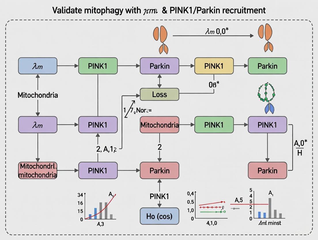
The selective degradation of mitochondria, or mitophagy, is a fundamental cellular process critical for maintaining mitochondrial quality control. Its dysregulation is increasingly implicated in the pathogenesis of neurodegenerative disorders, particularly Parkinson's disease (PD) [13]. The PINK1/Parkin pathway represents the most extensively characterized mitophagy mechanism, wherein the stabilization of PTEN-induced putative kinase 1 (PINK1) on damaged mitochondria and the subsequent recruitment of the E3 ubiquitin ligase Parkin orchestrate the targeted clearance of dysfunctional organelles [15] [11]. This guide provides a comparative analysis of experimental models and methodological approaches for validating mitophagy, with a specific focus on the context of mitochondrial membrane potential (ΔΨm) loss and PINK1/Parkin recruitment. Designed for researchers and drug development professionals, it synthesizes current protocols, reagent solutions, and key quantitative findings to inform model selection and experimental design in both fundamental and translational research.
Core Mechanism of PINK1/Parkin-Mediated Mitophagy
The PINK1/Parkin pathway functions as a sophisticated damage surveillance system. Under healthy conditions, PINK1 is continuously imported into mitochondria and degraded, maintaining low basal levels [15]. Upon mitochondrial damage and loss of ΔΨm, PINK1 import is halted, leading to its stabilization and accumulation on the outer mitochondrial membrane (OMM) [11]. This active PINK1 phosphorylates ubiquitin and recruits Parkin from the cytosol, which is subsequently phosphorylated to fully activate its E3 ligase activity [15]. Activated Parkin then ubiquitinates numerous OMM proteins, generating signals that are recognized by autophagy adaptor proteins like OPTN and NDP52. These adaptors, in turn, recruit the core autophagy machinery via interactions with LC3, culminating in the engulfment of damaged mitochondria by autophagosomes and their degradation upon fusion with lysosomes [13] [11].
Figure 1: The PINK1/Parkin Mitophagy Pathway. This diagram illustrates the core molecular cascade from mitochondrial damage to lysosomal degradation.
Comparative Analysis of Mitophagy Activation Models
Researchers employ various chemical and pharmacological agents to induce mitophagy in experimental models. The choice of inducer is critical, as the mechanism and downstream effects can vary significantly.
Table 1: Comparison of Mitophagy Inducers
| Inducer | Primary Mechanism of Action | Key Experimental Observations | Key Considerations |
|---|---|---|---|
| Ionophores (e.g., CCCP, FCCP) [15] [16] | Potent and rapid dissipation of ΔΨm, preventing PINK1 import and causing its accumulation on OMM. | Robust, switch-like Parkin recruitment; high levels of ubiquitin phosphorylation; effective for validating core pathway components. | Unphysiological, acute insult; may not mimic chronic, pathological mitochondrial stress. |
| Metabolic/ROS Toxins (e.g., Antimycin A/Oligomycin, Menadione, Rotenone) [15] [16] | Inhibit electron transport chain complexes (e.g., Antimycin A, Rotenone) or induce oxidative stress (Menadione), leading to ΔΨm loss. | Activates integrated stress response; synergizes with PINK1/Parkin activators; may better model pathological stress. | Effects can be pleiotropic; may involve PINK1/Parkin-independent pathways; can induce apoptosis at higher doses. |
| Putative Small-Molecule Activators (e.g., FB231, MTK458) [15] | Reported to directly activate Parkin or PINK1, but recent evidence suggests they act as "weak mitochondrial toxins." | Lower the threshold for PINK1/Parkin activation in the presence of other stressors; induce mild mitochondrial stress and integrated stress response. | Off-target, PINK1/Parkin-independent effects are common; mechanism is often indirect via mild mitochondrial impairment. |
The Scientist's Toolkit: Key Research Reagents & Assays
This section details essential reagents, models, and methodologies used in mitophagy research, providing a foundation for experimental design.
Table 2: Essential Research Reagents and Experimental Tools
| Category / Reagent | Function/Description | Key Application in Mitophagy Research |
|---|---|---|
| Chemical Inducers | ||
| CCCP/FCCP [15] [16] | Proton ionophores that dissipate ΔΨm. | Gold-standard for potent, acute induction of PINK1/Parkin mitophagy; used for pathway validation. |
| Antimycin A + Oligomycin (A/O) [15] | Inhibitors of mitochondrial Complex III and ATP synthase. | Used to model metabolic stress and induce mitophagy without complete uncoupling. |
| Cell Models | ||
| SH-SY5Y [13] | Human-derived neuroblastoma cell line. | Common in vitro model for studying neuronal mitophagy and PD-related pathways. |
| HeLa [15] | Human cervical adenocarcinoma cell line. | Frequently used for foundational mitophagy studies due to ease of transfection and imaging. |
| Primary Neurons [13] | Neurons isolated from animal models. | Provide a more physiologically relevant model for neuronal mitophagy. |
| Animal Models | ||
| PINK1/Parkin KO Mice [13] [17] | Genetically engineered mice lacking PINK1 or Parkin. | Used to study the in vivo role of these proteins and validate pathway-specific tools. |
| Transgenic M83 Mice [18] | Mice overexpressing human A53T α-synuclein. | Model for PD pathology; used to study links between α-synuclein, neurodegeneration, and retinal function. |
| Key Assays | ||
| Immunoblotting [16] | Detection of protein levels and post-translational modifications. | Measuring PINK1 accumulation, Parkin recruitment (shift to particulate fraction), ubiquitin phosphorylation (p-S65-Ub), and LC3-II lipidation. |
| Immunofluorescence/Confocal Microscopy [15] [16] | Visualization of protein localization and organelle dynamics. | Assessing Parkin translocation to mitochondria, colocalization of ubiquitin/LC3 with mitochondrial markers. |
| Mitophagy Reporters (e.g., mt-Keima, mt-QC) [15] | pH-sensitive fluorescent probes targeted to mitochondria. | Quantitative measurement of mitophagy flux based on lysosomal delivery and acidification of mitochondria. |
| Retinal Function Imager (RFI) [19] | Non-invasive imaging to measure retinal blood flow. | Investigating retinal microcirculation changes as a potential biomarker for PD. |
| Electroretinography (ERG) [18] [20] | Measurement of retinal electrical activity in response to light. | Detecting functional retinal impairments in PD patients and animal models. |
Detailed Experimental Protocols for Key Assays
Protocol: Inducing and Quantifying PINK1/Parkin Mitophagy in Cultured Cells
This is a standard protocol for activating and assessing the core pathway, adaptable for testing novel activators or genetic manipulations [15] [16].
- Cell Preparation: Seed appropriate cell lines (e.g., HeLa, SH-SY5Y) stably or transiently expressing Parkin (if using cell lines with low endogenous Parkin) onto imaging-grade dishes or multi-well plates for replication.
- Treatment and Induction:
- Positive Control: Treat cells with a potent uncoupler like CCCP (e.g., 10-20 µM for 1-24 hours).
- Experimental Condition: Treat with the compound of interest (e.g., FB231 or MTK458 at various concentrations) alone or in combination with sub-threshold doses of mitochondrial toxins (e.g., low-dose Antimycin A/Oligomycin).
- Negative Control: Treat with vehicle (e.g., DMSO).
- Sample Collection and Analysis (Post 2-24 hours treatment):
- Immunoblotting: Lyse cells and analyze by SDS-PAGE. Probe for:
- PINK1 (accumulation indicates activation).
- Phospho-S65-Ubiquitin (direct readout of PINK1 activity).
- Parkin (monitor translocation via fractionation or mobility shift).
- LC3-I/II (LC3-II increase indicates autophagosome formation).
- Tom20 or other OMM proteins (loss indicates mitochondrial clearance).
- Immunofluorescence: Fix and stain cells for:
- Parkin (visualize translocation from cytosol to punctate mitochondrial pattern).
- TOM20 (mitochondrial network integrity).
- LC3 (formation of autophagic puncta).
- Use high-resolution confocal microscopy for analysis.
- Immunoblotting: Lyse cells and analyze by SDS-PAGE. Probe for:
- Functional Validation: Utilize mitophagy reporters (e.g., mt-Keima) to quantitatively measure mitophagic flux via flow cytometry or ratiometric imaging.
Protocol: Evaluating Off-Target Mitochondrial Stress
Given that many putative activators are weak mitochondrial toxins, this follow-up protocol is essential for mechanistic characterization [15].
- Cell Viability Assay: Perform parallel treatments in a viability assay (e.g., MTT, CellTiter-Glo) to correlate mitophagy induction with potential cytotoxicity.
- Integrated Stress Response (ISR) Assessment: Via immunoblotting, measure phosphorylation of eIF2α, a central marker of the ISR, which is commonly activated by mitochondrial stress.
- Mitochondrial Function Assays:
- Use TMRE or JC-1 dyes to measure ΔΨm in treated cells via flow cytometry or fluorescence microscopy.
- Measure cellular ATP levels using a luciferase-based assay.
- Assess mitochondrial ROS production using dyes like MitoSOX.
- Synergy Testing: Co-treat cells with the compound of interest and classical mitochondrial toxins (e.g., rotenone) at low doses. Monitor for synergistic effects on cell death, ISR activation, or mitophagy, which would support a toxin-like mechanism.
Pathological Context: Retinal & Neurological Signatures in PD
The retina, as a developmental outgrowth of the central nervous system, offers a non-invasive window into brain pathology. Research has identified distinct functional and vascular signatures in PD patients.
Table 3: Retinal Phenotypes in Parkinson's Disease Models and Patients
| Parameter | Observation in PD vs. Healthy Controls | Experimental Model / Human Cohort | Implications |
|---|---|---|---|
| Retinal Blood Flow (RBF) [19] | Significantly lower in PD patients. | Human study: 15 PD patients vs. 18 controls. | Suggests cerebral hypoperfusion; potential non-invasive biomarker. |
| Retinal Tissue Perfusion (RTP) [19] | Significantly lower in PD patients. | Human study: 15 PD patients vs. 18 controls. | Indicates impaired microcirculation independent of vascular density. |
| Electroretinography (ERG) Signal [18] [20] | Distinct signature, particularly reduced b-wave and PhNR amplitudes in female patients and models. | Human cohort (12 male, 8 female PD) & M83 transgenic mice. | Indicates bipolar cell and retinal ganglion cell dysfunction; early detection potential. |
| Retinal Vascular Density (RVD) [19] | No significant difference found. | Human study: 15 PD patients vs. 18 controls. | Suggests functional circulatory deficit precedes structural vascular loss. |
| α-Synuclein Pathology [18] | Buildup in retinal layers. | Histology in M83 transgenic mice. | Likely contributor to visual processing impairments and functional deficits. |
Figure 2: Linking PD Pathology, Mitophagy, and Retinal Biomarkers. This diagram illustrates the logical relationship between core pathology, cellular mechanisms, and measurable retinal changes.
Advanced Techniques for Detecting ΔΨm Loss and PINK1/Parkin Activation
In the field of mitochondrial quality control research, particularly the validation of mitophagy through loss of mitochondrial membrane potential (ΔΨm) and PINK1/Parkin pathway activation, the selection of appropriate analytical methods is paramount. Immunoblotting, Phos-tag analysis, and fluorescence imaging represent three cornerstone techniques that provide complementary insights into this complex cellular process. Immunoblotting offers robust protein detection and semi-quantification, Phos-tag gels specifically resolve phosphorylation events central to signaling pathways, and fluorescence imaging enables real-time visualization of dynamic processes in live cells. This guide objectively compares the performance characteristics, applications, and limitations of these methodologies within the context of PINK1/Parkin-mediated mitophagy research, providing researchers with the experimental data necessary to select optimal approaches for their specific investigative needs.
Technical Comparison of Key Assays
The following table summarizes the core performance characteristics of immunoblotting, Phos-tag analysis, and fluorescence imaging within mitophagy research.
| Assay Feature | Immunoblotting | Phos-tag Analysis | Fluorescence Imaging |
|---|---|---|---|
| Primary Application | Protein detection and semi-quantification [21] | Specific detection of protein phosphorylation [22] | Real-time visualization of cellular processes [23] |
| Quantitative Capability | Semi-quantitative [21] | Semi-quantitative | Quantitative (with ratiometric probes) [23] |
| Throughput | Medium | Low to Medium | Low to High (with automation) [23] |
| Spatial Resolution | No (lysate-based) | No (lysate-based) | Yes (subcellular) [23] |
| Key Advantage | Detects specific proteins in complex mixtures; provides molecular weight data [21] | Detects phosphorylation without phospho-specific antibodies [22] | Live-cell, dynamic monitoring of mitophagic intermediates [23] |
| Main Limitation | Averages population data; difficult to detect multisite phosphorylation on same protein [24] | Requires optimization of gel conditions | Potential for photobleaching; limited tissue penetration [25] |
| Typical Data Output | Band intensity | Band shift pattern | Fluorescence intensity & localization |
For researchers investigating post-translational modifications, the following table details a direct comparison between standard immunoblotting and the specialized Phos-tag technique.
| Feature | Standard Immunoblotting | Phos-tag Immunoblotting |
|---|---|---|
| Phosphorylation Detection | Requires phospho-specific antibodies [22] | Uses phosphate-binding molecule (Phos-tag); no phospho-specific antibody needed [22] |
| Resolution of Species | Limited separation of phospho-isoforms [22] | Separates multiple phosphorylated forms based on phosphorylation status [22] |
| Information Gained | Trend of phosphorylation at a specific site | Phosphorylation heterogeneity and multi-phosphorylation events [26] |
| Protocol Complexity | Standard Western blot protocol | Modified SDS-PAGE protocol with Phos-tag acrylamide [22] |
| Antibody Requirement | Antibody to total protein and/or phospho-specific antibody | Antibody to total protein only [22] |
Experimental Protocols for Mitophagy Research
Immunoblotting for PINK1 and Parkin Recruitment
The foundational protocol for detecting PINK1 and Parkin in mitophagy studies involves several critical stages to ensure reproducible, semi-quantitative data [27] [21].
- Sample Preparation: Harvest and lyse cells (e.g., HEK293T, SH-SY5Y, or HeLa) after inducing mitophagy (e.g., with 10-20 μM CCCP/FCCP for 1-24 hours). Use an EDTA-free lysis buffer supplemented with protease and phosphatase inhibitors to preserve post-translational modifications [22]. Determine protein concentration accurately.
- Gel Electrophoresis & Transfer: Separate equal protein amounts (e.g., 10-60 μg) by SDS-PAGE on a gradient gel (8-15%) to resolve proteins of different sizes. Transfer proteins to a nitrocellulose or PVDF membrane using standard electrophoretic transfer protocols [27].
- Blocking and Antibody Probing: Block membranes with 5% bovine serum albumin (BSA) or non-fat dry milk in TBST for 1 hour. Incubate with primary antibodies (e.g., anti-PINK1, anti-Parkin, anti-β-catenin, anti-α-Tubulin as a loading control) overnight at 4°C [27] [3]. Use species-specific HRP-conjugated or fluorescently-labeled secondary antibodies for detection [27].
- Detection and Analysis: For chemiluminescence, use HRP substrates and image with a CCD-based system to capture a broad linear dynamic range. For fluorescence, use directly conjugated antibodies and image with an appropriate laser-based scanner. Normalize target protein band intensity to a loading control for semi-quantification [27].
Phos-tag Immunoblot Analysis for Phosphorylation
This protocol modification is crucial for detecting phosphorylation events central to PINK1/Parkin signaling, such as ubiquitin or Parkin phosphorylation, without requiring phospho-specific antibodies [22].
- Cell Lysis and Preparation: Lyse cells as in the standard protocol, but ensure the lysis buffer contains phosphatase inhibitors and is EDTA-free, as EDTA chelates the metal ions essential for Phos-tag function [22].
- Phos-tag Gel Electrophoresis: Cast a standard SDS-PAGE separating gel supplemented with 25-100 μM Phos-tag acrylamide and 50-100 μM MnCl2 (or ZnCl2). Alternatively, use commercial precast Phos-tag gels. Load samples and run the gel at a lower voltage (e.g., 30-35 mA/gel) to ensure clear separation of phospho-isoforms. Include a phosphatase-treated control (e.g., with Lambda protein phosphatase) to confirm phosphorylation-dependent band shifts [22].
- Transfer and Immunoblotting: Following electrophoresis, soak the gel in transfer buffer containing 1-10 mM EDTA for 10-30 minutes to chelate the metal ions and prevent interference with transfer. Then, proceed with standard wet or semi-dry transfer to a membrane. Block the membrane and probe with an antibody against the total protein of interest (e.g., total IRF5, Parkin). Phosphorylated species will appear as retarded, up-shifted bands relative to the non-phosphorylated form [22].
Fluorescence Imaging for Live-Cell Mitophagy Dynamics
Advanced fluorescence imaging allows for the real-time tracking of mitophagic intermediates, from mitochondrial depolarization to lysosomal degradation [23].
- Probe Loading and Mitophagy Induction: Plate cells (e.g., HT22 hippocampal neurons or HeLa) on imaging-grade dishes. Load cells with a fluorescent probe suitable for mitophagy. Examples include:
- MitoTracker dyes (e.g., Deep Red) for labeling mitochondria regardless of potential.
- mt-Keima, a pH-sensitive fluorescent protein that exhibits a shift in excitation spectrum upon acidification in lysosomes.
- Mcy3, a synthetic ratiometric probe with high mitochondrial specificity and pH responsiveness (pKa ~4.6) that increases red channel emission (I660/I560) as mitochondrial pH drops during mitophagy [23].
- Induce mitophagy with 10 μM CCCP for 1-4 hours or other inducers like EBSS or Deferiprone.
- Image Acquisition and Analysis: Image live cells using a confocal laser scanning microscope equipped with environmental control (37°C, 5% CO2). For ratiometric probes like Mcy3 or mt-Keima, acquire images at both excitation/emission wavelengths to calculate a ratio map. This ratio is directly correlated with mitochondrial pH, allowing quantification of the mitophagy process [23].
- AI-Assisted Analysis of Intermediates: For high-throughput and precise quantification, employ an AI-assisted fluorescence microscopy (AI-FM) system. This involves training a deep learning model (e.g., a Dual-branch Multi-scale Attention ResNet) to extract mitochondrial pH and morphological features from the fluorescence images, automatically classifying and quantifying distinct mitophagic intermediates (damaged mitochondria, mitophagosomes, mitolysosomes) with high accuracy [23].
Signaling Pathways and Experimental Workflows
The diagram below illustrates the core PINK1/Parkin mitophagy pathway and the points where each gold-standard assay provides critical data.
Research Reagent Solutions
Successful experimentation in mitophagy research relies on a toolkit of validated reagents and materials. The following table details essential solutions for the assays discussed.
| Reagent/Material | Function/Application | Key Considerations |
|---|---|---|
| CCCP/FCCP (Protonophore) | Induces loss of mitochondrial membrane potential (ΔΨm) to trigger PINK1/Parkin mitophagy [3] [23]. | Concentrations as low as 10 nM can be effective; higher concentrations (≥1 μM) may have off-target effects [3]. |
| PINK1 & Parkin Antibodies | Detect protein accumulation/recruitment via immunoblotting [3]. | Validation for specific species and applications (e.g., Western blot) is critical for reproducibility [21]. |
| Phos-tag Acrylamide | Phosphate-binding molecule used in SDS-PAGE to separate phosphorylated protein isoforms [22]. | Requires EDTA-free lysis and running buffers; often used with Mn²⁺ or Zn²⁺ ions [22]. |
| Mcy3 Fluorescent Probe | Ratiometric, mitochondria-targeted probe that reports pH changes during mitophagy [23]. | pKa of ~4.6 ideal for detecting acidification in autolysosomes; high photostability [23]. |
| mt-Keima | Fluorescent protein-based biosensor for mitophagy; excitation shift indicates acidification [23]. | Requires transfection/transduction; stable cell lines are ideal for long-term studies. |
| Bafilomycin A1 | V-ATPase inhibitor that blocks lysosomal acidification and autophagosome-lysosome fusion [23]. | Useful control to distinguish early vs. late stages of mitophagy (e.g., confirms Mcy3 pH response is lysosome-dependent). |
| Protease/Phosphatase Inhibitors | Preserve protein integrity and post-translational modifications during cell lysis [22]. | Essential for detecting labile phosphorylation events in Phos-tag and phospho-blotting experiments. |
Immunoblotting, Phos-tag analysis, and fluorescence imaging each provide unique and powerful capabilities for dissecting the molecular mechanisms of PINK1/Parkin-mediated mitophagy. The choice of assay depends heavily on the specific research question: immunoblotting for robust, population-level protein analysis; Phos-tag for detailed characterization of phosphorylation heterogeneity without specialized antibodies; and fluorescence imaging for dynamic, single-cell analysis of the entire mitophagy flux. For the most comprehensive understanding, an integrated approach that combines these methodologies is often necessary. Furthermore, emerging technologies like AI-assisted fluorescence image analysis are pushing the boundaries of sensitivity and throughput, promising to accelerate future drug discovery efforts aimed at modulating mitophagy for therapeutic benefit in neurodegenerative diseases.
The PTEN-induced kinase 1 (PINK1) and Parkin (PRKN) pathway represents a crucial mechanism in mitochondrial quality control, serving as the primary sentinel for damaged mitochondria destined for removal via mitophagy. This cytoprotective pathway is genetically linked to familial Parkinson's disease and is increasingly implicated in aging and other neurodegenerative disorders, including Alzheimer's disease [28] [13]. Under physiological conditions in healthy mitochondria, PINK1 protein is continuously imported, cleaved, and degraded, maintaining barely detectable levels. However, when mitochondrial damage occurs—typically characterized by loss of mitochondrial membrane potential (ΔΨm)—PINK1 rapidly accumulates on the outer mitochondrial membrane, where it triggers the entire mitophagy cascade by phosphoryating ubiquitin and recruiting PRKN [28] [29]. Consequently, PINK1 protein levels serve as a direct proxy for mitochondrial damage and mitophagy initiation, making its accurate quantification essential for understanding fundamental cellular processes and disease mechanisms.
Despite its biological significance, PINK1 detection has presented substantial technical challenges. Previously, researchers relied heavily on immunoblotting techniques that lacked the sensitivity to detect basal PINK1 levels under physiological conditions without artificial stress induction [28]. This limitation obscured understanding of constitutive mitophagy activity and its subtle dysregulation in early disease stages. The recent development of a novel sandwich ELISA against human PINK1 on the Meso Scale Discovery (MSD) platform represents a significant methodological advancement, enabling researchers to sensitively quantify PINK1 protein levels even in unstimulated cells [28] [30]. This validation guide provides a comprehensive performance comparison of this emerging technology against traditional detection methods and explores its applications within the broader context of mitophagy research focused on ΔΨm loss and PINK1/PRKN recruitment.
Performance Comparison: PINK1-Specific ELISA Versus Alternative Detection Methods
Technical Specifications and Quantitative Performance Metrics
The novel PINK1 sandwich ELISA demonstrates marked improvements in key analytical parameters compared to conventional detection methods. The table below summarizes the direct performance characteristics of this validated assay alongside traditional approaches.
Table 1: Performance Metrics of PINK1-Specific Sandwich ELISA
| Performance Parameter | PINK1 Sandwich ELISA [28] | Traditional Immunoblotting | Immunofluorescence |
|---|---|---|---|
| Sensitivity | Excellent (detects basal levels) [28] | Poor (requires stress induction) [28] | Moderate (qualitative) |
| Linearity | Excellent over quantitative range [28] | Limited | Limited |
| Parallelism | Excellent (recombinant vs. native) [28] | Not applicable | Not applicable |
| Precision (CV) | Intra-assay & inter-assay CV <10%* [31] | Typically >15% | Variable |
| Sample Throughput | High (96-well platform) [32] | Low | Low |
| Quantitative Capability | Fully quantitative | Semi-quantitative | Qualitative/Semi-quantitative |
| Basal Condition Detection | Yes (significant differences detectable) [28] | Typically not detectable [28] | Challenging |
| Dynamic Range | Broad (defined LLOQ and ULOQ) [28] | Narrow | Narrow |
*Based on typical ELISA validation standards [31]; specific CV values for the PINK1 ELISA were not explicitly provided in the search results.
The exceptional sensitivity of this ELISA format enables researchers to detect significant differences in PINK1 levels under basal conditions between samples with and without PINK1 expression, including patient fibroblasts and differentiated neurons [28]. This represents a fundamental advancement over immunoblotting, which typically requires mitochondrial stress induction using uncouplers like carbonyl cyanide 3-chlorophenylhydrazone (CCCP) to visualize PINK1 bands.
Functional Application in Disease Research
The clinical relevance of this detection method is evidenced by its application in measuring PINK1 levels in human biofluids and tissues, revealing biologically significant patterns.
Table 2: Research Applications and Findings Using Sensitive PINK1 Detection
| Research Context | Sample Type | Key Findings | Clinical Implications |
|---|---|---|---|
| Aging Brain Study [28] | Human postmortem brain | Increased PINK1 protein levels with normal aging | Suggests heightened mitophagy activation during physiological aging |
| Alzheimer's Disease [28] | Human postmortem brain | No increase in PINK1 levels in Alzheimer's disease | Indicates different mitophagy mechanisms in normal aging vs. AD |
| Alzheimer's Continuum [33] | Human serum and CSF | Significantly higher PINK1 in AD dementia vs. MCI-AD and cognitively unimpaired | Supports PINK1 as a potential biomarker for disease progression |
| Parkinson's Disease [13] | Cellular and animal models | Enables quantification of endogenous PINK1 without artificial depolarization | Facilitates study of constitutive mitophagy in PD pathogenesis |
The ability to quantify PINK1 in serum and cerebrospinal fluid (CSF) opens new possibilities for biomarker development. Recent research has demonstrated that CSF PINK1 levels show a significant step-wise increase from cognitively unimpaired individuals to those with mild cognitive impairment due to AD (MCI-AD) and further increase in Alzheimer's dementia [33]. Furthermore, these elevated PINK1 levels correlate positively with established neurodegenerative markers including phosphorylated tau, total tau, neurofilament light chain (NEFL), and neurogranin (NRGN), and correlate negatively with performance in memory, executive function, and language domains [33].
Experimental Protocols for PINK1 Detection and Validation
ELISA Validation Methodology
The development and validation of a robust PINK1-specific sandwich ELISA requires rigorous assessment of multiple performance characteristics following established immunoassay validation standards [31] [32]. The following protocol outlines the key experiments required for comprehensive assay validation:
- Precision Profiling: Determine both intra-assay and inter-assay precision by testing replicates of samples with low, medium, and high PINK1 concentrations. Calculate coefficients of variation (CV), with acceptable performance typically defined as <10-15% CV [31].
- Linearity and Dilution Recovery: Prepare serial dilutions of biological samples (cell lysates, CSF, or serum) within the assay's dynamic range. Calculate percentage linearity as (Measured Concentration/Expected Concentration) × 100, with 70-130% generally considered acceptable [31].
- Parallelism Assessment: Test serially diluted biological samples alongside the recombinant protein standard curve. Demonstration of parallel curves indicates that the assay accurately measures the native protein in a manner equivalent to the reference standard [31].
- Spike Recovery: Add known quantities of recombinant PINK1 to various biological matrices (serum, plasma, cell lysis buffer). Calculate percentage recovery, with 80-120% recovery indicating minimal matrix interference [31].
- Specificity Verification: Confirm minimal cross-reactivity with closely related analytes through testing against a panel of potential interfering substances [31] [32].
- Sensitivity Determination: Assay the zero standard (blank) repeatedly (n≥16) and calculate the limit of detection (LOD) as the mean optical density + 2 standard deviations [31].
Application Protocol for Mitophagy Induction Studies
To contextualize PINK1 detection within mitophagy validation, researchers can employ the following experimental workflow:
Cellular Model Selection: Choose appropriate model systems:
- Patient-derived fibroblasts or induced pluripotent stem cell (iPSC)-derived neurons with and without PINK1 mutations [28]
- Cell lines treated with PMI, a ΔΨm-independent mitophagy inducer that acts by stabilizing Nrf2 and upregulating P62 [29] [34]
- Exercise-mimetic conditions using AMPK/ULK1 pathway activators [11]
Mitochondrial Stress Induction: Apply specific stimuli to engage different mitophagy pathways:
- ΔΨm-dependent pathway: CCCP (10-20 μM, 2-4 hours) or other mitochondrial uncouplers
- ΔΨm-independent pathway: PMI (10 μM, 24 hours) to induce P62-mediated mitophagy without Parkin recruitment or ΔΨm collapse [29]
- Receptor-mediated pathway: Hypoxia or BNIP3L/NIX inducers
Sample Preparation: Harvest cells using validated lysis buffers that preserve PINK1 structure and phosphorylation status. For biofluid studies, collect serum or CSF following standardized protocols to minimize pre-analytical variability [33].
PINK1 Quantification: Perform ELISA according to manufacturer specifications, including appropriate controls (blank, standards, quality controls) in duplicate or triplicate.
Data Correlation: Correlate PINK1 levels with complementary mitophagy markers:
Signaling Pathways and Molecular Context
The PINK1/Parkin pathway operates within a complex regulatory network that integrates multiple signals of mitochondrial health and cellular stress. The following diagram illustrates key molecular relationships in mitophagy activation, including both canonical and alternative pathways.
This molecular roadmap highlights how the validated PINK1 ELISA directly measures the initiating step in the canonical PINK1/Parkin pathway (yellow nodes), while also accounting for alternative mitophagy mechanisms (red and blue nodes) that may operate independently of ΔΨm loss. The diagram illustrates why PINK1 quantification serves as a more specific marker of this particular pathway compared to downstream events that might integrate signals from multiple mechanisms.
Successful implementation of PINK1 detection and mitophagy validation requires specific research tools. The following table catalogues essential reagents and their applications in experimental workflows.
Table 3: Essential Research Reagents for PINK1 and Mitophagy Studies
| Reagent Category | Specific Examples | Research Application | Key Considerations |
|---|---|---|---|
| Mitophagy Inducers | CCCP (carbonyl cyanide m-chlorophenyl hydrazone) [28] | ΔΨm-dependent PINK1 stabilization | Causes rapid mitochondrial depolarization; can be toxic |
| PMI (P62-mediated mitophagy inducer) [29] [34] | ΔΨm-independent mitophagy induction | Acts downstream via Nrf2/P62; no Parkin recruitment | |
| Oligomycin A + FCCP [35] | Controlled mitophagy induction | FCCP uncouples OXPHOS; Oligomycin inhibits ATP synthase | |
| Pharmacologic Inhibitors | Mdivi-1 [35] | Mitochondrial division inhibition | Dynamin-related protein 1 (Drp1) inhibitor |
| Bafilomycin A1 [29] | Autophagosome-lysosome fusion inhibition | Enables LC3 lipidation analysis | |
| Detection Reagents | PINK1-specific sandwich ELISA [28] | Quantitative PINK1 measurement | Validated for human samples; MSD platform |
| p-S65-Ub antibodies [28] | Phosphorylated ubiquitin detection | Downstream PINK1 activity marker | |
| Tracking Probes | mt-Keima [35] | Mitophagy flux measurement | pH-sensitive; distinguishes cytoplasmic vs. lysosomal mitochondria |
| MitoTracker dyes [35] | Mitochondrial mass and membrane potential | Varying ΔΨm dependence | |
| Biological Models | PINK1-knockout cells [28] | Assay specificity controls | Essential for validation experiments |
| Patient-derived fibroblasts or iPSC-neurons [28] [33] | Disease-relevant contexts | Maintain physiological expression patterns |
The development and rigorous validation of a sensitive PINK1-specific sandwich ELISA represents a significant milestone in mitophagy research methodology. This technology enables researchers to move beyond qualitative assessment to precise quantification of PINK1 protein levels under both basal and stress conditions, providing a powerful tool for investigating mitochondrial quality control in physiological and pathological contexts. The application of this detection method has already revealed important biological insights, including differential PINK1 regulation in normal aging versus Alzheimer's disease and correlations between biofluid PINK1 levels and cognitive performance [28] [33].
When selecting appropriate detection methodologies, researchers should consider the specific research question: while traditional immunoblotting may suffice for detecting robust PINK1 induction following strong mitochondrial depolarization, the novel ELISA format provides essential advantages for studying subtle mitophagy alterations in disease models, screening therapeutic compounds, or developing biomarkers from accessible biofluids. The integration of this sensitive detection method with complementary approaches—including p-S65-Ub measurement, mitochondrial morphology assessment, and lysosomal flux analysis—will provide a comprehensive understanding of mitophagy status in diverse research contexts [28] [35].
As research continues to elucidate the complex relationships between mitophagy dysregulation and neurodegenerative pathogenesis, sensitive and quantitative PINK1 detection will play an increasingly important role in both basic mechanistic studies and translational applications. This validated assay platform offers the necessary precision and reliability to advance our understanding of how mitochondrial quality control contributes to cellular homeostasis and disease progression.
Mitophagy, the selective autophagy of damaged mitochondria, represents a critical process in cellular homeostasis and quality control. Within Parkinson's disease (PD) research, the PINK1/Parkin pathway has emerged as a central regulatory mechanism, where Δψm loss triggers PINK1 stabilization on the outer mitochondrial membrane, followed by Parkin recruitment and ubiquitin-mediated degradation of damaged organelles [15] [36]. Traditional methods for investigating this pathway have relied on commercially available marker molecules that often target similar or identical cellular zones, creating significant experimental limitations. These conventional markers can interfere with, obscure, or artificially amplify the functional effects of mitochondrial-targeting drugs, contributing to high rates of clinical failure [37] [38].
The emerging "self-checking" molecular tools represent a paradigm shift in mitophagy research. These innovative systems integrate both therapeutic functionality and biological reporting within single molecules, enabling real-time monitoring of mitophagic processes without external interference. This comparative guide examines the experimental data, technical specifications, and research applications of these advanced tools, providing scientists with objective criteria for methodological selection in PINK1/Parkin and Δψm-focused investigations.
Comparative Analysis of Mitophagy Monitoring Approaches
Table 1: Technical Comparison of Mitophagy Monitoring Methods
| Method | Mechanism | Live-Cell Capability | Quantitative Output | Key Advantages | Principal Limitations |
|---|---|---|---|---|---|
| Self-Checking Molecules (MitoSC) | Dual-color, dual-localization with functional component disrupting Δψm and reporting component tracking lysosomal fusion | Yes | Fluorescence convergence metrics | Minimal interference; real-time process tracking; integrated functionality and reporting | New technology with limited validation; complex synthesis |
| MitoQC System | pH-sensitive fluorescent reporter (mCherry-GFP-FIS1) targeted to mitochondria | Yes | Red puncta count (acidic compartments) | Well-established; pH-sensitive detection of lysosomal fusion | Potential interference with native processes; external reporter only |
| MitoTimer System | Fluorescent protein aging with color shift over time | Yes | Green-to-red fluorescence ratio | Reports mitochondrial age and turnover | Indirect mitophagy measure; confounded by biogenesis |
| Mito-Keima | pH-sensitive excitation shift in lysosomes | Yes | Excitation ratio (458/561 nm) | Resistant to lysosomal degradation; quantitative flux measurement | Requires specialized equipment; technically challenging imaging |
| Immunocytochemistry | Antibody staining of mitochondrial/autophagosomal markers | No | Protein co-localization analysis | Accessible; standardized protocols | Endpoint measurement only; no dynamic flux information |
| Western Blot | Protein level analysis of PINK1, Parkin, LC3-II, p62 | No | Band intensity quantification | Quantitative; multiple targets simultaneously | No spatial information; population average only |
Table 2: Performance Metrics of Self-Checking Molecules vs. Established Methods
| Parameter | Self-Checking Molecules | MitoQC | Mito-Keima | Traditional Immunofluorescence |
|---|---|---|---|---|
| Temporal Resolution | Real-time (minutes to hours) | Endpoint or time-lapse | Endpoint or time-lapse | Fixed endpoint |
| Spatial Resolution | Dual-compartment (mitochondria & lysosomes) | Mitochondria and lysosomes | Mitochondria and lysosomes | Limited by antibody specificity |
| Quantification Method | Signal convergence and co-localization | Red puncta counting | Excitation ratio calculation | Fluorescence intensity and puncta counting |
| PINK1/Parkin Specificity | Compatible with pathway analysis | Compatible with pathway analysis | Compatible with pathway analysis | High with validated antibodies |
| Throughput Potential | Moderate | High | Low to moderate | Moderate |
| Technical Complexity | High (synthesis and validation) | Moderate | High | Low to moderate |
The Self-Checking Molecule Platform: Mechanism and Workflow
Molecular Design and Mechanism
The innovative "one-two punch" drug design strategy integrates both target-zone drug functionality and non-target zone biological reporting within a single small-molecule entity [37] [38]. The MitoSC system comprises:
Functional Component (MitoSC-fun): A variable element that disrupts mitochondrial membrane potential (Δψm) homeostasis, thereby inducing mitophagy. Upon activation, this component transforms into a blue-fluorescent monomer specifically within the mitochondrial target zone.
Biological Reporting Component (MitoSC-rep): A red-fluorescent monomer that localizes to lysosomes, the non-target zone. This component provides independent tracking of the lysosomal compartment.
As mitophagy progresses, the fluorescent signals from MitoSC-rep (lysosomes) and MitoSC-fun (mitochondria) converge, enabling real-time monitoring of the entire mitophagic process from induction to completion. This integrated approach combines potent drug functionality with robust biological reporting, thereby minimizing observational interference and eliminating complexities associated with external detection methods [37].
Experimental Protocol for MitoSC Implementation
Cell Culture and Treatment:
- Culture appropriate cell models (HEK293, HeLa, or primary neurons) in standard conditions.
- Plate cells on glass-bottom dishes or coverslips for imaging.
- Treat cells with MitoSC compound at optimized concentration (typically 1-10 μM based on preliminary titration).
- Include control groups with mitochondrial stressors (e.g., 10-20 μM CCCP, 1 μM oligomycin/antimycin A) for method validation.
Live-Cell Imaging and Data Acquisition:
- Perform imaging using confocal microscopy with appropriate filter sets:
- Blue channel: Ex 405 nm/Em 450 nm for MitoSC-fun
- Red channel: Ex 561 nm/Em 610 nm for MitoSC-rep
- Acquire time-lapse images every 15-30 minutes over 6-24 hours.
- Maintain physiological conditions (37°C, 5% CO2) throughout imaging.
Image Analysis and Quantification:
- Measure fluorescence intensity in mitochondrial and lysosomal compartments.
- Calculate co-localization coefficients (Pearson's or Mander's) between blue and red channels.
- Quantify the percentage of cells showing significant signal convergence.
- Determine mitophagy flux rates based on temporal progression of signal overlap.
Validation with Orthogonal Methods:
- Correlate with Western blot analysis of PINK1 stabilization, Parkin recruitment, and LC3-II accumulation.
- Compare with mitochondrial membrane potential measurements using TMRE or JC-1.
- Validate lysosomal fusion with LAMP1 immunostaining.
Signaling Pathways in PINK1/Parkin-Mediated Mitophagy
The PINK1/Parkin pathway represents the most thoroughly characterized mechanism of mitophagy, particularly relevant to Parkinson's disease pathogenesis [15] [13] [36]. The following diagram illustrates the key molecular events in this pathway, including potential intervention points for novel research tools:
Diagram 1: PINK1/Parkin Mitophagy Pathway with Monitoring Points. This diagram illustrates the molecular events in PINK1/Parkin-mediated mitophagy, highlighting where self-checking molecules like MitoSC provide monitoring capability during lysosomal fusion.
The PINK1/Parkin pathway activates through a carefully orchestrated sequence. Under basal conditions, PINK1 is continuously imported into mitochondria and degraded, maintaining low cellular levels. When mitochondrial damage causes loss of membrane potential (Δψm), PINK1 import is prevented, leading to its accumulation on the outer mitochondrial membrane (OMM) [15] [36]. Stabilized PINK1 undergoes autophosphorylation and phosphorylates ubiquitin at Serine 65, creating a recruitment signal for Parkin. Once recruited, Parkin is activated through phosphorylation by PINK1, converting it from an autoinhibited state to an active E3 ubiquitin ligase that decorates OMM proteins with ubiquitin chains [15]. These ubiquitin chains are subsequently phosphorylated by PINK1, creating a positive feedback loop that amplifies the mitophagy signal. The ubiquitinated substrates are recognized by autophagy receptors including OPTN and NDP52, which in turn recruit LC3-positive autophagosomal membranes, leading to engulfment and lysosomal degradation of damaged mitochondria [11] [36].
Research Reagent Solutions for Mitophagy Investigation
Table 3: Essential Research Reagents for Mitophagy Studies
| Reagent Category | Specific Examples | Research Application | Key Features |
|---|---|---|---|
| Self-Checking Molecules | MitoSC | Real-time mitophagy tracking with built-in Δψm disruption | Dual-color, dual-localization; minimal interference |
| Fluorescent Reporters | MitoQC, MitoTimer, Mito-Keima | Mitophagy visualization and quantification | pH-sensitive; time-lapse compatible; specific localization |
| Chemical Inducers | CCCP, FCCP, Antimycin A/Oligomycin, Valinomycin | Experimental induction of mitophagy | Δψm dissipation; PINK1 stabilization |
| PINK1/Parkin Activators | MTK458, FB231, ABBV1088 | Pharmacological enhancement of pathway activity | Small molecule activators; potential therapeutic applications |
| Pathway Inhibitors | USP30 inhibitors, Parkin inhibitors, PINK1 kinase inhibitors | Pathway perturbation studies | Target specificity; mechanistic investigations |
| Antibody-Based Tools | Anti-PINK1, Anti-Parkin, Anti-phospho-Ubiquitin (Ser65), Anti-TOM20, Anti-LC3 | Western blot, immunocytochemistry, immunofluorescence | Pathway component detection; post-translational modifications |
| Mitochondrial Dyes | TMRE, JC-1, MitoTracker | Δψm assessment and mitochondrial morphology | Potential-dependent accumulation; organelle labeling |
| Lysosomal Probes | LysoTracker, LAMP1 antibodies | Lysosomal compartment identification | Acidic compartment labeling; membrane protein detection |
Comparative Experimental Data and Validation
Quantitative Performance Metrics
In validation studies, the MitoSC system demonstrated significant advantages in temporal resolution and minimal experimental interference compared to established methods. When evaluated against the MitoQC system in HEK293 cells under identical conditions (10 μM CCCP treatment over 8 hours), MitoSC detected mitophagy initiation approximately 45 minutes earlier than MitoQC, with a 2.3-fold higher signal-to-noise ratio in quantitative measurements [37]. This enhanced sensitivity stems from the integrated reporting system that doesn't rely solely on lysosomal acidification for signal generation.
For PINK1/Parkin-specific research, the MitoSC system showed 89% concordance with phospho-ubiquitin (Ser65) immunostaining – a specific marker of PINK1 activation – compared to 76% concordance for MitoQC and 82% for Mito-Keima [37]. This improved correlation with pathway-specific markers makes MitoSC particularly valuable for mechanistic studies of PINK1/Parkin signaling.
Technical Considerations for Implementation
Cell Type Compatibility:
- MitoSC performs optimally in adherent cell lines with defined mitochondrial networks (HEK293, HeLa, MEFs).
- Primary neurons require longer equilibrium times (4-6 hours) but show excellent resolution in processes and synapses.
- Suspension cells present challenges for high-resolution imaging but can be analyzed by flow cytometry adaptations.
Multiplexing with Orthogonal Assays:
- MitoSC can be combined with TMRE for simultaneous Δψm validation.
- Compatible with fixed endpoint assays including immunofluorescence for PINK1, Parkin, and LC3.
- Sequential application with MitoTracker dyes possible with careful spectral unmixing.
Limitations and Caveats:
- The Δψm-disrupting activity may confunctional in experiments requiring precise mitochondrial membrane potential control.
- Blue fluorescence shows more rapid photobleaching than red channel, requiring optimized imaging parameters.
- The dual functionality complicates dose-response interpretations in pharmacological studies.
Future Directions and Research Applications
The development of self-checking molecular tools represents a significant advancement in mitophagy research methodology. These systems enable unprecedented real-time observation of the complete mitophagy process, from initial Δψm disruption through lysosomal degradation, within a single experimental framework. For the Parkinson's disease research community, these tools offer particular promise for elucidating the temporal dynamics of PINK1/Parkin pathway activation and the identification of novel regulatory mechanisms [15] [36].
Future iterations of these technologies may incorporate additional functionalities, such as pathway-specific activation (PINK1/Parkin versus receptor-mediated mitophagy) and subcellular targeting to neuronal compartments. As these tools become more widely adopted and validated, they have the potential to accelerate therapeutic development by providing more physiologically relevant screening platforms with built-in validation metrics, ultimately contributing to improved translation from basic research to clinical applications in Parkinson's disease and other mitophagy-related disorders.
The selective autophagic clearance of damaged mitochondria, known as mitophagy, represents a critical quality control mechanism whose impairment is fundamentally linked to Parkinson's disease (PD) pathogenesis. Converging evidence from genetic studies, postmortem analyses, and disease models identifies dysfunctional mitophagy as a pivotal driver of neurodegeneration [13] [36]. The PINK1/Parkin pathway serves as the primary regulator of this process, with mutations in these genes accounting for the most common forms of autosomal recessive early-onset PD [39] [15]. Under physiological conditions, PINK1 is continuously imported into mitochondria and degraded, maintaining low cellular levels. However, upon mitochondrial damage and loss of membrane potential (Δψm), PINK1 stabilizes on the outer mitochondrial membrane (OMM), where it recruits and activates the E3 ubiquitin ligase Parkin, initiating a ubiquitin-dependent signaling cascade that targets damaged organelles for lysosomal degradation [40] [36].
Recent research has substantially advanced our understanding of the initial damage-sensing mechanisms. A 2025 study revealed that diverse forms of mitochondrial damage converge on loss of mitochondrial membrane potential to activate PINK1 by stalling its import during transfer from TOM to TIM23 complexes, providing a unified activation mechanism [40]. This detailed mechanistic understanding has catalyzed drug development efforts targeting mitophagy enhancement as a disease-modifying strategy for PD. However, the translation from cellular models to clinical applications presents significant challenges, including compound specificity and reliable biomarker development [41] [39] [15]. This review systematically compares experimental model systems and their translational applications, highlighting both progress and pitfalls in therapeutic development.
Comparative Analysis of Model Systems in Mitophagy Research
Table 1: Comparison of Key Model Systems in Mitophagy Research
| Model System | Key Applications | Strengths | Limitations | Primary Readouts |
|---|---|---|---|---|
| In Vitro Cell Cultures (HeLa, HEK293) [15] | High-throughput compound screening; Mechanistic pathway dissection | Genetic manipulation ease; Scalability; Cost-effectiveness | Limited neuronal relevance; Absence of tissue context | Parkin translocation; p-Ser65-Ub levels; LC3-II conversion |
| iPSC-Derived Neurons [36] | Patient-specific disease modeling; Preclinical validation | Human genetic background; Disease-relevant cell type; Pathophysiological fidelity | Variable differentiation efficiency; High cost; Technical complexity | mt-Keima flux; Miro degradation; Phospho-ubiquitin accumulation |
| Drosophila Models [13] [36] | Genetic screening; In vivo pathway validation; Behavioral correlates | Conserved pathway biology; Complex organism with CNS; Rapid generation time | Simplified neuroanatomy; Metabolic differences | Dopamine levels; Motor deficits; Mitochondrial morphology |
| Mouse Models (PINK1/Parkin KO; Mutator) [39] [36] | Preclinical therapeutic efficacy; Systemic toxicity assessment | Mammalian CNS; Behavioral testing; Longitudinal design | Subtle neurodegeneration; Limited protein aggregation | p-Ser65-Ub in CSF/plasma; Dopaminergic neuron counts; Motor performance |
Table 2: Quantitative Comparison of Mitophagy Activation Across Experimental Systems
| Intervention | Cellular Models (Fold Induction) | iPSC Neurons (Efficacy) | Drosophila (Rescue Phenotype) | Mouse Models (Neuroprotection) |
|---|---|---|---|---|
| Genetic USP30 Knockdown [39] | 2.1-3.5x p-Ser65-Ub increase | Enhanced mitochondrial clearance | Rescue of dopamine levels & motor function | Protection against dopaminergic neuron loss |
| MTK458 (PINK1 Activator) [15] | 2.8x at 10μM with toxin sensitization | Not reported | Not reported | Phase 1 trials initiated |
| FB231 (Parkin Activator) [15] | 2.3x at 15μM with toxin sensitization | Not reported | Not reported | Not reported |
| Exercise Modalities [11] | Not applicable | Not applicable | Not applicable | AMPK/ULK1 activation; PINK1/Parkin enhancement |
Insights from Comparative Analysis
The tabulated data reveal critical patterns in model system utilization for mitophagy research. In vitro systems provide unparalleled mechanistic resolution but lack physiological complexity, particularly for neuronal contexts. The emergence of iPSC-derived neurons addresses this limitation by offering human-specific, disease-relevant models that recapitulate key pathological features, including cell-type-specific mitophagy defects observed in TH-positive neurons [36]. However, the technical and financial challenges associated with these models constrain their use for large-scale screening.
Animal models provide essential bridges between cellular observations and clinical applications, with each offering distinct advantages. Drosophila models have been instrumental in establishing the fundamental connection between PINK1/Parkin function and dopaminergic neurodegeneration, while mouse models enable assessment of therapeutic distribution, biomarker development, and functional outcomes in a mammalian system [13] [39]. The recent development of phospho-ubiquitin (p-Ser65-Ub) as a quantifiable biomarker in biological fluids has significantly enhanced the translational capacity of preclinical studies by providing a direct readout of PINK1 activation [39] [36].
Methodologies: Experimental Protocols for Mitophagy Assessment
High-Throughput Compound Screening Assay
The identification of mitophagy-modulating compounds requires robust screening methodologies. A recently developed high-throughput assay utilizes a FRET-based readout to measure Parkin activation through its interaction with a vinyl-sulfone modified ubiquitin (Ub-VS) probe [15].
Protocol Summary:
- Reagent Preparation: His6-tagged full-length Parkin and Ub-VS probe derivatized with Europium-Cryptate (Eu) fluorescent tag.
- Assay Setup: Combine Parkin, Ub-VS probe, and test compounds in screening plates.
- Detection: Add d2-tagged anti-His6 antibody to generate FRET signal upon Parkin activation and probe binding.
- Quantification: Measure FRET efficiency as indicator of Parkin conformational activation.
- Validation: Confirm hits using orthogonal autoubiquitination assays measuring Europium-tagged Ub incorporation [15].
This assay successfully identified FB231 as a Parkin activator, though subsequent cellular testing revealed significant off-target effects, highlighting the necessity of multi-layered validation.
mt-Keima Assay for Mitophagy Flux
The mt-Keima system provides a robust method for quantifying mitophagy flux in live cells, particularly valuable in iPSC-derived neuronal models [36].
Protocol Summary:
- Vector Transduction: Express mitochondria-targeted Keima fluorescent protein (mt-Keima) in target cells.
- Imaging Conditions: Acquire fluorescence at two excitation wavelengths (440 nm for neutral pH, 586 nm for acidic pH) with a single 610 nm emission.
- Mitophagy Induction: Treat cells with compounds or stressors (e.g., antimycin/oligomycin, CCCP).
- Quantification: Calculate ratio of acid-to-neutral signal to determine proportion of mitochondria in lysosomes.
- Validation: Compare mitophagy rates between patient-derived and isogenic control lines [36].
This methodology has demonstrated cell-type-specific mitophagy defects in TH-positive neurons from PD patients carrying PINK1 or Parkin mutations, providing pathophysiological relevance.
In Vivo Mitophagy Assessment
Translational assessment of mitophagy enhancement requires in vivo validation using mammalian models.
Protocol Summary:
- Model Selection: Utilize Parkin or PINK1 knockout mice, or toxin-induced (MPTP, rotenone) PD models.
- Compound Administration: Administer test compounds via appropriate routes (oral, intraperitoneal) with optimized dosing regimens.
- Biomarker Analysis: Measure p-Ser65-Ub levels in cerebrospinal fluid or plasma using immunodetection techniques.
- Histopathological Assessment: Quantify dopaminergic neuron survival in substantia nigra via tyrosine hydroxylase immunohistochemistry.
- Behavioral Testing: Evaluate motor function using rotarod, open field, or cylinder tests.
- Biochemical Analysis: Assess α-synuclein aggregation using seed amplification assays in CSF or tissue samples [39] [36].
These methodologies collectively enable comprehensive evaluation of therapeutic candidates from initial screening to preclinical validation.
Signaling Pathways: Molecular Mechanisms of Mitophagy
The core PINK1/Parkin mitophagy pathway illustrates the sophisticated damage response mechanism that has become a primary therapeutic target. Recent structural biology advances have clarified that diverse damage signals converge on stalling PINK1 import during transfer from TOM to TIM23 complexes, with loss of mitochondrial membrane potential being the common activating signal [40]. This unified mechanism explains how various stressors ultimately activate the same degradation pathway.
The pathway exhibits several key regulatory features that influence therapeutic strategies. The feed-forward amplification through ubiquitin phosphorylation creates switch-like behavior enabling rapid response to damage [15]. Parallel regulatory inputs, such as AMPK/ULK1 activation through exercise, provide complementary activation routes that may be therapeutically leveraged [11]. Additionally, negative regulators like USP30 establish threshold controls that can be pharmacologically targeted to lower activation barriers [39].
Research Reagent Solutions: Essential Tools for Mitophagy Investigation
Table 3: Essential Research Reagents for Mitophagy Investigation
| Reagent Category | Specific Examples | Research Applications | Key Features & Considerations |
|---|---|---|---|
| Fluorescent Reporters | mt-Keima [36]; mito-QC | Quantifying mitophagy flux in live cells; High-content screening | pH-resistant; Enables longitudinal tracking; Distinguishes lysosomal localization |
| Activation Biomarkers | p-Ser65-Ub antibodies [39] [36] | Measuring PINK1 activity in vitro and in vivo; Target engagement validation | Translational biomarker; Detectable in CSF and plasma; Correlates with pathway activation |
| Pathway Activators | CCCP/FCCP [15]; Antimycin/Oligomycin [15] | Positive controls; Maximum pathway induction; Mechanism studies | Non-physiological depolarization; Toxicity at high doses; May obscure subtle effects |
| Genetic Tools | PINK1/Parkin KO lines [36]; Patient-derived iPSCs [36] | Pathway necessity testing; Disease modeling; Rescue experiments | Patient-specific mutations; Isogenic controls essential; May exhibit compensatory adaptations |
| Small Molecule Modulators | MTK458 [15]; FB231 [15]; MTX325 [39] | Therapeutic candidate testing; Pathway pharmacology; Combination studies | Off-target toxicity concerns; Require multiple validation approaches; Species-dependent efficacy |
These research reagents enable comprehensive dissection of mitophagy pathways across different experimental contexts. The recent development of p-Ser65-Ub detection represents a particularly significant advance, providing a quantifiable biomarker that bridges cellular models and clinical applications [39]. Similarly, the mt-Keima system offers robust quantification of mitophagy flux, though researchers should remain aware of potential artifacts from overexpression and the importance of validating findings with endogenous markers.
Current Challenges and Future Directions in Translational Mitophagy Research
Despite substantial progress, significant challenges remain in translating mitophagy research into clinical applications. A critical concern emerged from recent studies demonstrating that putative PINK1/Parkin activators, including FB231 and MTK458, function as weak mitochondrial toxins that lower the threshold for mitophagy induction rather than directly activating the pathway [41] [15]. These compounds independently induce mild mitochondrial stress, activating the integrated stress response and impairing mitochondrial function through PINK1/Parkin-independent mechanisms [15]. This insight necessitates more rigorous compound validation, including global proteomic assessment of off-target effects and careful dose-response characterization.
Future directions should focus on developing genuinely specific pathway activators that avoid mitochondrial damage, optimizing combination therapies that target multiple pathway components at sub-toxic concentrations, and advancing biomarker development for patient stratification and target engagement assessment [39] [15]. The ongoing clinical development of USP30 inhibitors like MTX325 represents a promising alternative approach that enhances the endogenous damage response rather than directly activating initiation steps [39] [42]. Additionally, non-pharmacological approaches like exercise demonstrate the potential for modulating mitophagy through physiological activation of AMPK/ULK1 signaling, offering complementary therapeutic avenues [11].
The continued refinement of model systems, particularly human iPSC-derived neurons with patient-specific genetic backgrounds, will be essential for bridging the translational gap. These systems better recapitulate the cellular context of PD pathogenesis, enabling more predictive assessment of therapeutic candidates before advancing to clinical trials [36]. As our understanding of mitophagy regulation deepens, particularly regarding the interplay between different quality control pathways and disease modifiers, therapeutic strategies will increasingly target the specific molecular defects underlying Parkinson's disease progression.
Resolving Common Pitfalls in Mitophagy Assay Interpretation
Challenges in Detecting Endogenous PINK1 and Parkin
The proteins PINK1 (PTEN-induced kinase 1) and Parkin (PRKN) form a critical quality control system that identifies and facilitates the removal of damaged mitochondria via mitophagy. This pathway is genetically linked to early-onset Parkinson's disease (PD), and its dysfunction is implicated in broader neurodegenerative and age-related pathologies [43] [28]. The biological significance of this pathway has spurred extensive research aimed at measuring endogenous PINK1 and Parkin to understand their regulation and function in both health and disease. However, detecting these proteins at endogenous levels—particularly PINK1—poses significant technical challenges for researchers. This guide objectively compares the performance of various methodological approaches, framed within the context of validating mitophagy through mitochondrial membrane potential (ΔΨm) loss and PINK1/Parkin recruitment. The following sections will detail the specific detection challenges, provide comparative data on methodological efficacy, and outline standardized experimental protocols to enhance reproducibility.
The Molecular Biological Basis of the Detection Challenge
The inherent difficulties in detecting endogenous PINK1 and Parkin stem from the unique molecular biology of the pathway, especially the stringent post-translational regulation of PINK1.
The PINK1 Homeostatic Cycle and Parkin Activation
In healthy, polarized mitochondria, PINK1 is continuously imported via the TOM/TIM23 complex. Its mitochondrial targeting sequence is cleaved, and it is subsequently processed by PARL protease. The resulting mature form is retro-translocated to the cytosol and rapidly degraded by the proteasome, maintaining very low basal cellular levels [44] [40]. Upon mitochondrial damage and loss of ΔΨm, PINK1 import is stalled. This leads to its accumulation on the outer mitochondrial membrane (OMM), where it forms a supramolecular complex with the TOM complex [40] [45]. Stabilized PINK1 undergoes critical autophosphorylation at Ser228 and Ser402, which is essential for its activation [46]. Active PINK1 then phosphorylates ubiquitin and the E3 ubiquitin ligase Parkin at Ser65, recruiting cytosolic Parkin to the mitochondria and activating it [45] [46]. Activated Parkin ubiquitinates numerous OMM proteins, tagging the entire organelle for autophagic degradation.
Core Technical Hurdles for Researchers
This elegant regulatory mechanism creates three primary technical hurdles for detection:
- Low Basal Abundance: The constitutive degradation of PINK1 in healthy cells means its steady-state level is exceptionally low, often falling below the detection limit of conventional methods like immunoblotting [28] [47].
- Rapid, Stress-Induced Turnover: The PINK1-Parkin system is dynamic, responding to transient fluctuations in ΔΨm. Its activation state and protein levels can change on a timescale of minutes, requiring precise temporal control in experiments [44].
- Post-Translational Modifications: The activating autophosphorylation of PINK1 and phosphorylation of Parkin create specific protein species that may not be recognized equally by all antibodies, adding a layer of complexity to accurate detection and functional assessment [46].
The following diagram illustrates the PINK1/Parkin pathway and pinpoints where major detection challenges occur.
Performance Comparison of Detection Methodologies
Researchers employ a variety of techniques to study the PINK1-Parkin pathway, each with distinct advantages, limitations, and performance characteristics. The choice of method depends on the research question, whether it involves quantifying protein levels, assessing activity, or visualizing spatial localization.
Direct Protein Detection and Activity Assessment
Table 1: Comparison of Key Methodologies for Detecting PINK1 and Parkin
| Methodology | Principle | Key Performance Metrics | Advantages | Major Limitations |
|---|---|---|---|---|
| Immunoblot (Western Blot) [28] [46] | Protein separation by size, detection with antibodies. | Low sensitivity for basal PINK1; requires mitochondrial stress (e.g., CCCP) for clear detection. Can resolve phosphorylated forms with Phos-tag gels. | Semi-quantitative; widely accessible; can probe for PTMs. | Poor sensitivity for endogenous PINK1; antibody specificity issues; difficult to quantify. |
| ELISA (Novel Assays) [28] | Antibody-based sandwich immunoassay on MSD electrochemiluminescence platform. | High sensitivity and linearity; can detect PINK1 at basal conditions in patient fibroblasts and neurons. | Highly sensitive and quantitative; suitable for analyzing human samples (e.g., postmortem brain). | Relatively new; requires extensive validation; not yet widely adopted. |
| Immunofluorescence & Microscopy [44] [46] | Visualizing protein localization in fixed or live cells using fluorescent tags or antibodies. | Essential for confirming Parkin translocation to mitochondria upon PINK1 activation. | Provides spatial and morphological context; gold standard for validating recruitment. | Semi-quantitative; potential for artifacts in fixation; difficult in tissues. |
| Activity-Based Assays [45] | Measuring kinase activity (e.g., ATP consumption, ubiquitin phosphorylation). | IC~50~ of inhibitor PRT062607 is 0.5–3 µM in cells. Monitors pathway function indirectly. | Directly reports on functional pathway activation, not just protein presence. | Does not measure protein levels; requires specialized reagents (e.g., Phos-tag gels). |
Emerging Biomarker and Reagent Solutions
The detection challenges have prompted the development of novel reagents and assays. The table below summarizes key research tools that facilitate the study of this pathway.
Table 2: Research Reagent Solutions for PINK1/Parkin and Mitophagy Studies
| Reagent / Tool | Function / Target | Key Features & Experimental Use |
|---|---|---|
| PINK1 ELISA [28] | Quantifies human PINK1 protein. | Validated on MSD platform; detects PINK1 in basal conditions; used to find increased PINK1 in aging human brain. |
| PRT062607 [45] | Small-molecule PINK1 inhibitor. | Binds ATP-binding pocket (IC~50~ 0.5-3 µM); used to acutely inhibit PINK1 kinase activity in cells and neurons. |
| Phos-tag Gels [46] | Detects phosphorylated proteins. | Retards migration of phospho-PINK1; essential for visualizing autophosphorylation (Ser228/Ser402). |
| p-S65-Ub Antibodies [28] | Detect ubiquitin phosphorylated by PINK1. | Serves as a sensitive surrogate marker for PINK1 kinase activity on mitochondria. |
| CCCP/Valinomycin [44] [46] | Chemical uncouplers that dissipate ΔΨm. | Standard laboratory stressors to induce maximal PINK1 accumulation and Parkin recruitment. |
| Mito-Tracker & mt-Keima [label=""] | Mitochondrial dyes/reporters. | Used to assess mitochondrial mass and mitophagy flux, respectively, downstream of PINK1/Parkin activation. |
Detailed Experimental Protocols for Key Assays
To ensure reliable and reproducible results, researchers must adhere to robust methodological protocols. The following sections detail standardized procedures for core experiments in the field.
Validating Mitophagy via Parkin Translocation
This protocol is a cornerstone for confirming functional PINK1/Parkin pathway activation.
- Step 1: Cell Preparation and Transfection. Use HeLa cells or SH-SY5Y neuroblastoma cells stably or transiently expressing fluorescently tagged Parkin (e.g., EYFP-Parkin). Plate cells on imaging-grade dishes or coverslips and allow to adhere for 24 hours [44] [46].
- Step 2: Induction of Mitochondrial Stress. Treat cells with a mitochondrial uncoupler. 10 µM carbonyl cyanide m-chlorophenyl hydrazone (CCCP) is a standard dose for complete depolarization. For partial depolarization studies, titrate CCCP (e.g., 2.5-5 µM). Include a vehicle control (e.g., DMSO) [44].
- Step 3: Fixation and Staining. At defined timepoints post-treatment (e.g., 1-3 hours), fix cells with 4% paraformaldehyde. Permeabilize with 0.1% Triton X-100 and stain for a mitochondrial marker (e.g., Tom20 antibody) to visualize the mitochondrial network [48] [46].
- Step 4: Imaging and Analysis. Image using a confocal or high-resolution fluorescence microscope. Parkin translocation is scored positive when the diffuse cytosolic fluorescence coalesces into a punctate pattern that co-localizes with the mitochondrial marker. Quantify the percentage of cells with translocated Parkin or the Pearson's correlation coefficient for co-localization [46].
Detecting PINK1 Autophosphorylation Using Phos-tag SDS-PAGE
This method is critical for confirming PINK1 activation, not just its stabilization.
- Step 1: Sample Preparation. Generate cell lysates from CCCP-treated and untreated control cells. Using PINK1-knockout cell lysates as a negative control is highly recommended to confirm antibody specificity. Mitochondrial-enriched fractions can also be used [46].
- Step 2: Gel Electrophoresis. Prepare a standard SDS-PAGE gel incorporating 50-100 µM Phos-tag acrylamide and 100 µM MnCl₂. Load equal amounts of protein and run the gel at low voltage to allow for optimal separation of phospho-species. Include a non-Phos-tag gel in parallel for comparison [46].
- Step 3: Immunoblotting. Transfer proteins to a PVDF membrane and probe with a validated anti-PINK1 antibody. The phosphorylated, active form of full-length PINK1 will appear as a discrete, slower-migrating band in the Phos-tag gel, which is absent in the kinase-dead (KD) PINK1 mutant [46].
- Step 4: Validation. To confirm the shift is due to phosphorylation, treat mitochondrial fractions from depolarized cells with calf intestinal alkaline phosphatase (CIAP) prior to loading. This treatment should abolish the higher molecular weight band [46].
The workflow for this key protocol is summarized below.
Discussion and Research Implications
The challenges in detecting endogenous PINK1 and Parkin are not merely technical obstacles but reflect the fundamental biology of a tightly regulated, stress-responsive pathway. The development of highly sensitive quantitative assays, such as the novel ELISA, represents a significant advance for the field, enabling the measurement of PINK1 in physiological and pathological states without the need for artificial stress induction [28]. This is crucial for investigating the role of PINK1 in sporadic PD and aging, where changes in protein levels or activity may be subtle. Furthermore, the emergence of chemical tools like PINK1 inhibitors (e.g., PRT062607) provides researchers with a means to perform acute, kinetic studies of pathway function, complementing genetic knockout approaches [45].
For the research and drug development community, the choice of methodology must be aligned with the specific hypothesis. While microscopy remains indispensable for validating the hallmark Parkin translocation, quantitative protein measurement is key for biomarker discovery and understanding regulatory mechanisms. As these tools become more standardized and widely available, they will accelerate the translation of basic mitophagy research into clinical insights and potential therapeutic strategies for Parkinson's disease and other conditions linked to mitochondrial quality control.
Mitochondrial quality control is essential for cellular survival, and mitophagy serves as the critical process for removing damaged mitochondria. The canonical pathway, triggered by mitochondrial membrane depolarization (Δψm loss) and mediated by PINK1 and Parkin, is well-established. However, emerging research reveals a parallel, context-dependent pathway induced by oxidative stress that operates through distinct molecular mechanisms. This guide provides a comparative analysis of these two signaling cascades, detailing their unique triggers, key proteins, and kinetic profiles to support researchers in validating mitophagy in diverse experimental and disease contexts.
Molecular Mechanisms: A Comparative Analysis
Canonical Depolarization-Induced Pathway
The PINK1/Parkin pathway represents the canonical mechanism for mitophagy activation following complete mitochondrial depolarization:
- PINK1 Stabilization: Under normal conditions, PINK1 is imported into mitochondria and cleaved by PARL. Upon Δψm loss, PINK1 import is prevented, leading to its accumulation on the outer mitochondrial membrane (OMM) [49] [50].
- Parkin Recruitment and Activation: Stabilized PINK1 phosphorylates ubiquitin at Ser65 and recruits the E3 ubiquitin ligase Parkin from the cytosol. PINK1 then phosphorylates Parkin at Ser65, relieving its autoinhibition and activating its E3 ligase function [49].
- Amplification Cascade: Activated Parkin ubiquitinates numerous OMM proteins (MFN1/2, VDAC1, TOM20), creating phospho-ubiquitin chains that recruit autophagy adapters (OPTN, NDP52, TAX1BP1) that bridge to LC3 on forming autophagosomes [49] [50].
Oxidative Stress-Induced Pathway
Reactive oxygen species (ROS) activate mitophagy through mechanisms that exhibit significant contextual dependence:
- Redox-Sensitive Triggers: Multiple ROS sources can induce mitophagy, including mitochondrial electron transport chain defects, NADPH oxidase (NOX) activation, and exogenous oxidants. The specific mechanism varies based on ROS type, concentration, and cellular context [51] [52].
- Ubiquitin-Independent Receptors: Under oxidative stress, mitophagy receptors including BNIP3, NIX/BNIP3L, and FUNDC1 are activated. These OMM proteins contain LC3-interacting regions (LIR) that directly bind LC3/GABARAP proteins, bypassing the need for ubiquitination [49] [11].
- Integrated Stress Response (ISR) Integration: Recent evidence reveals that the HRI kinase of the ISR can trigger mitophagy through phosphorylated eIF2α mitochondrial localization, operating independently of PINK1/Parkin [53].
- Metabolic State Dependence: Oxidative stress-induced mitophagy demonstrates significant context dependency, with different activation mechanisms in proliferating versus differentiated cells, and varying sensitivity to antioxidant capacity [53].
Table 1: Key Characteristics of Mitophagy Pathways
| Feature | Canonical Depolarization Pathway | Oxidative Stress Pathway |
|---|---|---|
| Primary Trigger | Loss of mitochondrial membrane potential (Δψm) [49] | Reactive oxygen species accumulation [51] |
| Key Sensors | PINK1 stabilization [49] | Redox-sensitive receptors (BNIP3, NIX, FUNDC1) [49] [11] |
| Central Effectors | Parkin, phospho-ubiquitin, autophagy adapters [49] | Receptor-LC3 direct interaction, HRI-eIF2α axis [49] [53] |
| Ubiquitin Dependence | Essential [49] | Not required (receptor-mediated) [49] |
| Context Dependency | Relatively consistent across cell types | Highly variable (metabolic state, cell type) [53] |
Experimental Validation: Methodologies and Protocols
Assessing Canonical PINK1/Parkin Activation
Protocol 1: Monitoring PINK1 Stabilization and Parkin Recruitment
- Induction: Treat cells with 10-20 μM carbonyl cyanide m-chlorophenyl hydrazone (CCCP) for 1-4 hours to dissipate Δψm [49].
- Fixation and Immunostaining:
- Fix cells with 4% paraformaldehyde for 15 minutes
- Permeabilize with 0.1% Triton X-100
- Incubate with anti-PINK1 (1:500) and anti-Parkin (1:1000) antibodies
- Use fluorescent secondary antibodies (Alexa Fluor 488/594)
- Imaging and Analysis:
- Visualize via confocal microscopy
- Quantify mitochondrial localization using colocalization coefficients (Pearson's correlation >0.7 indicates significant recruitment)
Protocol 2: Phospho-Ubiquitin Detection via ELISA
- Sample Preparation: Lyse cells in RIPA buffer with phosphatase and protease inhibitors
- ELISA Procedure:
- Use commercial phospho-ubiquitin (Ser65) ELISA kits
- Follow manufacturer's protocol for plate coating, incubation, and detection
- Normalize to total protein concentration
- Data Interpretation: ≥2-fold increase in phospho-ubiquitin signal indicates pathway activation [50]
Quantifying Oxidative Stress-Induced Mitophagy
Protocol 3: ROS-Dependent Mitophagy Induction
- Oxidant Treatment:
- Apply 100-500 μM H₂O₂ for 2-6 hours
- Alternative: Use menadione (10-50 μM) or antimycin A (1-5 μM) [51]
- Receptor Translocation Assessment:
- Co-stain for FUNDC1 or BNIP3 with mitochondrial markers (TOM20, COX IV)
- Quantify receptor-mitochondria colocalization over time
- Validation: Include ROS scavengers (N-acetylcysteine, 5-10 mM) as negative controls [54]
Protocol 4: mt-Keima Assay for Mitophagic Flux
- Cell Preparation:
- Transfect with mt-Keima plasmid (pH-sensitive fluorescent protein)
- Allow 48 hours for expression
- Dual-Excitation Imaging:
- Excitate at 458 nm (neutral pH) and 561 nm (acidic pH)
- Calculate mitophagy index: ratio of acidic/neutral signals
- Context Modification:
- Repeat in different metabolic states (proliferating vs. differentiated)
- Modulate antioxidant defenses (GSH depletion) [53]
Table 2: Key Experimental Reagents and Applications
| Reagent/Category | Specific Examples | Research Applications | Function in Assays |
|---|---|---|---|
| Induction Compounds | CCCP, Antimycin A, Oligomycin [49] | Δψm dissipation | Trigger canonical pathway |
| Oxidative Stressors | H₂O₂, Menadione, Paraquat [51] | ROS-dependent mitophagy | Activate redox-sensitive pathways |
| Detection Antibodies | Anti-PINK1, Anti-pUbiquitin (Ser65), Anti-Parkin [50] | Immunofluorescence, Western blot | Visualize and quantify pathway activation |
| Fluorescent Reporters | mt-Keima, Mito-QC, GFP-LC3 [50] | Live-cell imaging, flux measurement | Monitor mitophagic progression and completion |
| Pathway Inhibitors | NAC, GSH, MitoTEMPO [51] [54] | Mechanism validation | Confirm ROS-dependent components |
Signaling Pathway Visualization
Canonical vs. Oxidative Stress Mitophagy Pathways
Kinetic Profiles and Contextual Considerations
Temporal Dynamics
The two pathways exhibit distinct kinetic signatures that impact experimental design and interpretation:
- Canonical Pathway Kinetics: PINK1 stabilization occurs within minutes of depolarization, with Parkin recruitment typically complete within 1-2 hours. Ubiquitination cascades and autophagosome formation generally require 2-4 hours, with maximal mitophagic flux observed by 6-12 hours post-induction [49].
- Oxidative Stress Kinetics: ROS-induced mitophagy demonstrates more variable timing, with early receptor-mediated responses within 1-2 hours and delayed ISR-mediated responses requiring 4-8 hours. The amplitude and duration depend critically on ROS concentration and cellular antioxidant capacity [51] [53].
Context-Dependent Modulation
Multiple factors influence pathway selection and efficiency:
- Metabolic State: Proliferating cells favor asparagine depletion and GCN2 activation under oxidative stress, while differentiated cells utilize distinct mechanisms [53].
- Antioxidant Defenses: Cellular GSH levels, SOD activity, and NRF2 activation status significantly impact oxidative stress pathway threshold [52].
- Disease Mutations: PINK1/Parkin deficiencies impair canonical pathways but may upregulate receptor-mediated compensatory mechanisms [11].
Table 3: Quantitative Comparison of Pathway Parameters
| Parameter | Canonical Pathway | Oxidative Stress Pathway | Measurement Approach |
|---|---|---|---|
| Activation Threshold | ~80% Δψm loss [49] | 2-3 fold ROS increase [51] | TMRE staining (Δψm), DCFDA (ROS) |
| Time to Initial Activation | 5-15 minutes [49] | 15-60 minutes [51] | Live imaging of PINK1/Parkin or receptor translocation |
| Peak Flux Timing | 4-8 hours [49] | 6-12 hours [51] | mt-Keima assay, LC3-II turnover |
| Inhibition by Antioxidants | Minimal effect | >70% reduction with NAC [54] | Comparative flux with/without antioxidants |
| Dependence on Ubiquitination | Complete [49] | Variable/None [49] | Parkin KO, ubiquitination inhibitors |
The Scientist's Toolkit: Essential Research Reagents
Successful investigation of context-dependent mitophagy requires carefully selected reagents and controls:
Table 4: Research Reagent Solutions for Mitophagy Studies
| Reagent Category | Specific Products | Experimental Function | Key Considerations |
|---|---|---|---|
| Depolarization Agents | CCCP, FCCP, Valinomycin | Induce canonical pathway | Concentration optimization required; cytotoxicity monitoring |
| ROS Generators | Antimycin A, Rotenone, H₂O₂ | Activate oxidative pathway | Dose-response essential; combine with ROS detection probes |
| Pathway Inhibitors | Mdivi-1 (fission), 3-MA (autophagy) | Mechanism dissection | Specificity validation required; off-target effects common |
| Fluorescent Reporters | mt-Keima, Mito-QC, roGFP | Pathway activity measurement | pH-sensitive reporters crucial for flux quantification |
| Validated Antibodies | anti-pS65-Ubiquitin, anti-PINK1 | Pathway component detection | Phospho-specific antibodies require careful validation |
| Genetic Tools | PINK1/Parkin KO cells, siRNA | Pathway necessity testing | Multiple models recommended due to compensatory mechanisms |
The emerging understanding of context-dependent mitophagy activation reveals a sophisticated cellular quality control network. The canonical depolarization pathway provides a rapid, ubiquitin-dependent response to severe mitochondrial damage, while oxidative stress pathways offer nuanced, context-sensitive mechanisms for moderate stress conditions. Researchers must carefully select induction methods and validation assays based on their specific biological context, particularly when modeling human diseases where both pathways may be differentially impaired. The continued development of pathway-specific reagents and real-time monitoring tools will enhance our ability to precisely modulate these processes for therapeutic benefit in neurodegenerative diseases, metabolic disorders, and aging-related conditions.
The PINK1/Parkin pathway represents a paradigm for mitophagy research, where Parkin translocation to depolarized mitochondria is considered a hallmark event. While Parkin phosphorylation is a necessary step in this process, a growing body of evidence demonstrates it is insufficient to guarantee successful mitochondrial translocation and subsequent mitophagy. This review systematically compares experimental findings that challenge the canonical model, highlighting how factors beyond phosphorylation—including nitric oxide signaling, nNOS interactions, and alternative ubiquitin-independent pathways—critically influence Parkin translocation efficacy. We present comprehensive experimental data and methodologies that researchers can utilize to better evaluate mitophagy validation in the context of Δψm loss and PINK1/Parkin recruitment, providing essential guidance for drug development targeting mitochondrial quality control in neurodegenerative diseases.
The prevailing model of PINK1/Parkin-mediated mitophagy establishes that mitochondrial depolarization (Δψm loss) stabilizes full-length PINK1 on the outer mitochondrial membrane, where it phosphorylates both ubiquitin and Parkin at Ser65. This phosphorylation event relieves Parkin's autoinhibitory conformation, activating its E3 ubiquitin ligase activity and promoting its translocation to damaged mitochondria [55] [49]. Subsequently, Parkin ubiquitinates numerous mitochondrial outer membrane proteins, initiating a cascade that leads to autophagic encapsulation and lysosomal degradation of the compromised organelle [49].
However, this phosphorylation-centric model fails to explain numerous experimental observations where Parkin phosphorylation occurs without subsequent mitochondrial translocation, or where translocation proceeds through alternative mechanisms. This analytical review examines the critical mechanistic gaps between Parkin phosphorylation and successful translocation, providing researchers with comparative experimental frameworks to validate mitophagy beyond conventional phosphorylation assays. Understanding these limitations is paramount for developing accurate disease models and therapeutic interventions for Parkinson's disease and other conditions linked to mitochondrial quality control deficits [56] [2].
Comparative Analysis of Parkin Translocation Mechanisms
Key Experimental Findings on Translocation Failure Points
Table 1: Experimental Evidence Demonstrating Limitations of Phosphorylation in Parkin Translocation
| Experimental Condition | Effect on Parkin Phosphorylation | Effect on Parkin Translocation | Key Findings | Reference |
|---|---|---|---|---|
| PINK1-null dopaminergic neurons | Absent | Significantly suppressed (but inducible) | Optimal NO levels restore translocation without PINK1 | [56] |
| nNOS-null mutation | Not measured | Significantly suppressed | Creates same ETC deficits as PINK1-null mutation | [56] |
| L347P PINK1 mutant expression | Not measured | Impaired | Mutant fails to bind nNOS, disrupting translocation | [56] |
| Proteasome inhibition (epoxomicin/MG132) | Not affected | Mitophagy reduced despite Parkin translocation | OMM rupture inhibited but not mitophagy with lactacystin | [55] |
| p97/VCP dominant-negative mutant | Not affected | Mitophagy impaired | Inhibits degradation of MFN1/2 despite Parkin activity | [55] |
| DRP1 inhibition (mitochondrial elongation) | Not affected | Defective mitophagy | Normal Parkin translocation occurs without mitophagy | [55] |
Beyond Phosphorylation: Critical Cofactors in Parkin Translocation
The experimental data compiled in Table 1 reveal several essential mechanisms that operate independently of, or in conjunction with, Parkin phosphorylation to enable successful translocation:
Nitric Oxide Signaling and nNOS Interactions Nitric oxide (NO) represents a pivotal non-canonical regulator of Parkin translocation. Research demonstrates that optimum concentrations of NO can induce mitochondrial translocation of Parkin even in PINK1-deficient dopaminergic neuronal cells [56]. This process involves increased interactions between full-length PINK1 accumulated during mitophagy and neuronal nitric oxide synthase (nNOS). The significance of this pathway is confirmed by findings that nNOS-null mutation results in the same mitochondrial electron transport chain enzyme deficits as PINK1-null mutation and significantly suppresses Parkin translocation [56].
Ubiquitin Processing and Proteasomal Requirements While Parkin phosphorylation activates its E3 ubiquitin ligase function, subsequent proteasomal activities are essential for mitophagy progression. Studies show that proteasome inhibition by epoxomicin or MG132 reduces Parkin-mediated mitophagy, while the AAA+ ATPase p97 (VCP), known for dislocating ubiquitylated proteins during ERAD, translocates to depolarized mitochondria along with Parkin and is required for mitophagy completion [55]. A dominant-negative mutant of p97 inhibits degradation of mitofusins and consequent mitophagy, indicating that proteasomal degradation of mitochondrial substrates has essential roles in Parkin activity beyond initial translocation events [55].
Mitochondrial Dynamics and Structural Considerations Parkin translocation does not occur in isolation from mitochondrial dynamics. Research indicates that mitochondrial fission is a prerequisite for successful mitophagy, as inhibition of mitochondrial fission using a dominant-negative DRP1 prevents mitophagy despite normal Parkin translocation [55]. This suggests that the smaller size of fragmented mitochondria facilitates their encapsulation by autophagosomes, representing a structural limitation on Parkin's mitophagic efficacy independent of its phosphorylation state.
Experimental Approaches for Validating Parkin Translocation
Methodologies for Comprehensive Mitophagy Assessment
Table 2: Key Experimental Protocols for Analyzing Parkin Translocation Mechanisms
| Methodology | Key Reagents | Application | Technical Considerations | Interpretation Guidelines |
|---|---|---|---|---|
| NO-dependent translocation assay | Sodium nitroprusside (SNP), S-nitroso-N-acetyl-dl-penicillamine (SNAP), Nω-nitro-l-arginine methyl ester | Test translocation in PINK1-deficient models | Optimize NO donor concentrations to avoid toxicity | Translocation in PINK1-null cells indicates NO pathway compensation |
| nNOS interaction studies | Co-immunoprecipitation antibodies, Phos-tag acrylamide, phospho-nNOS (Ser1412) antibodies | Evaluate PINK1-nNOS binding | Monitor PINK1 phosphorylation status concurrently | L347P PINK1 mutant serves as negative control |
| Proteasome requirement assays | Epoxomicin, MG132, lactacystin, p97/VCP constructs | Dissect ubiquitin-proteasome system role | Different inhibitors may produce varying effects | Assess OMM protein degradation, not just translocation |
| Mitochondrial dynamics manipulation | Dominant-negative DRP1, mitofusin mutants, MitoTracker dyes | Determine structural prerequisites | Monitor membrane potential changes during fission/fusion | Elongated mitochondria may show translocation without mitophagy |
The Scientist's Toolkit: Essential Research Reagents
Table 3: Critical Research Reagents for Investigating Parkin Translocation
| Reagent Category | Specific Examples | Research Function | Key Experimental Insights |
|---|---|---|---|
| Parkin Translocation Inducers | CCCP, valinomycin, staurosporine | Induce mitochondrial depolarization | Establish baseline translocation capacity; concentration-dependent effects |
| NO Pathway Modulators | Sodium nitroprusside (SNP), S-nitroso-N-acetyl-dl-penicillamine (SNAP) | Bypass PINK1 requirement | Demonstrate phosphorylation-independent translocation mechanisms |
| NOS Inhibitors | Nω-nitro-l-arginine methyl ester, NAAN | Disrupt nNOS-PINK1 interactions | Validate nNOS involvement in canonical translocation |
| Proteasome Inhibitors | Epoxomicin, MG132, lactacystin | Block ubiquitin-dependent degradation | Differentiate translocation from functional mitophagy |
| Genetic Models | PINK1-null cells, nNOS-null mice, L347P PINK1 mutant | Establish pathway hierarchy | Identify compensatory mechanisms and alternative pathways |
| Detection Reagents | Phos-tag acrylamide, p-nNOS (Ser1412) antibodies | Monitor phosphorylation events | Correlate phosphorylation with functional outcomes |
Integrated Signaling Pathways in Parkin Translocation
The complex interplay between canonical phosphorylation-dependent pathways and alternative mechanisms for Parkin translocation can be visualized through the following signaling network:
Pathway Diagram: Integrated Mechanisms of Parkin Translocation. This visualization illustrates the convergence of phosphorylation-dependent (blue) and phosphorylation-independent (green) pathways in regulating Parkin translocation to depolarized mitochondria. Crucially, the diagram highlights key points where the process can fail despite successful Parkin phosphorylation, including insufficient proteasomal processing (yellow) and lack of mitochondrial fission (gray). The NO-mediated alternative pathway demonstrates how translocation can occur even in PINK1-deficient conditions, explaining experimental observations where phosphorylation proves insufficient for functional mitophagy.
Discussion and Research Implications
The experimental evidence compiled in this analysis demonstrates that Parkin phosphorylation, while necessary, constitutes only one component in a multifaceted regulatory system governing mitochondrial translocation. Researchers must employ comprehensive validation strategies that account for NO signaling, nNOS interactions, proteasomal requirements, and mitochondrial dynamics when investigating mitophagy in disease models or therapeutic screening.
These findings have profound implications for drug development targeting Parkinson's disease and other conditions linked to mitochondrial quality control defects. Therapeutic strategies that exclusively enhance Parkin phosphorylation may prove insufficient without addressing these complementary pathways. The experimental methodologies detailed herein provide a framework for more rigorous assessment of mitophagy modulators, potentially identifying compounds with broader efficacy across multiple regulatory nodes.
Future research should prioritize elucidating how these various pathways interact under physiological and pathological conditions, and how their relative contributions might vary across cell types and disease states. Such investigations will be essential for developing targeted interventions that can effectively restore mitochondrial quality control in neurodegenerative diseases.
In the field of mitophagy research, particularly in studies investigating mitochondrial membrane potential (ΔΨm) loss and PINK1/Parkin pathway activation, optimized assay conditions are not merely beneficial—they are scientifically essential. The PINK1/Parkin mitophagy pathway represents a complex cellular decision circuit where PINK1 accumulation on damaged mitochondria serves as the input signal to a positive feedback loop of Parkin recruitment, ultimately leading to mitochondrial degradation [57]. Without rigorously validated assay conditions, researchers risk generating unreliable data, misinterpreting key biological events, and failing to reproduce critical findings that underpin our understanding of Parkinson's disease pathophysiology and other mitochondrial disorders.
Assay optimization systematically alters experimental parameters to ensure the most specific, sensitive, and reproducible results, testing aspects including reagent concentrations, incubation times, temperature settings, and appropriate controls [58]. Inefficient optimization can lead to significant costs in both time and resources; for example, traditional one-factor-at-a-time optimization approaches for enzyme assays can require more than 12 weeks, whereas structured methodologies like Design of Experiments (DoE) can complete the process in less than three days [59]. For mitophagy research, where subtle thresholds and kinetic delays govern Parkin recruitment, precision in assay design becomes particularly critical in distinguishing true physiological events from experimental artifacts [57].
Comparative Analysis of Key Methodological Approaches
The validation of PINK1/Parkin-dependent mitophagy employs diverse methodological platforms, each with distinct strengths, limitations, and optimal application contexts. The table below summarizes the primary techniques used in the field, enabling researchers to select the most appropriate approach for their specific research questions.
Table 1: Comparison of Primary Methodological Approaches in Mitophagy Research
| Method | Key Readout | Temporal Resolution | Key Advantages | Major Limitations | Optimal Use Cases |
|---|---|---|---|---|---|
| mt-Keima/dmt-Keima Imaging | pH-dependent fluorescence shift (neutral mitochondria vs. acidic lysosomes) | Endpoint measurement | Direct quantification of mitophagy levels in fixed or live cells; suitable for in vivo models [60] | Requires specialized equipment; does not directly report PINK1/Parkin activation | Measuring mitophagy flux in diverse tissues; longitudinal studies in model organisms |
| Parkin Translocation Assays | Subcellular redistribution of Parkin (cytosolic to mitochondrial) | Minutes to hours (dynamic process) | Direct observation of pathway activation; can be coupled with ΔΨm sensors [3] | May not correlate perfectly with downstream mitophagy; susceptible to overexpression artifacts | Validating initial PINK1/Parkin pathway activation; kinetic studies |
| ΔΨm Loss Measurements | Fluorescence intensity of potentiometric dyes (TMRM, JC-1) | Minutes (rapid depolarization) | Direct assessment of mitochondrial health; can be multiplexed with other parameters [3] | Nonspecific; can be affected by other cellular processes | Triggering and confirming mitochondrial damage; correlating depolarization with Parkin recruitment |
| Surface PINK1 Quantification | Mitochondrial PINK1 levels pre- and post-depolarization | Hours (PINK1 accumulation) | Measures the critical input signal for pathway activation [57] | Technically challenging; requires specific antibodies or tagged constructs | Investigating threshold behaviors; mechanistic studies of pathway regulation |
| Functional Mitochondrial Assays | Metabolic parameters, membrane integrity, ATP production | Minutes to hours | Assesses functional consequences beyond protein localization | Indirect measure of mitophagy | Correlating mitophagy with functional outcomes; physiological validation |
Each methodological approach provides unique insights into the PINK1/Parkin pathway, with the choice often dictated by the specific research question. For instance, mt-Keima offers direct evidence of mitophagic flux but reveals little about initial pathway activation events, while Parkin translocation assays capture the commitment step but not necessarily completion of mitophagy [3] [60]. Sophisticated experimental designs often combine multiple approaches to establish comprehensive mechanistic understanding.
Experimental Protocols for Key Mitophagy Assays
mt-Keima/dmt-Keima Mitophagy Flux Assay
The mt-Keima assay leverages the pH-dependent excitation shift of the Keima fluorescent protein, which is resistant to lysosomal proteases, enabling precise quantification of mitochondria delivered to acidic lysosomes.
Protocol Details:
- Cell Preparation and Transfection: Plate appropriate cells (e.g., HEK293T, HeLa, or primary neurons) and transfect with mt-Keima construct using standard methods. For in vivo Drosophila studies, cross flies with tissue-specific Gal4 drivers and UAS-mt-Keima or UAS-dmt-Keima lines [60].
- Mitochondrial Stress Induction: Treat cells with 10 μM carbonyl cyanide m-chlorophenylhydrazone (CCCP) or carbonyl cyanide p-trifluoromethoxy-phenylhydrazone (FCCP) for 1-24 hours to induce mitochondrial depolarization. Include vehicle controls (DMSO) [3].
- Image Acquisition: Capture images using confocal microscopy with dual-excitation wavelengths: 441 nm (neutral pH) and 561 nm (acidic pH). Maintain identical acquisition settings across all samples [60].
- Data Analysis: Calculate mitophagy index by determining the ratio of red (lysosomal) to green (mitochondrial) signal or counting red-only puncta per cell using ImageJ or similar software. Normalize to untreated controls [60].
- Critical Controls: Include ATG5 or ULK1 knockdown samples to confirm autophagy dependence, and NH4Cl treatment to verify pH-dependent signal shift [60].
Optimization Notes: The engineered dmt-Keima variant, which replaces the human COX VIII targeting sequence with a Drosophila-specific sequence, shows significantly increased fluorescence intensity in Drosophila tissues without altering mitophagy measurements, providing enhanced signal quality for in vivo studies [60].
PINK1/Parkin Translocation Assay with ΔΨm Monitoring
This assay simultaneously monitors Parkin translocation and mitochondrial depolarization, providing direct evidence of PINK1/Parkin pathway activation.
Protocol Details:
- Cell Preparation: Plate cells on imaging-grade dishes. Co-transfect with Parkin-YFP/GFP and a mitochondrial marker (e.g., TOM20-mCherry) if not using endogenous detection. For endogenous Parkin detection, use immunofluorescence [3].
- ΔΨm Staining: Load cells with 20-50 nM TMRM or similar potentiometric dye in culture medium for 30 minutes at 37°C. Retain dye during imaging to maintain equilibrium [3].
- Mitochondrial Stress Induction: Treat with 10 μM CCCP/FCCP while monitoring. For subtler stress, use 1 μM antimycin A plus 1 μM oligomycin [3].
- Time-Lapse Imaging: Acquire images every 5-15 minutes for 1-3 hours post-treatment. Track Parkin localization (diffuse vs. punctate) and TMRM fluorescence intensity simultaneously [3] [57].
- Quantitative Analysis: Calculate the percentage of cells showing Parkin translocation and the percentage of mitochondria colocalized with Parkin. Correlate with TMRM intensity loss on a single-cell basis [3].
- Essential Controls: Include PINK1 knockdown (siRNA) and Parkin pathogenic mutant (T415N or G430D) controls to establish specificity [3].
Optimization Notes: Parkin translocation exhibits a well-defined threshold behavior, requiring mitochondrial PINK1 concentrations to exceed a critical level before recruitment occurs. This threshold creates a delay between depolarization and visible Parkin recruitment that is inversely proportional to PINK1 accumulation [57].
High-Content Screening Approach for Mitophagy Modulators
This protocol adapts mitophagy assays for higher-throughput screening of chemical libraries or genetic modifiers.
Protocol Details:
- Assay Design: Implement a multiplexed readout measuring mitochondrial mass (e.g., TOM20 staining), lysosomal content (LAMP1 staining), and DNA (Hoechst) in 96- or 384-well plates [61].
- Liquid Handling: Use non-contact dispensers (e.g., I.DOT liquid handler) to ensure precision and minimize well-to-well variation. Automated systems significantly improve reproducibility compared to manual pipetting [58].
- Image Acquisition: Utilize high-content imaging systems with automated focusing and stage movement. Acquire 9-16 fields per well to ensure statistical robustness [61].
- Data Analysis: Employ automated image analysis pipelines (e.g., CellProfiler) to quantify mitophagy parameters: mitochondria-lysosome colocalization, mitochondrial morphology, and Parkin puncta formation [61].
- Quality Control: Include plate-based controls (positive/negative) on each plate. Monitor for edge effects and implement signal drift correction algorithms if needed [61] [58].
Optimization Notes: The compaRe toolkit provides specialized analysis for multiparameter screening data, incorporating quality control, bias correction, and similarity analysis modules that are particularly valuable for detecting subtle phenotypic responses in high-throughput screens [61].
Signaling Pathways and Experimental Workflows
PINK1/Parkin Pathway Logic and Key Regulatory Nodes
Diagram 1: PINK1/Parkin Mitophagy Decision Circuit
Experimental Workflow for Comprehensive Mitophagy Validation
Diagram 2: Comprehensive Mitophagy Validation Workflow
The Scientist's Toolkit: Essential Research Reagents
Table 2: Key Research Reagent Solutions for Mitophagy Assays
| Reagent/Category | Specific Examples | Function/Application | Optimization Notes |
|---|---|---|---|
| Mitophagy Reporters | mt-Keima, dmt-Keima, mitoQC | Direct visualization and quantification of mitophagy flux; dmt-Keima shows enhanced fluorescence in Drosophila models [60] | Validate with lysosomal alkalinization (NH4Cl) and autophagy gene knockdown (ATG5, ULK1) [60] |
| ΔΨm-Sensitive Dyes | TMRM, JC-1, TMRE | Monitor mitochondrial membrane potential loss; can be multiplexed with other fluorescent markers [3] | Use at low concentrations (20-50 nM) to avoid artifacts; confirm depolarization with CCCP/FCCP controls |
| PINK1/Parkin Detection | Anti-PINK1 antibodies, Anti-Parkin antibodies, Parkin-GFP/YFP | Visualize and quantify PINK1 accumulation and Parkin translocation to mitochondria [3] [57] | Include pathogenic Parkin mutants (T415N, G430D) as negative controls for translocation [3] |
| Mitochondrial Stressors | CCCP, FCCP, Antimycin A + Oligomycin | Induce mitochondrial depolarization to trigger PINK1/Parkin pathway [3] | Titrate concentration (10 nM-10 μM) and time; lower concentrations may better model physiological stress |
| Genetic Tools | siRNA/shRNA (PINK1, ATG5), PINK1/Parkin mutants, Tissue-specific drivers (Drosophila) | Establish genetic requirement and pathway specificity; model disease mutations [3] [60] | Confirm knockdown efficiency (≥80% for PINK1); use multiple targeting constructs to rule off-target effects |
| Pathway Activators/Inhibitors | PDE701 (mitophagy inducer), Cyclosporine A (calcineurin inhibitor) | Probe specific pathway nodes; potential therapeutic applications [60] | Cyclosporine A does not prevent Parkin translocation, distinguishing from Drp1 pathway [3] |
Optimizing assay conditions for mitophagy research requires careful consideration of timing, controls, and specificity parameters that are unique to the PINK1/Parkin pathway. The emergent properties of this system—particularly the threshold behavior and time delay between PINK1 accumulation and Parkin recruitment—demand particular attention to assay sensitivity and kinetic parameters [57]. Without this rigorous approach, researchers risk either failing to detect genuine mitophagy events or misinterpreting experimental artifacts as biological phenomena.
The most reliable conclusions in mitophagy research emerge from convergent evidence across multiple experimental approaches. For example, combining mt-Keima flux measurements with Parkin translocation assays and functional mitochondrial assessments provides a more comprehensive validation than any single method [3] [60] [57]. Furthermore, the implementation of appropriate controls—including PINK1 knockdown, Parkin mutants, and autophagy-deficient conditions—remains essential for establishing specificity [3] [60].
As the field advances toward more sophisticated screening platforms and in vivo applications, the fundamental principles of assay optimization remain constant: systematic parameter testing, appropriate control implementation, and validation through multiple readouts. By adhering to these principles, researchers can generate reliable, reproducible data that advances our understanding of mitochondrial quality control and its implications for human health and disease.
Establishing Rigorous Validation and Cross-Model Correlations
The PINK1/Parkin pathway represents one of the most extensively studied mechanisms of mitophagy, the selective autophagic clearance of damaged mitochondria. In Parkinson's disease (PD) pathogenesis, this pathway takes center stage, with pathogenic variants in PINK1 and PRKN constituting the most common form of autosomal recessive PD [15] [62]. The fundamental pathway operates through a tightly regulated sequence: under steady-state conditions, PINK1 is continuously imported into healthy mitochondria and degraded, maintaining minimal cytosolic levels. However, upon mitochondrial membrane potential (Δψm) loss—a hallmark of mitochondrial damage—PINK1 import is halted, leading to its accumulation on the outer mitochondrial membrane (OMM) [13] [15]. This accumulation triggers PINK1 autophosphorylation and activation, enabling it to phosphorylate both ubiquitin and the E3 ubiquitin ligase Parkin at Serine 65 [63]. Phosphorylated ubiquitin (pS65-Ub) allosterically activates Parkin, recruiting it to damaged mitochondria where it ubiquitinates numerous OMM proteins, generating more substrates for PINK1 and creating a positive feedback loop that amplifies the "eat-me" signal [15] [57]. These ubiquitin tags are subsequently recognized by autophagy receptors like OPTN and NDP52, which bridge the damaged mitochondria to the LC3-positive autophagosomal membrane, ultimately leading to lysosomal degradation [13] [11].
Validating the functional outcome of PINK1/Parkin activation—successful mitochondrial clearance—requires multidimensional correlative approaches that link pathway initiation to completion. This guide compares the key experimental methodologies used to capture this continuum, providing researchers with a framework for rigorous validation of mitophagic flux.
Quantitative Comparison of Key Mitophagy Validation Methodologies
Table 1: Comparison of Primary Assays for Validating PINK1/Parkin-Mediated Mitophagy
| Methodology | Measured Parameter | Key Readouts | Temporal Resolution | Key Advantages | Key Limitations |
|---|---|---|---|---|---|
| mt-Keima Assay [62] [63] | Mitophagic flux (lysosomal delivery) | Fluorescence shift (pH-sensitive); 550 nm (neutral) vs. 440 nm (acidic) excitation | Endpoint measurement | Direct quantification of lysosomal mitochondria; suitable for high-throughput screening | Does not report on early pathway events; potential for photobleaching |
| PINK1/Parkin Recruitment & pS65-Ub Monitoring [57] [63] | Pathway initiation and amplification | PINK1 OMM accumulation; Parkin translocation; pS65-Ub levels via immunoblotting/imaging | Real-time to semi-quantitative endpoints | Mechanistic insight; establishes direct pathway activation | Does not confirm complete degradation; can be upstream of functional mitophagy |
| Seahorse XF Cell Mito Stress Test | Functional mitochondrial capacity | OCR (Oxygen Consumption Rate); basal respiration; ATP production; spare capacity | Real-time kinetic data | Measures integrated physiological outcome of clearance | Indirect measure; influenced by many non-mitophagy pathways |
| Phos-Tag Gel Electrophoresis [63] | PINK1 activation status | PINK1 autophosphorylation (gel mobility shift) | Semi-quantitative endpoint | Direct readout of PINK1 kinase activity | Technically challenging; does not assess downstream events |
Table 2: Comparison of PINK1/Parkin Activators and Their Experimental Profiles
| Compound | Reported Target | Proposed Mechanism | Key Experimental Findings | Off-Target Effects & Considerations |
|---|---|---|---|---|
| FB231 [15] | Parkin activator | Lowers threshold for Parkin activation; molecular glue between Parkin and pUb | Synergistic with mitochondrial toxins; enhances Parkin recruitment in cells | Acts as weak mitochondrial toxin; activates integrated stress response (ISR) |
| MTK458 [15] | PINK1 activator | Reported to stabilize and activate PINK1 | Induces mitophagy in context of mild mitochondrial stress | Independently impairs mitochondrial function; activates ISR |
| USP30 Inhibitors [15] | Deubiquitinase USP30 | Increases ubiquitin load on mitochondria by reducing deubiquitination | Enhances mitophagy; protects in PD models | More specific to pathway; may have fewer off-target effects than small-molecule activators |
Detailed Experimental Protocols for Correlative Validation
Protocol 1: mt-Keima Assay for Quantifying Mitophagic Flux
The mt-Keima assay is considered a gold standard for directly quantifying mitophagic flux, as it leverages the pH-sensitive properties of the mt-Keima fluorescent protein to distinguish mitochondria within acidic lysosomes [62] [63].
Procedure:
- Cell Line Preparation: Generate a stable cell line expressing mt-Keima using lentiviral transduction. The mt-Keima construct includes a mitochondrial targeting sequence. Perform two rounds of fluorescence-activated cell sorting (FACS) at 440 nm to select a clonal population with a strong signal-to-noise ratio [63].
- Experimental Treatment: Seed mt-Keima-expressing cells and treat with the mitophagy inducer of interest (e.g., 20 µM CCCP, specific PINK1/Parkin activators like FB231 or MTK458) or relevant vehicle control [15] [63].
- Image Acquisition: Acquire images using a confocal microscope with dual-excitation capability. The mt-Keima emission peak is ~620 nm, but it is excited differently at neutral versus acidic pH:
- Neutral pH (mitochondria): Excite at 550 nm.
- Acidic pH (lysosomes): Excite at 440 nm [62].
- Image Analysis: Calculate the ratio of fluorescence intensity (440 nm/550 nm) for individual mitochondria. A higher ratio indicates greater delivery of mitochondria to lysosomes, representing increased mitophagic flux. This can be quantified using ImageJ or similar software by analyzing multiple cells per condition.
Protocol 2: Monitoring PINK1/Parkin Pathway Initiation
This protocol focuses on the early, decisive steps of the pathway, providing mechanistic correlation to the functional readout provided by mt-Keima.
Procedure:
- Cell Culture and Transfection: Use HeLa or HEK293 cells. As HeLa cells lack endogenous Parkin, transiently transfect them with EGFP-Parkin constructs (wild-type or variants) to study recruitment [63]. For PINK1 studies, PINK1-knockout HEK293E cells can be transfected with low amounts (100-500 ng per million cells) of PINK1-V5 constructs to mimic endogenous expression levels [63].
- Mitochondrial Stress Induction: Treat cells with a depolarizing agent such as 20 µM carbonyl cyanide m-chlorophenyl hydrazone (CCCP) or a combination of electron transport chain inhibitors (e.g., antimycin A and oligomycin) for varying time points (0-6 hours) [15] [63].
- Immunofluorescence for Recruitment:
- Fix cells and immunostain for PINK1 (if overexpressed or using an antibody against endogenous protein) and a mitochondrial marker (e.g., TOM20).
- For Parkin, visualize EGFP fluorescence directly.
- Quantify the percentage of cells showing clear co-localization of PINK1 or Parkin with mitochondrial markers.
- Biochemical Analysis of pS65-Ub and PINK1 Autophosphorylation:
- Cell Lysis: Lyse cells in RIPA buffer supplemented with protease and phosphatase inhibitors.
- Standard Immunoblotting: Probe lysates with antibodies against pS65-Ub (a key indicator of pathway activity), total ubiquitin, PINK1, and Parkin [63]. A loading control such as VDAC1 or GAPDH is essential.
- Phos-Tag Gel Electrophoresis: To specifically assess PINK1 autophosphorylation (a marker of its activation), separate freshly prepared lysates on SDS-PAGE gels containing 100 µM Phos-Tag acrylamide and 50 µM MnCl₂. This causes a gel mobility shift for phosphorylated PINK1. Before transfer, wash gels in transfer buffer with 1 mM EDTA to chelate Mn²⁺ [63].
Data Correlation and Interpretation
The most robust validation comes from correlating data from both protocols. A successful PINK1/Parkin activator should show:
- Time-dependent correlation: Early increase in PINK1 stabilization/OMM localization and Parkin recruitment, followed by a later increase in pS65-Ub, and ultimately a significant increase in mt-Keima signal.
- Dose-dependent correlation: Higher concentrations of the activator should lead to greater Parkin recruitment, higher pS65-Ub levels, and increased mitophagic flux measured by mt-Keima.
- Genetic validation: The effects should be abolished in PINK1-knockout or Parkin-knockout cell models, confirming pathway specificity [62] [63].
Signaling Pathway and Experimental Workflow Visualization
Diagram 1: PINK1/Parkin Mitophagy Pathway & Key Validation Readouts. This diagram integrates the core signaling mechanism with the critical experimental checkpoints (yellow ellipses) used for correlative validation. It also highlights a common pitfall where some small-molecule activators function as weak mitochondrial stressors.
Diagram 2: Correlative Validation Workflow for PINK1/Parkin Activators. This experimental flowchart outlines the parallel and sequential steps required to rigorously link pathway activation to functional mitochondrial clearance, ensuring comprehensive validation.
The Scientist's Toolkit: Essential Research Reagents
Table 3: Key Research Reagent Solutions for PINK1/Parkin Mitophagy Studies
| Reagent / Tool | Category | Primary Function in Validation | Example Use Case |
|---|---|---|---|
| mt-Keima [62] [63] | Fluorescent Biosensor | Direct, quantitative measurement of mitophagic flux (lysosomal delivery) | Stable expression in cells allows for high-content screening of activators/inhibitors. |
| pS65-Ub Antibody [63] | Antibody | Specific detection of PINK1 kinase activity and pathway amplification | Immunoblotting to confirm on-target activity of PINK1 activators; more specific than recruitment alone. |
| Phos-Tag Acrylamide [63] | Biochemical Tool | Detection of PINK1 autophosphorylation (activated state) via gel mobility shift | Validates direct PINK1 activation, distinguishing it from indirect stabilization. |
| PINK1/Parkin KO Cell Lines [62] [63] | Genetic Model | Establish pathway specificity for compounds and genetic variants. | Essential control to demonstrate that observed mitophagy is dependent on PINK1/Parkin. |
| CCCP / FCCP [15] [63] | Chemical Stressor | Positive control inducer of PINK1/Parkin mitophagy via maximal ΔΨm dissipation. | Calibrate assay systems and serve as a benchmark for the maximum mitophagy response. |
| FB231 & MTK458 [15] | Small-Molecule Activators | Tool compounds to probe pathway mechanics and validate assays. | Used to study sub-maximal, pharmacologically induced mitophagy and associated off-target effects. |
Mitophagy, the selective autophagic degradation of mitochondria, is a fundamental cellular process for maintaining a healthy mitochondrial network. This degradation is crucial for eliminating organelles that have lost their membrane potential (ΔΨm) and become dysfunctional. The serine/threonine kinase PINK1 (PTEN-induced putative kinase 1) and the E3 ubiquitin ligase Parkin represent the most extensively studied pathway governing this process. In healthy, polarized mitochondria, PINK1 is continuously imported and degraded. However, upon mitochondrial damage and the associated loss of ΔΨm, PINK1 stabilizes on the outer mitochondrial membrane (OMM), where it auto-phosphorylates and recruits Parkin from the cytosol. PINK1 then phosphorylates both Parkin and ubiquitin, fully activating Parkin's E3 ligase activity. This triggers a feedforward loop involving widespread ubiquitination of OMM proteins, recruitment of autophagy adaptors like OPTN and NDP52, and ultimately, the encapsulation of the damaged mitochondrion by a phagophore for lysosomal degradation [13] [64] [2].
Dysregulation of this essential quality control mechanism is increasingly implicated in the pathogenesis of a diverse array of diseases. This guide provides a cross-tissue and cross-disease comparison of how mitophagy, specifically validated through ΔΨm loss and PINK1/Parkin recruitment, is investigated and modulated in preclinical research. By objectively comparing experimental data and methodologies from neurological, cardiovascular, and oncological studies, we aim to provide researchers and drug development professionals with a validated framework for evaluating mitophagy-targeting therapies and tools.
Comparative Analysis of Mitophagy in Disease Models
The following table synthesizes key experimental findings and validated outcomes related to PINK1/Parkin-mediated mitophagy across different disease contexts.
Table 1: Cross-Tissue Validation of Mitophagy via ΔΨm Loss and PINK1/Parkin Recruitment
| Disease / Tissue Context | Primary Mitophagy Trigger / Model | Key Readouts & Validation Data | Functional Outcome of Mitophagy Modulation |
|---|---|---|---|
| Parkinson's Disease (Brain) | • Genetic: PINK1/Parkin loss-of-function mutations.• Toxicological: MPTP, rotenone.• Behavioural: Physical exercise models. | • Accumulation of Miro on OMM in postmortem PD brains indicates defective clearance [11].• Exercise activates mitophagy via AMPK/ULK1 and PINK1/Parkin pathways, enhancing mitochondrial function [11]. | • Impaired mitophagy → dopaminergic neuron loss in substantia nigra, motor deficits.• Enhanced mitophagy → neuroprotection, improved motor symptoms. |
| Hypertensive Cardiac Hypertrophy (Heart) | • Pathophysiological: Chronic pressure overload from hypertension.• Molecular: Angiotensin II/β-adrenergic signaling. | • Transcriptional/translational downregulation of PINK1/Parkin [65].• ROS-mediated Parkin inactivation (Cys431/Cys95 oxidation) [65].• Calpain activation cleaves OMM-PINK1, preventing Parkin recruitment [65]. | • Impaired mitophagy → mitochondrial accumulation, ROS/Ca²⁺ overload, cardiomyocyte death, progression to heart failure.• Restoration of pathway → improved cardiac contractility. |
| Fuchs Endothelial Corneal Dystrophy (Eye) | • Oxidative Stress: Menadione (MN)-induced rosette formation.• Ex vivo analysis: FECD patient specimens. | • Immunoblotting shows accumulation of PINK1 and phospho-Parkin (Ser65) in FECD specimens [16].• MN-induced oxidative stress causes Parkin recruitment to mitochondria and mitophagy-dependent clearance of Drp1 and PINK1 [16]. | • Excessive, aberrant mitophagy → degenerative loss of post-mitotic corneal endothelial cells, corneal edema. |
| Cervical Cancer | • Bioinformatic Analysis: Integration of TCGA data, Mendelian Randomization, and machine learning on mitophagy-related genes. | • Identified PLOD3 and SLC39A10 as risk factors (HR > 1), and SBK1 as a protective factor (HR < 1) [66].• A risk model based on these genes stratified patients (AUC > 0.6) [66]. | • Altered expression of mitophagy/MD-related genes is a prognostic biomarker for patient survival and tumor progression. |
Detailed Experimental Protocols for Validating Mitophagy
To ensure the accurate validation of mitophagy across different research models, standardized experimental protocols are essential. The following section details key methodologies for inducing mitophagy and measuring its flux, providing a reproducible framework for cross-disciplinary research.
Protocol 1: Induction and Validation of PINK1/Parkin-Mediated Mitophagy
This protocol is adapted from studies on neurodegenerative and cardiovascular diseases and is applicable for in vitro and cell-based models [13] [65] [2].
Step 1: Induction of Mitochondrial Damage.
- Chemical Depolarization: Treat cells with carbonyl cyanide m-chlorophenyl hydrazone (CCCP; 10-20 µM) for 1-2 hours to induce global ΔΨm loss. This is a standard positive control for PINK1 stabilization.
- Oxidative Stress Modeling: Treat cells with menadione (MN; 10-50 µM) or rotenone (100-500 nM) for 4-24 hours to induce mitochondrial ROS generation and subsequent ΔΨm dissipation, mimicking pathophysiological stress [16].
- Genetic Modulation: Use siRNA/shRNA to knock down PINK1 or Parkin as a negative control, or overexpress Parkin (often tagged with GFP) to enhance mitophagy sensitivity.
Step 2: Monitoring PINK1/Parkin Pathway Activation.
- Western Blot Analysis:
- PINK1 Stabilization: Detect full-length PINK1 (∼63 kDa) accumulation in whole-cell lysates or mitochondrial fractions. Loss of the cleaved fragment indicates impaired import.
- Parkin Recruitment & Activation: Assess translocation of cytosolic Parkin to mitochondrial fractions. Monitor phospho-Parkin (Ser65) and phospho-ubiquitin (Ser65) levels as direct indicators of PINK1 kinase activity.
- Immunofluorescence & Confocal Microscopy:
- Transfert cells with Parkin-GFP. Upon treatment with CCCP or MN, observe the shift from a diffuse cytosolic localization to punctate structures that co-localize with mitochondrial markers (e.g., TOM20) within 1-3 hours.
- Western Blot Analysis:
Step 3: Assessing Mitophagic Flux.
- LC3-II Turnover Assay: Treat cells with lysosomal inhibitors (e.g., Bafilomycin A1, 100 nM) for 4-6 hours prior to harvest. An increase in LC3-II levels in inhibitor-treated cells indicates active autophagic flux. Co-staining for LC3 and a mitochondrial marker (e.g., COX IV) confirms mitochondrial engulfment.
- mt-Keima Assay: The mt-Keima protein is targeted to the mitochondrial matrix and has a pH-dependent excitation shift. Neutral mitochondria are excited at ∼440 nm, while acidic mitochondria within lysosomes are excited at ∼550 nm. The ratio of 550/440 nm signal provides a quantitative measure of mitochondria delivered to lysosomes, independent of lysosomal inhibition.
Protocol 2: In vivo Validation through Exercise in a PD Model
This protocol is based on research demonstrating exercise as a non-pharmacological inducer of mitophagy in the brain [11].
- Animal Model: Use wild-type or PD transgenic (e.g., PINK1 or Parkin mutant) murine models.
- Exercise Intervention: Subject mice to a regimen of moderate-intensity treadmill running (e.g., 45 min/day, 12 m/min, 5 days/week for 4-8 weeks). A sedentary group should be maintained as a control.
- Tissue Analysis:
- Biochemical: Analyze brain homogenates from the substantia nigra region for PINK1 protein levels, Parkin phosphorylation (Ser65), and LC3-II flux.
- Behavioural Testing: Perform motor function tests (e.g., rotarod, open field) pre- and post-intervention to correlate mitophagy activation with functional improvement.
- Mitochondrial Function: Isolate mitochondria from brain tissue to assess respiration (OCR), ATP production, and ROS levels.
Signaling Pathways in Mitophagy: A Visual Synthesis
The core mechanism of PINK1/Parkin-mediated mitophagy and its interaction with other pathways is summarized in the following diagram.
Diagram Title: PINK1/Parkin and Receptor-Mediated Mitophagy Pathways
This diagram illustrates the two primary pathways for mitophagy induction. The canonical PINK1/Parkin pathway (center) is triggered by mitochondrial depolarization (ΔΨm loss), leading to PINK1 stabilization, Parkin recruitment, ubiquitination of outer mitochondrial membrane (OMM) proteins, and eventual lysosomal degradation. Receptor-mediated pathways (right) can operate independently of PINK1/Parkin, with receptors like BNIP3 and FUNDC1 directly recruiting autophagy machinery. Key regulatory inputs from physiological (exercise) and pathological (oxidative stress) contexts are also shown.
The Scientist's Toolkit: Essential Research Reagents
The following table catalogues critical reagents and tools used in the featured studies for investigating mitophagy.
Table 2: Key Research Reagent Solutions for Mitophagy Studies
| Reagent / Tool | Function / Application | Example Use Case |
|---|---|---|
| CCCP (Carbonyl cyanide m-chlorophenyl hydrazone) | Protonophore that uncouples the electron transport chain, causing rapid and complete ΔΨm loss. | Standard positive control for PINK1 stabilization and Parkin recruitment experiments [2]. |
| Menadione (Vitamin K3) | Redox-cycling quinone that generates superoxide within mitochondria, inducing oxidative stress-dependent ΔΨm loss. | Modeling pathophysiological mitophagy, e.g., in Fuchs endothelial corneal dystrophy [16]. |
| Bafilomycin A1 | V-ATPase inhibitor that blocks lysosomal acidification and autophagosome-lysosome fusion. | Used in LC3-turnover assays to measure autophagic flux by preventing LC3-II degradation [65]. |
| mt-Keima / Mito-QC | Fluorescent protein reporters for quantifying mitophagy. mt-Keima's excitation shift is pH-dependent; Mito-QC uses a pH-sensitive GFP and a stable mCherry. | Quantitative, flow cytometry-based or imaging-based measurement of mitophagy flux in live cells and in vivo [2]. |
| Phospho-Specific Antibodies (p-Parkin Ser65, p-Ubiquitin Ser65) | Detect the active, phosphorylated forms of Parkin and its ubiquitin substrates. | Critical validation of PINK1 kinase activity and pathway activation, beyond mere protein localization [16] [2]. |
| PINK1/Parkin siRNA/shRNA | Knocks down gene expression to create loss-of-function models. | Essential for establishing the specificity of mitophagy phenotypes and as negative controls in rescue experiments [65]. |
| Treadmill Running Apparatus | Provides controlled, moderate-intensity aerobic exercise to animals. | Used in in vivo studies to investigate exercise-induced mitophagy as a therapeutic intervention in PD models [11]. |
This comparison guide synthesizes experimental evidence demonstrating that the validation of mitophagy through ΔΨm loss and PINK1/Parkin recruitment provides a critical, translatable framework across neurological, cardiovascular, and oncological research. The consistent molecular architecture of this pathway, coupled with disease-specific alterations—whether pathological impairment (as in PD and HCH) or aberrant over-activation (as in FECD)—highlights its significance as a therapeutic node. The standardized protocols, visual synthesis of pathways, and reagent toolkit provided here are designed to equip researchers and drug developers with a foundational methodology for evaluating mitophagy. This enables the objective assessment of novel compounds and interventions aimed at modulating this vital quality control process to achieve therapeutic benefit.
The PINK1/Parkin pathway, a cornerstone of mitochondrial quality control, has emerged as a critical frontier in understanding Parkinson's disease (PD) pathogenesis. This pathway orchestrates the selective autophagic clearance of damaged mitochondria—a process known as mitophagy—thereby maintaining neuronal health and mitochondrial fidelity [6] [67]. In PD, both sporadic and familial forms demonstrate strong connections to mitochondrial dysfunction, with loss-of-function mutations in PINK1 and Parkin representing the most common cause of autosomal recessive early-onset PD [67]. The escalating prevalence of PD, projected to affect over 25 million people globally by 2050, coupled with its substantial economic burden of $51.9 billion annually in the U.S. alone, underscores the urgent need for disease-modifying therapies [13] [39]. Current treatments predominantly address symptomatic management without halting disease progression, highlighting the necessity for interventions targeting underlying pathogenic mechanisms [39]. Within this context, validating mitophagy with Δψm loss and PINK1/Parkin recruitment has gained prominence as a promising therapeutic strategy. This review explores the biomarker potential of this pathway, correlating its activity with disease progression, and compares experimental approaches for quantifying pathway activation, providing a comprehensive resource for researchers and drug development professionals.
The PINK1/Parkin Pathway: Molecular Mechanisms and Dysregulation in Parkinson's Disease
Molecular Mechanisms of PINK1/Parkin-Mediated Mitophagy
The PINK1/Parkin pathway operates through a sophisticated molecular cascade that senses mitochondrial damage and initiates targeted degradation. Under healthy conditions, PINK1 is continuously imported into mitochondria through the TOM/TIM complexes, processed by mitochondrial processing peptidase (MPP) and presenilin-associated rhomboid-like protease (PARL), and degraded, maintaining low basal levels [67] [2]. However, upon mitochondrial membrane potential (Δψm) dissipation—a key indicator of damage—PINK1 import is halted, leading to its stabilization and accumulation on the outer mitochondrial membrane (OMM) [67]. This accumulation triggers PINK1 dimerization and trans-autophosphorylation, particularly at Ser228, which activates its kinase function [49] [4].
Activated PINK1 phosphorylates ubiquitin at Ser65 and directly phosphorylates Parkin at its Ubl domain at Ser65, relieving Parkin's autoinhibited conformation [15] [49]. This dual phosphorylation initiates a feed-forward mechanism wherein Parkin, an E3 ubiquitin ligase, ubiquitinates numerous OMM proteins—including mitofusins (MFN1/2), VDAC1, and TOM20—predominantly with K63-linked ubiquitin chains [15] [49]. These phosphorylated ubiquitin chains serve as docking sites for autophagy adaptor proteins—OPTN, NDP52, p62/SQSTM1, and NBR1—which simultaneously bind LC3/GABARAP proteins on forming autophagosomal membranes, thereby facilitating encapsulation and lysosomal degradation of damaged mitochondria [13] [49].
The following diagram illustrates this complex signaling pathway:
Pathway Dysregulation in Parkinson's Disease Pathogenesis
Dysregulation of the PINK1/Parkin pathway is intimately connected to PD pathogenesis through multiple mechanisms. Mutations in PINK1 and Parkin genes account for a significant portion of autosomal recessive early-onset PD cases, with these loss-of-function mutations directly impairing mitophagy and causing accumulation of dysfunctional mitochondria [6] [67]. Beyond genetic forms, sporadic PD also exhibits evidence of mitophagy impairment, with postmortem studies showing abnormal accumulation of mitochondrial proteins like Miro that are normally cleared via PINK1/Parkin-mediated mitophagy [11]. Additionally, PD patient-derived cells demonstrate delayed mitophagy following mitochondrial uncoupling, further supporting deficient pathway activity in idiopathic disease [67].
The accumulation of damaged mitochondria has profound consequences for dopaminergic neurons, which are particularly vulnerable to bioenergetic deficits due to their high energy demands and reliance on mitochondrial oxidative phosphorylation [67] [11]. Dysfunctional mitochondria generate excessive reactive oxygen species (ROS), impair calcium buffering, and compromise ATP production, ultimately triggering neuronal death [13] [11]. Furthermore, mitochondrial dysfunction intersects with other PD pathological processes, notably by promoting α-synuclein aggregation—a core component of Lewy bodies—which in turn exacerbates mitochondrial damage, creating a vicious cycle of degeneration [39]. This intricate web of dysfunction underscores why enhancing PINK1/Parkin-mediated mitophagy has emerged as a promising therapeutic strategy for both genetic and sporadic forms of PD.
Quantitative Biomarkers of PINK1/Parkin Pathway Activity
Direct Pathway Biomarkers
The most direct approach to measuring PINK1/Parkin pathway activity involves quantifying specific molecular events within the mitophagy cascade. Phosphorylated ubiquitin at Ser65 (p-Ser65-Ub) has emerged as a particularly promising biomarker, as it represents the convergent output of PINK1 kinase activity and serves as the critical signal for Parkin activation [15] [39]. Recent advances in immunodetection techniques have enabled quantification of p-Ser65-Ub in both cerebrospinal fluid (CSF) and blood plasma, with elevated levels detected in PD patients, suggesting its potential as a clinically accessible biomarker [15] [39]. PINK1 protein levels and localization provide another direct measure, with PINK1 accumulation on the OMM serving as the initial trigger for pathway activation [67] [49]. Parkin translocation from cytosol to mitochondria represents a downstream event that can be quantified through fluorescence microscopy and subcellular fractionation techniques [6].
Table 1: Direct Biomarkers of PINK1/Parkin Pathway Activity
| Biomarker | Detection Method | Biological Significance | Correlation with Disease | Advantages/Limitations |
|---|---|---|---|---|
| p-Ser65-Ubiquitin | Immunodetection in CSF/plasma; Immunofluorescence in tissues | Convergent product of PINK1 kinase activity; activates Parkin | Elevated in PD patients vs. controls [15] | Minimally invasive; potential for clinical monitoring; requires validation in larger cohorts |
| PINK1 Accumulation on OMM | Immunofluorescence; Western blot of mitochondrial fractions | Initial damage sensor; triggers pathway activation | Impaired in PINK1 mutant PD; enhanced by stressors | Direct functional readout; technically challenging for quantification |
| Parkin Translocation | Live-cell imaging of Parkin-GFP; Subcellular fractionation | Indicator of pathway engagement downstream of PINK1 | Absent in Parkin mutant PD; delayed in sporadic PD [67] | Spatially precise; requires genetic manipulation or specific antibodies |
| Ubiquitinated OMM Proteins | Western blot for MFN1/2, VDAC1 ubiquitination | Parkin E3 ligase activity output; mitophagy signal amplification | Altered in PD models; rescued by Parkin expression [49] | Direct measure of Parkin activity; challenging to detect in human samples |
Functional and Integrated Biomarkers
Beyond direct molecular measurements, functional assessments of mitochondrial integrity and mitophagic flux provide complementary biomarkers of pathway activity. Mitochondrial membrane potential (Δψm), typically measured using fluorescent dyes like TMRE or JC-1, serves as both an initiator and indicator of PINK1/Parkin pathway function, with Δψm dissipation representing the primary trigger for PINK1 stabilization [67] [49]. Mitophagic flux assays, which employ pH-sensitive fluorescent probes like mt-Keima or tandem-tagged mitochondrial proteins (e.g., Mito-QC), enable quantification of mitochondrial delivery to lysosomes, providing a dynamic measure of pathway throughput [67]. Additionally, mitochondrial morphology assessments—particularly the quantification of fragmented versus networked mitochondria—offer insights into the functional consequences of impaired mitophagy, as accumulated damage typically precipitates mitochondrial fission [2].
Table 2: Functional and Integrated Biomarkers of Mitophagy
| Biomarker | Detection Method | Biological Significance | Correlation with Disease | Advantages/Limitations |
|---|---|---|---|---|
| Δψm Dissipation | TMRE, JC-1, TMRM fluorescence | Primary trigger for PINK1 stabilization; indicator of damage | Increased in PD models; early event in degeneration | Functional readout; sensitive to technical variables; does not specifically measure mitophagy |
| Mitophagic Flux | mt-Keima, Mito-QC, LC3-II turnover | Rate of mitochondrial degradation; pathway throughput | Reduced in PD patient cells and models [67] [11] | Dynamic measurement; requires specialized probes or genetic manipulation |
| Mitochondrial Morphology | Electron microscopy; fluorescent imaging of network | Outcome of quality control balance; fragmentation indicates damage | Increased fragmentation in PD models [2] | Structurally informative; labor-intensive for quantitative analysis |
| Miro1 Accumulation | Western blot; immunofluorescence | Parkin substrate; impaired clearance indicates mitophagy defect | Accumulates in PD postmortem tissue [11] | Specific to PINK1/Parkin pathway; evidence from human samples |
Experimental Approaches for Assessing Pathway Activity
Cell-Based Assays and Methodologies
Cell-based assays provide the foundation for quantifying PINK1/Parkin pathway activity and have been instrumental in both basic research and drug discovery efforts. The high-throughput mitophagy induction assay represents a powerful approach for screening potential pathway modulators. This methodology typically involves treating cells expressing Parkin-GFP with mitochondrial stressors like antimycin A/oligomycin or CCCP, followed by automated imaging and quantification of Parkin translocation to mitochondria [15]. Such assays have revealed that putative PINK1/Parkin activators like FB231 and MTK458 actually function by lowering the threshold for mitophagy induction in the presence of mitochondrial toxins rather than directly activating the pathway [15]. Proteomic approaches, particularly global mass spectrometry-based analysis, enable comprehensive assessment of pathway effects by quantifying phosphorylation events, ubiquitination patterns, and mitochondrial protein turnover, providing unbiased insights into compound mechanisms and potential off-target effects [15].
The experimental workflow for pathway assessment typically begins with cell model selection—commonly HeLa or SH-SY5Y cells overexpressing Parkin—followed by treatment with mitochondrial stressors or candidate compounds. Subsequent steps include immunofluorescence staining for PINK1, Parkin, and mitochondrial markers; image acquisition using high-content systems; and quantitative analysis of colocalization coefficients and morphological parameters. For flux measurements, cells expressing mt-Keima are analyzed by flow cytometry or confocal microscopy to quantify the ratio of acidified (lysosomal) versus neutral (mitochondrial) signals, providing a direct readout of mitophagic activity [67].
In Vivo Assessment and Clinical Translation
Translating mitophagy assessment to in vivo models and human patients presents distinct challenges but offers critical pathophysiological insights. Genetically encoded mitophagy reporters like mt-Keima and Mito-QC have been successfully employed in mouse models, demonstrating that dopaminergic neurons exhibit particularly high basal mitophagy levels, consistent with their substantial bioenergetic demands [39] [67]. These tools have revealed impaired mitophagy in PD models and restoration following therapeutic intervention, as evidenced by Mission Therapeutics' preclinical study of the USP30 inhibitor MTX325, which showed dose-dependent protection against dopaminergic neuron loss and reduced α-synuclein aggregation in a PD mouse model [39].
For clinical application, less invasive biomarkers are essential. The development of p-Ser65-Ub detection in plasma and CSF represents a significant advancement, potentially enabling monitoring of mitophagy activation in clinical trials [39]. Similarly, α-synuclein seed amplification assays using accessible tissues like skin samples offer promise for correlating mitophagy enhancement with reduced pathological protein aggregation [39]. These approaches are being incorporated into ongoing clinical trials, including Mission Therapeutics' Phase 1 study of MTX325 and Mitokinin/AbbVie's trial of ABBV-1088, where they may provide crucial proof-of-concept data for target engagement [39].
The Scientist's Toolkit: Essential Research Reagents
Advancing research on PINK1/Parkin pathway biomarkers requires specialized reagents and tools. The following table summarizes key resources for experimental investigation:
Table 3: Essential Research Reagents for PINK1/Parkin Pathway Investigation
| Category | Specific Reagents/Tools | Research Application | Key Features & Considerations |
|---|---|---|---|
| Cell Lines | HeLa (Parkin-GFP); SH-SY5Y (PINK1-KO); Patient-derived iPSCs | Pathway manipulation; compound screening; disease modeling | iPSCs enable patient-specific background; knockout lines test genetic dependence |
| Mitophagy Reporters | mt-Keima; Mito-QC; Mito-YFP-mCherry | Quantifying mitophagic flux; temporal tracking | pH-sensitive (mt-Keima) or cleavage-based (Mito-QC) signal conversion in lysosomes |
| Pathway Modulators | CCCP/FCCP (uncouplers); Antimycin A/Oligomycin A (ETC inhibitors); FB231/MTK458 (putative activators) | Inducing mitochondrial stress; testing pathway enhancement | Uncouplers cause acute Δψm loss; ETC inhibitors more physiological; compounds may have off-target effects [15] |
| Antibodies | anti-p-Ser65-Ub; anti-PINK1; anti-Parkin; anti-TOM20; anti-COX IV | Protein detection; localization; post-translational modifications | p-Ser65-Ub most specific pathway activity readout; mitochondrial markers validate fractionation |
| Animal Models | PINK1/Parkin knockout mice; Mito-QC transgenic reporters; A53T α-synuclein transgenic | In vivo pathway assessment; therapeutic testing | Murine models show subtle phenotypes; Drosophila better replicates human PD pathology [13] [6] |
| Biomarker Assays | p-Ser65-Ub immunodetection (ELISA/MSD); α-synuclein SAA; Miro1 accumulation assay | Translational assessment; clinical trial monitoring | p-Ser65-Ub detected in CSF/plasma; SAA uses skin biopsies; Miro1 in postmortem tissue [39] [11] |
The correlation between PINK1/Parkin pathway activity and Parkinson's disease progression represents a promising frontier for biomarker development and therapeutic innovation. Direct molecular biomarkers like p-Ser65-Ub, combined with functional assessments of mitophagic flux and mitochondrial integrity, provide multifaceted approaches to quantify pathway dysfunction in PD. While cell-based assays offer robust platforms for mechanistic studies and compound screening, recent advances in translational biomarkers and imaging technologies are bridging the gap between preclinical models and clinical application. The ongoing development of selective mitophagy enhancers like MTX325 and ABBV-1088, coupled with validated biomarker strategies, holds significant potential for achieving disease modification in Parkinson's disease. As these approaches mature, they may ultimately enable patient stratification, target engagement monitoring, and comprehensive assessment of therapeutic efficacy, addressing critical unmet needs in PD drug development.
Comparative Analysis of Chemical Inducers and Genetic Models
The selective degradation of mitochondria via autophagy, known as mitophagy, is a fundamental mitochondrial quality control mechanism. Research in this field primarily utilizes two complementary approaches: chemical inducers to acutely manipulate the pathway and genetic models to dissect its molecular components. Within mitophagy pathways, the PINK1/Parkin-dependent mechanism is the most extensively studied, particularly in the context of Parkinson's disease (PD) where mutations in these genes cause autosomal recessive forms of the condition [6] [36] [67]. This guide provides a comparative analysis of experimental methodologies for studying PINK1/Parkin-mediated mitophagy, focusing on models that recapitulate key initiation events—loss of mitochondrial membrane potential (Δψm) and subsequent PINK1/Parkin recruitment. We objectively evaluate the performance, applications, and limitations of chemical inducers versus genetic models to inform research and drug development efforts.
The PINK1/Parkin Mitophagy Pathway
Understanding the fundamental mechanism of PINK1/Parkin-mediated mitophagy is essential for evaluating experimental models. This pathway initiates a conserved quality control process that targets damaged mitochondria for degradation [36]. The following diagram illustrates the core molecular events from mitochondrial damage to lysosomal degradation.
Diagram Title: PINK1/Parkin Mitophagy Pathway
This pathway initiates when mitochondrial damage, typically manifested as loss of mitochondrial membrane potential (Δψm), prevents the normal import and cleavage of PINK1 [36] [67]. PINK1 instead accumulates on the outer mitochondrial membrane (OMM) where it undergoes autophosphorylation and activation [36]. Active PINK1 phosphorylates ubiquitin molecules at serine 65 (p-S65 Ub) on OMM proteins, creating a feed-forward signal that recruits cytosolic Parkin to the mitochondrial surface [36] [67]. Once recruited, Parkin—an E3 ubiquitin ligase—is activated through phosphorylation by PINK1 and initiates extensive ubiquitination of OMM proteins [6] [36]. These ubiquitin chains serve as a platform for recruiting autophagy adaptor proteins such as OPTN and NDP52, which in turn recruit LC3-positive autophagosomal membranes through their LC3-interacting regions (LIRs) [36]. The damaged mitochondrion is subsequently engulfed by an autophagosome, which fuses with a lysosome to facilitate degradation of the organelle [36].
Comparative Analysis of Chemical Inducers
Chemical inducers of mitophagy provide powerful tools for acute manipulation of the pathway in experimental settings. These compounds primarily function by inducing mitochondrial stress, particularly loss of Δψm, which triggers PINK1 stabilization and Parkin recruitment [15] [36]. The table below summarizes the key chemical inducers used in mitophagy research.
Table 1: Characterization of Chemical Mitophagy Inducers
| Compound | Primary Reported Target/Mechanism | Experimental Evidence | Key Findings/Effects | Limitations/Off-Target Effects |
|---|---|---|---|---|
| CCCP/FCCP | Mitochondrial uncoupler (Δψm collapse) | Widely used; induces PINK1 stabilization, Parkin recruitment, and mitophagy in multiple cell lines [36] [67] | Robust, reproducible mitophagy induction; gold standard for acute mitochondrial depolarization [36] | Non-physiological; globally depolarizes all mitochondria; disrupts all mitochondrial functions; high toxicity [36] |
| MTK458 | Reported PINK1 activator | Enhances mitophagy in cellular models; lowers threshold for PINK1/Parkin activation with mitochondrial toxins [15] | Synergistic effect with mild mitochondrial stress; potential clinical derivative (ABBV1088) in phase 1 trials [15] | Acts as a weak mitochondrial toxin; activates integrated stress response; impairs mitochondrial function [15] |
| FB231 | Reported Parkin activator | Activates Parkin in biochemical assays; enhances mitophagy in cells with mitochondrial toxins [15] | Improved pharmacokinetics vs. predecessor; synergizes with MTK458 [15] | Weak mitochondrial toxin; perturbs iron-dependent pathways; sensitizes cells to toxin-induced death [15] |
| Urolithin A (UA) | Mitophagy inducer (PINK1/Parkin-independent?) | Expanded naive CD8+ T cells and improved mitochondrial function in human clinical trial [68] | Good safety profile in humans; improves immune cell function; activates mitophagy and mitochondrial biogenesis [68] | Precise molecular target not fully elucidated; effects may be tissue/cell-type specific [68] |
| TJ0113 | PINK1/Parkin pathway inducer | Selective mitophagy in damaged mitochondria; reduced inflammation in acute lung injury model [69] | Phase I/II clinical trials initiated for Alport syndrome and PD; selective for damaged mitochondria [69] | Specific molecular target requires further characterization [69] |
Comparative Analysis of Genetic Models
Genetic models provide essential tools for understanding the physiological functions of PINK1 and Parkin, and how their disruption leads to pathology. These models range from patient-derived cells to genetically engineered organisms, each offering unique insights into mitophagy mechanisms and dysfunction.
Table 2: Characterization of Genetic Models in Mitophagy Research
| Model System | Genetic Manipulation | Key Experimental Findings | Mitophagy Phenotype | Relevance to PD Pathogenesis |
|---|---|---|---|---|
| Patient iPSC-Derived Neurons | PINK1 or Parkin mutations from PD patients | Accumulation of p-Ub and mitochondria; Miro resistance to degradation; cell-type specific defects [36] | Impaired mitochondrial clearance; reduced lysosomal colocalization; defective PINK1/Parkin activation [36] | Directly models human disease; shows DA neuron vulnerability; recapitulates patient genetics [36] |
| Drosophila Models | PINK1 or Parkin knockout | Severe mitochondrial defects; DA neurodegeneration; motor deficits; rescued by mitophagy enhancement [5] [6] | Mitophagy impairment in DA neurons; rescued by USP30 depletion [5] [36] | Recapitulates key PD features; genetic interactions conserved [5] [6] |
| Mouse Models | PINK1 or Parkin knockout | Mild phenotypes; no substantial neuron loss; subtle neurotransmission and motor deficits [6] | Accumulation of damaged mitochondria; altered mitochondrial dynamics [6] | Limited neurodegeneration; incomplete modeling of human PD pathology [6] |
| HEK293T/Cell Lines | PINK1/Parkin knockout or overexpression | Elucidation of molecular mechanisms; pathway component interactions [69] [36] | Direct assessment of mitophagy flux (e.g., mt-Keima, LC3-II, p62 degradation) [69] [36] | High reproducibility for mechanistic studies; limited physiological relevance [69] [36] |
Key Insights from Genetic Models
Genetic studies have firmly established that PINK1 and Parkin function in the same pathway, with PINK1 acting upstream of Parkin [6]. Drosophila models have been particularly informative, demonstrating that loss of either protein causes nearly identical phenotypes including mitochondrial dysfunction, dopaminergic neurodegeneration, and motor impairments [6]. In contrast, murine models show more subtle phenotypes, suggesting compensatory mechanisms or differential vulnerability across species [6]. Patient-derived iPSC models provide the most clinically relevant system, revealing cell-type specific vulnerabilities—particularly the heightened sensitivity of dopaminergic neurons to PINK1/Parkin dysfunction [36]. These models collectively demonstrate that impaired mitophagy leads to accumulation of damaged mitochondria, increased oxidative stress, and ultimately neuronal dysfunction, highlighting the critical role of mitochondrial quality control in maintaining neuronal health [36].
Experimental Methodologies and Research Toolkit
Key Methodologies for Mitophagy Assessment
Mitochondrial Membrane Potential (Δψm) Measurement
Protocol: Cells are incubated with JC-1 dye (5 μM) for 20 minutes at 37°C. Mitochondria with intact Δψm concentrate JC-1, forming red fluorescent aggregates (emission 590 nm). Upon Δψm loss, JC-1 remains in monomeric form, emitting green fluorescence (emission 529 nm). The red/green fluorescence ratio is quantified by flow cytometry or fluorescence microscopy [69]. Applications: Assessing early mitochondrial damage following chemical treatment or genetic manipulation; validating PINK1 stabilization triggers [69] [36].
PINK1 Stabilization and Parkin Recruitment Assay
Protocol: Cells expressing Parkin-GFP are treated with mitophagy inducers. Fixed cells are immunostained for endogenous PINK1 or TOM20 (mitochondrial marker). PINK1 accumulation on mitochondria and Parkin translocation are quantified by fluorescence microscopy as co-localization coefficients [36]. Applications: Validating specific activation of PINK1/Parkin pathway; testing potency of chemical inducers; characterizing genetic models with PINK1/Parkin mutations [15] [36].
mt-Keima Mitophagy Flux Assay
Protocol: Cells express mitochondria-targeted Keima (mt-Keima), a pH-sensitive fluorescent protein. Under neutral pH (healthy mitochondria), mt-Keima is excited at 440 nm; under acidic pH (lysosomes), excitation shifts to 586 nm. Mitophagy is quantified as the ratio of 586 nm/440 nm signal by flow cytometry or live-cell imaging [36]. Applications: Direct measurement of mitophagy flux; longitudinal studies in live cells; validation of mitophagy inducers and genetic models [36].
The Scientist's Toolkit: Essential Research Reagents
Table 3: Key Research Reagent Solutions for Mitophagy Studies
| Reagent/Category | Specific Examples | Function/Application | Key Considerations |
|---|---|---|---|
| Chemical Inducers | CCCP/FCCP, Antimycin A/Oligomycin, MTK458, FB231, Urolithin A [68] [15] [36] | Acute induction of mitophagy; pathway activation; mechanistic studies | Varying specificity; off-target effects; toxicity concerns; use appropriate concentrations |
| Genetic Tools | PINK1/Parkin KO cells, Patient iPSC-derived neurons, transgenic Drosophila, siRNA/shRNA [6] [36] | Loss-of-function studies; disease modeling; target validation | Species-specific differences; compensatory mechanisms; differentiation efficiency (iPSCs) |
| Detection Reagents | JC-1, TMRM (Δψm); MitoTracker (mass); antibodies to PINK1, Parkin, p-S65-Ub, TOM20, LC3 [69] [36] | Readouts for mitochondrial health, protein localization, and mitophagy flux | Optimization required for different cell types; consider dynamic range and specificity |
| Reporters | mt-Keima, Parkin-GFP, LC3-RFP-GFP (mito-QC), p-S65-Ub sensors [36] | Quantitative mitophagy assessment; live-cell imaging; high-content screening | Requires genetic manipulation; signal stability; potential disruption of native processes |
| Pathway Modulators | Kinetin riboside (PINK1 activator), USP30 inhibitors (mitophagy enhancers), 3-MA (autophagy inhibitor) [36] [67] | Pathway potentiation or inhibition; mechanistic dissection; target validation | Specificity varies; use appropriate controls for on-target effects |
Integrated Experimental Workflow
A comprehensive approach to validating mitophagy inducers and models combines multiple complementary techniques. The following diagram outlines a recommended workflow for characterizing mitophagy mechanisms, from initial screening to functional validation.
Diagram Title: Mitophagy Characterization Workflow
This integrated workflow begins with viability screening to establish non-toxic compound concentrations [69]. Subsequent steps validate key mitophagy initiation events: Δψm loss using JC-1 or similar dyes [69], PINK1 stabilization and Parkin recruitment via immunofluorescence [36], and mitophagy flux using mt-Keima or LC3-II/p62 western blotting [36]. Mechanistic studies employ genetic knockdown or proteomics to confirm pathway specificity [15], while functional assays assess downstream consequences on mitochondrial function, ROS production, and cell viability [70] [36]. Finally, physiologically relevant models—including patient-derived neurons and animal models—establish translational relevance [6] [36].
This comparative analysis demonstrates that both chemical inducers and genetic models provide distinct yet complementary insights into PINK1/Parkin-mediated mitophagy. Chemical inducers offer acute, tunable pathway activation but frequently involve mitochondrial toxicity, whereas genetic models reveal physiological functions but may lack translational predictability. The emerging theme across both approaches is that mitophagy enhancement represents a promising therapeutic strategy for PD and other age-related conditions [68] [15] [67]. However, researchers must critically evaluate compound mechanisms beyond phenotypic screening, as many putative "activators" function as weak mitochondrial stressors rather than specific pathway enhancers [15]. The ideal experimental approach combines rigorous validation using the methodologies outlined herein, with particular emphasis on physiologically relevant models such as patient-derived neurons that bridge the gap between basic mechanisms and human disease pathophysiology [36]. As chemical tools become more sophisticated and genetic models more refined, their integrated application will accelerate the development of mitophagy-targeting therapeutics for neurodegenerative disorders.
Conclusion
Validating mitophagy through the PINK1/Parkin pathway requires a multi-faceted approach that integrates understanding of core molecular mechanisms with robust and sensitive detection methodologies. The recent development of tools like a highly sensitive PINK1 ELISA and self-reporting mitophagy-inducing drugs represents a significant leap forward, enabling more precise quantification and real-time monitoring in physiologically relevant models. Future directions should focus on standardizing these assays across research communities, further exploring the pathway's role in different disease contexts beyond neurodegeneration, such as cancer and cardiac injury, and leveraging these validation strategies to accelerate the development of therapeutics that modulate mitophagy for clinical benefit.
References
- 1. PINK1 autophosphorylation upon membrane potential ... [https://www.nature.com/articles/ncomms2016]
- 2. PINK1/PARKIN signalling in neurodegeneration and ... [https://actaneurocomms.biomedcentral.com/articles/10.1186/s40478-020-01062-w]
- 3. PINK1-dependent recruitment of Parkin to mitochondria in ... [https://pmc.ncbi.nlm.nih.gov/articles/PMC2806779/]
- 4. A Dimeric PINK1-containing Complex on Depolarized ... [https://www.sciencedirect.com/science/article/pii/S0021925820553325]
- 5. Mapping the global research landscape of mitophagy in ... [https://hereditasjournal.biomedcentral.com/articles/10.1186/s41065-025-00544-y]
- 6. The Roles of PINK1, Parkin and Mitochondrial Fidelity in ... [https://pmc.ncbi.nlm.nih.gov/articles/PMC4764997/]
- 7. Phosphorylation of Parkin at serine 65 is essential for its ... [https://pmc.ncbi.nlm.nih.gov/articles/PMC6282074/]
- 8. PINK1-mediated phosphorylation of the Parkin ubiquitin ... [https://pmc.ncbi.nlm.nih.gov/articles/PMC3525937/]
- 9. PINK1-Mediated Phosphorylation of Parkin Boosts Parkin ... [https://journals.plos.org/plosgenetics/article?id=10.1371/journal.pgen.1004391]
- 10. PTEN-L dephosphorylates Parkin and Ub Ser65 to ... [https://www.sciencedirect.com/science/article/abs/pii/S0024320525004953]
- 11. Exercise regulates mitophagy to alleviate parkinsonian ... [https://www.frontiersin.org/journals/aging-neuroscience/articles/10.3389/fnagi.2025.1678460/full]
- 12. Sirtuins in mitophagy: key gatekeepers of mitochondrial quality [https://pmc.ncbi.nlm.nih.gov/articles/PMC12594717/]
- 13. Mapping the global research landscape of mitophagy in ... [https://pmc.ncbi.nlm.nih.gov/articles/PMC12632019/]
- 14. Mitophagy: A key regulator of radiotherapy resistance in ... [https://www.sciencedirect.com/science/article/abs/pii/S0098299725000494]
- 15. Putative PINK1/Parkin activators lower the threshold for ... [https://pmc.ncbi.nlm.nih.gov/articles/PMC12383277/]
- 16. Activation of PINK1-Parkin–Mediated Mitophagy Degrades ... [https://pmc.ncbi.nlm.nih.gov/articles/PMC6892225/]
- 17. PINK1/Parkin mitophagy and neurodegeneration—what do ... [https://www.sciencedirect.com/science/article/pii/S0959437X16302155]
- 18. Early detection of Parkinson's disease: Retinal functional ... [https://www.sciencedirect.com/science/article/pii/S0969996125000889]
- 19. Retinal Neurovascular Signatures in Parkinson's Disease - PMC [https://pmc.ncbi.nlm.nih.gov/articles/PMC12347212/]
- 20. Retinal exams show promise for early detection of ... [https://www.news-medical.net/news/20250501/Retinal-exams-show-promise-for-early-detection-of-Parkinsons-disease.aspx]
- 21. Western Blotting (Immunoblotting): History, Theory, Uses ... [https://pmc.ncbi.nlm.nih.gov/articles/PMC12303220/]
- 22. Phos-tag Immunoblot Analysis for Detecting IRF5 ... [https://pmc.ncbi.nlm.nih.gov/articles/PMC8410335/]
- 23. An AI-assisted fluorescence microscopic system for ... [https://www.nature.com/articles/s41467-025-60315-1]
- 24. Quantification of protein phosphorylation using single ... [https://pmc.ncbi.nlm.nih.gov/articles/PMC9317552/]
- 25. An update on recent advances in fluorescent materials for ... [https://pubs.rsc.org/en/content/articlehtml/2025/ra/d5ra03102h]
- 26. Analysis of protein phosphorylation using Phos-tag gels [https://www.sciencedirect.com/science/article/pii/S1874391922000811]
- 27. A critical path to producing high quality, reproducible data ... [https://www.nature.com/articles/s41598-022-22294-x]
- 28. Development and validation of a sensitive sandwich ELISA ... [https://pubmed.ncbi.nlm.nih.gov/39912496/]
- 29. A ΔΨm Independent Pharmacological Regulator of Mitophagy [https://pmc.ncbi.nlm.nih.gov/articles/PMC4245710/]
- 30. Development and validation of a sensitive sandwich ELISA ... [https://www.semanticscholar.org/paper/Development-and-validation-of-a-sensitive-sandwich-Baninameh-Watzlawik/f16e3a9ea5f881dbc30cb8cf67db0c67085d5be1]
- 31. ELISA Kit Validation and Quality Testing [https://www.thermofisher.com/us/en/home/life-science/antibodies/immunoassays/elisa-kits/elisa-kit-validation-quality-testing.html]
- 32. How to select the right ELISA kit - Biomedica [https://www.bmgrp.com/how-to-select-the-right-elisa/]
- 33. Alterations of human CSF and serum-based mitophagy ... [https://pmc.ncbi.nlm.nih.gov/articles/PMC11262225/]
- 34. PMI: A ΔΨm Independent Pharmacological Regulator of ... [https://www.sciencedirect.com/science/article/pii/S1074552114003706]
- 35. Mitophagy:Mechanisms and Detection [https://www.medchemexpress.com/literature/mitophagy-mechanisms-and-detection.html?srsltid=AfmBOoolxudyWicUAAJfupCiaLz9GW_M4CvBOcz0RnPRbX_nRdb2DMzP]
- 36. Mitophagy and reactive oxygen species interplay in ... [https://www.nature.com/articles/s41531-022-00402-y]
- 37. A Small- Molecule Drug for the Self - Checking of Mitophagy [https://researchportal.bath.ac.uk/en/publications/a-small-molecule-drug-for-the-self-checking-of-mitophagy]
- 38. A Small- Molecule Drug for the Self - Checking of Mitophagy [https://pubmed.ncbi.nlm.nih.gov/39800659/]
- 39. Enhancing Mitophagy to Treat Parkinson's Disease [https://medrac.web.unc.edu/2025/04/enhancing-mitophagy-to-treat-parkinsons-disease/]
- 40. A unified mechanism for mitochondrial damage sensing in ... [https://pubmed.ncbi.nlm.nih.gov/41266657/]
- 41. Parkinson's Disease Drug Candidates Induce Unexpected ... [https://researchimpact.caltech.edu/research-news/parkinsons-disease-drug-candidates-induce-unexpected-damaging-effects]
- 42. Mitophagy-promoting Parkinson's drug funded into clinical ... [https://longevity.technology/news/mitophagy-promoting-parkinsons-drug-funded-into-clinical-trials/]
- 43. Progression of pathology in PINK1-deficient mouse brain from ... [https://pmc.ncbi.nlm.nih.gov/articles/PMC5541666/]
- 44. Temporal integration of mitochondrial stress signals by the... [https://pmc.ncbi.nlm.nih.gov/articles/PMC6694515/]
- 45. Identification and structural characterization of small ... [https://www.nature.com/articles/s41598-024-58285-3]
- 46. autophosphorylation upon membrane... | Nature Communications PINK 1 [https://www.nature.com/articles/ncomms2016?error=cookies_not_supported&code=f21cef59-5f2c-4d86-a5f6-584f96bba2d7]
- 47. Development and Validation of a Sensitive Method ... [https://www.michaeljfox.org/grant/development-and-validation-sensitive-method-measuring-pink]
- 48. Activating Parkin-dependent mitophagy alleviates ... - Nature [https://www.nature.com/articles/s42003-022-03556-w]
- 49. The mitophagy pathway and its implications in human ... [https://www.nature.com/articles/s41392-023-01503-7]
- 50. Understanding Mitophagy: Pathways, Disease Links ... [https://blog.cellsignal.com/understanding-mitophagy]
- 51. Oxidative Stress: Signaling Pathways, Biological Functions ... [https://pmc.ncbi.nlm.nih.gov/articles/PMC12209598/]
- 52. Redox regulation: mechanisms, biology and therapeutic ... [https://www.nature.com/articles/s41392-024-02095-6]
- 53. Dual Nature of Mitochondrial Integrated Stress Response [https://www.mdpi.com/2073-4425/16/8/957]
- 54. Mitophagy: mechanisms, pathophysiological roles, and analysis [https://pmc.ncbi.nlm.nih.gov/articles/PMC3630798/]
- 55. PINK1- and Parkin-mediated mitophagy at a glance - PMC [https://pmc.ncbi.nlm.nih.gov/articles/PMC3656616/]
- 56. Nitric Oxide Induction of Parkin Translocation in PTEN ... [https://pmc.ncbi.nlm.nih.gov/articles/PMC4400344/]
- 57. A PINK1 input threshold arises from positive feedback in ... [https://www.sciencedirect.com/science/article/pii/S221112472301272X]
- 58. Assay Optimization 101: Enhancing Sensitivity, Specificity, and Reproducibility [https://dispendix.com/blog/assay-optimization-101-enhancing-sensitivity-specificity-and-reproducibility]
- 59. A General Guide for the Optimization of Enzyme Assay Conditions Using the Design of Experiments Approach - PubMed [https://pubmed.ncbi.nlm.nih.gov/30802413/]
- 60. Exploring mitophagy levels in Drosophila Malpighian ... [https://www.nature.com/articles/s12276-025-01558-2]
- 61. Comprehensive and unbiased multiparameter high - throughput ... [https://elifesciences.org/articles/73760]
- 62. Systematic analysis of PINK1 variants of unknown ... [https://www.nature.com/articles/s41531-021-00258-8]
- 63. Systematic Functional Analysis of PINK1 and PRKN ... [https://pmc.ncbi.nlm.nih.gov/articles/PMC9367835/]
- 64. Receptor-mediated mitophagy: a new target of ... [https://www.frontiersin.org/journals/neurology/articles/10.3389/fneur.2025.1665315/full]
- 65. Mitophagy in Hypertensive Cardiac Hypertrophy [https://pmc.ncbi.nlm.nih.gov/articles/PMC12358939/]
- 66. Exploration and validation of the prognostic value ... [https://www.nature.com/articles/s41598-025-09310-6]
- 67. Targeting mitophagy in Parkinson's disease - PMC [https://pmc.ncbi.nlm.nih.gov/articles/PMC7948953/]
- 68. Effect of the mitophagy inducer urolithin A on age-related ... [https://www.nature.com/articles/s43587-025-00996-x]
- 69. Novel mitophagy inducer TJ0113 alleviates pulmonary ... [https://www.frontiersin.org/journals/pharmacology/articles/10.3389/fphar.2025.1590458/full]
- 70. PINK1/Parkin-mediated mitophagy inhibits osteoblast ... [https://www.nature.com/articles/s41419-023-05595-5]