Visualizing Cell Death: Time-Lapse Video Microscopy for Detecting Apoptosis and Membrane Blebbing
This article provides a comprehensive overview of the application of time-lapse video microscopy (TLVM) for the detection and analysis of apoptosis, with a specific focus on membrane blebbing and the...
Visualizing Cell Death: Time-Lapse Video Microscopy for Detecting Apoptosis and Membrane Blebbing
Abstract
This article provides a comprehensive overview of the application of time-lapse video microscopy (TLVM) for the detection and analysis of apoptosis, with a specific focus on membrane blebbing and the formation of apoptotic bodies. Tailored for researchers, scientists, and drug development professionals, it covers the foundational morphological hallmarks of apoptosis, advanced methodological approaches including deep learning and label-free detection, strategies for troubleshooting and optimizing live-cell imaging assays, and a comparative validation of TLVM against traditional biochemical methods. The integration of these insights aims to equip the audience with the knowledge to implement robust, high-throughput apoptosis assays for advancing therapeutic discovery and fundamental cell biology research.
The Cellular Drama of Death: Exploring Apoptotic Morphology and Membrane Dynamics
Apoptosis, a form of programmed cell death, is characterized by a sequence of highly coordinated morphological changes, culminating in the fragmentation of the cell into membrane-bound apoptotic bodies (ApoBDs) [1] [2]. This process is essential for normal development, tissue homeostasis, and the removal of damaged or infected cells [1]. The journey from initial membrane blebbing to the final release of ApoBDs represents a critical phase of apoptotic cell disassembly, which can be precisely captured and quantified using time-lapse video microscopy (TLVM) [3] [4]. Within the context of a broader thesis on TLVM, understanding these morphological hallmarks is paramount for researchers and drug development professionals aiming to quantify cell death dynamics, screen novel therapeutics, and investigate the complex roles of extracellular vesicles in cell communication [5] [6] [7].
The disintegration of an apoptotic cell is not a random process but a carefully orchestrated series of events. It begins with membrane blebbing, driven by actomyosin-mediated contractions that result from caspase-mediated cleavage of cytoskeletal proteins [2]. This is followed by the formation of membrane protrusions and ultimately, the fragmentation of the cell into discrete ApoBDs [6]. These vesicles, which can contain intact organelles, nuclear fragments, and other cellular components, are then efficiently cleared by phagocytes to prevent inflammatory responses [1] [8]. Recent research has revealed that ApoBDs are more than mere cellular debris; they function as bioactive vesicles with significant roles in immunomodulation, disease progression, and intercellular communication [6] [2].
Quantitative Profiling of Apoptotic Morphology and Vesicles
The hallmarks of apoptosis can be quantified through various parameters, from the dynamic behavior of membranes to the physical characteristics of the resulting vesicles. The table below summarizes key quantitative data related to different vesicle types and apoptotic stages, providing a framework for experimental analysis.
Table 1: Quantitative Profiling of Apoptotic Morphology and Extracellular Vesicles
| Feature | Description / Size Range | Key Markers & Functional Notes | Primary Detection Methods |
|---|---|---|---|
| Membrane Blebbing | Dynamic, cyclical protrusion and retraction of the plasma membrane [5]. | Driven by actomyosin contractility; requires ATP from functional mitochondria [5] [2]. | Time-lapse video microscopy (DIC, phase-contrast) [5] [3]. |
| Apoptotic Bodies (ApoBDs) | 50–5000 nm; commonly 1–5 μm [6] [8] [2]. | Phosphatidylserine (PS) exposure, cleaved caspase-3, caspase-cleaved Pannexin 1 [6] [8]. Contain fragmented DNA and cellular organelles [8]. | Flow cytometry (Annexin V), EM, dynamic light scattering [6] [8]. |
| Blebbisomes | Exceptionally large EVs (avg. 10 μm, up to 20 μm) [5]. | Contain functional organelles (mitochondria, ER, Golgi); lack a nucleus; rich in immune checkpoint proteins (e.g., PD-L1) [5]. | DIC microscopy, epifluorescence, super-resolution iSIM [5]. |
| Exosomes | 50–150 nm [2]. | Syntenin-1, TSG101 [5]. | Density-gradient fractionation, proteomics [5]. |
| Microvesicles | 50–1000 nm [2]. | Annexin A1, A2 [5]. | Density-gradient fractionation, proteomics [5]. |
Beyond physical characteristics, the biochemical events of apoptosis can be measured using specific assays. The following table compares common methods used for detecting key apoptotic markers in a high-throughput or high-content screening environment.
Table 2: Key Assays for Detecting Apoptotic Markers
| Assay Target | Detection Method | Readout | Stage of Apoptosis | Key Advantages |
|---|---|---|---|---|
| Caspase-3/7 Activity | Luminescent/Fluorogenic substrates (e.g., DEVD-aminoluciferin) [9] [7]. | RLU (Relative Luminescence Units) or RFU (Relative Fluorescence Units) [9]. | Mid/Late Executioner | High sensitivity (luminescent), adaptable to HTS/UHTS, indicates "point of no return" [9]. |
| PS Externalization | Annexin V binding (e.g., fluorescent or luciferase-based complementation) [9] [7]. | Fluorescence intensity or luminescence [9]. | Early (before membrane integrity loss) | Distinguishes early apoptosis (Annexin V+/PI-) from late apoptosis/necrosis (Annexin V+/PI+) [7]. |
| DNA Fragmentation | TUNEL assay [1] [7]. | Fluorescence from labeled dUTP [1]. | Late | Considered a hallmark; specific for apoptosis [1] [8]. |
| Membrane Integrity | Propidium Iodide (PI) or FITC-dextran exclusion [6] [7]. | Fluorescence intensity [6]. | Late Apoptosis/Necrosis | Simple, distinguishes viable from non-viable cells [7]. |
Experimental Protocols for TLVM of Apoptosis
Protocol 1: Live-Cell TLVM for Visualizing Membrane Blebbing and ApoBD Formation
This protocol is designed to capture the dynamic process of apoptotic cell disassembly using TLVM, which is critical for kinetic analysis and understanding the temporal sequence of events [3] [4].
Cell Preparation and Plating:
- Seed an appropriate cell line (e.g., bone marrow-derived macrophages (iBMDMs) [6] or MDA-MB-231 [3]) into a multi-well plate (e.g., 24-well or 96-well) with a glass bottom suitable for high-resolution microscopy.
- Allow cells to adhere and grow to 50-70% confluency under standard culture conditions (37°C, 5% CO₂).
Apoptosis Induction and Staining (Optional):
- Induce apoptosis by adding a chemical inducer. For iBMDMs, a BH3 mimetic cocktail (2 μM ABT-737 and 10 μM S63845) for 4 hours is effective [6].
- If desired, add fluorescent probes to visualize specific structures:
- MitoTracker or TMRE: To label functional mitochondria and assess ATP production capability within blebs and blebbisomes [5].
- Caspase-3/7 fluorogenic substrate: To correlate caspase activation with morphological changes [9] [7].
- Annexin V-FITC: To detect phosphatidylserine exposure on the outer leaflet [7].
TLVM Setup and Image Acquisition:
- Transfer the plate to a microscope stage equipped with a mini-incubator system that maintains stable conditions at 37°C, 5% CO₂, and high humidity to ensure cell viability during long-term imaging [3].
- Use a 20x or 40x objective on an inverted microscope. Differential Interference Contrast (DIC) or phase-contrast is ideal for visualizing morphology without fluorescence [5] [6].
- Set the time-lapse acquisition parameters. For monitoring blebbing and ApoBD formation, an interval of 1-5 minutes over a period of 12-24 hours is typically sufficient [6] [3].
- If using fluorescence, set appropriate exposure times and wavelengths while minimizing light exposure to prevent phototoxicity.
Data Analysis:
- Use automated or manual tracking software to quantify parameters such as:
- The percentage of cells undergoing membrane blebbing.
- The timing and frequency of bleb formation and retraction.
- The number and size of ApoBDs generated per cell.
- The colocalization of fluorescent signals with morphological events [4].
- Use automated or manual tracking software to quantify parameters such as:
Protocol 2: Isolation and Characterization of Apoptotic Bodies
This protocol describes a differential centrifugation method for isolating ApoBDs from cell culture supernatants for downstream analysis, such as flow cytometry, proteomics, or functional studies [6] [8].
Sample Collection:
- Induce apoptosis in a large culture of cells (e.g., iBMDMs) as described in Protocol 1.
- Collect the cell culture supernatant containing released vesicles after the appropriate induction time.
Differential Centrifugation:
- Step 1: Remove cells and large debris. Centrifuge the supernatant at 300 × g for 10 minutes at 4°C. Transfer the supernatant to a new tube.
- Step 2: Remove larger vesicles and dead cells. Centrifuge the resulting supernatant at 2,000 × g for 20 minutes at 4°C. This pellet may contain some ApoBDs but is often enriched in larger debris.
- Step 3: Pellet ApoBDs. Centrifuge the supernatant from Step 2 at 10,000 × g for 30 minutes at 4°C [6] [8].
- Step 4: Wash (Optional). Resuspend the pellet in phosphate-buffered saline (PBS) and centrifuge again at 10,000 × g for 30 minutes to wash the ApoBDs.
Characterization and Validation:
- Flow Cytometry: Resuspend the ApoBD pellet in Annexin V binding buffer. Stain with Annexin V-FITC to confirm PS exposure, a key marker of ApoBDs [6] [8]. Use size-calibrated beads for approximate sizing.
- Electron Microscopy: Fix the pellet and process for TEM to visualize the classic round-shaped, membrane-bound structures with electron-dense chromatin [8].
- Immunoblotting: Analyze the protein content of the ApoBD lysate for markers like cleaved caspase-3 and the absence of organelle-specific markers from non-apoptotic vesicles [6].
Visualization of Apoptotic Signaling and Experimental Workflow
Apoptotic Signaling Pathway Diagram
The following diagram illustrates the core signaling pathways that lead to caspase activation and the execution of apoptosis, including key regulatory nodes.
Integrated Experimental Workflow Diagram
This diagram outlines the comprehensive experimental workflow, from cell preparation and TLVM to ApoBD isolation and analysis, as detailed in the protocols.
The Scientist's Toolkit: Essential Research Reagents and Materials
Successful execution of TLVM apoptosis research requires a suite of reliable reagents and specialized equipment. The following table details key solutions for these experiments.
Table 3: Essential Research Reagent Solutions for TLVM Apoptosis Studies
| Category | Item | Function & Application Notes |
|---|---|---|
| Apoptosis Inducers | BH3 Mimetics (e.g., ABT-737, S63845) [6] | Chemically induces the intrinsic apoptotic pathway; used for robust and synchronized apoptosis induction in cell cultures. |
| Caspase Activity Assays | Caspase-Glo 3/7 Assay [9] [6] | Luminescent, lytic assay for measuring executioner caspase activity. Highly sensitive, adaptable to HTS, and suitable for multiplexing with other assays post-measurement. |
| PS Exposure Detection | Recombinant Annexin V (FITC, Luciferase-based) [9] [7] | Binds to externalized phosphatidylserine on the surface of apoptotic cells and ApoBDs. The luciferase-based format enables no-wash, homogeneous assays for HTS. |
| Viability & Membrane Integrity | Propidium Iodide (PI) [7] | A fluorescent DNA dye that is excluded by live cells with intact membranes. Used to distinguish late apoptotic and necrotic cells (Annexin V+/PI+). |
| Mitochondrial Function | Tetramethylrhodamine Ethyl Ester (TMRE) [5] | A cell-permeant dye that accumulates in active mitochondria based on membrane potential. Used to assess mitochondrial functionality within blebbisomes and apoptotic cells. |
| Key Cell Lines | Immortalized Bone Marrow-Derived Macrophages (iBMDMs) [6] | Commonly used for studying ApoBD biogenesis and clearance. HeLa, Jurkat, and MCF-7 cells are also widely used in apoptosis research [7]. |
| Critical Equipment | CO₂ Mini-Incubator [3] | A portable stage-top incubator that maintains stable temperature, CO₂, and humidity on a microscope stage, enabling long-term live-cell TLVM. |
The study of programmed cell death, or apoptosis, has long focused on characteristic morphological changes, with membrane blebbing being a classic hallmark. However, advancements in Time-Lapse Video Microscopy (TLVM) have unveiled a more complex narrative, revealing the critical role of various extracellular vesicles (EVs) in this process [10] [11]. This protocol details the methodologies for investigating these vesicles, particularly a proposed entity termed the 'FOotprint Of Death' (FOOD), within the context of TLVM apoptosis research. These vesicles are not merely byproducts but are active in intercellular communication, potentially carrying specific cargo that can influence neighboring cells' fate. The ability to isolate, characterize, and track these vesicles in real-time provides an unprecedented window into the molecular mechanisms of cell death, with significant implications for drug development, especially in oncology and neurobiology.
Key Experimental Protocols
Time-Lapse Video Microscopy (TLVM) for Apoptosis and Vesicle Dynamics
Purpose: To visualize and quantify the temporal dynamics of apoptosis, including membrane blebbing and the formation/release of vesicles, in live cells. Background: CVTL microscopy has been instrumental in demonstrating the wide disparity in the timing of radiation-induced cell death, capturing rapid-interphase apoptosis and delayed, mitosis-related events [10].
Materials:
- Live-Cell Imaging System: Microscope equipped with an environmental chamber to maintain 37°C and 5% CO₂.
- Cameras: High-sensitivity CCD or sCMOS camera.
- Software: Image acquisition and analysis software capable of handling multi-dimensional datasets.
- Cell Lines: Adherent or suspension cells (e.g., ST4, L5178Y-S, MOLT-4 lymphoid lines, or HeLa cells) [10] [11].
- Culture Vessels: Glass-bottom dishes or plates suitable for high-resolution microscopy.
- Induction Agent: Cytotoxic drug (e.g., non-selective cytotoxic agent) or other apoptosis inducer (e.g., X-radiation) [10] [11].
- Optional Fluorescent Probes: Dyes for labeling membranes, nuclei, or apoptotic markers (e.g., Cy5-conjugated Annexin V).
Procedure:
- Cell Preparation: Seed cells at an appropriate density onto glass-bottom dishes and allow them to adhere and stabilize for 24 hours.
- Treatment: Introduce the apoptosis-inducing agent (e.g., cytotoxic drug, X-radiation) to the culture medium.
- Microscope Setup: Place the culture dish in the environmental chamber and allow the system to equilibrate to maintain cell viability.
- Image Acquisition:
- Data Analysis:
- Review the image sequence to identify key apoptotic events: membrane blebbing, cell swelling, formation of apoptotic bodies, and vesicle release.
- Quantify the timing of onset, duration, and frequency of these events.
Isolation and Purification of Extracellular Vesicles
Purpose: To isolate a pure population of FOOD and other vesicles from cell culture supernatant or plant extracts for downstream characterization and functional studies. Background: Ultracentrifugation remains a cornerstone technique for EV isolation, while sucrose density gradients can enhance purity [12].
Materials:
- Centrifuge and Rotors: Ultracentrifuge with fixed-angle or swinging-bucket rotors.
- Polycarbonate Bottles or Tubes: Certified for ultracentrifugation.
- Filtration Units: 0.22 µm filters.
- Sucrose Solutions: Pre-prepared sucrose density gradients (e.g., 30%, 45%, 60%).
- Phosphate-Buffered Saline (PBS): Filtered and chilled.
Procedure:
- Sample Collection: Collect cell culture supernatant post-apoptosis induction or homogenize edible plant material (e.g., ginger, grapefruit) [12].
- Differential Centrifugation:
- Centrifuge at 300 × g for 10 min to pellet cells.
- Transfer supernatant and centrifuge at 2,000 × g for 20 min to remove dead cells.
- Transfer supernatant and centrifuge at 10,000 × g for 30 min to remove large debris.
- Filter the supernatant through a 0.22 µm filter.
- Ultracentrifugation: Transfer the filtered supernatant to ultracentrifuge tubes and spin at >100,000 × g for 70-120 min at 4°C to pellet vesicles [12].
- Wash: Resuspend the pellet in a large volume of PBS and repeat ultracentrifugation to improve purity.
- Sucrose Density Gradient (Optional): Layer the resuspended EV pellet onto a discontinuous sucrose gradient. Centrifuge at >100,000 × g overnight. Collect the fraction containing vesicles (typically at density 1.1-1.2 g/mL) and dilute in PBS. Re-pellet vesicles via ultracentrifugation [12].
Characterization of Isolated Vesicles
Purpose: To confirm the identity, size, concentration, and molecular composition of the isolated vesicles. Background: A combination of techniques is required for comprehensive EV characterization, as no single method is sufficient [12].
Materials:
- Transmission Electron Microscope (TEM)
- Nanoparticle Tracking Analysis (NTA) Instrument
- Dynamic Light Scattering (DLS) Instrument
- Protein Assay Kit (e.g., BCA Assay)
- SDS-PAGE and Western Blot Apparatus
- Antibodies: Against potential EV markers.
Procedure:
- Morphology (TEM): Negative stain the EV sample with uranyl acetate. Apply to a copper grid and image under TEM to visualize the saucer-like structure of vesicles [12].
- Size and Concentration (NTA/DLS): Dilute the EV sample in PBS. Inject into the NTA instrument to measure particle size distribution and concentration based on Brownian motion. Alternatively, use DLS for hydrodynamic diameter analysis [12].
- Protein Quantification (BCA Assay): Lyse a small aliquot of the EV sample. Perform a BCA assay to determine total protein content, which can be used for dosage normalization in functional studies [12].
- Marker Analysis (Western Blot): Separate EV proteins via SDS-PAGE, transfer to a membrane, and probe for common EV-associated proteins (e.g., CD63, CD81, ALIX) or plant-specific markers [12].
Table 1: Plant-Derived Extracellular Vesicle (EV) Sources and Yields [12]
| Plant Source | Primary Extraction Method | Approximate Yield (per 100g plant material) | Key Identified Components |
|---|---|---|---|
| Grape | Sucrose density gradient centrifugation | Information not specified | Lipids, microRNAs, siRNAs |
| Grapefruit | Ultracentrifugation | Information not specified | Lipids, microRNAs |
| Ginger | Ultracentrifugation | 320-450 mg (total EV protein) | Lipids, proteins, non-coding RNAs |
| Carrot | Ultracentrifugation | 320-450 mg (total EV protein) | Lipids, proteins, non-coding RNAs |
| Lemon | Ultracentrifugation | Information not specified | Lipids, microRNAs |
| Blueberry | Ultracentrifugation | Information not specified | Lipids, microRNAs |
Table 2: Temporal Characteristics of Apoptosis in Different Cell Lines Induced by X-Radiation (4 Gy) [10]
| Cell Line | Apoptosis Type | Time to Onset Post-Irradiation | Key Morphological Events |
|---|---|---|---|
| ST4 (Murine lymphoma) | Rapid-interphase | Within 2 hours | 10-20 min burst of membrane blebbing, followed by swelling and cell collapse (no apoptotic bodies). |
| L5178Y-S (Murine lymphoma) | Delayed / Post-mitotic | 18-30 hours (first division attempt) | Long G2 delay, abnormal cell enlargement, aberrant mitosis, fragmentation-refusion events, complex membrane blebbing, apoptotic body formation. |
| MOLT-4 (Human lymphoid) | Mixed (24% interphase, 76% post-mitotic) | 18-30 hours (for post-mitotic) | Similar to L5178Y-S: large cells, aberrant division, membrane blebbing, and eventual collapse. |
Signaling Pathways and Experimental Workflows
Apoptotic Vesicle Biogenesis and Secretion
Vesicle Isolation and Analysis Workflow
The Scientist's Toolkit: Research Reagent Solutions
Table 3: Essential Materials for TLVM Apoptosis and Vesicle Research
| Item | Function/Benefit | Application Example |
|---|---|---|
| Live-Cell Imaging System | Enables continuous, high-resolution imaging of live cells by maintaining physiological conditions (37°C, 5% CO₂). | Capturing the entire timeline of apoptosis, from initial blebbing to final collapse, over 22-60 hours [10] [11]. |
| Fluorescent Dyes (e.g., Cy5) | Allows specific labeling and tracking of cellular components or processes (e.g., membrane integrity, apoptosis) via fluorescence overlay. | Visualizing apoptosis in real-time by detecting phosphatidylserine exposure on the outer membrane leaflet [11]. |
| Ultracentrifuge | Separates nanoparticles like vesicles from other cellular components based on high gravitational force (>100,000 × g). | Isolating a pure pellet of FOOD or other EVs from cell culture supernatant or plant homogenates [12]. |
| NTA Instrument | Measures the size distribution and concentration of particles in a liquid suspension based on light scattering and Brownian motion. | Characterizing the size profile (e.g., 50-200 nm) and quantifying the yield of isolated vesicles post-ultracentrifugation [12]. |
| Transmission Electron Microscope (TEM) | Provides high-resolution, nanometer-scale images of vesicle morphology and structure. | Confirming the saucer-like or cup-shaped structure of isolated vesicles after negative staining [12]. |
| Edible Plant EVs (e.g., Ginger, Grapefruit) | Naturally derived nanoparticles that show promise as biocompatible delivery vehicles for bioactive compounds (siRNA, drugs). | Exploring their use as carriers for therapeutic agents in anti-inflammatory or anti-cancer treatments [12]. |
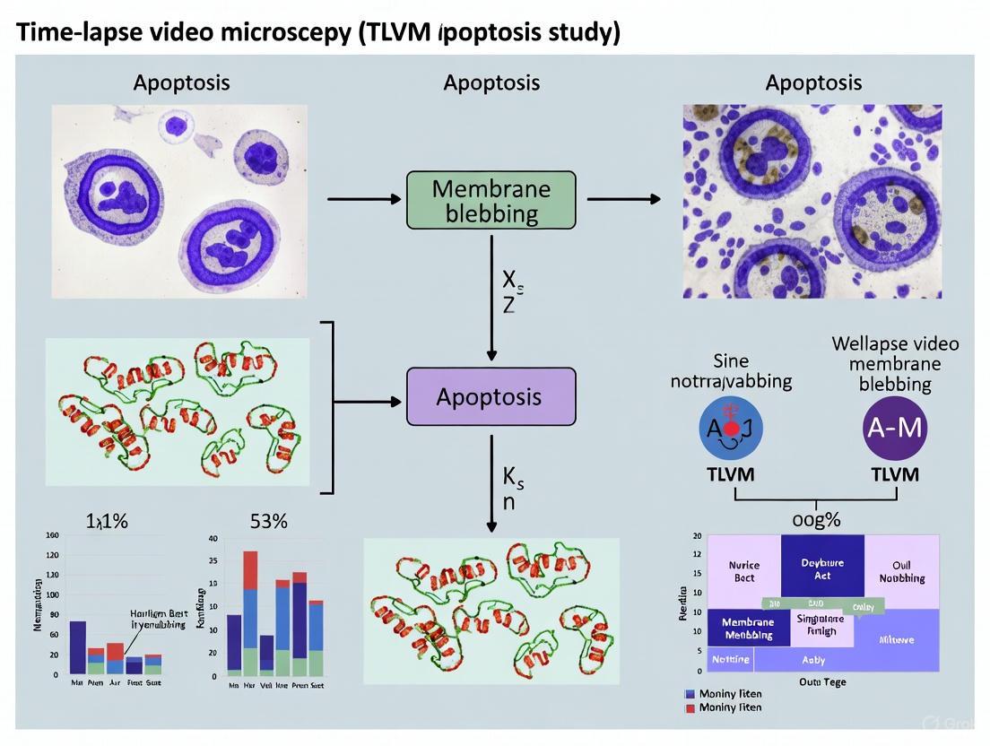
Apoptotic membrane blebbing is a fundamental morphological hallmark of programmed cell death, representing one of the visually distinctive characteristics of a cell undergoing apoptosis. This process involves the formation of dynamic, outward protrusions of the plasma membrane that eventually give rise to apoptotic bodies—small membrane-bound vesicles that facilitate the organized disposal of cellular components [13]. Within the context of time-lapse video microscopy (TLVM) research, blebbing serves as a critical visual indicator for identifying and quantifying apoptotic events in live cells, providing researchers with a window into the dynamic process of cell death [14] [15].
The molecular regulation of membrane blebbing centers on a carefully orchestrated interplay between specific kinases and structural proteins. ROCK1 (Rho-associated coiled-coil containing protein kinase 1) emerges as a primary regulator of this process, with its activation triggering a cascade of events that ultimately drive membrane protrusion [13]. Through its kinase activity, ROCK1 directly influences the contractile forces generated by the actin cytoskeleton, while recent evidence suggests that vimentin, a type III intermediate filament protein, contributes to the structural integrity and spatial organization of the blebbing apparatus [16]. The interplay between these molecular players enables the precisely controlled cellular fragmentation that characterizes apoptosis, distinguishing it from other forms of cell death such as necrosis [17]. Understanding these regulatory mechanisms provides valuable insights for both basic cell biology research and drug development efforts targeting pathological cell survival, particularly in cancer therapeutics [18].
Molecular Mechanisms of Blebbing Regulation
Central Signaling Pathway
The initiation and execution of apoptotic membrane blebbing are governed by a well-defined molecular pathway that integrates proteolytic signals with cytoskeletal remodeling. This pathway can be visualized through the following signaling cascade:
Key Molecular Regulators
ROCK1 Kinase
ROCK1 serves as the central regulator of the membrane blebbing process. This serine/threonine protein kinase becomes activated through cleavage by caspase-3 during apoptosis, which triggers its role in cytoskeletal reorganization [13]. Structurally, ROCK1 contains a kinase domain at the N-terminus, a coiled-coil region housing the Rho-binding domain (RBD), and a Pleckstrin homology (PH) domain with an integrated cysteine-rich domain (CRD) at the C-terminus [16]. Recent research has identified a novel RhoA binding site within the ROCK1 PHC1 tandem domain that is sufficient for dynamic recruitment to regions of active Rho signaling, leading to increased contractility in specific subcellular regions [19]. Once activated, ROCK1 phosphorylates multiple downstream targets that collectively drive actomyosin contractility. Specifically, it phosphorylates the myosin light chain (MLC), which enhances actin-myosin interaction and force generation, while simultaneously inactivating myosin phosphatase through phosphorylation of its regulatory subunit, creating a dual mechanism to ensure sustained contractile activity [13].
Actin Cytoskeleton
The actin cytoskeleton serves as the structural engine for membrane blebbing, undergoing dramatic reorganization during apoptosis. ROCK1-mediated phosphorylation directly promotes the assembly of actin filaments into contractile bundles through its effects on myosin-based contractility [16]. This leads to the formation of a condensed actomyosin ring beneath the plasma membrane that generates the intracellular pressure required for bleb formation. As contraction proceeds, hydrostatic pressure forces the plasma membrane to detach from the underlying cortex in regions where actin density is lowest, creating membrane blebs that expand rapidly until a new actin cortex reassembles within the protrusion [13]. This continuous cycle of bleb formation, expansion, and retraction creates the characteristic dynamic membrane protrusions observed in apoptotic cells through time-lapse video microscopy.
Vimentin Intermediate Filaments
While historically less emphasized than actin, vimentin has emerged as a significant contributor to the spatial organization and structural integrity of apoptotic blebs. As a major component of the intermediate filament network, vimentin undergoes caspase-mediated cleavage during apoptosis that alters its organizational state [16]. Research indicates that ROCK1 can directly or indirectly influence vimentin organization, potentially through phosphorylation events that regulate its assembly dynamics [16]. In the context of apoptotic blebbing, vimentin filaments appear to provide structural support that shapes the formation and expansion of membrane protrusions. The specific subcellular localization of ROCK1 to actomyosin filament bundles suggests a coordinated mechanism by which ROCK1 simultaneously regulates both the contractile actin machinery and the structural vimentin network to achieve spatially controlled membrane blebbing [16].
Experimental Protocols for Blebbing Analysis
Time-Lapse Video Microscopy of Apoptotic Cells
The dynamic nature of membrane blebbing makes time-lapse video microscopy (TLVM) an indispensable tool for capturing its progression in living cells. The following protocol details the setup for imaging and analyzing blebbing dynamics in apoptotic cells:
Equipment and Reagents:
- Inverted phase-contrast or fluorescence microscope with environmental chamber (37°C, 5% CO₂)
- High-sensitivity CCD or sCMOS camera
- Phase-contrast optics or differential interference contrast (DIC)
- Cell culture vessels suitable for microscopy (e.g., glass-bottom dishes)
- Apoptosis-inducing agents (e.g., γ-secretase inhibitors, topotecan, birinapant)
- Fluorescent dyes for viability assessment (optional)
Procedure:
- Cell Preparation: Plate cells at appropriate density (typically 50-70% confluency) in glass-bottom imaging dishes 24 hours before experimentation. Use suspended cells like K562 leukemic cells for clearer cell boundary visualization or adherent cells for surface attachment studies [20].
Apoptosis Induction: Apply apoptosis-inducing agent at predetermined concentration. For K562 cells, 20μM γ-secretase inhibitor (GSI-XXI) for 72 hours has demonstrated efficacy in inducing apoptosis with characteristic membrane blebbing [20].
Microscope Configuration:
- Set environmental chamber to maintain 37°C and 5% CO₂ throughout imaging
- Configure phase-contrast or DIC optics for optimal visualization of membrane dynamics
- Set imaging intervals between 30 seconds to 2 minutes depending on blebbing kinetics
- Adjust exposure times to minimize phototoxicity while maintaining image quality
Image Acquisition: Capture time-lapse sequences for 2-8 hours depending on experimental objectives. For comprehensive analysis of blebbing progression, continue imaging until complete fragmentation into apoptotic bodies occurs [14].
Data Extraction: Analyze time-lapse sequences to quantify blebbing parameters including time to bleb initiation, bleb frequency, bleb duration, and bleb size distribution using image analysis software such as ImageJ or Imaris.
Immunofluorescence Analysis of Blebbing-Associated Biomarkers
This protocol enables the specific identification of apoptotic cells through the detection of cleaved caspase-3 (CC3) in conjunction with morphological evidence of membrane blebbing, providing a high-specificity method for quantifying apoptosis in fixed samples [21].
Equipment and Reagents:
- Formalin-fixed, paraffin-embedded (FFPE) cell pellets or tissue sections
- Primary antibodies: anti-cleaved caspase-3 (CC3), anti-γH2AX, anti-Na+/K+-ATPase
- Fluorescently-labeled secondary antibodies
- Mounting medium with DAPI
- Fluorescence microscope with 20× and 40× objectives
Procedure:
- Sample Preparation: Fix cells in 4% paraformaldehyde for 15 minutes at room temperature followed by paraffin embedding according to standard histological procedures.
Immunostaining:
- Deparaffinize and rehydrate FFPE sections using standard protocols
- Perform antigen retrieval using citrate buffer (pH 6.0) at 95-100°C for 20 minutes
- Block nonspecific binding with 5% normal serum for 1 hour at room temperature
- Incubate with primary antibody cocktail (anti-CC3, anti-γH2AX, anti-Na+/K+-ATPase) overnight at 4°C
- Apply appropriate fluorescent secondary antibodies for 1 hour at room temperature
- Counterstain with DAPI and mount with antifade medium
Image Acquisition: Acquire multi-channel fluorescence images using a microscope equipped with appropriate filter sets. Capture multiple non-overlapping fields to ensure statistical robustness (minimum 10 fields per sample).
Image Analysis:
- Identify CC3-positive cells exhibiting punctate staining pattern associated with membrane blebbing [21]
- Quantify cells displaying colocalization of γH2AX (DNA damage marker) and CC3 blebbing structures
- Use plasma membrane staining (Na+/K+-ATPase) to confirm membrane blebbing morphology
- Calculate the percentage of CC3(bleb)+ cells relative to total cell count
Pharmacodynamic Assay for Apoptosis Detection in Tumor Tissues
This specialized protocol has been validated in xenograft models and canine lymphoma specimens, providing a robust method for distinguishing apoptosis-specific DNA fragmentation from direct drug-induced DNA damage [21].
Equipment and Reagents:
- FFPE tumor tissue sections (4-5μm thickness)
- Primary antibodies: anti-γH2AX (ser139) and anti-cleaved caspase-3 (CC3)
- Isotype-matched control antibodies
- Automated image analysis system or fluorescence microscope with counting software
Procedure:
- Tissue Processing: Section FFPE tissue blocks and mount on charged slides. Bake slides at 60°C for 1 hour to ensure adhesion.
Immunofluorescence Staining: Perform simultaneous detection of γH2AX and CC3 using standardized immunofluorescence protocols with tyramide signal amplification if needed.
Automated Enumeration:
- Utilize nuclear segmentation based on DAPI staining to identify individual cells
- Apply ring-based nuclear border dilation to define cytoplasmic regions
- Use spot detection algorithms to identify CC3 puncta associated with membrane blebbing
- Quantify γH2AX-positive nuclei with and without associated CC3 blebbing structures
Data Interpretation:
- γH2AX-positive/CC3(bleb)-negative cells indicate direct drug-induced DNA damage
- γH2AX-positive/CC3(bleb)-positive cells confirm apoptosis-mediated DNA fragmentation
- Calculate the percentage of each population relative to total tumor cell count
Research Reagent Solutions
The following table compiles essential reagents and tools for investigating ROCK1-mediated apoptotic blebbing:
Table 1: Essential Research Reagents for Apoptotic Blebbing Studies
| Reagent/Category | Specific Examples | Research Application | Experimental Function |
|---|---|---|---|
| ROCK Inhibitors | Fasudil, Y-27632 | Pathway inhibition | Validate ROCK1 dependence in blebbing by inhibiting kinase activity [16] |
| Apoptosis Inducers | γ-Secretase inhibitors (GSI-XXI), Topotecan, Birinapant | Model establishment | Trigger apoptotic signaling cascades leading to membrane blebbing [20] [21] |
| Cell Lines | K562 (suspension), HeLa (adherent) | Cellular models | Provide optimized systems for blebbing analysis; K562 offers clear cell boundaries [20] |
| Antibodies | Anti-cleaved caspase-3, Anti-γH2AX, Anti-ROCK1 | Biomarker detection | Identify apoptotic cells and detect DNA damage response; confirm ROCK1 expression [21] |
| Fluorescent Reporters | CaspACE-FITC-VAD-FMK, SYBR Green I | Live/dead cell analysis | Detect caspase activity and DNA fragmentation in live or fixed cells [20] |
Quantitative Analysis of Blebbing Parameters
The systematic quantification of membrane blebbing dynamics provides crucial insights into the regulation and progression of apoptosis. The following table summarizes key quantitative parameters from recent studies:
Table 2: Quantitative Parameters of Apoptotic Membrane Blebbing
| Parameter | Experimental System | Reported Values | Significance |
|---|---|---|---|
| Blebbing Timeline | K562 cells + GSI-XXI | Caspase activation → DNA fragmentation → Membrane blebbing | Defines temporal sequence of apoptotic events [20] |
| CC3(bleb)+ Cells | Canine lymphoma post-LMP744 | Pre-dose: 0.3%; 6h post-dose: 3.3% | Quantifies apoptosis induction in clinical specimens [21] |
| γH2AX/CC3 Colocalization | MDA-MB-231 xenografts + birinapant | Dose-dependent increase | Confirms apoptosis as primary mechanism of action [21] |
| Therapeutic Response Correlation | Topotecan vs. cisplatin regimens | Tumor regression with mixed γH2AX sources; Growth delay with direct DNA damage only | Elucidates mechanism of drug action [21] |
| AI Classification Accuracy | Phase-contrast images of K562 cells | High accuracy (F-values) for caspase+/frag+ cells | Enables stain-free apoptosis detection [20] |
Technical Considerations and Optimization
Methodological Challenges and Solutions
Implementing robust assays for apoptotic membrane blebbing requires addressing several technical challenges. Phototoxicity represents a significant concern during live-cell imaging, as excessive illumination can alter cellular physiology and induce non-apoptotic membrane blebbing. This can be mitigated through optimized imaging protocols utilizing minimal exposure times, near-infrared light-emitting diodes synchronized with acquisition periods, and the implementation of lattice light-sheet microscopy (LLSM) which significantly reduces photodamage while providing high spatiotemporal resolution [22]. For suspension cells like K562 leukemic cells, which offer superior visualization of cell boundaries, maintaining cell viability during extended imaging sessions requires precise environmental control and the use of specialized culture media formulations such as CMRL supplemented with Knock Out serum and L-glutamine [22].
The specificity of apoptosis detection poses another challenge, particularly when distinguishing true apoptotic events from other cellular processes. The combination of morphological assessment (membrane blebbing) with molecular markers (CC3 puncta and γH2AX) significantly enhances detection specificity compared to single-parameter approaches [21]. Furthermore, the implementation of AI-based classification systems trained on phase-contrast images of apoptotic cells has demonstrated promising capabilities for accurate, stain-free identification of apoptosis, potentially overcoming limitations associated with chemical staining and fluorescent dye toxicity [20].
Data Analysis and Interpretation
Advanced image analysis approaches are essential for extracting meaningful quantitative data from blebbing experiments. For fixed tissue analysis, automated algorithms that combine nuclear segmentation (based on DAPI), cytoplasmic region definition (through ring-based nuclear border dilation), and spot detection (for CC3 puncta identification) provide robust quantification of apoptotic cells while minimizing operator bias [21]. In live-cell imaging applications, tracking individual cells throughout the apoptotic process enables lineage tracing and the determination of cell cycle length, offering insights into the temporal dynamics of blebbing progression [15].
The interpretation of γH2AX staining requires particular care, as this marker can indicate either direct drug-induced DNA damage or apoptosis-mediated DNA fragmentation. The critical distinction lies in the association with CC3 blebbing structures—γH2AX signal colocalized with CC3 puncta confirms apoptosis as the underlying mechanism, while γH2AX in the absence of CC3 blebbing suggests direct DNA damage response [21]. This differentiation has profound implications for understanding the mechanism of action of investigational anticancer agents in clinical trials.
Visualizing the Experimental Workflow
The comprehensive analysis of ROCK1-mediated apoptotic blebbing integrates multiple experimental approaches that can be visualized as a connected workflow:
The molecular regulation of apoptotic membrane blebbing represents a sophisticated cellular process coordinated through the integrated actions of ROCK1, actin cytoskeletal networks, and vimentin filaments. The experimental approaches outlined in this document provide researchers with comprehensive tools to investigate this dynamic process, from live-cell imaging using time-lapse video microscopy to sophisticated immunofluorescence assays that distinguish apoptosis-specific DNA fragmentation. The continuing refinement of these methodologies, including the development of AI-based classification systems and highly specific pharmacodynamic assays, promises to enhance our understanding of apoptotic regulation and accelerate the development of therapeutics that target cell death pathways. As these techniques become increasingly accessible and standardized, they will undoubtedly yield new insights into the fundamental biology of apoptosis and its manipulation for therapeutic benefit.
Time-lapse video microscopy (TLVM) has emerged as a powerful tool for capturing the dynamic process of apoptotic cell death, particularly the critical phenomenon of membrane blebbing. Unlike endpoint assays, TLVM enables researchers to observe and quantify the temporal sequence of morphological changes in individual living cells, providing unprecedented insight into the progression and mechanisms of programmed cell death. This capability is especially valuable for distinguishing apoptosis from other forms of cell death, such as necrosis, and for assessing the efficacy of therapeutic agents in drug development pipelines.
The process of apoptosis is characterized by well-defined morphological stages, including cell shrinkage, chromatin condensation, and plasma membrane blebbing—the formation of spherical protrusions from the cell surface. These membrane blebs represent a critical visual indicator of early apoptosis and can be dynamically tracked using TLVM. For cancer researchers and drug development professionals, understanding these transitions is paramount, as resistance to apoptosis is a recognized hallmark of cancer, and inducing apoptotic cell death remains a key therapeutic strategy.
Morphological Stages of Apoptosis Visualized by TLVM
The progression of apoptosis follows a defined sequence of morphological events that can be quantitatively monitored using TLVM. The table below summarizes the key transitions, their temporal characteristics, and specific TLVM detection methods for each stage.
Table 1: Key Morphological Transitions in Apoptosis Visualized by TLVM
| Stage | Time Frame | Key Morphological Features | TLVM Detection Methods |
|---|---|---|---|
| Early Apoptosis | 0-6 hours post-induction | Cell shrinkage, loss of cell-cell contacts, membrane blebbing begins | Phase contrast, DIC, QPI for label-free detection; Caspase-3/7 fluorescent reporters |
| Membrane Blebbing | 30 minutes - 3 hours | Spherical protrusions of plasma membrane; bleb expansion and retraction | High-resolution DIC, QPI for dry mass quantification, fluorescent membrane markers |
| Late Apoptosis | 3-12 hours | Nuclear condensation, apoptotic body formation, maintained membrane integrity | DIC with fluorescent DNA dyes (Hoechst, DAPI), Annexin V probes |
| Terminal Phase | 12-24 hours | Phagocytosis by neighboring cells, minimal inflammatory response | Phase contrast for morphology, fluorescence tags for phagocytosis detection |
The visualization of membrane blebbing dynamics represents a particularly valuable application of TLVM in apoptosis research. As demonstrated in research on WEHI-3B leukemia cells, early apoptosis signs, including membrane blebbing, can be observed within 6 hours post-treatment with apoptotic inducers like Newcastle Disease Virus [23]. Quantitative phase imaging (QPI), a specialized TLVM modality, has enabled researchers to not only visualize but precisely quantify the dry mass dynamics of individual blebs in CHO-K1 and U937 cells following exposure to pulsed electric fields [24]. This level of quantitative analysis provides biophysical parameters that correlate with apoptotic progression.
Experimental Protocols for TLVM of Apoptotic Blebbing
Basic TLVM Setup for Apoptosis Detection
The following protocol outlines the essential steps for configuring TLVM to capture apoptotic membrane blebbing using transmitted light microscopy, which enables label-free detection of morphological changes without fluorescent probes.
Table 2: Essential Equipment and Reagents for Basic TLVM Apoptosis Detection
| Category | Specific Products/Models | Application/Function |
|---|---|---|
| Microscope System | Nikon Eclipse Ti inverted microscope with Perfect Focus system | Maintains focus during long-term time-lapse imaging |
| Imaging Modalities | Differential Interference Contrast (DIC), Phase Contrast (PC) | Label-free visualization of cellular morphology |
| Environmental Control | POCmini-2 Cell Cultivation System (PeCon GmbH) | Maintains 37°C, 5% CO2 during live imaging |
| Detection Reagents | NucView 488 caspase-3/7 substrate (Biotium) | Fluorescent detection of caspase activation |
| Apoptosis Inducers | Staurosporine (1-10 µM in 1% DMSO) | Protein kinase inhibitor induces intrinsic apoptosis |
| Image Analysis Software | NIS-Elements, Fiji (ImageJ) | Time-lapse processing and quantitative analysis |
Procedure:
- Cell Preparation: Plate cells in glass-bottom imaging dishes (e.g., MatTek 35mm dishes) at appropriate density (e.g., 1×10⁵ cells/mL for most adherent lines) and culture for 24 hours prior to imaging in phenol red-free medium to reduce background fluorescence [25].
- Apoptosis Induction: Prepare fresh staurosporine solution in DMSO and dilute to working concentration (typically 1-10 µM) in pre-warmed culture medium. Replace medium in imaging dishes with staurosporine-containing medium 30 minutes prior to initiating time-lapse acquisition [25].
- Microscope Configuration:
- Set environmental chamber to maintain 37°C and 5% CO₂ throughout imaging.
- For DIC imaging, use shuttered, green (510-560 nm) heat-filtered light from 100W tungsten bulbs to minimize phototoxicity.
- Configure camera settings for 2-4 frames/minute framing rate with minimal exposure to maintain cell viability.
- Engage perfect focus system if available to maintain focus throughout extended acquisitions.
- Time-lapse Acquisition: Program software to acquire images from multiple positions at regular intervals (2-4 minutes) for 8-24 hours depending on experimental needs. Include phase contrast/DIC and fluorescence channels if using caspase reporters.
- Data Processing: Export time-lapse sequences for analysis in Fiji/ImageJ or NIS-Elements. Quantify parameters including bleb formation kinetics, cell shrinkage, and caspase activation kinetics.
Advanced Quantitative Phase Imaging for Bleb Dynamics
For researchers requiring precise biophysical measurements of bleb formation, quantitative phase imaging (QPI) offers label-free quantification of dry mass dynamics during apoptosis.
Specialized Equipment:
- Quantitative phase imaging system (e.g., laser-based interferometric setup)
- High-speed camera for capturing rapid bleb dynamics (≥100 fps)
- Pulsed electric field generator (for controlled bleb induction if studying electroporation-mediated blebbing) [24]
Procedure:
- Cell Preparation: Seed cells on specialized optical imaging chambers at optimal density for single-cell analysis.
- QPI Configuration: Calibrate the QPI system according to manufacturer specifications, ensuring stable interference pattern acquisition.
- Bleb Induction: For controlled studies, expose cells to 600 ns, 21.2 kV/cm electric pulses to induce synchronized bleb formation [24].
- High-speed Acquisition: Initiate time-lapse QPI immediately post-induction with temporal resolution sufficient to capture rapid bleb dynamics (≥10 fps).
- Mass Quantification: Process QPI data to calculate dry mass distribution and transfer between cell body and blebs over time.
This protocol has been successfully implemented to visualize and quantify the formation of membrane blebs following exposure to pulsed electric fields, revealing that blebs can contain significant cellular dry mass and undergo complex dynamics including merging and retraction [24].
The Scientist's Toolkit: Essential Research Reagents and Materials
Table 3: Research Reagent Solutions for TLVM of Apoptotic Membrane Blebbing
| Reagent/Material | Function/Application | Example Products/Specifications |
|---|---|---|
| Caspase-3/7 Reporters | Fluorescent detection of early apoptosis activation | NucView 488, CellEvent Caspase-3/7 Green |
| Plasma Membrane Probes | Visualization of membrane dynamics during blebbing | BioTracker Apo-15, Annexin V conjugates |
| Viability Markers | Distinguishing apoptosis from necrosis | Propidium iodide, SYTOX Green |
| Apoptosis Inducers | Experimental initiation of apoptotic pathways | Staurosporine, Aspirin, Newcastle Disease Virus strains |
| Specialized Media | Maintaining cell health during extended imaging | Phenol red-free DMEM, FluoroBrite DMEM |
| Environmental Control | Maintaining physiological conditions during imaging | PeCon cellVivo incubation system |
| Image Analysis Software | Quantifying morphological parameters | Fiji/ImageJ with TrackMate, NIS-Elements, Imaris |
Signaling Pathways and Experimental Workflows
The following diagrams illustrate key apoptotic signaling pathways and experimental workflows for TLVM investigation of membrane blebbing.
Diagram 1: Apoptosis Signaling to Membrane Blebbing
Diagram 2: TLVM Experimental Workflow
Time-lapse video microscopy provides an unparalleled window into the dynamic process of apoptotic membrane blebbing, enabling researchers to capture key morphological transitions with high temporal and spatial resolution. The protocols and methodologies outlined in this application note offer both foundational approaches and advanced techniques for investigating these critical cellular events. By implementing these TLVM strategies, researchers in drug development and basic cancer biology can gain deeper insights into apoptotic mechanisms, enhance compound screening efforts, and ultimately contribute to the development of more effective therapeutic agents that modulate programmed cell death pathways.
From Images to Insights: Methodologies for TLVM Apoptosis Assays
Time-lapse video microscopy (TLVM) is an indispensable tool for capturing the dynamic morphological changes that characterize apoptosis, particularly plasma membrane blebbing. This process, a hallmark of the execution phase of programmed cell death, is a rapid and asynchronous event within a cell population. TLVM enables researchers to observe and quantify this blebbing in real-time, providing kinetic data that endpoint assays cannot capture. The integrity of this data is heavily dependent on the precise configuration of hardware components and strict control of the cellular environment throughout the duration of the experiment.
Essential Hardware Configuration
A TLVM system for apoptosis research is an integrated setup designed to maintain cell viability while acquiring high-quality, time-resolved images. The core components must work in harmony to ensure that observed morphological changes are a true response to the experimental treatment and not an artifact of suboptimal conditions.
Table 1: Core Hardware Components for TLVM Apoptosis Studies
| Component | Key Specifications | Role in Apoptosis Blebbing Studies |
|---|---|---|
| Inverted Microscope | Phase-contrast or Nomarski (DIC) optics | Enables observation of unlabeled, living cells and clear visualization of membrane blebs without cytotoxic staining. |
| Microscope Incubator | Maintains 37°C and 5% CO₂ | Preserves cell health and normal physiology over multi-hour imaging sessions. |
| High-Resolution Camera | Cooled CCD or sCMOS sensor | Captures fine morphological details of blebs with high sensitivity during sequential frames. |
| Image Acquisition Software | Automated, scheduled capture | Allows images to be collected at frequent intervals (e.g., every 2.5-5 minutes) over 24+ hours. |
| Vibration Isolation Table | Stable, dampened platform | Prevents motion blur that could obscure the visualization of small, dynamic blebs. |
Additional Critical Hardware Considerations
Beyond the core components, several other factors are crucial for a successful setup. Vibration isolation is paramount; even minor disturbances can cause blurring in high-magnification images and complicate the analysis of delicate membrane structures. For fluorescence-based assays, such as those using annexin V binding, the appropriate filter sets and a high-sensitivity camera are required. Furthermore, the use of a motorized stage is highly recommended for multiposition experiments, allowing for the simultaneous tracking of apoptosis progression in multiple treatment groups or replicates within a single run.
Controlling the Cellular Environment
Maintaining a constant and physiologically relevant environment is arguably the most critical aspect of long-term live-cell imaging. Fluctuations in temperature, CO₂, and humidity can induce cellular stress, leading to experimental artifacts and compromising data validity.
Table 2: Environmental Control Parameters for Live-Cell TLVM
| Parameter | Optimal Setting | Rationale & Method of Control |
|---|---|---|
| Temperature | 37°0°C | Maintains normal enzymatic activity (e.g., caspase function). Controlled via a microscope-top incubator or an environmental chamber. |
| CO₂ Level | 5% | Regulates pH of standard bicarbonate-buffered media. Controlled by a gas mixer or with pre-mixed gas in a sealed chamber. |
| Humidity | Near 100% | Preents evaporation from the culture medium, which would alter osmolarity and concentrate reagents. Achieved by using a layer of sterile mineral oil over the medium or a humidified gas stream. |
| Medium Volume & Vessel | 50-240 µL in a 96-well plate | Minimizes medium usage and allows for high-throughput screening. Evaporation is mitigated by overlaying with 50 µL of sterile mineral oil. |
Implementing Environmental Controls
The search results highlight specific methodologies for environmental control. For instance, one protocol details plating cells in 240 µL aliquots in a 96-well plate and then layering 50 µL of sterile mineral oil on top to prevent evaporation and CO₂ escape during a 24-hour incubation in the TLVM system [26]. This step is vital for experiments that track the steep linear increase in cells with membrane blebbing, as any change in medium conditions could alter the kinetics of apoptosis.
Key Signaling Pathways in Apoptotic Membrane Blebbing
The formation of membrane blebs during apoptosis is not a passive process but is actively driven by specific biochemical signaling pathways that lead to actomyosin-based contraction. Research has identified two key regulators: the caspase-activated Rho effector protein ROCK I, and myosin light chain kinase (MLCK).
The following diagram illustrates the core signaling pathway that regulates apoptotic membrane blebbing, integrating the key molecular players identified in the research:
ROCK I is cleaved and activated by caspases during apoptosis [27]. This active form of ROCK I, along with MLCK, promotes the phosphorylation of the myosin regulatory light chain (MLC) [28]. Phosphorylated MLC activates myosin II ATPase, leading to forceful interactions with actin filaments and generating the contractile forces that drive the plasma membrane away from the cortical cytoskeleton, forming a bleb [28] [27]. This process is dependent on an intact actin cytoskeleton [28].
Experimental Protocol: TLVM for Drug-Induced Apoptosis
This protocol outlines the methodology for using TLVM to study apoptotic membrane blebbing in response to chemotherapeutic agents, based on established procedures [26].
Materials and Reagents
- Cells: Adherent cell line (e.g., PC-12, Rat-1 fibroblasts) or suspension cells (e.g., HL-60) [28] [26].
- Inducers: Apoptosis-inducing agents (e.g., Etoposide, Cisplatin, Serum Withdrawal) [28] [26].
- Inhibitors: Caspase inhibitor (z-VAD-FMK, 100 µM), MLCK inhibitors (ML-7, ML-9), ROCK inhibitor (e.g., Y-27632) [28].
- Culture Vessels: 96-well microtiter plate, collagen-coated if required for adhesion.
- TLVM System: Configured as described in Section 2.
Step-by-Step Procedure
Cell Preparation:
- Harvest exponentially growing cells and wash to remove serum components.
- Resuspend cells in complete medium or serum-free medium, depending on the apoptosis induction model.
- Plate cells in a 96-well microtiter plate at a density of (2 \times 10^5) cells/ml in a 240 µL aliquot [26]. For adherent cells, plate onto collagen-coated wells.
Pre-incubation:
- Incubate the plate for 60 minutes in a fully humidified atmosphere of 5% CO₂ at 37°C to allow cells to equilibrate and adhere.
Drug Application & Experimental Setup:
- Add 10 µL aliquots of the drug (e.g., Etoposide, Cisplatin) at the desired concentration to the wells. Include vehicle control wells.
- For inhibition studies, pre-treat cells with inhibitors (e.g., 100 µM z-VAD-FMK) before adding the apoptotic inducer [28].
- Layer 50 µL of sterile mineral oil on top of the medium in each well to prevent evaporation and CO₂ escape during imaging [26].
TLVM Acquisition:
- Place the microtiter plate into the pre-warmed and gas-controlled chamber of the TLVM system.
- Select fields for imaging under phase-contrast or Nomarski optics.
- Configure the acquisition software to collect images at 2.5 to 5-minute intervals for a period of up to 24 hours [26].
Data Analysis:
- In sequential frames, count the number of cells exhibiting dynamic plasma membrane protrusions (blebs).
- Express the data as the percentage of blebbing cells versus time.
- Plot the kinetic curve of apoptosis progression. The time to the maximum response (Tm) indicates the peak of blebbing activity in the population [26].
Research Reagent Solutions
The following table details key pharmacological tools used to dissect the mechanisms of apoptotic membrane blebbing.
Table 3: Essential Reagents for Studying Apoptotic Blebbing Mechanisms
| Reagent | Function / Target | Application in Blebbing Research |
|---|---|---|
| z-VAD-FMK | Pan-caspase inhibitor | Blocks caspase activity; used to synchronize cells in the blebbing phase and confirm caspase-dependence [28]. |
| Blebbistatin | Allosteric myosin II inhibitor | Inhibits actomyosin contractility; used to directly test the role of myosin motor activity in bleb formation [29]. |
| C3 Transferase | Rho inhibitor | Inhibits Rho family GTPases; used to demonstrate the role of Rho/ROCK signaling in apoptotic blebbing [28]. |
| ML-7 / ML-9 | Myosin Light Chain Kinase (MLCK) inhibitors | Blocks MLC phosphorylation via MLCK; used to delineate the contribution of MLCK to the blebbing process [28]. |
| Cytochalasin D | Actin polymerization inhibitor | Disrupts the actin cytoskeleton; used to confirm the necessity of F-actin for bleb formation and stability [28]. |
| Calyculin A | Protein phosphatase inhibitor | Prevents dephosphorylation of MLC and other proteins; can enhance or sustain contractile forces [28]. |
Programmed cell death (PCD), particularly apoptosis, represents a fundamental cellular process critical in oncology, immunology, and drug development. The detection of apoptotic events, with a specific focus on membrane blebbing and apoptotic body formation, provides crucial insights into cellular responses to therapeutic interventions. Time-lapse video microscopy (TLVM) has emerged as a powerful technique for monitoring these dynamic morphological changes in living cells over extended periods. Within this context, researchers face a fundamental methodological choice: employing fluorescent staining techniques that provide molecular specificity or leveraging label-free approaches that capitalize on inherent cellular morphology. Fluorescent methods utilize molecular markers such as Annexin-V, which binds to phosphatidylserine exposed on the outer leaflet of the apoptotic cell membrane, providing a well-established biochemical confirmation of apoptosis. In contrast, label-free techniques exploit the inherent morphological hallmarks of apoptosis—including cell shrinkage, membrane blebbing, and apoptotic body formation—using phase-contrast or quantitative phase imaging (QPI), coupled with advanced computational analysis. The decision between these strategies involves careful consideration of multiple factors, including experimental goals, potential cellular perturbations, temporal resolution requirements, and analytical capabilities. This application note provides a structured comparison of these methodologies, supported by quantitative data and detailed protocols, to guide researchers in selecting the optimal detection strategy for their TLVM apoptosis research.
Technical Comparison of Detection Methodologies
Performance Metrics and Key Characteristics
Table 1: Comparative Analysis of Fluorescent and Label-Free Apoptosis Detection Methods
| Parameter | Fluorescent Staining | Label-Free Detection |
|---|---|---|
| Molecular Basis | Binding to specific biomarkers (e.g., PS exposure via Annexin-V) [30] [31] | Analysis of morphological changes (e.g., membrane blebbing, apoptotic bodies) [30] [32] |
| Primary Readout | Fluorescence intensity at specific wavelengths [33] | Cellular morphology and texture features from phase-contrast images [32] |
| Typical Accuracy | Varies; Annexin-V missed ~70% of events detected via ApoBDs in one study [30] | 92% accuracy (ApoBD detection), 96.4% accuracy (D-MAINS for multiple states) [30] [32] |
| Temporal Resolution | Limited by phototoxicity and photobleaching [33] | High; continuous monitoring possible (e.g., 5-min intervals) [30] |
| Cellular Perturbation | Yes; biochemical perturbation, phototoxicity [30] | Minimal; no chemical labels or dedicated fluorescent channels required [30] [34] |
| Key Advantage | Molecular specificity, well-established protocols [31] | Non-invasiveness, earlier detection potential, continuous monitoring [30] [32] |
| Main Limitation | Late and inconsistent indication for some cell types, photobleaching [30] [33] | Requires sophisticated computational analysis (e.g., deep learning) [30] [34] |
Research Reagent Solutions Toolkit
Table 2: Essential Reagents and Materials for Apoptosis Detection Assays
| Reagent/Material | Function/Application | Key Considerations |
|---|---|---|
| Annexin-V (e.g., Alexa Fluor 647 conjugate) | Flags early apoptosis by binding to externalized phosphatidylserine [30] [31] | Can provide inconsistent and late indication in some models (e.g., human melanoma) [30] |
| Propidium Iodide (PI) / SYTOX Green | Stains nucleic acids in cells with compromised membranes (necrosis) [32] | Used to differentiate apoptosis from necrosis; requires viable cells for apoptosis assessment [32] |
| AztecBleb Probes | Novel fluorescent reporters targeting blebbing dynamics and microtubule interactions [35] | Pregnenolone-based scaffold for selective localization in blebs; enables real-time imaging of blebbing mechanisms [35] |
| PKH67 (Green) & PKH26 (Red) Cell Linkers | Fluorescent cytoplasmic dyes for long-term cell tracking [30] | Used to differentiate effector and target cells in co-culture assays (e.g., immune cell-cancer cell interactions) [30] |
| Polydimethylsiloxane (PDMS) Nanowell Arrays | Microfabricated platform for single-cell analysis and time-lapse imaging [30] | Enables high-throughput, single-cell resolution monitoring of cell-cell interactions within confined volumes [30] |
| Deep-Learning Models (e.g., D-MAINS, ResNet50) | Computational tools for label-free classification of cell states [30] [32] | Distinguishes mitosis, apoptosis, interphase, necrosis, and senescence based on phase-contrast morphology [32] |
Detailed Experimental Protocols
Protocol 1: Label-Free Detection of Apoptosis via Apoptotic Body (ApoBD) Analysis
This protocol details a label-free method for detecting apoptosis by identifying membrane-bound apoptotic bodies using time-lapse phase-contrast microscopy and deep learning analysis [30].
Materials and Equipment:
- TIMING (Time-lapse Imaging Microscopy In Nanowell Grids) system or equivalent live-cell imaging setup [30]
- Phase-contrast microscope (e.g., Axio microscope with 20× 0.8 NA objective) [30]
- Polydimethylsiloxane (PDMS) nanowell arrays [30]
- Cell culture reagents and appropriate media
- ResNet50 network or similar deep learning model for image analysis [30]
Procedure:
- Sample Preparation:
- Culture target cells (e.g., Mel526 melanoma cell line) and effector cells (e.g., tumor-infiltrating lymphocytes, TILs) as required [30].
- Optionally, label TILs with PKH67 (green) and melanoma cells with PKH26 (red) fluorescent linkers for cell type differentiation, though this is not required for the primary label-free detection [30].
- Load cells onto PDMS nanowell chips at a concentration of 2 million effector cells and 1 million target cells per milliliter [30].
- Immerse the chip in phenol red-free cell-culture media. Do not add apoptosis-specific fluorescent markers [30].
Image Acquisition:
- Place the prepared chip in a humidity/CO₂ controlled chamber on the microscope stage [30].
- Acquire time-lapse phase-contrast images every 5 minutes for the desired duration (e.g., 24-72 hours) [30].
- If using fluorescent labels for secondary validation, acquire images in the relevant channels (PKH26, PKH67) at each time point, but minimize exposure to reduce phototoxicity [30].
Image Processing and ApoBD Detection:
- Process raw images to transform them into 16-bit Tagged Image File Format [30].
- Use the TIMING pipeline or equivalent software for nanowell detection and cell detection to obtain multi-channel images of individual nanowells [30].
- Apply a trained ResNet50 network to identify frames demonstrating the release of ApoBDs within the nanowells [30].
- Implement a three-frame temporal constraint to distinguish actual death events from noise: when ApoBDs are detected in three consecutive frames, assign the starting frame as the time of apoptosis onset [30].
Data Analysis:
- The model provides an Intersection over Union (IoU) accuracy for apoptotic body segmentation (approximately 75%), enabling associative identification of the apoptotic cell [30].
- Onset of apoptosis is predicted with an error of approximately one frame (corresponding to ±5 minutes in this setup) [30].
Protocol 2: Fluorescence-Based Apoptosis Detection with Annexin-V Staining
This protocol describes the standard method for detecting apoptosis using fluorophore-conjugated Annexin-V to label phosphatidylserine exposure on the outer leaflet of the apoptotic cell membrane [30] [31].
Materials and Equipment:
- Fluorophore-conjugated Annexin-V (e.g., Annexin-V-Alexa Fluor 647) [30]
- Propidium Iodide (PI) or SYTOX Green stain [32]
- Live-cell imaging chamber with environmental control (temperature, CO₂) [30]
- Fluorescence microscope with appropriate filter sets
- Cell culture reagents and appropriate media
Procedure:
- Sample Preparation and Staining:
- Culture and prepare cells according to experimental requirements.
- For co-culture assays, label different cell populations with cytoplasmic dyes (e.g., PKH67, PKH26) if desired [30].
- Add Annexin-V conjugate to the culture media at the recommended dilution (e.g., 1:60 for Annexin-V-Alexa Fluor 647) [30].
- Add PI or SYTOX Green at the recommended concentration to distinguish late apoptotic and necrotic cells [32].
Image Acquisition:
- Place the stained cells in the live-cell imaging chamber with controlled temperature and CO₂.
- Acquire time-lapse images in all relevant fluorescent channels (e.g., PKH26, PKH67, Annexin-V, PI) along with phase-contrast or bright-field images [30].
- Set the imaging interval to balance temporal resolution with phototoxicity concerns (e.g., every 15-30 minutes). Reduced frequency and exposure times help minimize photodamage [33].
Image Processing and Analysis:
- Process images to delineate individual cells and quantify fluorescence signals.
- Define an Annexin-V positive cell by computing the Intersection over Union (IoU) value between the binarized Annexin-V mask and the cell mask from the detection module [30].
- Calculate the IoU as: IoU(maskA, maskB) = Area(maskA AND maskB) / Area(maskA OR maskB) [30].
- Cells with IoU above a defined threshold are considered apoptotic.
Validation (Optional):
Visualizing Experimental Workflows and Signaling Pathways
Experimental Workflow for Apoptosis Detection
Apoptosis Signaling Pathways and Detection Windows
The choice between fluorescent and label-free detection strategies for TLVM apoptosis research depends critically on experimental priorities. Fluorescent staining provides molecular specificity and is well-suited for confirming specific biochemical events, such as phosphatidylserine externalization. However, evidence indicates that label-free methods, particularly those leveraging deep learning to analyze morphological features like apoptotic bodies, can detect a significantly larger proportion of apoptotic events—in some cases identifying 70% more events than Annexin-V staining alone [30]. Label-free approaches offer the additional advantages of continuous monitoring without phototoxicity concerns, earlier detection potential, and preservation of samples for subsequent analyses.
For research focused on kinetic profiling of apoptotic events and high-throughput screening, label-free methods coupled with computational analysis (e.g., D-MAINS, ResNet50) provide superior performance. Conversely, for studies requiring confirmation of specific biochemical pathways or multiplexing with other fluorescent markers, fluorescent staining remains valuable. The most robust experimental designs may incorporate both approaches, using fluorescent markers for initial validation and label-free methods for comprehensive temporal analysis, thereby leveraging the complementary strengths of both detection paradigms.
The study of apoptotic cells, characterized by morphological hallmarks such as membrane blebbing, is crucial for biomedical research in cancer, immunology, and drug development [36]. Time-lapse video microscopy (TLVM), particularly intravital two-photon microscopy (2P-IVM), enables the real-time, in vivo visualization of these dynamic processes within living tissues [36]. However, the manual analysis of the resulting complex image data is time-consuming, subjective, and low-throughput. This creates a critical bottleneck, underscoring the need for automated, accurate, and reliable analysis methods.
Deep Learning (DL), a subset of artificial intelligence, has dramatically advanced the field of computer vision, offering powerful solutions for image analysis tasks [37]. This document introduces the application of two advanced DL approaches for the automated detection of apoptosis in TLVM data: the Apoptosis Detection System (ADeS), a novel theoretical framework designed for holistic analysis of temporal image sequences, and ResNet-50, a well-established convolutional neural network for image classification. By leveraging these tools, researchers can transform their analytical capabilities, accelerating the pace of discovery in cell death research and drug safety profiling.
Technical Foundations of the Featured Deep Learning Architectures
ResNet-50: A Deep Convolutional Network for Feature Extraction
ResNet-50 is a 50-layer deep convolutional neural network (CNN) pre-trained on a million images from the ImageNet database, which contains over 1000 categories [38]. Its key innovation is the residual block, which uses identity connections (or skip connections). These connections take the input of a block directly to its output, bypassing one or more layers [38]. This architecture mitigates the vanishing gradient problem, a common issue in very deep networks that makes them difficult to train. By preventing this, ResNet-50 can effectively learn from deep architectures, leading to excellent generalization performance and lower error rates in visual recognition tasks [38]. In the context of apoptosis detection, ResNet-50 can serve as a powerful feature extractor, identifying critical spatial patterns—such as cell shrinkage and membrane blebbing—from individual microscopy frames.
ADeS: A Historical Awareness Framework for Temporal Analysis
While ResNet-50 analyzes static images, the Apoptosis Detection System (ADeS) is a theoretical framework inspired by modern deep learning architectures designed for sequential data. ADeS draws from the principles of the Historical Awareness Multi-Level Embedding (HAMLE) model [39]. The core idea of ADeS is to move beyond analyzing individual frames in isolation. Instead, it treats a time-lapse sequence as a holistic historical profile of a cell, integrating information from past states to understand the current context better.
Theoretically, ADeS is guided by the transfer of learning, which explains how prior knowledge can be retrieved and integrated to solve new tasks [39]. Technically, it could be built upon a Bidirectional Encoder Representations from Transformers (BERT) architecture with self-attention layers [39]. The self-attention mechanism allows the model to determine which parts of the historical sequence are most relevant for detecting an apoptotic event at the current time point, mirroring how a human expert would track a cell's morphological evolution over time.
Table 1: Comparison of ResNet-50 and the ADeS Framework for Apoptosis Detection
| Feature | ResNet-50 | ADeS Framework |
|---|---|---|
| Primary Strength | Spatial feature extraction from single images | Temporal context modeling from video sequences |
| Core Innovation | Residual blocks with identity connections | Historical profile integration with self-attention |
| Data Input Type | Static images (2D) | Sequential data / Video frames (2D + time) |
| Typical Task | Image classification (e.g., "apoptotic" vs "normal") | Event detection and sequence classification |
| Theoretical Basis | Deep convolutional networks [38] | Transfer of learning, attention mechanisms [39] |
Experimental Protocols for Apoptosis Detection
Protocol 1: Apoptosis Classification with ResNet-50
This protocol details the process of adapting and training a ResNet-50 model to classify individual microscopy frames as containing apoptotic cells or not.
1. Hardware and Software Setup:
- Hardware: Utilize a GPU-enabled environment (e.g., NVIDIA GPU) to significantly accelerate model training [38].
- Software: A Python environment with deep learning libraries such as TensorFlow/Keras or PyTorch is required. MindSpore is another framework that provides a ResNet-50 implementation [40].
2. Data Preparation and Preprocessing:
- Data Loading: Load the dataset of annotated microscopy frames. The
Cifar10DatasetAPI from MindSpore is an example of a built-in data loader [40]. - Data Augmentation: Apply random transformations to the training data to improve model robustness and prevent overfitting. Common operations include:
- Data Normalization: Normalize pixel values to a standard range. This often involves rescaling (e.g.,
1.0/255.0) and applying a dataset-specific normalization (e.g.,Normalizewith mean and standard deviation) [40]. - Resizing: Resize all input images to 224x224 pixels, which is the standard input size for the pre-trained ResNet-50 model [38].
- Batching: Shuffle the dataset and group images into batches (e.g.,
batch_size=32) for efficient training [40].
3. Model Instantiation and Training:
- Model Creation: Instantiate the ResNet-50 model with pre-trained weights (e.g., from ImageNet). Replace the final classification layer to match the number of your classes (e.g., 2 for "apoptotic" and "normal") [38] [40].
- Define Loss Function and Optimizer:
- Model Training: Train the model using the preprocessed data. Use a high-level
ModelAPI if available, and configure callbacks to save the model checkpoints periodically [40].
4. Model Inference:
- Load the saved model and use it to make predictions on new, unseen microscopy images [40].
Protocol 2: Temporal Event Detection with the ADeS Framework
This protocol outlines the steps for implementing a theoretical ADeS-like system for detecting apoptosis across time-lapse sequences.
1. Data Curation and Annotation:
- Data Source: Acquire a curated dataset of 2P-IVM movies featuring immune cells (e.g., neutrophils, eosinophils) under inflammatory conditions, similar to the dataset described by [36].
- Event Annotation: Each apoptotic event in the video must be manually annotated by trained operators. Annotations should include [36]:
- The spatial coordinates (centroid) of the event in each frame.
- The start and end frames defining the duration of the event.
- Frame-by-frame semantic labels describing the cell's morphology (e.g., "blebbing," "apoptotic body formation").
2. Construction of Historical Profiles:
- For each cell or region of interest at time t, compile its "historical profile" – a sequential series of image patches and features extracted from frames t-n to t-1 [39].
3. Model Design and Training:
- Architecture: Implement a model that can process both historical and current data. This could be a BERT-like transformer architecture with self-attention layers [39].
- Multi-Level Embedding: The model should create embeddings (numerical representations) for both the visual content of individual frames (potentially using a CNN like ResNet-50 as a backbone) and the sequential relationship between them [39].
- Feature Fusion: Fuse the spatial features from the current frame with the contextual, temporal features from the historical profile. This fused representation is then fed into a classification head to make a prediction [41].
- Training: Train the model end-to-end using the annotated video data, with the objective of correctly classifying each time point.
The following diagram illustrates the conceptual workflow of the ADeS framework, from data acquisition to detection.
The Scientist's Toolkit: Essential Research Reagents and Materials
The following table lists key reagents and materials required for generating and analyzing TLVM data for apoptosis research, as derived from the cited literature.
Table 2: Key Research Reagents and Materials for TLVM Apoptosis Studies
| Reagent / Material | Function and Description | Example from Literature |
|---|---|---|
| Genetically Modified Mice | Provides a source of fluorescently-labeled immune cells for in vivo imaging. | Mice with fluorescent protein expression under cell-specific promoters (e.g., CD11c-EYFP for dendritic cells) [36]. |
| Fluorescent Labels & Reporters | Tags specific cell types or structures, enabling their visualization under 2P-IVM. | Antibodies or genetic reporters for markers like CXCR3 (neutrophils), CD11c (dendritic cells), and IL-5 (eosinophils) [36]. |
| Anesthetic Cocktail | Ensures the animal remains sedated and immobile during the surgical and imaging procedures. | A mixture of xylazine (10 mg/Kg) and ketamine (100 mg/Kg) [36]. |
| Two-Photon Microscope | The core imaging equipment that allows for deep-tissue, time-lapse imaging with minimal photodamage. | A system such as the TrimScope (LaVision BioTec) equipped with Ti:sapphire lasers [36]. |
| Curated Apoptosis Dataset | A collection of annotated microscopy videos essential for training and validating deep learning models like ADeS and ResNet-50. | A dataset of 2P-IVM movies with manual annotations of apoptotic events, including centroid trajectories and morphological labels [36]. |
Visualization and Interpretation of Results
The integration of Explainable AI (XAI) techniques is critical for building trust in deep learning models and generating biological insights. Methods such as Grad-CAM, LIME, and SHAP can be applied to both ResNet-50 and ADeS.
For ResNet-50, Grad-CAM can produce a heatmap overlay on the input image, highlighting the spatial regions (e.g., a cell with a blebbing membrane) that most influenced the model's decision to classify it as apoptotic [41]. This allows researchers to verify that the model is focusing on biologically relevant features.
For the ADeS framework, attention mechanisms can be visualized to show not only where the model is looking in a frame, but also when it is attending to specific moments in the historical profile. This can reveal the model's reliance on the temporal progression of morphology, which is a hallmark of apoptosis. Studies have shown that such attention heatmaps can achieve over 87% overlap with pathologist-identified regions of interest [41].
The following diagram maps the logical pathway from raw data to an interpretable result, integrating XAI.
Time-lapse video microscopy (TLVM) has emerged as a powerful tool for dynamically monitoring cellular processes, particularly apoptosis, in live cells. The ability to capture key morphological events like membrane blebbing in real-time provides invaluable insights for immunotherapy development and toxicological screening. This application note details protocols and methodologies for employing TLVM to investigate apoptosis within the context of immuno-oncology research and compound safety assessment. The non-invasive, continuous nature of TLVM allows researchers to quantify the temporal dynamics of cell death processes, enabling more accurate evaluation of therapeutic efficacy and toxicity profiles [42] [43].
Apoptosis, characterized by a series of defined morphological changes including cell shrinkage, membrane blebbing, and phosphatidylserine externalization, represents a critical endpoint in both immunotherapy efficacy and compound toxicity [44] [45]. TLVM facilitates the observation of these events without fixed timepoint biases, capturing heterogeneous cellular responses that might be missed in endpoint assays. This dynamic perspective is particularly valuable for distinguishing between different cell death modalities and understanding the kinetics of therapeutic response, which directly informs drug development decisions and safety assessments [46].
TLVM in Immunotherapy Efficacy Assessment
Monitoring Tumor Cell Death in Response to Immunotherapeutic Agents
The application of TLVM in immunotherapy research enables direct visualization of tumor cell death mechanisms activated by immune-based therapies. Researchers can quantitatively assess how checkpoint inhibitors, CAR-T cells, or other immunomodulators induce apoptosis in cancer target cells. A novel fluorescent reporter technology that enables real-time visualization of apoptosis inside living cells has been developed, overcoming limitations of conventional detection methods [46]. This biosensor utilizes caspase-3, the key executioner enzyme of apoptosis, which cleaves a specific DEVDG amino acid sequence precisely inserted into GFP structure, causing fluorescence loss at the moment apoptosis occurs [46].
TLVM allows researchers to capture the entire temporal sequence of apoptotic events in tumor cells following exposure to immunotherapeutic agents. This includes initial morphological changes, membrane blebbing dynamics, and eventual cell disintegration. The technology is particularly valuable for evaluating the potency of emerging immunotherapies, including the 17 new FDA-approved immunotherapy treatments introduced in 2024, which include checkpoint inhibitors with expanded indications and novel cellular therapies like tumor-infiltrating lymphocyte (TIL) therapy [47]. By providing kinetic data on tumor cell death, TLVM contributes to understanding why certain patients respond to immunotherapies while others do not, potentially informing patient stratification strategies based on dynamic biomarkers of treatment response.
Table 1: Quantitative Parameters for TLVM Assessment of Immunotherapy Efficacy
| Parameter | Measurement | Significance in Immunotherapy |
|---|---|---|
| Time to Apoptosis Onset | Interval from treatment to first morphological changes | Indicates speed of therapeutic effect |
| Membrane Blebbing Frequency | Number of blebbing events per unit time | Correlates with apoptosis execution phase intensity |
| Phosphatidylserine Externalization | Timing and extent of PS exposure | Marks early apoptotic event; can promote procoagulant activity [44] |
| Caspase-3 Activation Kinetics | Time from treatment to caspase activation | Measures key apoptotic pathway engagement [46] |
| Complete Cell Disintegration Time | Duration from apoptosis initiation to final disintegration | Indicates overall cell death efficiency |
Protocol: Evaluating Immunotherapy-Induced Apoptosis Using TLVM
Materials and Equipment:
- Inverted time-lapse fluorescence microscope with environmental chamber (maintaining 37°C, 5% CO₂)
- Camera suitable for long-term time-lapse imaging (e.g., EM-CCD or sCMOS)
- Phase contrast and fluorescence capabilities
- Specific apoptosis reporter (e.g., caspase-3 sensitive biosensor [46])
- Cancer cell lines appropriate for immunotherapy testing
- Immunotherapeutic agents (checkpoint inhibitors, bispecific antibodies, etc.)
Procedure:
- Cell Preparation and Labeling:
Microscope Setup and Image Acquisition:
- Configure time-lapse system to acquire images at 5-10 minute intervals for 24-72 hours.
- Set multiple stage positions for parallel condition testing.
- For fluorescence imaging, use minimal exposure times to reduce phototoxicity while maintaining signal quality.
Treatment Application:
- After establishing baseline imaging (2-4 hours), add immunotherapeutic agents directly to chambers during time-lapse sequence.
- Include appropriate controls (untreated cells, isotype controls).
Data Analysis:
This protocol enables quantitative assessment of immunotherapy efficacy through direct visualization of apoptosis kinetics, providing valuable information for mechanism-of-action studies and dose-response evaluations.
TLVM in Compound Toxicity Screening
Assessing Compound-Induced Cytotoxicity
In toxicity screening, TLVM offers the significant advantage of detecting early indicators of compound-induced cytotoxicity before irreversible cell death occurs. By monitoring morphological changes in real-time, researchers can identify subtle alterations in cell behavior that predict adverse outcomes. This approach is particularly valuable in drug development, where understanding the toxicity profile of candidate compounds is essential for lead optimization and candidate selection.
TLVM enables the discrimination between different modes of cell death (apoptosis, necrosis, necroptosis) based on characteristic morphological features. This distinction is critically important as apoptosis and necroptosis have different implications for safety assessment and potential inflammatory consequences [44] [45]. The technology has been successfully applied to detect the influence of agents which inhibit actin polymerization (cytochalasin B) or interfere with the maintenance of cell polarity (methyl-beta-cyclodextrin) on cell migration, demonstrating its sensitivity in detecting cytoskeletal alterations that may precede overt toxicity [43].
Table 2: TLVM Parameters for Compound Toxicity Assessment
| Toxicity Parameter | TLVM Measurement | Toxicological Significance |
|---|---|---|
| Cell Membrane Integrity | Rate of propidium iodide uptake | Indicates loss of membrane integrity in necrosis |
| Mitochondrial Function | JC-1 or TMRM fluorescence intensity changes | Measures early mitochondrial dysfunction |
| Cell Proliferation | Cell counting and confluence over time | Reveates cytostatic effects |
| Motility Changes | Average speed and displacement [42] | Detects sublethal functional impairment |
| Morphological Changes | Cell shape analysis and membrane blebbing | Identifies stress responses preceding death |
Protocol: High-Content Toxicity Screening Using TLVM
Materials and Equipment:
- Automated live-cell imaging system with environmental control
- Multi-well plates (96- or 384-well format)
- Fluorescent viability indicators (e.g., caspase sensors, membrane integrity dyes)
- Test compounds at relevant concentrations
- Cell lines appropriate for toxicity assessment (primary hepatocytes, cardiomyocytes, etc.)
Procedure:
- Assay Setup:
- Seed cells in multi-well plates at optimized density for the cell type.
- Load cells with appropriate fluorescent biosensors if using caspase activation or mitochondrial membrane potential indicators.
Image Acquisition Parameters:
- Program automated microscope to capture images at multiple positions per well at 15-30 minute intervals.
- Include both phase contrast and fluorescence channels as required.
- Schedule imaging for 24-72 hours depending on compound kinetics.
Compound Application:
- Add test compounds using robotic liquid handling during time-lapse sequence.
- Include positive controls (known toxic compounds) and vehicle controls.
Data Analysis and Interpretation:
- Use image analysis software to quantify multiple parameters simultaneously:
- Cell viability (count of intact cells over time)
- Apoptosis incidence (caspase activation or morphological changes)
- Cell motility (tracking individual cell movements) [42]
- Morphological alterations (cell rounding, membrane blebbing)
- Calculate IC50 values for various toxicity endpoints based on kinetic data.
- Use image analysis software to quantify multiple parameters simultaneously:
This protocol enables comprehensive characterization of compound toxicity profiles, providing rich datasets that inform structure-activity relationships and help prioritize compounds with favorable safety characteristics.
Advanced TLVM Techniques and Image Analysis
Image Restoration and Analysis Methods
Advanced computational methods have significantly enhanced the capabilities of TLVM for deep-tissue imaging and quantitative analysis. The InfraRed-mediated Image Restoration (IR2) approach employs convolutional neural networks to augment live-imaging data with deep-tissue images taken on fixed samples, effectively restoring contrast in GFP-based time-lapse imaging using paired final-state datasets acquired using near-infrared dyes [48]. This methodology is particularly valuable for extending live imaging to depths otherwise inaccessible, enabling more physiologically relevant toxicity and efficacy assessments in three-dimensional model systems.
For quantitative analysis of cell migration and morphological changes, several computational approaches have been developed. A fast and robust quantitative time-lapse assay involves binarizing cell images obtained after thresholding, cumulatively projecting them, and measuring the covered areas to determine the time course of the track area successively covered by the cell population [43]. This method provides a robust index of cell motility under conditions where cell growth is negligible, making it particularly suitable for toxicity assessments where compound effects on cell migration are relevant.
Signaling Pathways in Apoptosis
The following diagram illustrates key apoptosis signaling pathways relevant to immunotherapy response and toxicity screening, incorporating mechanisms identified in the search results:
Experimental Workflow for TLVM Apoptosis Studies
The following diagram outlines a comprehensive workflow for TLVM-based assessment of apoptosis in immunotherapy and toxicity studies:
Research Reagent Solutions
Table 3: Essential Research Reagents for TLVM Apoptosis Studies
| Reagent Category | Specific Examples | Function in TLVM Apoptosis Studies |
|---|---|---|
| Apoptosis Reporters | Caspase-3 biosensor (DEVDG sequence in GFP) [46] | Enables real-time visualization of apoptosis execution phase via fluorescence switch-off mechanism |
| Viability Indicators | Propidium iodide, Hoechst 33258 [43] | Distinguishes viable from non-viable cells; stains nuclei of living cells for tracking |
| Morphological Dyes | Membrane dyes (e.g., CellMask), mitochondrial potential sensors | Visualizes cellular structures and organelle integrity during apoptosis |
| Immunotherapy Agents | Checkpoint inhibitors (anti-PD-1, anti-PD-L1), CAR-T cells [47] | Provides test compounds for efficacy assessment against cancer cell lines |
| Cell Lines | Cancer cell lines (B16-F10, Met-1, MDA-MB-231) [44] [42] | Provides biologically relevant models for immunotherapy and toxicity testing |
| Image Analysis Tools | Slidebook, ImageJ with tracking plugins [42] | Enables quantification of migration parameters, apoptosis incidence, and morphological changes |
Time-lapse video microscopy represents a transformative technology for advancing both immunotherapy development and compound toxicity screening. By enabling real-time, dynamic assessment of apoptotic processes including membrane blebbing and other morphological changes, TLVM provides rich kinetic data that enhances our understanding of therapeutic mechanisms and safety profiles. The protocols and methodologies outlined in this application note offer researchers comprehensive frameworks for implementing TLVM in their experimental workflows, with particular emphasis on quantitative analysis and standardized assessment criteria. As immunotherapy continues to evolve with novel modalities like CAR-T cells and checkpoint inhibitors, and as toxicity screening demands more predictive in vitro models, TLVM stands as an essential technology for capturing the temporal dynamics of cellular responses that ultimately determine therapeutic success and patient safety.
Navigating Live-Cell Imaging Challenges: A Troubleshooting Guide
Mitigating Phototoxicity and Photobleaching for Long-Term Imaging
In time-lapse video microscopy (TLVM) research focused on apoptosis and membrane blebbing, phototoxicity presents a significant challenge. The illumination required for fluorescence imaging can itself induce cellular damage, disrupting the very dynamic processes—such as the formation of membrane blebs and apoptotic bodies—that researchers aim to study [49] [50]. This light-induced stress can compromise mitochondrial function, generate reactive oxygen species (ROS), and ultimately lead to artifacts in experimental data, including premature or aberrant apoptosis [49] [51]. For researchers investigating delicate, long-term processes like apoptotic cell disassembly and the formation of specialized structures such as the 'FOotprint Of Death' (FOOD), mitigating these effects is not merely optional but essential for data integrity [52]. This Application Note provides a structured framework of strategies and protocols to minimize photodamage, ensuring robust and reliable observation of cellular dynamics.
Core Principles: Understanding and Quantifying Photodamage
Photobleaching and phototoxicity share a common root cause: the interaction of light with fluorescent molecules and cellular components. Photobleaching is the irreversible loss of fluorescence upon irradiation, while phototoxicity refers to the light-induced cellular damage that can manifest as membrane blebbing, vacuole formation, and even cell death [50]. A key mechanism involves the generation of reactive oxygen species (ROS), with mitochondria being particularly sensitive targets [51].
Quantifying the impact of different mitigation strategies is crucial for experimental design. The following table summarizes the quantitative benefits of various approaches as demonstrated in recent studies.
Table 1: Quantitative Efficacy of Phototoxicity Mitigation Strategies
| Mitigation Strategy | Experimental Context | Key Quantitative Outcome | Reference |
|---|---|---|---|
| Specialized Imaging Media | Human cortical neurons imaged for 33 days | Brainphys Imaging medium supported neuron viability and outgrowth significantly better than Neurobasal medium. | [49] |
| NIR Co-illumination (RISC) | EGFP in live HeLa cells; wide-field microscopy | Reduced photobleaching 1.5 to 9.2-fold across various FPs; substantially reduced phototoxicity in neutrophil migration. | [53] |
| Camera-based Confocal | Live-cell imaging with high sensitivity | 3 to 5 times increase in detector sensitivity allows for lower laser powers and shorter exposures. | [50] |
Methodologies: Protocols for Mitigating Photodamage
Optimizing the Cellular Microenvironment
The health of cells under the stress of imaging is paramount. Protocol optimization should focus on culture conditions that provide robust physiological support.
- Procedure:
- Coat culture vessels with a synergistic combination of Poly-D-Lysine (10 µg/mL) and a suitable laminin isoform (e.g., 10 µg/mL murine or human-derived LN521/LN511) to promote adhesion and maturation [49].
- Plate cells at a density of 2 × 10^5 cells/cm² to foster autocrine/paracrine signaling of protective neurotrophins, which is particularly beneficial for neuronal cultures [49].
- Use specialized imaging media, such as Brainphys Imaging medium with SM1 system, for long-term experiments. These media are formulated with a rich antioxidant profile and omit reactive components like riboflavin to actively curtail ROS production [49].
- Maintain optimal environmental control throughout imaging, ensuring stable temperature (37°C), humidity, and CO₂ (5%) levels to prevent additional stress [54].
Implementing NIR Co-illumination to Exploit Reverse Intersystem Crossing
This recently developed technique reduces photobleaching by manipulating the photophysical states of fluorescent proteins.
- Principle: Near-infrared (NIR) light at ~900 nm accelerates the relaxation of fluorophores from the reactive triplet state back to the ground state via reverse intersystem crossing (RISC), thereby reducing the time available for photobleaching reactions and ROS generation [53].
- Procedure:
- Modify a wide-field fluorescence microscope by integrating an 885-900 nm laser diode into the epifluorescence illuminator path to ensure co-illumination over the same field of view as the visible excitation light [53].
- Set the visible excitation intensity to ≤100 W/cm² for optimal effect. The RP (Reduced Photobleaching) effect diminishes at higher intensities [53].
- Set the NIR co-illumination intensity to ~0.8-2.0 kW/cm². The effect plateaus at higher intensities [53].
- Confirm the method's efficacy for your specific FPs. The strongest RP effects (1.5-9.2 fold) are observed in green and yellow FPs (e.g., EGFP, YPet), while cyan FPs show no effect and red FPs exhibit complex behavior [53].
Configuring Microscopy Hardware for Minimal Photodamage
Instrument settings and choice of technology are critical for preserving cell health during long-term TLVM.
- Procedure:
- Select a sensitive detector. Use camera-based confocal systems (e.g., spinning disk) with high quantum-efficiency (QE > 90%) sCMOS or EMCCD cameras. This allows for a 3-5x reduction in laser power compared to conventional point-scanning confocals [50].
- Use the longest possible wavelength for excitation. Near-infrared (NIR) light is less energetic and causes less photodamage than blue or green light [50] [53].
- Minimize light exposure. Use ultra-fast shutters (active blanking) to precisely synchronize illumination with camera exposure, reducing total light dose [50].
- Optimize acquisition parameters. Use the lowest laser power, shortest exposure time, and largest pixel size compatible with resolving the structures of interest. For fast dynamics, reduce the temporal resolution (longer intervals) where possible [55] [54].
- Employ adaptive illumination if available, where system software adjusts parameters in response to biological events to further minimize cumulative light dose [54].
The logical relationship and workflow for implementing these strategies is outlined below.
The Scientist's Toolkit: Essential Reagents and Materials
Successful long-term imaging requires a combination of specialized reagents and tools. The following table lists key solutions for apoptosis and membrane blebbing research.
Table 2: Research Reagent Solutions for Live-Cell Apoptosis Imaging
| Item Name | Function/Application | Specific Example(s) |
|---|---|---|
| Specialized Imaging Media | Provides physiological support and reduces ROS during imaging; crucial for neuronal cultures. | Brainphys Imaging medium with SM1 [49] |
| Extracellular Matrix (ECM) Proteins | Provides biological cues for cell adhesion, maturation, and health under imaging stress. | Human- or murine-derived Laminin isoforms (e.g., LN511) used with Poly-D-Lysine coating [49] |
| No-Wash Apoptosis Reagents | Enables kinetic, long-term quantification of apoptosis without disruptive wash steps. | Incucyte Annexin V Dyes (various fluorophores); Incucyte Caspase-3/7 Dyes [56] |
| NIR Co-illumination System | Add-on laser system to reduce photobleaching and phototoxicity of green/yellow FPs. | 885-900 nm laser diode integrated into wide-field microscope [53] |
| Camera-based Confocal System | High-speed, sensitive imaging system that minimizes light dose to the sample. | Spinning disk confocal (e.g., Dragonfly) coupled with high-QE sCMOS/EMCCD cameras [50] |
Workflow Integration: An Applied Protocol for Apoptosis Imaging
Integrating the above strategies, this protocol provides a practical workflow for capturing membrane blebbing in apoptotic cells.
Cell Preparation:
- Use A431, HeLa, or other adherent cells relevant to your apoptosis model.
- Seed cells on a glass-bottom dish coated with PDL (10 µg/mL) and human fibronectin (5 µg/mL) or laminin at the recommended density [49] [52].
- Transfer cells to phenol-red-free imaging medium, such as Brainphys, supplemented with appropriate serum or growth factors 1-2 hours before imaging [49] [57].
Microscope Setup:
- Employ a spinning disk confocal system with a high-QE camera or a wide-field system modified for NIR co-illumination [50] [53].
- For wide-field with NIR, set the FP excitation light to ≤100 W/cm² and the NIR co-illumination to 0.8 kW/cm² at 885 nm [53].
- Place the sample in an environmentally controlled chamber set to 37°C and 5% CO₂.
- Use a 60x or 40x high-numerical-aperture (NA) oil-immersion objective [55].
Image Acquisition for Membrane Blebbing:
- Initiate apoptosis using a stimulus of choice (e.g., BH3-mimetic cocktail, UV irradiation, etoposide) [52].
- Add a no-wash apoptosis reagent (e.g., Incucyte Annexin V Dye) to the medium to label phosphatidylserine externalization [56].
- Begin time-lapse acquisition immediately after inducing apoptosis. For capturing rapid blebbing dynamics, set an interval of 30-60 seconds. For slower processes like FOOD formation, intervals of 2-5 minutes may suffice [52].
- Limit Z-stacking to a few focal planes if 3D information is required, as this significantly increases light exposure.
Data Analysis:
- Use automated image analysis software (e.g., Imaris, FIJI) to track the formation, movement, and lifetime of membrane blebs and FOOD-derived apoptotic bodies [49] [50].
- Quantify fluorescence intensity over time in blebbing regions to monitor photobleaching.
- Correlate apoptotic fluorescence signals with phase-contrast images to validate morphological hallmarks of apoptosis, such as cell shrinkage and blebbing [56].
The following diagram illustrates the key stages of apoptotic membrane remodeling that can be captured using these optimized conditions.
Optimizing Signal-to-Noise Ratio in Dense Tissues and 3D Cultures
In the study of apoptosis via time-lapse video microscopy (TLVM), particularly the dynamic process of membrane blebbing, achieving a high signal-to-noise ratio (SNR) is paramount. This is especially challenging when working with dense tissues and three-dimensional (3D) cell cultures, which better model the physiological complexity of tumors and tissues but introduce significant optical scattering and background fluorescence. Optimizing SNR is not merely a technical exercise; it is a prerequisite for generating quantitative, reliable data on cellular events such as the regulation of apoptotic membrane blebs by ROCK1 and caspase-3 [58]. This application note provides detailed protocols and strategies to enhance SNR for researchers investigating apoptosis in complex biological systems.
The Critical Role of SNR in Apoptosis Imaging
The SNR is a fundamental metric that quantifies the ability of an imaging system to distinguish a true signal from background noise. A high SNR is critical for accurate background subtraction and the identification of microscopic disease or subtle cellular events, a capability that is paramount in apoptosis research [59].
In the context of apoptosis, membrane blebbing is a key characteristic. Research has shown that the size and dynamics of these blebs change over the course of apoptosis, with ROCK1 activation being essential for early-stage bleb formation [58]. Furthermore, the use of FRET-based caspase sensors allows for the direct visualization of initiator and effector caspase activity in real-time, which is vital for understanding the regulatory logic of cell death [60] [61]. However, these dynamic processes and molecular events can be obscured by noise in thick samples. For instance, a low SNR can make it difficult to resolve the precise localization of Rnd3 and RhoA GTPases during the expansion and retraction phases of apoptotic blebs, potentially leading to incorrect biological interpretations [58].
Quantitative Analysis of SNR Factors
The table below summarizes key parameters that influence SNR and their specific impact on imaging apoptosis in dense tissues and 3D cultures.
Table 1: Key Factors Affecting SNR in Imaging of Dense Tissues and 3D Cultures
| Factor | Impact on SNR | Considerations for Apoptosis/Membrane Blebbing Studies |
|---|---|---|
| Sample Thickness | Inversely proportional to SNR; scattering and absorption increase with depth. | Limits imaging depth; can obscure internal details of 3D spheroids where hypoxia-induced apoptosis may occur [62]. |
| Algorithm Choice (e.g., HT-SHiLo) | Can significantly enhance SNR and double the imaging depth in thick tissues [63]. | Enables clearer observation of bleb dynamics and caspase activation throughout a larger sample volume. |
| Illumination Scheme | Structured illumination (OS-SIM) provides optical sectioning, rejecting out-of-focus light [63]. | Reduces blur from cell debris and dying cells in time-lapse sequences of apoptosis. |
| Spectral Channel SNR | Channels with higher SNR contribute more reliably to tissue classification [64]. | Critical for multiplexed imaging, e.g., correlating caspase activity (via FRET) with membrane blebbing (phase contrast). |
| Pixel Size | An optimal pixel size maximizes SNR; too small increases noise, too large washes out signal [59]. | Must be fine enough to resolve individual blebs yet large enough to ensure sufficient signal from fluorescent caspase reporters. |
Protocols for Enhanced SNR in 3D Apoptosis Studies
Protocol 4.1: Long-Term Time-Lapse Imaging of Apoptosis with Environmental Control
This protocol ensures cell viability and minimizes environmental noise during long-term TLVM of apoptosis, adapted from established live-cell imaging methodologies [65] [3].
Key Research Reagent Solutions:
- CO₂ Mini-Incubator: Maintains stable pH, temperature, and humidity on the microscope stage for days [3].
- Glass-Bottom Dishes (#1.5): Optimized for high-resolution objective lenses.
- Phenol-Free Imaging Medium: Reduces autofluorescence.
- Genetically-Encoded Biosensors (e.g., FUCCI, FRET-caspase sensors): For monitoring cell cycle, caspase activity, and other processes with high specificity [65] [61].
Procedure:
- Sample Preparation: Plate cells in a glass-bottom dish. For 3D cultures, generate uniform spheroids using methods like the Hanging Drop technique. MDA-MB-231 breast cancer spheroids of ~550 μm diameter, for example, effectively model pathophysiological gradients [62].
- Environmental Chamber Set-up:
- Microscope and Imaging Parameters:
- Use an inverted, automated epifluorescence microscope.
- Select a 20x 0.7 NA objective or higher for a balance between resolution and light collection.
- For fluorescence, use low exposure times and near-infrared light-emitting diodes (LEDs) for illumination to minimize phototoxicity and photobleaching [66] [65].
- Set the acquisition interval to every 1-5 minutes, depending on the dynamics of interest.
- Image Acquisition: Transport the prepared sample to the pre-equilibrated mini-incubator and commence the time-lapse experiment.
Protocol 4.2: Applying the HT-SHiLo Algorithm for Noise Suppression
This protocol outlines the use of a modern processing algorithm to enhance SNR in optical sectioning structured illumination microscopy (OS-SIM), particularly for thick samples [63].
Procedure:
- Image Acquisition for OS-SIM: Collect raw images using a structured illumination microscope. This typically involves capturing multiple images with a patterned illumination grid at different phases and orientations.
- Algorithm Processing: Process the raw image stack using the HT-SHiLo algorithm, which combines:
- Hilbert-transform decoding: Used to extract the optically sectioned image.
- Space-domain high-low frequency processing (SHiLo): A noise-suppression technique that improves the final SNR.
- Result: The output is a high-SNR, optically sectioned image that enables clear visualization of cellular structures in thick tissues like mouse brain or 3D organoids, effectively doubling the useful imaging depth [63].
Protocol 4.3: Monitoring Apoptosis via FRET and Light Sheet Fluorescence Microscopy (LSFM)
This protocol combines a specific molecular reporter for apoptosis with an advanced microscopy technique that inherently provides high SNR and low photodamage for 3D samples [60].
Key Research Reagent Solutions:
- FRET-based Caspase Sensor: A construct expressing ECFP and EYFP linked by the caspase-3 cleavable sequence, DEVD. Upon caspase-3 activation, cleavage separates the fluorophores, eliminating FRET [60] [61].
- Light Sheet Fluorescence Microscope: A system that illuminates only a thin plane of the sample, drastically reducing out-of-focus light and photon exposure.
Procedure:
- Cell Preparation: Generate 3D multicellular tumor spheroids from a cell line stably expressing the FRET-based caspase sensor.
- Induction and Staining: Induce apoptosis (e.g., with 1-2 μM staurosporine). For parallel validation, stain with fluorescent dyes like AO/EB to visualize live, apoptotic, and necrotic cells [62].
- Light Sheet Microscopy:
- Mount the spheroid in agarose within the LSFM sample chamber.
- Acquire 3D image stacks by scanning the light sheet through the sample.
- For FRET, acquire images in the CFP and FRET (YFP) channels.
- Data Analysis:
- Calculate the FRET ratio (YFP/CFP) or fluorescence lifetime of the donor (FLIM). A decrease in the FRET ratio or a prolongation of the donor lifetime indicates caspase-3 activation and apoptosis [60].
- Correlate caspase activation with morphological changes, such as membrane blebbing, visible in the transmission images.
Signaling Pathways and Experimental Workflows
The following diagrams illustrate the core apoptosis pathway relevant to membrane blebbing and a generalized workflow for optimizing SNR in TLVM experiments.
Diagram 1: Apoptosis and Membrane Blebbing Pathway. This diagram illustrates the key molecular events during apoptosis, highlighting the role of ROCK1 in driving the formation of membrane blebs. Early, small blebs facilitate the physical disruption of the nucleus, while later, large blebs mature into apoptotic bodies [58].
Diagram 2: SNR Optimization Workflow for TLVM. A logical flow of the key experimental and computational steps required to achieve high-quality data in long-term apoptosis imaging studies, integrating components from multiple protocols [63] [65] [3].
The Scientist's Toolkit: Essential Reagents and Materials
Table 2: Key Research Reagent Solutions for Apoptosis and SNR Optimization
| Category | Item | Specific Function |
|---|---|---|
| Microscopy Hardware | CO₂ Mini-Incubator | Maintains physiological conditions on the microscope stage for long-term viability [3]. |
| LED Light Source | Provides bright, stable illumination with minimal heat and phototoxicity [66]. | |
| High-Sensitivity CCD/CMOS Camera | Maximizes photon collection efficiency, improving SNR. | |
| Molecular Biosensors | FRET-based Caspase Sensor (e.g., DEVD linker) | Reports initiator/effector caspase activity in real-time in single cells [61]. |
| FUCCI Biosensors | Visualizes cell cycle progression, allowing correlation with drug response [65]. | |
| Assay Kits & Reagents | Annexin V-FITC / Propidium Iodide | Standard flow cytometry or fluorescence microscopy assay for quantifying apoptosis [62]. |
| MTT / Crystal Violet Assay | Measures cell viability and cytotoxicity in 2D and 3D cultures [62]. | |
| Software & Algorithms | HT-SHiLo Algorithm | Advanced noise-suppression algorithm for optical sectioning in thick tissues [63]. |
| Image Analysis Software (e.g., ImageJ, Commercially) | For tracking bleb dynamics, calculating FRET ratios, and quantifying fluorescence. |
Addressing Cell Heterogeneity in Apoptotic Response
In the context of time-lapse video microscopy (TLVM) for apoptosis research, a significant challenge is the inherent heterogeneity in how individual cells within a population undergo programmed cell death. Observations of membrane blebbing, a key morphological event, reveal that its timing, duration, and intensity can vary dramatically from cell to cell. This variability can obscure population-average measurements, making it difficult to accurately assess the efficacy of therapeutic agents. This application note details a methodology that combines TLVM with single-cell tracking and analysis to deconstruct this heterogeneity, providing a more nuanced and quantitative profile of apoptotic response. The goal is to equip researchers with a framework for identifying distinct sub-populations of responders and non-responders, thereby refining the drug development process.
Quantitative Profiling of Apoptotic Heterogeneity
The analysis of TLVM data generates multiple quantitative descriptors for each individual cell. Summarizing these parameters allows for the direct comparison of treatment effects and the identification of heterogeneous responses.
Table 1: Key Single-Cell Quantitative Parameters for Profiling Apoptotic Heterogeneity
| Parameter Category | Specific Metric | Description & Biological Significance | Measurement Unit |
|---|---|---|---|
| Temporal | Latency to Initial Blebbing | Time from stimulus to the first observed membrane bleb. Indicates initiation speed of execution phase. | Minutes (min) |
| Blebbing Duration | Total time from the first to the last observed bleb. Reflects the sustained activity of the apoptotic machinery. | Minutes (min) | |
| Morphological | Bleb Frequency | Average number of blebs formed per unit time during the active blebbing phase. | Blebs per minute (bpm) |
| Bleb Size / Area | Mean projected area of individual blebs. | Square Micrometers (µm²) | |
| Cell Shrinkage Rate | Rate of reduction in cellular volume or projected area. | µm²/min or %/min | |
| Biochemical (Proxy) | Caspase Activation Kinetics | Time from stimulus to a rapid increase in caspase biosensor fluorescence (if used). | Minutes (min) |
Table 2: Example Population Analysis from Single-Cell Data
| Cell Sub-Population | Prevalence in Vehicle | Prevalence after Drug A | Prevalence after Drug B | Characteristic Signature |
|---|---|---|---|---|
| Rapid Responders | 5% | 45% | 15% | Short latency, high bleb frequency. |
| Delayed Responders | 2% | 25% | 50% | Long latency, sustained blebbing duration. |
| Non-Responders | 93% | 30% | 35% | No blebbing or caspase activity over assay duration. |
Experimental Protocols
Protocol for TLVM of Apoptosis with Membrane Blebbing Analysis
This protocol is designed for live-cell imaging of apoptosis in adherent cell cultures, optimized for capturing membrane blebbing dynamics.
I. Materials
- Cell Line: Adherent cancer cells of interest (e.g., prostate cancer cell lines from [67]).
- Culture Vessels: 35-mm glass-bottom dishes (e.g., Fluorodish, WPI [67]).
- Microscope: Inverted time-lapse microscope with environmental chamber (37°C, 5% CO₂).
- Staining Reagent: Cell-permeant fluorescent caspase biosensor (e.g., CellEvent Caspase-3/7).
- Imaging Medium: Phenol-red free physiological saline solution (PSS: 150 mM NaCl, 4 mM KCl, 1 mM MgCl₂, 2 mM CaCl₂, 10 mM HEPES, 10 mM D-glucose [67]).
II. Procedure
- Cell Seeding: Plate 5 x 10⁴ cells in a 35-mm glass-bottom dish and culture for 24-48 hours to achieve 60-70% confluence [67].
- Staining (Optional): Incubate cells with the caspase biosensor (according to manufacturer's instructions) in imaging medium at 37°C for 30 minutes.
- Setup and Acquisition:
- Replace medium with fresh, pre-warmed imaging medium.
- Add apoptosis-inducing agent or vehicle control directly to the dish.
- Immediately mount the dish on the microscope stage within the environmental chamber.
- Acquire images using a 40x or 60x oil-immersion objective.
- Acquisition Parameters: Capture brightfield (for blebbing) and fluorescence (for caspase activity) channels every 2-5 minutes for 8-24 hours.
III. Data Analysis
- Single-Cell Tracking: Use tracking software to follow individual cells through the entire time series.
- Feature Extraction: For each cell, extract the parameters listed in Table 1.
- Clustering: Apply unsupervised clustering algorithms (e.g., graph-based clustering as in [67]) to the single-cell data to identify distinct response subgroups.
Protocol for Apoptosis DNA Fragmentation Analysis
This semi-quantitative endpoint assay provides biochemical validation of apoptosis, complementing the dynamic TLVM data [68].
I. Materials
- Lysis Buffer: 10 mM Tris (pH 7.4), 5 mM EDTA, 0.2% Triton X-100.
- Enzymes: DNase-free RNase, Proteinase K.
- Precipitation Reagents: 5 M NaCl, 3 M sodium-acetate (pH 5.2), absolute ethanol.
- Gel Electrophoresis: 2% agarose gel, ethidium bromide or safer alternative, DNA ladder standard.
II. Procedure [68]
- Harvest Cells: Pellet approximately 10⁶ cells by centrifugation.
- Cell Lysis: Resuspend the pellet in 0.5 mL of lysis buffer, vortex, and incubate on ice for 30 minutes.
- Centrifuge at 27,000 x g for 30 minutes. Carefully transfer the supernatant (containing fragmented DNA) to a new tube.
- DNA Precipitation: To the supernatant, add 50 µL of 5 M NaCl and 600 µL of ethanol. Mix and incubate at -80°C for 1 hour. Centrifuge at 20,000 x g for 20 minutes to pellet DNA. Discard supernatant.
- DNA Treatment: Dissolve the pellet in 400 µL of Tris/EDTA buffer. Add DNase-free RNase and incubate at 37°C for several hours. Then add Proteinase K and incubate overnight at 65°C.
- Final Precipitation & Visualization: Re-extract DNA with phenol/chloroform, precipitate with ethanol, and air-dry the pellet. Resuspend the DNA and separate on a 2% agarose gel. A characteristic "ladder" pattern indicates internucleosomal DNA fragmentation, a hallmark of apoptosis.
Visualizing the Experimental Workflow
The following diagram illustrates the integrated workflow from live-cell imaging to data analysis, as described in the protocols.
Workflow for Heterogeneity Analysis
The Scientist's Toolkit: Essential Research Reagents and Materials
The following table lists key materials required for implementing the described methodologies.
Table 3: Research Reagent Solutions for TLVM Apoptosis Research
| Item | Function / Application | Specific Example / Note |
|---|---|---|
| Glass-Bottom Culture Dishes | Provides optimal optical clarity for high-resolution live-cell imaging. | Fluorodish [67]; compatible with high-magnification oil objectives. |
| Live-Cell Imaging Medium | Maintains cell health and physiology during extended imaging without autofluorescence. | Phenol-red free Physiological Saline Solution (PSS) [67]. |
| Fluorescent Caspase Biosensor | Visualizes and quantifies the activation of executioner caspases, a key biochemical apoptotic event. | CellEvent Caspase-3/7; becomes fluorescent upon cleavage by active caspases. |
| Apoptosis Inducers | Positive control stimuli to trigger apoptosis and validate the assay system. | Staurosporine, Actinomycin D, or therapeutic compounds of interest. |
| DNA Fragmentation Assay Kit | Provides a standardized, semi-quantitative biochemical endpoint to confirm apoptosis. | Kits include all necessary buffers and reagents for DNA ladder detection [68]. |
| AI/ML Analysis Software | Enables automated single-cell tracking, feature extraction, and clustering of heterogeneous responses. | Open-source platforms (CellProfiler, ImageJ) or commercial solutions [69]. |
Time-lapse video microscopy (TLVM) has emerged as a transformative technology for capturing the dynamic cellular events of apoptosis, particularly membrane blebbing. This process involves a characteristic sequence of morphological events including cell shrinkage, formation of plasma membrane protrusions (blebs), nuclear fragmentation, and eventual cell disintegration [26]. The ability to monitor these events in real-time provides significant advantages over endpoint assays, allowing researchers to capture both the timing and extent of apoptotic responses, which are crucial for determining the apoptosis-inducing potency of chemical agents and therapeutic compounds [26].
However, the transition from conventional microscopy to advanced TLVM systems has introduced substantial data management challenges. Modern lattice light-sheet microscopy systems and other high-resolution imaging platforms can generate terabytes of image data from a single experimental session, creating computational bottlenecks that require specialized handling approaches [70] [22]. This application note addresses these challenges by providing detailed protocols and data management strategies specifically tailored for apoptosis research focusing on membrane blebbing dynamics, enabling researchers and drug development professionals to effectively manage and extract meaningful biological insights from these complex datasets.
Computational Challenges and Solutions in Large-Scale Time-Lapse Data Analysis
Data Management Challenges in TLVM
The application of TLVM to apoptosis research generates unique computational challenges that distinguish it from conventional image analysis. Membrane blebbing dynamics occur at multiple temporal scales, with early apoptosis characterized by small, rapidly regressing blebs and late apoptosis featuring larger, slower-regressing blebs [58]. Capturing this progression requires high spatial and temporal resolution over extended periods, resulting in massive datasets that can comprise several terabytes of image data [70]. These datasets exhibit specific characteristics that complicate analysis: they are sparse (with 80-95% of voxels representing background), contain relatively textureless objects that appear blurred due to the microscope's point-spread-function, and frequently show neighboring objects with similar appearance undergoing multiple motion patterns simultaneously [70].
Additional challenges include the need to track highly dynamic processes such as cell divisions, cell migration, and rapid morphological changes during apoptosis, all while maintaining cell identity across frames. The conventional approach of applying general-purpose optical flow methods developed for natural images often proves inadequate for these specialized datasets, necessitating tailored computational solutions [70].
Advanced Optical Flow Solutions
To address these challenges, specialized optical flow methods have been developed specifically for large 3D time-lapse microscopy datasets. The key innovation involves defining a Markov random field (MRF) over super-voxels in the foreground and applying motion smoothness constraints between super-voxels instead of at the voxel level [70]. This approach is particularly effective for apoptosis research as it improves registration in textureless areas (common in membrane blebs), efficiently propagates motion information between neighboring cellular structures, and reduces the problem dimensionality by an order of magnitude [70].
This method has demonstrated significant performance improvements, being on average 10× faster than commonly used optical flow implementations in the Insight ToolKit (ITK) and reducing the average flow end point error by 50% in regions with complex dynamic processes such as cell divisions [70]. The implementation involves several key steps: conservative foreground/background segmentation to eliminate uninformative regions, super-voxel generation to group flows into small subsets, and construction of a volume partition graph over the set of super-voxels where all smoothness constraints are applied between neighboring super-voxels rather than adjacent voxels [70].
Table 1: Comparison of Optical Flow Methods for Time-Lapse Microscopy Data
| Method | Computational Efficiency | Accuracy on Complex Dynamics | Suitability for Large Datasets | Key Advantages |
|---|---|---|---|---|
| Traditional ITK Implementations | Baseline | Moderate | Limited | Established methodology, multi-threaded |
| Super-voxel MRF Approach | 10× faster than ITK | 50% reduction in end point error | Excellent | Tailored to microscopy data, handles textureless objects |
| Lucas-Kanade Method | Fast | Limited in uniform regions | Poor for sparse data | Computationally efficient for small 2D images |
| Horn-Schunck Method | Slow | Good for smooth motions | Moderate | Produces dense flow fields |
Experimental Protocols for Time-Lapse Imaging of Apoptosis
Sample Preparation and Mounting for Apoptosis Imaging
Proper sample preparation is critical for successful time-lapse imaging of apoptotic membrane blebbing. For cellular systems, HL-60 acute promyelocytic leukemia cells have been extensively used as a model system for apoptosis studies. These cells should be maintained in RPMI-1640 medium without phenol red, supplemented with 10% heat-inactivated fetal bovine serum, 100 U/ml penicillin, and 100 μg/ml streptomycin in completely humidified air with 5% CO₂ at 37°C [26]. For apoptosis induction, exponentially growing cells should be harvested, washed with prewarmed RPMI-1640 medium, and resuspended at required concentrations (typically 2 × 10⁵ cells/ml) in complete medium before adding apoptosis-inducing agents such as etoposide or cisplatin [26].
For specialized imaging systems such as lattice light-sheet microscopy (LLSM), mounting techniques must be adapted to minimize photodamage while maintaining viability. The Zeiss LLSM L7 system generates a thin light-sheet derived from two-dimensional optical lattices of interfering Bessel beams, providing exceptional resolution with reduced phototoxicity [22]. When imaging delicate samples such as post-implantation mouse embryos or regenerating Arabidopsis roots, specific mounting protocols must be followed using glass capillaries and vacuum grease barriers to create appropriate imaging chambers [22] [71]. For plant samples, specimens must be embedded in low-melt agarose media that has been filter-sterilized for optical clarity, and media blankets should be pre-chilled for easier manipulation [71].
Image Acquisition Parameters and Optimization
Establishing appropriate acquisition parameters is essential for capturing membrane blebbing dynamics without inducing excessive photodamage. The microculture kinetic (MiCK) assay protocol for apoptosis monitoring involves reading optical density at 600 nm every 5 minutes for 24 hours, which has been shown to correlate with linear increases in cells with plasma membrane blebbing observed via TLVM [26]. For higher-resolution imaging, the Mizar TILT light sheet system used with a Leica Dmi8 microscope with 40x high NA objective (HC PL APO 40x/1.1 W CORR CS2) has proven effective for capturing cellular dynamics [71].
Time-lapse intervals should be optimized based on the specific apoptotic process being studied. For early membrane blebbing events characterized by small, rapidly regressing blebs, shorter intervals (2.5-5 minutes) are necessary, while later stages with larger, slower blebs can be captured with longer intervals [58]. For comprehensive apoptosis progression studies, a 24-hour acquisition period with 10-minute time resolution has been successfully used to capture full z-depth and multiple fluorescent reporters [71]. It is crucial to balance temporal resolution with viability, as excessive illumination can compromise sample health, particularly in sensitive systems such as early post-implantation embryos [22].
Data Processing Workflows and Analysis Frameworks
Processing Pipeline for Apoptosis Feature Extraction
A robust processing pipeline is essential for extracting meaningful information on membrane blebbing dynamics from raw time-lapse data. The workflow begins with preprocessing steps including background subtraction, flat-field correction, and noise reduction to enhance image quality. For apoptosis-specific analysis, the pipeline should incorporate segmentation of individual cells, followed by detection and tracking of membrane blebs throughout the time series [26] [70].
Quantification of blebbing dynamics should capture multiple parameters: bleb size, expansion and retraction rates, frequency of bleb formation, and spatial distribution around the cell cortex. During early apoptosis, blebs are typically smaller (approximately 2-5 μm) and form more frequently (multiple blebs per 10-minute period), while late apoptotic blebs are larger (often exceeding 10 μm) with slower regression speeds [58]. These morphological changes correlate with biochemical events including cytochrome c release, phosphatidylserine externalization, and HMGB1 translocation to the cytoplasm [58].
Advanced analysis should incorporate molecular reporters where possible. The FlipGFP-based caspase-3 reporter provides a fluorogenic readout of caspase activation, allowing direct correlation of blebbing dynamics with enzymatic activity in live cells [58]. Similarly, annexin V binding assays can be integrated to monitor phosphatidylserine externalization, though this typically requires endpoint analysis or specialized reagents for live-cell imaging [26].
Molecular Regulation of Apoptotic Membrane Blebbing
The formation and dynamics of membrane blebs during apoptosis are regulated by a specific molecular pathway distinct from blebbing during cell migration. A key regulator is ROCK1 (Rho-associated coiled-coil containing protein kinase 1), which is cleaved and activated by caspase-3 during apoptosis [58]. This caspase-mediated activation is Rho-independent and enhances contractility of the actomyosin cortex, increasing intracellular pressure that drives plasma membrane detachment from the underlying cortex [58].
The dynamics of apoptotic blebbing evolve through distinct phases regulated by different molecular mechanisms. Early phase apoptosis is characterized by small blebs with rapid regression, requiring enhanced ROCK1 activity. This early blebbing plays an essential role in physically disrupting the nuclear membrane, facilitating Lamin A degradation by caspases [58]. In the late phase of apoptosis, loss of phospholipid asymmetry enables bleb enlargement, allowing translocation of damage-associated molecular patterns (DAMPs) such as HMGB1 to the bleb cytoplasm and maturation of functional apoptotic bodies [58].
Two small GTPases, Rnd3 and RhoA, coordinate the bleb expansion and retraction cycle through a reciprocal negative feedback mechanism. Rnd3 localizes to the plasma membrane during bleb expansion where it inhibits RhoA through activation of p190-RhoGAP, while RhoA localizes to the membrane during retraction where it activates ROCK1 to promote actin cortex reassembly [58]. This regulatory system ensures proper cycling between expansion and retraction phases, though the dynamics change substantially as apoptosis progresses.
Essential Research Reagents and Computational Tools
Table 2: Research Reagent Solutions for Time-Lapse Apoptosis Studies
| Reagent/Tool | Function | Application Notes | Key References |
|---|---|---|---|
| HL-60 Cells | Model system for apoptosis studies | Human promyelocytic leukemia cells; maintain in RPMI-1640 with 10% FBS | [26] |
| Etoposide | Topoisomerase II inhibitor (apoptosis inducer) | Use at 1-20 μmol/L concentrations; timing and extent of response dose-dependent | [26] |
| Cisplatin | DNA intercalating agent (apoptosis inducer) | Use at 1-20 μmol/L concentrations; apoptotic response differs from etoposide | [26] |
| FlipGFP Caspase-3 Reporter | Fluorogenic caspase-3 activity sensor | Allows correlation of blebbing dynamics with caspase activation | [58] |
| Annexin V Binding Assay | Detects phosphatidylserine externalization | Early apoptosis marker; can be adapted for live-cell imaging | [26] |
| Y-27632 | ROCK inhibitor | Suppresses bleb formation; 10-20 μmol/L confirms ROCK1 role in apoptotic blebbing | [58] |
| Super-voxel MRF Optical Flow | Motion estimation algorithm | 10× faster than ITK with 50% error reduction; handles textureless objects | [70] |
| Lattice Light-Sheet Microscopy | High-resolution live imaging | Minimizes photodamage; suitable for sensitive samples like embryos | [22] |
| Mizar TILT System | Light-sheet microscope add-on | Compatible with existing confocal systems; lower cost implementation | [71] |
Quantitative Analysis of Apoptotic Membrane Blebbing
Kinetic Parameters of Apoptosis Progression
The microculture kinetic (MiCK) assay provides a quantitative framework for analyzing apoptosis progression through continuous monitoring of optical density changes. This approach has demonstrated that both the extent and timing of apoptotic responses are dependent on the specific drug and its concentration [26]. For example, HL-60 cells exposed to 10 μmol/L etoposide show dramatically different apoptotic kinetics compared to those exposed to 5 μmol/L cisplatin, highlighting the importance of kinetic analysis in evaluating apoptosis-inducing agents [26].
Critical timing parameters include the time to maximum response (Tm), which represents the period between initial exposure and maximum optical density; initiation time (Ti), from beginning of exposure until the rapidly rising segment of the OD curve; and development time (Td), from the beginning of the rapid rise until maximum OD [26]. These parameters provide a quantitative basis for comparing the potency and mechanism of different apoptotic agents, essential for drug development applications.
Correlation Between Detection Methods
Different apoptosis detection methods yield varying results due to their focus on different temporal phases of the process. Studies comparing multiple endpoint assays have shown that maximum apoptotic responses can vary dramatically - from 22.5% to 72% in etoposide-treated cells and from 30% to 57% in cisplatin-treated cells depending on the detection method used [26]. The annexin V binding assay typically detects maximum apoptosis 4-5 hours earlier than Giemsa-stained preparations and 8 hours earlier than DNA fragmentation assays [26].
These variations highlight the importance of method selection based on the specific apoptotic phase of interest. For membrane blebbing dynamics specifically, TLVM and the MiCK assay show the best correlation in both extent and timing of apoptosis, making them particularly suitable for kinetic studies of morphological changes during apoptosis [26].
Table 3: Quantitative Comparison of Apoptosis Detection Methods
| Detection Method | Basis of Detection | Time of Maximum Detection | Maximum Apoptosis Percentage | Advantages | Limitations |
|---|---|---|---|---|---|
| MiCK Assay | Optical density changes from membrane blebbing | 12-18 hours (drug-dependent) | 60-72% (etoposide) | Real-time kinetic data, non-disruptive | Specialized equipment required |
| Time-Lapse Video Microscopy | Direct visualization of membrane blebbing | 12-18 hours (drug-dependent) | Similar to MiCK | Direct morphological assessment | Labor-intensive analysis |
| Annexin V Binding | Phosphatidylserine externalization | 8-10 hours (earliest detection) | 22-30% (etoposide) | Early apoptosis detection | Endpoint assay only |
| Giemsa Staining | Nuclear fragmentation morphology | 12-14 hours (intermediate detection) | 45-55% (etoposide) | Standard methodology, accessible | Fixed cells only |
| DNA Fragmentation | Internucleosomal DNA cleavage | 16-20 hours (latest detection) | 60-72% (etoposide) | Late apoptosis confirmation | Late stage detection only |
Benchmarking TLVM: Validation Against Traditional Apoptosis Assays
Within the context of apoptosis research, particularly studies focused on the dynamic process of membrane blebbing, the selection of an appropriate detection methodology is paramount. Time-lapse video microscopy (TLVM) and flow cytometry represent two powerful, yet fundamentally different, approaches for quantifying programmed cell death, specifically through the use of Annexin-V staining. This application note provides a direct comparison of these techniques, framing them within a broader thesis on TLVM apoptosis membrane blebbing research. Apoptosis is a dynamic process wherein a characteristic morphological or biochemical event used in an assay as a specific marker may be observed over a limited period [26]. The asynchronous involvement of cells in apoptosis results in different proportions of apoptotic cells with blebbed membranes, broken nuclei, or modified mitochondrial units coexisting in a culture at any single moment [26]. Consequently, the extent of apoptosis determined in the same cell population can vary significantly depending on the method used [26]. TLVM provides real-time kinetic analysis of morphological changes like membrane blebbing, while flow cytometry offers high-throughput, multiparameter quantification of biochemical events such as phosphatidylserine (PS) externalization. Understanding the correlation and discrepancies between these methods is essential for researchers and drug development professionals to accurately interpret data and select the optimal tool for their specific applications.
Technical Comparison of Core Methodologies
Fundamental Principles and Detection Targets
The core distinction between TLVM and flow cytometry-based Annexin-V assays lies in their fundamental detection targets: TLVM directly visualizes early morphological changes, whereas flow cytometry quantifies the biochemical exposure of phosphatidylserine.
- Time-Lapse Video Microscopy (TLVM): TLVM functions as a real-time kinetic assay that enables continuous observation of non-disturbed cell microcultures [26]. It captures the early morphological hallmarks of apoptosis, including cell shrinkage and the formation of plasma membrane protrusions, or blebs [26]. An early study comparing methodological approaches demonstrated that steep linear increases in the proportion of cells with plasma membrane blebbing, as quantified by TLVM, correlated strongly with increases in optical density measured by a microculture kinetic (MiCK) assay [26]. The biochemical pathway driving this blebbing has been identified; it results from caspase-mediated activation of ROCK I, which promotes actin-myosin contractility [27].
- Flow Cytometry with Annexin V Staining: This technique is a powerful endpoint assay that detects the translocation of phosphatidylserine (PS) from the inner to the outer leaflet of the plasma membrane, a key biochemical event in early apoptosis [72] [73]. Annexin V is a 35-36 kDa calcium-dependent phospholipid-binding protein with a high affinity for PS [72] [74]. When conjugated to a fluorochrome (e.g., FITC, APC, PE), it allows for the quantification of PS-exposing cells within a heterogeneous population via flow cytometry [72] [75]. The assay is typically performed in conjunction with a membrane-impermeant viability dye like propidium iodide (PI) or 7-AAD to differentiate between early apoptotic cells (Annexin V-positive, PI-negative) and late apoptotic or necrotic cells (Annexin V-positive, PI-positive) [72] [73] [76].
Side-by-Side Technical Comparison
The table below provides a structured, quantitative comparison of the core technical specifications and performance characteristics of TLVM and Annexin V-based flow cytometry.
Table 1: Direct technical comparison between TLVM and Flow Cytometry for apoptosis detection.
| Feature | Time-Lapse Video Microscopy (TLVM) | Annexin V Flow Cytometry |
|---|---|---|
| Primary Detection Target | Morphological changes (cell shrinkage, membrane blebbing) [26] | Biochemical change (Phosphatidylserine externalization) [72] [73] |
| Temporal Resolution | Real-time kinetic analysis (e.g., images every 2.5 minutes) [26] | Endpoint/snapshot analysis (multiple timepoints required for kinetics) |
| Data Output | Visual footage, quantitative kinetics of blebbing | Quantitative population statistics (percentages of viable, early, and late apoptotic cells) [72] [76] |
| Throughput | Low to medium (limited by microscope field and analysis time) | High (thousands of cells per second) [73] [77] |
| Key Advantage | Provides dynamic information on the sequence and timing of morphological events in single cells [26] | High-throughput, multiparameter quantification of defined cell subpopulations [72] [73] |
| Key Limitation | Lower throughput; does not directly probe biochemical PS exposure | Provides a population snapshot; loses temporal and morphological context of single cells |
Comparative Experimental Data and Correlation Studies
Empirical studies directly comparing TLVM and flow cytometry have yielded critical insights into the timing and extent of apoptosis detected by each method, revealing both correlations and notable discrepancies.
Quantitative Correlation of Apoptosis Kinetics
A foundational study using HL-60 cells exposed to etoposide and cisplatin provided direct quantitative comparisons between these methodologies [26]. The research demonstrated that steep linear increases in optical density, indicative of apoptosis in the MiCK assay (which, like TLVM, tracks morphological changes), strongly correlated with both linear increases in the proportion of cells with plasma membrane blebbing observed via TLVM and with increased side scattering properties of apoptotic cells measured by flow cytometry [26]. This confirms that the morphological events captured by TLVM are indeed reflected in the light-scattering properties of cells analyzed by flow cytometry.
Discrepancies in Apoptosis Timing and Maximum Detection
While correlated, these techniques can show significant differences in the reported timing and maximum level of apoptosis. In the same HL-60 study, during a 24-hour culture period, the maximum apoptotic responses detected by three different endpoint assays (applied 12 times at 2-hour intervals) varied considerably [26]. For etoposide-treated cells, maximum apoptosis ranged from 22.5% to 72%, and for cisplatin-treated cells, it ranged from 30% to 57% [26]. Crucially, the annexin V binding assay consistently detected peak apoptosis 4 to 5 hours earlier than morphological assessment in Giemsa-stained preparations and 8 hours earlier than detection by DNA fragmentation assay [26]. Furthermore, the absolute values for the maximum extent of apoptosis varied, being the lowest with the annexin V assay and the greatest with the DNA fragmentation assay [26].
Table 2: Comparative analysis of apoptosis detection in HL-60 cells treated with 10 μmol/L etoposide, as reported in a methodological comparison study [26].
| Method of Detection | Nature of Assay | Time of Maximum Detection | Maximum Apoptotic Response |
|---|---|---|---|
| Time-Lapse Video Microscopy (TLVM) | Real-time kinetic (morphology) | Specific timepoint observed | Quantitative data on blebbing kinetics |
| Annexin V Binding Assay | Endpoint (biochemical) | ~4-5 hours earlier than morphology | Lowest among the three endpoint assays compared |
| DNA Fragmentation Assay | Endpoint (biochemical) | ~8 hours later than Annexin V | Greatest among the three endpoint assays compared |
Detailed Methodological Protocols
Protocol for Kinetic Analysis of Apoptosis via TLVM
This protocol is designed to capture the real-time dynamics of membrane blebbing, a hallmark morphological event of apoptosis.
- Step 1: Cell Preparation and Plating. Suspend cells (e.g., HL-60) in complete medium at a concentration of (2 \times 10^5) cells/mL. Plate 240 µL aliquots into wells of a 96-well microtiter plate and incubate for 60 minutes in a fully humidified atmosphere of 5% CO₂ at 37°C [26].
- Step 2: Induction of Apoptosis. Add a 10 µL aliquot of the apoptosis-inducing agent (e.g., etoposide, cisplatin) at the desired concentration to the well. To prevent evaporation and CO₂ escape, layer 50 µL of sterilized mineral oil on top of the medium [26].
- Step 3: Microscope Setup and Image Acquisition. Place the microtiter plate in a 37°C culture chamber fitted to an inverted microscope. Select a field containing approximately 150 cells and observe under phase-contrast or Nomarski optics. Collect sequential images at frequent intervals (e.g., every 2.5 minutes) over the desired experimental period (e.g., 24 hours) [26].
- Step 4: Data Analysis and Quantification. Analyze the collected images by counting cells with visible membrane protrusions (blebs) in consecutive frames. Express the proportions of blebbing cells as percentages of the total number of cells in the field and plot these values against time to generate a kinetic profile of apoptosis [26].
Protocol for Annexin V/Propidium Iodide Staining for Flow Cytometry
This is a standardized protocol for the quantitative assessment of early and late apoptosis using dual staining, which can be combined with antibody labeling for multiparameter analysis [72] [75] [78].
- Step 1: Cell Harvesting. For adherent cells, gently detach using non-enzymatic methods or trypsin/EDTA, neutralizing with complete medium. Collect both supernatant (containing dead cells) and detached cells. For suspension cells, collect directly. Wash cells twice with cold, calcium-free PBS to remove residual serum and calcium chelators like EDTA that inhibit Annexin V binding [72] [76].
- Step 2: Preparation for Staining. Resuspend the cell pellet in 1X Annexin V Binding Buffer (e.g., 10 mM HEPES/NaOH pH 7.4, 140 mM NaCl, 2.5 mM CaCl₂) at a concentration of (1 \times 10^6) cells/mL [75] [78]. Transfer 100 µL of the cell suspension (~(1 \times 10^5) cells) to a flow cytometry tube.
- Step 3: Staining with Annexin V and PI. Add 5 µL of fluorochrome-conjugated Annexin V (e.g., Annexin V-FITC) and 5 µL of Propidium Iodide (PI) staining solution (or an appropriate volume as per manufacturer's instructions) to the cell suspension [75] [78]. Gently vortex the tubes and incubate for 15 minutes at room temperature in the dark.
- Step 4: Termination and Analysis. After incubation, add 400 µL of 1X Binding Buffer to each tube. Keep the samples on ice and analyze by flow cytometry as soon as possible (within 1 hour) [78]. Do not wash the cells after adding PI, as this can remove the dye from non-viable cells.
- Step 5: Gating and Compensation. Use single-stained controls (cells stained with Annexin V only and PI only) to perform compensation and correct for spectral overlap. In the experimental samples, gate cell populations as follows:
- Viable cells: Annexin V-negative, PI-negative
- Early apoptotic cells: Annexin V-positive, PI-negative
- Late apoptotic/necrotic cells: Annexin V-positive, PI-positive [72]
Integrated Signaling Pathways and Experimental Workflows
The following diagrams illustrate the apoptotic signaling pathway relevant to membrane blebbing and the comparative workflows for TLVM and flow cytometry analysis.
Diagram 1: Caspase-mediated pathway leading to membrane blebbing.
Diagram 2: Comparative experimental workflows for TLVM and Flow Cytometry.
Essential Research Reagent Solutions
The following table details key reagents and their critical functions in conducting Annexin V-based apoptosis assays.
Table 3: Key research reagents for Annexin V-based apoptosis detection.
| Reagent | Function / Role in Apoptosis Detection |
|---|---|
| Fluorochrome-conjugated Annexin V (e.g., FITC, APC, PE) | Binds to phosphatidylserine (PS) exposed on the outer leaflet of the plasma membrane during early apoptosis in a calcium-dependent manner [72] [75]. |
| Viability Stain (e.g., Propidium Iodide (PI), 7-AAD) | Membrane-impermeant dye that distinguishes late apoptotic/necrotic cells (PI-positive) from early apoptotic cells (PI-negative) by staining DNA in cells with compromised membranes [72] [73] [76]. |
| Annexin V Binding Buffer (with CaCl₂) | Provides the optimal calcium-containing ionic environment required for efficient and specific binding of Annexin V to PS [75] [78]. |
| Apoptosis Inducer (e.g., Staurosporine, Camptothecin, Doxorubicin) | Used to generate positive control samples for validating the staining protocol and instrument setup [76] [74]. |
| Fixable Viability Dyes (FVD) | Allow for discrimination of live/dead cells in experiments that require subsequent cell fixation and permeabilization for intracellular staining, as they covalently bind to amines in non-viable cells [75]. |
TLVM and flow cytometry-based Annexin V staining are complementary techniques that provide different, yet equally valuable, perspectives on the apoptotic process. TLVM is unparalleled for its ability to deliver real-time kinetic data on morphological events like membrane blebbing in single, undisturbed cells, making it ideal for investigating the dynamics and mechanisms of the cell death sequence. In contrast, flow cytometry offers high-throughput, multiparameter quantification of biochemical markers like PS exposure, enabling precise statistical analysis of cell population distributions across different stages of apoptosis. The choice between these methods should not be viewed as a question of superiority, but rather of application-specific suitability. For a comprehensive understanding of drug-induced apoptosis, particularly within membrane blebbing research, an integrated approach that leverages the strengths of both TLVM and flow cytometry is highly recommended. This combined strategy allows researchers to correlate the kinetic morphological timeline with the biochemical landscape of PS exposure, providing a more complete and mechanistically insightful picture of cellular demise.
Time-lapse video microscopy (TLVM) has emerged as a powerful real-time kinetic technique for visualizing the dynamic process of apoptosis in live cell cultures. Unlike endpoint assays that provide single-timepoint snapshots, TLVM enables researchers to capture the entire temporal sequence of apoptotic events as they unfold, providing unparalleled insights into the timing, sequence, and morphological changes characteristic of programmed cell death. This Application Note details how TLVM detects both early and late apoptotic events with high sensitivity and specificity, with particular focus on membrane blebbing as a key morphological indicator. The technique allows for continuous monitoring of undisturbed cell microcultures at frequent intervals—as often as every 2.5 minutes over 24-hour periods—enabling precise characterization of apoptosis-inducing potency of therapeutic agents and establishing both the maximum apoptotic response and the time at which it is achieved [26].
Within the context of a broader thesis on TLVM apoptosis membrane blebbing research, this protocol highlights the critical importance of membrane blebbing as an early and specific marker of apoptosis. Membrane blebbing represents a characteristic morphological event during the execution phase of apoptosis, resulting from complex interactions between cytoskeletal proteins and signaling molecules [28]. TLVM provides a unique window into these dynamic membrane changes, allowing researchers to quantify the proportion of cells exhibiting plasma membrane protrusions throughout the apoptotic timeline and correlate these observations with other biochemical and molecular markers of cell death.
TLVM Advantages Over Endpoint Apoptosis Assays
TLVM offers significant advantages over traditional endpoint apoptosis detection methods, primarily through its ability to capture the asynchronous nature of apoptosis in cell populations. While endpoint assays provide valuable data at specific timepoints, they often miss the dynamic progression of apoptotic events and can yield varying results depending on when measurements are taken. Research demonstrates that during a 24-hour culture period, maximum apoptotic responses detected by endpoint assays varied considerably—from 22.5% to 72% in etoposide-treated cells and from 30% to 57% in cisplatin-treated cells—depending on the assay method used and the timing of assessment [26].
The real-time kinetic analysis provided by TLVM enables researchers to overcome these limitations. Studies comparing TLVM with endpoint methods have revealed that maximum apoptosis detection could be identified 4-5 hours earlier with membrane blebbing observation compared to morphological assessment of Giemsa-stained preparations, and 8 hours earlier than detection via DNA fragmentation assays [26]. This temporal advantage makes TLVM particularly valuable for studying the kinetics of drug-induced apoptosis and for screening compounds where timing of therapeutic effect is crucial.
Furthermore, TLVM demonstrates excellent correlation with other kinetic methods such as the microculture kinetic (MiCK) assay, with steep linear increases in optical density in the MiCK assay correlating closely with linear increases in the proportion of cells with plasma membrane blebbing observed via TLVM [26]. This validation confirms that TLVM reliably captures early apoptotic events as they occur in live cells, without the need for fixation or staining that might alter cellular morphology or introduce artifacts.
Table 1: Comparison of Apoptosis Detection Methods
| Method | Detection Principle | Temporal Resolution | Key Apoptotic Stage Detected | Advantages | Limitations |
|---|---|---|---|---|---|
| TLVM | Morphological changes (membrane blebbing) | Real-time (2.5-5 min intervals) | Early execution phase | Continuous monitoring of undisturbed cells; kinetic data | Does not directly measure biochemical events |
| Annexin V Binding | Phosphatidylserine externalization | Endpoint measurement | Early apoptosis | Quantitative; can distinguish early/late apoptosis | Cannot track temporal progression in same cells |
| DNA Fragmentation | Internucleosomal DNA cleavage | Endpoint measurement | Late apoptosis | Well-established; specific | Late event; misses early apoptosis |
| Caspase Activation | Protease activity | Endpoint or semi-kinetic | Mid-apoptosis | Mechanistic insight | May not correlate with morphological changes |
Quantitative Assessment of TLVM Sensitivity and Specificity
Rigorous studies have quantified the sensitivity and specificity of TLVM in detecting apoptotic events. In investigations using HL-60 cells exposed to etoposide and cisplatin, TLVM demonstrated the ability to detect increases in cells with plasma membrane blebbing that closely correlated with increases in optical density measured by the microculture kinetic (MiCK) assay and with enhanced side scattering properties observed in flow cytometry [26]. This multi-method correlation provides strong evidence for the sensitivity of TLVM in capturing genuine apoptotic events.
The sensitivity of TLVM in detecting early apoptosis is further enhanced by its ability to monitor individual cells continuously, allowing for identification of the initial signs of membrane blebbing before other biochemical markers become apparent. Research indicates that membrane blebbing represents one of the earliest morphological indicators of the execution phase of apoptosis, preceding other hallmark events such as chromatin condensation and DNA fragmentation [28]. By focusing on this specific morphological change, TLVM can identify apoptotic commitment before irreversible damage occurs, providing valuable insights for therapeutic interventions aimed at modulating cell death.
Specificity of TLVM for apoptosis detection is supported by mechanistic studies of membrane blebbing regulation. Investigations have revealed that apoptotic membrane blebbing is regulated by specific signaling pathways involving myosin light chain kinase (MLCK) and the small G protein Rho [28]. Phosphorylation of myosin regulatory light chain (MLC) serves as a critical control point, and inhibition of either MLCK or Rho signaling effectively blocks membrane blebbing without preventing other apoptotic events. This molecular understanding of the blebbing mechanism provides a mechanistic foundation for the specificity of TLVM observations when properly contextualized with appropriate controls.
Table 2: Temporal Sequence of Apoptotic Events Detectable by TLVM
| Apoptotic Stage | Key Detectable Events | Typical Timeframe after Induction | TLVM Detection Sensitivity |
|---|---|---|---|
| Early Execution | Initial membrane blebbing | 2-4 hours (drug-dependent) | High - direct visual observation |
| Mid Execution | Dynamic blebbing extension/retraction | 4-8 hours | High - continuous tracking |
| Late Execution | Cell shrinkage, apoptotic body formation | 8-12 hours | Moderate - may require higher magnification |
| Terminal | Final cell disintegration | 12-24 hours | Variable - depends on cell type |
Detailed TLVM Protocol for Apoptosis Detection
Equipment and Reagent Setup
Essential Equipment:
- Inverted microscope (e.g., Nikon Diaphot or Zeiss LSM510 Meta) with phase-contrast or Nomarski optics [26] [79]
- Environmental chamber maintaining 37°C and 5% CO₂ [26]
- High-resolution digital camera capable of time-lapse imaging
- Computer with image acquisition and analysis software (e.g., Axio Vision Rel, ImageJ) [79]
Cell Culture and Staining:
- Appropriate cell line (e.g., HL-60, PC12, or HeLa cells) [26] [28]
- Complete culture medium (e.g., RPMI-1640 with 10% FBS for HL-60 cells) [26]
- Apoptosis-inducing agents (e.g., etoposide, cisplatin, staurosporine) [26] [28]
- Mineral oil (sterilized) for evaporation prevention [26]
- Optional: fluorescent probes for correlative staining (e.g., annexin V, caspase substrates) [80]
Step-by-Step Imaging Protocol
Cell Preparation:
Experimental Setup:
- Plate cells in 240μl aliquots in 96-well microtiter plates [26].
- Incubate for 60 minutes in fully humidified atmosphere of 5% CO₂ at 37°C [26].
- Add apoptosis-inducing agents in 10μl aliquots to achieve desired final concentrations [26].
- Layer 50μl of sterile mineral oil on top of each microculture to prevent evaporation and CO₂ escape [26].
Microscopy Configuration:
- Place microtiter plate in pre-warmed environmental chamber on microscope stage [26].
- Select field with approximately 150 cells for optimal counting statistics [26].
- Configure time-lapse settings: acquire images every 2.5-5 minutes for 24 hours [26] [79].
- Use either phase-contrast or Nomarski optics for optimal visualization of membrane details [26].
Image Acquisition and Analysis:
- Collect sequential images over the entire experimental period [26].
- Count cells with visible membrane protrusions in consecutive frames [26].
- Express results as percentage of apoptotic cells versus time [26].
- Analyze blebbing dynamics (extension/retraction cycles) for additional kinetic parameters [28].
Data Interpretation and Quality Control
Key Analysis Parameters:
- Percentage of cells with membrane blebbing over time [26]
- Time to initiation (Ti) of blebbing [26]
- Time to maximum blebbing response (Tm) [26]
- Blebbing duration and intensity [28]
Quality Control Measures:
- Include untreated control cells to account for spontaneous apoptosis [26]
- Validate TLVM findings with complementary endpoint assays (e.g., annexin V, DNA fragmentation) [26]
- Use specific inhibitors (e.g., MLCK inhibitors, C3 transferase) to confirm blebbing specificity [28]
- Ensure consistent focus and illumination throughout acquisition period
Signaling Pathways Regulating Apoptotic Membrane Blebbing
The membrane blebbing observed via TLVM is not a passive consequence of cell death but an actively regulated process controlled by specific signaling pathways. Research has identified that phosphorylation of myosin regulatory light chain (MLC) serves as a central control point for apoptotic membrane blebbing [28]. This phosphorylation is mediated by myosin light chain kinase (MLCK) and regulated by the small G protein Rho through its effect on Rho kinase (ROK).
The signaling cascade begins with caspase activation during the execution phase of apoptosis, though the specific caspase substrates that initiate the blebbing pathway are still being elucidated. Activated MLCK phosphorylates MLC on serine 19, promoting actin-myosin interactions and generating the contractile forces necessary for membrane blebbing [28]. Simultaneously, Rho signaling activates ROK, which phosphorylates and inactivates MLC phosphatase, further increasing MLC phosphorylation levels [28]. This dual regulation ensures robust control of the blebbing process.
The importance of the actin cytoskeleton in membrane blebbing is demonstrated by experiments showing that disruption of F actin using cytochalasin D inhibits bleb formation [28]. Similarly, inhibition of either MLCK (using ML-7 or ML-9) or Rho signaling (using C3 transferase) effectively blocks apoptotic membrane blebbing without preventing cell death, indicating that these pathways specifically regulate the morphological changes rather than the apoptotic process itself [28].
Diagram 1: Signaling pathways regulating apoptotic membrane blebbing. The diagram illustrates key molecular events detectable via TLVM, highlighting points of pharmacological inhibition that validate the specificity of blebbing observations.
The Scientist's Toolkit: Essential Research Reagents
Table 3: Key Research Reagent Solutions for TLVM Apoptosis Studies
| Reagent/Chemical | Function in TLVM Apoptosis Research | Example Applications | Considerations |
|---|---|---|---|
| z-VAD-FMK (pan-caspase inhibitor) | Synchronizes cells in execution phase; blocks apoptotic progression beyond membrane blebbing | Studying blebbing mechanisms without cell disintegration [28] | Can create artificially enriched blebbing populations |
| Etoposide (topoisomerase II inhibitor) | Induces intrinsic apoptosis pathway; produces reproducible kinetic responses | Dose-response studies of drug-induced apoptosis [26] | Timing and extent of response varies with concentration |
| Cisplatin (DNA crosslinker) | Triggers DNA damage-induced apoptosis with different kinetics than etoposide | Comparative studies of apoptosis mechanisms [26] | Shows concentration-dependent timing of response |
| ML-7/ML-9 (MLCK inhibitors) | Specifically inhibits myosin light chain kinase; tests blebbing mechanism | Validating role of MLC phosphorylation in blebbing [28] | Does not prevent cell death, only morphological change |
| C3 Transferase (Rho inhibitor) | Blocks Rho signaling pathway; tests Rho involvement in blebbing | Demonstrating Rho/ROK pathway role in apoptotic morphology [28] | Specific for Rho without affecting MLCK directly |
| Annexin V conjugates | Labels phosphatidylserine externalization; correlates with early blebbing | Multi-parameter validation of early apoptosis [26] [80] | Requires endpoint fixation or specialized live-cell imaging |
| Cytochalasin D (actin disruptor) | Disassembles F actin cytoskeleton; tests actin requirement in blebbing | Confirming actin's essential role in bleb formation [28] | Causes generalized cytoskeletal disruption |
Advanced Applications and Correlation with Complementary Techniques
TLVM of apoptotic membrane blebbing serves as a cornerstone technique that integrates effectively with numerous complementary approaches to provide comprehensive understanding of cell death mechanisms. The real-time kinetic data obtained from TLVM correlates strongly with several biochemical and biophysical measurements, enhancing its validation as a sensitive detection method.
Flow cytometry analysis of light scattering properties has shown that increased side scattering of light correlates temporally with steep increases in membrane blebbing observed via TLVM [26]. This correlation confirms that the morphological changes observed microscopically correspond to measurable physical changes in cellular properties. Similarly, the microculture kinetic (MiCK) assay, which monitors changes in optical density of cell cultures, demonstrates linear increases that parallel the rising proportion of blebbing cells quantified by TLVM [26].
Advanced applications of TLVM in apoptosis research include combination with fluorescent probes for multi-parameter detection. While classic fluorescent dyes like annexin V, PI, and DAPI have limitations in specificity and real-time monitoring [80], emerging luminescence-based methods and nanoscale sensors offer promising avenues for correlation with TLVM data. These innovative approaches provide increased sensitivity, time efficiency, and pathway specificity while minimizing cytotoxicity [80] [81].
For specialized applications requiring high spatial and temporal resolution with minimal photodamage, lattice light-sheet microscopy (LLSM) offers an advanced alternative to conventional TLVM [22]. This technique is particularly valuable for imaging sensitive samples such as post-implantation mouse embryos, stem cell-derived models, and organoids, where viability maintenance during extended imaging is crucial [22]. The development of such advanced imaging modalities continues to expand the applications of live-cell imaging in apoptosis research.
Diagram 2: TLVM workflow integration with complementary techniques. The diagram illustrates how TLVM serves as a core methodology that correlates with biochemical and biophysical assays for comprehensive apoptosis assessment.
Troubleshooting and Technical Considerations
Successful implementation of TLVM for apoptosis detection requires attention to several technical considerations. Maintaining cell viability throughout extended imaging sessions is paramount, as phototoxicity can induce artificial stress responses or alter apoptotic kinetics. Strategies to minimize photodamage include using low-light exposure settings, employing neutral density filters, and utilizing advanced illumination techniques such as lattice light-sheet microscopy for sensitive samples [22].
Environmental control represents another critical factor, as fluctuations in temperature, CO₂ levels, or humidity can significantly impact apoptotic progression and introduce artifacts. The use of mineral oil overlay effectively prevents evaporation while permitting gas exchange, but requires validation for each cell type to ensure normal physiological function [26].
For quantitative analysis, establishing consistent criteria for identifying genuine membrane blebbing is essential. Apoptotic blebs are characteristically dynamic, extending and retracting over minutes, which helps distinguish them from fixed morphological abnormalities or necrotic bulges [28]. Implementing blinded scoring protocols and establishing inter-observer reliability metrics improves the rigor of quantitative analyses.
When correlating TLVM data with endpoint assays, temporal alignment is crucial. Research demonstrates that different apoptotic markers peak at different times, with membrane blebbing typically preceding other markers such as phosphatidylserine externalization and DNA fragmentation [26]. Understanding these temporal relationships ensures appropriate experimental design and accurate interpretation of multi-method datasets.
Time-lapse video microscopy represents a sensitive and specific approach for detecting apoptotic events, particularly through the observation of membrane blebbing as an early morphological indicator of the execution phase. The technique's ability to provide real-time kinetic data from undisturbed cell cultures offers significant advantages over endpoint assays, enabling precise characterization of apoptotic timing and progression in response to various stimuli.
The sensitivity of TLVM in detecting early apoptosis stems from its direct visualization of membrane dynamics as they occur, while its specificity is supported by well-defined signaling mechanisms involving MLCK and Rho pathways. When implemented with appropriate controls and integrated with complementary techniques, TLVM provides powerful insights into apoptotic mechanisms that are essential for basic research, drug discovery, and therapeutic development.
As imaging technologies continue to advance, with developments in lattice light-sheet microscopy, improved fluorescent probes, and computational analysis methods, the applications of TLVM in apoptosis research will continue to expand. These advancements promise to further enhance the sensitivity, specificity, and utility of TLVM as a cornerstone technique for dynamic cell death analysis.
The study of dynamic cellular processes, such as apoptosis observed through time-lapse video microscopy (TLVM), fundamentally relies on technologies that can capture biological truth without artificial interference. Label-free detection methodologies have emerged as powerful tools that fulfill this requirement by exploiting the innate biophysical and biochemical properties of cells and molecules. Label-free detection refers to a suite of analytical biosensing technologies that monitor biomolecular interactions in real-time without the need for fluorescent dyes, radioactive tags, or other synthetic markers [82] [83]. In the specific context of apoptosis research involving membrane blebbing, these techniques provide a significant advantage by eliminating the risk of probe-induced artifacts, thereby allowing researchers to observe the authentic progression of cell death morphology.
The core principle underlying label-free detection is the direct measurement of inherent molecular or cellular properties. These include changes in refractive index at biosensor surfaces, cellular mass, viscoelastic properties, and dielectric characteristics [82] [84]. For apoptosis research, this means that critical events like membrane blebbing, cell shrinkage, and the formation of apoptotic bodies can be monitored as they naturally occur, providing kinetic data and morphological insights that are unattainable through endpoint assays or methods that potentially alter cell behavior through labeling [82] [85].
Key Advantages of Label-Free Methodologies
Elimination of Label-Induced Artifacts
The most significant advantage of label-free detection is its capacity to observe biological phenomena in their native state. Introducing fluorescent markers or other labels can inadvertently alter cell behavior, potentially leading to a biased interpretation of the investigated biological phenomena [82]. Labels, especially large fluorescent proteins, may sterically hinder molecular interactions, disrupt normal cellular function, or even induce cytotoxicity, which is particularly problematic in sensitive assays like apoptosis monitoring where preservation of native biochemical pathways is crucial [83] [84]. Label-free methods circumvent these issues entirely, ensuring that observed membrane blebbing and other apoptotic hallmarks genuinely represent the cellular response to experimental conditions rather than artifacts of the detection method itself.
Real-Time Kinetic Monitoring
Label-free biosensors excel at providing continuous, real-time data on cellular and molecular interactions. Unlike fluorescent methods that often provide single time-point snapshots, technologies such as surface plasmon resonance (SPR) and reflectometric interference spectroscopy (RIfS) enable researchers to monitor the entire timeline of apoptotic events from initial stimulus to final cell disintegration [82] [86]. This capability is invaluable for capturing the dynamics of membrane blebbing, which typically occurs in specific temporal patterns during apoptosis. Researchers can obtain precise kinetic information about bleb formation, expansion, and retraction cycles, along with the subsequent formation of apoptotic bodies, all without disturbing the native cellular environment [82] [85].
Preservation of Cellular Viability and Function
For studies requiring downstream analysis or therapeutic use of investigated cells, label-free detection offers the distinct advantage of preserving cellular viability and function. Since no foreign markers are introduced, cells remain unperturbed and can be recovered for subsequent processing such as transcriptome analysis, cloning, or functional assays [82]. This is particularly valuable in drug development workflows where the same cell population must be characterized both functionally and molecularly throughout the apoptotic process. Furthermore, the absence of phototoxic effects associated with fluorescent excitation light sources helps maintain cell health over extended time-lapse experiments, ensuring that observed cell death results from experimental conditions rather than imaging stress [87].
Table 1: Quantitative Comparison of Detection Methodologies for Apoptosis Research
| Parameter | Label-Free Detection | Fluorescent-Based Detection |
|---|---|---|
| Measurement Type | Real-time, continuous kinetic data | Typically endpoint or limited time-points |
| Artifact Potential | Minimal (measures native properties) | Significant (label-induced effects) |
| Sample Preparation | Minimal processing required | Extensive labeling and washing steps |
| Live Cell Compatibility | Excellent (non-perturbative) | Moderate to poor (phototoxicity, label effects) |
| Kinetic Resolution | High (monitors binding/events as they occur) | Limited by sampling frequency and photobleaching |
| Downstream Applications | Cells available for further analysis | Cells typically compromised by fixation/labels |
| Cost per Sample | Lower reagent costs, higher instrument costs | Higher reagent costs, potentially lower instrument costs |
Research Reagent Solutions for Label-Free Apoptosis Studies
Implementing successful label-free apoptosis research requires specific tools and reagents optimized for observing native cellular processes. The following table outlines essential components for establishing a label-free workflow focused on membrane blebbing and apoptotic morphodynamics:
Table 2: Essential Research Reagents for Label-Free Apoptosis Studies
| Reagent/Category | Function in Label-Free Apoptosis Research | Specific Examples/Properties |
|---|---|---|
| Specialized Cell Culture Substrates | Enable high-contrast imaging without labels; facilitate specific biosensing principles | Glass-bottom dishes with optimized coatings; RIfS-compatible sensor chips; SPR-active gold films |
| Apoptosis Inducers | Stimulate controlled, synchronous apoptosis for consistent observation | BH3-mimetics (ABT-737, S63845); UV irradiation systems; chemical inducers (etoposide, staurosporine) |
| Extracellular Matrix Coatings | Mimic physiological attachment conditions for adherent cells during apoptosis | Neutralized type I collagen; human fibronectin; fibronectin-enriched collagen mixtures |
| Pharmacologic Inhibitors | Probe molecular mechanisms of membrane blebbing and apoptosis | ROCK1 inhibitors (Y-27632); caspase inhibitors; migration inhibitors (jasplakinolide) |
| Biosensor Systems | Transduce molecular interactions into detectable signals without labels | SPR systems (Biacore); RIfS platforms; Quartz Crystal Microbalance (QCM) instruments |
| Advanced Microscopy Systems | Capture high-resolution temporal data of apoptotic morphodynamics | Lattice light-sheet microscopes; multiphoton intravital systems; interference contrast optics |
Label-Free Detection Techniques for Apoptosis Research
Optical Biosensors: SPR and RIfS
Surface Plasmon Resonance (SPR) represents one of the most established label-free technologies for monitoring biomolecular interactions. SPR functions by detecting changes in the refractive index near a metal surface (typically gold) where cellular events occur [86] [84]. When apoptosis is induced in cells cultured on SPR-active surfaces, the massive reorganization of cellular components during membrane blebbing and cell shrinkage produces detectable signals that can be monitored in real-time. The technology is particularly valuable for quantifying the cellular avidity of therapeutic agents to their targets on apoptotic cells, an important aspect in characterizing T-cell and antibody effector functions in cancer research [82].
Reflectometric Interference Spectroscopy (RIfS) offers a complementary approach based on white light interference at transparent thin layers. This technique measures changes in optical thickness (the product of refractive index and physical thickness) that occur when cells undergo morphological changes during apoptosis [86]. RIfS has been successfully applied to monitor cell adhesion and activation on functionalized solid substrates, making it ideal for investigating how apoptotic cells interact with their environment. The technique's simplicity and robustness make it suitable for long-term time-lapse studies of apoptosis, where monitoring consistency is crucial for capturing rare events [86].
Computational and AI-Driven Detection Methods
Recent advances in deep learning (DL) and computer vision have enabled the development of sophisticated label-free detection systems that identify apoptosis based solely on morphological criteria. The Apoptosis Detection System (ADeS) represents a groundbreaking approach in this category, utilizing a transformer-based deep learning architecture to compute the location and duration of multiple apoptotic events in live-cell imaging data [88]. This system is trained to recognize characteristic apoptotic features such as membrane blebbing, cell shrinkage, and the formation of apoptotic bodies without the need for fluorescent markers [88] [14].
These computational methods leverage the principle of activity recognition (AR) to classify temporal sequences of cellular morphology, achieving classification accuracy above 98% in validated datasets [88]. This approach is particularly valuable for intravital microscopy studies where fluorescent labeling might interfere with physiological processes, and for high-content screening applications where the cost and potential artifacts of extensive labeling become prohibitive. The ability to detect apoptosis label-free in complex tissue environments represents a significant advancement for both basic research and drug development [88].
Advanced Microscopy Techniques
Label-free fluorescence microscopy exploits naturally occurring fluorescent molecules within cells to study metabolic states and cellular processes without exogenous labels. Key endogenous fluorophores include NAD(P)H and flavin adenine dinucleotide (FAD), which serve as reliable indicators of metabolic activities and mitochondrial functionality [87]. During apoptosis, the metabolic state of cells undergoes dramatic changes that can be detected through measurements of fluorescence lifetime imaging (FLIM) of these intrinsic fluorophores.
Multiphoton microscopy combined with FLIM and hyperspectral imaging (HSI) provides a powerful platform for investigating apoptosis through the autofluorescence of metabolic cofactors [87]. The fluorescence lifetime of NAD(P)H changes upon binding to proteins/enzymes (approximately 0.4 ns for free version and 2-9 ns for bound), providing a sensitive readout of cellular metabolic state that shifts during apoptosis [87]. These techniques can be further enhanced by model-free analysis approaches such as phasor plots, which enable researchers to detect subtle changes in cellular metabolism that precede and accompany apoptotic events without any fluorescent labeling [87].
Experimental Protocols for Label-Free Apoptosis Detection
Protocol: Monitoring Membrane Blebbing Using SPR-Based Biosensors
This protocol details the procedure for real-time observation of apoptotic membrane blebbing using surface plasmon resonance technology.
Materials Required:
- SPR instrument with temperature control
- SPR sensor chips compatible with cell culture
- Appropriate cell culture reagents
- Apoptosis inducers (e.g., BH3-mimetics, staurosporine)
- Cell culture medium suitable for long-term imaging
- Phase-contrast or differential interference contrast optics
Procedure:
- Sensor Chip Preparation:
- Sterilize SPR sensor chips using UV irradiation for 30 minutes.
- Coat chips with appropriate extracellular matrix proteins (e.g., fibronectin at 5 μg/mL) for 1 hour at 37°C to promote cell adhesion.
- Rinse twice with sterile PBS to remove unbound protein.
Cell Seeding and Culture:
- Harvest cells using gentle dissociation methods to preserve viability.
- Seed cells onto prepared SPR chips at 70-80% confluence in complete medium.
- Allow cells to adhere and spread for 12-16 hours under standard culture conditions.
Baseline Acquisition:
- Mount the cell-seeded sensor chip in the SPR instrument.
- Establish stable baseline readings in serum-free or low-serum medium for 1-2 hours until signal stabilizes.
- Maintain constant temperature at 37°C with 5% CO2 if possible.
Apoptosis Induction and Monitoring:
- Introduce apoptosis inducers at desired concentrations while continuously monitoring SPR response.
- Record SPR signal every 10-30 seconds for up to 24 hours to capture blebbing dynamics.
- Correlate SPR signal shifts with visual observation of membrane blebbing using integrated microscopy.
Data Analysis:
- Normalize SPR response curves to baseline signal.
- Identify characteristic signal oscillations corresponding to membrane blebbing phases.
- Quantify blebbing frequency and amplitude from kinetic data.
SPR Apoptosis Detection Workflow
Protocol: Automated Detection of Apoptosis Using ADeS AI Platform
This protocol describes the implementation of the ADeS (Apoptosis Detection System) for label-free identification of apoptotic cells in time-lapse microscopy data.
Materials Required:
- Time-lapse microscopy system with environmental control
- Appropriate cell culture vessels for microscopy
- Computational workstation with GPU capability
- ADeS software platform (available from original developers)
- Training datasets for system validation
Procedure:
- Data Acquisition:
- Acquire time-lapse images of unlabeled cells using phase-contrast or differential interference contrast microscopy.
- Maintain focus stability throughout acquisition using hardware autofocus systems.
- Capture images at 2-5 minute intervals for 24-72 hours to encompass full apoptotic progression.
Data Preprocessing:
- Convert image sequences to compatible format (e.g., TIFF stack).
- Apply background subtraction and flat-field correction if needed.
- Generate training subsets for system fine-tuning if necessary.
Model Implementation:
- Load pretrained ADeS transformer model or train new model with annotated data.
- Configure detection parameters based on cell type and apoptotic characteristics.
- Set confidence thresholds for event classification (recommended: >0.95).
Apoptosis Event Detection:
- Process time-lapse sequences through the ADeS pipeline.
- Validate detected events against morphological hallmarks (blebbing, shrinkage).
- Export quantitative data: event timing, duration, and spatial coordinates.
Result Interpretation:
- Analyze temporal patterns of apoptosis onset and duration.
- Correlate apoptotic rates with experimental conditions.
- Generate kinetic profiles of cell death population dynamics.
AI-Based Apoptosis Detection Workflow
Label-free detection technologies represent a paradigm shift in apoptosis research, offering unprecedented capability to observe cell death processes in their native state. The elimination of fluorescent markers not only prevents artifacts but also enables true kinetic analysis of dynamic processes like membrane blebbing. As these technologies continue to evolve, particularly with advances in computational approaches like the ADeS platform, researchers are gaining powerful tools to unravel the complex spatial-temporal regulation of apoptosis [88].
The future of label-free detection in apoptosis research lies in the integration of multiple complementary technologies. Combining the sensitivity of SPR with the artificial intelligence capabilities of systems like ADeS, while incorporating metabolic information from autofluorescence imaging, will provide a multidimensional view of apoptotic processes [84] [88] [87]. Furthermore, the growing emphasis on studying apoptosis in physiological contexts through intravital microscopy positions label-free methods as essential tools for understanding cell death in authentic biological environments [14]. As these technologies become more accessible and user-friendly, they will undoubtedly become standard methodologies in both basic apoptosis research and drug development workflows where understanding authentic cellular behavior is paramount.
In time-lapse video microscopy (TLVM) research of apoptosis, membrane blebbing serves as a critical, visually identifiable hallmark of programmed cell death. The dynamic process of blebbing, characterized by the formation of protrusions and eventual release of apoptotic bodies, presents a key morphological target for automated detection [58]. The validation of deep learning (DL) models designed to identify these events hinges entirely on the quality and accuracy of manual annotation used as ground truth. This case study examines the methodologies, challenges, and best practices for establishing reliable manual annotations to validate DL models in TLVM-based apoptosis research, with a specific focus on membrane blebbing dynamics.
Establishing the Ground Truth: Manual Annotation Protocols
The creation of a robust ground truth dataset requires a meticulous, multi-stage protocol. The following procedures are adapted from established methods in the field [30] [88] [89].
Cell Culture and Apoptosis Induction
- Cell Lines: Human cell lines such as epithelial cells (e.g., DLD1, HeLa) or melanoma cells (e.g., Mel526) are commonly used. Adherent cells are preferred for ease of tracking in time-lapse experiments [58] [88] [90].
- Culture Conditions: Cells are maintained in appropriate media (e.g., RPMI-1640 supplemented with 10% fetal bovine serum and antibiotics) at 37°C in a humidified incubator with 5% CO₂ [89].
- Induction of Apoptosis: Apoptosis is triggered using chemical inducers. Common reagents include:
Time-Lapse Imaging for Apoptosis Analysis
- Imaging Setup: Imaging is performed using inverted microscopes (e.g., Carl Zeiss Axio) equipped with environmental chambers (37°C, 5% CO₂) to maintain cell viability during long-term experiments [30] [89].
- Modalities:
- Phase-Contrast or Quantitative Phase Imaging (QPI): Used for label-free detection, allowing observation of subtle changes in cell mass, membrane blebbing, and apoptotic body formation without fluorescent markers [30] [89].
- Fluorescence Microscopy: Used in conjunction with specific stains to confirm apoptotic events. Common stains include:
- Annexin-V (Alexa Fluor 647): Binds to phosphatidylserine exposed on the outer leaflet of the plasma membrane [30].
- CellEvent Caspase-3/7 Green: Activated upon cleavage by executioner caspases [89].
- Nuclear Markers (e.g., Hoechst 33342): Allow visualization of nuclear condensation and fragmentation [88] [89].
- Image Acquisition Parameters: Images are typically acquired every 5-30 minutes over 24-48 hours to capture the entire apoptosis timeline. Using a 20x objective is common for balancing field of view and cellular detail [30] [90].
Manual Annotation Criteria and Workflow
The manual annotation process defines the gold standard against which DL models are trained and validated. The following criteria, derived from established morphological hallmarks, should be applied [92] [88] [89]:
Key Annotation Criteria for Apoptosis:
- Cell Shrinkage: A measurable reduction in cell volume and area.
- Membrane Blebbing: The appearance of dynamic, spherical protrusions from the plasma membrane. This is a primary feature for detection in label-free imaging [58].
- Apoptotic Body Formation: The separation of cell fragments into small, membrane-bound vesicles (0.5–2.0 µm in diameter) [30].
- Nuclear Condensation: Chromatin condensation and nuclear shrinkage, visible with nuclear markers [88].
- Loss of Membrane Integrity: A late event detectable by dyes like propidium iodide [89].
Annotation Workflow:
- Data De-identification: Anonymize image sequences by removing identifying file names to prevent annotator bias.
- Multi-Annotator Labeling: Have at least two trained cell biologists annotate the same dataset independently.
- Frame-by-Frame Analysis: For each cell or region of interest, annotators record the frame number at which key apoptotic events begin.
- Software Aids: Use image analysis software (e.g., ZEN, FIJI/ImageJ) to track cells and mark events.
Handling Discrepancies: A third, senior researcher adjudicates any discrepancies between annotators to establish a final, consensus ground truth label for each event.
Deep Learning Models for Apoptosis Detection
Several deep learning architectures have been developed to automate the detection of apoptosis using the manually annotated ground truth. The table below summarizes and compares state-of-the-art models.
Table 1: Comparison of Deep Learning Models for Apoptosis Detection in TLVM
| Model Name | Architecture | Primary Detection Target | Key Advantages | Reported Performance |
|---|---|---|---|---|
| ADeS [88] | Transformer | Multiple apoptotic events in full videos | Detects location & duration; outperforms human annotators in some tasks | >98% classification accuracy |
| Custom CNN [30] | ResNet50 | Apoptotic bodies (ApoBDs) | Label-free; predicts apoptosis onset with high temporal resolution | 92% accuracy (ApoBD detection); 75% IoU (segmentation) |
| LSTM Network [89] | LSTM on QPI features | Caspase-dependent vs. independent death | Uses dynamic, time-series data (e.g., Cell Density, Cell Dynamic Score) | 75.4% prediction accuracy for death subroutine |
| ApoBD Project [30] | CNN-based Classifier & Segmenter | Apoptotic body release | Detects ~70% of events missed by Annexin-V staining | 5-min/frame onset prediction error |
Validation Metrics and Quantitative Data Analysis
The performance of a DL model is quantitatively assessed by comparing its output to the manual annotation ground truth. The following metrics are standard in the field.
Table 2: Key Performance Metrics for Model Validation
| Metric | Formula/Definition | Interpretation in Apoptosis Detection |
|---|---|---|
| Accuracy | (TP + TN) / (TP + TN + FP + FN) | Overall correctness in identifying apoptotic and non-apoptotic cells. |
| Intersection over Union (IoU) | Area of Overlap / Area of Union | Accuracy of segmenting apoptotic bodies or blebbing regions. |
| Temporal Error | Absolute( Predicted Onset Frame - True Onset Frame ) | Measures how accurately the model pinpoints the start of apoptosis in the time-lapse. |
| Sensitivity (Recall) | TP / (TP + FN) | Ability to correctly identify all true apoptotic events. |
| Specificity | TN / (TN + FP) | Ability to correctly rule out non-apoptotic events. |
Table 3: Exemplar Quantitative Results from Validated Models
| Model / Study | Detection Target | Key Quantitative Outcome vs. Manual Ground Truth |
|---|---|---|
| ADeS [88] | Apoptotic leukocytes (in vivo) | Achieved a classification accuracy of >98% on a dataset of over 10,000 apoptotic instances. |
| ApoBD Project [30] | Apoptotic bodies (in vitro) | Achieved 92% accuracy in identifying nanowells with apoptotic bodies and an IoU of 75% for segmenting them. |
| QPI with LSTM [89] | Cell death subroutine | Distinguished caspase 3,7-dependent/independent death with 75.4% accuracy using Cell Density and Dynamic Score. |
The Scientist's Toolkit: Essential Reagents and Materials
This table catalogs the key reagents, tools, and software essential for conducting TLVM apoptosis experiments and analysis.
Table 4: Research Reagent Solutions for TLVM Apoptosis Studies
| Item Name | Function/Application | Example Source/Reference |
|---|---|---|
| Staurosporine | Induces apoptosis via the intrinsic pathway; a positive control. | Sigma-Aldrich [91] [89] |
| Annexin-V (Alexa Fluor 647) | Fluorescent marker for detecting phosphatidylserine exposure on the cell surface. | Life Technologies [30] |
| CellEvent Caspase-3/7 Green | Fluorogenic substrate for detecting activation of executioner caspases. | Life Technologies [89] |
| ROCK Inhibitor (Y-27632) | Chemical inhibitor used to suppress membrane blebbing and study its role in apoptosis. | [58] |
| PKH67/PKH26 Cell Linkers | Fluorescent dyes for stable cytoplasmic membrane labeling to track effector and target cells. | Sigma-Aldrich [30] |
| Q-PHASE Microscope | Multimodal holographic microscope for quantitative phase imaging (QPI). | TELIGHT [89] |
| TIMING Pipeline | A specialized software pipeline for processing time-lapse images from nanowell arrays. | [30] |
| μ-Slide I Lauer Family | Flow chambers for cell cultivation during time-lapse experiments. | Ibidi [89] |
Visualizing the Workflow and Signaling Pathway
To clearly articulate the experimental and computational workflow, as well as the underlying biology, the following diagrams are provided.
This case study underscores that the reliability of any deep learning model for detecting apoptosis in TLVM is fundamentally constrained by the quality of its manual annotation ground truth. A rigorous protocol for generating this ground truth—encompassing precise cell culture, multi-modal imaging, and a consensus-based annotation process guided by clear morphological criteria—is paramount. The validated models and methodologies detailed herein provide a robust framework for advancing high-throughput, label-free analysis of apoptotic dynamics, with significant potential for accelerating drug discovery and fundamental biological research.
Conclusion
Time-lapse video microscopy has fundamentally transformed the study of apoptosis, moving beyond static snapshots to provide a dynamic, high-resolution view of cell death. By integrating foundational knowledge of morphological hallmarks with advanced computational tools like the ADeS platform and ResNet50 networks, researchers can now achieve automated, accurate, and early detection of apoptosis, often outperforming traditional methods like Annexin-V staining. The ability to conduct these analyses in a label-free manner further reduces experimental artifacts. Future directions point toward the increased use of AI-assisted analysis of apoptotic extracellular vesicles (ApoEVs) as biomarkers, the application of these techniques in complex 3D and in vivo models, and the translation of TLVM into standardized clinical tools for personalized medicine and drug efficacy testing. The continued refinement of these methodologies promises to unlock deeper insights into cell death mechanisms and accelerate therapeutic development.
References
- 1. Programmed cell death detection methods - PubMed Central [https://pmc.ncbi.nlm.nih.gov/articles/PMC9308588/]
- 2. Apoptotic bodies: bioactive treasure left behind by the dying ... [https://jnanobiotechnology.biomedcentral.com/articles/10.1186/s12951-023-01969-1]
- 3. Facilitating long-term cell examinations and time-lapse ... [https://www.nature.com/articles/s41598-024-52866-y]
- 4. Video microscopy: an old story with a bright biological future [https://biomedical-engineering-online.biomedcentral.com/articles/10.1186/s12938-025-01375-8]
- 5. Blebbisomes are large, organelle-rich extracellular ... [https://www.nature.com/articles/s41556-025-01621-0]
- 6. NINJ1 oligomerises on large apoptotic cell-derived ... [https://www.frontiersin.org/journals/immunology/articles/10.3389/fimmu.2025.1599809/full]
- 7. High-Content Screening Using Fluorescent Apoptosis ... [https://www.cytion.com/us/Knowledge-Hub/Blog/High-Content-Screening-Using-Fluorescent-Apoptosis-Indicators/]
- 8. Isolation and Quantification of Blood Apoptotic Bodies, a Non ... [https://pmc.ncbi.nlm.nih.gov/articles/PMC7395395/]
- 9. Apoptosis Marker Assays for HTS - NCBI [https://www.ncbi.nlm.nih.gov/books/NBK572437/]
- 10. Computerized video time-lapse microscopy studies of ionizing ... [https://pubmed.ncbi.nlm.nih.gov/10630976/]
- 11. HeLa cells undergo apoptosis in timelapse video [https://www.linkedin.com/posts/nanolive-sa_livecellimaging-technology-nanolive-activity-7326598811068362752-cKwf]
- 12. Edible plant extracellular vesicles: An emerging tool for ... [https://pmc.ncbi.nlm.nih.gov/articles/PMC9773994/]
- 13. Tailoring of apoptotic bodies for diagnostic and therapeutic ... [https://translational-medicine.biomedcentral.com/articles/10.1186/s12967-024-05451-w]
- 14. An in vivo microscopy dataset for the characterization of ... [https://www.nature.com/articles/s41597-025-04632-6]
- 15. Time-Lapse Video Microscopy - an overview [https://www.sciencedirect.com/topics/medicine-and-dentistry/time-lapse-video-microscopy]
- 16. ROCK1 - an overview [https://www.sciencedirect.com/topics/biochemistry-genetics-and-molecular-biology/rock1]
- 17. Biomarkers of apoptosis - PMC [https://pmc.ncbi.nlm.nih.gov/articles/PMC2538762/]
- 18. Apoptosis pathway-targeted drugs—from the bench to ... [https://www.sciencedirect.com/science/article/abs/pii/S0304419X04000617]
- 19. The ROCK1 PHC1 domain interacts with active Rho to ... [https://sciety-labs.elifesciences.org/articles/by?article_doi=10.1101/2025.09.12.675761]
- 20. AI-based Apoptosis Cell Classification Using Phase ... [https://ar.iiarjournals.org/content/44/3/935]
- 21. Development of a quantitative pharmacodynamic assay for ... [https://pmc.ncbi.nlm.nih.gov/articles/PMC5908309/]
- 22. Protocol for lattice light-sheet time-lapse imaging of early post ... [https://pmc.ncbi.nlm.nih.gov/articles/PMC12593599/]
- 23. Ultrastructure Microscopic Characterization of Apoptosis in ... [https://pmc.ncbi.nlm.nih.gov/articles/PMC11700348/]
- 24. Visualizing bleb mass dynamics in single cells using ... [https://opg.optica.org/ao/abstract.cfm?uri=ao-60-25-G10]
- 25. Light Microscopy as a Tool to Detect Apoptosis and Other ... [https://pmc.ncbi.nlm.nih.gov/articles/PMC12051451/]
- 26. Comparative Analysis of Different Methodological ... [https://pmc.ncbi.nlm.nih.gov/articles/PMC1867030/]
- 27. Membrane blebbing during apoptosis results from caspase ... [https://pubmed.ncbi.nlm.nih.gov/11283606/]
- 28. Apoptotic Membrane Blebbing Is Regulated by Myosin ... [https://pmc.ncbi.nlm.nih.gov/articles/PMC2140178/]
- 29. Bleb (cell biology) [https://en.wikipedia.org/wiki/Bleb_(cell_biology)]
- 30. Automated detection of apoptotic bodies and cells in label-free ... [https://pmc.ncbi.nlm.nih.gov/articles/PMC10563152/]
- 31. Programmed cell death detection methods: a systematic ... [https://link.springer.com/article/10.1007/s10495-022-01735-y]
- 32. D-MAINS: A Deep-Learning Model for the Label-Free ... [https://www.mdpi.com/2073-4409/13/12/1004]
- 33. Using Time-lapse Fluorescence Microscopy to Study Gene ... [https://pmc.ncbi.nlm.nih.gov/articles/PMC6589118/]
- 34. Beyond fluorescence: advances in computational label ... [https://www.sciencedirect.com/science/article/pii/S0958166923001647]
- 35. Fluorescent probes to track complex membrane blebbing [http://www.sciencedirect.com/science/article/pii/S2590137025000111]
- 36. An in vivo microscopy dataset for the characterization ... [https://pmc.ncbi.nlm.nih.gov/articles/PMC11982338/]
- 37. A Comprehensive Review of Deep Learning: Architectures ... [https://www.mdpi.com/2078-2489/15/12/755]
- 38. Simple Image Classification with ResNet-50 [https://medium.com/@nina95dan/simple-image-classification-with-resnet-50-334366e7311a]
- 39. Historical profile will tell? A deep learning-based multi ... [https://www.sciencedirect.com/science/article/abs/pii/S0167923622001038]
- 40. Image Classification Using ResNet-50 Network [https://www.mindspore.cn/tutorial/training/en/r1.1/advanced_use/cv_resnet50.html]
- 41. Ensembling Vision Transformers and ResNet-50 for ... [https://pubmed.ncbi.nlm.nih.gov/41233642/]
- 42. Quantitative Analysis of Random Migration of Cells Using ... [https://pmc.ncbi.nlm.nih.gov/articles/PMC3466931/]
- 43. A fast and robust quantitative time-lapse assay for cell ... [https://www.sciencedirect.com/science/article/abs/pii/S0014482705004325]
- 44. Apoptotic cells promote circulating tumor cell survival and ... [https://www.nature.com/articles/s42003-025-08541-7]
- 45. Current understanding of eryptosis - PubMed Central - NIH [https://pmc.ncbi.nlm.nih.gov/articles/PMC12216432/]
- 46. "Reading cell death with light: Real-time visualization of ... [https://www.eurekalert.org/news-releases/1096590]
- 47. 2024-2025 Cancer Immunotherapy Breakthroughs: FDA ... [https://euromedfoundation.com/2024-2025-cancer-immunotherapy-breakthroughs-fda-approvals-new-treatments-and-patient-eligibility-guide/]
- 48. Image restoration of degraded time-lapse microscopy data ... [https://www.nature.com/articles/s41592-023-02127-z]
- 49. Optimizing the in vitro neuronal microenvironment to mitigate ... [https://stemcellres.biomedcentral.com/articles/10.1186/s13287-025-04591-0]
- 50. Overcoming Photobleaching and Phototoxicity in Imaging [https://www.oxinst.com/learning/view/article/how-to-overcome-photobleaching-and-phototoxicity-in-live-cell-imaging]
- 51. Exploring mitochondrial phototoxicity: Mechanisms, ... [https://pubmed.ncbi.nlm.nih.gov/40633802/]
- 52. The formation of the 'footprint of death' as a mechanism for ... [https://www.nature.com/articles/s41467-025-64206-3]
- 53. Near-infrared co-illumination of fluorescent... | Nature Biotechnology [https://www.nature.com/articles/s41587-023-01893-7?error=cookies_not_supported]
- 54. Advancing Live-Cell and Time - Lapse Imaging with Digital Microscopy [https://www.labx.com/resources/advancing-live-cell-and-time-lapse-imaging-with-digital-microscopy/5472]
- 55. Unlocking Developmental Secrets with Time - Lapse Microscopy [https://www.numberanalytics.com/blog/unlocking-developmental-secrets-time-lapse-microscopy]
- 56. Kinetic quantification of apoptosis using Incucyte® live-cell ... [https://www.news-medical.net/whitepaper/20251027/Kinetic-quantification-of-apoptosis-using-Incucytec2ae-live-cell-imaging-assays.aspx]
- 57. Photobleaching Regions of Living Cells to Monitor ... [https://pmc.ncbi.nlm.nih.gov/articles/PMC4266277/]
- 58. Coordinated changes in cell membrane and cytoplasm during ... [https://pmc.ncbi.nlm.nih.gov/articles/PMC7185959/]
- 59. Signal to Noise Ratio as a Cross-Platform Metric for ... [https://pmc.ncbi.nlm.nih.gov/articles/PMC7139168/]
- 60. Monitoring of apoptosis in 3D cell cultures by FRET and ... [https://pubmed.ncbi.nlm.nih.gov/25761242/]
- 61. Quantitative analysis of pathways controlling extrinsic ... [https://pmc.ncbi.nlm.nih.gov/articles/PMC2858979/]
- 62. Monitoring ZEO apoptotic potential in 2D and 3D cell ... [https://www.nature.com/articles/s41598-017-02633-z]
- 63. High SNR 3D Imaging from Millimeter-scale Thick Tissues ... [https://arxiv.org/abs/2412.05677]
- 64. Signal to noise ratio quantifies the contribution of spectral ... [https://pmc.ncbi.nlm.nih.gov/articles/PMC9884103/]
- 65. Time-lapse Microscopy and Longitudinal Tracking of Single ... [https://pmc.ncbi.nlm.nih.gov/articles/PMC4942147/]
- 66. Optimization of Cell Cycle Measurement by Time-Lapse ... [https://www.sciencedirect.com/science/article/abs/pii/B978012405914600007X]
- 67. Investigation of the heterogeneity of cancer cells using single ... [https://pmc.ncbi.nlm.nih.gov/articles/PMC12492923/]
- 68. Apoptosis DNA fragmentation analysis protocol [https://www.abcam.com/en-us/technical-resources/protocols/apoptosis-dna-fragmentation]
- 69. Leveraging automated time-lapse microscopy coupled with ... [https://www.frontiersin.org/journals/oncology/articles/10.3389/fonc.2025.1520972/full]
- 70. Fast and robust optical flow for time-lapse microscopy using ... [https://pmc.ncbi.nlm.nih.gov/articles/PMC3562071/]
- 71. A long term time lapse microscopy technique for ... [https://www.frontiersin.org/journals/plant-science/articles/10.3389/fpls.2025.1601397/full]
- 72. Flow cytometry-based quantitative analysis of cellular ... [https://pmc.ncbi.nlm.nih.gov/articles/PMC11260931/]
- 73. Flow cytometry-based apoptosis detection - PMC [https://pmc.ncbi.nlm.nih.gov/articles/PMC3863590/]
- 74. Annexin V Stain Protocol | Flow Cytometry Core | ECU [https://medicine.ecu.edu/flow-core/annexin-v-stain-protocol/]
- 75. Annexin V Staining Protocol for Flow Cytometry [https://www.thermofisher.com/us/en/home/references/protocols/cell-and-tissue-analysis/protocols/annexin-v-staining-flow-cytometry.html]
- 76. Annexin V PI Staining Guide for Apoptosis Detection [https://www.bosterbio.com/blog/post/methods/flow-cytometry-with-annexin-v-pi-staining?srsltid=AfmBOoqD5SzatAh3AksByir6_It5jCQ8U7-MQ5cwaPsjVKaMNb3SQN7V]
- 77. Detection of Apoptosis by Flow Cytometry: To Be or Not to Be [https://bitesizebio.com/30740/detection-apoptosis-flow-cytometry/]
- 78. Annexin V Staining Protocol [https://www.bdbiosciences.com/en-us/resources/protocols/annexin-v-staining-protocol]
- 79. 2.6. Time-Lapse Video Microscopy and Cell Migration ... [https://bio-protocol.org/exchange/minidetail?id=7284078&type=30]
- 80. Current trends in luminescence-based assessment of ... [https://pmc.ncbi.nlm.nih.gov/articles/PMC10613953/]
- 81. Current trends in luminescence-based assessment of ... [https://pubs.rsc.org/en/content/articlelanding/2023/ra/d3ra05809c]
- 82. Label-Free - an overview [https://www.sciencedirect.com/topics/physics-and-astronomy/label-free]
- 83. Label and Label-Free Detection Techniques for Protein ... [https://pmc.ncbi.nlm.nih.gov/articles/PMC4996399/]
- 84. Recent Advances in Real-Time Label-Free Detection of ... [https://www.mdpi.com/2079-6374/14/2/80]
- 85. The formation of the 'footprint of death' as a mechanism for ... [https://pmc.ncbi.nlm.nih.gov/articles/PMC12528690/]
- 86. Potential of label-free detection in high-content-screening ... [https://www.sciencedirect.com/science/article/abs/pii/S0021967307010278]
- 87. Label-free fluorescence microscopy - PubMed Central - NIH [https://pmc.ncbi.nlm.nih.gov/articles/PMC10480099/]
- 88. Transformer-based spatial-temporal detection of apoptotic ... [https://elifesciences.org/reviewed-preprints/90502v2/pdf]
- 89. The Quantitative-Phase Dynamics of Apoptosis and Lytic ... [https://www.nature.com/articles/s41598-020-58474-w]
- 90. Adapting Video Diffusion Models for Time-Lapse Microscopy [https://arxiv.org/html/2503.18583v1]
- 91. A new way of measuring apoptosis by absolute quantitation of ... [https://pmc.ncbi.nlm.nih.gov/articles/PMC3424536/]
- 92. Apoptosis: A Review of Programmed Cell Death - PMC [https://pmc.ncbi.nlm.nih.gov/articles/PMC2117903/]